Embed Size (px)
Citation preview

Interleukin-6 Promotes Pulmonary EmphysemaAssociated with Apoptosis in Mice
Saleela M. Ruwanpura1, Louise McLeod1, Alistair Miller1, Jessica Jones2, Steven Bozinovski2, Ross Vlahos2,Matthias Ernst3, Jane Armes4, Philip G. Bardin5, Gary P. Anderson2, and Brendan J. Jenkins1
1Centre for Innate Immunity and Infectious Diseases, Monash Institute of Medical Research, Monash University, Clayton, Victoria, Australia;2Department of Medicine and Pharmacology, University of Melbourne, Parkville, Victoria, Australia; 3Colon Molecular and Cell Biology Laboratory,Ludwig Institute for Cancer Research, Parkville, Victoria, Australia; 4Mater Health Services, South Brisbane, Queensland, Australia; and 5Respiratory
and Sleep Medicine, Monash Medical Centre, Clayton, Victoria, Australia
The IL-6 cytokine family, which signals via the shared gp130coreceptor, is linked with the pathogenesis of emphysema. How-ever, the definitive mechanisms by which these cytokines causeemphysema remain ill-defined. We took an in vivo genetic comple-mentation approach to identify the specific IL-6 cytokine familymembers and gp130-regulated cellular processes that cause emphy-sema. We used gp130F/F mice homozygous for a subtle knock-inmutation in gp130thatderegulates intracellular signalingby the IL-6cytokine family. The gp130F/F mice spontaneously develop emphy-sema by age 6 months. Within the IL-6 cytokine family, only IL-6 wassignificantly up-regulated in the lungs of gp130F/F mice, and thegenetic targeting of IL-6 in gp130F/F mice (gp130F/F:IL-62/2) pre-vented emphysema. By contrast, the genetic ablation of receptorsignalingvia IL-11,which like IL-6 signalsviaa gp130homodimeranduses the same signaling machinery, failed to ameliorate emphysemain gp130F/F mice. Among the disease-associated processes exam-ined, emphysema strongly correlated with elevated alveolar cellapoptosis. Acute (4-day) exposure to cigarette smoke (CS) furtheraugmented the expression of IL-6 in lungs of gp130F/F mice, andsubchronic (6-week) exposure to CS exacerbated emphysematousand apoptotic changes in the lungs of gp130F/F but not gp130F/F:IL-62/2 mice. IL-6 is the main causative agent of IL-6 cytokine family–induced emphysema, and operates to induce apoptosis in the lung.We propose that the discrete targeting of IL-6 signaling may providean effective therapeutic strategy against human lung disease.
Keywords: gp130; receptor; IL-6; cytokines; apoptosis
Emphysema, the pathological enlargement of alveolar airspacesin the lung, is a major component of the debilitating chronicobstructive pulmonary disease (COPD), the fifth most commoncause of death worldwide (1). The pathogenesis of emphysema,for which exposure to cigarette smoke (CS) is a primary riskfactor, is multifactorial and involves functional interactionsamong inflammation, apoptosis, excessive protease activity,and oxidative stress, which collectively promote the progressiveloss of lung parenchyma, leading to emphysema (1–3). How-ever, fundamental to elucidating the complex molecular basis ofemphysema is the identification of gene and signaling networksthat regulate these disease-associated processes, and that re-main ill-defined.
The IL-6 cytokine family (e.g., IL-6, IL-11, IL-27, andleukemia inhibitory factor) plays a vital role in regulating manycellular processes, and is implicated in a host of pathologicalresponses, especially inflammation and carcinogenesis (4, 5).Moreover, IL-6 itself is a COPD candidate gene (6). A key rolefor IL-6 family cytokines in the pathogenesis of emphysema isfurther suggested by clinical studies demonstrating IL-6 andIL-11 polymorphisms (7, 8) or elevated systemic IL-6 concen-trations (9, 10) in patients with emphysema. Although thetherapeutic targeting of IL-6 with clinically available antibodieswas effective in the treatment of some inflammatory diseases(e.g., Crohn disease [5]), such strategies, to the best of ourknowledge, have not been applied to COPD or emphysema.
These cytokines elicit cellular responses via the gp130common signal–transducing receptor, which facilitates the acti-vation of the Janus kinase (Jak)–Signal transducer and activatorof transcription (Stat)3/1 pathways (11) and the Shp2–Mitogen-activated protein kinase (Mapk) and Shp2–Phosphoinositide3-kinase (PI3K)–Akt signaling cascades (12). Specifically, IL-6and IL-11 trigger intracellular signaling through the homodi-merization of gp130, whereas all other IL-6 cytokine familymembers signal via the heterodimerization of gp130 with otherstructurally related chains (12). In support of the contribution ofhyperactivated gp130 signaling pathways to the pathogenesis ofemphysema, the artificial lung–specific overexpression of eitherIL-6 or IL-11 in transgenic mice causes the enlargement ofalveolar airspaces (13). On the other hand, the Cre-mediatedpostnatal depletion of gp130 in lungs of mice also causes en-largement of the alveolar airspaces (14). These contradictoryfindings challenge the conventional concept that a single gp130signaling pathway promotes emphysema, and suggest that alter-nate gp130 pathways may promote emphysematous changes inthe lung upon their down-regulation or up-regulation. Further-more, these findings highlight the clear need for new, informa-tive animal models to formally define ‘‘endogenous’’ gp130-actingcytokines and the downstream molecular pathways and cellularprocesses that promote emphysema.
Here, we report that gp130F/F mice homozygous for a phe-nylalanine (F) knock-in substitution of tyrosine (Y)757 in gp130,the molecular consequences of which involve the exaggeratedactivation of Stat3/1 and impaired activation of Shp2–Mapk andShp2–PI3K–Akt pathways (12), spontaneously develop emphy-sema by age 6 months. Emphysema in gp130F/F mice correlatedwith the up-regulated lung expression of IL-6, and geneticcomplementation studies revealed an essential role for IL-6,but not IL-11, in the pathogenesis of emphysema. Notably,the increased apoptosis of alveolar cells was the predominantfeature of emphysema. Furthermore, exposure to CS exacer-bated emphysema and apoptosis in the lungs of gp130F/F mice,but not gp130F/F: IL-62/2 mice. Collectively, our data stronglyimply a role for IL-6–driven apoptosis as a novel mechanismpromoting the development of emphysema, with clear relevance
(Received in original form November 8, 2010 and in final form February 3, 2011)
This work was supported by a research grant from the National Health and
Medical Research Council of Australia (B.J.J. and P.G.B.).
Correspondence and requests for reprints should be addressed to Brendan J.
Jenkins, Ph.D., Centre for Innate Immunity and Infectious Diseases, Monash
Institute of Medical Research, Monash University, 27–31 Wright Street, Clayton
3168, Victoria, Australia. E-mail: [email protected]
This article has an online supplement, which is accessible from this issue’s table of
contents at www.atsjournals.org
Am J Respir Cell Mol Biol Vol 45. pp 720–730, 2011
Originally Published in Press as DOI: 10.1165/rcmb.2010-0462OC on February 17, 2011
Internet address: www.atsjournals.org
to CS-induced lung disease. As such, our data may be in-
formative in identifying patient subpopulations who might best
respond to currently available anti–IL-6 interventions, and inguiding the development of more refined IL-6–targeted strate-gies. The results of this study were previously presented in partas abstracts at an International Conference of the AmericanThoracic Society (15, 16).
MATERIALS AND METHODS
Mice
The gp130F/F, gp130F/F:IL-62/2, and gp130F/F:IL-11R2/2 mice used inthis study were previously described (4, 12, 17, 18). All experimentswere approved by the Animal Ethics Committee at Monash MedicalCentre ‘‘A,’’ and included genetically matched gp1301/1 littermatecontrols.
Exposure to CS
Mice aged 4–6 weeks were subjected to whole-body exposure to CS for4 days (acute) or 6 weeks (subchronic), as previously described (19).This protocol of exposure to smoke caused acute inflammation, with noacute lung injury.
Antibodies
See the online supplement for further details.
Tissue Collection and Sampling
For stereology, murine lungs were perfusion-fixed with a 1.5% glutar-aldehyde/1.5% paraformaldehyde mixture in 0.04 M PBS at a hydrostaticpressure of 20 cm H2O. After fixation, total lung volume was deter-mined according to the fluid displacement method (20). The lung wassliced, sampled (20), dehydrated, and embedded in glycol methacrylate.
For histology and immunohistochemistry, lungs were perfusion-fixed with 10% neutral buffered formalin (pH 7.4) and sampled asalready described for stereology. Each lung slice was embedded inparaffin wax, and 4-mm sections were cut.
Stereology
Lung stereology was performed on methylene blue–stained tissuesections by the point count method (20), using computer-assistednewCAST software (version 2.14; Visiopharm, Hørsholm, Denmark).See the online supplement for further details.
Histology and Immunohistochemistry
See the online supplement for further details.
Analyses of Lung Function
The assessment of lung function on anesthetized mice was performedusing the FlexiVent system (SCIREQ, Montreal, Quebec, Canada) (21).
Bronchoalveolar Lavage
Lungs from mice were lavaged in situ as previously described (19). Thetotal viable cell number in bronchoalveolar lavage (BAL) fluid wasdetermined using an automated hematology analyzer (Sysmex Corp.,Kobe, Japan), after which 5 3 104 cells per sample were cytocentri-fuged and stained with DiffQuick (Dade Baxter, Dudingen, Switzer-land) to assess morphology according to light microscopy.
Flow Cytometry of Lung Single-Cell Suspensions
See the online supplement for further details.
Protein Extraction and Immunoblotting
Lysates were prepared from snap-frozen lung tissue and subjected toimmunoblot analyses with the indicated primary antibodies, as pre-viously described (4). Proteins were visualized using the OdysseyInfrared Imaging System (LI-COR, Lincoln, NE) with the appropriatesecondary antibodies.
RNA Isolation and Quantitative Analysis of Expression
See the online data supplement for further details.
Analysis of Protease Expression
Zymography was used to assess protease activity in lysates preparedfrom snap-frozen lung tissue, as previously described (19).
Oxyblot Assays
Immunoblotting was performed to identify dinitrophenylhydrazonederivatives of protein carbonyls, using the Protein Oxidation Kit(Millipore, Billerica, MA). Proteins were visualized by autoradio-graphy, and the intensity of each lane was quantified by densitometricscanning and normalized against concentrations of actin protein.
Statistical Analyses
All statistical analyses were performed using GraphPad Prism forWindows, version 5.0 (La Jolla, CA). Because the data were normallydistributed, gp1301/1 and gp130F/F mice were analyzed using t tests.One-way ANOVA was used to determine differences between allgenotypes. P , 0.05 was considered statistically significant. Data areexpressed as the mean 6 SEM.
RESULTS
Pulmonary Emphysema in gp130F/F Mice
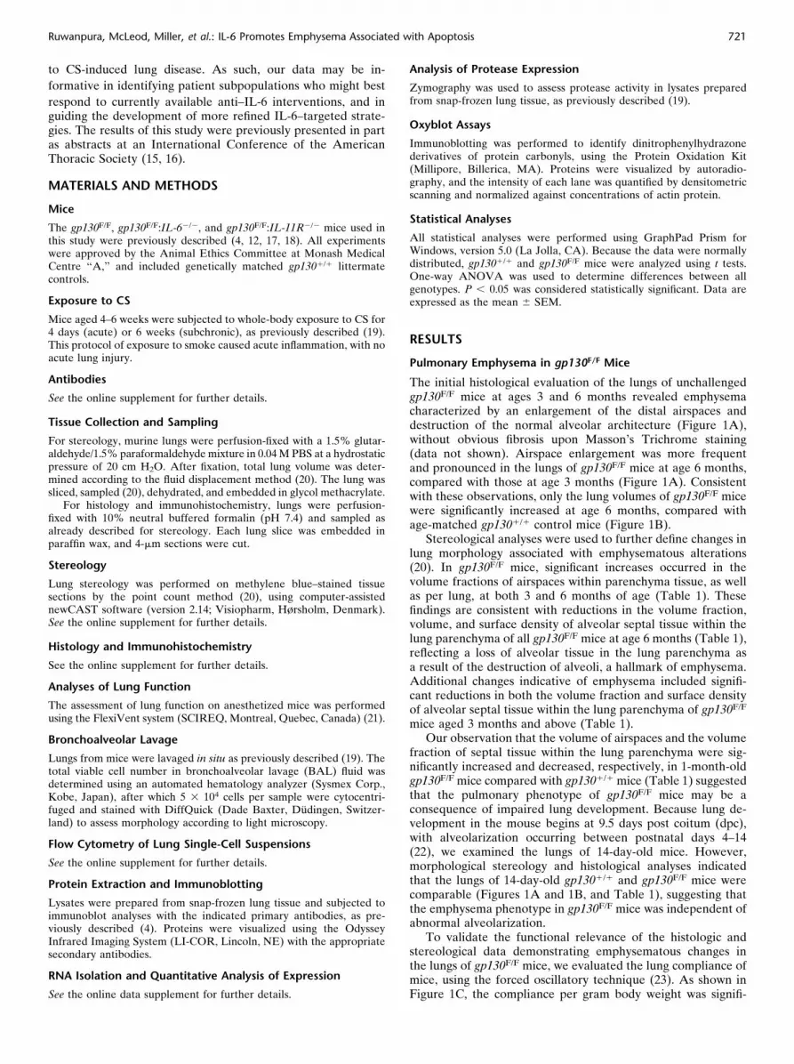
The initial histological evaluation of the lungs of unchallengedgp130F/F mice at ages 3 and 6 months revealed emphysemacharacterized by an enlargement of the distal airspaces anddestruction of the normal alveolar architecture (Figure 1A),without obvious fibrosis upon Masson’s Trichrome staining(data not shown). Airspace enlargement was more frequentand pronounced in the lungs of gp130F/F mice at age 6 months,compared with those at age 3 months (Figure 1A). Consistentwith these observations, only the lung volumes of gp130F/F micewere significantly increased at age 6 months, compared withage-matched gp1301/1 control mice (Figure 1B).
Stereological analyses were used to further define changes inlung morphology associated with emphysematous alterations(20). In gp130F/F mice, significant increases occurred in thevolume fractions of airspaces within parenchyma tissue, as wellas per lung, at both 3 and 6 months of age (Table 1). Thesefindings are consistent with reductions in the volume fraction,volume, and surface density of alveolar septal tissue within thelung parenchyma of all gp130F/F mice at age 6 months (Table 1),reflecting a loss of alveolar tissue in the lung parenchyma asa result of the destruction of alveoli, a hallmark of emphysema.Additional changes indicative of emphysema included signifi-cant reductions in both the volume fraction and surface densityof alveolar septal tissue within the lung parenchyma of gp130F/F
mice aged 3 months and above (Table 1).Our observation that the volume of airspaces and the volume
fraction of septal tissue within the lung parenchyma were sig-nificantly increased and decreased, respectively, in 1-month-oldgp130F/F mice compared with gp1301/1 mice (Table 1) suggestedthat the pulmonary phenotype of gp130F/F mice may be aconsequence of impaired lung development. Because lung de-velopment in the mouse begins at 9.5 days post coitum (dpc),with alveolarization occurring between postnatal days 4–14(22), we examined the lungs of 14-day-old mice. However,morphological stereology and histological analyses indicatedthat the lungs of 14-day-old gp1301/1 and gp130F/F mice werecomparable (Figures 1A and 1B, and Table 1), suggesting thatthe emphysema phenotype in gp130F/F mice was independent ofabnormal alveolarization.
To validate the functional relevance of the histologic andstereological data demonstrating emphysematous changes inthe lungs of gp130F/F mice, we evaluated the lung compliance ofmice, using the forced oscillatory technique (23). As shown inFigure 1C, the compliance per gram body weight was signifi-
Ruwanpura, McLeod, Miller, et al.: IL-6 Promotes Emphysema Associated with Apoptosis 721

cantly increased in both male and female gp130F/F mice at age 6months, compared with gp1301/1 mice. Collectively, these datademonstrate morphological and functional changes in the lungsof gp130F/F mice consistent with emphysema.
Cellular and Molecular Alterations Associated with Increased
Inflammation in the Lungs of gp130F/F Mice
The proinflammatory actions of the IL-6 cytokine family areimplicated in the pathogenesis of numerous inflammatorydisorders (5), and lung inflammation is evident in humanemphysema and in various animal models of emphysema (24,25). Consistent with these observations, the histological exam-ination of the lungs of gp130F/F mice also demonstrated thepresence of inflammatory cell foci from age 3 months onward(Figure 2A). Flow cytometry performed on whole-lung cellsuspensions further indicated that the proportion of CD451
immune cells in lungs from gp130F/F mice was significantlyhigher than in lungs from gp1301/1 mice (Figure 2B), andconsisted of a twofold increase in the frequency of B2201 Bcells (Figure 2C). Conversely, a slight decrease occurred in thefrequency of single positive CD41 and CD81 T cells in the
lungs of gp130F/F mice (Figure 2D). In contrast to these alte-rations to adaptive immune cell populations, the distribution oflung Gr11Mac11 and Mac11 myeloid (innate immune) cells ingp130F/F mice was normal (Figure 2E), which was consistentwith normal numbers of macrophages and neutrophils in theBAL fluid of gp130F/F mice (Table E1). Taken together, thesedata suggest a correlation between pulmonary inflammationand the onset of emphysema in the gp130F/F murine model.
Increased Apoptosis and Protease Activity, but Not Oxidative
Stress, in the Lungs of gp130F/F Mice
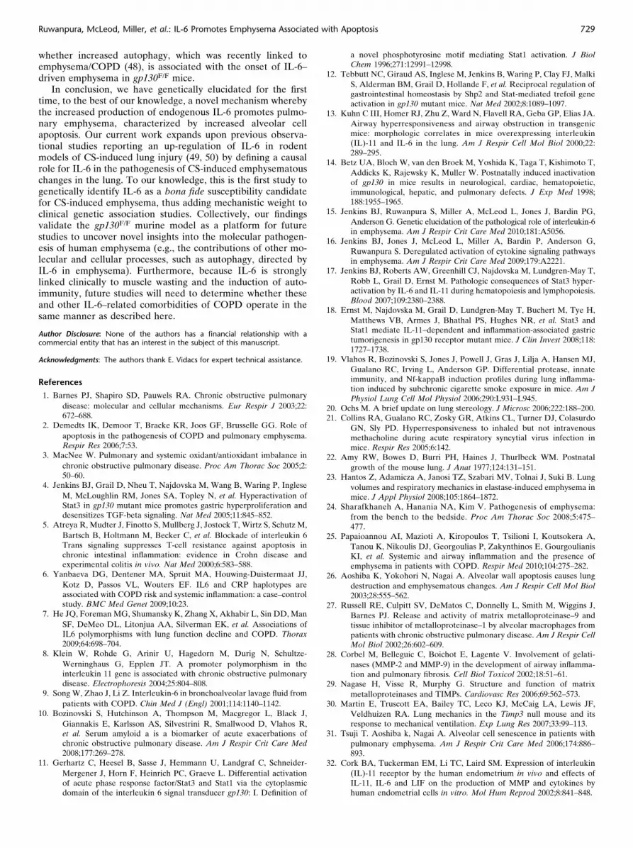
Because apoptosis is a key process associated with the de-velopment of emphysema in humans and in several animalmodels of the disease (2, 26), we investigated whether apoptosiswas increased in the lungs of gp130F/F mice by performingterminal deoxynucleotidyl transferase (TdT)-mediated dUTPnick end labeling (TUNEL) staining on lung sections. Increasednumbers of TUNEL-stained cells were evident in the lungs ofgp130F/F mice compared with gp1301/1 control mice at ages 3and 6 months, and alveolar septal cells comprised the predominantcell type affected (Figure 3A). Stereological quantification revealed
Figure 1. Alveolar airspace enlargement, increased lung
volume, and static compliance in gp130F/F mice. (A)
Representative methylene blue–stained cross sections of
lungs from gp1301/1 (1/1; top) and gp130F/F (F/F;bottom) mice aged 14 days (d) and 1, 3, and 6 months
(mo). Arrows indicate enlarged airspaces. Scale bars,
100 mm. (B) Lung volume per body weight of 1/1 andF/F mice at the indicated ages. ***P , 0.001, versus age-
matched 1/1 mice. (C ) Static compliance of 6-month-
old male and female 1/1 and F/F mice was assessed
according to FlexiVent analysis of lung function. Data areexpressed as mean 6 SEM, n 5 at least 3 mice per sex
or age per genotype. **P , 0.01, ***P , 0.001, versus
1/1 mice.
TABLE 1. COMPARATIVE STEREOLOGICAL ANALYSES OF LUNGS FROM GP1301/1 (1/1) AND GP130 F/F (F/F) MICE
14 Days 1 Month 3 Months 6 Months
1/1 F/F 1/1 F/F 1/1 F/F 1/1 F/F
Vv (par/lung) (%) 89.5 (6 1.9) 90.9 (6 3.7) 88.3 (6 1.5) 87.5 (6 1.3) 91.4 (6 1.1) 86.7 (6 1.8) 86.4 (6 0.7) 88.9* (6 1.2)
Vv (airsp/par) (%) 75.3 (6 3.0) 74.9 (6 2.5) 77.9 (6 3.9) 84.8 (6 0.8) 72.7 (6 1.3) 82.6** (6 1.1) 69.8 (6 1.3) 79.3*** (6 1.9)
V (airsp/lung) (cm3) 18.9 (6 2.5) 19.1 (6 6.6) 30.0 (6 5.1) 43.7* (6 2.0) 43.2 (6 2.9) 69.4* (6 8.7) 58.4 (6 3.1) 82.2* (6 2.6)
Vv (sep/par) (%) 21.2 (6 2.4) 20.8 (6 3.0) 18.0 (6 2.5) 12.1* (6 0.5) 21.5 (6 0.8) 14.3*** (6 1.0) 23.99 (6 1.1) 16.1*** (6 0.9)
V (sep/lung) (cm3) 5.5 (6 1.5) 4.8 (6 1.0) 7.1 (6 1.7) 6.2 (6 0.4) 12.9 (6 1.4) 12.0 (6 1.9) 20.2 (6 2.0) 16.7 (6 1.1)
Sv (sep/par) (1/cm) 446 (6 27) 438 (6 64) 508 (6 46) 431 (6 5) 679 (6 28) 492* (6 21) 639 (6 36) 538* (6 17)
S (sep/lung) (cm2) 115 (6 21) 115 (6 64) 200 (6 35) 222 (6 610) 407 (6 42) 421 (6 68) 535 (6 49) 560 (6 33)
Definition of abbreviations: airsp, airspace; par, parenchyma; S, surface area; sep, septal tissue; Sv, surface density; VV, volume fraction.
Data are expressed as mean (6 SEM). n 5 at least 3 mice per genotype for each age group. *P , 0.05, **P , 0.01, and ***P , 0.001 versus age-matched 1/1 mice.
722 AMERICAN JOURNAL OF RESPIRATORY CELL AND MOLECULAR BIOLOGY VOL 45 2011

that approximately 4.5% and 1% (P , 0.05) of cells undergoapoptosis in the lungs of gp130F/F mice at ages 3 and 6 months,respectively, whereas lungs from age-matched gp1301/1 micecontained few or no TUNEL-stained cells (Figure 3B).
Another cellular process linked to the destruction of alveolarseptal tissue in emphysema involves the excessive protease
activity often caused by the deregulated expression or activityof matrix metalloproteinases (MMPs) (1). Within this family ofproteolytic enzymes, the increased expression and activity ofthe gelatinases MMP-2 and MMP-9 were detected in the BALfluid and lung tissue of patients with COPD and emphysema(27, 28), and along with MMP-12, MMP-2 and MMP-9 were
Figure 2. Distribution of inflammatory cells in lungs of
gp130F/F mice. (A) Representative hematoxylin-and-eosin–
stained cross sections of lungs from 6-month-old gp1301/1
(1/1) and gp130F/F (F/F) mice. Arrows indicate patches
of inflammatory cell infiltrates. Scale bars, 100 mm. (B–E)
Whole-lung, single-cell suspensions from 6-month-old
1/1 and F/F mice were subjected to multicolor flowcytometric analysis with the antibodies indicated, and live
cells were gated by propidium iodide (PI) exclusion.
Representative histograms show (B) CD451 cells (solid
line; dotted line 5 unstained cells) and (C ) B2201 cells,the latter plotted as a proportion of CD451 cells. In
histograms, the x axis represents intensity of FITC, and
the y axis represents relative cell percentage or cellnumber. Representative dot plots show relative propor-
tions of (D) CD41 T-helper (lower gated region) or CD81 T
cytotoxic (upper gated region) cells, and of (E ) Mac11
(upper left gated region) or Mac11Gr11 (upper right gatedregion), as percentages of CD451 cells. For dot plots, the x
axis represents intensity of either FITC or Pacific blue, and
the y axis represents intensity of phycoerythrin (PE). Flow
cytometric profiles are representative of at least sevenmice per genotype.
Ruwanpura, McLeod, Miller, et al.: IL-6 Promotes Emphysema Associated with Apoptosis 723

reported to play a causal role in several murine models ofexperimentally induced emphysema (28, 29). As shown inFigure 3C, gelatinase zymography detected major bands atapproximately 90 kD and approximately 60 kD, correspondingto MMP-9 and MMP-2, respectively, in both genotypes withcomparable intensity at age 3 months. By contrast, increasedMMP-9 protease activity was detected only in 6-month-oldgp130F/F mice (Figure 3C). The augmented activity of gelatinasewas not a consequence of impaired expression of the negativeregulators of these proteases, namely, tissue inhibitor of MMP-1(Timp-1) and Timp-2 (29), because their mRNA levels wereelevated and unchanged, respectively, in the lungs of gp130F/F
mice (Figure 3D). Similarly, the expression of Timp-3, whichwas implicated in the pathogenesis of emphysema (30), was un-altered (Figure 3D). Rather, we observed significant increasesin concentrations of Mmp-9 and Mmp-2 mRNA at age 6 monthsin the lungs of gp130F/F mice compared with gp1301/1 mice(Figure 3D), consistent with the reported up-regulation of theseMMPs via the IL-6 cytokine family (28).
In recent years, it has emerged that oxidative stress can beassociated with inducing inflammation, excessive proteaseactivity, and apoptosis in the emphysematous lung, and in-creased oxidative stress was observed in a subset of patientswith emphysema/COPD and in animal models mimickingemphysema (3). Therefore, to determine whether increased
concentrations of oxidants were present in the lungs of gp130F/F
mice, we performed immunoblotting to identify the dinitrophe-nylhydrazone derivatives of protein carbonyls, a conventionalmarker of oxidative stress generated by reactive oxygen species.However, in gp130F/F mice, the formation of protein carbonylsshowed no qualitative or quantitative differences compared withgp1301/1 mice (Figure 3E), indicating that oxidative stress is notaffected by IL-6 family cytokines in the gp130F/F murine lung.
Premature alveolar cell senescence leading to acceleratedaging of the lung was proposed as another mechanism linkedto emphysema (31). To address this possibility in our gp130F/F
murine model, we measured the expression of the senescence-related marker b-galactosidase, which is increased duringsenescence. However, the immunoblotting of whole-lung lysatesindicated that concentrations of b-galactosidase in gp130F/F
murine lungs were unaltered compared with gp1301/1 mice(Figure E1). Collectively, our data suggest that increased in-flammation, apoptosis, and protease activity are features of IL-6family cytokine–driven spontaneous emphysema in gp130F/F
mice, all of which are shared traits with human disease.
Genetic Ablation of IL-6 in gp130F/F Mice Prevents the
Development of Emphysema
We previously reported that concentrations of IL-6 are elevatedin patients with COPD (10), and that the artificial overexpression
Figure 3. Increased pulmonary apoptosis,excessive protease activity, and comparable
oxidative stress in gp130F/F mice. (A) Repre-
sentative photomicrographs show TUNEL-
stained cells (arrows) in a cross section oflungs from gp1301/1 (1/1) and gp130F/F
(F/F) mice at ages 3 and 6 months. Scale
bars, 100 mm. (B) Stereological quantifica-tion of percentages of TUNEL-stained cells in
lungs of mice. Data are expressed as mean 6
SEM, n 5 at least 3 mice per genotype per
age group. *P , 0.05, versus age-matched1/1 mice. (C ) Zymography of gelatinase
activity was performed on lung-tissue lysates
from 1/1 and F/F mice aged 3 and 6
months. The 90-kD and 60-kD bands corre-spond to matrix metalloproteinase–9 (MMP-9)
and MMP-2, respectively. Each lane repre-
sents an individual murine lung lysate. (D)Quantitative PCR expression analyses of tis-
sue inhibitor of MMP-1 (Timp-1), Timp-2,
Timp-3, Mmp-9, and Mmp-2 were per-
formed on lung cDNA from 3-month-oldand 6-month-old mice of the indicated
genotypes. Expression data represent at
least n 5 4 per genotype per age group
after normalization for expression of 18S,and are presented after triplicate analyses as
mean 6 SEM. *P , 0.05, versus expression
in age-matched 1/1 mice. (E ) Levels ofprotein carbonylation were determined by
Oxyblots of lung-protein lysates from 3-
month-old and 6-month-old 1/1 and F/F
mice. Each lane represents tissue from anindividual mouse. Densitometric quantifica-
tion of total protein carbonylation in repre-
sentative samples per genotype was
performed and normalized against the levelsof actin protein in each sample. Data are
presented as mean fold induction 6 SEM for
at least n 5 4 per genotype per age group.
724 AMERICAN JOURNAL OF RESPIRATORY CELL AND MOLECULAR BIOLOGY VOL 45 2011

of IL-6 or IL-11 in the murine lung can induce emphysema-likeairspace enlargement (13). We therefore investigated whichendogenous IL-6 cytokine family members were involved inpromoting emphysema in gp130F/F mice. Among members ofthe IL-6 family of cytokines, only concentrations of Il-6 mRNAwere significantly elevated by approximately 8-fold and 18-foldat ages 3 and 6 months, respectively, in the lungs of gp130F/F
mice compared with gp1301/1 control mice (Figure 4A). Inaddition, the levels of gene expression for the ligand-binding a
subunits of IL-6 (Il-6ra) and IL-11 (Il-11ra), as well as thegp130 b subunit itself, in the lungs of gp130F/F mice were notsignificantly altered (Figure 4A).
Based on the augmented Il-6 gene expression observed inthe lungs of gp130F/F mice, we used a genetic approach toexplore a possible causal link between IL-6 and the emphysemaphenotype. Indeed, the histological evaluation of gp130F/F:IL-62/2 mice lacking IL-6 (17) revealed morphologically nor-mal alveolar architecture (Figure 4B) at age 6 months. Simi-larly, both the static lung compliance and lung volumes ofgp130F/F:IL-62/2 mice were restored to normal levels (Figure4C). Furthermore, the stereological assessment of gp130F/F:IL-62/2 murine lungs revealed that all stereological parameters,such as volume fractions of airspaces and alveolar septal tissueand surface density within the lung parenchyma, and the ab-solute volumes of lungs, were comparable to wild-type levels atage 6 months (Table E2).
The IL-6 cytokine family member IL-11, which like IL-6 usesa gp130 homodimer to transmit intracellular signals, has alsobeen implicated in the pathogenesis of emphysema (13). We
therefore genetically determined whether IL-11 also contrib-uted to the pathogenesis of emphysema in gp130F/F mice byusing gp130F/F:IL-11R2/2 mice in which the IL-11 receptor a
subunit had been genetically ablated (17), However, the lungsof compound mutant gp130F/F:IL-11R2/2 mice displayed em-physematous morphological changes (Figure 4B and Table E2),increased lung volumes, and altered respiratory mechanics(Figure 4C) comparable to those of gp130F/F mice. Collectively,these data identify an essential and specific role for endogenousIL-6 in the pathogenesis of emphysema in this model.
Increased Apoptosis Is the Primary Pathological Process
Associated with IL-6–Driven Emphysema in gp130F/F Mice
To investigate further the molecular and cellular mechanismsinvolved in the pathogenesis of IL-6–driven emphysema, weexamined whether any correlation existed between the extent ofpulmonary inflammation and emphysema among variousgp130F/F compound mutant mice. The histologic evaluation oflung sections from 6-month-old mice revealed that the focalinflammatory infiltrates characteristic of gp130F/F murine lungswere absent in both emphysema-free (gp130F/F:IL-62/2) andemphysematous (gp130F/F:IL-11R2/2) mice (Figure 5A). There-fore, these data clearly demonstrate that the onset of emphy-sema in gp130F/F mice is not reliant upon an obligatory pulmonaryproinflammatory microenvironment.
We next examined whether the excessive pulmonary gelat-inase activity of MMP-9 displayed by gp130F/F mice was alsoapparent in emphysematous gp130F/F:IL-11R2/2 mice, andwhether it was conversely diminished in emphysema-free
Figure 4. IL-6, but not IL-11, promotes emphysema in
gp130F/F mice. (A) Quantitative PCR expression analyses
of the indicated genes were performed on lung cDNA
from 3-month-old and 6-month-old gp1301/1 (1/1) andgp130F/F (F/F) mice. Expression data were normalized for
18S from at least n 5 4 mice per age and genotype, and
are presented as mean 6 SEM. *P , 0.05 versus expres-
sion in age-matched 1/1 mice. Lif 5 leukemia inhibitoryfactor, Osm 5 oncostatin-M. (B) Representative methy-
lene blue–stained cross sections of lungs from 1/1, F/F,
and compound mutant F/F mice lacking IL-6 (F/F:62/2)and IL-11Ra (F/F:11R2/2) and aged 6 months. Arrows
indicate enlarged airspaces. Scale bars, 100 mm. (C ) Static
compliance and lung volume of 6-month-old mice of the
indicated genotypes. Data represent n 5 5 per genotypeas the mean 6 SEM per body weight. *P , 0.05, **P ,
0.01, and ***P , 0.001, versus 1/1 mice.
Ruwanpura, McLeod, Miller, et al.: IL-6 Promotes Emphysema Associated with Apoptosis 725

gp130F/F:IL-62/2 mice. The gelatinase zymography of lunglysates from 6-month-old mice demonstrated that MMP-9 pro-tease activity was reduced to wild-type levels in both gp130F/F:IL-62/2 and gp130F/F:IL-11R2/2 murine lungs (Figure 5B).These observations correlated with decreased lung mRNA
concentrations for Mmp-9 in gp130F/F:IL-62/2 and gp130F/F:IL-11R2/2 mice compared with gp130F/F mice at age 6 months(Figure 5C), and are consistent with previous reports on theregulatory actions of IL-6 and IL-11 on Mmp-9 gene expression(32, 33). Nonetheless, these data do not demonstrate a corre-lation between augmented lung expression and the activity ofMMPs and gp130-driven emphysema.
To extend our data revealing that apoptosis was elevated inthe lungs of gp130F/F mice, we assessed the extent of apoptoticTUNEL-stained cells in the lungs of 6-month-old compoundmutant mice. Stereological quantification revealed that TUNELstaining in the lungs of emphysema-free gp130F/F:IL-62/2 micewas comparable to that in gp1301/1 mice at age 6 months(Figure 5D). By contrast, the percentage of TUNEL-stainedcells in the lungs of emphysematous gp130F/F:IL-11R2/2 miceremained significantly elevated compared with gp1301/1 mice(Figure 5D), demonstrating a positive correlation betweenincreased apoptosis and gp130-driven emphysema.
To investigate the molecular basis for the increased apopto-sis observed in the lungs of gp130F/F mice, we initially focusedon the vascular endothelial growth factor (VEGF) pathway,because the impaired expression of VEGF or downstreamsignaling components is associated with human COPD/emphy-sema and several animal disease models (2, 34). However, themRNA concentrations of Vegf as well as Vegfr1 and Vegfr2receptor subunits were unaltered (Figure 6A). We thereforeundertook a more global approach to identify changes in theexpression profiles of genes associated with increased pulmo-nary apoptosis in emphysematous lungs of gp130F/F mice byusing Mouse Apoptosis RT2 Profiler PCR Arrays (SABioscien-ces, Frederick, MD). Among 84 key genes involved in apopto-sis, five genes were down-regulated by at least twofold in thelungs of 3-month-old and 6-month-old gp130F/F mice comparedwith gp1301/1 mice (Table 2), whereas none of the genes in thearrays were consistently up-regulated. The five down-regulatedgenes were assigned a prosurvival/antiapoptotic function. Atf5and Trp63 were implicated in the activation of caspase-8 (35,36), Naip1 and Pak7 are death regulators associated with theintrinsic/mitochondria apoptotic pathway (37, 38), and Dapk1was also linked to autophagy, a process that can mediate eithercell survival or apoptosis (39). The altered expression profile ofthese genes was further verified in additional 3-month-old mice(n 5 4 per genotype), using subsequent arrays confirming thatthe expression of Atf5, Trp63, Naip1, Pak7, and Dapk1 wasreduced in gp130F/F mice compared with gp1301/1 control mice(Figure 6B).
Exposure to CS Exacerbates IL-6–Driven Emphysematous and
Apoptotic Changes in the Lungs of gp130F/F Mice
Our data thus far reveal that IL-6–driven emphysematouschanges in the lungs of gp130F/F mice correlate exclusively withincreased apoptosis. Considering that CS is the major trigger ofemphysema in humans, and that genetic susceptibility com-pounded by lung insult is the most common path to disease, weassessed whether our mechanistic findings for spontaneous IL-6–driven emphysema are relevant to CS-induced lung disease.Specifically, we exposed 4–6-week-old mice (before lung in-flammation in gp130F/F mice) to acute (4-day) and subchronic(6-week) CS models, to measure molecular and pathologicalchanges, respectively, related to apoptosis and emphysema inthe lungs of mice. Compared with mice not exposed to CS,acute exposure to CS in gp1301/1 and gp130F/F mice increasedIl-6 mRNA levels by 3-fold and 2-fold, respectively (Figure 7A).The response of both gp1301/1 and gp130F/F murine lungs toacute CS exposure was further confirmed by the CS-inducedincrease in whole-lung mRNA levels of Cxcl2, a neutrophil-
Figure 5. Increased apoptosis, but not inflammation and excessive
lung protease activity, correlated with emphysema. (A) Representative
hematoxylin-and-eosin–stained cross sections of lungs from 6-month-old
mice of the indicated genotypes. Arrows indicate patches of inflamma-tory cell infiltrates. Scale bars, 100 mm. (B) Zymography of gelatinase
activity was performed on lung-tissue lysates from 6-month-old mice of
the indicated genotypes. Each lane represents an individual mouse
lung lysate. (C ) Q-PCR analyses of Mmp-9 gene expression wereperformed on lung cDNA from 6-month-mice of the indicated geno-
types. Expression data from 3–5 samples per genotype are shown
following normalization for the expression of 18S, and are presentedfrom triplicate analysis as the mean 6 SEM. *P , 0.05 versus expression
in gp1301/1 mice. (D) Stereological quantification of percentages of
TUNEL-stained cells in lungs of mice from the indicated genotypes at
age 6 months. Data are expressed as mean 6 SEM, n 5 at least 3 miceper genotype per age group. *P , 0.05, versus age-matched 1/1
mice.
726 AMERICAN JOURNAL OF RESPIRATORY CELL AND MOLECULAR BIOLOGY VOL 45 2011

attracting chemokine that is up-regulated during CS-inducedlung inflammation (40) (Figure 7A). By contrast, mRNA levelsof the five antiapoptotic markers Atf5, Trp63, Naip1 (P , 0.05),Pak7 (P , 0.05), and Dapk1 were reduced in gp130F/F miceexposed to CS, compared with non-CS–exposed littermates(Figure 7A).
In response to subchronic exposure to CS, we observedsignificant increases in the lung volumes of both gp1301/1
(23%) and gp130F/F (37%) mice, consistent with emphyse-matous lung changes, compared with their age-matched, non-CS–exposed counterparts (Figure 7B). Moreover, the lungvolumes of CS-exposed and non-CS–exposed gp130F/F:IL-62/2
mice were indistinguishable (Figure 7B), indicating that gp130F/F:IL-62/2 mice are protected against CS-induced, emphysema-associated increases in lung volumes. These findings correlatedwith our data demonstrating that exposure to CS increasedTUNEL staining in the lungs of gp130F/F mice (albeit modestly,considering the already high level of TUNEL-stained cells innon-CS–exposed mice) and gp1301/1 mice, but not gp130F/F:IL-62/2 mice, compared with their non-CS–exposed counter-parts (Figure 6C). Collectively, these data validate the rele-vance of our IL-6–driven gp130F/F emphysematous murinemodel to the pathogenesis of CS-induced emphysema.
DISCUSSION
To the best of our knowledge, we provide the first definitive,genetic proof that the exaggerated, endogenous production ofIL-6 in the lungs causes emphysema. We also reveal thatincreased pulmonary apoptosis is a consequence of IL-6 up-regulation in the lung, and represents the most likely processinvolved in the pathogenesis of IL-6–driven emphysema. Fur-thermore, we demonstrate redundancy among IL-6 family cy-
tokines (namely, IL-6 and IL-11) in mediating pulmonaryinflammation and excessive protease activity, two processesthat are not related to the pathogenesis of emphysema in thegp130F/F murine model. We also demonstrate a key role for IL-6in promoting alveolar cell death and emphysematous changes inthe lungs of mice upon exposure to CS, which not only validatesour mechanistic findings in terms of CS-induced human lungdisease, but also provides insights into the gene–environmentinteractions that cause lung disease. Together with our identifi-cation of a proapoptotic signature associated with emphysema-tous changes, our results have considerable potential translationalsignificance in providing both a predictor and a biomarker ofsubpopulations with emphysema and COPD that may gain themost benefit from anti–IL-6–directed therapies. This is partic-ularly important because antibodies directed against IL-6 andits receptor have been introduced into clinical medicine forother inflammatory diseases (5, 41, 42), but have not yet beenapplied to COPD or emphysema.
The evidence for a role of IL-6 in the pathogenesis of humanemphysema/COPD is now very strong. For instance, an in-creased production of IL-6 is evident in patients with COPDand emphysema (9, 10), and IL-6 gene polymorphisms arelinked to the rapid decline in lung function and susceptibility toCOPD in smokers (7). Despite these key observations in humandisease, the advancement in our understanding of how IL-6contributes to emphysema has been severely hampered by thelack of informative animal models to genetically elucidate thedownstream molecular and cellular events regulated by IL-6 inthe pathogenesis of disease. In this respect, the artificial over-expression of IL-6 and IL-11 in the lungs of transgenic micecauses alveolar airspace enlargement, accompanied by mono-nuclear cell inflammatory infiltrates (13). Although the mech-anisms by which the overexpression of IL-6 and IL-11 causedalveolar enlargement in these models were not clear, thesemechanisms may involve a developmental defect most likelyrepresenting alveolar hypoplasia because of the impairedalveolarization of the developing lung (13). By contrast, ourpresent results reveal that emphysema driven by endogenousIL-6, but not IL-11, is unlikely to be related to a defect in thedeveloping lung, because emphysematous changes in the lungsof gp130F/F mice were apparent only from age 1 month onward,after the completion of alveolarization. The most likely reasonfor these discrepancies involves the artificial temporal andkinetic expression of IL-6 and IL-11 in transgenic mice com-pared with the expression of these cytokines from their endog-enous promoters in gp130F/F mice. Nonetheless, our findingsthat the expressions of IL-6 and IL-11, both of which signal viaa gp130 homodimer and use the same signaling machinery, aredifferentially regulated in the lung are reminiscent of ourprevious observations that IL-11, but not IL-6, is up-regulatedin the stomach of mice in a Stat3-dependent manner thatpromotes gastric disease (4, 18). The expression of IL-27, whichto our knowledge has not been linked to the pathogenesis of
Figure 6. Expression profiles of antiapoptotic genes in lungs of
emphysematous gp130F/F mice. (A) Quantitative PCR expression anal-
yses of Vegf, Vegfr1, and Vegfr2 were performed on lung cDNA from 3-
month-old and 6-month-old gp1301/1 (1/1) and gp130F/F (F/F) mice.VEGF, vascular endothelial growth factor. Expression data are shown
for at least n 5 4 per genotype per age group after normalization for
the expression of 18S, and represent triplicate analyses as mean 6 SEM.
(B) Expression profiles of antiapoptotic genes Atf5, Trp63, Naip1, Pak7,and Dapk1 in lungs of 3-month-old 1/1 and F/F mice, using Mouse
Apoptosis RT2 Profiler PCR Arrays. *P , 0.05, versus expression in 1/1
mice.
TABLE 2. DOWN-REGULATED ANTIAPOPTOTIC GENES IN THELUNGS OF GP1301/1 (1/1) AND GP130 F/F (F/F) MICE
Fold Difference (F/F versus 1/1)
Gene Name 3 Months 6 Months Protein Function
Atf5 0.54 0.16 Antiapoptotic
Trp63 0.13 0.03 Antiapoptotic
Naip1 0.15 0.21 Antiapoptotic
Pak7 0.34 0.15 Antiapoptotic
Dapk1 0.12 0.06 Antiapoptotic
Data are expressed for n 5 2 mice per genotype for each age group.
Ruwanpura, McLeod, Miller, et al.: IL-6 Promotes Emphysema Associated with Apoptosis 727

emphysema, was also increased (although not significantly) ingp130F/F murine lungs. These observations highlight differencesin the tissue-specific regulation of these gp130-acting cytokinesthat underlie their differential involvement in promotingdisease, and that warrant further investigation into the tran-scriptional activators of their gene promoters in the lung.Furthermore, a potential role for IL-27 in promoting gp130-driven emphysema is under investigation in gp130F/F micedeficient in the IL-27 receptor, WSX-1.
Although these transgenic models imply a ‘‘gain-of-function’’gp130 signaling phenotype, the (partial) conditional deletion ofgp130 (i.e., ‘‘loss-of-function’’) postnatally in the lungs of micecan also trigger alveolar airspace enlargement in aging mice,albeit by unknown mechanisms (14). Accordingly, these murinemodels suggest the existence of alternate pathological gp130signaling pathways, which upon their increased (transgenic) orreduced (conditional knockout) activation promote alveolarairspace enlargement. This notion is consistent with the aggre-gate of human studies that have not revealed a single signalingpathway to disease, but that suggest a host of different pathwayscontributing to emphysema in different patient subgroups. Inthis regard, the fact that each of the discrete gp130 pathways isactivated by different parts of the gp130 coreceptor impliesthe feasibility of inactivating selected pathways in vivo. Indeed,the combination of such genetic interventions with conventionalgene-targeting (genetic complementation) in the gp130F/F
murine model has the enormous potential to identify down-
stream IL-6/gp130 signaling mediators associated with emphy-sema. Although the identity of such mediators remainsunknown, one such candidate may be Stat3, which was impli-cated in emphysema, albeit by conflicting reports, throughthe use of murine models artificially engineered to either sup-press or augment Stat3 activity in the respiratory epithelium(43, 44).
Our study produced the key novel finding that IL-6–drivenpulmonary emphysema is associated with increased apoptosis.Based on recent evidence that increased apoptosis contributesto the pathogenesis of emphysema, distinct apoptotic mecha-nisms are thought to exist in the lung. For instance, blocking theVEGF pathway in mice promotes inflammation-independent,apoptosis-associated emphysema (45), similar to the emphy-sema characterized by increased apoptosis yet independent ofpulmonary inflammation in gp130F/F mice. Although the ex-pression of VEGF signaling pathway components was unalteredin gp130F/F mice, we demonstrated that a subset of antiapop-totic genes assigned to the caspase-8 or caspase-9 apoptoticpathways, and autophagy, are down-regulated in IL-6–drivenemphysema. Among these genes, Atf5, Trp63, and Dapk1 werelinked to the development and progression of lung cancer (39,46, 47). However, their involvement in emphysema remainsunknown. Moreover, the expression of Pak7 and Naip1 in thelung was not reported previously, to the best of our knowledge.Clearly, further studies are needed to clarify not only the role ofPak7 and Naip1 in apoptotic pathways, but to investigate
Figure 7. Altered gene expression, lung volume, and
apoptosis in lungs of mice after 4 days or 6 weeks of
exposure to cigarette smoke (CS). (A) Quantitative PCRexpression analyses of the indicated genes were per-
formed on lung cDNA from 1-month-old gp1301/1
(1/1) and gp130F/F (F/F) mice with (1) or without (2)
exposure to CS. Expression data from at least n 5 4 pergenotype per treatment group after normalization against
their unexposed littermates are shown, and represent
triplicate analyses as the mean 6 SEM. *P , 0.05, versusexpression in genotype-matched mice without exposure
to CS. (B) Lung volume and (C ) quantification of the
percentage of TUNEL-stained cells from the lungs of mice
of the indicated genotypes at age 3 months were assessedafter 6 weeks of exposure to CS. Data are expressed as
mean 6 SEM; n 5 at least 3 mice per genotype. *P ,
0.05, versus expression in genotype-matched, non-CS–
exposed mice.
728 AMERICAN JOURNAL OF RESPIRATORY CELL AND MOLECULAR BIOLOGY VOL 45 2011
whether increased autophagy, which was recently linked toemphysema/COPD (48), is associated with the onset of IL-6–driven emphysema in gp130F/F mice.
In conclusion, we have genetically elucidated for the firsttime, to the best of our knowledge, a novel mechanism wherebythe increased production of endogenous IL-6 promotes pulmo-nary emphysema, characterized by increased alveolar cellapoptosis. Our current work expands upon previous observa-tional studies reporting an up-regulation of IL-6 in rodentmodels of CS-induced lung injury (49, 50) by defining a causalrole for IL-6 in the pathogenesis of CS-induced emphysematouschanges in the lung. To our knowledge, this is the first study togenetically identify IL-6 as a bona fide susceptibility candidatefor CS-induced emphysema, thus adding mechanistic weight toclinical genetic association studies. Collectively, our findingsvalidate the gp130F/F murine model as a platform for futurestudies to uncover novel insights into the molecular pathogen-esis of human emphysema (e.g., the contributions of other mo-lecular and cellular processes, such as autophagy, directed byIL-6 in emphysema). Furthermore, because IL-6 is stronglylinked clinically to muscle wasting and the induction of auto-immunity, future studies will need to determine whether theseand other IL-6–related comorbidities of COPD operate in thesame manner as described here.
Author Disclosure: None of the authors has a financial relationship with acommercial entity that has an interest in the subject of this manuscript.
Acknowledgments: The authors thank E. Vidacs for expert technical assistance.
References
1. Barnes PJ, Shapiro SD, Pauwels RA. Chronic obstructive pulmonary
disease: molecular and cellular mechanisms. Eur Respir J 2003;22:672–688.
2. Demedts IK, Demoor T, Bracke KR, Joos GF, Brusselle GG. Role of
apoptosis in the pathogenesis of COPD and pulmonary emphysema.Respir Res 2006;7:53.
3. MacNee W. Pulmonary and systemic oxidant/antioxidant imbalance in
chronic obstructive pulmonary disease. Proc Am Thorac Soc 2005;2:50–60.
4. Jenkins BJ, Grail D, Nheu T, Najdovska M, Wang B, Waring P, Inglese
M, McLoughlin RM, Jones SA, Topley N, et al. Hyperactivation ofStat3 in gp130 mutant mice promotes gastric hyperproliferation anddesensitizes TGF-beta signaling. Nat Med 2005;11:845–852.
5. Atreya R, Mudter J, Finotto S, Mullberg J, Jostock T, Wirtz S, Schutz M,
Bartsch B, Holtmann M, Becker C, et al. Blockade of interleukin 6Trans signaling suppresses T-cell resistance against apoptosis inchronic intestinal inflammation: evidence in Crohn disease andexperimental colitis in vivo. Nat Med 2000;6:583–588.
6. Yanbaeva DG, Dentener MA, Spruit MA, Houwing-Duistermaat JJ,
Kotz D, Passos VL, Wouters EF. IL6 and CRP haplotypes areassociated with COPD risk and systemic inflammation: a case–controlstudy. BMC Med Genet 2009;10:23.
7. He JQ, Foreman MG, Shumansky K, Zhang X, Akhabir L, Sin DD, Man
SF, DeMeo DL, Litonjua AA, Silverman EK, et al. Associations ofIL6 polymorphisms with lung function decline and COPD. Thorax2009;64:698–704.
8. Klein W, Rohde G, Arinir U, Hagedorn M, Durig N, Schultze-
Werninghaus G, Epplen JT. A promoter polymorphism in theinterleukin 11 gene is associated with chronic obstructive pulmonarydisease. Electrophoresis 2004;25:804–808.
9. Song W, Zhao J, Li Z. Interleukin-6 in bronchoalveolar lavage fluid from
patients with COPD. Chin Med J (Engl) 2001;114:1140–1142.10. Bozinovski S, Hutchinson A, Thompson M, Macgregor L, Black J,
Giannakis E, Karlsson AS, Silvestrini R, Smallwood D, Vlahos R,et al. Serum amyloid a is a biomarker of acute exacerbations ofchronic obstructive pulmonary disease. Am J Respir Crit Care Med2008;177:269–278.
11. Gerhartz C, Heesel B, Sasse J, Hemmann U, Landgraf C, Schneider-
Mergener J, Horn F, Heinrich PC, Graeve L. Differential activationof acute phase response factor/Stat3 and Stat1 via the cytoplasmicdomain of the interleukin 6 signal transducer gp130: I. Definition of
a novel phosphotyrosine motif mediating Stat1 activation. J BiolChem 1996;271:12991–12998.
12. Tebbutt NC, Giraud AS, Inglese M, Jenkins B, Waring P, Clay FJ, Malki
S, Alderman BM, Grail D, Hollande F, et al. Reciprocal regulation ofgastrointestinal homeostasis by Shp2 and Stat-mediated trefoil geneactivation in gp130 mutant mice. Nat Med 2002;8:1089–1097.
13. Kuhn C III, Homer RJ, Zhu Z, Ward N, Flavell RA, Geba GP, Elias JA.
Airway hyperresponsiveness and airway obstruction in transgenicmice: morphologic correlates in mice overexpressing interleukin(IL)-11 and IL-6 in the lung. Am J Respir Cell Mol Biol 2000;22:289–295.
14. Betz UA, Bloch W, van den Broek M, Yoshida K, Taga T, Kishimoto T,
Addicks K, Rajewsky K, Muller W. Postnatally induced inactivationof gp130 in mice results in neurological, cardiac, hematopoietic,immunological, hepatic, and pulmonary defects. J Exp Med 1998;188:1955–1965.
15. Jenkins BJ, Ruwanpura S, Miller A, McLeod L, Jones J, Bardin PG,
Anderson G. Genetic elucidation of the pathological role of interleukin-6in emphysema. Am J Respir Crit Care Med 2010;181:A5056.
16. Jenkins BJ, Jones J, McLeod L, Miller A, Bardin P, Anderson G,
Ruwanpura S. Deregulated activation of cytokine signaling pathwaysin emphysema. Am J Respir Crit Care Med 2009;179:A2221.
17. Jenkins BJ, Roberts AW, Greenhill CJ, Najdovska M, Lundgren-May T,
Robb L, Grail D, Ernst M. Pathologic consequences of Stat3 hyper-activation by IL-6 and IL-11 during hematopoiesis and lymphopoiesis.Blood 2007;109:2380–2388.
18. Ernst M, Najdovska M, Grail D, Lundgren-May T, Buchert M, Tye H,
Matthews VB, Armes J, Bhathal PS, Hughes NR, et al. Stat3 andStat1 mediate IL-11–dependent and inflammation-associated gastrictumorigenesis in gp130 receptor mutant mice. J Clin Invest 2008;118:1727–1738.
19. Vlahos R, Bozinovski S, Jones J, Powell J, Gras J, Lilja A, Hansen MJ,
Gualano RC, Irving L, Anderson GP. Differential protease, innateimmunity, and Nf-kappaB induction profiles during lung inflamma-tion induced by subchronic cigarette smoke exposure in mice. Am JPhysiol Lung Cell Mol Physiol 2006;290:L931–L945.
20. Ochs M. A brief update on lung stereology. J Microsc 2006;222:188–200.21. Collins RA, Gualano RC, Zosky GR, Atkins CL, Turner DJ, Colasurdo
GN, Sly PD. Hyperresponsiveness to inhaled but not intravenousmethacholine during acute respiratory syncytial virus infection inmice. Respir Res 2005;6:142.
22. Amy RW, Bowes D, Burri PH, Haines J, Thurlbeck WM. Postnatal
growth of the mouse lung. J Anat 1977;124:131–151.23. Hantos Z, Adamicza A, Janosi TZ, Szabari MV, Tolnai J, Suki B. Lung
volumes and respiratory mechanics in elastase-induced emphysema inmice. J Appl Physiol 2008;105:1864–1872.
24. Sharafkhaneh A, Hanania NA, Kim V. Pathogenesis of emphysema:
from the bench to the bedside. Proc Am Thorac Soc 2008;5:475–477.
25. Papaioannou AI, Mazioti A, Kiropoulos T, Tsilioni I, Koutsokera A,
Tanou K, Nikoulis DJ, Georgoulias P, Zakynthinos E, GourgoulianisKI, et al. Systemic and airway inflammation and the presence ofemphysema in patients with COPD. Respir Med 2010;104:275–282.
26. Aoshiba K, Yokohori N, Nagai A. Alveolar wall apoptosis causes lung
destruction and emphysematous changes. Am J Respir Cell Mol Biol2003;28:555–562.
27. Russell RE, Culpitt SV, DeMatos C, Donnelly L, Smith M, Wiggins J,
Barnes PJ. Release and activity of matrix metalloproteinase–9 andtissue inhibitor of metalloproteinase–1 by alveolar macrophages frompatients with chronic obstructive pulmonary disease. Am J Respir CellMol Biol 2002;26:602–609.
28. Corbel M, Belleguic C, Boichot E, Lagente V. Involvement of gelati-
nases (MMP-2 and MMP-9) in the development of airway inflamma-tion and pulmonary fibrosis. Cell Biol Toxicol 2002;18:51–61.
29. Nagase H, Visse R, Murphy G. Structure and function of matrix
metalloproteinases and TIMPs. Cardiovasc Res 2006;69:562–573.30. Martin E, Truscott EA, Bailey TC, Leco KJ, McCaig LA, Lewis JF,
Veldhuizen RA. Lung mechanics in the Timp3 null mouse and itsresponse to mechanical ventilation. Exp Lung Res 2007;33:99–113.
31. Tsuji T. Aoshiba k, Nagai A. Alveolar cell senescence in patients with
pulmonary emphysema. Am J Respir Crit Care Med 2006;174:886–893.
32. Cork BA, Tuckerman EM, Li TC, Laird SM. Expression of interleukin
(IL)-11 receptor by the human endometrium in vivo and effects ofIL-11, IL-6 and LIF on the production of MMP and cytokines byhuman endometrial cells in vitro. Mol Hum Reprod 2002;8:841–848.
Ruwanpura, McLeod, Miller, et al.: IL-6 Promotes Emphysema Associated with Apoptosis 729

33. Chen Q, Rabach L, Noble P, Zheng T, Lee CG, Homer RJ, Elias JA.IL-11 receptor alpha in the pathogenesis of IL-13–induced inflamma-tion and remodeling. J Immunol 2005;174:2305–2313.
34. Suzuki M, Betsuyaku T, Nagai K, Fuke S, Nasuhara Y, Kaga K, Kondo S,Hamamura I, Hata J, Takahashi H, et al. Decreased airway expression ofvascular endothelial growth factor in cigarette smoke–induced emphy-sema in mice and COPD patients. Inhal Toxicol 2008;20:349–359.
35. Persengiev SP, Green MR. The role of ATF/CREB family members incell growth, survival and apoptosis. Apoptosis 2003;8:225–228.
36. Borrelli S, Candi E, Alotto D, Castagnoli C, Melino G, Vigano MA,Mantovani R. P63 regulates the caspase-8–flip apoptotic pathway inepidermis. Cell Death Differ 2009;16:253–263.
37. Cotteret S, Jaffer ZM, Beeser A, Chernoff J. P21-activated kinase 5(PAK5) localizes to mitochondria and inhibits apoptosis by phos-phorylating bad. Mol Cell Biol 2003;23:5526–5539.
38. Maier JK, Lahoua Z, Gendron NH, Fetni R, Johnston A, Davoodi J,Rasper D, Roy S, Slack RS, Nicholson DW, et al. The neuronalapoptosis inhibitory protein is a direct inhibitor of caspases 3 and 7.J Neurosci 2002;22:2035–2043.
39. Gade P, Singh AK, Roy SK, Reddy SP, Kalvakolanu DV. Down-regulation of the transcriptional mediator subunit MED1 contributesto the loss of expression of metastasis-associated DAPK1 in humancancers and cancer cells. Int J Cancer 2009;125:1566–1574.
40. Thatcher TH, McHugh NA, Egan RW, Chapman RW, Hey JA, TurnerCK, Redonnet MR, Seweryniak KE, Sime PJ, Phipps RP. Role ofCXCR2 in cigarette smoke–induced lung inflammation. Am J PhysiolLung Cell Mol Physiol 2005;289:322–328.
41. Yokota S, Imagawa T, Mori M, Miyamae T, Aihara Y, Takei S, Iwata N,Umebayashi H, Murata T, Miyoshi M, et al. Efficacy and safety oftocilizumab in patients with systemic-onset juvenile idiopathic arthri-tis: a randomised, double-blind, placebo-controlled, withdrawal PhaseIII trial. Lancet 2008;371:998–1006.
42. Smolen J, Beaulieu A, Rubbert-Roth A, Ramos-Remus C, Rovensky J,Alecock E, Woodworth T, Alten R. OPTION Investigators: effect of
interleukin-6 receptor inhibition with tocilizumab in patients withrheumatoid arthritis (OPTION Study): a double-blind, placebo-controlled, randomised trial. Lancet 2008;371:987–997.
43. Kida H, Mucenski ML, Thitoff AR, Le Cras TD, Park KS, Ikegami M,Muller W, Whitsett JA. Gp130–Stat3 regulates epithelial cell migra-tion and is required for repair of the bronchiolar epithelium. Am JPathol 2008;172:1542–1554.
44. Lian X, Qin Y, Hossain SA, Yang L, White A, Xu H, Shipley JM, Li T,Senior RM, Du H, et al. Overexpression of Stat3C in pulmonaryepithelium protects against hyperoxic lung injury. J Immunol 2005;174:7250–7256.
45. Tang K, Rossiter HB, Wagner PD, Breen EC. Lung-targeted VEGFinactivation leads to an emphysema phenotype in mice. J ApplPhysiol 2004;97:1559–1566.
46. Fernandez P, Carretero J, Medina PP, Jimenez AI, Rodriguez-Perales S,Paz MF, Cigudosa JC, Esteller M, Lombardia L, Morente M, et al.Distinctive gene expression of human lung adenocarcinomas carryingLKB1 mutations. Oncogene 2004;23:5084–5091.
47. Koster MI, Lu SL, White LD, Wang XJ, Roop DR. Reactivation ofdevelopmentally expressed p63 isoforms predisposes to tumor de-velopment and progression. Cancer Res 2006;66:3981–3986.
48. Chen ZH, Kim HP, Sciurba FC, Lee SJ, Feghali-Bostwick C, Stolz DB,Dhir R, Landreneau RJ, Schuchert MJ, Yousem SA, et al. EGR-1regulates autophagy in cigarette smoke–induced chronic obstructivepulmonary disease. PLoS ONE 2008;3:e3316.
49. Doz E, Noulin N, Boichot E, Guenon I, Fick L, Le Bert M, Lagente V,Ryffel B, Schnyder B, Quesniaux VF, et al. Cigarette smoke–inducedpulmonary inflammation is TLR4/MYD88 and IL-1R1/MYD88 sig-naling dependent. J Immunol 2008;180:1169–1178.
50. Halappanavar S, Russell M, Stampfli MR, Williams A, Yauk CL.Induction of the interleukin 6/signal transducer and activatorof transcription pathway in the lungs of mice sub-chronicallyexposed to mainstream tobacco smoke. BMC Med Genomics2009;21:56.
730 AMERICAN JOURNAL OF RESPIRATORY CELL AND MOLECULAR BIOLOGY VOL 45 2011


