Embed Size (px)
Citation preview
Interleukin-10 Promotes Pathological Angiogenesis byRegulating Macrophage Response to Hypoxia duringDevelopmentDru S. Dace1, Aslam A. Khan1, Jennifer Kelly1, Rajendra S. Apte1,2*
1 Department of Ophthalmology and Visual Sciences, Washington University School of Medicine, St. Louis, Missouri, United States of America, 2 Developmental Biology
Program, Washington University School of Medicine, St. Louis, Missouri, United States of America
Abstract
Aberrant angiogenesis in the eye is the most common cause of blindness. The current study examined the role ofinterleukin-10 (IL-10) in ischemia-induced pathological angiogenesis called neovascularization during postnataldevelopment. IL-10 deficiency resulted in significantly reduced pathological retinal angiogenesis. In contrast to thechoroicapillaris where IL-10 interferes with macrophage influx, IL-10 did not prevent anti-angiogenic macrophages frommigrating to the retina in response to hypoxia. Instead, IL-10 promoted retinal angiogenesis by altering macrophageangiogenic function, as macrophages from wild-type mice demonstrated increased vascular endothelial growth factor(VEGF) and nitric oxide (NO) compared to IL-10 deficient macrophages. IL-10 appears to directly affect macrophageresponsiveness to hypoxia, as macrophages responded to hypoxia with increased levels of IL-10 and STAT3 phosphorylationas opposed to IL-10 deficient macrophages. Also, IL-10 deficient macrophages inhibited the proliferation of vascularendothelial cells in response to hypoxia while wild-type macrophages failed to do so. These findings suggest that hypoxiaguides macrophage behavior to a pro-angiogenic phenotype via IL-10 activated pathways.
Citation: Dace DS, Khan AA, Kelly J, Apte RS (2008) Interleukin-10 Promotes Pathological Angiogenesis by Regulating Macrophage Response to Hypoxia duringDevelopment. PLoS ONE 3(10): e3381. doi:10.1371/journal.pone.0003381
Editor: Patricia Bozza, Instituto Oswaldo Cruz and FIOCRUZ, Brazil
Received June 17, 2008; Accepted September 17, 2008; Published October 13, 2008
Copyright: � 2008 Dace et al. This is an open-access article distributed under the terms of the Creative Commons Attribution License, which permitsunrestricted use, distribution, and reproduction in any medium, provided the original author and source are credited.
Funding: NIH Grant K08EY016139 (R.S.A.), Carl Marshall Reeves and Mildred Almen Reeves Foundation Inc. Award (R.S.A.), RPB Career Development Award(R.S.A.), American Federation for Aging Research Grant (R.S.A.), The Knights Templar Eye Foundation, Inc. (D.S.D.) and the International Retina ResearchFoundation Alston Callahan Scholar Award (D.S.D)
Competing Interests: The authors have declared that no competing interests exist.
* E-mail: [email protected]
Introduction
Angiogenesis is a critical process in maintaining tissue
homeostasis during numerous physiological functions, such as
wound-healing, reproduction, and embryonic development. How-
ever, unbridled angiogenesis can result in fulminant host disease.
Abnormal angiogenesis is critical to the pathophysiology of diverse
disease processes such as atherosclerotic heart disease and several
cancers [1,2,3].
In the eye, this becomes especially important as abnormal
angiogenesis (neovascularization) leads to blindness in several
disease processes. Intraocular neovascularization, as characterized
by abnormal retinal or choroidal angiogenesis, is a major cause of
decreased vision in patients with diseases such as proliferative
diabetic retinopathy (PDR): the leading cause of blindness in
working adults, age-related macular degeneration (AMD): the
leading cause of blindness in the elderly, and retinopathy of
prematurity (ROP): the leading cause of blindness in premature
infants [4,5]. In diabetic retinopathy, retinal neovascularization
occurs in up to 20% of patients with diabetes [6]. Current laser
ablation treatment for PDR has changed little over the 50 years
since its first inception, and is applied only after onset of
neovascularization. Although it reduces the risk of severe vision
loss, laser photocoagulation reduces peripheral and night vision,
and is uncomfortable and expensive [7]. There is recent evidence
that the pathobiology of PDR is more complex. Immunological
mechanisms, including exudation, upregulation of inflammatory
mediators, and immune cell infiltration have been implicated in
PDR [8]. Retinopathy of prematurity blinds 50,000 newborn
babies annually worldwide. Peripheral retinal ischemia and the
cessation of normal retinal vessel growth leads to compensatory
angiogenesis, tractional retinal detachment, and blindness. Al-
though diseases resulting in ocular neovascularization differ in
many aspects, it is believed that tissue ischemia is the underlying
cause leading to compensatory angiogenesis. Tissue ischemia can
also result in cellular inflammation, including the infiltration of
macrophages to the site of ischemia.
Macrophages carry out a wide variety of biological functions,
including participation in neovascularization [9]. Macrophages can
exhibit both pro-angiogenic and anti-angiogenic functions. This
dual function of macrophages seems to be largely dependent upon
the polarization of macrophages. Polarization, in turn, seems to be
regulated by the production of cytokines in the resident tissue micro-
milieu [10,11,12,13]. Macrophages stimulated in the presence of
interferon gamma (IFN-c), lipopolysaccharide (LPS), or granulocyte
macrophage colony-stimulating factor (GM-CSF) produce high
levels IL-12, IL-23, IL-6, and tumor necrosis factor alpha (TNF-a),
and low levels of IL-10. This ‘‘classically-activated’’ macrophage, or
M1 macrophage, displays an anti-angiogenic phenotype, and plays
an important role in anti-bacterial and pro-inflammatory functions.
Macrophages stimulated in the presence of IL-10, IL-4, or IL-13
produce high levels of IL-10 and low levels of pro-inflammatory
PLoS ONE | www.plosone.org 1 October 2008 | Volume 3 | Issue 10 | e3381
cytokines such as IL-6 and TNF-a. These ‘‘alternatively-activated’’
macrophages, or M2 macrophages, are pro-angiogenic. Of these
cytokines, IL-10 may possess the most significant influence on the
polarization of macrophages and their ability to regulate angiogen-
esis in the eye [10,14].
AMD is a disease of the elderly characterized by blindness that
is secondary to post-developmental choroidal angiogenesis.
Termed choroidal neovascularization (CNV), this aberrant ocular
angiogenesis develops in senescent tissues. In a mouse model of
CNV, it has been shown that IL-10 promotes CNV by preventing
macrophage infiltration into the choroid [14]. As the eye ages, IL-
10 gene expression is upregulated, resulting in increased CNV in
senescent tissues due to the ability of IL-10 to polarize
macrophages towards a pro-angiogenic phenotype [10]. Macro-
phages also seem to be involved in PDR, as macrophages have
been identified in the vitreous humor of diabetic patients [15], and
have also been found in epiretinal membranes surgically removed
from the eyes of diabetic patients [16]. In this study, we sought to
determine if IL-10 affects murine retinal neovascularization during
postnatal development, the cause of blindness in infants with ROP.
Results
IL-102/2 mice demonstrate significantly reduced retinalneovascularization in response to ischemia
In order to determine if IL-10 affects developmental angiogen-
esis in the retina, we utilized the oxygen-induced retinopathy
(OIR) model to induce tissue ischemia and compensatory retinal
neovascularization [17]. Newborn C57BL/6 and IL-102/2 pups
were exposed to 75% oxygen for 5 days, between P7 and P12, and
then returned to normal air conditions. The initial exposure to
high oxygen levels causes central retinal vascular growth to slow or
cease completely, and also causes developed retinal vessels to
regress. As the pup then matures in a normoxic environment, the
non-vascularized retina becomes increasingly metabolically active.
The absence of adequate vascularization leads to tissue ischemia
and hypoxia. This results in compensatory retinal neovasculariza-
tion. To measure retinal neovascularization, P17 animals were
perfused with FITC-dextran, eyes were harvested, and retinal
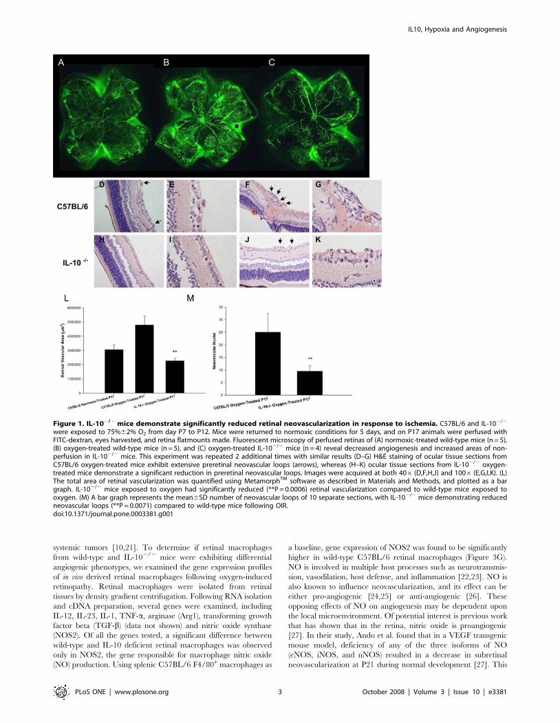
flatmounts were made. We found that compared to pups that have
never been subjected to high oxygen (Figure 1A), age-matched
oxygen-treated C57BL/6 mice (Figure 1B) demonstrated signifi-
cantly more neovascularization with an abundance of new blood
vessel tufts, a central loss of normal retinal blood vessels, and
increased retinal vascular tortuosity. Oxygen-treated IL-102/2
mice (Figure 1C), however, demonstrated not only significantly
reduced neovascularization, but also increased areas of ischemia
and nonperfusion that lacked any vasculature. The total area of
retinal vascularization was quantified by MetamorphTM software,
and oxygen-treated IL-102/2 mice demonstrated significantly less
retinal vascularization compared to normoxic and hyperoxic wild-
type mice (Figure 1L). Differences in retinal vasculature between
wild-type and IL-102/2 mice occur only following exposure to
oxygen, as P17 normoxic IL-102/2 retinas show no vascular
differences compared to normoxic wild-type P17 retinas (data not
shown).
In addition to retinal flatmounts, eyes from oxygen-treated mice
were harvested for histological examination. Eyes were fixed in
formalin and 4-mm sections were made and stained with
hematoxylin and eosin. The neovascularization was assessed
histologically by counting the number of vascular endothelial cell
nuclei vitreal to the inner-limiting membrane. The retinas of
oxygen-treated wild-type mice contained multiple neovascular
tufts extending into the vitreous (Figures 1D–1G), whereas retinas
from oxygen-treated IL-102/2 mice had significantly fewer
vascular endothelial cells at these locations (Figures 1H–1K).
The neovascularization was quantified by counting and averaging
5 separate sections, revealing a significantly higher quantity of
neovascular cell nuclei in the oxygen-treated wild-type mice
compared to oxygen-treated IL-102/2 mice (Figure 1M).
Anti-IL-10 immunotherapy results in significantlyreduced retinal neovascularization in response tohypoxia
Since we observed less retinal neovascularization in IL-102/2
mice, we sought to determine if IL-10 may be an ideal therapeutic
target for the treatment of aberrant retinal angiogenesis.
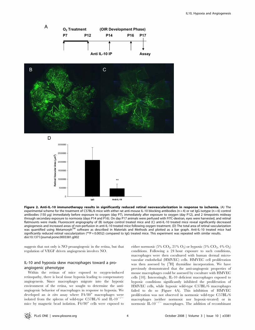
Therefore, we administered blocking antibodies against murine
IL-10 in wild-type mice. Anti-IL-10 or IgG antibodies (150 mg)
were injected into wild-type mice immediately before exposure to
oxygen (day P7), immediately following exposure to oxygen (day
P12), and midway between secondary normoxia exposure (days
P14 and P16) (Figure 2A). On day P17 mice were perfused with
FITC-dextran and retinal flatmounts were made. Fluorescein
angiography revealed that compared to IgG treated mice
(Figure 2B), mice treated with anti-IL-10 blocking antibodies
developed reduced retinal neovascularization following OIR
(Figure 2C). Analysis of retinal flatmounts with MetamorphTM
software revealed a statistically significant reduction in retinal
vascularization (Figure 2D). This suggests that anti-IL-10 immu-
notherapy may be an attractive avenue for the treatment of
retinopathy of prematurity.
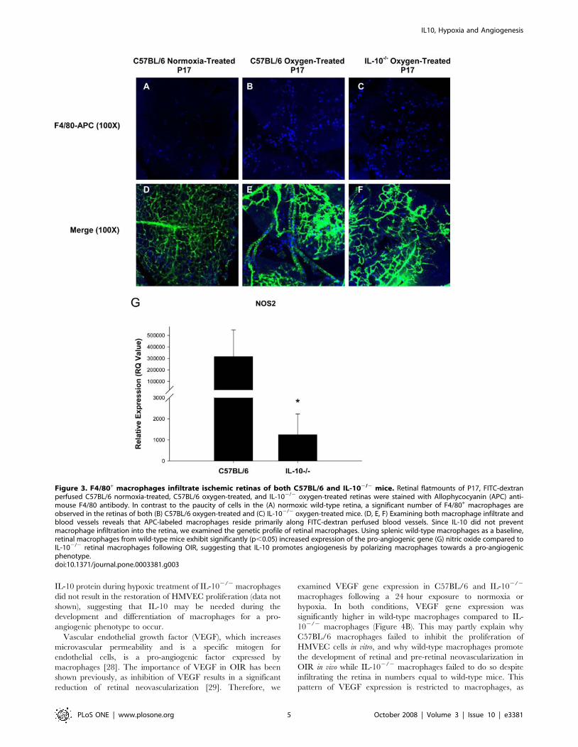
IL-10 does not alter infiltration of F4/80+ macrophagesinto ischemic retinas, but leads to significantly increasedproduction of pro-angiogenic NO
It has been previously shown that macrophages infiltrate the
retinas of mice following OIR [18,19,20]. Our laboratory has
shown that IL-10 prevents the infiltration of macrophages into the
choroidal vasculature of the eye in murine models of CNV [14].
We sought to determine if IL-10 prevents the infiltration of
macrophages into a distinct vascular plexus of the eye, the retina,
following OIR. We stained FITC-perfused retinal flatmounts with
an allophycocyanin (APC)-labeled anti-mouse F4/80 antibody to
detect the presence of infiltrating macrophages. Wild-type
normoxia retinas had very few F4/80+ cells (Figure 3A), which
might not be infiltrating macrophages but rather resident F4/80+
retinal microglia [19]. As expected, retinas from oxygen-treated
C57BL/6 mice displayed a significant increase in the number of
infiltrating F4/80+ macrophages compared to retinas from
normoxic control mice (Figure 3B). Surprisingly, retinas from
oxygen-treated IL-102/2 mice did not display an increased
number of F4/80+ macrophages compared to retinas from wild-
type oxygen-treated mice, as similar numbers of F4/80+ cells are
observed (Figure 3C). Therefore, the lack of neovascularization
observed in IL-102/2 mice following OIR is not simply due to an
increased influx of anti-angiogenic F4/80+ macrophages infiltrat-
ing the retina. Interestingly, infiltrating F4/80+ macrophages in
both groups of oxygen-treated mice resided along FITC-dextran
labeled blood vessels (Figures 3E and 3F), supporting previous
work that has observed infiltrating macrophages aligning along
neovascular tufts following OIR and suggesting macrophages may
modulate vessel growth and regression [19,20].
Since IL-10 did not alter the infiltration of macrophages into the
retina following OIR, we hypothesized that IL-10 may be altering
the angiogenic phenotype of macrophages in the local tissues, as
seen in the senescent choroid during CNV in the eye and in
IL10, Hypoxia and Angiogenesis
PLoS ONE | www.plosone.org 2 October 2008 | Volume 3 | Issue 10 | e3381
systemic tumors [10,21]. To determine if retinal macrophages
from wild-type and IL-102/2 mice were exhibiting differential
angiogenic phenotypes, we examined the gene expression profiles
of in vivo derived retinal macrophages following oxygen-induced
retinopathy. Retinal macrophages were isolated from retinal
tissues by density gradient centrifugation. Following RNA isolation
and cDNA preparation, several genes were examined, including
IL-12, IL-23, IL-1, TNF-a, arginase (Arg1), transforming growth
factor beta (TGF-b) (data not shown) and nitric oxide synthase
(NOS2). Of all the genes tested, a significant difference between
wild-type and IL-10 deficient retinal macrophages was observed
only in NOS2, the gene responsible for macrophage nitric oxide
(NO) production. Using splenic C57BL/6 F4/80+ macrophages as
a baseline, gene expression of NOS2 was found to be significantly
higher in wild-type C57BL/6 retinal macrophages (Figure 3G).
NO is involved in multiple host processes such as neurotransmis-
sion, vasodilation, host defense, and inflammation [22,23]. NO is
also known to influence neovascularization, and its effect can be
either pro-angiogenic [24,25] or anti-angiogenic [26]. These
opposing effects of NO on angiogenesis may be dependent upon
the local microenvironment. Of potential interest is previous work
that has shown that in the retina, nitric oxide is proangiogenic
[27]. In their study, Ando et al. found that in a VEGF transgenic
mouse model, deficiency of any of the three isoforms of NO
(eNOS, iNOS, and nNOS) resulted in a decrease in subretinal
neovascularization at P21 during normal development [27]. This
Figure 1. IL-102/2 mice demonstrate significantly reduced retinal neovascularization in response to ischemia. C57BL/6 and IL-102/2
were exposed to 75%62% O2 from day P7 to P12. Mice were returned to normoxic conditions for 5 days, and on P17 animals were perfused withFITC-dextran, eyes harvested, and retina flatmounts made. Fluorescent microscopy of perfused retinas of (A) normoxic-treated wild-type mice (n = 5),(B) oxygen-treated wild-type mice (n = 5), and (C) oxygen-treated IL-102/2 mice (n = 4) reveal decreased angiogenesis and increased areas of non-perfusion in IL-102/2 mice. This experiment was repeated 2 additional times with similar results (D–G) H&E staining of ocular tissue sections fromC57BL/6 oxygen-treated mice exhibit extensive preretinal neovascular loops (arrows), whereas (H–K) ocular tissue sections from IL-102/2 oxygen-treated mice demonstrate a significant reduction in preretinal neovascular loops. Images were acquired at both 406 (D,F,H,J) and 1006 (E,G,I,K). (L)The total area of retinal vascularization was quantified using MetamorphTM software as described in Materials and Methods, and plotted as a bargraph. IL-102/2 mice exposed to oxygen had significantly reduced (**P = 0.0006) retinal vascularization compared to wild-type mice exposed tooxygen. (M) A bar graph represents the mean6SD number of neovascular loops of 10 separate sections, with IL-102/2 mice demonstrating reducedneovascular loops (**P = 0.0071) compared to wild-type mice following OIR.doi:10.1371/journal.pone.0003381.g001
IL10, Hypoxia and Angiogenesis
PLoS ONE | www.plosone.org 3 October 2008 | Volume 3 | Issue 10 | e3381
suggests that not only is NO proangiogenic in the retina, but that
regulation of VEGF driven angiogenesis involves NO.
IL-10 and hypoxia skew macrophages toward a pro-angiogenic phenotype
Within the retinas of mice exposed to oxygen-induced
retinopathy, there is local tissue hypoxia leading to compensatory
angiogenesis. Since macrophages migrate into the hypoxic
environment of the retina, we sought to determine the anti-
angiogenic behavior of macrophages in response to hypoxia. We
developed an in vitro assay where F4/80+ macrophages were
isolated from the spleens of wild-type C57BL/6 and IL-102/2
mice by magnetic bead isolation. F4/80+ cells were exposed to
either normoxic (5% CO2, 21% O2) or hypoxic (5% CO2, 4% O2)
conditions. Following a 24 hour exposure to such conditions,
macrophages were then cocultured with human dermal micro-
vascular endothelial (HMVEC) cells. HMVEC cell proliferation
was then assessed by [3H] thymidine incorporation. We have
previously demonstrated that the anti-angiogenic properties of
mouse macrophages could be assessed by coculture with HMVEC
cells [10]. Interestingly, IL-10 deficient macrophages exposed to
hypoxic conditions significantly inhibited the proliferation of
HMVEC cells, while hypoxic wild-type C57BL/6 macrophages
failed to do so (Figure 4A). This inhibition of HMVEC
proliferation was not observed in normoxic wild-type C57BL/6
macrophages (neither normoxic nor hypoxic-treated) or in
normoxic IL-102/2 macrophages. The addition of recombinant
Figure 2. Anti-IL-10 immunotherapy results in significantly reduced retinal neovascularization in response to ischemia. (A) Theexperimental scheme for the treatment of C57BL/6 mice with either rat anti-mouse IL-10 blocking antibodies (n = 4) or rat IgG isotype (n = 6) controlantibodies (150 mg) immediately before exposure to oxygen (day P7), immediately after exposure to oxygen (day P12), and 2 timepoints midwaythrough secondary exposure to normoxia (days P14 and P16). On day P17 animals were perfused with FITC-dextran, eyes were harvested, and retinalflatmounts were made. Fluorescent angiography of (B) isotype control treated mice and (C) anti-IL-10 treated mice reveal significantly decreasedangiogenesis and increased areas of non-perfusion in anti-IL-10 treated mice following oxygen treatment. (D) The total area of retinal vascularizationwas quantified using MetamorphTM software as described in Materials and Methods and plotted as a bar graph. Anti-IL-10 treated mice hadsignificantly reduced retinal vascularization (**P = 0.0052) compared to IgG treated mice. This experiment was repeated with similar results.doi:10.1371/journal.pone.0003381.g002
IL10, Hypoxia and Angiogenesis
PLoS ONE | www.plosone.org 4 October 2008 | Volume 3 | Issue 10 | e3381
IL-10 protein during hypoxic treatment of IL-102/2 macrophages
did not result in the restoration of HMVEC proliferation (data not
shown), suggesting that IL-10 may be needed during the
development and differentiation of macrophages for a pro-
angiogenic phenotype to occur.
Vascular endothelial growth factor (VEGF), which increases
microvascular permeability and is a specific mitogen for
endothelial cells, is a pro-angiogenic factor expressed by
macrophages [28]. The importance of VEGF in OIR has been
shown previously, as inhibition of VEGF results in a significant
reduction of retinal neovascularization [29]. Therefore, we
examined VEGF gene expression in C57BL/6 and IL-102/2
macrophages following a 24 hour exposure to normoxia or
hypoxia. In both conditions, VEGF gene expression was
significantly higher in wild-type macrophages compared to IL-
102/2 macrophages (Figure 4B). This may partly explain why
C57BL/6 macrophages failed to inhibit the proliferation of
HMVEC cells in vitro, and why wild-type macrophages promote
the development of retinal and pre-retinal neovascularization in
OIR in vivo while IL-102/2 macrophages failed to do so despite
infiltrating the retina in numbers equal to wild-type mice. This
pattern of VEGF expression is restricted to macrophages, as
Figure 3. F4/80+ macrophages infiltrate ischemic retinas of both C57BL/6 and IL-102/2 mice. Retinal flatmounts of P17, FITC-dextranperfused C57BL/6 normoxia-treated, C57BL/6 oxygen-treated, and IL-102/2 oxygen-treated retinas were stained with Allophycocyanin (APC) anti-mouse F4/80 antibody. In contrast to the paucity of cells in the (A) normoxic wild-type retina, a significant number of F4/80+ macrophages areobserved in the retinas of both (B) C57BL/6 oxygen-treated and (C) IL-102/2 oxygen-treated mice. (D, E, F) Examining both macrophage infiltrate andblood vessels reveals that APC-labeled macrophages reside primarily along FITC-dextran perfused blood vessels. Since IL-10 did not preventmacrophage infiltration into the retina, we examined the genetic profile of retinal macrophages. Using splenic wild-type macrophages as a baseline,retinal macrophages from wild-type mice exhibit significantly (p,0.05) increased expression of the pro-angiogenic gene (G) nitric oxide compared toIL-102/2 retinal macrophages following OIR, suggesting that IL-10 promotes angiogenesis by polarizing macrophages towards a pro-angiogenicphenotype.doi:10.1371/journal.pone.0003381.g003
IL10, Hypoxia and Angiogenesis
PLoS ONE | www.plosone.org 5 October 2008 | Volume 3 | Issue 10 | e3381
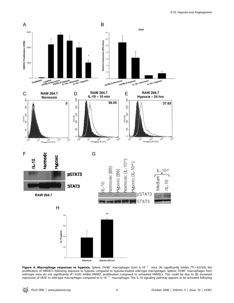
Figure 4. Macrophage responses to hypoxia. Splenic F4/80+ macrophages from IL-102/2 mice (A) significantly inhibit (*P = 0.0163) theproliferation of HMVECs following exposure to hypoxia compared to hypoxia-treated wild-type macrophages. Splenic F4/80+ macrophages fromwild-type mice do not significantly (P.0.05) inhibit HMVEC proliferation compared to untreated HMVECs. This could be due to (B) increasedexpression of VEGF in wild-type macrophages compared to IL-102/2 macrophages. The IL-10 signaling pathway appears to be activated following
IL10, Hypoxia and Angiogenesis
PLoS ONE | www.plosone.org 6 October 2008 | Volume 3 | Issue 10 | e3381
VEGF in whole retinal tissue from oxygen-treated wild-type mice
was not significantly elevated compared to normoxic and oxygen-
treated IL-102/2 whole retinal tissue (data not shown). Therefore,
the localized expression of VEGF on macrophages may be
influencing the development of retinal blood vessels. This
correlates with the work of Naug et al, who found VEGF-
expressing macrophages associated with vitreal loops in wild-type
mice following OIR [30].
We next examined whether hypoxia induced IL-10 signaling in
immunocompetent wild-type macrophages. IL-10 protein binds to
the IL-10 receptor (IL-10R), resulting in phosphorylation and
activation of the signal transducer and activator of transcription 3
(STAT3). STAT3 phosphorylation and activation is essential for all
known function of IL-10 [31]. To determine if immunocompetent
macrophages were undergoing IL-10 signaling, RAW 264.7 murine
macrophages were assessed for phosphorylation of STAT3 by flow
cytometry and western blot analysis. RAW macrophages cultured in
normoxic conditions (21% O2) did not demonstrate increased
staining of phospho-STAT3 antibody compared to isotype control
antibody stained cells (Figure 4C). RAW macrophages pulsed with
recombinant IL-10 protein (100 ng/ml) for 10 minutes demonstrated
a significant increase in phospho-STAT3 staining compared to
normoxic treated cells (Figure 4D), confirming that IL-10 signaling in
macrophages results in phosphorylation of STAT3. RAW macro-
phages cultured in hypoxic conditions (4% O2) for 24 hours also
demonstrated a significant increase in phospho-STAT3 staining
compared to normoxic treated cells (Figure 4E). This suggests that
when immunocompetent macrophages infiltrate hypoxic regions
such as the retina in OIR, the IL-10 signaling pathway is induced.
Flow cytometric analysis of STAT3 phosphorylation was confirmed
by western blot examination (Figure 4F). RAW macrophages treated
with either recombinant IL-10 protein (100 ng/ml) for 10 minutes or
hypoxia for 24 hours demonstrated increased phosphorylation of
STAT3. These findings in RAW macrophages were also observed in
primary macrophages. Wild-type macrophages treated with IL-10
protein for 10 minutes or hypoxia for 24 hours demonstrated
increased phosphorylation of STAT3 compared to baseline normoxic
levels (Figure 4G). IL-102/2 macrophages, however, demonstrated
decreased levels of pSTAT3 at baseline normoxic levels compared to
wild-type normoxic macrophages, and did not upregulate pSTAT3
in response to hypoxia. STAT3 signaling is still intact in IL-102/2
macrophages, as phosphorylation of STAT3 is increased in IL-102/2
macrophages following stimulation with recombinant IL-10 protein
(Figure 4G). Interestingly, supernatants from RAW macrophages
cultured under hypoxic conditions demonstrate a significant increase
in the production of IL-10 protein (Figure 4H). Therefore,
phosphorylation of STAT-3 in macrophages following hypoxia
may be due to autocrine secretion of IL-10 by macrophages in
response to hypoxia and subsequent engagement of the IL-10
receptor. It has been shown that STAT3 activation and signaling can
result in the expression of VEGF [32], correlating with our earlier
observation of increased VEGF gene expression levels in immuno-
competent macrophages.
Discussion
In summary, IL-10 promotes retinal neovascularization in a
mouse model of oxygen-induced retinopathy. In contrast to previous
reports that analyzed the regulation of choroidal macrophages by IL-
10 [14], IL-10 did not prevent the infiltration of macrophages into
the retina, but rather polarized the macrophage genetic profile
towards a pro-angiogenic phenotype. This difference in IL-10
function may be attributable to the local tissue micro milieu, as the
hypoxic drive and regional soluble and membrane-bound signals in
diverse vascular beds may differ, i.e. the retinal vasculature versus the
choriocapillaris. Although IL-10 promotes angiogenesis by prevent-
ing the infiltration of anti-angiogenic macrophages into the choroid
[14], this study suggests that in the retina, IL-10 promotes
angiogenesis by directly polarizing macrophages towards a pro-
angiogenic phenotype. Unlike choroidal neovascularization, hypoxia
or ischemia is the prime driver of retinal and pre-retinal
neovascularization observed in diabetic retinopathy and ROP.
Hypoxia induces a profound change in the phenotype of
macrophages. A recent study demonstrated that more than 30 pro-
angiogenic genes were upregulated by hypoxia in primary
macrophages [33], including VEGF and inducible nitric oxide
synthase (iNOS). Most of the work on hypoxia and macrophages has
focused on tumor-associated macrophages (TAMs). Macrophages
have been shown to accumulate in hypoxic regions of tumors, and
secrete multiple growth factors and pro-angiogenic cytokines that
promote tumor growth, angiogenesis, metastasis, and immune
evasion [34]. To our knowledge, this is the first study that reveals
that hypoxia and IL-10 function synergistically to influence
macrophage function and angiogenic homeostasis. It is also
noteworthy that the pro-angiogenic effects of hypoxic macrophages
are IL-10 dependent and require IL-10 mediated signal transduction.
The diverse and complex regulation of angiogenesis by IL-10 is
highlighted by the finding that in a mouse model of post-
developmental surgically-induced hindlimb ischemia, IL-10 ap-
pears to function in an anti-angiogenic manner, and does so by
downregulating VEGF expression [35]. These findings illustrate
the multifaceted role of IL-10 in regulating angiogenesis. The
vascular bed and the tissue type likely have a profound impact on
the ability of IL-10 to regulate macrophage behavior after tissue
injury. The success of anti-IL-10 immunotherapy in our studies of
mice during OIR was achieved by systemic depletion of IL-10 by
intraperitoneal injection. If IL-10 immunotherapy is to be
translated to humans as a potential treatment modality for
retinopathy, local intraocular injection of anti-IL-10 antibodies
or other IL-10 inhibitors, similar to anti-VEGF injections for
exposure to hypoxia. RAW 264.7 murine macrophages (C) do not exhibit increased levels of phosphorylation of STAT3 under normoxic conditions(open histogram) compared to IgG stained cells (shaded histogram). However, (E) exposure to hypoxia (open histogram) results in a significantincrease in phosphorylation of STAT3 compared to normoxia-treated cells (shaded histogram). This increase in phosphorylation of STAT3 is at levelssimilar to (D) RAW 264.7 cells exposed to recombinant IL-10 protein for 10 min (open histogram) versus normoxic cells (shaded histogram). Insetnumbers indicate percentage of positive cells above normoxic controls. Phosphorylation of STAT3 protein following hypoxia in RAW 264.7 cells wasalso confirmed with (F) western blot analysis. Phosphorylation of STAT3 occurred following exposure to both recombinant IL-10 protein for 10 minand a 24 hour exposure to hypoxia. Probing of total STAT3 protein was used as a loading control. These findings in RAW macrophages were alsoobserved in primary macrophages (G). Wild-type macrophages treated with recombinant IL-10 protein for 10 minutes or hypoxia for 24 hoursdemonstrated increased phosphorylation of STAT3 compared to baseline normoxic levels. IL-102/2 macrophages, however, demonstrated decreasedlevels of pSTAT3 at baseline normoxic levels compared to wild-type normoxic macrophages, and did not upregulate pSTAT3 in response to hypoxia.STAT3 signaling is still intact in IL-102/2 macrophages, as phosphorylation of STAT3 in increased in IL-102/2 macrophages following stimulation withrecombinant IL-10 protein. This increase in STAT3 phosphorylation may be due to the (H) significantly increased (*P = 0.0123) production of IL-10protein by RAW 264.7 macrophages following exposure to hypoxia compared to normoxia-treated RAW macrophages.doi:10.1371/journal.pone.0003381.g004
IL10, Hypoxia and Angiogenesis
PLoS ONE | www.plosone.org 7 October 2008 | Volume 3 | Issue 10 | e3381
AMD patients, may be desirable to minimize the systemic effects
in non-ocular vascular beds.
The role of IL-10 in the induction of developmental angiogenesis
seems to be a highly choreographed process involving multiple
factors. Interestingly, all of the genes that were upregulated in
macrophages following oxygen-induced retinopathy were found to
be 1) significantly upregulated in C57BL/6 macrophages and not in
IL-10 deficient macrophages, 2) proangiogenic, and 3) cross
regulative. IL-10 has previously been shown to upregulate NO
production in activated macrophages in a dose dependent manner
[36]. Also, nitric oxide has previously been reported to be
upregulated in hypoxic retinas [37]. Our findings demonstrate that
this increase in NOS2 expression in the retina may be attributable
to infiltrating retinal macrophages. The relationship between nitric
oxide and VEGF is highly complex. In other models of hypoxia-
induced angiogenesis, VEGF and NO were responsible for
increased vascular permeability [38]. In the eye, transgenic mice
with increased expression of VEGF in photoreceptors had
decreased subretinal neovascularization when nitric oxide was
deleted [27], suggesting that VEGF driven angiogenesis was NO
dependent. It has also been shown in other cell types that hypoxia
results in STAT3 phosphorylation, which increases cellular levels of
hypoxia-inducible factor (HIF-1a) which in turn, increases VEGF
expression [39]. Additionally, increased STAT3 and pSTAT3 levels
have been found in the retina of mice following OIR[40].
The role of macrophages in preventing retinal angiogenesis
appears to be by inhibiting the proliferation of vascular
endothelium. Inhibition of vascular endothelial cell proliferation
observed in vitro following co-culture with hypoxia-treated IL-102/
2 macrophages (Fig. 4A) may be attributable to multiple factors.
Our previous work suggests that IL-10 deficient macrophages tend
to skew towards a pro-inflammatory/anti-angiogenic phenotype
(M1) following activation, whereas wild-type macrophages have a
mixed pro/anti inflammatory phenotype [10,14]. In this study,
hypoxia is the stimulus that activates and skews IL-102/2
macrophages towards an M1 phenotype. These M1 polarized
IL-102/2 macrophages likely inhibit HMVEC proliferation due to
a deficiency of NO (Fig. 3G), or increased expression of FasL, as
shown previously [14]. Under normoxic environments, IL-102/2
macrophages are in an inactive, non-polarized state, and thus do
not affect HMVEC proliferation.
Taken as a whole, inhibition of IL-10 signaling in macrophages
may be beneficial in not only preventing aberrant angiogenesis in
the eye, but may also be relevant in the prevention of tumor-
infiltrating macrophages from becoming pro-angiogenic TAMs
that have been implicated in tumor progression and metastasis.
Materials and Methods
MiceC57BL/6 and IL-102/2 (B6.129P2-Il10tm1Cgn/J) mice were
purchased from the Jackson Laboratory (Bar Harbor, ME). All
work was carried out in accordance with Association for Research
in Vision and Ophthalmology (ARVO) guidelines. Comprehensive
protocols of animal care and experimental design outlined in this
study were approved by the Animal Studies Committee of
Washington University.
Oxygen-induced Retinopathy and Quantification ofNeovascularization
Oxygen-induced retinopathy was produced in mice by the method
described by Smith et al. [17]. On day 7 postpartum (P7), neonatal
mice and their nursing mothers were exposed to 75%62% oxygen
for 5 days. On day P12, mice were then returned to room air for a
period of 5 days. At day P17, mice were anesthetized with 0.66 mg/
kg ketamine hydrochloride (Vetalar; Parke-Davis) given i.p. Mice
were sacrificed by intracardiac perfusion with 2 ml of 20 mg/ml
26106 molecular weight FITC-dextran (Sigma-Aldrich, St. Louis,
MO) in PBS. Eyes were enucleated, fixed in 4% paraformaldehye for
45 min at 4uC, and placed in PBS for 15 min at 4uC. A dissecting
microscope was used to remove the cornea and lens and gently
separate the retina from the underlying choroid and sclera.
Microscissors were used to make four radial incisions of the retinal
eyecup in order to prepare retinal flat mounts on glass slides. Flat
mounts were immersed in Gel/Mount (Biomeda Corp., Foster City,
CA) and coverslips were carefully placed. Retinal vascularization was
analyzed by fluorescent microscopy (Leica LMD 6000 fluorescent
microscope; Wetzlar, Germany). Separate images were taken at 636magnification, and images were merged using the ‘specimen
overview’ function of the microscope software. The extent of retinal
vascularization was then quantified by MetamorphTM Imaging
software (Universal Imaging, Sunnyvale, CA). Briefly, the entire
retina was outlined to identify the total retinal area of each eye. Then,
the color threshold of each image was adjusted to highlight only the
FITC-perfused vessels of the inclusively highlighted retina. This
allowed the determination of total blood vessel area of each retina and
the percentage of each retina that is occupied with blood vessels. Data
was reported as total retinal vascular area, and the vascular area for
all eyes in a treatment group were averaged and compared
individually to controls using Student’s t test.
Retinal neovascularization was also examined by histology. The
eyes of P17 mice (n$3 per group) were enucleated, fixed in 10%
formalin, and embedded in paraffin. Serial 4-mm-thick sections of
the eye were obtained and stained with hematoxylin and eosin.
Images were obtained at 406and 1006magnification. All retinal
vascular cell nuclei anterior to the internal-limiting membrane
were counted. 10 sections were examined, and the average6SD of
neovascular cell nuclei per sections was obtained. No vascular cell
nuclei anterior to the internal-limiting membrane were observed
in normoxic control animals.
In Vivo Anti-IL-10 Antibody TreatmentRat anti-mouse IL-10 (JES5.2A5; Genzyme) and isotype control
antibody (Rat IgG; Sigma) were administered to wild-type mice by
intraperitoneal (i.p.) injection. Antibodies (150 mg) were administered
at days P7 (before oxygen exposure), P12 (after oxygen exposure),
P14, and P16, following the same timeline used previously for the i.p.
injections during OIR [41]. Retinal neovascularization was then
evaluated on day P17 as described above.
Staining of retinal macrophagesRetinas were also examined for infiltration of macrophages. Flat-
mounts of FITC-dextran perfused retinas were prepared as
described above. Retinas were washed with PBS and blocked with
3% BSA in PBS for 30 min at 37uC. Following a subsequent
washing with PBS, retinas were stained with 1 mg/ml Allophyco-
cyanin (APC)-conjugated anti-mouse F4/80 antibody (eBioscience,
San Diego, CA) for 40 min at 37uC. Retinas were washed again
with PBS, immersed in Gel/Mount (Biomeda Corp., Foster City,
CA), coverslipped, and examined by confocal microscopy.
In vitro Hypoxic Treatment of MacrophagesSpleens were isolated from wild-type C57BL/6 and IL-102/2
mice. Single-cell suspensions were obtained and stained with 1 mg/ml
PE-conjugated anti-mouse F4/80 (eBioscience). PE-labeled cells were
isolated by positive selection using magnetic separation (Stemcell
Technologies, Inc.). F4/80+ macrophages were then exposed to
normoxic (5% CO2, 21% O2) or hypoxic (5% CO2, 4% O2)
IL10, Hypoxia and Angiogenesis
PLoS ONE | www.plosone.org 8 October 2008 | Volume 3 | Issue 10 | e3381
conditions at 37uC for 24 hours. Cells were either immediately lysed
for RNA isolation or used in the vascular endothelial cell proliferation
assay.
Vascular endothelial cell proliferation assayHuman dermal microvascular endothelial cells (HMVECs)
(Lonza, Walkersville, MD) (56104) in log phase growth were
placed in EGM2V media (Lonza, Walkersville, MD) in 96-well
round bottom plates and allowed to adhere for 24 hours at 37uC.
Normoxic or hypoxic macrophages were then added to HMVECs
and allowed to co-culture for an additional 12 hours. Macro-
phages were removed by repeated and gentle pipetting, and
HMVECs were incubated with 40 mCi/ml of [3H] thymidine
(TRA61- GE Health Care, Piscataway, NJ) for 12 hours. The
tissue culture plate was harvested and read using a Topcount
harvester and microplate reader (Packard, Meriden, CT). The
amount of [3H] thymidine incorporated into the HMVEC cells is
directly proportional to the proliferation of the cells.
Isolation of Retinal MacrophagesRetinas from P17 oxygen-treated mice were isolated from the rest
of the eye as described above and placed directly in 1 ml of a 1 mg/ml
collagenase (Sigma)/PBS solution. Retinas were then passed through
a 18-gauge needle to obtain a single-cell suspension, and then allowed
to incubate in collagenase for 30 min at 37uC. The collagenase
solution was then brought to a total volume of 5 ml, and layered on
top of 3 ml of Histopaque-1077 (Sigma) solution. Cells were spun at
2000 RPM for 30 min. During centrifugation, mononuclear cells
remain at the PBS-Histopaque interface. Cells were isolated from the
interface and immediately lysed for RNA isolation.
Quantitative Analysis of Gene Expression (QuantitativeReal-Time RT-PCR)
Total RNA was prepared from retinal macrophages from P17
oxygen-treated C57BL/6 and IL-102/2 mice using the RNeasy mini
kit (Qiagen, Valencia, CA). RNA was purified by gDNA eliminator
spin columns and subsequent DNase treatment in order to minimize
the amount of gDNA cross-contamination. cDNA was prepared
using the High Capacity cDNA Archive Kit (Applied Biosystems,
Foster City, CA). Relative levels of target gene expression were
measured on the 7500 Real-time RT-PCR system (Applied
Biosystems). For fluorescence measure and assay preparation, specific
FAM-490 based Taqman Gene Expression Assay Mix (Applied
Biosystems) for each gene of interest and Taqman Universal Master
Mix (Applied Biosystems) were used. Relative Quantification PCR
analysis was performed using the ABI 7500 SDS Software. The
threshold cycle (Ct) values were measured for our endogenous control
(b-actin) and the genes of interest at the logarithmic phase of
amplification. The DCt values for the calibrator (negative baseline
control) were calculated as the difference between the Ct value of the
endogenous control and the target gene of interest (Ct b-actin – Ct
target). This DCt value for the calibrator was then compared to the
DCt value of each of our unknown cDNA samples, the difference of
these two values were taken to achieve the DDCt value for each gene
(DCt unknown – DCt calibrator). The relative fold expression
difference was calculated and graphed using the formula: 2(2DDCt).
Taqman Gene Expression Assay Mixes used (primer/probe sets)
include b-actin (Mm00607939_s1); TNF-a (Mm00443258_m1); IL-6
(Mm00446190_m1); IL-12 (Mm01288989_m1); IL-23
(Mm00518984_m1); IL-10 (Mm00439616_m1); FasL
(Mm00438864_m1); IL-1b (Mm00434228_m1); IFN-c(Mm00801778_m1); TGF-b (Mm03024053_m1); Arg1
(Mm00475988_m1); NOS2 (Mm00440485_m1); and VEGF-a(Mm00437304_m1).
For RT-PCR of in vitro treated macrophages, IL-102/2
normoxic macrophages were used as a baseline comparison. For
in vivo retinal macrophages, splenic F4/80+ macrophages were
used as a baseline comparison.
Assessment of macrophages for phospho-STAT3expression
RAW 264.7 murine macrophages (ATCC; Manassas, VA) or
splenic F4/80+ cells from C57BL/6 or IL-102/2 mice were placed
in complete RPMI media and incubated either in normoxic (21%
oxygen) or hypoxic (4% oxygen) conditions for 24 hours. For flow
cytometric analysis, cells were fixed and permeabilized with BD
Cytofix/Cytoperm (Becton Dickinson). Cells were then stained with
either rabbit IgG (2 mg/ml; Sigma) or rabbit anti-phospho STAT3
(1:500; Sigma), followed by FITC-conjugated goat anti-rabbit
antibody (2 mg/ml; Sigma). Macrophages stimulated with recom-
binant murine IL-10 (100 ng/ml; R&D Systems) for 10 min were
used as a positive control for staining. Cells were washed,
resuspended in PBS, and analyzed using a FACScan flow cytometer
(BD Biosciences). Results were analyzed using CellQuest v.3.1f
software (BD Biosciences). For examination of STAT3 phosphor-
ylation by western blot, normoxic and hypoxic macrophages were
lysed with Triton X-100 buffer (150 mM NaCl, 20 mM 1% Triton
X-100), run on SDS-PAGE (10% NuPAGE gel from Invitrogen,
Carlsbad, CA), and transferred onto a nitrocellulose membrane
(150 mAmp for 90 min). The membrane was incubated with 5%
nonfat dry milk for 1 h and washed three times for 10 min each with
TBS-T. The blot was then incubated either with anti-phospho
STAT3 (Cell Signaling Tech., Danvers, MA) or anti-STAT3
(Sigma) and then washed with TBS-T three times. The blot was
then incubated with the corresponding secondary antibody
conjugated with HRP for 1 h, washed three times with TBS-T for
10 min each, and then developed using ECL reagent.
IL-10 enzyme-linked immunosorbent assay (ELISA)RAW 264.7 macrophages were incubated in either normoxic or
hypoxic conditions as described above. Cell supernatants were
then harvested and levels of IL-10 protein in cell supernatants
were determined using a mouse IL-10 Quantikine ELISA kit
(R&D Systems).
Statistical analysisResults are presented as mean6standard deviation. A Student’s
t-test was used to assess the statistical significance of the differences
between compared groups. A P-value of ,0.05 was considered
significant.
Author Contributions
Conceived and designed the experiments: DSD RSA. Performed the
experiments: DSD AAK JK. Analyzed the data: DSD AAK JK RSA.
Wrote the paper: DSD JK RSA.
References
1. Nakao S, Kuwano T, Tsutsumi-Miyahara C, Ueda S, Kimura YN, et al. (2005)
Infiltration of COX-2-expressing macrophages is a prerequisite for IL-1 beta-
induced neovascularization and tumor growth. J Clin Invest 115: 2979–2991.
2. Taylor PR, Martinez-Pomares L, Stacey M, Lin HH, Brown GD, et al. (2005)
Macrophage receptors and immune recognition. Annu Rev Immunol 23:
901–944.
IL10, Hypoxia and Angiogenesis
PLoS ONE | www.plosone.org 9 October 2008 | Volume 3 | Issue 10 | e3381
3. Hansson GK (2005) Inflammation, atherosclerosis, and coronary artery disease.
N Engl J Med 352: 1685–1695.4. Yoshida A, Yoshida S, Ishibashi T, Inomata H (1999) Intraocular neovascu-
larization. Histol Histopathol 14: 1287–1294.
5. Prasad AG, Schadlu R, Apte RS (2007) Intravitreal pharmacotherapy:applications in retinal disease. Compr Ophthalmol Update 8: 259–269.
6. Arfken CL, Reno PL, Santiago JV, Klein R (1998) Development of proliferativediabetic retinopathy in African-Americans and whites with type 1 diabetes.
Diabetes Care 21: 792–795.
7. Gariano RF, Gardner TW (2005) Retinal angiogenesis in development anddisease. Nature 438: 960–966.
8. Adamis AP, Berman AJ (2008) Immunological mechanisms in the pathogenesisof diabetic retinopathy. Semin Immunopathol 30: 65–84.
9. Sunderkotter C, Steinbrink K, Goebeler M, Bhardwaj R, Sorg C (1994)Macrophages and angiogenesis. J Leukoc Biol 55: 410–422.
10. Kelly J, Ali Khan A, Yin J, Ferguson TA, Apte RS (2007) Senescence regulates
macrophage activation and angiogenic fate at sites of tissue injury in mice. J ClinInvest 117: 3421–3426.
11. Mantovani A, Sica A, Locati M (2005) Macrophage polarization comes of age.Immunity 23: 344–346.
12. Sica A, Schioppa T, Mantovani A, Allavena P (2006) Tumour-associated
macrophages are a distinct M2 polarised population promoting tumourprogression: potential targets of anti-cancer therapy. Eur J Cancer 42: 717–727.
13. Mosser DM (2003) The many faces of macrophage activation. J Leukoc Biol 73:209–212.
14. Apte RS, Richter J, Herndon J, Ferguson TA (2006) Macrophages inhibitneovascularization in a murine model of age-related macular degeneration.
PLoS Med 3: e310.
15. Canataroglu H, Varinli I, Ozcan AA, Canataroglu A, Doran F, et al. (2005)Interleukin (IL)-6, interleukin (IL)-8 levels and cellular composition of the
vitreous humor in proliferative diabetic retinopathy, proliferative vitreoretino-pathy, and traumatic proliferative vitreoretinopathy. Ocul Immunol Inflamm
13: 375–381.
16. Esser P, Heimann K, Wiedemann P (1993) Macrophages in proliferativevitreoretinopathy and proliferative diabetic retinopathy: differentiation of
subpopulations. Br J Ophthalmol 77: 731–733.17. Smith LE, Wesolowski E, McLellan A, Kostyk SK, D’Amato R, et al. (1994)
Oxygen-induced retinopathy in the mouse. Invest Ophthalmol Vis Sci 35:101–111.
18. Yoshida S, Yoshida A, Ishibashi T, Elner SG, Elner VM (2003) Role of MCP-1
and MIP-1alpha in retinal neovascularization during postischemic inflammationin a mouse model of retinal neovascularization. J Leukoc Biol 73: 137–144.
19. Davies MH, Eubanks JP, Powers MR (2006) Microglia and macrophages areincreased in response to ischemia-induced retinopathy in the mouse retina. Mol
Vis 12: 467–477.
20. Shen J, Xie B, Dong A, Swaim M, Hackett SF, et al. (2007) In VivoImmunostaining Demonstrates Macrophages Associate with Growing and
Regressing Vessels. Invest Ophthalmol Vis Sci 48: 4335–4341.21. Mantovani A, Schioppa T, Porta C, Allavena P, Sica A (2006) Role of tumor-
associated macrophages in tumor progression and invasion. Cancer MetastasisRev 25: 315–322.
22. Christopherson KS, Bredt DS (1997) Nitric oxide in excitable tissues:
physiological roles and disease. J Clin Invest 100: 2424–2429.23. MacMicking J, Xie QW, Nathan C (1997) Nitric oxide and macrophage
function. Annu Rev Immunol 15: 323–350.
24. Parenti A, Morbidelli L, Cui XL, Douglas JG, Hood JD, et al. (1998) Nitric
oxide is an upstream signal of vascular endothelial growth factor-inducedextracellular signal-regulated kinase1/2 activation in postcapillary endothelium.
J Biol Chem 273: 4220–4226.
25. Papapetropoulos A, Garcia-Cardena G, Madri JA, Sessa WC (1997) Nitric oxideproduction contributes to the angiogenic properties of vascular endothelial
growth factor in human endothelial cells. J Clin Invest 100: 3131–3139.26. Tsurumi Y, Murohara T, Krasinski K, Chen D, Witzenbichler B, et al. (1997)
Reciprocal relation between VEGF and NO in the regulation of endothelial
integrity. Nat Med 3: 879–886.27. Ando A, Yang A, Mori K, Yamada H, Yamada E, et al. (2002) Nitric oxide is
proangiogenic in the retina and choroid. J Cell Physiol 191: 116–124.28. Berse B, Brown LF, Van de Water L, Dvorak HF, Senger DR (1992) Vascular
permeability factor (vascular endothelial growth factor) gene is expresseddifferentially in normal tissues, macrophages, and tumors. Mol Biol Cell 3:
211–220.
29. Aiello LP, Pierce EA, Foley ED, Takagi H, Chen H, et al. (1995) Suppression ofRetinal Neovascularization in vivo by Inhibition of Vascular Endothelial Growth
Factor (VEGF) Using Soluble VEGF-Receptor Chimeric Proteins. Proceedingsof the National Academy of Sciences 92: 10457–10461.
30. Naug HL, Browning J, Gole GA, Gobe G (2000) Vitreal macrophages express
vascular endothelial growth factor in oxygen-induced retinopathy. ClinExperiment Ophthalmol 28: 48–52.
31. Hu X, Chen J, Wang L, Ivashkiv LB (2007) Crosstalk among Jak-STAT, Toll-like receptor, and ITAM-dependent pathways in macrophage activation.
J Leukoc Biol 82: 237–243.32. Sumimoto H, Imabayashi F, Iwata T, Kawakami Y (2006) The BRAF-MAPK
signaling pathway is essential for cancer-immune evasion in human melanoma
cells. J Exp Med 203: 1651–1656.33. White JR, Harris RA, Lee SR, Craigon MH, Binley K, et al. (2004) Genetic
amplification of the transcriptional response to hypoxia as a novel means ofidentifying regulators of angiogenesis. Genomics 83: 1–8.
34. Lewis C, Murdoch C (2005) Macrophage responses to hypoxia: implications for
tumor progression and anti-cancer therapies. Am J Pathol 167: 627–635.35. Silvestre J-S, Mallat Z, Duriez M, Tamarat R, Bureau MF, et al. (2000)
Antiangiogenic Effect of Interleukin-10 in Ischemia-Induced Angiogenesis inMice Hindlimb. Circ Res 87: 448–452.
36. Jacobs F, Chaussabel D, Truyens C, Leclerq V, Carlier Y, et al. (1998) IL-10 up-regulates nitric oxide (NO) synthesis by lipopolysaccharide (LPS)-activated
macrophages: improved control of Trypanosoma cruzi infection. Clin Exp
Immunol 113: 59–64.37. Sennlaub F, Courtois Y, Goureau O (2001) Inducible nitric oxide synthase
mediates the change from retinal to vitreal neovascularization in ischemicretinopathy. J Clin Invest 107: 717–725.
38. Fischer S, Clauss M, Wiesnet M, Renz D, Schaper W, et al. (1999) Hypoxia
induces permeability in brain microvessel endothelial cells via VEGF and NO.Am J Physiol Cell Physiol 276: C812–820.
39. Jung JE, Lee HG, Cho IH, Chung DH, Yoon S-H, et al. (2005) STAT3 is apotential modulator of HIF-1-mediated VEGF expression in human renal
carcinoma cells. FASEB J 19: 1296–1298.40. Mechoulam H, Pierce EA (2005) Expression and activation of STAT3 in
ischemia-induced retinopathy. Invest Ophthalmol Vis Sci 46: 4409–4416.
41. Qiao H, Sonoda KH, Ikeda Y, Yoshimura T, Hijioka K, et al. (2007)Interleukin-18 regulates pathological intraocular neovascularization. J Leukoc
Biol 81: 1012–1021.
IL10, Hypoxia and Angiogenesis
PLoS ONE | www.plosone.org 10 October 2008 | Volume 3 | Issue 10 | e3381


