Embed Size (px)
Citation preview

Interneuron Diversity series:Fast in, fast out – temporal and spatialsignal processing in hippocampalinterneuronsPeter Jonas1, Josef Bischofberger1, Desdemona Fricker2 and Richard Miles2
1Physiologisches Institut der Universitat Freiburg, Hermann-Herder-Strasse 7, D-79104 Freiburg, Germany2INSERM EMI0224, CHU Pitie-Salpetriere, University Paris VI, 105 Boulevard de l’Hopital, Paris 75013, France
The operation of neuronal networks crucially depends
on a fast time course of signaling in inhibitory inter-
neurons. Synapses that excite interneurons generate
fast currents, owing to the expression of glutamate
receptors of specific subunit composition. Interneurons
generate brief action potentials in response to transient
synaptic activation and discharge repetitively at very
high frequencies during sustained stimulation. The abil-
ity to generate short-duration action potentials at high
frequencies depends on the expression of specific
voltage-gated K1 channels. Factors facilitating fast
action potential initiation following synaptic excitation
include depolarized interneuron resting potential, sub-
threshold conductances and active dendrites. Finally,
GABA release at interneuron output synapses is rapid
and highly synchronized, leading to a faster inhibition
in postsynaptic interneurons than in principal cells.
Thus, the expression of distinct transmitter receptors
and voltage-gated ion channels ensures that inter-
neurons operate with high speed and temporal
precision.
Interneurons play a key role in the operation of neuronalnetworks. Inhibitory cells control both the number ofactive pyramidal cells and their firing frequency byfeedforward and feedback inhibition [1,2]. Interneuronsalso control the timing of principal cell discharge [1–3] andare thought to play a pivotal role in the generation ofnetwork oscillations [4–6]. In neuronal networks in vivounder a variety of behavioral conditions, interneuronsgenerate action potentials precisely time-locked to thecycles of theta-, gamma- and high-frequency oscillations[7–11]. This suggests that interneurons could be involvedin the generation and distribution of rhythmic activity inneuronal networks. Many of these functions of inter-neurons crucially depend on the fast and temporallyprecise conversion of an excitatory synaptic input into aninhibitory synaptic output. The complete sequence ofinterneuron activation, from synaptic excitation viadendritic integration to action potential initiation and
GABA release, can occur within 1–2 ms [12–14]. Thisreview examines, step by step, the molecular and cellularbasis of this remarkably fast signal flow. For the purpose ofillustration, it will focus on the hippocampus, a brainregion with a well-defined anatomical layout. Althoughinhibitory cells are known to be highly diverse [15,16], thisarticle will concentrate on interneuron subtypes that arewell characterized, such as parvalbumin-positive basketcells (which form perisomatic contacts) and somatostatin-positive oriens alveus interneurons (which establishdendritic synapses on their target cells) [17,18].
Fast glutamate-mediated excitation of interneurons
The first step is the excitation of GABAergic interneuronsat glutamatergic synapses (Figure 1). As at otherexcitatory synapses, glutamate preferentially activatesAMPA-type glutamate receptors. However, at excitatorysynapses on interneurons, the AMPA-receptor-mediatedexcitatory postsynaptic current (EPSC) rises and decaysrapidly [14,19,20] (Figure 1c). AMPA-receptor-mediatedEPSCs at mossy fiber–basket cell synapses of the dentategyrus, at mossy fiber–stratum lucidum interneuronsynapses in the CA3 region, and at synapses on cerebellarinterneurons have decay time constants of ,1.5 ms at228C and ,0.5 ms at 348C [14,19,20] (Figure 1c). Thesefast kinetics depend primarily on the type of postsynapticneuron, because AMPA-receptor-mediated EPSCs atmossy fiber synapses on interneurons are three timesfaster than those at mossy fiber synapses on CA3pyramidal cells [21]. In addition, the type of presynapticneuron might be important because AMPA-receptor-mediated EPSCs are significantly faster at mossy fibersynapses than at CA3 pyramidal cell collateral synapseson stratum lucidum interneurons (decay time constants of,1.5 ms versus,2.5 ms at 228C) [20] (for characterizationof stratum lucidum interneuron morphology, see Ref. [22]).High synchrony of quantal release, rapid clearance oftransmitter at spine-free dendrites and fast gating ofpostsynaptic AMPA receptors help to generate rapidEPSCs at principal neuron–interneuron synapses [19].In another review of this series, Lawrence and McBain [23]summarized how the gating properties of postsynapticCorresponding author: Peter Jonas ([email protected]).
Review TRENDS in Neurosciences Vol.27 No.1 January 200430
http://tins.trends.com 0166-2236/$ - see front matter q 2003 Elsevier Ltd. All rights reserved. doi:10.1016/j.tins.2003.10.010

receptors are determined by the expression of an inter-neuron-specific genetic program. Unlike principal neuronAMPA receptors, which are often GluR1flip/GluR2flip
heteromers, basket cell AMPA receptors are primarilyassembled from GluR1flop subunits [23,24], with GluR1 toGluR4 (GluR-A to GluR-D) indicating the four mammalianAMPA receptor subunits and flip and flop denoting the twovariants of each subunit generated by alternative splicing(Figure 1d).
In addition to AMPA receptors, glutamate releasedat excitatory synapses on interneurons can coactivatekainate receptors and NMDA receptors [25]. Both types ofreceptor activate and deactivate more slowly than AMPAreceptors: kainate receptors are gated on a timescale oftens of milliseconds [26,27], whereas NMDA receptorsoperate on a timescale of hundreds of milliseconds [25].Thus, the different types of glutamate receptor couldencode different features of afferent activity. For bothkainate receptors and NMDA receptors, the amplitudecontribution is highly variable. No kainate receptorcomponent was observed in stratum radiatum inter-neurons in CA1 of the guinea pig [28]. By contrast, asignificant kainate receptor component was found inEPSCs of oriens alveus interneurons and stratum
radiatum interneurons in CA1 of the rat [26,27]. Likewise,a small NMDA receptor contribution was observed atmossy fiber–basket cell synapses of the dentate gyrus, atmossy fiber–stratum lucidum interneuron synapses inCA3, and in a subset of stratum radiatum interneurons inCA1 [19,29–31]. By contrast, a large contribution wasfound at pyramidal neuron–stratum lucidum interneuronsynapses in CA3 and a different subset of stratumradiatum interneurons in CA1 [29,31]. Interestingly, theproperties of AMPA receptor and NMDA receptor com-ponents are inversely correlated in CA3 stratum luciduminterneurons – a fast AMPA receptor component goeshand in hand with a small NMDA receptor component[29]. The correlation extends to the expression of receptorsubunits – when AMPA receptors lack the GluR2 (GluR-B)subunit, NMDA receptors include the NR2B subunit. Thisco-regulation could arise at the level of gene expression,trafficking of subunit proteins to synapses, or via protein–protein interactions in the postsynaptic density [32].
Thus, glutamate-mediated excitation of interneuronsinvolves an especially rapid AMPA receptor component,which ensures fast and reliable activation of inhibitorycells. Distinct contributions of slower kainate receptorand NMDA receptor components might confer specific
Figure 1. Fast AMPA-receptor-mediated excitation of hippocampal interneurons. (a) Morphological reconstruction of a reciprocally coupled granule cell (soma and den-
drites, green; axon, red) and basket cell (soma and dendrites, black; axon, gray) pair in the dentate gyrus, with three putative excitatory synaptic contacts (red dots). Scale
bar, 100 mm. Abbreviations: GCL, granule cell layer; H, hilus. (b) Unitary excitatory postsynaptic potentials (EPSPs) at a pyramidal neuron–interneuron synapse in the CA3
region (paired recording). The top trace shows a presynaptic (i.e. pyramidal neuron; PN) action potential and the middle trace shows a unitary EPSP in the postsynaptic
interneuron (IN). The bottom trace shows the response of the postsynaptic interneuron to a depolarizing current pulse used to probe the membrane time constant. Record-
ing temperature <37 8C. (c) Unitary excitatory postsynaptic currents (EPSCs) at the granule cell–basket cell synapse (paired recording). The top trace shows presynaptic
action potential; the middle traces are individual evoked EPSCs in the postsynaptic basket cell (BC) and the bottom trace is an average EPSC. Recording temperature
<34 8C. (d) Single-cell reverse transcription polymerase chain reaction (RT-PCR) analysis of AMPA receptor subunit expression in a dentate gyrus basket cell compared
with a CA3 pyramidal neuron; differential hybridization of gels with selective radiolabeled oligonucleotide probes specific for GluR1 to GluR4 (GluR-A to GluR-D) AMPA
receptor subunits is shown. Note low relative abundance of GluR2 (GluR-B) in the basket cell. Panels (a) and (c) reproduced, with permission, from Ref. [19]; (b) reproduced,
with permission, from Ref [12]; (d) reproduced, with permission, from Ref. [24].
PN
BC
5 ms
500 pA
100 mV
GluR4GluR1 GluR2 GluR3
PN
BC
IN
PN
10 ms
1 mV
5 mV
20 mV
(a) Principal neuron Basket cell
(c) EPSC
(b) EPSP
(d) AMPA receptor mRNA
CA3
GCLGCLGCL
H
Review TRENDS in Neurosciences Vol.27 No.1 January 2004 31
http://tins.trends.com

computational properties on different principal neuron–interneuron connections.
Spatiotemporal signal processing in interneuron
dendrites
Synaptic integration, in addition to depending on the timecourse of the postsynaptic conductance, depends on thedendritic location of the synapse and the cable propertiesof the dendrite [33,34]. In different interneuron subtypes,electron microscopical analysis suggests that 70–90% ofsynapses are excitatory [35]. The total population of exci-tatory synapses appears to be distributed uniformly overthe somatodendritic domain, with 95–97% of synapsesterminating on dendrites [35,36]. However, subsets ofexcitatory synapses originating from specific input path-ways can be preferentially located on either proximal ordistal dendritic locations [19,20].
How do interneuron dendrites process excitatory syn-aptic events? Locally, excitatory postsynaptic potential(EPSP) kinetics are similar to the time course of thepostsynaptic conductance, and EPSP amplitude dependson synaptic peak conductance and dendritic impedance atthe postsynaptic site [19,37–40]. As EPSPs propagatefrom the dendrites to the soma, their amplitude isattenuated and their time course is slowed. The EPSPdecay approaches the membrane time constant (tm) forlong propagation distances but is faster and multi-exponential for intermediate propagation distances. Thedegree offiltering depends not only on synapse location butalso on dendritic geometry and the specific resistance (Rm,resistance for a given membrane area) of the dendriticmembrane. Several interneuron subtypes have a low inputresistance and a fast tm, suggesting a low Rm (becausetm ¼ RmCm, where Cm is capacitance for a given mem-brane area [41–43]); this especially applies to parval-bumin-positive basket cells (tm ,10 ms). By contrast,principal cells have much higher input resistance andlonger membrane time constant (,30–70 ms), implying ahigher Rm [44]. As a consequence of fast glutamate-mediated excitation and specific dendritic properties, theEPSP arriving at the interneuron soma has a relativelyshort half-duration (4–10 ms at physiological tempera-ture) [12,19,45–50] (Figure 1b) compared with that inprincipal neurons [51]. At some principal neuron–inter-neuron synapses, the decay of the somatic unitary EPSP(as quantified by mono-exponential fit or amplitude-weighted average decay time constant) appears to befaster than the membrane time constant [12,19] (Figure 1b).This would be expected for a perisomatic synapse with ashort EPSP propagation distance [19].
As the length constant of a passive dendritic cable isl ¼ (rRm/2Ri)
0.5, where r is the radius and Ri is cytoplasmicresistivity [33], a low Rm implies that interneurondendrites conduct voltage signals with strong attenuation.Although this might seem inefficient, the low Rm hasseveral advantages for signal processing. First, the short llimits postsynaptic interactions within the dendritic tree,leading to functional compartmentalization. Second,because the conduction velocity of a passive cylindricalcable approximates to v ¼ 2l/tm for the half-amplitudepoint of a propagating wave [33], the passive properties
imply that interneuron dendrites conduct synaptic poten-tials slightly faster than those of principal neurons.Finally, the fast tm restricts temporal summation andassists coincidence detection, because the decay timecourse of the somatic EPSP is limited by tm. In granulecell–basket cell synapses, the window for temporalsummation of EPSPs is ,5 ms [19].
Thus, the low Rm of the interneuron membrane shapesthe spatiotemporal integration of synaptic input, leadingto effective EPSP summation only for synchronoussynaptic inputs.
Tuning of resting membrane potential by
neuromodulators
As first suggested by Eccles, interneurons are moreexcitable than principal cells [52]. More recent evidencesuggests that this increased excitability is partly due to therelatively depolarized resting potential of interneurons,which sets them in a ready-to-fire mode. Both non-invasivecell-attached recordings [53,54] and conventional whole-cell techniques [42,43,55] revealed that the restingpotential of different interneuron subtypes (e.g. basketcells and stratum radiatum interneurons) is ,10–15 mVmore depolarized than that of pyramidal cells in the samecircuit under comparable conditions, whereas the voltagethresholds for action potential initiation appear to becomparable. Several conductances contribute to the rest-ing potential of interneurons and presumably to thedifferent resting potentials of interneurons and principalcells. These include two-pore domain ‘leak’ Kþ channels[56–58], hyperpolarization-activated and cyclic-nucleo-tide-activated channels (HCN channels, also termed Ih)[59,60], G-protein-coupled inwardly rectifying Kþ (GIRK)channels [57], and low-threshold Ca2þ channels [61].Collectively, these conductances also represent the molecu-lar basis of the low somatodendritic Rm, although thecontribution of individual channels remains to be defined. Acomplete understanding of the molecular mechanisms of theinterneuron resting potential also requires consideration oftransporters, especially the electrogenic Naþ/Kþ pump [55].
Although the resting potential of interneurons is moredepolarized than that of pyramidal neurons, it is regulateddynamically by ionotropic and metabotropic transmitterreceptors (Figure 2). Various interneuron types expressnicotinic ACh receptors [62,63] and ionotropic 5-hydroxy-tryptamine (5-HT) receptors [15]. Furthermore, CA1stratum radiatum interneurons exhibit a tonic, picro-toxin-sensitive conductance mediated by extrasynapticGABAA receptors, which appears to be absent in pyrami-dal neurons [64]. Finally, interneurons in hippocampusand neocortex also express a rich repertoire of metabo-tropic receptors for neurotransmitters, mainly withexcitatory actions. These include muscarinic ACh recep-tors [65–67] (Figure 2a), metabotropic glutamate recep-tors [15,68] (Figure 2b), 5-HT receptors [15] (Figure 2c),adrenoceptors [69] (Figure 2d), GABAB receptors [70],opioid receptors [71], and dopamine receptors [57,72]. As ageneral rule, multiple metabotropic receptors can becoexpressed by a single interneuron [15] (Figure 2e) anda given receptor can affect multiple molecular targets,such as both two-pore domain channels and GIRK
Review TRENDS in Neurosciences Vol.27 No.1 January 200432
http://tins.trends.com
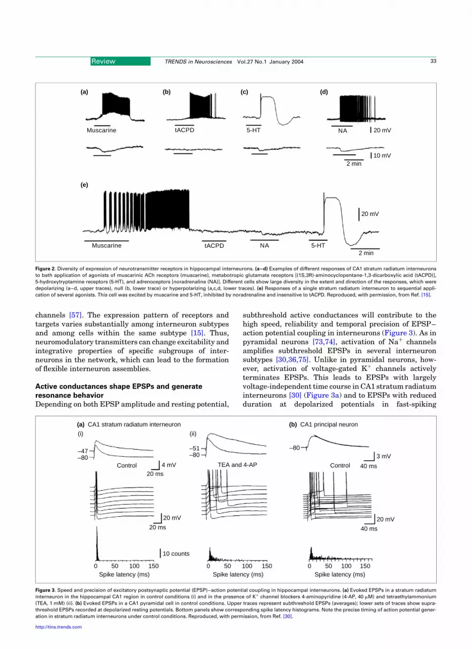
channels [57]. The expression pattern of receptors andtargets varies substantially among interneuron subtypesand among cells within the same subtype [15]. Thus,neuromodulatory transmitters can change excitability andintegrative properties of specific subgroups of inter-neurons in the network, which can lead to the formationof flexible interneuron assemblies.
Active conductances shape EPSPs and generate
resonance behavior
Depending on both EPSP amplitude and resting potential,
subthreshold active conductances will contribute to thehigh speed, reliability and temporal precision of EPSP–action potential coupling in interneurons (Figure 3). As inpyramidal neurons [73,74], activation of Naþ channelsamplifies subthreshold EPSPs in several interneuronsubtypes [30,36,75]. Unlike in pyramidal neurons, how-ever, activation of voltage-gated Kþ channels activelyterminates EPSPs. This leads to EPSPs with largelyvoltage-independent time course in CA1 stratum radiatuminterneurons [30] (Figure 3a) and to EPSPs with reducedduration at depolarized potentials in fast-spiking
Figure 2. Diversity of expression of neurotransmitter receptors in hippocampal interneurons. (a–d) Examples of different responses of CA1 stratum radiatum interneurons
to bath application of agonists of muscarinic ACh receptors (muscarine), metabotropic glutamate receptors [(1S,3R)-aminocyclopentane-1,3-dicarboxylic acid (tACPD)],
5-hydroxytryptamine receptors (5-HT), and adrenoceptors [noradrenaline (NA)]. Different cells show large diversity in the extent and direction of the responses, which were
depolarizing (a–d, upper traces), null (b, lower trace) or hyperpolarizing (a,c,d, lower traces). (e) Responses of a single stratum radiatum interneuron to sequential appli-
cation of several agonists. This cell was excited by muscarine and 5-HT, inhibited by noradrenaline and insensitive to tACPD. Reproduced, with permission, from Ref. [15].
Muscarine tACPD 5-HT NA
Muscarine tACPD 5-HTNA2 min
2 min
20 mV
10 mV
20 mV
(b) (c) (d)(a)
(e)
Figure 3. Speed and precision of excitatory postsynaptic potential (EPSP)–action potential coupling in hippocampal interneurons. (a) Evoked EPSPs in a stratum radiatum
interneuron in the hippocampal CA1 region in control conditions (i) and in the presence of Kþ channel blockers 4-aminopyridine (4-AP, 40 mM) and tetraethylammonium
(TEA, 1 mM) (ii). (b) Evoked EPSPs in a CA1 pyramidal cell in control conditions. Upper traces represent subthreshold EPSPs (averages); lower sets of traces show supra-
threshold EPSPs recorded at depolarized resting potentials. Bottom panels show corresponding spike latency histograms. Note the precise timing of action potential gener-
ation in stratum radiatum interneurons under control conditions. Reproduced, with permission, from Ref. [30].
4 mV
20 mV
10 counts
20 ms
20 ms
Spike latency (ms)
–47–80
20 mV
40 ms
3 mV
40 ms
–80–51–80
0 50 100 150Spike latency (ms)
0 50 100 150Spike latency (ms)
0 50 100 150
Control ControlTEA and 4-AP
(a) CA1 stratum radiatum interneuron (b) CA1 principal neuron
(i) (ii)
Review TRENDS in Neurosciences Vol.27 No.1 January 2004 33
http://tins.trends.com

neocortical interneurons [75]. Thus, EPSPs in inter-neurons trigger action potentials more precisely thanEPSPs in pyramidal cells [30] (Figure 3b). Experiments inwhich EPSP waveforms were applied as voltage-clampcommands revealed that subthreshold EPSPs activate abiphasic sequence of inward (Naþ)–outward (Kþ) currents,which restricts the time window for spike initiation [30,75].
Subthreshold active conductances also confer reson-ance characteristics on interneurons (Figure 4). Severalinterneurons show oscillations during sustained currentinjection or application of depolarizing agonists [76–79],with frequencies in the theta (4–7 Hz; Figure 4a,b) orgamma (30–90 Hz; Figure 4c,d) ranges. These oscillationsare apparently generated by the interplay of voltage-gatedNaþ channels and delayed-rectifier Kþ channels [76,77].Furthermore, interneurons show subthreshold resonantbehavior in response to sinusoidal inputs [78,80]. Finally,the reliability and temporal precision of action potentialgeneration shows marked frequency dependence inresponse to repetitive suprathreshold inputs; the highestreliability and precision of spiking occurs at frequenciessimilar to those of the subthreshold oscillations [78–81].Therefore, subthreshold oscillations and resonance beha-vior endow inhibitory interneurons with band-pass filtercharacteristics. The filter frequencies, however, differmarkedly among interneuron types. Regularly spikinginterneurons such as lacunosum-moleculare interneuronsof the CA1 region show theta-frequency preference [76,77](Figure 4a,b), whereas fast-spiking interneurons in hippo-campus, neocortex and striatum oscillate at gammafrequencies [78,79] (Figure 4c,d). Thus, rhythmic input
can recruit specific subsets of interneurons in neuronalnetworks. Although network oscillations are primarilydriven by synaptic mechanisms, they can be enhanced bysubthreshold oscillations and resonance phenomena, asdescribed in the article by Whittington and Traub in thisseries [11].
Molecular determinants of the fast-spiking phenotype
Mountcastle et al. [82] first described neocortical neuronswith ‘thin spikes’ in extracellular recordings. McCormicket al. [83] identified cortical cells with brief actionpotentials as GABAergic neurons and showed that thesecould fire repetitively without accommodation at frequen-cies .200 Hz. However, subsequent analysis of differentinterneuron subtypes indicated that many, but not all,interneurons show this fast-spiking phenotype (Figure 5).Basket cells, hilar interneurons with axons co-alignedwith the commissural–associational pathway (HICAP)cells in the dentate gyrus, and bistratified cells in CA1 andCA3 can all fire spikes at high frequency during sustainedcurrent injection (200–500 Hz at physiological tempera-ture) [17,42,43] (Figure 5a). Oriens alveus interneuronswere also categorized as ‘fast spiking’, although theirmaximal action potential frequency is closer to 100 Hzunder similar experimental conditions [84–90]. Finally,other interneuron types, such as stratum radiatum andlacunosum-moleculare interneurons in the CA1 region,even show regular spiking with substantial accommo-dation [91], comparable to principal neurons. Thus, theaction potential phenotype of interneurons is a spectrum,
Figure 4. Subthreshold oscillations in the theta-frequency and gamma-frequency bands. (a,b) Intrinsic theta oscillations in lacunosum-moleculare interneurons in CA1.
Membrane potential recordings are shown (mean values indicated) (a), with corresponding power spectra (b). The peak of the spectrum occurred roughly in the theta-
frequency range (4–7 Hz). (c,d) Intrinsic gamma oscillations in fast-spiking interneurons in the striatum. Membrane potential oscillations are shown from periods in which
no action potentials were generated (c), with the corresponding power spectrum (d). The peak of the spectrum occurred in the gamma-frequency range (30–90 Hz). Panels
(a,b) reproduced, with permission, from Ref. [77]; (c,d) reproduced, with permission, from Ref. [79].
CA1 interneuron Striatal interneuron
1 s4 mV
40 mV
400 ms
50 ms
4 mV
–52 mV–54 mV–56 mV–58 mV
Pow
er (
mV
2 /H
z)
00 5 10 15 20 100 200 300 400 Frequency (Hz)Frequency (Hz)
Pow
er (
mV
2 )
0.05
0.04
0.03
0.02
0.01
0.00
1.5
1.0
0.5
0.0
–52 mV
–54 mV
–56 mV
–58 mV
(a) (c)
(b) (d)
Review TRENDS in Neurosciences Vol.27 No.1 January 200434
http://tins.trends.com

with parvalbumin-positive basket cells representing thefunctional extreme.
Action potentials of fast-spiking interneurons aresucceeded by a fast, large-amplitude afterhyperpolariza-tion (AHP) [83,92]. This might seem counter-intuitive, as alarge AHP would be expected to delay the onset of the nextspike and thus reduce the firing frequency [93]. Howshould an AHP accelerate action potential initiation? Thekinetics of the AHP appear to be optimal for a maximalrecovery of Naþ channels from inactivation and a minimaldelay in the onset of the action potential initiation [90,94].A shorter AHP, by contrast, would not permit sufficientrecovery of Naþ channels from inactivation, leading tolonger refractory periods.
Which channels underlie the fast-spiking phenotype?Although the properties of both Naþ channels [94] andvoltage-gated Kþ channels [85–87,89] differ betweeninterneurons and principal cells, the characteristic actionpotential pattern of interneurons is largely shaped by theKþ channels. Kþ currents in basket cells of the dentategyrus and oriens alveus interneurons include fast delayed-rectifier, slow delayed-rectifier, inactivating A-type andCa2þ-activated Kþ currents [85–87,89]. The fast delayedrectifier forms a large part of the macroscopic Kþ currentfor both types of interneuron (Figure 5b). It contributes,60% of responses to rectangular stimuli, and .90% ofresponses to action-potential-like voltage-clamp com-mands [87] (Figure 5c). Immunocytochemical and single-cell reverse transcription polymerase chain reaction(RT-PCR) analysis revealed that the channels mediating
the fast delayed rectifier Kþ current are assembled fromKv3 subunits, particularly Kv3.1 and Kv3.2 [87,89,95–99](Figure 5d). Thus, the characteristic action potentialphenotype results from an interneuron-specific geneticprogram. How do Kv3 channels facilitate fast spiking?Dynamic-clamp experiments in which artificial Kþ con-ductances were added to real neurons [90,100] revealedthat the gating properties of Kv3 channels, in particularfast deactivation, high activation threshold and lack ofinactivation, are crucial for fast spiking. By contrast, a lowactivation threshold leads to adaptation, and an inacti-vation process leads to action potential broadening; theseare hallmarks of action potential phenotypes of principalcells [90]. As both density and gating properties of Kv3channels are regulated by gene expression and phos-phorylation [89,97,101], differential tuning of Kv3 chan-nels could contribute to the differences in firing propertiesbetween interneuron types.
Thus, many interneurons discharge brief action poten-tials at high frequencies without accommodation duringlong-lasting stimulation. Kv3 channels appear to be neces-sary for the fast-spiking phenotype, and their gating proper-ties favor high-frequency action potential generation.
Active dendritic propagation of action potentials
In pyramidal neurons, EPSPs propagate from the dendriteto the soma and the axon initial segment, the primary siteof integration and action potential initiation [102]. Bycontrast, interneurons can use a different signalingstrategy (Figure 6). First, in some interneuron subtypes
Figure 5. Kv3 channels – a major determinant of fast spiking. (a) Action potentials evoked by a 1 s depolarizing current pulse in a dentate gyrus basket cell. (b) Functional
properties of Kv3-like delayed-rectifier Kþ channels. Upper graph: traces of Kþ current evoked by test pulses of stepwise increasing amplitude in a nucleated patch from a
basket cell soma (inset). Lower graph: plot of conductance ratio (G/Gmax) against membrane potential. The Kv3 component was isolated by pharmacological subtraction.
(c) Kþ current in a basket cell nucleated patch during a high-frequency train of action potentials. An experimentally recorded action potential pattern was applied as a vol-
tage-clamp command, and the resulting Kþ current was measured in the absence (top trace) and in the presence (bottom trace) of 4-aminopyridine (4-AP, 0.2 mM). (d)
Single-cell reverse transcription polymerase chain reaction (RT-PCR) analysis of Kv3 subunit expression in dentate gyrus basket cells and CA1 pyramidal neurons. Ethidium
bromide-stained gels of the RT-PCR products amplified with primers specific for Kv3.2 transcripts. Left lanes show material from basket cells (BCs); right lanes show that
from pyramidal cells (PCs). Reproduced, with permission, from Ref [87], q (1998) the Society for Neuroscience.
Basket cell
200 ms
100
mV
50 p
A50
pA
100 ms
Control
4-AP 0.2 mM
1 nA
Kv3.2
635 bp517 bp
V (mV)
G/G
max
BCs PCs
0 20
30 ms
40 50 80–20–40–60–80
1.0
0.8
0.4
0.2
0.0
0.6
(a) (b)
(c) (d)
Review TRENDS in Neurosciences Vol.27 No.1 January 2004 35
http://tins.trends.com

the axon emerges from a principal dendrite rather thanthe soma. Dendritic origin of the axon, first described inGABAergic neurons of the substantia nigra [103], has beenreported for oriens alveus interneurons in the hippocam-pal CA1 region [36] (Figure 6a) and somatostatin-positivebipolar interneurons in the neocortex [104]. Second, thedendrites of some interneuron subtypes have activeproperties, supporting action potential backpropagationand dendritic action potential initiation [36] (Figure 6b–d).Both the dendritic emergence of the axon and the activeproperties of interneuron dendrites can speed up EPSP–action potential conversion.
Active properties of interneuron dendrites were firstpostulated on theoretical grounds [105] and more recentlyhave been demonstrated directly using dendritic record-ings. These experiments revealed that the dendrites oforiens alveus interneurons in the CA1 region of thehippocampus express a high density of voltage-gated Naþ
and Kþ channels; the estimated Naþ conductance density
was ,110 pS mm22 [36] (Figure 6b), approximately three-fold larger than in principal neuron dendrites [102].Furthermore, dendritic recordings showed that actionpotentials in both oriens alveus interneurons in hippo-campus and somatostatin-positive interneurons in neo-cortex can be initiated at multiple sites and propagate overthe somatodendritic domain with constant amplitude andtime course [36,104] (Figure 6c). Ca2þ-imaging experi-ments confirmed the active properties of interneurondendrites [104,106]; action-potential-induced dendriticCa2þ transients showed large amplitudes for distances ofup to 400 mm from the soma in somatostatin-positiveneocortical interneurons [104] (Figure 6d). Thus, voltage-gated Naþ channels apparently dominate the functionalproperties of dendrites of these interneurons, supportingthe active dendritic propagation of action potentials. Bycontrast, action-potential-induced Ca2þ transients declinedas a function of distance from the soma in parvalbumin-and calretinin-positive neocortical interneurons [106],
Figure 6. Active dendrites in somatostatin-positive interneuron subtypes. (a) Morphological reconstruction of an oriens alveus interneuron in the hippocampal CA1 region
(soma and dendrites, black; axon, red). Abbreviations: str. l.-m., stratum lacunosum-moleculare; str. ori., stratum oriens; str. pyr., stratum pyramidale; str. rad., stratum
radiatum. (b) Naþ (solid symbols and continuous lines) and Kþ (open symbols and broken lines) current densities, calculated from maximal inward or outward current at
210 mV and plotted against distance from the soma; positive values indicate the axon-bearing dendrite. Naþ and Kþ current traces are shown as insets. (c) Swapping of
action potential initiation between dendrite and soma. Simultaneous current-clamp recordings from dendrite (red traces) and soma (black traces) in an oriens alveus inter-
neuron. Action potential initiation was close to the dendritic recording site with a long pulse of low intensity (upper traces), but was shifted towards the somatic recording
site with a brief pulse of high intensity (lower traces). Numbers in (b) and (c) indicate dendritic recording distances. (d) Active dendrites in neocortical bitufted neurons.
Dependence on distance from the soma of the peak amplitude of intracellular Ca2þ concentration ([Ca2þ]I) transients evoked by four action potentials at 80 Hz. Data were
normalized to the amplitude of the dendritic [Ca2þ]i transient closest to the soma. Reconstruction of bitufted neuron and examples of [Ca2þ]i transients are shown as insets.
Panels (a–c) reproduced, with permission from, Ref. [36]; (d) reproduced, with permission, from Ref. [104].
Neocortical interneuron
Dendrite
Soma
Soma
Dendrite
1 ms10
0 m
V
(90 µm)
100 µm
60 ms
250
pA
2 ms
25 p
A
Distance from soma (µm) Distance from soma (µm)
I Na
and
I K (
pA µ
m–2
)
∆[C
a2+] i
(nor
mal
ized
)
50 µm
0.5 s
100 nM
0 0 100 200 300 400–40–80 40 80
100
80
60
40
20
0
3
2
1
0
4
Dendrite (65 µm) Dendrite (85 µm)
(a)
(b)
(c)
(d)
Review TRENDS in Neurosciences Vol.27 No.1 January 200436
http://tins.trends.com

indicating that voltage-gated Kþ channels could activelyinhibit the dendritic propagation of action potentials inthese interneurons.
In addition to acceleration of EPSP–action potentialconversion, active conductances in interneuron dendritescould serve several other functions. They might reducelocation-dependent variability in action potential initia-tion in response to different spatial patterns of synapticinputs [107] and facilitate retrograde propagation of actionpotentials from the soma into the dendrites [36,104].Backpropagated action potentials potentially contribute tothe induction of plasticity at glutamatergic synapses oninterneurons [108,109] and could trigger the release ofGABA and other retrograde messengers from interneurondendrites [110].
Synchronous GABA release at interneuron output
synapses
Once initiated, interneuron action potentials propagatealong the axon and trigger GABA release from inhibitoryterminals (Figure 7). The reliability of impulse conductionin morphologically complex interneuron axons isunknown. However, both the high release probability atthe basket cell–granule cell synapse in the dentate gyrus[111] and the reliability of action-potential-evoked Ca2þ
signals in axons of cerebellar interneurons [112] suggestthat conduction failures are rare. Kainate increases axonalexcitability of stratum radiatum interneurons in CA1 [28],suggesting that kainate receptors regulate impulse con-duction via depolarization of the axon. The shape ofpresynaptic action potentials that trigger Ca2þ inflow andtransmitter release at inhibitory terminals has not beenmeasured directly [113]. However, the highly synchronizedGABA release at the basket cell–granule cell synapse inthe dentate gyrus (half-duration of the time course ofquantal release is ,300 ms at 348C; Figure 7b,c) suggeststhat they might even be shorter than somatic spikes [111].
In addition to forming inhibitory synapses on principalneurons, interneurons also form synaptic contacts ontoeach other [114–119]. After all previous considerations, itmight not come as a surprise that the synaptic communi-cation among interneurons is particularly optimized forspeed. In comparison with interneuron–principal neuronsynapses, two major differences of interneuron–inter-neuron synapses are apparent. First, mutual synapticinhibition between hippocampal and neocortical inter-neurons is mediated by both electrical and chemicalsynapses [114–119]. Because electrical coupling is medi-ated by perisomatic gap junctions [120], the time course ofthe electrical postsynaptic currents is fast, following the
Figure 7. Fast signaling at GABAergic interneuron output synapses. (a) Morphological reconstruction of a reciprocally coupled granule cell (i.e. principal neuron; soma and
dendrites, green; axon, red) and basket cell (soma and dendrites, black; axon, blue) pair (the same neurons as shown in Figure 1). Two putative inhibitory synaptic contacts
(blue dots) are shown. (b) Recording of unitary inhibitory postsynaptic currents (IPSCs) at the basket cell (BC)–granule cell (PN) synapse at different Ca2þ and Mg2þ concen-
trations (2 mM Ca2þ; 1 mM Mg2þ and 0.5 mM Ca2þ; 2.5 mM Mg2þ). (c) Time course of quantal release at the basket cell–granule cell synapse. First latency distribution (filled
bars) and time course of quantal release (open bars), calculated from first latencies in 0.5 mM Ca2þ and 2.5 mM Mg2þ. (d) Synchrony of GABA release is independent of the
nature of the postsynaptic target cell, whereas the IPSC decay time constant is target-cell-dependent. Sequential triple recording from a presynaptic and a postsynaptic bas-
ket cell (left traces, first pair) and the same presynaptic basket cell and a postsynaptic granule cell (right traces, second pair). Presynaptic action potentials (top), single uni-
tary IPSCs (center) and average IPSC (bottom) are depicted. Note the difference in decay time course. Also note the fast current component due to gap-junction coupling in
the basket cell–basket cell connection. Panel (a) reproduced, with permission, from Ref [19]; (b) and (c) reproduced, with permission, from Ref [111], q (2000) the Society
for Neuroscience; (d) reproduced, with permission, from Ref [114], q (2001) the Society for Neuroscience.
(a)
(b)
(c)
BCPN
BC
BC PN
(d)
2 mM Ca2+
10 ms
10 ms
500
pA
500
pA
100 mV 0.5 mM Ca2+ 100 mV
10 ms
50 mV
50 pA100 pA
0 1.5 2.00.5 1.0Latency (ms)
40
30
20
10
0
Num
ber
of e
vent
s
Basket cell Principal neuron
CA3H
GCL
Review TRENDS in Neurosciences Vol.27 No.1 January 2004 37
http://tins.trends.com

time course of the presynaptic action potential. Second, thetime course of the GABA-mediated inhibitory postsynapticcurrents (IPSCs) in hippocampal interneurons and neo-cortical fast-spiking interneurons is markedly faster thanthe kinetics of IPSCs in principal neurons of the samecircuit [6,114,117] (Figure 7d). Thus, if two interneuronsare coupled via both electrical and chemical synapses, anaction potential in the presynaptic interneuron triggers abiphasic sequence of excitatory (inward)–inhibitory (out-ward) currents in the postsynaptic cell, promoting precise,synchronous action potential initiation.
Both electrical and chemical synaptic signalingbetween interneurons are crucial for network activity.Electrical coupling can selectively regulate the coherenceof high-frequency network oscillations, whereas the timecourse of the chemical GABAA-receptor-mediated com-ponent can control both coherence and frequency [6].Furthermore, both electrical coupling between inter-neurons and disynaptic feedforward inhibition (i.e. prin-cipal neurons excite an interneuron, which inhibitsanother interneuron) will promote synchrony detectionin interneuron networks [75], amplifying the fast signal-ing processes present at the level of a single cell. The earlyexcitatory electrical component will boost synchronousEPSPs, whereas the later inhibitory postsynaptic poten-tial (IPSP) will suppress asynchronous events. Thus,interneurons in the network can ultimately detect syn-chronous excitatory activity with ,1 ms precision [75].
Concluding remarks
In both hippocampal and neocortical microcircuits, GABA-ergic interneurons differ radically from glutamatergicprincipal cells. Some of the properties of interneurons arereminiscent of neurons in the auditory pathway, wheretemporal precision is crucial for sound localization.Remarkable similarities between interneurons and audi-tory neurons include the expression of special AMPAreceptors mediating fast synaptic excitation, the rapidmembrane time constant, and the expression of Kv3channels [121,122]. Together with other interneuron-specific mechanisms, these properties ensure that inhibi-tory interneurons operate with high speed, reliability andprecision. This is likely to be necessary for the generationof precise signals for temporal coding of information in thebrain and for the control over spike-timing-dependentplasticity at glutamatergic synapses [123–125].
AcknowledgementsWe thank Marlene Bartos, Jan Behrends, Michael Hausser, ManfredHeckmann, Chris McBain, Jean-Christophe Poncer, Greg Stuart and ImreVida for reading previous versions of the manuscript. Work of the authorswas supported by the Deutsche Forschungsgemeinschaft, the Alexander-von-Humboldt Foundation, the Human Frontiers Science ProgramOrganization, INSERM, the National Institutes of Health (MH54671),and the Ministere de la Recherche
References
1 Cobb, S.R. et al. (1995) Synchronization of neuronal activity inhippocampus by individual GABAergic interneurons. Nature 378,75–78
2 Pouille, F. and Scanziani, M. (2001) Enforcement of temporal fidelityin pyramidal cells by somatic feed-forward inhibition. Science 293,1159–1163
3 Miles, R. et al. (1996) Differences between somatic and dendriticinhibition in the hippocampus. Neuron 16, 815–823
4 Whittington, M.A. et al. (1995) Synchronized oscillations in inter-neuron networks driven by metabotropic glutamate receptor acti-vation. Nature 373, 612–615
5 Wang, X-J. and Buzsaki, G. (1996) Gamma oscillation by synapticinhibition in a hippocampal interneuronal network model.J. Neurosci. 16, 6402–6413
6 Bartos, M. et al. (2002) Fast synaptic inhibition promotes synchro-nized gamma oscillations in hippocampal interneuron networks.Proc. Natl. Acad. Sci. U. S. A. 99, 13222–13227
7 Bragin, A. et al. (1995) Gamma (40–100 Hz) oscillation in thehippocampus of the behaving rat. J. Neurosci. 15, 47–60
8 Ylinen, A. et al. (1995) Sharp wave-associated high-frequencyoscillation (200 Hz) in the intact hippocampus: Network andintracellular mechanisms. J. Neurosci. 15, 30–46
9 Klausberger, T. et al. (2003) Brain-state- and cell-type-specific firingof hippocampal interneurons in vivo. Nature 421, 844–848
10 Buzsaki, G. and Henze, D. Interneuron Diversity series: GABAergicsystems in vivo. Trends Neurosci. (in press)
11 Whittington, M.A. and Traub, R.D. (2003) Interneuron Diversityseries: Inhibitory interneurons and network oscillations in vitro.Trends Neurosci. doi:10.1016/j.tins.2003.09.016
12 Miles, R. (1990) Synaptic excitation of inhibitory cells by single CA3hippocampal pyramidal cells of the guinea-pig in vitro. J. Physiol.
428, 61–7713 Csicsvari, J. et al. (1998) Reliability and state dependence of
pyramidal cell–interneuron synapses in the hippocampus: anensemble approach in the behaving rat. Neuron 21, 179–189
14 Carter, A.G. and Regehr, W.G. (2002) Quantal events shape cerebellarinterneuron firing. Nat. Neurosci. 5, 1309–1318
15 Parra, P. et al. (1998) How many subtypes of inhibitory cells in thehippocampus? Neuron 20, 983–993
16 Vida, I. et al. (1998) Unitary IPSPs evoked by interneurons at thestratum radiatum–stratum lacunosum-moleculare border in the CA1area of the rat hippocampus in vitro. J. Physiol. 506, 755–773
17 Han, Z-S. et al. (1993) A high degree of spatial selectivity in the axonaland dendritic domains of physiologically identified local-circuitneurons in the dentate gyrus of the rat hippocampus. Eur.J. Neurosci. 5, 395–410
18 Sik, A. et al. (1995) Hippocampal CA1 interneurons: an in vivointracellular labeling study. J. Neurosci. 15, 6651–6665
19 Geiger, J.R.P. et al. (1997) Submillisecond AMPA receptor-mediatedsignaling at a principal neuron–interneuron synapse. Neuron 18,1009–1023
20 Walker, H.C. et al. (2002) Activation of kinetically distinct synapticconductances on inhibitory interneurons by electrotonically over-lapping afferents. Neuron 35, 161–171
21 Jonas, P. et al. (1993) Quantal components of unitary EPSCs at themossy fibre synapse on CA3 pyramidal cells of rat hippocampus.J. Physiol. 472, 615–663
22 Vida, I. and Frotscher, M. (2000) A hippocampal interneuronassociated with the mossy fiber system. Proc. Natl. Acad. Sci.U. S. A. 97, 1275–1280
23 Lawrence, J.J. and McBain, C.J. (2003) Interneuron Diversity series:Containing the detonation – feedforward inhibition in the CA3hippocampus. Trends Neurosci. doi:10.1016/j.tins.2003.09.007
24 Geiger, J.R.P. et al. (1995) Relative abundance of subunit mRNAsdetermines gating and Ca2þ permeability of AMPA receptors inprincipal neurons and interneurons in rat CNS. Neuron 15, 193–204
25 Bekkers, J.M. and Stevens, C.F. (1989) NMDA and non-NMDAreceptors are co-localized at individual excitatory synapses incultured rat hippocampus. Nature 341, 230–233
26 Cossart, R. et al. (1998) GluR5 kainate receptor activation ininterneurons increases tonic inhibition of pyramidal cells. Nat.
Neurosci. 1, 470–47827 Frerking, M. et al. (1998) Synaptic activation of kainate receptors on
hippocampal interneurons. Nat. Neurosci. 1, 479–48628 Semyanov, A. and Kullmann, D.M. (2001) Kainate receptor-depen-
dent axonal depolarization and action potential initiation ininterneurons. Nat. Neurosci. 4, 718–723
29 Lei, S. and McBain, C.J. (2002) Distinct NMDA receptors provide
Review TRENDS in Neurosciences Vol.27 No.1 January 200438
http://tins.trends.com

differential modes of transmission at mossy fiber–interneuronsynapses. Neuron 33, 921–933
30 Fricker, D. and Miles, R. (2000) EPSP amplification and the precisionof spike timing in hippocampal neurons. Neuron 28, 559–569
31 Maccaferri, G. and Dingledine, R. (2002) Control of feedforwarddendritic inhibition by NMDA receptor-dependent spike timing inhippocampal interneurons. J. Neurosci. 22, 5462–5472
32 Bischofberger, J. and Jonas, P. (2002) TwoB or not twoB: Differentialtransmission at glutamatergic mossy fiber–interneuron synapses inthe hippocampus. Trends Neurosci. 25, 600–603
33 Jack, J.J.B. et al. (1983) Electric Current Flow in Excitable Cells,Clarendon Press
34 Johnston, D. and Wu, S.M-S. (1995) Foundations of CellularNeurophysiology, MIT Press
35 Gulyas, A.I. et al. (1999) Total number and ratio of excitatory andinhibitory synapses converging onto single interneurons of differenttypes in the CA1 area of the rat hippocampus. J. Neurosci. 19,10082–10097
36 Martina, M. et al. (2000) Distal initiation and active propagation ofaction potentials in interneuron dendrites. Science 287, 295–300
37 Softky, W. (1994) Sub-millisecond coincidence detection in activedendritic trees. Neuroscience 58, 13–41
38 Stuart, G. et al. (1999) Dendrites, Oxford University Press39 Geiger, J.R.P. et al. (1999) Glutamate-mediated synaptic excitation of
cortical interneurons. In Ionotropic Glutamate Receptors in the CNS.Handbook of Experimental Pharmacology (Vol. 141) (Jonas, P. andMonyer, H., eds), pp. 363–398, Springer
40 Emri, Z.S. et al. (2001) Electrotonic profile and passive propagation ofsynaptic potentials in three subpopulations of hippocampal CA1interneurons. Neuroscience 104, 1013–1026
41 Morin, F. et al. (1996) Membrane properties and synaptic currentsevoked in CA1 interneuron subtypes in rat hippocampal slices.J. Neurophysiol. 76, 1–16
42 Mott, D.D. et al. (1997) Interneurons of the dentate–hilus border ofthe rat dentate gyrus: morphological and electrophysiologicalheterogeneity. J. Neurosci. 17, 3990–4005
43 Lubke, J. et al. (1998) Specialized electrophysiological properties ofanatomically identified neurons in the hilar region of the rat fasciadentata. J. Neurophysiol. 79, 1518–1534
44 Spruston, N. and Johnston, D. (1992) Perforated patch-clampanalysis of the passive membrane properties of three classes ofhippocampal neurons. J. Neurophysiol. 67, 508–529
45 Scharfman, H.E. et al. (1990) Synaptic connections of dentate granulecells and hilar neurons: results of paired intracellular recordings andintracellular horseradish peroxidase injections. Neuroscience 37,693–707
46 Debanne, D. et al. (1995) Physiology and pharmacology of unitarysynaptic connections between pairs of cells in areas CA3 and CA1 ofrat hippocampal slice cultures. J. Neurophysiol. 73, 1282–1294
47 Buhl, E.H. et al. (1997) Effect, number and location of synapses madeby single pyramidal cells onto aspiny interneurones of cat visualcortex. J. Physiol. 500, 689–713
48 Lacaille, J-C. et al. (1987) Local circuit interactions between oriens/alveus interneurons and CA1 pyramidal cells in hippocampal slices:electrophysiology and morphology. J. Neurosci. 7, 1979–1993
49 Ali, A.B. and Thomson, A.M. (1998) Facilitating pyramid tohorizontal oriens-alveus interneurone inputs: dual intracellularrecordings in slices of rat hippocampus. J. Physiol. 507, 185–199
50 Angulo, M.C. et al. (1999) Postsynaptic glutamate receptors andintegrative properties of fast-spiking interneurons in the ratneocortex. J. Neurophysiol. 82, 1295–1302
51 Markram, H. et al. (1997) Physiology and anatomy of synapticconnections between thick tufted pyramidal neurones in the devel-oping rat neocortex. J. Physiol. 500, 409–440
52 Eccles, J.C. (1969) The Inhibitory Pathways of the Central Nervous
System, Liverpool University Press53 Fricker, D. et al. (1999) Cell-attached measurements of the firing
threshold of rat hippocampal neurones. J. Physiol. 517, 791–80454 Verheugen, J.A.H. et al. (1999) Noninvasive measurements of the
membrane potential and GABAergic action in hippocampal inter-neurons. J. Neurosci. 19, 2546–2555
55 Ross, S.T. and Soltesz, I. (2000) Selective depolarization of inter-
neurons in the early posttraumatic dentate gyrus: Involvement of theNaþ/Kþ-ATPase. J. Neurophysiol. 83, 2916–2930
56 Koh, D-S. et al. (1992) A TEA-insensitive flickering potassiumchannel active around the resting potential in myelinated nerve.J. Membr. Biol. 130, 149–162
57 Gorelova, N. et al. (2002) Mechanisms of dopamine activation of fast-spiking interneurons that exert inhibition in rat prefrontal cortex.J. Neurophysiol. 88, 3150–3166
58 Goldstein, S.A.N. et al. (2001) Potassium leak channels and theKCNK family of two-P-domain subunits. Nat. Rev. Neurosci. 2,175–184
59 Maccaferri, G. and McBain, C.J. (1996) The hyperpolarization-activated current (Ih) and its contribution to pacemaker activity inrat CA1 hippocampal stratum oriens-alveus interneurones.J. Physiol. 497, 119–130
60 Lupica, C.R. et al. (2001) Contribution of the hyperpolarization-activated current (Ih) to membrane potential and GABA release inhippocampal interneurons. J. Neurophysiol. 86, 261–268
61 Fraser, D.D. and MacVicar, B.A. (1991) Low-threshold transientcalcium current in rat hippocampal lacunosum-moleculare inter-neurons: kinetics and modulation by neurotransmitters. J. Neurosci.11, 2812–2820
62 Jones, S. and Yakel, J.L. (1997) Functional nicotinic ACh receptors oninterneurones in the rat hippocampus. J. Physiol. 504, 603–610
63 McQuiston, A.R. and Madison, D.V. (1999) Nicotinic receptoractivation excites distinct subtypes of interneurons in the rathippocampus. J. Neurosci. 19, 2887–2896
64 Semyanov, A. et al. (2003) GABA uptake regulates cortical excitabilityvia cell type-specific tonic inhibition. Nat. Neurosci. 6, 484–490
65 McQuiston, A.R. and Madison, D.V. (1999) Muscarinic receptoractivity has multiple effects on the resting membrane potentials ofCA1 hippocampal interneurons. J. Neurosci. 19, 5693–5702
66 Behrends, J.C. and ten Bruggencate, G. (1993) Cholinergic modu-lation of synaptic inhibition in the guinea pig hippocampus in vitro:Excitation of GABAergic interneurons and inhibition of GABA-release. J. Neurophysiol. 69, 626–629
67 Hajos, N. et al. (1998) Distinct interneuron types express M2muscarinic receptor immunoreactivity on their dendrites or axonterminals in the hippocampus. Neuroscience 82, 355–376
68 McBain, C.J. et al. (1994) Activation of metabotropic glutamatereceptors differentially affects two classes of hippocampal inter-neurons and potentiates excitatory synaptic transmission.J. Neurosci. 14, 4433–4445
69 Bergles, D.E. et al. (1996) Excitatory actions of norepinephrine onmultiple classes of hippocampal CA1 interneurons. J. Neurosci. 16,572–585
70 Mott, D.D. et al. (1999) GABAB-receptor-mediated currents ininterneurons of the dentate–hilus border. J. Neurophysiol. 82,1438–1450
71 Svoboda, K.R. and Lupica, C.R. (1998) Opioid inhibition of hippo-campal interneurons via modulation of potassium and hyperpolar-ization-activated cation (Ih) currents. J. Neurosci. 18, 7084–7098
72 Zhou, F-M. and Hablitz, J.J. (1999) Dopamine modulation ofmembrane and synaptic properties of interneurons in rat cerebralcortex. J. Neurophysiol. 81, 967–976
73 Magee, J.C. and Johnston, D. (1995) Synaptic activation of voltage-gated channels in the dendrites of hippocampal pyramidal neurons.Science 268, 301–304
74 Stuart, G. and Sakmann, B. (1995) Amplification of EPSPs byaxosomatic sodium channels in neocortical pyramidal neurons.Neuron 15, 1065–1076
75 Galarreta, M. and Hestrin, S. (2001) Spike transmission andsynchrony detection in networks of GABAergic interneurons. Science292, 2295–2299
76 Chapman, C.A. and Lacaille, J-C. (1999) Cholinergic induction oftheta-frequency oscillations in hippocampal inhibitory interneuronsand pacing of pyramidal cell firing. J. Neurosci. 19, 8637–8645
77 Chapman, C.A. and Lacaille, J-C. (1999) Intrinsic theta-frequencymembrane potential oscillations in hippocampal CA1 interneurons ofstratum lacunosum-moleculare. J. Neurophysiol. 81, 1296–1307
78 Fellous, J-M. et al. (2001) Frequency dependence of spike timingreliability in cortical pyramidal cells and interneurons.J. Neurophysiol. 85, 1782–1787
Review TRENDS in Neurosciences Vol.27 No.1 January 2004 39
http://tins.trends.com

79 Bracci, E. et al. (2003) Voltage-dependent membrane potentialoscillations of rat striatal fast-spiking interneurons. J. Physiol. 549,121–130
80 Pike, F.G. et al. (2000) Distinct frequency preferences of differenttypes of rat hippocampal neurones in response to oscillatory inputcurrents. J. Physiol. 529, 205–213
81 Richardson, M.J.E. et al. (2003) From subthreshold to firing-rateresonance. J. Neurophysiol. 89, 2538–2554
82 Mountcastle, V.B. et al. (1969) Cortical neuronal mechanisms influtter-vibration, studied in unanesthetized monkeys. Neuronalperiodicity and frequency discrimination. J. Neurophysiol. 32,452–484
83 McCormick, D.A. et al. (1985) Comparative electrophysiology ofpyramidal and sparsely spiny stellate neurons of the neocortex.J. Neurophysiol. 54, 782–806
84 Lacaille, J-C. and Williams, S. (1990) Membrane properties ofinterneurons in stratum oriens-alveus of the CA1 region of rathippocampus in vitro. Neuroscience 36, 349–359
85 Zhang, L. and McBain, C.J. (1995) Voltage-gated potassium currentsin stratum oriens-alveus inhibitory neurones of the rat CA1hippocampus. J. Physiol. 488, 647–660
86 Zhang, L. and McBain, C.J. (1995) Potassium conductances under-lying repolarization and afterhyperpolarization in rat CA1 hippo-campal interneurones. J. Physiol. 488, 661–672
87 Martina, M. et al. (1998) Functional and molecular differencesbetween voltage-gated Kþ channels of fast-spiking interneurons andpyramidal neurons of rat hippocampus. J. Neurosci. 18, 8111–8125
88 Oliva, A.A. Jr et al. (2000) Novel hippocampal interneuronal subtypesidentified using transgenic mice that express green fluorescentprotein in GABAergic interneurons. J. Neurosci. 20, 3354–3368
89 Lien, C-C. et al. (2002) Gating, modulation and subunit composition ofvoltage-gated Kþ channels in dendritic inhibitory interneurones ofrat hippocampus. J. Physiol. 538, 405–419
90 Lien, C-C. and Jonas, P. (2003) Kv3 potassium conductance isnecessary and kinetically optimized for high-frequency actionpotential generation in hippocampal interneurons. J. Neurosci. 23,2058–2068
91 Lacaille, J-C. and Schwartzkroin, P.A. (1988) Stratum lacunosum-moleculare interneurons of hippocampal CA1 region. I. Intracellularresponse characteristics, synaptic responses, and morphology.J. Neurosci. 8, 1400–1410
92 Connors, B.W. and Gutnick, M.J. (1990) Intrinsic firing patterns ofdiverse neocortical neurons. Trends Neurosci. 13, 99–104
93 Hille, B. (2001) Ion Channels of Excitable Membranes, 3rd edn,Sinauer
94 Martina, M. and Jonas, P. (1997) Functional differences in Naþ
channel gating between fast-spiking interneurones and principalneurones in rat hippocampus. J. Physiol. 505, 593–603
95 Du, J. et al. (1996) Developmental expression and functionalcharacterization of the potassium-channel subunit Kv3.1b in parv-albumin-containing interneurons of the rat hippocampus.J. Neurosci. 16, 506–518
96 Erisir, A. et al. (1999) Function of specific Kþ channels in sustainedhigh-frequency firing of fast-spiking neocortical interneurons.J. Neurophysiol. 82, 2476–2489
97 Atzori, M. et al. (2000) H2 histamine receptor-phosphorylation ofKv3.2 modulates interneuron fast spiking. Nat. Neurosci. 3, 791–798
98 Lau, D. et al. (2000) Impaired fast-spiking, suppressed corticalinhibition, and increased susceptibility to seizures in mice lackingKv3.2 Kþ channel proteins. J. Neurosci. 20, 9071–9085
99 Rudy, B. and McBain, C.J. (2001) Kv3 channels: voltage-gated Kþ
channels designed for high-frequency repetitive firing. TrendsNeurosci. 24, 517–526
100 Ma, M. and Koester, J. (1996) The role of Kþ currents in frequency-dependent spike broadening in Aplysia R20 neurons: A dynamic-clamp analysis. J. Neurosci. 16, 4089–4101
101 Macica, C.M. and Kaczmarek, L.K. (2001) Casein kinase 2 deter-mines the voltage dependence of the Kv3.1 channel in auditoryneurons and transfected cells. J. Neurosci. 21, 1160–1168
102 Stuart, G.J. and Sakmann, B. (1994) Active propagation of somatic
action potentials into neocortical pyramidal cell dendrites. Nature367, 69–72
103 Hausser, M. et al. (1995) Axonal initiation and active dendriticpropagation of action potentials in substantia nigra neurons. Neuron15, 637–647
104 Kaiser, K.M.M. et al. (2001) Back-propagating action potentialsmediate calcium signalling in dendrites of bitufted interneurons inlayer 2/3 of rat somatosensory cortex. J. Physiol. 535, 17–31
105 Traub, R.D. and Miles, R. (1995) Pyramidal cell-to-inhibitory cellspike transduction explicable by active dendritic conductances ininhibitory cell. J. Comput. Neurosci. 2, 291–298
106 Goldberg, J.H. et al. (2003) Ca2þ imaging of mouse neocorticalinterneurone dendrites: Ia-type Kþ channels control action potentialbackpropagation. J. Physiol. 551, 49–65
107 Cook, E.P. and Johnston, D. (1997) Active dendrites reduce location-dependent variability of synaptic input trains. J. Neurophysiol. 78,2116–2128
108 Perez, Y. et al. (2001) A hebbian form of long-term potentiationdependent on mGluR1a in hippocampal inhibitory interneurons.Proc. Natl. Acad. Sci. U. S. A. 98, 9401–9406
109 Alle, H. et al. (2001) PTP and LTP at a hippocampal mossy fiber–interneuron synapse. Proc. Natl. Acad. Sci. U. S. A. 98, 14708–14713
110 Zilberter, Y. et al. (1999) Dendritic GABA release depresses excitatorytransmission between layer 2/3 pyramidal and bitufted neurons in ratneocortex. Neuron 24, 979–988
111 Kraushaar, U. and Jonas, P. (2000) Efficacy and stability of quantalGABA release at a hippocampal interneuron–principal neuronsynapse. J. Neurosci. 20, 5594–5607
112 Forti, L. et al. (2000) Action potential-evoked Ca2þ signals andcalcium channels in axons of developing rat cerebellar interneurones.J. Physiol. 527, 33–48
113 Southan, A.P. et al. (2000) Hyperpolarization-activated currents inpresynaptic terminals of mouse cerebellar basket cells. J. Physiol.526, 91–97
114 Bartos, M. et al. (2001) Rapid signaling at inhibitory synapses in adentate gyrus interneuron network. J. Neurosci. 21, 2687–2698
115 Gibson, J.R. et al. (1999) Two networks of electrically coupledinhibitory neurons in neocortex. Nature 402, 75–79
116 Galarreta, M. and Hestrin, S. (1999) A network of fast-spiking cells inthe neocortex connected by electrical synapses. Nature 402, 72–75
117 Galarreta, M. and Hestrin, S. (2002) Electrical and chemical synapsesamong parvalbumin fast-spiking GABAergic interneurons in adultmouse neocortex. Proc. Natl. Acad. Sci. U. S. A. 99, 12438–12443
118 Deans, M.R. et al. (2001) Synchronous activity of inhibitory networksin neocortex requires electrical synapses containing connexin36.Neuron 31, 477–485
119 Hormuzdi, S.G. et al. (2001) Impaired electrical signaling disruptsgamma frequency oscillations in connexin 36-deficient mice. Neuron31, 487–495
120 Tamas, G. et al. (2000) Proximally targeted GABAergic synapses andgap junctions synchronize cortical interneurons. Nat. Neurosci. 3,366–371
121 Trussell, L.O. (1999) Physiology of glutamatergic transmission atcalyceal and endbulb synapses of the central auditory pathway. InIonotropic Glutamate Receptors in the CNS. Handbook of Exper-imental Pharmacology (Vol. 141) (Jonas, P. and Monyer, H., eds),pp. 399–418, Springer
122 Wang, L-Y. et al. (1998) Contribution of the Kv3.1 potassium channelto high-frequency firing in mouse auditory neurones. J. Physiol. 509,183–194
123 Fuchs, E.C. et al. (2001) Genetically altered AMPA-type glutamatereceptor kinetics in interneurons disrupt long-range synchrony ofgamma oscillation. Proc. Natl. Acad. Sci. U. S. A. 98, 3571–3576
124 Katona, I. et al. (1999) Postsynaptic targets of somatostatin-immunoreactive interneurons in the rat hippocampus. Neuroscience88, 37–55
125 Normann, C. et al. (2000) Associative long-term depression in thehippocampus is dependent on postsynaptic N-type Ca2þ channels.J. Neurosci. 20, 8290–8297
Review TRENDS in Neurosciences Vol.27 No.1 January 200440
http://tins.trends.com