Embed Size (px)
Citation preview

Molecular Biology of the CellVol. 10, 4355–4367, December 1999
Involvement of Type 4 cAMP-Phosphodiesterase inthe Myogenic Differentiation of L6 CellsFabio Naro,* Claudio Sette,† Elena Vicini,* Vania De Arcangelis,*Muriel Grange,‡ Marco Conti,§ Michel Lagarde,‡ Mario Molinaro,*Sergio Adamo,*i¶ and Georges Nemoz‡i
*Dipartimento di Istologia ed Embriologia Medica, Universita “La Sapienza,” 00161 Rome, Italy;†Dipartimento di Sanita Pubblica e Biologia Cellulare, Universita “Tor Vergata,” 00133 Rome, Italy;‡Unite 352, Institut National de la Sante et de la Recherche Medicale, Biochimie et Pharmacologie,Institut National des Sciences Appliquees de Lyon, 69621 Villeurbanne, France; and §Division ofReproductive Biology, Department of Gynecology and Obstetrics, Stanford University School ofMedicine, Stanford, California 94305
Submitted September 7, 1999; Accepted October 4, 1999Monitoring Editor: Marc Mumby
Myogenic cell differentiation is induced by Arg8-vasopressin, whereas high cAMP levels andprotein kinase A (PKA) activity inhibit myogenesis. We investigated the role of type 4 phospho-diesterase (PDE4) during L6-C5 myoblast differentiation. Selective PDE4 inhibition resulted insuppression of differentiation induced by vasopressin. PDE4 inhibition prevented vasopressin-induced nuclear translocation of the muscle-specific transcription factor myogenin without affect-ing its overall expression level. The effects of PDE4 inhibition could be attributed to an increaseof cAMP levels and PKA activity. RNase protection, reverse transcriptase PCR, immunoprecipi-tation, Western blot, and enzyme activity assays demonstrated that the PDE4D3 isoform is themajor PDE4 expressed in L6-C5 myoblasts and myotubes, accounting for 75% of total cAMP-hydrolyzing activity. Vasopressin cell stimulation caused a biphasic increase of PDE4 activity,which peaked at 2 and 15 min and remained elevated for 48 h. In the continuous presence ofvasopressin, cAMP levels and PKA activity were lowered. PDE4D3 overexpression increasedspontaneous and vasopressin-dependent differentiation of L6-C5 cells. These results show thatPDE4D3 plays a key role in the control of cAMP levels and differentiation of L6-C5 cells. Throughthe modulation of PDE4 activity, vasopressin inhibits the cAMP signal transduction pathway,which regulates myogenesis possibly by controlling the subcellular localization of myogenin.
INTRODUCTION
During skeletal muscle development, cells of mesodermalorigin become committed to the myogenic lineage, migratetoward their final destination, and become postmitotic(Cossu et al., 1996). Myoblasts fuse into multinucleated myo-tubes and begin accumulating muscle-specific products(e.g., M-creatine kinase, myosin and other sarcomeric pro-teins, and acetylcholine receptor subunits; O’Neill and
Stockdale 1972; Nadal-Ginard 1978). This process is markedby the sequential expression of specific genes and requiresan adequate level of functional muscle regulatory factors(myogenin, myoD, myf-5, and mrf-4) whose temporal ex-pression pattern and relative role may vary among organ-isms and experimental models of myogenic differentiation(Braun et al., 1989; Ludolph and Konieczny 1995).
Several cultured cell models allow the study of at leastportions of the myogenic developmental process. Both invivo and in culture, myogenic differentiation is under theinfluence of both inhibitory and stimulatory extracellularsignals (Olson et al., 1986; Clegg et al., 1987; Florini et al.,1991). Until recently, insulin-like growth factors (IGFs) wereconsidered as the main myogenic differentiation factors (Flo-rini 1987; Engert et al., 1996). We reported that Arg8-vaso-pressin (AVP) also acts as a positive effector in several typesof skeletal myogenic cells, including rat L6-C5 myoblasts(Nervi et al., 1995; Minotti et al., 1998). Unlike IGFs, which
i Joint senior authors.¶ Corresponding author. E-mail address: [email protected].
Abbreviations: AVP, Arg8-vasopressin; b-gal; b-galactosidase;8-Br-cAMP, 8-bromo-cAMP; CK, creatine kinase; CMV, cyto-megalovirus; DME, Dulbecco’s modified Eagle’s; IGF1, insulin-like growth factor 1; PA, phosphatidic acid; PDE, phosphodies-terase; PKA, protein kinase A; PKI, protein kinase inhibitor;RPA, RNase protection assay; RT, reverse transcriptase.
© 1999 by The American Society for Cell Biology 4355

require the presence of other factors supplied by serum toexpress their full myogenic potential, AVP promotes myo-genic differentiation in the absence of additional exogenousfactors (Minotti et al., 1998). AVP induces the activation ofboth phospholipase C and phospholipase D in L6-C5 myo-genic cells. However, the concentrations of AVP required toinduce myogenesis are compatible only with the activationof phospholipase D, whereas 100-fold higher AVP doses arerequired to trigger phospholipase C stimulation (Teti et al.,1993; Naro et al., 1997).
It is well established that elevation of intracellular levels ofthe second messenger cAMP is sufficient to silence the myo-genic program (Wahrman et al., 1973; Winter et al., 1993).Elevated cAMP levels inhibit both the expression of endog-enous myogenin and the transcriptional activity of a trans-fected myogenin promoter (Salminen et al., 1991). Further-more, overexpression of the catalytic subunit of proteinkinase A (PKA) inhibits myogenic differentiation (Winter etal., 1993). PKA has also been shown to phosphorylate over-expressed myogenin in COS-1 cells, although this phosphor-ylation did not affect the ability of myogenin to bind to DNA(Li et al., 1992). Thus, although the exact mechanism is stillunknown, cAMP and PKA exert a negative effect on myo-genic cell differentiation.
The intracellular cAMP concentration is regulated at thesynthesis level by adenylyl cyclase and at the hydrolysislevel by phosphodiesterase (PDE) activity. Among the 10families of PDEs described in mammalian tissues, the PDE4family specifically hydrolyzes cAMP with high affinity(Conti et al., 1995; Soderling et al., 1998). This family includesa number of isoforms deriving from the expression of fourgenes in rat (Colicelli et al., 1989; Davis et al., 1989; Swinnenet al., 1989) and in human (Bolger et al., 1993). These isoformsshare the cAMP specificity and the sensitivity to inhibitionby rolipram (Beavo, 1995; Conti et al., 1995). In L6, as well asin other cell types, the expression of the PDE4D gene, one ofthe four genes encoding type 4 PDEs, is up-regulated bycAMP (Kovala et al., 1994; Conti et al., 1995; Vicini and Conti,1997). In addition to transcriptional regulation, it has beenshown that one of the isoforms deriving from PDE4D geneexpression, PDE4D3, is regulated by PKA-dependent phos-phorylation (Sette et al., 1994a,c; Sette and Conti 1996). ThePDE4D3 isoform contains an amino-terminal regulatory re-gion, which is absent in other isoforms derived from thesame gene, and phosphorylation of serine 54 in this regionrelieves an inhibitory constraint and activates the enzyme(Sette and Conti 1996). Activation of PDE4D3 is also ob-tained by interaction of this region of the enzyme withnegatively charged phospholipids, such as phosphatidicacid (PA) (Nemoz et al., 1997; Grange et al., 1998). Thetranscriptional and posttranslational regulation of PDE4 ac-tivity has been proposed to play a role in short- and long-term desensitization to hormonal stimulation of target cells(Sette et al., 1994b; Vicini and Conti 1997) (reviewed by Contiet al., 1995).
Although cAMP plays a negative role during myogenicdifferentiation, scarce information is available on the role ofPDE activity in this process and its regulation by extracel-lular factors. To address this point, in the present study, wefirst investigated the effect of PDE4-specific inhibitors onL6-C5 cell differentiation and observed that inhibition ofPDE4 strongly suppresses myogenesis. We characterized the
PDE4 isoforms expressed in L6-C5 cells and showed thatAVP stimulation modulates PDE4 activity. The observationthat AVP markedly stimulates PDE4 and decreases cAMPlevels and PKA activity in differentiating L6-C5 cells led usto hypothesize that PDE4 modulation plays a physiologicalrole in myogenic differentiation.
MATERIALS AND METHODS
MaterialsSynthetic AVP, snake venom from Crotalus atrox, Kemptide, PKApeptide inhibitor (PKI), a creatine kinase (CK) assay kit, and Trireagent were purchased from Sigma (St. Louis, MO). Rolipram[(4,3-butoxy-4-methoxybenzyl)-2-imidazolidone], milrinone, andzaprinast were obtained from Calbiochem (La Jolla, CA). IGF1 waspurchased from Chemicon (Temecula, CA). RS 23544 was a kind giftfrom Dr. R. Alvarez (Syntex, Palo Alto, CA). Fatty acid-free BSA,Fugene 6, and PCR reagents were from Boehringer Mannheim(Indianapolis, IN). The anti-myogenin mAb F5D developed by Dr.W.E. Wright (University of Texas, Dallas, TX) was obtained from theDevelopmental Studies Hybridoma Bank maintained by the Depart-ment of Biological Sciences, University of Iowa (Iowa City, IA); themAb to sarcomeric myosin MF20 was a kind gift from Dr. D.Fischman (Cornell University Medical College, New York, NY);anti-PKA catalytic and regulatory subunit (Ia and IIa) antibodieswere from Transduction Laboratories (Lexington, KY). [g-32P]ATP(3000 Ci/mmol) and a cAMP-125I radioimmunoassay kit were fromDuPont NEN (Boston, MA). [3H]cAMP and an ECL Western blotdetection kit were from Amersham Pharmacia Biotech (Uppsala,Sweden). AG1-X2 resin was from Bio-Rad (Hercules, CA).
Cell CultureSubcloning and characterization of L6 (Yaffe 1968) rat myogenic cellclones were previously reported (Teti et al., 1993). Cells of thesubclone C5 (L6-C5), a clone that had shown significant differenti-ation ability (Nervi et al., 1995; Minotti et al., 1998), were usedthroughout this study. The cells were routinely seeded at the den-sity of 10,000/cm2 in Dulbecco’s modified Eagle’s (DME) mediumsupplemented with 100 U/ml penicillin, 100 mg/ml streptomycin,and 10% heat-inactivated FBS. Twenty-four hours after plating,cultures were extensively washed with DME medium and shifted toserum-free medium consisting of DME medium supplemented with1% (wt/vol) fatty acid-free BSA with or without other additions.Full terminal myogenic differentiation was morphologically evalu-ated after 6 d by assessing the presence of multinucleated myotubesin May Grunwald-Giemsa–stained cultures.
CK AssayAfter 7 d of culture, cells were washed with PBS and homogenizedin 30 mM HEPES and 1 mM EDTA, pH 7.2. The 20,000 3 g super-natant was used to measure CK activity as previously described(Minotti et al., 1998), and the pellet was used to measure total DNAcontent as previously reported (Nervi et al., 1995).
Plasmid ConstructionThe pMYO184-luciferase plasmid was derived from thepMYO184CAT plasmid (Edmondson et al., 1992). PCR-based strat-egy was used because of the absence of compatible restriction sitesin pMYO184CAT and pGl2-Basic (Promega, Madison, WI) polylink-ers. Briefly, the myogenin 184-bp fragment was obtained by PCRfrom the original plasmid using oligonucleotides containing theappropriate restriction sites. The PCR product was purified, di-gested, and subcloned in the pG12-Basic vector. The construct wasanalyzed by sequencing to avoid PCR-introduced mutations.
F. Naro et al.
Molecular Biology of the Cell4356

Transfections and Gene Reporter AssaysTransient cotransfections were performed by using Fugene 6 fol-lowing the manufacturer’s instructions, using 1 mg of reporter con-struct DNA/plate and 0.3 mg of cytomegalovirus plasmid (pCMV)-b-galactosidase (b-gal) to allow normalization. Seventy-two hoursafter transfection individual dishes were washed twice with PBSand then scraped in 13 reporter lysis buffer (Promega). The celllysates were centrifuged (16,000 3 g for 2 min) at 4°C, and thesupernatants were assayed. Luciferase activity (Brasier et al., 1989)was assayed in duplicate by mixing 20 ml of cell extract with 100 mlof luciferase assay reagent (Promega). The produced light wasmeasured and expressed as relative light units. b-Gal assay wasperformed in duplicate as previously described (Vicini and Conti1997).
Myogenin Expression and TranslocationFor myogenin localization, monolayers of L6-C5 cells were fixed in4% paraformaldehyde in PBS for 30 min at 4°C and permeabilizedin 0.2% Triton X-100 in PBS for 30 min. Cells were washed with 1%BSA in PBS and incubated overnight at room temperature with theundiluted supernatant of F5D hybridoma cells. After extensivewashing with 1% BSA in PBS, the cells were incubated for 1 h atroom temperature with fluorescein-conjugated goat anti-mouse im-munoglobulin G (Cappel, West Chester, PA; dilution, 1:50) (CusellaDe Angelis et al., 1992). The amount of myogenin expressed in L6-C5cells was evaluated using F5D monoclonal antibody as primaryantibody in Western blotting analysis performed as described be-low.
Myosin Expression and QuantificationA monoclonal antibody to the myosin heavy chain (MF20 antibody),which recognizes all sarcomeric myosin, was used (Bader et al.,1982). Cells were fixed and treated as described above and incu-bated overnight with MF20 at 4°C. Secondary antibody conjugatedto HRP (Bio-Rad) was added (final dilution 1:100), and the reactionwas visualized using the diaminobenzidine substrate as previouslyreported (Minotti et al., 1998). Myosin was quantified by indirectELISA. Briefly, cells were solubilized in radioimmunoprecipitationassay buffer and centrifuged at 10,000 3 g for 10 min, and thesupernatant was collected. Microtitration plates (96 wells; Falcon)were coated overnight at 37°C with either 50 ml/well of differentknown amounts of bovine myosin dissolved in radioimmunopre-cipitation assay buffer or 50 ml of cell extract. The assay was carriedout as previously described (Naro et al., 1991) using MF20 as theprimary antibody (1:50 in PBS), and the peroxidase reaction wasvisualized by the Peroxidase Substrate System kit (Kirkegaard &Perry, Gaithersburg, MD) according to the manufacturer’s proce-dure. Optical absorbance was read in a Benchmark microplatereader (Bio-Rad). Each determination was performed in triplicate.
RNase Protection Assay (RPA)Run-off transcripts were synthesized from each linearized templateas previously described (Vicini and Conti 1997), using a Transcrip-tion In vitro System kit (Promega) and either T3 or T7 polymerase.The full-length single-stranded RNA probes were purified byPAGE. Poly(A)1 RNA was purified from L6-C5 cells using a QuickPrep mRNA purification kit (Amersham Pharmacia Biotech) accord-ing to the supplier’s protocol. RPA was performed with RPA II kit(Ambion, Austin, TX) using 5 mg of extracted mRNA and 1.5–2 3105 cpm of labeled probe for each reaction. Nuclease-resistantprobes were visualized by gel electrophoresis (5% acrylamide, 8 Murea, 90 mM Tris-borate, and 2 mM EDTA) and autoradiography.
Reverse Transcriptase (RT)-PCRRNA was prepared, using the Tri Reagent procedure as indicated bythe manufacturer, from rat brain from L6-C5 myoblasts (cultured
for 2 d in serum-free medium) and from L6-C5 myotubes (culturedfor 6 d in serum-free medium and 0.1 mM AVP). Five micrograms ofRNA from each sample were reverse transcribed using Moloneymurine leukemia virus RT and oligo-DT. The PCR reaction wascarried out in a final volume of 50 ml in buffer containing 1 ml of RTreaction (equivalent to 1 mg of total RNA), 200 mM dNTPs, 1.5 mMMgCl2, a 0.5 mM concentration of each primer, and 1 U of Taq-DNApolymerase. PCR conditions were 30 cycles, 94°C (45 s), 50°C (45 s),and 72°C (45 s). The following primers from the four different PDE4genes were used to amplify cDNA fragments of rat brain or L6-C5cDNA:
PDE4A gene: oligo A (59-tcaacaccaattcggagctgg-39), sense onrat PDE4A cDNA position 464 – 484; and oligo B (59-gtcttcaggt-cagccaggagg-39), antisense on rat PDE4A cDNA position 660 – 680(GenBank accession number M28411), expected amplified frag-ment size, 216 bp.
PDE4B gene: oligo C (59-aggattctgaaggaccgg-39), sense on ratPDE4B cDNA position 2085–2102; and oligo D (59-agattatgtgtc-gatcag-39) antisense on rat PDE4B cDNA positon 2222–2239 (Gen-Bank accession number L27058), expected amplified fragmentsize, 154 bp.
PDE4C gene: oligo E (59-tcaacaccaattcggagctgg-39), sense on ratPDE4C cDNA position 464 – 484; and oligo F (59-cagagtagttgtc-caagagc-39) antisense on rat PDE4C cDNA position 720 –739(GenBank accession number M25347), expected amplified frag-ment size, 275 bp.
PDE4D gene: oligo G (59-ggcttcatagactacattg-39) sense on ratPDE4D cDNA position 1495–1513; and oligo H (59-ttacactgttacgt-gtcagg-39) antisense on rat PDE4D cDNA position 1894 –1913(GenBank accession number U09455), expected amplified frag-ment size, 418 bp.
Rat brain cDNA amplification was used as a positive control forthe detection of PDE4A, PDE4B, and PDE4D transcripts. Controlsperformed with brain and L6-C5 RNA processed in the absence ofRT did not give rise to any amplification. The integrity of mRNAand equal cDNA loading in the PCR reactions was checked byquantification of b-actin mRNA levels in the samples. PCR productswere analyzed on 2% agarose gels.
Immunoprecipitation of PDE4 from L6-C5 CellsSubconfluent L6-C5 cell monolayers in 90-mm dishes were culturedfor 48 h in serum-free medium. Cells were rinsed twice with ice-coldPBS, harvested in hypotonic homogenization buffer (20 mM Tris-Cl,pH 8.0, 10 mM NaF, 1 mM EDTA, 0.2 mM EGTA, 0.7 M 2-mercap-toethanol, 10 mg/ml leupeptin, 10 mg/ml aprotinin, and 10 mg/mlpepstatin), and homogenized on ice. Soluble cell extracts were sep-arated by centrifugation (15,000 3 g, 10 min at 4°C). For immuno-precipitation experiments, the anti-PDE4 antibodies used wereK116, a rabbit polyclonal antibody raised against a peptide sequenceconserved among the PDE4A, PDE4B, and PDE4D isoforms; Ac-55,a rabbit polyclonal antibody raised against a GST-PDE4A fusionprotein; K118, a rabbit polyclonal antibody raised against a GST-PDE4B fusion protein; and M3S1, a monoclonal antibody raisedagainst a GST-PDE4D fusion protein. The characterization of theseantibodies was reported elsewhere (Sette et al., 1994a; Naro et al.,1996; Iona et al., 1998). K116, Ac-55, and K118 (1:100 dilution) werepreincubated for 60 min with protein A-Sepharose beads (Sigma),whereas M3S1 was preincubated for 60 min with protein G-Sepha-rose beads (Amersham Pharmacia Biotech). At the end of the incu-bation, the beads were washed once with 20 mM Tris-Cl, pH 7.8,containing 0.5 M NaCl, and twice with 20 mM Tris-Cl, pH 7.8, andthen incubated for 90 min at 4°C with soluble L6-C5 cell extracts (1mg of protein) under constant shaking. Protein A- or protein G-Sepharose–bound immunocomplexes were rinsed three times withPBS containing 0.05% BSA, and aliquots of the immunoprecipitateswere assayed for PDE activity as described below. After two addi-tional washes with PBS, immunocomplexes were eluted in SDS-PAGE sample buffer (62.5 mM Tris-Cl, pH 6.8, 10% glycerol, 2%
cAMP-Phosphodiesterase in Myogenesis
Vol. 10, December 1999 4357

[wt/vol] SDS, 0.7 M 2-mercaptoethanol, and 0.0025% [wt/vol]bromphenol blue) for Western blot analysis.
Western Blot AnalysisImmunoprecipitated proteins and/or cell extracts were separatedon 10% SDS-PAGE, transferred onto a nitrocellulose membrane(Amersham Pharmacia Biotech), and subjected to Western blot anal-ysis with different antibodies as previously described (Iona et al.,1998). Briefly, for the analysis of PDE4, the first antibody incubation(90 min at room temperature) was carried out with a 1:500 dilutionof the rabbit polyclonal K116 antiserum; for myogenin determina-tion, F5D hybridoma supernatant was used at a 1:50 dilution, forPKAc, PKARIa, and PKARIIa; antibodies were diluted at 1:250. Sec-ond antibody incubation was carried out with a 1:10,000 dilution ofeither anti-rabbit or anti-mouse immunoglobulin G antibody conju-gated to HRP (Amersham Pharmacia Biotech). Immunostainedbands were detected by the ECL method.
PKA AssayCells were washed twice with cold PBS, scraped in PBS, and pel-leted by centrifugation for 5 min at 1000 rpm. Cell pellets from60-mm plates were resuspended in 60 ml of hypotonic buffer (20 mMTris-Cl, pH 7.5, 2 mM EGTA, 0.5 mg/ml leupeptin, 0.7 mg/mlpepstatin, and 4 mg/ml aprotinin) for 10 min at 4°C and centrifugedfor 10 min at 15,000 3 g. Protein content of the supernatant wasmeasured according to the method of Bradford (1976). PKA activitywas evaluated by measuring the incorporation of labeled phosphatefrom [g-32P]ATP into the synthetic peptide substrate Kemptide.Reactions were carried out for 10 min at 30°C in 25 ml of 100 mMTris-Cl containing 20 mM MgCl2, 0.4 mM ATP, 10 mCi [g-32P]ATP,and 0.2 mM Kemptide, with or without either 5 mg/ml PKI or 1 mMcAMP, using 2–5 mg of cell extracts. To stop the reaction, 20 ml ofreaction mixture were spotted onto 1-cm2 phosphocellulose papersquares and immediately immersed in 0.1% (vol/vol) phosphoricacid. Paper squares were washed five times in the same solution,dried, and counted in a liquid scintillation counter. PKA activitywas evaluated as the fraction of cAMP-dependent activity that wasspecifically inhibited by PKI.
cAMP PDE AssayAt the end of the treatment period, the cells were washed with coldPBS and scraped into 300 ml of homogenization buffer (20 mMTris-Cl, pH 8, 1 mM EDTA, 0.2 mM EGTA, 1.25 mM 2-mercapto-ethanol, 50 mM benzamidine, 0.5 mg/ml leupeptin, 0.7 mg/ml pep-statin, 4 mg/ml aprotinin, and 2 mM PMSF). Cells were homoge-nized and immediately assayed for PDE activity using 1 mM cAMPas substrate, according to the method of Thompson et al. (1974), aspreviously described. Samples were assayed in a final volume of 200ml of a solution composed of 40 mM Tris-Cl, pH 8, 1 mM MgCl2, 1.25mM 2-mercaptoethanol, 1 mM cAMP, 0.2 mg gelatin, and 0.1 mCi[3H]cAMP. When appropriate, rolipram, a specific inhibitor of thecAMP-PDEs (Schwabe et al., 1976), was added to the incubationmixture at a final concentration of 10 mM. The samples were incu-bated at 34°C for 10 min, and the reaction was stopped by additionof 200 ml of a solution containing 40 mM Tris-Cl, pH 7.5, and 10 mMEDTA, followed by heat denaturation for 50 s at 100°C. To convertAMP to adenosine, 50 mg of C. atrox snake venom were added toeach sample. The reaction was allowed to proceed for 20 min at34°C. The reaction products were separated by anion exchangechromatography performed on 1 ml of AG1-X2 resin (as a 1:4 slurryin water), and the amount of unbound [3H]adenosine was quanti-tated by scintillation counting.
cAMP AssayBefore harvesting, cells were washed twice with cold PBS, and 0.5ml of ice-cold 10% trichloroacetic acid were added. Cells extracts
were collected and centrifuged at 10,000 3 g for 15 min. Superna-tants were extracted five times with diethyl ether to eliminate tri-chloroacetic acid. cAMP was assayed by RIA, according to themanufacturer’s recommendations, using the acetylation procedure.
Statistical AnalysisData are presented as average 6 SE or as otherwise indicated.Statistical analysis was performed by ANOVA.
RESULTS
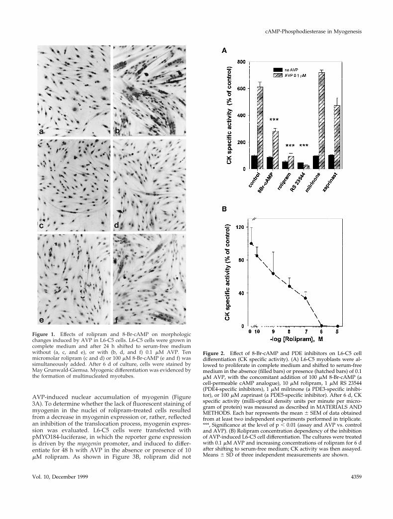
PDE4 Inhibitors Suppress Myogenic Differentiationof L6-C5 CellsIncubation of L6-C5 cells with AVP induced myogenic dif-ferentiation, as indicated morphologically by the formationof multinucleated myotubes (Figure 1, a and b) and bio-chemically by an increase in the activity of the myogenicmarker enzyme CK (Figure 2A). Both AVP effects werecompletely suppressed by incubation of the cells with thePDE4-specific inhibitor rolipram (10 mM) (Figures 1, c and d,and 2, A and B). The PDE5-specific inhibitor zaprinast (100mM) and the PDE3-specific inhibitor milrinone (1 mM) hadno significant effect on AVP-induced CK activity level (Fig-ure 2A). To rule out the possibility that the effect of rolipramis nonspecific, we used a structurally unrelated PDE4-spe-cific inhibitor, RS 23544 (1 mM) (Alvarez et al., 1995). Asshown for rolipram, RS 23544 completely suppressed theAVP-induced increase in CK activity (Figure 2A) andchanges in cell morphology (our unpublished results). Notoxic effects were evident with any of the treatments, andthere was no significant difference in DNA and proteincontent between cells treated with the inhibitors and therespective controls. A dose–response study of the effect ofrolipram showed that half-maximal inhibition of differenti-ation was achieved at 10 nM rolipram (Figure 2B), a concen-tration compatible with that necessary to inhibit the PDE4activity. The ability of PDE4 inhibitors to suppress AVP-induced differentiation is due to an increase in intracellularcAMP levels, because incubation of L6-C5 cells with 8-bro-mo-cAMP (8-Br-cAMP), a cell-permeable cAMP analoguethat is slowly hydrolyzed by PDE, almost suppressed mor-phological changes and strongly reduced the increase in CKafter AVP treatment (Figures 1, e and f, and 2A). Further-more, incubation of the cells with 10 mM rolipram induced asixfold increase in cAMP (control cells, 3.8 6 0.18 pmol ofcAMP/mg of protein; rolipram-treated cells, 23.6 6 3.2 pmolof cAMP/mg of protein; n 5 6; p , 0.001) and a significantincrease in PKA activity (expressed as 2cAMP:1cAMP spe-cific activity ratio: control cells, 0.128 6 0.012; rolipram-treated cells, 0.22 6 0.019; n 5 6; p , 0.01) after 48 h oftreatment.
Rolipram Blocks AVP-induced NuclearTranslocation of MyogeninAn early event accompanying L6-C5 cell differentiation isthe expression of the transcription factor myogenin and itsnuclear accumulation. Immunofluorescence analysis ofmyogenin indicated that 48 h AVP treatment of L6-C5 cellsinduced nuclear accumulation of the protein (Figure 3A), aspreviously reported (Minotti et al., 1998). Incubation of thecells with the PDE4 inhibitor rolipram completely prevented
F. Naro et al.
Molecular Biology of the Cell4358
AVP-induced nuclear accumulation of myogenin (Figure3A). To determine whether the lack of fluorescent staining ofmyogenin in the nuclei of rolipram-treated cells resultedfrom a decrease in myogenin expression or, rather, reflectedan inhibition of the translocation process, myogenin expres-sion was evaluated. L6-C5 cells were transfected withpMYO184-luciferase, in which the reporter gene expressionis driven by the myogenin promoter, and induced to differ-entiate for 48 h with AVP in the absence or presence of 10mM rolipram. As shown in Figure 3B, rolipram did not
Figure 1. Effects of rolipram and 8-Br-cAMP on morphologicchanges induced by AVP in L6-C5 cells. L6-C5 cells were grown incomplete medium and after 24 h shifted to serum-free mediumwithout (a, c, and e), or with (b, d, and f) 0.1 mM AVP. Tenmicromolar rolipram (c and d) or 100 mM 8-Br-cAMP (e and f) wassimultaneously added. After 6 d of culture, cells were stained byMay Grunwald-Giemsa. Myogenic differentiation was evidenced bythe formation of multinucleated myotubes.
Figure 2. Effect of 8-Br-cAMP and PDE inhibitors on L6-C5 celldifferentiation (CK specific activity). (A) L6-C5 myoblasts were al-lowed to proliferate in complete medium and shifted to serum-freemedium in the absence (filled bars) or presence (hatched bars) of 0.1mM AVP, with the concomitant addition of 100 mM 8-Br-cAMP (acell-permeable cAMP analogue), 10 mM rolipram, 1 mM RS 23544(PDE4-specific inhibitors), 1 mM milrinone (a PDE3-specific inhibi-tor), or 100 mM zaprinast (a PDE5-specific inhibitor). After 6 d, CKspecific activity (milli-optical density units per minute per micro-gram of protein) was measured as described in MATERIALS ANDMETHODS. Each bar represents the mean 6 SEM of data obtainedfrom at least two independent experiments performed in triplicate.***, Significance at the level of p , 0.01 (assay and AVP vs. controland AVP). (B) Rolipram concentration dependency of the inhibitionof AVP-induced L6-C5 cell differentiation. The cultures were treatedwith 0.1 mM AVP and increasing concentrations of rolipram for 6 dafter shifting to serum-free medium; CK activity was then assayed.Means 6 SD of three independent measurements are shown.
cAMP-Phosphodiesterase in Myogenesis
Vol. 10, December 1999 4359

significantly modify AVP-stimulated luciferase activity. Thisresult was confirmed at the level of protein expression byWestern blot analysis: the amount of myogenin was in-creased by 48 h of AVP stimulation, but it was not modifiedby rolipram treatment of the cells (Figure 3C). These dataindicate that PDE4 inhibition does not influence the level ofexpression of myogenin but, rather, affects the nuclear trans-location of the transcription factor.
Type 4 PDE Expression in L6-C5 CellsTo investigate which PDE4 isoforms are present in L6-C5myogenic cells, we used different approaches. First, by usingthe specific PDE4 inhibitor rolipram, it was assessed that76 6 4% (n 5 3) of the total cAMP-PDE activity was attrib-utable to type 4 enzymes. The cytosolic fraction obtainedafter homogenization of L6-C5 cells retained most of thePDE activity (80 6 5%; n 5 3). The cytosolic cAMP-PDEactivity was mainly due to type 4 PDEs, because rolipraminhibited it by 82 6 3% (n 5 3).
To determine which genes and isoforms are expressedin L6-C5 cells, a panel of probes corresponding to three ofthe four different PDE4 genes (PDE4A, PDE4B, andPDE4D) was used in RPA experiments. A first set ofriboprobes was complementary to the sequence encodingthe catalytic region of each isoform, which is conserved inall variants deriving from a given gene. As shown inFigure 4A, only the PDE4D gene was expressed in L6-C5cells, whereas PDE4A and PDE4B transcripts were notpresent. Because it has been previously shown that at leastfive transcripts are encoded by the PDE4D gene in differ-ent rat cell types (Sette et al., 1994b,c; Vicini and Conti Figure 3, B and C.
Figure 3. Rolipram inhibits the AVP-dependent nuclear translocation ofmyogenin but not its expression. (A)Immunofluorescence analysis of theexpression of myogenin in L6-C5 cells.The cells, cultured as described in MA-TERIALS AND METHODS, were leftuntreated for 48 h (a) or were treatedwith 0.1 mM AVP (b), 10 mM rolipram(c), or both 10 mM rolipram and 0.1 mMAVP (d) for 48 h. Myogenin was de-tected by using the anti-myogenin an-tibody F5D. (B) Lack of effect of rolip-ram on the AVP inducibility of themyogenin promoter. Cells were trans-fected with pMYO184-luciferase plas-mid containing the myogenin pro-moter linked to the luciferase gene, asdescribed in MATERIALS ANDMETHODS. Twenty-four hours aftertransfection, the cells were shifted toserum-free medium and incubatedwith 0.1 mM AVP for 48 h, either in thepresence or absence of 10 mM rolipram.Luciferase activity was normalized forb-gal activity. Data are the means 6 SDof three different experiments per-formed in duplicate. (C) Western blot
analysis of myogenin expression in L6-C5 cells. The cells, cultured as described above, were left untreated for 48 h (control) or were treatedwith 10 mM rolipram, 0.1 mM AVP, or both 10 mM rolipram and 0.1 mM AVP. Total protein concentrations were determined by Bradford(1976) protein assay, and well loading was normalized. Myogenin was detected by using the anti-myogenin antibody F5D. The experimentshown is representative of four experiments giving similar results.
F. Naro et al.
Molecular Biology of the Cell4360

1997; Jin et al., 1998), we investigated which PDE4D iso-forms are expressed in L6-C5 myoblasts. For this purpose,two probes that are complementary to the unique 59 endsof the PDE4D1/2 and PDE4D3/4 isoforms, respectively,were used. Figure 4B shows that only PDE4D3 and/orPDE4D4 mRNA are expressed. An identical pattern ofPDE4D expression was observed in both myoblasts (cul-tured in serum-free medium) and myotubes (cultured inserum-free medium and 0.1 mM AVP) (Figure 4B), sug-gesting that no other PDE4D isoform is expressed duringcell differentiation. These data were confirmed by RT-PCRexperiments using specific primer pairs allowing the se-lective amplification of sequences of the transcripts orig-inating from the expression of each of the four PDE4genes. Only the presence of PDE4D gene transcripts couldbe detected in L6-C5 myoblasts as well as in AVP-differ-entiated myotubes (our unpublished results).
To confirm at the level of protein expression the dataobtained with RPA analysis of PDE4 mRNAs and RT-PCR, we used different anti-PDE4 antibodies in immuno-precipitation and immunoblotting experiments. The non–isoform-selective antiserum K116, which recognizes allPDE4 isoforms (Sette et al., 1994a), immunoprecipitated66% of total cytosolic cAMP-PDE activity (correspondingto ;80% of rolipram-sensitive PDE activity; Figure 5A).However, among the isoform-selective antibodies used(Sette et al., 1994a), only the anti-PDE4D M3S1 antibody(which recognizes all the PDE variants originating fromthe expression of gene PDE4D) was able to immunopre-cipitate a significant amount of cAMP-PDE activity (;30%of total and 40% of rolipram-sensitive cAMP-PDE activi-ty), whereas neither the anti-PDE4A Ac-55 antiserum northe anti-PDE4B K118 antibody immunoprecipitated sig-nificant amounts of PDE activity (Figure 5A). Western blotanalysis of the immunopellets with the K116 antibodyshowed the presence of a single protein, with an apparentmolecular mass of 93 kDa, immunoprecipitated withM3S1 (Figure 5B). The 93-kDa protein migrated at thesame level as a recombinant PDE4D3 isoform expressed in293 cells (Figure 5B). Neither a 67- nor a 74-kDa band wasobserved, confirming the lack of expression of PDE4D2and PDE4D1 at the protein level. The 93-kDa band wasnot detected when the K118 or Ac-55 immunopellets wereanalyzed (our unpublished results). These data stronglysupport the conclusion that the major PDE4 proteinpresent in L6-C5 myoblasts is the PDE4D3 isoform.
PDE4 Activity Is Stimulated after AVP TreatmentThe strong inhibition of differentiation elicited by PDE4inhibitors suggests that the PDE4D3 isoform plays a keyrole in the control of cAMP levels in L6-C5 cells. Wetherefore evaluated the hypothesis that AVP-induced dif-ferentiation is accompanied by changes in PDE4 activity.AVP treatment of L6-C5 cells induced a biphasic increaseof total cAMP-PDE activity: a first transient peak occurredafter 2 min of stimulation, followed by a slower increasereaching a maximum after 15 min. Activity remainedsignificantly elevated even after 48 h from AVP addition(Figure 6, A and inset). The AVP-induced PDE activationwas restricted to the type 4 component of activity, becauseno significant change in activity was observed after AVPtreatment when PDE was assayed in the presence of roli-
Figure 4. RNase protection analysis of the PDE4D genes expressedin L6-C5 myoblasts and myotubes. L6-C5 cells were cultured for24 h in complete medium and then shifted to serum-free medium inthe absence (myoblasts, MB) or presence of 0.1 mM AVP (myotubes,MT). After 5 d, Poly(A)1 RNA was extracted, and RPA was per-formed as detailed in MATERIALS AND METHODS. Autoradiog-raphies of the PAGE gels of protected fragments are shown. (A) Theexperiment was performed on myoblast mRNA with one of threeprobes recognizing all the products of PDE4A (expected size ofprotected fragment, 215 bp), PDE4B (expected size of protectedfragment, 153 bp), and PDE4D genes (size of the protected frag-ment, 270 bp). mRNA integrity was assessed by glyceraldehyde-3-phosphate dehydrogenase–protected fragments (size, 130 bp). (B)The experiment was performed with either a probe recognizing aregion common to all PDE4D transcripts (a) or a mixture of probesrecognizing, respectively, the 59 end of PDE4D1 and PDE4D2 tran-scripts (expected sizes of protected fragments, respectively, 293 and170 bp) (b) and the 59 end of PDE4D3 and PDE4D4 transcripts (sizeof protected fragment, 350 bp) (c). The size of the major bandindicates that a fragment corresponding to PDE4D3/4 was pro-tected, whereas no band corresponding to the size of the PDE4D1/2-specific probe could be detected.
cAMP-Phosphodiesterase in Myogenesis
Vol. 10, December 1999 4361

pram (our unpublished results). Conversely, when therolipram-sensitive fraction of PDE activity was consid-ered, AVP-induced stimulation was more pronouncedand clearly showed a biphasic pattern, with 90% increaseat 2 min and 110% increase at 15 min of treatment (Figure6B). Both phases of PDE activation were dependent onAVP concentration: the early phase (2 min of stimulation)showed a maximal response at 10 nM AVP and an EC50 of2.5 nM; the later phase (15 min of stimulation) showed amaximal response at 100 nM AVP and an EC50 of 3.6 nM(Figure 7, A and B).
Interestingly IGF1, which is unable by itself to inducedifferentiation in L6-C5 cells in serum-free medium (Minottiet al., 1998), did not induce modifications of PDE activity forup to 60 min of stimulation (Figure 6A).
Figure 5. Characterization of PDE4 proteins expressed in L6-C5cells. (A) Selective immunoprecipitation of PDE4D. L6-C5 cells werehomogenized in hypotonic homogenization buffer as detailed inMATERIALS AND METHODS. Aliquots of the soluble fractionobtained after centrifugation for 15 min at 15,000 3 g were immu-noprecipitated with the different antibodies. PDE activity was as-sayed in the starting cytosolic fraction, in the supernatants, and inthe immunopellets. The experiment was performed three times withsimilar results. (B) Western blot analysis of PDE4 proteins expressedin L6-C5 cells. Immunopellet of the L6-C5 cell soluble fractionprecipitated with M3S1 antibody was subjected to Western blotanalysis, using K116 antiserum for the immunodetection of blots.For comparison, recombinant PDE4D3 protein obtained in 293 cellsas previously described (Sette et al., 1994c) was analyzed, in parallelwith an extract of mock-transfected 293 cells.
Figure 6. PDE4 activity is stimulated by AVP-treatment of L6-C5cells. (A) Effects of AVP and IGF1 on total cAMP-PDE activity ofL6-C5 cells. AVP (1 mM), IGF1 (1 nM), or saline (control) was addedat time 0 to L6-C5 myoblasts, which had been cultured for 2 d inserum-free medium. Cells were harvested at the indicated times (anextended time course up to 48 h is shown in the inset), andimmediately homogenized and assayed for total cAMP-PDEactivity. Data are means 6 SEM for nine or more samples in atleast five different experiments for AVP (F) and five or moresamples in three different experiments for IGF1 treatment (Œ). (B)Effect of AVP treatment on PDE4 activity of L6-C5 cells. Cellswere treated with 1 mM AVP ([F) or left untreated (E) for theindicated times. They were then rapidly homogenized and as-sayed for cAMP-PDE activity, in the presence and absence of 10mM rolipram. PDE4 activity was evaluated as the rolipram-in-hibited fraction of PDE activity. Data are means 6 SEM for n 55–7 samples from four different experiments. *, Significance atp , 0.05; **, p , 0.02; ***, p , 0.01.
F. Naro et al.
Molecular Biology of the Cell4362

cAMP Level and PKA Activity Decrease under AVPTreatmentTo evaluate the physiological consequences of PDE activationinduced by AVP treatment of L6-C5 cells, we measured cAMP
level and PKA activity at different times after hormone addi-tion. AVP treatment induced a reduction in cAMP concentra-tion, which was maximal at 24 h (Figure 8A). AVP also induceda significant decrease in PKA activity ratio, which was evidentafter 3 h of hormonal stimulation and continued up to 48 hafter the onset of treatment (Figure 8B). Addition of 10 mMrolipram to the cell culture medium totally prevented the AVP-dependent decrease of PKA activity (our unpublished results).The PKA activity ratio decrease was not related to modifica-tions in PKA protein levels, as assessed by Western blot anal-ysis of the expression of the catalytic and regulatory subunitsRIa and RIIa (Figure 8C), and can thus be attributed to de-creased cAMP availability. Such decreases in cAMP concentra-tion and PKA activity are consistent with the observation of anelevated capacity of myoblasts to hydrolyze cAMP after AVPstimulation and could be part of the mechanism that leads tomyoblast differentiation.
By contrast, treatment of L6-C5 cells with IGF1, which doesnot increase PDE4 activity, induced no significant change ineither cAMP levels or PKA activity (Figure 8, A and B).
Effect of PDE4D3 Overexpression on L6-C5 CellDifferentiationTo confirm whether the increase in PDE4D3 activity is in-volved in the differentiation process, this enzyme was over-expressed by transient transfection in L6-C5 myoblasts, andthe expression of both myogenin (a muscle-specific tran-scription factor whose nuclear accumulation represents anearly marker of myogenic differentiation) and sarcomericmyosin (a marker of terminal myogenic differentiation) was
Figure 7. Dose-dependent effects of AVP on the rapid and slow compo-nents of PDE activation. L6-C5 cells were treated with increasing concen-trations of AVP. Total cAMP-PDE activity was measured after 2 min (A)and 15 min (B) of AVP stimulation. Data represent the means of four tonine measurements performed in three independent experiments.
Figure 8. AVP treatment of L6-C5 cells induces a decrease of cAMP and PKA activity without affecting PKA expression. (A) cAMPconcentration was measured after incubation of cells with 1 mM AVP or 1 nM IGF1 for the indicated times. The cells were treated at differenttimes, and all the samples (control and AVP treated) were harvested and assayed at the same time, i.e., 2 d after shifting to serum-freemedium. cAMP was measured as described in MATERIALS AND METHODS. Results are the means 6 SEM of at least three independentmeasurements performed in triplicate. *, Significance at p , 0.05; ***, p , 0.01. (B) PKA activity was measured after incubation of the cellsas described above. PKA assays were conducted on cell soluble extracts prepared as detailed in MATERIALS AND METHODS. Results arethe means 6 SEM of six measurements performed in three different experiments for AVP and four measurements in two differentexperiments for IGF1. *, Significance at p , 0.05; **, p , 0.02; ***, p , 0.01. (C) PKA catalytic subunit (40 kDa) and regulatory subunit RIaand RIIa (49 and 51 kDa, respectively) expression was evaluated on cells treated as described above. Cell samples were prepared by addingdirectly concentrated sample buffer to 10-cm dishes. Western blot analysis was carried out using specific mAbs for the immunodetection ofblots. Total protein concentrations were determined by Bradford (1976) protein assay, and well loading was normalized.
cAMP-Phosphodiesterase in Myogenesis
Vol. 10, December 1999 4363

evaluated. After 48 h of culture in serum-free medium, withor without AVP addition, transfected cells were immuno-stained with anti-myogenin antibody. As shown in Figure9A, cultures of cells transfected with PDE4D3 showed amarked increase of myogenin-positive nuclei (b and d) com-pared with mock-transfected cell cultures (a and c). Thisincrease was evident in untreated cells (a vs. b) as well as inAVP-treated cells (c vs. d). When transfected L6-C5 cellswere further cultured for 6 d, a significant increase in myo-tube formation and in the expression of myosin was ob-served in cells overexpressing PDE4D3 compared withmock-transfected cells, by immunochemical staining of thecells (Figure 9B) as well as by ELISA quantification of my-osin in cell extracts (Table 1). In this case, too, overexpres-sion of PDE4D3 induced a higher expression of the differ-entiation marker in both AVP-treated and untreated cells(Figure 9B and Table 1).
DISCUSSION
This work demonstrates that type 4 PDEs play a role in thecontrol of myogenic differentiation of L6-C5 myoblasts. Weshow that selective inhibition of PDE4 enzymes by twospecific, structurally unrelated inhibitors, namely rolipram(Schwabe et al., 1976) and RS 23544 (Alvarez et al., 1995),suppresses differentiation induced by AVP in these cells.Indeed, it prevents translocation of the muscle-specific tran-scription factor myogenin to the nucleus, the formation ofmyotubes, and ultimately the expression of the terminaldifferentiation marker CK. We also demonstrate that AVPstimulation of L6-C5 cells leads to activation of PDE4 and adecrease of cAMP level and PKA activity. Because PKAactivation is considered a negative signal for myoblast dif-ferentiation, our data suggest that AVP-dependent down-regulation of PKA activity allows the full expression of themyogenic program in L6-C5 cells. The importance of the roleplayed by PDE4 in the control of myogenic differentiation isfurther supported by experiments showing that overexpres-sion of the PDE4D3 isoform in L6-C5 cells increased theirdifferentiation (both spontaneous and AVP induced), asevaluated by means of markers of both early phases ofdifferentiation (nuclear accumulation of myogenin), and ter-minal differentiation (myotube formation and myosin ex-pression).
The ability of PDE4 inhibitors to completely prevent dif-ferentiation can be ascribed to increased cAMP intracellularlevels and PKA activation, because the PDE4-specific inhib-itor rolipram induced a large increase of endogenous cAMPlevels together with PKA activation in the absence of stim-ulation of adenylyl cyclase. Furthermore, addition of acAMP cell-permeable analogue, 8-Br-cAMP, which is slowlyhydrolyzed by PDEs, partially reproduced the inhibition ofdifferentiation. The negative regulation of myogenic differ-entiation by cAMP and PKA has been previously establishedin several cellular models, including L6 cells, induced todifferentiate by reducing the serum concentration of theculture medium, or by adding insulin (Wahrman et al., 1973;Hu and Olson 1988; Salminen et al., 1991; Winter et al., 1993).The present results allow extension of this concept to themodel of AVP-induced myogenic differentiation. We havepreviously shown that AVP induction of myogenesis inL6-C5 cells is related to an accumulation of the muscle
Figure 9. Effect of PDE4D3 overexpression on L6-C5 cell differen-tiation. L6-C5 myoblasts were transiently transfected in suspensionby using Fugene 6 with 1 mg of pCMV5 expression vector contain-ing rat PDE4D3 cDNA (Sette et al., 1994c) or the empty vector andplated in 10% FCS-containing medium. After 24 h, the medium wasreplaced by serum-free medium with or without addition of 0.1 mMAVP. (A) Immunofluorescence analysis of the expression of myo-genin in transfected L6-C5 cells. Mock-transfected cells (a and c) andpCMV5-PDE4D3-transfected cells (b and d) were left untreated(a and b) or were treated with 0.1 mM AVP for 48 h (c and d).Myogenin was detected by using the anti-myogenin antibody F5D.(B) Immunochemical detection of myosin expression in transfectedL6-C5 cells. Mock-transfected cells (a and c) and pCMV5-PDE4D3-transfected cells (b and d) were left untreated (a and b) or weretreated with 0.1 mM AVP for 6 d (c and d). Myosin was detected byusing the anti-myosin antibody MF20.
F. Naro et al.
Molecular Biology of the Cell4364

transcription factor myogenin in the cell nucleus (Minotti etal., 1998). We now observe that, in the presence of rolipram,myogenin is not detectable in myoblast nuclei, althoughAVP treatment increases the amount of myogenin. Thisresult suggests that the cAMP signal transduction pathwaycontrols the subcellular localization of myogenin. No infor-mation is available yet about the mechanism by whichcAMP regulates myogenin import, although phosphoryla-tion steps may conceivably be involved. Nuclear myogeninimport could be an important control point in regulatingmyogenic differentiation.
Our results highlight the importance of the function oftype 4 PDEs in the regulation of cAMP levels during L6-C5myoblast differentiation. Indeed, inhibitors specific for otherclasses of PDEs, milrinone and zaprinast (which respectivelyinhibit type 3 and type 5 PDEs), were devoid of inhibitoryeffects. It is thus conceivable that in L6-C5 cells a functionalpool of cAMP under the sole control of PDE4 is able tomodulate the differentiative response. Both rolipram and RS23544 appear as very potent inhibitors of AVP-induced myo-genic differentiation. Half-maximal inhibition of differentia-tion, as evaluated by CK activity, was achieved at a rolipramconcentration of 10 nM. This can be ascribed to an effect ofthe drug directed to PDE4 species present under a “highaffinity rolipram state” (Huston et al., 1996; Sette and Conti1996) sensitive to nanomolar concentrations of rolipram (re-viewed by Souness and Rao 1997; Houslay et al., 1998).
Identification of type 4 PDE expressed in L6-C5 cells byRPA, RT-PCR, immunoprecipitation, and Western blot anal-ysis with isoform-selective anti-PDE4 antibodies led to theconclusion that only the PDE4D gene is expressed in thesecells, confirming previous observations based only on RT-PCR data (Kovala et al., 1994). Furthermore, we found thatthe PDE4D3 isoform, one of the five variants encoded by thePDE4D gene (Sette et al., 1994c; Bolger et al., 1997), is themajor isoform present in these cells. This result is onlyapparently in contrast with the observation made by othersthat PDE4D1 mRNA is the major species expressed in L6myoblasts. The latter data were obtained after a prolongedpharmacological treatment of the cells by cAMP-elevatingagents (Kovala et al., 1994). Because it is well established thatthe PDE4D1 and PDE4D2 isoforms, but not the PDE4D3isoform, are up-regulated by cAMP at the transcriptionallevel in different cell types (Sette et al., 1994b,c; Vicini and
Conti 1997), we can infer that the major type 4 PDE in theabsence of a cAMP-elevating pharmacological treatment isthe PDE4D3 isoform in undifferentiated myoblasts as well asin differentiated myotubes (Figure 4B).
L6-C5 myoblasts only express the V1 receptors for AVP,which have no direct relationships with adenylyl cyclaseregulation (Wakelam et al., 1987) but trigger the activation ofphospholipases C and D (Naro et al., 1997). AVP seems tomodulate cAMP levels and PKA activity in these cells bymodifying cAMP hydrolysis rather than by acting on cAMPsynthesis. Indeed two phases of PDE activation were appar-ent in response to AVP addition: a fast activation, whichpeaked at 2 min, and a slower activation, which peaked at 15min and was significantly maintained even after 48 h ofhormonal treatment. That the AVP-induced PKA activitydecrease is mainly dependent on PDE4 activation and un-related to changes in adenylyl cyclase activity is confirmedby the observation that rolipram, when added to the culturemedium, totally suppressed the kinase activity reduction(our unpublished results).
In addition to PKA-dependent phosphorylation (Sette etal., 1994b; Sette and Conti 1996), PDE4D3 is specificallyactivated by PA, the product of phospholipase D action, in acell-free system (Nemoz et al., 1997; Grange et al., 1998). Bothphospholipid binding and PKA phosphorylation sites residein the same amino-terminal region, because isoforms lackingthis region, such as PDE4D1 and PDE4D2, are not activatedby PKA phosphorylation or by phospholipid interaction(Sette et al., 1994b; Nemoz et al., 1997). Because phospho-lipase D activation is a primary signaling event triggered byAVP in L6-C5 cells (Naro et al., 1997), the possibility existsthat PDE4D3 is stimulated by PA, which rapidly accumu-lates in the cells under AVP stimulation. Thus both PA-induced stimulation and phosphorylation processes mightbe responsible for the increase in PDE4 activity observed inL6-C5 cells. The EC50 value observed for the first phase ofAVP-induced PDE stimulation (2.5 nM) is compatible withthe 0.4 nM EC50 previously reported for AVP activation ofphospholipase D (Naro et al., 1997), suggesting that the rapidPDE stimulation could be linked to phospholipase D activa-tion and production of PA. Furthermore, the kinetics of PAproduction by AVP-stimulated L6-C5 cells are in agreementwith this hypothesis, because the PA level reaches its max-imum by 1 min (Naro et al., 1997). Experiments are inprogress to more clearly define the mechanism(s) involved.
In view of the profound effect of PDE4 inhibition on thedifferentiation process, it can be postulated that an increasein PDE4 activity plays a physiological role in lowering theintracellular cAMP concentration, thus preventing inhibitionof the myogenic program by cAMP. The sustained decreasein both cAMP level and PKA activity that we observed inAVP-treated myoblasts supports this hypothesis. Further-more, the observation that overexpression of the type 4 PDEisoform PDE4D3 could positively influence myogenic differ-entiation provides confirmation of this model. Thus, PDE4activation appears an essential step in myogenesis, and thelack of effect of IGF1 in inducing terminal myogenic differ-entiation in the absence of serum or AVP (Minotti et al.,1998) may be attributed to its inability to induce stimulationof PDE4 activity. This supports the conclusion that cAMPlevels must be tightly controlled during myogenesis and thatPDE4 plays a key role in such control.
Table 1. Effect of PDE4D3 overexpression on L6 cell differentiation(myosin accumulation)
Culture mediumMock transfected
(ng/mg DNA)PDE4D3 transfected
(ng/mg DNA)
1% BSA 45 6 2.25 100 6 1.1a
1% BSA 1 0.1 mM AVP 320 6 25.3 530 6 9.1a
L6 myoblasts were transiently transfected in suspension by usingFugene 6 with 1 mg of the pCMV5-PDE4D3 plasmid or the emptyvector and plated in 10% FCS medium. After 24 h, the medium wasreplaced as indicated, with or without addition of 0.1 mM AVP.After 6 d of culture, the cells were solubilized, and their myosincontent was measured by ELISA. Results are the means 6 SD of sixmeasurements performed in two independent experiments.a Significantly different from mock-transfected cells, p , 0.01.
cAMP-Phosphodiesterase in Myogenesis
Vol. 10, December 1999 4365

ACKNOWLEDGMENTS
We thank Dr. Bianca Maria Scicchitano for the construction of themyogenin promoter-luciferase construct, Dr. Raffaella Curci for ex-pert assistance, and Prof. Giulio Cossu for helpful discussion andsuggestions. This work was supported in part by grants from theItalian Ministry of University and Scientific and Technological Re-search (to S.A.), from the Associazione Italiana per la Ricerca sulCancro AIRC (to M.M.) and from Consiglio Nazionale delleRicerche grants 96.0600.PF39 (to M.M.) and 96.00651.39 (to S.A.).The exchanges between the collaborating institutions were sup-ported by a Consiglio Nazionale delle Ricerche–Institut National dela Sante et de la Recherche Medicale joint program grant (to G.N.and S.A.).
REFERENCES
Alvarez, R., Sette, C., Yang, D., Eglen, R.M., Wilhelm, R., Shelton,E.R., and Conti, M. (1995). Activation and selective inhibition of acyclic AMP-specific phosphodiesterase, PDE4D-3. Mol. Pharmacol.48, 616–622.
Bader, D., Masaki, T., and Fischman D.A. (1982). Immunochemicalanalysis of myosin heavy chain during avian myogenesis in vivoand in vitro. J. Cell Biol. 95, 763–770.
Beavo, J.A. (1995). Cyclic nucleotide phosphodiesterases: functionalimplications of multiple isoforms. Physiol. Rev. 75, 725–748.
Bolger, G.B., Erdogan, S., Jones, R.E., Loughney, K., Scotland, G.,Hoffmann, R., Wilkinson, I., Farrell, C., and Houslay, M.D. (1997).Characterization of five different proteins produced by alternativelyspliced mRNAs from the human cAMP-specific phosphodiesterasePDE4D gene. Biochem. J. 328, 539–548.
Bolger, G.B., Michaeli, T., Martins T., St. John, T., Steiner, B., Rodg-ers, L., Riggs, M., Wigler, M., and Ferguson, K. (1993). A family ofhuman phosphodiesterases homologous to the dunce learning andmemory gene product of Drosophila melagonaster are potential tar-gets for antidepressant drugs. Mol. Cell. Biol. 13, 6558–6571.
Bradford, M.M. (1976). A rapid and sensitive method for the quan-titation of microgram quantities of protein utilizing the principle ofprotein-dye binding. Anal. Biochem. 72, 248–254.
Brasier, A.R., Tate, J.E., and Habener, J.F. (1989). Optimized use ofthe firefly luciferase assay as a reporter gene in mammalian celllines. Biotechniques 7, 1116–1122.
Braun, T., Bober, E., Buschhausen-Denker, G., Kothz, S., Grzeschik,K.H., and Arnold, H.H. (1989). Differential expression of myogenicdetermination genes in muscle cells: possible autoactivation by theMyf gene products. EMBO J. 8, 3617–3625.
Clegg, C.H., Linkhart, T.A., Olwin, B.B., and Hauschka, S.D. (1987).Growth factor control of skeletal muscle differentiation: committ-ment to terminal differentiation occurs in G1 phase and is repressedby fibroblast growth factor. J. Cell Biol. 105, 949–956.
Colicelli, J., Birchmeier, C., Michaeli, T., O’Neill, K., Riggs, M., andWigler, M. (1989). Isolation and characterization of a mammaliangene encoding a high-affinity cAMP phosphodiesterase. Proc. Natl.Acad. Sci. USA 86, 3599–3603.
Conti, M., Nemoz, G., Sette, C., and Vicini, E. (1995). Recentprogress in understanding the hormonal regulation of phosphodi-esterases. Endocr. Rev. 16, 370–389.
Cossu, G., Tajbakhsh, S., and Buckingham, M. (1996). How is myo-genesis initiated in the embryo? Trends Genet. 12, 218–223.
Cusella De Angelis, M.G., et al. (1992). MyoD, myogenin indepen-dent differentiation of primordial myoblasts in mouse somites.J. Cell Biol. 116, 1243–1255.
Davis, R.L., Takayasu, H., Eberwine, M., and Myres, J. (1989). Clon-ing and characterization of mammalian homologs of the Drosophiladunce1 gene. Proc. Natl. Acad. Sci. USA 86, 3604–3608.
Edmondson, D.G., Cheng, T.C., Cserjesi, P., Chakraborty, T., andOlson, E.N. (1992). Analysis of the myogenin promoter reveals anindirect pathway for positive autoregulation mediated by the mus-cle-specific enhancer factor MEF-2. Mol. Cell. Biol. 12, 3665–3677.
Engert, J.C., Berglund, E.B., and Rosenthal, N. (1996). Proliferationprecedes differentiation in IGF-I stimulated myogenesis. J. Cell Biol.135, 431–440.
Florini, J.R. (1987). Hormonal control of muscle growth. MuscleNerve 10, 577–598.
Florini, J.R., Ewton, D.Z., and Magri, K.A. (1991). Hormones, growthfactors, and myogenic differentiation. Annu. Rev. Physiol. 53, 201–216.
Grange, M., Picq, M., Prigent, A.F., Lagarde, M., and Nemoz, G.(1998). Regulation of PDE-4 cAMP phosphodiesterases by phospha-tidic acid. Cell Biochem. Biophys. 29, 1–17.
Houslay, M.D., Sullivan, M., and Bolger, G.B. (1998). The multien-zyme PDE4 cyclic adenosine monophosphate-specific phosphodies-terase family: intracellular targeting, regulation, and selective inhi-bition by compounds exerting ant-inflammatory and antidepressantactions. Adv. Pharmacol. 44, 225–342.
Hu, J.S., and Olson, E.N. (1988). Regulation of differentiation of theBC3H1 muscle cell line through cAMP-dependent and -indepen-dent pathways. J. Biol. Chem. 263, 19670–19677.
Huston, E., Pooley, L., Julien, P., Scotland, G., McPhee, I., Sullivan,M., Bolger, G.B., and Houslay, M.D. (1996). The human cyclic AMP-specific phosphodiesterase PDE-46 (HSPDE4A4B) expressed intransfected COS7 cells occurs as both particulate and cytosolic spe-cies that exhibit distinct kinetics of inhibition by the antidepressantrolipram. J. Biol. Chem. 271, 31334–31344.
Iona, S., Cuomo, M., Bushnik, T., Naro, F., Sette, C., Hess, M.,Shelton, E.R., and Conti, M. (1998). Characterization of the rolipram-sensitive, cAMP specific phosphodiesterases: identification and dif-ferential expression of immunologically distinct forms in the ratbrain. Mol. Pharmacol. 53, 23–32.
Jin, S.L.C., Bushnik, T., Lan, L., and Conti, M. (1998). Subcellularlocalization of rolipram-sensitive, cAMP-specific phosphodiester-ases. J. Biol. Chem. 273, 19672–19678.
Kovala, T., Lorimer, I.A.J., Brickenden, A.M., Ball, E.H., and Sanwal,B.D. (1994). Protein kinase A regulation of cAMP phosphodiesteraseexpression in rat skeletal myoblasts. J. Biol. Chem. 269, 8680–8685.
Li, L., Heller-Harrison, R., Czech, M., and Olson, E.N. (1992). CyclicAMP-dependent protein kinase activity inhibits the activity of myo-genic helix-loop-helix proteins. Mol. Cell. Biol. 12, 4478–4485.
Ludolph, D.C., and Konieczny, S.F. (1995). Transcription factor fam-ilies: muscling in on the myogenic program. FASEB J. 9, 1595–1604.
Minotti, S., Scicchitano, B.M., Nervi, C., Scarpa, S., Lucarelli, M.,Molinaro, M., and Adamo, S. (1998). Vasopressin and insulin-likegrowth factors synergistically induce myogenesis in serum-free me-dium. Cell Growth & Differ. 9, 155–163.
Nadal-Ginard, B. (1978). Commitment, fusion and biochemical dif-ferentiation of a myogenic cell line in the absence of DNA synthesis.Cell 15, 855–864.
Naro, F., Donchenko, V., Minotti, S., Zolla, L., Molinaro, M., andAdamo, S. (1997). Role of phospholipase C and D signaling path-ways in vasopressin-dependent myogenic differentiation. J. Cell.Physiol. 171, 34–42.
Naro, F., Fazzini, A., Citro, G., Malatesta, F., Antonini, G., Sarti, P.,Brunori, M., Franconi, F., and Giotti, A. (1991). Immunoquantitationof cytochrome c in cardiac perfusate. J. Immunoassay 12, 251–262.
F. Naro et al.
Molecular Biology of the Cell4366
Naro, F., Zhang, R., and Conti, M. (1996). Developmental regulationof unique cyclic adenosine 39-59-monophosphate-specific phospho-diesterase variants during rat spermatogenesis. Endocrinology 137,2464–2472.
Nemoz, G., Sette, C., and Conti, M. (1997). Selective activation ofrolipram-sensitive, cAMP-specific phosphodiesterase isoforms byphosphatidic acid. Mol. Pharmacol. 51, 242–249.
Nervi, C., Benedetti, L., Minasi, A., Molinaro, M., and Adamo, S.(1995). Arginine-vasopressin induces differentiation of skeletal myo-genic cells and up-regulation of myogenin and myf-5. Cell Growth& Differ. 6, 81–89.
Olson, E.N., Sternberg, E., Hu, J.S., Spizz, G., and Wilcox, C. (1986).Regulation of myogenic differentiation by type b transforminggrowth factor. J. Cell Biol. 103, 1799–1805.
O’Neill, M.C., and Stockdale, F.E. (1972). A kinetic analysis of myo-genesis in vitro. J. Cell Biol. 52, 52–65.
Salminen, A., Braun, T., Buchberger, A., Jurs, S., Winter, B., andArnold, H.H. (1991). Transcription of the muscle regulatory geneMYF4 is regulated by serum components, peptide growth factorsand signaling pathways involving G proteins. J. Cell Biol. 115,905–917.
Schwabe, U., Miyake, M., Ohga, Y., and Daly, J.W. (1976). 4-(-3-Cyclopentyloxy-4-methoxyphenyl)-2-pyrrolidone (ZK 62711): a po-tent inhibitor of adenosine cyclic 39 59-mono-phosphate-phosphodi-esterases in homogenates and tissue slices from rat brain. Mol.Pharmacol. 12, 900–910.
Sette, C., and Conti, M. (1996). Phosphorylation and activation of acAMP-specific phosphodiesterase by the cAMP-dependent proteinkinase. Involvement of serine 54 in the enzyme activation. J. Biol.Chem. 271, 16526–16534.
Sette, C., Iona, S., and Conti, M. (1994a). The short-term activation ofa rolipram-sensitive, cAMP-specific phosphodiesterase by thyroid-stimulating hormone in thyroid FRTL-5 cells is mediated by a cAMPdependent phosphorylation. J. Biol. Chem. 269, 9245–9252.
Sette, C., Vicini, E., and Conti, M. (1994b). Modulation of cellularresponses by hormones: role of cAMP specific, rolipram sensitivephosphodieserases. Mol. Cell. Endocrinol. 100, 75–79.
Sette, C., Vicini, E., and Conti, M. (1994c). The ratPDE3/IVd phos-phodiesterase gene codes for multiple proteins differentially acti-vated by cAMP-protein kinase. J. Biol. Chem. 269, 18271–18274.
Soderling, S.H., Bayuga, S.J., and Beavo, J.A. (1998). Cloning andcharacterization of a cAMP-specific cyclic nucleotide phosphodies-terase. Proc. Natl. Acad. Sci. USA 95, 8991–8996.
Souness, J.E., and Rao, S. (1997). Proposal for pharmacologicallydistinct conformers of PDE4 cyclic AMP phosphodiesterases. Cell.Signalling 9, 227–236.
Swinnen, J.V., Joseph, D.R., and Conti, M. (1989). Molecular cloningof rat homologues of the Drosophila melanogaster dunce cAMP phos-phodiesterase: evidence for a family of genes. Proc. Natl. Acad. Sci.USA 86, 5325–5329.
Teti, A., Naro, F., Molinaro, M., and Adamo, S. (1993). Transductionof the arginine-vasopressin signal in skeletal myogenic cells. Am. J.Physiol. 265, C113–C121.
Thompson, W.J., Brooker, G., and Appleman, M.M. (1974). Assay ofcyclic nucleotide phosphodiesterase with radioactive substrates.Methods Enzymol. 38, 205–212.
Vicini, E., and Conti, M. (1997). Characterization of an intronicpromoter of a cyclic adenosine 39,59-monophosphate (cAMP)-spe-cific phosphodiesterase gene that confers hormone and cAMP in-ducibility. Mol. Endocrinol. 11, 839–850.
Wahrman, J.P., Winand, R., and Luzzatti, D. (1973). Effect of cyclicAMP on growth and morphological differentiation of an establishedmyogenic cell line. Nature 245, 112–113.
Wakelam, M.J.O., Patterson, S., and Hanley, M.R. (1987). L6 skeletalmuscle cells have functional V1 vasopressin receptors coupled tostimulated inositol phospholipid metabolism. FEBS Lett. 210, 181–184.
Winter, B., Braun, T., and Arnold, H.H. (1993). cAMP-dependentprotein kinase represses myogenic differentiation and the activity ofthe muscle-specific helix-loop-helix transcription factors Myf-5 andMyoD. J. Biol. Chem. 268, 9869–9878.
Yaffe, D. (1968). Retention of differentiation potentialities duringprolonged cultivation of myogenic cells. Proc. Natl. Acad. Sci. USA61, 477–483.
cAMP-Phosphodiesterase in Myogenesis
Vol. 10, December 1999 4367


