Embed Size (px)
Citation preview

Iron Overload Favors the Elimination of Leishmaniainfantum from Mouse Tissues through Interaction withReactive Oxygen and Nitrogen SpeciesSılvia Vale-Costa1,2, Sandra Gomes-Pereira1,3, Carlos Miguel Teixeira1, Gustavo Rosa1, Pedro
Nuno Rodrigues1,2, Ana Tomas1,2, Rui Appelberg1,2, Maria Salome Gomes1,2*
1 IBMC - Instituto de Biologia Molecular e Celular, Universidade do Porto, Porto, Portugal, 2 ICBAS - Instituto de Ciencias Biomedicas de Abel Salazar, Universidade do
Porto, Porto, Portugal, 3 CISA-ESTSP - Nucleo de Investigacao em Farmacia, Centro de Investigacao em Saude e Ambiente, Escola Superior de Tecnologia da Saude do
Porto, Instituto Politecnico do Porto, Porto, Portugal
Abstract
Iron plays a central role in host-parasite interactions, since both intervenients need iron for survival and growth, but aresensitive to iron-mediated toxicity. The host’s iron overload is often associated with susceptibility to infection. However, ithas been previously reported that iron overload prevented the growth of Leishmania major, an agent of cutaneousleishmaniasis, in BALB/c mice. In order to further clarify the impact of iron modulation on the growth of Leishmania in vivo,we studied the effects of iron supplementation or deprivation on the growth of L. infantum, the causative agent ofMediterranean visceral leishmaniasis, in the mouse model. We found that dietary iron deficiency did not affect theprotozoan growth, whereas iron overload decreased its replication in the liver and spleen of a susceptible mouse strain. Thefact that the iron-induced inhibitory effect could not be seen in mice deficient in NADPH dependent oxidase or nitric oxidesynthase 2 suggests that iron eliminates L. infantum in vivo through the interaction with reactive oxygen and nitrogenspecies. Iron overload did not significantly alter the mouse adaptive immune response against L. infantum. Furthermore, theinhibitory action of iron towards L. infantum was also observed, in a dose dependent manner, in axenic cultures ofpromastigotes and amastigotes. Importantly, high iron concentrations were needed to achieve such effects. In conclusion,externally added iron synergizes with the host’s oxidative mechanisms of defense in eliminating L. infantum from mousetissues. Additionally, the direct toxicity of iron against Leishmania suggests a potential use of this metal as a therapeutic toolor the further exploration of iron anti-parasitic mechanisms for the design of new drugs.
Citation: Vale-Costa S, Gomes-Pereira S, Teixeira CM, Rosa G, Rodrigues PN, et al. (2013) Iron Overload Favors the Elimination of Leishmania infantum from MouseTissues through Interaction with Reactive Oxygen and Nitrogen Species. PLoS Negl Trop Dis 7(2): e2061. doi:10.1371/journal.pntd.0002061
Editor: Yara M. Traub-Cseko, Instituto Oswaldo Cruz, Brazil
Received July 25, 2012; Accepted January 2, 2013; Published February 14, 2013
Copyright: � 2013 Vale-Costa et al. This is an open-access article distributed under the terms of the Creative Commons Attribution License, which permitsunrestricted use, distribution, and reproduction in any medium, provided the original author and source are credited.
Funding: SVC thanks FCT for the doctoral grant SFRH/BD/36661/2007. The funders had no role in study design, data collection and analysis, decision to publish,or preparation of the manuscript.
Competing Interests: The authors have declared that no competing interests exist.
* E-mail: [email protected]
Introduction
Leishmania are trypanosomatid protozoans that alternate between
two forms: the extracellular motile promastigote in the gut of
phlebotomine insects and the intracellular non-motile amastigote
inside the macrophages of mammalian hosts. These parasites cause
leishmaniasis, a spectrum of human diseases that range from self-
healing cutaneous ulcers to fatal visceralizing infection. Every year,
approximately 2.0 million people develop symptomatic disease (0.5
million of them the visceral form) [1]. In Europe, visceral
leishmaniasis is caused almost exclusively by L. infantum, which is
transmitted as a zoonosis. The domestic dog is one of the main
reservoirs of this parasite and canine leishmaniasis is an important
veterinary problem in European Mediterranean countries [2].
There are currently no effective vaccines to prevent human
leishmaniasis [3]. Therefore, management of the disease relies on
chemotherapy. However, available drugs are highly toxic and the
frequency of resistant parasite strains is increasing worldwide [4,5].
The improvement of our knowledge on the mechanisms of host
resistance to Leishmania is important to contribute to the
development of new therapeutic strategies.
The important role played by iron metabolism in the interaction
between host and pathogens is being increasingly highlighted by
recent research [6,7]. Both the host and the pathogens absolutely
need iron for survival and must have efficient mechanisms for its
acquisition together with adequate mechanisms of cell defense to
avoid iron toxicity. Data obtained in human patients, as well as in
different animal infection models indicate that in many cases iron
availability favors the multiplication of pathogens, whereas iron
deprivation impairs their growth [8]. Interestingly, the vertebrates
innate immune response to infection includes several mechanisms
of iron with-holding such as lactoferrin, hepcidin, Nramp1 or
lipocalin2 [6]. Still, adequate concentrations of iron are required
to support macrophagic killing mechanisms during infection
[9,10]. Contrary to what happens with other pathogens, the
growth of Brucella abortus inside macrophages [11] and that of L.
major in the mouse [12] are decreased by host’s iron overload. In
both cases, killing was correlated to oxidative burst [11,13]. These
PLOS Neglected Tropical Diseases | www.plosntds.org 1 February 2013 | Volume 7 | Issue 2 | e2061

examples highlight that iron can be exploited, in some cases, by
the host to strengthen its antimicrobial defense mechanisms.
Our group has previously shown that the infection by
Mycobacterium avium, an intra-macrophagic pathogen, is clearly
exacerbated by host’s iron overload, either genetically determined
[14] or caused by iron-dextran injection [15,16]. Furthermore, we
have also demonstrated that iron chelation can be used to inhibit
the growth of M. avium [17]. In the present work, we evaluated the
effect of iron on the growth of L. infantum, the agent of European
visceral leishmaniasis. We found that iron consistently inhibited
the replication of L. infantum both by a direct effect and through the
activity of the host’s macrophage.
Materials and Methods
Animals and ethics statementBALB/c and C57BL/6 mice were purchased from Charles
River (Madrid, Spain). Mice deficient on the p47 subunit of the
NADPH oxidase complex, on a C57BL/6 background
(p47phox2/2), were bred at IBMC from a breeding pair
purchased from Taconic (Lille Skensved, Denmark). The
p47phox2/2 mice were administered trimethoprimsulfamethox-
azole (Bactrim; 600 mgl21) in the drinking water, as prophylactic
treatment against bacterial infection. This treatment was ceased
when infection experiments began. Mice deficient in the nitric
oxide synthase 2, on a C57BL/6 background (NOS22/2), were
bred at IBMC from a breeding pair kindly provided by Drs. J.
Mudgett, J. D. MacMicking and C. Nathan (Cornell University,
New York, USA). Mice deficient in HFE, on a C57BL/6
background (Hfe2/2) were bred at IBMC from a breeding pair
obtained from Centre Nationale de la Recherche Scientifique. All
animals were housed at IBMC facilities under specific pathogen
free conditions and fed ad libitum, except for the diet experiments
indicated below. All animals were used at 8 to 16 weeks of age.
Only female mice (average 20–25 g) were used for in vivo
experiments. Mice were euthanized by isofluorane anesthesia
followed by cervical dislocation and tissues were collected in
aseptic conditions.
The experimental animal procedures were approved by the
Local Animal Ethics Committee of IBMC and licensed by the
Portuguese General Directory of Veterinary (DGV, Ministry of
Agriculture, Rural Development and Fishing), in May 18, 2006
with reference 520/000/000/2006. All animals were handled in
strict accordance with good animal practice as defined by national
authorities (DGV, Law nu1005/92 from 23rd October) and
European legislation EEC/86/609.
ParasitesAll experiments were performed with L. infantum strain MHOM/
MA/67/ITMAP-263 (zymodeme MON-1). For each experiment,
parasites were obtained from the spleens of infected mice.
Promastigotes were differentiated from spleen amastigotes by
culturing at 25uC in complete Schneider’s medium (Sigma-Aldrich
Co., St Louis, MO, USA) supplemented with 20% heat-inactivated
fetal bovine serum (FBS), 100 Uml21 penicillin, 100 mgml21
streptomycin (all from Gibco, Life Technologies, Carlsbad, CA,
USA), 2% human urine, 5 mgml21 phenol red (Sigma) and 5 mM
HEPES sodium salt (Sigma) pH 7.4. Promastigote cultures were
expanded at 25uC, for a maximum of 5 passages, in RPMI 1640
GlutaMAXTM-I medium (Gibco, Life Technologies), containing
20% FBS, 50 Uml21 penicillin, 50 mgml21 streptomycin and
25 mM HEPES sodium salt pH 7.4. Promastigote differentiation
from the exponential to the stationary phase was promoted by
culture at 25uC, without medium renovation, for 4 to 5 days.
Axenic amastigotes were derived from the above-mentioned L.
infantum strain, by the culture at 37uC with 7% CO2 atmosphere,
in a medium for axenic amastigotes supplemented with 2 mM L-
glutamine (GlutaMAXTM-I, Gibco, Life Technologies) and 20%
FBS (MAA20 medium, adapted from [18]).
Direct effect of iron on Leishmania promastigote andamastigote cultures
Promastigotes (26106/well) and amastigotes (46105/well) of L.
infantum were cultured in complete RPMI (25uC) or MAA20
(37uC) medium, respectively, supplemented with iron-dextran
(Fe3+ hydroxide-dextran complex, Sigma), iron citrate (Fe3+,
Sigma) and iron sulphate (Fe2+, Merck) in the concentrations of
0.018, 0.035, 0.070, 0.14, 0.28, 0.56, 1.1, 2.2, 4.5, 9 and 18 mM
(96 well plates). Equivalent concentrations of dextran (Sigma), tri-
sodium citrate (Merck) and magnesium sulphate (Merck) were
used as controls. After 24 h of culture, 20 ml of a 2.5 mM
resazurin solution (freshly prepared and filtered in phosphate
buffered saline, pH 7.4, Sigma) was added to each well. The
fluorescence intensity (excitation at 560 nm and emission at
590 nm) was determined 24 h (for amastigotes) or 48 h (for
promastigotes) after resazurin addition to allow conversion to
fluorescent resorufin, with a fluorometer SpectraMAX GeminiXS
(Molecular Devices LLC, Sunnyvale, CA). In order to exclude a
possible interference of iron with resazurin conversion to resorufin,
appropriate controls were included without cells. No resazurin
conversion was detected in the absence of parasites. Complete
RPMI and MAA20 medium contains approximately 6 and
7.7 mM of iron, respectively, according to supplier’s information.
Animal experimental infection and parasite burdenquantification
Mice were injected in the lateral vein of the tail with 26107 L.
infantum stationary promastigotes in 200 ml of phosphate buffered
saline pH 7.4 (PBS). At defined time points, the animals were
Author Summary
Leishmania are important vector-borne protozoan patho-gens that cause different forms of disease, ranging fromcutaneous self-healing lesions to life-threatening visceralinfection. L. infantum is the most common species causingvisceral leishmaniasis in Europe and the Mediterraneanbasin. Iron plays a critical role in host-pathogen interac-tions. Both the microorganism and its host need iron forgrowth. However, iron may promote the formation of toxicreactive oxygen species, which contribute to pathogenelimination, but also to host tissue pathology. Weinvestigated the effect of manipulating host iron statuson the outcome of L. infantum infection, using the mouseas an experimental model. We found that dietary irondeprivation had no effect on L. infantum growth, and iron-dextran injection decreased the multiplication of L.infantum in mouse organs. The fact that this anti-parasiticeffect of iron was not observed in mice geneticallydeficient in superoxide and nitric oxide synthesis pathwaysindicates that iron is likely to act in synergy with reactiveoxygen and nitrogen species produced by the host’smacrophages. This work clearly shows that iron supple-mentation improves the host’s capacity to eliminate L.infantum parasites and suggests that iron may be furtherexplored as a therapeutic tool to fight this type ofinfection.
Iron Eliminates L.infantum through ROS
PLOS Neglected Tropical Diseases | www.plosntds.org 2 February 2013 | Volume 7 | Issue 2 | e2061

euthanized and total livers and spleens were removed and
homogenized, respectively, in 3.5 ml and 3 ml of complete
Schneider’s medium. These suspensions were further diluted
1:100 (liver) or 1:10 (spleen). Four-fold serial dilutions of the
homogenized tissue suspensions were performed in quadruplicate
(96 well plates). After 7 to 14 days at 25uC, the wells were
examined for viable promastigotes. The reciprocal of the highest
dilution that was positive for parasites was considered to be the
number of parasites per ml of suspension and was used to calculate
the number of parasites per organ (parasite burden).
Animal experimental iron overloadIn the kinetics experiments, 1 mg of iron, as iron-dextran
(Sigma), was administered every other day i.p. to each mouse,
from day 220 to day 22 of infection (a total of 10 mg of iron per
mouse). Control mice received equivalent amounts of dextran
(Sigma) by the same route. Parallel studies allowed us to verify that
the administration of 10 mg of iron in a single injection produced
the same effect in terms of outcome of Leishmania infection and also
that dextran alone had no effect on the course of infection.
Consequently, in subsequent experiments, iron overload was
achieved by one single injection with 10 mg of iron per mouse.
Control animals were injected with saline solution pH 6.0. None
of the iron-dextran doses tested were toxic to the mice, as treated
but uninfected mice remained healthy.
Animal experimental iron deprivationMice were fed iron-free chow (Mucedola, Milan, Italy), from
weaning until the end of the experiment (24 weeks). Control mice
were fed a chow that differed only at its iron content
(180 mgKg21, Mucedola, Milan, Italy). Chow was administered
in plastic recipients to avoid metal contamination. Animals were
allowed to drink deionized water.
Tissue iron determinationNon-heme iron was measured in tissues by the bathophenan-
throline method [19]. Briefly, tissue samples (30–100 mg) were
weighted, placed in iron-free Teflon vessels (ACV-Advanced
Composite Vessel, CEM Corporation, Matthews NC, USA) and
dried in a microwave oven (MDS 2000, CEM Corporation).
Subsequently, dry tissue weights were determined and samples
digested in an acid mixture (30% hydrochloric acid and 10%
trichloroacetic acid) for 20 h at 65uC. After digestion, a
chromogen reagent (5 volumes of deionized water, 5 volumes of
saturated sodium acetate and 1 volume of 0.1% bathophenanthro-
line sulfonate/1% thioglycollic acid) was added to the samples in
order to react with iron and obtain a colored product that was
measured spectrophotometrically at 535 nm. The extinction
coefficient for bathophenanthroline is 22.14 mM21cm21. Iron
content in tissues was expressed as mg non-heme iron/organ.
Cytokine quantificationLiver (100 mg) and spleen (50 mg) samples were lysed in Bio-
plex cell lysis buffer containing 2 mM phenylmethanesulfonyl
fluoride (PMSF, Bio-Rad Laboratories Inc., CA, USA), sonicated
for 5 min in a refrigerated bath and centrifuged at 4500 g for
4 min at 4uC to remove debris. Total protein was quantified in
supernatants with the MicroBCA kit (Pierce, Thermo Fisher
Scientific). Clear homogenates were diluted to 1.5 mgml21 in PBS
pH 7.4 containing 0.5% bovine serum albumin and again
centrifuged at 16000 g for 10 min at 4uC. Cytokine quantification
in liver and spleen homogenates was performed following Bio-plex
assay (Bio-Rad) instructions.
Flow cytometry analysisSpleen cells were obtained by teasing these organs gently with
forceps and incubating them in NH4Cl haemolytic buffer to lyse
any remaining erythrocytes. Cell suspensions were then washed
with HBSS and resuspended in DMEM/10% FBS.
For immunofluorescence staining, 106 splenic cells were
incubated for 15 min at 4uC, in a 96 well plate, with fluorescein
isothiocyanate (FITC)-conjugated anti-DX5 (1:200), anti-Gr1
(1:800) or anti-CD19 (1:200) antibodies, phycoerythrin (PE)-
conjugated anti-CD3 (1:200), anti-CD11c (1:200) or anti-CD11b
(1:400) antibodies and allophycocyanin-conjugated anti-mouse
F4/80 (1:200) antibodies (BD Pharmingen, San Diego, CA, USA),
in PBS containing 1% FBS in order to analyze spleen cell
populations during infection. The cells were washed twice with
PBS/1% FBS. The analysis of the cell populations was based on
the acquisition of 10 000 events in a Becton Dickinson (BD,
Franklin Lakes, NJ, USA) FACSCalibur equipped with BD
CELLQuest and FlowJo (Tree Star Inc., Ashland, OR, USA)
software.
Histological analysisLiver samples (50 mg) were fixated in 4% buffered paraformal-
dehyde pH 7.4 and embedded in paraffin. Tissue sections (5 mm)
were stained with Perls’ blue stain for iron detection. Represen-
tative pictures were obtained with an Olympus CX31 light
microscope equipped with a DP-25 camera (Imaging Software
CellB, Olympus, Center Valley, PA, USA).
Statistical analysisStatistical analysis was carried out using GraphPad Prism 5.0
software (GraphPad Software Inc., La Jolla, CA, USA). Student’s
t-test was used to estimate the statistical significance of the
differences between groups. Multiple comparisons were performed
with One-way ANOVA followed by Dunnett or Student New-
man-Keuls post hoc test. Differences between groups were
considered statistically significant when p value was less than
0.05 (*p,0.05; **p,0.01; ***p,0.001).
Results
Iron deprivation does not affect L. infantum growth inmouse tissues
Iron withdrawal has been suggested as a means of controlling
the growth of several unrelated pathogens [8,20]. To evaluate the
effect of iron deprivation on the growth of L. infantum, BALB/c
mice were fed normal or iron-deficient chow for the first 120 days
of life. They were subsequently infected and kept in the respective
diets for the next 60 days, before being euthanized. Non-heme
iron quantification in the liver and spleen confirmed that mice
kept on an iron-deficient diet had less than half the amount of iron
found in controls (Figure 1A). However, no differences in parasite
load were observed between groups fed control or iron-deficient
diets (Figure 1B), indicating that a mild iron deficiency has no
impact on L. infantum replication in mouse tissues.
Iron overload decreases the growth of L. infantum in themouse tissues
Iron overload correlates with increased susceptibility to a great
variety of pathogens [8,20]. In order to determine the effect of
host’s iron overload on the infection by L. infantum, BALB/c mice
were injected with 10 mg of iron-dextran or an equivalent amount
of dextran before infection. Parasite burdens were determined in
the livers and spleens at 7, 15, 30 and 60 days after infection. The
Iron Eliminates L.infantum through ROS
PLOS Neglected Tropical Diseases | www.plosntds.org 3 February 2013 | Volume 7 | Issue 2 | e2061

Figure 1. Effect of iron deprivation or overload on the growth of L. infantum in the mouse. A. B. BALB/c mice were kept on a control oriron deficient diet for 120 days prior to their i.v. infection with 26107 stationary promastigotes of L. infantum. Mice were sacrificed 60 days afterinfection. A. Non-heme iron content of liver and spleen was quantified in mice fed the control (white bars) or iron deficient diet (black bars). Thegraph shows the average+standard deviation of the non-heme iron content, expressed as mg/organ (n = 6). B. The parasite burden in the liver and
Iron Eliminates L.infantum through ROS
PLOS Neglected Tropical Diseases | www.plosntds.org 4 February 2013 | Volume 7 | Issue 2 | e2061

growth of L. infantum in the liver and spleen of non-treated mice
followed kinetics similar to that previously reported [21,22].
During the first 30 days of infection, the parasite numbers
increased significantly in both organs, returning to baseline levels
in the liver thereafter, while continuing to grow in the spleen
(Figure 1C). Conversely, parasite load was significantly lower in
both organs of iron-overloaded mice throughout the experiment
period (Figure 1C). Non-heme iron quantification confirmed that
iron-overloaded animals had 8 and 3 times more iron, respectively,
in the liver and spleen than control mice at 60 days after infection
(Figure 1D). Iron distribution was analyzed in the liver, by Perl’s
staining. In those animals that were not injected with iron-dextran,
iron deposition was very rarely seen (Figure 1E, G), the exception
being the faint staining of some cell infiltrates (Figure 1G). The
administration of iron to non-infected mice led to its accumulation
predominantly in Kupffer cells (Figure 1F, black arrows), as
expected from previous reports [23,24]. Infection with L. infantum,
led to the appearance of heavily iron-loaded macrophages inside
cell infiltrates, the areas presumed to correspond to parasite
containment (Figure 1H) [25,26].
In order to investigate whether the in vivo anti-leishmanial effect
of iron could be attributed to a generalized host tissue oxidative
damage, we measured DNA cleavage and lipid peroxidation in the
hepatic parenchyma through the immunofluorescence staining for
TUNEL and 4-hydroxy-nonenal (4-HNE), respectively. More-
over, we assessed the formation of protein carbonyl groups in liver
protein lysates by western blotting. However, we could not find
any differences between control and iron-treated mice (Figures
S1–S3 in Text S1), indicating that iron supplementation in our
model did not cause a generalized oxidative damage to the tissue.
These experiments revealed that the accumulation of iron inside
macrophages at L. infantum infection foci correlates with reduced
parasite’s multiplication, but not to generalized tissue damage.
High concentrations of iron inhibit the axenic growth ofL. infantum
In order to clarify the mechanisms by which iron exerts its anti-
leishmanial effect in our model, we first asked whether iron could
be exerting a toxic effect directly on the parasites.
Axenic promastigotes (Figure 2A–C) or amastigotes (Figure 2D–
F) of L. infantum were grown in the presence of increasing
concentrations of iron (0.018–18 mM) in the form of either
dextran (A,D) or citrate (B,E) complexes or sulphate (C,F) salt. L.
infantum viability was measured, based on the parasite’s capacity to
metabolize the dye resazurin. Iron concentrations below 0.56 mM
had no effect on the multiplication of the parasites (Figure 2).
Amastigotes seemed to be more susceptible to iron toxicity, as iron
decreased the viability of these parasites in a dose dependent
manner from 0.56 to 18 mM irrespective of its molecular form
(Figure 2D–F). Promastigotes were inhibited by iron-dextran (Fe3+)
at 0.56 mM or above (Figure 2A), while iron citrate (Fe3+) and iron
sulphate (Fe2+) were active against promastigotes only above
4.5 mM (Figure 2B, C). Promastigotes exposed to 9–18 mM of
iron in any form displayed oval shape, atrophied cell body and
reduced motility (not shown), changes which are characteristic of
stress situations [27]. The results obtained by resazurin reduction
were confirmed by the visual microscopic quantification of the
parasites in a Neubauer chamber, on selected samples (not shown).
Overall, these results indicate that iron can inhibit the growth or
even kill L. infantum promastigotes and amastigotes, although the
concentrations needed to achieve that effect are relatively high.
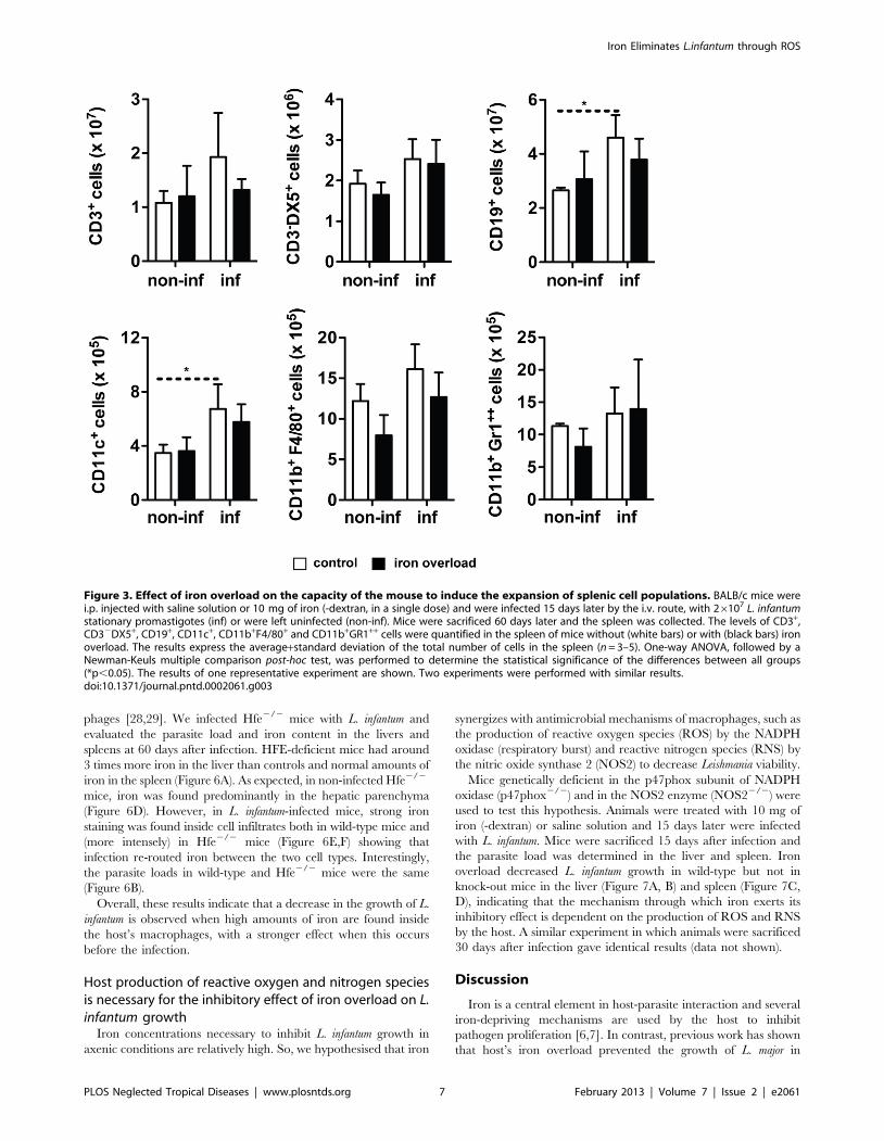
Iron overload does not affect the capacity of the L.infantum infected mice to induce expansion of spleniccell populations or expression of key cytokines
Since previous studies had suggested that host’s iron-overload
interfered with the development of a protective immune response
[12], we evaluated the impact of iron-overload on the induction of
protective cytokines and specific splenic cell populations in our
model of visceral leishmaniasis.
BALB/c mice were treated with 10 mg of iron (given as iron-
dextran) or saline solution 15 days prior to infection. They were
infected with L. infantum and sacrificed 60 days later. Groups of
non-infected mice were kept as controls. We performed a
cytometric analysis of the number of splenic CD3+ (T cells),
CD32DX5+ (NK cells), CD19+ (B cells), CD11c+ (Dendritic cells),
CD11b+F4/80+ (Macrophages) and CD11b+Gr1++ (Neutrophils)
cells. The infection with L. infantum resulted in a significant
increase in the numbers of CD19+ and CD11c+ cells, while other
splenic cell sub-sets remained unaltered (Figure 3). More
importantly, the number of cells belonging to each of the
abovementioned populations was the same in control and iron
overloaded groups (Figure 3). Additionally, the in situ production of
a number of cytokines was measured, using a multiplex assay. The
results of this screening revealed that infection with L. infantum did
not have a dramatic impact on cytokine production. Only IL-1b,
IL-6, TNF and IL-4 were significantly induced by infection in the
spleen (the latter also in the liver, Figure 4), while the production of
IL-12p70 and IL-13 decreased with infection, in the liver
(Figure 4). No significant differences were found between iron-
overloaded and control infected animals in any of the cytokines
tested (Figure 4). The determination of cytokine mRNA expression
in the tissues at earlier time-points did not reveal any differences
between iron-overloaded and control infected mice (not shown).
Overall, these experiments indicate that the inhibitory effect of
iron on the growth of L. infantum in the mouse does not result from
an improvement of the activation of protective cells or increased
production of protective cytokines.
High iron doses and administration prior to infection arenecessary for the inhibitory effect of iron
In order to further understand the mechanisms of the iron
inhibitory effect on L. infantum, we tested the importance of
different experimental parameters.
spleen of groups fed the control (white bars) or iron deficient (black bars) diet was quantified by limiting dilution. The graph shows theaverage+standard deviation of the log10 number of parasites per organ (n = 6). C. BALB/c mice were i.p. injected with 1 mg of iron (given as dextran),in 10 alternate days, from day 220 to day 22 of infection (a total of 10 mg of iron per animal). Control mice received equivalent amounts of dextranby the same route. Mice were infected i.v. with 26107 stationary promastigotes of L. infantum and were sacrificed at 7, 15, 30 and 60 days of infection.The parasite burden in the liver and spleen of control (white circles) or iron overloaded (black circles) groups was quantified by limiting dilution(n = 5). D. Non-heme iron content in the liver and spleen was quantified in control (white bars) and iron overloaded (black bars) groups at 60 daysafter infection (n = 4–5). Student’s t-test was performed to determine the statistical significance of the differences between groups (**p,0.01;***p,0.001). E. F. G. H. Perl’s blue staining was performed in liver sections of uninfected (E), uninfected iron overloaded (F), 60 days-infected (G)and 60 days-infected iron overloaded (H) mice. Black bar corresponds to 50 mm. The results of one representative experiment are shown. Twoexperiments were performed with similar results.doi:10.1371/journal.pntd.0002061.g001
Iron Eliminates L.infantum through ROS
PLOS Neglected Tropical Diseases | www.plosntds.org 5 February 2013 | Volume 7 | Issue 2 | e2061

First, we decided to assess if the same protective effect could be
obtained with lower iron doses. BALB/c mice were injected with
different amounts of iron (given as iron-dextran complex) prior to
infection with L. infantum. Parasite loads were determined 30 days
after infection. A significant inhibitory effect on the growth of L.
infantum in the liver and spleen was seen only in mice that received
10 mg of iron (Figure 5A).
Next, we asked whether iron would still have an inhibitory effect
on L. infantum growth when given after infection. We administered
10 mg of iron (-dextran) or saline solution to BALB/c mice, 1 or
15 days after infection. Parasite loads were determined 60 days
post-infection. In the liver, similar levels of growth inhibition were
seen when iron was given either before, 1 day after or 15 days after
infection (Figure 5B). However, in the spleen and in contrast with
iron pre-loading, no significant reduction on the growth of L.
infantum was detected when the administration of iron was done
after infection (Figure 5B).
These results suggested that high amounts of iron present in the
host prior to infection favor the decrease of the multiplication of L.
infantum. In the model used, iron accumulation is observed in
macrophages. To assess the relevance of the cellular location of
iron deposition at the time of infection, we used HFE-deficient
mice which are used as a model of human hemochromatosis and
accumulate iron in parenchymal cells rather than inside macro-
Figure 2. Effect of different forms of iron on the axenic growth of L. infantum pro- and amastigotes. Promastigotes in the exponentialphase of growth (26106/well; A, B, C) or amastigotes (46105/well; D, E, F) were incubated in RPMI (25uC) or MAA20 (37uC) medium, respectively,with iron-dextran (A, D), iron citrate (B, E) and iron sulphate (C, F) (black circles) in the concentrations of 0.018, 0.035, 0.070, 0.14, 0.28, 0.56, 1.1, 2.2,4.5, 9 and 18 mM. Equivalent concentrations of dextran (A, D), tri-sodium citrate (B, E) and magnesium sulphate (C, F) were used as controls (whitecircles). Resazurin dye was added after 24 h of culture. The fluorescence was measured 24 h (for amastigotes) or 48 h (for promastigotes) afterresazurin addition. The results show the average 6 standard deviation of the percentage viability in relation to the non-treated control (n = 3). One-way ANOVA, followed by a Newman-Keuls multiple comparison post-hoc test, was performed to determine the statistical significance of thedifferences between control (white circles) and iron treated (black circles) groups (*p,0.05, **p,0.01; ***p,0.001). The results of one representativeexperiment are shown. Three experiments were performed with similar results.doi:10.1371/journal.pntd.0002061.g002
Iron Eliminates L.infantum through ROS
PLOS Neglected Tropical Diseases | www.plosntds.org 6 February 2013 | Volume 7 | Issue 2 | e2061
phages [28,29]. We infected Hfe2/2 mice with L. infantum and
evaluated the parasite load and iron content in the livers and
spleens at 60 days after infection. HFE-deficient mice had around
3 times more iron in the liver than controls and normal amounts of
iron in the spleen (Figure 6A). As expected, in non-infected Hfe2/2
mice, iron was found predominantly in the hepatic parenchyma
(Figure 6D). However, in L. infantum-infected mice, strong iron
staining was found inside cell infiltrates both in wild-type mice and
(more intensely) in Hfe2/2 mice (Figure 6E,F) showing that
infection re-routed iron between the two cell types. Interestingly,
the parasite loads in wild-type and Hfe2/2 mice were the same
(Figure 6B).
Overall, these results indicate that a decrease in the growth of L.
infantum is observed when high amounts of iron are found inside
the host’s macrophages, with a stronger effect when this occurs
before the infection.
Host production of reactive oxygen and nitrogen speciesis necessary for the inhibitory effect of iron overload on L.infantum growth
Iron concentrations necessary to inhibit L. infantum growth in
axenic conditions are relatively high. So, we hypothesised that iron
synergizes with antimicrobial mechanisms of macrophages, such as
the production of reactive oxygen species (ROS) by the NADPH
oxidase (respiratory burst) and reactive nitrogen species (RNS) by
the nitric oxide synthase 2 (NOS2) to decrease Leishmania viability.
Mice genetically deficient in the p47phox subunit of NADPH
oxidase (p47phox2/2) and in the NOS2 enzyme (NOS22/2) were
used to test this hypothesis. Animals were treated with 10 mg of
iron (-dextran) or saline solution and 15 days later were infected
with L. infantum. Mice were sacrificed 15 days after infection and
the parasite load was determined in the liver and spleen. Iron
overload decreased L. infantum growth in wild-type but not in
knock-out mice in the liver (Figure 7A, B) and spleen (Figure 7C,
D), indicating that the mechanism through which iron exerts its
inhibitory effect is dependent on the production of ROS and RNS
by the host. A similar experiment in which animals were sacrificed
30 days after infection gave identical results (data not shown).
Discussion
Iron is a central element in host-parasite interaction and several
iron-depriving mechanisms are used by the host to inhibit
pathogen proliferation [6,7]. In contrast, previous work has shown
that host’s iron overload prevented the growth of L. major in
Figure 3. Effect of iron overload on the capacity of the mouse to induce the expansion of splenic cell populations. BALB/c mice werei.p. injected with saline solution or 10 mg of iron (-dextran, in a single dose) and were infected 15 days later by the i.v. route, with 26107 L. infantumstationary promastigotes (inf) or were left uninfected (non-inf). Mice were sacrificed 60 days later and the spleen was collected. The levels of CD3+,CD32DX5+, CD19+, CD11c+, CD11b+F4/80+ and CD11b+GR1++ cells were quantified in the spleen of mice without (white bars) or with (black bars) ironoverload. The results express the average+standard deviation of the total number of cells in the spleen (n = 3–5). One-way ANOVA, followed by aNewman-Keuls multiple comparison post-hoc test, was performed to determine the statistical significance of the differences between all groups(*p,0.05). The results of one representative experiment are shown. Two experiments were performed with similar results.doi:10.1371/journal.pntd.0002061.g003
Iron Eliminates L.infantum through ROS
PLOS Neglected Tropical Diseases | www.plosntds.org 7 February 2013 | Volume 7 | Issue 2 | e2061
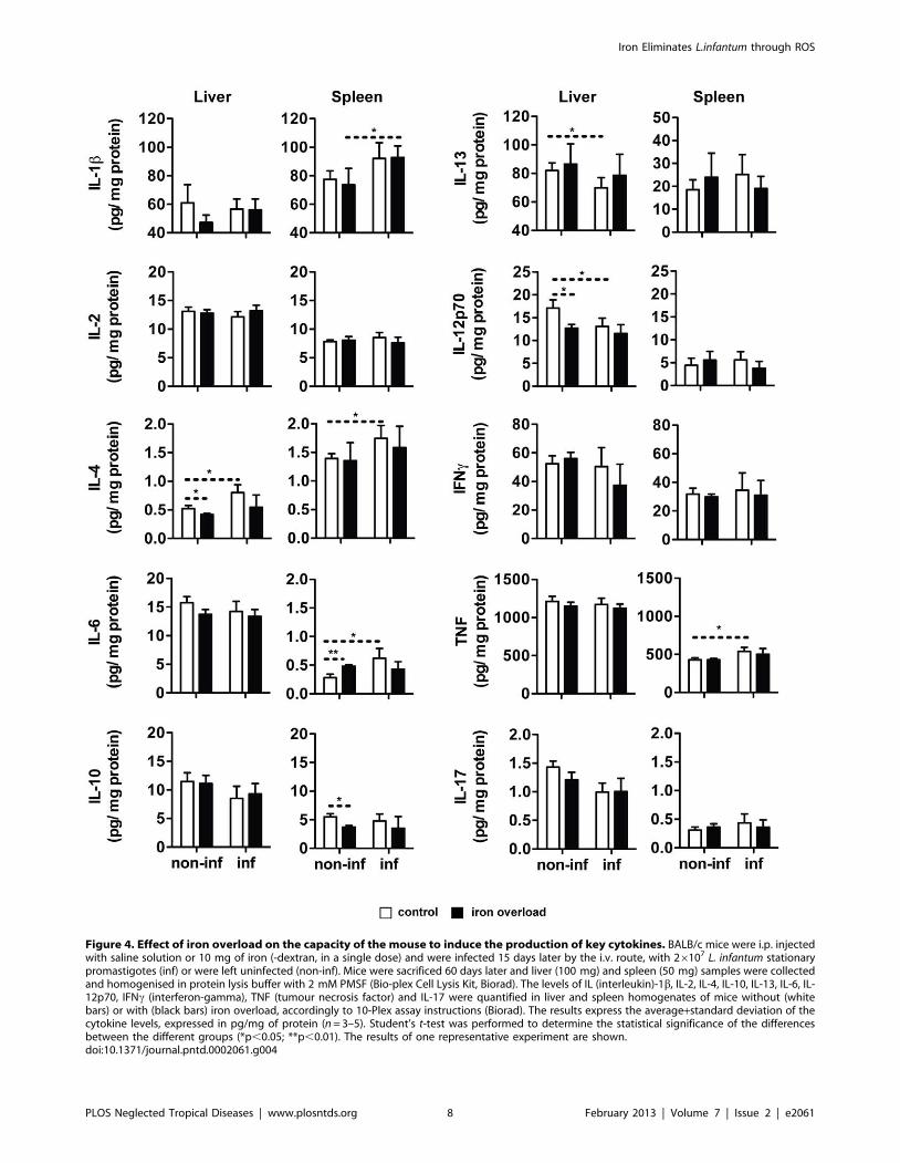
Figure 4. Effect of iron overload on the capacity of the mouse to induce the production of key cytokines. BALB/c mice were i.p. injectedwith saline solution or 10 mg of iron (-dextran, in a single dose) and were infected 15 days later by the i.v. route, with 26107 L. infantum stationarypromastigotes (inf) or were left uninfected (non-inf). Mice were sacrificed 60 days later and liver (100 mg) and spleen (50 mg) samples were collectedand homogenised in protein lysis buffer with 2 mM PMSF (Bio-plex Cell Lysis Kit, Biorad). The levels of IL (interleukin)-1b, IL-2, IL-4, IL-10, IL-13, IL-6, IL-12p70, IFNc (interferon-gamma), TNF (tumour necrosis factor) and IL-17 were quantified in liver and spleen homogenates of mice without (whitebars) or with (black bars) iron overload, accordingly to 10-Plex assay instructions (Biorad). The results express the average+standard deviation of thecytokine levels, expressed in pg/mg of protein (n = 3–5). Student’s t-test was performed to determine the statistical significance of the differencesbetween the different groups (*p,0.05; **p,0.01). The results of one representative experiment are shown.doi:10.1371/journal.pntd.0002061.g004
Iron Eliminates L.infantum through ROS
PLOS Neglected Tropical Diseases | www.plosntds.org 8 February 2013 | Volume 7 | Issue 2 | e2061

BALB/c mice [12,13]. In those studies, it is shown that iron
overload correlates with increased production of ROS upon L.
major infection [13,30]. In the present work, we treated mice with
iron-dextran and infected them with L. infantum, an agent of
visceral leishmaniasis. We observed iron-loaded macrophages
inside hepatic infiltrates, the areas of parasite containment,
concomitantly with the decrease of tissue parasite loads. Such
iron-associated decrease did not occur in p47phox- or NOS2-
deficient mice, suggesting that iron exerts its effects through the
combination with ROS and/or RNS produced by the macro-
phage. In fact, superoxide (O2N2) and nitric oxide (NON),
synthesized by the phagocytic NADPH oxidase and the nitric
oxide synthase 2 (NOS2), respectively, have been implicated in the
elimination of Leishmania by the host’s macrophages
[31,32,33,34,35,36]. Moreover, both macrophagic NADPH oxi-
dase and NOS2 require iron for proper function [37]. In a few
models of macrophage infections with different bacteria, macro-
phages were shown to need iron to exert their antimicrobial
activity. Iron increases the capacity of macrophages to eliminate or
prevent the multiplication of B. abortus, by catalyzing the
production of hydroxyl radical [11] and iron loading of
Staphylococcus aureus prior to infection enhances bacterial killing
Figure 5. Effect of different iron doses and periods of administration on the growth of L. infantum. A. BALB/c mice were injected i.p. with1 or 10 mg of iron (-dextran), in a single dose, 12 days before the infection. B. BALB/c mice were injected i.p. with 10 mg of iron (-dextran), in a singledose, 15 days before or 1 and 15 days after the infection (215, +1 and +15 days, respectively). A. B. Control mice received saline solution by the sameroute. Mice were sacrificed 30 (A) or 60 (B) days after the i.v. infection with 26107 stationary promastigotes of L. infantum. The parasite burden in theliver and spleen of mice without (white bar) and with (gray - black bars) iron administration was determined by limiting dilution. The data shows theaverage+standard deviation of the log10 number of parasites per organ (n = 5). One-way ANOVA, followed by a Dunnett’s multiple comparison post-hoc test, was performed to determine the statistical significance of the differences between each of the iron treated groups and control group(*p,0.05; **p,0.01). The results of one representative experiment are shown. Two experiments were performed with similar results.doi:10.1371/journal.pntd.0002061.g005
Iron Eliminates L.infantum through ROS
PLOS Neglected Tropical Diseases | www.plosntds.org 9 February 2013 | Volume 7 | Issue 2 | e2061

by monocytes, most likely by promoting oxidative damage [38].
Also in the case of Salmonella, iron seems to be needed for
intramacrophagic killing of these bacteria [10]. This suggests that
iron, a well known pro-oxidant, promotes oxidative microbicidal
mechanisms inside macrophages, to which Leishmania is sensitive
[31,33,34,35,36,39]. The fact that the anti-parasitic effect of iron is
lost in mice deficient in only one of the two enzymes, either NOS2
or NADPH oxidase, suggests that both ROS and RNS are
simultaneously required for iron to exert its anti-leishmanial effect.
Iron can possibly favour the formation of peroxynitrite (ONOO2),
a strong oxidizing species formed by the reaction of NON with
O2N2 [40]. Since we could not find evidences of tissue oxidative
damage (DNA damage, lipid peroxidation and protein oxidation)
in iron-treated mice, we suggest that this formation of highly
reactive ROS and RNS in combination with iron has a highly
localized activity, inside the macrophage.
Figure 6. Effect of genetically determined iron overload on the growth of L. infantum in mice. A. B. Wild-type (WT) and Hfe2/2 mice werei.v. infected with 26107 stationary promastigotes of L. infantum and were sacrificed 60 days after infection. A. Non-heme iron content in both organswas quantified in WT (white bars) and Hfe2/2 (black bars) groups. The data shows the average+standard deviation of the non-heme iron content,expressed in mg/organ (n = 4–6). Student’s t-test was performed to determine the statistical significance of the differences between WT and Hfe2/2
groups (***p,0.001). B. The parasite burden in the liver and spleen of wild-type (WT; white bars) and Hfe2/2 (black bars) mice was determined bylimiting dilution (n = 5–6). C–F: Perl’s blue staining was performed in liver sections of uninfected (C) and infected (E) WT and uninfected (D) andinfected (F) Hfe2/2 mice. Black bar corresponds to 50 mm. The results of one representative experiment are shown. Two experiments were performedwith similar results.doi:10.1371/journal.pntd.0002061.g006
Iron Eliminates L.infantum through ROS
PLOS Neglected Tropical Diseases | www.plosntds.org 10 February 2013 | Volume 7 | Issue 2 | e2061

It was somewhat surprising that Hfe2/2 mice, which have
spontaneous iron overload, predominantly in the liver, had tissue
parasite loads similar to those of wild-type mice, when infected
with L. infantum. This could be justified by the fact that Hfe2/2
mice develop spontaneous iron overload predominantly in
hepatocytes, keeping macrophages relatively iron depleted
[28,29,41]. When Hfe2/2 mice were infected with L. infantum,
we could see iron accumulation inside macrophages at the
infection foci, similarly to what we had previously reported in
M. avium infection [14]. However, as suggested by the experiments
in which we treated mice with iron-dextran after infection, the
inhibitory effect of iron is best accomplished when the macro-
phages are iron-loaded prior to infection. Another hypothesis to
explain the lack of an impact of Hfe2/2 iron overload on the
growth of L. infantum is the level of iron overload in the tissues.
Indeed, Hfe2/2 mice had tissue iron levels that were significantly
lower than those found in iron-dextran-injected mice. The fact
that even with iron-dextran injection, we needed high iron doses to
decrease the parasite burden in tissues, argues for this hypothesis.
In the mouse model of cutaneous leishmaniasis, iron-induced
respiratory burst at the onset of L. major infection is coupled to later
activation of the nuclear transcription factor NF- kB [30] and to
the display of a protective immune response [12]. Iron and ROS
can modulate the activation of NF-kB signaling pathways [42],
known to regulate several genes involved in immune and
inflammatory responses [43]. In the case of L. major infection,
mouse resistance is clearly related to an IL-12-driven, IFN-c-
dominated Th1 immune response, whereas susceptibility corre-
Figure 7. Effect of iron overload on the growth of L. infantum in mice: role of NADPH oxidase and NOS2. Wild-type (WT) and p47phox2/2
(A, C) or NOS22/2 (B, D) mice were i.p. injected with saline solution or 10 mg of iron (-dextran, in a single dose) 15 days before i.v. infection with26107 stationary promastigotes of L. infantum and were sacrificed 15 days later. The liver (A, B) and spleen (C, D) were removed and the parasiteburden in mice without (white bars) or with (black bars) iron overload was quantified by limiting dilution. The results express the average+standarddeviation of the log10 number of parasites per organ (n = 3–8). One-way ANOVA, followed by a Newman-Keuls multiple comparison post-hoc test, wasperformed to determine the statistical significance of the differences between all groups (*p,0.05; **p,0.01; ***p,0.001). The results of onerepresentative experiment are shown. At least two experiments were performed with similar results.doi:10.1371/journal.pntd.0002061.g007
Iron Eliminates L.infantum through ROS
PLOS Neglected Tropical Diseases | www.plosntds.org 11 February 2013 | Volume 7 | Issue 2 | e2061

lates with an IL-4-driven Th2 response [44]. Experimentally iron
overloaded BALB/c mice, infected with L. major, exhibited a Th1-
type immune response, with increased levels of IFNc and NOS2
and decreased levels of IL-4 and IL-10 transcripts compared to
untreated mice [12]. In accordance, supplementation of rats with
iron-dextran [45] or saccharated colloidal iron [46] potentiated
the induction of hepatic NOS2 and the production of NON by
LPS. However, the decrease of NON production has also been
observed in mice [47] and macrophages [48] treated with different
iron sources. Additionally, delayed Th1 immune responses and
Th2 phenotypes have been observed in response to iron
supplementation in mice infected with Cryptoccocus neoformans [49]
and Candida albicans [50], indicating that each particular host-
pathogen interaction responds differently to iron overload.
In the case of visceral leishmaniasis, an efficient control of
infection is also dependent on Th1 responses, although a mixed
Th1/Th2 cytokine profile is detected during the course of
infection [51,52]. So, iron supplementation in our model, besides
exerting a direct toxic effect on parasites in conjunction with ROS
and RNS, could be improving the host’s capacity to control the
infection, by modulating the adaptive immune response. When we
evaluated the cytokine response to Leishmania infection, we saw a
discrete induction of IL-4 both in the liver and the spleen of
infected mice, together with increases in the expression of the pro-
inflammatory cytokines, IL-1b, IL-6 and TNF. However, iron
overload did not significantly alter the immune response profile
induced by infection, leading us to conclude that the modulation of
the adaptive immune response does not contribute significantly to
the protective effect of iron.
Malnutrition is associated with susceptibility to visceral leish-
maniasis in humans [53,54] and mice [55]. Although iron
deficiency is the most common micronutrient deficiency in the
human population [56], the relationship between human iron
deficiency alone and increased risk of acquiring visceral leishman-
iasis has never been investigated. In our model, feeding mice with
an iron deficient diet did not affect L. infantum growth. These
nutritionally iron-deprived animals had half the normal iron stores
in the liver and the spleen, but presented normal haematocrit and
body weight (not shown). We hypothesize that the low levels of
iron in tissue stores were probably sufficient to maintain the
growth of Leishmania and not low enough to impact on the host’s
capacity to control the infection. Observations regarding the
effects of iron chelators on Leishmania growth are conflicting.
Treatment of mice with desferrioxamine (DFO) led to the decrease
of L. infantum proliferation [57] but not that of L. major [12]. Also,
in in vitro models of macrophage infection, DFO has shown either
no effect [58] or an inhibitory effect [59,60,61] on Leishmania
growth. Finally, when tested on Leishmania promastigotes growing
in culture medium, hydroxypiridinone-derived chelators showed
an inhibitory effect, which was higher than that of DFO [62].
Thus, the available data do not allow inferring that Leishmania
infections are amenable to treatment by iron depletion. It may be
valuable to further explore the effects of different iron chelating
ligands or of more drastic iron depletion protocols.
In addition to the results obtained in vivo and discussed above,
we found axenic cultures of L. infantum to be sensitive to the direct
toxicity of iron. It is plausible that high concentrations of iron
(.0.56 mM) may have promoted the endogenous generation or
propagation of reactive species in both parasite stages. On the
other hand, lower iron doses (,0.56 mM) may not have been
sufficient to overcome the antioxidant capacity of axenic L.
infantum. In this regard, L. infantum promastigotes have been shown
to accumulate iron in catalytically active forms, which contribute
to their sensitivity to killing by hydrogen peroxide [63,64], possibly
through the Fenton reaction. Besides, upon exposure to high doses
of this metal, L. infantum promastigotes exhibited impaired motility
and morphological changes identical to those reported to occur
after exposure to antimony (III) and arsenic (III) [27] (not shown).
These metalloids, used for a long time as first line treatments
against trypanosomatid infections, were recently found to act
through the induction of oxidative damage in L. donovani [27,65].
Interestingly, increased intracellular iron levels directly correlate to
the parasite sensitivity to these drugs [27]. The influence of iron on
anti-leishmanial drug activity also includes non-metalloid drugs.
Iron potentiates the leishmanicidal activity of artemisinin [66], by
inducing oxidative injury that culminates in cell death of L.
donovani promastigotes. Furthermore, iron treatment can induce
accumulation of pentamidine in the mitochondria of L. enriettii
promastigotes, hence increasing their sensitivity to the drug. This
effect is probably due to the action of the multidrug resistance
protein 1 (LeMDR1), a putative mitochondrial iron importer [67].
Hence, the increase of intracellular iron levels in Leishmania overall
increases its vulnerability to chemotherapy.
The interaction between pathogens and their hosts are complex
processes dependent not only on the genome of both, but also on
nutritional factors. In most of the reported cases, iron excess
increases and iron chelation decreases susceptibility to infection
[8,20]. However, this is not observed in murine models of infection
by Leishmania. Moreover, despite several reports that iron
supplementation (to correct nutritional iron deficiency) can
significantly increase the risk of several infections [8,56], no
correlation between iron administration and susceptibility to
human leishmaniasis has, to our knowledge, ever been described.
Although iron chelation has been suggested as an effective
therapeutic strategy against several infections, in the case of
leishmaniasis and especially in areas where this disease and
malnutrition coexist, iron chelation may be inappropriate. Iron
overload decreases Leishmania proliferation and induces parasite
death, probably by promoting oxidative reactions pernicious to the
parasite. The further investigation of the molecular mechanisms of
these effects will be fundamental to explore a potential utilization
of iron itself as a therapeutic tool and also to understand and
improve the mechanisms of action of other anti-leishmanial drugs.
Supporting Information
Text S1 Effect of iron overload on the carbonylation ofproteins, peroxidation of lipids and integrity of DNA inthe mouse liver. Figure S1. BALB/c mice were i.p. injected
with saline solution or 10 mg of iron (-dextran, in a single dose)
and were infected 15 days later by the i.v. route, with 26107
L.infantum stationary promastigotes. Mice were sacrificed 60 days
later and liver (100 mg) samples were collected and homogenised
in protein lysis buffer. Liver protein lysates were reacted with
DNP-hydrazone and then separated by SDS-PAGE followed by
Western blotting (n = 4–5). No differences in the protein carbonyl
levels were observed between control and iron-treated mice.
Figure S2. C57BL/6 mice were fed a control (A) or a 2.5% iron-
carbonyl diet (B) for 15 days. Liver samples were assayed by
immunofluorescence to detect 4-hydroxynonenal (4-HNE) staining
(green). Nuclei were counterstained with DAPI (blue). No 4-HNE
staining was detected in the liver tissue of BALB/c mice treated for
15 days with saline solution or 10 mg of iron-dextran prior to a 30-
and 60-day infection with L.infantum (staining was identical to A).
Figure S3. BALB/c mice were i.p. injected with saline solution
(A) or 10 mg of iron (B) (-dextran, in a single dose) and were
infected 15 days later by the i.v. route, with 26107 L.infantum
stationary promastigotes. Mice were sacrificed 60 days later and
Iron Eliminates L.infantum through ROS
PLOS Neglected Tropical Diseases | www.plosntds.org 12 February 2013 | Volume 7 | Issue 2 | e2061

liver samples were assayed by immunofluorescence to detect
TUNEL staining (green). Nuclei were counterstained with DAPI
(blue). No TUNEL staining was observed in animals receiving
saline solution or iron treatment, except in the positive control (C,
sample treated with DNase I).
(DOCX)
Acknowledgments
The authors would like to acknowledge Carolina Caldas (IBMC) for
processing and staining histological samples.
Author Contributions
Conceived and designed the experiments: SVC PNR RA AT MSG.
Performed the experiments: SVC SGP CMT GR. Analyzed the data: SVC
MSG. Wrote the paper: SVC MSG.
References
1. WHO (2010) Control of the Leishmaniasis. WHO Technical Report Series 949:
1–186.
2. Ready PD (2010) Leishmaniasis emergence in Europe. Euro Surveillance 15:
19505.
3. Nagill R, Kaur S (2011) Vaccine candidates for leishmaniasis: a review. Int
Immunopharmacol 11: 1464–1488.
4. Mondal S, Bhattacharya P, Ali N (2010) Current diagnosis and treatment of
visceral leishmaniasis. Expert Rev Anti Infect Ther 8: 919–944.
5. Maltezou HC (2010) Drug resistance in visceral leishmaniasis. J Biomed
Biotechnol 2010: 617521.
6. Ganz T (2009) Iron in innate immunity: starve the invaders. Curr Opin
Immunol 21: 63–67.
7. Nairz M, Schroll A, Sonnweber T, Weiss G (2010) The struggle for iron - a
metal at the host-pathogen interface. Cell Microbiol 12: 1691–1702.
8. Weinberg ED (2009) Iron availability and infection. Biochim Biophys Acta 1790:
600–605.
9. Alford CE, King TE, Jr., Campbell PA (1991) Role of transferrin, transferrin
receptors, and iron in macrophage listericidal activity. Journal Exp Med 174:
459–466.
10. Collins HL, Kaufmann SH, Schaible UE (2002) Iron chelation via deferoxamine
exacerbates experimental salmonellosis via inhibition of the nicotinamide
adenine dinucleotide phosphate oxidase-dependent respiratory burst.
J Immunol 168: 3458–3463.
11. Jiang X, Baldwin CL (1993) Iron augments macrophage-mediated killing of
Brucella abortus alone and in conjunction with interferon-gamma. Cell
Immunol 148: 397–407.
12. Bisti S, Konidou G, Papageorgiou F, Milon G, Boelaert JR, et al. (2000) The
outcome of Leishmania major experimental infection in BALB/c mice can be
modulated by exogenously delivered iron. Eur J Immunol 30: 3732–3740.
13. Bisti S, Konidou G, Boelaert J, Lebastard M, Soteriadou K (2006) The
prevention of the growth of Leishmania major progeny in BALB/c iron-loaded
mice: a process coupled to increased oxidative burst, the amplitude and duration
of which depend on initial parasite developmental stage and dose. Microbes
Infect 8: 1464–1472.
14. Gomes-Pereira S, Rodrigues PN, Appelberg R, Gomes MS (2008) Increased
susceptibility to Mycobacterium avium in hemochromatosis protein HFE-
deficient mice. Infect Immun 76: 4713–4719.
15. Gomes MS, Boelaert JR, Appelberg R (2001) Role of iron in experimental
Mycobacterium avium infection. J Clin Virol 20: 117–122.
16. Gomes MS, Appelberg R (1998) Evidence for a link between iron metabolism
and Nramp1 gene function in innate resistance against Mycobacterium avium.
Immunology 95: 165–168.
17. Fernandes SS, Nunes A, Gomes AR, de Castro B, Hider RC, et al. (2010)
Identification of a new hexadentate iron chelator capable of restricting the
intramacrophagic growth of Mycobacterium avium. Microbes Infect 12: 287–
294.
18. Sereno D, Lemesre JL (1997) Axenically cultured amastigote forms as an in vitro
model for investigation of antileishmanial agents. Antimicrob Agents Chemother
41: 972–976.
19. Torrance JD, Bothwell TH (1980) Tissue iron stores. In: Cook JD, editor.
Methods in Hematology. New York, NY: Churchill Livingstone Press. pp. 104–
109.
20. Weinberg ED (1999) The role of iron in protozoan and fungal infectious
diseases. J Eukaryot Microbiol 46: 231–238.
21. Ahmed S, Colmenares M, Soong L, Goldsmith-Pestana K, Munstermann L, et
al. (2003) Intradermal infection model for pathogenesis and vaccine studies of
murine visceral leishmaniasis. Infect Immun 71: 401–410.
22. Carrion J, Nieto A, Iborra S, Iniesta V, Soto M, et al. (2006) Immunohistological
features of visceral leishmaniasis in BALB/c mice. Parasite Immunol 28: 173–
183.
23. Halliday JW, Searle J (1996) Hepatic iron deposition in human disease and
animal models. Biometals 9: 205–209.
24. Papanikolaou G, Pantopoulos K (2005) Iron metabolism and toxicity. Toxicol
Appl Pharmacol 202: 199–211.
25. Stanley AC, Engwerda CR (2007) Balancing immunity and pathology in visceral
leishmaniasis. Immunol Cell Biol 85: 138–147.
26. Murray HW (2001) Tissue granuloma structure-function in experimental
visceral leishmaniasis. Int J Exp Pathol 82: 249–267.
27. Mehta A, Shaha C (2006) Mechanism of metalloid-induced death in Leishmania
spp.: role of iron, reactive oxygen species, Ca2+, and glutathione. Free Radic
Biol Med 40: 1857–1868.
28. Bahram S, Gilfillan S, Kuhn LC, Moret R, Schulze JB, et al. (1999)
Experimental hemochromatosis due to MHC class I HFE deficiency: immune
status and iron metabolism. PNAS 96: 13312–13317.
29. Zhou XY, Tomatsu S, Fleming RE, Parkkila S, Waheed A, et al. (1998) HFE
gene knockout produces mouse model of hereditary hemochromatosis. PNAS
95: 2492–2497.
30. Bisti S, Soteriadou K (2006) Is the reactive oxygen species-dependent-NF-
kappaB activation observed in iron-loaded BALB/c mice a key process
preventing growth of Leishmania major progeny and tissue-damage? Microbes
Infect 8: 1473–1482.
31. Murray HW, Nathan CF (1999) Macrophage microbicidal mechanisms in vivo:
reactive nitrogen versus oxygen intermediates in the killing of intracellular
visceral Leishmania donovani. J Exp Med 189: 741–746.
32. Linares E, Giorgio S, Mortara RA, Santos CX, Yamada AT, et al. (2001) Role
of peroxynitrite in macrophage microbicidal mechanisms in vivo revealed by
protein nitration and hydroxylation. Free Radic Biol Med 30: 1234–1242.
33. Blos M, Schleicher U, Soares Rocha FJ, Meissner U, Rollinghoff M, et al. (2003)
Organ-specific and stage-dependent control of Leishmania major infection by
inducible nitric oxide synthase and phagocyte NADPH oxidase. Eur J Immunol
33: 1224–1234.
34. Stenger S, Thuring H, Rollinghoff M, Bogdan C (1994) Tissue expression of
inducible nitric oxide synthase is closely associated with resistance to Leishmania
major. J Exp Med 180: 783–793.
35. Wei XQ, Charles IG, Smith A, Ure J, Feng GJ, et al. (1995) Altered immune
responses in mice lacking inducible nitric oxide synthase. Nature 375: 408–411.
36. Melby PC, Chandrasekar B, Zhao W, Coe JE (2001) The hamster as a model of
human visceral leishmaniasis: progressive disease and impaired generation of
nitric oxide in the face of a prominent Th1-like cytokine response. J Immunol
166: 1912–1920.
37. Fang FC (2004) Antimicrobial reactive oxygen and nitrogen species: concepts
and controversies. Nat Rev Microbiol 2: 820–832.
38. Hoepelman IM, Bezemer WA, Vandenbroucke-Grauls CM, Marx JJ, Verhoef J
(1990) Bacterial iron enhances oxygen radical-mediated killing of Staphylococ-
cus aureus by phagocytes. Infect Immun 58: 26–31.
39. Melby PC, Yang YZ, Cheng J, Zhao W (1998) Regional differences in the
cellular immune response to experimental cutaneous or visceral infection with
Leishmania donovani. Infect Immun 66: 18–27.
40. Halliwell B, Gutteridge JCM (2007) Free Radicals in Biology and Medicine.
New York: Oxford University Press. 851 p.
41. Porto G, De Sousa M (2007) Iron overload and immunity. World J Gastroenterol
13: 4707–4715.
42. Xiong S, She H, Takeuchi H, Han B, Engelhardt JF, et al. (2003) Signaling role
of intracellular iron in NF-kappaB activation. J Biol Chem 278: 17646–17654.
43. Bonizzi G, Karin M (2004) The two NF-kappaB activation pathways and their
role in innate and adaptive immunity. Trends Immunol 25: 280–288.
44. Sacks D, Noben-Trauth N (2002) The immunology of susceptibility and
resistance to Leishmania major in mice. Nat Rev Immunol 2: 845–858.
45. Galleano M, Simontacchi M, Puntarulo S (2004) Nitric oxide and iron: effect of
iron overload on nitric oxide production in endotoxemia. Mol Aspects Med 25:
141–154.
46. Hida AI, Kawabata T, Minamiyama Y, Mizote A, Okada S (2003) Saccharated
colloidal iron enhances lipopolysaccharide-induced nitric oxide production in
vivo. Free Radic Biol Med 34: 1426–1434.
47. Komarov AM, Mattson DL, Mak IT, Weglicki WB (1998) Iron attenuates nitric
oxide level and iNOS expression in endotoxin-treated mice. FEBS Lett 424:
253–256.
48. Weiss G, Werner-Felmayer G, Werner ER, Grunewald K, Wachter H, et al.
(1994) Iron regulates nitric oxide synthase activity by controlling nuclear
transcription. J Exp Med 180: 969–976.
49. Barluzzi R, Saleppico S, Nocentini A, Boelaert JR, Neglia R, et al. (2002) Iron
overload exacerbates experimental meningoencephalitis by Cryptococcus
neoformans. J Neuroimmunol 132: 140–146.
50. Mencacci A, Cenci E, Boelaert JR, Bucci P, Mosci P, et al. (1997) Iron overload
alters innate and T helper cell responses to Candida albicans in mice. J Infect Dis
175: 1467–1476.
Iron Eliminates L.infantum through ROS
PLOS Neglected Tropical Diseases | www.plosntds.org 13 February 2013 | Volume 7 | Issue 2 | e2061

51. McMahon-Pratt D, Alexander J (2004) Does the Leishmania major paradigm of
pathogenesis and protection hold for New World cutaneous leishmaniases or thevisceral disease? Immunol Rev 201: 206–224.
52. Rolao N, Cortes S, Gomes-Pereira S, Campino L (2007) Leishmania infantum:
mixed T-helper-1/T-helper-2 immune response in experimentally infectedBALB/c mice. Exp Parasitol 115: 270–276.
53. Cerf BJ, Jones TC, Badaro R, Sampaio D, Teixeira R, et al. (1987) Malnutritionas a risk factor for severe visceral leishmaniasis. J Infect Dis 156: 1030–1033.
54. Harrison LH, Naidu TG, Drew JS, de Alencar JE, Pearson RD (1986)
Reciprocal relationships between undernutrition and the parasitic diseasevisceral leishmaniasis. Rev Infect Dis 8: 447–453.
55. Anstead GM, Chandrasekar B, Zhao W, Yang J, Perez LE, et al. (2001)Malnutrition alters the innate immune response and increases early visceraliza-
tion following Leishmania donovani infection. Infect Immun 69: 4709–4718.56. Oppenheimer SJ (2001) Iron and its relation to immunity and infectious disease.
J Nutr 131: 616S–633S; discussion 633S–635S.
57. Malafaia G, Marcon Lde N, Pereira Lde F, Pedrosa ML, Rezende SA (2011)Leishmania chagasi: effect of the iron deficiency on the infection in BALB/c
mice. Exp Parasitol 127: 719–723.58. Murray HW, Granger AM, Teitelbaum RF (1991) Gamma interferon-activated
human macrophages and Toxoplasma gondii, Chlamydia psittaci, and
Leishmania donovani: antimicrobial role of limiting intracellular iron. InfectImmun 59: 4684–4686.
59. Borges VM, Vannier-Santos MA, de Souza W (1998) Subverted transferrintrafficking in Leishmania-infected macrophages. Parasitol Res 84: 811–822.
60. Das NK, Biswas S, Solanki S, Mukhopadhyay CK (2009) Leishmania donovani
depletes labile iron pool to exploit iron uptake capacity of macrophage for itsintracellular growth. Cell Microbiol 11: 83–94.
61. Segovia M, Navarro A, Artero JM (1989) The effect of liposome-entrapped
desferrioxamine on Leishmania donovani in vitro. Ann Trop Med Parasitol 83:357–360.
62. Soteriadou K, Papavassiliou P, Voyiatzaki C, Boelaert J (1995) Effect of ironchelation on the in-vitro growth of Leishmania promastigotes. J Antimicrob
Chemother 35: 23–29.
63. Wilson ME, Vorhies RW, Andersen KA, Britigan BE (1994) Acquisition of ironfrom transferrin and lactoferrin by the protozoan Leishmania chagasi. Infect
Immun 62: 3262–3269.64. Zarley JH, Britigan BE, Wilson ME (1991) Hydrogen peroxide-mediated toxicity
for Leishmania donovani chagasi promastigotes. Role of hydroxyl radical andprotection by heat shock. J Clin Invest 88: 1511–1521.
65. Wyllie S, Cunningham ML, Fairlamb AH (2004) Dual action of antimonial
drugs on thiol redox metabolism in the human pathogen Leishmania donovani.J Biol Chem 279: 39925–39932.
66. Sen R, Saha P, Sarkar A, Ganguly S, Chatterjee M (2010) Iron enhancesgeneration of free radicals by Artemisinin causing a caspase-independent,
apoptotic death in Leishmania donovani promastigotes. Free Radic Res 44:
1289–1295.67. Wong IL, Chow LM (2006) The role of Leishmania enriettii multidrug
resistance protein 1 (LeMDR1) in mediating drug resistance is iron-dependent.Mol Biochem Parasitol 150: 278–287.
Iron Eliminates L.infantum through ROS
PLOS Neglected Tropical Diseases | www.plosntds.org 14 February 2013 | Volume 7 | Issue 2 | e2061


