Embed Size (px)
Citation preview

Organic codes and their identification:
Is the histone code a true organic code?
by
Stefan Kuhn
Thesis presented in partial fulfilment of the requirements
for the degree of Master of Science (Biochemistry) in the
Faculty of Science at Stellenbosch University
Department of Biochemistry
University of Stellenbosch
Private Bag X1, 7602 Matieland, South Africa
Supervisor: Prof. J.-H.S. Hofmeyr (supervisor)
March 2014

Declaration
By submitting this thesis electronically, I declare that the entirety of the
work contained therein is my own, original work, that I am the sole author
thereof (save to the extent explicitly otherwise stated), that reproduction
and publication thereof by Stellenbosch University will not infringe any
third party rights and that I have not previously in its entirety or in part
submitted it for obtaining any qualification.
March 2014Date: . . . . . . . . . . . . . . . . . . . . . . . . . . . . .
Copyright c© 2014 Stellenbosch University
All rights reserved.
i
Stellenbosch University http://scholar.sun.ac.za

Acknowledgements
• Prof. Hofmeyr, your boundless enthusiasm for life, the universe, and
everything else has been a source of inspiration for me.
• Marcello Barbieri for kickstarting code biology and his fiery convic-
tion.
• The NRF of South Africa for funding.
• My family for unquestioning support.
• The office — Chris, Jalene, it’s been a pleasure.
• Meghan and Sarah, for keeping the faith.
ii
Stellenbosch University http://scholar.sun.ac.za

Meinen Eltern, meiner Oma, und Bienchen — ohne euch ware dieses Werk
nie entstanden
iii
Stellenbosch University http://scholar.sun.ac.za

Contents
Declaration i
Contents iv
List of Figures vi
List of Tables viii
Summary ix
Opsomming xi
1 Introduction 1
2 Code biology 5
2.1 On codes . . . . . . . . . . . . . . . . . . . . . . . . . . . . . 9
2.2 Evolution by natural conventions . . . . . . . . . . . . . . . 12
2.3 Information . . . . . . . . . . . . . . . . . . . . . . . . . . . 14
3 Some organic codes 18
3.1 The genetic code . . . . . . . . . . . . . . . . . . . . . . . . 21
3.2 The metabolic code . . . . . . . . . . . . . . . . . . . . . . . 23
3.3 The signal transduction code . . . . . . . . . . . . . . . . . . 25
iv
Stellenbosch University http://scholar.sun.ac.za

Contents
3.4 The sugar code . . . . . . . . . . . . . . . . . . . . . . . . . 27
3.5 The splicing code . . . . . . . . . . . . . . . . . . . . . . . . 28
3.6 The ubuiquitin code . . . . . . . . . . . . . . . . . . . . . . 30
3.7 The compartment code . . . . . . . . . . . . . . . . . . . . . 31
3.8 The regulatory code . . . . . . . . . . . . . . . . . . . . . . 32
3.9 The Hox code . . . . . . . . . . . . . . . . . . . . . . . . . . 34
4 The histone code 36
4.1 What are histones and the ‘histone code’? . . . . . . . . . . 37
4.2 The function of post-translational histone modifications . . . 38
4.3 The histone post-translational modification zoo . . . . . . . 39
Acetylation . . . . . . . . . . . . . . . . . . . . . . . . . . . 41
Methylation . . . . . . . . . . . . . . . . . . . . . . . . . . . 45
Ubiquitylation . . . . . . . . . . . . . . . . . . . . . . . . . . 50
4.4 Binding domains: The adaptors of the histone code . . . . . 52
Acetyl-recognising domains . . . . . . . . . . . . . . . . . . . 52
Methyl-recognising domains . . . . . . . . . . . . . . . . . . 53
Ubiquitin-recognising domains . . . . . . . . . . . . . . . . . 54
4.5 How does it all fit together? Is the histone code an organic
code? . . . . . . . . . . . . . . . . . . . . . . . . . . . . . . . 54
4.6 Criticisms of the histone code model . . . . . . . . . . . . . 59
5 The Gorlich-Dittrich algorithm for identifying ‘molecular
codes’: A critique 63
6 Discussion 69
Bibliography 74
v
Stellenbosch University http://scholar.sun.ac.za

List of Figures
2.1 Mappings f and g between a set of nucleotide triplets and a set
of amino acids . . . . . . . . . . . . . . . . . . . . . . . . . . . . 7
4.1 The structure of a nucleosome. . . . . . . . . . . . . . . . . . . 37
4.2 Major sites of post-translational modifications of histones H2A,
H2B, H3 and H4. Symbols: A denotes acetylation, M methy-
lation and U ubiquitylation. Sites on the polypeptide chains
are numbered and identified with the one-letter abbreviations of
their amino acids. . . . . . . . . . . . . . . . . . . . . . . . . . . 40
4.3 A: In the absence of acetyl groups on lysine 9 and 14 on histone
3, the double bromodomains of TAFII250 are unable to bind to
H3K9 and H3K14 and as a result, TAFII250 does not phospho-
rylate TAFIIF, which in turn does not lead to transcriptional
initiation. B: once H3K9 and H3K14 have been acetylated (red
circles), the double bromodomains are now able to recognise and
bind the H3K9ac and H3K14ac, which allows TAFII250 to phos-
phorylate TAFIIF and thus permit transcription to proceed. . . 58
5.1 A binary molecular code according to Gorlich and Dittrich [59].
The set, S = {A,B}, is mapped to the set M = {C,D} by the
contexts, C = {E,G} or C ′ = {F,H}. . . . . . . . . . . . . . . 64
vi
Stellenbosch University http://scholar.sun.ac.za

List of Figures
5.2 The mapping of the reaction network mapping d-Glucose and
d-Talose onto d-Mannose and d-Galactose . . . . . . . . . . . . 68
vii
Stellenbosch University http://scholar.sun.ac.za
List of Tables
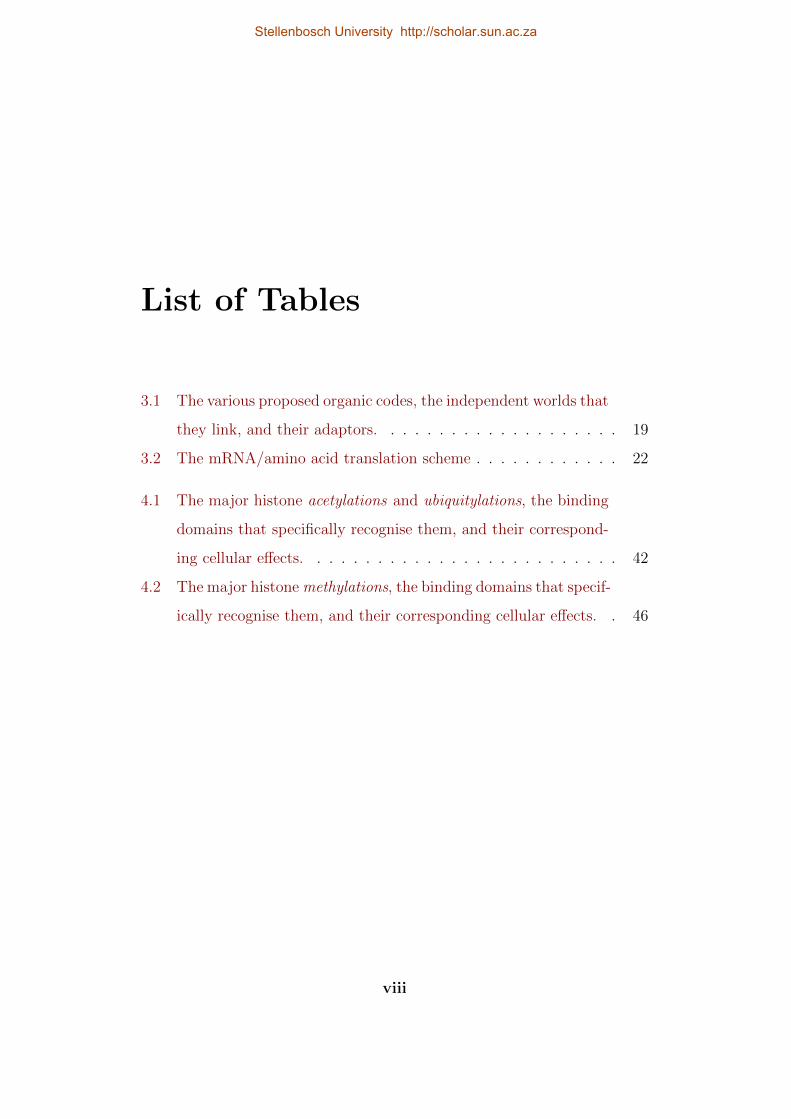
3.1 The various proposed organic codes, the independent worlds that
they link, and their adaptors. . . . . . . . . . . . . . . . . . . . 19
3.2 The mRNA/amino acid translation scheme . . . . . . . . . . . . 22
4.1 The major histone acetylations and ubiquitylations, the binding
domains that specifically recognise them, and their correspond-
ing cellular effects. . . . . . . . . . . . . . . . . . . . . . . . . . 42
4.2 The major histone methylations, the binding domains that specif-
ically recognise them, and their corresponding cellular effects. . 46
viii
Stellenbosch University http://scholar.sun.ac.za

Summary
Codes are ubiquitous in culture—and, by implication, in nature. Code
biology is the study of these codes. However, the term ‘code’ has assumed
a variety of meanings, sowing confusion and cynicism. The first aim of this
study is therefore to define what an organic code is. Following from this, I
establish a set of criteria that a putative code has to conform to in order
to be recognised as a true code. I then offer an information theoretical
perspective on how organic codes present a viable method of dealing with
biological information, as a logical extension thereof.
Once this framework has been established, I proceed to review several of
the current organic codes in an attempt to demonstrate how the definition
of and criteria for identifying an organic code may be used to separate the
wheat from the chaff. I then introduce the ‘regulatory code’ in an effort
to demonstrate how the code biological framework may be applied to novel
codes to test their suitability as organic codes and whether they warrant
further investigation.
Despite the prevalence of codes in the biological world, only a few have
been definitely established as organic codes. I therefore turn to the main
aim of this study which is to cement the status of the histone code as a
true organic code in the sense of the genetic or signal transduction codes.
I provide a full review and analysis of the major histone post-translational
ix
Stellenbosch University http://scholar.sun.ac.za

modifications, their biological effects, and which protein domains are re-
sponsible for the translation between these two phenomena. Subsequently
I show how these elements can be reliably mapped onto the theoretical
framework of code biology.
Lastly I discuss the validity of an algorithm-based approach to iden-
tifying organic codes developed by Gorlich and Dittrich. Unfortunately,
the current state of this algorithm and the operationalised definition of
an organic code is such that the process of identifying codes, without the
neccessary investigation by a scientist with a biochemical background, is
currently not viable.
This study therefore demonstrates the utility of code biology as a theo-
retical framework that provides a synthesis between molecular biology and
information theory. It cements the status of the histone code as a true
organic code, and criticises the Gorlich and Dittrich’s method for finding
codes by an algorithm based on reaction networks and contingency criteria.
x
Stellenbosch University http://scholar.sun.ac.za

Opsomming
Kodes is alomteenwoordig in kultuur—en by implikasie ook in die natuur.
Kodebiologie is die studie van hierdie kodes. Tog het die term ‘kode’ ’n
verskeidenheid van betekenisse en interpretasies wat heelwat verwarring
veroorsaak. Die eerste doel van hierdie studie is dus om te bepaal wat
’n organiese kode is en ’n stel kriteria te formuleer wat ’n vermeende kode
aan moet voldoen om as ’n ware kode erken te word. Ek ontwikkel dan ’n
inligtings-teoretiese perspektief op hoe organiese kodes ‘n manier bied om
biologiese inligting te hanteer as ’n logiese uitbreiding daarvan.
Met hierdie raamwerk as agtergrond gee ek ‘n oorsig van ’n aantal van
die huidige organiese kodes in ’n poging om aan te toon hoe die definisie
van en kriteria vir ’n organiese kode gebruik kan word om die koring van
die kaf te skei. Ek stel die ‘regulering kode’ voor in ’n poging om te wys
hoe die kode-biologiese raamwerk op nuwe kodes toegepas kan word om hul
geskiktheid as organiese kodes te toets en of dit die moeite werd is om hulle
verder te ondersoek.
Ten spyte daarvan dat kodes algemeen in die biologiese wereld voorkom,
is relatief min van hulle onomwonde bevestig as organiese kodes. Die hoof-
doel van hierdie studie is om vas te stel of die histoonkode ’n ware organiese
kode is in die sin van die genetiese of seintransduksie kodes. Ek verskaf ’n
volledige oorsig en ontleding van die belangrikste histoon post-translasionele
xi
Stellenbosch University http://scholar.sun.ac.za

modifikasies, hul biologiese effekte, en watter proteıendomeine verantwo-
ordelik vir die vertaling tussen hierdie twee verskynsels. Ek wys dan hoe
hierdie elemente perfek inpas in die teoretiese raamwerk van kodebiologie.
Laastens bespreek ek die geldigheid van ’n algoritme-gebaseerde be-
nadering tot die identifisering van organiese kodes wat deur Gorlich en
Dittrich ontwikkel is. Dit blyk dat hierdie algoritme en die geoperasion-
aliseerde definisie van ’n organiese kode sodanig is dat die proses van die
identifisering van kodes sonder die nodige ondersoek deur ’n wetenskaplike
met ’n biochemiese agtergrond tans nie haalbaar is nie.
Hierdie studie bevestig dus die nut van kodebiologie as ’n teoretiese
raamwerk vir ’n sintese tussen molekulere biologie en inligtingsteorie, beves-
tig die status van die histoonkode as ’n ware organiese kode, en kritiseer
Gorlich en Dittrich se poging om organiese kodes te identifiseer met ’n al-
goritme wat gebaseer is op reaksienetwerke en ‘n kontingensie kriterium.
xii
Stellenbosch University http://scholar.sun.ac.za

Chapter 1
Introduction
Code biology, the study of all codes of life, holds that the 4 billion-year
history of life on earth saw the appearance of more than just the genetic
code at the beginning and the various cultural codes at the end [14].
The concept of codes in biology is by no means new; the mRNA-tRNA-
amino acid translation code, known as the genetic code, was the first code
to be discovered and elucidated in the early 1960s [35, 114, 153]. After a
hiatus of a decade the use of the term ‘code’ reared its head again in the
1970s in the context of a ‘metabolic code’ [161] and an ‘epigenetic code’ [44].
The code concept only really started to gain momentum in the early days
of the new millennium, when Turner [166] proposed an ‘epigenetic code’,
Strahl and Allis [156] the ‘histone code’, and Gabius [51] the ‘sugar code’.
Recently, amongst others, there has been talk of a ‘cytoskeleton code’ [58]
and a ‘ubiquitin code’ [83]. Despite these uses, the ‘codes’ they refer to
lacked a general framework that defines what a biological code is and which
components it should have in order to be classified as such; it was not
at all clear whether these proposed codes were really true biological codes
and whether they forced us to view life differently. The question remained
1
Stellenbosch University http://scholar.sun.ac.za

whether we should not just view such codes as the majority of biologists do
the genetic code: as oddities, ‘frozen accidents’. Such a unifying framework
was provided by Barbieri with his general concept of an organic code [8],
one that arose from his earlier work on semantic biology [6] and which
now forms the basis for the new research field of code biology [13] which
has already recognised a number of other biological codes. Code biology
recognises that biological codes are ubiquitous and absolutely essential for
life, that coding in fact provides for a mechanism of evolution by natural
conventions [7] that is distinct from the copying mechanism that underlies
evolution by natural selection. The establishment of new organic codes
introduce absolute novelties into the evolutionary process and are associated
with major evolutionary transitions and increases in biocomplexity; natural
selection, on the other hand, only provides for relative novelties [11].
In the following text I aim to (1) establish a set of criteria against
which future codes may be tested to ascertain their veracity, (2) provide
an overview of some of those biological codes currently thought to exist, as
well as test them against the criteria set forth in 1, and (4). test, in depth,
whether the ‘histone code’ conforms to the precepts of an organic code.
Chapter 2 will deal with the question, “What is code biology?”. Here
I shall provide a detailed overview of code biology, focusing on concepts
such as organic signs, organic meanings, and adaptors. I shall also provide
a clear definition of what a code is and contrast this with the somewhat
haphazard usage it has suffered to date. Then I will provide a list of criteria,
or questions that should be answered when considering whether a putative
organic code is indeed a bona fide organic code. Furthermore, I will attempt
to provide a brief overview of the use and importance of the concept of
‘information’ in biology. The conclusion to this chapter shall deal with the
2
Stellenbosch University http://scholar.sun.ac.za

concept of ‘evolution by natural conventions’ as an extension to current
thinking on evolutionary theory.
Chapter 3 will provide a brief, but thorough summary of several pu-
tative organic codes. Herein I shall also demonstrate how the previously
mentioned criteria can be put to good use in identifying bona fide organic
codes. The codes I shall be dealing with are as follows:
Genetic code: As the oldest and unanimously recognised biological code,
the mapping of mRNA codons to amino acids, known as the genetic
code, is a ‘safe’ test case to explore and test the criteria against.
Metabolic code: This code, proposed in 1975 by Tomkins [161], consid-
ers the association between certain ‘indicator’ molecules (putatively
termed ‘symbols’) and unique metabolic states which they are a symp-
tom of.
Signal transduction code: The associations between the various 1st and
2nd messengers are the subject of the signal transduction code, after
the genetic code probably the most important code for life on earth.
Sugar code: The associations between various mono/oligosaccharides
and the biological effects specified by them.
Splicing code: The system of signs that governs the correct splicing of
an mRNA transcript at a given time and place.
Ubiquitin code: The mapping of ubiquitin ‘tags’ to unique biological
effects in the context of post-translational protein modification.
Compartment code: This code details the process of recognition and
translation whereby a protein is assigned the correct cellular compart-
ment.
3
Stellenbosch University http://scholar.sun.ac.za

Hox code: The idea that in the timing and distribution of Hox gene
expression there lies a code. However, whether this is a code according
to the definition and precepts of an organic code that I provide in
Chapter 2 remains to be seen.
Regulatory code: A speculative code governing the associations between
allosteric effector molecules and their effects on enzymes. To date, no
work exists on the regulatory code; I explore a possibility of such a
code as well as the form it could take.
Chapter 4 is the body of work representing the histone code. I begin
with an introduction to the basic biochemistry of histones and then proceed
to the possible functions of the histone code as it pertains to the role it plays
in eukaryotic life. I then provide a detailed overview of the major histone
post-translational modifications and the unique biological effects which they
specify. Following this I spend some time identifying the adaptor molecules
in the histone code, as well as the effector proteins they form part of. I then
test the precepts of the histone code against the criteria I have previously
defined.
Chapter 5 considers the efforts of Gorlich and Dittrich [59] at designing
an algorithm capable of identifying what they call ‘molecular codes’. I
provide a brief overview of the methods they use as well as an analysis of
the veracity of their results and the feasibility of trying to identify codes
algorithmically.
Chapter 6 offers a summary and discussion of the foregoing work, with
a final section on possible avenues of investigation that future work shall
bring.
4
Stellenbosch University http://scholar.sun.ac.za

Chapter 2
Code biology
Our social life is inextricably linked with codes. From the codes governing
the various languages, religious doctrines, judicial systems, to the rules
of games and, in modernity, those of programming languages, codes are
ubiquitous in culture. Further, codes are necessary in culture: without
these codes and many more, society as we know it simply would not exist.
For this reason codes were long thought to affirm the nature/culture divide
that has characterised scientific inquiry of the 20th century. The discovery
of the genetic code in the 1960s threatened to upend this long-standing
convention. For the first time, codes had become a part of the natural
world. However, the science of the time needed to be reducible and the
concept of a ‘code’ was therefore reduced to a metaphor - a ‘protective belt’
had enveloped it and robbed it of much of its potential [12].
It was soon pointed out that the presence of the genetic code implied
that the cell is a physical system controlled by symbols [121]. Simultane-
ously, Thomas Sebeok argued that if man has roots in nature, so too must
culture have roots in nature [12]. Thus began the inquiry in earnest into
biosemiotics.
5
Stellenbosch University http://scholar.sun.ac.za

Barbieri [11] provides a preliminary definition of a semiotic system as
a system consisting of two independent worlds, signs and meanings, that
are connected by the conventional rules of a code. The introduction of
‘signs’ and ‘meanings’ to the molecular world invited the unwelcome guest
of ‘interpretation’, for in order to divine meaning from a sign, one would
need interpretation, and if interpretation is implied, does this not imply and
interpreter - a mind? Indeed it would, if we were dealing with the cultural
codes, where subjectivity is a factor. However, on the molecular scale there
is no need for interpretation. All that is required is for some ‘thing’ to link
these two ‘worlds’ of sign and meaning. This thing (henceforth adaptor)
would be required to do little more than to instantiate the correct sign→
meaning mapping.
Such a mapping often takes the form of Fig. 2.1. This details a typical
mapping as it occurs in the genetic code. As one can see, it is possible
for more than one sign to map to the correct meaning (given by the func-
tions f and g), however it is rare that a single adaptor molecule is able to
link more than a single binary code pair. Such a ‘many-to-one’ mapping is
called a degenerate code. It is possible that such degeneracy became part
of biological codes in an effort to increase the robustness of the code. Fur-
thermore, biological codes, as opposed to some cultural ones, do not allow
for bidirectional mapping; a biological code is strictly a one-way mapping
from sign to meaning. This does not, however, preclude the meanings from
acting as signs in another code.
An organic code is therefore a molecular system for translating an or-
ganic sign into its biological meaning. In the genetic code, which has been
shown to be a true organic code [8], the organic signs are triplet sequences
of three nucleotides in mRNA which has been transcribed from DNA and
6
Stellenbosch University http://scholar.sun.ac.za

A B
UUU
GAU
UUC
GAC
Phe
Asp
gA1,B1
fA2,B1
fA3,B2
gA4,B2
Figure 2.1: Mappings f and g between a set of nucleotide triplets and a setof amino acids
subsequently processed into a mature form. The 64 possible triplet se-
quences are called codons. These codons are recognised by complementary
nucleotide triplets, called anticodons, on tRNA molecules that have been
charged with amino acids. Each codon/anticodon pair corresponds to a par-
ticular amino acid according to a convention called the genetic code; the
amino acid is therefore the biological meaning of the codon sign. Since more
than one codon/anticodon pair can be associated with a particular amino
acid the genetic code is a degenerate code. A sequence of mRNA codons is
translated into a corresponding sequence of amino acids in a polypeptide in
a process called translation, which is catalysed by a ribosome. On a higher
level a particular mRNA nucleotide sequence can be regarded as the organic
sign that is decoded into its biological meaning, here a specific polypeptide.
It should be remembered that the worlds of nucleotide sequences on the one
hand and amino acid sequences on the other are completely independent of
each other. The set of rules of the genetic code that associate codons with
amino acids are conventional in nature since the specificity of this corre-
spondence is not dictated by the laws of chemistry but have been fixed in
7
Stellenbosch University http://scholar.sun.ac.za

the course of an evolutionary process. There are no deterministic reasons
for the rules of the genetic code; in this sense they are arbitrary, but once
fixed they remain frozen.
Prior to the discovery of the genetic code the concept of a code in molec-
ular biology was already put forth by Schrodinger [139]. In this scenario
the chromosomes were thought to contain a ‘code-script’ that orchestrates
the endeavour of genetic translation; they were simultaneously a container
for the description of the organism, including themselves, as well as the
implementers of this code [139]. In tandem with the discovery of the ge-
netic code came John von Neumann’s theory of self-replicating automata
[170]. Herein he suggested that any self-replicating automaton would first
need to possess a description of itself, which would function as a template
for self-replication. Such an internally asserted description of structure and
function is according to Barbieri [11] what makes life an act of “artifact-
making” and provides biological systems with closure, instead of invoking
the need for an externally imposed description. Secondly, such a description
would need to be symbolic in nature [170]. The importance of symbols and
signs as information carriers was further stressed by Pattee [120]. Signs
that act as information carriers in turn act as constraints upon dynamic
processes; they restrict the number of allowed physical interactions from
the pool of possible physical interactions. Moreover, information (and by
implication signs) only has meaning in events where the outcome could be
otherwise, they provide a necessary distinction between events with multi-
ple outcomes [120]. In other words, they make the arbitrariness of codes
possible.
8
Stellenbosch University http://scholar.sun.ac.za

2.1. On codes
2.1 On codes
The term ‘code’ has seen much use in biological studies since the 1960s,
however, rarely with a formal definition in tow. The most common use
of the term appears to be in conjunction with state-dependent ‘snapshots’
of metabolic states. The Hox code [69] for example is used to describe a
‘readout table’ detailing which combination of Hox genes are active in which
tissues at which time. The metabolic code on the other hand claims that
certain key metabolites are symbols for particular metabolic states, much
like a red light at a traffic light would designate ‘stop’, however, no mention
is made of the driver or adaptor that is able to link the symbol with the
state.
For the purpose of this thesis, I shall employ a definition, slightly adapted
from Barbieri et al. [16] and Brier and Joslyn [27]:
An organic code is a mapping that describes the associations
between two discrete organic ‘worlds’: one, a set of biomolecules
that act as organic signs and, two, a set of biomolecules or
biological effects that act as organic meanings. The link between
these two worlds is created by an adaptor molecule that is able
to recognise an organic sign on the one end, and mediate the
organic meaning on the other. These associations are arbitrary
in the sense that they exist independent of physical or chemical
necessity and are therefore purely due to natural convention.
Therefore, in order to correctly identify a putative code as a bona fide
organic code, one needs to:
1. Demonstrate that the code links two independent worlds, namely that
9
Stellenbosch University http://scholar.sun.ac.za

2.1. On codes
of organic signs to their biological meanings. The organic signs will be
biomolecules, but their biological meanings need not necessarily be;
instead of molecules they can, for example, be biological effects such
as activation or repression of gene transcription, which is relevant in,
for example, the case of histone modifications. Independence implies
that in the absence of the code there is no deterministic relationship
between an organic sign and its biological meaning. The relationship
between organic sign and its meaning is therefore a natural convention.
2. Identify the set of adaptor molecules that instantiate the rules of the
putative organic code. On the one hand, such an adaptor must specif-
ically recognise the organic sign molecule and, on the other hand,
translate this sign into its biological meaning, either directly or in-
directly. The charged tRNA in the genetic code is an example of
indirect translation: uncharged tRNA on its own can only recognise a
codon; it needs another agent, a specific aminoacyl-tRNA synthetase,
to create the translation to an amino acid. The signal-transduction
code [8] is an example of direct translation, where the adaptor, here
a protein complex spanning the cell membrane, both recognises the
external organic sign (first messenger) and mediates the production
of the internal second messenger, the biological meaning of the first
messenger.
3. Show that the set of rules that implement the code is conventional in
nature in that it can be experimentally altered and still act as a code,
albeit now with different rules. Alternatively, it may be that nature
has provided alternative implementations of the code in question, such
as, for example, the 20 known versions of the genetic code [78, 117].
10
Stellenbosch University http://scholar.sun.ac.za

2.1. On codes
However, unlike the first two identification criteria, this contingency
criterion is neither necessary nor sufficient, but provides verification
of the conventional nature of the organic code in question. This point
will be taken up in Chapter 5 in the discussion of a proposed algorithm
for discovering molecular codes.
The signature component of any organic code is the adaptor molecule
that links the world of organic signs to the world of its biological meanings.
In the genetic code this role is played by the charged tRNAs. One could
say the genetic code is realised in these adaptors. However, the ‘writers’ of
the genetic code are the aminoacyl-tRNA synthetases that charge tRNAs
with their correct amino acids. All of these components of the genetic code
are produced by the cell itself; the cell is therefore what Barbieri [9] calls
the codemaker.
An adaptor molecule should therefore exhibit the following properties:
• An adaptor molecule must be an independent third-party to the or-
ganic sign/meaning-system. Much like an enzyme is able to catalyse
a reaction without itself being altered significantly by the reaction, an
adaptor molecule needs to remain independent of any chemical pro-
cesses that occur during translation—it should therefore not change
the meaning of the sign during the process of translation. Imagine the
chaos were a tRNA molecule to decide, willy-nilly, to which amino acid
it would translate a codon.
• The adaptor molecule has a dual function: on the hand it must recog-
nise the organic sign and on the other it must produce or mediate
the biological meaning, either a biomolecule or a biological effect. In
those codes that we have so far verified, the organic sign is a partic-
11
Stellenbosch University http://scholar.sun.ac.za

2.2. Evolution by natural conventions
ular biomolecule or part of a biomolecule. For example, the tRNA
molecule has a specific RNA sequence, the anticodon, which specif-
ically recognises and binds to the corresponding codon on a mature
mRNA transcript. The recognition site for the biological meaning
however, does not always bind a biomolecule. Since a significant por-
tion of the organic codes tend to follow a molecule→ effect trajectory,
the recognition site for the sign is often attached to an effector protein
of sorts. This is especially prominent in the sugar code (Chapter 3)
and the histone code (Chapter 4).
Code biology views the cell as a ‘codepoietic’ system; one which is able
to create and conserve its own codes [14]. Often these codes are not ex-
pressly defined in the DNA of a cell, however the fact remains that cells
are able to implement the rules of these codes nonetheless. The genetic
code, as expansive as it is, does not code for every chemical or physical
interaction between the various components of a cell. It is not a director
of events as originally thought. For example, while the genetic code would
specify the identity of a particular amino acid in a particular position of
a particular polypeptide sequence, whether or not this amino acid will be
subject to post-translational modification or not, is not under the purview
of the genetic code.
2.2 Evolution by natural conventions
A defining element of code biology is evolution by natural conventions [7],
which is not meant to replace or invalidate evolution by natural selection,
but rather provide an extension thereof.
12
Stellenbosch University http://scholar.sun.ac.za

2.2. Evolution by natural conventions
However, before I can fully delve into the details of evolution by natural
conventions, I need to highlight the differences between the two molecu-
lar mechanisms that underlie natural selection and natural conventions—
namely copying and coding.
Copying concerns the replication of information with high fidelity. In
the biological context, copying operates on individual molecules (eg., DNA)
and errors or variation in these molecules are able to change the information
contained therein, but not the meaning. We can therefore say that copying,
the processs that underlies evolution by natural selection, introduces relative
novelties by modifying existing entities.
Coding on the other hand involves a collective set of rules for translat-
ing information. Changes to these rules, or the introduction of new rules,
alter the meaning of the information they pertain to. These changes—and
the resulting effect on the meaning of information—are what underlie the
evolution by natural conventions and therefore we can say that this process
produces absolute novelties [12].
Natural selection is a mechanism based on copying (DNA replication and
DNA transcription to RNA). However, copying is not a process with 100%
fidelity; in DNA replication, for example, for every one million bases copied
at least one will be copied incorrectly. What this means is that a unique, but
relative change in the current message (DNA) is introduced, which results
in a variation in form or function of an existing structure (RNA or protein)
[11]. If this variation is beneficial to an organism in a given environment,
the chances for that organism surviving increases; ultimately that variation
is propagated until the point where it becomes detrimental to an organism
in a given (albeit different) environment.
Absolute novelties must have been part of the evolutionary process at
13
Stellenbosch University http://scholar.sun.ac.za

2.3. Information
least once, however, it is more likely that during the course of evolutionary
history, absolute novelties, i.e., new biological codes, arose several times. By
linking molecular worlds that were not related before, each new code opens
up a set of new possibilities for the organism to explore. This could offer
an explanation for the major evolutionary transitions and sudden increases
in biocomplexity not yet fully explained by the modern synthesis. The
number of codes an organism is able to use could be seen as a measure of
biocomplexity—more complex organisms are able to employ more codes.
Nucleotides and amino acids for example, necessarily pre-date the ge-
netic code, but the mapping of nucleotide sequences to amino acid sequences
is the start of a 4 billion-year story which still has not reached its conclu-
sion. The absolute novelty here is the mapping and it has, undeniably,
resulted in a sudden increase in biocomplexity [7, 12, 13]. The appearance
and ‘settling in’ of such mappings, or codes, is what we call the evolution
by natural convention.
2.3 Information
The concomitant discoveries of the genetic code and protein translation sug-
gested that the DNA molecule carried information and that this information
could be translated to give rise to new structures. This revelation quickly
became the ‘central dogma’ of modern biology [145], as counter-intuitive as
that seems (dogmas usually being anathema to science). Regardless, this
discovery did necessitate a conceptual framework for the management of
information in biology.
Barbieri asserts that information is a new observable that can not be
measured, in the physical sense, other than by naming it—the sequence
14
Stellenbosch University http://scholar.sun.ac.za

2.3. Information
or structure of the information you are dealing with [13]. Barbieri asserts
that information is the result of “a template-dependent copying process”
[10], which is undoubtedly true. But I believe biological information can
also be produced in other ways: protein post-translational modifications—
processes that undoubtedly alter the information present in a protein—are
not the result of template-dependent copying, but they are iterable, that is
to say that they can be repeated ad infinitum given suitable materials and
conditions. Similarly when one considers the sugar code, the saccharides are
not produced according to a template, however they are able to inform the
lectins of specific functions that are in turn performed. Template-dependent
copying should therefore, in my opinion, be regarded as a special case of in-
formation production rather than being the rule when considering biological
information.
To further talk about biological information we need to approach the
topic from two angles. Firstly, Shannon [144], considered the meaning of
information “irrelevant to the engineering problem”. Rather, as an engi-
neer, his main concern was the reliable transfer of information from source
to receiver. Since a great deal of biological systems are concerned with
communication, one consideration of information is the sound arrival of the
exact message (or a close approximation thereof) that has been fabricated
at one end, at another distant point [19]. A relevant biological example
would be the vertical transfer of genetic information (hereditary) from one
generation to the next. Herein it is important that the ‘message’ (in this
case genetic information of the progenitor) arrives at the receiver (the next
generation) in a manner resembling the original message as exactly as pos-
sible. However, virtually all channels of communication are unreliable and,
inevitably, the message shall suffer decay [144]. In order to combat this,
15
Stellenbosch University http://scholar.sun.ac.za

2.3. Information
messages are encoded with redundant bits [20]. This is a form of encoding
where the message proper is peppered with nonsense bits, short sequences
that have no value. Therefore, if decay occurs, it is less likely to affect
a bit of the original message, preserving the original content. Again, an
analogue presents itself in the biological world in the form of introns and
non-coding DNA. I therefore propose that these sequences are conserved
within the genome precisely to increase the robustness thereof, making it
less susceptible to deleterious mutations.
Ultimately, the sound transfer of information is a concept that deals with
the copying of information since this does not deal with the actual meaning
of information. In other words, DNA replication and transcription, the
processes of copying a strand of DNA into DNA and RNA respectively,
deal with just such an issue.
The second consideration of biological information concerns the meaning
thereof. Once a message has been properly encoded and sent, the next
logical step would be, upon reception, for this message to be decoded by
removing the redundant bits and translating it. The processes of mRNA
editing (splicing) and protein translation come to mind as analogues of these
processes.
The following would therefore be logical necessities for the decoding of
information:
• A description of the original message in terms of a specific set of signs
that are independent of the translated message insofar that the latter
does not affect the content of the former.
• A schematic, or code, detailing the translation of the sent information
into a form that is usable by whichever system received the message.
16
Stellenbosch University http://scholar.sun.ac.za

2.3. Information
• An adaptor, able to link the signs to their designated meanings with-
out having any impact upon the information carried by either sign or
meaning. In other words, a ‘blind’ adaptor.
Organic codes are therefore superbly suited to the task of translating
information into meaning. Firstly, they are mappings from one set of (or-
ganic) signs to another, independent set of (biological) meanings, secondly,
codes are used to decode structural or sequence information to other, mean-
ingful information, and lastly these codes do not depend on the individual
features of the information [4]. Information however, only becomes mean-
ing when it is translated according to the rules of the appropriate code.
For example, the genetic code is nonsense when translated into the English
language, but when it is translated into a polypeptide sequence it makes
biological sense in the context of the cell. Codes therefore, are necessary
for the meaningful translation of biological information and for the correct
function of the various biological system under their purview.
17
Stellenbosch University http://scholar.sun.ac.za

Chapter 3
Some organic codes
In this chapter I will review some of those biological systems thought to be
codes. Since the advent of biological codes with the genetic code, many bio-
logical systems have (sometimes falsely) been called codes. In the following
discussion I shall adhere to the definition of a code set forth in Chapter 2,
because often a ‘code’ is not a code as defined there. I will therefore dis-
tinguish between those codes I believe are self-evident, those that warrant
further investigation, and those that do not conform to the precepts of an
organic code.
Table 3.1 provides a cursory overview of the organic codes as they are
presently known.
Of the known organic codes there are several that conform to an organic
code prima facie; these include the genetic, signal transduction, splicing,
sugar, and regulatory codes. These codes all nominally possess the required
two worlds, specialised adaptor molecules, and the arbitrariness which de-
fines an organic code. Although these codes appear on solid ground, more
detail is required on the exact functioning of these codes in order to properly
cement their status as bona fide organic codes. Another possible code that
18
Stellenbosch University http://scholar.sun.ac.za

Tab
le3.
1:T
he
vari
ous
pro
pos
edor
ganic
codes
,th
ein
dep
enden
tw
orld
sth
atth
eylink,
and
thei
rad
apto
rs.
Code
Worl
d1
Adapto
rW
orl
d2
Refe
rence
sG
enet
icco
de
mR
NA
codon
sch
arge
dtR
NA
sam
ino
acid
s[3
5,11
4,15
3]Splici
ng
code
intr
on/e
xon
bou
ndar
ies
splice
osom
epro
tein
spro
per
lyjo
ined
exon
s[4
9]
Seq
uen
ceco
des
DN
Ase
quen
ces
pro
tein
rece
pto
r/eff
ecto
rco
mple
x
tran
scri
pti
onal
beh
avio
ur
[8,
163]
Sig
nal
tran
sduct
ion
code
1st
mes
senge
rstr
ansm
embra
ne
rece
pto
rs2n
dm
esse
nge
rs
Suga
rco
de
sacc
har
ides
lect
ins
bio
logi
cal
effec
ts[ 5
1]
Com
par
tmen
tal
code
pro
tein
sign
als
endop
lasm
icre
ticu
lum
/Gol
giap
par
atus
cellula
rlo
cati
on[8
]
Ubiq
uit
inco
de
ubiq
uit
in‘t
ags’
ubiq
uit
inbin
din
gdom
ains
bio
logi
cal
effec
ts[8
3]
His
tone
code
pos
t-tr
ansl
atio
nal
his
tone
modifi
cati
ons
pro
tein
dom
ains
bio
logi
cal
effec
ts[ 1
56]
Reg
ula
tory
code
allo
ster
iceff
ecto
rsal
lost
eric
bin
din
gsi
tes
enzy
mat
icac
tiva
tion
orin
hib
itio
nM
etab
olic
code
mol
ecula
rsy
mb
ols
the
scie
nti
stm
etab
olic
stat
es[ 1
61]
Hox
code
Hox
tran
scri
pti
onst
ates
the
scie
nti
stdev
elop
men
tal
stag
es[ 6
9]
Tubulin
code
mic
rotu
bule
modifi
cati
ons
map
s,+
TIP
s,m
otor
pro
tein
s
cellula
rtr
affick
ing,
mit
osis
,as
sem
bly
ofce
llula
rst
ruct
ure
s,i.e.
,ci
lia
[ 169
]
Cyto
skel
etal
code
mic
rotu
bule
san
chor
ing
mol
ecule
sce
llula
rst
ruct
ure
s[8
]A
pop
tosi
sco
de
pro
tein
modifi
cati
ons
cell
dea
th[8
,18
,50
]N
ucl
ear
sign
alling
code
phos
phoi
nos
itid
esnucl
ear
rece
pto
rstr
ansc
ripti
onal
regu
lati
on[ 9
7]
Adhes
ion
code
cadher
ins
rece
pto
rsi
teon
hom
otypic
cadher
ins
spec
ific
cell–c
ell
adhes
ion
[ 130
]
Quor
um
sensi
ng
code
auto
induce
rsbac
teri
alre
cepto
rpro
tein
sge
ne
tran
scri
pti
on
19
Stellenbosch University http://scholar.sun.ac.za

would be easy to cast in the code biological framework would be that of quo-
rum sensing in bacteria. Quorum sensing involves two (or more) different
species of bacteria that are able to send, receive, and properly respond to
chemical messages; these responses range from alterations in the virulence
of a species to the suppression or incitement of growth.
The largest category is that of the possible organic codes; this is the set
of proposed codes that have not been properly verified yet or where doubts
as to their plausibility as an organic code exist. Several examples of such
possible codes are currently available: the ubiquitin code, compartmental
code, cytoskeletal code, and adhesion code to name but a few. Although
these systems to conform nominally to the precepts of an organic code, the
question remains whether they necessitate their own code or whether they
could be assimilated in the larger project of constructing a protein post-
translational modification code. It may perhaps be simpler to construct
these codes as individual entities first and then integrate them into a larger
whole as this would simplify our understanding of these codes immensely;
one would be able to deal with a particular system without necessitating
the comprehensive knowledge of the entire protein post-translational mod-
ification system.
As I’ve mentioned in Chapter 2, there are instances where the term
‘code’ has been used to describe something akin to a fingerprint rather
than an organic code. The metabolic and Hox codes are, as I will discuss
in sections 3.2 and 3.9, precisely such instances.
A recent paper by Stergachis et al. [155] has generated much furore in
the media as a ‘second’ genetic code. Upon closer inspection, however,
it appears that much of this hype is misplaced as the idea that certain
sequences in the genome are able to affect the binding of transcription
20
Stellenbosch University http://scholar.sun.ac.za
3.1. The genetic code
factors is not new [163], and indeed has seen use in other codes as well (eg.,
the splicing code, see section 3.5). Thus it would be more appropriate to
term this the transcription factor code. The paper itself, however, is less
concerned with the codification of these elements than with the impact they
may have had on the evolution of proteins. This once again highlights the
confusion that may arise when the term ‘code’ is used ad hoc to describe a
particular biological system.
3.1 The genetic code
The first universally recognised organic code was the genetic code. Discov-
ered and codified in the 1960s by Crick et al. [35], Nirenberg et al. [114] and
Soll et al. [153], the mRNA/amino acid translation scheme revolutionised
molecular biology. However, the concept of coding at the molecular level
was quickly dismissed as it went against the deterministic bent of molecular
biology at the time. The concept of an organic code therefore was dismissed
as a mere metaphor [15]. Nevertheless, it would be useful to test this pri-
mal organic code within the framework provided by code biology since it
appears prima facie to fulfil the criteria for an organic code.
The genetic code describes the association of one of the 64 triplet codons
formed by the four N-bases of mRNA with either one of 20 amino acids or
with one of three ‘stop’ signals as detailed in Table 3.2. The universality
of the code is near absolute, however in certain organisms the associations
between codon and amino acid are different, owing to the degeneracy of the
genetic code. A degenerate code does not describe a one-to-one mapping
(as one would find with the Morse code), rather it appears that a level of
redundancy has evolved that allows several similar signs to code for one
21
Stellenbosch University http://scholar.sun.ac.za

3.1. The genetic code
Table 3.2: The mRNA/amino acid translation scheme
Nucleotides U C A G
U
U Phe Phe Leu LeuC Ser Ser Ser SerA Tyr Tyr Stop StopG Cys Cys Stop Trp
C
U Leu Leu Leu LeuC Pro Pro Pro ProA His His Gln GlnG Arg Arg Arg Arg
A
U Ile Ile Ile MetC Thr Thr Thr ThrA Asn Asn Lys LysG Ser Ser Arg Arg
G
U Val Val Val ValC Ala Ala Ala AlaA Asp Asp Glu GluG Gly Gly Gly Gly
meaning. In the genetic code this is exemplified by the six codons that
code for the single amino acid, leucine.
The translation from codon to amino acid is enabled by a correctly
charged tRNA molecule. In one of its unpaired loops (the so-called anti-
codon loop) this RNA possesses a specific triplet sequence, the anticodon,
capable of pairing with a specific codon on a mature mRNA molecule. At
the 3′-end is a sequence to which the amino acid corresponding to the
anticodon is ligated by the amino-acyl tRNA synthetase specific for that
tRNA/amino acid combination.
The malleability of the genetic code has been amply demonstrated by
the artificial creation of quadruplet and quintuplet codons, of ribosomes
and tRNA molecules that recognise and decode quadruplet codons, the in-
corporation of unnatural amino acids, and the creation of a 65th codon
[3, 23, 66, 112, 173]. This malleability is however not restricted to the labo-
22
Stellenbosch University http://scholar.sun.ac.za
3.2. The metabolic code
ratory. Nature herself has demonstrated, with at least 20 known variations1,
that the genetic code is an arbitrary association of mRNA codons and amino
acids. Mitochondrial genetic codes detail different mRNA→ amino acid
mappings when compared to nuclear genetic codes, and the bacterial genus
Mycoplasma is known to employ a genetic code that differs to that used by,
for example, humans [77].
In conclusion, the genetic code establishes a conventional relationship
between two independent worlds, that of mRNA codons and that of amino
acids. These worlds would not be linked to one another were it not for the
properly charged tRNA molecules that act as adaptor molecules. Therefore,
the genetic code can be considered a bona fide organic code.
3.2 The metabolic code
The metabolic code was proposed by Tomkins [161], who explored the possi-
bility that particular organic molecules (specifically cyclic AMP, guanosine-
pentaphosphate, and hormones) could act as ‘symbols’ denoting unique
metabolic states. For example, in Escherichia coli, the presence of cAMP
was thought to symbolise carbon starvation since the production of this
particular metabolite is increased dramatically during periods of carbon
starvation. Similarly, in mammals, cAMP production is up-regulated as a
result of increased glucagon and epinephrine production during periods of
starvation. The metabolic code thus constitutes a ‘fingerprint’ of cellular ac-
tivity,which gives us an idea of what occurs within cellular metabolism at a
given time. Further, Tomkins [161] theorised that each symbol has under its
purview a set of biological processes and molecules, called its ‘domain’. He
1http://www.ncbi.nlm.nih.gov/Taxonomy/Utils/wprintgc.cgi
23
Stellenbosch University http://scholar.sun.ac.za

3.2. The metabolic code
thought that, although each symbol has its own, unique domain, processes
and molecules can be shared amongst domains and therefore amongst sym-
bols. With the advent of metazoan life there appeared progressively larger
and more complicated forms. Hormones were thought to have evolved as
more stable symbols, since cAMP and ppGpp were in a continuous state of
flux. Hormones therefore, were ‘encoded’ with a specific message, secreted
into the organism and, at their specific receptors, ‘decoded’ into very spe-
cific biological effects. In reality these effects took the forms of cAMP of
ppGpp (or, in a more modern context, secondary messenger molecules) and
thus each hormone carried with it a symbol-message which, once decoded,
indicated the particular metabolic state of a cell.
If the metabolic code were a true organic code it would be unique in the
sense that it is a mapping from larger phenomena, such as metabolic states
or biological effects, to biomolecules. This is because the symbol molecules
appear as a result of the foregoing biological phenomena. These symbol
molecules are thus indicators of a particular metabolic state. However,
for the metabolic code to be considered an organic code according to the
definition laid out in the foregoing chapter, it would require an adaptor
molecule. Upon inspection, it seems doubtful that an adaptor molecule
is a useful concept for the metabolic code. The only beneficiary of the
metabolic code would be the scientist, who upon measuring the levels of a
particular metabolite would then be able to deduce certain aspects of the
cellular metabolism—the cell is already “aware” of its metabolic state since
it is producing the metabolite in question. It does appear however that
certain aspects of the metabolic code could be subsumed by the signal-
transduction code (which is dealt with in the next section), particularly
those dealing with the communication of states such as glucose starvation
24
Stellenbosch University http://scholar.sun.ac.za

3.3. The signal transduction code
(cAMP), amino acid shortages (ppGpp), or satiety or fear (hormones).
While it does appear to link two worlds, (metabolic state and biomolecule)
the question must be asked: what is the adaptor? What is the codemaker?
Concerning the metabolic code, this would be the scientist observing the
cellular system that is undergoing a particular form of stress-response. A
mind is necessary to interpret these molecular symbols. These two points: a
non-molecular agent and interpretation, disqualify the metabolic code from
being an organic code. In conclusion, the metabolic code appears to fall
into the category of a molecular fingerprint, not an organic code.
3.3 The signal transduction code
A logical step for a subject dealing with the nature of signs, meanings, and
codes would be to take a look at signal transduction. Signal transduction
provided the cell with the means to react to the external environment [9], it
was thus a ‘sensing’ mechanism, which allowed to the cell to respond to vari-
ous environmental stimuli; chemotaxis comes to mind as an example hereof.
In this process a micro-organism detects the concentration of metabolites in
its environment and move towards (nutrients) or away (toxins) from them.
Signal transduction involves the sensing of an extracellular stimulus by
a set of highly specialised receptor proteins that in turn translate this ex-
tracellular event into the production of an intracellular messenger molecule.
Most often the extracellular signal takes the form of a specific metabolite,
such as an ionic species (Ca2+, for example) or a small biomolecule or a hor-
mone; the exceptions are some neural cells where the extracellular signal
is an electrical impulse. Whether neural signals warrant their own code or
whether they can be incorporated into the signal transduction code proper
25
Stellenbosch University http://scholar.sun.ac.za

3.3. The signal transduction code
is still a matter of some uncertainty. The difference between synaptic signal
transmission and signal transduction must be stressed; the transmission of
neural signals is a process of sequential changes in polarity as an electrical
signal is channelled along neurons. The transduction of this signal occurs
when it reaches a synapse and results in the release of specific neural trans-
mitters (acetylcholine for example) that cross the synaptic gap and in turn
are able to effect a de- or hyper-polarisation. Signal transduction therefore
involves the relay of a message by an intermediary in a form different to
that of the received message.
Each type of extracellular signal is recognised and bound by a specific
transmembrane receptor protein. These receptors are in turn bound to
specific proteins or protein complexes that are able to synthesise a specific
second messenger (where the initial extracellular signal is the first mes-
senger). In eukaryotic cells, these second messengers are any one of the
following four: diacylglycerol (DAG), inositol triphosphate (IP3), ionic cal-
cium (Ca2+), and cyclic adenosine monophosphate (cAMP).
The association between first and second messenger is entirely arbitrary
since there is no chemical necessity for a particular first messenger to specify
a particular second messenger. This association has become ‘locked-in’ over
the millennia.
Signal transduction makes a very clear case for an organic code. Two
worlds, first messengers that act as organic signs and second messengers
that act as biological meaning, are linked to one another by an adaptor
molecule—the transmembrane receptor protein.
26
Stellenbosch University http://scholar.sun.ac.za

3.4. The sugar code
3.4 The sugar code
With the introduction of ‘information’ as a biological concept, certain groups
began to explore the possibility that information transfer outside of the ge-
netic code was possible. The sugar code presented such a possibility [51].
Post-translational protein modification undeniably expands the range of
functions of any protein [54] (this will be explored in depth in Chapter 4
in the context of the histone code). Protein glycosylation, the addition of
a carbohydrate molecule to a protein, and the recognition of these glycosy-
lated proteins by specific protein molecules, called lectins, forms the basis
of the sugar code [51–54].
Protein glycosylation is estimated to occur in >70% of proteins across
all organisms [51, 111] and easily outstrips the genetic code in terms of sheer
complexity, with over 1000 unique N -glycan structures already catalogued
by the CarbBank database [52]. The position of these glycan structures as
well as their length and modification status (e.g., O-acetylation, sulfation)
are able to confer new ‘meaning’ upon the glycans since the altered struc-
ture necessitates a different lectin to bind to it, which in turn results in
a function different to that specified by the prior modification status [51].
These qualities of glycans are all highly malleable and occur in a state of
high-turnover, hinting that the sugar code may be responsible for transient
metabolic regulation.
Glycoproteins assume a wide variety of functions such as cell-adhesion,
receptor-targeting, and growth control, each of which appears to be con-
trolled by a specific sugar/lectin pair, where the lectin appears to act as
both receptor and effector [51, 111].
The adaptors for the sugar code are therefore thought to be the lectins,
a class of proteins that possess no catalytic activity on carbohydrates [52].
27
Stellenbosch University http://scholar.sun.ac.za

3.5. The splicing code
Further, lectins possess a high degree of selectivity for the various carbohy-
drates [54], making them ideal candidates for possible adaptor molecules.
Moreover, it appears that the sugar code can be altered experimentally
with the introduction of biomimetic glycoclusters, strengthening the suspi-
cion that lectins are able to act as molecular adaptors [111].
In conclusion it appears that the sugar code can be viewed as a potential
organic code; it contains the necessary two worlds as well as a possible
adaptor that is able to link these two worlds to one another. The sugar
code is an example where a world of biomolecules is linked to a world of
biological effects, rather than a different set of biomolecules.
3.5 The splicing code
A typical gene consists of various coding and non-coding elements, exons
and introns respectively. While exons are relatively short, 100 to 300 bp,
an intron can assume a length of up to 100 kpb. Were a cell to translate
all the introns and exons present in a gene it would be presented with a
cumbersome, and wasteful, task indeed. Splicing is the process whereby
introns and exons are separated from one another and the exons are in
turn the joined together in the order that they occur in DNA to form an
mRNA transcript. When the order in which exons are joined is shuffled
the process is called alternative splicing, which allows for the creation of
a much larger, diverse set of proteins than specified by genes alone. For
example, the Drosophila cell-surface protein, Dscam has, due to alternative
splicing, more than 38,000 isoforms [136]. In humans, 95% of multi-exon
genes are consistently spliced in a variety of ways depending on cell and
tissue type and mutations in the splicing mechanism accounts for some
28
Stellenbosch University http://scholar.sun.ac.za

3.5. The splicing code
15–50% of genetic diseases [5].
Each intron contains 5′ and 3′ splicing sites, as well as a branch point
sequence, that are recognised several times during spliceosome assembly by
a variety of proteins: the U1 and U6 snRNPs (small nuclear ribonucleic par-
ticles) and SF1/mBBP and U2 snRNP respectively [174]. These sequence
features are present in each and every intron. However, the cell is then pre-
sented with another problem in the form of pseudo-exons, DNA sequences
that lie in between introns and possess similarity to exons, but translate
into nonsense. Indeed, the abundance of pseudo splice sites, which give rise
to pseudo exons, has the capacity to outnumber the real exons [42].
The splicing machinery is able to differentiate the real exons from the
pseudo exons; however, since real exons contain key sequence features that
define them, known as exonic splicing enhancers (ESEs) and exonic splicing
silencers (ESSs) and their intronic counterparts (ISEs and ISSs) [49, 101,
123]. The splicing enhancers tend to recruit members of the SR protein
family whereas the splicing silencers recruit from the divers hnRNP class
of proteins [174].
The splicing code, therefore, would have to be an association between
‘real’ exons and a mature mRNA transcript. The adaptors of the splicing
code would therefore lie within those proteins that recognise, firstly, the 5′
and 3′ sites and, secondly, the ESSs, ESEs, ISSs, and ISEs. However, the
evidence, while not conclusive, suggests that the splicing code deserves more
attention at the very least. A further dimension of the splicing code is that
the mRNA transcripts vary in terms of their exon composition depending
on the cell or tissue type they originate from [5]. This hints that there may
be another code, one of tissue-dependent splicing that may be worth a look.
29
Stellenbosch University http://scholar.sun.ac.za

3.6. The ubuiquitin code
3.6 The ubuiquitin code
Ubiquitin is a small protein of ca. 76 amino acid residues found in almost
all types of eukaryotic tissues, hence the name. One of the major post-
translational modifications involves the addition of a ubiquitin molecule
(ubiquitylation) to a protein, most commonly at a lysine residue. However,
ubiquitylation is not limited to the addition of a single ubiquitin molecule
or the formation of linear chains. Multimono-ubiquitylation and branched
or unbranched ubiquitin chains are all possible. Ubiquitylation involves the
ubiquitin-activating enzymes (E1s), ubiquitin-conjugating enzymes (E2s),
and the ubiquitin ligase enzymes (E3s), which ultimately catalyse the ad-
dition of a ubiquitin molecule to the target protein [83].
Ubiquitin has been implicated in a variety of functions, mainly pro-
tein degradation [64, 83] and its role has expanded considerably since its
discovery. Ubiquitylated proteins are intricately linked to processes such as
transcriptional regulation [74], cell-cycle control [177], and membrane trans-
port [65]. The appointment of each of these functions depends on the length
of the ubiquitin chain and the degree of branching that the ubiquitylation
forms [65, 83].
The execution of these functions is achieved by a variety of proteins,
but only a limited number of ubiquitin-binding motifs exist. These are
specialised protein structural domains that recognise and bind, with high
specificity, particular ubiquitylated proteins. Currently ca. 20 families of
ubiquitin-binding domains have been recognised[72], but that number is
sure to expand. These binding domains are usually bound to a particular
effector protein that is able to execute the function specified by the unique
ubiquitin tag; these two domains, binding and effector (or catalytic) are
separate from one another in terms of their position on a protein. This is
30
Stellenbosch University http://scholar.sun.ac.za

3.7. The compartment code
exemplified by the histone deacetylase, HDAC6, where the ubiquitin bind-
ing domain, the zinc finger, is responsible for the recognition of a ubiquitin
‘tag’ on a protein (in this case a histone) and found at the C-terminus of the
protein, but is otherwise separate from the catalytic domain (the effector
protein/deacetylase), which is found toward the N-terminus [67, 71].
The ubiquitin system does appear to fit the criteria for an organic code;
two independent worlds (biomolecule and biological effect) that are linked
by an adaptor molecule, in this case any one of the various ubiquitin-binding
domains. Further evidence would be needed, in particular whether the
binding domains are interchangeable and thus whether one is able to ‘re-
write’ the ubiquitin code.
3.7 The compartment code
Eukaryotic cells, with all the various membranes and compartments they
possess, need a process that enables them to correctly assign each protein
to its compartment, be it the cell membrane, the nuclear membrane, the
mitochondria, etc. The cell is able to accomplish this in two stages. First,
after a protein has been synthesised, it may contain a leader or signal pep-
tide. These short amino acid sequences determine whether the protein is
destined for the endoplasmic reticulum or, if they are absent, the cytosol
[8]. Once the protein has reached the cytosol, its journey is at an end. If,
however, the protein has been sent to the endoplasmic reticulum, it then
enters the second stage. The endoplasmic reticulum packages the protein
into a vesicle that is to be sent to the Golgi apparatus. Once there, the
protein is, depending on the leader peptide, packaged into vesicles destined
either for intra- or extracellular transport, or, if a specific destination signal
31
Stellenbosch University http://scholar.sun.ac.za

3.8. The regulatory code
is absent, the default destination is the plasma membrane [8].
The system of cellular comparmentalisation is thus subject to codified
behaviour. The presence, nature of, as well as the absence of these peptide
signals are analogous to organic signs in that they specify, without a de-
terministic link, the cellular location of a protein. This location is in turn
analogous to the biological meaning of an organic code. Lastly, no organic
code would be one without an adaptor molecule. In this case I believe that
it may exist in two stages, firstly a recognition site on the endoplasmic retic-
ulum that is able to ferry the nascent protein on its way (should it contain
a leader peptide). Secondly, a recognition site on the Golgi apparatus that
is able to bind the leader peptide and then shuffle the protein toward the
intra or extracellular environments it is destined for.
3.8 The regulatory code
An allosteric molecule is a small bio-molecule that is able to regulate the
activity of a protein by enhancing or diminishing the affinity of the protein
for its substrate or the activity (kcat) of the enzyme [61]. It achieves this by
binding to a specialised ‘allosteric’ site on a protein. Allosteric modulation
is different to ‘classical’ reversible enzyme regulation since the allosteric
molecule does not, unlike traditional agonists or antagonists, bind to the
active site of a protein [140]. In fact, the allosteric site and active site of
such a protein are suitably spatially separated from one another for us to
assume them to be independent [179]. Further, there is no apparent need for
an allosteric modulator to be chemically similar to the endogenous ligand
of a protein in order to affect the function of said protein [88]. Allosteric
regulation is also subject to cooperativity: subsequent binding of the same
32
Stellenbosch University http://scholar.sun.ac.za

3.8. The regulatory code
modulator to other subunits of the multimeric enzyme serves to reinforce
the effect brought on by the initial binding of a specific modulator [40].
Allosteric regulation is present a variety of proteins, such as the GPCRs
(G-protein coupled receptors) or the 7TM (7-helix transmembrane protein)
or hemoglobin [22, 103, 140]. The most common consequence following the
binding of an allosteric modulator is a conformational change in the protein,
but this is not always the case. Recently there has been a shift away from the
dogmatic view of allosteric regulation, namely the structural view, in favour
of allosteric communication based on thermodynamic fluctuations [164].
For example, the enzyme DHDPS (dihydrodipicolinate synthase), which is
inhibited by lysine (the end-product of the pathway DHDPS is the first step
of), shows no conformational change (at least none that is detectable) upon
the binding of lysine [88]. This suggests that conformational changes alone
do not account for the full story of allosteric regulation.
Another factor that supports the concept of a regulatory code is the
mutability of the code. Allosteric sites can be engineered to recognise spe-
cific modulators that are not endogenous to a protein without significant
disruption of biochemical activity [91, 179]. Thus one is able to re-write the
regulatory code, indicating that the association between allosteric modula-
tor and biological effect is arbitrary in nature.
The regulatory code would therefore explore the possibility that allosteric
modulation is part of a two-world system: allosteric modulator and biolog-
ical effect, linked by an adaptor molecule, in this case a specific recognition
site on a dynamic protein (an effector protein). The state of the field is
such that, to date, no thought has been given to a regulatory code. How-
ever, I do believe that the evidence warrants a closer look at the specifics of
allosteric regulation in order to solidify, or debunk, its status as an organic
33
Stellenbosch University http://scholar.sun.ac.za

3.9. The Hox code
code.
3.9 The Hox code
Hox genes are those responsible for the correct patterning and segmentation
of metazoan cells during cellular differentiation. Incorrect translation of the
Hox genes results in fatal pheonotypes.
The idea that within the expression of the Hox genes lies a code was de-
veloped by Hunt et al. [68, 69] during their investigation of the development
of the vertebrate head. The definition of ‘code’ by Hunt et al. [69] and Ryan
et al. [134] as the patterns of combinatorial gene expression rather than a
mapping between two independent worlds with an adaptor linking them is
nevertheless incorrect.
Although the Hox genes are sensitive to certain signals such as retinoic
acid [99], this can be explained by the function of other codes, such as the
signal transduction code or the histone code.
It appears that most of the current aspects of the Hox code can be ex-
plained by the presence of other codes. For example the proper translation
of the Hox genes is the domain of the genetic code, whereas the correct
spatio-temporal distribution of gene product as well as the timing of gene
translation or repression is explained by the histone code, while the sen-
sitivity to environmental disturbances or chemicals is under the purview
of the signal transduction code. However, the possibility does exist that a
‘meta-code’ does exist which allows for the proper synchronisation of the
above-mentioned codes, but this is pure speculation for now. In summation,
as they stand currently, the precepts of the Hox code are insufficient in or-
der for it to qualify as an independent organic code as the crucial element,
34
Stellenbosch University http://scholar.sun.ac.za

3.9. The Hox code
a unique adaptor molecule, is missing.
35
Stellenbosch University http://scholar.sun.ac.za

Chapter 4
The histone code
One of the major aims of code biology, besides that of discovering and
elucidating new biological codes, is to examine all previously proposed bi-
ological codes in the light of Barbieri’s framework in order to test whether
they truly are organic codes. In order to do this for the histone code, I first
provide a detailed overview of the complexities of the post-translational
modifications of histones, the subsequent recognition of these modifications
by specialised protein domains, and the resulting biological effects. This
then makes it possible to tackle the objective of testing the histone code
against the criteria that characterise a true organic code. In order for this to
be accomplished, it implies that I will be able to identify certain elements
of the histone regulatory system as organic signs, organic meanings, and
adaptor molecules.
36
Stellenbosch University http://scholar.sun.ac.za

4.1. What are histones and the ‘histone code’?
H2A H2B
H4 H3
H1
Figure 4.1: The structure of a nucleosome.
4.1 What are histones and the ‘histone
code’?
Histones are small, basic proteins that complex together to form a core
particle around which DNA wraps to form a nucleosome [128]. This core
particle consist of two molecules each of four histone types: H2A, H2B, H3,
H4 that associate in two H2A-H2B dimers and one H3-H4-H3-H4 tetramer
to form an octamer [178]. A ca. 146bp length of DNA, called the core DNA,
wraps around this bead of histones in roughly 1.75 turns [128]. Nucleosomes
are linked by stretches of DNA called linker DNA. For each nucleosome,
a fifth histone type, the linker histone H1, binds to both incoming and
outgoing linker DNA and joins nucleosomes to one another in strings of
several thousand nucleosomes (see Figure 4.1. These long, 11nm thick fibres,
called chromatin, are subject to super-helical winding and torsional forces
that arrange them from 11nm to a 30nm thick and ultimately a 600nm
thick fibre, called the chromosome.
37
Stellenbosch University http://scholar.sun.ac.za

4.2. The function of post-translational histone modifications
Jutting out from the core particle, and into solution, are the N-terminal
tails of the histones, which are rich in basic amino acids such as lysine and
arginine. These residues are often subjected to post-translational modifica-
tions (PTMs) through the addition of small organic molecules [84, 178]. The
PTMs identified so far are acetylation, methylation, phosphorylation, ubiq-
uitylation, SUMOylation and ADP-ribosylation [17]. These PTMs provide
‘marks’ that in turn are recognised by and bind to specialised protein do-
mains that locally alter the chromatin structure, causing specific effects such
as transcriptional activation or repression [37, 76]. It is this modification-
recognition-effect system, which Strahl and Allis [156] dubbed the ‘histone
code’, that will be described in detail in the next section.
4.2 The function of post-translational
histone modifications
Histones play a crucial role in the development of eukaryotic life. In con-
trast, prokaryotes, with the exception of the Archaea, have no histones. In
Archaea, histones are thought to have a purely structural function since they
are involved in the condensation of DNA, but do not possess the various
sites for post-translational modification that eukaryotic histones contain.
Thus, while the histones of the Archaea maintain genomic integrity, they
do not regulate the expression of specific genes as the eukaryotic histones
are able to do [122].
What do histone PTMs allow eukaryotes to do that prokaryotes can-
not? They constitute a type of epigenetic memory [94] that enables new
cells to “remember” what their predecessors were and develop accordingly.
This memory also allows certain cells to remember specific previous states.
38
Stellenbosch University http://scholar.sun.ac.za

4.3. The histone post-translational modification zoo
For example, neural cells have an epigenetic memory mechanism coded into
the histones that allows them to generate action potentials faster the more
they are used; this in turn has a further effect on the histone/chromatin
structure, strengthening this memory, and making it even easier to gen-
erate subsequent action potentials [94]. The modification of histones and
the resulting effects are also responsible for the spatio-temporal regulation
of genetic activity during cell differentiation and development. In higher
eukaryotes, for example, the Hox genes, which are responsible for proper
embryonic segmentation and development, are regulated through histone
modifications [171, 183]. Regulation through the histone code extends to
virtually every gene and is absolutely crucial to the normal functioning of
an organism.
Histone PTMs have been of particular importance in recent advances in
stem cell research. Without the proper epigenetic programming provided by
histone PTMs (in terms of timing, localisation and specificity), the project
of inducing stem cells to differentiate into the desired tissue type would be
impossible to realise. Those research groups that have realised this are now
making the necessary effort to understand and harness the histone PTMs
[57].
4.3 The histone post-translational
modification zoo
What follows is a discussion of the major histone modifications through
acetylation, methylation and ubiquitylation. The list of modifications is
not exhaustive; I chose those modifications for which there is enough infor-
mation about their recognition and effects to enable us to judge them as
39
Stellenbosch University http://scholar.sun.ac.za

4.3. The histone post-translational modification zoo
possible elements of an organic code. For most modifications by phospho-
rylation, SUMOylation and ADP-ribosylation the required information is
to our knowledge not yet available. These modifications and their positions
are summarised in Fig. 4.2).
A
M M A A
A R T K Q T A R K S T G G K A P R K
1 2 3 4 5 6 7 8 9 10 11 12 13 14 15 16 17 18
A M M
Q L A T K A A E K S A P A T G G V K
19 20 21 22 23 24 25 26 27 28 29 30 31 32 33 34 35 36
M
K D F K T D L
37 . . . 77 78 79 80 81 82
A A A
S G R G K G G K G L G K G G A K R H
1 2 3 4 5 6 7 8 9 10 11 12 13 14 15 16 17 18
M
R K V L R D
19 20 21 22 23 24
U
A V L L P K K T E S H H
113 114 115 116 117 118 119 120 121 122 123 124
U U
K V T K Y T S S K
120 121 122 123 124 125 126 127 128
Figure 4.2: Major sites of post-translational modifications of histones H2A,H2B, H3 and H4. Symbols: A denotes acetylation, M methylation and Uubiquitylation. Sites on the polypeptide chains are numbered and identifiedwith the one-letter abbreviations of their amino acids.
Histone post-translational modifications are highly conserved across all
eukaryotic life, from S. cerevisiae to H. sapiens [96]. The best-studied post-
40
Stellenbosch University http://scholar.sun.ac.za
4.3. The histone post-translational modification zoo
translational modifications are the histone acetylations at lysine residues
[175]. Originally it was thought that the decrease in positive charge brought
about by the acetylation of lysine residues in histones would affect the elec-
trochemical association between histones and DNA, and that this would
result in the variety of biological effects so far observed [24, 142]. While
partly true, we now know that this is not the entire picture. Several more re-
cent discoveries have cast reasonable doubt on this hypothesis [1, 124]. The
discovery of modifications through methylation, ubiquitylation and phos-
phorylation, none of which introduce a change in charge, provided suffi-
cient evidence that if there is an effect on the electrical charge of histones,
it would be ancillary to the main event [124].
In what follows the nomenclature convention HxKy is used, where x
identifies the histone in question, and y the lysine position (K is the one-
letter abbreviation for the amino acid lysine, the three-letter abbrevia-
tion is Lys). Acetylation, methylation, and ubiquitylation are indicated
by HxKyac, HxKymet, and HxKyq respectively.
Acetylation
First identified almost 50 years ago by Allfrey et al. [2], histone acetyla-
tions by the histone acetyltransferases (HATs) have become the best char-
acterised of histone PTMs [166]. Of the histones, H3 and H4 are most often
acetylated, with lysine residues being the common target Rice and Allis
[131]. Currently it seems that only lysine residues in histones are acetylated
[85]. These acetylated histones are mostly associated with the activation
of transcription [162]. The acetylation of histones is reversible through the
action of histone deacetylases (HDACs) [28], which keeps them in a state
of high turnover. Furthermore, it seems that the functions associated with
41
Stellenbosch University http://scholar.sun.ac.za

4.3. The histone post-translational modification zoo
acetylated histones (mostly restricted to transcriptional activation) are less
diverse than those associated with methylated histones. Table 4.1 sum-
marises the major histone acetylations, which are discussed in detail in the
following.
Table 4.1: The major histone acetylations and ubiquitylations, the bindingdomains that specifically recognise them, and their corresponding cellulareffects.
Modification Binding do-main
Biological effect References
H3K9ac bromodomain Transcriptional initiation [1]H3K14ac bromodomain Transcriptional initiation [126]H4K5ac bromodomain Transcriptional initiation in
embryonic genes[31, 133]
H4K8ac bromodomain Transcriptional initiationand chromatin remodelling
[1]
H4K16ac bromodomain Transcriptional initiation(prevents the binding of theISWI and Sir3 transcrip-tional silencers)
[56, 86, 87]
H2BK120ac bromodomain Provides a binding site foran E3 ubiquitin ligase toubiquitylate H2AK119
[55]
H2Bk120q Cps35 Methylation of H3K79,transcriptional activation,transcriptional repression
[75, 118,181]
H2BK119q unkowndomain
Transcriptional repression [172]
H3K9: Acetylation of H3K9 to H3K9ac is associated with the initiation
and elongation phases of transcription [1, 126, 175]. This mark acts oppo-
sitely to methylation of the Lys9 residue, which codes for transcriptional
repression. Strasak et al. [157] have shown that H3K9ac is also important
for nuclear reorganisation of the chromatin, since the inhibition of HDACs
42
Stellenbosch University http://scholar.sun.ac.za

4.3. The histone post-translational modification zoo
caused a spike in acetylation and prevented the binding of heterochromatin
protein 1 (HP1) to H3K9met. The Gcn5 HAT enzyme binds to H3K9 prior
to initiation of transcription and there is a subsequent peak in acetylated
histones associated with the transcriptional start site of active genes [126].
H3K14: Like H3K9ac, H3K14ac is associated with the initiation of tran-
scription and is similarly tied to Gcn5 recruitment at the transcriptional
start sites of various genes [126]. This mark also correlates with an increase
in transcription rates.
H3K18: Of all the histones tested by Kurdistani et al. [87], acetylation of
H3K18 increased transcriptional activity to the highest degree. Acetylation
of H3K18 seems to prevent that of H4K16 and vice versa, although by which
mechanism is not known.
H3K23: H3K23ac is acetylated at Lys23 and with, no methyl group at
Lys36, binds to TRIM24, a chromatin and estrogen response modulator.
These two sites are recognised by the two binding domains of the PHD-
bromo cassette on TRIM24 [165] (the plant-homeo finger and bromo do-
mains of proteins that bind to histones will be discussed in a later section).
This TRIM24-H3K23ac complex is associated with the up-regulation of
estrogen-related genes associated with cell-proliferation and tumorigenesis.
Aberrant TRIM24 expression (and the resulting disruption in H3K23ac
patterns) have adverse consequences on breast cancer survival rate [165].
Dimethylated and trimethylated H3K4met are other marks that counter
activation by H3K23ac by preventing the binding of TRIM24 and thus sup-
pressing activation of transcription [127, 165].
43
Stellenbosch University http://scholar.sun.ac.za

4.3. The histone post-translational modification zoo
H4K5: H4K5ac plays a crucial role in the earliest stages of embryonic
development [31, 146] and is, like most other acetylated histones, strongly
correlated with transcriptional activation [60, 133].
H4K8: H4K8ac is associated with SWI/SNF recruitment, which in turn
is responsible for ATP-dependent chromatin remodelling [1].
H4K16: Unlike other acetylated histones, the absence as well as the pres-
ence of an acetylated Lys16 on H4K16 has a function. Like most acetylated
histones, H4K16ac is linked to the up-regulation of transcription [56, 156].
However, removing the acetyl group from H4K16ac allows for the interac-
tion of the Sir3 transcription-silencing protein with H4 [86]. Similarly the
ISWI protein, which is part of a nucleosome-remodelling complex associated
with silent chromatin, interacts with de-acetylated H4K16 [87]. Further, as
mentioned before, it appears that there is an antagonistic relationship be-
tween H4K16ac and H3K18ac in that an increase in H4K16ac decreases
H3K18ac and vice versa [87]. The precise reason and mechanism for this is
not yet known.
H4K16ac also engages in extensive cross-talk with the transcription-
ally repressive mark, H3K36met2/3. The latter recruits an H4K16-specific
deacetylase which abolishes the transcriptional activation of H4K16ac [21].
H2BK120: Acetylation of H2BK120 is closely related to its ubiquityla-
tion [55]. The addition of ubiquitin to H2BK120 needs the presence of an
acetyl group at Lys120 since the inhibition of KAT3A/B, the acetylase
that acetylates Lys120, by siRNA prevents ubiquitylation of H2BK120.
However, these two marks are inversely correlated in that an increase in
H2BK120q is accompanied by a decrease in H2BK120ac. Preliminary ev-
44
Stellenbosch University http://scholar.sun.ac.za

4.3. The histone post-translational modification zoo
idence suggests that the acetylation of H2BK120 acts as a ‘hot’ switch,
essentially keeping the position primed for ubiquitylation by an E3-ligase.
Methylation
Methylated histones were originally and exclusively associated with tran-
scriptional repression [28]. However, since the discovery of histone demethy-
lases (HDMTs), their role in regulation has been found to be more diverse
than previously thought [131, 159]. This discovery laid to rest the hypoth-
esis that histone methylation is a permanent mark and therefore ‘locks’
the chromatin in its heterochromatin state. Histone methylation also cast
doubt on the idea that the variety of structural and genetic activity that
was found in chromatin was due to changes in charge-charge interactions
since methylations do not alter the ionic properties of a histone [124]. How-
ever it is possible that steric hindrance or interactions may still play a role
in the biological effects associated with the methylation of a histone. In
the following the various functions of methylated histones that have been
discovered to date (see Table 4.2) are discussed.
H3K4: Methylated H3K4 is associated with two different functions, de-
pending on the type of protein domain that it binds to. Originally thought
to facilitate mostly transcriptional activation and elongation [148], evidence
also exists that H3K4met can cause transcriptional repression [147]. Tran-
scriptional elongation is mediated by the chromodomains of transcriptional
activator Chd1 that recognise di or tri-methylated H3K4met2/3 [127, 148]
(chromodomains as adaptors will be discussed in a later section). In addi-
tion to possessing two chromodomains, Chd1 also contains a DNA-binding
domain and a helicase domain [149]. When bound to H3K4met2/3 via its
45
Stellenbosch University http://scholar.sun.ac.za

4.3. The histone post-translational modification zoo
Table 4.2: The major histone methylations, the binding domains that specif-ically recognise them, and their corresponding cellular effects.
Modification Binding do-main
Biological effect References
H3K4met various DNA repair [82]H3K4met2/3 chromodomain Transcriptional initiation [127, 148]H3K4met3 PHD Finger Transcriptional repression [147]H3K9met2/3 chromodomain DNA methylation and tran-
scriptional repression[26, 152]
H3K27met3 chromodomain Transcriptional repression,H2BK119 ubiquitylation,and H3K27 transmethyla-tion
[98, 100,159]
H3K36met3 chromodomain Transcriptional initiation [181]H3K36met2 chromodomain Histone deacetylation [29, 92]H3K79met2/3 tudor Transcriptional initiation
and DNA repair[75, 113]
H4K20met2 tudor DNA repair [25]
chromodomain, Chd1 associates with various transcription elongation fac-
tors (Spt5, Pob3, Rtf1) [104, 149]. Evidence also suggests that in higher
eukaryotes Chd1 is also responsible for transcriptional regulation and ter-
mination [104]. Schneider et al. [137] have discovered that the presence of
H3K4met decreases toward the 3′ end of genes, suggesting that the absence
of this modification provides a signal for the termination of transcription.
On the other hand, proteins that bind to H3K4met3 via a PHD finger
usually cause active repression of genes [147]. However, even this role is not
as clear-cut as it seems, as there are several instances where the PHD finger
is part of a protein that is involved in the activation of gene transcription
[148]. Here is a clear case of the flexibility often encountered in regulatory
mechanisms. Not only is H3K4met associated with activation and repres-
sion of transcription, but also with activities such as DNA repair, chromatin
remodelling, and sporulation [82, 132]. Even the unmethylated H3K4 has a
46
Stellenbosch University http://scholar.sun.ac.za
4.3. The histone post-translational modification zoo
known function: binding to the PHD domain of the autoimmune regulator
(AIRE) resulting in chromatin remodelling and active transcription [116].
In Drosophila, trimethylated H3K4met3 prevents binding of H3K27met3 to
polycomb group (PcG) proteins and, therefore, repression of transcription
caused by PcG proteins [141].
H3K9: To date, di- and tri-methylated H3K9 has been associated pre-
dominantly with transcriptional repression [152, 167]. This is due to H3K9met3
acting as a ‘beacon’ to which several DNA-methylases (DNMTs) are re-
cruited [26]. It appears that repression is due to a cooperation between
HP1, the protein that recognises the H3K9met3 mark, and the various DN-
MTs (1, 3a, 3b) [151].
Vakoc et al. [168], however, reported that H3K9met2/3 can also be asso-
ciated with active transcription. It appears that H3K9met2/3 seems to sta-
bilise the open reading frame to permit transcriptional elongation to occur,
rather than being associated with the initiation of transcription. Certain
genes show increases in H3K9met3 and H3K4met3 levels upon initiation
of transcription, but, despite this apparent correlation, the evidence that
suggests that these methyl marks interact with one another is weak [168].
H3K27: H3K27 is methylated by the SET-containing E(Z) (EZH2 in hu-
mans) proteins which form part of the Polycomb group (PcG) of proteins
[100]. H3K27met3 is read by the Polycomb repressor-complex 1 (PRC1),
which contains a chromodomain. H3K27met3 is mainly associated with
the repression of transcription, X chromosome inactivation and genomic
imprinting [100]. Furthermore, it appears that H3K27met is an important
signal for the localisation of a PRC1-like E3 ligase that ubiquitylates his-
tone 2A at Lys119 since, in the absence of E(Z), H2AK119 ubiquitylation
47
Stellenbosch University http://scholar.sun.ac.za

4.3. The histone post-translational modification zoo
does not occur [100]. However while they are related, the methyl mark is
not dependent on the ubiquitin mark, indicating that H2AK119q depends
on H3K27met3, and not vice-versa [172].
So far, H3K27met3 has been linked only to transcriptional repression
[159], which is due to the interactions between H3K27met3 and various
regulatory complexes, and not to alteration of the nucleosomal architecture
[150]. This holds for mono-, di-, and tri-methyl marks [100]. It was first
thought that these methyl marks cause permanent repression, but the recent
discovery of a H3K27-specific demethylase undermines this idea [39].
Another interesting feature of H3K27met3 is that it is a self-perpetuating
mark [98] like H3K9. H3K27me3 recruits a histone methyltransferase (HMT)
to monomethylated H3K27me2 to its trimethyl form. Of the three types
of methyl marks, dimethylated H3K27me2 is the most abundant, being
present in roughly 50% of nucleosomes [98, 141]. While H3K27met2 itself is
of limited importance in gene repression, evidence suggests that it may not
only act as an inactive precursor of H3K27met3, but also prevents methy-
lated H3K27 from being acetylated to H3K27ac[98], a mark associated with
active transcription and antagonistic to H3K27met3 [36].
H3K36: H3K36met prevents the methylation of H3K27 and is commonly
associated with the activation of transcription. Experimental evidence
shows that the repressive mark H3K27met3 rarely co-exists with the ac-
tivating H3K36met2/3 in the same histone [181]. This study also showed
that histones rarely, if ever, exist without one of these modifications, and
that nucleosomes therefore do not exist in a ‘blank’ state. It also showed
that although a pre-existing H3K36-methyl mark does inhibit the methy-
lation of H3K27 by PRC1, the reverse is not true. This indicates that the
48
Stellenbosch University http://scholar.sun.ac.za

4.3. The histone post-translational modification zoo
unmethylated H3K36 position could play a role in the binding of PRC1
or, alternatively, that the methylated H3K36 somehow prevents the bind-
ing of PRC1. The exact mechanism is not known, but it is clear that the
Lys36 position in H3 is important since a mutation of this lysine to alanine
decreases PRC1 activity considerably [181].
The Eaf3 protein subunit of the deacetylase Rpd3 contains a chromod-
omain essential to the recognition of H3K36met2 by Rpd3. The absence of
this subunit or its chromodomain has been shown to leave histone acetyla-
tion levels unchanged [92]. Interestingly, Carrozza et al. [29] have identified
the H3K36me2 mark in actively transcribed regions of the genome and it
has been positively associated with transcription elongation. However, the
deacetylation that results from the binding of Rpd3 is linked to transcrip-
tional repression [92].This mark has also been strongly associated with the
recruitment of a HDAC to H4K16ac during active transcription [21] effec-
tively inducing transcriptional silencing. This suggests an intricate web of
cross-talk and inter-regulation between the various histone modifications.
A possible function of this deacetylation is to prevent spurious transcription
from being initiated [92] or to regulate the length of the open reading frame
in order to allow alternate transcripts to be produced or simply to signal
the end of transcription, allowing the euchromatic area to condense to its
heterochromatin state again.
H3K79: Methylated H3K79 is associated mostly with transcriptional ac-
tivation, however it has also been found to occasionally result in transcrip-
tional repression [154, 158]. The most interesting aspect of H3K79 is the
extensive crosstalk with H2BK120q [75]. H2BK120q has been shown to
recruit the Dot1 methylase, which is responsible for more than 90% of the
49
Stellenbosch University http://scholar.sun.ac.za

4.3. The histone post-translational modification zoo
H3K79 methylations [75, 105, 108]. Khan and Hampsey [80] have shown
that it is indeed H3K79met that is associated with transcriptional acti-
vation, since the replacement of Lys79, or the deletion of the dot1 gene,
both result in the silencing of a particular genic region. The methylated
Lys79 seems to be particularly enriched on the histone variant H3.3, which
is prevalent in actively transcribed regions of the genome, but there is no
clear understanding as to why this is so [106].
H4K20: Unlike the previously mentioned histone methylations, the methy-
lation of H4K20 is possesses a single function only; trimethylated H4K20 is
associated with transcriptional repression [138].
Ubiquitylation
Ubiquitylation of histones involves the addition of ubiquitin, a highly con-
served, 76 amino acid protein molecule, to the lysine residues in a histone. It
was the ubiquitylation of H2A that first heralded the discovery of ubiquitin
and the modification of histones [118, 135]. Unfortunately the importance
of both discoveries has been underestimated for some time. Unlike acety-
lated or methylated histones, the two instances of histone ubiquitylation
which have been studied in some detail (see Table 4.1) seem to be recog-
nised by structurally unrelated binding domains, although this is not yet
certain. Ubiquitin is ubiquitous (hence the name) and diverse enough in
function to perhaps warrant the investigation of a code of its own [83]. Orig-
inally it was proposed that histone-ubiquitylation affected transcription via
three possible mechanisms: firstly ubiquitin itself, due to its relatively large
size, directly affected the chromatin structure and histone/DNA affinity,
secondly that ubiquitin acted as a beacon for the recruitment of various
50
Stellenbosch University http://scholar.sun.ac.za

4.3. The histone post-translational modification zoo
regulatory proteins, and thirdly that ubiquitin affected transcription by
directly influencing the other histone modifications [182]. Currently the
evidence seems to favour the third option, however the first two have not
been entirely ruled out.
H2AK119: In most most eukaryotes, the ubiquitylated H2AK119 (H2AK119q)
is present in 5–15 % of histones [38]. H2AK119q is overwhelmingly associ-
ated with transcriptional repression, either directly or through the indirect
mechanism mentioned earlier that involves the recruitment of ubiquitin to
transcriptionally silent chromatin (such as a Barr body) that has already
been marked by H3K27met3 [38, 172]. Ubiquitin is ligated to H2A by
the PRC1-like proteins Ring1A and Ring1B, both of which are crucial for
forming and maintaining the H2AK119q mark [38]. These PRC1-like pro-
teins contain a chromodomain that specifically reads the H3K27met3 mark,
directly implicating it in the ubiquitylation of H2AK119 [100]. However,
Tavares et al. [160] have recently shown that while H3K27met3 does code
for H2AK119q, it is not essential since PRC2-null mutants, which abolish
H3K27met3 entirely, show near normal levels of H2AK119q. The precise
mechanism by which H2AK119q is able to facilitate the repression of trans-
mission is not yet known.
H2BK120: H2B is ubiquitylated in higher eukaryotes at Lys120 and in
lower eukaryotes at Lys123. Both H2BK120q and H2BK123q have been
shown to directly influence the methylation of H3K79 by the Dot1 methy-
lase [75, 105] and the methylation of H3K4 by COMPASS, a Set1 methyl-
transferase [89, 118]. Both H3K79met and H3K4met are associated with
transcriptional regulation, however the majority of H2BK120q-mediated
methylation is responsible for transcriptional activation. It is interesting
51
Stellenbosch University http://scholar.sun.ac.za
4.4. Binding domains: The adaptors of the histone code
that the presence of H2BK120q does not affect the methylation of H3K36,
which is also linked to active transcription [181]. It also seems that H2K123q
is not essential for the monomethylation of histones, since in the absence of
H2BK123q, Dot1 and COMPASS still monomethylate H3K4 and H3K79,
however what is impeded is their ability to di and trimethylated these posi-
tions in the presence of a pre-existing monomethyl mark [143]. Osley [118]
has shown that H2BK120q/H2BK123q can also function as a transcription-
repressing mark, although the precise mechanism for this is unknown. It is
thought that, as with transcriptional activation, this is due to the effect that
ubiquitylated histones have on the other histone modifications [43, 182].
4.4 Binding domains: The adaptors of the
histone code
Acetyl-recognising domains
Currently, the only protein domain known to be capable of recognising
acetyl-lysines is the bromodomain [110]. For each acetylated Lys in a par-
ticular histone there is a specific bromodomain. Within their ca. 110 amino
acid structure, bromodomains contain a conserved hydrophobic pocket of
aromatic amino acids that specifically recognises a specific acetyl-lysine
[110, 119]. Studies have shown that if one or more of the critical residues in
this pocket are mutated, the bromodomain loses its ability to recognise a
specific acetyl-lysine [109]. This shows that the bromodomain is absolutely
essential for the recognition of acetyl-lysines and that this structural domain
fulfils one of criteria for being an adaptor in the histone code, recognising
the sign posed by a specific acetyl-lysine.
52
Stellenbosch University http://scholar.sun.ac.za

4.4. Binding domains: The adaptors of the histone code
Methyl-recognising domains
Unlike acetylated histones, the methylated histones recognised, depending
on their location and degree of methylation, by a much greater variety of
protein domains, such as the Royal family of protein domains [102]. The
most common of these are the chromodomains, Tudor, plant-Agenet, MBT
and PHD finger domains, as well as several smaller domains, such as PWWP
and JMJ [30, 81, 107]. Each of these domains discriminate according to
the degree of methylation, the position of the lysine and frequently even
according to the surrounding residues, although there is evidence that sug-
gests that the latter serve only to strengthen binding: the crucial element
remains the modified residue [85].
Evidence is emerging which suggests that the chromodomains can be
experimentally exchanged between proteins [47]. In this study the chro-
modomains of the protein Polycomb (Pc) and of HP1 were interchanged,
giving PcHP1 and HP1Pc respectively. As a result, PcHP1 recognised the
original target of HP1 and HP1Pc recognised that of Pc (H3K27met and
H3K9met respectively).
This suggests that although these domains give each protein a specific
identity in terms of being able to recognise a specific modification, they
are not ‘locked’ to a protein. If these findings can be confirmed, and even
expanded to include inter-domain exchanges, they would further cement
the claim that a protein domain acts as the molecular adaptor for the
histone code. We can however say with confidence that those domains
involved in the recognition of methyl-lysines are molecular adaptors for the
histone code. They ably recognise the methyl-lysines as organic signs and
subsequently translate them into their corresponding biological effect.
53
Stellenbosch University http://scholar.sun.ac.za

4.5. How does it all fit together? Is the histone code an organic code?
Ubiquitin-recognising domains
While the number of domains that interact with ubiquitin is large, very
few have been found in proteins that specifically interact with histone-
bound ubiquitin. One of these is the zinc-finger (ZnF), ubiquitin-specific
processing protease (UBP) which is found in the HDAC6 deacetylase [71,
72].
Unfortunately it seems that, currently, no ubiquitin-binding domain
has been identified on the Dot1 or COMPASS methylases which bind to
H2BK120q. However if one is to be found, it is likely to be found on Cps35
which binds to H2BK120q and then recruits the COMPASS methylase [93].
The domain responsible for the binding of Dot1 to ubiquitin is to the best
of our knowledge not known.
4.5 How does it all fit together? Is the
histone code an organic code?
As discussed earlier, for the histone modification system to act as an or-
ganic code we need to demonstrate that not only does it consist of two
independent worlds, here that of histone modifications (which would act as
organic signs) and their biological effects (the meaning of the signs), but
that there are chemical molecules, called adaptors, that recognise the signs
with absolute specificity and translate them into their meanings. Further-
more, it must be possible in principle (and, preferably, by experiment) to
alter the rules of the code, i.e., the relationships between organic signs and
their meanings, by interchanging those parts of the adaptor molecules that
recognise the signs.
54
Stellenbosch University http://scholar.sun.ac.za

4.5. How does it all fit together? Is the histone code an organic code?
From the above discussion of histone post-translational modifications
it is clear that each of these modifications is linked to a highly specific
biological effect; to our knowledge there are no instances where a particular
PTM in a particular organism results in more than one biological effect.
These relationships can therefore be regarded as a set of rules between
the independent worlds of PTMs (the organic signs) and biological effects
(biological meanings). In order for this set of rules to be regarded as code,
it is, however, necessary to establish that are molecules that act as the
adaptors that translate signs into their meanings. From the details of the
histone PTMs it is clear that the role of adaptors is played by the effector
proteins that consist of a binding domain that specifically recognises the
PTM and a domain that acts as a mediator of biological effect associated
with that PTM, albeit transcriptional regulation, structural remodelling of
chromation, or even a post-translational modification of another histone.
As explained previously, the relationship between an organic sign and
its meaning in an organic code must be arbitrary in the sense that it is
not determined by the laws of chemistry or physics (although completely
compatible with these laws), but rather has the nature of a convention
that arose naturally through evolution. The relationship between a histone
PTM and its biological effect fulfils this criterion. The recognition of a
specific histone PTM by its corresponding binding domain is analogous to
the interaction of a codon on mRNA with its corresponding anticodon on
an tRNA. Without the binding domain being part of the effector protein,
the effect specified by a certain PTM will not come to be. For example, the
mammalian Brd4 protein, a protein involved in transcriptional regulation,
contains two bromodomains, and the deletion of even one of these bromod-
omains abolishes the interaction of Brd4 with acetylated histones [41] and
55
Stellenbosch University http://scholar.sun.ac.za

4.5. How does it all fit together? Is the histone code an organic code?
prevents the biological effect of Brd4. In another study, Flanagan et al. [48]
showed that the mutation of tryptophan 64 or 67 in the active site of one of
the two chromodomains of a CHD1 protein significantly reduced the ability
of this protein to bind H3Kmet4.
That the histone code exhibits the required arbitrariness of an organic
code has been proven by experimentally altering the coding scheme. As
previously mentioned, Fischle et al. [47] replaced the binding domain of one
effector protein with that of a different effector protein; the modified effector
protein now had the binding specificity of the other one. More specifically,
they interchanged the chromodomain of the polycomb (PC) protein with
the chromodomain of heterochromatin protein 1 (HP1). The hybrid HP1PC
now only recognised the H3K27met mark, the target of PC, instead of the
original H3K9met. Similarly, the hybrid PCHP1 now recognised H3K9met
instead of H3K27met.
An example of the histone code in action is provided by the TAFII250
(transcription initiation factor TFIID 250 kDa subunit) protein that orches-
trates transcriptional activation. TAFII250 achieves this by binding to the
promoter of a gene, thus acting as a scaffold for the assembly of the tran-
scription complex, and positioning RNA polymerase correctly. The primary
targets for TAFII250 were shown to lie on either H4 (at the lysine residues
K5, K8, K12, and K16) or on H3 (the lysine pair K9 and K14) [37, 73], with
the latter pair being the more prominent. Functionally TAFII250 appears
to be incredibly diverse, from histone acetylation or ubiquitylation, to phos-
phorylation of other transcription factors [115, 125, 176]. The underlying
theme however, is that in all cases TAFII250 is responsible for the initiation
and progression of transcription.
A typical day in the life of TAFII250 begins as follows:
56
Stellenbosch University http://scholar.sun.ac.za
4.5. How does it all fit together? Is the histone code an organic code?
1. H3K9 and H3K14 become acetylated. These newly acetylated residues
exist without function until they are bound by TAFII250. Usually,
H3K9 and H3K14 become acetylated in response to an environmental
stimulus (such as a viral infection [1]) that resulted in the recruitment
of a histone acetyltransferase such as Gcn5.
2. TAFII250 binds via both of its bromodomains to H3K9ac and H3K14ac.
The bromodomains specifically and discriminately recognise and bind
to these acetylated lysines.
3. Once bound, TAFII250 either acetylates upstream histones, ubiqui-
tylates histone H1, or phosphorylates TFIIF, which in turn promotes
transcription.
Figure 4.3, depicts how the double bromodomains, in the absence of
the requisite acetyllysines, are unable to bind to H3. Only once H3K9 and
H3K14 have been acetylated is TAFII250 able to bind H3 and perform its
function(s).
In other words, we have the creation of an organic sign, the binding of
an adaptor molecule, and the translation of that organic sign into biological
meaning.
Transplanting the double bromodomains of TAFII250 onto another pro-
tein, Brd2, confers the binding specificity of TAFII250 onto this protein [79],
indicating that the double bromodomains are the molecular adaptors in this
case, as they confer their binding specificity to other proteins regardless of
the original target of those proteins.
The biological meaning of the H3K9 and H3K14 signs, here transcrip-
tion initiation, would not have come about if a) H3K9 and H3K14 had not
been acetylated [1], or b) if the TAFII250 protein were made non-functional
57
Stellenbosch University http://scholar.sun.ac.za

4.5. How does it all fit together? Is the histone code an organic code?
K9
K14
K9
K14
A B
TAFII250
TAFII250
Figure 4.3: A: In the absence of acetyl groups on lysine 9 and 14 on histone3, the double bromodomains of TAFII250 are unable to bind to H3K9 andH3K14 and as a result, TAFII250 does not phosphorylate TAFIIF, which inturn does not lead to transcriptional initiation. B: once H3K9 and H3K14have been acetylated (red circles), the double bromodomains are now ableto recognise and bind the H3K9ac and H3K14ac, which allows TAFII250to phosphorylate TAFIIF and thus permit transcription to proceed.
by either silencing the TAF1 gene or removing either one of the bromod-
omains [62]. This emphasises that both the post-translational modifications
(organic sign) and the bromodomains (adaptor molecules) are necessary for
transcription initiation (biological meaning).
These considerations show that the histone code fulfils all the criteria for
an organic code. Although we probably do not yet know the complete his-
tone code, we have, as argued in this paper, more than enough information
to be able to recognise the histone code as a bona fide organic code.
Whereas the genetic code, which after its discovery came as a “bolt from
the blue”, was quickly surrounded by a “protective belt” that emptied it
from all its revolutionary potential [11], we hope that we have ensured with
this paper that the histone code does not suffer the same fate.
58
Stellenbosch University http://scholar.sun.ac.za

4.6. Criticisms of the histone code model
4.6 Criticisms of the histone code model
Recently, the histone code hypothesis has been criticised by Liu et al. [95]
and Rando [129].
The study by Rando [129] makes the argument that the loss of certain
residues (in terms of a K → A mutation) differs little from a similar muta-
tion at another residue. For example, it is asserted that the loss of H3K9
acetylation is similar to the loss of H3K18 acetylation. The histone code
developed in the preceding sections confirms that, indeed, both H3K9ac and
H3K18ac code for transcriptional activation and that loss of acetylation at
either residue would negatively influence this process. However, what is
not mentioned is that different residues are often modified in response to
different stresses as varied as salt-stress responses (in Arabidopsis thaliana)
or T-cell activation (in mouse tumor cells) [32, 33]. Rando [129] goes on to
question whether histone modifications do anything at all, citing that the
deletion of the H3K4 methylase Set1 has a minor effect on transcriptional
activity. However, what the author fails to mention is that H3K4met0 also
has an effect. The auto-immune regulator (AIRE), for example, contains a
PHD domain that is able to recognise and bind unmethylated H3K4, and
elicit a transcriptional response [34]. It is the modification state rather
than the modification itself that must be regarded as coding for biological
effects. Further, Rando [129] seems to argue that the differences between in
vitro and in vivo histone modification patterns show that histone-associated
proteins lack the necessary specificity to participate in a code.
Crucially, however, Rando [129] appears to use the term ‘code’ when
referring to the pattern of histone modifications and the resulting localisa-
tion of the associated proteins. The study therefore does not examine the
histone code hypothesis from the point of organic codes. While it is true
59
Stellenbosch University http://scholar.sun.ac.za

4.6. Criticisms of the histone code model
that, nominally, a link between a post-translational histone modification
(or patterns thereof) and a localisation event has been assumed, the salient
point is that the histone code describes a mapping from modification to
biological effect. Indeed, the author makes certain points for the histone
code as being an organic code. He mentions that the deletion of the Eaf3
chromodomain of the Rpd3S complex has no effect on the localisation of the
complex (it binds to the RNA Polymerase II C-terminal domain), but the
loss of interaction with H3K36met3 affects the functioning of Rpd3S, which
is a histone deacetylase [129]. Thus, while the author mentions the various
binding domains, they are never more than vehicles that localise the vari-
ous protein complexes to their respective targets instead of the mediators
between the histone modification and the resulting biological effect—they
are therefore not seen as adaptor molecules.
The criticism by Liu et al. [95] appears to be focused on the combina-
torial nature of the histone code, in particular that more than one modifi-
cation, or combination of modifications, can result in similar outcomes (in
this case, transcriptional activation). The authors’ view of the ‘code’ aspect
suggests that this degeneracy is not suggestive of a code.
The authors mention that modifications rarely occur in discrete states,
rather they seem to exist in a continuum of modification states [95]. How-
ever, when viewed in the context of their assertion that histone modifi-
cations are subject to high turnover and that the methodology they em-
ployed did not allow for the examination of single nucleosomes, but rather
provided a population average of modification states that could not rule
out the possibility of discrete states being obscured, we see that a con-
tinuum of states is not unexpected, especially when we consider that, as
Fischer et al. [45], Rando [129], Wang et al. [175] have pointed out, there
60
Stellenbosch University http://scholar.sun.ac.za

4.6. Criticisms of the histone code model
is combinatorial complexity in how histone modifications are read to bring
about biological effects. The matter is further complicated when we con-
sider that many histone modifications are the result of cross-talk with one
another [46, 70, 90, 180]. For example, the ubiquitylation of H2BK120 re-
sults in the subsequent methylation of H3K79 and H3K4 by the Dot1 and
COMPASS methylases respectively (see section 4.3). This lead to Henikoff
[63] asking the question whether there is true combinatorial complexity
or whether it is cumulative simplicity, since the rapid turnover of histone
acetylations (in particular) complicates matters when attempting to tease
apart this question. Liu et al. [95] conclude that the modification patterns
they observed are often the result of rather than the cause of transcription.
This is true, to a degree. The histone acetyltransferase Gcn5 has been
shown by Pokholok et al. [126] to be recruited to and acetylate H3K9 prior
to initiation of transcription. It is however important to remember that
many histone modifications are implicated in the elongation phases of tran-
scription, particularly H3K9ac [126], H4K5ac [148], and H3K9met2/3 [168].
Further, Henikoff [63] describes a mechanism whereby nucleosomes are ex-
cised during transcription and moved further along the chromatin strand
before being reinserted, potentially confusing experimental data concern-
ing which modifications or patterns thereof occur when and where. Lastly,
Liu et al. [95] performed their study on actively dividing yeast. During
this process the entirety of the yeast genome would be subject to active
transcription, potentially muddying the data they gathered concerning the
transcriptional nature of histone modifications. As mentioned previously,
histone modifications can often be the result of environmental stimuli—in
a state of pervasive transcription, in the absence of disruptive environmen-
tal stimuli, it is unclear how much of the more subtle behaviour of histone
61
Stellenbosch University http://scholar.sun.ac.za

4.6. Criticisms of the histone code model
modifications was lost. Finally, when one considers that these conclusions
were made in the absence of histone methylation or histone ubiquitylation
data, I suggest that the combinatorial complexity of histone modifications
is more nuanced than suggested by the authors.
62
Stellenbosch University http://scholar.sun.ac.za

Chapter 5
The Gorlich-Dittrich algorithm
for identifying ‘molecular
codes’: A critique
In a recent paper, Gorlich and Dittrich [59] propose an algorithm for iden-
tifying codes and classifying the elements along the lines of signs and mean-
ings. In doing so they first operationalise (partially) the concept of a code.
While they do, nominally, take their definition of a code from Barbieri as,
“a mapping from sign to meaning”, they use contingency as their defining
criterion for a code. Although they do mention adaptors in passing, this
crucial element of organic codes are not part of their definition of what they
call ‘molecular codes’. Instead they use a reaction network-based approach
where the concepts of ‘sign’ and ‘meaning’ ultimately lose their value as
they become stand-ins for ‘left-hand side’ and ‘right-hand side’ molecules.
The authors state that a reaction network is able to implement a molec-
ular code if one set of the molecular species can be mapped onto another
set of molecular species. This is exemplified by Fig. 5.1, where the left set,
63
Stellenbosch University http://scholar.sun.ac.za

which contains the elements, A and B, is mapped by one of two ‘contexts’
(E and G, or F and H) that are in reality closer to chemical reactions, to
the right set containing the elements C and D [59]. (E,G) and (F,H) can be
thought of as pairs of enzymes that exist under different sets of conditions.
A
B
C
D
E
F
G
H
Figure 5.1: A binary molecular code according to Gorlich and Dittrich [59].The set, S = {A,B}, is mapped to the set M = {C,D} by the contexts,C = {E,G} or C ′ = {F,H}.
Further, a molecular code is contingent—the mappings could be different—
and, following from this, alternative mappings must exist for a reaction
network to implement a code, since the contingency of a code implies that
another context exists under which the signs are interpreted differently [59].
While it is true that this ‘contingency’ is a quality of most codes, it is not
a necessary quality for a code. Imagine that the genetic code was universal
64
Stellenbosch University http://scholar.sun.ac.za

with no variations and therefore no contingency. In terms of the Gorlich-
Dittrich definition their algorithm would not recognise it as a code. How-
ever, for the biochemists who discovered the genetic code (and at that time
that was the only known instance) this lack of contingency posed no prob-
lem for them to recognise the genetic code as a true organic code. From
purely chemical considerations it was clear enough that there is no prior
deterministic relationship between nucleotide and amino acid sequences.
Another necessity for a molecular code is the molecular context. The
definition provided for the molecular context is however highly ambiguous;
according to Gorlich and Dittrich [59], the molecular context is, “necessary
for the reaction to happen”. This definition offers two problems: firstly, an
organic code (defined in Section 2.1), is not a transformative chemical reac-
tion, and secondly this definition does not implicate adaptors specifically,
rather it allows for a slew of other agents to be ‘necessary’. The genetic
code for example necessitates the various RNA polymerases, an mRNA
molecule, a pool of amino acids, GTP, and a host of other molecules—each
of which is necessary for the reaction (here being translation) to happen.
This issue is further reinforced by the reaction network approach where the
authors imply that a code details a transformative reaction rather than an
association. Reaction networks detail deterministic relationships between
related molecular species, whereas organic codes map out non-deterministic
relationships between independent molecular worlds.
The epimerisation of d-glucose and d-talose as described in Figure 5.2
would satisfy their criteria for a molecular code, but fall short of those set
for an organic code. In this reaction network it is clear that one set of
molecular species are mapped onto another: d-glucose and d-talose onto
d-mannose and d- galactose. This mapping is contingent in the sense that
65
Stellenbosch University http://scholar.sun.ac.za

an alternative molecular context (C-2 or C-4 epimerisation) that is able to
alter the mapping and that at no point is it dictated by natural law which of
these mappings are preferred. The algorithm devised by the authors would
therefore identify the epimerisation of d-glucose and d-talose as a molecular
code. This would certainly not convince any biochemist or molecular biol-
ogist. A molecular code therefore also refer to a situation where the signs
are indexical in nature rather than symbolic. Indexical signs (d-glucose
and d-talose) represent objects (d-mannose and d-galactose) by virtue of
a physical link that exists between them (in this case a strong structural
similarity). Symbolic signs (a specifically acetylated histone, or a nucleotide
codon) represent objects (a particular biological function or an amino acid)
by entirely arbitrary links that have no established physical link between
them (H3K14ac could just as easily have specified a silent transcriptional
state, or UUU could have specified serine instead of phenylalanine).
Further, how the algorithm would be able to identify the ‘signs’ and
‘meanings’ by itself is unknown. It seems likely that, if presented with a
reaction network the algorithm would not be possible to identify true or-
ganic codes. Instead it would identify a slew of mappings that could by
no means be considered a code. The authors have demonstrated this by
amalgamating 17 of the known genetic codes into a single reaction network
and subjecting this to the algorithm. While the algorithm did manage to
identify some 16 molecular codes, it did not identify a single cogent genetic
code. Rather it identified multiple instances where more than one codon
specified for an amino acid across each of the genetic codes [59]. Another
problem that crops up is how the algorithm distinguishes between sign and
meaning if this is not explicitly defined prior to running the algorithm? If
the mappings of a reaction network are not made explicit, but instead the
66
Stellenbosch University http://scholar.sun.ac.za

algorithm is provided exclusively with the signs and meanings (it is there-
fore blind to which is which), it would not be possible for it to reliably
distinguish between these sets. Here it would have been useful to introduce
the concept of an adaptor (as with organic codes) as this molecule is the
‘fingerprint’ of an organic code—the sign-recognition site would have imme-
diately identified which of the entities are the signs (as with the anticodon
of a tRNA molecule) and the meaning-implementation site would have im-
mediately identied which is the biological meaning (as with the effector site
on chromatin-associated proteins). This also highlights a crucial failing of
the algorithm, it functions only when presented with a reaction network,
the large set of organic codes that deal with molecule→ effect mappings
would remain untouched.
The identification of organic codes by algorithms therefore, seems to
be a task that, for the time-being, is not possible. Firstly, an adaptor
molecule—the molecule that ultimately associates the world of signs with
that of meanings—is not part of the definition. Secondly, unless the associ-
ations are made explicit, the algorithm seems unlikely to be able to identify
them correctly. Thirdly, the reaction network approach opens up much
scope for ambiguity that allows for the identification of molecular codes
(such as the example in Fig. 5.2) that clearly do not function as an organic
code, robbing the term of much of its impact. Lastly, the algorithm is only
able to identify contingent molecule→ molecule mappings, this implies that
a large number of codes such as the histone code, the sugar code, the com-
partment code, the regulatory code and many more would not qualify as
molecular codes.
67
Stellenbosch University http://scholar.sun.ac.za

OH
H OH
H OH
HOH
H OH
O
d-Glucose
OH
H OH
HOH
HOH
HOH
O
d-Talose
OH
H OH
H OH
HOH
HOH
O
d-Mannose
OH
H OH
HOH
HOH
H OH
O
d-Galactose
C-2 epimerisation
C-4 epimerisation
C-4 epimerisation
C-2 epimerisation
Figure 5.2: The mapping of the reaction network mapping d-Glucose andd-Talose onto d-Mannose and d-Galactose
68
Stellenbosch University http://scholar.sun.ac.za

Chapter 6
Discussion
The use of the term ‘code’ has cropped up time and again in the biological
sciences—more so since the discovery of the genetic code. However, what
has rarely been made explicit is precisely what is meant by ‘code’. There-
fore, one of the first tasks or the research described in this dissertation was
to formulate a definition of an organic code based on Barbieri’s work on
code biology. This definition as well as the ensuing characteristics a puta-
tive coding system needs to fulfill form the bedrock of this thesis. Without
a unifying framework, the field of biology would be replete with codes that
in reality are not codes. In Chapter 3 I touched upon two systems where
the classification of a system as a code has been a misnomer: the ‘metabolic
code’ and the ‘Hox code’ were shown not to be codes according to the cri-
teria that I laid out in Chapter 2. Rather than forming organic codes, these
systems are in reality closer to being ‘fingerprints’, which in both instances
need a scientist to interpret them. Code biology is not the study of these
molecular fingerprints; it is rather the study of how cells themselves are
able to implement coding systems without the need of an outside agent to
provide interpretation. Cells are able to translate organic signs into biolog-
69
Stellenbosch University http://scholar.sun.ac.za
ical meaning in the absence of a mind or the need for interpretation. This
distinction between molecular fingerprinting and biological coding has been
necessary for some time since since code biology is currently in the pro-
cess of cataloguing the various biological codes with little emphasis given
to their suitability as organic codes.
When we regard a code, it would be remiss to do so without paying
due consideration to the role that information plays in the function of a
code. To that effect I distinguished between two ways in which we deal
with information: one concerned with only the reliable transfer thereof and
the other with the reliable translation thereof. These two processes under-
pin much of information theory as it applies to biological systems. First I
demonstrated that real parallels exist between biological mechanisms and
those proposed by information theorists. The reliable transfer of informa-
tion finds succour in the biological world as the transmission of DNA from
one generation to the next. In particular, I believe that the explanation
of sequence redundancy as a feature to improve overall robustness of the
sequence itself is an important step toward the synthesis of information
theory and biology. Building on the reliable transmission of information,
the next, and possibly more important, consideration deals with the reli-
able translation of this information so that it may be used in one form or
another. To wit, the removal of redundancies is of paramount importance
since the translation of redundant bits (non-coding DNA) would be a waste.
Once the meaningful bits are extricated, they must be translated (much like
the strings of 1s and 0s must be converted to English for this to be legible).
The parallels I point to here are the splicing of mRNA in order to remove
non-coding sequences (introns) and, of course, protein translation—the pro-
cess that translates mRNA sequences to amino acid sequences. Finally I
70
Stellenbosch University http://scholar.sun.ac.za

demonstrated that the concepts of codes and information are inextricably
linked—information makes sense only when viewed through the lense of
the appropriate code—and that organic codes in particular offer a suitable
framework for the integration of these concepts into molecular biology.
Bearing this and the foregoing discussion of the criteria of an organic
code in mind, I then turn to an analysis of several systems thought to be
codes. The genetic code makes an excellent and obvious choice to test
against the criteria for an organic code. As befits the progenitor of organic
codes, it passes the test. There are codes that do not pass this test such as
the Hox code or the metabolic code. The ubiquitin code, while conforming
to the criteria, presents a problem in the sense that there is uncertainty
whether it is a discrete code or whether it is subsumed by a more inclu-
sive ‘protein post-translational modification’ code. Ultimately I present a
novel framework for the identification and classification of a ‘regulatory’
code that describes the association between allosteric effectors and enzy-
matic behaviours, although not as fully understood as the genetic code (or
the histone code), I do believe the preliminary inspection of the associated
elements warrants further investigation. Interestingly, what does crystallise
from this chapter (Chapter 3) is that a large cluster of organic codes (pos-
sibly the majority) are concerned with a molecule→ effect correspondence
rather than a molecule→ molecule correspondence as with the genetic or
signal transduction codes.
What exactly do I mean by ‘further investigation’? Chapter 4, which
concerns a thorough investigation into the elements of the histone code, is
just such an investigation. In this chapter I explore, thoroughly, the crucial
elements of the histone code: the post-translational histone modifications,
the slew of associated biological effects, and the specific binding/effector
71
Stellenbosch University http://scholar.sun.ac.za

molecule pairings that allow for the translation between these phenomena.
I then draw parallels between these elements and those of organic codes,
identifying them as organic sign, biological meaning, and adaptor molecules
respectively. I believe that I have, in this chapter, presented sufficient
evidence for the identification of the histone code as a true organic code.
What the foregoing attempts at elucidating a histone code have all lacked
was either the unifying framework provided by code biology or the in-depth
analysis and synthesis of the existing elements.
Finally I turned to the attempt to identify codes by algorithmic means.
While it would be welcome to obviate the necessity for an exhaustive in-
vestigation into the elements of a putative code in favour of a speedier,
computer-based approach, the theoretical framework that underlies just
such an approach (as advocated by Gorlich and Dittrich [59], is flawed.
Not only does their concept of a ‘molecular code’ not make any mention of
an adaptor, the reaction network based approach is one founded on transfor-
mative reactions and indexical signs rather than an association of symbolic
signs. This opens the door for the misidentification of deterministic reac-
tions (such as the epimerisations in Fig. 5.2) as molecular codes, when in
fact these are not codes in the least. The other problem with the algorith-
mic approach is that, in order for the algorithm to identify a code from
a reaction network, it needs to be told which of the elements constitute
the signs and meanings, and further, the mappings that exist (or the re-
actions that convert one to the other) would need to be made explicit in
order for the algorithm to work. As a result I believe that the algorithmic
identification of organic codes is not yet applicable.
However, that is not to say that, in the years to come, such an approach
(with due refinements and a theoretical overhaul) will not be of use. As
72
Stellenbosch University http://scholar.sun.ac.za

such it may help in the classification of the plethora of codes that, to date,
have not been catalogued or identified. Of these, I believe the regulatory
and the quorum sensing codes should be a priority. Another, and possibly
larger task remains that of investigating the current codes as thoroughly as
I have done with the histone code (which is by no means complete as it relies
heavily on experimental data in order to expand). Subsequently it would
be necessary to separate the true codes from those that have been falsely
called thus. Further, to strengthen the theoretic framework of code biology,
a description of organic coding in terms of category theoretical mappings
would be very useful.
The concepts that I have dealt with here are, I believe, essential to a
complete understanding of life on earth.
73
Stellenbosch University http://scholar.sun.ac.za

Bibliography
[1] Agalioti, T., Chen, G. and Thanos, D. [2002] “Deciphering the tran-
scriptional histone acetylation code for a human gene” Cell 111, 381–
392.
[2] Allfrey, V. G., Faulkner, R. and Mirsky, A. E. [1964] “Acetylation
and methylation of histones and their possible role in the regulation
of RNA synthesis” Proc. Natl. Acad. Sci. U.S.A. 51, 786.
[3] Anderson, J. C., Wu, N., Santoro, S. W., Lakshman, V., King, D. S.
and Schultz, P. G. [2004] “An expanded genetic code with a functional
quadruplet codon” Proc. Natl. Acad. Sci. U.S.A. 101, 7566–7571.
[4] Artmann, S. [2009] “Basic semiosis as code-based control” Biosemi-
otics 2, 31–38.
[5] Barash, Y., Calarco, J., Gao, W., Pan, Q., Wang, X., Shai, O.,
Blencowe, B. and Frey, B. [2010] “Deciphering the splicing code” Na-
ture 465, 53–59.
[6] Barbieri, M. [1985] The semantic theory of evolution Harwood Aca-
demic Publishers.
[7] Barbieri, M. [1998] “The organic codes. The basic mechanism of
macroevolution.” Riv. Biol. 91, 481.
74
Stellenbosch University http://scholar.sun.ac.za
Bibliography
[8] Barbieri, M. [2003] The organic codes: an introduction to semantic
biology Cambridge University Press, Cambridge.
[9] Barbieri, M. [2005] “Life is “artifact-making”” J. Biosemiotics 1, 81–
101.
[10] Barbieri, M. [2006] “Life and semiosis: The real nature of information
and meaning” Semiotica 2006, 233–254.
[11] Barbieri, M. [2008] “Biosemiotics: a new understanding of life” Natur-
wissenschaften 95, 577–599.
[12] Barbieri, M. [2009] “A short history of biosemiotics” Biosemiotics 2,
221–245.
[13] Barbieri, M. [2012] “Code biology—a new science of life” Biosemiotics
pp. 1–27.
[14] Barbieri, M. [2012] “Codepoiesis—the deep logic of life” Biosemiotics
pp. 1–3.
[15] Barbieri, M. [2012] “The paradigms of biology” Biosemiotics pp. 1–
27.
[16] Barbieri, M., de Beule, J. and Hofmeyr, J.-H. S. [2012] “Code biology:
A glossary of terms and concepts” .
URL: http://www.codebiology.org/glossary.html
[17] Bartova, E., Krejcı, J., Harnicarova, A., Galiova, G. and Kozubek, S.
[2008] “Histone modifications and nuclear architecture: A review” J.
Histochem. Cytochem. 56, 711–721.
75
Stellenbosch University http://scholar.sun.ac.za

Bibliography
[18] Basanez, G. and Hardwick, J. M. [2008] “Unravelling the bcl-2 apop-
tosis code with a simple model system” PLoS Biol. 6, e154.
[19] Battail, G. [2009] “Applying semiotics and information theory to bi-
ology: A critical comparison” Biosemiotics 2, 303–320.
[20] Battail, G. [2012] “Biology needs information theory” Biosemiotics
pp. 1–27.
[21] Bell, O., Wirbelauer, C., Hild, M., Scharf, A. N., Schwaiger, M.,
MacAlpine, D. M., Zilbermann, F., van Leeuwen, F., Bell, S. P.,
Imhof, A., Garza, D., Peters, A. H. F. M. and Schubeler, D. [2007]
“Localized h3k36 methylation states define histone h4k16 acetylation
during transcriptional elongation in drosophila” EMBO J. 26, 4974–
4984.
[22] Benesch, R. and Benesch, R. E. [1967] “The effect of organic phos-
phates from the human erythrocyte on the allosteric properties of
hemoglobin” Biochem. Biophys. Res. Commun. 26, 192–167.
[23] Benner, S. A. [1994] “Expanding the genetic lexicon: incorporating
non-standard amino acids into proteins by ribosome-based synthesis”
Trends Biol. 12, 158–163.
[24] Berger, S. [2002] “Histone modifications in transcriptional regulation”
Curr. Opin. Genet. Dev. 12, 142–148.
[25] Botuyan, M. V., Lee, J., Ward, I. M., Kim, J.-E., Thompson, J. R.,
Chen, J. and Mer, G. [2006] “Structural basis for the methylation
state-specific recognition of histone H4-K20 by 53BP1 and Crb2 in
DNA repair” Cell 127, 1361–1373.
76
Stellenbosch University http://scholar.sun.ac.za

Bibliography
[26] Brenner, C. and Fuks, F. [2007] “A methylation rendezvous: reader
meets writers” Dev. Cell 12, 843–844.
[27] Brier, S. and Joslyn, C. [2012] “What does it take to produce in-
terpretation? Informational, Peircean and Code-Semiotic Views on
Biosemiotics” Biosemiotics pp. 1–17.
[28] Brownell, J. E., Zhou, J., Ranalli, T., Kobayashi, R., Edmondson,
D. G., Roth, S. Y., Allis, C. D. et al. [1996] “Tetrahymena histone
acetyltransferase A: a homolog to yeast Gcn5p linking histone acety-
lation to gene activation” Cell 84, 843–852.
[29] Carrozza, M. J., Li, B., Florens, L., Suganuma, T., Swanson, S. K
.and Lee, K. K., Shia, W.-J., Anderson, S., Yates, J., Washburn,
M. P. and Workman, J. L. [2005] “Histone H3 methylation by Set2
directs deacetylation of coding regions by Rpd3S to suppress spurious
intragenic transcription” Cell 123, 581–592.
[30] Cavalli, G. and Paro, R. [1998] “Chromo-domain proteins: linking
chromatin structure to epigenetic regulation” Curr. Opin. Cell Biol.
10, 354–360.
[31] Chen, C.-H., Chang, W.-F., Liu, C.-C., Su, H.-Y., Shyue, S.-K.,
Cheng, W. T., Chen, Y. E., Wu, S.-C., Du, F., Sung, L.-Y. et al.
[2012] “Spatial and temporal distribution of Oct-4 and acetylated
H4K5 in rabbit embryos” Reprod. Biomed. Online 24, 433–442.
[32] Chen, L.-T., Luo, M., Wang, Y.-Y. and Wu, K. [2010] “Involvement
of Arabidopsis histone deacetylase HDA6 in ABA and salt stress re-
sponse” J. Exp. Bot. 61, 3345–3353.
77
Stellenbosch University http://scholar.sun.ac.za

Bibliography
[33] Chen, X., Wang, J., Woltring, D., Gerondakis, S. and Shannon, M. F.
[2005] “Histone dynamics on the interleukin-2 gene in response to T-
cell activation” Mol. Cell. Biol. 25, 3209–3219.
[34] Chignola, F., Hetenyi, C., Gaetani, M., Rebane, A., Liiv, I., Maran,
U., Mollica, L., Bottomley, M. J., Musco, G. and Peterson, P. [2008]
“The autoimmune regulator PHD finger binds to non-methylated hi-
stone H3K4 to activate gene expression” EMBO Rep. 9, 370–376.
[35] Crick, F., Barnett, L., Brenner, S. and Watts-Tobin, R. [1961] “Gen-
eral nature of the genetic code for proteins” Nature 192, 1227–1232.
[36] Cui, K., Zang, C., Roh, T.-Y., Schones, D. E., Childs, R. W., Peng,
W. and Zhao, K. [2009] “Chromatin signatures in multipotent human
hematopoietic stem cells indicate the fate of bivalent genes during
differentiation” Cell Stem Cell 4, 80–93.
[37] de la Cruz, X., Lois, S., Sanchez-Molina, S. and Martınez-Balbas,
M. [2005] “Do protein motifs read the histone code?” Bioessays 27,
164–175.
[38] de Napoles, M., Mermoud, J. E., Wakao, R., Tang, Y. A., Endoh,
M., Appanah, R., Nesterova, T. B., Silva, J., Otte, A. P., Vidal,
M., Koseki, H. and Brockdorff, N. [2004] “Polycomb group proteins
Ring1A/B link ubiquitylation of histone H2A to heritable gene silenc-
ing and X inactivation” Dev. Cell 7, 663–676.
[39] De Santa, F., Totaro, M. G., Prosperini, E., Notarbartolo, S., Testa,
G. and Natoli, G. [2007] “The histone H3 lysine-27 demethylase Jmjd3
links inflammation to inhibition of polycomb-mediated gene silencing”
Cell 130, 1083–1094.
78
Stellenbosch University http://scholar.sun.ac.za

Bibliography
[40] Denisov, I. G. and Sligar, S. G. [2012] “A novel type of allosteric
regulation: Functional cooperativity in monomeric proteins” Arch.
Biochem. Biophys. 519, 91–102.
[41] Dey, A., Chitsaz, F., Abbasi, A., Misteli, T. and Ozato, K. [2003]
“The double bromodomain protein Brd4 binds to acetylated chro-
matin during interphase and mitosis” Proc. Natl. Acad. Sci. U.S.A.
100, 8758–8763.
[42] Dhir, A., Buratti, E., van Santen, M. A., Luhrmann, R. and Baralle,
F. E. [2010] “The intronic splicing code: multiple factors involved in
ATM pseudoexon definition” EMBO J. 29, 749–760.
[43] Dover, J., Schneider, J., Tawiah-Boateng, M. A., Wood, A., Dean,
K., Johnston, M. and Shilatifard, A. [2002] “Methylation of histone
H3 by COMPASS requires ubiquitination of histone H2B by Rad6”
J. Biol. Chem. 277, 28368–28371.
[44] Elder, D. [1979] “An epigenetic code” Differentiation 14, 119–122.
[45] Fischer, J., Toedling, J., Krueger, T., Schueler, M., Huber, W. and
Sperling, S. [2008] “Combinatorial effects of four histone modifications
in transcription and differentiation” Genomics 91, 41–51.
[46] Fischle, W., Wang, Y., Allis, C. D. et al. [2003] “Histone and chro-
matin cross-talk” Curr. Opin. Cell Biol. 15, 172–183.
[47] Fischle, W., Wang, Y., Jacobs, S. A., Kim, Y., Allis, C. D. and
Khorasanizadeh, S. [2003] “Molecular basis for the discrimination of
repressive methyl-lysine marks in histone H3 by Polycomb and HP1
chromodomains” Gene. Dev. 17, 1870–1881.
79
Stellenbosch University http://scholar.sun.ac.za

Bibliography
[48] Flanagan, J. F., Mi, L.-Z., Chruszcz, M., Cymborowski, M., Clines,
K. L., Kim, Y., Minor, W., Rastinejad, F. and Khorasanizadeh, S.
[2005] “Double chromodomains cooperate to recognize the methylated
histone H tail” Nature 438, 1181–1185.
[49] Fu, X. [2004] “Towards a splicing code” Cell 119, 736–738.
[50] Fullgrabe, J., Hajji, N. and Joseph, B. [2010] “Cracking the death
code: Apoptosis-related histone modifications” Cell Death Diff. 17,
1238–1243.
[51] Gabius, H. [2000] “Biological information transfer beyond the genetic
code: the sugar code” Naturwissenschaften 87, 108–121.
[52] Gabius, H., Andre, S., Kaltner, H. and Siebert, H. [2002] “The sugar
code: functional lectinomics” Biochim. Biophys. Acta 1572, 165–177.
[53] Gabius, H., Siebert, H., Andre, S., Jimenez-Barbero, J. and Rudiger,
H. [2004] “Chemical biology of the sugar code” ChemBiochem 5, 740–
764.
[54] Gabius, H.-J., Andre, S., Jimenez-Barbero, J., Romero, A. and Solıs,
D. [2011] “From lectin structure to functional glycomics: principles
of the sugar code” Trends Biochem. Sci. 36, 298–313.
[55] Gatta, R., Dolfini, D., Zambelli, F., Imbriano, C., Pavesi, G. and
Mantovani, R. [2011] “An acetylation-monoubiquitination switch on
lysine 120 of H2B” Epigenetics 6, 630–637.
[56] Gelbart, M. E., Larschan, E., Peng, S., Park, P. J. and Kuroda, M. I.
[2009] “Drosophila MSL complex globally acetylates H4K16 on the
80
Stellenbosch University http://scholar.sun.ac.za

Bibliography
male X chromosome for dosage compensation” Nat. Struct. Mol. Biol.
16, 825–832.
[57] Gifford, C. A., Ziller, M. J., Gu, H., Trapnell, C., Donaghey, J.,
Tsankov, A., Shalek, A. K., Kelley, D. R., Shishkin, A. A., Issner,
R., Zhang, X., Coyne, M., Fostel, J. L., Holmes, L., Meldrim, J.,
Guttman, M., Epstein, C., Park, H., Kohlbacher, O., Rinn, J., Gnirke,
A., Lander, E. S., Bernstein, B. E. and Meisnner, A. [2013] “Tran-
scriptional and epigenetic dynamics during specification of human
embryonic stem cells” Cell .
[58] Gimona, M. [2008] “Protein linguistics and the modular code of the
cytoskeleton” in The Codes of Life ( Barbieri, M., ed.) pp. 189–206
Springer, Berlin.
[59] Gorlich, D. and Dittrich, P. [2013] “Molecular codes in biological and
chemical reaction networks” PloS One 8, e54694.
[60] Grunstein, M. [1997] “Histone acetylation in chromatin structure and
transcription” Nature 389, 349–352.
[61] Gunasekaran, K., Ma, B. and Nussinov, R. [2004] “Is allostery an
intrinsic property of all dynamic proteins?” Proteins: Struct., Funct.,
Bioinf. 57, 433–443.
[62] Hassan, A. H., Prochasson, P., Neely, K. E., Galasinski, S. C.,
Chandy, M., Carrozza, M. J. and Workman, J. L. [2002] “Function
and selectivity of bromodomains in anchoring chromatin-modifying
complexes to promoter nucleosomes” Cell 111, 369–379.
81
Stellenbosch University http://scholar.sun.ac.za

Bibliography
[63] Henikoff, S. [2005] “Histone modifications: Combinatorial complexity
or cumulative simplicity?” Proc. Natl. Acad. Sci. U.S.A. 102, 5308–
5309.
[64] Hershko, A. and Ciechanover, A. [1998] “The ubiquitin system” Annu.
Rev. Biochem. 67, 425–479.
[65] Hicke, L. [2001] “Protein regulation by monoubiquitin” Nat. Rev. Mol.
Cell. Biol. 2, 195–201.
[66] Hohsaka, T., Ashizuka, Y., Murakami, H. and Sisido, M. [2001] “Five-
base codons for incorporation of nonnatural amino acids into pro-
teins” Nucleic Acids Res. 29, 3646–3651.
[67] Hook, S. S., Orian, A., Cowley, S. M. and Eisenman, R. N. [2002]
“Histone deacetylase 6 binds polyubiquitin through its zinc finger
(PAZ domain) and copurifies with deubiquitinating enzymes” Proc.
Natl. Acad. Sci. U.S.A. 99, 13425–13430.
[68] Hunt, P., Gulisano, M., Cook, M., Sham, M.-H., Faiella, A., Wilkin-
son, D., Boncinelli, E. and Krumlauf, R. [1991] “A distinct H ox code
for the branchial region of the vertebrate head” Nature 353, 861–864.
[69] Hunt, P., Whiting, J., Nonchev, S., Sham, M.-H., Marshall, H., Gra-
ham, A., Cook, M., Alleman, R., Rigby, P. W. and Gulisano, M. [1991]
“The branchial H ox code and its implications for gene regulation,
patterning of the nerous system and head evolution” Development 2,
63–77.
[70] Hunter, T. [2007] “The age of crosstalk: phosphorylation, ubiquitina-
tion, and beyond” Mol. Cell 28, 730–738.
82
Stellenbosch University http://scholar.sun.ac.za

Bibliography
[71] Hurley, J., Lee, S. and Prag, G. [2006] “Ubiquitin-binding domains”
Biochem. J 399, 361–372.
[72] Husnjak, K. and Dikic, I. [2012] “Ubiquitin-binding proteins: de-
coders of ubiquitin-mediated cellular functions” Annu. Rev. Biochem.
81, 291–322.
[73] Jacobson, R. H., Ladurner, A. G., King, D. S. and Tjian, R. [2000]
“Structure and function of a human TAFII250 double bromodomain
module” Science 288, 1422–1425.
[74] Jason, L. J., Moore, S. C., Lewis, J. D., Lindsey, G. and Ausio, J.
[2002] “Histone ubiquitination: A tagging tail unfolds?” Bioessays
24, 166–174.
[75] Jeltsch, A. and Rathert, P. [2008] “Putting the pieces together: his-
tone H2B ubiquitylation directly stimulates histone H3K79 methyla-
tion” ChemBiochem 9, 2193–2195.
[76] Jenuwein, T. and Allis, C. [2001] “Translating the histone code” Sci.
STKE 293, 1074.
[77] Jukes, T. H. [1985] “A change in the genetic code in Mycoplasma
capricolum” J. Mol. Evol. 22, 361–362.
[78] Jukes, T. H. and Osawa, S. [1993] “Evolutionary changes in the ge-
netic code” Comp. Biochem. Physiol. B 106, 489–494.
[79] Kanno, T., Kanno, Y., Siegel, R. M., Jang, M. K., Lenardo, M. J.
and Ozato, K. [2004] “Selective recognition of acetylated histones by
bromodomain proteins visualized in living cells” Mol. Cell 13, 33–43.
83
Stellenbosch University http://scholar.sun.ac.za

Bibliography
[80] Khan, A. U. and Hampsey, M. [2002] “Connecting the DOTs: covalent
histone modifications and the formation of silent chromatin” Trends
Genet. 18, 387–389.
[81] Kim, J., Daniel, J., Espejo, A., Lake, A., Krishna, M., Xia, L., Zhang,
Y. and Bedford, M. T. [2006] “Tudor, MBT and chromo domains
gauge the degree of lysine methylation” EMBO Rep. 7, 397–403.
[82] Koche, R. P., Smith, Z. D., Adli, M., Gu, H., Ku, M., Gnirke, A.,
Bernstein, B. E. and Meissner, A. [2011] “Reprogramming factor ex-
pression initiates widespread targeted chromatin remodeling” Cell
Stem Cell 8, 96–105.
[83] Komander, D. and Rape, M. [2012] “The ubiquitin code” Annu. Rev.
Biochem. 81, 203–229.
[84] Kornberg, R. and Lorch, Y. [1999] “Twenty-five years of the nucle-
osome, fundamental particle of the eukaryote chromosome” Cell 98,
285–294.
[85] Kouzarides, T. [2007] “Chromatin modifications and their function”
Cell 128, 693.
[86] Kurdistani, S. K. and Grunstein, M. [2003] “Histone acetylation and
deacetylation in yeast” Nat. Rev. Mol. Cell Biol. 4, 276–284.
[87] Kurdistani, S. K., Tavazoie, S. and Grunstein, M. [2004] “Mapping
global histone acetylation patterns to gene expression” Cell 117, 721–
733.
84
Stellenbosch University http://scholar.sun.ac.za
Bibliography
[88] Laskowski, R. A., Gerick, F. and Thornton, J. M. [2009] “The struc-
tural basis of allosteric regulation in proteins” FEBS Lett. 583, 1692–
1698.
[89] Latham, J. A., Chosed, R. J., Wang, S. and Dent, S. Y. [2011] “Chro-
matin signaling to kinetochores: transregulation of Dam1 methylation
by histone H2B ubiquitination” Cell 146, 709–719.
[90] Latham, J. A. and Dent, S. Y. R. [2007] “Cross-regulation of histone
modifications” Nat. Struct. Mol. Biol. 14, 1017–1024.
[91] Lee, J., Natarajan, M., Nahine, V. C., Socolich, M., Vo, T., Russ,
W. P., Benkovic, S. J. and Ranganathan, R. [2008] “Surface sites for
engineering allosteric control in proteins” Science 322, 438–442.
[92] Lee, J.-S. and Shilatifard, A. [2007] “A site to remember: H3K36
methylation a mark for histone deacetylation” Mutation Res. Funda-
mental Mol. Mech. Mutagen. 618, 130–134.
[93] Lee, J.-S., Shukla, A., Schneider, J., Swanson, S. K., Washburn,
M. P., Florens, L., Bhaumik, S. R. and Shilatifard, A. [2007] “His-
tone crosstalk between H2B monoubiquitination and H3 methylation
mediated by COMPASS” Cell 131, 1084–1096.
[94] Levenson, J. M. and Sweatt, J. D. [2005] “Epigenetic mechanisms in
memory formation” Nat. Rev. Neurosci. 6, 108–118.
[95] Liu, C. L., Kaplan, T., Kim, M., Buratowski, S., Schreiber, S. L.,
Friedman, N. and Rando, O. J. [2005] “Single-nucleosome mapping
of histone modifications in S. cerevisiae” PLoS Biol. 3, e328.
85
Stellenbosch University http://scholar.sun.ac.za

Bibliography
[96] Lo, W., Henry, K., Schwartz, M. and Berger, S. [2003] “Histone modi-
fication patterns during gene activation” Methods Enzymol. 377, 130–
153.
[97] Maraldi, N. M. [2008] “A lipid-based code in nuclear signalling” in
The Codes of Life ( Barbieri, M., ed.) pp. 207–221 Springer.
[98] Margueron, R. and Reinberg, D. [2011] “The Polycomb complex
PRX2 and its mark in life” Nature 469, 343–349.
[99] Marshall, H., Nonchev, S., Sham, M.-H., Muchamore, I., Lumsden,
A. and Krumlauf, R. [1992] “Retinoic acid alters hindbrain H ox code
and induces trasnformation of rhombomeres 2/3 into 3/4 identity”
Nature 360, 737–741.
[100] Martin, C. and Zhang, Y. [2005] “The diverse functions of histone
lysine methylation” Nat. Rev. Mol. Cell Biol. 6, 838–849.
[101] Matlin, A., Clark, F. and Smith, C. [2005] “Understanding alternative
splicing: towards a cellular code” Nat. Rev. Mol. Cell Biol. 6, 386–
398.
[102] Maurer-Stroh, S., Dickens, N. J., Hughes-Davies, L., Kouzarides, T.,
Eisenhaber, F. and Ponting, C. P. [2003] “The tudor domain ‘Royal
family’: tudor, plant agenet, chromo, PWWP and MBT domains”
Trends Biochem. Sci. 28, 69–74.
[103] May, L. T., Leach, K., Sexton, P. M. and Christopoulos, A. [2007]
“Allosteric modulation of G protein-coupled receptors” Annu. Rev.
Pharmacol. Toxicol. 47, 1–51.
86
Stellenbosch University http://scholar.sun.ac.za

Bibliography
[104] McDaniel, I. E., Lee, J. M., Berger, M. S., Hanagami, C. K. and Arm-
strong, J. A. [2008] “Investigations of CHD1 function in transcription
and development of Drosophila melanogaster” Genetics 178, 583–587.
[105] McGinty, R. K., Kim, J., Chatterjee, C., Roeder, R. G. and Muir,
T. W. [2008] “Chemically ubiquitylated histone H2B stimulates
hDot1L-mediated intranucleosomal methylation” Nature 453, 812–
816.
[106] McKittrick, E., Gafken, P. R., Ahmad, K. and Henikoff, S. [2004]
“Histone H3. 3 is enriched in covalent modifications associated with
active chromatin” Proc. Natl. Acad. Sci. U.S.A. 101, 1525–1530.
[107] Mellor, J. [2006] “It takes a PHD to read the histone code” Cell 126,
22–24.
[108] Mellor, J. [2009] “Linking the cell cycle to histone modifications:
Dot1, G1/S, and cycling K79me2” Mol. Cell 35, 729–730.
[109] Mujtaba, S., He, Y., Zeng, L., Farooq, A., Carlson, J. E., Ott,
M., Verdin, E. and Zhou, M.-M. [2002] “Structural basis of lysine-
acetylated HIV-1 Tat recognition by PCAF bromodomain” Mol. Cell
9, 575–586.
[110] Mujtaba, S., Zeng, L. and Zhou, M. [2007] “Structure and acetyl-
lysine recognition of the bromodomain” Oncogene 26, 5521–5527.
[111] Murphy, P., Andre, S. and Gabius, H.-J. [2013] “The third dimension
of reading the sugar code by lectins: design of glycoclusters with cyclic
scaffolds as tools with the aim to define correlations between spatial
presentation and activity” Molecules 18, 4026–4053.
87
Stellenbosch University http://scholar.sun.ac.za

Bibliography
[112] Neumann, H., Wang, K., Davis, L., Garcia-Alai, M. and Chin, J. W.
[2010] “Encoding multiple unnatural amino acids via evolution of a
quadruplet-decoding ribosome” Nature 464, 441–444.
[113] Nguyen, A. T. and Zhang, Y. [2011] “The diverse functions of Dot1
and H3K79 methylation” Gene. Dev. 25, 1345–1358.
[114] Nirenberg, M., Leder, P., Bernfield, M., Brimacombe, R., Trupin,
J., Rottman, F. and O’neal, C. [1965] “RNA codewords and protein
synthesis, VII. on the general nature of the RNA code” Proc. Natl.
Acad. Sci. U.S.A. 53, 1161.
[115] O’Brien, T. and Tjian, R. [1998] “Functional analysis of the human
TAii 250 N-terminal kinase domain” Mol. Cell 1, 905–911.
[116] Org, T., Chignola, F., Hetenyi, C., Gaetani, M., Rebane, A., Liiv,
I., Maran, U., Mollica, L., Bottomley, M. J., Musco, G. and Peter-
son, P. [2008] “The autoimmune regulator PHD finger binds to non-
methylated histone H3K4 to activate gene expression” EMBO Rep.
9, 370–376.
[117] Osawa, S., Jukes, T. H., Watanabe, K. and Muto, A. [1992] “Recent
evidence for evolution of the genetic code.” Microbiol Rev 56, 229–
264.
[118] Osley, M. A. [2004] “H2B ubiquitylation: the end is in sight” Biochim.
Biophys. Acta 1677, 74–78.
[119] Owen, D. J., Ornaghi, P., Yang, J.-C., Lowe, N., Evans, P. R.,
Ballario, P., Neuhaus, D., Filetici, P. and Travers, A. A. [2000]
“The structural basis for the recognition of acetylated histone H4
88
Stellenbosch University http://scholar.sun.ac.za

Bibliography
by the bromodomain of histone acetyltransferase Gcn5p” EMBO J.
19, 6141–6149.
[120] Pattee, H. H. [2012] “Epistemic, evolutionary, and physical conditions
for biological information” Biosemiotics pp. 1–23.
[121] Pattee, H. H. H. [1967] “The physical basis of coding and reliability
in biological evolution” in Toward a theoretical biology ( Waddington,
C. H., ed.) vol. 1 Edinburgh University Press, Edinburgh.
[122] Pereira, S. L., Grayling, R. A., Lurz, R. and Reeve, J. N. [1997]
“Archaeal nucleosomes” Proc. Natl. Acad. Sci. U.S.A. 94, 12633–
12637.
[123] Pertea, M., Mount, S. M. and Salzber, S. L. [2007] “A computational
survey of candidate exonic splicing enhancer motifs in the model plant
Arabidopsis thaliana” BMC Bioinformatics 8.
[124] Peterson, C. L., Laniel, M.-A. et al. [2004] “Histones and histone
modifications” Curr. Biol. 14, 546–551.
[125] Pham, A.-D. and Sauer, F. [2000] “Ubiquitin-activating/conjugating
activity of TAFII250, a mediator of activation of gene expression in
Drosophila” Science 289, 2357–2360.
[126] Pokholok, D. K., Harbison, C. T., Levine, S., Cole, M., Hannett,
N. M., Lee, T. I., Bell, G. W., Walker, K., Rolfe, P. A., Herbolsheimer,
E. et al. [2005] “Genome-wide map of nucleosome acetylation and
methylation in yeast” Cell 122, 517–527.
89
Stellenbosch University http://scholar.sun.ac.za

Bibliography
[127] Pray-Grant, M. G., Daniel, J. A., Schieltz, D., Yates, J. R. and Grant,
P. A. [2005] “Chd1 chromodomain links histone H3 methylation with
SAGA-and SLIK-dependent acetylation” Nature 433, 434–438.
[128] Ramakrishnan, V. [1997] “Histone structure and the organization of
the nucleosome” Annu. Rev. Biophys. Biomol. Struct. 26, 83–112.
[129] Rando, O. [2012] “Combinatorial complexity in chromatin structure
and function: revisiting the histone code” Curr. Opin. Genet. Dev.
22, 148–155.
[130] Redies, C. and Takeichi, M. [1996] “Cadherins in the developing cen-
tral nervous system: An adhesive code for segmental and functional
subdivisions” Dev. Biol. 180, 413–423.
[131] Rice, J. C. and Allis, C. D. [2001] “Histone methylation versus histone
acetylation: New insights into epigenetic regulation” Curr. Opin. Cell
Biol. 13, 263–273.
[132] Roest, H. P., Baarends, W. M., de Wit, J., van Klaveren, J. W.,
Wassenaar, E., Hoogerbrugge, J. W., van Cappellen, W. A., Hoei-
jmakers, J. H. and Grootegoed, J. A. [2004] “The ubiquitin-
conjugating DNA repair enzyme HR6A is a maternal factor essen-
tial for early embryonic development in mice” Mol. Cell. Biol. 24,
5485–5495.
[133] Rundlett, S. E., Carmen, A. A., Suka, N., Turner, B. M. and
Grunstein, M. [1998] “Transcriptional repression by UME6 involves
deacetylation of lysine 5 of histone H4 by RPD3” Nature 392, 831–
835.
90
Stellenbosch University http://scholar.sun.ac.za

Bibliography
[134] Ryan, J. F., Mazza, M. E., Pang, K., Matus, D. Q., Baxevanis, A. D.,
Martindale, M. Q. and Finnerty, J. R. [2007] “Pre-bilaterian origins
of the H ox cluster and the H ox code: evidence from the sea anemome
Nematostella vectensis” PLoS One 2, e153.
[135] Schlesinger, D. H., Goldstein, G. and Niall, H. D. [1975] “Complete
amino acid sequence of ubiquitin, an adenylate cyclase stimulating
polypeptide probably universal in living cells” Biochemistry (Mosc.)
14, 2214–2218.
[136] Schmucker, D. and Flanagan, J. G. [2004] “Generation of recognition
diversity in the nervous system” Neuron 44, 219–222.
[137] Schneider, R., Bannister, A. J., Myers, F. A., Thorne, A. W., Crane-
Robinson, C. and Kouzarides, T. [2003] “Histone H3 lysine 4 methy-
lation patterns in higher eukaryotic genes” Nat. Cell Biol. 6, 73–77.
[138] Schotta, G., Lachner, M., Sarma, K., Ebert, A., Sengupta, R., Reuter,
G., Reinberg, D. and Jenuwein, T. [2004] “A silencing pathway to
induce H3-K9 and H4-K20 trimethylation at constitutive heterochro-
matin” Gene. Dev. 18, 1251–1262.
[139] Schrodinger, E. [1944] What is life? The physical aspect of the living
cell Cambridge University Press, Cambridge.
[140] Schwartz, T. W. and Holst, B. [2007] “Allosteric enhancers, allosteric
agonists and ago-allosteric modulators: where do they bind and how
do they act?” Trends Pharmacol. Sci. 28, 366–373.
[141] Schwartz, Y. B. and Pirrotta, V. [2008] “Polycomb complexes and
epigenetic states” Curr. Opin. Cell Biol. 20, 266 – 273.
91
Stellenbosch University http://scholar.sun.ac.za

Bibliography
[142] Shahbazian, M. D. and Grunstein, M. [2007] “Functions of site-
specific histone acetylation and deacetylation” Annu. Rev. Biochem.
76, 75–100.
[143] Shahbazian, M. D., Zhang, K. and Grunstein, M. [2005] “Histone H2
ubiquitylation controls processive methylation but not monomethyla-
tion by Dot1 and Set1” Mol. Cell 19, 271–277.
[144] Shannon, C. E. [1948] “A mathematical theory of communication”
ACM SIGMOBILE Mob. Comp. Comm. Rev. 5, 3–55.
[145] Shapiro, J. [2009] “Revisiting the central dogma in the 21st century”
Ann. N. Y. Acad. Sci. 1178, 6–28.
[146] Shi, L., Ai, J., Ouyang, Y., Huang, J., Lei, Z., Wang, Q., Yin, S.,
Han, Z., Sun, Q. and Chen, D. [2008] “Trichostatin A and nuclear
reprogramming of cloned rabbit embryos” J. Anim. Sci. 86, 1106–
1113.
[147] Shi, X., Hong, T., Walter, K. L., Ewalt, M., Michishita, E., Hung,
T., Carney, D., Pena, P., Lan, F., Kaadige, M. R. et al. [2006] “ING2
PHD domain links histone H3 lysine 4 methylation to active gene
repression” Nature 442, 96–99.
[148] Shilatifard, A. [2008] “Molecular implementation and physiological
roles for histone H3 lysine 4 (H3K4) methylation” Curr. Opin. Cell
Biol. 20, 341–348.
[149] Simic, R., Lindstrom, D. L., Tran, H. G., Roinick, K. L., Costa, P. J.,
Johnson, A. D., Hartzog, G. A. and Arndt, K. M. [2003] “Chromatin
remodeling protein Chd1 interacts with transcription elongation fac-
tors and localizes to transcribed genes” EMBO J. 22, 1846–1856.
92
Stellenbosch University http://scholar.sun.ac.za

Bibliography
[150] Simon, J. A. and Kingston, R. E. [2009] “Mechanisms of polycomb
gene silencing: knowns and unknowns” Nat. Rev. Mol. Cell Biol. 10,
697–708.
[151] Smallwood, A., Esteve, P.-O., Pradhan, S. and Carey, M. [2007]
“Functional cooperation between HP1 and DNMT1 mediates gene
silencing” Gene. Dev. 21, 1169–1178.
[152] Snowden, A., Gregory, P., Case, C. and Pabo, C. [2002] “Gene-specific
targeting of H3K9 methylation is sufficient for initiating repression in
vivo” Curr. Biol. 12, 2159–2166.
[153] Soll, D., Ohtsuka, E., Jones, D., Lohrmann, R., Hayatsu, H.,
Nishimura, S. and Khorana, H. [1965] “Studies on polynucleotides,
XLIX. stimulation of the binding of aminoacyl-sRNA’s to ribosomes
by ribotrinucleotides and a survey of codon assignments for 20 amino
acids.” Proc. Natl. Acad. Sci. U.S.A. 54, 1378.
[154] Steger, D. J., Lefterova, M. I., Ying, L., Stonestrom, A. J., Schupp,
M., Zhuo, D., Vakoc, A. L., Kim, J.-E., Chen, J., Lazar, M. A., Blo-
bel, G. A. and Vakoc, C. R. [2008] “DOT1L/KMT4 recruitment and
H3K79 methylation are ubiquitously coupled with gene transcription
in mammalian cells” Mol. Cell. Biol. 28, 2825–2839.
[155] Stergachis, A. B., Haugen, E., Shafer, A., Fu, W., Vernot, B.,
Reynolds, A., Raubitschek, A., Ziegler, S., LeProust, E. M., Akey,
J. M. and Stamatoyannopoulos, J. A. [2013] “Exonic transcription
factor binding directs codon choice and affects protein evolution” Sci-
ence 342, 1367–1372.
93
Stellenbosch University http://scholar.sun.ac.za
Bibliography
[156] Strahl, B. and Allis, C. [2000] “The language of covalent histone mod-
ifications” Nature 403, 41.
[157] Strasak, L., Bartova, E., Harnicarova, A., Galiova, G., Krejcı, J. and
Kozubek, S. [2009] “H3K9 acetylation and radial chromatin position-
ing” J. Cell. Physiol. 220, 91–101.
[158] Sun, Z.-W. and Allis, C. D. [2002] “Ubiquitination of histone H2B
regulates H3 methylation and gene silencing in yeast” Nature 418,
104–108.
[159] Swigut, T. and Wysocka, J. [2007] “H3K27 demethylases, at long
last” Cell 131, 29–32.
[160] Tavares, L., Dimitrova, E., Oxley, D., Webster, J., Poot, R., Dem-
mers, J., Bezstarosti, K., Taylor, S., Ura, H., Koide, H., Wutz, A.,
Vidal, M., Elderkin, S. and Brockdorff, N. [2012] “RYBP-PRC1 com-
plexes mediate H2A ubiquitylation at polycomb target sites indepen-
dently of PRC2 and H3K27me3” Cell .
[161] Tomkins, G. [1975] “The metabolic code” Science 189, 760–763.
[162] Toth, K. F., Knoch, T. A., Wachsmuth, M., Frank-Stohr, M., Stohr,
M., Bacher, C. P., Muller, G. and Rippe, K. [2004] “Trichostatin
A-induced histone acetylation causes decondensation of interphase
chromatin” J. Cell Sci. 117, 4277–4287.
[163] Trifonov, E. [1989] “The multiple codes of nucleotide sequences” Bull.
Math. Biol. 51, 417–432.
94
Stellenbosch University http://scholar.sun.ac.za

Bibliography
[164] Tsai, C.-J., Del Sol, A. and Nussinov, R. [2008] “Allostery: absence
of a change in shape does not imply that allostery is not at play” J.
Mol. Biol. 378, 1–11.
[165] Tsai, W.-W., Wang, Z., Yiu, T. T., Akdemir, K. C., Xia, W., Winter,
S., Tsai, C.-Y., Shi, X., Schwarzer, D., Plunkett, W., Aronow, B., Or,
G., Fischle, W., Hung, M.-C., Patel, D. J. and Barton, M. C. [2010]
“TRIM24 links a non-canonical histone signature to breast cancer”
Nature 468, 927–932.
[166] Turner, B. [2000] “Histone acetylation and an epigenetic code” Bioes-
says 22, 836–845.
[167] Turner, B. [2002] “Cellular memory and the histone code” Cell 111,
285–291.
[168] Vakoc, C. R., Mandat, S. A., Olenchock, B. A. and Blobel, G. A.
[2005] “Histone H3 lysine 9 methylation and HP1γ are associated
with transcription elongation through mammalian chromatin” Mol.
Cell 19, 381–391.
[169] Verhey, K. J. and Gaertig, J. [2007] “The tubulin code” Cell Cycle 6,
2152–2160.
[170] von Neumann, J. [1966] Theory of self-reproducing automata (edited
by Arthur W. Burks) University of Illinois Press, Urbana, Illinois.
[171] Wang, G. G., Cai, L., Pasillas, M. P. and Kamps, M. P. [2007]
“NUP98–NSD1 links H3K36 methylation to Hox-A gene activation
and leukaemogenesis” Nat. Cell Biol. 9, 804–812.
95
Stellenbosch University http://scholar.sun.ac.za

Bibliography
[172] Wang, H., Wang, L., Erdjument-Bromage, H., Vidal, M., Tempst, P.,
Jones, R. S. and Zhang, Y. [2004] “Role of histone H2A ubiquitination
in Polycomb silencing” Nature 431, 873–878.
[173] Wang, K., Schmied, W. and Chin, J. [2012] “Reprogramming the
genetic code: From triplet to quadruplet codes” Angew. Chem., Int.
Ed. 51, 2288–2297.
[174] Wang, Z. and Burge, C. [2008] “Splicing regulation: from a parts
list of regulatory elements to an integrated splicing code” RNA 14,
802–813.
[175] Wang, Z., Zang, C., Rosenfeld, J. A., Schones, D. E., Barski, A., Cud-
dapah, S., Cui, K., Roh, T.-Y., Peng, W., Zhang, M. Q. et al. [2008]
“Combinatorial patterns of histone acetylations and methylations in
the human genome” Nat. Genet. 40, 897–903.
[176] Wassarman, D. A., Aoyagi, N., Pile, L. A. and Schlag, E. M. [2000]
“TAF250 is required for multiple developmental events in Drosophila”
Proc. Natl. Acad. Sci. U.S.A. 97, 1154–1159.
[177] Wickliffe, K., Williamson, A., Jin, L. and Rape, M. [2009] “The multi-
ple layers of ubiquitin-dependent cell cycle control” Chem. Rev. 109,
1537–1548.
[178] Wu, J. and Grunstein, M. [2000] “25 years after the nucleosome model:
Chromatin modifications” Trends Biochem. Sci. 25, 619 – 623.
[179] Yang, J.-S., Seo, S. W., Jang, S., Jung, G. Y. and Kim, S. [2012] “Ra-
tional engineering of enzyme allosteric regulation through sequence
evolution analysis” PLoS Comput. Biol. 8, e1002612.
96
Stellenbosch University http://scholar.sun.ac.za

Bibliography
[180] Yang, X.-J. and Seto, E. [2008] “Lysine acetylation: codified crosstalk
with other posttranslational modifications” Mol. Cell 31, 449–461.
[181] Yuan, W., Xu, M., Huang, C., Liu, N., Chen, S. and Zhu, B. [2011]
“H3K36 methylation antagonizes PRC2-mediated H3K27 methyla-
tion” J. Biol. Chem. 286, 7983–7989.
[182] Zhang, Y. [2003] “Transcriptional regulation by histone ubiquitina-
tion and deubiquitination” Gene. Dev. 17, 2733–2740.
[183] Zhu, B., Zheng, Y., Pham, A.-D., Mandal, S. S., Erdjument-Bromage,
H., Tempst, P. and Reinberg, D. [2005] “Monoubiquitination of hu-
man histone H2B: The factors involved and their roles in HOXs gene
regulation” Mol. Cell 20, 601–611.
97
Stellenbosch University http://scholar.sun.ac.za


