Embed Size (px)
Citation preview
PEDIATRICS Vol. 93 No. 5 May 1994 703
Early Prediction of the Development of Microcephaly After Hypoxic-
Ischemic Encephalopathy in the Full-Term Newborn
Isabel Cordes, MD*; Elke H. Roland, MD*; Brian A. Lupton, MB�; and Alan Hill, MD, PhD*
ABSTRACT. The development of microcephaly aftersignificant hypoxic-ischemic cerebral injury in the full-term newborn has major prognostic significance. How-ever, the onset of microcephaly in this context may bedelayed more than 12 months.
Objectives. To determine whether serial head circum-ference measurements and decreased rate of head growthin asphyxiated full-term newborns during the first fewmonths of life may predict the development of eventualmicrocephaly.
Methodology. Serial head circumference measure-ments at 4, 8, and 18 months of age were obtained in 54
full-term newborns who had acute, hypoxic-ischemic en-cephalopathy. The rate of head growth was determined onthe basis of changes in head circumference ratios whichare calculated as follows: actual head circumference/meanhead circumference for age x 100%. Head circumferenceratios were correlated with severity of newborn encepha-lopathy and outcome at 18 months.
Results. A decrease in head circumference ratios of>3.1% between birth and 4 months of age was highly pre-dictive of the eventual development of microcephaly be-fore 18 months (sensitivity 90%, specificity 85%).
Conclusions. These data demonstrate that serial headcircumference measurements during the first 4 months oflife and calculation of decreased rate of head growth infull-term newborns with hypoxic-ischemic encephalopa-thy may predict microcephaly before its actual occurrence.Pediatrics 1994;93:703-707; hypoxic-ischemic encephalopa-thy, head circumference, microcephaly, newborn.
ABBREVIATIONS. HC, head circumference; HIE, hypoxic-ischemic encephalopathy; NCHS, National Center for Health
Statistics; HCR, head circumference ratio.
The development of microcephaly after acute in-trapartum hypoxic-ischemic insult in the full-termnewborn has major prognostic significance. Thus, de-creased rate of head growth during the early months
of life correlates closely with neurological dysfunc-tion, eg, cognitive impairment, cerebral palsy andseizures,�3 and cerebral atrophy. Most full-term new-
borns who sustain acute intrapartum hypoxic-ischemic insult initially have normal head circumfer-ence (HC) measurements at birth. In fact, the presence
of microcephaly at birth raises concern regarding pos-
sible chronic, intrauterine insult or underlying cere-
From the Divisions of *Neurology* and �Neonatology, Department of Fe-
diatrics, University of British Columbia, Vancouver, Canada.
Received for publication Jul 27, 1993; accepted Dec 17, 1993.
Reprint requests to (A. H.) Division of Neurology, British Columbia’s Chil-
dren’s Hospital, 4480 Oak St. Vancouver, BC, Canada V6H 3V4.
PEDIATRICS (ISSN 0031 4005). Copyright © 1994 by the American Acad-
emy of Pediatrics.
bral dysgenesis. Significant acute intrapartum insultmay result in decreased rate of head growth during
the first months of life and eventual development ofmicrocephaly, often after 6 months of age. However,
serial HC measurements during the first months maypermit early recognition of decreased rate of headgrowth and prediction of eventual microcephaly be-
fore its actual occurrence. Thus, it permits early iden-tification of infants who are at high risk for majorneurological sequelae.
The objectives of this study are to delineate the rate
of head growth between birth and 18 months of agein full-term newborns who had acute hypoxic-ischemic encephalopathy (HIE) in the neonatal periodand to determine whether the early rate of headgrowth (during the first 4 months of life) predicts thedevelopment of microcephaly by 18 months of age.Further, this study correlates the rate of head growthduring the first 18 months with the severity of HIEduring the newborn period and with neurological
outcome at 18 months of age.
PATIENTS AND METHODS
Study Population
The study population comprised 54 consecutive full-term new-
borns (gestational age �37 weeks) with acute HIE who were ad-mitted to the neonatal intensive care unit at British Columbia’sChildren’s Hospital between April 1, 1984 and July 31, 1989. There
were 33 male and 21 female infants. The ethnic composition of thepopulation was as follows: white (35), oriental (11), North Ameti-can Indian (3), and other ethnic origin (5). In each case, the preg-
nancy was uneventful and birth weight was appropriate for ges-
tational age. Infants with evidence of intracranial hemorrhage,
sepsis, or congenital infections, congenital malformations, meta-
bolic disease, or traumatic injuries which may mimic HIE were
excluded from the study.
The diagnosis of HIE was based on the presence of clinical
neurological abnormalities during the first week of life associatedwith one or more of the following indicators of intrapartum hy-
poxic-ischemic insult:
I . fetal bradycardia (fetal heart rate less than 80 beats per minute
for at least 60 seconds) or persistent late decelerations duringlabor,
2. Apgar score <5 at 5 minutes of age,
3. requirement for positive pressure ventilation for at least 2 mm-
utes after delivery,
4. acidosis (pH < 7.1) during the first hour of life.
The severity of HIE was classified as mild, moderate, or severe,
using a modified Sarnat classification.24 Mild encephalopathy was
characterized by increased irritability and jitteriness for <24 hours
after delivery. Moderate encephalopathy was characterized bylethargy, hypotonia, suppressed primitive reflexes, and seizures.
Features of severe encephalopathy included coma, seizures, and
brainstem dysfunction, sometimes associated with clinically rec-
ogrnzable increased intracranial pressure. All newborns had corn-puted tomography scans of the head performed before 5 days ofage.
at Viet Nam:AAP Sponsored on September 1, 2020www.aappublications.org/newsDownloaded from
704 EARLY PREDICTION OF THE DEVELOPMENT OF MICROCEPHALY
Head Circumference Measurements
The maximum occipitofrontal HC was measured serially at
birth, 4, 8, and 18 months of age. The percentiles of HC measure-
ments were calculated according to the reference data of the Na-
tional Center for Health Statistics (NCHS) which are the recom-
mended reference for international comparison.�7 Because the
lower limit of the “normal” range of HC on the NCHS charts is
based on the fifth percentile (-1.6 SD), the second percentile limit
(-2 SD) was extrapolated for all ages studied (assuming a normal
distribution of HC measurements).
Because our study population comprised different ethnic
groups, whereas the NCHS data were derived from a population
of white children, we also calculated percentile values for HCmeasurements in our population according to reference data pub-
lished by Nellhaus.8 These reference data were derived from a
composite of 14 studies which included different ethnic groups.
“Absolute” microcephaly was defined as HC >2 SD below the
mean (below 2nd percentile) based on the NCHS reference data.
“Relative” microcephaly was defined as a continuous decline of
>50 centiles between birth and 18 months of age in infants in
whom HC at birth was above the 52nd percentile according to
NCHS reference data. Normocephaly was defined as HC between
2 SD of the mean, ie, between 2nd and 98th percentiles, of NCHS
reference data.
Calculation of Head Circumference Ratio (HCR)
Absolute HC measurements and possibly, head growth, differ
between male and female infants in both normocephalic and mi-
crocephalic groups!’” To eliminate this possible factor of gender
differences and the variable HC growth rates of infants whose HC
measurements are on different percentiles, the HCR was calcu-
lated. The HCR is defined as the ratio of the observed HC to the
“expected” HC, expressed as a percentage. The “expected HC”value was defined as either the 50th percentile of NCHS reference
data or the Nellhaus mean values at specific ages, ie, birth, 4, 8,and 18 months. Both the NCHS and Nellhaus growth charts were
used for comparison at all ages of follow-up. The changes in HCR
were calculated between the following time intervals: birth and 4months, 4 and 8 months, and 8 and 18 months of age to determine
deviations from normal rate of head growth during these inter-
vals. The HCR data were correlated with the severity of neonatal
encephalopathy and neurological outcome at 18 months of age.
Outcome
All infants were followed up prospectively at 4, 8, and 18
months of age by a pediatric neurologist, psychologist, physio-
therapist, and speech therapist, and the Bayley Scales of Infant
Development#{176} were performed. Follow-up assessments could notbe performed in a blinded fashion.
Neurological outcome at 18 months of age was classified as
follows: (1) normal, (2) mild/moderate abnormalities: abnormal
muscle tone or developmental delay and Bayley scores <1.5 SD
below the mean; (3) severe abnormalities: unequivocal neurologi-cal abnormalities and Bayley scores >1.5 SD below the mean; most
of the infants with severe abnormalities had spastic quadriplegia,
seizures, and mental retardation (see “Results”).
Statistical Analysis
The statistical analysis of the data was performed by Student’st-test, �2 test, and linear regression analysis using the software
program “systat R” (Systat Inc., IL).
RESULTS
Head Circumference Measurements
Twenty of 54 infants (13 boys, 7 girls) developedmicrocephaly before 18 months of age: 15 (75%) had
“absolute microcephaly”, ie, HC >2 SD below themean and 5 (25%) had “relative microcephaly”, ie, adecrease of HC measurements of >50 percentileswithin this period (18 months). Three of the five in-
fants who had relative microcephaly developed ab-solute microcephaly subsequently after 18 months ofage.
Using the NCHS reference data, a diagnosis of ab-solute microcephaly was made in 6 infants (30%) by4 months of age and in 11 (55%) by the age of 8months. Using the Nellhaus reference data, early di-
agnosis of microcephaly could be made in nine infants(45%) by 4 months of age. However, at 8 and 18
months of age, the number of children who fulfilledthe criteria for microcephaly were the same using
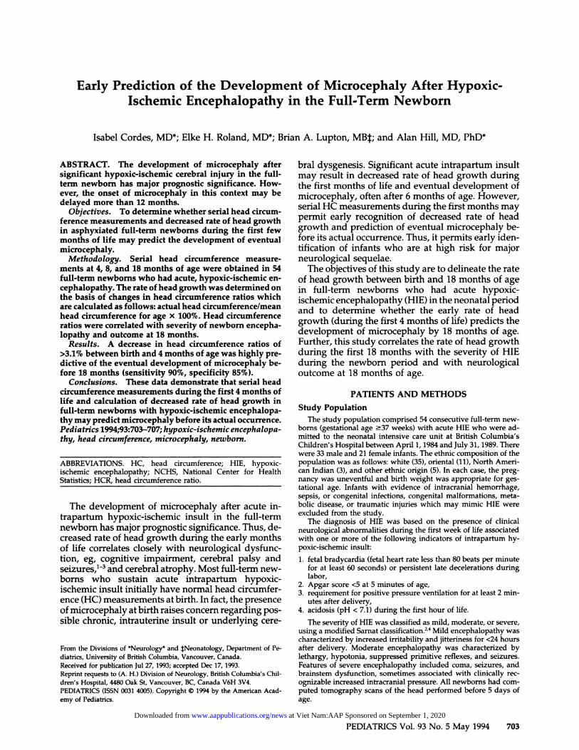
either the NCHS or Nelihaus reference data.There was a similar distribution of HC measure-
ments at birth in both normocephalic and microce-
phalic groups when boys and girls were considered
separately (Fig 1). In contrast, the mean HC measure-ments in both boys (Fig 1, top) and girls (Fig 1, bot-
tom) who eventually developed microcephaly weresignificantly lower at 4, 8, and 18 months of age com-
pared with normocephalic infants (boys: P < .01, P <
.001, P < .001, respectively; girls: P < .001 at all ages,
Student’s t-test). However, despite diminished headgrowth and decreasing mean HC values, the mdi-
vidual HC measurements in the microcephalic grouphad not necessarily decreased below the normal rangeby 4, 8, or even 18 months.
A comparison of the absolute increases in HCmeasurements in both normocephalic and microce-phalic infants at different ages are summarized inTable I . To avoid statistical errors related to the
small population size, we combined the values forboys and girls. The mean head growth in infants
who developed microcephaly was significantlylower at all ages compared with that of normoce-phalic children (P < .001) (Table 1).
Head Circumference Ratios
Based on the assumption that HC data are derivedfrom a group of healthy individuals, a mean HCR of100% would indicate optimal concordance. Using theNCHS data, the mean HCR for our normocephalicpopulation at different ages ranged between 99.4%
and 100.4% in boys and 100.0% and 100.4% in girls. Incontrast, using the Nelihaus data, the mean HCR innormocephalic infants ranged between 99.6% and
100.7% (boys) and 99.7% and 100.8% (girls). Thus, al-though our population comprised different ethnicgroups, the mean HCR of our normocephalic popu-lation had better concordance with NCHS datathan with Nellhaus data. Therefore, NCHS data wereused for calculations of HCR and the definitions ofmicrocephaly.
Although the HCR at 4 months of age correlated
closely with HCR at 18 months (r = .87, P < .001), wewere unable to predict microcephaly reliably on thebasis of HCR calculations alone.
Calculation of the change in HCRs for differenttime intervals, ie, between birth and 4 months, 4 and8 months, 8 and 18 months, demonstrates a reduction
in rate of head growth during each interval in infants
who developed microcephaly (Fig 2 and Table 2). In
those children who eventually developed micro-cephaly, the largest decrease in HCR was evident by4 months of age (Fig 2 and Table 2). Between 4 and 18months, there were further reductions in HCR which,
although less marked, were still significant statisti-cally. Because the rate of head growth is greatest be-
at Viet Nam:AAP Sponsored on September 1, 2020www.aappublications.org/newsDownloaded from
Fig 1. Top, comparison of mean head
circumference measurements between
normocephalic and microcephalic boys
(birth to 18 months). Bottom, compari-
son of mean head circumference mea-
surements between normocephalic and
microcephalic girls (birth to 18
months). NCHS, National Center for
Health Statistics.
Mlcroosph
INormocephI-LM�c.ph
CONPOENCE
4n�on P<O.OO1
&flOfl P*O.OOI
limon P<O.OO18
E
Ag. (months)
F � Circumferenc#{149}within +- 25D (b.s.d on NCHS charts)
GIRLS
10 14 16 18Age (months)
I______Circumference within +- 2SD (based on NCHS charts)
BOYS
ARTICLES 705
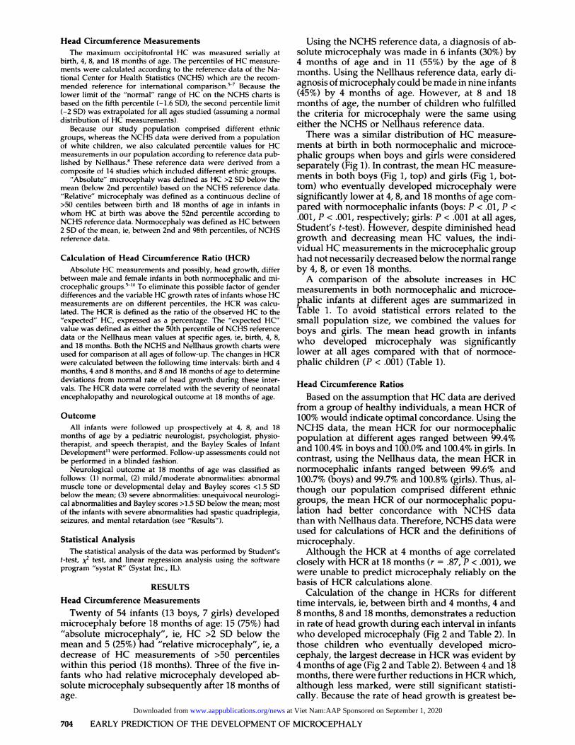
TABLE 1. Comparison of Absolute Head Growth Between 0
and 18 Months of Age in Normocephalic and Microcephalic In-
fants After Hypoxic-Ischemic Insult
Age Absolute Head Growth in cm (Mean ± SD)
Intervals, �-�--- . ---� . . �
mo Normocephalic Infants Microcephalic Infants(n=34) (n=20)
0-4 6.9 ± 1.0 4.6 ± 1.4* Mt
4-18 3.1 ± 0.7 2.2 ± 0.9* 68t
8-18 3.3 ± 0.6 2.5 ± 0.9* 74t
* Significant differences between groups, P < .001.
t Percent of expected head growth calculated based on headgrowth of normocephalic group.
tween birth and 4 months of age, it may be expected
that the greatest change in HCR would occur duringthis interval. Furthermore, the change in HCR be-tween birth and 4 months correlates with HCR at 18
months (r = .62, P < .001). The major change in HCRbetween birth and 4 months suggests that impaired
head growth and microcephaly may be predicted as
early as 4 months of age. Using either the NCHS orNellhaus reference values it seems that a decrease in
HCR of >3.1 % between birth and 4 months of age washighly predictive of the development of microcephaly
(sensitivity 90%, specificity 85%) (Table 3).
Severity of Hypoxic-Ischemic Encephalopathy and
Head Circumference
Table 4 correlates the severity of HIE in the new-
born period both with HC measurements at 18
months of age and change in HCR between birth and4 months. None of the infants with mild HIE (n = 13)
developed microcephaly. In contrast, all infants withsevere HIE (n = 9) developed microcephaly by 18months of age. Among infants with moderate HIE
at Viet Nam:AAP Sponsored on September 1, 2020www.aappublications.org/newsDownloaded from
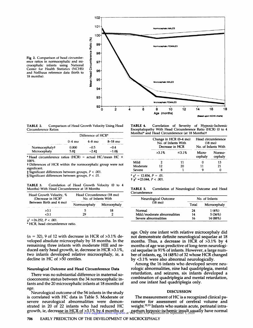
Fig 2. Comparison of head circumfer-
ence ratios in normocephalic and mi-crocephalic infants using National
Center for Health Statistics (NCHS)
and Nellhaus reference data (birth to
18 months).
102
101
100
99
98
97
96
95
94
93.
Norrnoc.phahc MALES
\Norrnoc.ph�ic FEMALES
.. . Miocc.phahc MALES
Mi�oc.ph.hc FEMALES
0 � ‘� 6 8 10 12 14 16 18Age (months)
(B.Md upon NCHS thar�)
706 EARLY PREDICTION OF THE DEVELOPMENT OF MICROCEPHALY
0
cc
a)C-)Ca)a)
E0
0Vcca)
ICcca)
TABLE 2. Comparison of Head Growth Velocity Using Head
Circumference Ratios
Difference of HCR*
0-4 mo 4-8 mo 8-18 mo
Normocephalyt 0.000 -0.5 -0.4
Microcephaly 5.81 -2.41 -1.0�
* Head circumference ratios (HCR) = actual HC/mean HC X
100%.t Differences of HCR within the normocephalic group were notsignificant.
1:Significant differences between groups, P < .001.§ Significant differences between groups, P < .01.
TABLE 3. Correlation of Head Growth Velocity (0 to 4
Months) With Head Circumference at 18 Months
Head Growth Velocity, % Head Circumference (18 mo)(Decrease in HCR* No. of Infants With
BetweenBirthand4mo) � _______Normocephaly Microcephaly
>3.1 5 18
<3.1 29 2
x2 =26.252, P < .001.* HCR, head circumference ratio.
(n = 32), 9 of 12 with decrease in HCR of >3.1 % de-veloped absolute microcephaly by 18 months. In theremaining three infants with moderate HIE and re-duced early head growth, ie, decrease in HCR >3.1%,two infants developed relative microcephaly, ie, adecline in HC of >50 centiles.
Neurological Outcome and Head Circumference Data
There was no substantial difference in maternal so-cioeconomic status between the 34 normocephalic in-
fants and the 20 microcephalic infants at 18 months ofage.
Neurological outcome of the 54 infants in the studyis correlated with HC data in Table 5. Moderate or
severe neurological abnormalities were demon-strated in 20 of 22 infants who had reduced HCgrowth, ie, decrease in HCR of >3.1% by 4 months of
TABLE 4. Correlation of Severity of Hypoxic-IschemicEncephalopathy With Head Circumference Ratio (HCR) (0 to 4Months)* and Head Circumference (at 18 Months)t
Change in HCR (0-4 mo)No. of Infants With
Decrease in HCR
Head circumference(18 mo)
No. of Infants With
Micro- Normo-cephaly cephaly
>3.1% �3.1%
Mild 2 ii 0 13Moderate 12 20 11 21Severe 8 1 9 0
*x2 = 12.854, P = .01.
t;k2 =23.044,P< .001.
TABLE 5. Correlation of Neurological Outcome and HeadCircumference
Neurological Outcome(18 mo)
N o. of Infants
Total.
Microcephaly
Normal 24 1 (4%)Mild/moderate abnormalities 14 5(36%)Severe abnormalities 16 14 (88%)
age. Only one infant with relative microcephaly didnot demonstrate definite neurological sequelae at 18months. Thus, a decrease in HCR of >3.1% by 4months of age was predictive of long-term neurologi-
cal sequelae in 91 % of infants. However, a large num-ber of infants, eg, 14 (48%) of 32 whose HCR changed
by <3.1% were also abnormal neurologically.Among the 16 infants who developed severe neu-
rologic abnormalities, nine had quadriplegia, mental
retardation, and seizures, six infants developed acombination of quadriplegia and mental retardation,and one infant had quadriplegia only.
DISCUSSION
The measurement of HC is a recognized clinical pa-
rameter for assessment of cerebral volume and
weight.’2”3 Infants who sustain acute, perinatal intra-partum hypoxic-ischemic insult usually have normal
at Viet Nam:AAP Sponsored on September 1, 2020www.aappublications.org/newsDownloaded from
ARTICLES 707
HC at birth. After significant intrapartum hypoxic-
ischemic cerebral injury, the development of second-
ary microcephaly correlates closely with major long-term neurological sequelae, eg, cerebral palsy andintellectual impairment. Neuropathological corre-
lates commonly include cerebral atrophy and multi-cystic encephalomalacia.”2 There are some limitations
regarding the use of HC measurements as an indica-tor of brain size, related principally to scalp edema at
birth, as well as head shape, skull, and hair thickness.Furthermore, the HC of parents of our patients werenot measured, which precludes comment on possible
genetic influences on head growth.After significant intrapartum hypoxic-ischemic ce-
rebral injury, microcephaly may develop before 4
months of age.’4 However, similar to previous reportsby Rosman et al,’4 our data demonstrate that second-
ary microcephaly developed later in the majority of
this group of patients. Thus, microcephaly was evi-dent by 4 months of age only in approximately 40%of infants who eventually developed microcephaly by18 months of age. In our population the development
of microcephaly could be predicted by 4 months ofage in 80% of infants based on simple calculations ofrate of head growth between birth and 4 months.
Recent studies of head growth and neurologicaloutcome in infants of very low birth weight havedemonstrated that subnormal HC at 8 months of ageis associated with abnormal neurological outcome
and cognitive impairment at school age.1�’7 In a seriesof infants with birth weight <1500 g, infants with nor-mal HC at birth and decreased rate of head growth
between birth and 6 weeks of age, 44% had neuro-logical abnormalities at 15 months, although only 23%had developed microcephaly by that age.’8 In con-
trast, infants who were normocephalic at birth andmaintained normal postnatal rate of head growth hadnormal neurological outcome. Similarly, our data
suggest that impaired head growth in full-term in-fants who sustained acute hypoxic-ischemic cerebralinjury is a useful clinical parameter for prediction of
eventual development of microcephaly and abnormal
neurological outcome. Early recognition of reducedrate of head growth may have important implicationsfor prognosis, especially in infants who exhibit mod-erate clinical encephalopathy during the neonatal pe-nod and who have been reported previously to havevariable outcome.2’4’19’�#{176} Thus, 9 of 12 infants who hadmoderate HIE and reduced rate of head growth dur-
ing the first 4 months of life, developed microcephalyand 11 of 12 infants had evidence of major neuro-logical sequelae at 18 months. Conversely, normalhead growth does not preclude significant long-termneurological abnormalities. Thus, in our study popu-lation, 14 of 32 infants with normal rate of headgrowth were abnormal neurologically by 18 months
of age.The value of HCR for prediction of neurological
outcome has been established previously in a popu-lation of very low birth weight infants)6 In our popu-lation of full-term newborns, the development of mi-
crocephaly could be predicted by 4 months of age bycalculation of the change in HCR between birth and4 months of age. The high predictive value of earlyhead growth between birth and 4 months relates pre-
sumably to the relatively rapid rate of myelination,glial proliferation, and elaboration of axonal ramifi-
cations during this period.17 However, it is importantto emphasize that although microcephaly at 4 monthsof age is associated with a high probability of neu-
rological abnormalities, this group comprises only aminority of infants who sustained hypoxic-ischemiccerebral insult and subsequently developed majorlong-term neurological sequelae.
In summary, a decreased rate of head growth in thefirst 4 months of life after acute, intrapartum hypoxic-ischemic encephalopathy, correlates closely with thedevelopment of secondary microcephaly. Infantswith reduced head growth have a high probability of
long-term neurological sequelae.
REFERENCES
1. Volpe JJ. Neurology of the Newborn. Philadelphia: W. B. Saunders; 1987
2. Hill A. Current concepts of hypoxic-ischemic cerebral injury in the
newborn. Pediatr Neurol. 1991;7:317-3253. Laurence KM, Cavanagh JB. Progressive degeneration of the cerebral
cortex in infancy. Brain. 1968;91:261-280
4. Sarnat HB, Sarnat MS. Neonatal encephalopathy following fetal
distress: a clinical electroencephalographic study. Arch Neurol. 1976;
33:696-705
5. Hamill PVV, Drizd TA, Johnson CL, Reed DB, Roche AF. NCHS growth
curves for children birth - 18 years. Washington, DC: US Government
Printing Office; 1977. US Dept of Health, Education Welfare publication
PHS78-1650
6. Hamill PVV, Drizd TA, Johnson CL, et al. Physical growth, National
Center for Health Statistics percentiles. Am I Clin Nutr. 1979;32:607-6297. Roche AF, Mukherjee D, Guo 5, Moore WM. Head circumference ref-
erence data: birth to 18 years. Pediatrics. 1987;79:706-712
8. Nellhaus C. Head circumference from birth to eighteen years: practical
composite international interracial graphs. Pediatrics. 1968;41:106-1 14
9. Dorman C. Microcephaly intelligence. Dcv Med Child Neurol. 1991;33:
267-269
10. Guo 5, Roche AF, Moore WM. Reference data for head circumference I
month increments from I to 12 months of age. Pediatrics. 1988;1 13:
490-494
I 1 . Bayley N. Bayley Scales of Infant Development. New York: Psychological
Corp; 1969
12. Cooke RW, Lucas A, Yudkin PLN, Pryse-Davies J. Head circumference
as an index of brain weight in the fetus newborn. Early Hum Dcv.
I977;1 �l45-149
13. Lemons A, Schreiner RL, Gresham EL. Relationship of brain weight to
head circumference in early infancy. Hum Biol. 1981;53:351-354
14. Rosman NP, Rivkin M, Mannheim GB. Acquired microcephaly predic-
lion of later development. Neurology. 1990;40(Suppl I):357. Abstract
15. Hack M, Breslau N. Very low birth weight infants: effects of brain
growth during infancy on intelligence quotient at 3 years of age.
Pediatrics. 1986;77:196-202
16. Hack M, Breslam N, Weisman B, Aram D, Klein N, Borawski E. Effect
of very low birthweight subnormal head size on cognitive abilities at
school age. N Engi I Med. 1991;325:231-239
17. Volpe JJ. Cognitive deficits in premature infants. N Engl I Med. 1991;
325:276-277. Editorial
18. Gross SJ, Ochler JM, Eckerman CO. Head growth developmental out-
come in very low-birth-weight infants. Pediatrics. 1983;71:70-75
19. Finer NN, Robertson CM, Richards RT, Pinnell LE, Peters KL. Hypoxic-
ischemic encephalopathy in term newborns: perinatal factors outcome.
I Pediatr. 1981;98:112-1I7
20. Robertson CM, Finer NN. Term infants with hypoxic-ischemic
encephalopathy: outcome at 3.5 years. Dec Med Child Neurol. 1985;27:
473-484
at Viet Nam:AAP Sponsored on September 1, 2020www.aappublications.org/newsDownloaded from

1994;93;703Pediatrics Isabel Cordes, Elke H. Roland, Alan Hill and Brian A. Lupton
Encephalopathy in the Full-Term NewbornEarly Prediction of the Development of Microcephaly After Hypoxic-Ischemic
ServicesUpdated Information &
http://pediatrics.aappublications.org/content/93/5/703including high resolution figures, can be found at:
Permissions & Licensing
http://www.aappublications.org/site/misc/Permissions.xhtmlentirety can be found online at: Information about reproducing this article in parts (figures, tables) or in its
Reprintshttp://www.aappublications.org/site/misc/reprints.xhtmlInformation about ordering reprints can be found online:
at Viet Nam:AAP Sponsored on September 1, 2020www.aappublications.org/newsDownloaded from
1994;93;703Pediatrics Isabel Cordes, Elke H. Roland, Alan Hill and Brian A. Lupton
Encephalopathy in the Full-Term NewbornEarly Prediction of the Development of Microcephaly After Hypoxic-Ischemic
http://pediatrics.aappublications.org/content/93/5/703the World Wide Web at:
The online version of this article, along with updated information and services, is located on
American Academy of Pediatrics. All rights reserved. Print ISSN: 1073-0397. American Academy of Pediatrics, 345 Park Avenue, Itasca, Illinois, 60143. Copyright © 1994 by thebeen published continuously since 1948. Pediatrics is owned, published, and trademarked by the Pediatrics is the official journal of the American Academy of Pediatrics. A monthly publication, it has
at Viet Nam:AAP Sponsored on September 1, 2020www.aappublications.org/newsDownloaded from


