Embed Size (px)
Citation preview
Eur Food Res Technol (2008) 226:653–659
DOI 10.1007/s00217-007-0574-3ORIGINAL PAPER
Isolation of a hydroxytyrosol-rich extract from olive leaves (Olea Europaea L.) and evaluation of its antioxidant properties and bioactivity
Antonella De Leonardis · Alessandra Aretini · Gabriele Alfano · Vincenzo Macciola · Giancarlo Ranalli
Received: 13 July 2006 / Revised: 10 January 2007 / Accepted: 15 January 2007 / Published online: 15 February 2007© Springer-Verlag 2007
Abstract In this research, a phenol extract of highhydroxytyrosol (OLPE) content was obtained fromolive leaves (Olea europaea L.), and subsequentlytested under diVerent contexts. The method used toobtain the OLPE basically involved two steps: the useof strongly-acid aqueous steam, generated from 10%HCl (v/v) at 100°C, to directly hydrolyse the nativecomplex phenols from integral olive leaves, and OLPErecovery by liquid–liquid extraction with ethyl acetate.Hydrolysis time was 1 h. Finally, the dried extract wasdissolved in distilled water. The OLPE total phenolswere determined by Folin–Ciocalteu’s method and byHPLC analysis. Hydroxytyrosol was about 92% of thetotal phenols present in OLPE, and the yield was about0.2% on fresh leaves. OLPE showed antioxidant eVectson diVerent food lipids and did not inhibit lactic acidbacteria growth; however, it showed cytotoxicity onNIH/3T3 Wbroblasts and human umbilical vein endo-thelial cells at concentrations higher than 0.32 mM (ashydroxytyrosol).
Keywords Olive leaf · Hydroxytyrosol · Natural antioxidants · Antimicrobial activity · Cytotoxicity
AbbreviationsHT HydroxytyrosolOLPE Olive leaf phenol extractOLNP Olive leaf native phenolsPF Protection factor
Introduction
Since ancient times, Mediterranean countries havecultivated Olea Europaea to produce both oil andmedicinal compounds. In the last decades, the positiveeVects of olive biophenols on human health have beenscientiWcally demonstrated on quite a few occasions[1–3]. Typical phenols of O. Europaea are the secoirid-oids, complex molecules with molecular weight up to600 composed by one or two hydroxyaromatics thatare linked to other variable non-aromatic compounds[4].
Hydroxytyrosol (HT), or 3,4-dihydroxyphenyletha-nol, is one of the hydroxyaromatic components of seco-iridoids. It is a very bio-active alcoholic ortho-diphenol.In several speciWc studies, it has been demonstratedthat HT is antioxidant and antimicrobial [5, 6] and thatit has beneWcial eVects on the cardio-vascular systemand in several human diseases [7–9].
Native HT is rarely in the free form in nature withthe exception of ripened olives where it occurs throughthe hydrolysis of oleuropein. Therefore HT productionrequires enzymatic or chemical hydrolysis of the com-plex phenols that contain it. The natural mechanismthat occurs when the olive tree forms free HT is enzy-matic hydrolysis, and speciWc native �-glycosidase andesterase are implicated. On the contrary, acid hydroly-sis is the more used mechanism in the laboratory and
A. De Leonardis (&) · G. Alfano · V. Macciola · G. RanalliDepartment of Agricultural, Food, Environmental and Microbiological Science and Technologies (DiSTAAM), University of Molise, Via De Sanctis, 86100 Campobasso, Italye-mail: [email protected]
A. AretiniDepartment of AngioCardioNeurology, IRCCS Neuromed, Località Camerelle, 86077 Pozzilli (IS), Italy
123
654 Eur Food Res Technol (2008) 226:653–659
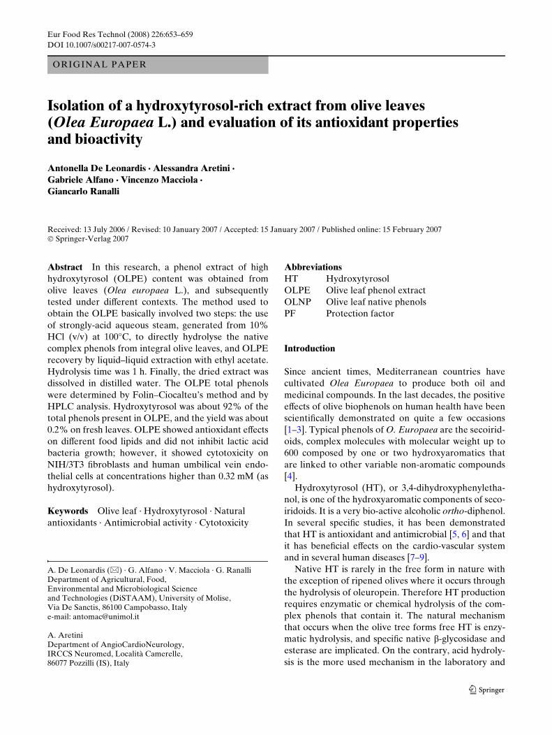
industrial processes. The enzymatic and hydrolytic-acidsplitting of oleuropein is shown in Fig. 1.
Nowadays, pure HT and diVerent HT formulationsare available commercially. Several studies have con-sidered procedures that produce high quantities of HTthrough the use of olive oil mill by-products. Above all,olive oil mill wastewaters have been investigated, sincethese are actually already very rich in HT in the freeform [10–13]. In alternative, synthetic enzymatic proce-dures of HT have been proposed [14]. However, todate, HT production from the olive leaf has receivedlittle consideration and generally it is carried out in twosteps: (1) extraction of total phenols and (2) enzymaticor chemical hydrolysis of total phenols extracted [15–17].
Olive leaves are a copious by-product deriving fromolive tree cultivation and olive oil mills. Large amountsof leaves are principally generated during pruning ofthe trees and harvesting and working of the olives.Olive tree pruning is responsible for the most abundantleaf amounts, but their quantity has been never calcu-lated statistically. Otherwise, it is estimated that forevery 100 kg of worked olives about 3–5 kg of leavesremain. Spain, Italy and Greece are the principal coun-tries that produce olive oil, and therefore olive leaves.
At present the olive leaf business is substantiallyinsigniWcant, and, in general, olive leaves are burned orcomposted at the original farms. The industrial use ofolive leaves is limited to animal feed [18] and phyto-therapy.
Indeed, there is compelling scientiWc evidence thatolive leaf polyphenols are bio-active compounds. Oliveleaves, or their speciWc organics, show antiviral [19],antimicrobial [20], antioxidant and anti-inXammatory
[21, 22] properties, atherosclerosis inhibition and hypo-tensive action [23–26], anti-carcinogenic propertiesthat lead to the prevention of some cancers [27] and,Wnally, stimulation of the thyroid [28]. An inhibitoractivity of phenols from olive oil mill wastewatersagainst diVerent bacterium phyto-pathogens has alsobeen observed [29–31].
In recent years, the developed countries havearrived at new food formulations, derived from thecombination of nutraceutical compounds and probioticmicro-organisms, and such formulations are settingquite a trend. Indeed, co-cultures of Streptococcus ther-mophilus and Lactobacillus delbruechii ssp. bulgaricusare generally used in yoghurt and similar fermentedmilk processes. With regard to lactic acid bacteria,there are few studies that address the eVect of HT orolive phenols.
The polyphenol content in the olive leaf is still rela-tively unknown, but it could range from 1.5 to 7.0 g per100 g in fresh leaves [12]. However, it is known thatolive leaf polyphenol composition is similar to that ofolive oil. Oleuropein and other secoiridoids are theprincipal compounds, while simple phenols, enclosedhydroxytyrosol, are present but in lower amounts [2].
The present research discusses a method to producean olive leaf phenol extract of high hydroxytyrosol con-tent. The method, applied directly to integral oliveleaves, is essentially based on a strongly-acid steamhydrolysis of complex phenols and after that the phe-nol substances are extracted with ethyl acetate. Someof the properties and bio-activities of the olive leafphenol extract were tested in diVerent contexts, andantioxidant and antimicrobial eVects, together withcytotoxicity, were particularly investigated.
Fig. 1 Hydroxytyrosol pro-duction by enzymatic and acid hydrolysis of the oleuropein
123
Eur Food Res Technol (2008) 226:653–659 655
Materials and methods
Procedure for the production of olive-leaf phenol extracts
In July 2005, olive leaves were randomly and directlypicked from an olive tree (Ortice variety) native tosouthern Italy (Campania region); the leaves were fro-zen at ¡18°C in the laboratory. A 500 ml glass conicXask had 100 ml of HCl 10% (v/v) put into it. A steelgrill was Wxed about 2 cm above the HCl solution and60 g of intact frozen leaves were placed on it. The Xaskwas connected to a coil-condenser and kept in athermo-regulated water bath at 100°C for 60 min.Finally, the acid solution was passed through Wlterpaper into a separator funnel where it was washedthree times with 25 ml ethyl acetate. After evaporationof the ethyl acetate by rotavapor, the dry residue wasre-dissolved in 25 ml of distilled water. The Wnal solu-tion, labelled OLPE, was again Wltered through Wlterpaper in order to remove any possible insoluble sub-stances.
Total native phenols (OLNP) were extracted fromthe olive leaves as follows: about 30 g of integral frozenolive leaves were treated three times with 150 ml ofmethanol:water 80:20 (v/v) in an ultrasonic bath for15 min. After solvent evaporation, the dry residue wasdissolved in 25 ml of distilled water.
Analysis of the phenols
Standard compounds: Pure oleuropein was pur-chased from the IndoWne Chemical Company Inc.(Belle Mead, NJ, USA). Hydroxytyrosol (HT) wassynthesised in the laboratory by acid hydrolysis ofpure oleuropein, according to Fernández-Bolañoset al. [11].
Phenol determinations: Total phenols were esti-mated by the Folin–Ciocalteu method using hydroxy-tyrosol calibration lines for OLPE and that ofoleuropein for OLNP. The HPLC analysis was per-formed with an instrument Model ProStar 230 (Var-ian, Mulgrave, AUS) with an attached UV-Visiblespectrophotometer, and equipped with a Luna 5uphenyl-hexyl, 250 mm £ 4.6 mm column, purchasedfrom Phenomenex (Torrance, USA). The operativeconditions employed methanol as mobile phase Aand 2% acetic acid as mobile phase B; the elutionprogram was A(%)/B(%): 95/5, 0 min; 75/25, 10 min;50/50, 20 min; 100/0, 30 min; 5/95, 40 min; eluent Xow1 ml min¡1; injection quantity 20 �l; Wxed wavelength280 nm.
Oxidation stability of diVerent food fats
The antioxidant eVects of OLPE were tested usingsamples of lard, butter and cod liver oil purchased atthe local market and analysed by using the methodsreported in De Leonardis et al. [6]. The OLPE aque-ous solution was added, at concentrations of 25–200 mg kg¡1 of fat (as hydroxytyrosol determined byFolin–Ciocalteu method), directly into the reactionvessel containing the fat samples, homogenising theWnal mixture by a mixer in a test tube for 30 s. Volumeof OLPE added varied with its concentration but how-ever it was less than 0.15 ml; a similar volume of purewater was added to the fat sample control. The induc-tion time was determined by a Rancimat MetrohmInstrument mod. 730 (AG, Herisau, Switzerland)under 20 l h¡1 air Xow at 120°C for lard and butter and100°C for cod liver oil.
The protection factor was calculated as the ratio ofthe lipid induction time with and without the antioxi-dant.
Antimicrobial activity (in vitro)
The OLPE antimicrobial activity at concentrationsranging from 25 to 3,200 �g ml¡1 (as hydroxytyrosoldetermined by Folin–Ciocalteu method) was evaluatedby assessing the inhibition of microbial growth on co-cultures of Streptococcus thermophilus and Lactobacil-lus delbruechii ssp. bulgaricus (DiSTAAM CultureCollection, Campobasso, Italy), using the broth dilu-tion method [32]. The culture media for the co-cultureswere, respectively, King B medium (proteose peptone20 g/l; glycerol 10 g/l; K2HPO4 1.5 g/l; MgSO4·7H2O1.5 g/l; pH 7.2) and MRS (Oxoid, Basingstoke,England). BrieXy, 0.5 ml of serial twofold dilutions ofsamples was mixed with 9.5 ml of cultural medium.After an adequate incubation period (36 and 48 h,28°C), the inhibitor activity of the sample was assessed.The results were conWrmed in the same cultural brothmedia using a microbiology computerised reader-ana-lyzer that is able to assess multiple microbial growth(turbidity development) through the simultaneousrecording of the growth curve parameters and the min-imal antimicrobial activity (MIC) responses (BioscreenC, Labsystems, Helsinki, Finland).
Cytotoxicity
Dulbecco’s ModiWed Eagle’s Medium (DMEM) andRPMI 1640 (without red phenols), phosphate buVeredsaline (PBS), fetal bovine serum (FBS) and the antibi-otic were from Invitrogen Corporation (Carlsbad, CA,
123
656 Eur Food Res Technol (2008) 226:653–659
USA), while [3-(4,5-dimethyl-2-thiazolyl)-2,5-diphe-nyl-2H-tetrazolium bromide] (MTT) was from SigmaChemical CO. (St. Louis, Mo, USA). The cells wereNIH/3T3 Wbroblasts and human endothelial cells deriv-ing from umbilical veins (HUVEC), obtained fromClonetics. The HUVEC were cultured with endothelialgrowth medium (EGM), and NIH/3T3 was grown inDMEM, both containing the appropriate factors. Bothcell types were grown on 100 mm plates and weremaintained in a humidiWed atmosphere with 5% CO2at 37°C. When the cells reached 80% conXuence theywere treated with 0.25% trypsin and 1 mM EDTA.They were then counted and 10,000 cells were seededin 96-wells and serum deprived for 24 h. The cells werestimulated with OLPE (0.01–1.28 mM as HT) for 12, 24and 48 h. Cytotoxic eVect was measured by the MTTmethod [6]. Each condition was assayed in triplicate.
Elaboration data
Generally, analytical determinations were carried outin three–four replicates, expressing the results asmean § standard deviation. Statistical analysis wasperformed by SPSS 10.0 software (Chicago, Illinois,USA), evaluating signiWcance by the Student’s test atP < 0.05.
Results and discussion
Production and analysis of olive leaf phenol extract
The method developed to obtain olive leaf phenolextract (OLPE) consisted essentially of two steps.First, the native complex phenols were hydrolyseddirectly on integral olive leaves using strongly-acidaqueous steam, the OLPE was recovered by liquid–liq-uid extraction with ethyl acetate.
The leaves are preliminarily frozen at ¡18°C for atleast 48 h. This practice was fundamental as the freez-ing supported the dissolution of the phenolic com-pounds and facilitated the hydrolysis process. Contraryto our expectations, it was observed that the mechani-cal grinding of the leaves was not needed and wasindeed unfavourable. In fact, the Wnal acid extractobtained from the mechanically ground leaves wasclearly turbid, and additional steps were needed toeliminate the great amount of impurities.
The hydrolysis was performed in a closed conicalXask in which frozen integral leaves were treated withstrongly-acid aqueous steam for 1 h, keeping the leavesseparate and out of contact with the HCl acid solutionby steel grill. When the HCl solution reached boiling
point, the strongly-acid steam generated within theXask enveloped the leaves and cooked them. In fact, atthe end, they looked similar to cooked vegetables andtheir odour was like that of tea.
At the end of the hydrolysis step the residual limpidand yellow-coloured acid solution was washed repeat-edly with ethyl acetate. This is a very selective solventfor simple phenols like HT [3].
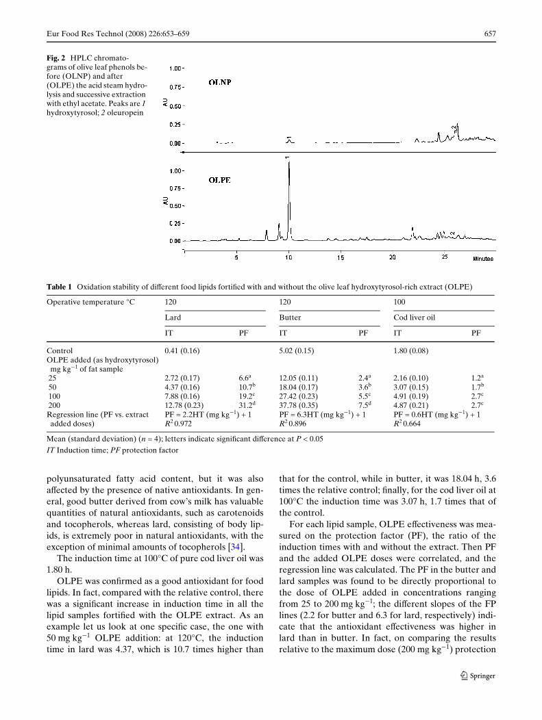
The usefulness and eYcacy of the performed proce-dure was conWrmed by an analysis of the OLPE com-position. Figure 2 shows the HPLC chromatograms ofthe native phenols extracted from olive leaves (OLNP)and the phenols of the OLPE. It can be seen that forthe OLNP only traces of HT were detectable, while forthe OLPE the HT represented 92% of the total phe-nols (calculated by HPLC analysis).
The OLNP were 0.6 g per 100 g of fresh olive leaves(as oleuropein; by Folin–Ciocalteu method), a valuemuch lower than reported in the speciWc literature [13,17, 33]. However, there are actually few current studieson the content and composition of olive leaf phenols.In fact, inXuences like cultivars, climatic and geo-graphic conditions, physiological stage and plant ageand other factors aVecting olive leaf phenols have notyet been fully investigated, and could be the object offuture research.
In the OLPE, the HT amount was 0.2 g/100 g offresh olive leaves (by HPLC method). The HT yieldwas about 33.3% (w/w) on native total phenols, a per-centage yield comparable with that reported by Bou-aziz and Sayadi [17]. Although in that study it was notconsidered necessary to carry out a further subsequentpuriWcation step of HT from OLPE, several HT puriW-cation procedures have been validated [12, 17].
Oxidation stability of diVerent food lipids
The antioxidant eVectiveness of OLPE was evaluatedon butter, lard and cod liver oil using the Rancimatapparatus and OLPE aqueous dosages of 25–200 mg kg¡1 of fat (as hydroxytyrosol). Through pre-liminary analytical determinations, the oxidation testsuitability of fat samples was predetermined (data notshown). The lipid samples diVered among themselves,especially in their fatty acid composition; the polyun-saturated fatty acid percentages were 2.8, 11.7 and 21.9for butter, lard and cod liver oil, respectively.
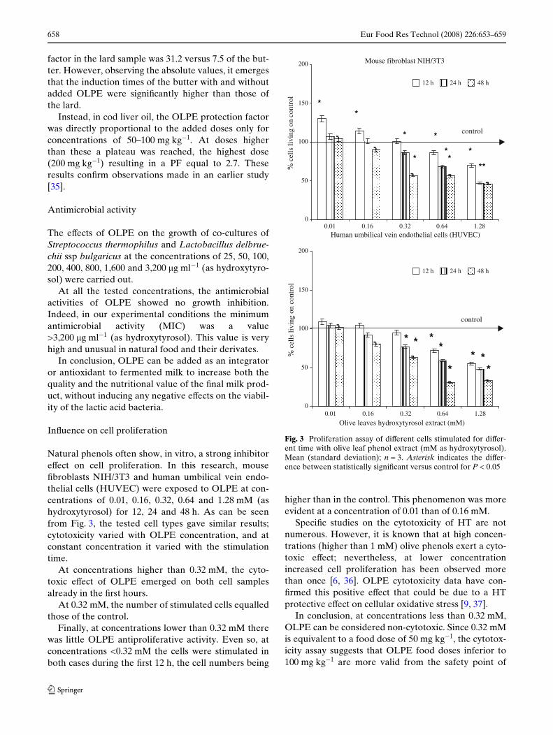
Table 1 shows the results relative to oxidative tests.At high temperature, the butter and lard diVered in
oxidative resistance. In fact, at 120°C the butter-con-trol showed an induction time of 5.02 h, while that ofthe lard-control occurred as early as 0.41 h. Thisinduction time variability was due to the diVerent
123
Eur Food Res Technol (2008) 226:653–659 657
polyunsaturated fatty acid content, but it was alsoaVected by the presence of native antioxidants. In gen-eral, good butter derived from cow’s milk has valuablequantities of natural antioxidants, such as carotenoidsand tocopherols, whereas lard, consisting of body lip-ids, is extremely poor in natural antioxidants, with theexception of minimal amounts of tocopherols [34].
The induction time at 100°C of pure cod liver oil was1.80 h.
OLPE was conWrmed as a good antioxidant for foodlipids. In fact, compared with the relative control, therewas a signiWcant increase in induction time in all thelipid samples fortiWed with the OLPE extract. As anexample let us look at one speciWc case, the one with50 mg kg¡1 OLPE addition: at 120°C, the inductiontime in lard was 4.37, which is 10.7 times higher than
that for the control, while in butter, it was 18.04 h, 3.6times the relative control; Wnally, for the cod liver oil at100°C the induction time was 3.07 h, 1.7 times that ofthe control.
For each lipid sample, OLPE eVectiveness was mea-sured on the protection factor (PF), the ratio of theinduction times with and without the extract. Then PFand the added OLPE doses were correlated, and theregression line was calculated. The PF in the butter andlard samples was found to be directly proportional tothe dose of OLPE added in concentrations rangingfrom 25 to 200 mg kg¡1; the diVerent slopes of the FPlines (2.2 for butter and 6.3 for lard, respectively) indi-cate that the antioxidant eVectiveness was higher inlard than in butter. In fact, on comparing the resultsrelative to the maximum dose (200 mg kg¡1) protection
Fig. 2 HPLC chromato-grams of olive leaf phenols be-fore (OLNP) and after (OLPE) the acid steam hydro-lysis and successive extraction with ethyl acetate. Peaks are 1 hydroxytyrosol; 2 oleuropein
Table 1 Oxidation stability of diVerent food lipids fortiWed with and without the olive leaf hydroxytyrosol-rich extract (OLPE)
Mean (standard deviation) (n = 4); letters indicate signiWcant diVerence at P < 0.05
IT Induction time; PF protection factor
Operative temperature °C 120 120 100
Lard Butter Cod liver oil
IT PF IT PF IT PF
Control 0.41 (0.16) 5.02 (0.15) 1.80 (0.08)OLPE added (as hydroxytyrosol)
mg kg¡1 of fat sample25 2.72 (0.17) 6.6a 12.05 (0.11) 2.4a 2.16 (0.10) 1.2a
50 4.37 (0.16) 10.7b 18.04 (0.17) 3.6b 3.07 (0.15) 1.7b
100 7.88 (0.16) 19.2c 27.42 (0.23) 5.5c 4.91 (0.19) 2.7c
200 12.78 (0.23) 31.2d 37.78 (0.35) 7.5d 4.87 (0.21) 2.7c
Regression line (PF vs. extract added doses)
PF = 2.2HT (mg kg¡1) + 1R2 0.972
PF = 6.3HT (mg kg¡1) + 1R2 0.896
PF = 0.6HT (mg kg¡1) + 1R2 0.664
123
658 Eur Food Res Technol (2008) 226:653–659
factor in the lard sample was 31.2 versus 7.5 of the but-ter. However, observing the absolute values, it emergesthat the induction times of the butter with and withoutadded OLPE were signiWcantly higher than those ofthe lard.
Instead, in cod liver oil, the OLPE protection factorwas directly proportional to the added doses only forconcentrations of 50–100 mg kg¡1. At doses higherthan these a plateau was reached, the highest dose(200 mg kg¡1) resulting in a PF equal to 2.7. Theseresults conWrm observations made in an earlier study[35].
Antimicrobial activity
The eVects of OLPE on the growth of co-cultures ofStreptococcus thermophilus and Lactobacillus delbrue-chii ssp bulgaricus at the concentrations of 25, 50, 100,200, 400, 800, 1,600 and 3,200 �g ml¡1 (as hydroxytyro-sol) were carried out.
At all the tested concentrations, the antimicrobialactivities of OLPE showed no growth inhibition.Indeed, in our experimental conditions the minimumantimicrobial activity (MIC) was a value>3,200 �g ml¡1 (as hydroxytyrosol). This value is veryhigh and unusual in natural food and their derivates.
In conclusion, OLPE can be added as an integratoror antioxidant to fermented milk to increase both thequality and the nutritional value of the Wnal milk prod-uct, without inducing any negative eVects on the viabil-ity of the lactic acid bacteria.
InXuence on cell proliferation
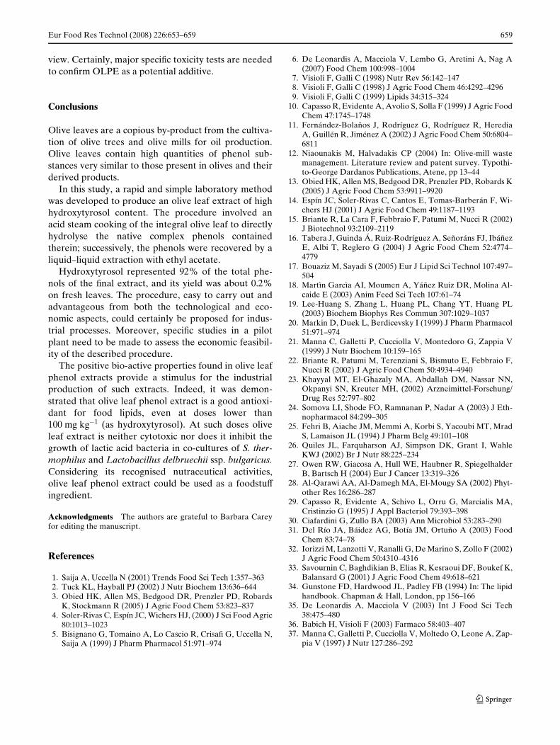
Natural phenols often show, in vitro, a strong inhibitoreVect on cell proliferation. In this research, mouseWbroblasts NIH/3T3 and human umbilical vein endo-thelial cells (HUVEC) were exposed to OLPE at con-centrations of 0.01, 0.16, 0.32, 0.64 and 1.28 mM (ashydroxytyrosol) for 12, 24 and 48 h. As can be seenfrom Fig. 3, the tested cell types gave similar results;cytotoxicity varied with OLPE concentration, and atconstant concentration it varied with the stimulationtime.
At concentrations higher than 0.32 mM, the cyto-toxic eVect of OLPE emerged on both cell samplesalready in the Wrst hours.
At 0.32 mM, the number of stimulated cells equalledthose of the control.
Finally, at concentrations lower than 0.32 mM therewas little OLPE antiproliferative activity. Even so, atconcentrations <0.32 mM the cells were stimulated inboth cases during the Wrst 12 h, the cell numbers being
higher than in the control. This phenomenon was moreevident at a concentration of 0.01 than of 0.16 mM.
SpeciWc studies on the cytotoxicity of HT are notnumerous. However, it is known that at high concen-trations (higher than 1 mM) olive phenols exert a cyto-toxic eVect; nevertheless, at lower concentrationincreased cell proliferation has been observed morethan once [6, 36]. OLPE cytotoxicity data have con-Wrmed this positive eVect that could be due to a HTprotective eVect on cellular oxidative stress [9, 37].
In conclusion, at concentrations less than 0.32 mM,OLPE can be considered non-cytotoxic. Since 0.32 mMis equivalent to a food dose of 50 mg kg¡1, the cytotox-icity assay suggests that OLPE food doses inferior to100 mg kg¡1 are more valid from the safety point of
Fig. 3 Proliferation assay of diVerent cells stimulated for diVer-ent time with olive leaf phenol extract (mM as hydroxytyrosol).Mean (standard deviation); n = 3. Asterisk indicates the diVer-ence between statistically signiWcant versus control for P < 0.05
Mouse fibroblast NIH/3T3
0
50
100
150
200
0
50
100
150
200
% c
ells
livi
ng o
n co
ntro
l
control
**
*
*
*
**
*
**
Human umbilical vein endothelial cells (HUVEC)
0.01 0.16 0.32 0.64 1.28
0.01 0.16 0.32 0.64 1.28
Olive leaves hydroxytyrosol extract (mM)
% c
ells
livi
ng o
n co
ntro
l
12 h 24 h 48 h
12 h 24 h 48 h
control
* * **
** *
*
123
Eur Food Res Technol (2008) 226:653–659 659
view. Certainly, major speciWc toxicity tests are neededto conWrm OLPE as a potential additive.
Conclusions
Olive leaves are a copious by-product from the cultiva-tion of olive trees and olive mills for oil production.Olive leaves contain high quantities of phenol sub-stances very similar to those present in olives and theirderived products.
In this study, a rapid and simple laboratory methodwas developed to produce an olive leaf extract of highhydroxytyrosol content. The procedure involved anacid steam cooking of the integral olive leaf to directlyhydrolyse the native complex phenols containedtherein; successively, the phenols were recovered by aliquid–liquid extraction with ethyl acetate.
Hydroxytyrosol represented 92% of the total phe-nols of the Wnal extract, and its yield was about 0.2%on fresh leaves. The procedure, easy to carry out andadvantageous from both the technological and eco-nomic aspects, could certainly be proposed for indus-trial processes. Moreover, speciWc studies in a pilotplant need to be made to assess the economic feasibil-ity of the described procedure.
The positive bio-active properties found in olive leafphenol extracts provide a stimulus for the industrialproduction of such extracts. Indeed, it was demon-strated that olive leaf phenol extract is a good antioxi-dant for food lipids, even at doses lower than100 mg kg¡1 (as hydroxytyrosol). At such doses oliveleaf extract is neither cytotoxic nor does it inhibit thegrowth of lactic acid bacteria in co-cultures of S. ther-mophilus and Lactobacillus delbruechii ssp. bulgaricus.Considering its recognised nutraceutical activities,olive leaf phenol extract could be used as a foodstuVingredient.
Acknowledgments The authors are grateful to Barbara Careyfor editing the manuscript.
References
1. Saija A, Uccella N (2001) Trends Food Sci Tech 1:357–3632. Tuck KL, Hayball PJ (2002) J Nutr Biochem 13:636–6443. Obied HK, Allen MS, Bedgood DR, Prenzler PD, Robards
K, Stockmann R (2005) J Agric Food Chem 53:823–8374. Soler-Rivas C, Espín JC, Wichers HJ, (2000) J Sci Food Agric
80:1013–10235. Bisignano G, Tomaino A, Lo Cascio R, CrisaW G, Uccella N,
Saija A (1999) J Pharm Pharmacol 51:971–974
6. De Leonardis A, Macciola V, Lembo G, Aretini A, Nag A(2007) Food Chem 100:998–1004
7. Visioli F, Galli C (1998) Nutr Rev 56:142–1478. Visioli F, Galli C (1998) J Agric Food Chem 46:4292–42969. Visioli F, Galli C (1999) Lipids 34:315–324
10. Capasso R, Evidente A, Avolio S, Solla F (1999) J Agric FoodChem 47:1745–1748
11. Fernández-Bolakos J, Rodríguez G, Rodríguez R, HerediaA, Guillén R, Jiménez A (2002) J Agric Food Chem 50:6804–6811
12. Niaounakis M, Halvadakis CP (2004) In: Olive-mill wastemanagement. Literature review and patent survey. Typothi-to-George Dardanos Publications, Atene, pp 13–44
13. Obied HK, Allen MS, Bedgood DR, Prenzler PD, Robards K(2005) J Agric Food Chem 53:9911–9920
14. Espín JC, Soler-Rivas C, Cantos E, Tomas-Barberán F, Wi-chers HJ (2001) J Agric Food Chem 49:1187–1193
15. Briante R, La Cara F, Febbraio F, Patumi M, Nucci R (2002)J Biotechnol 93:2109–2119
16. Tabera J, Guinda Á, Ruiz-Rodríguez A, Señoráns FJ, IbáñezE, Albi T, Reglero G (2004) J Agric Food Chem 52:4774–4779
17. Bouaziz M, Sayadi S (2005) Eur J Lipid Sci Technol 107:497–504
18. Martìn Garcìa AI, Moumen A, Yáñez Ruiz DR, Molina Al-caide E (2003) Anim Feed Sci Tech 107:61–74
19. Lee-Huang S, Zhang L, Huang PL, Chang YT, Huang PL(2003) Biochem Biophys Res Commun 307:1029–1037
20. Markin D, Duek L, Berdicevsky I (1999) J Pharm Pharmacol51:971–974
21. Manna C, Galletti P, Cucciolla V, Montedoro G, Zappia V(1999) J Nutr Biochem 10:159–165
22. Briante R, Patumi M, Terenziani S, Bismuto E, Febbraio F,Nucci R (2002) J Agric Food Chem 50:4934–4940
23. Khayyal MT, El-Ghazaly MA, Abdallah DM, Nassar NN,Okpanyi SN, Kreuter MH, (2002) Arzneimittel-Forschung/Drug Res 52:797–802
24. Somova LI, Shode FO, Ramnanan P, Nadar A (2003) J Eth-nopharmacol 84:299–305
25. Fehri B, Aiache JM, Memmi A, Korbi S, Yacoubi MT, MradS, Lamaison JL (1994) J Pharm Belg 49:101–108
26. Quiles JL, Farquharson AJ, Simpson DK, Grant I, WahleKWJ (2002) Br J Nutr 88:225–234
27. Owen RW, Giacosa A, Hull WE, Haubner R, SpiegelhalderB, Bartsch H (2004) Eur J Cancer 13:319–326
28. Al-Qarawi AA, Al-Damegh MA, El-Mougy SA (2002) Phyt-other Res 16:286–287
29. Capasso R, Evidente A, Schivo L, Orru G, Marcialis MA,Cristinzio G (1995) J Appl Bacteriol 79:393–398
30. Ciafardini G, Zullo BA (2003) Ann Microbiol 53:283–29031. Del Río JA, Báidez AG, Botía JM, Ortuño A (2003) Food
Chem 83:74–7832. Iorizzi M, Lanzotti V, Ranalli G, De Marino S, Zollo F (2002)
J Agric Food Chem 50:4310–431633. Savournin C, Baghdikian B, Elias R, Kesraoui DF, Boukef K,
Balansard G (2001) J Agric Food Chem 49:618–62134. Gunstone FD, Hardwood JL, Padley FB (1994) In: The lipid
handbook. Chapman & Hall, London, pp 156–16635. De Leonardis A, Macciola V (2003) Int J Food Sci Tech
38:475–48036. Babich H, Visioli F (2003) Farmaco 58:403–40737. Manna C, Galletti P, Cucciolla V, Moltedo O, Leone A, Zap-
pia V (1997) J Nutr 127:286–292
123


