Embed Size (px)
Citation preview
Natural Product Libraries to Accelerate the High-ThroughputDiscovery of Therapeutic LeadsTyler A. Johnson,*,†,‡ Johann Sohn,† Wayne D. Inman,‡ Samarkand A. Estee,‡ Steven T. Loveridge,‡
Helene C. Vervoort,‡ Karen Tenney,‡ Junke Liu,▽ Kenny Kean-Hooi Ang,⊥ Joseline Ratnam,⊥
Walter M. Bray,§ Nadine C. Gassner,§ Young Y. Shen,▽ R. Scott Lokey,‡,§ James H. McKerrow,∥
Kyria Boundy-Mills,○ Arif Nukanto,# Atit Kanti,# Heddy Julistiono,# Leonardus B. S. Kardono,△
Leonard F. Bjeldanes,† and Phillip Crews*,‡
†Department of Nutritional Sciences & Toxicology, University of California, Berkeley, California 94720, United States‡Department of Chemistry & Biochemistry, University of California, Santa Cruz, California 95064, United, States§UCSC Chemical Screening Center, University of California, Santa Cruz, California 95064, United States⊥Sandler Center for Drug Discovery, University of California, San Francisco, California 94143, United States∥Small Molecule Discovery Center, University of California, San Francisco, California 94158, United States▽Natural Product Lead Discovery, Eisai Inc., Andover, Massachusetts 01810, United States○Phaff Yeast Culture Collection, Food Science and Technology, University of California Davis, Davis, California 95616, United States#Research Center for Biology, Indonesian Institute of Science (LIPI), Cibinong, 16911, Indonesia△Research Center for Chemistry, Indonesian Institute of Science (LIPI), Serpong, Tangerang 15310, Indonesia
*S Supporting Information
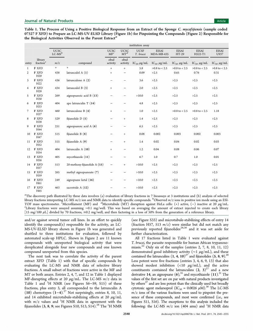
ABSTRACT: A high-throughput (HT) paradigm generating LC-MS-UV-ELSD-based natural product libraries to discovercompounds with new bioactivities and or molecular structures is presented. To validate this methodology, an extract of the Indo-Pacific marine sponge Cacospongia mycof ijiensis was evaluated using assays involving cytoskeletal profiling, tumor cell lines, andparasites. Twelve known compounds were identified including latrunculins (1−4, 10), fijianolides (5, 8, 9), mycothiazole (11),aignopsanes (6, 7), and sacrotride A (13). Compounds 1−5 and 8−11 exhibited bioactivity not previously reported against theparasite T. brucei, while 11 showed selectivity for lymphoma (U937) tumor cell lines. Four new compounds were also discoveredincluding aignopsanoic acid B (13), apo-latrunculin T (14), 20-methoxy-fijianolide A (15), and aignopsane ketal (16).Compounds 13 and 16 represent important derivatives of the aignopsane class, 14 exhibited inhibition of T. brucei withoutdisrupting microfilament assembly, and 15 demonstrated modest microtubule-stabilizing effects. The use of removable well platelibraries to avoid false positives from extracts enriched with only one or two major metabolites is also discussed. Overall, theseresults highlight the advantages of applying modern methods in natural products-based research to accelerate the HT discoveryof therapeutic leads and/or new molecular structures using LC-MS-UV-ELSD-based libraries.
The role of natural products or their derivatives as tools indevelopmental therapeutics programs has been substan-
tial.1−4 However, despite a sustained record of importantcontributions, during the last 15 years there has been a de-emphasis especially by the biopharmaceutical industry on
natural products-based discovery programs.5 A major reasoncited for dropping early stage natural products discovery
Received: August 12, 2011Published: November 30, 2011
Article
pubs.acs.org/jnp
© 2011 American Chemical Society andAmerican Society of Pharmacognosy 2545 dx.doi.org/10.1021/np200673b | J. Nat. Prod. 2011, 74, 2545−2555

programs includes the lengthy time scales involved in thebioassay-guided pursuit to identify or dereplicate potential newlead compounds.6 Skepticism has also been expressed about theprospects of designing effective natural products-based plat-forms that would incorporate modern high-throughput screen-ing (HTS).5
There has been a precipitous decrease in new molecularentity (NME) approved drugs by the U.S. Food and DrugAdministration (FDA) over the last 20 years. For example, thecount of 45 agents approved in 1990 decreased to 21 in 2010.1,7
In our view, there appears to be a positive correlation with thediminished focus on natural products as sources of newtherapeutic leads and the drop in the number of NMEapproved drugs. Whether these trends are causal orcoincidental is open to debate, but few would disagree on thesignificant role of natural products in providing sources andinspiration for new therapeutic leads.8 One bright spot amidstthis controversy is that interest in natural products-based dis-covery programs in the developing world has increaseddramatically since the adoption of the Convention onBiological Diversity (CBD) in 1993.6 As another development,several companies engaged in natural products researchincluding Sequoia Sciences,9 AnalytiCon Discovery,10 andWyeth11 plus a small number of academic groups havepublished first-generation results showing that high-throughput(HT) HPLC purification methods can be interfaced withmodern HTS bioassays.12−15 Surprisingly, only a few studies ofthis type from academic groups have taken the next stepinvolving the use of such HT approaches culminating in thedisclosure of compounds with completely new biologicalactivities or molecular structures.13−17 As one exception, theUC Santa Cruz consortium has recently revealed thatcombining HT HPLC methods with a HT yeast halo assaysuccessfully pinpointed the unreported antifungal bioactivity of
crambescidin 800.14 We have now further optimized thisstrategy to identify lead compounds through an approach thatincorporates systematic LC-MS-UV-ELSD analysis. Theimpetus for these changes stemmed from partnershipsdeveloped in a multidisciplinary campaign as part of a naturalproducts-based International Cooperative Biodiversity Group(ICBG)6 initiative. We have formed a powerful alliance thatinvolves contributions from Indonesian professionals workingalongside investigators from four University of Californiacampuses.A key tool introduced to guide our ICBG programs consists
of a refined HT screening paradigm. The goal is to accelerateidentifying compounds with unreported bioactivity and or newstructures, and it involves the four-step process outlined inScheme 1. (1) Raw materials, which can vary from marinesponges, tropical plants, or culture broths from micro-organisms, are prefractionated using traditional methods18 orwith our previously described HT approach of acceleratedsolvent extraction (ASE),19 which reduces the extraction cycletimes from hours/days to minutes. This creates semicrudeextract assortments (SCEAs), and only the MeOH plantextracts are further pretreated in the first step using solid phaseextraction (SPE) cartridges, to remove polyphenols, which canact as false bioassay positives.12 (2) The SCEAs are evaluated ina panel of HT bioassays20 involving cytoskeleton activity,21
immune modulation,18 parasites,22 tumor cell lines,18,23 andopioid receptors.24 (3) Prioritized active extracts are thenselected for LC-MS-UV-ELSD library creation into 96-wellplates with subsequent follow-up HT bioassay evaluation. (4)When fractions (containing potential lead compounds) thateither exceed the potency of the SCEAs or possess new m/z ionvalues are identified, further processing is immediately initiated.This involves using the same column and LC-MS-UV-ELSDconditions of step 3 to scale up into 20 mL vials (or 50 mL test
Scheme 1. High-Throughput Flow of Raw Materials Involving (1) Extraction (ASE), (2) Bioassays, (3) Automated LibraryPurification and Fractionation, and (4) Final Processing Using Automated Scale-up HPLC to Initiate Dereplication or StructureElucidation of Lead Compoundsa
aPolyamide solid phase extraction (SPE) cartridges were used to remove polyphenols from MeOH plant extracts.
Journal of Natural Products Article
dx.doi.org/10.1021/np200673b | J. Nat. Prod. 2011, 74, 2545−25552546
tubes), followed by evaporation, NMR, dereplication, and/orstructure elucidation. This protocol addresses several key HTparameters11,25 including (a) modest expense is accompaniedby an easy setup and implementation, (b) minimal volumes ofsolvent are required to process ASE extracts,19 96-well plates, or20 mL vial scale-up fractions, (c) nuisance substances (e.g., saltsor polyphenols) that can interfere with bioassays are effectivelyremoved, (d) the rapid processing provides high-quality puri-fication followed by concentration of lead compounds into alibrary for direct HTS bioassay, and (e) frontline acquisitionLC-MS m/z ion, UV, and evaporative light scattering detector(ELSD) data analysis serves to jumpstart dereplication andstructure elucidation efforts.We implemented the process outlined in Scheme 1 to test
the hypothesis that using a focused HT strategy would providerapid identification of lead compounds with important activityproperties and the discovery of new compounds from even well-studied species. The proof-of-concept trial involved investigation ofindividual specimens of the Indo-Pacific sponge Cacospongiamycof ijiensis. Extracts of this sponge have been the source of sevenmajor structural classes having various forms of bioactivity, somewith unique mechanisms of action. The structures include thelatrunculins (antitumor, microfilament disruption),26,27 fijiano-lides (syn laulimalide, antitumor, microtubule stabilizing),28,29
dendrolasen (cytotoxic, target unknown),30 mycothiazole (solidtumor selective, mitochondria complex I),31,32 dactylolide(cytotoxic, microtubule stabilizing),33 CTP-431 (cytotoxic, targetunknown),34 and the aignopsanes (moderate antiparasitic,mechanism unknown).35 The results of our HT survey ofC. mycof ijiensis plus additional insights discovered during ourbrief evaluation using LC-MS-UV-ELSD of other assemblagesare discussed below.
■ RESULTS AND DISCUSSION
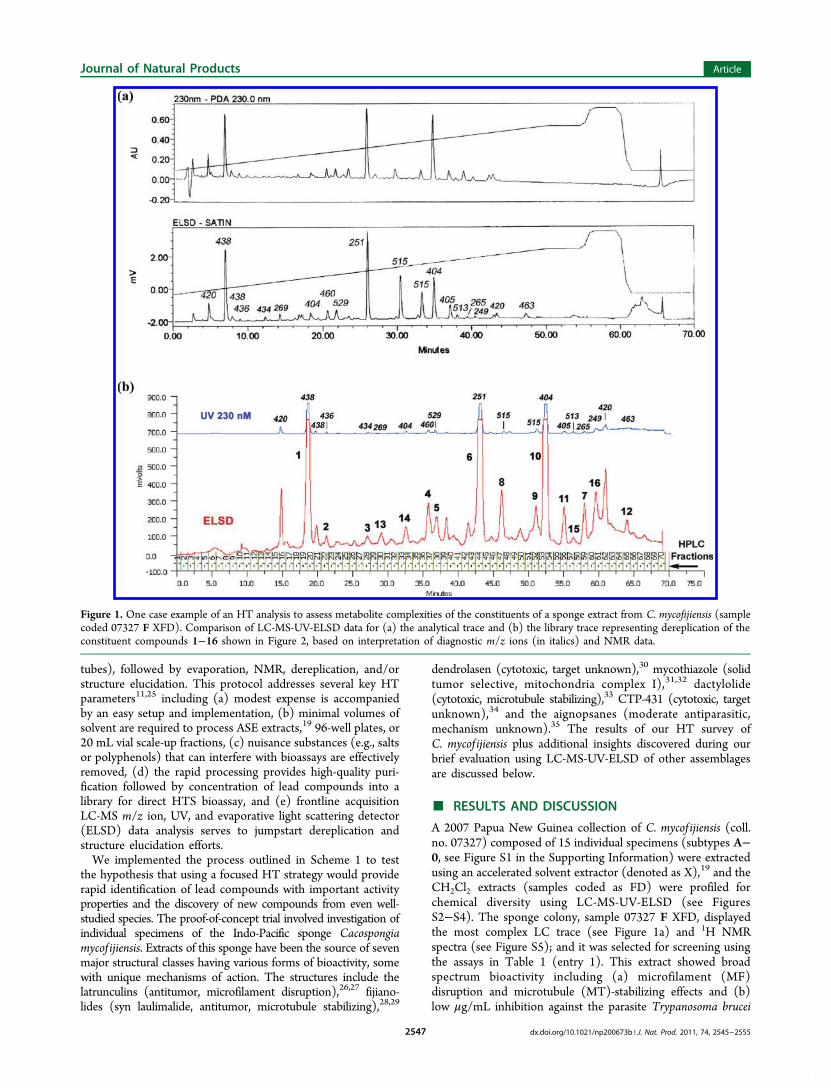
A 2007 Papua New Guinea collection of C. mycof ijiensis (coll.no. 07327) composed of 15 individual specimens (subtypes A−0, see Figure S1 in the Supporting Information) were extractedusing an accelerated solvent extractor (denoted as X),19 and theCH2Cl2 extracts (samples coded as FD) were profiled forchemical diversity using LC-MS-UV-ELSD (see FiguresS2−S4). The sponge colony, sample 07327 F XFD, displayedthe most complex LC trace (see Figure 1a) and 1H NMRspectra (see Figure S5); and it was selected for screening usingthe assays in Table 1 (entry 1). This extract showed broadspectrum bioactivity including (a) microfilament (MF)disruption and microtubule (MT)-stabilizing effects and (b)low μg/mL inhibition against the parasite Trypanosoma brucei
Figure 1. One case example of an HT analysis to assess metabolite complexities of the constituents of a sponge extract from C. mycofijiensis (samplecoded 07327 F XFD). Comparison of LC-MS-UV-ELSD data for (a) the analytical trace and (b) the library trace representing dereplication of theconstituent compounds 1−16 shown in Figure 2, based on interpretation of diagnostic m/z ions (in italics) and NMR data.
Journal of Natural Products Article
dx.doi.org/10.1021/np200673b | J. Nat. Prod. 2011, 74, 2545−25552547
and/or against several tumor cell lines. In an effort to quicklyidentify the compound(s) responsible for the activity, an LC-MS-UV-ELSD library shown in Figure 1b was generated andshuttled to three institutions for evaluation, followed byautomated scale-up HPLC. Shown in Figure 2 are 11 knowncompounds with unreported biological activity that weredereplicated alongside four new compounds and one knowncompound unreported from this species.The next task was to correlate the activity of the parent
extract XFD (Table 1) with that of specific compounds byevaluating the LC-MS and NMR data of selective libraryfractions. A small subset of fractions were active in the MF andMT or both assays. Entries 2, 4, 7, and 12 in Table 1 displayedMF-disrupting effects at 20 μg/mL. The LC-MS m/z data inTable 1 and 1H NMR (see Figures S6−S9, S15) of thesefractions, plus entry 3, all corresponded to the latrunculin A(10) chemotypes (1−4).27 Not surprisingly, entries 8, 10, 11,and 14 exhibited microtubule-stabilizing effects at 20 μg/mLwith m/z values and 1H NMR data in agreement with thefijianolides (5, 8, 9; see Figures S10, S13, S14).28 The 1H NMR
(see Figure S33) and microtubule-stabilizing effects of entry 14(fraction H57, 513 m/z) were similar but did not match anypreviously reported fijianolides28,36 and it was set aside forfurther characterization.All 17 fractions listed in Table 1 were evaluated against
T. brucei, the parasite responsible for human African trypanoso-miasis.22 Only six of the samples (entries 2, 7, 8, 10, 11, 12)demonstrated good inhibitory activity (∼1 μg/mL), and thesecontained the latrunculins (1, 4, 10)27 and fijianolides (5, 8, 9).28
Less potent were five fractions (entries 3, 4, 6, 9, 13) that alsoshowed modest inhibition (<10 μg/mL), and the activeconstituents contained the latrunculins (2, 3)27 and a newderivative 14, an aignopsane (6),35 and mycothiazole (11).31 Thevalues of the first set are on par with natural products investigatedby others37 and are less potent than the clinically used but broadlycytotoxic agent melarsoprol (IC50 = 0.0026 μM).38 The LC-MSm/z ions of the various fractions were used to pinpoint the pre-sence of these compounds, and most were confirmed (i.e., seeFigures S11, S16). The exceptions to this analysis included thefollowing: the LC-MS m/z ion (404 amu) and 1H NMR data
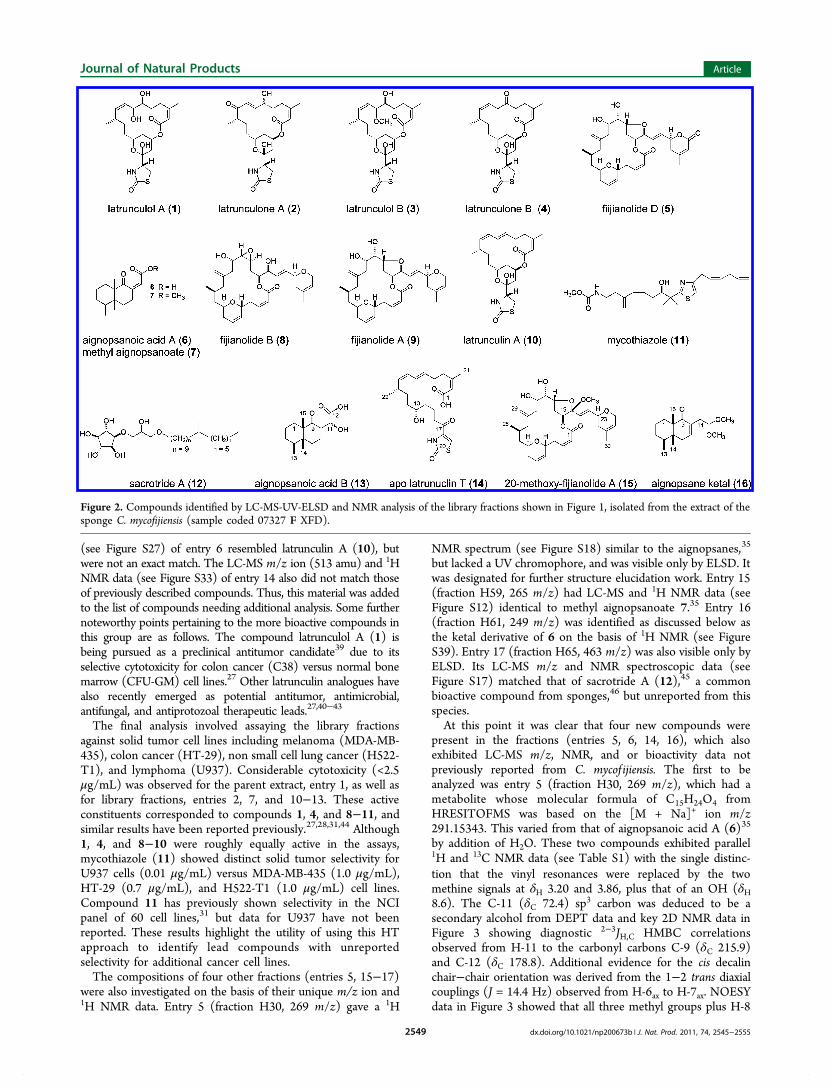
Table 1. The Process of Using a Positive Biological Response from an Extract of the Sponge C. mycof ijiensis (sample coded07327 F XFD) to Prepare an LC-MS-UV-ELSD Library (Figure 1b) for Pinpointing the Compounds (Figure 2) Responsible forthe Biological Activities Observed in the Parent Extracta
institution assay
UCSCLC-MSb
UCSCMFc
UCSCMTd
UCSFT. brucei
EISAIMDA-MB-435
EISAIHT-29
EISAIH522-T1
EISAIU937
entrylibraryfractione m/z compound
obsdactivity
obsdactivity IC50, μg/mL IC50, μg/mL IC50, μg/mL IC50, μg/mL IC50, μg/mL
1 F XFD * * + + 5.8 >0.8 to < 2.5 >0.8 to < 2.5 >0.8 to < 2.5 >0.8 to < 2.52 F XFD
H20438 latrunculol A (1) + − 0.09 >2.5 0.65 0.76 0.31
3 F XFDH22
436 latrunculone A (2) − − 3.6 >2.5 >2.5 >2.5 >2.5
4 F XFDH28
434 latrunculol B (3) + − 2.0 >2.5 >2.5 >2.5 >2.5
5 F XFDH30
269 aignopsanoic acid B (13) − − >10.0 >2.5 >2.5 >2.5 >2.5
6 F XFDH33
404 apo latrunculin T (14) − − 4.8 >2.5 >2.5 >2.5 >2.5
7 F XFDH37
460 latrunculone B (4) + − 1.0 >2.5 >0.8 to < 2.5 >0.8 to < 2.5 1.18
8 F XFDH38
529 fijianolide D (5) − + 1.4 >2.5 >2.5 >2.5 >2.5
9 F XFDH44
251 aignopsanoic acid A (6) − − 6.5 >2.5 >2.5 >2.5 >2.5
10 F XFDH47
515 fijianolide B (8) − + 0.08 0.002 0.003 0.002 0.003
11 F XFDH52
515 fijianolide A (9) − + 1.4 0.02 0.04 0.02 0.03
12 F XFDH54
404 latrunculin A (10) + − 1.2 0.04 0.08 0.06 0.07
13 F XFDH56
405 mycothiazole (11) − − 6.7 1.0 0.7 1.0 0.01
14 F XFDH57
513 20-methoxy-fijianolide A (15) − + >10.0 >2.5 >2.5 >2.5 >2.5
15 F XFDH59
265 methyl aignopsanoate (7) − − >10.0 >2.5 >2.5 >2.5 >2.5
16 F XFDH61
249 aignopsane ketal (16) − − >10.0 >2.5 >2.5 >2.5 >2.5
17 F XFDH65
463 sacrotride A (12) − − >10.0 >2.5 >2.5 >2.5 >2.5
aThe discovery path illustrated by these data involves (a) evaluation of library fractions in 7 bioassays at 3 institutions and (b) analysis of selectedlibrary fractions interpreting LC-MS m/z ion and NMR data to identify specific compounds. bObserved m/z ions in positive ion mode using an ESI-TOF mass spectrometer. cMicrofilament (MF) and dMicrotubule (MT) disruption against HeLa cells: (+) active, (−) inactive at 20 μg/mL.eLibrary fractions were assayed assuming ∼0.1 mg/well. This was based on averaging the amount of extract injected to create each library[15 mg/100 μL] divided by 70 fractions, ≈0.2 mg/well, and then factoring in a loss of 50% from the generation of a reference library.
Journal of Natural Products Article
dx.doi.org/10.1021/np200673b | J. Nat. Prod. 2011, 74, 2545−25552548
(see Figure S27) of entry 6 resembled latrunculin A (10), butwere not an exact match. The LC-MS m/z ion (513 amu) and 1HNMR data (see Figure S33) of entry 14 also did not match thoseof previously described compounds. Thus, this material was addedto the list of compounds needing additional analysis. Some furthernoteworthy points pertaining to the more bioactive compounds inthis group are as follows. The compound latrunculol A (1) isbeing pursued as a preclinical antitumor candidate39 due to itsselective cytotoxicity for colon cancer (C38) versus normal bonemarrow (CFU-GM) cell lines.27 Other latrunculin analogues havealso recently emerged as potential antitumor, antimicrobial,antifungal, and antiprotozoal therapeutic leads.27,40−43
The final analysis involved assaying the library fractionsagainst solid tumor cell lines including melanoma (MDA-MB-435), colon cancer (HT-29), non small cell lung cancer (H522-T1), and lymphoma (U937). Considerable cytotoxicity (<2.5μg/mL) was observed for the parent extract, entry 1, as well asfor library fractions, entries 2, 7, and 10−13. These activeconstituents corresponded to compounds 1, 4, and 8−11, andsimilar results have been reported previously.27,28,31,44 Although1, 4, and 8−10 were roughly equally active in the assays,mycothiazole (11) showed distinct solid tumor selectivity forU937 cells (0.01 μg/mL) versus MDA-MB-435 (1.0 μg/mL),HT-29 (0.7 μg/mL), and H522-T1 (1.0 μg/mL) cell lines.Compound 11 has previously shown selectivity in the NCIpanel of 60 cell lines,31 but data for U937 have not beenreported. These results highlight the utility of using this HTapproach to identify lead compounds with unreportedselectivity for additional cancer cell lines.The compositions of four other fractions (entries 5, 15−17)
were also investigated on the basis of their unique m/z ion and1H NMR data. Entry 5 (fraction H30, 269 m/z) gave a 1H
NMR spectrum (see Figure S18) similar to the aignopsanes,35
but lacked a UV chromophore, and was visible only by ELSD. Itwas designated for further structure elucidation work. Entry 15(fraction H59, 265 m/z) had LC-MS and 1H NMR data (seeFigure S12) identical to methyl aignopsanoate 7.35 Entry 16(fraction H61, 249 m/z) was identified as discussed below asthe ketal derivative of 6 on the basis of 1H NMR (see FigureS39). Entry 17 (fraction H65, 463 m/z) was also visible only byELSD. Its LC-MS m/z and NMR spectroscopic data (seeFigure S17) matched that of sacrotride A (12),45 a commonbioactive compound from sponges,46 but unreported from thisspecies.At this point it was clear that four new compounds were
present in the fractions (entries 5, 6, 14, 16), which alsoexhibited LC-MS m/z, NMR, and or bioactivity data notpreviously reported from C. mycof ijiensis. The first to beanalyzed was entry 5 (fraction H30, 269 m/z), which had ametabolite whose molecular formula of C15H24O4 fromHRESITOFMS was based on the [M + Na]+ ion m/z291.15343. This varied from that of aignopsanoic acid A (6)35
by addition of H2O. These two compounds exhibited parallel1H and 13C NMR data (see Table S1) with the single distinc-tion that the vinyl resonances were replaced by the twomethine signals at δH 3.20 and 3.86, plus that of an OH (δH8.6). The C-11 (δC 72.4) sp3 carbon was deduced to be asecondary alcohol from DEPT data and key 2D NMR data inFigure 3 showing diagnostic 2−3JH,C HMBC correlationsobserved from H-11 to the carbonyl carbons C-9 (δC 215.9)and C-12 (δC 178.8). Additional evidence for the cis decalinchair−chair orientation was derived from the 1−2 trans diaxialcouplings (J = 14.4 Hz) observed from H-6ax to H-7ax. NOESYdata in Figure 3 showed that all three methyl groups plus H-8
Figure 2. Compounds identified by LC-MS-UV-ELSD and NMR analysis of the library fractions shown in Figure 1, isolated from the extract of thesponge C. mycofijiensis (sample coded 07327 F XFD).
Journal of Natural Products Article
dx.doi.org/10.1021/np200673b | J. Nat. Prod. 2011, 74, 2545−25552549
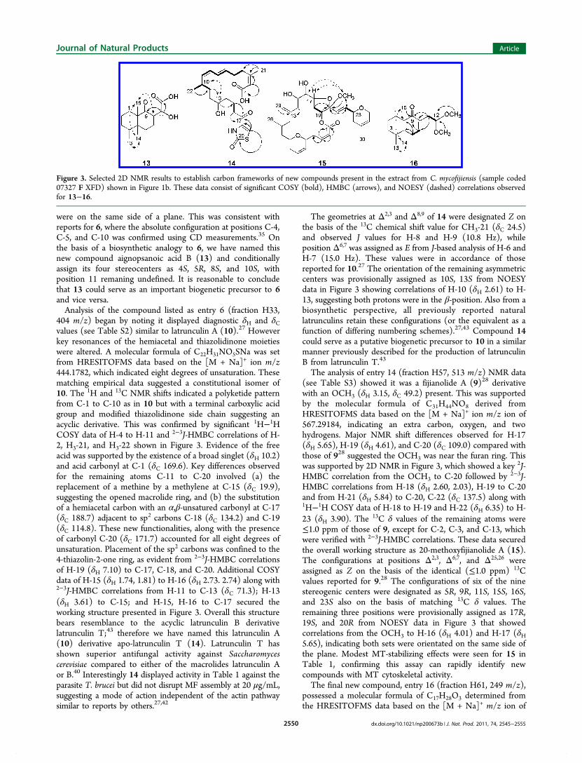
were on the same side of a plane. This was consistent withreports for 6, where the absolute configuration at positions C-4,C-5, and C-10 was confirmed using CD measurements.35 Onthe basis of a biosynthetic analogy to 6, we have named thisnew compound aignopsanoic acid B (13) and conditionallyassign its four stereocenters as 4S, 5R, 8S, and 10S, withposition 11 remaning undefined. It is reasonable to concludethat 13 could serve as an important biogenetic precursor to 6and vice versa.Analysis of the compound listed as entry 6 (fraction H33,
404 m/z) began by noting it displayed diagnostic δH and δCvalues (see Table S2) similar to latrunculin A (10).27 Howeverkey resonances of the hemiacetal and thiazolidinone moietieswere altered. A molecular formula of C22H31NO5SNa was setfrom HRESITOFMS data based on the [M + Na]+ ion m/z444.1782, which indicated eight degrees of unsaturation. Thesematching empirical data suggested a constitutional isomer of10. The 1H and 13C NMR shifts indicated a polyketide patternfrom C-1 to C-10 as in 10 but with a terminal carboxylic acidgroup and modified thiazolidinone side chain suggesting anacyclic derivative. This was confirmed by significant 1H−1HCOSY data of H-4 to H-11 and 2−3J-HMBC correlations of H-2, H3-21, and H3-22 shown in Figure 3. Evidence of the freeacid was supported by the existence of a broad singlet (δH 10.2)and acid carbonyl at C-1 (δC 169.6). Key differences observedfor the remaining atoms C-11 to C-20 involved (a) thereplacement of a methine by a methylene at C-15 (δC 19.9),suggesting the opened macrolide ring, and (b) the substitutionof a hemiacetal carbon with an α,β-unsatured carbonyl at C-17(δC 188.7) adjacent to sp2 carbons C-18 (δC 134.2) and C-19(δC 114.8). These new functionalities, along with the presenceof carbonyl C-20 (δC 171.7) accounted for all eight degrees ofunsaturation. Placement of the sp2 carbons was confined to the4-thiazolin-2-one ring, as evident from 2−3J-HMBC correlationsof H-19 (δH 7.10) to C-17, C-18, and C-20. Additional COSYdata of H-15 (δH 1.74, 1.81) to H-16 (δH 2.73. 2.74) along with2−3J-HMBC correlations from H-11 to C-13 (δC 71.3); H-13(δH 3.61) to C-15; and H-15, H-16 to C-17 secured theworking structure presented in Figure 3. Overall this structurebears resemblance to the acyclic latrunculin B derivativelatrunculin T;43 therefore we have named this latrunculin A(10) derivative apo-latrunculin T (14). Latrunculin T hasshown superior antifungal activity against Saccharomycescerevisiae compared to either of the macrolides latrunculin Aor B.40 Interestingly 14 displayed activity in Table 1 against theparasite T. brucei but did not disrupt MF assembly at 20 μg/mL,suggesting a mode of action independent of the actin pathwaysimilar to reports by others.27,42
The geometries at Δ2,3 and Δ8,9 of 14 were designated Z onthe basis of the 13C chemical shift value for CH3-21 (δC 24.5)and observed J values for H-8 and H-9 (10.8 Hz), whileposition Δ6,7 was assigned as E from J-based analysis of H-6 andH-7 (15.0 Hz). These values were in accordance of thosereported for 10.27 The orientation of the remaining asymmetriccenters was provisionally assigned as 10S, 13S from NOESYdata in Figure 3 showing correlations of H-10 (δH 2.61) to H-13, suggesting both protons were in the β-position. Also from abiosynthetic perspective, all previously reported naturallatrunculins retain these configurations (or the equivalent as afunction of differing numbering schemes).27,43 Compound 14could serve as a putative biogenetic precursor to 10 in a similarmanner previously described for the production of latrunculinB from latrunculin T.43
The analysis of entry 14 (fraction H57, 513 m/z) NMR data(see Table S3) showed it was a fijianolide A (9)28 derivativewith an OCH3 (δH 3.15, δC 49.2) present. This was supportedby the molecular formula of C31H44NO8 derived fromHRESITOFMS data based on the [M + Na]+ ion m/z ion of567.29184, indicating an extra carbon, oxygen, and twohydrogens. Major NMR shift differences observed for H-17(δH 5.65), H-19 (δH 4.61), and C-20 (δC 109.0) compared withthose of 928 suggested the OCH3 was near the furan ring. Thiswas supported by 2D NMR in Figure 3, which showed a key 2J-HMBC correlation from the OCH3 to C-20 followed by 2−3J-HMBC correlations from H-18 (δH 2.60, 2.03), H-19 to C-20and from H-21 (δH 5.84) to C-20, C-22 (δC 137.5) along with1H−1H COSY data of H-18 to H-19 and H-22 (δH 6.35) to H-23 (δH 3.90). The 13C δ values of the remaining atoms were≤1.0 ppm of those of 9, except for C-2, C-3, and C-13, whichwere verified with 2−3J-HMBC correlations. These data securedthe overall working structure as 20-methoxyfijianolide A (15).The configurations at positions Δ2,3, Δ6,7, and Δ25,26 wereassigned as Z on the basis of the identical (≤1.0 ppm) 13Cvalues reported for 9.28 The configurations of six of the ninestereogenic centers were designated as 5R, 9R, 11S, 15S, 16S,and 23S also on the basis of matching 13C δ values. Theremaining three positions were provisionally assigned as 17R,19S, and 20R from NOESY data in Figure 3 that showedcorrelations from the OCH3 to H-16 (δH 4.01) and H-17 (δH5.65), indicating both sets were orientated on the same side ofthe plane. Modest MT-stabilizing effects were seen for 15 inTable 1, confirming this assay can rapidly identify newcompounds with MT cytoskeletal activity.The final new compound, entry 16 (fraction H61, 249 m/z),
possessed a molecular formula of C17H28O3 determined fromthe HRESITOFMS data based on the [M + Na]+ m/z ion of
Figure 3. Selected 2D NMR results to establish carbon frameworks of new compounds present in the extract from C. mycofijiensis (sample coded07327 F XFD) shown in Figure 1b. These data consist of significant COSY (bold), HMBC (arrows), and NOESY (dashed) correlations observedfor 13−16.
Journal of Natural Products Article
dx.doi.org/10.1021/np200673b | J. Nat. Prod. 2011, 74, 2545−25552550
303.1918. This represented the loss of one degree ofunsaturation versus that of 6. The 1H and 13C NMR data(see Table S4) indicated a substitution of the exocyclic doubleand acid moiety for a dimethoxy ethyl side chain with OCH3groups (δH 3.33, δC 53.5, 53.7), methylenes C-11 (δH 2.54,2.46; δC 34.1), and methine C-12 (δH 4.51, δC 103.4). Arearranged α,β-unsatured ketone C-9 (δC 204.1) with anendocyclic double C-8 (δC 133.1), C-7 (δH 6.53, δC 142.7) wasalso apparent. These conclusions accounted for the loss of onedegree of unsaturation and were supported by 2D NMR data inFigure 3, showing 2J-HMBC correlations from the OCH3groups to C-12, 1H−1H COSY data from H-12 to H-11, and2−3J-HMBC correlations from H-11 to C-7−C-9. AdditionalCOSY data from H-7 to H-6 (δH 2.39, 2.29) followed by 2−3J-HMBC correlations from H-7 to C-9; H-6 to C-5 (δC 41.4), C-10 (δC 50.7); and H3-15 (δH 0.98) to C-9, C-10 linked the Bring together in only one way, as aignopsane ketal (16). Theremaining atoms (C-1 to C-5 and C-13 to C-15) displayed 13CNMR with values ≤1 ppm of those of 6, confirming theexistence of the A ring and the overall working structure of 16.The geometry of Δ7,8 was assigned as Z on the basis of theobserved vicinal coupling of 6.0 Hz between H-6 and H-7 thatwas in agreement with blancasterol,47 which shares a similarα,β-unsaturated ketone motif. Determination of the relativeconfiguration of the asymmetric centers was set from NOESYdata in Figure 3, which paralleled 13, indicating the 4S, 5R, 10Sorientation. It is possible to conclude that the unique structureof 16 arose from either 6 or 13 after a 48 h exposure toMeOH−H2O during processing and/or transport, which mayhave led to the formation of its dimethyl acetal functionality.Although the above example served to rapidly identify
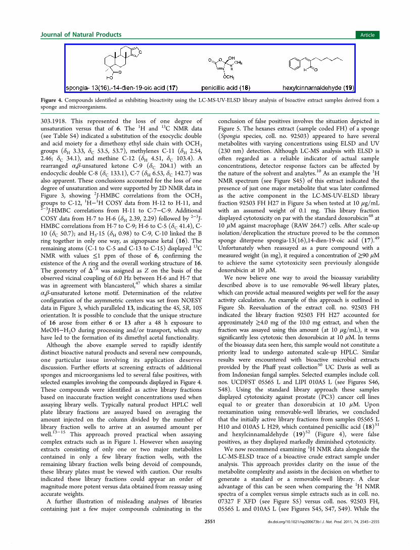
distinct bioactive natural products and several new compounds,one particular issue involving its application deservesdiscussion. Further efforts at screening extracts of additionalsponges and microorganisms led to several false positives, withselected examples involving the compounds displayed in Figure 4.These compounds were identified as active library fractionsbased on inaccurate fraction weight concentrations used whenassaying library wells. Typically natural product HPLC wellplate library fractions are assayed based on averaging theamount injected on the column divided by the number oflibrary fraction wells to arrive at an assumed amount perwell.13−15 This approach proved practical when assayingcomplex extracts such as in Figure 1. However when assayingextracts consisting of only one or two major metabolitescontained in only a few library fraction wells, with theremaining library fraction wells being devoid of compounds,these library plates must be viewed with caution. Our resultsindicated these library fractions could appear an order ofmagnitude more potent versus data obtained from reassay usingaccurate weights.A further illustration of misleading analyses of libraries
containing just a few major compounds culminating in the
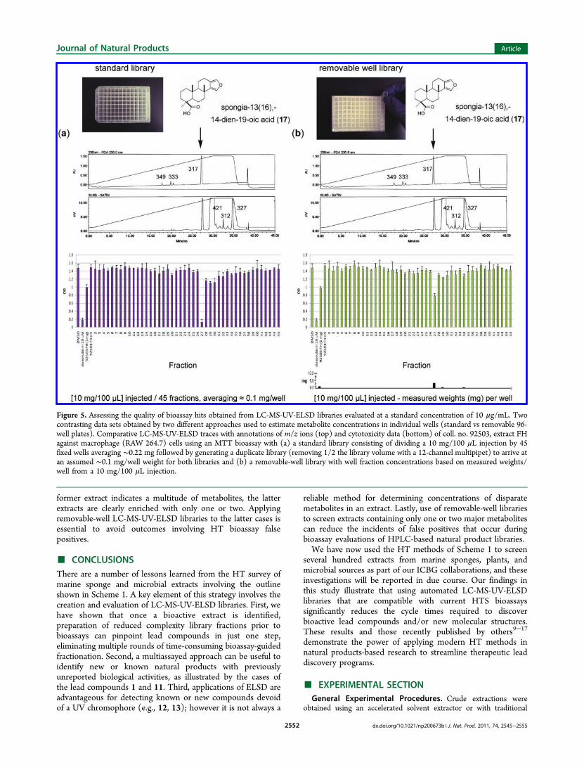
conclusion of false positives involves the situation depicted inFigure 5. The hexanes extract (sample coded FH) of a sponge(Spongia species, coll. no. 92503) appeared to have severalmetabolites with varying concentrations using ELSD and UV(230 nm) detection. Although LC-MS analysis with ELSD isoften regarded as a reliable indicator of actual sampleconcentrations, detector response factors can be affected bythe nature of the solvent and analytes.10 As an example the 1HNMR spectrum (see Figure S45) of this extract indicated thepresence of just one major metabolite that was later confirmedas the active component in the LC-MS-UV-ELSD libraryfraction 92503 FH H27 in Figure 5a when tested at 10 μg/mLwith an assumed weight of 0.1 mg. This library fractiondisplayed cytotoxicity on par with the standard doxorubicin48 at10 μM against macrophage (RAW 264.7) cells. After scale-upisolation/dereplication the structure proved to be the commonsponge diterpene spongia-13(16),14-dien-19-oic acid (17).49
Unfortunately when reassayed as a pure compound with ameasured weight (in mg), it required a concentration of ≥90 μMto achieve the same cytotoxicity seen previously alongsidedoxorubicin at 10 μM.We now believe one way to avoid the bioassay variability
described above is to use removable 96-well library plates,which can provide actual measured weights per well for the assayactivity calculation. An example of this approach is outlined inFigure 5b. Reevaluation of the extract coll. no. 92503 FHindicated the library fraction 92503 FH H27 accounted forapproximately ≥4.0 mg of the 10.0 mg extract, and when thefraction was assayed using this amount (at 10 μg/mL), it wassignificantly less cytotoxic then doxorubicin at 10 μM. In termsof the bioassay data seen here, this sample would not constitute apriority lead to undergo automated scale-up HPLC. Similarresults were encountered with bioactive microbial extractsprovided by the Phaff yeast collection50 UC Davis as well asfrom Indonesian fungal samples. Selected examples include coll.nos. UCDFST 05565 L and LIPI 010A5 L (see Figures S46,S48). Using the standard library approach these samplesdisplayed cytotoxicity against prostate (PC3) cancer cell linesequal to or greater than doxorubicin at 10 μM. Uponreexamination using removable-well libraries, we concludedthat the initially active library fractions from samples 05565 LH10 and 010A5 L H29, which contained penicillic acid (18)51
and hexylcinnamaldehyde (19)52 (Figure 4), were falsepositives, as they displayed markedly diminished cytotoxicity.We now recommend examining 1H NMR data alongside the
LC-MS-ELSD trace of a bioactive crude extract sample underanalysis. This approach provides clarity on the issue of themetabolite complexity and assists in the decision on whether togenerate a standard or a removable-well library. A clearadvantage of this can be seen when comparing the 1H NMRspectra of a complex versus simple extracts such as in coll. no.07327 F XFD (see Figure S5) versus coll. nos. 92503 FH,05565 L and 010A5 L (see Figures S45, S47, S49). While the
Figure 4. Compounds identified as exhibiting bioactivity using the LC-MS-UV-ELSD library analysis of bioactive extract samples derived from asponge and microorganisms.
Journal of Natural Products Article
dx.doi.org/10.1021/np200673b | J. Nat. Prod. 2011, 74, 2545−25552551
former extract indicates a multitude of metabolites, the latterextracts are clearly enriched with only one or two. Applyingremovable-well LC-MS-UV-ELSD libraries to the latter cases isessential to avoid outcomes involving HT bioassay falsepositives.
■ CONCLUSIONSThere are a number of lessons learned from the HT survey ofmarine sponge and microbial extracts involving the outlineshown in Scheme 1. A key element of this strategy involves thecreation and evaluation of LC-MS-UV-ELSD libraries. First, wehave shown that once a bioactive extract is identified,preparation of reduced complexity library fractions prior tobioassays can pinpoint lead compounds in just one step,eliminating multiple rounds of time-consuming bioassay-guidedfractionation. Second, a multiassayed approach can be useful toidentify new or known natural products with previouslyunreported biological activities, as illustrated by the cases ofthe lead compounds 1 and 11. Third, applications of ELSD areadvantageous for detecting known or new compounds devoidof a UV chromophore (e.g., 12, 13); however it is not always a
reliable method for determining concentrations of disparatemetabolites in an extract. Lastly, use of removable-well librariesto screen extracts containing only one or two major metabolitescan reduce the incidents of false positives that occur duringbioassay evaluations of HPLC-based natural product libraries.We have now used the HT methods of Scheme 1 to screen
several hundred extracts from marine sponges, plants, andmicrobial sources as part of our ICBG collaborations, and theseinvestigations will be reported in due course. Our findings inthis study illustrate that using automated LC-MS-UV-ELSDlibraries that are compatible with current HTS bioassayssignificantly reduces the cycle times required to discoverbioactive lead compounds and/or new molecular structures.These results and those recently published by others9−17
demonstrate the power of applying modern HT methods innatural products-based research to streamline therapeutic leaddiscovery programs.
■ EXPERIMENTAL SECTIONGeneral Experimental Procedures. Crude extractions were
obtained using an accelerated solvent extractor or with traditional
Figure 5. Assessing the quality of bioassay hits obtained from LC-MS-UV-ELSD libraries evaluated at a standard concentration of 10 μg/mL. Twocontrasting data sets obtained by two different approaches used to estimate metabolite concentrations in individual wells (standard vs removable 96-well plates). Comparative LC-MS-UV-ELSD traces with annotations of m/z ions (top) and cytotoxicity data (bottom) of coll. no. 92503, extract FHagainst macrophage (RAW 264.7) cells using an MTT bioassay with (a) a standard library consisting of dividing a 10 mg/100 μL injection by 45fixed wells averaging ∼0.22 mg followed by generating a duplicate library (removing 1/2 the library volume with a 12-channel multipipet) to arrive atan assumed ∼0.1 mg/well weight for both libraries and (b) a removable-well library with well fraction concentrations based on measured weights/well from a 10 mg/100 μL injection.
Journal of Natural Products Article
dx.doi.org/10.1021/np200673b | J. Nat. Prod. 2011, 74, 2545−25552552
methods reported previously.18,19 Optical rotations were obtained on aJASCO DIP-370 digital polarimeter, while UVmax data were obtainedusing a Waters 996 photodiode array (PDA) detector. All NMRexperiments were run on a Varian UNITY 500 (500 and 125 MHz for1H and 13C, respectively) or Varian INOVA 600 spectrometer (600and 150 MHz for 1H and 13C, respectively). The 600 MHzspectrometer was equipped with a 5 mm triple resonance (HCN)cryogenic probe. Sample amounts smaller than 2.0 mg were analyzedusing 3 and 5 mm Shigemi tubes where available. High-resolution massspectrometry measurements were obtained using a Mariner ESI-TOF-MS. Analytical LC-MS analysis was performed on samples at aconcentration of approximately 5 mg/mL, using a reversed-phase150 × 4.60 mm 5 μm C18 Phenomenex Luna column in conjunctionwith a 4.0 × 3.0 mm C18 (Octadecyl) guard column and cartridge(holder part number: KJ0-4282, cartridge part number: AJ0-4287,Phenomenex, Inc., Torrance, CA, USA). Samples were injected ontothe column using a volume of 15 μL, with a flow rate of 1 mL/min thatwas monitored using a Waters model 996 PDA UV detector. Theelution was subsequently split (1:1) between a SEDERE model 55ELSD and an Applied Biosystems Mariner electrospray ionizationtime-of-flight (ESI-TOF) mass spectrometer.Biological Material, Collection, and Identification. Specimens
of the sponge Cacospongia mycof ijiensis profiled for these experiments(coll. nos. 07327 A−J, L−O, 387 g wet wt) were collected in 2007 offthe northern coastlines of New Britain, Papua New Guinea.35
Taxonomic identifications were based on comparison of the biologicalfeatures to other voucher samples in our repository and confirmed byNicole J. de Voogd of the National Museum of Natural History, TheNetherlands. The secondary metabolite chemistry is also consistentwith these identifications. Voucher specimens and underwater photosare available. The extract of sample coll. no. 92503 was identified asbelonging to the genus Spongia and was obtained from the UCSCmarine natural products repository as an archived 1992 Indonesianexpedition sample. Taxonomic identification was performed byChristina Diaz. The filamentous fungus UCDFST 05565 was providedby the Phaff Yeast Culture Collection50 UC Davis and identified asHyalodendriella betulae, while the microbial sample coll. no. 010A5 wasan unidentified filamentous fungus specimen provided by the ResearchCenter for Biology & Chemistry, Indonesian Institute of Science.Extraction and Prefractionation. Sponge samples were pre-
served in the field by being immersed in a 50:50 MeOH−H2Osolution. After approximately 48 h this solution was decanted anddiscarded. The damp organisms were placed in collection bottles(Nalgene) and shipped back to UCSC at ambient temperatureand then stored at 4 °C until further processed. Specimens ofC. mycof ijiensis coll. nos. 07327 A−J, L−O (387 g wet wt) wereprocessed using the high-throughput method of accelerated solventextraction19 to generate four extracts sequentially. Samples were firstextracted with H2O, to remove inorganic salts (sample coded XWW),followed by hexanes to remove unwanted steroid and lipidcomponents (sample coded XFH), CH2Cl2 (sample coded XFD),and MeOH (sample coded XFM). Samples 07327 F afforded 221.2mg of XWW, 42.3 mg of XFH, 78.2 mg of XFD, and 56.5 mg of XFMextracts. The repository Spongia specimen (coll. no. 92503) wasextracted using traditional methods of solvent partitioning.19 Plantmaterials were extracted with MeOH and hexanes. Methanol extractsof these samples were further fractionated with SPE cartridges toremove polyphenols using a 700 mg polyamide-filled cartridge (Sigma-Aldrich, St. Louis, MO, USA) according to reported methods.12
Microbial specimens coll. nos. 05565 (H. betulae) and 010A5 wereextracted using traditional methods involving EtOAc and MeOH.18
LC-MS-UV-ELSD with Fraction Collection and Scale-UpIsolation. A representation of the LC-MS-UV-ELSD instrumentationsetup is shown in Figure S50. LC-MS-UV-ELSD analysis wasperformed using two Waters 510 pumps, controlled with Empower2 software. A Waters 717plus autosampler was used for sample loadingand injection. Separation was performed on a Luna 5 μm, C18(2)100 Å 10 × 250 mm column (Phenomenex, Inc.) in conjunction witha guard column using a larger 10.0 × 10.0 mm C18 (ODS) cartridge(holder part number: AJ0-7220, cartridge part number: AJ0-7221).
Spectra from three detectors were acquired during each run: Waters996 photo diode array, SEDEX 55 ELSD, and Mariner 5054 ESI-TOF-MS. Solvent flow was controlled through back-pressure regulation. Asimple low dead volume cross union from IDEX Health & Science wasused to split solvent. Desired flow rates were used to determine theappropriate tubing diameter and length. A solvent flow rate of 1.9 mL/minof solvent flowing, to the fraction collector, through 60 in. of 0.01 in.inner diameter (i.d.) tubing will result in 68.5 psi of back-pressure.Solvent flowing at a rate of 0.08 mL/min to the ELSD through 36 in.of 0.004 in. i.d. tubing will result in 67.6 psi of back-pressure. Solventflowing at a rate of 0.02 mL/min to the mass spectrometer through22 in. of 0.0025 in. i.d. tubing results in 67.7 psi of back-pressure. Allvalues of psi were calculated using a viscosity of 1. Some time delaybetween instrumentation will happen, but has been kept to aminimum. The mobile phase parameters are CH3CN (A) and H2O(B) with a flow rate of 2 mL/min and a gradient of 0 min, 10:90; 40min, 100:0; and 60 min, 100:0. Injection amounts range from 10 mg/100−150 μL to 15 mg/150−200 μL.
Sample collection was performed using a Gilson 215 liquid handlercontrolled with Gilson Unipoint LC software. Samples were collectedinto BD Biosciences 96 deep-well plates, with a working volume of2 mL (part number: 353966). Simport Plastics 96 removable-wellplates (part number: T105-50) were also used and allowed eachsample well to be preweighed and reweighed after sample collectionfor accurate sample weight determination. Scale-up HPLC fractionswere collected into 20 mL scintillation vials using two Gilsoncollection racks (number coded 204). Alternatively, larger HPLCscale-up fractions can be generated using 50 mL test tubes with thetwo Gilson collection racks (number coded 225). Fractions werecollected every minute. Sample workup sheets for standard andremovable 96-well plates are shown in Figures S51−S52. An exampleof proper weighing technique using removable 96-well plate librariesto maximize accuracy is shown in Figure S53.
After the LC-MS-UV-ELSD library is collected, a duplicate plate isgenerated for analytical reference using a 12-channel pipet, creating anexact copy and counter balance for centrifugal drying. Plates weredried and concentrated using a Savant AES2010 SpeedVac. Driedplates were reconstituted in DMSO to a concentration of either 10 or20 μg/mL for bioassay unless otherwise specified.
The XFD extract (78.2 mg) of coll. no. 07327 F was used to preparefour LC-MS-UV-ELSD libraries into 96-well plates (40.1 mg, [10 mg/100 μL] × 4 injections) for eight HT bioassay evaluations using amodified gradient of 30:70 to 80:20 CH3CN−H2O over 70 min tomaximize baseline peak separation. Reference libraries were generatedfrom each of the four original libraries using a 12-channel pipet,creating an exact copy and counter balance for centrifugal drying.Using the remaining 38.1 mg, automated scale-up HPLC into 20 mLvials ([9.5 mg/100 μL] × 4 injections) was performed with the same5 μm column and conditions used for the LC-MS-UV-ELSD librarypurifications. Reference library well fractions were combined withcorresponding 20 mL HPLC vial fractions based on parallel LC-MS-UV-ELSD data to provide the total amounts of the selected purecompounds as H20 latrunculol A (1, 4.6 mg), H22 latrunculone A(2, 1.2 mg), H28 latrunculol B (3, 1.3 mg), H37 latrunculone B(4, 2.1 mg), H38 fijianolide D (5, 2.1 mg), H44 aignopsanoic acid A(6, 4.8 mg), H59 methyl aignopsanoate (7, 1.5 mg), H47 fijianolide B(8, 2.7 mg), H52 fijianolide A (9, 2.3 mg), H54 latrunculin A (10, 4.6mg), H56 mycothiazole (11, 1.9 mg), H65 sacrotride A (12, 1.3 mg),H30 aignopsanoic acid B (13, 1.4 mg), H33 apo-latrunculin T (14, 1.7mg), H57 20-methoxyfijianolide A (15, 1.0 mg), and H61 aignopsaneketal (16, 1.3 mg).
The hexanes extract (sample coded FH, 1.2 g) of the sponge sampleSpongia coll. no. 92503 was used to make an LC-MS-UV-ELSDstandard library involving a 10 mg/100 μL injection using a gradient of10:90 to 100:0 CH3CN−H2O over 50 min. The library was transferredinto a duplicate well plate for reference using a 12-channel pipet,creating an exact copy and counter balance for centrifugal drying. Asecond removable-well library was also generated using the sameconditions and above protocol. The amounts of the pre- and post-tared weights per well were measured using a Mettler AE 200
Journal of Natural Products Article
dx.doi.org/10.1021/np200673b | J. Nat. Prod. 2011, 74, 2545−25552553
analytical balance. An example of the removable-well library weighingprotocol is outlined in Figure S53. Reference fractions of bothstandard and removable-well libraries were combined (based onparallel LC-MS-UV-ELSD data) with one automated scale-up HPLCinjection [10.0 mg/100 μL] that was fractionated into a 20 mL vial(fraction H27) to yield pure spongia-13(16),14-dien-19-oic acid (17,5.3 mg).The EtoAc extract (sample coded L, 16.4 mg) of the yeast specimen
H. betulae coll. no. 05565 was used to make an LC-MS-UV-ELSDstandard library involving a 10 mg/100 μL injection using a gradient of10:90 to 100:0 CH3CN−H2O over 50 min. The library was transferredinto a duplicate well plate for reference using a 12-channel pipet, tocreate an exact copy and counter balance for centrifugal drying. Asecond removable-well library was also generated using the sameconditions and above protocol. The amounts of the pre- and post-tared weights per well were measured using a Mettler AE 200analytical balance. Reference fractions of both standard and removable-well libraries were combined (based on parallel LC-MS-UV-ELSDdata) with one automated scale-up HPLC injection [6.0 mg/100 μL]that was fractionated into a 20 mL vial (fraction H10) to yield purepenicillic acid (2.3 mg).51
The EtoAc extract (sample coded L, 39.5 mg) of the unidentifiedfilamentous fungi specimen coll. no. 010A5 was used to make an LC-MS-UV-ELSD standard library involving a 10 mg/100 μL injectionusing a gradient of 10:90 to 100:0 CH3CN−H2O over 50 min. Thelibrary was transferred into a duplicate well plate for reference using a12-channel pipet, to create an exact copy and counter balance forcentrifugal drying. A second removable-well library was also generatedusing the same conditions and above protocol. The amounts of thepre- and post-tared weights per well were measured using a Mettler AE200 analytical balance. Reference fractions of both standard andremovable-well libraries were combined (based on parallel LC-MS-UV-ELSD data) with one automated scale-up HPLC injection [10.0mg/100 μL] that was fractionated into a 20 mL vial (fraction H29) toyield pure hexyl cinnamaldehyde (4.2 mg).52
Cytoskeletal Assay. HeLa cells were plated in 384-well tissueculture treated plates (Corning 3712) at a density of 1500 cells perwell. After incubating at 37 °C with 5% CO2 overnight extracts andlibrary well fractions were pinned into plates using the Janus MDT(PerkinElmer). After 24 h cells were fixed in 4% formaldehyde for20 min, then washed with PBS using an automated plate washer(BioTek). The cells were then treated with 0.5% TritonX-100 in PBSfor 10 min, washed, and then blocked with a 2% BSA PBS solution for20 min. Actin was stained with rhodamine-phalloidin (synthesizedaccording to reported methods53) for 20 min and then washed. Lastly,the DNA was stained with Hoechst 33342 (AnaSpec Inc.) followed bya wash with the automated plate washer. The plates are then stored ina 0.1% azide PBS solution. Images were taken using the ImageXpress(Molecular Devices) automated fluorescence microscope at a 10×magnification.
Trypanosoma brucei brucei Assay. The growth inhibition assayfor T. brucei brucei was conducted as described previously.22
Bloodstream forms of the monomorphic T. b. brucei clone 427-221awere grown in complete HMI-9 medium containing 10% FBS, 10%Serum Plus medium (Sigma Inc.), 50 U/mL penicillin, and 50 μg/mLstreptomycin (Invitrogen) at 37 °C under a humidified atmosphereand 5% CO2. Extracts and library well fractions were screened at 12.5and 1.25 μg/mL for percent inhibition values or serially diluted in therange 25−0.001 μg/mL for IC50 determinations. A 0.5 μL amount ofeach dilution was added to 100 μL of diluted parasites (1 × 10⁴ cellsper well) in sterile Greiner 96-well flat, white, opaque culture platessuch that the final DMSO concentration was 0.5%. The 0% inhibitioncontrol wells contained 0.5% DMSO, while 100% inhibition controlwells contained 50 μM thimerosal (Sigma). After compound addition,plates were incubated for 40 h at 37 °C. At the end of the incubationperiod, 50 μL of CellTiter-Glo reagent (Promega Inc.) was added toeach well and plates were placed on an orbital shaker at roomtemperature for 2 min to induce lysis. After a 10 min incubation tostabilize the signal, the ATP-bioluminescence of each well wasdetermined using an Analyst HT plate reader (Molecular Devices).
Raw values were converted to log10, and percentage inhibition wascalculated relative to the controls. IC50 curve fittings were performedwith Prism 4 software as above.Antiproliferative Bioassays. Antiproliferative effects of extracts
and library well fractions were evaluated in four cultured human cancercell lines shown in Table 1. The cells were placed into 96-well platesand grown in the absence or continuous presence of 1.5−50 000 nMtest compounds for 96 h as reported previously.23 Cell growth wasassessed using the CellTiter-Glo luminescent cell viability assay(Promega) according to the manufacturer’s recommendations.Luminescence was read on a Victor2 V 1420 MultiLabel HTS counter(Perkin-Elmer/Wallac). IC50 values were determined as the concen-tration of a compound that inhibits cell growth by 50% compared tountreated cell populations. Two separate replicate experiments wereperformed.MTT Cytotoxicity Assay. Extracts and library well fractions were
tested at 10 and 20 μg/mL, respectively, using a previously reportedMTT assay18 in murine macrophage (RAW264.7) and prostate cancer(PC3) cell lines to determine cytotoxic activity. Cells in 96-well platesin the required growth medium were treated with extracts dissolved inDMSO for 20 h (RAW264.7). After incubation, MTT solution wasadded to the wells, which were incubated for another 2 h. Media wereremoved, and DMSO was added to dissolve purple precipitates. Thenplates were read at 570 nm using a plate reader.
Compounds 1−12 and 17−19: The known compounds wereidentified by comparison of spectroscopic data with those of literaturevalues (see Supporting Information for 1H NMR and LRMS data, aswell as literature references).
Aignopsanoic acid B (13): white, amorphous powder; [α]23D −6.0(c 0.05, MeOH); 1H and 13C NMR data in Table S1 in SupportingInformation; LRESITOFMS m/z 291.1 [M + Na]+, 269.1 [M + H]+;HRESITOFMS m/z 291.1547 [M + Na]+ (calcd for C15H24O4Na,291.1556).
Apo-latrunculin T (14): white oil; [α]23D +41 (c 0.1, MeOH); UV(CH3CN−H2O−0.1% formic acid) λmax 236, 290 nm; 1H and 13CNMR (see Table S2 in Supporting Information); HRESIMS m/z444.1782 [M + Na]+ (calcd for C22H31NO5SNa, 444.1815).
20-Methoxyfijianolide A (15): white oil; [α]23D −67 (c 0.1,MeOH); UV (CH3CN−H2O−0.1% formic acid) λmax 224 nm; 1H and13C NMR (see Table S3); LRESITOFMS m/z 513.1 [M − CH3OH +H]+, HRESIMS m/z 567.29184 [M + Na]+ (calcd for C31H44NO8Na567.29284).
Aignopsane ketal (16): white, amorphous oil; [α]23D 32 (c 0.05,MeOH); UV (CH3CN−H2O−0.1% formic acid) λmax 217 nm; 1H and13C NMR data in Table S4 in Supporting Information; LRESITOFMSm/z 249.1 [M − CH3OH + H]+, 303.1 [M + Na]+; HRESITOFMSm/z ion of 303.1918 [M + Na]+ (calcd for C17H28O3Na, 303.1930).
■ ASSOCIATED CONTENT*S Supporting InformationFour tables and 53 figures are provided. These data include the1H NMR spectra for compounds 1−13 and 17−19 along with1D and 2D NMR spectra for compounds 14−16, bioassay datafor 18 and 19, an instrumentation diagram for the LC-MS-UV-ELSD library setup, and demonstrations of how to weighremovable 96-well plate libraries including a sample workupsheet. This material is available free of charge via the Internet athttp://pubs.acs.org.
■ AUTHOR INFORMATIONCorresponding Author*(T.A.J.) Tel: (831) 459-4280. E-mail: [email protected].(P.C.) Tel: (831) 459-2603. E-mail: [email protected].
■ ACKNOWLEDGMENTSThis work was supported by grants from the NIH R01 CA47135 (P.C.), NIH Fogarty International Center, International
Journal of Natural Products Article
dx.doi.org/10.1021/np200673b | J. Nat. Prod. 2011, 74, 2545−25552554
Cooperat ive Biodivers ity Groups, Award number1U01TW008160-01, and Agricultural Food Research Initiativeof the National Institute of Food and Agriculture, USDA, Grant#35621-04750 (L.F.B., K.B.M.). Support also was obtainedfrom the Sandler Family Foundation (P.C.), the CaliforniaInstitute for Quantitative Biosciences (P.C., J.M.). We are alsograteful to Prof. T. Matainaho (U PNG) for assistance inpermit collection approval in Papua New Guinea.
■ DEDICATION
Dedicated to Professor Joseph F. Bunnett on the occasion ofhis 90th birthday.
■ REFERENCES(1) Li, J. W. H.; Vederas, J. C. Science 2009, 325, 161−165.(2) Cragg, G. M.; Grothaus, P. G.; Newman, D. J. Chem. Rev. 2009,109, 3012−3043.(3) Rishton, G. M. Am. J. Cardiol. 2008, 101, 43d−49d.(4) Newman, D. J.; Cragg, G. M. J. Nat. Prod. 2007, 70, 461−477.(5) Koehn, F. E.; Carter, G. T. Nat. Rev. Drug Discovery 2005, 4,206−220.(6) Kingston, D. G. I. J. Nat. Prod. 2011, 74, 496−511.(7) http://www.fda.gov/Drugs/NewsEvents/UCM130961. AccessedMay 25, 2011.(8) Danishefsky, S. Nat. Prod. Rep. 2010, 27, 1114−1116.(9) Zeng, L.; Eldridge, G. R.; Vervoort, H. C.; Lee, C. M.; Cremin, P. A.;Williams, C. T.; Hart, S. M.; Goering, M. G.; O’Neil-Johnson, M. Anal.Chem. 2002, 74, 3963−3971.(10) Wolf, D.; Siems, K. Chimia 2007, 61, 339−345.(11) Wagenaar, M. M. Molecules 2008, 13, 1406−1426.(12) Tu, Y.; Jeffries, C.; Ruan, H.; Nelson, C.; Smithson, D.; Shelat,A. A.; Brown, K. M.; Li, X. C.; Hester, J. P.; Smillie, T.; Khan, I. A.;Walker, L.; Guy, K.; Yan, B. J. Nat. Prod. 2010, 73, 751−754.(13) Bugni, T. S.; Richards, B.; Bhoite, L.; Cimbora, D.; Harper, M. K.;Ireland, C. M. J. Nat. Prod. 2008, 71, 1095−1098.(14) Gassner, N. C.; Tamble, C. M.; Bock, J. E.; Cotton, N.; White,K. N.; Tenney, K.; St Onge, R. P.; Proctor, M. J.; Giaever, G.; Nislow,C.; Davis, R. W.; Crews, P.; Holman, T. R.; Lokey, R. S. J. Nat. Prod.2007, 70, 383−390.(15) Lang, G.; Mitova, M. I.; Ellis, G.; Van der Sar, S.; Phipps, R. K.;Blunt, J. W.; Cummings, N. J.; Cole, A. L. J.; Munro, M. H. G. J. Nat.Prod. 2006, 69, 621−624.(16) Mitova, M. I.; Murphy, A. C.; Lang, G.; Blunt, J. W.; Cole, A. L.J.; Ellis, G.; Munro, M. H. G. J. Nat. Prod. 2008, 71, 1600−1603.(17) Lang, G.; Mayhudin, N. A.; Mitova, M. I.; Sun, L.; van der Sar,S.; Blunt, J. W.; Cole, A. L. J.; Ellis, G.; Laatsch, H.; Munro, M. H. G.J. Nat. Prod. 2008, 71, 1595−1599.(18) Wu, Q. X.; Crews, M. S.; Draskovic, M.; Sohn, J.; Johnson, T. A.;Tenney, K.; Valeriote, F. A.; Yao, X. J.; Bjeldanes, L. F.; Crews, P. Org.Lett. 2010, 12, 4458−4461.(19) Johnson, T. A.; Morgan, M. V. C.; Aratow, N. A.; Estee, S. A.;Sashidhara, K. V.; Loveridge, S. T.; Segraves, N. L.; Crews, P. J. Nat.Prod. 2010, 73, 359−364.(20) Harvey, A. L.; Cree, I. A. Planta Med. 2010, 76, 1080−1086.(21) Watts, K. R.; Morinaka, B. I.; Amagata, T.; Robinson, S. J.;Tenney, K.; Bray, W. M.; Gassner, N. C.; Lokey, R. S.; Media, J.;Valeriote, F. A.; Crews, P. J. Nat. Prod. 2011, 74, 341−351.(22) Mackey, Z. B.; Baca, A. M.; Mallari, J. P.; Apsel, B.; Shelat, A.;Hansell, E. J.; Chiang, P. K.; Wolff, B.; Guy, K. R.; Williams, J.;McKerrow, J. H. Chem. Biol. Drug Des. 2006, 67, 355−363.(23) Inman, W. D.; Bray, W. M.; Gassner, N. C.; Lokey, R. S.;Tenney, K.; Shen, Y. Y. C.; TenDyke, K.; Suh, T.; Crews, P. J. Nat.Prod. 2010, 73, 255−257.(24) Nielsen, C. K.; Simms, J. A.; Pierson, H. B.; Li, R.; Saini, S. K.;Ananthan, S.; Bartlett, S. E. Biol. Psychiatry 2008, 64, 974−981.
(25) Buss, A. D.; Butler, M. S. In Natural Products Chemistry for DrugDiscovery; Bugni, T. S., Harper, M. K., McCulloch. M. W. B., Whitson,E. L., Eds.; RSC Publishing: Cambridge, UK, 2010; pp 272−298.(26) Longley, R. E.; Mcconnell, O. J.; Essich, E.; Harmody, D. J. Nat.Prod. 1993, 56, 915−920.(27) Amagata, T.; Johnson, T. A.; Cichewicz, R. H.; Tenney, K.;Mooberry, S. L.; Media, J.; Edelstein, M.; Valeriote, F. A.; Crews, P.J. Med. Chem. 2008, 51, 7234−7242.(28) Johnson, T. A.; Tenney, K.; Cichewicz, R. H.; Morinaka, B. I.;White, K. N.; Amagata, T.; Subramanian, B.; Media, J.; Mooberry, S.L.; Valeriote, F. A.; Crews, P. J. Med. Chem. 2007, 50, 3795−3803.(29) Corley, D. G.; Herb, R.; Moore, R. E.; Scheuer, P. J.; Paul, V. J.J. Org. Chem. 1988, 53, 3644−3646.(30) Kakou, Y.; Crews, P.; Bakus, G. J. J. Nat. Prod. 1987, 50, 482−484.(31) Sonnenschein, R. N.; Johnson, T. A.; Tenney, K.; Valeriote, F. A.;Crews, P. J. Nat. Prod. 2006, 69, 145−147.(32) Morgan, J. B.; Mahdi, F.; Liu, Y.; Coothankandaswamy, V.;Jekabsons, M. B.; Johnson, T. A.; Sashidhara, K. V.; Crews, P.; Nagle,D. G.; Zhou, Y. D. Bioorg. Med. Chem. 2010, 18, 5988−5994.(33) Field, J. J.; Singh, A. J.; Kanakkanthara, A.; Halafihi, T.;Northcote, P. T.; Miller, J. H. J. Med. Chem. 2009, 52, 7328−7332.(34) Johnson, T. A.; Amagata, T.; Oliver, A. G.; Tenney, K.;Valeriote, F. A.; Crews, P. J. Org. Chem. 2008, 73, 7255−7259.(35) Johnson, T. A.; Amagata, T.; Sashidhara, K. V.; Oliver, A. G.;Tenney, K.; Matainaho, T.; Ang, K. K. H.; McKerrow, J. H.; Crews, P.Org. Lett. 2009, 11, 1975−1978.(36) Tanaka, J.; Higa, T.; Bernardinelli, G.; Jefford, C. W. Chem. Lett.1996, 255−256.(37) Watts, K. R.; Tenney, K.; Crews, P. Curr. Opin. Biotechnol. 2010,21, 808−818.(38) Schirmeister, T.; Vicik, R.; Hoerr, V.; Glaser, M.; Schultheis,M.; Hansell, E.; McKerrow, J. H.; Holzgrabe, U.; Caffrey, C. R.;Ponte-Sucre, A.; Moll, H.; Stich, A. Bioorg. Med. Chem. Lett. 2006, 16,2753−2757.(39) Shaw, J.; Valeriote, F. A.; Media, J.; Johnson, T. A.; Amagata, T.;Tenney, K.; Crews, P. Anal. Bioanal. Chem. 2010, 396, 1741−1744.(40) Kudrimoti, S.; Ahmed, S. A.; Daga, P. R.; Wahba, A. E.; Khalifa,S. I.; Doerksen, R. J.; Hamann, M. T. Bioorg. Med. Chem. 2009, 17,7517−7522.(41) El Sayed, K. A.; Khanfar, M. A.; Shallal, H. M.; Muralidharan, A.;Awate, B.; Youssef, D. T. A.; Liu, Y.; Zhoo, Y. D.; Nagle, D. G.; Shah,G. J. Nat. Prod. 2008, 71, 396−402.(42) Ahmed, S. A.; Odde, S.; Daga, P. R.; Bowling, J. J.; Mesbah, M.K.; Youssef, D. T.; Khalifa, S. I.; Doerksen, R. J.; Hamann, M. T. Org.Lett. 2007, 9, 4773−4776.(43) El Sayed, K. A.; Youssef, D. T.; Marchetti, D. J. Nat. Prod. 2006,69, 219−223.(44) Mahler, G.; Serra, G.; Dematteis, S.; Saldana, J.; Dominguez, L.;Manta, E. Bioorg. Med. Chem. Lett. 2006, 16, 1309−1311.(45) Kim, D. K.; Lim, Y. J.; Kim, J. S.; Park, J. H.; Kim, N. D.; Im, K. S.;Hong, J.; Jung, J. H. J. Nat. Prod. 1999, 62, 773−776.(46) Costantino, V.; Fattorusso, E.; Imperatore, C.; Mangoni, A.J. Nat. Prod. 2002, 65, 883−886.(47) Pika, J.; Andersen, R. J. Tetrahedron 1993, 49, 8757−8760.(48) Hassan, F.; Islam, S.; Mu, M. M.; Ito, H.; Koide, N.; Mori, I.;Yoshida, T.; Yokochi, T. Mol. Cancer Res. 2005, 3, 373−379.(49) Capelle, N.; Braekman, J. C.; Daloze, D.; Tursch, B. Bull. Soc.Chim. Belg. 1980, 89, 399−404.(50) Fell, J. W.; Scorzetti, G.; Statzell-Tallman, A.; Boundy-Mills, K.Fems. Yeast Res. 2007, 7, 1399−1408.(51) He, J.; Wijeratne, E. M. K.; Bashyal, B. P.; Zhan, J. X.; Seliga, C. J.;Liu, M. P. X.; Pierson, E. E.; Pierson, L. S.; VanEtten, H. D.; Gunatilaka,A. A. L. J. Nat. Prod. 2004, 67, 1985−1991.(52) Talzi, V. P. Russ. J. Appl. Chem. 2006, 79, 107−116.(53) Schuresko, L. A.; Lokey, R. S. Angew. Chem., Int. Ed. 2007, 46,3547−3549.
Journal of Natural Products Article
dx.doi.org/10.1021/np200673b | J. Nat. Prod. 2011, 74, 2545−25552555


