Embed Size (px)
Citation preview
Journal Pre-proof
Microneedles for painless transdermal immunotherapeuticapplications
Hamed Amani, Mohammad-Ali Shahbazi, Carmine D'Amico,Flavia Fontana, Samin Abbaszadeh, Hélder A. Santos
PII: S0168-3659(20)30743-4
DOI: https://doi.org/10.1016/j.jconrel.2020.12.019
Reference: COREL 10717
To appear in: Journal of Controlled Release
Received date: 27 September 2020
Revised date: 11 December 2020
Accepted date: 14 December 2020
Please cite this article as: H. Amani, M.-A. Shahbazi, C. D'Amico, et al., Microneedlesfor painless transdermal immunotherapeutic applications, Journal of Controlled Release(2020), https://doi.org/10.1016/j.jconrel.2020.12.019
This is a PDF file of an article that has undergone enhancements after acceptance, suchas the addition of a cover page and metadata, and formatting for readability, but it isnot yet the definitive version of record. This version will undergo additional copyediting,typesetting and review before it is published in its final form, but we are providing thisversion to give early visibility of the article. Please note that, during the productionprocess, errors may be discovered which could affect the content, and all legal disclaimersthat apply to the journal pertain.
© 2020 Published by Elsevier.
1
Microneedles for Painless Transdermal Immunotherapeutic Applications
Hamed Amania,b
, Mohammad-Ali Shahbazia,c
*, Carmine D'Amicoa, Flavia Fontana
a, Samin
Abbaszadehd and Hélder A. Santos
a,e*
a Drug Research Program, Division of Pharmaceutical Chemistry and Technology, Faculty of
Pharmacy, University of Helsinki, Helsinki FI-00014, Finland
b Department of Medical Nanotechnology, Faculty of Advanced Technologies in Medicine, Iran
University of Medical Science, Tehran, Iran
c Zanjan Pharmaceutical Nanotechnology Research Center (ZPNRC), Zanjan University of
Medical Sciences, 45139-56184 Zanjan, Iran
d Department of Pharmacology, School of Medicine, Zanjan University of Medical Sciences,
Zanjan, Iran
e Helsinki Institute of Life Science (HiLIFE), University of Helsinki, FI-00014
Helsinki, Finland.
Correspondence to:
[email protected]; [email protected]
Keywords: Microneedles; Cancer therapy; Vaccination; Autoimmune disease; Allergy; Infection
Jour
nal P
re-p
roof
Journal Pre-proof
2
Abstract
Immunotherapy has recently garnered plenty of attention to improve the clinical outcomes in the
treatment of various diseases. However, owing to the dynamic nature of the immune system, this
approach has often been challenged by concerns regarding the lack of adequate long-term
responses in patients. The development of microneedles (MNs) has resulted in the improvement
and expansion of immuno-reprogramming strategies due to the housing of high accumulation of
dendritic cells, macrophages, lymphocytes, and mast cells in the dermis layer of the skin. In
addition, MNs possess many outstanding properties, such as the ability for the painless traverse
of the stratum corneum, minimal invasiveness, facile fabrication, excellent biocompatibility,
convenient administration, and bypassing first-pass metabolism that allows direct translocation
of therapeutics into the systematic circulation. These advantages make MNs excellent candidates
for the delivery of immunological biomolecules to the dermal antigen-presenting cells in the skin
with the aim of vaccinating or treating different diseases, such as cancer and autoimmune
disorders, with minimal invasiveness and side effects. This review discusses the recent advances
in engineered MNs and tackles limitations relevant to traditional immunotherapy of various hard-
to-treat diseases.
Jour
nal P
re-p
roof
Journal Pre-proof
3
1. Introduction
Microneedles (MNs) are needle-like structures with microscale diameter and lengths up to 1 mm
that can penetrate into the stratum corneum (10–40 µm in thickness), and enter the
epidermis/dermis layers without touching blood vessels and pain-sensing neurons, while the
administration is easy enough to avoid the need for professional training [1-3]. Therefore, MNs
have garnered great attention for transdermal immunotherapy since they can bypass the stratum
corneum layer and directly deliver antibodies, allergens, and therapeutic antigens into the skin,
painless and with minimal invasiveness [4, 5]. All types of MNs can promote the delivery of
immunostimulatory or immunosuppressive payloads into the immune cell-rich
microenvironment of the dermis layer [6, 7], while controlling the dosage and improving the
consistency of therapeutic response is achievable [8-11].
Immunotherapy has been proposed as a promising strategy to manage or fight different diseases
through the activation or suppression of the patient’s immune system [12]. The controlled
modulation of the immune system is an important issue that should be taken into account during
the design of novel immune-formulations, in order to achieve desired therapeutic effects without
off-target responses [13]. For example, in the case of cancer and infectious diseases,
immunomodulators should activate immune cells and elicit stimulatory responses. In contrast, in
the context of allergies, autoimmune disorders, transplantation, and wound healing,
immunomodulators are applied to hinder the activation of immune cells in hyperactive biological
environments to accelerate treatment or tissue regeneration [14, 15]. Therefore, immunotherapy
can be achieved by various approaches, including immune-modifying agents (e.g., cytokines and
vaccines), oncolytic viruses, adoptive cell therapy, immune checkpoint inhibitors, etc. [16, 17].
The approaches can be used for either passive therapy, which refers to the use of cytokines,
Jour
nal P
re-p
roof
Journal Pre-proof
4
antibodies, and immune cells in patients to trigger anti-tumor action without generating
immunological memory, or active immunotherapy by triggering the immune system of the
patient to create a long-lasting antigen-specific response. Nevertheless, there are still significant
limitations to overcome, such as off-target toxicity, tissue heterogeneity, and insufficient
durability, demonstrating the necessity for further investigations of advanced immunotherapeutic
formulations due to the unpredictable efficacy, weak immunogenicity, and reduced tissue
infiltration of the current formulations [18]. The successful implementation of immunotherapies
and breakthroughs in clinical practice by new techniques and formulations depends on an
adequate dose of immunomodulators, suitable delivery technique, and the right location of their
infusion [19, 20]. It is well-known that both the antigen type and the route by which antibodies,
therapeutic antigens, and allergens are delivered to the desired region strongly influence the
resulting immune response [21]. For example, the initiation of a T-helper 2 (Th2)-based immune
response against the allergen results in the maintenance or exacerbation of allergic inflammation
in patients. In contrast, initiation of a Th1 type immune response against the allergen and viruses
might create beneficial effects [22, 23].
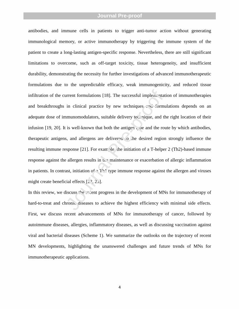
In this review, we discuss the recent progress in the development of MNs for immunotherapy of
hard-to-treat and chronic diseases to achieve the highest efficiency with minimal side effects.
First, we discuss recent advancements of MNs for immunotherapy of cancer, followed by
autoimmune diseases, allergies, inflammatory diseases, as well as discussing vaccination against
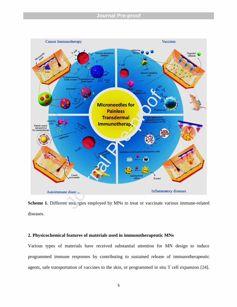
viral and bacterial diseases (Scheme 1). We summarize the outlooks on the trajectory of recent
MN developments, highlighting the unanswered challenges and future trends of MNs for
immunotherapeutic applications.
Jour
nal P
re-p
roof
Journal Pre-proof
5
Scheme 1. Different strategies employed by MNs to treat or vaccinate various immune-related
diseases.
2. Physicochemical features of materials used in immunotherapeutic MNs
Various types of materials have received substantial attention for MN design to induce
programmed immune responses by contributing to sustained release of immunotherapeutic
agents, safe transportation of vaccines to the skin, or programmed in situ T cell expansion [24].
Jour
nal P
re-p
roof
Journal Pre-proof
6
Moreover, using biomaterials within the structure of MNs can trigger a series of pathways for
recruitment or reprogramming of immune cells, which results in more localized immune
responses in comparison with systemic methods [25]. Successful immunotherapy by MNs
strongly depends on the physiochemical properties of materials that are used to fabricate these
needles. The most common materials include silicon [26, 27], metals [28, 29], glasses [30],
ceramics [31], and polymers [32], which their mechanical strength, porosity, charge, and
molecular weight can highly affect antigen stability, antigen or vaccine loading into the MNs or
even control the kinetics of vaccine transportation in vivo [33]. For example, Kathuria et al.
showed that the dissolution rate of polyvinylpyrrolidone (PVP)-based dissolvable MN patches
can be tuned by the incorporation of hydroxypropyl methylcellulose (HPMC) and
methylcellulose (MC) with different molecular weights as dissolution modifier [34]. The PVP
MNs showed a rapid dissolution profile within 0.75 h, while incorporating HPMC with high
molecular weight (K100LV, or K100M) into their structure resulted in dissolution profiles
ranging from 2-2.5 h. Likewise, incorporating low molecular weight HPMCs (E3LV, or E15LV)
into the structure of the PVP MNs resulted in dissolution time of higher than 16 h.
Moreover, fabrication methods of MNs can strongly affect the cost-effectiveness of
immunotherapy. For example, although micromolding is the most common fabrication
technique for dissolving MNs, its efficiency for immunotherapy has been often challenged by
concerns regarding noticeable antigen wastage in this method and lack of cost-saving for
manufacturers [35]. In addition, the selection of a suitable material that contributes to the
localization of antigen within the needles is a substantial issue that needs much attention to
optimize immunotherapy of various diseases by MNs. For example, Prausnitz et al. improved the
localization of active therapeutic molecules to the needles through casting a highly concentrated
Jour
nal P
re-p
roof
Journal Pre-proof
7
polymer solution that was able to increase viscosity or contribute to the incorporation of an air
bubble at the base of the MN to hamper diffusion of therapeutic molecules into the backing [36].
A summary of materials used in the structure of MNs towards immunotherapy, as well as a
comparison of their advantages and disadvantages, are shown in Table 1.
Jo
urna
l Pre
-pro
of
Journal Pre-proof
8
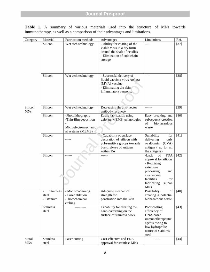
Table 1. A summary of various materials used into the structure of MNs towards
immunotherapy, as well as a comparison of their advantages and limitations.
Category Material Fabrication methods Advantages Limitations Ref.
Silicon Wet etch technology - Ability for coating of the
viable virus in a dry form
around the shaft of needles
- Elimination of cold chain
storage
---- [37]
Silicon Wet etch technology - Successful delivery of
liquid vaccinia virus Ankara
(MVA) vaccine
- Eliminating the skin
inflammatory response
----- [38]
Silicon
MNs
Silicon Wet etch technology Decreasing the anti-vector
antibody response
------ [39]
Silicon -Photolithography
-Thin-film deposition
-
Microelectromechanic
al systems (MEMS)
Easily fabrication using
existing MEMS technologies
Easy breaking and
subsequent creation
of biohazardous
waste
[40]
Silicon
-----
- Capability of surface
decoration of silicon with
pH-sensitive groups towards
burst release of antigen
within 15s
Suitability for
delivering only
ovalbumin (OVA)
antigen ( no for all
the antigens)
[41]
Silicon ------ ------ -Lack of FDA
approval for silicon
- Requiring
extensive
processing and
clean-room
facilities for
fabricating silicon
MNs
[42]
- Stainless
steel
- Titanium
- Micromachining
- Laser ablation
-Photochemical
etching
Adequate mechanical
strength for
penetration into the skin
Possibility of
creating a potential
biohazardous waste
[40]
Stainless
steel
---------- Capability for creating the
nano-patterning on the
surface of stainless MNs
Poor coating
efficiency of
DNA-based
immunotherapeutic
agents owing to
low hydrophilic
nature of stainless
steel
[43]
Metal
MNs
Stainless
steel
Laser cutting Cost-effective and FDA
approval for stainless MNs
----- [44]
Jour
nal P
re-p
roof
Journal Pre-proof
9
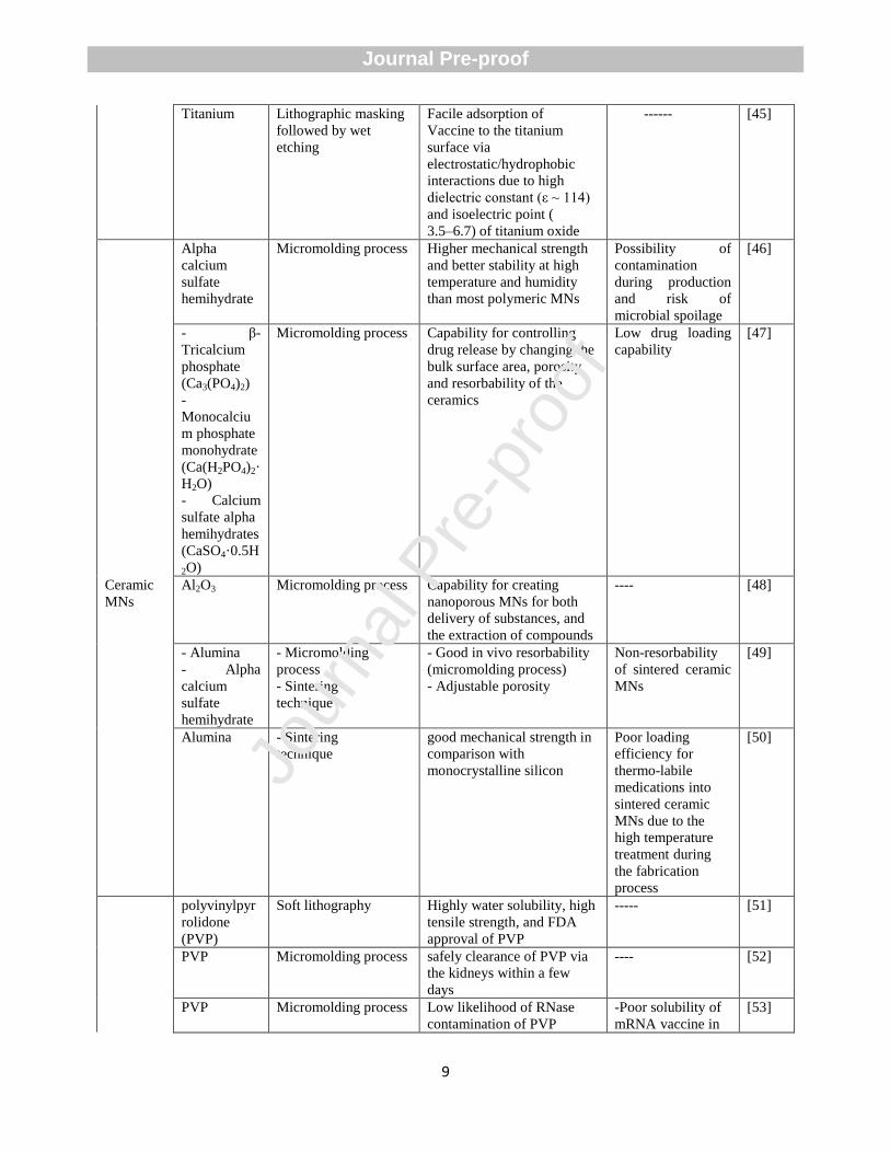
Titanium Lithographic masking
followed by wet
etching
Facile adsorption of
Vaccine to the titanium
surface via
electrostatic/hydrophobic
interactions due to high
dielectric constant (ε ~ 114)
and isoelectric point (
3.5–6.7) of titanium oxide
------ [45]
Alpha
calcium
sulfate
hemihydrate
Micromolding process Higher mechanical strength
and better stability at high
temperature and humidity
than most polymeric MNs
Possibility of
contamination
during production
and risk of
microbial spoilage
[46]
- β-
Tricalcium
phosphate
(Ca3(PO4)2)
-
Monocalciu
m phosphate
monohydrate
(Ca(H2PO4)2·
H2O)
- Calcium
sulfate alpha
hemihydrates
(CaSO4·0.5H
2O)
Micromolding process Capability for controlling
drug release by changing the
bulk surface area, porosity
and resorbability of the
ceramics
Low drug loading
capability
[47]
Ceramic
MNs
Al2O3 Micromolding process Capability for creating
nanoporous MNs for both
delivery of substances, and
the extraction of compounds
---- [48]
- Alumina
- Alpha
calcium
sulfate
hemihydrate
- Micromolding
process
- Sintering
technique
- Good in vivo resorbability
(micromolding process)
- Adjustable porosity
Non-resorbability
of sintered ceramic
MNs
[49]
Alumina - Sintering
technique
good mechanical strength in
comparison with
monocrystalline silicon
Poor loading
efficiency for
thermo-labile
medications into
sintered ceramic
MNs due to the
high temperature
treatment during
the fabrication
process
[50]
polyvinylpyr
rolidone
(PVP)
Soft lithography Highly water solubility, high
tensile strength, and FDA
approval of PVP
----- [51]
PVP Micromolding process safely clearance of PVP via
the kidneys within a few
days
---- [52]
PVP Micromolding process Low likelihood of RNase
contamination of PVP
-Poor solubility of
mRNA vaccine in
[53]
Jour
nal P
re-p
roof
Journal Pre-proof
10
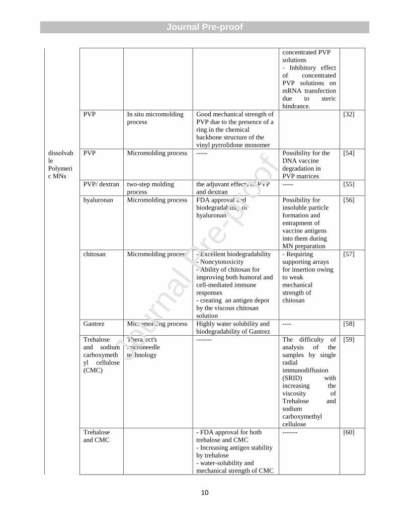
concentrated PVP
solutions
- Inhibitory effect
of concentrated
PVP solutions on
mRNA transfection
due to steric
hindrance.
PVP In situ micromolding
process
Good mechanical strength of
PVP due to the presence of a
ring in the chemical
backbone structure of the
vinyl pyrrolidone monomer
[32]
dissolvab
le
Polymeri
c MNs
PVP Micromolding process ----- Possibility for the
DNA vaccine
degradation in
PVP matrices
[54]
PVP/ dextran two-step molding
process
the adjuvant effects of PVP
and dextran
----- [55]
hyaluronan Micromolding process FDA approval and
biodegradability of
hyaluronan
Possibility for
insoluble particle
formation and
entrapment of
vaccine antigens
into them during
MN preparation
[56]
chitosan Micromolding process - Excellent biodegradability
- Noncytotoxicity
- Ability of chitosan for
improving both humoral and
cell-mediated immune
responses
- creating an antigen depot
by the viscous chitosan
solution
- Requiring
supporting arrays
for insertion owing
to weak
mechanical
strength of
chitosan
[57]
Gantrez Micromolding process Highly water solubility and
biodegradability of Gantrez
---- [58]
Trehalose
and sodium
carboxymeth
yl cellulose
(CMC)
TheraJect's
microneedle
technology
------- The difficulty of
analysis of the
samples by single
radial
immunodiffusion
(SRID) with
increasing the
viscosity of
Trehalose and
sodium
carboxymethyl
cellulose
[59]
Trehalose
and CMC
- FDA approval for both
trehalose and CMC
- Increasing antigen stability
by trehalose
- water-solubility and
mechanical strength of CMC
------- [60]
Jour
nal P
re-p
roof
Journal Pre-proof

11
CMC and
amylopectin
Micromolding process --- Producing
deformed
microneedles with
weak mechanical
strength at high
viscosity of CMC
[61]
Sodium
Alginate
Micromold casting
technique
- Adjuvant properties of
sodium alginate
- Good biocompatibility
and biodegradability
- Utilizing sodium alginate
as a permeation enhancer
---- [62]
Sodium
Alginate
Spin-casting approach ---- Less physical
robustness than
biodegradable
polylactic-co-
glycolic acid MN
[63]
Silk fibroin
/poly(acryli
c acid)
Micromolding process - Good
Biocompatibility and
Biodegradability of silk
fibroin
- Simple one-step process for
loading antigens in silk
protein matrices
- Facile stabilization of
immunotherapeutic agents
and vaccines in silk at room
temperature for more than
two months
- Rapid dissolution of PPA
in the skin
- Sustained vaccine release
from silk protein matrices
(over 1–2 weeks)
- The high
brittleness of silk
fibroin
[33]
poly(lactid
e-
coglycolide)
(PLGA)/PP
A
Micromolding process - Rapid dissolution of
PPA in the skin
- Adjustable sustained
release of encapsulated
vaccines based on the PLGA
molecular weight
- Complexity of
the vaccine loading
process in PLGA
polymer
[64]
Carboxyme
thylcellulos
e and
trehalose as
coating
Dip-coating - Improving antigen stability
during drying by trehalose
disaccharide
- Improving the retention of
Hemagglutination activity of
influenza vaccine after
drying by trehalose
Loss of
Hemagglutination
(HA) activity at
high concentrations
of CMC (1wt%)
[24]
Carboxyme
thylcellulos
e and
trehalose as
coating
Dip-coating --- Increasing
trehalose
crystallization and
vaccine separation
from the trehalose
crystal matrix
during
crystallization
result in
[65]
Jour
nal P
re-p
roof
Journal Pre-proof
12
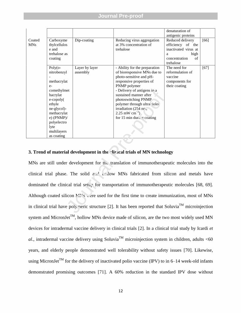
denaturation of
antigenic proteins
Coated
MNs
Carboxyme
thylcellulos
e and
trehalose as
coating
Dip-coating Reducing virus aggregation
at 3% concentration of
trehalose
Reduced delivery
efficiency of the
inactivated virus at
a high
concentration of
trehalose
[66]
Poly(o-
nitrobenzyl
-
methacrylat
e-
comethylmet
hacrylat
e-copoly(
ethyle
ne-glycol)-
methacrylat
e) (PNMP)/
polyelectro
lyte
multilayers
as coating
Layer by layer
assembly
- Ability for the preparation
of bioresponsive MNs due to
photo-sensitive and pH-
responsive properties of
PNMP polymer
- Delivery of antigens in a
sustained manner after
photoswitching PNMP
polymer through ultraviolet
irradiation (254 nm,
2.25 mW cm−2
)
for 15 min during coating
The need for
reformulation of
vaccine
components for
their coating
[67]
3. Trend of material development in the clinical trials of MN technology
MNs are still under development for the translation of immunotherapeutic molecules into the
clinical trial phase. The solid and hollow MNs fabricated from silicon and metals have
dominated the clinical trial setup for transportation of immunotherapeutic molecules [68, 69].
Although coated silicon MNs were used for the first time to create immunization, most of MNs
in clinical trial have polymeric structure [2]. It has been reported that SoluviaTM
microinjection
system and MicronJetTM
, hollow MNs device made of silicon, are the two most widely used MN
devices for intradermal vaccine delivery in clinical trials [2]. In a clinical trial study by Icardi et
al., intradermal vaccine delivery using SoluviaTM
microinjection system in children, adults <60
years, and elderly people demonstrated well tolerability without safety issues [70]. Likewise,
using MicronJetTM
for the delivery of inactivated polio vaccine (IPV) to in 6–14 week-old infants
demonstrated promising outcomes [71]. A 60% reduction in the standard IPV dose without
Jour
nal P
re-p
roof
Journal Pre-proof
13
decreasing in antibody titers was found after using MicronJetTM injection in human
immunodeficiency virus (HIV)-infected patients compared to intramuscular (IM) injection [72].
Although above-mentioned studies show that silicon or metal MNs might possess beneficial
effects on clinical trial outcomes, their clinical translation has been often challenged by concerns
regarding lack of FDA approval for silicon and producing biohazardous sharp wastes by metals
[73]. Currently, polymers are accelerating into the clinic for MN fabrication due to increasing
interest in biocompatible systems [74]. Phase 1 of the clinical trial studies have shown that using
biocompatible and dissolvable polymeric MNs for influenza vaccination was well tolerated and
generated robust antibody responses compared to IM injection [75]. Hirobe et al. reported that
using a self-dissolving MN (MicroHyala; MH) made of hyaluronic acid (HA) and collagen in 20
healthy volunteers enrolled in a clinical study effectively increased antibody titer in comparison
with transcutaneous immunization (TCI) without any severe adverse reactions [76][77].
Likewise, a clinical trials study showed that dissolvable polymeric MNs composed of 50% (w/w)
polyvinyl alcohol (molecular weight 31 kDa) and 50% (w/w) sucrose did not create pain or
swelling in the skin, and only mild erythema localized to the injection site was found after
administration [78]. Considering the above examples, we estimate that in the near future we will
observe a high interest in the design and fabrication of biocompatible and dissolvable polymeric
MN systems for immunotherapeutic applications.
4. Cancer immunotherapy by MN patches
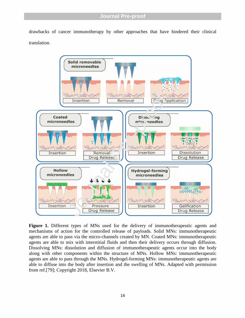
Various types of MNs, including solid removable, coated, dissolving, hollow, and hydrogel-
forming ones, have been proposed (Figure 1) [9, 79], in order to overcome the challenges and
Jour
nal P
re-p
roof
Journal Pre-proof
14
drawbacks of cancer immunotherapy by other approaches that have hindered their clinical
translation.
Figure 1. Different types of MNs used for the delivery of immunotherapeutic agents and
mechanisms of action for the controlled release of payloads. Solid MNs: immunotherapeutic
agents are able to pass via the micro-channels created by MN. Coated MNs: immunotherapeutic
agents are able to mix with interstitial fluids and then their delivery occurs through diffusion.
Dissolving MNs: dissolution and diffusion of immunotherapeutic agents occur into the body
along with other components within the structure of MNs. Hollow MNs: immunotherapeutic
agents are able to pass through the MNs. Hydrogel-forming MNs: immunotherapeutic agents are
able to diffuse into the body after insertion and the swelling of MNs. Adapted with permission
from ref.[79]; Copyright 2018, Elsevier B.V.
Jour
nal P
re-p
roof
Journal Pre-proof
15
In general, current limitations include inadequate infiltration of lymphocytes during the evolution
of tumoral immune escape, the presence of immune checkpoints in the tumor site, the high cost
of immune checkpoint inhibitors, and the possible development of dosage-related autoimmune
side effects through off-target binding of therapeutic agents to healthy tissues [80-83]. In this
section, the discussion is subdivided based on the biological pathways and biological molecules
used for MN-mediated cancer immunotherapy.
4.1. Design of MNs for immune checkpoint inhibition
Tumor cells possess several mechanisms to conceal themselves as ―healthy cells‖ and prevent
their detection and digestion by the immune system. These mechanisms have been summarized
in the review by Liu et al.[84]. Although immune checkpoint molecules display powerful roles
in the prevention of autoimmunity and tissue damage following the immune reaction in the
pathogenic infection, dysregulating their expression in cancer tissue results in innate- and
adaptive immune resistance of tumor [85]. Generally, the programmed cell death protein 1
(PD1), indoleamine 2,3-dioxygenase (IDO), and the cytotoxic T-lymphocyte-associated antigen
4 (CTLA4) are the three most important immune checkpoint molecules that are involved in the
regulation of T-cell function and are extensively used to modulate antitumor immunity [86].
4.1.1. Design of MNs for cancer immunotherapy by IDO blockade
IDO is an enzyme with the ability to degrade essential amino acid, tryptophan, in an independent
process of normal tryptophan homeostasis. IDO is highly expressed in both tumor cells and
stromal cells and contributes to the establishment of peripheral tolerance to tumor antigens. IDO
give power to tumor cells to escape from T-cell-dependent immune attack and improve tumor
Jour
nal P
re-p
roof
Journal Pre-proof
16
survival and outgrowth through the creation of pathogenic inflammatory states [87]. Although
photothermal therapy (PTT) has been reported to be a promising approach for the treatment of
metastatic tumors through alteration of immune response with the help of releasing neoantigens
and damage-associated molecular patterns, the upregulation of IDO by mild heating limits
effective immunotherapeutic outcome. PTT can increase interferon-γ (IFN-γ) secretion and
subsequently enhance the IDO expression in tumor cells and antigen-presenting cells (APCs)
[88]. High expression levels of IDO suppresses APCs activation and decline their antigen-
presenting efficacy [89]. IDO is able to catalyze the degradation of tryptophan into kynurenine,
which, in turn, gives rise to impairment of CD8+ T cells activation and inhibition of their
antitumor ability via increased activity of regulatory T cells (Tregs) [90, 91]. To address these
limitations of PPT, Chen et al. engineered an ingenious core-shell structure MN (CSMNs) array
that was able to synergistically boost robust immune response through intralesional co-delivery
of a photosensitizer and IDO blocking agent [92]. They loaded 1-methyl-tryptophan (1-MT) into
the cross-linked PVP and poly (vinyl alcohol) gel as the MN core and encapsulated
photosensitizer indocyanine green into chitosan nanoparticles (ICG-NPs), followed by
concentrating on the tip shell of MNs. The in vivo experiments in the lung metastatic tumor
model showed that treatment with 1-MT@ICG-NPs-MN+L (laser irradiation) resulted in 1.8-
fold higher percentage of CD8+ T cells in the primary tumors than blank group (Figure 2A and
2B). The immunofluorescence staining demonstrated that that treatment with 1-MT@ICG-NPs-
MN+L significantly increased the amount of CD8+ T cells in the lung slices whereas markedly
reduced the expressions of IFN-γ in the metastatic nodules compared to the blank group (Figure
2C and 2D). Likewise, H&E staining and terminal deoxynucleotidyl transferase dUTP nick-end
labeling staining demonstrated that apoptosis and proliferation inhibition of the cancer cells were
Jour
nal P
re-p
roof
Journal Pre-proof
17
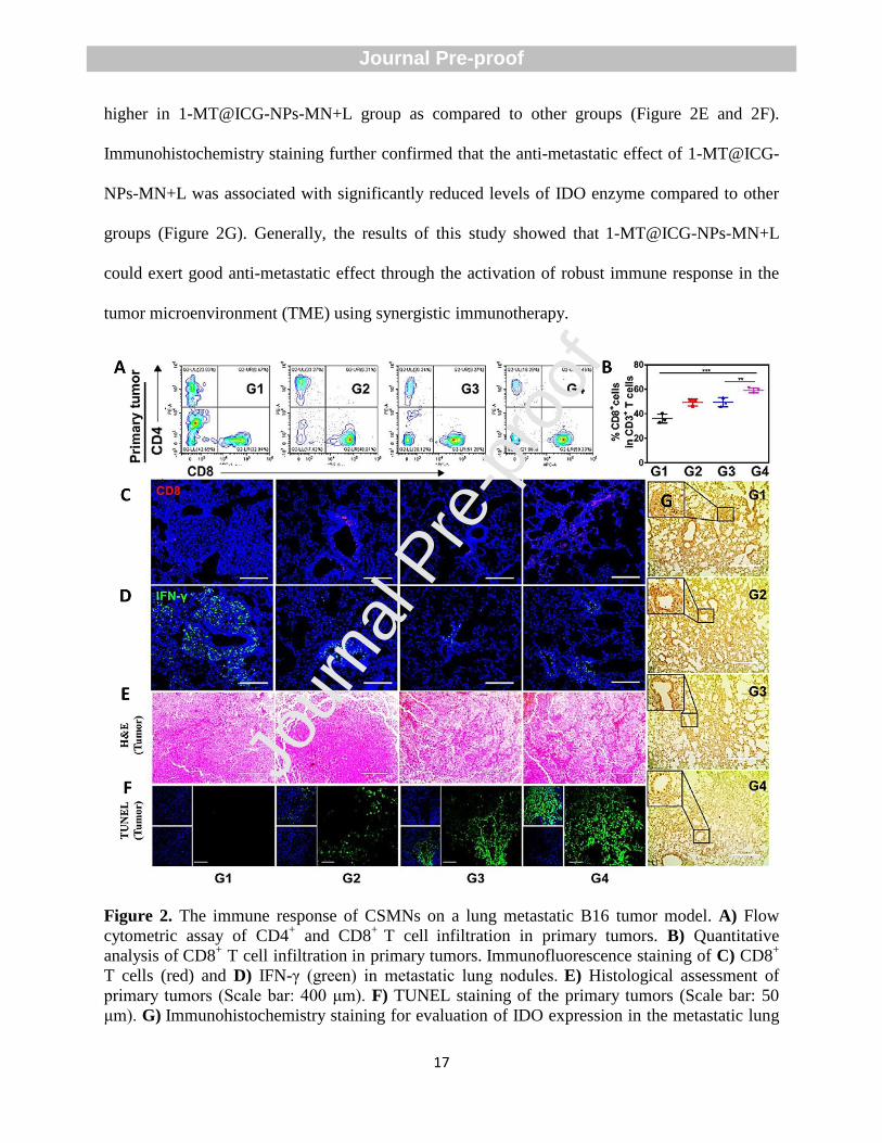
higher in 1-MT@ICG-NPs-MN+L group as compared to other groups (Figure 2E and 2F).
Immunohistochemistry staining further confirmed that the anti-metastatic effect of 1-MT@ICG-
NPs-MN+L was associated with significantly reduced levels of IDO enzyme compared to other
groups (Figure 2G). Generally, the results of this study showed that 1-MT@ICG-NPs-MN+L
could exert good anti-metastatic effect through the activation of robust immune response in the
tumor microenvironment (TME) using synergistic immunotherapy.
Figure 2. The immune response of CSMNs on a lung metastatic B16 tumor model. A) Flow
cytometric assay of CD4+ and CD8
+ T cell infiltration in primary tumors. B) Quantitative
analysis of CD8+ T cell infiltration in primary tumors. Immunofluorescence staining of C) CD8
+
T cells (red) and D) IFN-γ (green) in metastatic lung nodules. E) Histological assessment of
primary tumors (Scale bar: 400 μm). F) TUNEL staining of the primary tumors (Scale bar: 50
μm). G) Immunohistochemistry staining for evaluation of IDO expression in the metastatic lung
Jour
nal P
re-p
roof
Journal Pre-proof
18
nodules of the B16 tumors after various treatments (Scale bar: 200 μm). (G1) Blank, (G2) 1-MT-
MN, (G3) ICG-NPs-MN+L, (G4) 1-MT@ICG-NPs-MN+L. L= (laser irradiation) in G3 and G4
groups (* P<0.05, ** P<0.01, *** P<0.001). Reproduced with permission from ref.[92];
Copyright 2020, American Chemical Society.
4.1.2. Design of MNs for cancer immunotherapy by PD-L1 blockade
The interplay between programmed cell death receptor 1 (PD-1), present on the surface of
activated anti-tumor cytotoxic T-cells, and PD ligand one (PD-L1), found on the surface of
tumor cells, results in facilitating tumor immune escape. Indeed, the interaction between PD-1
and PD-L1 results in the initiation of apoptosis in the tumor-specific cytotoxic T-cell, annealing
their antitumoral immune effects [93-95]. Immune checkpoint inhibitors against PD-1, such as
nivolumab, are routinely employed for the treatment of metastatic melanoma [96]. Nevertheless,
as an expensive therapeutic option, this drug cannot bring optimal results in up to 80% of
patients due to the presence or development of resistance mechanisms or the instauration of off-
site immunotoxicity derived from the systemic administration of the medicines [96, 97]. To
address these limitations, a biodegradable MN was designed to target the PD-1 pathway by
delivering a checkpoint blockade antibody against melanoma locally in the TME [98]. In this
study, Wang et al. embedded the anti-PD-1 antibodies (aPD1) and glucose oxidase
(GOx)/catalase (CAT) enzymatic system within pH-sensitive nanoparticles composed of ketal
modified dextran and then loaded it into HA-based MNs (Figure 3A and 3B) [98]. Various
microscopic methods (scanning electron microscope (SEM) and fluorescence) confirmed the
distribution of the loaded pH-sensitive NPs at the tips of the MNs (Figure 3C–3E). In vivo
experiments demonstrated that the MNs were capable of penetrating the TME to a depth of
approximately 200 μm (Figure 3F–3H). The MN patch used in this study showed a sustained
release profile and improved retention of aPD1 into the TME. The release profile was modulated
Jour
nal P
re-p
roof
Journal Pre-proof
19
by the CAT enzymatic system: the enzymes convert glucose into gluconic acid, decreasing the
pH in their immediate surrounding, dissolving the NPs and releasing the antibody. The MN patch
elicited a robust immune response against B16F10 mouse melanoma in comparison to free aPD1,
completely eradicating the tumors in some of the animals treated. Their results showed that
treatment with this aPD1 patch resulted in the survival of 40% of mice after 40 days, while all
the mice treated with free aPD1 or a MN patch without the CAT enzyme died within 30 days
after treatment due to tumor relapsing.
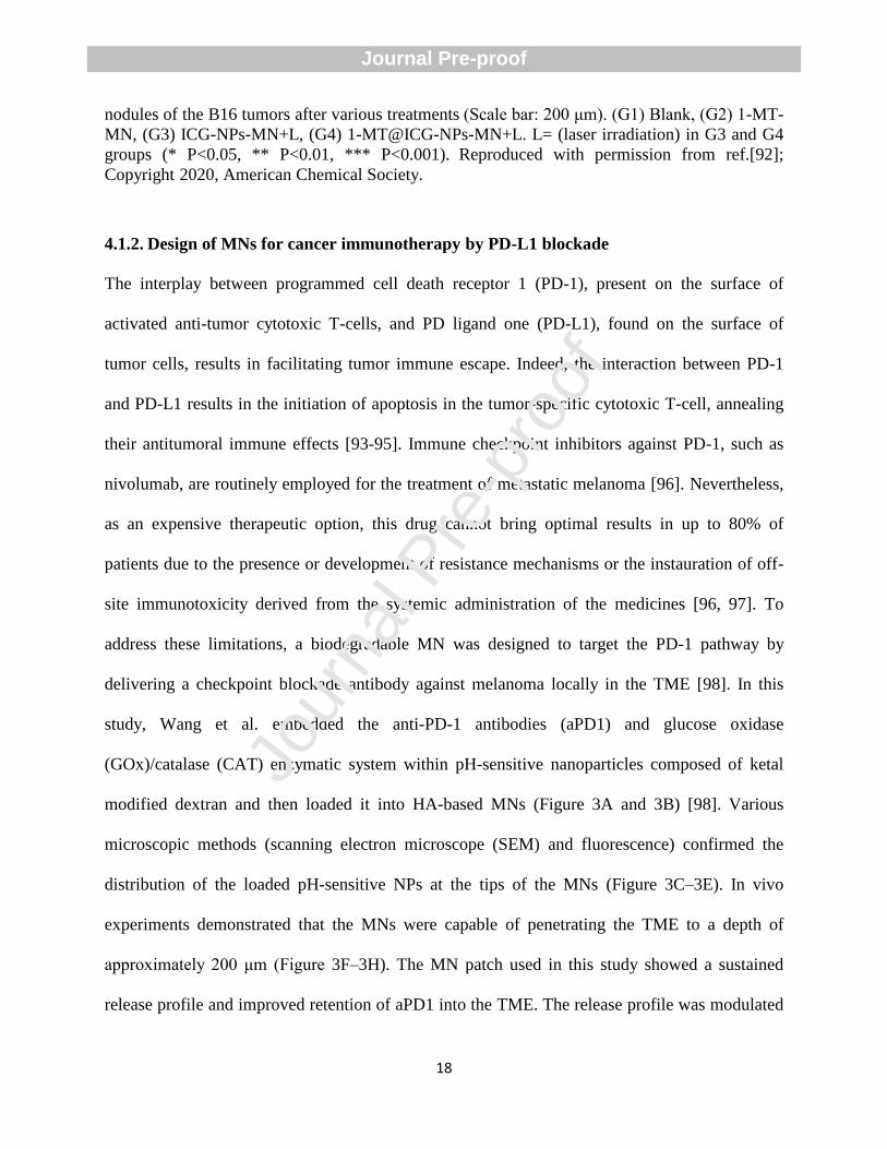
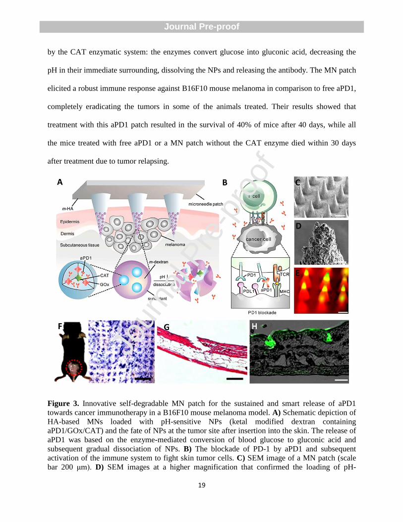
Figure 3. Innovative self-degradable MN patch for the sustained and smart release of aPD1
towards cancer immunotherapy in a B16F10 mouse melanoma model. A) Schematic depiction of
HA-based MNs loaded with pH-sensitive NPs (ketal modified dextran containing
aPD1/GOx/CAT) and the fate of NPs at the tumor site after insertion into the skin. The release of
aPD1 was based on the enzyme-mediated conversion of blood glucose to gluconic acid and
subsequent gradual dissociation of NPs. B) The blockade of PD-1 by aPD1 and subsequent
activation of the immune system to fight skin tumor cells. C) SEM image of a MN patch (scale
bar 200 μm). D) SEM images at a higher magnification that confirmed the loading of pH-
Jour
nal P
re-p
roof
Journal Pre-proof
20
sensitive NPs into MNs (scale bar 5 μm). E) Fluorescence imaging of MNs containing FITC-
antibody loaded NPs (scale bar 200 μm). F) Desired region for the insertion of MNs on the
mouse skin (red dashed line) and proof of effective delivery by trypan blue staining (scale bar 1
mm). G) H&E stain of mouse skin region penetrated by MN (scale bar 200 μm). H) Merged
fluorescence and bright field image demonstrating the presence of FITC-antibody loaded MN in
mouse skin after insertion (green: aPD1) (scale bar 200 μm). Reproduced with permission from
ref.[98]; Copyright 2016, American Chemical Society.
In another study, a transdermal hollow structured MN array (MNA) patch was designed to
enable cold atmospheric plasma (CAP)-mediated aPD-L1 therapy [99]. The application of CAP
improved the transportation of the payload into the TME by the MN patch. Furthermore, CAP
was channeled to the tumor, inducing the death of cancer cells and the release of tumor-
associated antigens, which resulted in the significant maturation of dendritic cells (DCs) and
presentation of the antigens to T-cells in the draining lymph nodes. The release of tumor-
associated antigens combined with the simultaneous release of aPD-L1 antibody controlled the
tumor growth in both primary tumors and distant tumors, demonstrating the effective priming of
a systemic antitumoral immune response. Since the therapeutic potential of various approaches
might be undermined due to the diversity, complexity, and heterogeneity of tumors, MNs can
provide a paradigm shift for combination therapy to enhance treatment efficacy [100]. For
example, Yanqi et al. developed a transcutaneous delivery platform for aPD1. The technology
combined nanocapsules of HA modified with 1-MT, an inhibitor of IDO, embedded within MNs
[101]. The IDO enzyme is responsible for maintaining DCs in an immature state, suppressing
antigen-specific T cell proliferation by increasing their sensitivity to apoptosis [102].
Furthermore, this enzyme has been correlated with an increased number of anti-inflammatory
Tregs [103]. The TME is characterized by the overexpression of hyaluronidase, which controls
the release of aPD1 through the enzymatic digestion of HA-based MNs. In vivo experiments on a
B16.F10 mouse model of melanoma showed that the MNs induced a significant increase in the
amount of T-cell, and, in particular, in CD8+ cytotoxic T-cells, while reducing the number of
Jour
nal P
re-p
roof
Journal Pre-proof
21
Tregs in the TME. However, the authors did not evaluate whether the increased presence of
cytotoxic T-cells is correlated with an increase in the number of antigen-specific cytotoxic T-
cells.
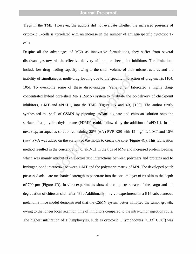
Despite all the advantages of MNs as innovative formulations, they suffer from several
disadvantages towards the effective delivery of immune checkpoint inhibitors. The limitations
include low drug loading capacity owing to the small volume of their microstructures and the
inability of simultaneous multi-drug loading due to the specific interaction of drug-matrix [104,
105]. To overcome some of these disadvantages, Yang et al. fabricated a highly drug-
concentrated hybrid core-shell MN (CSMN) system to facilitate the co-delivery of checkpoint
inhibitors, 1-MT and aPD-L1, into the TME (Figure 4A and 4B) [106]. The author firstly
synthesized the shell of CSMN by pipetting sodium alginate and chitosan solution onto the
surface of a polydimethylsiloxane (PDMS) mold, followed by the addition of aPD-L1. In the
next step, an aqueous solution containing 25% (w/v) PVP K30 with 15 mg/mL 1-MT and 15%
(w/v) PVA was added on the surface of the molds to create the core (Figure 4C). This fabrication
method resulted in the concentration of aPD-L1 in the tips of MNs and increased protein loading,
which was mainly attributed to electrostatic interactions between polymers and proteins and to
hydrogen-bond interaction between 1-MT and the polymeric matrix of MN. The developed patch
possessed adequate mechanical strength to penetrate into the corium layer of rat skin to the depth
of 700 μm (Figure 4D). In vitro experiments showed a complete release of the cargo and the
degradation of chitosan shell after 48 h. Additionally, in vivo experiments in a B16 subcutaneous
melanoma mice model demonstrated that the CSMN system better inhibited the tumor growth,
owing to the longer local retention time of inhibitors compared to the intra-tumor injection route.
The highest infiltration of T lymphocytes, such as cytotoxic T lymphocytes (CD3+ CD8
+) was
Jour
nal P
re-p
roof
Journal Pre-proof
22
found in the tumor site of mice treated with the aPD-L1/1-MT CS-CSMN in comparison with
blank CS-CSMN control (Figure 4E and 4F).
Figure 4. A highly drug-concentrated hybrid CSMN system for co-delivery of checkpoint
inhibitors, including 1-MT and aPD-L1 and cancer immunotherapy in a B16 subcutaneous
melanoma mice model. A) Schematic representation of CSMN-assisted co-delivery of aPD-L1
and 1-MT for the treatment of melanoma. B) A representative photograph of the CS-CSMN
patch. C) Bright-field image of CS-CSMN (scale bar 500 μm). D) H&E staining image of rat
skin after insertion of CS-CSMN patch (scale bar 100 μm). The numbers of E) CD3+ T cells and
F) CD3+ CD8
+ T cells per 10,000 of cells in tumor tissue after the removal of red blood cells
at12 days after treatment. Data are expressed as Mean ±SD (n = 3 animals per group). ** P<0.0
and *** P<0.001 versus the aPD-L1/1-MT CS-CSMN group. Reproduced with permission from
ref.[106]; Copyright 2020, Acta Materialia Inc. Published by Elsevier Ltd.
Jour
nal P
re-p
roof
Journal Pre-proof
23
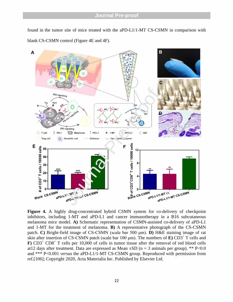
In another interesting example, Lan et al. developed a MN patch loaded with pH-responsive
tumor-targeted lipid NPs, which provide the possibility of local delivery of aPD-1 and cisplatin
(CDDP) to create a synergistic immuno-chemotherapy [107]. They firstly synthesized the aPD-
1/CDDP@NPs using a reverse-phase microemulsion technique. Then, aPD-1/CDDP@NPs were
further encapsulated into dissolving MNs made of PVP using the molding method (Figure 5A).
Lipid coated NPs were able to facilitate drug release and tumor-targeting. The in vivo
experiments showed that MN group cause the most notable tumor regression (strongest effect on
reducing tumor volume and tumor weight) in comparison with PBS, aPD-1, aPD1 + CDDP, and
aPD-1/CDDP@NPs groups (Figure 5B and 5C). Additionally, the authors investigated body
weight loss, the blood urea nitrogen (BUN) value, and total immunoglobulin G (IgG) value in the
serum to assess the systemic toxicity and side effects of engineered MNs. Their results showed
that the BUN values in all the MN patch groups were within the normal range (Figure 5D).
Likewise, IgG values were markedly increased in the aPD-1/CDDP@NP MNs group compared
to other groups (Figure 5E). Histopathological assessments using H&E staining indicated that the
CDDP group and the aPD-1 plus CDDP group exhibited severe toxic tubular necrosis (the
glomeruli and Bowman’s capsule collapsing), while no evidence of renal damage was seen in
mice treated with aPD-1, aPD-1 MNs, and aPD-1/CDDP@NP MNs. Jour
nal P
re-p
roof
Journal Pre-proof
24
Figure 5. MNs loaded with anti-PD-1–cisplatin NPs for synergistic cancer
immunochemotherapy. A) Schematic representation of MNs loaded with aPD-1/CDDP@NPs
towards synergistic cancer immunochemotherapy. The combination of chemotherapy and
immunotherapy was carried out through encapsulation of aPD-1 and CDDP into NPs and then
embedding into the MNs. The aPD-1 was able to block the binding of PD-L1 to PD-1 that
confers the activation of T-cells whereas intracellular release of CDDP facilitated the death of
tumor cells via inducing direct cytotoxicity to them. B) Tumor volume and (C) tumor weight
following sacrifice. When the tumor volume reached 10 mm3, the treatment of each group was
started (Three treatments for each group and lasted for three cycles). The measurements of tumor
volume were performed before the treatment and 3 times after the treatment. D) The BUN value
and E) total IgG value in the serum. Data were expressed as ± S.D (n = 7 per group). Statistical
analysis was carried out based on the Mann–Whitney U test (P-value: *p < 0.05, ***p < 0.001).
Reproduced with permission from ref. [107]; Copyright 2020, The Royal Society of Chemistry.
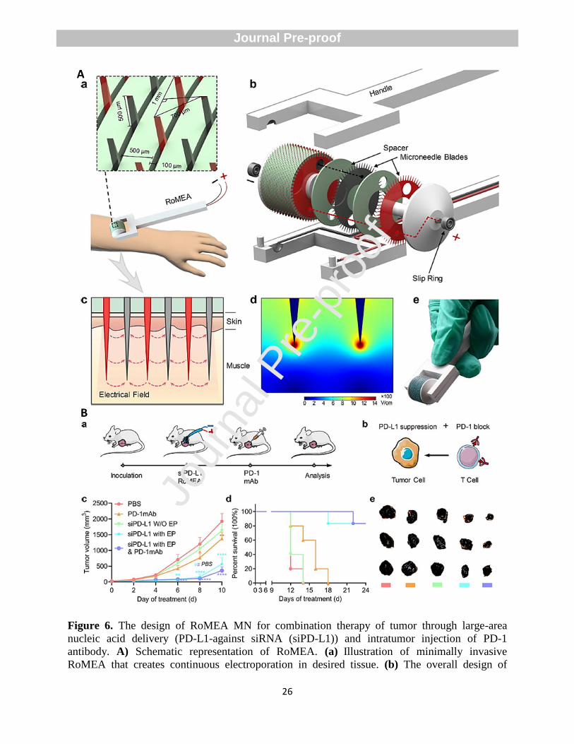
In another study by Yang et al., a rolling MN electrode array (RoMEA) was engineered to inhibit
tumor growth in both B16F10 and CT26 xenograft murine models [108]. RoMEA was able to
conduct low-voltage and large-area nucleic acid delivery (PD-L1-against siRNA (siPD-L1))
Jour
nal P
re-p
roof
Journal Pre-proof
25
through electroporation without any noticeable damage to the skin (Figure 6A). The MN
electrode array contributed to reducing the pulse voltage by penetrating via high-resistance
stratum corneum layer. The penetration depth of RoMEA was about 500 µm that was larger than
the epidermal skin thickness of mice (200 µm), ensuring MN entrance into subcutaneous tissue.
The researchers employed anti-PD-1 monoclonal antibody or CpG oligodeoxynucleotides
(CpGODNs) of CpG 2395 (immunoadjuvant) to synergistically improve the antineoplastic
effects through blocking PD-L1/PD-1 recognition between tumor cells and T cells and increasing
populations of CD8 + T cells and CD4
+ T cells. For combination therapy using siPD-L1 and PD-
1mAb, the antibody was infused into the tumor after siRNA electroporation to synergistically
block PD-1 on lethal T cells (Figure 6B, parts a and b). In vivo experiments showed that siPD-L1
without electric pulse stimulation failed to create protective effects and exhibited similar tumor
growth and survival profiles to PBS, all mice died within two weeks (Figure 6B, parts c-e).
Although treatment with only anti-PD-1 antibody repressed tumor growth and reduced tumor
volume, the differences did not reach significant levels, and the survival time was increased to 18
days. The animals treated with siPD-L1 by RoMEA or in combination with anti-PD-1 antibody
injection exhibited the highest inhibition of tumor growth, survival profiles, and reducing tumor
volume (Figure 6B, parts c-e). Jour
nal P
re-p
roof
Journal Pre-proof
26
Figure 6. The design of RoMEA MN for combination therapy of tumor through large-area
nucleic acid delivery (PD-L1-against siRNA (siPD-L1)) and intratumor injection of PD-1
antibody. A) Schematic representation of RoMEA. (a) Illustration of minimally invasive
RoMEA that creates continuous electroporation in desired tissue. (b) The overall design of
Jour
nal P
re-p
roof
Journal Pre-proof

27
RoMEA device that shows the stainless steel 316 parallel circular blades with MN arrays on edge
as the electrodes. Two adjacent MN blades connect to the anode (red) and cathode (black),
respectively. (c) Schematic representation of MN electroporation. (d) Simulation of electric field
using the RoMEA (50 V). (e) The RoMEA prototype in hand. B) RoMEA-mediated
immunotherapy in the B16F10 model and its effects on tumor growth and survival profiles. (a)
Schematic representation of the experiment protocol in a melanoma tumor model in C57BL/6
mice. (b) Conceptual design of combined immunotherapy approach used in this study. (c and d)
The tumor growth and survival curves of the mice during the treatment period. (e)
Representative optical images of the melanoma tumors excised on day 14 post-treatment.
Information about experimental groups: PBS (control without any treatment), PD-1mAb (mice
treated with only PD-1mAb), siPD-L1 W/O EP (siPD-L1 without electroporation), siPD-L1 with
EP (siPD-L1 electroporated with RoMEA), and siPD-L1 with EP & PD-1mAb (siPD-L1
electroporated with RoMEA plus PD-1 mAb). Data were expressed as the mean ± SEM. * P <
0.05; ** P < 0.01;*** P < 0.001, **** P < 0.0001 vs the control group. Reproduced with
permission from ref.[108]; Copyright 2020, Elsevier B.V.
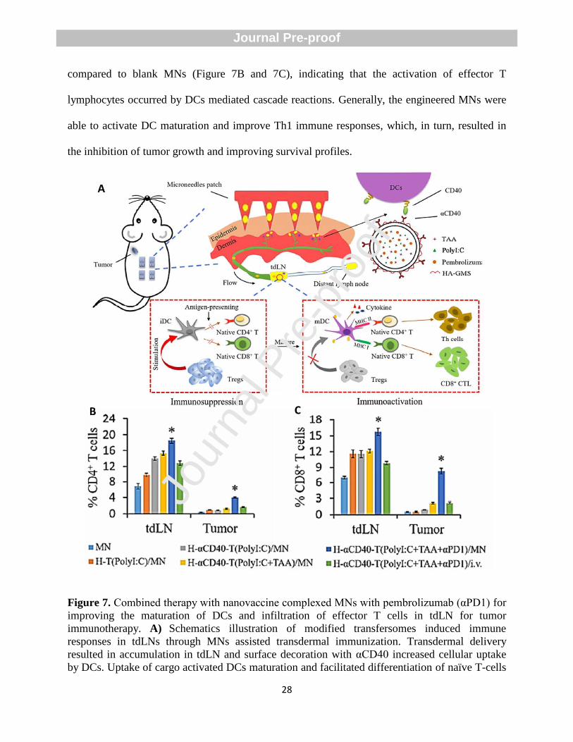
In 2020, Zhou et al. engineered HA MNs loaded with anti-CD40 antibody (DCs targeting
antibody) and surface decorated by GMS (monostearin)-conjugated HA transfersomes co-
encapsulating poly I:C adjuvant, the tumor-associated antigen (tyrosinase-related protein-2),
ovalbumin (OVA), and pembrolizumab (anti-PD1 antibody) to improve the maturation of DCs
and infiltration of effector T cells in tumor-draining lymph node (tdLN) and tumor tissue towards
synergistic reinforcement of anti-PD1 immunotherapy (Figure 7A) [109]. The authors monitored
intradermal fluorescence intensity to evaluate the distribution of transfersomes (DiI labeled) in
vivo. The fluorescence distribution was investigated in lymph nodes, hearts, livers, spleens, and
kidneys to ascertain tdLNs targeting capacity of transfersomes based nanovaccine- complexed
MNs. At 48 h after H-αCD40 T(OVA+PolyI:C)/MNs transdermal administration, the DiI
fluorescence appeared at the lymph node and reached the maximum at 96 h, indicating
accumulation of the transfersomes in the lymph nodes. Likewise, a gradual increase of the
fluorescence intensity was found in livers after 96 h. The highest photon quantum intensity was
seen in lymph nodes (~25 × 104/g; 2-fold higher than livers at 96 h). A significant increase in the
number of CD4+ T or CD8
+ T cells was found in H-αCD40-T(PolyI:C+TAA+αPD1)/MN
Jour
nal P
re-p
roof
Journal Pre-proof
28
compared to blank MNs (Figure 7B and 7C), indicating that the activation of effector T
lymphocytes occurred by DCs mediated cascade reactions. Generally, the engineered MNs were
able to activate DC maturation and improve Th1 immune responses, which, in turn, resulted in
the inhibition of tumor growth and improving survival profiles.
Figure 7. Combined therapy with nanovaccine complexed MNs with pembrolizumab (αPD1) for
improving the maturation of DCs and infiltration of effector T cells in tdLN for tumor
immunotherapy. A) Schematics illustration of modified transfersomes induced immune
responses in tdLNs through MNs assisted transdermal immunization. Transdermal delivery
resulted in accumulation in tdLN and surface decoration with αCD40 increased cellular uptake
by DCs. Uptake of cargo activated DCs maturation and facilitated differentiation of naïve T-cells
Jour
nal P
re-p
roof
Journal Pre-proof
29
into effector T lymphocyte. In the next step, reducing regulatory T lymphocytes conferred
reversion of immunosuppressive microenvironment in tdLN into immune activation. B) Representative fluorescence images indicating retention of DiI labeled transfersomes in various
tissues in vivo at 48, 96, 120, and 168 h after insertion of transfersomes complexed MNs. C) and
D) Flow cytometry assay of the percentages of CD4+ T or CD8
+ T cells among all lymph nodes
or tumor tissue cells (n = 4). Data are expressed as mean ± SD. *p < 0.05, compared with MN. Reproduced with permission from ref.[109]; Copyright 2020, Springer Nature.
4.1.3. Design of MNs for cancer immunotherapy by CTLA-4 blockade
CTLA-4 is known as a protein receptor that acts as an immune checkpoint. CTLA-4 is expressed
on T cells in lymph nodes, and its physiological interaction with APCs results in suppressing the
activation of T-cells and the inflammatory response [82, 110]. Antibodies directed against
CTLA-4 have been the first treatments in the class of immune checkpoint inhibitors to gain
approval for the treatment of melanoma [111]. However, this target is systemically expressed,
and the interference with this signal induces the emergence of immune-related side effects [112].
In order to improve the local delivery of anti- CTLA-4 and hinder systemic drug exposure,
nanotopography-based MN array (MNA) can be applied. As an example, Kwon et al. designed a
nanotopography-based MNA composed of a single-use, 66 mm2 arrays of 100 MNs of 110 μm
diameter, 350 μm long, and with a 30 μm hole located off-center (named SOFUSATM
) to allow
the delivery of anti- CTLA-4 into tdLN in an orthotopic mammary carcinoma murine model
[113]. Repeated treatment with SOFUSATM
inhibited tumor growth and metastasis development
in bone, lymph nodes, and lungs better than the traditional systemic administration, an
intraperitoneal administration (IP) of anti-CTLA-4 at 10 mg/kg. Moreover, the authors evaluated
the transport of liquid from SOFUSATM
to the brachial lymph nodes by infusing 100 μL/h of
indocyanine green (ICG) into the epidermal spaces using near-infrared fluorescence imaging
(NIRF). The lymphatic vessels collect the dye and transport it to the lymph nodes.
Jour
nal P
re-p
roof
Journal Pre-proof
30
As mentioned above about cancer immunotherapy by PD-L1 blockade, a single treatment
modality might be unable to efficiently treat malignant skin melanoma. To this end, Chen et al.
designed a physiologically self-degradable MN-assisted platform to combine immunotherapy
and photodynamic therapy (PDT) through controlled co-delivery of checkpoint inhibitor anti-
CTLA4 antibody (aCTLA4) and photosensitizer (PS) and create synergistic effects against
tumors [114]. The hydrophobic (Zinc Phthalocyanine) and hydrophilic agents (aCTLA4) were
synchronically encapsulated into the pH-sensitive dextran NPs using a double emulsion
water/oil/water (w/o/w) evaporation method. The acetylation of the dextran pendant hydroxyl
moiety resulted in the co-loading of hydrophobic photosensitizer/hydrophilic antibodies into the
NPs. UV-Vis spectra of co-encapsulated NPs showed an absorption peak around 490 nm that
was an indicator of the successful encapsulation of aCTLA4-FITC. Then, the pH-sensitive
dextran NPs were embedded into the biocompatible HA MNs. In vivo experiments in 4T1 mouse
models showed that three-times of MN insertion in combination with laser resulted in sustained
tumor inhibition, while other treatment groups failed to create this outcome. The authors stated
that the first destroying of partial tumor by PDT resulted in the initiation of the immune response
that in turn, facilitated aCTLA4-mediated immunotherapy in the next step. Additionally,
preliminary systemic assessments demonstrated that the engineered MNs had favorable safety
without causing any systemic immune disorder.
4.2. Design of MNs for the delivery of therapeutic cancer vaccines
4.2.1. MNs for effective delivery of DNA cancer vaccines
Jour
nal P
re-p
roof
Journal Pre-proof
31
Unique properties of DNA vaccine technology, including stability, simplicity, and safety, make
them attractive immunotherapeutic approaches to treat cancers [115]. In this technology, the
resulting immune responses can be manipulated by designing genes in DNA vaccines to encode
immunomodulatory molecules and various antigens [116]. Currently, several research groups
loaded DNA vaccines in the MNs to treat various cancers [52, 117, 118].
The poor targeting of APCs and the lack of appropriate adjuvants have been major limitations in
the transdermal delivery of a DNA vaccine for cancer immunotherapy [119]. To circumvent
these shortcomings, Xu et al. fabricated a MN composed of a DNA vaccine in a polymeric
nanocomplex, encapsulating a low concentration of paclitaxel (PTX) [120]. This MN patch was
developed as a DC-targeted transdermal strategy for cancer immunotherapy, exploiting low-dose
PTX as an adjuvant. They firstly synthesized a DNA plasmid that encodes GM-CSF, a DC
chemoattractant cytokine, and a fusion protein of tyrosinase-related protein-2 (Trp-2), as a
melanoma tumor antigen. In order to facilitate targeting to the mannose receptors present on
DCs, the resulting plasmid was incorporated into a mannosylated N,N,N-trimethyl chitosan
(mTMC) solution. At the same time, PTX at a low concentration was encapsulated in
sulfobutylether-β-cyclodextrin (SBE), as a solubility enhancer and polyanionic linker. In the next
step, ionic interactions between the cationic complex of mTMC/DNA and the negatively charged
inclusion complex of PTX/SBE resulted in the creation of the PTX/SBE-mTMC/DNA
nanocomplex. This DC-targeted nanocomplex can efficiently improve the maturation of DCs,
with an increase in the secretion of IL-12p70. Furthermore, DCs pulsed with the nanocomplex
enhance the proliferation of CD4+ and CD8
+ T cells, as well as decrease the percentage of
immunosuppressive FoxP3+ Tregs. The co-delivery of DNA vaccine, mannosylated chitosan,
and PTX as a combination of antigen and adjuvant results in stronger suppression of tumor
Jour
nal P
re-p
roof
Journal Pre-proof
32
growth compared to only DNA vaccine or only mannosylated chitosan/PTX in in vivo
experiments in mice. It has been reported that incorporation of pH-responsive copolymers into
the structure of MNs can accelerate the release of DNA vaccine in tumor acidic pH
microenvironment [121]. Recently, Duong et al. designed and fabricated a smart DNA vaccine
delivery system using polycarbonate MNs coated by layer-by-layer (LbL) deposition. The two
layers are composed of positively charged ultra-pH-responsive oligo sulfamethazine conjugated
poly(β-amino ester urethane) (OSM-(PEG-PAEU)) and a negatively charged immunostimulatory
adjuvant, polyriboinosinic:polyribocytidylic (poly(I:C)), at low pH, to facilitate the controlled
release of DNA vaccines and adjuvants in the immune cell-rich epidermis/dermis layer of the
skin [122]. It was shown that the pH-sensitivity of OSM-(PEG-PAEU) led to the protonation of
the copolymer to positively charge at pH 4.0, capable of forming a complex with poly(I:C) and
hampering the release of the OVA- expressing plasmid (pOVA) and poly(I:C) from the LbL
coated MNs. In addition, the presence of ionized sulfonamide moieties in the OSM oligomers
resulted in the deprotonation of OSM-(PEG-PAEU) copolymer at the physiological pH (pH 7.4),
resulting into a negative charge and facilitating the release of pOVA and poly(I:C) via
electrostatic repulsion. The combined delivery of the DNA vaccine and adjuvant by the LbL
coated MNs resulted in the induction of type I interferons followed by the production of antigen-
specific antibodies by B cells and the priming of CD8+ T-cell. The antigen-specific CD8
+ T-cells
induced the production of interferon-gamma (IFN-γ) and enhanced cancer cell death. In vivo
experiments showed that the LbL-coated MNs resulted in higher OVA protein expression
compared to the group where the plasmid was subcutaneously injected, which may be attributed
to the penetration of MNs, of ca. 600 μm height, to the dermis, and the presentation of
nanoengineered DNA polyplex to antigen-presenting cells. Compared to soluble DNA vaccine
Jour
nal P
re-p
roof
Journal Pre-proof
33
formulation, implantation of the LbL-coated MNs loaded with pOVA and poly(I:C) in mice
caused a threefold increase in IFN-γ positive tumor-infiltrating CD8+ T cells and threefold higher
amounts of anti-OVA IgG serum antibody, indicating suitable stimulation of the humoral and
cellular immune response. In another study by the same group, an array of dissolving MN was
prepared using a bioresorbable polypeptide matrix (mPEG5K–PN2LG30), with nanosized
polyplexes composed of pOVA and poly (I:C), which were loaded into high-transfection cationic
amphiphilic conjugates (DA3) and added to the polypeptide matrix (Figure 8A) [123]. The
positive charge of polyplex improved its uptake by DC 2.4 cells and RAW 264.7 macrophage
cells (Figure 8B and 8C) and created a repulsive force on the other cationic copolymers, which
resulted in burst release of ca. 85% of poly(I:C) and ca. 97% of pOVA within 5 min of
application of the MN. This indicated the suitability of the MNs for future clinical use owing to
the short treatment time with improvement of patient convenience. Additionally, the MN
dissolved in the interstitial fluids of skin, mainly due to the presence of the polyethylene glycol
(PEG) component in its structure. In vivo and in vitro experiments demonstrated the higher
expression of OVA in the dissolving MN group compared to control and subcutaneous injection
groups (Figure 8D), suggesting that the nanopolyplex can reach APCs, such as DCs and
macrophage cells in the epidermis/dermis layers, and can be effectively captured by them. The
administration of pOVA through dissolving MNs resulted in higher antibody titer (Figure 8E and
8F) while reducing the number of OVA-expressing metastatic foci through antibody-dependent
cellular cytotoxicity (ADCC) activity, prolonging the overall survival compared to traditional
vaccination.
Jour
nal P
re-p
roof
Journal Pre-proof
34
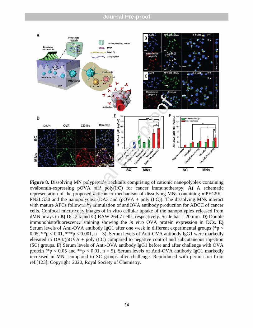
Figure 8. Dissolving MN polypeptide cocktails comprising of cationic nanopolyplex containing
ovalbumin-expressing pOVA and poly(I:C) for cancer immunotherapy. A) A schematic
representation of the proposed anticancer mechanism of dissolving MNs containing mPEG5K–
PN2LG30 and the nanopolyplex (DA3 and (pOVA + poly (I:C)). The dissolving MNs interact
with mature APCs followed by stimulation of antiOVA antibody production for ADCC of cancer
cells. Confocal microscopy images of in vitro cellular uptake of the nanopolyplex released from
dMN arrays in B) DC 2.4 and C) RAW 264.7 cells, respectively. Scale bar = 20 mm. D) Double
immunohistofluorescence staining showing the in vivo OVA protein expression in DCs. E)
Serum levels of Anti-OVA antibody IgG1 after one week in different experimental groups (*p <
0.05, **p < 0.01, ***p < 0.001, n = 3). Serum levels of Anti-OVA antibody IgG1 were markedly
elevated in DA3/(pOVA + poly (I:C) compared to negative control and subcutaneous injection
(SC) groups. F) Serum levels of Anti-OVA antibody IgG1 before and after challenge with OVA
protein (*p < 0.05 and **p < 0.01, n = 5). Serum levels of Anti-OVA antibody IgG1 markedly
increased in MNs compared to SC groups after challenge. Reproduced with permission from
ref.[123]; Copyright 2020, Royal Society of Chemistry.
Jour
nal P
re-p
roof
Journal Pre-proof
35
4.2.2. MNs for effective delivery of other cancer vaccines
One of the primary objectives of cancer immunotherapy is the establishment of a broad tumor-
targeting T cell repertoire that is able to recognize and destroy heterogeneous tumor cell
populations. One promising strategy is in situ vaccination that contributes to starting a selective
and durable adaptive immune response using the diverse collection of tumor antigens within the
tumor. Moreover, in situ vaccination plays an important role in reprogramming the TME toward
an immunostimulatory state. The high interstitial fluid pressure of the TME can act as a barrier
for immunotherapeutic agents to enter into the tumor. To address this limitation, Boone et al.
developed an autonomous active MN for the direct transportation of cowpea mosaic virus NPs
(CPMV) as potent immunoadjuvants towards the treatment of B16F10 melanoma in mice [124].
In this system, magnesium (Mg) microparticles were embedded into active MNs to facilitate the
entrance of the NP payload into the tumor through their reaction with the interstitial fluid in the
TME and creating a propulsive force. In vivo experiments demonstrated that active MNs strongly
increased tumor regression, improved survival profiles of tumor-bearing mice, and represented
enrichment in the CD8+ T cell population. As mentioned in the previous section, combination
therapy of cancer using immune-stimulating antigens and other techniques can be considered as
a valid approach to more effectively treat cancers than monotherapy. One of the promising
strategies to improve the anti-tumor immune responses is the vaccination with whole tumor
antigens derived from whole cell lysates. In addition to whole tumor antigens, the whole cell
lysates possess melanin that can as a photosensitizer for heat generation and subsequent PTT of
cancer [125]. As an example, Ye et al. reported the design and fabrication of a transdermal
cancer vaccine MN patch based on cross-linked HA materials [126]. The polymeric MNs were
Jour
nal P
re-p
roof
Journal Pre-proof
36
loaded with B16.F10 whole tumor lysate containing melanin and granulocyte-macrophage
colony-stimulating factor (GM-CSF). In addition to the sustained release of lysate into the skin,
melanin can act as a photosensitizer for heat generation through localized near-infrared light
(NIR) irradiation, while the presence of GM-CSF enhances the recruitment and activation of
immune cells. In vitro experiments demonstrated that 10 min of NIR irradiation provided an
optimal activation of matured DCs (CD80+/CD86
+ cells, increasing from 36.7 ± 2.3% to 48.9 ±
3.1% after 10 min) and did not reduce their viability and functionality. In vivo experiments in a
prophylactic set up in B16.F10 models indicated that the combined therapy of MNs loaded with
tumor lysate and GM-CSF with NIR irradiation resulted in complete tumor rejection in 87% of
the treated mice and long-term survival. Furthermore, the authors analyzed also the
immunological mechanisms responsible for the vaccination efficacy by testing the MNs in
animals genetically modified not to express lymphocytes (both T- and B- cells) to evaluate the
contribution of the adaptive immunity, or treated with diphtheria toxin receptor to deplete DC to
assess the contribution of the innate immunity and antigen processing and presentation. The
results highlight the importance of both components in the instauration of an optimal immune
memory against future tumor challenges. These MNs are also able to control tumor growth in a
therapeutic setup after NIR irradiation. Furthermore, this immune response is systemic and able
to affect distal, non-treated, or irradiated tumors.
In the case of IM or subcutaneous vaccination, patients might experience stress, fear, pain, and
undesirable specific immune responses. In this regard, using MNs can be a good choice to
achieve maximum delivery while limiting side effects. To this end, Lee et al. used a MN
containing the antigen OVA as an immune-stimulating antigen delivery system to activate anti-
tumor immunity into the skin of mice [127]. They observed that the OVA-loaded MN patch
Jour
nal P
re-p
roof
Journal Pre-proof
37
exhibited a significantly reduced tumor size (78.75±30.1 mm3) and weight (0.82±0.5 gr)
compared to a control patch, which had a tumor size and weight of 249.67±39.1 mm3 and
2.33±0.9 g, respectively. This observation was mainly attributed to the increased population of
OVA-specific CD8+ T cells and CD4
+ T cells. These cells were responsible for the cytotoxic
effect against the graft of OVA-expressing EG7 tumor cells.
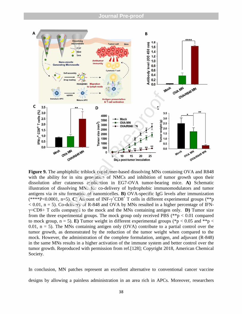
In another example, an amphiphilic triblock copolymer (Pluronic F27) was employed in
constructing a dissolving MN-based cancer vaccine able to create nanomicelles (NMCs) in the
intradermal fluid (Figure 9A) [128]. The amphiphilic property of the Pluronic F27 enabled the
co-delivery of OVA, hydrophilic, and resiquimod (R848), hydrophobic, into in situ micelles
formed after cutaneous application, contributing to the dissolution of R-848. In vitro experiments
confirmed the immunostimulatory potential of R848 encapsulated within the NMCs.
Furthermore, upon administration of the MN patch in vivo, the NMCs can efficiently migrate to
lymph nodes and induce antigen-specific humoral and cellular immunity in EG7-OVA tumor-
bearing mice (Figure 9B). Significantly increased levels of INF-γ+CD8
+ T cells and reduction in
tumor size and weight were found in mice treated with OVA/R-848 MNs compared to the mock
group (Figure 9C-E).
Jour
nal P
re-p
roof
Journal Pre-proof
38
Figure 9. The amphiphilic triblock copolymer-based dissolving MNs containing OVA and R848
with the ability for in situ generation of NMCs and inhibition of tumor growth upon their
dissolution after cutaneous application in EG7-OVA tumor-bearing mice. A) Schematic
illustration of dissolving MNs for co-delivery of hydrophobic immunomodulators and tumor
antigens via in situ formation of nanomicelles. B) OVA-specific IgG levels after immunization
(****P<0.0001, n=5). C) Amount of INF-γ+CD8
+ T cells in different experimental groups (**p
< 0.01, n = 5). Co-delivery of R-848 and OVA by MNs resulted in a higher percentage of IFN-
γ+CD8+ T cells compared to the mock and the MNs containing antigen only. D) Tumor size
from the three experimental groups. The mock group only received PBS (**p < 0.01 compared
to mock group, n = 5). E) Tumor weight in different experimental groups (*p < 0.05 and **p <
0.01, n = 5). The MNs containing antigen only (OVA) contribute to a partial control over the
tumor growth, as demonstrated by the reduction of the tumor weight when compared to the
mock. However, the administration of the complete formulation, antigen, and adjuvant (R-848)
in the same MNs results in a higher activation of the immune system and better control over the
tumor growth. Reproduced with permission from ref.[128]; Copyright 2018, American Chemical
Society.
In conclusion, MN patches represent an excellent alternative to conventional cancer vaccine
designs by allowing a painless administration in an area rich in APCs. Moreover, researchers
Jour
nal P
re-p
roof
Journal Pre-proof
39
have proposed innovative formulations to allow a simple simultaneous delivery of antigens and
adjuvants with opposite hydro/lipophilic characteristics or enabling efficient delivery of the
payload to the draining lymph nodes. Finally, MNs can also play a role in improving the efficacy
of immune checkpoint inhibitors, allowing for local delivery in the tumor area with a decrease in
the immune side effect associated with systemic delivery.
5. Autoimmune diseases
Currently, MNs have been extensively investigated to precisely treat or manage autoimmune
diseases, such as type 1 diabetes (T1D), alopecia areata, systemic lupus erythematosus, multiple
sclerosis, and rheumatoid arthritis (RA) [129-133]. The direct administration of
immunomodulatory peptides and immunosuppressive drugs to patients with the traditional
techniques is no longer applicable owing to their intrinsic limitations such as poor oral
bioavailability, gastrointestinal side-effects, enzymatic hydrolysis in the gastrointestinal tract,
rapid plasma clearance, and poor patient compliance [134]. To address some of these limitations,
Lin et al. developed dissolving MNA to deliver Thymopentin (TP5), a synthetic pentapeptide
with a very short half-life in plasma (about 30 s) and immunomodulating properties for the
treatment of autoimmune diseases, in immunosuppressed Sprague-Dawley rats [135]. A MN
array containing TP5 was fabricated by a modified two-step molding technology using bovine
serum albumin (BSA) as a mechanical strength regulator. The high-performance liquid
chromatography (HPLC) chromatogram confirmed the maintenance of the integrity of TP5 after
loading in the MN patch. The administration of TP5 through the MNs improved the levels of T-
cells and reversed the ratio of CD4+/CD8
+ 7 days after the treatment in immunosuppressed rats.
Jour
nal P
re-p
roof
Journal Pre-proof
40
In 2020, Ahmad et al. fabricated thiolated chitosan (TCS)-based MN patch containing tacrolimus
(TM), an immunosuppressant agent for the treatment of autoimmune disorders, by a mold casting
method. The TCS-based MN patch was able to improve the drug bioavailability through
circumvention of the hepatic first-pass metabolism and intestinal P-gp efflux and deliver the drug
in a more sustained manner than oral administration [136]. Attenuated total reflectance-Fourier
transform infrared (ATR-FTIR) analysis demonstrated that characteristic peaks of TM were
retained in MN patches, suggesting the stability of TM during the preparation process of the
patches. The author found the best tensile strength (0.05 mPa) at 2% concentration of TCS and a
higher skin distribution for MN-TM (15.34 ± 2.4%) compared to ointment (9.45 ± 3.2%).
Additionally, they further mentioned 84% penetration of TM with no breakage of the MNs and a
sustained release (82.5%) from patches with no visible erythema or edema.
5.1. Design of MNs for immunotherapy of type 1 diabetes
The loss of T-cell-dependent immunological tolerance to β-cell autoantigens has been proposed
as the most critical pathological event of T1D. A possible solution for the optimal treatment of
this disease is to reverse this process [137]. Antigen-specific immunotherapy (ASI) is a
promising therapeutic strategy to treat T1D through the induction of an immune regulatory
response to hinder autoimmune-mediated β-cell destruction to maintain insulin production [138].
Peptide immunotherapy has been proposed as promising strategy to induce tolerance to
pancreatic self-antigens. A key aspect to consider in MNs is to analyze their advantages
compared to an ID injection. In a comparative study for the delivery of four different antigenic
peptides, including WE 14, Insulin B9-23, Epstein–Barr virus peptide 280–288 and BDC2.5
mimotope in the non-obese diabetic (NOD) mice model, Zhao et al. found that dry-coated MN
Jour
nal P
re-p
roof
Journal Pre-proof
41
patches consisting of methyl butanol, polyvinyl alcohol, and acetic acid exhibited a more
effective delivery and prolonged antigen presentation in the skin [139]. Comparable reductions
in the auto-reactive cell proliferation were observed when the authors immunized the animals
with two concentrations of WE14 (6 μg of the peptide in dry-coated MNs and 50 μg of the
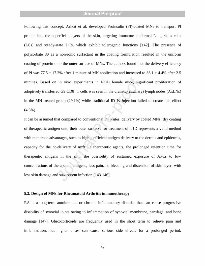
peptide in ID injection) (Figure 10A). The results can be mainly attributed to the higher retention
time of the peptide-loaded into the MN patch, which in turn resulted in providing enough time
for the APCs in the skin to tender antigenic peptides to the patrolling T cells and create tolerance
at lower peptide concentration. In order to minimize immune stimulation and control the
localized kinetics in immunotherapy of T1D, nanomaterials with anti-inflammatory properties
can be used as appropriate multi-cargo delivery platforms to present tolerogenic auto-antigens to
APCs and promote a regulatory response. In another example, Dul et al. used MicronJet600, a
clinically approved MN delivery system, to treat T1D through intradermal delivery of a human
leukocyte antigen-DR4 (HLA-DR4)-restricted peptide epitope of proinsulin (C19-A3) tethered to
gold NPs (GNPs) (Figure 10B) [140]. Ex vivo experiments showed that GNPs quickly diffused
throughout human skin. Additionally, in vitro experiments indicated that uptake of GNPs -
peptide complexes by DCs resulted in a reduction of their capacity to active naïve T cells.
One of the major disadvantages of the above-mentioned studies for T1D immunotherapy is the
loading of a single peptide autoantigen in the MNs, which in turn can limit their applications to
sub-populations of patients with a specific human leukocyte antigen (HLA) molecule [141]. It
has been reported that using coated MN systems loaded with complete protein containing
multiple epitopes provide high therapeutic potential for ASI of T1D because of the ability of
APC to synchronically generate tolerance to a range of epitopes and be therapeutically applicable
to more patients [139].
Jour
nal P
re-p
roof
Journal Pre-proof
42
Following this concept, Arikat et al. developed Proinsulin (PI)-coated MNs to transport PI
protein into the superficial layers of the skin, targeting immature epidermal Langerhans cells
(LCs) and steady-state DCs, which exhibit tolerogenic functions [142]. The presence of
polysorbate 80 as a non-ionic surfactant in the coating formulation resulted in the uniform
coating of protein onto the outer surface of MNs. The authors found that the delivery efficiency
of PI was 77.5 ± 17.3% after 1 minute of MN application and increased to 86.1 ± 4.4% after 2.5
minutes. Based on in vivo experiments in NOD female mice, significant proliferation of
adoptively transferred G9 CD8+ T cells was seen in the draining (axillary) lymph nodes (AxLNs)
in the MN treated group (29.1%) while traditional ID PI injection failed to create this effect
(4.6%).
It can be assumed that compared to conventional ID routes, delivery by coated MNs (dry coating
of therapeutic antigen onto their outer surface) for treatment of T1D represents a valid method
with numerous advantages, such as highly efficient antigen delivery to the dermis and epidermis,
capacity for the co-delivery of multiple therapeutic agents, the prolonged retention time for
therapeutic antigens in the skin, the possibility of sustained exposure of APCs to low
concentrations of therapeutic antigens, less pain, no bleeding and distension of skin layer, with
less skin damage and subsequent infection [143-146].
5.2. Design of MNs for Rheumatoid Arthritis immunotherapy
RA is a long-term autoimmune or chronic inflammatory disorder that can cause progressive
disability of synovial joints owing to inflammation of synovial membrane, cartilage, and bone
damage [147]. Glucocorticoids are frequently used in the short term to relieve pain and
inflammation, but higher doses can cause serious side effects for a prolonged period.
Jour
nal P
re-p
roof
Journal Pre-proof
43
Immunotherapy aims to reduce inflammation and protect the integrity of the joint, with
manageable side effects [148]. Topical drug delivery is only able to deliver therapeutic
molecules with proper oil-water partition coefficient, low molecular weight, and low melting
point due to the characteristics of the stratum corneum [149].
To address these challenges, Yao et al. developed a neurotoxin (NT)-loaded dissolving MNs
through two-step centrifugation, with 15.4 ± 0.5 μg of the drug-loaded in the upper part of each
needle [150]. The MN exhibited good mechanical properties and less toxicity on chondrocytes.
The MN could penetrate the skin at a depth higher than 70 μm. Also, the cumulative penetration
of NT in MNs reached 95.8% in 4 h, whereas a NT solution could hardly penetrate the skin.
After administration to animals, NT-MNs were capable of suppressing the secretion of pro-
inflammatory cytokines such as the tumor necrosis factor α (TNF-α) and interleukin 1 β (IL-1β)
as well as reduce the toe swelling of RA rats.
Although nonsteroidal anti-inflammatory drugs (NSAID), such as meloxicam, are widely used to
reduce inflammation and protect the integrity of the joint in RA patients, their long-term use
results in poor patient compliance due to their serious side effects such as hypertension, diabetes,
ulceration and gastrointestinal perforation [151].
In order to highlight the compliance of MN technology, Amodwala et al. developed fast-
dissolving MNs loaded with meloxicam as a patient-friendly tool to promote drug delivery to
deeper layers of skin and circumvent the drug’s poor patient compliance owing to
gastrointestinal disturbances after oral administration [152]. They reported a 2.58-enhanced
penetration, improved transdermal flux of 1.60 μg/cm2/h, and 63.37% deposition of meloxicam
in excised rat abdominal skin compared to the drug solution. Moreover, rats treated with the MN
exhibited a similar anti-inflammatory activity than the existing marketed oral tablet. In another
Jour
nal P
re-p
roof
Journal Pre-proof
44
interesting example, Chen et al. went on to address the limitations of NSAID through the design
and fabrication of tip-dissolving MNs. In their study, polymer solutions containing meloxicam
were added into the PDMS mold, and high pressure was exerted to drive the air bubbles out of
the cavities of the mold. In the next step, the residual solutions were removed to facilitate drying
the tip solutions in the cavities of mold. Finally, molten polycaprolactone (PCL) was poured into
the mold, and high pressure was exerted again [153]. The author pointed out that tip-dissolving
MNs exhibited burst release of the encapsulated meloxicam (91.72% within 30 min), excellent
bioavailability (122.3%), favorable drug transportation to the skin (79.18%), high anti-
inflammatory and analgesic activities without any adverse effects such as skin irritation. In
another study, Cao et al. engineered dissolvable HA crosslinked MNs as the transdermal
alternative to deliver etanercept (EN), a TNF- α inhibitor for RA therapy since the subcutaneous
(SC) injection of EN is associated with low compliance, infection risk, and other side effects
[154]. The engineered MNs exhibited good mechanical strength and excellent biocompatibility
and did not reduce the bioactivity of EN. In vivo experiments in adjuvant-induced arthritis mice
showed that treatment with both EN by SC (eSC) and EN by MN (eMN) reduced the paw
swelling ratio from 1.70 to 1.48 and 1.68 to 1.44 in 10 days, respectively, indicating anti-
inflammatory effects. Additionally, ELISA assay showed significantly reduced serum levels of
the TNF-α and IL-6 level in both eSC and eMN mice compared to the mice that were treated
with saline (SA) (Figure 10C and 10D).
Jour
nal P
re-p
roof
Journal Pre-proof
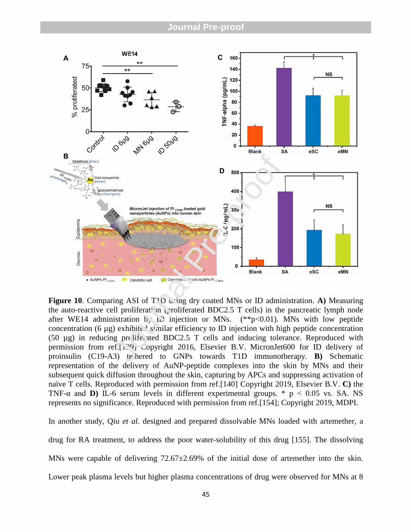
45
Figure 10. Comparing ASI of T1D using dry coated MNs or ID administration. A) Measuring
the auto-reactive cell proliferation (proliferated BDC2.5 T cells) in the pancreatic lymph node
after WE14 administration by ID injection or MNs. (**p<0.01). MNs with low peptide
concentration (6 µg) exhibited similar efficiency to ID injection with high peptide concentration
(50 µg) in reducing proliferated BDC2.5 T cells and inducing tolerance. Reproduced with
permission from ref.[139] Copyright 2016, Elsevier B.V. MicronJet600 for ID delivery of
proinsulin (C19-A3) tethered to GNPs towards T1D immunotherapy. B) Schematic
representation of the delivery of AuNP-peptide complexes into the skin by MNs and their
subsequent quick diffusion throughout the skin, capturing by APCs and suppressing activation of
naïve T cells. Reproduced with permission from ref.[140] Copyright 2019, Elsevier B.V. C) the
TNF-α and D) IL-6 serum levels in different experimental groups. * p < 0.05 vs. SA. NS
represents no significance. Reproduced with permission from ref.[154]; Copyright 2019, MDPI.
In another study, Qiu et al. designed and prepared dissolvable MNs loaded with artemether, a
drug for RA treatment, to address the poor water-solubility of this drug [155]. The dissolving
MNs were capable of delivering 72.67±2.69% of the initial dose of artemether into the skin.
Lower peak plasma levels but higher plasma concentrations of drug were observed for MNs at 8
Jour
nal P
re-p
roof
Journal Pre-proof
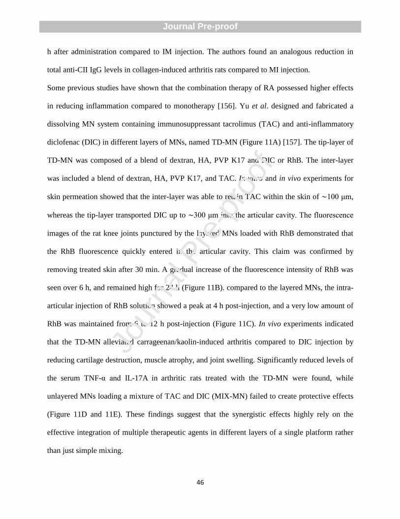
46
h after administration compared to IM injection. The authors found an analogous reduction in
total anti-CII IgG levels in collagen-induced arthritis rats compared to MI injection.
Some previous studies have shown that the combination therapy of RA possessed higher effects
in reducing inflammation compared to monotherapy [156]. Yu et al. designed and fabricated a
dissolving MN system containing immunosuppressant tacrolimus (TAC) and anti-inflammatory
diclofenac (DIC) in different layers of MNs, named TD-MN (Figure 11A) [157]. The tip-layer of
TD-MN was composed of a blend of dextran, HA, PVP K17 and DIC or RhB. The inter-layer
was included a blend of dextran, HA, PVP K17, and TAC. In vitro and in vivo experiments for
skin permeation showed that the inter-layer was able to retain TAC within the skin of ∼100 μm,
whereas the tip-layer transported DIC up to ∼300 μm into the articular cavity. The fluorescence
images of the rat knee joints punctured by the layered MNs loaded with RhB demonstrated that
the RhB fluorescence quickly entered in the articular cavity. This claim was confirmed by
removing treated skin after 30 min. A gradual increase of the fluorescence intensity of RhB was
seen over 6 h, and remained high for 24 h (Figure 11B). compared to the layered MNs, the intra-
articular injection of RhB solution showed a peak at 4 h post-injection, and a very low amount of
RhB was maintained from 6 to 12 h post-injection (Figure 11C). In vivo experiments indicated
that the TD-MN alleviated carrageenan/kaolin-induced arthritis compared to DIC injection by
reducing cartilage destruction, muscle atrophy, and joint swelling. Significantly reduced levels of
the serum TNF-α and IL-17A in arthritic rats treated with the TD-MN were found, while
unlayered MNs loading a mixture of TAC and DIC (MIX-MN) failed to create protective effects
(Figure 11D and 11E). These findings suggest that the synergistic effects highly rely on the
effective integration of multiple therapeutic agents in different layers of a single platform rather
than just simple mixing.
Jour
nal P
re-p
roof
Journal Pre-proof
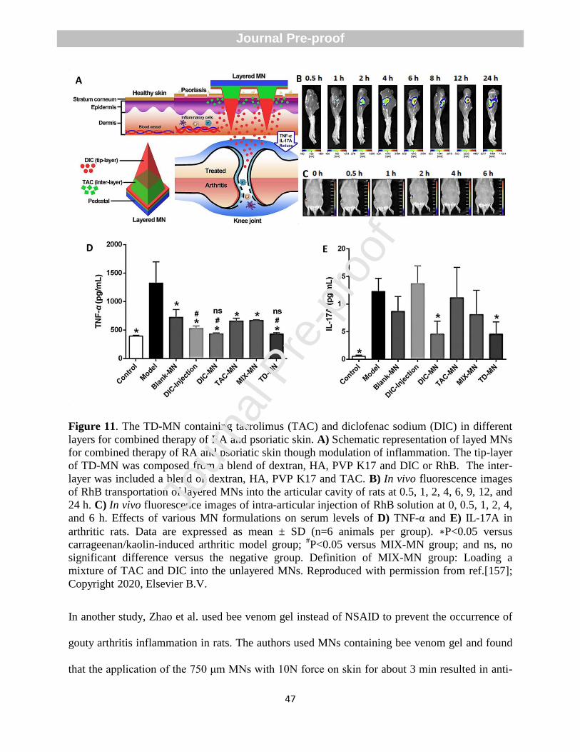
47
Figure 11. The TD-MN containing tacrolimus (TAC) and diclofenac sodium (DIC) in different
layers for combined therapy of RA and psoriatic skin. A) Schematic representation of layed MNs
for combined therapy of RA and psoriatic skin though modulation of inflammation. The tip-layer
of TD-MN was composed from a blend of dextran, HA, PVP K17 and DIC or RhB. The inter-
layer was included a blend of dextran, HA, PVP K17 and TAC. B) In vivo fluorescence images
of RhB transportation of layered MNs into the articular cavity of rats at 0.5, 1, 2, 4, 6, 9, 12, and
24 h. C) In vivo fluorescence images of intra-articular injection of RhB solution at 0, 0.5, 1, 2, 4,
and 6 h. Effects of various MN formulations on serum levels of D) TNF-α and E) IL-17A in
arthritic rats. Data are expressed as mean ± SD (n=6 animals per group). ∗P<0.05 versus
carrageenan/kaolin-induced arthritic model group; #P<0.05 versus MIX-MN group; and ns, no
significant difference versus the negative group. Definition of MIX-MN group: Loading a
mixture of TAC and DIC into the unlayered MNs. Reproduced with permission from ref.[157];
Copyright 2020, Elsevier B.V.
In another study, Zhao et al. used bee venom gel instead of NSAID to prevent the occurrence of
gouty arthritis inflammation in rats. The authors used MNs containing bee venom gel and found
that the application of the 750 μm MNs with 10N force on skin for about 3 min resulted in anti-
Jour
nal P
re-p
roof
Journal Pre-proof
48
inflammatory effects [158]. For example, another type of hydrogel MNs encapsulated triptolide-
loaded liposomes as novel systems to treat collagen-induced arthritis in rats [159]. In this study,
the pharmacodynamics experiments demonstrated that the MNs attenuated the degree of joint
swelling and serum levels of IL-1β and interleukin-6 (IL-6) through regulation of the balance
between Th1 and Th2 pathways. In summary, the possibility to transdermally deliver therapeutic
molecules using MNs for RA treatment represents a great advantage for their clinical translation
since they can reduce the adverse effects of RA drugs.
6. Defeat allergy by MN patches
6.1. Design of MNs for immunotherapy of food allergies
Food allergens like peanut, milk, gluten, and aeroallergens, such as pollen and dust, can
stimulate immune cells and result in the secretion of pro-inflammatory cytokines, activation of
IgE antibodies, mast cells, and eosinophils [160-162]. Allergy immunotherapy promotes
immunological tolerance, with a long-term effect lasting past treatment termination [163]. In a
study, Shakyaa et al. used peanut protein extract (PE)-coated MNs to provide desensitization of
peanut sensitized mice [164]. They firstly fabricated MNA from 50-μm thick stainless steel (304
grade) sheets through a wet etch process and then coated them by dipping in a coating solution
containing PE, carboxymethyl cellulose (as a viscosity enhancer), and Lutrol F-68 NF (as a
surfactant). The authors further revealed that PE-coated MN group had significant down-
regulation of systemic anaphylaxis mediators like mast cell protease-1 (MCPT-1) and histamine
and up-regulation of Th1 cytokines like interleukin-2 (IL-2) and IFN-γ compared to the untreated
group, indicating that PE-coated MNs contributed to immune regulation by targeting the Th1
pathway. In their work, histopathological assessments of small intestinal tissues showed that PE-
Jour
nal P
re-p
roof
Journal Pre-proof
49
coated MNs reduced the infiltration of eosinophils compared to the untreated group. It is well
documented that the allergen dose delivered into the skin is a decisive factor of successful
allergy immunotherapy [165]. As a result of some limitations of coated MNs, such as the
difficulty of precise coating and insufficient delivery of therapeutic allergen, some researchers
have used dissolvable MNA to ensure effective PE transportation into the epidermis/dermis
layers. For example, Yu et al. showed that a powder-laden, dissolvable MNA (PLD-MNA)
encapsulating peanut allergen (PNA)/ 1,25-dihydroxyvitamin D3 (VD3)/CpG could be used to
manage IgE-mediated allergies in peanut-sensitized mice [166]. The authors revealed that the
MNA was able to deliver most of its content after 1 h into the epidermis of mouse skin, with no
evident skin irritation or leakage into the circulation. The intravital confocal microscopy of MHC
class II–EGFP mice showed that OVA-555–laden PLD-MNA was dissolved into the mouse ears
within 15 min, and the APCs were highly accumulated around each powder dot (OVA), with a
peak on days 1 and 2. The emerged yellow colors of green and red on days 1 and 2 confirmed the
uptake of OVA by APCs. Decreasing powder quantity in the epidermis from day 1 to day 5
resulted in the gradual reduction in the number of APCs (Figure 12). The researchers further
stated that PLD-MNA encapsulating PNA/VD3/CpG reduced clinical allergy scores (from 3.5 to
1), the number of treatments, and the required dose of PNA in each treatment compared to
conventional intradermal immunotherapy.
Jour
nal P
re-p
roof
Journal Pre-proof
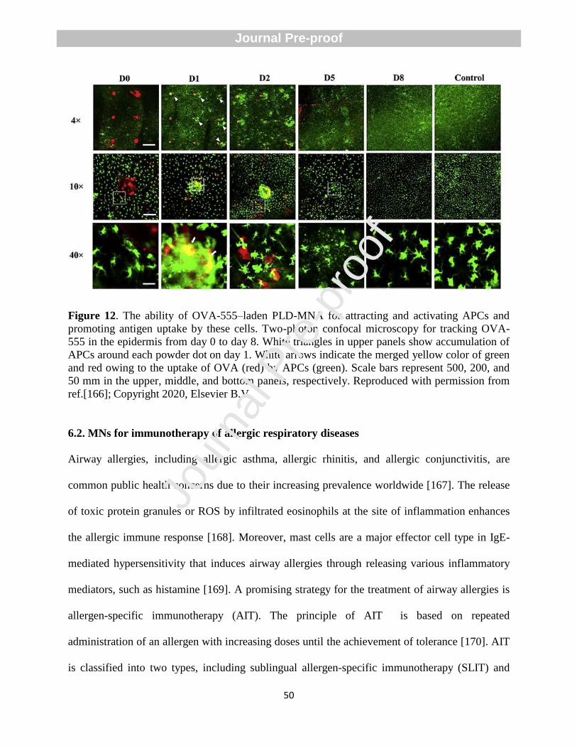
50
Figure 12. The ability of OVA-555–laden PLD-MNA for attracting and activating APCs and
promoting antigen uptake by these cells. Two-photon confocal microscopy for tracking OVA-
555 in the epidermis from day 0 to day 8. White triangles in upper panels show accumulation of
APCs around each powder dot on day 1. White arrows indicate the merged yellow color of green
and red owing to the uptake of OVA (red) by APCs (green). Scale bars represent 500, 200, and
50 mm in the upper, middle, and bottom panels, respectively. Reproduced with permission from
ref.[166]; Copyright 2020, Elsevier B.V.
6.2. MNs for immunotherapy of allergic respiratory diseases
Airway allergies, including allergic asthma, allergic rhinitis, and allergic conjunctivitis, are
common public health concerns due to their increasing prevalence worldwide [167]. The release
of toxic protein granules or ROS by infiltrated eosinophils at the site of inflammation enhances
the allergic immune response [168]. Moreover, mast cells are a major effector cell type in IgE-
mediated hypersensitivity that induces airway allergies through releasing various inflammatory
mediators, such as histamine [169]. A promising strategy for the treatment of airway allergies is
allergen-specific immunotherapy (AIT). The principle of AIT is based on repeated
administration of an allergen with increasing doses until the achievement of tolerance [170]. AIT
is classified into two types, including sublingual allergen-specific immunotherapy (SLIT) and
Jour
nal P
re-p
roof
Journal Pre-proof
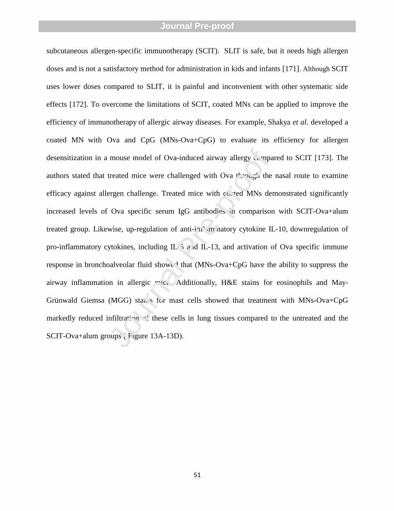
51
subcutaneous allergen-specific immunotherapy (SCIT). SLIT is safe, but it needs high allergen
doses and is not a satisfactory method for administration in kids and infants [171]. Although SCIT
uses lower doses compared to SLIT, it is painful and inconvenient with other systematic side
effects [172]. To overcome the limitations of SCIT, coated MNs can be applied to improve the
efficiency of immunotherapy of allergic airway diseases. For example, Shakya et al. developed a
coated MN with Ova and CpG (MNs-Ova+CpG) to evaluate its efficiency for allergen
desensitization in a mouse model of Ova-induced airway allergy compared to SCIT [173]. The
authors stated that treated mice were challenged with Ova through the nasal route to examine
efficacy against allergen challenge. Treated mice with coated MNs demonstrated significantly
increased levels of Ova specific serum IgG antibodies in comparison with SCIT-Ova+alum
treated group. Likewise, up-regulation of anti-inflammatory cytokine IL-10, downregulation of
pro-inflammatory cytokines, including IL-5 and IL-13, and activation of Ova specific immune
response in bronchoalveolar fluid showed that (MNs-Ova+CpG have the ability to suppress the
airway inflammation in allergic mice. Additionally, H&E stains for eosinophils and May-
Grünwald Giemsa (MGG) stains for mast cells showed that treatment with MNs-Ova+CpG
markedly reduced infiltration of these cells in lung tissues compared to the untreated and the
SCIT-Ova+alum groups ( Figure 13A-13D). Jour
nal P
re-p
roof
Journal Pre-proof
52
Figure 13. The effects of coated MN with Ova and CpG on the infiltration of eosinophils and
mast cells in lung tissues. A) H&E stained lung tissue sections for evaluation of eosinophil
infiltration. B) Eosinophil count. C) May-Grünwald Giemsa stained tissue sections for evaluation
of mast cell infiltration. D) Mast cell count. Arrows show cells or mucus deposition (n = 3
animals per group). Data are expressed as mean ± SEM. ** p ≤ 0.01, and ns: not significant. Reproduced with permission from ref.[173]; Copyright 2020, American Chemical Society.
In another interesting example, the same research group loaded Der p 1, a house dust mite
allergen, onto an MN patch composed of 57 total MNs to hamper the onset and progression of
Der p 1–induced airway allergy [174]. Their results by calibrated fluorescence spectroscopy
demonstrated that 73.3% ± 7.4% of coated Der p 1 was transported into the skin. The authors
further found that Der p 1 coated MNs created a more reliable anti–Der p 1 IgG response than
Jour
nal P
re-p
roof
Journal Pre-proof
53
the untreated and subcutaneous immunotherapy groups. The addition of CpG as an adjuvant to
Der p 1 coated MN resulted in a more significant anti–Der p 1 IgG2a response and higher IL-10
and transforming growth factor-beta (TGF-β) secretion than both the Der p 1 coated MN and
subcutaneous immunotherapy.
In another example, Shakya et al. coated MNs, using an in-house micro-precision dip-coating
device, in a solution containing OVA, Lutrol F-68 NF, and carboxymethylcellulose (1%, w/v) to
create a preventive vaccine against airway allergy [175]. The authors stated that the coated MNs
are beneficial for treating airway allergy in infants as the sublingual route is not suitable for
them, and injections are painful. A strong OVA-specific systemic immune response was induced
following the insertion of MN coated with OVA and CpG oligonucleotide as adjuvant into the
mice’s skin. The MNs modulated a variety of cytokines, including anti-inflammatory cytokines
(IL-10) and Th2 cytokines (IL-13, IL-4, and IL-5) in the bronchoalveolar fluid, as well as Th1
cytokines (IFN-γ and IL-2) in re-stimulated splenocyte cultures, indicating improvement of
clinical symptoms of allergy.
Owing to limitations of coated MNs, Park et al. developed a biodegradable MN patch (MNP)
comprising of hyaluronate and Dermatophagoides farinae (D. farinae) extract (DfE) using a
droplet-born air blowing method for transdermal immunotherapy (TDIT) to ameliorate airway
hyperresponsiveness (AHR) and inflammation in a house dust mite (HDM) asthma model
(Figure 14A and 14B) [176]. The delivery test showed that 77% and 85.4% of the allergen were
absorbed after 1 and 8 h of MNP insertion, respectively. In vivo experiments indicated that
treatment with 10 μg TDIT improved HDM-induced AHR most significantly in comparison with
low-dose (10 μg) and high-dose (100 μg) SCIT. TDIT had the strongest effect in reducing the
secretion of cytokines associated with the Th2 inflammatory response (IL-4, IL-5, and IL-13)
Jour
nal P
re-p
roof
Journal Pre-proof
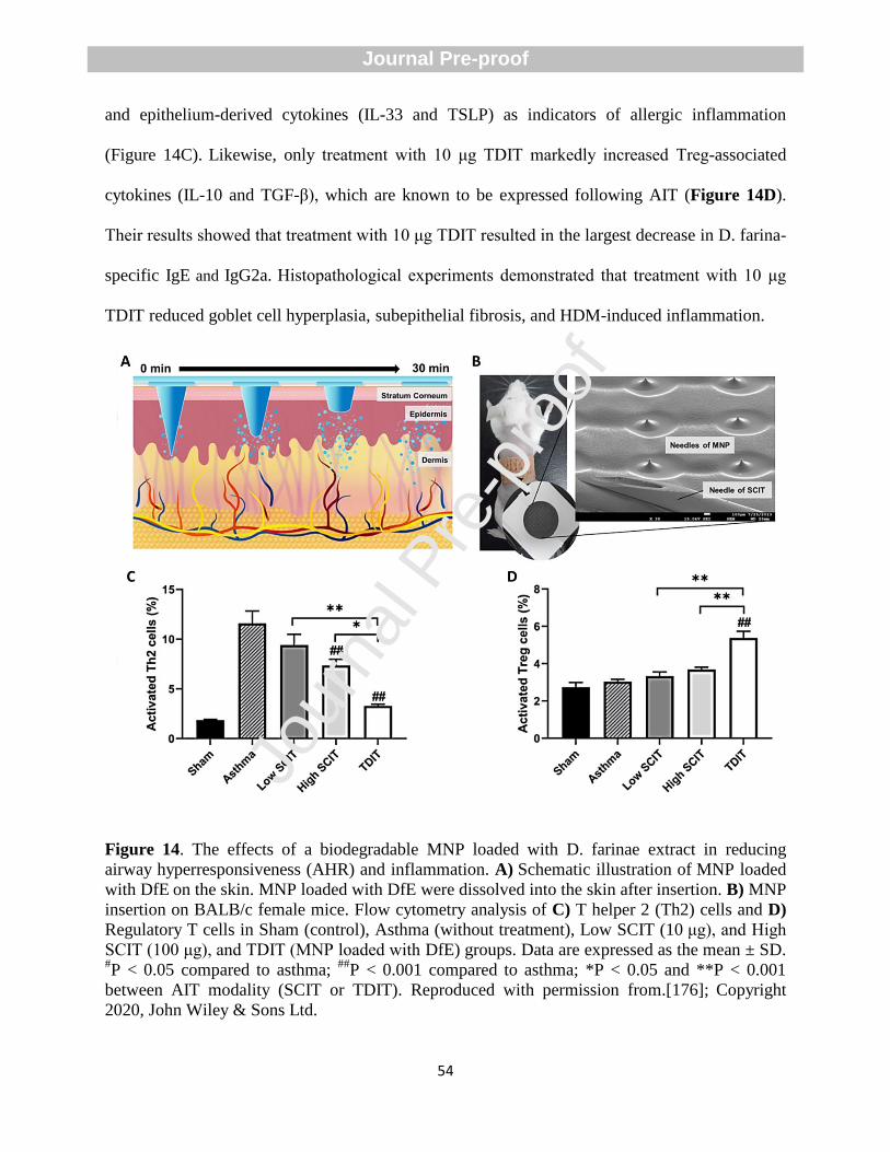
54
and epithelium-derived cytokines (IL-33 and TSLP) as indicators of allergic inflammation
(Figure 14C). Likewise, only treatment with 10 μg TDIT markedly increased Treg-associated
cytokines (IL-10 and TGF-β), which are known to be expressed following AIT (Figure 14D).
Their results showed that treatment with 10 μg TDIT resulted in the largest decrease in D. farina-
specific IgE and IgG2a. Histopathological experiments demonstrated that treatment with 10 μg
TDIT reduced goblet cell hyperplasia, subepithelial fibrosis, and HDM-induced inflammation.
Figure 14. The effects of a biodegradable MNP loaded with D. farinae extract in reducing
airway hyperresponsiveness (AHR) and inflammation. A) Schematic illustration of MNP loaded
with DfE on the skin. MNP loaded with DfE were dissolved into the skin after insertion. B) MNP
insertion on BALB/c female mice. Flow cytometry analysis of C) T helper 2 (Th2) cells and D)
Regulatory T cells in Sham (control), Asthma (without treatment), Low SCIT (10 μg), and High
SCIT (100 μg), and TDIT (MNP loaded with DfE) groups. Data are expressed as the mean ± SD. #P < 0.05 compared to asthma;
##P < 0.001 compared to asthma; *P < 0.05 and **P < 0.001
between AIT modality (SCIT or TDIT). Reproduced with permission from.[176]; Copyright
2020, John Wiley & Sons Ltd.
Jour
nal P
re-p
roof
Journal Pre-proof
55
6.3. MNs for allergen-specific immunotherapy
To compare the efficiency of MNs for cutaneous allergen-specific immunotherapy (ASIT) with
other conventional routes like the IP, IM, and subcutaneous allergen immunotherapy, Shakya et
al. fabricated MNs coated with OVA by a micro-precision dip coater and a coating solution
containing OVA, carboxymethyl cellulose and Lutrol F-68 NF [177]. They demonstrated that
most of the coating was transported into the skin following the insertion of the fluorescein-OVA
coated MN patch in a mouse model. The coated MN-treated animals demonstrated an increased
anti-OVA IgG response compared to IP and subcutaneous allergen immunotherapy and similar
anti-OVA IgG response to IM administration at 28 days after insertion. Generally, they observed
lower secretion of pro-inflammatory IL-1β, interleukin-13 (IL-13), interleukin-15 (IL-5), and IFN-
γ after the insertion of OVA coated MN patches compared to the other routes of administration,
as well as higher activation of the Th1 pathway, indicating their potential use for painless ASI.
The authors stated that a shift from Th2 to Th1 response is thought to be beneficial for ASI.
In 2019, the same researchers coated two stimulators of interferon genes (STING) agonists,
namely, cyclic diadenylate monophosphate (c-di-AMP) and cyclic diguanylate monophosphate
(c-di-GMP), along with OVA as a model allergen on MN patches characterized by 57 individual
micron-sized needles and transported them into mouse skin [178]. Calibrated fluorescence
spectroscopy confirmed delivery of 78.8% (±4.2) of coated OVA into the skin, while 4.2%
(±1.1) was seen on the skin and 17.0% (±4.0) OVA remained on the surface of MN patches. The
authors showed a higher OVA-specific IgG2a antibody in serum as a surrogate marker for Th1
type immune response and higher levels of Th1 cytokines (IFN-γ and IL-2) in re-stimulation of
splenocytes culture for coated MN groups compared to the approved subcutaneous hypodermic
injection.
Jour
nal P
re-p
roof
Journal Pre-proof
56
In a clinical trial conducted in Germany, Spina et al. compared MNs and adhesive-tape stripping
in skin preparation for epicutaneous allergen immunotherapy [179]. They observed that MNs
increased stratum corneum penetration in comparison to tape stripping and induced dendritic
cell-mediated T cell responses.
Benefitting from their tailorability to the location of different subtypes of dendritic cells, such as
LCs, MN patches can be designed to target LCs for allergy immunotherapy with minimal side
effects. The presence of LCs in the epidermal layer makes them the first line of APCs that meet
skin-invading antigens. This opens up a new avenue for immunotherapy of diseases without
causing noticeable adverse effects [180]. For example, Burg et al. designed microprojection
arrays (MPA) in polycarbonate material through a hot embossing procedure and coated OVA
onto the surface of the MPAs [181]. In this study, they found that slit-MPAs (110 μm ± 3 μm tall
projections at 7k p/cm2) with higher tip surface areas than conical-MPAs (207 μm ± 5 μm tall
projections at 10,000 (10k) projections/cm2 (p/cm
2)) were more suitable for only epidermal
delivery using low application energies. They further stated that the mouse epidermis (eMPA)
delivery with larger projection tip surface areas had a lower inflammatory response and
epidermal cell death, lesser erythema after 24 h, and epidermal swelling after 72 h than dermal-
targeted MPAs (dMPA). Flow cytometry analysis showed that only the eMPA increased the
migration of LCs with a low expression of major histocompatibility complex (MHC) II, without
migration of dDCs.
Overall, the delivery of allergens by MNs to both the epidermis and the dermis can be further
explored as a convenient and effective new means of allergen immunotherapy, which would
encourage patient compliance and minimize side effects.
Jour
nal P
re-p
roof
Journal Pre-proof
57
7. MN-based vaccines
The need for improvement of percutaneous vaccination arises from several major limitations of
this technique, including needle phobias, the need for high doses and reconstitution of
lyophilized vaccines, and the production of a huge amount of biohazardous waste, including
sharp materials, such as needles that require special disposal protocol [182].
Currently, vaccine-loaded MNs have attracted tremendous attention from the scientific
community as safe and dose-sparing tools to control vaccine release kinetics or improve immune
response through manipulation of the composites of MNs [183-185]. For example, DeMuth et al.
developed layer-by-layer assembled polyelectrolyte multilayers (Poly-1/ICMV) deposited onto
poly(lactic-co-glycolic acid) (PLGA) MNs, which provided a possibility to control the film
thickness and dosage of immunomodulators for adjustable releasing and stabilizing goals (Figure
15A) [186]. The authros firstly synthesized nanocapsules containing interbilayer covalent cross-
links among maleimide head moieties of adjacent phospholipid lamellae present in the walls of
multilamellar vesicles and then deposited them onto the surface of PLGA MNs. By co-
encapsulating OVA as an adjuvant and a fluorescent tracer in the structure of nanocapsules
loaded into MNs, they found that OVA was taken up by the APCs into the epidermis, which, in
turn, gave rise to higher serum levels of anti-OVA IgG titers and improved humoral immune
response in a mice model (Figure 15B).
In another study, Boopathy et al. developed solid pyramidal MNs composed of silk fibroin (SF)
protein tips encapsulating a stabilized HIV envelope immunogen and adjuvant to maintain drug
concentrations in the skin by tailoring the speed of release [187]. It was shown that Env trimer
was released over 2 weeks after immunization in the mice skin, resulting in increased germinal
center (GC) B cell responses, increasing bone marrow plasma cells (16-fold), and increasing
Jour
nal P
re-p
roof
Journal Pre-proof
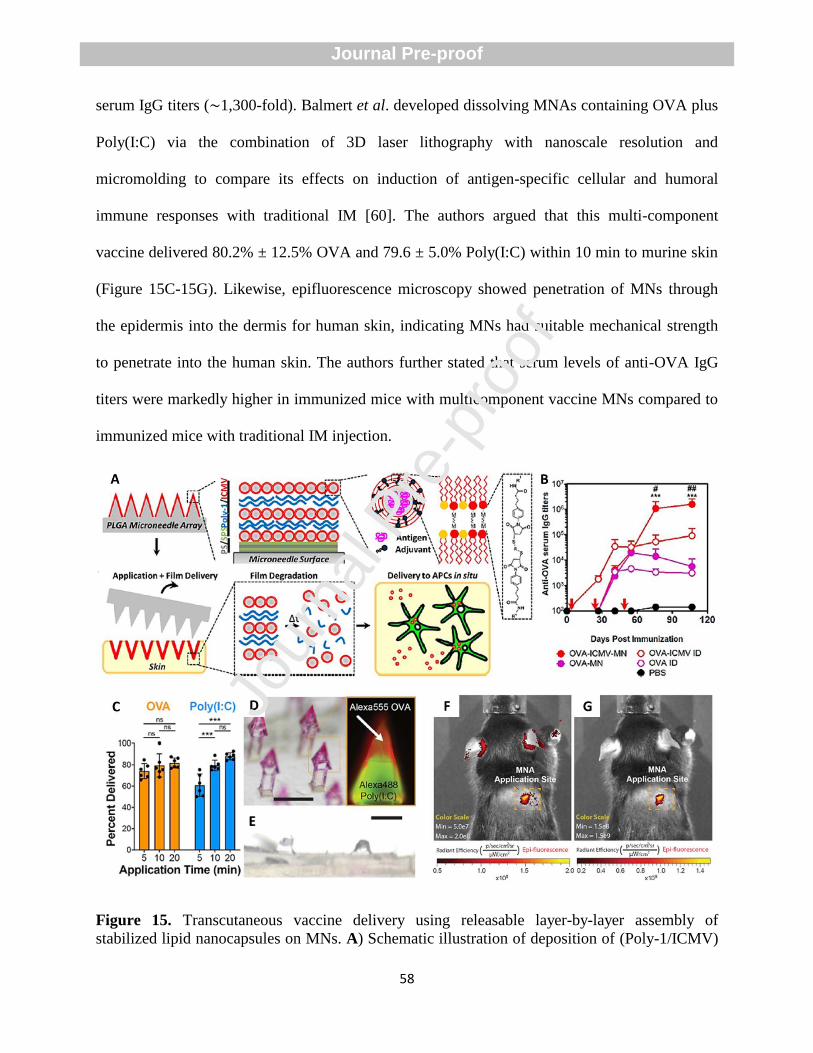
58
serum IgG titers (∼1,300-fold). Balmert et al. developed dissolving MNAs containing OVA plus
Poly(I:C) via the combination of 3D laser lithography with nanoscale resolution and
micromolding to compare its effects on induction of antigen-specific cellular and humoral
immune responses with traditional IM [60]. The authors argued that this multi-component
vaccine delivered 80.2% ± 12.5% OVA and 79.6 ± 5.0% Poly(I:C) within 10 min to murine skin
(Figure 15C-15G). Likewise, epifluorescence microscopy showed penetration of MNs through
the epidermis into the dermis for human skin, indicating MNs had suitable mechanical strength
to penetrate into the human skin. The authors further stated that serum levels of anti-OVA IgG
titers were markedly higher in immunized mice with multicomponent vaccine MNs compared to
immunized mice with traditional IM injection.
Figure 15. Transcutaneous vaccine delivery using releasable layer-by-layer assembly of
stabilized lipid nanocapsules on MNs. A) Schematic illustration of deposition of (Poly-1/ICMV)
Jour
nal P
re-p
roof
Journal Pre-proof
59
multilayers onto PLGA MN surfaces, transportation into the skin after insertion, and boosting
immune function. B) Serum levels of anti-OVA IgG titers at different days after immunization
(#p < 0.05 and
##p < 0.01, and ***p < 0.001). Reproduced with permission from ref.[186];
Copyright 2012, American Chemical Society. C) Amount of OVA and poly (I:C) transported
into the skin at different time points after MN insertion (***p < 0.001). No significant
differences for delivery of OVA were seen at different times after insertion (5, 10, and 20 min),
whereas the highest transportation of poly (I:C) was observed 20 min after insertion. D) Optical
stereomicroscopy and fluorescence images of MNs containing both Alexa555-OVA (red) and
Alexa488-Poly (I:C) (green). Scale bar: 500 μm. E) Optical stereomicroscopy photo of
OVA/Poly (I:C) loaded MNs after insertion in murine skin. Scale bar: 250 μm. Effective
transportation of (F) Alexa488-Poly (I:C) and (G) Alexa555-OVA into the mouse skin after MN
insertion. Reproduced with permission from ref.[60]; Copyright 2019, Elsevier B.V.
7.1. Design of MNs for transdermal delivery of viral vaccines
The utility of vaccines can be improved by the use of coated and dissolving MNs since they are
capable of carrying vaccines in a dried form [188]. The MNs can then release the components of
the vaccine after dissolution in the epidermis/dermis, resulting in more thermostability and
reducing the necessity for vaccine reconstitution as one of the steps of traditional percutaneous
vaccine administration [189, 190]. The improved thermostability by coated and dissolving MNs
provides the possibility to decrease or eliminate high costs relevant to the ―cold chain‖ for
storage or transportation of vaccines [191, 192]. Additionally, MNA vaccines are advantageous
to create immunization against infectious diseases (SARS and COVID-19) in that they possess a
high potential for self-administration without the need for any specialized equipment or an
applicator [193, 194]. Although it has been reported that hollow MNs can be applied to facilitate
penetration of NP loaded with therapeutic antigens into the skin and induce higher antibody
response and a higher amount of interferon-γ compared to IM injection, needle clogging during
piercing of skin and resistance to flow can limit their application for vaccination [195].
In the recent decade, a number of groups have used coated MNs for viral vaccinations since they
are able to introduce the vaccine in a dry state (without the need for reconstitution) and improve
Jour
nal P
re-p
roof
Journal Pre-proof
60
vaccine stability [196]. For example, Shin et al. coated 2 and 10μg of GMP with the influenza
vaccine onto the stainless steel MNs using the dip-coating process before delivering them into
the skin of a mice model to evaluate the reduction in influenza susceptibility [197]. In this study,
mice immunized with the vaccine antigen and 2μg of GMP demonstrated the highest level of IgG
at the second and fourth week after insertion (7-fold and 2.8-fold higher than the vaccine alone)
while mice immunized with the vaccine antigen and 10μg of GMP did not induce a significant
IgG response. Furthermore, mice immunized with 2μg GMP and the virus exhibited stronger
protective immunity, while mice immunized with 10μg GMP, and the virus demonstrated the
lowest lung viral titer and rapid recovery rate. Thereby, the simultaneous delivery of the
influenza vaccine and GMP by MNs improved both cellular and humoral immunity. In another
example, Nguyen et al. designed and fabricated 800-µm-long polylactic acid MNs coated with L-
HBsAg s (a third-generation hepatitis vaccine) to investigate its efficiency compared to IM
immunization [198]. It was shown that the coated MNs more effectively elevated IgG1 and
IgG2a titers and increased thermal and freeze-thaw stability compared to IM immunization.
Despite the potential benefits, several drawbacks of the coated MNs, such as poor coating
efficiency and immunogenicity, might limit their future clinical use for viral and bacterial
vaccinations [43]. The small surface area of the MNs results in delivering only small amounts of
therapeutic molecules and antigens into the skin. To address this limitation, Maaden et al. coated
pH-sensitive MNs with inactivated polio vaccine (IPV) particles and N-trimethyl chitosan
chloride (TMC) through electrostatic interactions to achieve a sufficiently high antigen dose
[199]. In their study, the MN surface was firstly decorated with pH-sensitive (pyridine) moieties
and then coated with negatively charged IPV and a positively charged TMC. The authors coated
ten layers of IPV alternately with TMC to achieve the required dose of antigen. With further
Jour
nal P
re-p
roof
Journal Pre-proof
61
analysis, the authors found 45 D antigen units IPV and 700 ng TMC per MN. The ex vivo
experiments demonstrated the release of both IPV and TMC into the human skin, and in vivo
experiments confirmed the induction of IPV specific antibody responses. Another possible
solution to overcome the limitation of coated MNs for vaccination is to use dissolvable MNs
[200].
In another example, Erdos et al. developed dissolvable MNAs, consisting of trehalose and
carboxymethyl cellulose, incorporating adenovirus vaccines (Ad5.OVA) with or without
Poly(I:C) using a spin-casting technique to enhance the immunogenicity of skin-targeted
adenovector vaccines [201]. The authors confirmed the dissolution of the MNAs upon skin
insertion using optical stereomicroscopy. The multicomponent MNA vaccine platforms
determined an increased OVA-specific lytic immunity compared to MNA containing
adenovectors alone. In 2019, Chen et al. demonstrated that when inactivated influenza virus or
OVA antigen is loaded into dissolving chitosan MNs, the delivery of inactivated influenza virus
occurred in a sustained manner (up to 28 days), and induction of influenza-virus-specific IgG
responses were higher than IM injection in mice (Figure 16A-C) [82].
7.2. Design of MNs for transdermal delivery of bacterial vaccines
In 2017, Pastor et al. developed outer membrane vesicle-loaded dissolving MNs to generate
intradermal immunization against enteropathogens like the bacterium Shigella through induction
of specific systemic IgG and mucosal IgA [202]. In 2019, Zhu et al. developed pertussis toxin
(PT) vaccine MNs to transdermally target immune cells against Pertussis (whooping cough)
caused by the bacterium Bordetella pertussis [203]. PT vaccine MNs released antigen in a more
sustained manner (Figure 16D) and efficiently reduced the dose necessary to generate a high
Jour
nal P
re-p
roof
Journal Pre-proof
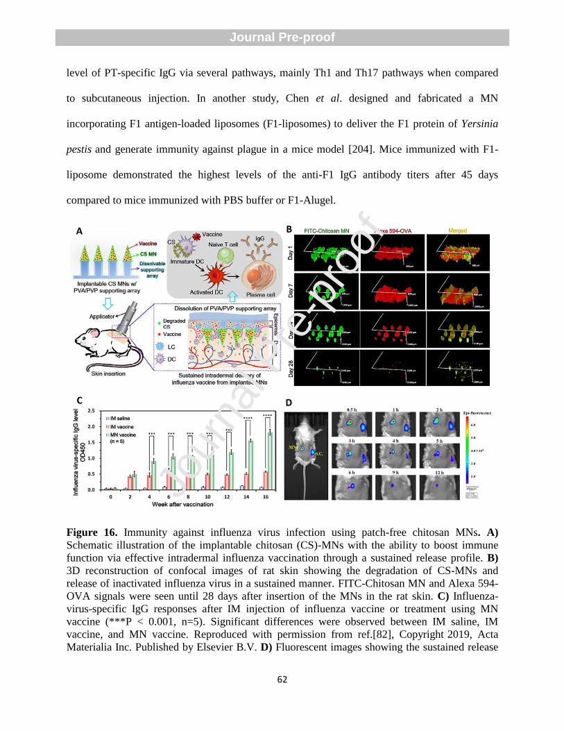
62
level of PT-specific IgG via several pathways, mainly Th1 and Th17 pathways when compared
to subcutaneous injection. In another study, Chen et al. designed and fabricated a MN
incorporating F1 antigen-loaded liposomes (F1-liposomes) to deliver the F1 protein of Yersinia
pestis and generate immunity against plague in a mice model [204]. Mice immunized with F1-
liposome demonstrated the highest levels of the anti-F1 IgG antibody titers after 45 days
compared to mice immunized with PBS buffer or F1-Alugel.
Figure 16. Immunity against influenza virus infection using patch-free chitosan MNs. A)
Schematic illustration of the implantable chitosan (CS)-MNs with the ability to boost immune
function via effective intradermal influenza vaccination through a sustained release profile. B)
3D reconstruction of confocal images of rat skin showing the degradation of CS-MNs and
release of inactivated influenza virus in a sustained manner. FITC-Chitosan MN and Alexa 594-
OVA signals were seen until 28 days after insertion of the MNs in the rat skin. C) Influenza-
virus-specific IgG responses after IM injection of influenza vaccine or treatment using MN
vaccine (***P < 0.001, n=5). Significant differences were observed between IM saline, IM
vaccine, and MN vaccine. Reproduced with permission from ref.[82], Copyright 2019, Acta
Materialia Inc. Published by Elsevier B.V. D) Fluorescent images showing the sustained release
Jour
nal P
re-p
roof
Journal Pre-proof
63
of PT vaccine MNs compared to subcutaneous injection. No signals were seen for subcutaneous
injection after 6h, while signals for MN insertion were observed until 12 h. Reproduced with
permission from ref.[203]; Copyright 2019, American Chemical Society.
8. MN-mediated treatment of inflammatory diseases
MNs can reduce the required dose and adverse effects of anti-inflammatory drugs and contribute
to the maintenance of the physical stability of their nanocarriers [205]. Currently, dissolving
MNs have garnered much attention from the scientific community for immunotherapy of
inflammatory diseases.
8.1. MN design for psoriasis treatment
Psoriasis is a chronic inflammatory disorder that is mainly characterized by vasodilation and
immunological imbalance. The activation of NF-κB by some factors results in the initiation of
inflammation in psoriasis [206]. The presence of T cells and DCs in the psoriatic lesions enhance
the secretion of pro-inflammatory cytokines such as TNFα, IL-1β, IFNγ, IL-17, IL-22, and IL-23
[207]. Currently, evidence has shown that MNs can effectively manage psoriasis [208].
An advantage of using the dissolving MNs for immunotherapy of inflammatory diseases
compared with other types of MNs and traditional needles is the dissolution of the needles within
the skin and hence no requirement for proper needle disposal or the fear of needle reuse [209].
Du et al. pioneered a project using dissolving MNP composed of HA loaded with Methotrexate
(MTX) using a two-step micromolding process. The aim of the study was to enhance the efficacy
of psoriasis treatment by targeting inflammatory responses [210]. The release of MTX reached
52% within 10 min, 76%, and 87% at 30 and 60 min, mainly due to the water solubility of HA.
In vivo experiments demonstrated a complete dissolution of MNP- MTX within 10 min resulted
in a reduction in ear thickness, suppression of the IL23/IL17 axis, and imiquimod (IMQ) -
induced psoriasis-like skin inflammation. They reported that both IMQ mice treated with MTX-
Jour
nal P
re-p
roof
Journal Pre-proof
64
loaded MNs (13.8 μg) and oral administration at a double dose (27.6 μg) of MTX exhibited
much milder psoriasiform dermatitis whereas oral administration at a same dose with MNs (13.8
μg) failed to exert such protective effects (Figure 17A).
In another study, Korkmaz et al. developed carboxymethyl cellulose-based dissolving MNs
containing anti-TNF–α for localized ID delivery to reduce the immunosuppression associated
with systemic administration of antibody and related adverse events [211]. The antibody cargo
was incorporated in the needle tips using a micromilling/spin-casting fabrication method. The
MNs nearly completely dissolved into the human skin explants and reduced the critical
biomarkers of psoriasiform inflammation (epidermal thickness and IL-1β expression) in a
psoriatic mouse model. The following year, the same group conjugated high molecular weight
HA to the anti-TNF-α antibody in the same dissolving MNs to hinder the fast diffusion of
antibodies from the target site to other body regions [212]. The high efficiency of this delivery
method was assessed in human skin explants without antibody diffusion from the delivery site.
8.2. MN for acne treatment
One of the main causes of acne is inflammatory responses that occur in the sebaceous glands of
hair follicles [213]. The proliferation of microorganisms in hair follicles in large quantities,
especially Propionibacterium acnes (P. acnes), results in free fatty acid production, excessive
production of reactive oxygen species (ROS), chemotaxis of inflammatory mediators,
inflammatory cells, and finally the formation of acne lesions [214]. Therefore, simultaneous
inhibiting bacterial growth and suppressing inflammatory responses are essential to achieve a
successful acne treatment.
Excessive production of ROS under pathological conditions (more than 500 μM) in
inflammatory tissues provides the possibility to design ROS-responsive MNs that are able to
Jour
nal P
re-p
roof
Journal Pre-proof
65
release therapeutic molecules in a sustained manner based on ROS-induced degradation into the
acne lesion site [215]. Based on this concept, Zhang et al. prepared ROS-responsive MNs
containing poly(vinyl alcohol) PVA/ clindamycin (CDM) networks that were loaded into a
substrate composed of HA and diatomaceous earth (DE) to improve interactions for the
treatment of acne vulgaris, a common inflammatory skin disease caused by colonization of P.
acnes (Figure 17B) [216]. The use of ROS-responsive MNs highly reduced the number of
infiltrated inflammatory cells into the dermis in a P. acnes-induced mouse model while a CDM
cream failed to create such protective effects (Figure 17C and 17D).
Jour
nal P
re-p
roof
Journal Pre-proof
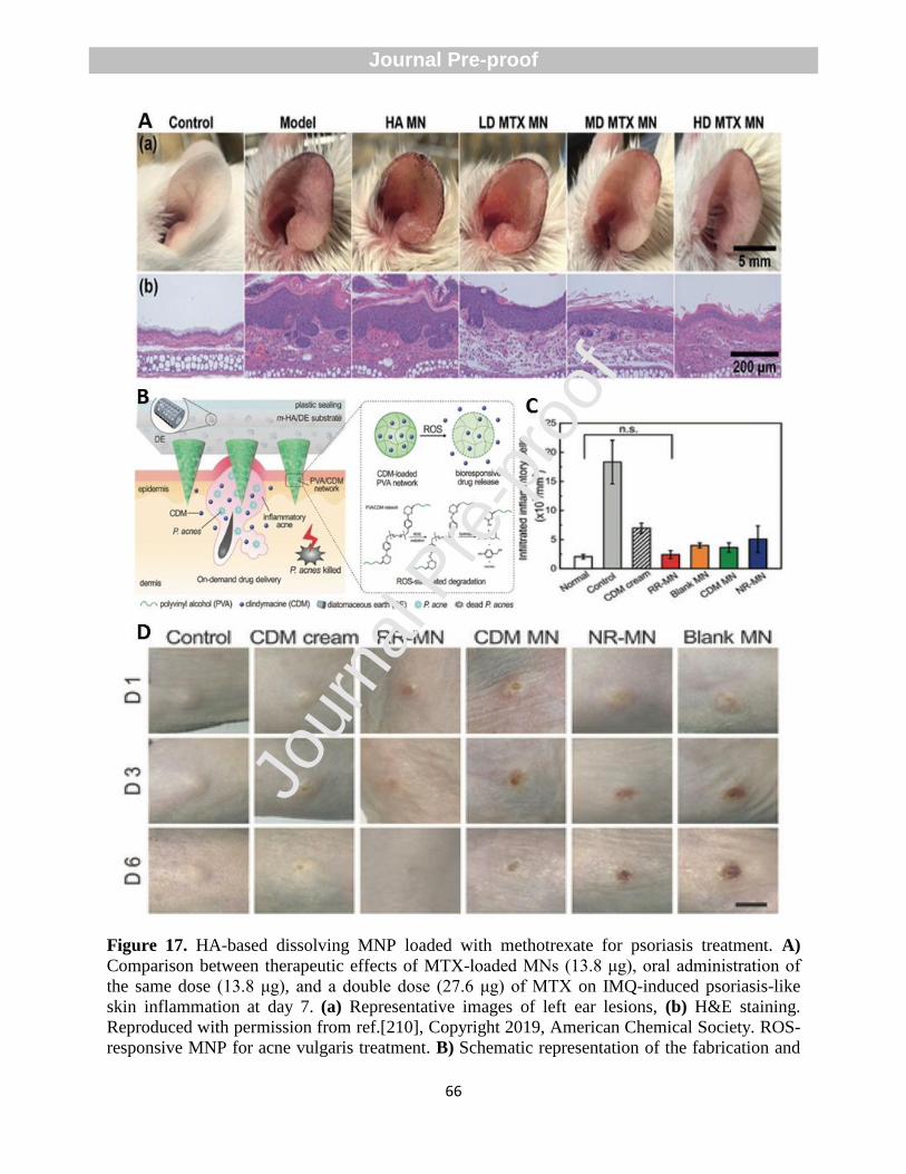
66
Figure 17. HA-based dissolving MNP loaded with methotrexate for psoriasis treatment. A)
Comparison between therapeutic effects of MTX-loaded MNs (13.8 μg), oral administration of
the same dose (13.8 μg), and a double dose (27.6 μg) of MTX on IMQ-induced psoriasis-like
skin inflammation at day 7. (a) Representative images of left ear lesions, (b) H&E staining.
Reproduced with permission from ref.[210], Copyright 2019, American Chemical Society. ROS-
responsive MNP for acne vulgaris treatment. B) Schematic representation of the fabrication and
Jour
nal P
re-p
roof
Journal Pre-proof
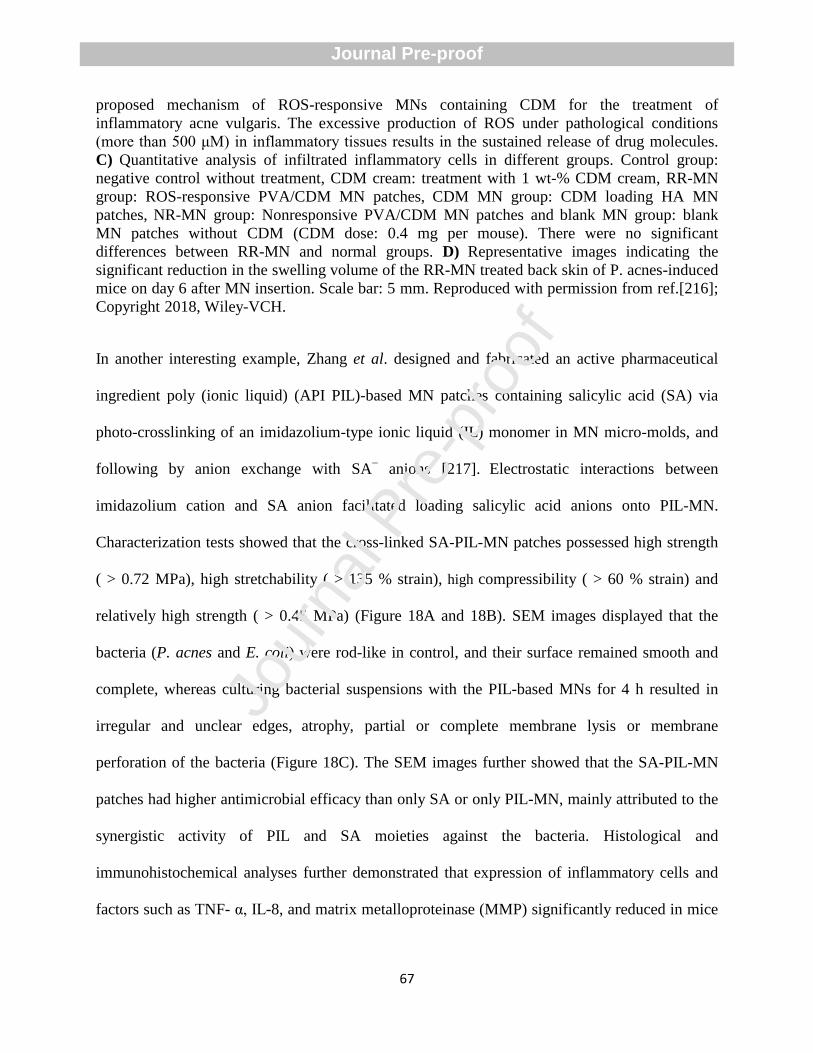
67
proposed mechanism of ROS-responsive MNs containing CDM for the treatment of
inflammatory acne vulgaris. The excessive production of ROS under pathological conditions
(more than 500 μM) in inflammatory tissues results in the sustained release of drug molecules.
C) Quantitative analysis of infiltrated inflammatory cells in different groups. Control group:
negative control without treatment, CDM cream: treatment with 1 wt-% CDM cream, RR-MN
group: ROS-responsive PVA/CDM MN patches, CDM MN group: CDM loading HA MN
patches, NR-MN group: Nonresponsive PVA/CDM MN patches and blank MN group: blank
MN patches without CDM (CDM dose: 0.4 mg per mouse). There were no significant
differences between RR-MN and normal groups. D) Representative images indicating the
significant reduction in the swelling volume of the RR-MN treated back skin of P. acnes-induced
mice on day 6 after MN insertion. Scale bar: 5 mm. Reproduced with permission from ref.[216];
Copyright 2018, Wiley-VCH.
In another interesting example, Zhang et al. designed and fabricated an active pharmaceutical
ingredient poly (ionic liquid) (API PIL)-based MN patches containing salicylic acid (SA) via
photo-crosslinking of an imidazolium-type ionic liquid (IL) monomer in MN micro-molds, and
following by anion exchange with SA− anions [217]. Electrostatic interactions between
imidazolium cation and SA anion facilitated loading salicylic acid anions onto PIL-MN.
Characterization tests showed that the cross-linked SA-PIL-MN patches possessed high strength
( > 0.72 MPa), high stretchability ( > 135 % strain), high compressibility ( > 60 % strain) and
relatively high strength ( > 0.45 MPa) (Figure 18A and 18B). SEM images displayed that the
bacteria (P. acnes and E. coli) were rod-like in control, and their surface remained smooth and
complete, whereas culturing bacterial suspensions with the PIL-based MNs for 4 h resulted in
irregular and unclear edges, atrophy, partial or complete membrane lysis or membrane
perforation of the bacteria (Figure 18C). The SEM images further showed that the SA-PIL-MN
patches had higher antimicrobial efficacy than only SA or only PIL-MN, mainly attributed to the
synergistic activity of PIL and SA moieties against the bacteria. Histological and
immunohistochemical analyses further demonstrated that expression of inflammatory cells and
factors such as TNF- α, IL-8, and matrix metalloproteinase (MMP) significantly reduced in mice
Jour
nal P
re-p
roof
Journal Pre-proof
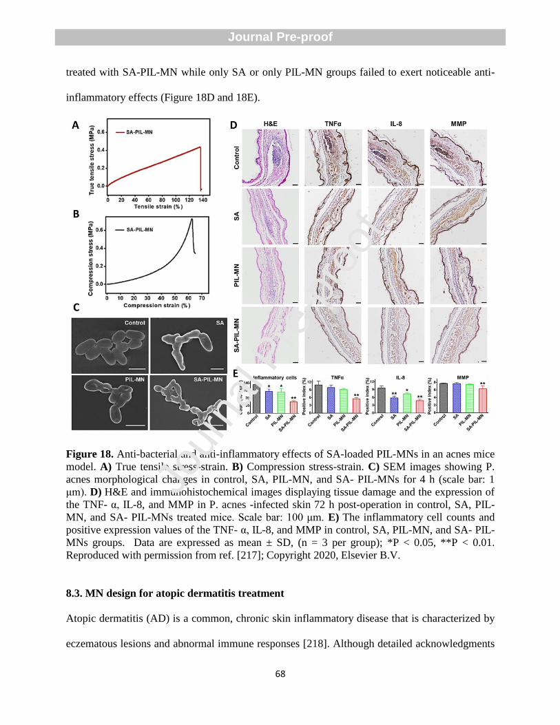
68
treated with SA-PIL-MN while only SA or only PIL-MN groups failed to exert noticeable anti-
inflammatory effects (Figure 18D and 18E).
Figure 18. Anti-bacterial and anti-inflammatory effects of SA-loaded PIL-MNs in an acnes mice
model. A) True tensile stress-strain. B) Compression stress-strain. C) SEM images showing P.
acnes morphological changes in control, SA, PIL-MN, and SA- PIL-MNs for 4 h (scale bar: 1
μm). D) H&E and immunohistochemical images displaying tissue damage and the expression of
the TNF- α, IL-8, and MMP in P. acnes -infected skin 72 h post-operation in control, SA, PIL-
MN, and SA- PIL-MNs treated mice. Scale bar: 100 μm. E) The inflammatory cell counts and
positive expression values of the TNF- α, IL-8, and MMP in control, SA, PIL-MN, and SA- PIL-
MNs groups. Data are expressed as mean ± SD, (n = 3 per group); *P < 0.05, **P < 0.01.
Reproduced with permission from ref. [217]; Copyright 2020, Elsevier B.V.
8.3. MN design for atopic dermatitis treatment
Atopic dermatitis (AD) is a common, chronic skin inflammatory disease that is characterized by
eczematous lesions and abnormal immune responses [218]. Although detailed acknowledgments
Jour
nal P
re-p
roof
Journal Pre-proof
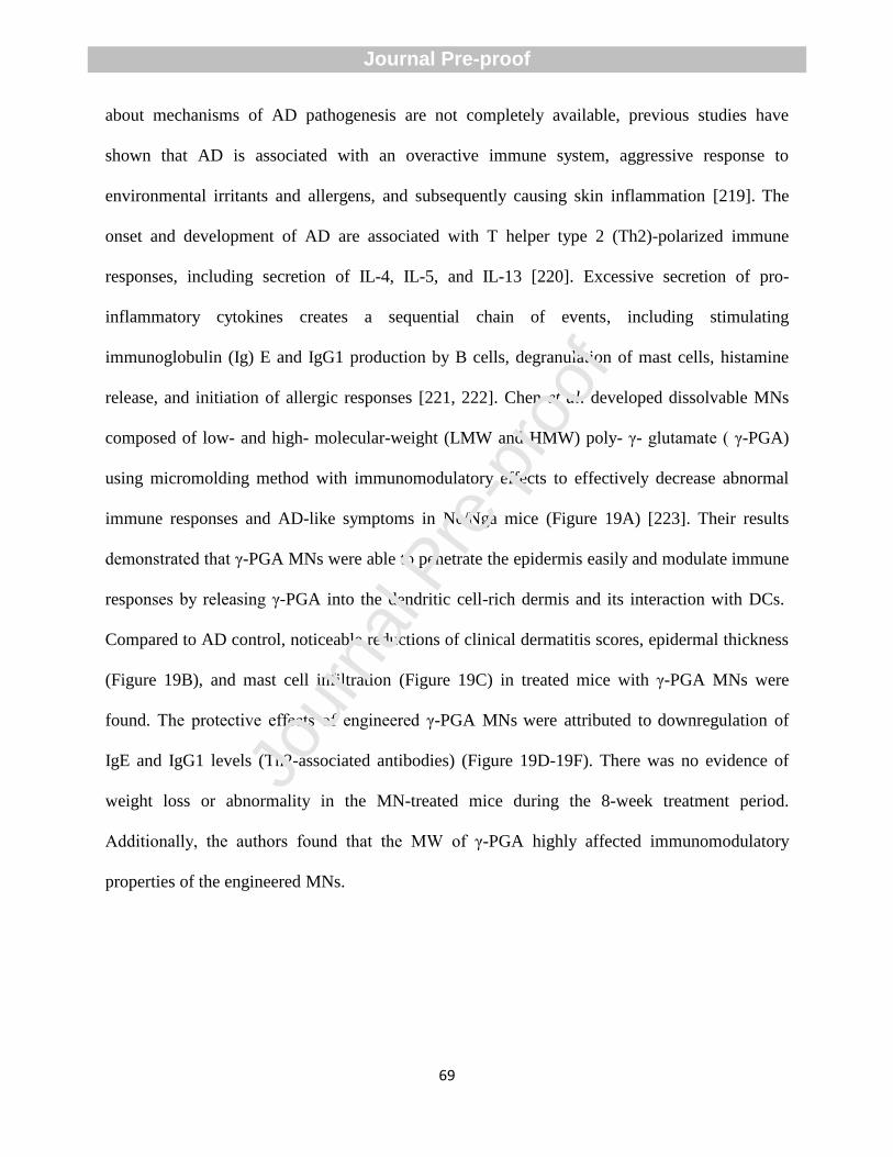
69
about mechanisms of AD pathogenesis are not completely available, previous studies have
shown that AD is associated with an overactive immune system, aggressive response to
environmental irritants and allergens, and subsequently causing skin inflammation [219]. The
onset and development of AD are associated with T helper type 2 (Th2)-polarized immune
responses, including secretion of IL-4, IL-5, and IL-13 [220]. Excessive secretion of pro-
inflammatory cytokines creates a sequential chain of events, including stimulating
immunoglobulin (Ig) E and IgG1 production by B cells, degranulation of mast cells, histamine
release, and initiation of allergic responses [221, 222]. Chen et al. developed dissolvable MNs
composed of low- and high- molecular-weight (LMW and HMW) poly- γ- glutamate ( γ-PGA)
using micromolding method with immunomodulatory effects to effectively decrease abnormal
immune responses and AD-like symptoms in Nc/Nga mice (Figure 19A) [223]. Their results
demonstrated that γ-PGA MNs were able to penetrate the epidermis easily and modulate immune
responses by releasing γ-PGA into the dendritic cell-rich dermis and its interaction with DCs.
Compared to AD control, noticeable reductions of clinical dermatitis scores, epidermal thickness
(Figure 19B), and mast cell infiltration (Figure 19C) in treated mice with γ-PGA MNs were
found. The protective effects of engineered γ-PGA MNs were attributed to downregulation of
IgE and IgG1 levels (Th2-associated antibodies) (Figure 19D-19F). There was no evidence of
weight loss or abnormality in the MN-treated mice during the 8-week treatment period.
Additionally, the authors found that the MW of γ-PGA highly affected immunomodulatory
properties of the engineered MNs.
Jour
nal P
re-p
roof
Journal Pre-proof
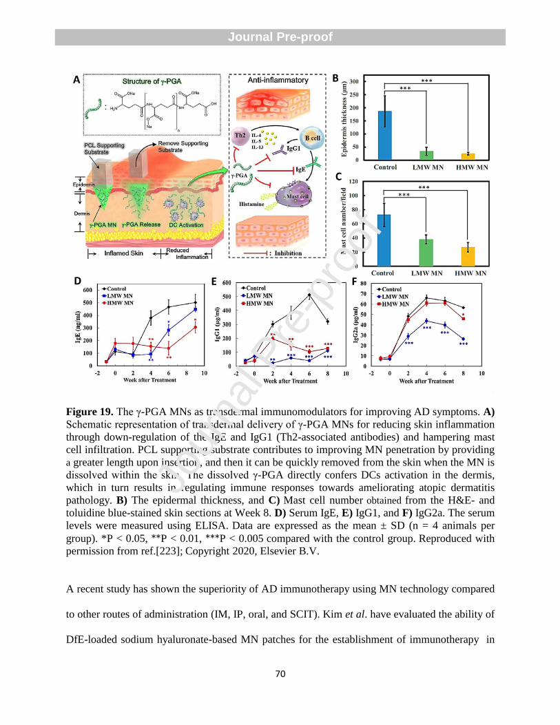
70
Figure 19. The γ-PGA MNs as transdermal immunomodulators for improving AD symptoms. A)
Schematic representation of transdermal delivery of γ-PGA MNs for reducing skin inflammation
through down-regulation of the IgE and IgG1 (Th2-associated antibodies) and hampering mast
cell infiltration. PCL supporting substrate contributes to improving MN penetration by providing
a greater length upon insertion, and then it can be quickly removed from the skin when the MN is
dissolved within the skin. The dissolved γ-PGA directly confers DCs activation in the dermis,
which in turn results in regulating immune responses towards ameliorating atopic dermatitis
pathology. B) The epidermal thickness, and C) Mast cell number obtained from the H&E- and
toluidine blue-stained skin sections at Week 8. D) Serum IgE, E) IgG1, and F) IgG2a. The serum
levels were measured using ELISA. Data are expressed as the mean ± SD (n = 4 animals per
group). *P < 0.05, **P < 0.01, ***P < 0.005 compared with the control group. Reproduced with
permission from ref.[223]; Copyright 2020, Elsevier B.V.
A recent study has shown the superiority of AD immunotherapy using MN technology compared
to other routes of administration (IM, IP, oral, and SCIT). Kim et al. have evaluated the ability of
DfE-loaded sodium hyaluronate-based MN patches for the establishment of immunotherapy in
Jour
nal P
re-p
roof
Journal Pre-proof
71
coping with AD [224]. The researchers found that an antigen extract’s concentration of 10 µg
into the MN patches created similar efficiency to subcutaneous immunotherapy with 100 µg of
the antigen extract, as quantified by the improvement of clinical appearance and reduction of
eosinophil cell counts, indicating that MN patches possess more substantial immunological
efficacy and safety. Thereby, MN patches could be considered as potential candidates for the
treatment of AD as they can transport antigen straightly into the epidermis and dermis at a lower
dose, hence reducing the risk of anaphylaxis.
8.4. MN for modulation of inflammation during the wound healing process
Inflammation displays a powerful role during the wound healing process. In the first hours after
injury, recruitment of inflammatory cells, and releasing different molecules by them contributes
to matrix remodeling and keratinocyte migration towards promoting the healing process. After
this initial response, which is mainly mediated by neutrophils, elimination of inflammation is
essential to accelerate the healing process [225]. To this end, Jeon et al. developed a hydrogel-
forming double-layered adhesive MN patch comprising of a non-swellable SF-based core (for
effective tissue fixation without delamination) and a swellable mussel adhesive protein (MAP)-
based shell that was able to improve wound sealing capacity through reduction of inflammatory
responses [226]. In this system, in-situ rigid base was comprised of the regenerated SF with
improved water solubility that was able to support effective tissue insertion of the adhesive MN
patch. The hydrogel-forming adhesive MN patches exhibited good adhesion on wet and dynamic
biological surfaces via both MAP-derived surface adhesive and swelling-mediated mechanical
interlocking (Figure 20A). Monitoring time-dependent shape changes of various DL-MN patches
demonstrated that water absorption of the 20% and 40% DL-MN patches was confined to only
Jour
nal P
re-p
roof
Journal Pre-proof
72
the end of tip region while the base region of 80% DL-MN patches began to swell up due to high
amounts of hydrophilic materials (Figure 20B). The authors reported the 80% DL-MN patch for
surgical closure owing to detachment of the swollen shell from the core that hampered stable
interfacial adhesion. A bud-like shaped MN structure was found for the 60% DL-MN patch at a
maximum swollen state that provided the possibility of physical interlocking with surrounding
tissues without delamination. In vivo experiments in a full-thickness incision of rat skin showed
that the strip-type of 60% DL-MN patch showed excellent adhesion liquid absorption ability,
which in turn resulted in good wound closure at days 4 and 7 compared to non-treatment and
suture (Figure 20C and 20D). Histological examination with H&E-staining indicated that the
60% DL-MN patch contributed to wound closure, re-epithelialization, and reducing
inflammation and inflammatory cells into the wound site, whereas prolonged intense
inflammation, delayed re-epithelialization, and impaired dermal healing over 21 days was seen
for non-treatment group (Figure 20E). Masson’s Trichrome staining further confirmed fast
deposition of produced collagen at the junction site and reducing inflammatory cells at days 4, 7,
14, and 21 (Figure 20F). The ex vivo experiments demonstrated that hydrogel-forming adhesive
MN patches had superior wound sealing capacity against luminal leaks (139.7 ± 14.1 mmHg) in
comparison with suture (151.0 ± 23.3 mmHg). Jour
nal P
re-p
roof
Journal Pre-proof
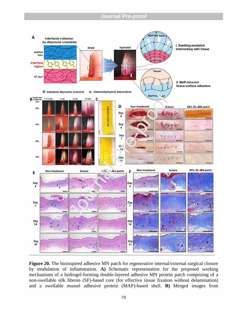
73
Figure 20. The bioinspired adhesive MN patch for regenerative internal/external surgical closure
by modulation of inflammation. A) Schematic representation for the proposed working
mechanisms of a hydrogel-forming double-layered adhesive MN protein patch comprising of a
non-swellable silk fibroin (SF)-based core (for effective tissue fixation without delamination)
and a swellable mussel adhesive protein (MAP)-based shell. B) Merged images from
Jour
nal P
re-p
roof
Journal Pre-proof
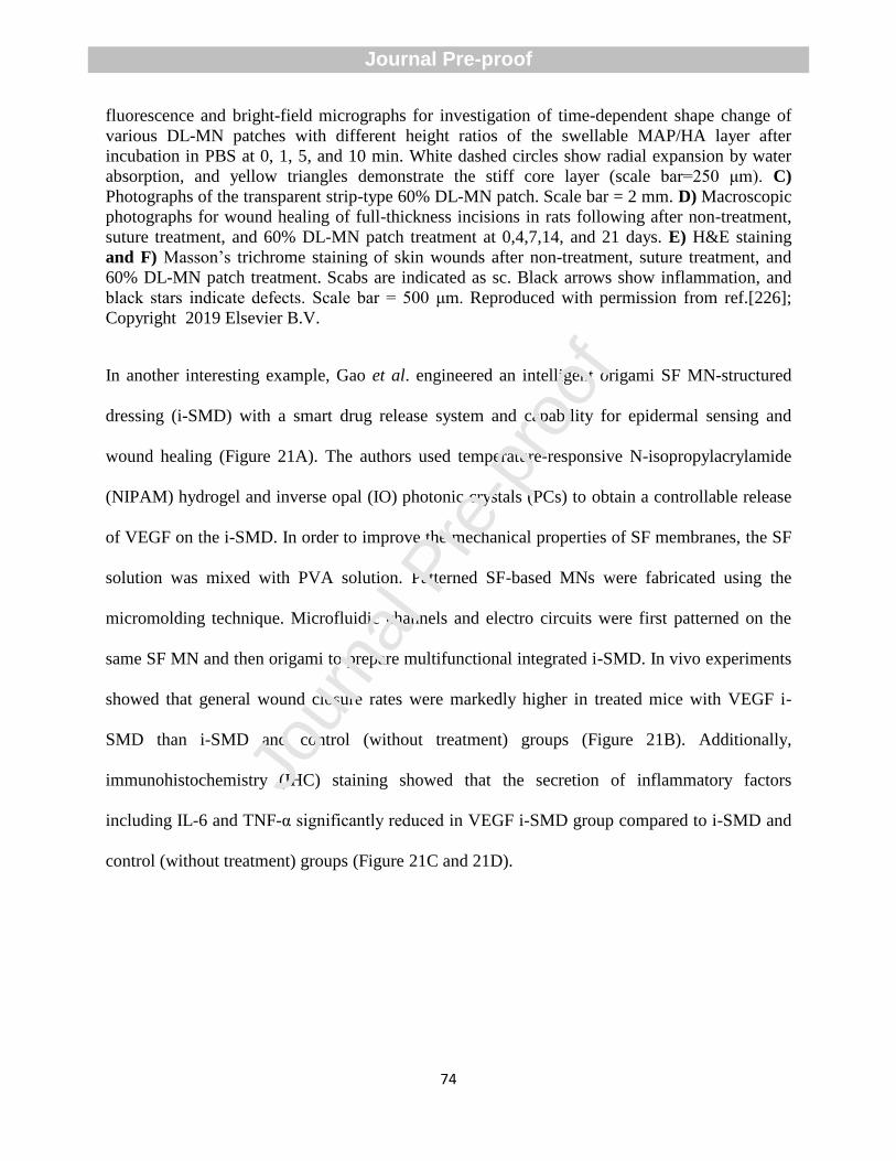
74
fluorescence and bright-field micrographs for investigation of time-dependent shape change of
various DL-MN patches with different height ratios of the swellable MAP/HA layer after
incubation in PBS at 0, 1, 5, and 10 min. White dashed circles show radial expansion by water
absorption, and yellow triangles demonstrate the stiff core layer (scale bar=250 μm). C)
Photographs of the transparent strip-type 60% DL-MN patch. Scale bar = 2 mm. D) Macroscopic
photographs for wound healing of full-thickness incisions in rats following after non-treatment,
suture treatment, and 60% DL-MN patch treatment at 0,4,7,14, and 21 days. E) H&E staining
and F) Masson’s trichrome staining of skin wounds after non-treatment, suture treatment, and
60% DL-MN patch treatment. Scabs are indicated as sc. Black arrows show inflammation, and
black stars indicate defects. Scale bar = 500 μm. Reproduced with permission from ref.[226]; Copyright 2019 Elsevier B.V.
In another interesting example, Gao et al. engineered an intelligent origami SF MN-structured
dressing (i-SMD) with a smart drug release system and capability for epidermal sensing and
wound healing (Figure 21A). The authors used temperature-responsive N-isopropylacrylamide
(NIPAM) hydrogel and inverse opal (IO) photonic crystals (PCs) to obtain a controllable release
of VEGF on the i-SMD. In order to improve the mechanical properties of SF membranes, the SF
solution was mixed with PVA solution. Patterned SF-based MNs were fabricated using the
micromolding technique. Microfluidic channels and electro circuits were first patterned on the
same SF MN and then origami to prepare multifunctional integrated i-SMD. In vivo experiments
showed that general wound closure rates were markedly higher in treated mice with VEGF i-
SMD than i-SMD and control (without treatment) groups (Figure 21B). Additionally,
immunohistochemistry (IHC) staining showed that the secretion of inflammatory factors
including IL-6 and TNF-α significantly reduced in VEGF i-SMD group compared to i-SMD and
control (without treatment) groups (Figure 21C and 21D).
Jour
nal P
re-p
roof
Journal Pre-proof
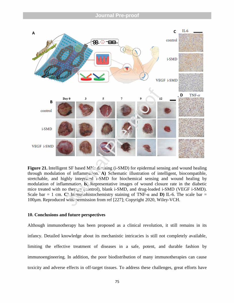
75
Figure 21. Intelligent SF based MNs dressing (i-SMD) for epidermal sensing and wound healing
through modulation of inflammation. A) Schematic illustration of intelligent, biocompatible,
stretchable, and highly integrated i-SMD for biochemical sensing and wound healing by
modulation of inflammation. B) Representative images of wound closure rate in the diabetic
mice treated with no therapy (control), blank i-SMD, and drug-loaded i-SMD (VEGF i-SMD). Scale bar = 1 cm. C) Immunohistochemistry staining of TNF-α and D) IL-6. The scale bar =
100µm. Reproduced with permission from ref [227]; Copyright 2020, Wiley-VCH.
10. Conclusions and future perspectives
Although immunotherapy has been proposed as a clinical revolution, it still remains in its
infancy. Detailed knowledge about its mechanistic intricacies is still not completely available,
limiting the effective treatment of diseases in a safe, potent, and durable fashion by
immunoengineering. In addition, the poor biodistribution of many immunotherapies can cause
toxicity and adverse effects in off-target tissues. To address these challenges, great efforts have
Jour
nal P
re-p
roof
Journal Pre-proof
76
been made to design MN transdermal delivery systems to locally deliver immunotherapeutic
with the highest efficiency and minimal invasiveness. Nevertheless, Concerns regarding the future
success of MNs for clinical immunotherapy is totally dependent on FDA approval for novel
biomaterials used in this delivery system for MN fabrication. In addition, future developments
should take into account some of the current challenges, including the difficulty of precise
coating of biomolecules on the MNs and insufficient delivery of therapeutic antigens, allergens,
or immunotherapeutic drugs to the desired site.
The improvement of the mechanical properties of MNs is another decisive factor for successful
clinical translation and future commercialization. The mechanical characteristics of MN must
meet two properties to ensure successful immunotherapy. First, the insertion force should be
enough to neglect the skin friction force for the insertion of the MNs before puncturing the skin.
Second, the insertion force must be higher than the skin force in order to perforate the skin and
create holes in the epidermis/dermis layer. The insertion force strongly depends on thickness, the
wall angle, and tip radius. Up to now, in vivo performance and efficiency of MN technology for
immunotherapies have been mainly investigated on small animals, such as mice, rats, and
rabbits, while the physiology, anatomy, and biomechanics of these animalsʼ skins are different
from the human’s skin. Future evaluations of MN technology for immunotherapies should be
implemented on the body of large animals (e.g., dogs and monkeys), on the excised human’s skin
models, or even human volunteers. However, the skin properties of each person and even of
various parts of the body are different; thereby additional investigation is required to determine
the best application areas. Another concern regarding the wide clinical use of MNs is their large-
scale production in sterile conditions, which needs a carefully thought setting. The material type
for the fabrication of MNs is another substantial issue that needs much attention. Although
Jour
nal P
re-p
roof
Journal Pre-proof
77
hollow and solid MNs fabricated by metals and silicon have mainly been applied in the clinical
trial setup to deliver various immunotherapeutic molecules for immunotherapy, the lack of
approval for silicon and the possibility of sharp hazardous tip wastes limit their clinical
applications. Owing to the increasing interest in the design of biocompatible systems, researchers
have recently focused on polymer science to fabricate biocompatible or dissolvable polymeric
MNs with minimum side effects. Therefore, it is expected to see a tremendous interest in
designing biocompatible and dissolvable MN systems in the near future with current advances in
polymer science. We believe future researches should focus on the scaling up of sterile MNs
with adequate loading capacity and minimal defects for clinical translation.
Conflict of interest
The authors declare no conflict of interest in the present study.
Acknowledgments
M.-A. Shahbazi acknowledges the financial support from the Academy of Finland (grant no.
317316). H. A. Santos acknowledges the financial support from the HiLIFE Research Funds, the
Sigrid Jusélius Foundation, and the Academy of Finland (grant no. 317042). Jour
nal P
re-p
roof
Journal Pre-proof
78
References
[1] G. Ma, C. Wu, Microneedle, bio-microneedle and bio-inspired microneedle: A review, J
Control Release, 251 (2017) 11-23.
[2] S. Bhatnagar, K. Dave, V.V.K. Venuganti, Microneedles in the clinic, J Control Release, 260
(2017) 164-182.
[3] K. van der Maaden, W. Jiskoot, J. Bouwstra, Microneedle technologies for (trans) dermal
drug and vaccine delivery, J Control Release, 161 (2012) 645-655.
[4] J. Zhu, X. Zhou, H.J. Kim, M. Qu, X. Jiang, K. Lee, L. Ren, Q. Wu, C. Wang, X. Zhu,
Gelatin Methacryloyl Microneedle Patches for Minimally Invasive Extraction of Skin Interstitial
Fluid, Small, 16 (2020) 1905910.
[5] A. Ramalheiro, J.L. Paris, B.F. Silva, L.R. Pires, Rapidly dissolving microneedles for the
delivery of cubosome-like liquid crystalline nanoparticles with sustained release of rapamycin,
Int. J. Pharm. 591 (2020) 119942.
[6] M.S. Arshad, S. Fatima, K. Nazari, R. Ali, M. Farhan, S.A. Muhammad, N. Abbas, A.
Hussain, I. Kucuk, M.-W. Chang, Engineering and characterisation of BCG-loaded polymeric
microneedles, J Drug Target. (2019) 1-8.
[7] C.B. Chesson, S. Ekpo-Otu, J.J. Endsley, J.S. Rudra, Biomaterials-based vaccination
strategies for the induction of CD8+ T cell responses, ACS Biomater. Sci. Eng. 3 (2017) 126-
143.
[8] Y.-C. Kim, J.-H. Park, M.R. Prausnitz, Microneedles for drug and vaccine delivery, Adv.
Drug Deliv. Rev. 64 (2012) 1547-1568.
[9] H. Chang, M. Zheng, S.W.T. Chew, C. Xu, Advances in the Formulations of Microneedles
for Manifold Biomedical Applications, Adv. Mater. Technol. 5 (2020) 1900552.
[10] H.L. Quinn, M.-C. Kearney, A.J. Courtenay, M.T. McCrudden, R.F. Donnelly, The role of
microneedles for drug and vaccine delivery, Expert Opin Drug Deliv. 11 (2014) 1769-1780.
[11] J.J. Norman, J.M. Arya, M.A. McClain, P.M. Frew, M.I. Meltzer, M.R. Prausnitz,
Microneedle patches: usability and acceptability for self-vaccination against influenza, Vaccine,
32 (2014) 1856-1862.
[12] L. Milling, Y. Zhang, D.J. Irvine, Delivering safer immunotherapies for cancer, Adv. Drug
Deliv. Rev. 114 (2017) 79-101.
[13] M.L. Bookstaver, S.J. Tsai, J.S. Bromberg, C.M. Jewell, Improving vaccine and
immunotherapy design using biomaterials, Trends Immunol. 39 (2018) 135-150.
[14] R. Zhang, M.M. Billingsley, M.J. Mitchell, Biomaterials for vaccine-based cancer
immunotherapy, J Control Release, 292 (2018) 256-276.
[15] M.O. Dellacherie, B.R. Seo, D.J. Mooney, Macroscale biomaterials strategies for local
immunomodulation, Nat. Rev. Mater. 4 (2019) 379-397.
[16] M. Rescigno, F. Avogadri, G. Curigliano, Challenges and prospects of immunotherapy as
cancer treatment, Biochim Biophys Acta Rev Cancer. 1776 (2007) 108-123.
[17] D.G. McNeel, Prostate cancer immunotherapy, Curr Opin Urol, 17 (2007) 175-181.
[18] R.S. Riley, C.H. June, R. Langer, M.J. Mitchell, Delivery technologies for cancer
immunotherapy, Nat. Rev. Drug Discov. 18 (2019) 175-196.
[19] C. Anfray, F. Mainini, F.T. Andón, Nanoparticles for immunotherapy, in: Front. Nanosci.
Elsevier, 2020, pp. 265-306.
[20] M. Leone, J. Mönkäre, J. Bouwstra, G. Kersten, Dissolving microneedle patches for dermal
vaccination, Pharm. Res. 34 (2017) 2223-2240.
Jour
nal P
re-p
roof
Journal Pre-proof
79
[21] L. Klimek, O. Pfaar, Asher, I.P.T.S. Group, Björkstén, I.P.I.S. Group, Maurer, Jacobsen, P.i.
group, Calderon, A comparison of immunotherapy delivery methods for allergen
immunotherapy, Expert Rev. Clin. Immunol. 9 (2013) 465-475.
[22] E. Maggi, T cell responses induced by allergen‐specific immunotherapy, Clin. Exp.
Immunol. 161 (2010) 10-18.
[23] S. Park, Y. Lee, Y.-M. Kwon, Y.-T. Lee, K.-H. Kim, E.-J. Ko, J.H. Jung, M. Song, B.
Graham, M.R. Prausnitz, Vaccination by microneedle patch with inactivated respiratory
syncytial virus and monophosphoryl lipid A enhances the protective efficacy and diminishes
inflammatory disease after challenge, PloS one, 13 (2018) e0205071.
[24] Y.-C. Kim, F.-S. Quan, R.W. Compans, S.-M. Kang, M.R. Prausnitz, Formulation and
coating of microneedles with inactivated influenza virus to improve vaccine stability and
immunogenicity, J Control Release, 142 (2010) 187-195.
[25] H.W. Yang, L. Ye, X.D. Guo, C. Yang, R.W. Compans, M.R. Prausnitz, Ebola vaccination
using a DNA vaccine coated on PLGA‐PLL/γPGA nanoparticles administered using a
microneedle patch, Adv.Healthcare Mater. 6 (2017) 1600750.
[26] W. Li, Z. Liu, F. Fontana, Y. Ding, D. Liu, J.T. Hirvonen, H.A. Santos, Tailoring porous
silicon for biomedical applications: from drug delivery to cancer immunotherapy, Adv. Mater.
30 (2018) 1703740.
[27] Y. Li, H. Zhang, R. Yang, Y. Laffitte, U. Schmill, W. Hu, M. Kaddoura, E.J. Blondeel, B.
Cui, Fabrication of sharp silicon hollow microneedles by deep-reactive ion etching towards
minimally invasive diagnostics, Microsyst. Nanoeng. 5 (2019) 1-11.
[28] M. Rajabi, N. Roxhed, R.Z. Shafagh, T. Haraldson, A.C. Fischer, W.v.d. Wijngaart, G.
Stemme, F. Niklaus, Flexible and stretchable microneedle patches with integrated rigid stainless
steel microneedles for transdermal biointerfacing, PloS one, 11 (2016) e0166330.
[29] E. Parker, M. Rao, K. Turner, C. Meinhart, N. MacDonald, Bulk micromachined titanium
microneedles, J Microelectromech Syst. 16 (2007) 289-295.
[30] S. Lee, W. Jeong, D.J. Beebe, Microfluidic valve with cored glass microneedle for
microinjection, Lab Chip. 3 (2003) 164-167.
[31] S. Olhero, E. Lopes, J. Ferreira, Fabrication of ceramic microneedles–The role of specific
interactions between processing additives and the surface of oxide particles in Epoxy Gel
Casting, J. Eur. Ceram. Soc. 36 (2016) 4131-4140.
[32] S.P. Sullivan, N. Murthy, M.R. Prausnitz, Minimally invasive protein delivery with rapidly
dissolving polymer microneedles, Adv. Mater. 20 (2008) 933-938.
[33] P.C. DeMuth, Y. Min, D.J. Irvine, P.T. Hammond, Implantable silk composite microneedles
for programmable vaccine release kinetics and enhanced immunogenicity in transcutaneous
immunization, Adv.Healthcare Mater. 3 (2014) 47-58.
[34] H. Kathuria, D. Lim, J. Cai, B.G. Chung, L. Kang, Microneedles with Tunable Dissolution
Rate, ACS Biomater. Sci. Eng. 6 (2020) 5061-5068.
[35] A.M. Rodgers, A.J. Courtenay, R.F. Donnelly, Dissolving microneedles for intradermal
vaccination: manufacture, formulation, and stakeholder considerations, in, Taylor & Francis,
2018. Volume 15, 2018 - Issue 11.
[36] L.Y. Chu, S.-O. Choi, M.R. Prausnitz, Fabrication of dissolving polymer microneedles for
controlled drug encapsulation and delivery: bubble and pedestal microneedle designs, J. Pharm.
Sci. 99 (2010) 4228-4238.
Jour
nal P
re-p
roof
Journal Pre-proof
80
[37] A. Vrdoljak, M.G. McGrath, J.B. Carey, S.J. Draper, A.V. Hill, C. O'Mahony, A.M. Crean,
A.C. Moore, Coated microneedle arrays for transcutaneous delivery of live virus vaccines, J
Control Release, 159 (2012) 34-42.
[38] J.B. Carey, F.E. Pearson, A. Vrdoljak, M.G. McGrath, A.M. Crean, P.T. Walsh, T. Doody,
C. O'Mahony, A.V. Hill, A.C. Moore, Microneedle array design determines the induction of
protective memory CD8+ T cell responses induced by a recombinant live malaria vaccine in
mice, PloS one, 6 (2011) e22442.
[39] J.B. Carey, A. Vrdoljak, C. O'mahony, A.V. Hill, S.J. Draper, A.C. Moore, Microneedle-
mediated immunization of an adenovirus-based malaria vaccine enhances antigen-specific
antibody immunity and reduces anti-vector responses compared to the intradermal route, Sci.
Rep. 4 (2014) 6154.
[40] K. Ita, Transdermal delivery of drugs with microneedles: Strategies and outcomes, J Drug
Deliv Sci Technol, 29 (2015) 16-23.
[41] K. van der Maaden, H. Yu, K. Sliedregt, R. Zwier, R. Leboux, M. Oguri, A. Kros, W.
Jiskoot, J.A. Bouwstra, Nanolayered chemical modification of silicon surfaces with ionizable
surface groups for pH-triggered protein adsorption and release: application to microneedles, J
Mater Chem B, 1 (2013) 4466-4477.
[42] R.F. Donnelly, D.I. Morrow, F. Fay, C.J. Scott, S. Abdelghany, R.R.T. Singh, M.J. Garland,
A.D. Woolfson, Microneedle-mediated intradermal nanoparticle delivery: potential for enhanced
local administration of hydrophobic pre-formed photosensitisers, Photodiagnosis Photodyn Ther,
7 (2010) 222-231.
[43] D. Jung, N.S. Rejinold, J.-E. Kwak, S.-H. Park, Y.-C. Kim, Nano-patterning of a stainless
steel microneedle surface to improve the dip-coating efficiency of a DNA vaccine and its
immune response, Colloids Surf. B, 159 (2017) 54-61.
[44] H.S. Gill, M.R. Prausnitz, Coated microneedles for transdermal delivery, J Control Release,
117 (2007) 227-237.
[45] H.-J. Choi, B.J. Bondy, D.-G. Yoo, R.W. Compans, S.-M. Kang, M.R. Prausnitz, Stability
of whole inactivated influenza virus vaccine during coating onto metal microneedles, J Control
Release, 166 (2013) 159-171.
[46] B. Cai, W. Xia, S. Bredenberg, H. Li, H. Engqvist, Bioceramic microneedles with flexible
and self-swelling substrate, Eur J Pharm Biopharm, 94 (2015) 404-410.
[47] B. Cai, W. Xia, S. Bredenberg, H. Engqvist, Self-setting bioceramic microscopic
protrusions for transdermal drug delivery, J. Mater. Chem. B, 2 (2014) 5992-5998.
[48] M. Verhoeven, S. Bystrova, L. Winnubst, H. Qureshi, T.D. De Gruijl, R.J. Scheper, R.
Luttge, Applying ceramic nanoporous microneedle arrays as a transport interface in egg plants
and an ex-vivo human skin model, Microelectron. Eng. 98 (2012) 659-662.
[49] H. Vallhov, W. Xia, H. Engqvist, A. Scheynius, Bioceramic microneedle arrays are able to
deliver OVA to dendritic cells in human skin, J. Mater. Chem. B, 6 (2018) 6808-6816.
[50] K. Ita, Ceramic microneedles and hollow microneedles for transdermal drug delivery: Two
decades of research, J Drug Deliv Sci Technol, 44 (2018) 314-322.
[51] K.A. Moga, L.R. Bickford, R.D. Geil, S.S. Dunn, A.A. Pandya, Y. Wang, J.H. Fain, C.F.
Archuleta, A.T. O'Neill, J.M. DeSimone, Rapidly–dissolvable microneedle patches via a highly
scalable and reproducible soft lithography approach, Adv. Mater. 25 (2013) 5060-5066.
[52] A.A. Ali, C.M. McCrudden, J. McCaffrey, J.W. McBride, G. Cole, N.J. Dunne, T. Robson,
A. Kissenpfennig, R.F. Donnelly, H.O. McCarthy, DNA vaccination for cervical cancer; a novel
Jour
nal P
re-p
roof
Journal Pre-proof
81
technology platform of RALA mediated gene delivery via polymeric microneedles,
Nanomedicine, 13 (2017) 921-932.
[53] K.J. Koh, Y. Liu, S.H. Lim, X.J. Loh, L. Kang, C.Y. Lim, K.K. Phua, Formulation,
characterization and evaluation of mRNA-loaded dissolvable polymeric microneedles
(RNApatch), Sci. Rep. 8 (2018) 1-11.
[54] G. Cole, J. McCaffrey, A.A. Ali, J.W. McBride, C.M. McCrudden, E.M. Vincente-Perez,
R.F. Donnelly, H.O. McCarthy, Dissolving microneedles for DNA vaccination: Improving
functionality via polymer characterization and RALA complexation, Hum Vaccin Immunother,
13 (2017) 50-62.
[55] Y. Li, X. Hu, Z. Dong, Y. Chen, W. Zhao, Y. Wang, L. Zhang, M. Chen, C. Wu, Q. Wang,
Dissolving Microneedle Arrays with Optimized Needle Geometry for Transcutaneous
Immunization, Eur J Pharm Sci, (2020) 105361.
[56] J. Mönkäre, M.R. Nejadnik, K. Baccouche, S. Romeijn, W. Jiskoot, J.A. Bouwstra, IgG-
loaded hyaluronan-based dissolving microneedles for intradermal protein delivery, J Control
Release, 218 (2015) 53-62.
[57] M.-C. Chen, S.-F. Huang, K.-Y. Lai, M.-H. Ling, Fully embeddable chitosan microneedles
as a sustained release depot for intradermal vaccination, Biomaterials, 34 (2013) 3077-3086.
[58] M. Zaric, O. Lyubomska, O. Touzelet, C. Poux, S. Al-Zahrani, F. Fay, L. Wallace, D.
Terhorst, B. Malissen, S. Henri, Skin dendritic cell targeting via microneedle arrays laden with
antigen-encapsulated poly-D, L-lactide-co-glycolide nanoparticles induces efficient antitumor
and antiviral immune responses, ACS nano, 7 (2013) 2042-2055.
[59] S. Kommareddy, B.C. Baudner, S. Oh, S.-y. Kwon, M. Singh, D.T. O'hagan, Dissolvable
microneedle patches for the delivery of cell-culture-derived influenza vaccine antigens, J. Pharm.
Sci. 101 (2012) 1021-1027.
[60] S.C. Balmert, C.D. Carey, G.D. Falo, S.K. Sethi, G. Erdos, E. Korkmaz, L.D. Falo Jr,
Dissolving undercut microneedle arrays for multicomponent cutaneous vaccination, J Control
Release, 317 (2020) 336-346.
[61] J.W. Lee, J.-H. Park, M.R. Prausnitz, Dissolving microneedles for transdermal drug
delivery, Biomaterials, 29 (2008) 2113-2124.
[62] M.S. Arshad, S. Fatima, K. Nazari, R. Ali, M. Farhan, S.A. Muhammad, N. Abbas, A.
Hussain, I. Kucuk, M.-W. Chang, Engineering and characterisation of BCG-loaded polymeric
microneedles, J Drug Target, 28 (2020) 525-532.
[63] Y.K. Demir, Z. Akan, O. Kerimoglu, Characterization of polymeric microneedle arrays for
transdermal drug delivery, PloS one, 8 (2013) e77289.
[64] P.C. DeMuth, W.F. Garcia‐Beltran, M.L. Ai‐Ling, P.T. Hammond, D.J. Irvine, Composite
dissolving microneedles for coordinated control of antigen and adjuvant delivery kinetics in
transcutaneous vaccination, Adv. Funct. Mater. 23 (2013) 161-172.
[65] H.-J. Choi, D.-G. Yoo, B.J. Bondy, F.-S. Quan, R.W. Compans, S.-M. Kang, M.R.
Prausnitz, Stability of influenza vaccine coated onto microneedles, Biomaterials, 33 (2012)
3756-3769.
[66] Y.-C. Kim, D.-G. Yoo, R.W. Compans, S.-M. Kang, M.R. Prausnitz, Cross-protection by
co-immunization with influenza hemagglutinin DNA and inactivated virus vaccine using coated
microneedles, J Control Release, 172 (2013) 579-588.
[67] P.C. DeMuth, Y. Min, B. Huang, J.A. Kramer, A.D. Miller, D.H. Barouch, P.T. Hammond,
D.J. Irvine, Polymer multilayer tattooing for enhanced DNA vaccination, Nat. Mater. 12 (2013)
367-376.
Jour
nal P
re-p
roof
Journal Pre-proof
82
[68] G.J. Fernando, J. Hickling, C.M.J. Flores, P. Griffin, C.D. Anderson, S.R. Skinner, C.
Davies, K. Witham, M. Pryor, J. Bodle, Safety, tolerability, acceptability and immunogenicity of
an influenza vaccine delivered to human skin by a novel high-density microprojection array
patch (Nanopatch™), Vaccine, 36 (2018) 3779-3788.
[69] P. Griffin, S. Elliott, K. Krauer, C. Davies, S.R. Skinner, C.D. Anderson, A. Forster, Safety,
acceptability and tolerability of uncoated and excipient-coated high density silicon micro-
projection array patches in human subjects, Vaccine, 35 (2017) 6676-6684.
[70] G. Icardi, A. Orsi, A. Ceravolo, F. Ansaldi, Current evidence on intradermal influenza
vaccines administered by Soluvia™ licensed micro injection system, Hum. Vaccines
Immunother. 8 (2012) 67-75.
[71] Y. Levin, E. Kochba, I. Hung, R. Kenney, Intradermal vaccination using the novel
microneedle device MicronJet600: Past, present, and future, Hum. Vaccines Immunother. 11
(2015) 991-997.
[72] S.B. Troy, D. Kouiavskaia, J. Siik, E. Kochba, H. Beydoun, O. Mirochnitchenko, Y. Levin,
N. Khardori, K. Chumakov, Y. Maldonado, Comparison of the immunogenicity of various
booster doses of inactivated polio vaccine delivered intradermally versus intramuscularly to
HIV-infected adults, J. Infect. Dis. 211 (2015) 1969-1976.
[73] L.Y. Chu, M.R. Prausnitz, Separable arrowhead microneedles, J Control Release, 149
(2011) 242-249.
[74] T. Liu, G. Luo, M. Xing, Biomedical Applications of Polymeric Microneedles for
Transdermal Therapeutic Delivery and Diagnosis: Current Status and Future Perspectives, Adv.
Ther. 3 (2020) 1900140.
[75] N.G. Rouphael, M. Paine, R. Mosley, S. Henry, D.V. McAllister, H. Kalluri, W. Pewin,
P.M. Frew, T. Yu, N.J. Thornburg, The safety, immunogenicity, and acceptability of inactivated
influenza vaccine delivered by microneedle patch (TIV-MNP 2015): a randomised, partly
blinded, placebo-controlled, phase 1 trial, The Lancet, 390 (2017) 649-658.
[76] S. Hirobe, H. Azukizawa, K. Matsuo, Y. Zhai, Y.-S. Quan, F. Kamiyama, H. Suzuki, I.
Katayama, N. Okada, S. Nakagawa, Development and clinical study of a self-dissolving
microneedle patch for transcutaneous immunization device, Pharm. Res. 30 (2013) 2664-2674.
[77] S. Hirobe, H. Azukizawa, T. Hanafusa, K. Matsuo, Y.-S. Quan, F. Kamiyama, I. Katayama,
N. Okada, S. Nakagawa, Clinical study and stability assessment of a novel transcutaneous
influenza vaccination using a dissolving microneedle patch, Biomaterials, 57 (2015) 50-58.
[78] J. Arya, S. Henry, H. Kalluri, D.V. McAllister, W.P. Pewin, M.R. Prausnitz, Tolerability,
usability and acceptability of dissolving microneedle patch administration in human subjects,
Biomaterials, 128 (2017) 1-7.
[79] A.S. Rzhevskiy, T.R.R. Singh, R.F. Donnelly, Y.G. Anissimov, Microneedles as the
technique of drug delivery enhancement in diverse organs and tissues, J Control Release, 270
(2018) 184-202.
[80] S. Mitragotri, P.A. Burke, R. Langer, Overcoming the challenges in administering
biopharmaceuticals: formulation and delivery strategies, Nat. Rev. Drug Discov. 13 (2014) 655-
672.
[81] A.P. Chapman, PEGylated antibodies and antibody fragments for improved therapy: a
review, Adv. Drug Deliv. Rev. 54 (2002) 531-545.
[82] Y.-H. Chen, K.-Y. Lai, Y.-H. Chiu, Y.-W. Wu, A.-L. Shiau, M.-C. Chen, Implantable
microneedles with an immune-boosting function for effective intradermal influenza vaccination,
Acta Biomater. 97 (2019) 230-238.
Jour
nal P
re-p
roof
Journal Pre-proof
83
[83] D. Mittal, M.M. Gubin, R.D. Schreiber, M.J. Smyth, New insights into cancer
immunoediting and its three component phases—elimination, equilibrium and escape, Curr.
Opin. Immunol. 27 (2014) 16-25.
[84] J. Liu, R. Zhang, Z.P. Xu, Nanoparticle‐Based Nanomedicines to Promote Cancer
Immunotherapy: Recent Advances and Future Directions, Small, 15 (2019) 1900262.
[85] Y. Cao, X. Wang, T. Jin, Y. Tian, C. Dai, C. Widarma, R. Song, F. Xu, Immune checkpoint
molecules in natural killer cells as potential targets for cancer immunotherapy, Signal Transduct
Target Ther, 5 (2020) 1-19.
[86] H.Y. Yoon, S.T. Selvan, Y. Yang, M.J. Kim, D.K. Yi, I.C. Kwon, K. Kim, Engineering
nanoparticle strategies for effective cancer immunotherapy, Biomaterials, 178 (2018) 597-607.
[87] G. Prendergast, Immune escape as a fundamental trait of cancer: focus on IDO, Oncogene,
27 (2008) 3889-3900.
[88] P. Terness, J.-J. Chuang, G. Opelz, The immunoregulatory role of IDO-producing human
dendritic cells revisited, Trends Immunol. 27 (2006) 68-73.
[89] A.L. Mellor, D.H. Munn, IDO expression by dendritic cells: tolerance and tryptophan
catabolism, Nat. Rev. Immunol. 4 (2004) 762-774.
[90] B. Ricciuti, G.C. Leonardi, P. Puccetti, F. Fallarino, V. Bianconi, A. Sahebkar, S. Baglivo,
R. Chiari, M. Pirro, Targeting indoleamine-2, 3-dioxygenase in cancer: Scientific rationale and
clinical evidence, Pharmacol. Ther. 196 (2019) 105-116.
[91] G.C. Prendergast, W.P. Malachowski, J.B. DuHadaway, A.J. Muller, Discovery of IDO1
inhibitors: from bench to bedside, Cancer Res. 77 (2017) 6795-6811.
[92] M. Chen, G. Quan, T. Wen, P. Yang, W. Qin, H. Mai, Y. Sun, C. Lu, X. Pan, C. Wu, Cold
to Hot: Binary Cooperative Microneedle Array Amplified Photo-Immunotherapy for Eliciting
Antitumor Immunity and Abscopal Effect, ACS Appl. Mater.Interfaces. 12 (2020) 32259–32269.
[93] N. Frydenlund, M. Mahalingam, PD-L1 and immune escape: insights from melanoma and
other lineage-unrelated malignancies, Hum. Pathol. 66 (2017) 13-33.
[94] J.A. Seidel, A. Otsuka, K. Kabashima, Anti-PD-1 and anti-CTLA-4 therapies in cancer:
mechanisms of action, efficacy, and limitations, Front. oncol. 8 (2018) 86.
[95] H.O. Alsaab, S. Sau, R. Alzhrani, K. Tatiparti, K. Bhise, S.K. Kashaw, A.K. Iyer, PD-1 and
PD-L1 checkpoint signaling inhibition for cancer immunotherapy: mechanism, combinations,
and clinical outcome, Front. Pharmacol. 8 (2017) 561.
[96] L. Spain, S. Diem, J. Larkin, Management of toxicities of immune checkpoint inhibitors,
Cancer Treat Rev, 44 (2016) 51-60.
[97] R.W. Jenkins, D.A. Barbie, K.T. Flaherty, Mechanisms of resistance to immune checkpoint
inhibitors, Br J Cancer, 118 (2018) 9-16.
[98] C. Wang, Y. Ye, G.M. Hochu, H. Sadeghifar, Z. Gu, Enhanced cancer immunotherapy by
microneedle patch-assisted delivery of anti-PD1 antibody, Nano Lett. 16 (2016) 2334-2340.
[99] G. Chen, Z. Chen, D. Wen, Z. Wang, H. Li, Y. Zeng, G. Dotti, R.E. Wirz, Z. Gu,
Transdermal cold atmospheric plasma-mediated immune checkpoint blockade therapy, Proc.
Natl. Acad. Sci. U.S.A. 117 (2020) 3687-3692.
[100] W. Fan, B. Yung, P. Huang, X. Chen, Nanotechnology for multimodal synergistic cancer
therapy, Chem. Rev. 117 (2017) 13566-13638.
[101] Y. Ye, J. Wang, Q. Hu, G.M. Hochu, H. Xin, C. Wang, Z. Gu, Synergistic transcutaneous
immunotherapy enhances antitumor immune responses through delivery of checkpoint inhibitors,
ACS nano, 10 (2016) 8956-8963.
Jour
nal P
re-p
roof
Journal Pre-proof
84
[102] E. Bracho-Sanchez, A. Hassanzadeh, M.A. Brusko, M.A. Wallet, B.G. Keselowsky,
Dendritic Cells Treated with Exogenous Indoleamine 2,3-Dioxygenase Maintain an Immature
Phenotype and Suppress Antigen-specific T cell Proliferation, J Immunol Regen Med, 5 (2019) 100015.
[103] L. Brochez, I. Chevolet, V. Kruse, The rationale of indoleamine 2,3-dioxygenase inhibition
for cancer therapy, Eur J Cancer, 76 (2017) 167-182.
[104] G. Yao, G. Quan, S. Lin, T. Peng, Q. Wang, H. Ran, H. Chen, Q. Zhang, L. Wang, X. Pan,
Novel dissolving microneedles for enhanced transdermal delivery of levonorgestrel: In vitro and
in vivo characterization, Int. J. Pharm. 534 (2017) 378-386.
[105] Q. Wang, G. Yao, P. Dong, Z. Gong, G. Li, K. Zhang, C. Wu, Investigation on fabrication
process of dissolving microneedle arrays to improve effective needle drug distribution, Eur J
Pharm Sci, 66 (2015) 148-156.
[106] P. Yang, C. Lu, W. Qin, M. Chen, G. Quan, H. Liu, L. Wang, X. Bai, X. Pan, C. Wu,
Construction of a Core-shell Microneedle System to Achieve Targeted Co-delivery of
Checkpoint Inhibitors for Melanoma Immunotherapy, Acta Biomater.104 (2020) 147-157.
[107] X. Lan, W. Zhu, X. Huang, Y. Yu, H. Xiao, L. Jin, J.J. Pu, X. Xie, J. She, V.W.Y. Lui,
Microneedles loaded with anti-PD-1–cisplatin nanoparticles for synergistic cancer immuno-
chemotherapy, Nanoscale, 12 (2020) 18885-18898.
[108] T. Yang, D. Huang, C. Li, D. Zhao, J. Li, M. Zhang, Y. Chen, Q. Wang, Z. Liang, X.-J.
Liang, Rolling microneedle electrode array (RoMEA) empowered nucleic acid delivery and
cancer immunotherapy, Nano Today, 36 (2021) 101017.
[109] Z. Zhou, J. Pang, X. Wu, W. Wu, X. Chen, M. Kong, Reverse immune suppressive
microenvironment in tumor draining lymph nodes to enhance anti-PD1 immunotherapy via
nanovaccine complexed microneedle, Nano Res. (2020) 1-10.
[110] Z.S. Dunn, J. Mac, P. Wang, T Cell Immunotherapy Enhanced by Designer Biomaterials,
Biomaterials, (2019) 119265.
[111] N.L. Syn, M.W. Teng, T.S. Mok, R.A. Soo, De-novo and acquired resistance to immune
checkpoint targeting, Lancet Oncol. 18 (2017) e731-e741.
[112] D.Y. Oh, J. Cham, L. Zhang, G. Fong, S.S. Kwek, M. Klinger, M. Faham, L. Fong,
Immune Toxicities Elicted by CTLA-4 Blockade in Cancer Patients Are Associated with Early
Diversification of the T-cell Repertoire, Cancer Res, 77 (2017) 1322-1330.
[113] S. Kwon, F.C. Velasquez, J.C. Rasmussen, M.R. Greives, K.D. Turner, J.R. Morrow, W.-J.
Hwu, R.F. Ross, S. Zhang, E.M. Sevick-Muraca, Nanotopography-based lymphatic delivery for
improved anti-tumor responses to checkpoint blockade immunotherapy, Theranostics, 9 (2019)
8332.
[114] S.-X. Chen, M. Ma, F. Xue, S. Shen, Q. Chen, Y. Kuang, K. Liang, X. Wang, H. Chen,
Construction of microneedle-assisted co-delivery platform and its combining
photodynamic/immunotherapy, J Control Release, 324 (2020) 218-227.
[115] B. Yang, J. Jeang, A. Yang, T.C. Wu, C.-F. Hung, DNA vaccine for cancer
immunotherapy, Hum. Vaccines Immunother. 10 (2014) 3153-3164.
[116] M. Lim, A.Z.M. Badruddoza, J. Firdous, M. Azad, A. Mannan, T.A. Al-Hilal, C.-S. Cho,
M.A. Islam, Engineered Nanodelivery Systems to Improve DNA Vaccine Technologies,
Pharmaceutics, 12 (2020) 30.
[117] G. Cole, A.A. Ali, E. McErlean, E.J. Mulholland, A. Short, C.M. McCrudden, J.
McCaffrey, T. Robson, V.L. Kett, J.A. Coulter, DNA vaccination via RALA nanoparticles in a
Jour
nal P
re-p
roof
Journal Pre-proof
85
microneedle delivery system induces a potent immune response against the endogenous prostate
cancer stem cell antigen, Acta Biomater. 96 (2019) 480-490.
[118] G. Cole, A.A. Ali, C.M. McCrudden, J.W. McBride, J. McCaffrey, T. Robson, V.L. Kett,
N.J. Dunne, R.F. Donnelly, H.O. McCarthy, DNA vaccination for cervical cancer: Strategic
optimisation of RALA mediated gene delivery from a biodegradable microneedle system, Eur J
Pharm Biopharm, 127 (2018) 288-297.
[119] T.L. Nguyen, Y. Yin, Y. Choi, J.H. Jeong, J. Kim, Enhanced Cancer DNA Vaccine via
Direct Transfection to Host Dendritic Cells Recruited in Injectable Scaffolds, ACS nano, 14
(2020) 11623-11636.
[120] J. Xu, B. Xu, J. Tao, Y. Yang, Y. Hu, Y. Huang, Microneedle‐Assisted, DC‐Targeted
Codelivery of pTRP‐2 and Adjuvant of Paclitaxel for Transcutaneous Immunotherapy, Small, 13
(2017) 1700666.
[121] H.T.T. Duong, N.W. Kim, T. Thambi, V.G. Phan, M.S. Lee, Y. Yin, J.H. Jeong, D.S. Lee,
Microneedle arrays coated with charge reversal pH-sensitive copolymers improve antigen
presenting cells-homing DNA vaccine delivery and immune responses, J Control Release, 269
(2018) 225-234.
[122] H.T.T. Duong, Y. Yin, T. Thambi, T.L. Nguyen, V.G. Phan, M.S. Lee, J.E. Lee, J. Kim,
J.H. Jeong, D.S. Lee, Smart vaccine delivery based on microneedle arrays decorated with ultra-
pH-responsive copolymers for cancer immunotherapy, Biomaterials, 185 (2018) 13-24.
[123] H.T.T. Duong, Y. Yin, T. Thambi, B.S. Kim, J.H. Jeong, D.S. Lee, Highly potent
intradermal vaccination by an array of dissolving microneedle polypeptide cocktail for cancer
immunotherapy, J. Mater. Chem. B, 8 (2020) 1171-1181.
[124] C.E. Boone, C. Wang, M.A. Lopez-Ramirez, V. Beiss, S. Shukla, P.L. Chariou, D. Kupor,
R. Rueda, J. Wang, N.F. Steinmetz, Active Microneedle Administration of Plant Virus
Nanoparticles for Cancer In Situ Vaccination Improves Immunotherapeutic Efficacy, ACS Appl.
Nano Mater. 3 (2020) 8037-8051.
[125] L.L. Colombo, S.I. Vanzulli, A. Blázquez-Castro, C.S. Terrero, J.C. Stockert,
Photothermal effect by 808-nm laser irradiation of melanin: a proof-of-concept study of
photothermal therapy using B16-F10 melanotic melanoma growing in BALB/c mice, Biomed.
Opt. Express, 10 (2019) 2932-2941.
[126] Y. Ye, C. Wang, X. Zhang, Q. Hu, Y. Zhang, Q. Liu, D. Wen, J. Milligan, A. Bellotti, L.
Huang, A melanin-mediated cancer immunotherapy patch, Sci. Immunol, 2 (2017) eaan5692.
[127] S.-J. Lee, H.-S. Lee, Y.-H. Hwang, J.-J. Kim, K.-Y. Kang, S.J. Kim, H.K. Kim, J.D. Kim,
D.H. Jeong, M.-J. Paik, Enhanced anti-tumor immunotherapy by dissolving microneedle patch
loaded ovalbumin, PloS one, 14 (2019) e0220382.
[128] N.W. Kim, S.-Y. Kim, J.E. Lee, Y. Yin, J.H. Lee, S.Y. Lim, E.S. Kim, H.T.T. Duong,
H.K. Kim, S. Kim, Enhanced cancer vaccination by in situ nanomicelle-generating dissolving
microneedles, ACS nano, 12 (2018) 9702-9713.
[129] N. Kohli, P. Sawadkar, S. Ho, V. Sharma, M. Snow, S. Powell, M.A. Woodruff, L. Hook,
E. García-Gareta, Pre-screening the intrinsic angiogenic capacity of biomaterials in an optimised
ex ovo chorioallantoic membrane model, J. Tissue Eng. 11 (2020) 2041731420901621-
2041731420901621.
[130] R. Fertig, A. Gamret, J. Cervantes, A. Tosti, Microneedling for the treatment of hair loss?,
J Eur Acad Dermatol Venereol, 32 (2018) 564-569.
Jour
nal P
re-p
roof
Journal Pre-proof
86
[131] P. Shende, M. Salunke, Transepidermal microneedles for co-administration of folic acid
with methotrexate in the treatment of rheumatoid arthritis, Biomed. Phys. Eng. Express, 5 (2019)
025023.
[132] D.M. Goldenberg, W.A. Wegener, Subcutaneous administration of anti-CD74 antibody for
systemic lupus erythematosus, in, Google Patents, 2019.
[133] C. Giorgio, G. Babino, S. Caccavale, T. Russo, A. De Rosa, R. Alfano, E. Fulgione, G.
Argenziano, Combination of photodynamic therapy with 5‐aminolaevulinic acid and
microneedling in the treatment of alopecia areata resistant to conventional therapies: our
experience with 41 patients, Clin Exp Dermatol, 45 (2020) 323-326.
[134] T. Zhang, X.-y. Qin, X. Cao, W.-h. Li, T. Gong, Z.-r. Zhang, Thymopentin-loaded
phospholipid-based phase separation gel with long-lasting immunomodulatory effects: in vitro
and in vivo studies, Acta Pharmacol. Sin. 40 (2019) 514-521.
[135] S. Lin, B. Cai, G. Quan, T. Peng, G. Yao, C. Zhu, Q. Wu, H. Ran, X. Pan, C. Wu, Novel
strategy for immunomodulation: Dissolving microneedle array encapsulating thymopentin
fabricated by modified two-step molding technology, Eur J Pharm Biopharm, 122 (2018) 104-
112.
[136] Z. Ahmad, M.I. Khan, M.I. Siddique, H.S. Sarwar, G. Shahnaz, S.Z. Hussain, N.I.
Bukhari, I. Hussain, M.F. Sohail, Fabrication and Characterization of Thiolated Chitosan
Microneedle Patch for Transdermal Delivery of Tacrolimus, AAPS PharmSciTech, 21 (2020) 1-
12.
[137] B.O. Roep, D.C. Wheeler, M. Peakman, Antigen-based immune modulation therapy for
type 1 diabetes: the era of precision medicine, Lancet Diabetes Endocrinol. 7 (2019) 65-74.
[138] R.J. Creusot, J. Postigo-Fernandez, N. Teteloshvili, Altered function of antigen-presenting
cells in type 1 diabetes: a challenge for antigen-specific immunotherapy?, Diabetes, 67 (2018)
1481-1494.
[139] X. Zhao, J.C. Birchall, S.A. Coulman, D. Tatovic, R.K. Singh, L. Wen, F.S. Wong, C.M.
Dayan, S.J. Hanna, Microneedle delivery of autoantigen for immunotherapy in type 1 diabetes, J
Control Release, 223 (2016) 178-187.
[140] M. Dul, T. Nikolic, M. Stefanidou, M. McAteer, P. Williams, J. Mous, B. Roep, E.
Kochba, Y. Levin, M. Peakman, Conjugation of a peptide autoantigen to gold nanoparticles for
intradermally administered antigen specific immunotherapy, Int. J. Pharm. 562 (2019) 303-312.
[141] M. So, C.M. Elso, E. Tresoldi, M. Pakusch, V. Pathiraja, J.M. Wentworth, L.C. Harrison,
B. Krishnamurthy, H.E. Thomas, C. Rodda, Proinsulin C-peptide is an autoantigen in people
with type 1 diabetes, Proc. Natl. Acad. Sci. U.S.A. 115 (2018) 10732-10737.
[142] F. Arikat, S.J. Hanna, R.K. Singh, L. Vilela, F.S. Wong, C.M. Dayan, S.A. Coulman, J.C.
Birchall, Targeting proinsulin to local immune cells using an intradermal microneedle delivery
system; a potential antigen-specific immunotherapy for type 1 diabetes, J Control Release, 322
(2020) 593-601.
[143] R.F. Donnelly, T.R.R. Singh, M.M. Tunney, D.I. Morrow, P.A. McCarron, C. O’Mahony,
A.D. Woolfson, Microneedle arrays allow lower microbial penetration than hypodermic needles
in vitro, Pharm. Res. 26 (2009) 2513-2522.
[144] J. Gupta, H.S. Gill, S.N. Andrews, M.R. Prausnitz, Kinetics of skin resealing after
insertion of microneedles in human subjects, J Control Release, 154 (2011) 148-155.
[145] L. Wei-Ze, H. Mei-Rong, Z. Jian-Ping, Z. Yong-Qiang, H. Bao-Hua, L. Ting, Z. Yong,
Super-short solid silicon microneedles for transdermal drug delivery applications, Int. J. Pharm.
389 (2010) 122-129.
Jour
nal P
re-p
roof
Journal Pre-proof
87
[146] S. Li, W. Li, M. Prausnitz, Individually coated microneedles for co-delivery of multiple
compounds with different properties, Drug Deliv. Transl. Res. 8 (2018) 1043-1052.
[147] G.S. Firestein, I.B. McInnes, Immunopathogenesis of rheumatoid arthritis, Immunity, 46
(2017) 183-196.
[148] F.M. Meier, M. Frerix, W. Hermann, U. Muller-Ladner, Current immunotherapy in
rheumatoid arthritis, Immunotherapy, 5 (2013) 955-974.
[149] E. Larraneta, R.E. Lutton, A.D. Woolfson, R.F. Donnelly, Microneedle arrays as
transdermal and intradermal drug delivery systems: Materials science, manufacture and
commercial development, Mater. Sci. Eng. R Rep. 104 (2016) 1-32.
[150] W. Yao, C. Tao, J. Zou, H. Zheng, J. Zhu, Z. Zhu, J. Zhu, L. Liu, F. Li, X. Song, Flexible
two-layer dissolving and safing microneedle transdermal of neurotoxin: A biocomfortable
attempt to treat Rheumatoid Arthritis, Int. J. Pharm. 563 (2019) 91-100.
[151] S. Thakur, B. Riyaz, A. Patil, A. Kaur, B. Kapoor, V. Mishra, Novel drug delivery systems
for NSAIDs in management of rheumatoid arthritis: An overview, Biomed. Pharmacother. 106
(2018) 1011-1023.
[152] S. Amodwala, P. Kumar, H.P. Thakkar, Statistically optimized fast dissolving microneedle
transdermal patch of meloxicam: a patient friendly approach to manage arthritis, Eur J Pharm
Sci, 104 (2017) 114-123.
[153] J. Chen, W. Huang, Z. Huang, S. Liu, Y. Ye, Q. Li, M. Huang, Fabrication of tip-
dissolving microneedles for transdermal drug delivery of meloxicam, AAPS PharmSciTech, 19
(2018) 1141-1151.
[154] J. Cao, N. Zhang, Z. Wang, J. Su, J. Yang, J. Han, Y. Zhao, Microneedle-Assisted
Transdermal Delivery of Etanercept for Rheumatoid Arthritis Treatment, Pharmaceutics, 11
(2019) 235.
[155] Y. Qiu, C. Li, S. Zhang, G. Yang, M. He, Y. Gao, Systemic delivery of artemether by
dissolving microneedles, Int. J. Pharm. 508 (2016) 1-9.
[156] X. Chen, X. Zhu, T. Xu, M. Xu, Y. Wen, Y. Liu, J. Liu, X. Qin, Targeted hexagonal Pd
nanosheet combination therapy for rheumatoid arthritis via the photothermal controlled release
of MTX, J. Mater. Chem. B, 7 (2019) 112-122.
[157] K. Yu, X. Yu, S. Cao, Y. Wang, Y. Zhai, F. Yang, X. Yang, Y. Lu, C. Wu, Y. Xu, Layered
dissolving microneedles as a need-based delivery system to simultaneously alleviate skin and
joint lesions in psoriatic arthritis, Acta Pharm. Sin. B. (2020).
[158] M. Zhao, J. Bai, Y. Lu, S. Du, K. Shang, P. Li, L. Yang, B. Dong, N. Tan, Anti-arthritic
effects of microneedling with bee venom gel, J. Tradit. Chinese Medical Sci. 3 (2016) 256-262.
[159] G. Chen, B. Hao, D. Ju, M. Liu, H. Zhao, Z. Du, J. Xia, Pharmacokinetic and
pharmacodynamic study of triptolide-loaded liposome hydrogel patch under microneedles on rats
with collagen-induced arthritis, Acta Pharm. Sin. B. 5 (2015) 569-576.
[160] H. Renz, K.J. Allen, S.H. Sicherer, H.A. Sampson, G. Lack, K. Beyer, H.C. Oettgen, Food
allergy, Nat. Rev. Dis. Primers. 4 (2018) 1-20.
[161] R. Valenta, A. Karaulov, V. Niederberger, P. Gattinger, M. van Hage, S. Flicker, B.
Linhart, R. Campana, M. Focke-Tejkl, M. Curin, Molecular aspects of allergens and allergy, in:
Advances in immunology, Elsevier, 2018, pp. 195-256.
[162] S. Ito, S. Hirobe, Y. Kuwabara, M. Nagao, M. Saito, Y.-S. Quan, F. Kamiyama, T.
Fujisawa, N. Okada, Immunogenicity of Milk Protein-Containing Hydrophilic Gel Patch for
Epicutaneous Immunotherapy for Milk Allergy, Pharm. Res. 37 (2020) 35.
Jour
nal P
re-p
roof
Journal Pre-proof
88
[163] J.N. Larsen, L. Broge, H. Jacobi, Allergy immunotherapy: the future of allergy treatment,
Drug Discov. Today, 21 (2016) 26-37.
[164] A.K. Shakya, R.S. Ingrole, G. Joshi, M.J. Uddin, S. Anvari, C.M. Davis, H.S. Gill,
Microneedles coated with peanut allergen enable desensitization of peanut sensitized mice, J
Control Release, 314 (2019) 38-47.
[165] H.A. Sampson, W.G. Shreffler, W.H. Yang, G.L. Sussman, T.F. Brown-Whitehorn, K.C.
Nadeau, A.S. Cheema, S.A. Leonard, J.A. Pongracic, C. Sauvage-Delebarre, Effect of varying
doses of epicutaneous immunotherapy vs placebo on reaction to peanut protein exposure among
patients with peanut sensitivity: a randomized clinical trial, Jama, 318 (2017) 1798-1809.
[166] Y. Yu, M.N.K. Kumar, M.X. Wu, Delivery of allergen powder for safe and effective
epicutaneous immunotherapy, J. Allergy Clin. Immunol. 145 (2020) 597-609.
[167] N.L. Krupp, S. Sehra, J.E. Slaven, M.H. Kaplan, S. Gupta, R.S. Tepper, Increased
prevalence of airway reactivity in children with eosinophilic esophagitis, Pediatr. Pulmonol. 51
(2016) 478-483.
[168] S.S. Possa, E.A. Leick, C.M. Prado, M.A. Martins, I.F.L.C. Tibério, Eosinophilic
inflammation in allergic asthma, Front. Pharmacol. 4 (2013) 46.
[169] S.I. Wasserman, Mast cells and airway inflammation in asthma, Am. J. Respir. Crit. Care
Med. 150 (1994) S39.
[170] C.A. Akdis, M. Akdis, Mechanisms of immune tolerance to allergens: role of IL-10 and
Tregs, J. Clin. Investig. 124 (2014) 4678-4680.
[171] P.G. Holt, P.D. Sly, H.A. Sampson, P. Robinson, R. Loh, H. Lowenstein, A. Calatroni, P.
Sayre, Prophylactic use of sublingual allergen immunotherapy in high-risk children: a pilot
study, J. Allergy Clin. Immunol. 132 (2013) 991-993. e991.
[172] M.H. Sohn, Efficacy and safety of subcutaneous allergen immunotherapy for allergic
rhinitis, Allergy Asthma Immunol. Res. 10 (2018) 1-3.
[173] A.K. Shakya, C.H. Lee, H.S. Gill, Microneedle-Mediated Allergen-Specific
Immunotherapy for the Treatment of Airway Allergy in Mice, Mol. Pharm. 17 (2020) 3033-
3042.
[174] A.K. Shakya, C.H. Lee, H.S. Gill, Coated microneedle-based cutaneous immunotherapy
prevents Der p 1–induced airway allergy in mice, J. Allergy Clin. Immunol. 142 (2018) 2007-
2011. e2003.
[175] A.K. Shakya, C.H. Lee, H.S. Gill, Cutaneous vaccination with coated microneedles
prevents development of airway allergy, J Control Release, 265 (2017) 75-82.
[176] K.H. Park, E.Y. Oh, H. Han, J.D. Kim, S.J. Kim, K.Y. Jeong, J.H. Kim, C.O. Park, S.R.
Kim, J.H. Lee, Efficacy of transdermal immunotherapy with biodegradable microneedle patches
in a murine asthma model, Clin. Exp. Allergy, 50 (2020) 1084-1092.
[177] A.K. Shakya, H.S. Gill, A comparative study of microneedle-based cutaneous
immunization with other conventional routes to assess feasibility of microneedles for allergy
immunotherapy, Vaccine, 33 (2015) 4060-4064.
[178] A.K. Shakya, C.H. Lee, M.J. Uddin, H.S. Gill, Assessment of Th1/Th2 Bias of sting
agonists coated on microneedles for possible use in skin allergen immunotherapy, Mol. Pharm.
15 (2018) 5437-5443.
[179] L. Spina, M. Weisskopf, S. von Moos, N. Graf, T.M. Kündig, G. Senti, Comparison of
microneedles and adhesive-tape stripping in skin preparation for epicutaneous allergen delivery,
Int. Arch. Allergy Immunol. 167 (2015) 103-109.
Jour
nal P
re-p
roof
Journal Pre-proof
89
[180] J. Pielenhofer, J. Sohl, M. Windbergs, P. Langguth, M.P. Radsak, Current Progress in
Particle-Based Systems for Transdermal Vaccine Delivery, Front. Immunol. 11 (2020) 266.
[181] N.M. van der Burg, A.C. Depelsenaire, M.L. Crichton, P. Kuo, S. Phipps, M.A. Kendall, A
low inflammatory, Langerhans cell-targeted microprojection patch to deliver ovalbumin to the
epidermis of mouse skin, J Control Release, 302 (2019) 190-200.
[182] M. Kermode, Unsafe injections in low-income country health settings: need for injection
safety promotion to prevent the spread of blood-borne viruses, Health Promot. Int. 19 (2004) 95-
103.
[183] Z. Li, Y. He, L. Deng, Z.-R. Zhang, Y. Lin, A fast-dissolving microneedle array loaded
with chitosan nanoparticles to evoke systemic immune responses in mice, J. Mater. Chem. B, 8
(2020) 216-225.
[184] A.P. Raphael, T.W. Prow, M.L. Crichton, X. Chen, G.J. Fernando, M.A. Kendall,
Targeted, needle‐free vaccinations in skin using multilayered, densely‐packed dissolving
microprojection arrays, Small, 6 (2010) 1785-1793.
[185] S. Moon, Y. Wang, C. Edens, J.R. Gentsch, M.R. Prausnitz, B. Jiang, Dose sparing and
enhanced immunogenicity of inactivated rotavirus vaccine administered by skin vaccination
using a microneedle patch, Vaccine, 31 (2013) 3396-3402.
[186] P.C. DeMuth, J.J. Moon, H. Suh, P.T. Hammond, D.J. Irvine, Releasable layer-by-layer
assembly of stabilized lipid nanocapsules on microneedles for enhanced transcutaneous vaccine
delivery, ACS nano, 6 (2012) 8041-8051.
[187] A.V. Boopathy, A. Mandal, D.W. Kulp, S. Menis, N.R. Bennett, H.C. Watkins, W. Wang,
J.T. Martin, N.T. Thai, Y. He, Enhancing humoral immunity via sustained-release implantable
microneedle patch vaccination, Proc. Natl. Acad. Sci. U.S.A. 116 (2019) 16473-16478.
[188] Q. Yan, H. Liu, Z. Cheng, Y. Xue, Z. Cheng, X. Dai, W. Shan, F. Chen,
Immunotherapeutic effect of BCG-polysaccharide nucleic acid powder on Mycobacterium
tuberculosis-infected mice using microneedle patches, Drug Deliv. 24 (2017) 1648-1653.
[189] M.J. Mistilis, A.S. Bommarius, M.R. Prausnitz, Development of a thermostable
microneedle patch for influenza vaccination, J. Pharm. Sci. 104 (2015) 740-749.
[190] J. Arya, M.R. Prausnitz, Microneedle patches for vaccination in developing countries, J
Control Release, 240 (2016) 135-141.
[191] V. Bachy, C. Hervouet, P.D. Becker, L. Chorro, L.M. Carlin, S. Herath, T. Papagatsias, J.-
B. Barbaroux, S.-J. Oh, A. Benlahrech, Langerin negative dendritic cells promote potent CD8+
T-cell priming by skin delivery of live adenovirus vaccine microneedle arrays, Proc. Natl. Acad.
Sci. U.S.A. 110 (2013) 3041-3046.
[192] C. Edens, M.L. Collins, J.L. Goodson, P.A. Rota, M.R. Prausnitz, A microneedle patch
containing measles vaccine is immunogenic in non-human primates, Vaccine, 33 (2015) 4712-
4718.
[193] E. Kim, G. Erdos, S. Huang, T.W. Kenniston, S.C. Balmert, C.D. Carey, V.S. Raj, M.W.
Epperly, W.B. Klimstra, B.L. Haagmans, Microneedle array delivered recombinant coronavirus
vaccines: Immunogenicity and rapid translational development, EBioMedicine, (2020) 102743.
[194] K.J. McHugh, L. Jing, S.Y. Severt, M. Cruz, M. Sarmadi, H.S.N. Jayawardena, C.F.
Perkinson, F. Larusson, S. Rose, S. Tomasic, Biocompatible near-infrared quantum dots
delivered to the skin by microneedle patches record vaccination, Sci. Transl. Med. 11 (2019).
[195] L. Niu, L.Y. Chu, S.A. Burton, K.J. Hansen, J. Panyam, Intradermal delivery of vaccine
nanoparticles using hollow microneedle array generates enhanced and balanced immune
response, J Control Release, 294 (2019) 268-278.
Jour
nal P
re-p
roof
Journal Pre-proof
90
[196] Q. Zhu, V.G. Zarnitsyn, L. Ye, Z. Wen, Y. Gao, L. Pan, I. Skountzou, H.S. Gill, M.R.
Prausnitz, C. Yang, Immunization by vaccine-coated microneedle arrays protects against lethal
influenza virus challenge, Proc. Natl. Acad. Sci. U.S.A. 106 (2009) 7968-7973.
[197] J.-H. Shin, J.-H. Lee, S.D. Jeong, J.-Y. Noh, H.W. Lee, C.-S. Song, Y.-C. Kim, C-di-GMP
with influenza vaccine showed enhanced and shifted immune responses in microneedle
vaccination in the skin, Drug Deliv. Transl. Res. (2020) 1-11.
[198] T.T. Nguyen, J.-a. Choi, J.S. Kim, H. Park, E. Yang, W.J. Lee, S.-K. Baek, M. Song, J.-H.
Park, Skin immunization with third-generation hepatitis B surface antigen using microneedles,
Vaccine, 37 (2019) 5954-5961.
[199] K. van der Maaden, E. Sekerdag, P. Schipper, G. Kersten, W. Jiskoot, J. Bouwstra, Layer-
by-layer assembly of inactivated poliovirus and N-trimethyl chitosan on pH-sensitive
microneedles for dermal vaccination, Langmuir, 31 (2015) 8654-8660.
[200] S.P. Sullivan, D.G. Koutsonanos, M. del Pilar Martin, J.W. Lee, V. Zarnitsyn, S.-O. Choi,
N. Murthy, R.W. Compans, I. Skountzou, M.R. Prausnitz, Dissolving polymer microneedle
patches for influenza vaccination, Nat. Med. 16 (2010) 915.
[201] G. Erdos, S.C. Balmert, C.D. Carey, G.D. Falo, N.A. Patel, J. Zhang, A. Gambotto, E.
Korkmaz, L.D. Falo Jr, Improved cutaneous genetic immunization by microneedle array delivery
of an adjuvanted adenovirus vaccine, J. Invest. Dermatol, 140 (2020) 2528–2531.e2.
[202] Y. Pastor, E. Larrañeta, Á. Erhard, G. Quincoces, I. Peñuelas, J.M. Irache, R. Donnelly, C.
Gamazo, Dissolving Microneedles for Intradermal Vaccination against Shigellosis, Vaccines, 7
(2019) 159.
[203] D.D. Zhu, X.L. Wang, X.P. Zhang, J.J. Ma, D.L. Kong, M.M. Zhang, X.D. Guo, C. Wang,
A Dissolvable Microneedle Formulation of Bordetella pertussis Subunit Vaccine: Translational
Development and Immunological Evaluation in Mice, ACS Appl. Bio Mater. 2 (2019) 5053-
5061.
[204] Y.-C. Chen, S.-J. Chen, H.-F. Cheng, M.-K. Yeh, Development of Yersinia pestis F1
antigen-loaded liposome vaccine against plague using microneedles as a delivery system, J Drug
Deliv Sci Technol, 55 (2020) 101443.
[205] T. Guo, N. Cheng, J. Zhao, X. Hou, Y. Zhang, N. Feng, Novel nanostructured lipid
carriers-loaded dissolving microneedles for controlled local administration of aconitine, Int. J.
Pharm. 572 (2019) 118741.
[206] J. Ye, H. Huang, G. Luo, L. Yin, B. Li, S. Chen, H. Li, Y. Yang, X. Yang, NB‐UVB
irradiation attenuates inflammatory response in psoriasis, Dermatol Ther.33 (2020) e13626.
[207] S.K. Mahil, F. Capon, J.N. Barker, Update on psoriasis immunopathogenesis and targeted
immunotherapy, in: Seminars in immunopathology, Springer, 2016, pp. 11-27.
[208] S.N. Bodkhe, R. Kale, K. Biyani, A Review of Microneedles–Elevation to TDDS
Approach and Function in Management of Psoriasis, AJPRD, 8 (2020) 114-122.
[209] A.M. Rodgers, A.S. Cordeiro, A. Kissenpfennig, R.F. Donnelly, Microneedle arrays for
vaccine delivery: the possibilities, challenges and use of nanoparticles as a combinatorial
approach for enhanced vaccine immunogenicity, Expert Opin Drug Deliv. 15 (2018) 851-867.
[210] H. Du, P. Liu, J. Zhu, J. Lan, Y. Li, L. Zhang, J. Zhu, J. Tao, Hyaluronic acid-based
dissolving microneedle patch loaded with methotrexate for improved treatment of psoriasis, ACS
Appl. Mater.Interfaces, 11 (2019) 43588-43598.
[211] E. Korkmaz, E.E. Friedrich, M.H. Ramadan, G. Erdos, A.R. Mathers, O.B. Ozdoganlar,
N.R. Washburn, L.D. Falo Jr, Therapeutic intradermal delivery of tumor necrosis factor-alpha
antibodies using tip-loaded dissolvable microneedle arrays, Acta Biomater. 24 (2015) 96-105.
Jour
nal P
re-p
roof
Journal Pre-proof
91
[212] E. Korkmaz, E.E. Friedrich, M.H. Ramadan, G. Erdos, A.R. Mathers, O.B. Ozdoganlar,
N.R. Washburn, L.D. Falo Jr, Tip-loaded dissolvable microneedle arrays effectively deliver
polymer-conjugated antibody inhibitors of tumor-necrosis-factor-alpha into human skin, J.
Pharm. Sci. 105 (2016) 3453-3457.
[213] H.-J. Lim, Y.-D. Jeon, S.-H. Kang, M.-K. Shin, K.-M. Lee, S.-E. Jung, J.-Y. Cha, H.-Y.
Lee, B.-R. Kim, S.-W. Hwang, Inhibitory effects of Euphorbia supina on Propionibacterium
acnes-induced skin inflammation in vitro and in vivo, BMC Compl Alternative Med, 18 (2018)
263.
[214] L.-T. Chuang, T.-H. Tsai, T.-J. Lien, W.-C. Huang, J.-J. Liu, H. Chang, M.-L. Chang, P.-J.
Tsai, Ethanolic extract of origanum vulgare suppresses propionibacterium acnes-induced
inflammatory responses in human monocyte and mouse ear edema models, Molecules, 23 (2018)
1987.
[215] S.H. Lee, M.K. Gupta, J.B. Bang, H. Bae, H.J. Sung, Current progress in reactive oxygen
species (ROS)‐responsive materials for biomedical applications, Adv. Healthcare Mater. 2
(2013) 908-915.
[216] Y. Zhang, P. Feng, J. Yu, J. Yang, J. Zhao, J. Wang, Q. Shen, Z. Gu, ROS‐Responsive
Microneedle Patch for Acne Vulgaris Treatment, Adv. Ther. 1 (2018) 1800035.
[217] T. Zhang, B. Sun, J. Guo, M. Wang, H. Cui, H. Mao, B. Wang, F. Yan, Active
pharmaceutical ingredient poly (ionic liquid)-based microneedles for the treatment of skin acne
infection, Acta Biomater. 115 (2020) 136-147.
[218] J.E. Kim, J.S. Kim, D.H. Cho, H.J. Park, Molecular mechanisms of cutaneous
inflammatory disorder: atopic dermatitis, Int. J. Mol. Sci. 17 (2016) 1234.
[219] T. Werfel, J.-P. Allam, T. Biedermann, K. Eyerich, S. Gilles, E. Guttman-Yassky, W.
Hoetzenecker, E. Knol, H.-U. Simon, A. Wollenberg, Cellular and molecular immunologic
mechanisms in patients with atopic dermatitis, J. Allergy Clin. Immunol. 138 (2016) 336-349.
[220] S.H. Park, J.E. An, S. Jang, J.Y. Kim, J.W. Lee, H.K. Kim, Gardenia jasminoides extract
without crocin improved atopic dermatitis-like skin lesions via suppression of Th2-related
cytokines in Dfe-induced NC/Nga mice, Journal of ethnopharmacology, 241 (2019) 112015.
[221] H.-Y. Jang, J.-H. Koo, S.-M. Lee, B.-H. Park, Atopic dermatitis-like skin lesions are
suppressed in fat-1 transgenic mice through the inhibition of inflammasomes, Exp. Mol. Med. 50
(2018) 1-9.
[222] K. Amin, The role of mast cells in allergic inflammation, Respir. Med. 106 (2012) 9-14.
[223] M.-C. Chen, C.-S. Chen, Y.-W. Wu, Y.-Y. Yang, Poly-γ-Glutamate microneedles as
transdermal immunomodulators for ameliorating atopic dermatitis-like skin lesions in Nc/Nga
mice, Acta Biomater. 114 (2020) 183-192.
[224] J.H. Kim, J.U. Shin, S.H. Kim, J.Y. Noh, H.R. Kim, J. Lee, H. Chu, K.Y. Jeong, K.H.
Park, J.D. Kim, Successful transdermal allergen delivery and allergen-specific immunotherapy
using biodegradable microneedle patches, Biomaterials, 150 (2018) 38-48.
[225] A. Gutiérrez-Fernández, M. Inada, M. Balbín, A. Fueyo, A.S. Pitiot, A. Astudillo, K.
Hirose, M. Hirata, S.D. Shapiro, A. Noël, Increased inflammation delays wound healing in mice
deficient in collagenase‐2 (MMP‐8), FASEB J. 21 (2007) 2580-2591.
[226] E.Y. Jeon, J. Lee, B.J. Kim, K.I. Joo, K.H. Kim, G. Lim, H.J. Cha, Bio-inspired swellable
hydrogel-forming double-layered adhesive microneedle protein patch for regenerative
internal/external surgical closure, Biomaterials, 222 (2019) 119439.
[227] B. Gao, M. Guo, K. Lyu, T. Chu, B. He, Intelligent Silk Fibroin Based Microneedle
Dressing (i‐SMD), Adv. Funct. Mater. (2020) 2006839.
Jour
nal P
re-p
roof
Journal Pre-proof

92
Jour
nal P
re-p
roof
Journal Pre-proof

93
Table 1. A summary of various materials used into the structure of MNs towards
immunotherapy, as well as a comparison of their advantages and limitations.
Category Material Fabrication methods Advantages Limitations Ref.
Silicon Wet etch technology - Ability for coating of the
viable virus in a dry form
around the shaft of needles
- Elimination of cold chain
storage
---- [37]
Silicon Wet etch technology - Successful delivery of
liquid vaccinia virus Ankara
(MVA) vaccine
- Eliminating the skin
inflammatory response
----- [38]
Silicon
MNs
Silicon Wet etch technology Decreasing the anti-vector
antibody response
------ [39]
Silicon -Photolithography
-Thin-film deposition
-
Microelectromechanic
al systems (MEMS)
Easily fabrication using
existing MEMS technologies
Easy breaking and
subsequent creation
of biohazardous
waste
[40]
Silicon
-----
- Capability of surface
decoration of silicon with
pH-sensitive groups towards
burst release of antigen
within 15s
Suitability for
delivering only
ovalbumin (OVA)
antigen ( no for all
the antigens)
[41]
Silicon ------ ------ -Lack of FDA
approval for silicon
- Requiring
extensive
processing and
clean-room
facilities for
fabricating silicon
MNs
[42]
- Stainless
steel
- Titanium
- Micromachining
- Laser ablation
-Photochemical
etching
Adequate mechanical
strength for
penetration into the skin
Possibility of
creating a potential
biohazardous waste
[40]
Stainless
steel
---------- Capability for creating the
nano-patterning on the
surface of stainless MNs
Poor coating
efficiency of
DNA-based
immunotherapeutic
agents owing to
low hydrophilic
nature of stainless
steel
[43]
Metal
MNs
Stainless
steel
Laser cutting Cost-effective and FDA
approval for stainless MNs
----- [44]
Jour
nal P
re-p
roof
Journal Pre-proof

94
Titanium Lithographic masking
followed by wet
etching
Facile adsorption of
Vaccine to the titanium
surface via
electrostatic/hydrophobic
interactions due to high
dielectric constant (ε ~ 114)
and isoelectric point (
3.5–6.7) of titanium oxide
------ [45]
Alpha
calcium
sulfate
hemihydrate
Micromolding process Higher mechanical strength
and better stability at high
temperature and humidity
than most polymeric MNs
Possibility of
contamination
during production
and risk of
microbial spoilage
[46]
- β-
Tricalcium
phosphate
(Ca3(PO4)2)
-
Monocalciu
m phosphate
monohydrate
(Ca(H2PO4)2·
H2O)
- Calcium
sulfate alpha
hemihydrates
(CaSO4·0.5H
2O)
Micromolding process Capability for controlling
drug release by changing the
bulk surface area, porosity
and resorbability of the
ceramics
Low drug loading
capability
[47]
Ceramic
MNs
Al2O3 Micromolding process Capability for creating
nanoporous MNs for both
delivery of substances, and
the extraction of compounds
---- [48]
- Alumina
- Alpha
calcium
sulfate
hemihydrate
- Micromolding
process
- Sintering
technique
- Good in vivo resorbability
(micromolding process)
- Adjustable porosity
Non-resorbability
of sintered ceramic
MNs
[49]
Alumina - Sintering
technique
good mechanical strength in
comparison with
monocrystalline silicon
Poor loading
efficiency for
thermo-labile
medications into
sintered ceramic
MNs due to the
high temperature
treatment during
the fabrication
process
[50]
polyvinylpyr
rolidone
(PVP)
Soft lithography Highly water solubility, high
tensile strength, and FDA
approval of PVP
----- [51]
PVP Micromolding process safely clearance of PVP via
the kidneys within a few
days
---- [52]
PVP Micromolding process Low likelihood of RNase
contamination of PVP
-Poor solubility of
mRNA vaccine in
[53]
Jour
nal P
re-p
roof
Journal Pre-proof

95
concentrated PVP
solutions
- Inhibitory effect
of concentrated
PVP solutions on
mRNA transfection
due to steric
hindrance.
PVP In situ micromolding
process
Good mechanical strength of
PVP due to the presence of a
ring in the chemical
backbone structure of the
vinyl pyrrolidone monomer
[32]
dissolvab
le
Polymeri
c MNs
PVP Micromolding process ----- Possibility for the
DNA vaccine
degradation in
PVP matrices
[54]
PVP/ dextran two-step molding
process
the adjuvant effects of PVP
and dextran
----- [55]
hyaluronan Micromolding process FDA approval and
biodegradability of
hyaluronan
Possibility for
insoluble particle
formation and
entrapment of
vaccine antigens
into them during
MN preparation
[56]
chitosan Micromolding process - Excellent biodegradability
- Noncytotoxicity
- Ability of chitosan for
improving both humoral and
cell-mediated immune
responses
- creating an antigen depot
by the viscous chitosan
solution
- Requiring
supporting arrays
for insertion owing
to weak
mechanical
strength of
chitosan
[57]
Gantrez Micromolding process Highly water solubility and
biodegradability of Gantrez
---- [58]
Trehalose
and sodium
carboxymeth
yl cellulose
(CMC)
TheraJect's
microneedle
technology
------- The difficulty of
analysis of the
samples by single
radial
immunodiffusion
(SRID) with
increasing the
viscosity of
Trehalose and
sodium
carboxymethyl
cellulose
[59]
Trehalose
and CMC
- FDA approval for both
trehalose and CMC
- Increasing antigen stability
by trehalose
- water-solubility and
mechanical strength of CMC
------- [60]
Jour
nal P
re-p
roof
Journal Pre-proof
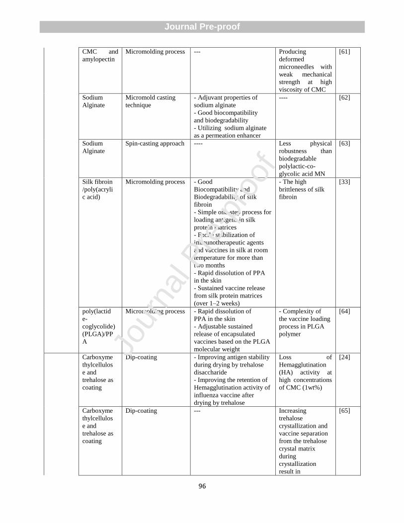
96
CMC and
amylopectin
Micromolding process --- Producing
deformed
microneedles with
weak mechanical
strength at high
viscosity of CMC
[61]
Sodium
Alginate
Micromold casting
technique
- Adjuvant properties of
sodium alginate
- Good biocompatibility
and biodegradability
- Utilizing sodium alginate
as a permeation enhancer
---- [62]
Sodium
Alginate
Spin-casting approach ---- Less physical
robustness than
biodegradable
polylactic-co-
glycolic acid MN
[63]
Silk fibroin
/poly(acryli
c acid)
Micromolding process - Good
Biocompatibility and
Biodegradability of silk
fibroin
- Simple one-step process for
loading antigens in silk
protein matrices
- Facile stabilization of
immunotherapeutic agents
and vaccines in silk at room
temperature for more than
two months
- Rapid dissolution of PPA
in the skin
- Sustained vaccine release
from silk protein matrices
(over 1–2 weeks)
- The high
brittleness of silk
fibroin
[33]
poly(lactid
e-
coglycolide)
(PLGA)/PP
A
Micromolding process - Rapid dissolution of
PPA in the skin
- Adjustable sustained
release of encapsulated
vaccines based on the PLGA
molecular weight
- Complexity of
the vaccine loading
process in PLGA
polymer
[64]
Carboxyme
thylcellulos
e and
trehalose as
coating
Dip-coating - Improving antigen stability
during drying by trehalose
disaccharide
- Improving the retention of
Hemagglutination activity of
influenza vaccine after
drying by trehalose
Loss of
Hemagglutination
(HA) activity at
high concentrations
of CMC (1wt%)
[24]
Carboxyme
thylcellulos
e and
trehalose as
coating
Dip-coating --- Increasing
trehalose
crystallization and
vaccine separation
from the trehalose
crystal matrix
during
crystallization
result in
[65]
Jour
nal P
re-p
roof
Journal Pre-proof
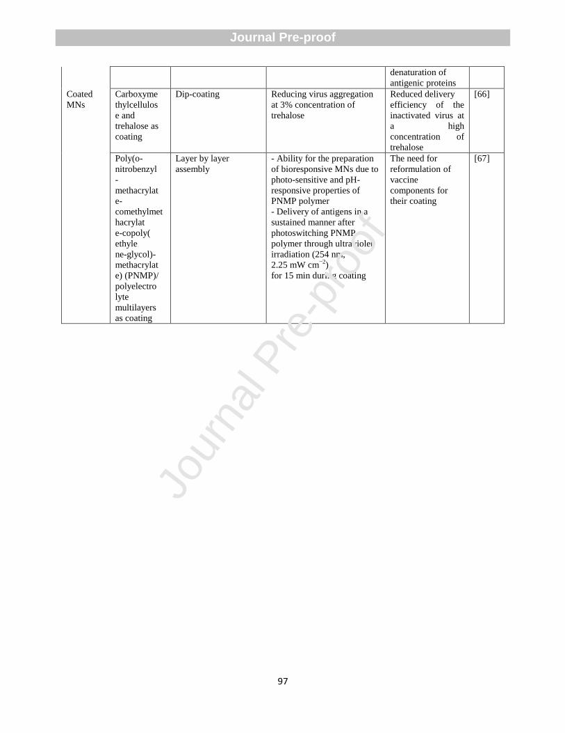
97
denaturation of
antigenic proteins
Coated
MNs
Carboxyme
thylcellulos
e and
trehalose as
coating
Dip-coating Reducing virus aggregation
at 3% concentration of
trehalose
Reduced delivery
efficiency of the
inactivated virus at
a high
concentration of
trehalose
[66]
Poly(o-
nitrobenzyl
-
methacrylat
e-
comethylmet
hacrylat
e-copoly(
ethyle
ne-glycol)-
methacrylat
e) (PNMP)/
polyelectro
lyte
multilayers
as coating
Layer by layer
assembly
- Ability for the preparation
of bioresponsive MNs due to
photo-sensitive and pH-
responsive properties of
PNMP polymer
- Delivery of antigens in a
sustained manner after
photoswitching PNMP
polymer through ultraviolet
irradiation (254 nm,
2.25 mW cm−2
)
for 15 min during coating
The need for
reformulation of
vaccine
components for
their coating
[67]
Jour
nal P
re-p
roof
Journal Pre-proof
98
Highlights
Microneedles are promising devices for painless drug delivery with high bioavailability.
Transdermal microneedle can improve the biological effect of drugs through adjustable drug
release.
Facile fabrication and versatility have caused high attraction towards microneedle-based drug
delivery.
Microneedles are suggested for immunotherapy due to the high abundance of immune cells
under the skin.
Jour
nal P
re-p
roof
Journal Pre-proof


![Antibiotics proof[1]](https://img.pdfslide.net/doc/110x75/634560b16cfb3d406409a867/antibiotics-proof1.jpg)


