Embed Size (px)
Citation preview
REGULAR PAPER
Yorito Anamizu Æ Hiroshi Kawaguchi Æ Atsushi SeichiShinji Yamaguchi Æ Emiko Kawakami Æ Naotoshi Kanda
Shiro Matsubara Æ Makoto Kuro-o Æ Yoichi Nabeshima
Kozo Nakamura Æ Kiyomitsu Oyanagi
Klotho insufficiency causes decrease of ribosomal RNA genetranscription activity, cytoplasmic RNA and rough ER in the spinalanterior horn cells
Received: 8 September 2004 / Revised: 29 November 2004 / Accepted: 29 November 2004 / Published online: 16 April 2005� Springer-Verlag 2005
Abstract The klotho gene was identified in 1997 as thegene whose severe insufficiency (kl/kl) causes a syndromeresembling human aging, such as osteoporosis, arterio-sclerosis, gonadal atrophy, emphysema, and short lifespan in a mouse strain. Regarding the gait disturbancereported in kl/kl mice, the present study examined thespinal cord of kl/kl mice, and revealed decreases in thenumber of large anterior horn cells (AHCs), the amountof cytoplasmic RNA, the number of ribosomes and
rough endoplasmic reticulum (rER), and the activity ofribosomal (r) RNA gene transcription without signifi-cant loss of the total number of neurons in the ventralgray matter. Increased immunostaining of phosphory-lated neurofilament in the AHCs and of glial fibrillaryacidic protein in reactive astrocytes in the anterior hornof kl/kl mice were also observed. On the other hand, theposterior horn was quite well preserved. The resultssuggest that the kl/kl insufficiency causes atrophy anddysfunction of the spinal AHCs through decreasedactivity of rRNA gene transcription, which may reducethe amount of cytoplasmic RNA and the number ofribosomes and rER. These findings resemble thosefound in the spinal cord of patients with classic amyo-trophic lateral sclerosis (ALS). The results show thatklotho gene insufficiency causes dysfunction of the pro-tein synthesizing system in the AHCs, and might indi-cate the klotho gene is involved in the pathologicalmechanism of classic ALS. The kl/kl is a new animalmodel of AHC degeneration, and may provide clues tounderstanding the etiology of classic ALS.
Keywords Anterior horn cell Æ Cytoplasmic RNA ÆKlotho Æ Ribosomal RNA gene Æ Transcription activity
Introduction
Mouse lines exhibiting symptoms of early-onset senes-cence have been established using insertion mutations; ithas been shown that the absence of a single gene pro-duces a variety of aging symptoms. The klotho gene hasbeen identified as the causal gene at 5G3 in mice and13q12 in humans [27]. Homozygotes with the klothogene mutation (kl/kl) are reported to demonstrate phe-notypes highly similar to human aging, including oste-oporosis, calcification of articular cartilage and softtissue, arteriosclerosis, decreased activity, emphysema,
Y. Anamizu Æ E. Kawakami Æ K. Oyanagi (&)Department of Neuropathology,Tokyo Metropolitan Institute for Neuroscience,2-6 Musashidai, Fuchu, 183-8526 Tokyo, JapanE-mail: [email protected].: +81-42-3253881 ext 4711Fax: +81-42-3218678
Y. Anamizu Æ H. Kawaguchi Æ A. Seichi Æ K. NakamuraDepartment of Orthopedic Surgery,Graduate School of Medicine,University of Tokyo, Tokyo, Japan
S. YamaguchiDepartment of Biochemistry,Molecular Biology and Cell Biology, Northwestern University,Evanston, Illinois, USA
N. KandaDepartment of Anatomy,Graduate School of Veterinary Medicine,Tokyo University of Agriculture and Technology,Tokyo, Japan
S. MatsubaraDepartment of Neurology,Tokyo Metropolitan Neurological Hospital,Fuchu, Tokyo, Japan
M. Kuro-oDepartment of Pathology,University of Texas Southwestern Medical Center,Dallas, Texas, USA
Y. NabeshimaDepartment of Tumor Biology,Graduate School of Medicine, Kyoto University,Kyoto, Japan
Acta Neuropathol (2005) 109: 457–466DOI 10.1007/s00401-004-0971-7

and gonadal, thymic, and dermal atrophy. The life spanis reported to be 8–9 weeks on average. The kl/kl miceare senescence-accelerated mice satisfying 7 among 21 ofthe pathophysiological and cellular criteria of aging [27].An association between the allele of the functional var-iant of the klotho gene and occult coronary artery dis-ease has been reported [3].
In addition to the above-mentioned symptoms, kl/klmice exhibit an abnormal gait. Reduction in the numberof cerebellar Purkinje cells [27] may be a factor relatingto the gait disturbance, but the spinal cord has not beenexamined genetically or histologically. To date, therehave been no reports on the expression of the klothogene in the spinal cord. An age-related reduction of thenumber of small neurons in the anterior horn has beenreported in humans [25, 26, 40], and it was thusimportant to examine whether or not similar changesoccur in kl/klmice. The purpose of the present study wasto clarify the genetic, histological, quantitative and ul-trastructural peculiarities in the spinal cord in kl/kl mice,and compare those with the findings in the human spinalcord in aging. We have found some degenerative chan-ges, similar to those reported in the human anterior horncells (AHCs) with neurodegenerative diseases includingclassic amyotrophic lateral sclerosis (ALS), in thissenescence model mouse.
Materials and methods
Animals
All animals were bred and kept in a breeder (Clea-JapanCorp., Tokyo, Japan) until just before the sacrifice. Atsacrifice, adequate measures were taken to minimizepain and discomfort to the animals, according to the‘‘Guidelines for Experiments of the Tokyo MetropolitanInstitute for Neuroscience’’. All animal experimentsconformed to the United States Public Health Service’sPolicy on Human Care and Use of Laboratory Animals.
The genetic background of the original klotho micewas a mixture of C57BL/6J and C3H/J species. We used21 male kl/kl mice and 19 male wild-type (WT) mice atthe age of 7 weeks, the period assumed to be that justbefore death. Genotypes of the sacrificed mice weredetermined by Southern blot analyses using their tailsand probes to detect the inserted plasmid located adja-cent to the klotho gene locus. The body and brainweights were measured at the time of sacrifice (Table 1).
Animals were deeply anesthetized with ether before theprocessing. For reverse transcription (RT)-PCR, thecervical spinal cords from kl/kl and WT mice (n=2,respectively) were taken up in lysis buffer for the isola-tion of total RNA (RNeasy kit; Qiagen, Chatsworth,CA). Fifteen kl/kl and 13 WT male mice were tran-scardially perfused with 4% paraformaldehyde (PFA) in0.1 M phosphate buffer (PB) (pH 7.3) for paraffinembedding. Four kl/kl and 4 WT mice were transcar-dially perfused with 2.5% glutaraldehyde (GA) and 1%PFA in 0.1 M cacodylate buffer (CB) (pH 7.3) for Eponembedding.
RT-PCR procedure of the spinal cord
RNA (0.3–1 lg) was dissolved in 30 ll H2O, and thefollowing was added: 2.5 ll 1 M TRIS (pH 7.4), 20 ll25 mM MgCl2, 2 ll RNase-free DNase (10 U/ll; RocheMolecular Biochemicals, Mannheim, Germany), and0.5 ll RNasin (40 U/ll; Promega, Madison, WI). Themixture was incubated for 20 min at 37�C and subse-quently divided into two parts. In one part, RNA wasreverse-transcribed using oligo(dT) primers (Life Tech-nologies, Rockville, MD) and Moloney murine leukemiavirus reverse transcriptase (Superscript II; Life Tech-nologies) at 45�C for 1 h. The other part was incubatedin the same mixture without the enzyme and was laterused as a control for DNA contamination in the PCR.The cDNA was purified (PCR purification kit; Qiagen)and used as a template for PCR in a 60-ll reaction vol-ume containing 1· PCR buffer containing 1.5 mMMgCl2 (Perkin-Elmer, Foster City, CA), 0.2 mMdNTPs, and 0.1 lM of each primer. Specific oligonu-cleotide primers (Life Technologies) for PCR were de-signed to amplify rat (GenBank accession no. AB005141)klotho mRNA (forward 5’-AGATGTGGCCAGCGAT-AGTTA-3’; reverse 5’-ACTTGACCTGACCACCG-AAGT-3’) [29]. A ‘‘hot start’’ was performed manuallyby adding 1.5 U AmpliTaq (Perkin-Elmer) after an ini-tial incubation of 5 min at 95�C in a thermocycler (Per-kin-Elmer, 9700). For each experiment the housekeepinggene glyceraldehyde-3-phosphate dehydrogenase (GAP-DH) was amplified with 20 and 22 cycles to normalize thecDNA content of the samples. Equal cDNA amountswere subsequently used for the amplification of klothogene. Amplification was performed for 20–40 cycles(94�C for 30 s, 50�C for 10 s, and 72�C for 90 s). Thelinear range of amplification was determined for eachprimer set. The amplicons were analyzed on a 1.5%agarose gel stained with ethidium bromide.
Histological and immunohistochemical examination
The 5th cervical and 4th lumbar segments were removedfrom the 4% PFA-fixed spinal cords under a dissectingmicroscope. Tissue blocks were dehydrated in ethanol
Table 1 The body and brain weight of kl/kl and WT mice at theage of 7 weeks. Values represent mean ± SD (kl/kl homozygoteswith the klotho mutation, WT wild-type)
Bodyweight (g)
Brainweight (g)
kl/kl (male,n=17) 7.6±1.1* 0.37±0.02*WT (male,n=17) 24.4±1.58 0.47±0.03
*P<0.0001
458

series, and embedded in paraffin. Serial transverse sec-tions (6 lm thick) were made, and were subjected tohematoxylin-eosin, Kluver-Barrera (K-B) and pyroninY preparation.
Sections, 6 lm thick, were also subjected to immu-nohistochemical staining using the avidin-biotin-per-oxidase complex (ABC) method with a Vectastain ABCkit (Vector, Burlingame, CA). The primary antibodiesused were rabbit anti-cow ubiquitin polyclonal anti-body (dilution 1:150; Dakopatts, Glostrup, Denmark),SMI-31 monoclonal antibody (dilution 1:1,000; Stern-berger Monoclonals, Baltimore, MD), and rabbit anti-glial fibrillary acidic protein (GFAP) polyclonal anti-body (dilution 1:1,000; Dakopatts). Lectin histochem-istry for microglia was performed using IB4 coupled tohorseradish peroxidase (HRP) (dilution 1:100, Sigma,St. Louis, MO). Antigenicity was increased for ubiqu-itin immunostaining by pretreating the sections with0.025% trypsin for 15 min at room temperature.Endogenous peroxidase activity was quenched byincubating the sections in 3% hydrogen peroxide inmethanol and then blocked by incubating for 1 h with3% normal goat or mouse serum in phosphate-bufferedsaline (PBS). The sections were incubated with the re-quired primary antibody overnight at 4�C, then incu-bated with the secondary reagent containingbiotinylated anti-rabbit or anti-mouse IgG (diluted1:200) for 2 h, and finally with the ABC solution for1 h. The sections were subjected to the peroxidasereaction using freshly prepared 0.02% 3,3’-diam-inobenzidine-tetrahydrochloride and 0.005% hydrogenperoxide in 0.05 M TRIS-HCl buffer pH 7.6 for 10 minat room temperature. As antibody controls, the pri-mary antisera were either omitted or replaced withnormal rabbit or mouse serum. Several specimens ofneural and non-neural tissue from the patients servedas positive or negative tissue controls.
Quantitative examination of the neurons in the ventralspinal gray matter
The number and sectional area of the neurons withnucleus in the ventral part (ventral side of the centralcanal level; which includes roughly laminae VII, VIIIand IX of Rexed’s [33] of the 5th cervical gray matter)were examined in three K-B-stained 6-lm-thick sec-tions (12-lm apart to avoid double counting of theneurons) on both sides in 11 kl/kl and 10 WT mice.Neurons were identified by the presence of Nissl sub-stance and prominent nucleoli. Sectional neuronal areaincluding cytoplasm and nucleus was measured by adigitizer (Measure 5; System Supply, Nagano, Japan).The number of neurons examined was 2,354 in kl/kland 2,110 in WT mice. The frequency distribution ofthe cell areas by 40-lm2 increments was obtained.Abercrombie’s correction factor [1] was applied forsplit cell error counting. We defined AHCs here aslarge neurons with a cell area greater than 400 lm2, on
the basis of their neuronal features and prominentnucleolus in the ventral part of the gray matter.
Measurement of diameter of nucleoli of neuronsin the ventral spinal gray matter
Light microscopic black and white pictures of neurons inthe ventral spinal gray matter were taken at 200-foldmagnification using K-B-stained 5th cervical segments.The film negatives were magnified 19-fold with amicroscopy reader RF-3A (Fuji Film corp. Tokyo, Ja-pan) and diameters of the nucleoli on the screen weremeasured with the ruler of the microscopy reader. Intotal, 94 neurons in three kl/kl and 87 neurons in threeWT mice were examined. The frequency distribution by0.3-lm increments was obtained.
Amount of cytoplasmic RNA in the AHCs
The cytoplasmic RNA content was measured in AHCswith nucleolus by the integrated OD of pyronin Y-stained 6-lm-thick sections. The measurement of thepyronin Y-stained area with an image analyzer has beenreported as a reliable method for quantitative assess-ment of RNA [36]. Integrated OD was measured in thecytoplasm in a routine bright field of Zeiss Axiovert 135with 12 bits camera (Carl Zeiss, Oberkochen, Germany)using the MetaMorph (Universal Imaging. Co., WestChester, PA, USA) software. Measurement was per-formed in 100 randomly distributed AHCs withnucleolus in klotho and WT mice each (10 AHCs/mouse,10 mice each).
Ultrastructural investigation of the AHCs
Tissue blocks of the 5th cervical cords, fixed with 3%GA-1% PFA in 0.1 M PB, were postfixed with 1% os-mium tetroxide, then dehydrated through a gradedethanol series, and embedded in Epon 812. Toluidineblue-stained 1-lm-thick sections were examined using alight microscope, and ultrathin sections of the ventralpart of the spinal cords were stained with lead citrateand uranyl acetate and examined using an electronmicroscope (H9000; Hitachi, Tokyo, Japan) at 100 kV.
Measurement of transcription activity of rRNA genein neurons in the ventral spinal gray matter
Silver staining of nucleolar organizer region-associatedproteins (AgNORs) [4, 6, 14, 18, 19, 21, 30] was used toevaluate the rRNA gene transcription activity in theneurons in the ventral gray matter in kl/kl and WT mice.PFA-fixed paraffin-embedded 6-lm-thick sections of the5th cervical segment 12 lm apart were used for thestaining.
459

Microscopic slides were deparaffinized with xylene,dehydrated in an alcohol series, rinsed with tap water,then stained with a 1:2 blended solution of freshly pre-pared: (i) 2% gelatin in 1% aqueous formic acid and (ii)50% silver nitrate solution in deionized and distilledwater (DDW). The slides were incubated at 37�C indarkness for 20 min, rinsed in DDW, dehydrated withan alcohol series and xylene, and covered with DPX.Areas of AgNOR-positive regions and the nucleus wereexamined by a digitizer (System Supply, Nagano, Ja-pan), and the ratio was evaluated. Two hundred thirtiesneurons from four kl/kl and 156 neurons from five WTmice were examined.
Statistical evaluation
Statistical evaluation was performed using the Mann-Whitney U-test to compare the ratio between kl/kl andWT mice.
Results
Body and brain weights
The body weight of kl/kl mice aged 7 weeks was aboutone-third that of the WT mice, and the brain weight wasabout four-fifths of the WT mice (Table 1).
RT-PCR for klotho gene expression in the spinal cords
After 40 cycles of RT-PCR, klotho gene expression wasseen both in WT and in kl/kl mice. The expression in kl/klmice was quite weak but significantly positive (Fig. 1).G3PDH was used as control, and the intensities did notdiffer between kl/kl and WT mice. The possibility ofDNA contamination as a source of amplified productswas believed to be excluded, since the klotho geneexpression was not seen in the negative control (data notshown).
Light microscopic findings of the central nervous system
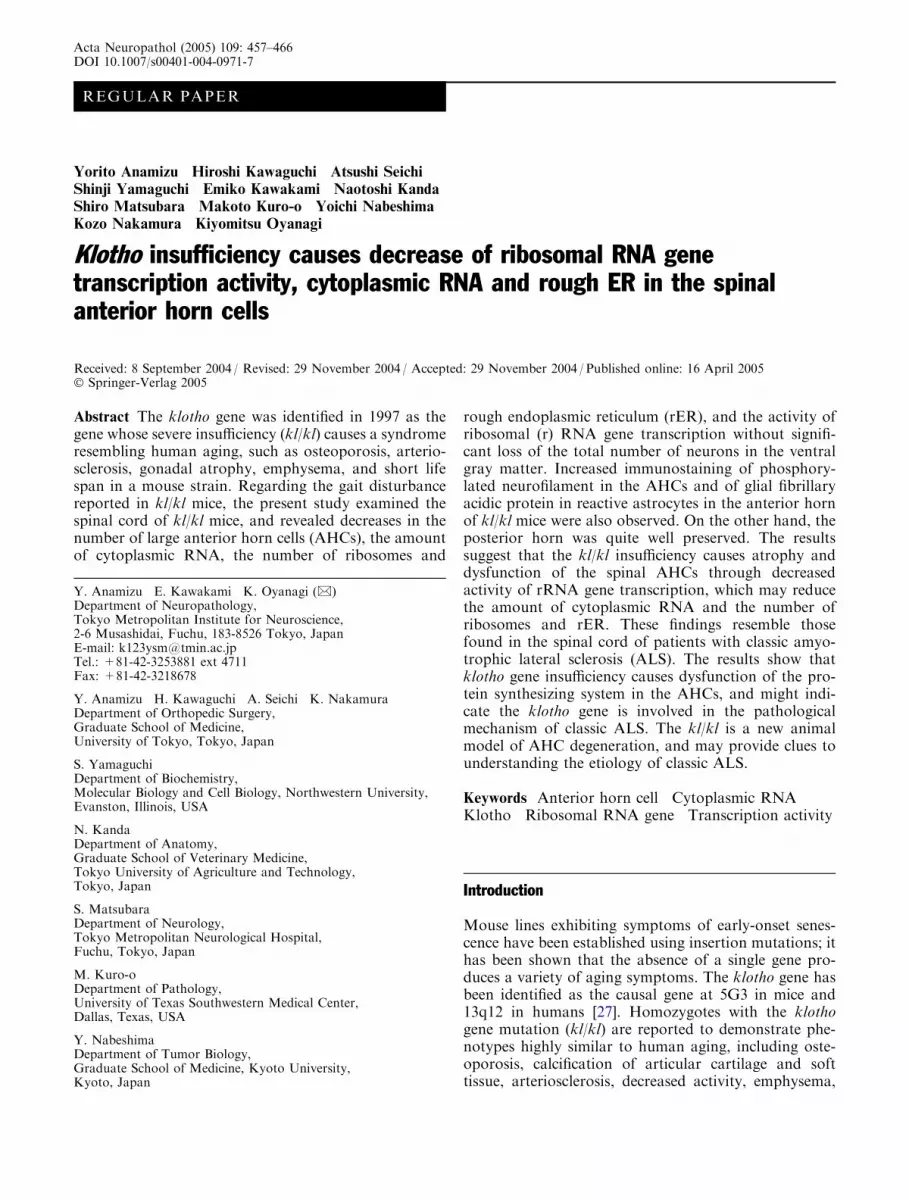
The neuronal population and size of neurons in thebrains of kl/kl mice did not show any apparent differ-ences from those of the controls. The cross-sectionalarea of the gray and white matter of the cervical andlumbar segments of the spinal cord of klkl mice wassmaller than that in WT mice, in spite of good preser-vation of the cross-sectional area (Fig. 2A, B) and theneurons of the posterior horn (Fig. 2C, D) of kl/kl mice.Large AHCs were seen more frequently in WT than inthe kl/kl mice, and small neurons were seen more oftenin the ventral gray matter in kl/kl mice (Fig. 2E, F). Inthe AHCs of kl/kl mice, the amount of Nissl substance,
i.e., rough endoplasmic reticulum (rER), was smaller(chromatolysis) than that in WT mice (Fig. 2G), andslight but evident accumulation of phosphorylatedneurofilaments was observed (Fig. 2I, J). GFAP-immu-nopositive reactive gliosis was seen in the ventral horn ofkl/kl mice (Fig. 2K, L), and a moderate increase ofnumber of the IB4-positive cells was seen in the anteriorhorn in kl/kl mice; no significant ubiquitin-immuno-positive accumulation, Bunina bodies, or spheroids wereobserved.
Quantitative examination of neurons in the ventralgray matter
There was no significant difference in the number of theneurons in the ventral part of the spinal gray matterbetween kl/kl and WT mice (Fig. 2H). However, thefrequency distribution of the sectional area of the neu-rons in the ventral gray matter of the 5th cervical cord inthe 40-lm2 increments showed one peak at 120 lm2 inkl/kl mice but 160 lm2 in WT mice, indicating that theneurons in kl/kl mice were smaller (Fig. 3A). Thenumber of large neurons with a cross-sectional area over400 lm2 was significantly lower in kl/klmice than in WT(P<0.0001) (Fig. 3B). The highest peaks of distributionfrequency by 0.3-lm increments of the diameter ofnucleoli of neurons in the ventral spinal gray matterwere at 2.1–2.4 lm in kl/kl mice, but at 2.7–3.0 lm in
Fig. 1 Klotho gene expression by RT-PCR in the spinal cord. After40 cycles of RT-PCR, klotho gene expression was seen in both WTand in kl/kl mice. klotho gene expression in kl/kl mice is quite weakbut significant. G3PDH was used as control, and the intensitieswere not different between the two genotypes (RT reversetranscription, WT wild-type, kl/kl homozygotes with the klothogene mutation)
460
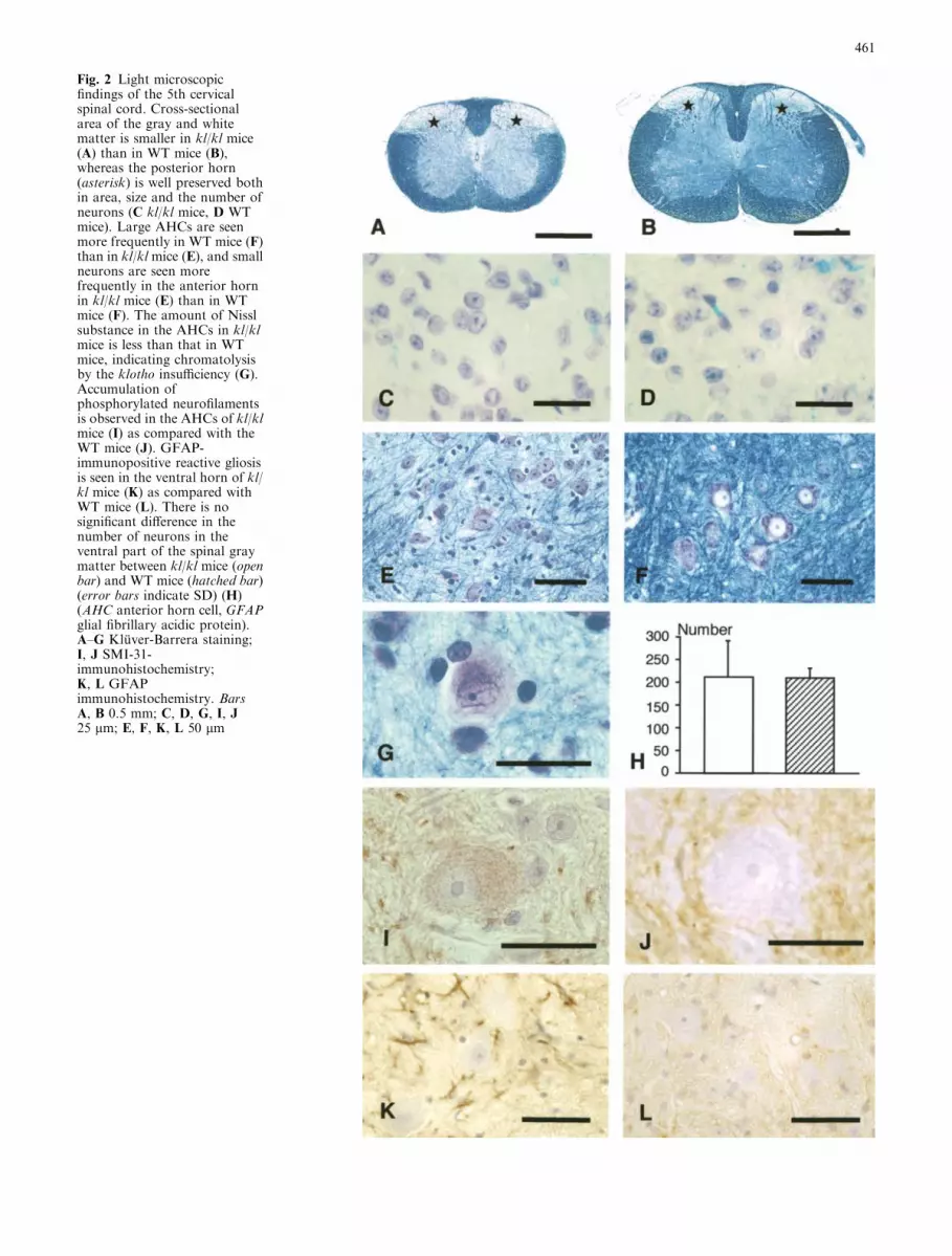
Fig. 2 Light microscopicfindings of the 5th cervicalspinal cord. Cross-sectionalarea of the gray and whitematter is smaller in kl/kl mice(A) than in WT mice (B),whereas the posterior horn(asterisk) is well preserved bothin area, size and the number ofneurons (C kl/kl mice, D WTmice). Large AHCs are seenmore frequently in WT mice (F)than in kl/kl mice (E), and smallneurons are seen morefrequently in the anterior hornin kl/kl mice (E) than in WTmice (F). The amount of Nisslsubstance in the AHCs in kl/klmice is less than that in WTmice, indicating chromatolysisby the klotho insufficiency (G).Accumulation ofphosphorylated neurofilamentsis observed in the AHCs of kl/klmice (I) as compared with theWT mice (J). GFAP-immunopositive reactive gliosisis seen in the ventral horn of kl/kl mice (K) as compared withWT mice (L). There is nosignificant difference in thenumber of neurons in theventral part of the spinal graymatter between kl/kl mice (openbar) and WT mice (hatched bar)(error bars indicate SD) (H)(AHC anterior horn cell, GFAPglial fibrillary acidic protein).A–G Kluver-Barrera staining;I, J SMI-31-immunohistochemistry;K, L GFAPimmunohistochemistry. BarsA, B 0.5 mm; C, D, G, I, J25 lm; E, F, K, L 50 lm
461

WT mice (Fig. 3C). The diameter of neuron nuclei in theventral spinal gray matter was 2.50±0.91 lm (mean ±SD) in kl/kl mice and 2.67±0.77 lm in WT mice; thosein kl/kl mice were significantly smaller (P<0.01).
Amount of cytoplasmic RNA in AHCs
The integrated OD value of the cytoplasmic RNA in thelarge AHCs stained with pyronin Y in kl/kl mice was64.4% of that seen in the WT mice (Table 2, Fig. 4).
Ultrastructural findings of the AHCs
For kl/kl mice, the rER was severely fragmented, andthe size of cisternae of the rER was reduced. The
number of the attached and free ribosomes was notice-ably reduced in the AHCs as compared to those in WTmice, while mitochondria, nuclear membrane, karyo-plasm and nucleolus in the AHCs in these mice appearednormal (Fig. 5A, B).
Transcription activity of rRNA gene in the AHCs
AgNOR-positive areas were clearly and exclusivelyobserved in dark fine granules within the nucleus andin the nucleolus. The ratios of AgNOR-positive areasto cross-sectional areas of the nucleus were significantlylower in the neurons of kl/kl mice than of WT(Fig. 6A–C).
Discussion
The klotho gene carries two varieties of mRNA. Themajority is mRNA coding a membrane-type Klothoprotein, from which a transmembrane-type Klothoprotein is translated [27, 29]. The transmembrane-typeprotein is composed of an N-terminal signal sequenceand a C-terminal transmembrane domain, and betweenthem, two domains (KL1, KL2) homologous to b-glu-cosidase hydrolyzing steroid b-glucuronides [39]. Theother mRNA variety gives rise to strap codons, whichtranslate an approximately half-length secretory Klotho
Fig. 3 Distribution of thesectional area of the neurons inthe ventral gray matter of the5th cervical cord. The frequencydistribution of the sectionalarea of the neurons shows onepeak at 120 lm2 in kl/kl micebut 160 lm2 in WT mice,indicating smaller neurons inthe kl/kl mice (A). The numberof large neurons with a cross-sectional area over 400 lm2 issignificantly lower in kl/kl micethan WT mice (B). Thefrequency distribution by 0.3-lm increments of the diameterof nucleoli of neurons in theventral spinal gray mattershows the highest peaks at 2.1–2.4 lm in kl/kl mice but at 2.7–3.0 lm in WT mice (C). Openbars: kl/kl mice, hatched bars:WT mice, error bars: SD
Table 2 The integrated optical density values (mean ± SD) of thecytoplasmic RNA in the large AHCs stained with pyronin Y in kl/kl and WT mice. Measurement was performed in 100 randomlydistributed AHCs with nucleolus (10 AHCs/mouse,10 mice each)(AHC anterior horn cell)
Integrated OD value
kl/kl (male) 5,424±3,450* (n=100)WT (male) 8,427±6,988 (n=100)
*P<0.05
462

protein. The secretory Klotho protein has only a signalsequence and KL1 domain. It is not known whetherKlotho proteins have enzymatic activity. The klothogene is reported to be strongly expressed in the kidneys,and weakly in the brain. Klotho gene expression has notbeen reported in the lungs, bones, or skin, which show
severe pathological changes in kl/kl mice. These factssuggest the possibility that pathological findings in kl/klmice are not a simple phenotype of the klotho gene, butthat the secretory Klotho protein may have some func-tion whereby pathological changes are suppressed in WTmice [27, 29].
Fig. 4 Pyronin Y-stainedAHCs in kl/kl (A) and WT (B)mice. Bars 20 lm
Fig. 5 Ultrastructure of theAHCs. The rERs are severelyfragmented, and the amount ofrER and the number of freeribosomes sre obviously lowerin the neurons of kl/kl mice (A)as compared with those in WTmice (B), whereasmitochondria, nuclearmembrane, and nuclearkaryoplasm and nucleolus inthe AHCs in kl/kl mice appearunchanged (rER roughendoplasmic reticulum,N nucleus). Uranyl-leadstaining. Bar 1 lm
463

Our present investigation on klotho gene expressionrevealed that the gene was observed strongly in WT andweakly, but significantly, in kl/kl mice after 40 cycles inRT-PCR in the spinal cord. This result demonstratesthat kl/kl mice are not null but severe hypomorph miceof the Klotho protein, and that the decrease of this
protein may induce various morphological alterationsobserved in the present study.
The differential AgNOR staining is believed to staincertain proteins combined specifically with an rRNAgene, and the quantity of the proteins (AgNOR-positiveareas) is believed to reflect the transcription activity ofthe rRNA gene [4, 6, 14]. To date, AgNOR stainabilityhas been reported to be an index of phenomenaincluding protein synthesis [4, 12], tumor proliferation[7, 8, 11, 31, 32, 34, 43], long-term changes duringdevelopment [12], brain augmentation due to learning[42], decline in the aging brain [22, 23], and age-relatedreduction among dermal fibroblasts [8, 38].
The present light microscopic study showed a loss ofNissl substance (chromatolysis) in the AHCs of kl/klmice. Reduction of integrated OD value of pyronin Y-positive material in the cytoplasm of AHCs in kl/klmice indicates a decrease of the cytoplasmic RNAcontent, and ultrastructural investigation of AHCs re-vealed a reduction of the number of attached and freeribosomes and of rER in the mice in the present study.The ratio between the activity of rRNA gene tran-scription and the size of the nucleus in the anteriorhorn cells were evaluated. The results indicated that thedepletion of the activity of rRNA gene transcriptionwas not proportional to overall cell size. Thus, de-creased transcription activity of the rRNA gene in thespinal neurons observed in the present study may causea decrease in cytoplasmic RNA, ribosomes and rER ofthe AHCs. The reduction of the amount of the ribo-somes and rER might induce small neurons in theAHCs of the spinal cord with sparing the posteriorhorn in kl/kl mice. To determine the cause and mech-anism, the amount of Klotho protein should beexamined both in the ventral and posterior horns. Inaddition, regarding the mechanism of reduction ofrRNA gene transcription activity, it should be eluci-dated whether or not the rRNA gene decreases in thechromosomal DNA; some genes are unavailable in thehybridization after being covered by proteins or othercross-linkers [13], and the RNA polymerase I, TFIIA,TFIIB, TATA box-binding protein, and CRE-bindingprotein exist normally in the AHCs [2].
A decreased number of small neurons and the ab-sence of chromatolysis have been reported in the AHCsof aged humans [5, 26, 37, 40]. These findings differ fromthose observed in the kl/kl mice in the present study.Thus, the pathological mechanisms occurring in thespinal cord in aged humans and kl/kl mice are consid-ered to be basically dissimilar. Further examination ofthe spinal cords in fetus and newborn, to analyze pos-sibly overlapping developmental disturbance, is neededto determine the cause of the severe reduction in thevolume of the spinal cord in kl/kl mice.
Kl/kl mice show a marked reduction in body weight.Thus, developmental retardation may exist in the mice.However, the brain is relatively well developed andpreserved. Neuronal population and the size of neuronsin the brains did not show any apparent differences from
Fig. 6 Transcription activity of rRNA gene in the AHCs. AgNOR-positive area in large AHCs of the kl/kl mice (A) and WT mice (B).The ratio of this area to cross-sectional cellular area of the nucleusis significantly lower in kl/klmice (open bar) than WT mice (hatchedbar) (error bars indicate SD) (C) (rRNA ribosomal RNA, AgNORsSilver staining of nucleolar organizer region-associated proteins)
464

those of the controls. In addition, the number and size ofthe neurons in the posterior horn in the spinal cordappeared to be preserved. These findings indicate thatnormal development and selective degeneration possiblyoccurred in the AHCs. It is hardly conceivable thatselective developmental retardation occurred in theAHCs, and that developmental retardation of the skel-etal muscles induce anterior horn degeneration showingrER reduction. There have been no reports that anycongenital muscle dystrophy in mice or humans inducesrER reduction in the AHCs.
On the other hand, a decreased number of largeneurons and reduced amount of the cytoplasmic RNA,rER and ribosomes, as observed in the spinal cord of kl/kl mice, have been noted in the motor neurons in thespinal cord and brain stem of patients with classic ALS[15, 16, 28]; however, Bunina bodies and spheroids wereabsent in these mice. An accumulation of neurofilamentsin the AHCs, as in patients with classic ALS [9, 24, 35],was also reported in the peripheral nerve axon in kl/klmice [41]. Decrease of rER (chromatolysis) and accu-mulation of neurofilaments have been considered to beearly changes in the AHCs in patients with classic ALS[17, 20]. Reactive astrocytosis observed in the anteriorhorn of the kl/kl mice in the present study relates to adegenerative process, and has been reported in theventral horn in patients with classic ALS. This resem-blance shows that klotho gene insufficiency causes neu-ronal dysfunction, and might indicate that the klothogene is involved in the pathological mechanism of classicALS. Mutant SOD mice may be a good tool for theresearch of familial ALS [10], and further study is nee-ded to evaluate whether the kl/kl mice, which aresenescence-accelerated mice showing decreased rER,ribosomes, and cytoplasmic RNA in the AHCs, is a newanimal model of AHC degeneration, and can provideclues to understanding the etiology of classic ALS.
Acknowledgements The authors are indebted to Dr. K. Watabe ofthe Department of Molecular Neuropathology, and Dr. J. Kimura-Kuroda and Dr. I. Nagata (Department of Brain Structure, TokyoMetropolitan Institute for Neuroscience), Dr. K. Honma (Gradu-ate School of Pharmaceutical Science, University of Tokyo), Ms.M. Shinohara (Department of Anatomy, School of VeterinaryMedicine, Tokyo University of Agriculture and Technology), andDr. A. Mabuchi (Department of Orthopedic Surgery, GraduateSchool of Medicine, University of Tokyo) for their help during theresearch. This work was supported in part by grants from theJapanese Ministry of Health, Labor and Welfare (to Y.N., andResearch on Psychiatric and Neurological Diseases and MentalHealth (H16-kokoro-017 to K.O.)); the Japanese Ministry ofEducation, Science, Sports and Culture (nos. 12137201 to H.K.,12307031 to K.N., 14657376 to E.K. and 14580735 to K.O.) andthe Japan Space Forum (to H.K.).
References
1. Abercrombie M (1946) Estimation of nuclear population frommicrotome sections. Anat Rec 94:239–247
2. Alberts B, Bray D, Johnson A, Lewis J, Raff M, RobertsK, Walter P (1998) Essential cell biology. Garland, NewYork
3. Arking DE, Becker DM, Yanek LR, Fallin D, Jdge DP, MoyTF, Becker LC, Dietz HC (2003) KLOTHO allele status andthe risk of early-onset occult coronary artery disease. Am JHum Genet 72:1154–1161
4. Babu KA, Verma RS (1985) Structural and functional aspectsof nucleolar organizer regions (NORs) of human chromo-somes. Int Rev Cytol 94:151–171
5. Bailey AA (1953) Changes with age in the spinal cord. ArchNeurol Psychiatry 70:299–309
6. Baldini A, Marleka P (1985) Hormone-modulated rRNA geneactivity is visualized by selective staining of the NOs. Cell BiolInt Rep 9:791–796
7. Brustmann H, Riss P, Naude S (1995) Nucleolar organizerregions as markers of endometrial proliferation: a study ofnormal, hyperplastic, and neoplastic tissue. Hum Pathol26:664–667
8. Buys CHCM, Osinga J, Anders GJPA (1979) Age-dependentvariability of ribosomal RNA-gene activity in man as deter-mined from frequencies of silver staining nucleolus organizingregions of metaphase chromosomes of lymphocytes and fibro-blasts. Mech Ageing Dev 11:55–75
9. Carpenter S (1968) Proximal axonal enlargement in motorneuron disease. Neurology 18:841–851
10. Cleveland DW, Rothstein JD (2001) From Charcot to LouGehrig: deciphering selective motor neuron death in ALS. NatRev Neurosci 2:806–8199
11. Crocker J, Boldy DAR, Egan MJ (1989) How should we countAgNORs? Proposals for a standardized approach. J Pathol158:185–188
12. Fushiki S, Kinoshita C, Tsutsumi Y, Nishizawa Y (1995) Age-related changes of the argyrophilic nucleolar organizer regionsin mouse neocortical neurons. Acta Histochem Cytochem28:533–538
13. Gaubatz J, Cutler RG (1978) Age-related differences in thenumber of ribosomal RNA genes of mouse tissue. Gerontology24:179–207
14. Goodpasture C, Bloom SE (1975) Visualization of nucleolarorganizer regions in mammalian chromosomes using silverstaining. Chromosoma 53:37–50
15. Hartmann HA, McMahon S, Sun DY, Abbs JH, Uemura E(1989) Neural RNA in nucleus ambiguous and nucleus hyp-oglossus of patients with amyotrophic lateral sclerosis. J Neu-ropathol Exp Neurol 48:669–673
16. Hirano A (1991) Cytopathology of amyotrophic lateral scle-rosis. Adv Neurol 56:91–101
17. Hirano A, Inoue K (1980) Early pathological changes of am-yotrophic lateral sclerosis. Electron microscopic study ofchromatolysis, spheroids and Bunina bodies. Neurol Med13:148–160
18. Howell WM, Black DA (1980) Controlled silver staining ofnucleolus organizer regions with a protective colloidal devel-oper: a 1-step method. Experientia 36:1014–1015
19. Hubbell HR (1985) Silver staining as an indicator of activeribosomal genes. Stain Technol 60:285–294
20. Inoue K, Hirano A (1979) Early pathological changes of am-yotrophic lateral sclerosis. Autopsy findings of a case of 10months’ duration. Neurol Med 11:448–455
21. Jimenez R, Burgos M, Diaz de la Guardia R (1988) A study ofthe Ag-staining significance in mitotic NORs. Heredity 60:125–127
22. Johnson R, Stehler BL (1972) Loss of genes codingfor ribosomal RNA in ageing brain cells. Nature 240:412–414
23. Johnson RW, Chrisp C, Stehler BL (1972) Selective loss ofribosomal RNA genes during the ageing of post-mitotic tissues.Mech Ageing Dev 1:183–198
24. Julien JP (1995) A role for neurofilaments in the pathogen-esis of amyotrophic lateral sclerosis. Biochem Cell Biol73:593–597
25. Kameyama T, Hashizume Y, Sobue G (1996) Morphologicfeatures of the normal human cadaveric spinal cord. Spine21:1285–1290
465

26. Kawamura Y, O’Brien P, Okazaki H Dyck PJ (1977) Lumbarmotoneurons of man. II. The number and diameter distributionof large-and intermediate-diameter cytons in ‘‘motoneuroncolumns’’ of spinal cord of man. J Neuropathol Exp Neurol36:861–870
27. Kuro-o M, Matsumura Y, Aizawa H, Kawaguch H, Suga T,Utsugi T, Ohyama Y, Kurabayashi M, Kaname T, Kume E,Iwasaki H, Iida A, Shiraki-Iida T, Nishikawa S, Nagai R,Nabeshima Y (1997) Mutation of the klotho gene leads to asyndrome resembling ageing. Nature 390:45–51
28. Mann DMA, Yates PO (1974) Motor neuron disease: thenature of the pathogenic mechanism. J Neuropathol ExpNeurol 37:1036–1046
29. Matsumura Y, Aizawa H, Shiraki-Iida T, Nagai R, Kuro-o M,Nabeshima Y (1998) Identication of the human klotho geneand its two transcripts encoding membrane and secreted Klo-tho protein. Biochem Biophys Res Commun 242:626–630
30. Morton CC, Brown JA, Holmes WM, Nance WE, Wolf B(1983) Stain intensity of human nucleolus organizer region re-flects incorporation of uridine into mature ribosomal RNA.Exp Cell Res 145:405–413
31. Nielsen AL, Nyholm HC, Engel P (1994) Expression of MIB-1(Paraffin ki-67) and AgNOR morphology in endometrial ade-nocartinomas of endometrioid type. Int J Gynecol Pathol 13:37–44
32. Niwa K, Yokoyama Y, Tanaka T, Mori H, Mori H, Tamaya T(1991) Silver-stained nucleolar organizer regions in the normal,hyperplastic and neoplastic endometrium. Virchows Arch [A]29:493–497
33. Rexed B (1952) The cytoarchitectonic organization of thespinal cord in the cat. J Comp Neurol 96:415–495
34. Ruschoff J, Plate K, Bittinger A, Thomas C (1989) Nucleolarorganizer regions (NORs). Basic concepts and practical appli-cation on tumor pathology. Pathol Res Pract 185:878–885
35. Schmidt M L, Carden MJ, Lee VM, Trojanowski JQ (1987)Phosphate dependent and independent neurofilament epitopesin the axonal swellings of patients with motor neuron diseaseand controls. Lab Invest 56:282–294
36. Schulte EK, Lyon HO, Hoyer PE (1992) Simultaneous quan-titafication of DNA and RNA in tissue sections. A comparativeanalysis of the methyl green-pyronin technique with the gallo-cyanin chromalum and Feulgen procedures using imagecytometry. Histochem J 24:305–310
37. Terao S, Sobue G, Hashizume Y, Li M, Inagaki T, Mitsuma T(1996) Age-related changes in human spinal ventral horn cellswith special reference to the loss of small nerurons in theintermediate zone: a quantitative analysis. Acta Neuropathol92:109–114
38. Thomas S, Mukherjee AB (1996) A longitudinal study of hu-man age-related ribosomal RNA gene activity as detected bysilver-stained NORs. Mech Ageing Dev 92:101–109
39. Tohyama O, Imura A, Iwao A, Freund JN, Henrissat B, Fu-jimori T, Nabeshima Y (2004) Klotho is a novel beta-glucu-ronidase capable of hydrolyzing steroid beta-glucuronides. JBiol Chem 279:9777–9784
40. Tomlinson BE, Irving D (1977) The number of limb motorneurons in the human lumbosacral cord throughout life. JNeurol Sci 34:213–219
41. Uchida A, Komiya Y, Tashiro T, Yorifuji H, Kishimoto T,Nabeshima Y, Hisanaga S (2001) Neurofilaments of Klotho,the mutant mouse prematurely displaying symptoms resem-bling human aging. J Neurosci Res 64:364–370
42. Vargas JP, Rodrıguez F, Lepez JC, Arias JL, Salas C (2000)Spatial learning-induced increase in the argyrophilic nucleolarorganizer regions of dorsolateral telencephalic neurons ingoldfish. Brain Res 865:77–84
43. Wilkinson N, Buckley H, Chawner L, Fox H (1990) Nucleolarorganizer regions in normal, hyperplastic, and neoplasticendometria. Int J Gynecol Patho l 9:55–59
466


