Embed Size (px)
Citation preview

Editor meets silencer: crosstalk between RNA editing and RNAinterference
Kazuko NishikuraThe Wistar Institute, Department of Gene Expression and Regulation, 3601 Spruce Street,Philadelphia, Pennsylvania 19104-4268, USAKazuko Nishikura: [email protected]
AbstractThe most prevalent type of RNA editing is mediated by ADAR (adenosine deaminase acting onRNA) enzymes, which convert adenosines to inosines (a process known as A→I RNA editing) indouble-stranded (ds)RNA substrates. A→I RNA editing was long thought to affect only selectedtranscripts by altering the proteins they encode. However, genome-wide screening has revealednumerous editing sites within inverted Alu repeats in introns and untranslated regions. Also, recentevidence indicates that A→I RNA editing crosstalks with RNA-interference pathways, which, likeA→I RNA editing, involve dsRNAs. A→I RNA editing therefore seems to have additional functions,including the regulation of retrotransposons and gene silencing, which adds a new urgency to thechallenges of fully understanding ADAR functions.
An RNA transcript is subjected to various maturation processes, such as 5′ capping, splicing,3′ processing and polyadenylation, after it is transcribed from the gene. Post-transcriptionalprocessing of primary transcripts is essential to generate mature messenger RNAs that are readyto be translated into proteins1. RNA editing is a post-transcriptional-processing mechanismthat results in an RNA sequence that is different from the one encoded by the genome, andthereby contributes to the diversity of gene products. There are different types of RNA-editingmechanism that either add or delete nucleotides, or that change one nucleotide into another2
(BOX 1).
The type of RNA editing that is most prevalent in higher eukaryotes converts adenosine (A)residues into inosine (I) in double-stranded (ds)RNAs through the action of ADAR (adenosinedeaminase acting on RNA) enzymes3–5. A→I RNA editing of a short dsRNA that has formedbetween a coding exon and nearby intron sequences can lead to a codon change and an alterationin the protein function. However, it was recently discovered that the most frequent targets ofA→I RNA editing seem to be long, but partially double-stranded, RNAs that are formed frominverted Alu repeats and long interspersed element (LINE) repeats located in introns anduntranslated regions (UTRs) of mRNAs6–9. Global editing of non-coding RNA might controlthe expression of genes that harbour these repeat sequences of retrotransposon origin.
Post-transcriptional gene regulation can also occur through RNA interference (RNAi), anevolutionarily conserved phenomenon that involves dsRNA molecules10,11. Small interferingRNAs (siRNAs) and microRNAs (miRNAs) are non-coding RNAs that are generated by aclass of RNase III ribonucleases (specifically, Dicer and Drosha). These small RNAs areincorporated into the RNA-induced silencing complex (RISC), which mediates the RNAiprocess12–16. The idea that the RNAi and A→I RNA editing pathways might compete for a
Competing interests statement: The author declares no competing financial interests.
NIH Public AccessAuthor ManuscriptNat Rev Mol Cell Biol. Author manuscript; available in PMC 2010 October 12.
Published in final edited form as:Nat Rev Mol Cell Biol. 2006 December ; 7(12): 919–931. doi:10.1038/nrm2061.
NIH
-PA Author Manuscript
NIH
-PA Author Manuscript
NIH
-PA Author Manuscript

common substrate dsRNA was originally proposed by Bass17. Recent studies showed thatprecursor RNAs of certain miRNAs indeed undergo A→I RNA editing18–21, and editing seemsto regulate the processing and expression of mature miRNAs19. Furthermore, one of themammalian ADAR-family members sequesters siRNAs, thereby reducing RNAi efficacy22.Last, analysis of ADAR-null Caenorhabditis elegans strains indicates that A→I RNA editingmight counteract RNAi silencing of endogenous genes and transgenes23–25.
In this review, I discuss recent findings on new functions of A→I RNA editing in the regulationof non-coding RNAs and on the interplay between RNA editing and RNAi pathways. Forcomprehensive reviews on A→I RNA editing, see REFS 3–5.
A→I RNA editing by ADARsDeamination of adenosine to inosine
During the A→I RNA editing process, adenosine is converted to inosine by hydrolyticdeamination of the adenine base26,27 (FIG. 1a). The translation machinery reads the inosineas if it were guanosine (G) (FIG. 1b), leading to the introduction of missense codons intomRNAs. Reverse transcriptase also reads inosine as guanosine; therefore, A→I RNA editingtranslates into an A→G change when analysing cDNA sequences.
Box 1
Different types of RNA editing
RNA editing is a post-transcriptional process that changes the nucleotide sequence of anRNA transcript from the DNA sequence encoded by the corresponding gene2. Editing ofmRNAs, transfer RNAs and ribosomal RNAs has been reported in bacteria to man. Thefirst example of mRNA editing, which involved the insertion or deletion of many uridine(U) residues, was reported 20 years ago for mRNAs that are encoded by the mitochondrialDNA of trypanosomes. Soon after, other types of RNA editing were discovered, and itbecame clear that RNA editing is a widespread phenomenon in all three kingdoms of life.In transcripts of the mitochondrial and chloroplast DNAs of plants, for example, theconversion of many cytidine (C) residues to uridine (C→U editing) and the less frequentU→C editing occur, whereas an insertion of guanosine (G) residues occurs in the codingmRNAs of negative-strand RNA viruses. In Physarum polycephalum, different types ofRNA editing occur in mitochondrial mRNA and rRNA; insertion of multiple cytidineresidues, dinucleotide insertion (CU, GU, UA, AA, UU and GC) and an AAAdeletion104,105. C→U editing occurs in the small subunit rRNA in Dictyosteliumdiscoideum mitochondria106.
In mammals, two separate nucleotide-substitution types of RNA editing have beenidentified. The conversion of a specific cytidine residue to uridine (C→U editing) inapolipoprotein B mRNA is mediated by APOBEC1 cytidine deaminase107. This C→Uediting results in the change of a glutamine codon to a translation stop codon and theconsequent synthesis of APOB48, a shorter isoform of APOB100, which is translated fromthe unedited apolipoprotein B mRNA. The second type, adenosine to inosine (A→I) RNAediting, which is the main focus of this review, is the most common type of mammalianRNA editing.
Various nucleotide alterations of tRNA sequences (tRNA editing) are also known. 5′-terminal editing of mitochondrial tRNAs occurs in the amoeboid protist Acanthamoebacastellanii108. A→I editing of tRNAs, which is mediated by ADAT (adenosine deaminaseacting on tRNA), occurs in eukaryotes and also in Escherichia coli109. ADAT1 edits A37(near the anticodon) of tRNAAla, and the heterodimeric ADAT2–ADAT3 complex edits
Nishikura Page 2
Nat Rev Mol Cell Biol. Author manuscript; available in PMC 2010 October 12.
NIH
-PA Author Manuscript
NIH
-PA Author Manuscript
NIH
-PA Author Manuscript

A34 at the wobble position of the anticodon of a subset of tRNAs3–5,110. ADAR genes arethought to have evolved from ADAT genes2–5,110.
ADAR genesThe catalytic reaction of A→I RNA editing is mediated by ADAR enzymes (FIG. 2a). ADARswere originally identified in Xenopus laevis eggs and embryos by their dsRNA-unwindingactivity28,29. Soon after, however, it was discovered that this activity is in fact a dsRNA-specific adenosine deaminase26,27. The first mammalian ADAR gene, human ADAR1, wascloned following the biochemical purification and microsequencing of the ADAR1protein30, which then led to the identification of ADAR2 (REFS 31–33) and ADAR3 (REFS 34,35) (FIG. 2a). The enzymatic activity of ADAR1 and ADAR2 has been shown30–33. ADAR3activity has not yet been shown, although functional domain features are conserved in thisfamily member34,35. Therefore, the function(s) of ADAR3 remains to be established.
These three ADARs, which were originally identified in human and rodent, are conserved invertebrates3–5. Only a few ADAR genes have been found in invertebrates. Drosophilamelanogaster have only a single ADAR2-like gene, Adar36, whereas C. elegans have twoADAR genes, adar-1 and adar-2 (REF. 24) (FIG. 2a). No ADAR genes have been identified inthe genomes of plants, fungi or yeasts.
Domain structure of ADARsMembers of the ADAR family contain common structural features (FIG. 2a). The dsRNA-binding domain (dsRBD; ∼65 amino acids) makes direct contact with the dsRNA37 and isrequired for dsRNA binding. The C-terminal region of ADAR contains amino-acid residuesthat are conserved in several cytidine deaminases and are predicted to participate in theformation of the catalytic centre of ADAR30,38. The crystal structure of the catalytic domainof human ADAR2 shows that His394, Glu396 and two Cys residues, Cys451 and Cys516, ofADAR2 are indeed involved in the coordination of a zinc atom and the formation of the catalyticcentre39. Most surprisingly, however, the structural studies also revealed the presence ofinositol hexakisphosphate (IP6) buried in the enzyme core, but located very close to the catalyticcentre. The IP6 molecule could have a crucial role during the deamination reaction39.
ADAR gene expression and regulationBoth ADAR1 and ADAR2 are present in many tissues, whereas ADAR3 is expressed only inthe brain30–35. Two isoforms of ADAR1, a full-length ADAR1L and a shorter, N-terminal-truncated ADAR1S, are known40. One of the three promoters that drive the ADAR1 gene isinterferon inducible, and the mRNA transcribed from this promoter directs the translation ofADAR1L, initiated from an upstream Met codon41. A substantial increase in ADAR1Lexpression occurs during experimentally induced inflammation in mice42. Two otherADAR1 mRNAs, transcribed from constitutive promoters, direct the synthesis of ADAR1S,which is initiated from a downstream Met codon due to alternative splicing and skipping ofthe exon that contains the upstream Met codon (FIG. 2a). ADAR2 expression is regulated bythe transcriptional activator cyclic-AMP-response-element binding (CREB) protein43, but theregulatory mechanism for ADAR3 is currently unknown.
ADAR1L is detected mainly in the cytoplasm, whereas ADAR1S localizes in the nucleoplasmand nucleolus40,44,45. ADAR2 localizes predominantly in the nucleolus44,46. The significanceof the nucleolar localization of ADAR1S and ADAR2 is not currently clear. The cellulardistribution of ADAR1L indicates the localization of its targets, possibly a different class ofdsRNA substrate (for example, siRNAs; see below), in the cytoplasm22.
Nishikura Page 3
Nat Rev Mol Cell Biol. Author manuscript; available in PMC 2010 October 12.
NIH
-PA Author Manuscript
NIH
-PA Author Manuscript
NIH
-PA Author Manuscript

Substrate and editing-site selectivityBoth intermolecular and intramolecular dsRNAs of >20 base pairs (bp) (two turns of thedsRNA helix) can serve as substrates for ADAR47. Many adenosine residues of a long,completely base-paired dsRNA (>100 bp) are edited non-selectively. By contrast, shortsdsRNAs (∼20–30 bp) or a long but partially dsRNA with mismatched bases, bulges and loops(imperfect dsRNAs) are edited selectively; only a few adenosines are specifically chosen,indicating that the secondary structure in ADAR substrates dictates editing-site selectivity48.For example, site-selective A→I RNA editing occurs on an imperfect fold-back dsRNAstructure that is formed between the exon sequence around an editing site(s) and a downstreamintronic complementary sequence, termed editing-site-complementary sequence (ECS), ofglutamate receptor-2 (GluR2) and serotonin (5-HT) receptor-2C (5-HT2CR) pre-mRNAs49,50
(see FIG. 3 and below). The ECS and the dsRNA structure are required for editing3,5,51,52.
Furthermore, some editing sites are preferentially edited only by ADAR1 or ADAR2 (FIG. 3),indicating a significant difference in their RNA–substrate interactions, possibly through theirdsRBDs (different numbers and spacing between different dsRBDs)53. The distinctive siteselectivity of ADAR1 and ADAR2 could also be mediated through functional interactionsbetween the two monomers of ADAR1 or ADAR2, as such interactions possibly positionspecific adenosine residues relative to the catalytic centre of ADAR53,54.
Physiological significance of editingEditing sites found in protein-coding regions
A limited number of targets (∼30 genes), such as mammalian GluR49 and 5-HT2CR55 as wellas potassium channel Kv1.1 (REF. 56) and D. melanogaster Na+-channel57 gene transcripts,have been identified that are subjected to A→I RNA editing in their coding sequences51,52,56. In addition to cellular genes, transcripts of certain viruses, such as hepatitis delta virus, arealso edited58.
Most often, RNA editing of protein-coding genes alters and diversifies the functions of therespective proteins, as shown by the two most studied examples (FIG. 3). Seeburg andcolleagues identified a total of eight A→I RNA editing sites in the coding regions of receptorsfor several GluR subunits49,51. Among the eight editing sites, the Gln/Arg (Q/R) site locatedin the channel-pore-loop domain of the GluR2 subunit has the most important role in ion-channel function; editing of this single site makes the tetrameric channel protein impermeableto Ca2+ (FIG. 3a). Emeson and colleagues discovered a total of five A→I RNA editing siteslocated in the second intracellular loop or G-protein-coupling domain of 5-HT2CR55.Combinatorial editing of the five sites results in changes in three codons, Ile, Asn and Ile, topossibly six different amino-acid residues, resulting in the expression of up to 24 receptorisoforms with altered G-protein-coupling functions. For example, the ligand (5-HT)responsiveness of the receptor that has been fully edited at all five sites is reduced by 20-foldcompared with that of the unedited receptor (FIG. 3b).
RNA-editing deficienciesThe inactivation of ADAR-gene-family members has significant physiological consequences,reported as phenotypic alterations of ADAR-gene mutants created in various species. Flieswith a homozygous deletion in the Adar gene exhibit brain-related changes such as a lack ofcoordinated locomotion and age-dependent neurodegeneration36. Strains of C. elegans thatcontain homozygous deletions of both adar-1 and adar-2 display defective chemotaxis24. Micewith a homozygous Adar2-null mutation die several weeks after birth. These mice experiencerepeated episodes of epileptic seizures that originate from excess influx of Ca2+ and consequentneuronal death caused by under-editing of GluR2 pre-mRNA at the Q/R site59, which is a major
Nishikura Page 4
Nat Rev Mol Cell Biol. Author manuscript; available in PMC 2010 October 12.
NIH
-PA Author Manuscript
NIH
-PA Author Manuscript
NIH
-PA Author Manuscript

target of ADAR2 (FIG. 3a). Last, the inactivation of ADAR1 leads to an embryonic lethalphenotype that is caused by defective erythropoiesis and widespread apoptosis60–62.
Human diseases or pathophysiologies can also be caused by dysfunction of the A→I RNAediting mechanism63,64. Heterozygosity for the ADAR1-gene functional-null mutation resultsin dyschromatosis symmetrica hereditaria, a human pigmentary genodermatosis of autosomal-dominant inheritance65. RNA-editing deficiencies also underlie disorders of the centralnervous system. Under-editing of the Q/R site of GluR2 pre-mRNA (FIG. 3a) has beenproposed to be responsible for the death of sporadic amyotrophic lateral sclerosis (ALS) motorneurons66, as well as apoptotic death of ischaemic neurons during ischaemia caused by cardiacarrest and disruption of the blood flow to the brain43. Last, RNA editing of 5-HT2CR mighthave some causative relevance to neuropsychiatric disorders, such as depression, as the editingpattern of 5-HT2CR mRNA (FIG. 3b) is significantly altered in the prefrontal cortex of suicidevictims64,67,68.
Global editing of non-coding RNAsThe initial identification of physiologically important editing target genes, such as GluR2, andthe consequent alterations of protein functions has fascinated many investigators. However,the number of genes that have been identified as editing targets has been far lower than thatpredicted by the amount of inosine that can be detected in rat brain poly(A)+ RNA69. This ledto global searches for A→I editing sites in coding and non-coding regions.
Bioinformatics screening for A→I RNA editing sitesSeveral groups have recently developed a systematic, computational analysis method for thegenome-wide identification of new A→I RNA editing sites6–9. Reverse transcriptaserecognizes inosine as if it were guanosine (FIG. 1b). Therefore, an A→I RNA editing site canbe identified when a cDNA sequence or an expressed sequence tag (EST) and the correspondinggenome sequence are aligned, given that guanosine residues reverse-transcribed from inosinesare detected in place of gene-encoded adenosines (FIG. 4a). The screening strategy consists ofan algorithm to align a cluster of A→G mismatches in cDNAs or ESTs to the genome sequenceand to assemble them into clusters that contain complete or partial genes in the dsRNA regions(as predicted by the presence of complementary sequences in a limited distance through acomputer-assisted programme). This is followed by the elimination of single nucleotidepolymorphisms (SNPs) and the evaluation of data quality. With this technique, a much largerthan expected number of human A→I RNA editing sites has been identified6–9. Mostsurprisingly, almost all of these new sites that were identified in the human transcriptome(∼15,000 sites, mapped in ∼2,000 different genes) reside in non-coding regions that consist ofinversely oriented repetitive elements (FIG. 4b), mostly Alu repeats (∼90%) and some LINErepeats (∼10%), representing ∼13% and ∼21% of the human genome, respectively.
On the basis of this analysis, it is predicted that >85% of pre-mRNAs are possibly edited, withthe vast majority being targeted in introns (∼90%) and the rest in UTRs6. A similar screeningstrategy that is restricted to coding regions resulted in the identification of only a few editingtarget genes70,71. Together, these results indicate that the most common targets of ADARs arethe non-coding sequences of transcriptomes and that protein re-coding as a result of A→I RNAediting is rare.
Editing of repeat RNAs in non-primate speciesIf global editing of non-coding Alu repeats in the human transcriptome has some biologicalsignificance, one might expect that the same is true in other organisms. Alu repeats are shortinterspersed elements (SINEs) that are unique to primates. However, SINE elements that are
Nishikura Page 5
Nat Rev Mol Cell Biol. Author manuscript; available in PMC 2010 October 12.
NIH
-PA Author Manuscript
NIH
-PA Author Manuscript
NIH
-PA Author Manuscript

considered to have a common evolutionary origin with Alu repeats do exist in other organisms.Therefore, computational analyses have been carried out to search for A→I RNA editing sitesin mouse EST databases8,72. The editing level in SINEs in mouse is at least an order ofmagnitude lower compared with Alu repeats in humans8,72. This substantial reduction infrequency might be explained by the differences in repeat length (∼300 bp versus ∼150 bp forhuman Alu and mouse SINE, respectively) and higher sequence homogeneity among humanAlu repeats compared with mouse SINEs8,72. Screening for A→I RNA editing sites in rat,chicken and fly transcriptomes showed that non-coding repeat sequences are major targets ofADARs, but the editing frequency is again much lower than that observed in humantranscriptomes72. So, although there is variability in the editing frequency of differentorganisms, A→I RNA editing of non-coding, repetitive RNA sequences seems to be awidespread phenomenon in the animal kingdom.
Editing of non-coding antisense transcriptsGlobal transcriptome analysis has shown that a large fraction of the genome producestranscripts from both sense and antisense strands (70%). Most sense and antisense transcriptpairs are coordinately expressed, which indicates that antisense transcription might contributeto the control of sense transcripts73,74. However, it is unknown how frequently mammaliansense and antisense transcripts form into intermolecular dsRNAs. Because A→I RNA editingoccurs only on dsRNA, the global examination of editing sites for sense and antisensetranscripts could provide useful information on the in vivo formation of intermolecular RNAduplexes that consist of sense and antisense transcript pairs (FIG. 4c).
Recent bioinformatics studies of human EST databases for sense and antisense RNA pairsindicate that A→I RNA editing is restricted to intramolecular RNA duplexes that consist ofinversely oriented repeat sequences of either sense or antisense RNA. However, A→I RNAediting is not detected in the regions outside of repetitive sequences75. PCR after reversetranscription of RNA (RT-PCR) and sequencing analysis of sense and antisense cyclinCNNM3 RNAs derived from an intronic region that contains two inverted Alu repeatsconfirmed that both sense and antisense RNAs are extensively edited, but only in theirintramolecular fold-back dsRNA structures76 (FIG. 4b). No editing was detected outside ofthe Alu sequences, which indicates that the formation of an intermolecular sense–antisenseRNA duplex does not occur76 (FIG. 4c). Interestingly, analysis of an equimolar mixture ofsense and antisense CNNM3 RNAs that were edited in vitro by recombinant ADAR1 andADAR2 indicate again that A→I RNA editing is restricted to the intramolecular fold-backstructure, which indicates that inversely oriented Alu repeats predominantly form anintramolecular dsRNA and that their interaction with ADARs might prevent the formation ofintermolecular RNA duplexes76.
Implications of repetitive RNA editingWhat are the implications of global A→I RNA editing of non-coding, repetitive sequences forthe control of gene expression (FIG. 5a)? The A→I sequence changes that are introduced inpre-mRNAs seem to be recognized by the splicing machinery. Furthermore, several cellularactivities seem to specifically recognize and function on inosine-containing RNA (I-RNA) ordsRNA (I-dsRNA).
Modulating splicing sites?An inosine is interpreted by the splicing machinery as a guanosine. A→I RNA editing couldtherefore create or delete splice donor and acceptor sites. For example, a highly conservedcanonical 5′-splice site dinucleotide recognition sequence, GU (AU→IU = GU), or a 3′-spliceacceptor site, AG (AA→AI = AG), can be created by editing5. Self-editing of the intronic
Nishikura Page 6
Nat Rev Mol Cell Biol. Author manuscript; available in PMC 2010 October 12.
NIH
-PA Author Manuscript
NIH
-PA Author Manuscript
NIH
-PA Author Manuscript

dsRNA sequence of ADAR2 pre-mRNA indeed results in the creation of an alternative 3′-spliceacceptor site and the suppression of ADAR2 expression77. Also, a number of genes (forexample, ADAR2b) that contain internal protein-coding Alu exons have been reported32,33,78,79. It is possible that some of these Alu exons are generated by the creation of splice sitesfollowing A→I RNA editing of Alu fold-back dsRNA (FIG. 5b). Several examples of exclusionand inclusion of the Alu exon due to editing of the Alu fold-back dsRNA sequence have beenidentified through the analysis of human cDNA sequences6 (FIG. 5b). A→I RNA editing mighttherefore affect alternative splicing, perhaps more than currently noted, of introns that containAlu fold-back dsRNA.
Nuclear retention?Affinity chromatography using I-RNA (that is, synthetic RNA that contains many inosines inplace of guanosines) led to the identification of p54nrb (REF. 80). p54nrb is a nuclear localizedmultifunctional protein that interacts with splicing factor PSF and matrin-3 (a nuclear matrixprotein), and it has been proposed to have a role in the mechanism for trapping extensivelyedited polyoma virus RNAs in the nucleus80. Previously, it was not known whether p54nrb
regulated the nuclear retention of any cellular RNAs that contain many inosines as a result ofA→I RNA editing. However, it now seems that A→I RNA editing of a long dsRNA formedon inverted repeats of SINEs that are present in the 3′ UTR of CTN-RNA and its binding top54nrb might be involved in the regulatory mechanism that retains this RNA in nuclear speckles(also known as interchromatin granule clusters)81. Under stress, CTN-RNA is post-transcriptionally cleaved and de novo polyadenylated at an alternative site to produce protein-coding Cat2 mRNA, which is then translated into cationic amino-acid transporter-2proteins81. The factors involved in the cleavage and de novo polyadenylation mechanisms areunknown (FIG. 5c).
Degradation?A ribonuclease activity that specifically cleaves I-dsRNA has been reported82. Preferentialcleavage by this ribonuclease occurs on both RNA strands of a dsRNA that contains multipleI·U base pairs82. The ribonuclease is specific to I-dsRNAs; dsRNAs that contain Watson–Crickbase pairs, or dsRNAs that contain G·U base pairs in place of I·U base pairs, are not cleaved.Interestingly, Tudor staphylococcal nuclease (Tudor-SN), a RISC-associated component thatlacks an assigned function in the RNAi mechanism16, has recently been identified as a potentialI-dsRNA-specific ribonuclease, or at least as an essential cofactor of the activity83. AlthoughTudor-SN localizes to the cytoplasm of X. laevis oocytes83, its cellular distribution in somaticcells remains to be established84. A→I RNA editing of Alu or LINE fold-back dsRNAstructures might therefore lead to the degradation of pre-mRNAs by Tudor-SN, which, in turn,might control the expression levels of genes that harbour repeat sequences (FIG. 5d).
Heterochromatic silencing?The possible involvement of A→I RNA editing in the heterochromatic silencing mechanismhas been proposed following the identification of Vigilin as another cellular factor that bindsto I-RNAs85. Vigilin is found in complexes that contain ADAR1, the Ku86–Ku70 heterodimer(DNA-binding proteins that are involved in the DNA-repair mechanism) and RNA helicase A(RHA). Vigilin localizes to heterochromatin, and the D. melanogaster homologue of Vigilin,DDP1, is essential for heterochromatic gene silencing in flies. RHA has been suggested to havevarious functions such as unwinding a dsRNA structure formed around the exon–intron of D.melanogaster Na+-channel gene, which is also one of the A→I RNA editing targets57. TheVigilin–ADAR1–Ku-heterodimer–RHA complex recruits the DNA-dependent protein kinasePKcs enzyme, which phosphorylates a set of targets including heterochromatin protein-1(HP1). HP1 has a major role in the chromatin-silencing mechanism85 (see also recent reviews
Nishikura Page 7
Nat Rev Mol Cell Biol. Author manuscript; available in PMC 2010 October 12.
NIH
-PA Author Manuscript
NIH
-PA Author Manuscript
NIH
-PA Author Manuscript

on heterochromatic silencing86,87). Although the findings described above are suggestive, thesignificance of Vigilin–ADAR1 complex formation and binding of I-RNAs to Vigilin, as wellas their relation to the heterochromatic silencing mechanism, remain to be established (FIG.5e).
Suppression of rasiRNA?The fold-back dsRNAs of C. elegans and D. melanogaster retrotransposons are processed intosiRNA-like molecules — rasiRNAs, also known as repeat-associated siRNAs — in germlinecells. rasiRNAs are proposed to constrain the expression of retro elements and protect thegenome integrity of eggs and early embryos by the RNAi-mediated heterochromatic silencingmechanism86,88,89. The details of how rasiRNAs activate the mechanism are unknown.
Are rasiRNAs generated and are they involved in a similar RNAi-mediated silencingmechanism in mammalian cells (see reviews on RNAi-mediated heterochromatic genesilencing86,90,91)? Numerous rasiRNAs have been recently identified in mouse eggs and earlyembryos, which shows that fold-back dsRNAs of mammalian retrotransposon sequences canbe processed to rasiRNAs92. Furthermore, rasiRNAs are reported to degrade a reporter targetmRNA that contains the repetitive element in the 3′ UTR when they are injected into mouseoocytes. This indicates that retrotransposons are suppressed through the RNAi pathway inmouse oocytes92. Because A→I RNA editing alters the fold-back dsRNA structure, processingof rasiRNAs might be affected by editing and therefore by ADAR expression levels (FIG. 5f).For example, the generation of rasi-RNAs might be suppressed through A→I RNA editing ofthe fold-back dsRNA in somatic cells and tissues so that mRNAs that harbour repetitiveelements in their UTRs are not silenced in trans (FIG. 5f). In support of this hypothesis, A→IRNA editing of repeat RNAs occurs only at low levels in ovaries and testes8. Furthermore, thenuclear versus cytoplasmic localization and activation of ADARs are regulated duringmaturation of oocytes and early embryos of X. laevis93.
Crosstalk between RNA editing and RNAiIn parallel with the recent findings on editing of non-coding repeat RNAs, a line of evidencehas been accumulating that A→I RNA editing and RNAi pathways frequently interact,revealing another new function of editing that also affects global expression of many genes.
Suppression of RNAi by A→I RNA editingRNAi, like A→I RNA editing, is a process that functions on viral and cellular dsRNAs14,16.Many proteins that are involved in the RNAi mechanism, such as Dicer, Drosha, DGCR8 andTRBP, contain dsRBDs, as do ADARs (FIG. 2b). Multiple adenosines of a long dsRNA canbe deaminated by ADAR, whereas the RNase III-like ribonuclease Dicer processes longdsRNAs to 19–21 bp siRNAs (FIG. 6). Subsequently, AGO2 nuclease, a component of RISC,degrades cognate mRNAs through the siRNA-guided RNAi mechanism14,16.
In general, dsRNA-binding proteins lack sequence specificity in the strict sense94. Therefore,it has been speculated that the A→I RNA editing mechanism might interact with the RNAipathway by competing for shared dsRNA substrates and reducing RNAi efficacy17. ThedsRNA that is extensively edited in vitro by ADAR indeed becomes resistant to Dicer, resultingin the generation of less siRNA and reduced RNAi95 (FIG. 6a). Dicer is thought to distinguishdsRNAs that contain I·U wobble base pairs from dsRNAs that contain only Watson–Crick basepairs95.
Strains of C. elegans that contain homozygous deletions of both adar-1 and adar-2 genes (FIG.2a) display defective chemotaxis24. These phenotypic alterations, however, can be reverted inC. elegans strains that have an RNAi deficiency, indicating that ADAR-null worm phenotypes
Nishikura Page 8
Nat Rev Mol Cell Biol. Author manuscript; available in PMC 2010 October 12.
NIH
-PA Author Manuscript
NIH
-PA Author Manuscript
NIH
-PA Author Manuscript

are RNAi dependent23. Expression of a gene that is involved in the chemotaxis mechanism(‘chemotaxis gene’) might be under control of the balance between A→I RNA editing andRNAi on dsRNA derived from the chemotaxis gene (FIG. 6a). It is assumed that overlyenhanced RNAi effects and suppression of the chemotaxis gene result in ADAR-null wormphenotypes, but details of this RNA editing and RNAi pathway interaction remain to beestablished.
In addition, studies on the expression of transgenes in ADAR-null worms indicate that A→IRNA editing of dsRNAs that are derived from inverted repeats of transgenes seems to preventsilencing of the transgenes by RNAi in C. elegans25 (FIG. 6a). The results indicate once againthe antagonistic effects of ADAR in vivo on RNAi that control the invasion of transgenes, viralinfection and activities of transposons11,14,16. This type of transgene silencing (co-suppression), as well as silencing of viral RNAs through RNAi, is efficient in plants and fungithat lack ADAR genes and the A→I RNA editing system11,14,16,96. In these organisms, RNAiseems to be the sole defence mechanism against invasion of transgenes and viral infection. TheA→I RNA editing system might have evolved to counteract RNAi in organisms in which moreadvanced immune systems developed.
Suppression of siRNA by ADAR1LIn the studies described above, long dsRNA was proposed to be the target of ADAR23,25. And,Dicer and ADAR are thought to compete for long dsRNA substrates (FIG.6a). In addition, thefunction of siRNAs, which have already been processed from the long dsRNA by Dicer, mightbe quenched in mammalian cells (FIG. 6b). Certain viral and cellular factors function assuppressors of RNAi. For example, ERI-1 is a 3′→5′ exonuclease that affects the efficacy ofthe endogenous RNAi mechanism by specifically degrading siRNAs97. By contrast, a 19-kDaprotein (p19) homodimer synthesized by tombusvirus binds tightly and specifically to siRNAs,thereby suppressing the host plant defence RNAi mechanism98,99. Cytoplasmic ADAR1L hasalso been reported to bind siRNA tightly22. Gene silencing by siRNA is significantly moreeffective in mouse fibroblasts that are homozygous for an Adar1-null mutation than in wild-type cells22. These findings implicate ADAR1L as a cellular factor that limits siRNA potencyin mammalian cells, as does p19, by decreasing the effective siRNA concentration and itsincorporation into RISC (FIG. 6b)22. Eri1 and Adar1 gene expression is induced in mice thathave been injected with high doses of non-specific siRNA100, which indicates the involvementof ADAR1 and ERI1 in a cellular feedback mechanism in response to siRNA. The endogenoussiRNAs or siRNA-like molecules that are regulated through binding of ADAR1L (for example,the rasiRNAs described above and in FIG. 5f) remain to be identified.
Editing of miRNA precursor sequencesNumerous cellular and viral small non-coding RNAs, which are known as miRNAs, have beendiscovered12–16. These small RNA molecules function through a mechanism that is similar tosiRNA-mediated RNAi13,15. Although miRNA is single stranded, it is generated from a longprimary transcript (pri-miRNA) that consists of an imperfect short dsRNA region and a loop(FIG. 7a). Nuclear Drosha, together with the dsRNA-binding protein DGCR8 (FIG. 2b),cleaves pri-miRNAs, releasing 60–70-nucleotide intermediate precursors (pre-miRNAs).Recognition of correctly processed pre-miRNAs and their nuclear export is carried out byexportin-5 and RanGTP. Cytoplasmic Dicer, together with the dsRNA-binding protein TRBP(FIG. 2b), then processes the pre-miRNAs into 20–22-nucleotide siRNA-like duplexes (FIG.7b)13,15. One or both strands of the duplex might serve as the mature miRNA. Following theirincorporation into RISC, miRNAs block the translation of partially complementary targets thatare located in the 3′ UTR of specific mRNAs or they guide the degradation of target mRNAs,as do siRNAs12–16. Any dsRNAs that are recognized by the RNAi mechanism are also potential
Nishikura Page 9
Nat Rev Mol Cell Biol. Author manuscript; available in PMC 2010 October 12.
NIH
-PA Author Manuscript
NIH
-PA Author Manuscript
NIH
-PA Author Manuscript

targets for A→I RNA editing, and the possibility that pri-miRNAs might be edited by ADARhas been pointed out previously61.
Recent studies showed that certain miRNA precursors are indeed edited by ADAR18–21. Asystematic survey of human pri-miRNA sequences identified A→I RNA editing sites in ∼6%of all pri-miRNAs examined21. However, this could be a low estimate21, and in vitro editingstudies of randomly selected pri-miRNAs predict that as many as 50% of all pri-miRNAs mighthave specific A→I RNA editing sites19. The editing of miRNA precursors could haveimportant implications for their processing, as well as the expression and the functions ofmature miRNAs. A→I RNA editing alters the fold-back dsRNA structure of miRNAprecursors; this might affect their subsequent processing and export steps.
Recent studies have revealed that the editing of two specific sites of pri-miRNA-142 (+4 and+5 sites in FIG. 7a) completely suppresses its cleavage by the Drosha–DGCR8 complex19.Also, Tudor-SN promotes the degradation of highly edited pri-miRNA-142 (REF. 19) (FIG. 7c).As expected, mature miRNA-142 expression levels are substantially increased in Adar1-nullor Adar2-null mutant mice19. Although this is yet to be shown, A→I RNA editing of certainpri-miRNAs at specific sites is expected to suppress pre-miRNA export from the nucleus byexportin-5 and RanGTP, and the cleavage of pre-miRNA to mature miRNA by the Dicer–TRBP complex (FIG. 7d). In the pri-miRNA-142 studies, editing of certain sites, such as the+40 site (FIG. 7a), did not affect cleavage by Drosha or Dicer19. So, the structural changes ofcertain miRNA precursors that are caused by editing at a few selected sites might be tolerated.This implies that editing of certain pri-miRNAs might result in the expression of edited maturemiRNAs, depending on the location of the editing site(s). Indeed, the expression of a Kaposi-sarcoma-associated virus miRNA (miRNA-K12-10b) that was edited at position 2 (+2 site)has been reported20. Edited miRNA can silence a set of target genes that are different fromthose silenced by the unedited miRNA, especially if an editing site is located in the ‘seedsequence’; that is, the 5′ half (+2 to +8) of the miRNA sequence that is important for pairingwith the target mRNA12,13 (FIG. 7e). Alternatively, editing might affect the selection of the‘effective’ miRNA strand that is loaded onto RISC and guides it to the target mRNA. Theselection of the ‘effective’ strand depends on the local stability of the sense–antisense miRNAduplex12,13,16. A→I RNA editing is expected to affect the local stability of the duplex.
Concluding remarks and outlookADARs were originally discovered as a mysterious dsRNA-unwinding activity, but they weresoon identified as enzymes that are involved in A→I RNA editing, which is essential for there-coding of important mammalian genes. The roles of ADAR genes and A→I RNA editing,however, need to be redefined, as we have now realized that non-coding, repetitive RNAs aretheir most frequent targets. Furthermore, recent findings all point to an intimate interplaybetween A→I RNA editing and RNAi. Indeed, we are just beginning to grasp the magnitudeof the biological significance of A→I RNA editing, with many questions remaining to beanswered.
The RNAi machinery is functional in ADAR-null worms and is therefore independent of A→IRNA editing25. Furthermore, ADAR genes are missing in plant, fungi and yeast genomes,whereas these species do have RNAi. Has A→I RNA editing evolved specifically to tune andregulate RNAi in the animal kingdom, possibly along with the expansion of repeat elementsin the genome? Although SINEs are edited genome wide in different species, the editingfrequency of primate-specific Alu repeats is substantially higher (30–40 fold) than that ofmouse SINE repeats. Does this mean that A→I RNA editing is less important in non-primatespecies, even though all three ADAR genes remain conserved among vertebrates?Alternatively, does this mean that the biologically most important dsRNA targets for A→I
Nishikura Page 10
Nat Rev Mol Cell Biol. Author manuscript; available in PMC 2010 October 12.
NIH
-PA Author Manuscript
NIH
-PA Author Manuscript
NIH
-PA Author Manuscript

RNA editing have yet to be discovered? A large new class of small RNAs (∼26–30 nucleotides)in complex with PIWI-family proteins (piRNAs, PIWI-interacting RNAs) has been reportedin mammalian testes92,101–103. It would be interesting to determine whether A→I RNA editingis at all involved in the suppression of piRNA biogenesis.
The inactivation of Adar1 leads to an embryonic lethal phenotype, which is caused bywidespread apoptosis60–62. It seems that the editing of currently unknown target dsRNA(s)protects developing embryos from massive apoptosis. Perhaps by addressing the questionsmentioned above and by achieving a better understanding of the interaction between A→IRNA editing and RNAi pathways, we might uncover the mechanism that underlies thephenotype of Adar1-null mutant mouse embryos.
AcknowledgmentsI am grateful to J. M. Gott, H. H. Kazazian, J. M. Murray and also members of my laboratory, especially L. Valenteand Y. Kawahara, for their comments and suggestions. This work was supported in part by grants from the US NationalInstitutes of Health, Juvenile Diabetes Research Foundation and the Commonwealth Universal Research EnhancementProgram of the Pennsylvania Department of Health.
References1. Bentley DL. Rules of engagement: co-transcriptional recruitment of pre-mRNA processing factors.
Curr Opin Cell Biol 2005;17:251–256. [PubMed: 15901493]2. Gott JM, Emeson RB. Functions and mechanisms of RNA editing. Annu Rev Genet 2000;34:499–531.
[PubMed: 11092837]3. Bass BL. RNA editing by adenosine deaminases that act on RNA. Annu Rev Biochem 2002;71:817–
846. [PubMed: 12045112]4. Keegan LP, Leroy A, Sproul D, O'Connell MA. Adenosine deaminases acting on RNA (ADARs):
RNA-editing enzymes. Genome Biol 2004;5:209. [PubMed: 14759252]5. Valente L, Nishikura K. ADAR gene family and A-to-I RNA editing: diverse roles in
posttranscriptional gene regulation. Prog Nucleic Acid Res Mol Biol 2005;79:299–338. [PubMed:16096031] An up-to-date review on A→I editing and ADAR genes.
6. Athanasiadis A, Rich A, Maas S. Widespread A-to-I RNA editing of Alu-containing mRNAs in thehuman transcriptome. PLoS Biol 2004;2:e391. [PubMed: 15534692]
7. Blow M, Futreal PA, Wooster R, Stratton MR. A survey of RNA editing in human brain. Genome Res2004;14:2379–2387. [PubMed: 15545495]
8. Kim DD, et al. Widespread RNA editing of embedded alu elements in the human transcriptome.Genome Res 2004;14:1719–1725. [PubMed: 15342557]
9. Levanon EY, et al. Systematic identification of abundant A-to-I editing sites in the humantranscriptome. Nature Biotechnol 2004;22:1001–1005. [PubMed: 15258596] References 6–9 report agenome-wide screening strategy, leading to the identification of numerous A→I editing sites in non-coding Alu repeat RNAs.
10. Fire A, et al. Potent and specific genetic interference by double-stranded RNA in Caenorhabditiselegans. Nature 1998;391:806–811. [PubMed: 9486653]
11. Filipowicz W, Jaskiewicz L, Kolb FA, Pillai RS. Post-transcriptional gene silencing by siRNAs andmiRNAs. Curr Opin Struct Biol 2005;15:331–341. [PubMed: 15925505]
12. Bartel DP. MicroRNAs: genomics, biogenesis, mechanism, and function. Cell 2004;116:281–297.[PubMed: 14744438]
13. Du T, Zamore PD. microPrimer: the biogenesis and function of microRNA. Development2005;132:4645–4652. [PubMed: 16224044]
14. Hannon GJ. RNA interference. Nature 2002;418:244–251. [PubMed: 12110901]15. Kim VN. MicroRNA biogenesis: coordinated cropping and dicing. Nature Rev Mol Cell Biol
2005;6:376–385. [PubMed: 15852042]
Nishikura Page 11
Nat Rev Mol Cell Biol. Author manuscript; available in PMC 2010 October 12.
NIH
-PA Author Manuscript
NIH
-PA Author Manuscript
NIH
-PA Author Manuscript

16. Meister G, Tuschl T. Mechanisms of gene silencing by double-stranded RNA. Nature 2004;431:343–349. [PubMed: 15372041]
17. Bass BL. Double-stranded RNA as a template for gene silencing. Cell 2000;101:235–238. [PubMed:10847677]
18. Luciano DJ, Mirsky H, Vendetti NJ, Maas S. RNA editing of a miRNA precursor. RNA2004;10:1174–1177. [PubMed: 15272117]
19. Yang W, et al. Modulation of microRNA processing and expression through RNA editing by ADARdeaminases. Nature Struct Mol Biol 2006;13:13–21. [PubMed: 16369484] Shows that A→I editingof a miRNA-142 precursor suppresses its processing by Drosha–DGCR8 and also that the highlyedited precursor RNAs are degraded by Tudor-SN.
20. Pfeffer S, et al. Identification of microRNAs of the herpesvirus family. Nature Methods 2005;2:269–276. [PubMed: 15782219]
21. Blow MJ, et al. RNA editing of human microRNAs. Genome Biol 2006;7:R27. [PubMed: 16594986]22. Yang W, et al. ADAR1 RNA deaminase limits short interfering RNA efficacy in mammalian cells.
J Biol Chem 2005;280:3946–3953. [PubMed: 15556947] Shows that ADAR1L functions as an RNAisuppressor by sequestering siRNAs.
23. Tonkin LA, Bass BL. Mutations in RNAi rescue aberrant chemotaxis of ADAR mutants. Science2003;302:1725. [PubMed: 14657490] Shows the RNAi dependence of ADAR-null wormphenotypes.
24. Tonkin LA, et al. RNA editing by ADARs is important for normal behavior in Caenorhabditiselegans. EMBO J 2002;21:6025–6035. [PubMed: 12426375]
25. Knight SW, Bass BL. The role of RNA editing by ADARs in RNAi. Mol Cell 2002;10:809–817.[PubMed: 12419225] Shows, for the first time, that A→I editing prevents RNAi-mediated transgenesilencing, which implies an interaction between RNAi and RNA-editing pathways.
26. Bass BL, Weintraub H. An unwinding activity that covalently modifies its double-stranded RNAsubstrate. Cell 1988;55:1089–1098. [PubMed: 3203381]
27. Wagner RW, Smith JE, Cooperman BS, Nishikura K. A double-stranded RNA unwinding activityintroduces structural alterations by means of adenosine to inosine conversions in mammalian cellsand Xenopus eggs. Proc Natl Acad Sci USA 1989;86:2647–2651. [PubMed: 2704740]
28. Bass BL, Weintraub H. A developmentally regulated activity that unwinds RNA duplexes. Cell1987;48:607–613. [PubMed: 2434241]
29. Rebagliati MR, Melton DA. Antisense RNA injections in fertilized frog eggs reveal an RNA duplexunwinding activity. Cell 1987;48:599–605. [PubMed: 2434240]
30. Kim U, Wang Y, Sanford T, Zeng Y, Nishikura K. Molecular cloning of cDNA for double-strandedRNA adenosine deaminase, a candidate enzyme for nuclear RNA editing. Proc Natl Acad Sci USA1994;91:11457–11461. [PubMed: 7972084]
31. Melcher T, et al. A mammalian RNA editing enzyme. Nature 1996;379:460–464. [PubMed: 8559253]32. Lai F, Chen CX, Carter KC, Nishikura K. Editing of glutamate receptor B subunit ion channel RNAs
by four alternatively spliced DRADA2 double-stranded RNA adenosine deaminases. Mol Cell Biol1997;17:2413–2424. [PubMed: 9111310]
33. Gerber A, O'Connell MA, Keller W. Two forms of human double-stranded RNA-specific editase 1(hRED1) generated by the insertion of an Alu cassette. RNA 1997;3:453–463. [PubMed: 9149227]
34. Melcher T, et al. RED2, a brain-specific member of the RNA-specific adenosine deaminase family.J Biol Chem 1996;271:31795–31798. [PubMed: 8943218]
35. Chen CX, et al. A third member of the RNA-specific adenosine deaminase gene family, ADAR3,contains both single- and double-stranded RNA binding domains. RNA 2000;6:755–767. [PubMed:10836796]
36. Palladino MJ, Keegan LP, O'Connell MA, Reenan RA. A-to-I pre-mRNA editing in Drosophila isprimarily involved in adult nervous system function and integrity. Cell 2000;102:437–449. [PubMed:10966106]
37. Ryter JM, Schultz SC. Molecular basis of double-stranded RNA-protein interactions: structure of adsRNA-binding domain complexed with dsRNA. EMBO J 1998;17:7505–7513. [PubMed: 9857205]
Nishikura Page 12
Nat Rev Mol Cell Biol. Author manuscript; available in PMC 2010 October 12.
NIH
-PA Author Manuscript
NIH
-PA Author Manuscript
NIH
-PA Author Manuscript

38. Lai F, Drakas R, Nishikura K. Mutagenic analysis of double-stranded RNA adenosine deaminase, acandidate enzyme for RNA editing of glutamate-gated ion channel transcripts. J Biol Chem1995;270:17098–17105. [PubMed: 7615504]
39. Macbeth MR, et al. Inositol hexakisphosphate is bound in the ADAR2 core and required for RNAediting. Science 2005;309:1534–1539. [PubMed: 16141067]
40. Patterson JB, Samuel CE. Expression and regulation by interferon of a double-stranded-RNA-specificadenosine deaminase from human cells: evidence for two forms of the deaminase. Mol Cell Biol1995;15:5376–5388. [PubMed: 7565688]
41. Kawakubo K, Samuel CE. Human RNA-specific adenosine deaminase (ADAR1) gene specifiestranscripts that initiate from a constitutively active alternative promoter. Gene 2000;258:165–172.[PubMed: 11111054]
42. Yang JH, et al. Widespread inosine-containing mRNA in lymphocytes regulated by ADAR1 inresponse to inflammation. Immunology 2003;109:15–23. [PubMed: 12709013]
43. Peng PL, et al. ADAR2-dependent RNA editing of AMPA receptor subunit GluR2 determinesvulnerability of neurons in forebrain ischemia. Neuron 2006;49:719–733. [PubMed: 16504947]
44. Desterro JM, et al. Dynamic association of RNA-editing enzymes with the nucleolus. J Cell Sci2003;116:1805–1818. [PubMed: 12665561]
45. Poulsen H, Nilsson J, Damgaard CK, Egebjerg J, Kjems J. CRM1 mediates the export of ADAR1through a nuclear export signal within the Z-DNA binding domain. Mol Cell Biol 2001;21:7862–7871. [PubMed: 11604520]
46. Sansam CL, Wells KS, Emeson RB. Modulation of RNA editing by functional nucleolar sequestrationof ADAR2. Proc Natl Acad Sci USA 2003;100:14018–14023. [PubMed: 14612560]
47. Nishikura K, et al. Substrate specificity of the dsRNA unwinding/modifying activity. EMBO J1991;10:3523–3532. [PubMed: 1915306]
48. Lehmann KA, Bass BL. The importance of internal loops within RNA substrates of ADAR1. J MolBiol 1999;291:1–13. [PubMed: 10438602]
49. Higuchi M, et al. RNA editing of AMPA receptor subunit GluR-B: a base-paired intron–exon structuredetermines position and efficiency. Cell 1993;75:1361–1370. [PubMed: 8269514]
50. Wang Q, et al. Altered G protein-coupling functions of RNA editing isoform and splicing variantserotonin2C receptors. J Neurochem 2000;74:1290–1300. [PubMed: 10693963]
51. Seeburg PH, Hartner J. Regulation of ion channel/neurotransmitter receptor function by RNA editing.Curr Opin Neurobiol 2003;13:279–283. [PubMed: 12850211]
52. Reenan RA. The RNA world meets behavior: A→I pre-mRNA editing in animals. Trends Genet2001;17:53–56. [PubMed: 11173098]
53. Stefl R, Xu M, Skrisovska L, Emeson RB, Allain FH. Structure and specific RNA binding of ADAR2double-stranded RNA binding motifs. Structure 2006;14:345–355. [PubMed: 16472753]
54. Cho DS, et al. Requirement of dimerization for RNA editing activity of adenosine deaminases actingon RNA. J Biol Chem 2003;278:17093–17102. [PubMed: 12618436]
55. Burns CM, et al. Regulation of serotonin-2C receptor G-protein coupling by RNA editing. Nature1997;387:303–308. [PubMed: 9153397]
56. Hoopengardner B, Bhalla T, Staber C, Reenan R. Nervous system targets of RNA editing identifiedby comparative genomics. Science 2003;301:832–836. [PubMed: 12907802]
57. Reenan RA, Hanrahan CJ, Ganetzky B. The mle(napts) RNA helicase mutation in Drosophila resultsin a splicing catastrophe of the para Na+ channel transcript in a region of RNA editing. Neuron2000;25:139–149. [PubMed: 10707979]
58. Polson AG, Bass BL, Casey JL. RNA editing of hepatitis delta virus antigenome by dsRNA-adenosinedeaminase. Nature 1996;380:454–456. [PubMed: 8602246]
59. Higuchi M, et al. Point mutation in an AMPA receptor gene rescues lethality in mice deficient in theRNA-editing enzyme ADAR2. Nature 2000;406:78–81. [PubMed: 10894545]
60. Wang Q, Khillan J, Gadue P, Nishikura K. Requirement of the RNA editing deaminase ADAR1 genefor embryonic erythropoiesis. Science 2000;290:1765–1768. [PubMed: 11099415]
61. Wang Q, et al. Stress-induced apoptosis associated with null mutation of ADAR1 RNA editingdeaminase gene. J Biol Chem 2004;279:4952–4961. [PubMed: 14613934]
Nishikura Page 13
Nat Rev Mol Cell Biol. Author manuscript; available in PMC 2010 October 12.
NIH
-PA Author Manuscript
NIH
-PA Author Manuscript
NIH
-PA Author Manuscript

62. Hartner JC, et al. Liver disintegration in the mouse embryo caused by deficiency in the RNA-editingenzyme ADAR1. J Biol Chem 2004;279:4894–4902. [PubMed: 14615479]
63. Maas S, Kawahara Y, Tamburro KM, Nishikura K. A-to-I RNA editing and human disease. RNABiol 2006;3:1–9. [PubMed: 17114938] An up-to-date review on human diseases caused by defectiveA→I editing.
64. Schmauss C. Regulation of serotonin 2C receptor pre-mRNA editing by serotonin. Int Rev Neurobiol2005;63:83–100. [PubMed: 15797466]
65. Miyamura Y, et al. Mutations of the RNA-specific adenosine deaminase gene (DSRAD) are involvedin dyschromatosis symmetrica hereditaria. Am J Hum Genet 2003;73:693–699. [PubMed: 12916015]
66. Kawahara Y, et al. Glutamate receptors: RNA editing and death of motor neurons. Nature2004;427:801. [PubMed: 14985749]
67. Gurevich I, et al. Altered editing of serotonin 2C receptor pre-mRNA in the prefrontal cortex ofdepressed suicide victims. Neuron 2002;34:349–356. [PubMed: 11988167]
68. Niswender CM, et al. RNA editing of the human serotonin 5-HT2C receptor. Alterations in suicideand implications for serotonergic pharmacotherapy. Neuropsychopharmacology 2001;24:478–491.[PubMed: 11282248]
69. Paul MS, Bass BL. Inosine exists in mRNA at tissue-specific levels and is most abundant in brainmRNA. EMBO J 1998;17:1120–1127. [PubMed: 9463389]
70. Levanon EY, et al. Evolutionarily conserved human targets of adenosine to inosine RNA editing.Nucleic Acids Res 2005;33:1162–1168. [PubMed: 15731336]
71. Clutterbuck DR, Leroy A, O'Connell MA, Semple CA. A bioinformatic screen for novel A–I RNAediting sites reveals recoding editing in BC10. Bioinformatics 2005;21:2590–2595. [PubMed:15797904] References 70 and 71 report that A→I editing of protein-coding regions is exceptionallyrare, as demonstrated by a genome-wide screening strategy.
72. Eisenberg E, et al. Is abundant A-to-I RNA editing primate-specific? Trends Genet 2005;21:77–81.[PubMed: 15661352]
73. Katayama S, et al. Antisense transcription in the mammalian transcriptome. Science 2005;309:1564–1566. [PubMed: 16141073]
74. Chen J, Sun M, Hurst LD, Carmichael GG, Rowley JD. Genome-wide analysis of coordinateexpression and evolution of human cis-encoded sense–antisense transcripts. Trends Genet2005;21:326–329. [PubMed: 15922830]
75. Neeman Y, Dahary D, Levanon EY, Sorek R, Eisenberg E. Is there any sense in antisense editing?Trends Genet 2005;21:544–547. [PubMed: 16099531]
76. Kawahara Y, Nishikura K. Extensive adenosine-to-inosine editing detected in Alu repeats of antisenseRNAs reveals scarcity of sense–antisense duplex formation. FEBS Lett 2006;580:2301–2305.[PubMed: 16574103] References 75 and 76 show that antisense RNA is extensively edited, but onlyin regions containing an inverted Alu repeat dsRNA, showing that the formation of sense–antisenseintermolecular dsRNAs is very rare.
77. Rueter SM, Dawson TR, Emeson RB. Regulation of alternative splicing by RNA editing. Nature1999;399:75–80. [PubMed: 10331393]
78. Sorek R, et al. Minimal conditions for exonization of intronic sequences: 5′ splice site formation inAlu exons. Mol Cell 2004;14:221–231. [PubMed: 15099521]
79. Dagan T, Sorek R, Sharon E, Ast G, Graur D. AluGene: a database of Alu elements incorporatedwithin protein-coding genes. Nucleic Acids Res 2004;32:D489–D492. [PubMed: 14681464]
80. Zhang Z, Carmichael GG. The fate of dsRNA in the nucleus: a p54nrb-containing complex mediatesthe nuclear retention of promiscuously A-to-I edited RNAs. Cell 2001;106:465–475. [PubMed:11525732]
81. Prasanth KV, et al. Regulating gene expression through RNA nuclear retention. Cell 2005;123:249–263. [PubMed: 16239143] Reports that A→I editing of SINE repeats located in the 3′ UTR mightregulate nuclear retention and the release of cationic amino-acid transporter-2 mRNAs.
82. Scadden AD, Smith CW. Specific cleavage of hyper-edited dsRNAs. EMBO J 2001;20:4243–4252.[PubMed: 11483527]
83. Scadden AD. The RISC subunit Tudor-SN binds to hyper-edited double-stranded RNA and promotesits cleavage. Nature Struct Mol Biol 2005;12:489–496. [PubMed: 15895094] Reports that Tudor-
Nishikura Page 14
Nat Rev Mol Cell Biol. Author manuscript; available in PMC 2010 October 12.
NIH
-PA Author Manuscript
NIH
-PA Author Manuscript
NIH
-PA Author Manuscript

SN, previously identified as a RISC-associated protein, is a ribonuclease specific for inosine-containing dsRNAs, revealing a mechanistic connection between RNAi and RNA-editing pathways.
84. Tong X, Drapkin R, Yalamanchili R, Mosialos G, Kieff E. The Epstein–Barr virus nuclear protein 2acidic domain forms a complex with a novel cellular coactivator that can interact with TFIIE. MolCell Biol 1995;15:4735–4744. [PubMed: 7651391]
85. Wang Q, Zhang Z, Blackwell K, Carmichael GG. Vigilins bind to promiscuously A-to-I-edited RNAsand are involved in the formation of heterochromatin. Curr Biol 2005;15:384–391. [PubMed:15723802] Vigilin in complex with ADAR1 binds to inosine-containing RNAs, revealing a possiblerole for A→I editing in the heterochomatic gene-silencing mechanism.
86. Martienssen RA, Zaratiegui M, Goto DB. RNA interference and heterochromatin in the fission yeastSchizosaccharomyces pombe. Trends Genet 2005;21:450–456. [PubMed: 15979194]
87. Shilatifard A. Chromatin modifications by methylation and ubiquitination: implications in theregulation of gene expression. Annu Rev Biochem. 2006
88. Aravin AA, et al. The small RNA profile during Drosophila melanogaster development. Dev Cell2003;5:337–350. [PubMed: 12919683]
89. Sijen T, Plasterk RH. Transposon silencing in the Caenorhabditis elegans germ line by natural RNAi.Nature 2003;426:310–314. [PubMed: 14628056]
90. Aravin A, Tuschl T. Identification and characterization of small RNAs involved in RNA silencing.FEBS Lett 2005;579:5830–5840. [PubMed: 16153643]
91. Matzke MA, Birchler JA. RNAi-mediated pathways in the nucleus. Nature Rev Genet 2005;6:24–35. [PubMed: 15630419]
92. Watanabe T, et al. Identification and characterization of two novel classes of small RNAs in the mousegermline: retrotransposon-derived siRNAs in oocytes and germline small RNAs in testes. Genes Dev2006;20:1732–1743. [PubMed: 16766679]
93. Saccomanno L, Bass BL. The cytoplasm of Xenopus oocytes contains a factor that protects double-stranded RNA from adenosine-to-inosine modification. Mol Cell Biol 1994;14:5425–5432.[PubMed: 8035819]
94. Saunders LR, Barber GN. The dsRNA binding protein family: critical roles, diverse cellular functions.FASEB J 2003;17:961–983. [PubMed: 12773480]
95. Scadden AD, Smith CW. RNAi is antagonized by A→I hyper-editing. EMBO Rep 2001;2:1107–1111. [PubMed: 11743024]
96. Vance V, Vaucheret H. RNA silencing in plants-defense and counterdefense. Science2001;292:2277–2280. [PubMed: 11423650]
97. Kennedy S, Wang D, Ruvkun G. A conserved siRNA-degrading RNase negatively regulates RNAinterference in C. elegans. Nature 2004;427:645–649. [PubMed: 14961122]
98. Vargason JM, Szittya G, Burgyan J, Tanaka Hall TM. Size selective recognition of siRNA by anRNA silencing suppressor. Cell 2003;115:799–811. [PubMed: 14697199]
99. Ye K, Malinina L, Patel DJ. Recognition of small interfering RNA by a viral suppressor of RNAsilencing. Nature 2003;426:874–878. [PubMed: 14661029]
100. Hong J, et al. High doses of siRNAs induce eri-1 and adar-1 gene expression and reduce theefficiency of RNA interference in the mouse. Biochem J 2005;390:675–679. [PubMed: 16004606]Reports the induction of ADAR-1 and ERI-1, an siRNA-specific ribonuclease and an RNAisuppressor, respectively, by high concentrations of siRNA. This indicates the presence of a feedbackmechanism.
101. Aravin A, et al. A novel class of small RNAs bind to MILI protein in mouse testes. Nature2006;442:203–207. [PubMed: 16751777]
102. Girard A, Sachidanandam R, Hannon GJ, Carmell MA. A germline-specific class of small RNAsbinds mammalian Piwi proteins. Nature 2006;442:199–202. [PubMed: 16751776]
103. Lau NC, et al. Characterization of the piRNA complex from rat testes. Science 2006;313:363–367.[PubMed: 16778019]
104. Byrne EM, Stout A, Gott JM. Editing site recognition and nucleotide insertion are separableprocesses in Physarum mitochondria. EMBO J 2002;21:6154–6161. [PubMed: 12426387]
Nishikura Page 15
Nat Rev Mol Cell Biol. Author manuscript; available in PMC 2010 October 12.
NIH
-PA Author Manuscript
NIH
-PA Author Manuscript
NIH
-PA Author Manuscript

105. Gott JM, Parimi N, Bundschuh R. Discovery of new genes and deletion editing in Physarummitochondria enabled by a novel algorithm for finding edited mRNAs. Nucleic Acids Res2005;33:5063–5072. [PubMed: 16147990]
106. Barth C, Greferath U, Kotsifas M, Fisher PR. Polycistronic transcription and editing of themitochondrial small subunit (SSU) ribosomal RNA in Dictyostelium discoideum. Curr Genet1999;36:55–61. [PubMed: 10447595]
107. Navaratnam N, Sarwar R. An overview of cytidine deaminases. Int J Hematol 2006;83:195–200.[PubMed: 16720547]
108. Lohan AJ, Gray MW. Methods for analysis of mitochondrial tRNA editing in Acanthamoebacastellanii. Methods Mol Biol 2004;265:315–331. [PubMed: 15103081]
109. Wolf J, Gerber AP, Keller W. tadA, an essential tRNA-specific adenosine deaminase fromEscherichia coli. EMBO J 2002;21:3841–3851. [PubMed: 12110595]
110. Gerber AP, Keller W. RNA editing by base deamination: more enzymes, more targets, newmysteries. Trends Biochem Sci 2001;26:376–384. [PubMed: 11406411]
111. Greger IH, Khatri L, Kong X, Ziff EB. AMPA receptor tetramerization is mediated by Q/R editing.Neuron 2003;40:763–774. [PubMed: 14622580]
112. Nishikura K. Editing the message from A to I. Nature Biotechnol 2004;22:962–963. [PubMed:15286646]
Glossary
ADAR An adenosine deaminase that catalyses an RNA-editing reactionwhereby an adenosine is converted to an inosine.
Alu repeat A dispersed, moderately repetitive DNA sequence found in thehuman genome with ∼1.4 million copies. The sequence is ∼300 basepairs long. The name Alu comes from the restriction endonuclease(AluI) that cleaves the sequence.
LINE A long interspersed element (LINE) sequence that is typically usedfor non-long terminal repeat retrotransposons.
Non-coding RNA RNA that is transcribed from DNA, but that is not translated intoprotein. Introns, 5′ and 3′ untranslated regions of mRNA, antisensetranscripts (RNAs transcribed from the antisense strand of DNA),siRNA, miRNA, RNAs transcribed from repetitive sequences,tRNA, rRNA, small nuclear (sn)RNA and small nucleolar (sno)RNA are all non-coding RNAs.
Retrotransposon A mobile genetic element; its DNA is transcribed into RNA, whichis reverse-transcribed into DNA and then is inserted into a newlocation in the genome.
RNA interference(RNAi)
A post-transcriptional gene-silencing process in which double-stranded (ds)RNA triggers the degradation of homologous mRNA.Degradation of the target mRNA is induced by siRNAs that arederived from long dsRNA.
Small interferingRNA (siRNA)
A small (19–23 base pair) non-coding double-stranded (ds)RNAthat is processed from a longer dsRNA. Such non-coding RNAshybridize with mRNA targets, and confer target specificity to thesilencing complexes in which they reside.
microRNA (miRNA) A small (19–23 nucleotide) single-stranded RNA that is processedfrom a precursor that consists of a short double-stranded (ds)RNAregion, internal loops or bulges, and a loop. miRNAs have anessential role in suppressing translation or in the degradation of a
Nishikura Page 16
Nat Rev Mol Cell Biol. Author manuscript; available in PMC 2010 October 12.
NIH
-PA Author Manuscript
NIH
-PA Author Manuscript
NIH
-PA Author Manuscript

target mRNA by the miRNA-mediated RNA-interferencemechanism.
RNase III family A group of double-stranded (ds)RNA-specific endonucleases thatcleave dsRNA into short fragments with a 3′ overhang and arecessed 5′ phosphate on each strand. Drosha and Dicer, which areessential for RNA interference, belong to this family.
RNA-inducedsilencing complex(RISC)
This complex, which contains siRNAs and protein factors, such asAGO2, mediates the degradation of target mRNAs with highsequence complementarity to the siRNA. A similar complex thatcontains miRNA instead of siRNA (miRISC) suppresses thetranslation of target mRNAs with partial complementarity to themiRNA.
Deamination The chemical process that replaces a primary amino group by ahydroxyl group, resulting in the conversion of one nucleoside toanother.
Double-strandedRNA-binding domain(dsRBD)
This compact (∼65 amino acids) domain with an α–β–β–β–αstructure makes direct contact with the dsRNA. Proteins thatfunction on dsRNAs contain a single or multiple dsRBDs.
Inositolhexakisphosphate(IP6)
A phospholipid that is widely distributed throughout the animalkingdom and is affiliated with a wide-ranging array of importantphysiological activities.
Z-DNA A left-handed DNA form that is different from the A and B formsand that is believed to be involved in specific biological functions.
Expressed sequencetag (EST)
A single-pass, short read of complementary DNA that is generatedfrom a transcribed region of the genome.
Single nucleotidepolymorphism (SNP)
Typically a bi-allelic base-pair substitution, which is the mostcommon form of genetic polymorphism.
SINE Short interspersed, repetitive sequences, such as Alu elements,generated by retrotransposition.
Nuclear speckle An irregularly shaped nuclear organelle that can be visualized byimmunofluorescence microscopy using anti-splicing-factorantibodies. Usually, ∼25–50 speckles are present in the interphasemammalian nucleus, and they are thought to constitute storage and/or assembly sites for certain splicing factors.
rasiRNA (repeat-associated siRNA)
siRNA derived from repetitive sequences such as Alu or LINEretrotransposon elements or centromeric repeat sequences.
Wobble base pair Non-G · C, A · U pairing, such as the thermodynamically less stableG · U, I · U pairing. Wobble base pairs, like Watson–Crick pairs,participate in forming helical regions in RNA folding.
piRNA An siRNA-like, small non-coding RNA (26–30 nucleotides) thatwas identified as an RNA component that is complexed with Piwi-family proteins in testes.
Nishikura Page 17
Nat Rev Mol Cell Biol. Author manuscript; available in PMC 2010 October 12.
NIH
-PA Author Manuscript
NIH
-PA Author Manuscript
NIH
-PA Author Manuscript

Figure 1. Deamination of adenosine to inosine by ADARa | A hydrolytic deamination reaction converts adenosine to inosine. b | Adenosine base-pairswith uridine. c | By contrast, inosine base-pairs, as if it were guanosine, in a Watson–Crick-bonding configuration with cytidine.
Nishikura Page 18
Nat Rev Mol Cell Biol. Author manuscript; available in PMC 2010 October 12.
NIH
-PA Author Manuscript
NIH
-PA Author Manuscript
NIH
-PA Author Manuscript

Figure 2. Types of dsRBD-containing protein: ADAR-family proteins and proteins that arerequired for miRNA biogenesisa | Three human ADAR (adenosine deaminase acting on RNA)-family members (ADAR1–3),Drosophila melanogaster (Dm) ADAR and two Caenorhabditis elegans (Ce) proteins,ADAR-1 and ADAR-2, share common functional domains: 2 or 3 repeats of the dsRBD anda catalytic deaminase domain. Certain structural features, such as Z-DNA-binding domainsand the Arg-rich (R) domain, are unique to particular ADAR members. Binding of ADAR todouble-stranded (ds)RNA substrates is mediated through dsRBDs38, whereas Z-DNA-bindingdomains might increase the affinity of ADAR1L specifically for short dsRNAs such assiRNAs22. Binding of the R domain to single-stranded RNAs has been reported, but itsbiological significance is currently unknown35. Two ADAR1 translation products, the isoformsADAR1L and ADAR1S, result from transcription from different promoters followed byalternative splicing. This leads to translation initiation from the upstream or downstream Metcodon41. b | Drosha and Dicer, two RNase III endonuclease family members, are essential formiRNA biogenesis. Drosha and Dicer, as well as cofactors DGCR8 and TRBP, contain one ormore dsRBDs. In addition to the catalytic domain RIIID, which is responsible for the RNaseIII endonucleolytic reaction, unique functional domains, such as the Pro-rich (P) and Arg–Ser-rich (RS) domains, are present in Drosha. By contrast, the DEAD-box RNA helicase, DUFand PAZ domains are present in Dicer. The PAZ domain binds to the 3′ end of miRNAs,whereas the precise role of the DEAD-box RNA helicase domain is unknown. The function ofthe DUF domain is also unknown. The WW motif of DGCR8 is likely to be involved in proteininteractions. Both ADARs and the proteins involved in the miRNA biogenesis pathway bindtheir dsRNA substrates through dsRBDs. The interaction between dsRNA and dsRBD is notRNA-sequence specific. Therefore, adenosine to inosine (A→I) editing and RNA-interferencemechanisms might compete for a common dsRNA substrate, such as primary transcript miRNA(FIGS 6,7). aa, amino acids.
Nishikura Page 19
Nat Rev Mol Cell Biol. Author manuscript; available in PMC 2010 October 12.
NIH
-PA Author Manuscript
NIH
-PA Author Manuscript
NIH
-PA Author Manuscript

Figure 3. Functional changes by A→I RNA editing of coding sequencesa | L-glutamate is the predominant excitatory neurotransmitter in vertebrate nervous systems,and the glutamate receptor (GluR) has been implicated in neuronal plasticity and higherfunctions such as memory and learning51. Adenosine to inosine (A→I) RNA editing of theGln/Arg (Q/R) site leads to the replacement of a Gln by an Arg residue49,51. Ion-channelreceptors that contain the edited GluR2 subunit are impermeable to Ca2+, whereas channelsthat lack the edited subunit permit influx of Ca2+. Q/R-site editing also regulates thetetramerization and intracellular trafficking of the receptor protein111. b | Serotonin receptorshave important roles in physiological and behavioural processes such as circadian rhythms,emotional control and feeding behaviour55,64. G-protein-coupling functions of serotonin (5-HT) receptor-2C (5-HT2CR) are dramatically reduced by A→I RNA editing that occurs at fivesites (A, B, C, D and E sites). For example, the potency of the agonist-stimulated G-protein-coupling activity of the fully edited receptor isoform (Val-Gly-Val) is reduced by 20-foldcompared with the unedited receptor isoform (Ile-Asn-Ile)50,55. The fold-back double-stranded(ds)RNA structure, which consists of short dsRNA regions, bulges and loops, is formed becauseof partial complementarity of the exon and intronic editing-site complementary sequence (ECS;which is essential for editing). The thick dark-blue line represents the exon, and the thin dark-blue line represents the intron. Certain sites are exclusively edited only by ADAR1 (adenosinedeaminase acting on RNA-1) or ADAR2; ADAR2 edits exclusively the Q/R site of GluR2subunit and the D site of 5-HT2CR, whereas ADAR1 selectively edits the A and B sites of 5-HT2CR. The molecular mechanism that underlies the editing-site selectivity is not yetcompletely understood. However, the secondary structure in the fold-back dsRNA substrates,as well as functional interactions between two monomers of ADAR1 or ADAR2, might dictate
Nishikura Page 20
Nat Rev Mol Cell Biol. Author manuscript; available in PMC 2010 October 12.
NIH
-PA Author Manuscript
NIH
-PA Author Manuscript
NIH
-PA Author Manuscript

editing-site selectivity. Several intronic editing sites that have been detected in GluR2 and 5-HT2CR dsRNAs are not shown.
Nishikura Page 21
Nat Rev Mol Cell Biol. Author manuscript; available in PMC 2010 October 12.
NIH
-PA Author Manuscript
NIH
-PA Author Manuscript
NIH
-PA Author Manuscript

Figure 4. Extensive A→I RNA editing of non-coding repeat sequencesa | A typical alignment of genomic and expressed sequence tag (EST) cDNA sequences isshown. Adenosine to inosine (A→I) RNA editing sites that have been identified as A→guanosine (G) changes are marked by arrows. b | Detection of A→I RNA editing sites (arrows)in intron 2 of the cyclin CNNM3 pre-mRNA. Both bioinformatics screening and PCR afterreverse transcription of RNA (RT-PCR) experiments have identified numerous extensivelyedited sites (some sites are 80–90% edited) in the double-stranded (ds)RNA structure thatcontains two inversely oriented Alu-subfamily members, AluSg+ and AluJb− (REF. 76). c |Scarcity of an intermolecular RNA duplex with sense and antisense transcript pair. Co-expression of CNNM3 pre-mRNAs and antisense transcripts in NT2-N neurons was discovered
Nishikura Page 22
Nat Rev Mol Cell Biol. Author manuscript; available in PMC 2010 October 12.
NIH
-PA Author Manuscript
NIH
-PA Author Manuscript
NIH
-PA Author Manuscript

while analysing RT-PCR products76. As with CNNM3 pre-mRNA, extensive A→I RNAediting of these antisense transcripts was limited to the Alu-repeat sequences (shown in redand green), which indicated that sense and antisense strand RNAs formed two separateintramolecular dsRNAs instead of a completely complementary, long intermoleculardsRNA76. Together, it seems that in vivo formation of intermolecular RNA duplexes of senseand antisense transcripts is very rare, if it occurs at all.
Nishikura Page 23
Nat Rev Mol Cell Biol. Author manuscript; available in PMC 2010 October 12.
NIH
-PA Author Manuscript
NIH
-PA Author Manuscript
NIH
-PA Author Manuscript

Figure 5. Possible regulatory functions for non-coding RNA editinga | Extensive adenosine to inosine (A→I) editing of an RNA-duplex structure that consists ofinverted Alu or LINE repeats. The inverted Alu or LINE repeats in introns and untranslatedregions (UTRs) form intramolecular RNA duplexes genome wide, which are then subjectedto A→I RNA editing by ADAR (adenosine deaminase acting on RNA). b | An inosine isinterpreted by the splicing machinery as a guanosine. Therefore, splice sites might be createdor deleted due to A→I editing of intronic Alu fold-back double-stranded (ds)RNAs, leadingto the inclusion or exclusion of Alu exons6. c | A→I editing of a SINE fold-back dsRNA presentin the 3′ UTR of CTN-RNA and its binding to p54nrb might be involved in the regulatorymechanism that retains this RNA in nuclear speckles81. When cells are placed under stress,
Nishikura Page 24
Nat Rev Mol Cell Biol. Author manuscript; available in PMC 2010 October 12.
NIH
-PA Author Manuscript
NIH
-PA Author Manuscript
NIH
-PA Author Manuscript
CTN-RNA is cleaved and de novo polyadenylated at an alternative site to release the protein-coding Cat2 mRNA, which is then translated into cationic amino-acid transporter-2protein81. The factors involved in the cleavage and de novo polyadenylation mechanisms areunknown. d | Tudor staphylococcal nuclease (Tudor-SN), an RNA-induced silencing complex(RISC)-associated component that lacks an assigned function in the RNA interference (RNAi)mechanism, has recently been identified as a potential inosine-containing dsRNA (I-dsRNA)-specific ribonuclease83. A→I editing of pre-mRNAs containing Alu or LINE fold-back dsRNAstructures might be degraded by Tudor-SN, which, in turn, might control the expression levelsof genes harbouring repeat sequences. e | The possibility that A→I RNA editing is involvedin the heterochromatic silencing mechanism has been indicated by findings of Vigilin–ADAR1complex formation and binding of Vigilin to inosine-containing RNAs85. Vigilin is an RNA-binding protein localized both in the nucleus and cytoplasm. The Drosophila melanogasterhomologue of Vigilin, DDP1, has been known to have a role in heterochromatic gene silencing.The heterochromatic silencing process modifies the chromatin structure through variousmechanisms, including histone H3 Lys9 methylation (H3K9me) and HP1 binding, which mighteventually lead to methylation of cytosines in DNA (see recent reviews on heterochromaticsilencing86,87,91). HP1, heterochromatin protein-1; RHA, RNA helicase A. f | In somatic cellsand tissues, A→I editing of Alu or LINE fold-back dsRNAs might suppress the generation ofrasiRNAs and therefore RNAi-mediated silencing in trans of genes that harbour the Alu orLINE sequence in UTRs. In mouse oocytes, rasiRNAs are generated92, possibly due to theabsence of A→I editing. Modified with permission from REF. 112 © (2004) MacMillanMagazines Ltd.
Nishikura Page 25
Nat Rev Mol Cell Biol. Author manuscript; available in PMC 2010 October 12.
NIH
-PA Author Manuscript
NIH
-PA Author Manuscript
NIH
-PA Author Manuscript

Figure 6. Interaction between RNA editing and RNA-interference pathwaysTwo ways of interaction between RNA editing and RNA-interference pathways have beenproposed. a | The introduction of many inosine·uridine (I·U) mismatched base pairs and thealteration of the double-stranded (ds)RNA structure by ADAR (adenosine deaminase actingon RNA) leads to the generation of fewer siRNAs by Dicer, because such dsRNAs that containmany I·U mismatched base pairs become resistant to Dicer cleavage95. b | Also, a fraction ofalready processed siRNAs might be sequestered by certain ADAR-gene-family members,reducing the effective siRNA concentration. For example, cytoplasmic ADAR1L binds siRNAtightly. Gene silencing by siRNA is significantly more effective in the absence of ADAR1,which indicates that ADAR1L is a cellular factor that limits siRNA potency in mammaliancells by decreasing the effective siRNA concentration and its incorporation into the RNA-induced silencing complex (RISC)22.
Nishikura Page 26
Nat Rev Mol Cell Biol. Author manuscript; available in PMC 2010 October 12.
NIH
-PA Author Manuscript
NIH
-PA Author Manuscript
NIH
-PA Author Manuscript
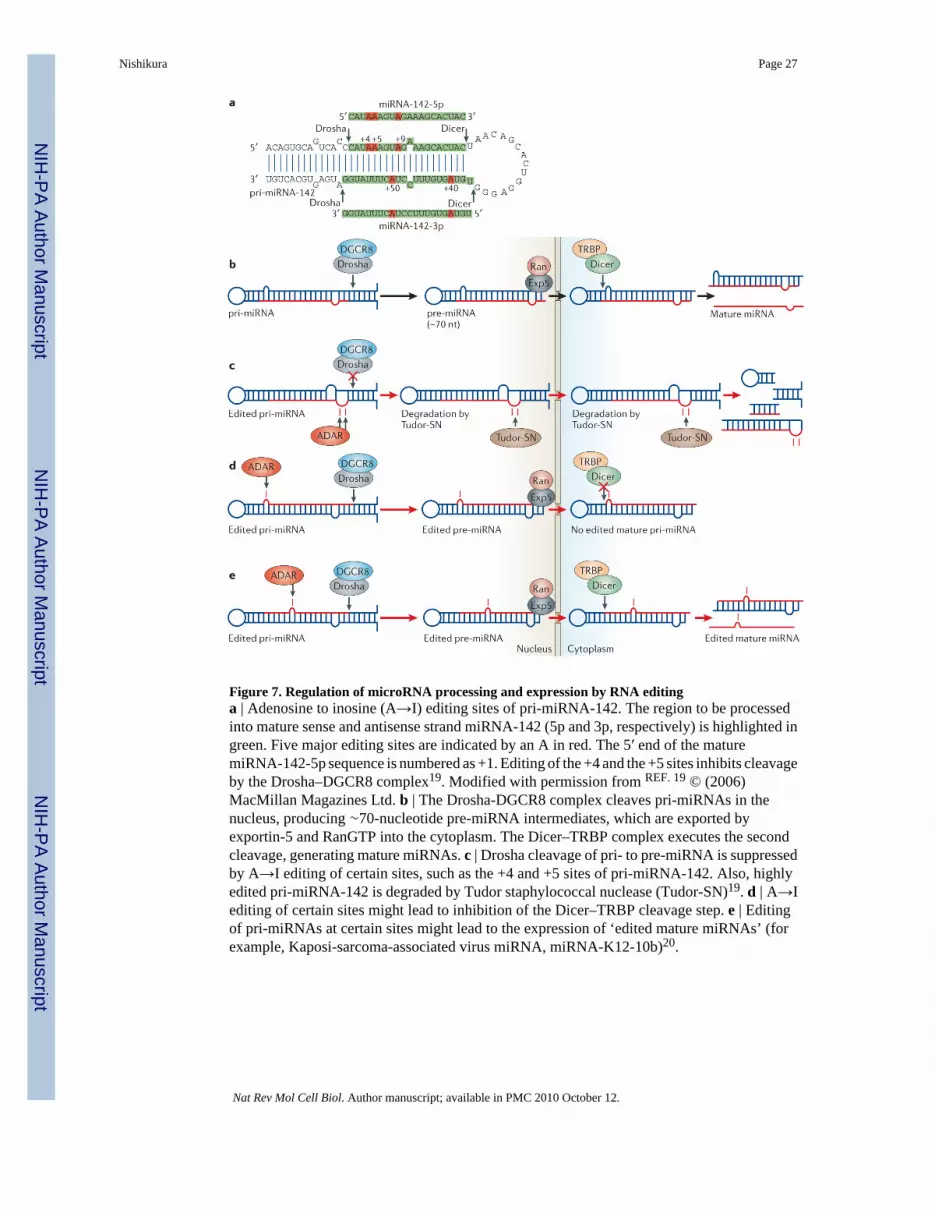
Figure 7. Regulation of microRNA processing and expression by RNA editinga | Adenosine to inosine (A→I) editing sites of pri-miRNA-142. The region to be processedinto mature sense and antisense strand miRNA-142 (5p and 3p, respectively) is highlighted ingreen. Five major editing sites are indicated by an A in red. The 5′ end of the maturemiRNA-142-5p sequence is numbered as +1. Editing of the +4 and the +5 sites inhibits cleavageby the Drosha–DGCR8 complex19. Modified with permission from REF. 19 © (2006)MacMillan Magazines Ltd. b | The Drosha-DGCR8 complex cleaves pri-miRNAs in thenucleus, producing ∼70-nucleotide pre-miRNA intermediates, which are exported byexportin-5 and RanGTP into the cytoplasm. The Dicer–TRBP complex executes the secondcleavage, generating mature miRNAs. c | Drosha cleavage of pri- to pre-miRNA is suppressedby A→I editing of certain sites, such as the +4 and +5 sites of pri-miRNA-142. Also, highlyedited pri-miRNA-142 is degraded by Tudor staphylococcal nuclease (Tudor-SN)19. d | A→Iediting of certain sites might lead to inhibition of the Dicer–TRBP cleavage step. e | Editingof pri-miRNAs at certain sites might lead to the expression of ‘edited mature miRNAs’ (forexample, Kaposi-sarcoma-associated virus miRNA, miRNA-K12-10b)20.
Nishikura Page 27
Nat Rev Mol Cell Biol. Author manuscript; available in PMC 2010 October 12.
NIH
-PA Author Manuscript
NIH
-PA Author Manuscript
NIH
-PA Author Manuscript


