Embed Size (px)
Citation preview
Lateralization of non-metric rhythm
Horváth RA1,6*, Schwarcz A2,3, Aradi M2,3, Auer T7, Fehér N6, Kovács N6,2, Tényi T4,
Szalay C5, Perlaki G2, Orsi G2, Komoly S6, Dóczi T3, Woermann FG1, Gyimesi Cs6 Janszky
J6,2
Key words: non-metric rhythm processing, lateralization, fMRI, rhythm perception
1 Epilepsy Center Bethel, Bielefeld, Germany 2Pecs Diagnostic Centre, Pecs, Hungary 3University of Pecs, Department of Neurosurgery, Pecs, Hungary 4University of Pecs, Department of Psychiatry, Pecs, Hungary 5University of Pecs, Department of Physiology, Pecs, Hungary 6 University of Pecs, Department of Neurology, Pecs, Hungary 7 Biomedizinische NMR Forschungs GmbH am Max-Planck Institut für Biophysikalische
Chemie, Göttingen, Germany
*Corresponding author:
Reka A. Horvath, MD
Epilepsy-Center Bethel, Clinic Mara I.
Maraweg 21, D-33617, Bielefeld, Germany
Tel: 0049 521 772 78870
Fax: 0049 521 772 78872
e-mail: [email protected]
This is a reprint version of the article published in Laterality. 2011 Sep;16(5):620-35.
doi: 10.1080/1357650X.2010.515990. Epub 2011 Jun 24..
Link:
http://www.tandfonline.com/doi/abs/10.1080/1357650X.2010.515990?url_ver=Z39.88-
2003&rfr_id=ori:rid:crossref.org&rfr_dat=cr_pub%3dpubmed#.U-Hey2P5cqw
© Taylor&Francais
2
Summary
Background and aim: There are contradictory results on lateralization and localization
of rhythm processing. Our aim was to test whether there is a hemispheric dissociation of
metric and non-metric rhythm processing. We created a non-metric rhythm stimuli without a
sense of meter and we measured the brain activities during the passive rhythm perception.
Methods: Eleven healthy, right-handed, Hungarian native, female speakers aged
21.3±1.1 were investigated by functional magnetic resonance imaging (fMRI) using a 3T MR
scanner. The experimental acoustic stimulus consisted of comprehensive sentences
transformed to Morse code which represent a non-metric rhythm with irregular perceptual
accent structure.
Results: Activations were found in the right hemisphere: in the posterior parts of the
right-sided superior and middle temporal gyri and temporal pole as well as in the orbital part
of the right inferior frontal gyrus. Additional activation appeared in the left-sided superior
temporal region.
Conclusion: Our study suggests that non-metric rhythm with irregular perceptual
accents structure is confined to the right hemisphere. Furthermore, a right lateralized
frontotemporal network extracts the continuous altering temporal structure of the non-metric
rhythm.
3
INTRODUCTION
Beyond language, the other structured auditory stimulus which plays a crucial role in human
life is music. The nature of hemispheric specialization for language and music processing is
still a central issue. In contrast the rigid distinction of classical view (left/language;
right/music), to date many findings have demonstrated that this hemispheric asymmetry is
rather relative than absolute. However, the dominant role of the left hemisphere in speech
processing and the dominant role of the right hemisphere in music processing has been
highlighted.
In contrast to music, language comprehension are primarily processed in left hemisphere
including separate circuits for phonological, syntactic and semantic information (Dapretto &
Bookheimer, 1999; Friederici, Rüschemeyer, Hahne & Fiebach, 2003; Friederici, Meyer, &
von Cramon, 2000) whereas sentence level prosody („speech melody”) might recruit right
hemispheric fronto-temporal network (Meyer, Alter, Friederici, Lohmann, & von Cramon,
2002). Processing of music is based on distinct neural processes corresponding to the basic
perceptual features (Pierce, 1983; Levitin, 1999). It has been suggested that the left
hemisphere appears to be devoted to processing of the integer-based rhythm (Sakai et al.,
1999) while there is a clear tendency for right-asymmetric activation in frontal and temporal-
lobe sites for pitch and melodic contour processing (Zatorre, Evan & Meyer, 1994). Lesion
studies on perception of timbre have reported that right temporal lobe lesions result in greater
impairment in timbre discrimination than left temporal lobe lesions (Milner, 1962; Samson &
Zatorre, 1994). However, a recent neuroimaging study indicates that focal regions of the left
and right temporal lobes are also involved in timbre processing (Menon, Levitin, Smith,
Lembke, Krasnow & Glazer, 2002).
4
In general, the temporal features of acoustic perception could be examined both
macroscopically (on the scale of seconds) and microscopically (on the scale of milliseconds).
Several studies have investigated the lateralization of auditory processing from microscopic
perspective using signals with differing spectrotemporal characteristics supporting a model in
which the left hemisphere is sensitive to temporal (Yamasaki, Goto, Taniwaki, Kinukawa,
Kira & Tobimatsu, 2005; Sidtis & Volpe, 1988; Robin, Tranel & Damasio, 1990), while the
right hemisphere more sensitive to spectral (Sidtis & Volpe, 1988; Robin, Tranel & Damasio,
1990; Alcock, Wade, Anslow & Passingham, 2000; Johnsrude, Penhune & Zatorre, 2000;
Zatorre & Belin, 2001; Jamison, Watkins, Bishop & Matthews, 2006) stimulus attributes.
Conversely, other investigations have argued that both hemispheres are sensitive to the
temporal structure of the speech or non-speech sound stimuli. These approaches are in accord
with the AST (“asymmetric sampling in time”) hypothesis. This model assumes that auditory
fields in the two hemispheres prefer different temporal integration windows. This hypothesis
proposes that acoustical processing of spoken language is elaborated asymmetrically in the
time domain: left auditory areas preferentially extract information from short temporal
integration windows (rapidly changing cues, 20-50 Hz), while the right hemisphere
homologues preferentially extract information from long integration windows (slowly
changing cues, 4-10 Hz) (Poeppel, 2003). It is further proposed that the posterior portion of
the auditory association cortex is the candidate region that accommodates this temporal
processing.
From macroscopic perspective, rhythm, which could be defined as a general sense of
movement in time that characterizes our experience of music (Apel, 1972), takes place on the
scale of seconds or longer. There are contradictory results in literature on the score of the
rhythm production and perception. Previous studies with brain-damaged patients on neural
processing of rhythm have found the right hemispheric contribution to rhythm processing
5
(Peretz, 1990; Penhune, Zatorre & Feindel, 1999; Kester, Saykin, Sperling, O’Connor,
Robinson & Gur, 1991; Samson, 2003). Although, others have suggested left hemispheric
dominance for rhythm perception (Sherwin & Efron, 1980; Robin, Tranel & Damasio, 1990;
Polk & Kertesz, 1993). Many rhythm studies have used tapping task, involving active
behavioral responses corresponding to the stimulus pattern (Desian & Honing, 2003; Krampe,
Kliegl, Mayr, Engbert & Vorberg, 2000) and suggested that premotor areas, SMA, preSMA
and the cerebellum are engaged (Rao, Mayer & Harrington, 2001; Schubotz, Friederici & Von
Cramon, 2000). The investigation of musicians could supply an evidence for enhanced and
altered activation pattern in right dorsolateral prefrontal cortex and right inferior frontal gyrus
as well in response to synchronization to varying levels of rhythm complexity in contrast to
non-musicians. These regions are involved working memory function for music as well
(Petrides, 2005). By means of these results, it could be assumed that subjects with long-term
musical training have a higher capacity to monitor and retrieve the temporal interval duration
in rhythmic sequences (Chen, Penhune & Zatorre, 2008). However, the correlates of passive
rhythmic perception without motor reproduction are less understood. Recent neuroimaging
studies have suggested that musical training leads to the employment of left-sided perisylvian
brain areas during passive rhythm perception (Limb, Kemeny, Ortigoza, Rouhani & Braun,
2006). Another fMRI study has also confirmed also the left hemisphere contribution in
rhythm perception, but only in the case of metric rhythm processing. This result has suggested
that there are two neural representations for rhythm depending on the interval ratio, which
correspond to metric and non-metric representations (Sakai et al., 1999). These results are in
line with the previous psychological studies which have shown that integer-based rhythm can
be reproduced more precisely and easily, than rhythms with larger intervallic values or
noninteger values (Novel, 1984; Povel & Essen, 1985).
6
The inconsistency among these studies may be attributable to using a various type of rhythms
which also have contained aspects of pitch and melody in some cases, thus making the
definition of neural substrate for rhythmic processing more complicated. The specialization
and complex interactions between the left and the right hemisphere in the perception of
temporally structured auditory stimuli still need further research. The aim of this study is to
investigate neural correlates of passive non-metric rhythm perception. According to the
previous studies, we might assume that perception of non-metric rhythms are related to the
right hemisphere in the case of non-musician subjects. It can be assumed that the processing
of metric and non-metric rhythm may apply at least partly different strategies leading to
different activation patterns.
With non-metric rhythm, time intervals are not fractions or multiples of an event or beat, and
each duration simple adds to the other. Extracting regularities from temporal sequences of
events is characteristic to human cognition. Behavioral evidence indicates that the
identification of patterns within event sequences is automatic and obligatory (Canfield &
Haith, 1991). The human brain tries to determine patterns in sequences of events, regardless
of whether a pattern truly exists, in order to predict future events. According to these
phenomena, we assume that non-metric rhythm processing might activate such mechanism.
To produce non-metric rhythm without a sense of meter, we transformed Hungarian sentences
to Morse code supposed to provide irregular perceptual accent structure to subjects who were
not familiar with the Morse code.
MATERIALS AND METHODS
Subjects
7
The participants in this experiment were 11 healthy, right-handed, non-musician women
with Hungarian native language. Their mean age was 21.3±1.1, and had no history of speech
or hearing difficulties. They were all undergraduate medical students, without knowledge of
Morse code. Handedness was determined by Edinburgh Handedness Inventory. All
experiments on human subjects were conducted in accordance with the Declaration of
Helsinki. An approval by the Institutional Review Board was obtained. Written informed
consent was obtained from all participants prior to the examinations.
fMRI Paradigm
The experimental stimuli consisted of two types of sounds: (i) comprehensive
Hungarian simple sentences transformed to Morse code, encoding characters digitally by
combinations of short and long signs (dots and dashes) with varying longitude of intervals (on
average at a speed of 3/sec; intra-character gap: 70 ms, gap between letters: 230 ms, gap
between words: 750 ms ). These sequences had non-integer intervallic ratio and were
employed as non-metric rhythm (test condition) and due to the different duration of the signs
the sequences had irregular perceptual accent structure, but without higher-level periodicity.
We expected that this stimulus would enhance listeners’ subjective perception of rhythm of
sequences and keep their attention at the same level during the whole task. (ii) Monotonic
Morse sounds (repeated dots on average at a speed of 7/sec) with equal inter-stimulus interval
known as an isochronic sequence served as baseline. Identical inter-onset intervals provide a
regular pulse and did not cause a sense of meter. The tones had a duration of 70 ms and the
onset interval was 70 ms.
The frequency range and the volume of the Morse sounds were kept at constant level,
whereas the signal sequence had a complex temporal structure. Both types of sound stimuli
(Morse coded text and baseline sounds) had similar spectral complexity with a frequency peak
8
at 700 Hz.
Acoustic stimuli were presented binaurally to the subjects through a commercially
available fMRI-compatible system (Siemens AG, Erlangen, Germany) and were presented at
a comfortable listening level that was clearly audible above the MRI scanner noise. The tone
frequency was the same during both task and baseline conditions. Sound pressure level at the
subjects’ ear was approximately +65 dB.
Experimental design
Subjects were instructed to listen passively to the sounds and their alteration. Seven
cycles of baseline (isochronic sequence) and test (non-metric rhythm with irregular perceptual
accent structure) stimuli were presented and each condition lasted 20 s (block-design), with
total design length was 4min 40s. Subjects were told not to move and to keep their eyes
closed during the whole investigation.
Data acquisition
Functional imaging was performed on a 3T MR scanner (Siemens Magnetom Trio,
Simmens AG, Erlangen, Germany) with 12-channel phased array TIM head coil for radio
frequency reception. We used a standard EPI sequence to obtain functional MR images with
the following parameters: TR: 2000ms; TE: 36ms; voxel size: 2x2x3 mm3; FoV: 192x192
cm2; 23 axial slices with a thickness of 3-mm and 1-mm gap; 80° flip angle; 1446-Hz receiver
bandwidth. We acquired 280 volumes per session. Anatomical images were acquired using a
magnetization prepared rapid gradient echo (MP-RAGE) sequence (TR: 1900ms; TE: 3.44ms,
9° flip angle, 180-Hz receiver bandwidth, 0.9x0.9x0.9 mm3 isotropic voxel size).
9
fMRI data processing
Data analysis was performed using the software package SPM5 (Wellcome Department
of Imaging Neuroscience, University College of London, UK) (Friston, Holmes, Worsley,
Polin, Firth & Frackowiack, 1995). The first three scans for each session were excluded from
data analysis because of the non-equilibrium state of magnetization. For each participant,
images underwent motion correction, and each volume was realigned to the mean of the
series. The anatomical scan was then co-registered to the mean of all functional images,
previously corrected for intensity inhomogenities through the bias correction algorithm
implemented in SPM5. EPI images were then normalized adopting the MNI152 template,
supplied by the Montreal Neurological Institute and distributed with the spm5 hig. Finally,
images were smoothed with Gaussian kernel of 5 mm. High-pass filtering (0.0078 Hz) was
applied to remove low-frequency drifts in signal.
Each subject’s data were analyzed by the general linear model (GLM) (Friston, Holmes,
Worsley, Polin, Firth & Frackowiack, 1995). The GLM was fit to the individual data, with the
experimental blocks modeled as a boxcar functions, and convolved with the canonical
hemodynamic response function (HRF). Individual models were separately estimated and
contrasts were defined in order to pick out the effects of test condition. The first level analysis
was carried out for the following contrast: non-metric rhythm with irregular perceptual accent
structure (auditory presented Morse coded text) versus isochronic sequence (monotonic
Morse sounds).
First level analysis results were then entered into the second level analysis using one-
sample t-test. The statistical significance levels were set to p=0.01, FDR-corrected for
multiple comparisons. Concurrent activation of a cluster of >5 neighboring voxels was
assumed to represent a true spot of activation.
Characterizing right-left asymmetry
10
We calculated an asymmetry index (AI) for the whole brain activation as used in earlier works
(Auer et al., 2009; Janszky et al., 2004).
AI = (activation in the left hemisphere - activation in the right hemisphere)/total activation
Thus, positive values indicate that the activation is more pronounced on the left than on
the right side. AI=1 corresponds a complete left-sided lateralization, AI=-1 a complete right-
sided speech lateralization. According to previous fMRI studies (Janszky et al., 2004;
Springer et al., 1999), AI >0.2 suggests a left-sided dominance AI <-0.2 suggests a right-sided
dominance of a particular function.
RESULTS
The comparison of passive perception of non-metric rhythm vs. the isochronous baseline
showed significant brain activations presented on the Figure 1, and listed in the Table 1.
Activations were primarily found in the right hemisphere: in the posterior parts of the right-
sided superior and middle temporal gyri, the right temporal pole as well as in the orbital part
of the right inferior frontal gyrus. Additional activation appeared in the left-sided superior
temporal region. The asymmetry index (AI) was -0.77 indicating a strong right-sided
dominance. We also tested the reverse comparison, in order to search for regions more
activated during baseline than task. This “reverse” comparison did not reveal any activated
areas. (Table 1) (Figure 1)
DISCUSSION
To examine the lateralization and localization of the perception of the complex, non-
metrically structured auditory stimuli, we were intended to determine brain structures
showing activation during listening of Morse code which represent a non-metric rhythm with
irregular accent structure. Morse coded text have been made by the rules of native language
11
resulting in a structured complex auditory stimulus which might be processed as a non-metric
rhythm in a naive subjects.
The main finding of this fMRI study is in concordance with our working hypothesis.
Activation caused by non-metrically structured auditory stimuli with irregular perceptual
accent are lateralized to the right hemisphere including the orbital part of the right inferior
frontal gyrus (IFGr) as well as the right middle and superior temporal gyrus (STGr,MTGr)
and right temporal pole. A considerably less extended activation was found in the left-sided
STG.
Essen and Povel (1985) argued that there are two type of representation of rhythm:
metric and non-metric. Metric rhythm has interval durations of integer ratios relative to the
shortest interval and this type of rhythm is reproduced with higher accuracy and easily in a
tapping task than non-metric rhythm (Povel & Essen, 1985). Moreover, perception of metric
rhythm often causes spontaneous synchronized movements. The experience of feeling the
beat during perception of metric rhythm, which is the most common in music, and
synchronization of body movement with it, are consistent with behavioral and
neuropsychological based motor theories of rhythm perception (Todd, O’Boyle, & Lee,
1999). Beat perception and movement synchronization are immediate, effortless and
automatic processing of timed acoustic patterns. It is also known that rhythm with integer
intervallic ratio (metric) and regular perceptual accent structure could induce the strongest
beat perception in subjects (Grahn & Brett, 2007). An accent can induce a beat intensification
highlighted by the manipulation of physical properties of sound, such as intensity or duration
(Lerdahl & Jackendoff, 1983). In general, meter (regular accent structure) refers to the
emergent temporal structure due to regularly accented events in auditory sequences; however
in sequences without regular accented events, listener will not be able to extract the meter, so
the perception of rhythm and meter are interdependent. In metric sequences, the parallel
12
presence of a regular accentuation enables a more accurate perception and encoding of
rhythms and can facilitate movement synchronization with rhythm by inducing strong beat
perception. (Parncutt, 1994; Patel, Iversen, Chen & Repp, 2005). According to the motor
theory of rhythm perception, many imaging studies on rhythm processing have described the
activation of motor cortices including premotor, preSMA, SMA, cerebellar areas and basal
ganglia. It has been well established that these regions were active not only in movement
synchronization task with auditory rhythms (Rao, Harrington, Haaland, Bobholz, Cox &
Binder, 1997; Chen, Zatorre & Penhune, 2006; Chen, Penhune & Zatorre, 2008), but also
shown activation during rhythm perception task (Rao, Mayer & Harrington, 2001; Schubotz,
Friederici & Cramon, 2000; Sakai et al., 1999). These areas have been also observed as active
in motor preparation tasks (Deiber, Ibanez, Sadato & Hallett, 1996). As for the basal ganglia
and cerebellum, these regions may play an important role in time perception and motor timing
(Harrington, Haaland, & Hermanowicz, 1998; Ivry, 1996; Ivry & Hazeltine, 1995; Ivry &
Schlerf, 2008) and may contribute to our sense of tempo as well (Levitin & Cook, 1996).
It can be suggested that a natural link between the auditory and motor systems may exist and
the beat perception innately relates to automatic rhythmic motor responses. A direct relation
between the movement and beat perception was found in infants (Phillips-Silver & Trainor,
2005), confirming the role of motor area. When nine-month-old infants listen to metric and
nonmetric rhythmic patterns, their ability to detect rhythmic changes is evident only in the
context of metric patterns (Bergeson & Trehub, 2006). It means that, the metric rhythm
preference can be identified in infants, relatively early in developmental phase These results
assess that rhythm is represented by default in a metric form which is also favored by
previous psychological studies (Povel, 1984; Povel & Essen, 1985) and it appears that the
capability of detecting beat in rhythmic sound sequences is already functional at birth as well.
(Winkler, Háden, Ladinigd, Szillere & Honing 2009). An internal temporal reference frame is
13
likely to exist, which may prefer the processing of small-integer ratio temporal intervals,
irrespectively of musical training. Moreover, the presence of regular accent structure can
shape our perception and expectations of the rhythm within this temporal framework. (Ivry &
Hazeltine, 1995; Pöppel, 1997; Sternberg, 1982; Povel, 1984; Povel & Essen, 1985; Handel,
1998).
From the evolutionary point of view, it seems that the ability to perceive a beat in music and
synchronization of movements is not a human unique phenomena and this may raise a further
questions over the evolution of human music (Schachner, Brady, Pepperberg & Hauser, 2009;
Patel, Iversen, Bregman & Schulz, 2009) .
These results may suggest that this system uses prediction mechanism that is used for the
preparation of motor responses. The brain could predict the patterns in sequences of events
and it may depend on the predictability of the stimuli as well. The predictable nature of the
stimuli could be varied at different level; one of them consists of metricality (integer or
noninteger ratio) and the perceptual accent structure. The most predictable rhythm stimuli
could be the metric rhythm with regular perceptual accent structure, and this type of rhythm
could also induce the strongest beat perception in subjects. It could explain why activation of
motor areas has been often observed during rhythm processing. We assume that here we could
present an indirect evidence to support this view, because we could not observe activation in
any motor regions mentioned before and our stimuli is unpredictable (non-metric rhythm with
irregular accent structure) and it is not a beat-inducing rhythm. It can be assumed that the
processing of metric and non-metric sequence with varying level of accent structure may
apply at least partly different strategy and as a result of this may employ partly different set of
neural network. A recent neuroimaging study has also confirmed that metric and non-metric
rhythm processing can recruit partly different neural networks, and non-metrical
14
representation lateralize to the right hemisphere (Sakai et al., 1999). However, it could be
possible that different activation networks for rhythm with integer and noninteger ratio in the
latter mentioned study were influenced by the varying level of regularity of the perceptual
accent structure (sense of meter). Therefore, during stimulus production we controlled
carefully their properties. According to the aim of our study, we used intervals related to
noninteger ratio. It is well known that metric rhythm with regular accent structure is the most
beat-inducing rhythm. However, studies have focused much less on the point, whether the
regular accent structure alone is able to induce a beat or subjective regularizing phenomenon
related to non-metrical rhythm could provoke it. Therefore, we generate a non-metrical
rhythm with irregular perceptual accent structure to avoid these disturbing effects.
Furthermore, we have used seven similarly structured but different sequences as test condition
in order to prevent the subjects from any impression about the relationships between the
intervals, because this may lead to the development of subjective regularizing phenomena.
Taken these together, we could examine non-metric rhythm processing itself. We assume that
our right lateralized frontotemporal network takes part in a suprasegmental auditory sequence
analysis as well. It has been well known that the primary auditory cortex is involved in early
stages of processing signal parameters such as pitch, duration, intensity and spatial location;
whereas more complex feature extraction, including temporally distributed patterns of stimuli,
is performed by the posterior region of secondary auditory cortices. Our stimuli represent a
temporally complex, non-metric sequences distributed in a long temporal integration window
without sense of meter and any melodic aspects. We suggest that our activation in right
posterior STG may reflect to the slowly changes of temporal structure of stimuli used in our
study. This result is in accordance with the AST (asymmetric sampling in time) hypothesis.
This hypothesis proposes that acoustical processing of speech signal is elaborated
asymmetrically in the time domain: left auditory areas preferentially extract information from
15
short temporal integration windows, while the right hemisphere homologues preferentially
extract information from long integration windows. It is proposed that the posterior portion of
the auditory association cortex is the candidate region that accommodates this temporal
processing. This localization is in line with our results. Moreover, our findings may suggest
that the right posterior STG is associated with not only coding the suprasegmental speech
rhythm but the slow temporal features of a non-metric rhythm as well. The other main region
in which we found activation was the orbital part of right inferior frontal gyrus (BA 47). The
frontal regions have been also known as a key structure for working memory (Courtney, Peti,
Maisog, Ungerleider & Haxby, 1998; Courtney, Ungerleider, Keil & Haxby, 1997) and
monitoring of memorized information (Petrides, 1994; Petrides, 2005). We assume that non-
metric rhythm might require continuous monitoring and encoding of the different time
intervals as opposed to a metric rhythm and this could be the cause of the observed activation
in the right inferior frontal region. This assumption was confirmed by a previous imaging
study on rhythm representation (Sakai et al., 1999), they have found frontal activation only in
the case of non-metric rhythm. The presence of activation in the right frontal region only in
the case of non-metric rhythm, and our similar findings could support the assumption that the
non-metric rhythm with irregularly changing time intervals may require a more explicit,
continuous monitoring strategy than metric rhythm. We suggest that right inferior frontal
cortex may participate in continuous mapping of temporal structure’s changes of non-metric
rhythm. Activation of this region has been reported in studies which were focused on
processing of temporal coherence of music (Poldrack, Wagner, Prull, Desmond, Glover &
Gabrieli, 1999; Levitin & Menon, 2003). These argue that Brodmann area 47 is a brain area
that organizes structural units in the perceptual stream to create larger, meaningful
representation. These approaches suggest that there is a cognitive system dedicated to the on-
line monitoring of temporal structural pattern (Huettel, Mack & McCarthy, 2002). Our
16
findings might reflect the aspect of long-range structure of our stimuli and the sensitivity of
pars orbitalis for temporal structural incongruity. Our results suggest that the right STG and
the right IFG compose a network specifically associated with coding, continuous monitoring
and reconciliation of the irregularly changing time intervals in auditory sequences.
Furthermore, there is also a considerable amount of research which has associated the right
anterior superior temporal cortex (right temporal pole, BA 38) with the representation of
complex melodies and harmonies (Brown, Martinez, Hodges, Fox & Parsons, 2004;
Patterson, Upperkamp, Johnsrude & Grifiths, 2002) as well as in the perception of non-speech
vocal sounds (Belin, Zatorre & Ahad, 2002). We might argue that this region could also be
involved in certain involuntary comparative processing while subjects attend to set the heard
atypical, non-metric auditory sequences against the implicit representation of the typical
metric rhythmic features.
Three previous studies investigated the functional anatomy of auditory perception of Morse-
coded text. In an early study before modern neuroimaging era, Papcun et al. (1974) presented
dichotically Morse code signals to Morse code operators and to naive subjects (Papcun,
Krashen, Terbeek, Remington & Harshman, 1974). They showed that if the stimuli were
longer than seven elements, the naive subjects showed left ear superiority (a presumed
activation of the right hemisphere), which results are consistent with ours. In contrast to our
study, Maier et al (2004) reported that Morse code operators showed predominantly left-sided
activation of the frontal and temporal perisylvian language areas, prefrontal cortex as well as
premotor cortex during listening to Morse coded auditory texts (Maier, Hartvig, Green &
Stodkilde-Jorgensen, 2004). The main difference between that study and the present work is
that their subjects were Morse operators, thus, they knew the Morse code; thus their stimulus
had semantic components. This underlies the sensitivity of the left-sided perisylvian region to
the semantic component of auditory stimuli. Recently, Schmidt-Wilcke et al have investigated
17
the relationship between changes in neural activity pattern and changes in gray matter density
associated with learning on healthy subjects who learned to resolve Morse code after Morse
code training. They have provided a clear evidence for neural plasticity associated with the
newly-acquired skill (Schmidt-Wilcke, Rosengarth, Luerding, Bogdahn & Greenlee, 2010).
Summarizing, our study suggests that non-metric rhythm with irregular perceptual accent
structure are confined to the right hemisphere. Furthermore, the extraction of the continuous
altering temporal structure of non-metric rhythm takes place in the right lateralized
frontotemporal network.
18
References
Alcock, K.J., Wade, D., Anslow, P., & Passingham, R.E. (2000) Pitch and timing abilities in
adult left-hemisphere-dysphasic and right-hemisphere damaged subjects. Brain and
Language, 75, 47-65.
Apel, W. (1972) Harvard dictionary of music (2nd ed.). Cambridge, MA: Belknap Press of
Harvard University Press.
Auer, T., Pinter, S., Kovacs, N., Kalmar, Z., Nagy, F., Horvath, R.A., et al. (2009) Does
obstetric plexus injury influence speech domiance? Annals of Neurology, 65, 57-66.
Belin, P., Zatorre, R. J., & Ahad, P. (2002). Human temporal-lobe response to vocal sounds.
Brain Research, Cognitive Brain Research, 13, 17–26.
Bergeson, T.R. & Trehub S.E. (2006) Infants’ perception of rhythmic patterns. Music
perception, 23, 345-360.
Brown, S., Martinez, M. J., Hodges, D. A., Fox, P. T., & Parsons, L. M. (2004). The song
system of the human brain. Brain Research, Cognitive Brain Research, 20, 363–375.
Canfield, R. L., & Haith, M.M. (1991) Young infants’ visual expectations for symmetric and
asymmetric stimulus sequences. Developmental Psychology, 27, 198–208.
Chen, J.L., Penhune, V.B. & Zatorre, R.J. (2008) Moving on time: brain network for auditory-
motor synchronization is modulated by rhythm complexity and musical training. The Journal
of Cognitive Neuroscience, 20, 226-239.
Chen, J.L., Zatorre, R.J. & Penhune, V.B. (2006) Interactions between auditory and dorsal
premotor cortex during synchronization to musical rhythms. Neuroimage, 32, 1771-1781.
19
Courtney, S.M., Peti, L., Maisog, J.M., Ungerleider, L.G., & Haxby, J.V. (1998) An area
specialized for spatial working memory in human frontal cortex. Science, 279, 1347–1351.
Courtney, S.M., Ungerleider, L.G., Keil, K., & Haxby, J.V. (1997) Transient and sustained
activity in a distributed neural system for human working memory. Nature, 386, 608–611.
Dapretto, M., & Bookheimer, S. Y. (1999). Form and content: Dissociating syntax and
semantics in sentence comprehension. Neuron, 24, 427–432.
Deiber, M.P., Ibanez, V., Sadato, N., & Hallett, M. (1996) Cerebral structures participating in
motor preparation in humans: positron emission tomograpy study. Journal of
Neurophisiology, 75, 233-247.
Desain, P., & Honing, H. (2003). The formation of rhythmic categories and metric priming.
Perception, 32, 341–365.
Friederici, A.D., Meyer, M., & von Cramon, D.Y. (2000). Auditory language comprehension:
An event-related fMRI study on the processing of syntactic and lexical information. Brain
and Language, 74, 289–300.
Friederici, A.D., Rüschemeyer, S.A., Hahne, A., & Fiebach, C. J. (2003). The role of left
inferior frontal and superior temporal cortex in sentence comprehension: Localizing syntactic
and semantic processes. Cerebral Cortex, 13, 117–177.
Friston, K., Holmes, A., Worsley, K., Polin, J.B., Frith, C., & Frackowiack, C. (1995)
Statistical parametric maps in functional imaging: A general linear approach. Human Brain
Mapping, 2, 189-210.
Grahn, J.A., & Brett, M. (2007) Rhythm and Beat Perception in Motor Areas of the brain.
Journal of cognitive Neuroscience, 19, 893-906.
20
Handel, S. (1998) The interplay between metric and figural rhythmic organization. Journal of
Experimental Psychology: Human Perception and Performance, 24, 1546–1561.
Harrington, D.L., Haaland, K.Y., & Hermanowicz, N. (1998) Temporal processing in the
basal ganglia. Neuropsychology, 12, 3–12.
Heim, S., Opitz, B., Müller, K. & Friederici, A.D. (2003). Phonological processing during
language production: fMRI evidence for a shared production-comprehension network.
Cognitive Brain Research, 16, 285-296.
Huettel, S. A., Mack, P. B., & McCarthy, G. (2002). Perceiving patterns in random series:
Dynamic processing of sequence in prefrontal cortex. Nature Neuroscience, 5, 485–490.
Ivry, R.B. & Schlerf, J.E. (2008) Dedicated and intrinsic models of time perception. Trends in
Cognitive Sciences, 12, 273-280.
Ivry, R.B. & Hazeltine, R.E. (1995) Perception and production of temporal intervals across a
range of durations: Evidence for a common timing mechanism. Journal of Experimental
Psychology: Human Perception and Performance, 21, 3-18.
Ivry, R.B. (1996) The representation of temporal information in perception and motor control.
Current Opinion in Neurobiology, 6, 851–857.
Jamison, H.L., Watkins, K.E., Bishop, D.V., & Matthews, P.M. (2006) Hemispheric
specialization for processing auditory nonspeech stimuli. Cerebral Cortex, 16, 1266-1275.
Janszky, J., Olech, I., Jokeit, H., Kontopoulou, K., Mertens, M., Ebner, et al. (2004) Epileptic
activity influences lateralization of mesiotemporal fMRI activity. Neurology, 63, 1813-1817.
21
Johnsrude, I.S., Penhune, V.B., & Zatorre, R.J. (2000) Functional specificity in the right
human auditory cortex for perceiving pitch direction. Brain, 123, 155-163.
Kester, D.B., Saykin, A.J., Sperling, M.R., O’Connor, M.J., Robinson, L.J., & Gur, R.C.
(1991) Acute effect of anterior temporal lobectomy on musical processing. Neuropsychologia,
29, 703–708.
Krampe, R.T., Kliegl, R., Mayr, U., Engbert, R., & Vorberg, D. (2000) The fast and the slow
of skilled bimanual rhythm production: parallel versus integrated timing. Journal of
Experimental Psychology: Human Perception and Performance, 26, 206–233.
Lerdahl, F.& Jackendoff, R., (1983) A Generative Theory of Tonal Music. Cambridge, MA :
MIT Press.
Levitin, D. J. (1999). Experimental design in psychoacoustic research. In P.R. Cook [Ed.],
Music, Cognition, and Computerized Sound: An Introduction to Psychoacoustics. Cambridge,
MA: MIT Press, 299-328.
Levitin, D.J. & Cook, P.R. (1996) Memory for musical tempo: Additional evidence that
auditory memory is absolute. Perception & Psychophysics, 58, 927–935.
Levitin, D.J., & Menon, V. (2003) Musical structure is processed in “language” areas of the
brain: A possible role for Brodmann Area 47 in temporal coherence. NeuroImage, 20, 2142–
2152.
Limb, C.J. (2006) Structural and functional neural correlates of music perception. The
Anatomical Record Part A: Discoveries in Molecular, Cellular, and Evolutionary Biology,
288, 435-446.
22
Limb, C.J., Kemeny, S., Ortigoza, S.B., Rouhani, & Braun, A.R. (2006) Left hemispheric
lateralization of brain activity during passive rhythm perception in musicians. The Anatomical
Record Part A: Discoveries in Molecular, Cellular, and Evolutionary Biology, 288, 382-389.
Maier, J., Hartvig, N.V., Green, A.C., & Stodkilde- Jorgensen, H. (2004) Reading with ears.
Neuroscience Letter 8, 185-188.
Menon, V., Levitin, D.J., Smith, B.K., Lembke, A., Krasnow, B.D. & Glazer, D. (2002)
Neural correlates of timbre change in harmonic sounds. Neuroimage, 17: 1742–54.
Meyer, M., Alter, K., Friederici, A.D., Lohmann, G., & von Cramon, D.Y. (2002). Functional
MRI reveals brain regions mediating slow prosodic modulations in spoken sentences. Human
Brain Mapping, 17, 73–88.
Milner, B. (1962). Laterality Effects in Audition. Johns Hopkins Press, Baltimore.
Papcun, G., Krashen, S., Terbeek, D., Remington, R., & Harshman R. (1974) Is the left
hemisphere specialized for speech, language and-or something else? Journal of the Acoustical
Society of America, 55, 319-27.
Parncutt, R. (1994) A perceptual model of pulse salience and metrical accent in musical
rhythms. Music Perception, 11, 409–464.
Patel, A.D., Iversen , J.R., Chen, Y. & Repp, B.H. (2005) The influence of metricality and
modality on synchronization with a beat. Experimental Brain Research, 163, 226–238.
Patel, A.D., Iversen, J.R., Bregman, M.R. & Schulz, I. (2009) Experimental evidence for
synchronization to a musical beat in a nonhuman animal. Current Biology, 19, 827-830.
23
Patterson, R. D., Uppenkamp, S., Johnsrude, I. S., & Griffiths, T. D. (2002). The processing
of temporal pitch and melody information in auditory cortex. Neuron, 36, 767–776.
Penhune, V.B., Zatorre, R.J., & Feindel, W.H. (1999) The role of auditory cortex in retention
of rhythmic patterns as studied in patients with temporal lobe removals including Heschl’s
gyrus. Neuropsychologia, 37, 315–331.
Peretz, I. (1990) Processing of local and global musical information by unilateral brain-
damaged patients. Brain, 113, 1185–1205.
Petrides, M (2005). Lateral prefrontal cortex: architectonic and functional organization.
Philosophical transactions of the Royal Society of London. Series B, Biological sciences, 360,
781-795.
Petrides, M. (1994) Frontal lobes and behavior. Curent Opinion in Neurobiology, 4, 207–
211.
Phillips-Silver, J., & Trainor, L.J. (2005) Feeling the beat: Movement influences infant
rhythm perception. Science, 308, 1430.
Pierce, J. R. (1983). The science of musical sound. New York, NY: Scientific American
Books, Inc.
Poeppel, D. (2003) The analysis of speech in different temporal integration windows: cerebral
lateralization as ‘asymmetric sampling in time’. Speech Communication, 41, 245-255.
24
Poldrack, R.A., Wagner, A.D., Prull, M.W., Desmond, J.E., Glover, G.H., & Gabrieli, J.D.E.
(1999) Functional specialization for semantic and phonological processing in the left inferior
prefrontal cortex. NeuroImage, 10, 15–35.
Polk, M., & Kertesz, A. (1993) Music and language in degenerative disease of the brain.
Brain Cognition, 22, 98 –117
Povel, D.J. (1984) A theoretical framework for rhythm perception. Psychology Research, 45,
315–337.
Povel, D.J., & Essens, P. (1985) Perception of temporal patterns. Music Perception, 2, 411–
440.
Pöppel, E. (1997) Hierarchical model of temporal perception. Trends in Cognitive Sciences,1,
56-61.
Rao, S.M., Harrington, D.L., Haaland, K.Y., Bobholz, Y.A., Cox, R.W. & Binder, J.R. (1997)
Distributed neural systems underlying the timing of movements. The Journal of
Neuroscience, 17, 5528–5535.
Rao, S.M., Mayer, A.R., & Harrington, D.L. (2001) The evolution of brain activation during
temporal processing . Nature Neuroscience, 4, 317-323.
Robin, D.A., Tranel, D., & Damasio, H. (1990) Auditory perception of temporal and spectral
events in patients with focal left and right cerebral lesions. Brain and Languge, 39, 539-555.
25
Sakai, K., Hikosaka, O., Miyauchi, S., Takino. R., Tamada, T., Iwata, N.K., & Nielsen, M.
(1999) Neural representation of a rhythm depends on its interval ratio. Journal of
Neuroscience, 19, 10074–10081.
Samson, S. (2003) Cerebral substrates for musical temporal processes. In: Peretz I, editor. The
cognitive neuroscience of music, Oxford: Oxford University Press. p 204–230.
Samson, S., & Zatorre, R. J. (1994) Contribution of the right temporal lobe to musical timbre
discrimination. Neuropsychologia, 32, 231–240.
Schachner, A., Brady, T.F., Pepperberg, I.M. & Hauser, M.D. (2009) Spontaneous motor
entrainment to music in multiple vocal mimicking species. Current Biology, 19, 831–836.
Schmidt-Wilcke, T., Rosengarth, K., Luerding, R., Bogdahn, U., & Greenlee, M.W. (2010)
Distinct patterns of functional and structural neuroplasticity associated with learning Morse
code. Neuroimage, 51, 1234-1241.
Schubotz, R.I., Friederici, A.D., & Von Cramon, D.Y. (2000) Time perception and motor
timing: a common cortical and subcortical basis revealed by fMRI. Neuroimage, 11, 1-12.
Sherwin, I., & Efron, R. (1980) Temporal ordering deficits following anterior temporal
lobectomy. Brain and Language, 11, 195–203.
Sidtis, J.J., & Volpe, B.T. (1988) Selective loss of complex-pitch or speech discrimination
after unilateral lesion. Brain and Language, 34, 235-245.
Springer, J.A., Binder J.R., Hammeke, T.A., Swanson, S.J., Frost, J.A. & Bellgowan, P.S.F.
(1999) Language dominance in neurologically normal and epilepsy subjects: a functional MRI
study. Brain, 122, 2033-2046.
Sternberg, S., Knoll, R.L. & Zukofsky, P. (1982) Timing by skilled musicians. In D. Deutsch
(Ed.) The psychology of music. New York: Academic Press., 181-239.
26
Todd, N.P.M., O’Boyle, D.J.O., & Lee, C.S. (1999) A sensory-motor-theory of rhythm, time
perception, and beat induction. Journal of New Music Research, 28, 5–28.
Winkler, I., Háden, G.P., Ladinigd, O., Szillere, I. & Honing, H. (2009) Newborn infants
detect the beat in music. Proceedings of the National Academy of Sciences, 106, 2468-2471.
Yamasaki, T., Goto, Y., Taniwaki, T., Kinukawa, N., Kira, J., & Tobimatsu, S. (2005) Left
hemisphere specialisation for rapid temporal processing: a study with auditory 40 Hz steady-
state responses. Clinical Neurophysiology, 116, 393-400.
Zatorre, R.J., & Belin, P. (2001) Spectral and temporal processing in human auditory cortex.
Cerebral Cortex, 11, 946-953.
Zatorre, R.J., Evans, A.C. & Meyer, E. (1994) Neural mechanisms underlying melodic
perception and memory for pitch. The Journal of Neuroscience, 14, 1908–1919.
27
Figure legends
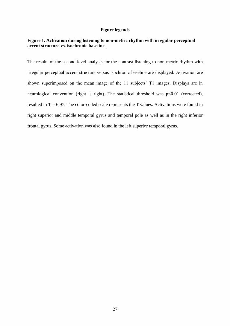
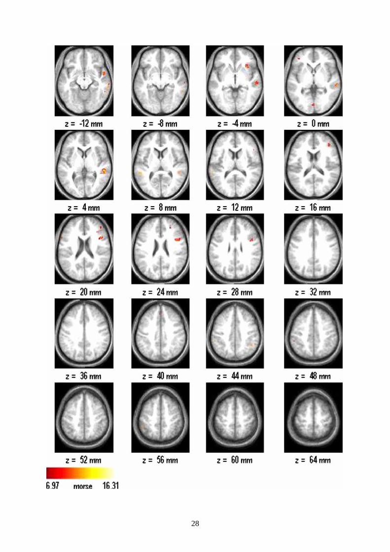
Figure 1. Activation during listening to non-metric rhythm with irregular perceptual
accent structure vs. isochronic baseline.
The results of the second level analysis for the contrast listening to non-metric rhythm with
irregular perceptual accent structure versus isochronic baseline are displayed. Activation are
shown superimposed on the mean image of the 11 subjects’ T1 images. Displays are in
neurological convention (right is right). The statistical threshold was p<0.01 (corrected),
resulted in T = 6.97. The color-coded scale represents the T values. Activations were found in
right superior and middle temporal gyrus and temporal pole as well as in the right inferior
frontal gyrus. Some activation was also found in the left superior temporal gyrus.
28


