Embed Size (px)
Citation preview
Citation: Troutman, G.S.; Genuardi,
M.V. Left Ventricular Assist Devices:
A Primer for the Non-Mechanical
Circulatory Support Provider. J. Clin.
Med. 2022, 11, 2575. https://doi.org/
10.3390/jcm11092575
Academic Editor: Francesco
Pelliccia
Received: 6 April 2022
Accepted: 30 April 2022
Published: 4 May 2022
Publisher’s Note: MDPI stays neutral
with regard to jurisdictional claims in
published maps and institutional affil-
iations.
Copyright: © 2022 by the authors.
Licensee MDPI, Basel, Switzerland.
This article is an open access article
distributed under the terms and
conditions of the Creative Commons
Attribution (CC BY) license (https://
creativecommons.org/licenses/by/
4.0/).
Journal of
Clinical Medicine
Review
Left Ventricular Assist Devices: A Primer for theNon-Mechanical Circulatory Support ProviderGregory S. Troutman 1 and Michael V. Genuardi 2,*
1 Department of Medicine, Hospital of the University of Pennsylvania, Philadelphia, PA 19104, USA;[email protected]
2 Perelman School of Medicine, University of Pennsylvania, Philadelphia, PA 19104, USA* Correspondence: [email protected]; Tel.: +1-215-615-0800
Abstract: Survival after implant of a left ventricular assist device (LVAD) continues to improve forpatients with end-stage heart failure. Meanwhile, more patients are implanted with a destinationtherapy, rather than bridge-to-transplant, indication, meaning the population of patients living long-term on LVADs will continue to grow. Non-LVAD healthcare providers will encounter such patientsin their scope of practice, and familiarity and comfort with the physiology and operation of thesedevices and common problems is essential. This review article describes the history, development,and operation of the modern LVAD. Common LVAD-related complications such as bleeding, infection,stroke, and right heart failure are reviewed and an approach to the patient with an LVAD is suggested.Nominal operating parameters and device response to various physiologic conditions, includinghypo- and hypervolemia, hypertension, and device failure, are reviewed.
Keywords: left ventricular assist devices; heart failure with reduced ejection fraction; right heart failure
1. Introduction
The prevalence of heart failure (HF) continues to rise in the U.S. as the populationages. An estimated 6 million American adults have HF according to 2015–2018 data [1].Pharmacologic and device-based treatment advances have helped to extend the lifespanand slow disease progression of HF, leading to a growing population that the EuropeanSociety for Cardiology describes as “advanced, chronic HF” [2].
After the careful exclusion and treatment of reversible causes, modern care of thepatient with HF with reduced ejection fraction (HFrEF) is focused on rapid initiationand uptitration of guideline-directed medical therapy with contemporary “quadruplemedical therapy”, as follows: angiotensin receptor-neprilysin inhibitors, evidence-basedbeta-blockers, mineralocorticoid receptor antagonists, and sodium glucose cotransporter2 inhibitors [3]. Aggressive implementation of quadruple therapy can lead to recovery andsignificant reduction of morbidity/mortality associated with HFrEF. Device therapies otherthan mechanical circulatory support (MCS), such as cardiac resynchronization therapy,have also lessened disease morbidity and mortality [4]. However, many patients will stilldevelop progressive disease despite the promise of these therapies and may require moreadvanced measures.
Heart failure ranges in severity, with the American Heart Association defining themost severe as Stage D HF, designating “the patient with end-stage disease who requiresspecialized treatment strategies such as mechanical circulatory support, continuous in-otropic infusions, cardiac transplantation, or hospice care” [5]. Left ventricular assist device(LVAD) therapy was initially developed as short-term MCS for patients with end-stageHFrEF as a bridge-to-transplant (BTT), but advances in the durability and portability ofLVADs over the past decades have led to a revolution in the typical LVAD patient profile.Now, most patients receiving an LVAD implant are destination therapy (DT), rather thanBTT [1,6]. Other device indications include bridge-to-recovery in patients who are hoped
J. Clin. Med. 2022, 11, 2575. https://doi.org/10.3390/jcm11092575 https://www.mdpi.com/journal/jcm

J. Clin. Med. 2022, 11, 2575 2 of 14
to improve enough for explant, and bridge-to-candidacy (or the essentially interchangeable“bridge-to-decision” or “DT-modifiable”), which intends to capture patients who are nottransplant candidates at the time of implant but have reversible contraindications. Severalresearch consortia, including the influential Interagency Registry for Mechanically AssistedCirculatory Support (Intermacs), as well as many LVAD clinicians colloquially, use theseterms to supplement DT and BTT where appropriate. Importantly, the Centers for Medicare& Medicaid Services, which sets standards for public insurance programs in the U.S., hadpreviously used only the DT and BTT labels. As of 2020, however, all reference to DT andBTT were removed from guidelines in an acknowledgement of the strong evidence base forLVAD therapy irrespective of implant indication [7].
Between 2010–2019, over 25,000 patients underwent implantation of a durable, continuous-flow LVAD, with nearly 3200 in 2019 [8]. The increasing proportion of annual DT implantsand the greater survival of patients on long-term MCS has created a growing populationof long-term LVAD patients presenting for care for myriad LVAD and non-LVAD relatedhealth conditions. This review is meant to serve as a primer for physicians and other careproviders who encounter LVAD patients in their regular scope of practice, although theyare not primarily advanced heart failure or MCS practitioners.
2. The LVAD Patient Profile
Determining optimal timing for LVAD referral can be challenging for heart failurepatients but is critical, as late referral is associated with poorer outcomes after LVAD implan-tation. In particular, secondary end-organ dysfunction often precludes LVAD implantationin patients who are not promptly referred or evaluated [9]. The phrase “I NEED HELP”is useful as a memory aide for the core factors defining the progression from stable toadvanced heart failure. [10]. The mnemonic stands for the following: Inotropes (historyof or current need), NYHA class (III or IV symptoms), End-organ dysfunction (particu-larly renal or hepatic dysfunction), Ejection fraction (<35%), Defibrillator shocks (recurrentfor ventricular tachycardia or fibrillation), Recurrent hospitalization (>1 in the past year),Edema/Escalating diuretic dosing (implying diuretic refractoriness), Low blood pressure(systolic < 90 mmHg), or Prognostic medications (intolerance of neurohormonal blockage).All of these “warning signs” attempt to identify patients with advanced and/or progressiveHFrEF despite optimal medical therapy who may be considered for LVAD implantation.The challenge remains to target these patients earlier in their advanced HF course, i.e., priorto the development of shock, renal or hepatic dysfunction, or hospital dependence [11].Delayed referral and LVAD implantation for patients with progressive decline on inotropicsupport or those in critical cardiogenic shock leads to increased complications and mortalityafter LVAD implantation [12].
It is important to note that, despite advances in medical and mechanical therapies,racial and ethnic disparities in heart failure persist across incidence, hospitalization, andoutcomes. Over the past two decades, disparities in heart failure mortality in AfricanAmericans have worsened [13,14]. Racial and ethnic disparities in LVAD utilization havenarrowed, however disparities in transplantation and outcomes remain [15,16]. Individual,organizational, and structural interventions are necessary to combat the structural racismthat exists in heart failures therapies and our medical system as a whole.
3. Toward the Modern Continuous Flow Era
It has been almost six decades since the first successful ventricular assist device wasused in a human patient after years of unsuccessful attempts to develop a practical totalartificial heart [17]. The first LVADs were primarily used for post-cardiotomy shock inwhat would now be termed a “bridge-to-recovery” indication. Simple in construction andexternal to the patient, they consisted of a hemi-spherical metallic casing containing aninflatable bladder, rhythmically inflated with CO2. The cyclical negative and positive pres-sure drew ventricular blood into the hemi-spherical housing before ejecting it into the targetblood vessel via surgically placed conduits. The device was timed via electrocardiographic

J. Clin. Med. 2022, 11, 2575 3 of 14
synchronization to pump during diastole, when the aortic valve was closed, or else timingcould be determined manually. Level of support (flow rate) was regulated by changingthe ratio of native cardiac cycles to LVAD inflations (analogous to the support ratio on amodern intra-aortic balloon pump) or by changing the bladder inflation volume. One-hourbattery powered excursions were possible.
The field of ventricular assist developed in parallel to cardiac transplantation, thelatter first performed in the late 1960s. Various devices were employed successfully forbridge-to-recovery or bridge-to-transplant throughout the 1980s and 1990s [18]. However,the most successful LVADs for intermediate and long-term support were pulsatile, retainingclose design similarity to the first devices used decades prior, such as the HeartMate IP andlater VE/XVE models and the Novacor LVAS [19]. The HeartMate VE/XVE system fromThoratec (Pleasanton, CA, USA) was a notable breakthrough in LVAD history, obtainingthe first approval for use as long-term support in transplant-ineligible patients, for socalled destination therapy. This approval was gained on the basis of 2001’s REMATCHtrial, showing superior mortality among end-stage heart failure patients implanted withthe fully implantable, battery-powered HeartMate VE as compared to medical therapyalone [6]. More recently, concerns over mechanical reliability and hemocompatibility haveushered in modern continuous flow pumps. The continuous flow long-term support erawas launched by the approval of the Thoratec HeartMate II axial flow device, based onclear improvements in device reliability and survival, in 2009 [20]. Specifically, overall2-year survival free of stroke or device malfunction rose dramatically, from 11 to 46%, withcontinuous axial flow as compared to pulsatile flow. Overall mortality was reduced byover 40%. Early fears about nonphysiologic continuous flow on organs such as the gutand brain did not materialize in a clinically significant way [21]. With the era of highlyreliable continuous flow pumps well established, the field shifted yet again when two newdevices miniaturized the pump rotor and moved the rotor housing from the abdomento intra-pericardial, directly adjacent to the LV apex. These devices, the Heartware VAD(“HVAD”, Medtronic, Minneapolis, MN, USA) and the HeartMate 3 (now owned by Abbottof Chicago, Chicago, IL, USA), had further reliability advantages [22,23]. The HVAD waswithdrawn from the market in 2021 over concerns about increased risk of stroke [24],leaving the HeartMate 3 system the only long-term ventricular assist device in NorthAmerica as of 2022.
4. The Left Ventricular Assist Device Components
The main components of the current era, continuous flow, LVADs include the LVADinflow cannula, pump, and outflow cannula, an electric controller, batteries and a per-cutaneous driveline (Figure 1). The percutaneous driveline carries electrical cables andan air vent to the battery packs worn on a shoulder holster and electric controls wornon a belt [25]. The inflow cannula is inserted into the apex of the left ventricle (LV) withthe outflow cannula delivering blood to the ascending aorta (Figure 2). Blood returnsfrom the lungs through left-sided native anatomy to the point of the LV, and exits via theLV apex across an inflow valve into the prosthetic pump and then the ascending aorta.There is typically some amount of competitive native flow across the aortic valve duringsystole, however in many cases the LVAD provides “full support” and the aortic valve doesnot open.

J. Clin. Med. 2022, 11, 2575 4 of 14
Figure 1. Contemporary LVAD device. Left ventricular assist device (LVAD) with full magneticlevitation with patient peripherals shown. The rotor pump is implanted on the left ventricular apexwith outflow graft directed to the aortic root (1). A controller unit is typically worn on the belt anddisplays device settings, status, and alerts/alarms (2). The controller is connected to two portablebattery packs (3), shown here suspended on the shoulders. The controller is attached to the LVADvia a driveline (4) that typically exits the skin in the upper abdomen and is covered with a steriledressing at all times. (Image reprinted with permission from Abbott Laboratories).
J. Clin. Med. 2022, 11, 2575 5 of 14
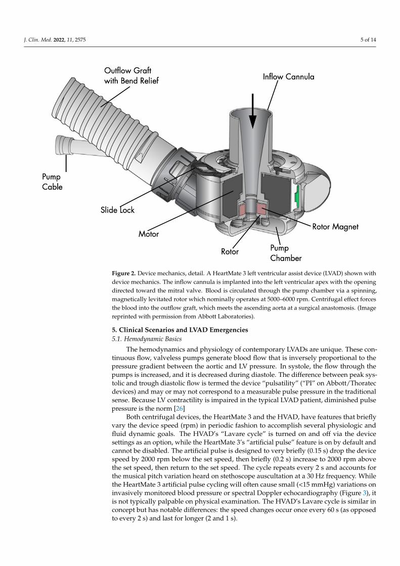
Figure 2. Device mechanics, detail. A HeartMate 3 left ventricular assist device (LVAD) shown withdevice mechanics. The inflow cannula is implanted into the left ventricular apex with the openingdirected toward the mitral valve. Blood is circulated through the pump chamber via a spinning,magnetically levitated rotor which nominally operates at 5000–6000 rpm. Centrifugal effect forcesthe blood into the outflow graft, which meets the ascending aorta at a surgical anastomosis. (Imagereprinted with permission from Abbott Laboratories).
5. Clinical Scenarios and LVAD Emergencies5.1. Hemodynamic Basics
The hemodynamics and physiology of contemporary LVADs are unique. These con-tinuous flow, valveless pumps generate blood flow that is inversely proportional to thepressure gradient between the aortic and LV pressure. In systole, the flow through thepumps is increased, and it is decreased during diastole. The difference between peak sys-tolic and trough diastolic flow is termed the device “pulsatility” (“PI” on Abbott/Thoratecdevices) and may or may not correspond to a measurable pulse pressure in the traditionalsense. Because LV contractility is impaired in the typical LVAD patient, diminished pulsepressure is the norm [26]
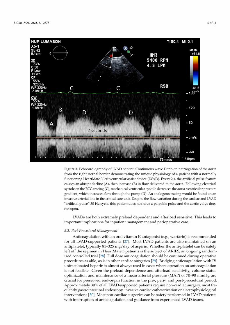
Both centrifugal devices, the HeartMate 3 and the HVAD, have features that brieflyvary the device speed (rpm) in periodic fashion to accomplish several physiologic andfluid dynamic goals. The HVAD’s “Lavare cycle” is turned on and off via the devicesettings as an option, while the HeartMate 3’s “artificial pulse” feature is on by default andcannot be disabled. The artificial pulse is designed to very briefly (0.15 s) drop the devicespeed by 2000 rpm below the set speed, then briefly (0.2 s) increase to 2000 rpm abovethe set speed, then return to the set speed. The cycle repeats every 2 s and accounts forthe musical pitch variation heard on stethoscope auscultation at a 30 Hz frequency. Whilethe HeartMate 3 artificial pulse cycling will often cause small (<15 mmHg) variations oninvasively monitored blood pressure or spectral Doppler echocardiography (Figure 3), itis not typically palpable on physical examination. The HVAD’s Lavare cycle is similar inconcept but has notable differences: the speed changes occur once every 60 s (as opposedto every 2 s) and last for longer (2 and 1 s).
J. Clin. Med. 2022, 11, 2575 6 of 14
Figure 3. Echocardiography of LVAD patient. Continuous wave Doppler interrogation of the aortafrom the right sternal border demonstrating the unique physiology of a patient with a normallyfunctioning HeartMate 3 left ventricular assist device (LVAD). Every 2 s, the artificial pulse featurecauses an abrupt decline (A), then increase (B) in flow delivered to the aorta. Following electricalsystole on the ECG tracing (C), mechanical ventricular systole decreases the aorta-ventricular pressuregradient, which increases flow through the pump (D). An analogous tracing would be found on aninvasive arterial line in the critical care unit. Despite the flow variation during the cardiac and LVAD“artificial pulse” 30 Hz cycle, this patient does not have a palpable pulse and the aortic valve doesnot open.
LVADs are both extremely preload dependent and afterload sensitive. This leads toimportant implications for inpatient management and perioperative care.
5.2. Peri-Procedural Management
Anticoagulation with an oral vitamin K antagonist (e.g., warfarin) is recommendedfor all LVAD-supported patients [27]. Most LVAD patients are also maintained on anantiplatelet, typically 81–325 mg/day of aspirin. Whether the anti-platelet can be safelyleft off the regimen in HeartMate 3 patients is the subject of ARIES, an ongoing random-ized controlled trial [28]. Full dose anticoagulation should be continued during operativeprocedures as able, as is in other cardiac surgeries [29]. Bridging anticoagulation with IVunfractionated heparin is almost always used in cases where operation on anticoagulationis not feasible. Given the preload dependence and afterload sensitivity, volume statusoptimization and maintenance of a mean arterial pressure (MAP) of 70–90 mmHg arecrucial for preserved end-organ function in the pre-, peri-, and post-procedural period.Approximately 30% of all LVAD-supported patients require non-cardiac surgery, most fre-quently gastrointestinal endoscopy, invasive cardiac catheterization or electrophysiologicalinterventions [30]. Most non-cardiac surgeries can be safety performed in LVAD patientswith interruption of anticoagulation and guidance from experienced LVAD teams.

J. Clin. Med. 2022, 11, 2575 7 of 14
5.3. Acute Bleeding
Patients with LVADs are at high risk for complications of bleeding. This is due toobligate anticoagulation and hemocompatibility—i.e., the blood-device interface betweenthe LVAD pump and blood itself [31]. Acquired von Willebrand syndrome, previouslydescribed in patients with severe aortic stenosis or severe heart failure, can be seen in LVAD-supported patients. This is thought to be due to degradation of von Willebrand Factor(vWF), the large multimeric glycoprotein that promotes platelet aggregation and hemostasisat sites of vascular injury. Almost all LVAD patients exhibit loss of vWF activity [32], whichresolves after LVAD explant or transplant [33]
Bleeding can occur in up to 22% of LVAD-supported patients [34]. In the case ofacute blood loss anemia, early management of LVAD patients mirrors that of non-LVADpatients. For severe gastrointestinal (GI) bleeding with hemodynamic compromise, MAPof 60–80 mmHg should be maintained, along with typical GI bleed management such asmaintaining adequate intravascular access and IV proton pump inhibitor therapy. Reversalof the vitamin K antagonist may be considered in cases of blood loss causing hemodynamiccompromise and supratherapeutic international normalized ratio (INR).
In addition to GI bleeding, intracranial/subarachnoid hemorrhage are feared com-plications [20,35]. Treatment decisions for hemorrhagic stroke are guided by whether thepatient had a primary hemorrhagic stroke (intracerebral hemorrhage or subarachnoidhemorrhage) versus hemorrhagic conversion of an ischemic stroke. Multidisciplinary con-sultation is recommended, given the need for complex decision-making regarding reversalof anticoagulation, risks/benefits of systemic lysis, and blood pressure management in thesetting of acute stroke. The risk of stroke was notably elevated in the now recalled HVAD,especially in patients with MAP > 90 mmHg [36]. Meanwhile, some evidence suggests thatchronic MAP < 75 mmHg is also associated with stroke and all-cause mortality in a popula-tion comprising multiple LVAD models [37]. An appropriate MAP target specific to theHeartMate 3 device that may prevent neurologic events, bleeding, and aortic insufficiencyis unknown and under active investigation.
5.4. Thrombosis
Pump thrombosis, though less common than with earlier generation devices, still mayoccur in anywhere from 2–13% of adult LVAD-supported patients [22,38]. Thrombosis canoccur in the inflow cannula which siphons blood from the LV apex, intra-device/pumprotor itself, or along the outflow cannula that feeds into the ascending aorta. It is possiblefor thrombi to be created within the LVAD, or to form in the native left atrium or LV andtravel into the pump housing.
Inflow cannula and intra-pump thrombosis are suspected in patients with worseningheart failure symptoms, particularly signs of left-sided heart failure such as pulmonaryedema. Ingestion of clots or formation of a clot on the pump rotor will lead to increasedpower consumption and so-called “power spikes” (Figure 4). Importantly, the LVAD flowdisplayed on the controller is imputed from power consumption. While rotor thrombosiswill cause increased power consumption (it requires more power to spin the clot-laden ro-tor), actual pump flow will decrease, despite the controller showing a flow increase (Table 1).
Pump thrombosis in the rotor itself should be suspected when patients present withsymptoms or laboratory findings of hemolysis, such as lactate dehydrogenase elevationor tea-colored urine. Occasionally, echocardiographic “ramp studies” may be helpful todiagnose thrombosis. In a ramp study, LV parameters such as size, aortic valve opening,mitral regurgitation, and inflow cannula turbulence are observed while varying pumpspeed [39]. A malfunctioning/thrombosed device will lead to only trivial changes in LVunloading, despite changes to pump speed.

J. Clin. Med. 2022, 11, 2575 8 of 14
Figure 4. Device thrombus. A controller console showing an illustrative example of left ventricularassist device (LVAD) thrombosis in an HVAD system. Note the time scale of 14 days on the x-axis.This patient developed acute thrombosis at the inflow cannula 13 days ago, leading to dramaticallyincreased afterload (increased effective gradient between left ventricle and aorta) and decreasedLVAD flow (A). The thrombosis partially broke up over the next 7 days. Five days ago, the pumpspeed was increased with minimal effect on flow (B). Two days ago, the thrombus suddenly dislodgedand was ingested by the rotor, causing a “power spike” (C), followed by return of baseline pumpfunction. This patient tolerated these events well and went on to receive a heart transplant.
Table 1. Parameters in selected physiologic states for centrifugal-flow LVADs.
Condition Flow Pulsatility/PI MAP Comments
HypovolumiaDecreased LV filling leads to decreased LV contractility and lower
pulsatility. Extreme hypovolemia leads to suction, and suddenincrease in pulsatility as flow intermittently drops to zero during
suck-down.
mild ⇔/⇓ ⇓ ⇔severe (with suction) ⇓ ⇑⇑ ⇔/⇓
HypervolemiaIncreased LV end diastolic pressure leads to increased LV
contractility and pulsatility. Unchecked, however, the patient mayprogress to RV failure, increased LV-RV interdependence,
impaired LV contractility and blood return.mild ⇔/⇑ ⇑ ⇔
severe (with RV failure) ⇓ ⇓ ⇔/⇓
Hypertension ⇓ ⇑ ⇑ Increased afterload leads to substantial decrease in diastolic >>systolic flow, leading to larger pulsatility/PI.
Inflow/outflowobstruction ⇓ ⇑ ⇔/⇓ Creates a high afterload condition analogous to hypertension.
Rotor thrombus “⇑⇑” ⇔/⇓ ⇔/⇓Increased power consumption from rotor weighted down by
thrombus causes “power spikes” and high flow/power alarms.High flow is an artifact—effective flow is low.
Pulsatility refers to variation in flow through the pump between systole (peak) and diastole (trough), butnot necessarily patient pulse pressure or palpable pulse on physical examination. LV = left ventricle,LVAD = left ventricular assist device, MAP = mean arterial pressure, PI = pulsatility index, RV = right ventricle.Key: ⇑ increased, ⇓ decreased, ⇔ no change.
J. Clin. Med. 2022, 11, 2575 9 of 14
Lastly, thrombosis can occur in the outflow graft, here both due to “internal” clotformation within the outflow graft itself and external compression from pressure on thisconduit. The rare but serious complication of outflow graft occlusion has been reportedin the HeartMate 3 due to extrinsic impingement [40] and twist in the outflow graft [41],the latter addressed with a technical advisory and change in surgical technique in LVADsimplanted since 2019.
First line therapy for device thrombosis at any location is emergent transport to awell-equipped LVAD center and systemic anticoagulation. Continued thrombosis, orthrombosis with heart failure symptoms or hemodynamic compromise, is generally treatedwith systemic lysis after careful exclusion for a source of occult hemorrhage, especiallyintra-cranial. Some cases require device exchange, which can be a morbid procedure andis reserved only for refractory cases [42]. In patients who are BTT or are transplantable,urgent transplant is a viable option. In the U.S., the United Network for Organ Sharingwaitlist criteria explicitly allows substantial escalation of waitlist priority for LVAD patientswith clinically significant thrombosis.
5.5. Infections
The driveline exit site in the abdominal wall is a common source of infection in LVADpatients, especially in patients with certain comorbidities common to the advanced HFpopulation, such as obesity or diabetes. The ISHLT Infectious Disease Working Group [43]has differentiated infections in LVAD patients to the following three categories: LVAD-specific infections, LVAD-related infections, and non-LVAD related infections. LVAD-specific infections include infections related directly to LVAD hardware and can often bedifficult to diagnose and eradicate due to biofilm formation. Thorough infectious work-up,initiation of broad antimicrobial treatment, and engagement of multidisciplinary teamsis crucial for treatment of suspected infections in LVAD-supported patients. Workupof suspected driveline infection should include photography and sterile culture of thedriveline exit site, blood cultures, abdominal well ultrasonography and/or chest/abdomenCT imaging, and empiric antibiotics as indicated.
5.6. Right Ventricular Failure
Right ventricular (RV) failure is a common complication after LVAD implantationand is associated with increased morbidity and mortality [44,45]. Prediction of RV failureafter LVAD can be challenging [46], and right heart failure is a graded spectrum, withseverity ranging from subtle findings to prolonged inotropic support or mechanical rightventricular support. The spectrum of severity aligns both with mortality [47] and functionalcapacity [48] after LVAD implantation. As such, it is critical to identify patients at high riskof post-operative RV failure, in order to select patients who might benefit from immediatetemporary right ventricular support device or who might not be candidates for LVADimplantation at all.
Diagnosis of RV failure is similar to diagnosis of HF in the general population, exceptthat symptoms will be right-sided. For example, fatigue, edema, ascites, liver and kidneyinjury, elevated jugular venous pressure, and GI complaints due to gut edema or lowcardiac output will be typically observed, whereas pulmonary edema and related symptomssuch as dyspnea, orthopnea, and paroxysmal nocturnal dyspnea will not be featured inthe presentation.
5.7. Hypertension
Hypertension, typically defined as MAP > 90 mmHg, Ref. [27] is common amongLVAD patients and is associated with ischemic stroke, intracranial hemorrhage, pumpthrombosis, aortic regurgitation, and ventricular arrhythmias [49,50]. In addition to sys-temic complications of elevated blood pressure, increased afterload will decrease LVADpump flow, leading to the typical combination of low flow alarms with high pulsatility.

J. Clin. Med. 2022, 11, 2575 10 of 14
As discussed above, typical LVAD patient pulse pressures are less than 15 mmHgand are non-palpable, nor detectable by an automated (oscillometric) blood pressure cuff.Arterial line direct measurement is the most accurate and reliable method for blood pressuremonitoring of LVAD patients, but not always necessary or feasible. For the majority ofLVAD patients, a vascular Doppler transducer placed over the brachial artery combinedwith manual sphygmomanometer can be used to measure a mean arterial pressure. Somecaution should be used in hypertensive or highly pulsatile patients, as the Doppler pressuremight more closely reflect a systolic pressure, rather than a MAP [51]. A combinationof blood pressure measurement techniques including Doppler, manual auscultatory, andoscillometric can be used when there is doubt.
Renin-angiotensin-aldosterone system blockage with angiotensin-converting enzymesinhibitors, angiotensin II receptor blockers, or mineralocorticoid receptor antagonists arehelpful agents, both for blood pressure control and prevention of continued adverse remod-eling in HFrEF patients [29]. Carvedilol is also helpful for mixed alpha/beta adrenergicantagonism, although caution is needed in patients with underlying RV dysfunction orfailure with the use of negative inotropy that comes with beta-blockade.
5.8. The Non-Responsive LVAD Patient
Patients with LVADs often lack a palpable pulse, and measurement of peripheraloxygen saturation may be challenging with standard equipment, making evaluation ofperfusion in the non-responsive patient occasionally challenging for an inexperiencedprovider. Apnea, agonal breathing, unresponsiveness, or pallor should all raise urgentconcern for lack of effective perfusion. Capnography can be particularly helpful to bothassess unresponsive patients and guide resuscitation efforts. Although chest compressionsfor the pulseless LVAD patient may be intuitive, there are small retrospective reports [52].that show a delay in cardiopulmonary resuscitation efforts for LVAD patients compared tonon-LVAD patients. This could be due to concern of causing pump dislodgement or othermechanical complications as well as the difficulty in rapidly assessing perfusion. Despitethese fears, current recommendations are for standard cardiopulmonary resuscitation inthe case of pulselessness/ill-perfusion [53].
6. Putting It Together: Approach to the LVAD Patient
Thoughtful evaluation of a patient with an LVAD implant takes into account bothpotential device-related and non-device-related problems. After an initial survey assessingperfusion, a patient history and physical examination should be undertaken, which focuseson both the patient’s chief complaint and also common LVAD-specific issues. Specifically,careful attention should be paid to subtle signs of anemia or hypovolemia, which maysuggest GI bleeding. Stool and urine quality and quantity should always be addressedas changes might suggest bleeding, hemolysis, or heart failure. Signs of left-sided heartfailure, such as dyspnea or orthopnea, should always prompt consideration of suboptimaldevice performance, such as thrombosis or obstruction or hypertension. LVAD patientsoften have impaired functional immunity and risk factors that predispose to infectionsuch as healthcare contact and diabetes. They are at high risk of serious complicationfrom infection including, from 2019 novel coronavirus disease [54]. Careful assessment ofdriveline/device and non-driveline/device infection is critical.
One of the more common reasons to present to medical care is the “low-flow” alarm.The HVAD device will alarm at flows < 1.0 L/min by default, and is typically set to alarmat <2.0 L/min. The HeartMate 3 will alarm at flow < 2.5 L/min by default. On the HVAD,low-flow is a medium priority alarm, and is signified by an audible alert and a flashingyellow light on the patient’s controller. The HVAD patient controller displays only activealarms; review of recent acknowledged/cleared alarms requires connection to the bedsidecontroller console by a healthcare provider. The HeartMate 3 will allow scrolling throughrecent alarms by pressing the display button (a round button with a square icon).

J. Clin. Med. 2022, 11, 2575 11 of 14
The HVAD typical operating range is 2400–3200 rpm. The device typically draws2.5–8.5 Watts, with higher power draws at faster set speeds, and will flow at 3–8 L/min,again depending on the set speed, hematocrit, and loading conditions. The “pulsatility,”or the variation in flow between the peak flow in systole and trough flow in diastole, isgenerally 1–3 L/min, and can only be assessed by connecting the device to the bedsidecontroller. Nominal operating speed on the HeartMate 3 is 4800–6500 rpm, drawing3–6 watts with mean flow 3–6 L/min. The HeartMate 3 does not produce real time flowestimation waveforms like the HVAD. However, both the patient and bedside controllersdisplay the pulsatility index—“PI”—a measure designed to indicate peak-trough flowpulsatility. PI is calculated by the device’s internal computer and is approximately equalto systolic minus diastolic flow divided by the mean. Typical PI values are 2–6, but somepatients will have baselines outside of that range, especially on the higher end. The typicalLVAD patient can often recall their typical pump parameters.
LVAD parameters that deviate from the patient or device baseline can be suggestive ofproblems, however pump parameters can never be interpreted without the context of ahistory and physical exam. As an illustrative example, hypovolemia will cause low flowand lower pulsatility /PI due to underfilling of the LV and decreased systolic contractility.Excessive hypovolemia, however, may lead to suction, evidenced by low-flow and higherpulsatility /PI due to intermittent flow stoppage when the myocardium occludes the inflowcannula. Hypervolemia, meanwhile, may lead to slightly higher mean flows with higherpulsatility /PI. However, excessive hypervolemia will lead to right ventricular failure,increased ventricular interdependence and septal leftward shift with compromise of LVcontractility. This leads to decreased flow and pulsatility /PI (Table 1). Thus, history-takingand physical examination are paramount, even in the setting of an advanced device suchas an LVAD.
7. Conclusions
Although not without complications as detailed in this review, LVADs add qualityand quantity of life for end-stage HF patients with limited options for medical therapyor transplantation. Survival after continuous-flow LVADs continues to improve, withone-year survival post-LVAD now comparable to heart transplantation and longer-termLVAD outcomes ever improving [55]. With a growing population on long-term mechanicalcirculatory support, it is crucial for non-mechanical circulatory support providers to befamiliarized with initial assessment and treatment of LVAD-supported patients. In all cases,coordination with an interdisciplinary LVAD team is necessary to support our patients withthe care standards they deserve.
Author Contributions: Writing—original draft preparation and review and editing (G.S.T. andM.V.G.). All authors have read and agreed to the published version of the manuscript.
Funding: This research received no external funding.
Conflicts of Interest: Michael V. Genuardi reports unpaid research agreement with Abbott and is aconsultant for Respicardia. Gregory S. Troutman has nothing to disclose.
References1. Virani, S.S.; Alonso, A.; Aparicio, H.J.; Benjamin, E.J.; Bittencourt, M.S.; Callaway, C.W.; Carson, A.P.; Chamberlain, A.M.;
Cheng, S.; Delling, F.N.; et al. Heart Disease and Stroke Statistics—2021 Update: A Report from the American Heart Association.Circulation 2021, 143, e254–e743. [CrossRef] [PubMed]
2. Metra, M.; Ponikowski, P.; Dickstein, K.; McMurray, J.J.; Gavazzi, A.; Bergh, C.-H.; Fraser, A.G.; Jaarsma, T.; Pitsis, A.; Mohacsi, P.;et al. Advanced chronic heart failure: A position statement from the Study Group on Advanced Heart Failure of the Heart FailureAssociation of the European Society of Cardiology. Eur. J. Heart Fail. 2007, 9, 684–694. [CrossRef] [PubMed]
3. Greene, S.J.; Butler, J.; Fonarow, G.C. Simultaneous or Rapid Sequence Initiation of Quadruple Medical Therapy for HeartFailure—Optimizing Therapy with the Need for Speed. JAMA Cardiol. 2021, 6, 743–744. [CrossRef] [PubMed]
4. Abraham, W.T.; Fisher, W.G.; Smith, A.L.; Delurgio, D.B.; Leon, A.R.; Loh, E.; Kocovic, D.Z.; Packer, M.; Clavell, A.L.; Hayes, D.L.;et al. Cardiac Resynchronization in Chronic Heart Failure. N. Engl. J. Med. 2002, 346, 1845–1853. [CrossRef]

J. Clin. Med. 2022, 11, 2575 12 of 14
5. Hunt, S.A.; Baker, D.W.; Chin, M.H.; Cinquegrani, M.P.; Feldmanmd, A.M.; Francis, G.S.; Ganiats, T.G.; Goldstein, S.;Gregoratos, G.; Jessup, M.L.; et al. ACC/AHA Guidelines for the Evaluation and Management of Chronic Heart Failurein the Adult: Executive Summary A Report of the American College of Cardiology/American Heart Association Task Force onPractice Guidelines (Committee to Revise the 1995 Guidelines for the Evaluation and Management of Heart Failure). Circulation2001, 104, 2996–3007. [CrossRef]
6. Rose, E.A.; Gelijns, A.C.; Moskowitz, A.; Heitjan, D.F.; Stevenson, L.W.; Dembitsky, W.P.; Long, J.W.; Ascheim, D.D.; Tierney, A.R.;Levitan, R.G.; et al. Long-Term Use of a Left Ventricular Assist Device for End-Stage Heart Failure. N. Engl. J. Med. 2001, 345,1435–1443. [CrossRef]
7. NCA—Artificial Hearts and Related Devices, including Ventricular Assist Devices for Bridge-to-Transplant and DestinationTherapy (CAG-00453N). Available online: https://www.cms.gov/medicare-coverage-database/view/nca.aspx?NCAId=298(accessed on 23 April 2022).
8. Molina, E.J.; Shah, P.; Kiernan, M.S.; Cornwell, W.K.; Copeland, H.; Takeda, K.; Fernandez, F.G.; Badhwar, V.; Habib, R.H.;Jacobs, J.P.; et al. The Society of Thoracic Surgeons Intermacs 2020 Annual Report. Ann. Thorac. Surg. 2021, 111, 778–792.[CrossRef]
9. Yancy, C.W.; Jessup, M.; Bozkurt, B.; Butler, J.; Casey, D.E., Jr.; Colvin, M.M.; Drazner, M.H.; Filippatos, G.S.; Fonarow, G.C.;Givertz, M.M.; et al. 2017 ACC/AHA/HFSA Focused Update of the 2013 ACCF/AHA Guideline for the Management of HeartFailure: A Report of the American College of Cardiology/American Heart Association Task Force on Clinical Practice Guidelinesand the Heart Failure Society of America. Circulation 2017, 136, e137–e161.
10. Baumwol, J. “I Need Help”—A mnemonic to aid timely referral in advanced heart failure. J. Heart Lung Transplant. 2017, 36,593–594. [CrossRef]
11. Stevenson, L.W.; Pagani, F.; Young, J.B.; Jessup, M.; Miller, L.; Kormos, R.L.; Naftel, D.C.; Ulisney, K.; Desvigne-Nickens, P.;Kirklin, J.K. INTERMACS Profiles of Advanced Heart Failure: The Current Picture. J. Heart Lung Transplant. 2009, 28, 535–541.[CrossRef]
12. Alba, A.C.; Rao, V.; Ivanov, J.; Ross, H.J.; Delgado, D.H. Usefulness of the INTERMACS Scale to Predict Outcomes after MechanicalAssist Device Implantation. J. Heart Lung Transplant. 2009, 28, 827–833. [CrossRef]
13. Glynn, P.; Lloyd-Jones, D.M.; Feinstein, M.J.; Carnethon, M.; Khan, S.S. Disparities in Cardiovascular Mortality Related to HeartFailure in the United States. J. Am. Coll. Cardiol. 2019, 73, 2354–2355. [CrossRef]
14. Nayak, A.; Hicks, A.J.; Morris, A.A. Understanding the Complexity of Heart Failure Risk and Treatment in Black Patients. Circ.Heart Fail. 2020, 13, e007264. [CrossRef]
15. Al-Khatib, S.M.; Hellkamp, A.S.; Hernandez, A.F.; Fonarow, G.C.; Thomas, K.L.; Al-Khalidi, H.R.; Heidenreich, P.A.; Hammill, S.;Yancy, C.; Peterson, E.D.; et al. Trends in Use of Implantable Cardioverter-Defibrillator Therapy among Patients Hospitalized forHeart Failure: Have the Previously Observed Sex and Racial Disparities Changed over Time? Circulation 2012, 125, 1094–1101.[CrossRef]
16. Mwansa, H.; Lewsey, S.; Mazimba, S.; Breathett, K. Racial/Ethnic and Gender Disparities in Heart Failure with Reduced EjectionFraction. Curr. Heart Fail. Rep. 2021, 18, 41–51. [CrossRef]
17. DeBakey, M.E. Left ventricular bypass pump for cardiac assistance: Clinical experience. Am. J. Cardiol. 1971, 27, 3–11. [CrossRef]18. Gemmato, C.J.; Forrester, M.D.; Myers, T.J.; Frazier, O.H.; Cooley, D.A. Thirty-five years of mechanical circulatory support at the
Texas Heart Institute: An updated overview. Tex. Heart Inst. J. 2005, 32, 168–177.19. Pennington, D.G.; McBride, L.R.; Peigh, P.S.; Miller, L.W.; Swartz, M.T. Eight years’ experience with bridging to cardiac
transplantation. J. Thorac. Cardiovasc. Surg. 1994, 107, 472–480. [CrossRef]20. Slaughter, M.S.; Rogers, J.G.; Milano, C.A.; Russell, S.D.; Conte, J.V.; Feldman, D.; Sun, B.; Tatooles, A.J.; Delgado, R.M.; Long, J.W.;
et al. Advanced Heart Failure Treated with Continuous-Flow Left Ventricular Assist Device. N. Engl. J. Med. 2009, 361, 2241–2251.[CrossRef]
21. Sayer, G.; Naka, Y.; Jorde, U.P. Ventricular Assist Device Therapy. Cardiovasc. Ther. 2009, 27, 140–150. [CrossRef]22. Mehra, M.R.; Uriel, N.; Naka, Y.; Cleveland, J.C.; Yuzefpolskaya, M.; Salerno, C.T.; Walsh, M.N.; Milano, C.A.; Patel, C.B.;
Hutchins, S.W.; et al. A Fully Magnetically Levitated Left Ventricular Assist Device—Final Report. N. Engl. J. Med. 2019, 380,1618–1627. [CrossRef] [PubMed]
23. Rogers, J.G.; Pagani, F.D.; Tatooles, A.J.; Bhat, G.; Slaughter, M.S.; Birks, E.J.; Boyce, S.W.; Najjar, S.S.; Jeevanandam, V.;Anderson, A.S.; et al. Intrapericardial Left Ventricular Assist Device for Advanced Heart Failure. N. Engl. J. Med. 2017, 376,451–460. [CrossRef] [PubMed]
24. Health C for D and R. Stop New Implants of the Medtronic HVAD System—Letter to Health Care Providers. FDA. Publishedonline 12 August 2021. Available online: https://www.fda.gov/medical-devices/letters-health-care-providers/stop-new-implants-medtronic-hvad-system-letter-health-care-providers (accessed on 4 March 2022).
25. HeartMate 3 LVAD|Abbott. Available online: https://www.cardiovascular.abbott/us/en/hcp/products/heart-failure/left-ventricular-assist-devices/heartmate-3/about.html (accessed on 8 March 2022).
26. Burkhoff, D.; Sayer, G.; Doshi, D.; Uriel, N. Hemodynamics of Mechanical Circulatory Support. J. Am. Coll. Cardiol. 2015, 66,2663–2674. [CrossRef] [PubMed]

J. Clin. Med. 2022, 11, 2575 13 of 14
27. Feldman, D.; Pamboukian, S.V.; Teuteberg, J.J.; Birks, E.; Lietz, K.; Moore, S.A.; Morgan, J.A.; Arabia, F.; Bauman, M.E.;Buchholz, H.W.; et al. The 2013 International Society for Heart and Lung Transplantation Guidelines for mechanical circulatorysupport: Executive summary. J. Heart Lung Transplant. 2013, 32, 157–187. [CrossRef]
28. The ARIES HeartMate 3 Pump IDE Study—Full Text View—ClinicalTrials.gov. Available online: https://clinicaltrials.gov/ct2/show/NCT04069156 (accessed on 2 April 2022).
29. Potapov, E.V.; Antonides, C.; Crespo-Leiro, M.G.; Combes, A.; Färber, G.; Hannan, M.M.; Kukucka, M.; De Jonge, N.; Loforte, A.;Lund, L.H.; et al. 2019 EACTS Expert Consensus on long-term mechanical circulatory support. Eur. J. Cardio-Thorac. Surg. 2019,56, 230–270. [CrossRef]
30. Dalia, A.A.; Cronin, B.; Stone, M.E.; Turner, K.; Hargrave, J.; Melo, M.F.V.; Essandoh, M. Anesthetic Management of Patientswith Continuous-Flow Left Ventricular Assist Devices Undergoing Noncardiac Surgery: An Update for Anesthesiologists. J.Cardiothorac. Vasc. Anesth. 2018, 32, 1001–1012. [CrossRef]
31. Mehra, M.R. The burden of haemocompatibility with left ventricular assist systems: A complex weave. Eur. Heart J. 2017, 40,673–677. [CrossRef]
32. Netuka, I.; Kvasnicka, T.; Kvasnicka, J.; Hrachovinová, I.; Ivak, P.; Marecek, F.; Bílková, J.; Malikova, I.; Jancová, M.; Malý, J.; et al.Evaluation of von Willebrand factor with a fully magnetically levitated centrifugal continuous-flow left ventricular assist devicein advanced heart failure. J. Heart Lung Transplant. 2016, 35, 860–867. [CrossRef]
33. Heilmann, C.; Trummer, G.; Beyersdorf, F.; Brehm, K.; Berchtold-Herz, M.; Schelling, J.; Geisen, U.; Zieger, B. Acquired VonWillebrand syndrome in patients on long-term support with HeartMate II. Eur. J. Cardio-Thorac. Surg. 2016, 51, 587–590. [CrossRef]
34. Crow, S.; Chen, D.; Milano, C.; Thomas, W.; Joyce, L.; Piacentino, V.; Sharma, R.; Wu, J.; Arepally, G.; Bowles, D.; et al. Acquiredvon Willebrand Syndrome in Continuous-Flow Ventricular Assist Device Recipients. Ann. Thorac. Surg. 2010, 90, 1263–1269.[CrossRef]
35. Pagani, F.D.; Miller, L.W.; Russell, S.D.; Aaronson, K.D.; John, R.; Boyle, A.J.; Conte, J.V.; Bogaev, R.C.; MacGillivray, T.E.; Naka, Y.;et al. Extended Mechanical Circulatory Support with a Continuous-Flow Rotary Left Ventricular Assist Device. J. Am. Coll.Cardiol. 2009, 54, 312–321. [CrossRef]
36. Teuteberg, J.J.; Slaughter, M.S.; Rogers, J.G.; McGee, E.C.; Pagani, F.; Gordon, R.; Rame, E.; Acker, M.; Kormos, R.L.; Salerno, C.;et al. The HVAD Left Ventricular Assist Device. JACC Heart Fail. 2015, 3, 818–828. [CrossRef]
37. Vidula, H.; Altintas, O.; McNitt, S.; DeVore, A.D.; Birati, E.Y.; Genuardi, M.V.; Sheikh, F.H.; Polonsky, B.; Alexis, J.D.; Gosev, I.;et al. Low Blood Pressure Threshold for Adverse Outcomes During Left Ventricular Assist Device Support. Am. J. Cardiol. 2022,169, 78–85. [CrossRef]
38. Barac, Y.; Schroder, J.N.; Daneshmand, M.A.; Patel, C.B.; Milano, C.A. Heartmate III Replacement for Recurring Left VentricularAssist Device Pump Thrombosis. ASAIO J. 2018, 64, 424–426. [CrossRef]
39. Jung, M.H.; Gustafsson, F.; Houston, B.; Russell, S.D. Ramp Study Hemodynamics, Functional Capacity, and Outcome in HeartFailure Patients with Continuous-Flow Left Ventricular Assist Devices. ASAIO J. 2016, 62, 442–446. [CrossRef]
40. Posada, J.G.D.; Moayedi, Y.; Alhussein, M.; Rodger, M.; Alvarez, J.; Wintersperger, B.J.; Ross, H.J.; Butany, J.; Billia, F.; Rao, V.Outflow Graft Occlusion of the HeartMate 3 Left Ventricular Assist Device. Circ. Heart Fail. 2017, 10, e004275. [CrossRef]
41. Mehra, M.R.; Salerno, C.; Naka, Y.; Uriel, N.; Cleveland, J.C.; Horstmanshof, D.; Goldstein, D.J. A tale of the twist in the outflowgraft: An analysis from the MOMENTUM 3 trial. J. Heart Lung Transplant. 2018, 37, 1281–1284. [CrossRef]
42. Givertz, M.M.; DeFilippis, E.M.; Colvin, M.; Darling, C.E.; Elliott, T.; Hamad, E.; Hiestand, B.C.; Martindale, J.L.; Pinney, S.P.;Shah, K.B.; et al. HFSA/SAEM/ISHLT Clinical Expert Consensus Document on the Emergency Management of Patients withVentricular Assist Devices. J. Card. Fail. 2019, 25, 494–515. [CrossRef]
43. Kusne, S.; Mooney, M.; Danziger-Isakov, L.; Kaan, A.; Lund, L.H.; Lyster, H.; Wieselthaler, G.; Aslam, S.; Cagliostro, B.; Chen, J.;et al. An ISHLT consensus document for prevention and management strategies for mechanical circulatory support infection.J. Heart Lung Transplant. 2017, 36, 1137–1153. [CrossRef]
44. Kormos, R.L.; Teuteberg, J.J.; Pagani, F.; Russell, S.D.; John, R.; Miller, L.W.; Massey, T.; Milano, C.A.; Moazami, N.;Sundareswaran, K.S.; et al. Right ventricular failure in patients with the HeartMate II continuous-flow left ventricular assistdevice: Incidence, risk factors, and effect on outcomes. J. Thorac. Cardiovasc. Surg. 2010, 139, 1316–1324. [CrossRef]
45. Drakos, S.G.; Janicki, L.; Horne, B.D.; Kfoury, A.G.; Reid, B.B.; Clayson, S.; Horton, K.; Haddad, F.; Li, D.Y.; Renlund, D.G.; et al.Risk Factors Predictive of Right Ventricular Failure after Left Ventricular Assist Device Implantation. Am. J. Cardiol. 2010, 105,1030–1035. [CrossRef]
46. Grandin, E.W.; Zamani, P.; Mazurek, J.A.; Troutman, G.; Birati, E.Y.; Vorovich, E.; Chirinos, J.A.; Tedford, R.J.; Margulies, K.B.;Atluri, P.; et al. Right ventricular response to pulsatile load is associated with early right heart failure and mortality after leftventricular assist device. J. Heart Lung Transplant. 2016, 36, 97–105. [CrossRef]
47. LaRue, S.J.; Raymer, D.S.; Pierce, B.R.; Nassif, M.E.; Sparrow, C.T.; Vader, J.M. Clinical outcomes associated with INTERMACS-defined right heart failure after left ventricular assist device implantation. J. Heart Lung Transplant. 2016, 36, 475–477. [CrossRef]
48. Grandin, E.W.; Troutman, G.S.; Gulati, A.A.; Zamani, P.; Mazurek, J.A.; Atluri, P.; Rame, J.E. A Modified Grading System for EarlyRight Heart Failure Matches Functional Outcomes and Survival after Left Ventricular Assist Devices. ASAIO J. 2020, 67, 185–191.[CrossRef]

J. Clin. Med. 2022, 11, 2575 14 of 14
49. Nassif, M.E.; Tibrewala, A.; Raymer, D.S.; Andruska, A.; Novak, E.; Vader, J.M.; Itoh, A.; Silvestry, S.C.; Ewald, G.A.; LaRue, S.J.Systolic blood pressure on discharge after left ventricular assist device insertion is associated with subsequent stroke. J. HeartLung Transplant. 2014, 34, 503–508. [CrossRef]
50. Saeed, O.; Jermyn, R.; Kargoli, F.; Madan, S.; Mannem, S.; Gunda, S.; Nucci, C.; Farooqui, S.; Hassan, S.; Mclarty, A.; et al. BloodPressure and Adverse Events during Continuous Flow Left Ventricular Assist Device Support. Circ. Heart Fail. 2015, 8, 551–556.[CrossRef]
51. Validity and Reliability of a Novel Slow Cuff-Deflation System for Noninvasive Blood Pressure Monitoring in Patients withContinuous-Flow Left Ventricular Assist Device—PubMed. Available online: https://pubmed.ncbi.nlm.nih.gov/23811966/(accessed on 2 April 2022).
52. Garg, S.; Ayers, C.R.; Fitzsimmons, C.; Meyer, D.; Peltz, M.; Bethea, B.; Cornwell, W.; Araj, F.; Thibodeau, J.; Drazner, M.H.In-Hospital Cardiopulmonary Arrests in Patients with Left Ventricular Assist Devices. J. Card. Fail. 2014, 20, 899–904. [CrossRef]
53. Peberdy, M.A.; Gluck, J.A.; Ornato, J.P.; Bermudez, C.A.; Griffin, R.E.; Kasirajan, V.; Kerber, R.E.; Lewis, E.F.; Link, M.S.; Miller, C.;et al. Cardiopulmonary Resuscitation in Adults and Children with Mechanical Circulatory Support: A Scientific Statement fromthe American Heart Association. Circulation 2017, 135, e1115–e1134. [CrossRef]
54. Characteristics and Outcomes of COVID-19 in Patients on Left Ventricular Assist Device Support—PubMed. Available online:https://pubmed.ncbi.nlm.nih.gov/33813838/ (accessed on 2 April 2022).
55. Teuteberg, J.J.; Cleveland, J.C.; Cowger, J.; Higgins, R.S.; Goldstein, D.J.; Keebler, M.; Kirklin, J.K.; Myers, S.L.; Salerno, C.T.;Stehlik, J.; et al. The Society of Thoracic Surgeons Intermacs 2019 Annual Report: The Changing Landscape of Devices andIndications. Ann. Thorac. Surg. 2020, 109, 649–660. [CrossRef]


