Embed Size (px)
Citation preview

http://vet.sagepub.com/Veterinary Pathology Online
http://vet.sagepub.com/content/33/1/29The online version of this article can be found at:
DOI: 10.1177/030098589603300104
1996 33: 29Vet PatholM. J. Day, G. R. Pearson, V. M. Lucke, S. J. Lane and R. S. J. Sparks
Lesions Associated with Mineral Deposition in the Lymph Node and Lung of the Dog
Published by:
http://www.sagepublications.com
On behalf of:
Pathologists.American College of Veterinary Pathologists, European College of Veterinary Pathologists, & the Japanese College of Veterinary
can be found at:Veterinary Pathology OnlineAdditional services and information for
http://vet.sagepub.com/cgi/alertsEmail Alerts:
http://vet.sagepub.com/subscriptionsSubscriptions:
http://www.sagepub.com/journalsReprints.navReprints:
http://www.sagepub.com/journalsPermissions.navPermissions:
by guest on July 15, 2011vet.sagepub.comDownloaded from

Vet Pathol33:29-42 (1996)
Lesions Associated with Mineral Deposition in the Lymph Node and Lung of the Dog
M. J. DAY, G. R. PEARSON, V. M. LUCKE, S. J. LANE, AND R. S. J. SPARKS
Department of Pathology and Microbiology, University of Bristol, Langford, UK (MJD, GRP, VML); and Department of Geology, University of Bristol, Bristol, UK (SJL, RSJS)
Abstract. This report includes details of the clinical and pathologic features of 31 dogs with a range of systemic illness and granulomatous lymphadenopathy associated with the presence of birefringent crystalline material within lymph nodes. Similar crystalline material was found in the lymph nodes of dogs with lymphoma (n = 9) and as an incidental finding within the canine lung (n = 9). The mineral content of these crystals was determined by electron microprobe analysis and interpreted in light of the composition of known geological or human-made compounds. A wide range of elements was identified including silicon, sulfur, copper, calcium, and aluminium, with lesser proportions of phosphorus, sodium, potassium, iron, magnesium, titanium, nickel, and chromium. Many of these compounds may have originated from exogenous natural and human-made sources, but some compounds (notably phosphates and sulfates) are uncommon or not found in nature and may have been formed within the tissues of the body (biomineralization). The inflammatory response induced by the presence of these minerals within lymphoid tissue may trigger altered immunoregulation, accounting for the spectrum of disease observed.
Key words: Dogs; lung; lymph node; mineral.
The introduction of various forms of exogenous mineral material into the body may result in disturbed homeostasis and the induction of overt clinical disease. The best studied examples of this phenomenon are occupational diseases of humans involving inhalation of aerosolized particles of naturally occurring agricul- tural or industrial silica compounds. Such material has been implicated as the cause of granulomatous to fi- brosing lesions of the lung and draining lymph nodes (silicosis, pneumoconiosis, pulmonary interstitial fi-
and there is a well-recognized association between the presence of silica in vivo and the devel- opment of subsequent autoimmunity (scleroderma, rheumatoid arthritis, glomerulonephritis) or neoplasia (bronchogenic carcinoma, lymphoma, oesophageal and stomach The entire subject of human disease associated with inhaled mineral dusts has re- cently been reviewed."
Percutaneous absorbtion of silica may also cause disease in humans. Cutaneous silica granuloma may be a sequel to traumatic impaction of foreign material,9 and absorption of silica from volcanic clay results in a granulomatous lymphadenitis termed nonfilarial en- demic elephantiasis.IO Long-term metallic surgical im- plants in humans can wear and disseminate potentially carcinogenic particles throughout the body.I9 In this regard, it has been suggested that the development of autoimmune connective tissue disorders may follow
mammary augmentation with silicone gel protheses. These cases may be associated with the in vivo deg- radation of silicone to silica or the incorporation of silica into the gel as a filler or vulcanizing agent.lS
The conditions described above all have in common the initiation of a granulomatous inflammatory re- sponse, and this intrinsic interaction of mineral with the host immune system may perturb immunoregu- lation and immune surveillance, permitting the emer- gence of associated autoimmune or neoplastic disease. There is a large body of experimental evidence con- firming the profound effects of silica on immune function6,7,I2,32,34 and a demonstrated association be- tween intrapleural inoculation of silica in rats and the subsequent development of malignant lymphoma.36
Deposition of crystalline mineral material has also been recorded within the tissues of animal species. Birefringent crystalline material has been recorded in the lymph nodes of cattle,'* and pulmonary silicosis has been documented in the horse,' dog,2 pig,25 badger,I4 l ~ w i , ~ l and pheasant.8 Although silicon is the major constituent of these pulmonary crystals, a range of oth- er elements is also present.2~8J4.25.31 In the badger, the pathology associated with the pulmonary crystals may result in immunosuppression with increased suscep- tibility to tuberculosis. Recent reports have implicated the immunologic effects of adjuvant-derived alumin- ium oxide in the pathogenesis of postvaccinal fibro-
29
by guest on July 15, 2011vet.sagepub.comDownloaded from

30 Day, Pearson, Lucke, Lane, and Sparks Vet Pathol 33:1, 1996
Table 1. Canine mineral-associated lymphadenopathy: clinical features.
Dog No.
Breed* Sext Sample Type* Major Clinical or Necropsy Findings5
1 8 Boxer F Biopsy Small skin mass lower leg (not examined), popliteal lymphadenopathy
Generalized lymphadenopathy Coughing, peribronchial density on radiogra-
phy; enlarged tracheobronchial, presternal, R prescapular, R popliteal nodes
Generalized lymphadenopathy, weight loss Generalized lymphadenopathy, splenomegaly,
Enlarged R submandibular, R prescapular, R
Chronic intermittent lymphadenopathy, py-
Recurrent pyoderma, lymphadenopathy Lymphoma diagnosed from submandibular
Following chemotherapy for 6 months, popli-
Generalized lymphadenopathy, mammary tu-
Lymphadenopathy, immune-mediated polyar-
anorexia, lethargy
popliteal nodes
rexia, weight loss
lymph node
teal node with no sign of neoplasia
mor (not examined)
thritis
2 10 9
GSD ESS
Biopsy FN Biopsy
5 7
Dachshund FN Biopsy Doberman Pinscher Biopsy
4 5
2.5 Standard Poodle M Biopsy
Cross-bred FN Biopsy 7 10
8 9
10 5
Boxer ESS
MN Biopsy F Biopsy 1
Biopsy 2
F Biopsy 10 12 JRT
11 6.5 Irish Setter M Biopsy
12 8 Labrador Retriever F Biopsy Generalized lymphadenopathy, weight loss
Cross-bred M Biopsy Enlarged R axillary node then deceased, but
Border Collie MN Biopsy Generalized lymphadenopathy, weight loss wss M PM Multiple intervertebral disc displacement,
but polyphagia
R popliteal enlarged (1 week later)
pancreatitis, gastric ulceration, splenic in- farct, basal cell tumor; generalized lymph- adenopathy; peribronchiolar birefringent crystals
MN Lymph Node Aspirate Immune-mediated polyarthritis, enlarged sub- mandibular lymph nodes
Collie FN Biopsy Autoimmune hemolytic anaemia, generalized
13 14
14 15
1 11
16
17
18 lymphadenopathy
metastasis; axillary and prescapular nodes contain some metastasis
L tibia1 osteosarcoma, popliteal and sublum- bar nodes examined
Tibia1 osteosarcoma, prostatic hyperplasia, enlarged popliteal node
Mixed mammary tumors, local lymph nodes for examination
Generalized lymphadenopathy, pyrexia, cra- nial nerve dysfunction
Generalized lymphadenopathy Dermal melanoma (toe), popliteal node for
examination Thyroid carcinoma, local lymph nodes exam-
ined Pyrexia 5 months; intermittent limb and fa-
cial edema; multifocal erythematous skin; R popliteal node for examination
Osteosarcoma R humerus with pulmonary Rottweiler M PM
19 5 Rottweiler M PM
Doberman Pinscher M PM
Cocker Spaniel F Biopsy
Spaniel M Biopsy
20 8
21 8
22 10
Gordon Setter FN Biopsy Labrador Retriever F N Biopsy
23 24
3 9
Boxer FN PM
Rough Collie F Biopsy
25 6
26 7
by guest on July 15, 2011vet.sagepub.comDownloaded from

Vet Pathol 33:1, 1996 Mineral Deposition in Canine Tissues 31
Table 1. Continued.
Breed* Sex? Sample Type$ Major Clinical or Necropsy Findings5 Dog Age No. (years)
27 12 GSHP F PM
28 6 Doberman Pinscher M Biopsy
29 English Setter M Biopsy 30 14 WHWT F Biopsy
31 Rottweiler M PM
Malignant aortic body tumor with pulmonary metastasis; enlarged presternal, prescapular, hilar nodes; peribronchiolar birefringent crystals
Polyuria/polydypsia 1 month, enlarged pre- scapular nodes
Polydypsia, generalized lymphadenopathy Generalized, intermittent lymphadenopathy 2
months Osteosarcoma R radius-ulna; enlarged R axil-
lary node; peribronchiolar birefringent crys- tals
* GSD = German Shepherd Dog; ESS = English Springer Spaniel; JRT = Jack Russell Terrier; WSS = Welsh Springer Spaniel; GSHP = German Shorthaired Pointer; WHWT = West Highland White Terrier. t F = female; FN = female neutered; M = male; MN = male neutered. $ PM = postmortem. 0 R = right; L = left.
sarcoma in the cat.I3 The syndrome of small intestinal lymphangiectasia in the dog has also been associated with the presence of granulomatous lesions, which sometimes contain birefringent crystalline
In 1986, Pearson and others published the first re- port of a granulomatous lymphadenopathy in two dogs associated with the presence of birefiingent needlelike particles identified by electron microscopic micro- analysis as aluminosilicate Since this initial report, mineral-associated lymphadenopathy in the dog has been recognized with increasing frequency and is now considered an important differential diagnosis for canine lymphadenopathy. In a recent study of 448 cases of lymph node enlargement in the dog, granulomatous inflammation associated with the presence of birefiin- gent crystalline material was demonstrated in 6% of these dogs. Similar crystalline material has also been identified within lymph nodes taken from dogs with lymphoma and has been an incidental finding in the lungs of a number of dogs (vide infra).
The aims of the present study were to define the clinical and pathologic features of dogs with mineral- associated lymphadenopathy and to determine the mineral composition of crystals found in affected lymph nodes. The composition of similar crystals within lym- phomatous lymph nodes and the lung was also ex- amined.
Materials and Methods
Case material
Dogs were included in this study on the basis of having birefringent crystalline material in either biopsy or post- mortem tissues. These clinically ill animals were presented to clinicians at the Department of Veterinary Clinical Sci- ence, University of Bristol, or to veterinary surgeons in pri-
vate practice. Biopsy samples or necropsy material were sub- mitted to the Comparative Pathology Laboratory, Depart- ment of Pathology and Microbiology, University of Bristol. The dogs were grouped according to the tissue in which the crystals were observed. The first group comprised 3 1 lymph nodes from dogs with mineral-associated lymphadenopathy, which were presented between 1986 and 1993. These animals either had clinical lymphadenopathy (n = 2 1) or their lymph nodes were examined because they drained tumor masses (n = 10). The specific lymph nodes involved (where recorded) are detailed in Table 1. Lymph nodes from dogs in the second group (Table 2) had concurrent lymphoma with crystalline material. This association was first recognized in 1992 (V. M. Lucke, personal observation) and following retrospective analysis of tissue from 134 cases of canine lymphoma (1 974- 1993), a further eight such cases were identified (Day, Jore, and Lawton, personal observation). The third group (Table 3) comprised lung tissue from nine dogs in which pulmonary crystals were identified as an incidental finding.
As a control, popliteal lymph nodes were obtained from five clinically normal adult Beagle dogs of both sexes for determination of the background level of mineral material in canine tissue.
Histopathology and electron microscopy
Tissues were fixed in 10% buffered formalin and processed by the paraffin method. Sections were cut at 4 hm, stained with hematoxylin and eosin (HE), and examined by light microscopy. Affected lymph nodes from three dogs were ex- amined by electron microscopy. Small portions of the lymph nodes were removed from the formalin-fixed tissue and post- fixed for 1 hour in 1% osmium tetroxide. Ultrathin sections were cut on an automatic Reichert ultramicrotome, stained with uranyl acetate and lead citrate, and examined using a Philips 20 1 transmission electron microscope. In one case, diagnosis was made by examination of a stained smear (Leishman’s stain) of material aspirated with a fine needle from an enlarged lymph node.
by guest on July 15, 2011vet.sagepub.comDownloaded from

32 Day, Pearson, Lucke, Lane, and Sparks Vet Pathol 33:1, 1996
Table 2. Details of dogs with concurrent lymphoma and crystalline deposits in lymph nodes.
Age Breed* Sext Sample Type Major Clinical Findings Dog No. (vears)
6.5
8
10
1.5
10 4
10 6
6
Cross-bred
Bull Mastiff
Cross-bred
OES
Cross- bred Basenji Cross- bred Doberman Pinscher
OES
M
F
FN
M
F F FN M
M
Biopsy
Biopsy
Biopsy
Biopsy
Biopsy Biopsy Biopsy Biopsy
Biopsy
Peripheral lymphadenopathy with widespread
Generalized peripheral lymphadenopathy
Generalized peripheral lymphadenopathy
Peripheral lymphadenopathy with vomiting
Generalized peripheral lymphadenopathy Generalized peripheral lymphadenopathy Generalized peripheral lymphadenopathy Generalized peripheral lymphadenopathy
Generalized peripheral lymphadenopathy
dermal lymphoma
with lymphoid leukemia
(malignant histiocytosis)
and diarrhea
with polyuria and polydypsia
* OES = Old English Sheepdog. M = male; F = female; FN = female neutered.
Mineral analysis
Sections from 14 affected lymph nodes, two lymphoma- tous lymph nodes, and five lungs were selected for electron microprobe analysis of the mineral content of the tissue crys- tals. Sections from the five control popliteal lymph nodes were similarly analyzed. For each tissue section, 5-1 2 indi- vidual crystals or particles were analyzed. Analyses were un- dertaken on a JEOL 8600 “Superprobe” with Link Systems ANI0/85S-Lemas automation. Crystals were identified as bright regions on back-scattered electron images of the sam- ple because the average atomic mass of mineral phases is greater than that of the surrounding organic tissue. Energy dispersive normalized analyses via oxygen stoichiometry were carried out with a rastered 15-kV, 2 0 - ~ m , 5-nA beam at 500 x magnification. Magnifications greater than this result- ed in massive sample damage. The analyses were only semi- quantitative because of the roughness of the biological sec-
tions and the difficulty of analyzing the very small areas necessary to give precise quantitative results.
The glass slides provided significant contamination in the analyses probably because of the cutting of the glass to mark sample numbers, which generated solid shrapnel on the spec- imen. Glass is unlike any natural mineral, so that such anal- yses were easily recognized and thus discarded. Some of the other analyses might contain a glass component, and there- fore there may be a tendency to overestimate the occurrence of sodium silicate compounds.
Results
Clinical presentation
Thirty-one dogs in the first group in this series were included on the basis of having crystalline deposits in lymph nodes (Table 1). These animals were 1-14 years
Table 3. Origin of pulmonary tissue containing mineral material.
Age Breed* Sex? Sample Type$ Major Clinical or Necropsy Findings (years)
Dog No.
1 6 ESS M Biopsy Granulomatous infiltrate of lung 2 12 Labrador F PM Esophageal leiomyosarcoma 3 8 GSD F PM Ovarian adenocarcinoma with ab-
4 6 N F FN PM Unilateral fungal pyelonephritis dominal seeding
5 10 Afghan MN Biopsy Bronchiolar-alveolar carcinoma 6 6 wss PM Multicentric lymphoma (myocardi-
um, stomach, intestine, kidneys, peripheral lymph nodes)
7 11 Cross-bred M PM Gastric adenocarcinoma 8 10 Border Collie MN PM Prostatitis 9 6 Cross-bred F PM Thoracic fibrosarcoma (intercostal
origin)
* ESS = English Springer Spaniel; GSD = German Shepherd Dog; NF = Newfoundland; WSS = Welsh Springer Spaniel. t M = male; MN = male neutered; F = female; FN = female neutered. * PM = postmortem.
by guest on July 15, 2011vet.sagepub.comDownloaded from

Vet Pathol 33:1, 1996 Mineral Deposition in Canine Tissues 33
Fig. 1. Lymph node; dog No. 22 Mineral-associated lymphadenopathy. Normal lymph node structure is replaced by a series of coalescing microgranulomas with peripheral fibrosis and lymphoplasmacytic aggregation. The positions of large extracellular fragments of mineral within such a granuloma are indicated (arrows). HE, polarized light. Bar = 200 pm.
of age (X k SD = 7.7 t 3.2 years), and the following breeds were represented more than once within the population: Boxer (1 O%), Doberman Pinscher (1 O%), Rottweiler (lo%), English Springer Spaniel (7%), Lab- rador Retriever (7'/0), and mixed breed (7%). Of these breed groups, only the Doberman Pinscher and Rott- weiler were represented with greater frequency than in the standard clinic population (4% and 1%, respec- tively) previously r e p ~ r t e d . ~ ? ~ Fifty percent of dogs were female (of which 43% were neutered) and 50% were male (of which 2 1% were neutered). Twenty-five dogs had clinical evidence of peripheral lymphadenopathy, and in 18 of these dogs the enlargement was recorded as being generalized. In the remaining dogs, the lymph nodes were not recorded as being of increased size.
The clinical presentations were of four broad forms: (1) lymphadenopathy associated with nonspecific ill- ness, including signs such as pyrexia, anorexia, poly- uria/polydypsia, and weight loss; (2) lymphadenopathy without documented clinical illness; (3) lymphadenop- athy associated with concurrent autoimmune hemo- lytic anemia or immune-mediated polyarthritis; and (4) cutaneous, mammary, bone, or endocrine neoplasia (of the lymph nodes [not necessarily enlarged] obtained from these dogs, some did and some did not have metastatic deposits).
The second group included nine dogs with crystalline material identified within peripheral lymph nodes in
which concurrent lymphoma was identified on histo- pathology (Table 2). All cases were biopsy submissions. These animals were 4-10 years of age (7.55 k 2.0 years), and cross-bred dogs (33%) and Old English Sheepdogs (22%) were more frequently represented. There were four intact males and five females, of which two were neutered.
In a third group of 9 dogs, birefringent crystalline material was observed within the lung (Table 3). Sam- ples from seven of these dogs were from necropsy ex- aminations and two were from biopsies. In two dogs, there was primary lung pathology, and in the remain- der, there were neoplastic or inflammatory diseases in which lung tissue was examined incidentally as part of a complete necropsy examination. In all of these dogs, the crystalline deposits were considered an incidental finding. Additionally, the lungs of seven dogs with min- eral-associated lymphadenopathy were examined postmortem, and of these, three (dog Nos. 15, 27, 3 1; Table 1) also had crystals within pulmonary tissue.
Pathologic findings
The gross appearance of 18/30 excised affected lymph nodes was recorded. Six of these had diffuse red-brown coloration and two others were diffusely congested. Corticomedullary differentiation on cut surface was ap- parent in 12 nodes, inapparent in one, and not recorded for the remaining samples.
by guest on July 15, 2011vet.sagepub.comDownloaded from

34 Day, Pearson, Lucke, Lane, and Sparks Vet Pathol 33:1, 1996
Fig. 2. Lymph node, dog No. 17. Autoimmune hemolytic anemia and mineral-associated lymphadenopathy. Medullary sinus macrophages have small needlelike intracellular crystals (small arrow) and large translucent extracellular crystals (large arrow). HE, polarized light. Bar = 50 pm.
Tissue sections from 30/31 affected lymph nodes were examined microscopically, and in the one re- maining node the diagnosis was made by cytologic examination of aspirated material. The lymph nodes sometimes displayed preservation of normal cortico- medullary microarchitecture ( 10/30) but were often reactive (1 7/30), with evidence of prominent second- ary follicle development (1 5/30), paracortical hyper- plasia (1 3/30), or large numbers of plasma cells within medullary cords (1 6/30). In three other lymph nodes, structure was either atrophic or otherwise abnormal, and in the one remaining node, normal structure was replaced by a series of coalescing microgranulomas with intervening areas of fibrosis (Fig. 1).
With the exception of this last node, the most com- mon microscopic feature was the presence of large numbers of macrophages within medullary sinuses or, less frequently, medullary cords. The macrophages fre- quently formed granulomatous aggregates (20/30), which were sometimes associated with the presence of sinus neutrophils (6/30) or eosinophils (4/30). In all nodes, these macrophages had extensive cytoplasm that was often vacuolated and contained variable quantities of intracytoplasmic crystalline material (27130). This material often took the form of small needlelike, trans- lucent particles approximately 2-5 pm in length. These
particles were birefringent under polarized light (Fig. 2). Less commonly, the particles were black and non- birefringent but of similar size and needlelike shape. In addition, in most nodes there were extracellular fragments of each of these crystalline forms. These fragments were usually irregular, angular particles, ap- proximately 20-1 00 pm in size, which sat surrounded by but not within phagocytic cells (Fig. 2). Both intra- and extracellular crystals were observed by polarized light microscopy in 27 nodes, in the lymph node of one dog extracellular crystals alone were recorded, and in the remaining two nodes, intracytoplasmic needle- like particles were only demonstrated on electron mi- croscopic examination. Electron microscopy was per- formed on three lymph nodes of the present series and confirmed that much of the particulate material was found within cytoplasmic vacuoles (Fig. 3).
A further characteristic of affected lymph nodes was the presence of variable quantities of hemosiderin within the macrophages of the medullary sinuses (20/ 30). In four nodes, the hemosiderin was associated with sinus or parenchymal haemorrhage, but in four other nodes there was evidence of hemorrhage without tissue hemosiderin deposition.
One case was diagnosed cytologically. The stained smear featured neutrophils, macrophages with intra-
by guest on July 15, 2011vet.sagepub.comDownloaded from

Vet Pathol 33:l. 1996 Mineral Deposition in Canine Tissues 35
Fig. 3. Transmission electron micrograph. Medullary sinus, lymph node; dog No. 3. Mineral-associated lymphadenop- athy. A plasma cell (PC) is shown adjacent to a sinus macrophage (M) with prominent cytoplasmic compartments containing needlelike crystalline fragments. Uranyl acetate and lead citrate. Bar = 0.5 pm.
cellular birefringent crystals, and some extracellular crystalline fragments. Some hemosiderin was noted in the associated background matrix.
Nine lymph nodes were examined in which there was microscopic evidence of lymphoma concurrent with the presence of birefringent crystalline material. In these sections there was replacement of normal lymph node architecture by sheets of neoplastic round cells; in one node these cells were histiocytic in ap- pearance. The quantity of crystalline material present ranged from occasional large extracellular crystals (2/ 9) or occasional crystals within macrophages and ex- tracellularly (5/9) to moderate amounts of both crys- talline forms (2/9; Fig. 4). In seven of these nodes, this crystalline material was located among the sheets of neoplastic lymphoid cells, but in two dogs the crystals were located within discernible medullary sinuses. Ad- ditionally, one of the dogs (No. 9, Table 1) was diag- nosed with lymphoma and given chemotherapy. Six months later, a further biopsy showed evidence only of mineral-associated lymphadenopathy.
Crystalline material was observed within the lungs of nine dogs. In addition, three dogs with mineral- associated lymphadenopathy (dog Nos. 15,27, 3 1; Ta-
ble 1) also had pulmonary crystalline deposits. The crystals were always associated with aggregated black granular pigment (carbon) and were located adjacent to bronchioles (9/ 12) and/or within the interalveolar interstitium (9/ 12). The crystals were generally small, needlelike, birefringent particles (approximately 5-20 pm) located within the cytoplasm of macrophages (1 1/ 12) or extracellularly (10112). In 3/12 nodes, larger extracellular, angular portions of crystal (approxi- mately 20-30 pm) were observed. The crystalline ma- terial and associated carbon was present focally in small amounts in seven lungs and widespread in larger quan- tities in the remaining five lungs (Fig. 5).
The popliteal lymph nodes from four of the five control dogs had evidence of a degree of reactive change characterized by the presence of secondary lymphoid follicles and dilatation of medullary sinuses, with some increase in the number of sinus macrophages. There was no crystalline material within any of these lymph nodes when examined under polarized light.
Mineral analysis
All data were obtained as spectral traces and mea- sured as weight percentage oxide. It is convenient to
by guest on July 15, 2011vet.sagepub.comDownloaded from

36 Day, Pearson, Lucke, Lane, and Sparks Vet Pathol 33:1, 1996
Fig. 4. Lymph node; dog. A 6-year-old male Doberman Pinscher with lymphoma has a diffuse sheet of neoplastic lymphoid cells interspersed with macrophages containing cytoplasmic needlelike crystals (arrows). HE, polarized light. Bar = 50 pm.
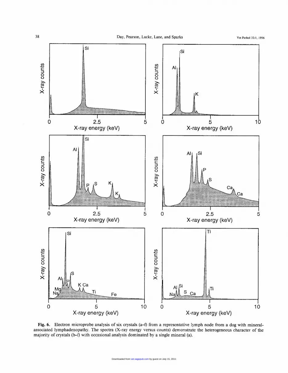
present analytical data on geologic materials as oxides of the major cations because oxygen is the major anion in such materials. This convention however, entails an assumption because the cations in the biological en- vironment may be in the form of other kinds of com- pounds. An example of typical spectra obtained from
Table 4. Characterization of mineral material in canine tissues: average chemical composition compared with com- position of average geologic environment.
crystals in one lymph node section are presented in Fig. 6, and the mineral analyses obtained from X-ray spectra are summarized in Tables 4-6. Taken overall, the particles had a wide range of composition (Table 4) compared with the average composition of natural minerals from the geologic environment. Individual analyses varied from those dominated by a single el- ement (e.g., Si or Cu) to analyses containing several elements. Each analysis was examined, and possible compounds were identified. The more elements in the analysis, the greater the uncertainty in the identifica- tion. Compounds with few elements were easily rec- ognized in analyses and were assumed to be most likely in those compounds containing several elements. For
Typical Average Composition Of
crust
Average Composition: All Analyses Upper Continental Compound
SiO, 45.22 67.0 TiO, 0.34 0.5 A1203 7.4 1 14.0 Fe203 1.70 3.0 MgO 1.33 1.50 CaO 9.06 4.0 Na20 4.4 1 4.0 K2O 3.46 4.0 CUO 13.70 NiO 0.25 ( 3 , 2 0 3 0.08 so4 17.31 p20, 5.68
example, samples containing calcium and sulfur were common and interpreted as being calcium sulfate. Thus, other analyses containing Ca and S were also inter- preted as the same compound.
Electron microprobe data alone cannot unambigu- ously identify specific minerals or compounds. Even for pure silica, there are several natural polymorphs, although quartz is by far the most abundant. In ad- dition, the probe cannot recognize very light elements such as carbon. Thus for example, some of the Ca in analyses could be calcium carbonate or calcium oxa- late, but our analysis could not recognize or distinguish these compounds.
by guest on July 15, 2011vet.sagepub.comDownloaded from

Vet Pathol 33:1, 1996 Mineral Deposition in Canine Tissues 3 1
Fig. 5. Lung; dog. A 10-year-old neutered male Border Collie with prostatitis has peribronchiolar aggregates of mac- rophages with cytoplasmic carbon particles and translucent needlelike crystals. HE, polarized light. Bar = 50 pm.
The particles could be broadly divided into silicates (including silica), making up 57.7% of analyses, and nonsilicates, making up 42.3% of analyses. Interpreted silicate compounds are listed in Table 5 , with common minerals equivalent to these compositions. Pure Si02 was the most abundant phase. Aluminium silicates also occurred in several samples and might be derived from natural clays such as kaolinite and gibbsite. Some samples comprised only two elements and were con- sistent with silicates that are rare or unknown in nature, such as sodium silicate. Alternatively, the areas ana- lyzed might be mixtures of silica with other Na or Ca compounds. Several samples contained S and Si alone that could be correlated with any known natural com- pound, although sulfate-silicic acid sodium complexes are known.*l
The nonsilicate compounds were diverse (Table 6 ) .
Sulfur-rich compounds, interpreted as sulfates, oc- curred in most specimens, and calcium sulfate was the most common compound, which occurs in nature as the mineral anhydrite and hydrated mineral gypsum. Calcium phosphate may be linked to bone, but it also occurs in nature in small amounts as the mineral ap- atite. Calcium could also plausibly be present as oxa- late or carbonate. Some specimens contained metals (copper, iron, titanium, chromium, aluminium, nick- el), which may have been present as metal oxides, el- emental metal, or more complex organometallic com- pounds.
There was no clear relationship between crystal/par- ticle composition and the tissue of origin. Most tissues examined contained a wide mixture of compounds, with silicates, silica, sulfates, and phosphates com- monly occurring. However, some compounds were
Table 5. Characterization of mineral material in canine tissues: silicate compounds,
Number of
Identified Possible Compounds Possible Minerals Analyses Where Main
Elements
Si Silica Ca, Si Calcium silicate, calcium oxalate + silica Ca, Mg, Si Ca, Mg silicates Mg, Fe, Si Mg, Fe silicates K, Al, Si Potassium, aluminium silicate K, Al, Si, S KAlSi,O,, K2S04, KAlSi,O,(SO,)(OH), Al, Si Aluminosilicate, silica, alumina S, Si Silica, sulfur Na, Si Sodium silicate
Quartz, cristobalite, amorphous silica Wollastonite Pyroxenes, amphiboles Pyroxene, amphiboles, olivine, serpentine, Alkali feldspar, clays (illite) Alkali feldspar, alunite Kyanite, kaolinite, gibbsite Unknown compound of Si-S Unknown natural mineral
45 5 1
talc 5 9 3 7
14 4
by guest on July 15, 2011vet.sagepub.comDownloaded from
Day, Pearson, Lucke, Lane, and Sparks Vet Pathol 33:1, 1996 38
Ti
0 2.5 5 0 5 1 X-ray energy (keV) X-ray energy (keV)
0 2.5 X-ray energy (keV)
v)
c 3 0 0 h
.4-.
F! x
0 2.5 5 X-ray energy (keV)
& Na..~:.~::.:.:.::~::~~:~::::.:.:.: . . . . . . . . . . . . . . . . . . . . . . . . . . . . . , . . . . . . , . . . . ,:.:,:.:,:, . , . . . ,
0 5 10 0 X-ray energy (keV) X-ray energy (keV)
Fig. 6 . Electron microprobe analysis of six crystals (a-f) from a representative lymph node from a dog with mineral- associated lymphadenopathy. The spectra (X-ray energy versus counts) demonstrate the heterogeneous character of the majority of crystals (b-f) with occasional analysis dominated by a single mineral (a).
by guest on July 15, 2011vet.sagepub.comDownloaded from

Vet Pathol 33:1, 1996 Mineral Deposition in Canine Tissues 39
Discussion
The present study documents the presence of min- eral material within canine lymph nodes in which there was concurrent granulomatous inflammation or neo- plastic change. Similar crystalline mineral was ob- served within canine lung but was not generally related to pulmonary pathology. Three major questions are raised by these findings concerning 1) the nature and origin of the minerals, 2) the mobility of the mineral within the tissues of the body, and 3) the potential biologic effects of the mineral particles.
The minerals detected in the present study are con- sidered to be derived from three different sources. Sil- ica and aluminosilicates are most plausibly natural minerals from an exogenous source. Some compounds, including copper, titanium, nickel, and possibly cal- cium silicate and sulfate (cement dusts), most likely derive from human-made products and must also have an exogenous origin. Finally, there are significant quan- tities of crystals, notably the sulfates and phosphates, that are unknown in nature and may thus be formed or modified within the chemical microenvironment of the body (biomineralization).
The evidence for these three origins comes from the electron microprobe data. Many of the observed min- erals occur in the natural geologic environment but were not found in the proportion that might be ex- pected from an average geologic source. For example, pure SiO, was the most frequently observed mineral, and there was a low abundance of feldspars and com- mon clay minerals. By contrast, the clay minerals are by far the most abundant compounds in the geologic en~ironment ,~ making up 60-70% of typical continen- tal crust, whereas quartz typically makes up 20-30%. There were no analyses comparable to plagioclase feld- spar and few corresponded to the alkali feldspars and potassic clays (alkali aluminosilicates). This finding may imply that significant biologic discrimination in the incorporation of natural compounds occurs.
The analysis of nonsilicate compounds revealed the presence of minerals only common in localized geo- logic environments (e.g., calcium sulfate) and easily soluble. It is hard to envision how their incorporation could be so selective when other much commoner min- erals are excluded. However, calcium sulfate is a com- mon constituent of cements. An association between building materials and crystals within affected lymph nodes has been previously ~ugges ted .~~ Moreover, the analyses identified minerals containing metal oxides or elemental metal (e.g., Ti, Ni) that are rare in nature and likely reflect the presence of human-made com- pounds from the environment. There was no apparent geographic localization of the affected dogs. Although most of these dogs were from southwest England, map- ping of cases recorded at a private diagnostic pathology
Table 6 . Characterization of mineral material in canine tissues: nonsilicate compounds.
Number of Possible Possible Minerals Where Analyses Identi- Main
ments Compounds fied
Ca, S Ca, P
Ca
S
Na, S
Fe, S
c u
Fe
Ti
Cr
A1
~ ~~
Calcium sulfate Gypsum, anhydrite Calcium phos- Apatite
CaO, calcium ox- Unknown
Sulfur Native sulfur is
Sodium sulfate, sodium sulfite
Potassium sul- fate; potassium sulfite
Iron sulfate, iron Sulfate (rare), pyri- sulfide tes, marcosite
Metallic copper, Native copper, cu- copper oxide prite (very rare
Metallic iron, Magnetite, hema- iron oxide tite
Metallic titani- Rutile (not com- um, titanium mon in nature) oxide
um, chromium common in na- oxide ture)
lic aluminium common in na-
phate
alate
rare in the UK Very rare in nature
Very rare in nature
in the UK)
Metallic chromi- Chromite (not
Alumina, metal- Corundum (not
ture)
15 2
2
3
6
3
1
7
3
2
1
2
largely confined to just one or two samples. Copper was abundant in only one lymph node, with a single copper analysis in another node. Both of these lymph nodes were from dogs with mineral-associated lymph- adenopathy associated with osteosarcoma (dog Nos. 19, 20; Table 1). Analyses of K, Na, and S only (as- sumed to be sulfates) were only found in the lymph nodes of two dogs with mineral-associated lymphade- nopathy (dog Nos. 9, 22; Table 1). Analyses of Na and Si alone (possible sodium silicate) were found in one lymph node from a dog with lymphoma with crystal- line deposits and in two samples of lung.
Electron microprobe analysis of control tissues re- vealed only occasional small particles (four or five per slide, <10 pm in size). These particles were not ob- viously intracellular or embedded in the tissue and they may have been artifacts. The spectra obtained from these particles were characterized by low-level single peaks representing either Si, Ca, C1, or K. There was no evidence of the more complex mineral species in affected tissue.
by guest on July 15, 2011vet.sagepub.comDownloaded from

40 Day, Pearson, Lucke, Lane, and Sparks Vet Pathol 33:1, 1996
laboratory revealed widespread distribution through- out the United Kmgdom (T. J. Whitbread, personal communication). The identification of such environ- mentally derived mineral materials in the tissues of dogs may have ramifications for human health; all af- fected dogs were companion animals sharing an en- vironment with humans. Dogs may therefore serve as useful monitors for the intake of environmental pol- lutants.
The analysis also identified mineral species that are extremely rare or unknown in geologic environments (e.g., sodium and potassium sulfates and some sili- cates). One possible explanation of this finding is that these minerals have been modified or formed in the body (biomineralization) following chemical alteration of interstitial fluid. In this regard, the occurrence of well-shaped crystals of a mineral containing K, Al, S, and Si (interpreted as alunite, Fig. 6c) is particularly interesting because in nature this mineral is formed where sulfuric acid attacks clays and feldspars. Sul- phur- and phosphate-rich compounds are important constituents of most of the dog samples but have not been reported in epidemiologic and pathologic studies of mineral dusts in humans." Further studies are re- quired to test the hypothesis that novel mineral species are formed by biomineralization.
The second question concerns the route of entry of the exogenous mineral particles to the body and their mobilization and mobility within the tissues. Three possibilities exist for a route of entry: percutaneous absorption, intestinal absorption, and inhalation. In the majority of dogs in this series, there was no firm clinical or pathologic evidence to support any one of these routes above another. Subsequent to the present series, one of us (M. J. Day, personal observation) identified an adult Doberman Pinscher with acral lick granuloma of the hock and mineral-associated gran- ulomatous inflammation of the draining popliteal lymph node. Cutaneous abrasion may have permitted entry of exogenous material, which was then translo- cated to the regional lymph node. Similarly, we studied an adult English Bulldog with recurrent interdigital inflammation associated with the presence of birefrin- gent crystalline material within lesional skin, although local lymph nodes were not examined (M. J. Day, per- sonal observation). Percutaneous absorption of calci- um chloride28 and calcium carbonate23 has been re- corded in the dog, but these chemicals were associated with mineralization of dermal collagen (calcinosis cu- tis) rather than crystal formation.
A second hypothesis is that the mineral enters the body by inhalation, and some findings of the present study lend support to this theory. Pulmonary deposi- tion of mineral was observed incidentally, associated with focal an thraco~is ,~~ or sometimes concurrent with
mineral-associated lymphadenopathy. For example in dog No. 27 (Table l), birefringent crystals were found in lung, hilar, presternal, and prescapular lymph nodes. In such dogs, the electron microprobe analysis revealed that the mineral in lung and lymph node had similar composition. Furthermore, if inhalation is a significant route of entry, the greater abundance of silica com- pounds compared with aluminosilicates may reflect the fact that silica is preferentially enriched in wind-blown dust.s.l7
Whatever the route of entry, the fact that different lymph nodes from the same dogs contained mineral of similar profile suggests mobility of the mineral from a primary focus via the vascular or lymphatic systems.20 The mineral particles are retained within lymph nodes, a phenomenon termed biopersistance, which reflects the rate of physical clearance together with the rate of chemical dissolution (biodurability). l 6
The final question posed by this study concerns the biologic effects of the mineral particles within lym- phoid tissue. The dogs in this series had mineral de- position in lymph nodes associated with a range of clinical presentations, which included nonspecific sys- temic illness, autoimmunity, and lymphoma or other neoplasia. Although we have no direct evidence to link the observed lymph node pathology with clinical ill- ness, we hypothesize that such an association exists and occurs as a spectrum of clinical severity.
In some dogs, a particular quantity of mineral may induce no detectable disease or may be associated with a clinically benign lymphadenopathy. In other dogs, the level of granulomatous inflammation and associ- ated cytokine production (e.g., interleukin- 1, tumour necrosis factor) may result in nonspecific systemic ill- ness characterized by pyrexia and a range of other changes. In other animals, the inflammatory change may be associated with perturbation of immunoreg- ulation, which is expressed as clinical autoimmunity or as chromosomal damage resulting in lymphoma. Evidence for this hypothesis is not strong. However, there are well-documented associations between in vivo exposure to silica and subsequent autoimmunity and lymphoma in h u m a n ~ ~ ~ ~ ~ ~ and lymphoma in Re- cently, the dissemination of mineral material from loosened human hip prostheses has been reported,I9 and a clinical association with lymphoma also pro- posed (C. P. Case, personal communication). In the case of pulmonary exposure to silica, a range of mech- anisms have been proposed to explain the association of chronic inflammation with DNA damage and sub- sequent n e ~ p l a s i a . ~ ~ - * ~ A recent study has suggested that silica may act as a superantigen and directly activate autoreactive T lymphocyte^.^^ Further studies of ca- nine mineral-associated lymphadenopathy must ad-
by guest on July 15, 2011vet.sagepub.comDownloaded from

Vet Pathol 33:1, 1996 Mineral Deposition in Canine Tissues 41
dress alteration of immunologic parameters in affected dogs.
In the United Kingdom, mineral-associated lymph- adenopathy is emerging as a significant cause of lymph node disease in the dog. Six percent of canine lymph node biopsy submissions in a recent survey had gran- ulomatous change associated with the presence of bi- refringent crystalline material,4 and this condition must now be considered as an important clinical differential for canine lymphadenopathy. Although clinical follow- up of our cases is not presented here, palliative anti- inflammatory therapy (corticosteroids) is clinically ef- fective (A. Hotston Moore, personal communication), and over time, there is sometimes a degree of crystal degradation and spontaneous recovery from the lymphadenopathy (V. M. Lucke, personal observa- tion).
Acknowledgements
We acknowledge the contribution of clinicians and pa- thologists from the Department of Veterinary Clinical Sci- ence, University of Bnstol, and veterinary surgeons in private practice, who provided the case material described in this study. We thank M. Palmer and B. Wood (Department of Geology, University of Bristol) for helpful discussions. J. Dalton carried out many of the microprobe analyses. The mineral analysis was funded by a grant from the Department of Pathology and Microbiology, University of Bristol.
References
Berry CR, O’Brien TR, Madigan JE, Hagar DA: Tho- racic radiographic features of silicosis in 19 horses. J Vet Intern Med 5248-256, 199 1 Canfield PJ, Rothwell TLW, Papadimitriou JM, Moore JD: Siliceous pneumoconiosis in two dogs. J Comp Pathol 100: 199-202, 1989 Day MJ, Hanlon L, Powell LM: Immune-mediated skin disease in the dog and cat. J Comp Pathol 109:395407, 1993 Day MJ, Whitbread TJ: Pathological diagnoses in dogs with lymph node enlargement. Vet Rec 136:72-73, 1995 Deer WA, Howie RA, Zussmann J: An Introduction to the Rock-Forming Minerals, p. 691. John Wiley and Sons, New York, NY, 1992 Driscoll KE: In vitro evaluation of mineral cytotoxicity and inflammatory activity. Rev Miner 28:489-5 1 1, 1993 Dubois CM, Bissonnette E, Rola-Pleszczynski M: As- bestos fibres and silica particles stimulate rat alveolar macrophages to release TNF. Am Rev Respir Dis 139:
Evans MG, Slocombe RF, Schwartz LD: Pulmonary silicosis in captive ring-necked pheasants: definitive di- agnosis by electron probe X-ray microanalysis. Vet Path-
Finley J, Knabb J: Cutaneous silica granuloma. Plast Reconstr Surg 69:340-343, 1982
1257-1264, 1989
01 25239-241, 1988
10 Fyfe NC, Price EW: The effects of silica on lymph nodes and vessels. Trans R SOC Trop Med Hyg 79:645-651, 1985
1 1 Guthrie GD, Mossman BT, ed.: Health effects of mineral dusts. Rev Miner 28, 1993
12 Heddenborg M, Klockars M: Quartz-dust-induced pro- duction of reactive oxygen metabolites by human gran- ulocytes. Lung 167:23-32, 1989
13 Hendrick MJ, Goldschmidt MH, Shofer FS, Wang YY, Somlyo AP: Postvaccinial sarcomas in the cat: epide- miology and electron probe microanalytical identifica- tion of aluminium. Cancer Res 52:5391-5394, 1992
14 Higgins DA, Kung ITM, Or RSB: Environmental silica in badger lungs: a possible association with susceptibility to Mycobacterium bovis infection. Infect Immun 48:252- 256, 1985
15 Houpt KR, Sontheimer RD: Autoimmune connective tissue disease and connective tissue disease-like illnesses after silicone gel augmentation mammoplasty. J Am Acad Dermatol31:626-642, 1994
16 Jurinski JB: Biodurability of mineral dusts. Proc Am Geol SOC 94, 1993
17 Klein C: Rocks, minerals and a dusty world. Rev Miner
18 Ladds PW: A Colour Atlas of Lymph Node Pathology in Cattle, p. 27. James Cook University, Queensland, Australia, 1986
19 Langkamer VG, Case CP, Heap P, Taylor A, Collins C, Pearse M, Solomon L: Systemic distribution of wear debris after hip replacement. J Bone Joint Surg 74:83 1- 839, 1992
20 Lehnert BE: Defense mechanisms against inhaled par- ticles and associated particle-cell interactions. Rev Miner 28:427-469, 1993
21 Marshall WI, Chen CA: Amorphous silica solubilisa- tion. 11. Postulated sulphate-silicic acid solution com- plex. Geochem Cosmochem Acta 46:367-370, 1982
22 Mossman BT: Cellular and molecular mechanisms of disease. Rev Miner 28:5 13-52 1, 1993
23 Paradis M, Scott DW: Calcinosis cutis secondary to per- cutaneous penetration of calcium carbonate in a Dal- matian. Can Vet J 30:57-59, 1989
24 Pearson GR, Longstaffe JA, Lucke VM, Yeo S, Hender- son WJ: Lymphadenopathy in dogs associated with alu- minosilicate. Vet Rec 118:450-453, 1986
25 Roperto F, Damlano S, DeVico G, Galati D: Silicate pneumoconiosis in pigs: optical and scanning electron microscopical investigations with X-ray microanalysis. J Comp Pathol 110:227-236, 1994
26 Ross M, Nolan RP, Langer AM, Cooper WC: Health effects of mineral dusts other than asbestos. Rev Miner 28:361-407, 1993
27 Saffiotti U, Daniel LN, Mao Y, Williams AO, Kaighn ME, Ahmed N, Knapton AD: Biological studies on the carcinogenic mechanisms of quartz. Rev Miner 28: 523- 544, 1993
28 Schick MP, Schick RO, Richardson JA: Calcinosis cutis secondary to percutaneous penetration of calcium chlo- ride in dogs. Am Vet Med Assoc 191:207-211, 1987
29 Shalaeva MP, Mordberg EL, Malashenko AV, Babak-
28~7-59, 1993
by guest on July 15, 2011vet.sagepub.comDownloaded from
42 Day, Pearson, Lucke, Lane, and Sparks Vet Pathol 33:1, 1996
hanian RV, Feklistova VP: SiO, in the lungs of patients with silicosis. Gig Tr Prof Zabol 4: 11-1 3, 1989
30 Silicosis and Silicate Disease Committee: Diseases as- sociated with exposure to silica and nonfibrous silicate minerals. Arch Pathol Lab Med 112:673-720, 1988
3 1 Smith BL, Poole WSH, Martinovich D: Pneumoconiosis in the captive New Zealand kiwi. Vet Pathol10:94-101, 1973
32 Struhar DJ, Harbeck RJ, Gegen N, Kawada H, Mason RJ: Increased expression of class I1 antigens of the major histocompatibility complex in an animal model of sili- cosis. Clin Exp Immunol77:281-284, 1989
33 Thomson RG: General Veterinary Pathology, pp. 84- 85. WB Saunders, Philadelphia, PA, 1978
34 Ueki A, Yamaguchi M, Ueki H, Watanabe Y, Ohsawa G, Kinugawa K, Kawakami Y, Hyodoh F: Polyclonal human T-cell activation by silicate in vitro. Immunology
35 Van Kruiningen HJ, Lees GE, Hayden DW, Meuten DJ, Rogers WA: Lipogranulomatous lymphangitis in canine intestinal lymphangiectasia. Vet Pathol21:377-383, 1984
36 Wagner MM: Pathogenesis of malignant histiocytic lym- phoma induced by silica in a colony of specific pathogen- free Wistar rats. J Natl Cancer Ins 57:509-518, 1976
82~332-335, 1994
Request reprints from Dr. M. J. Day, Department of Pathology and Microbiology, University of Bristol, Langford BS18 7DU (UK).
by guest on July 15, 2011vet.sagepub.comDownloaded from


