Embed Size (px)
Citation preview
Leukotriene C4 prevents the complete maturation of murinedendritic cells and modifies interleukin-12/interleukin-23 balance
Introduction
Dendritic cells (DCs) are highly specialized antigen-pre-
senting cells with a unique capability to activate naive T
lymphocytes and initiate the adaptive immune response,
as well as induce peripheral tolerance.1,2 In peripheral tis-
sues, immature DCs sense the microenvironment and
after their encounter with inflammatory stimuli or patho-
gens, DCs are activated; a process that is accompanied by
several physiological changes and that ends with the mat-
uration of DCs.3–5
Once initiated the process of DCs maturation, the
expression of CD80, CD86 and MHC class II molecules
increases.1–4 The DCs migrate to the draining lymph
nodes, as a result of the up-regulation of CCR7, which
renders them responsive to CCL19 and CCL21 chemokin-
es that direct their migration to the T-cell areas of lymph
nodes.6 Finally, the mature DCs present the antigen to
naive CD4+ and CD8+ T lymphocytes. The maturational
status can be modulated by different stimuli.5 The impact
of microbial products through Toll-like receptor leads to
DCs that produce interleukin-12 (IL-12)/IL-23 and prime
T helper type 1 (Th1)/Th17 responses.7,8 In contrast, in
the absence of inflammatory signals, ‘semi-mature’ DCs
produce IL-10, which primes a regulatory T-cell response.9
However, mediators other than cytokines and pathogens
have a great impact on the physiology of DCs. Prostaglan-
din E2 acting on mature DCs induces the differentiation
of CD4+ T cells in a Th2 profile.10,11 Also, histamine acti-
vates murine DCs through the increase of endocytosis and
cross-presentation of extracellular antigens.12
Leukotriene C4 (LTC4), a member of the cysteinyl leu-
kotriene family (CysLT), is a potent pro-inflammatory
lipid mediator, produced by inflammatory cells such as
mast cells, eosinophils, basophils and macrophages.13,14 It
is a potent spasmogen and vasoconstrictor, promotes
mucus secretion, and together with histamine is a known
immunomodulatory agent of allergic and inflammatory
Carolina Alvarez,1 Marıa M.
Amaral,1 Cecilia Langellotti2 and
Monica Vermeulen1
1Immunology Laboratory, Institute of Haema-
tologic Research, National Academy of Medi-
cine, Buenos Aires, and 2Institute of Virology,
CICVyA, INTA, Castelar, Argentina
doi:10.1111/j.1365-2567.2011.03478.x
Received 28 March 2011; revised 28 June
2011; accepted 29 June 2011.
Correspondence: Dr M. Vermeulen,
Departamento de Inmunologıa, Instituto de
Investigaciones Hematologicas, Academia
Nacional de Medicina, Pacheco de Melo
3081, 1425 Buenos Aires, Argentina.
Email: [email protected]
Senior author: Monica Vermeulen
Summary
Leukotriene C4 is an important mediator in the development of inflam-
matory reactions and ischaemia. Previous studies have shown that leuko-
triene C4 is able to modulate the function of dendritic cells (DCs) and
induce their chemotaxis from skin to lymph node. In this study, we
decided to evaluate the modulation exerted by leukotriene C4 on DCs,
depending on their status of activation. We showed for the first time that
leukotriene C4 stimulates endocytosis both in immature and lipopolysac-
charide (LPS) -activated DCs. Moreover, it suppressed the interleukin-
12p70 (IL-12p70) release, but induces the secretion of IL-23 by DCs
activated with LPS and promotes the expansion of T helper type 17
(Th17) lymphocytes. Furthermore, blocking the release of IL-23 reduced
the percentages of CD4+ T cells producing IL-17 in a mixed lymphocyte
reaction. Ours results suggest that leukotriene C4 interferes with the com-
plete maturation of inflammatory DCs in terms of phenotype and antigen
uptake, while favouring the release of IL-23, the main cytokine involved
in the maintenance of the Th17 profile.
Keywords: endocytosis; interleukin-23; inflammatory dendritic cells; leu-
kotriene C4; T helper type 17 lymphocytes
Abbreviations: CysLTR, cysteinyl receptors; DCs, dendritic cells; DX, dextran; HRP, horseradish peroxidase; LTC4, leukotriene C4;Zy, zymosan.
� 2011 The Authors. Immunology � 2011 Blackwell Publishing Ltd, Immunology, 134, 185–197 185
I M M U N O L O G Y O R I G I N A L A R T I C L E
reactions.15–17 The pharmacological effects of CysLT are
conducted through two types of membrane receptors –
CysLTR1 and CysLTR2 – which are coupled to protein-
G.18 Remarkably, these receptors were primarily described
at the level of lung mucosa and intestinal mucosa at the
ileum and colon.19 In many diseases affecting lung and
intestinal mucosa, such as asthma and interstitial cystitis,
the use of montelukast, a selective antagonist of CysLTR1,
minimizes the effects of these pathologies, probably
through the inhibition of cytosolic Ca2+.20–22
It is known that LTC4 induces the chemotaxis of DCs
from the skin.23 Zymosan, a Toll-like receptor 2 agonist,
but not lipopolysaccharide (LPS), a classic Toll-like recep-
tor 4 agonist, stimulates the production of CysLT by
DCs.24,25 Despite these observations, their impact on
cytokine production by DCs is unclear. In spite of the
close relationship between mast cells and DCs in mucosal
epithelium and skin, little progress has been made regard-
ing the impact of CysLT on the genesis of DCs.
In the present study, we analysed the effects of LTC4
on the phenotype and function of murine inflammatory
DCs.26 In particular, we studied the differential expression
of CysLT1 and CysLT2 receptors in immature and LPS-
activated DCs. Our results show that LTC4 interferes with
the maturation of DCs triggered by LPS, in terms of phe-
notype and antigen uptake. In this context, LTC4 induces
the release of IL-23 by inflammatory DCs, favouring the
expansion of Th17 cells.
Materials and methods
Mice
All experiments were carried out using 2-month-old vir-
gin female C57BL/6 mice raised at the National Academy
of Medicine, Buenos Aires, Argentina. They were housed
six per cage and kept at 20 ± 2� under an automatic
12 hr light–dark schedule. Animal care was in accordance
with institutional guidelines.
Generation of DCs from bone marrow cultures
The procedure used in this study was as described by
Inaba et al.27 with some minor modifications. Briefly,
bone marrow was flushed from the long bones of the
limbs using 2 ml RPMI-1640 (Gibco, Invitrogen, Carlsbad,
CA) with a syringe and 25-gauge needle. Red cells were
lysed with ammonium chloride. After washing, cells were
suspended at a concentration of 1 · 106 cells/ml in 70%
RPMI-1640 medium supplemented with 10% fetal calf
serum (FCS; Gibco), and 5�5 · 10)5 mercaptoethanol
(Sigma, St Louis, MO) (mouse complete medium) and
30% J588-GM cell line supernatant. The cultures were fed
every 2 days by gently swirling the plates, aspirating 50%
of the medium, and adding back fresh medium with J588-
GM cell line supernatant. At day 9 of the culture, > 85%
of the harvested cells expressed MHC class II, CD40 and
CD11c, but not Gr-1 (not shown).
Culture conditions
The standard medium used in this study was bicarbon-
ate-buffered RPMI-1640 (Invitrogen, Carlsbad, CA) sup-
plemented with 10% FCS, 50 U/ml penicillin, 50 lg/ml
streptomycin, 0�1 mM non-essential amino acids, and
5�5 · 10)5 mercaptoethanol (all from Invitrogen) (com-
plete medium).
Reagents
Horseradish peroxidase (HRP), dextran (DX,
40 000 molecular weight), Zymosan (Zy, from Saccharo-
myces cerevisiae), LPS from Escherichia coli (0111:B4),
were from Sigma Chemical Co. (St Louis, MO). SB-
202190 [p38 mitogen-activated protein kinase (MAPK)],
PD-98059 [extracellular signal-regulated kinase (ERK)/
MAP kinase Kinase (MEK) MAPK], were from Promega
Corporation (Madison, WI). The DX and Zy were conju-
gated with FITC, as described previously.28
Flow cytometry
Cells staining were performed using the following mono-
clonal antibodies (mAbs): FIYC-conjugated anti-CD11c,
anti-CD40-FITC, anti-I-Ad conjugated with phycoerythrin
(PE), GR1-PE and CD86-PE (Pharmingen, San Diego,
CA). Cell surface antigen expression was evaluated by sin-
gle staining, and analysis was performed using a FACS
flow cytometer and CELLQUEST software (Becton Dickinson,
San Jose, CA).
Endocytosis by FACS analysis
After different treatments, DCs were suspended in med-
ium RPMI-1640 at 37�. FIYC-DX was added at the final
concentration of 100 lg/ml. The cells were washed four
times with cold PBS containing 1% FCS and were analy-
sed on a FACS flow cytometer (Becton Dickinson). The
background (cells pulsed at 0�) was always subtracted.
Endocytosis of HRP
Endocytosis of HRP was performed as previously
described.29 Briefly, DCs were suspended in complete
medium; HRP was added at the final concentration of
150 lg/ml HRP, and cells were cultured for 30 min at
37�. Then, DCs were collected, washed four times in PBS
containing 1% FCS and four times in PBS alone, then
were lysed with 0�05% Triton X-100 in 10 mM Tris–HCl
buffer, for 30 min, and the enzyme activity of the lysate
186 � 2011 The Authors. Immunology � 2011 Blackwell Publishing Ltd, Immunology, 134, 185–197
C. Alvarez et al.
was measured using O-phenylenediamine and H2O2 as
substrates with reference to a standard curve, at 492 nm.
The amount of HRP taken up by DCs was determined as
the difference between HRP activities in disrupted and
non-disrupted cells. The HRP activity in non-disrupted
DCs was always < 15% compared with disrupted cells.
Expression of cysteinyl receptors
Total RNA was extracted from lung (positive control for
CysLT1 receptor), gut tissues (positive control for CysLT2
receptor) and mouse immature and LPS-treated DCs,
using Trizol reagent (Gibco-Life Technologies). The
reverse transcription reaction contained 3 lg total RNA
and was performed using the Moloney-murine leukaemia
virus reverse transcriptase enzyme (Promega). The prim-
ers were provided by Invitrogen: forward primers for the
CysLTR1 and CysLTR2: CAA CGA ACT ATC CAC CTT
CAC C and CCA AGG TCA CAA GAG GGT GT, respec-
tively. Reverse primers for the CysLTR1 and CysLTR2:
AGC CTT CTC CTA AAG TTT CC AC and GAG TTG
ACA GAG GCG AGG AC, respectively. A GeneAmp PCR
system (Perkin-Elmer/Applied Biosystems, Foster City,
CA) was used. The PCR products were separated on a
1�5% agarose gel, stained with ethidium bromide, and
visualized by a UV transilluminator.
Western blot analysis
Murine DCs were suspended in complete medium
(2 · 106/500 ll) were prewarmed for 30 min at 37�. The
DCs were treated without or with 1 lg/ml LPS for 20 min
at 37�. Then cells were washed and treated with or without
0�01 lM LTC4 for 5 min at 37�. The reaction was stopped
by adding cold PBS, the mixture was centrifuged and pel-
lets were resuspended at 3 · 106 cells/ml in Western sam-
ple buffer (100 mM Tris–HCl pH 6�8; 4% SDS, 0�2%
Bromophenol-Blue, 20% glycerol, 200 mM dithiothreitol)
and frozen at – 80�. Before the analysis, lysates were
thawed, heated for 3 min to 96� and finally homogenized
with a sonicator and 5 · 104 cells (10 ll extract) per lane
were separated onto 10% SDS–PAGE followed by elec-
troblotting. The membranes were blocked in PBS + 5%
milk powder for 2 hr, and then incubated with the follow-
ing primary antibodies in blocking buffer + 0�1% Tween-
20 overnight at 4�: anti-phospho-ERK1/2 (Thr202/Tyr204,
1 : 1000; Cell Signaling Technology, Boston, MA), anti-
phospho-p38K (1 : 1000; Cell Signaling). After washing,
secondary antibodies were applied in blocking buffer for
2 hr at room temperature: anti-rabbit or anti-mouse-HRP
mAb (1 : 3000; Cell Signaling). Membranes were washed
and specific bands were developed by enhanced chemilu-
minescence (Amersham Biosciences, Uppsala, Sweden).
Membranes were stripped and reproved with a rabbit
mAb against murine b-actin (Cell Signaling Technology).
Mixed leucocyte reaction
Splenocytes were obtained from the spleen of naive
BALB/c mice and cultured at 1�5 · 105 cells/well in 96-
well microplates with 5 · 104 immature DCs or LPS-
stimulated DCs from C57BL/6 mice, either treated
(0�01 lM) or untreated with LTC4. The cells were culti-
vated for 4 days in RPMI-1640 containing 10% FCS,
10 mM HEPES buffer and 5�5 · 10)5M mercaptoethanol
(Sigma). At day 4 of culture, cells were pulsed for 18 hr
with [3H]thymidine (1 lCi/well; DuPont, AR). Then, cells
were harvested using a cell harvester (Perkin-Elmer Inc.)
and the amount of [3H]thymidine incorporation was
determined in a b-scintillation counter. Intracellular
staining for cytokine production was performed after
stimulation of co-culture for 24 hr with PMA (10 ng/ml),
ionomycin (1 lg/ml) with or without IL-23p19 (10 lg/
ml) (ebiosciences, San Diego, CA) in the presence of bre-
feldin A for the last 6 hr. Finally, CD4+ IL-17A+ inter-
feron-c (IFN-c)+ cells were analysed by flow cytometry.
Intracytoplasmic cytokine staining
Two or three-colour analysis was performed using flow
cytometry, DCs were cultured without or with LPS
(1 lg/ml) for 20 min at 37�. After washing, DCs were
treated with or without 0�01 lM LTC4 for 30 min at 37�in complete medium in the presence of brefeldin A
(5 lg/ml). After 18 hr, cells were fixed in 4% parafor-
maldehyde and permeabilized with saponin (0�1% in
PBS). The permeabilized cells were incubated with a PE-
conjugated anti-IL-12p40 antibody (BD Pharmingen,
Trento, NJ) in PBS 0�5% BSA or similarly labelled iso-
type-matched control antibodies for 30 min. The stained
cells were washed with saponin buffer twice and resus-
pended in isoflow. In some cases, intracytoplasmic cyto-
kines were evaluated in co-cultures of mixed lymphocyte
reaction (MLR) and permeabilized cells were incubated
with PE-conjugated anti-IL17A and FITC-conjugated
anti-IFN-c antibodies (BD Pharmingen). In all cases, the
surface staining with FITC-conjugated anti-CD11c (DCs)
or Peridinin chlorophyll protein-conjugated anti-CD4
antibodies (BD Pharmingen) was performed before to
permeabilization. The staining was analysed by flow
cytometry on FACS using CELLQUEST software (BD Bio-
sciences, San Jose, CA).
Cytokines determination
The cytokine levels in supernatants of DCs were measured
by ELISA. Assays for IL-12p70, p40, IL-23, IL-6, tumour
necrosis factor-a (TNF-a), IFN-c (eBiosciences) and IL-17
(Quantikine; R&D Systems, Bs. AS, AR) were performed
according to the manufacturer’s protocols. The limits of
detection were: 15 pg/ml (IL-12p70; p35/p40), 30 pg/ml
� 2011 The Authors. Immunology � 2011 Blackwell Publishing Ltd, Immunology, 134, 185–197 187
Leukotriene C4 modifies IL-12/IL-23 balance on activated dendritic cells
(IL-23; p19/p40), 10 pg/ml (IL-12p40), 8 pg/ml (TNF-a),
4 pg/ml (IL-6), 15 pg/ml (IFN-c) and 5 pg/ml (IL-17).
Statistics
The significance between means was assessed by Student’s
paired t-test. P � 0�05 was determined to indicate statisti-
cal significance.
Results
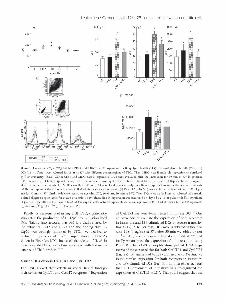
Exogenous LTC4 modulate phenotypical expressionfrom LPS-activated DCs
We decided to evaluate whether LTC4 is able to modulate
the central molecules expressed by DCs that are involved
in the activation of T lymphocytes.3,4 In the first place,
we studied the concentrations of LTC4 able to modulate
the expression of the MHC class II molecules. To analyse
this point, DCs were cultured in the presence of different
concentrations of LTC4 (10)5–10)9M) at 37�. After 18 hr,
its expression was analysed by flow cytometry. As shown
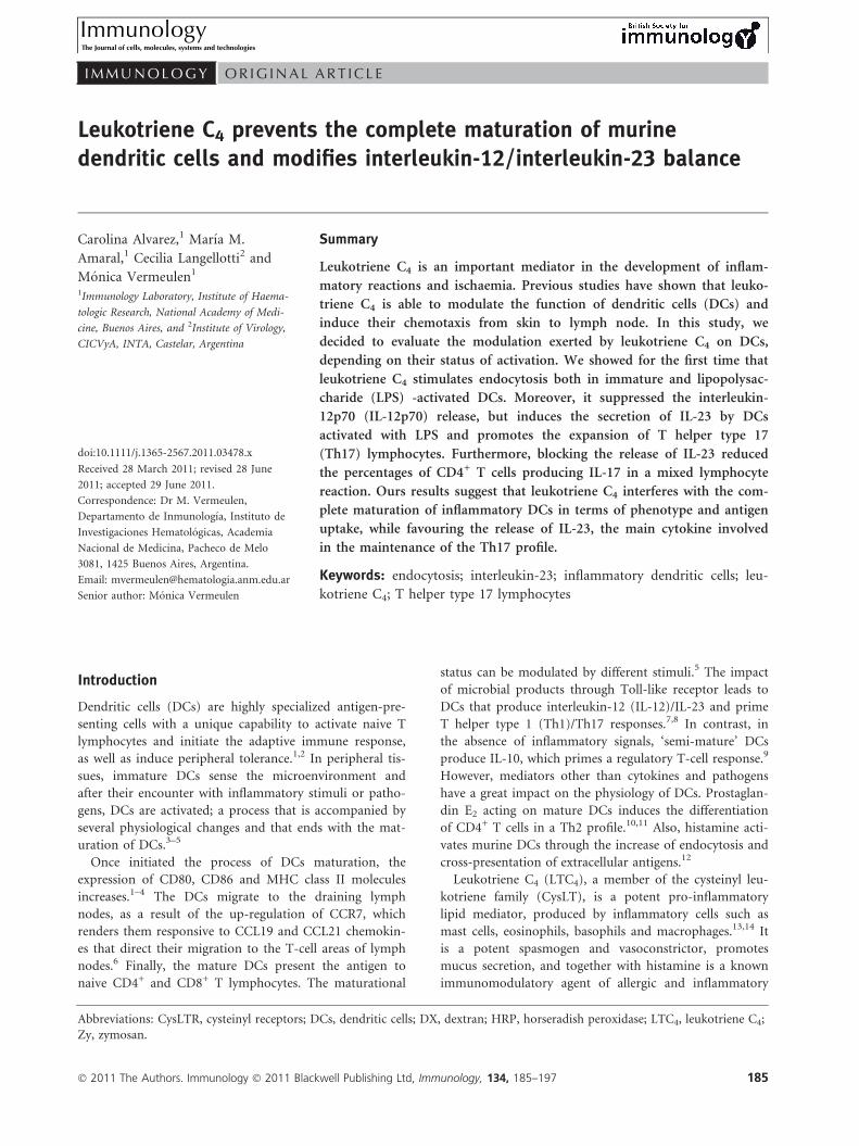
in Fig. 1(a), LTC4 increased in a dose-dependent manner,
the expression of MHC class II on immature DCs was
more significant at 10)8M, so the trials were conducted
using this concentration. Then, considering that LTC4 is
released during inflammatory responses,17,30 we studied
the effect of LTC4 (10)8M) on the phenotype of imma-
ture DCs and LPS-stimulated DCs. Interestingly, after
18 hr of culture, LTC4 strongly inhibited the expression
of CD86 and CD40 molecules (Fig. 1b,c,f) when DCs
were activated with 1 lg/ml LPS, whereas the lipid medi-
ator had no effect on immature DCs. However, in the
case of the class II molecules, LTC4 had antagonistic
effects depending on the activation status of DCs, increas-
ing its expression in immature DCs and inhibiting in
LPS-treated DCs (Fig. 1d,f). As shown in Fig. 1(g),
although MHC class II decreased its expression in LPS-
activated DCs, LTC4 had the ability to prime T lympho-
cytes, because it induced a low but significant increase in
the allostimulatory response mediated by activated DCs.
This effect was also observed in immature DCs, which
correlates with the increased expression of class II mole-
cules by LTC4.
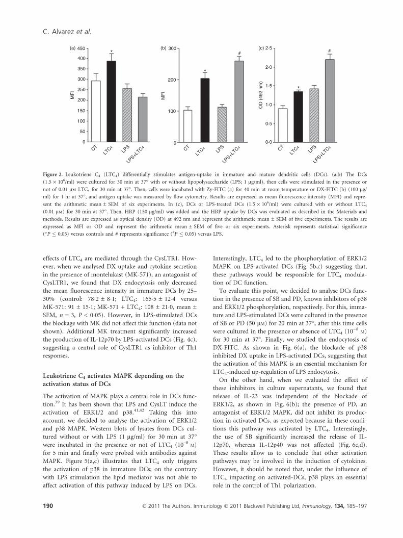
Leukotriene C4 counteracts the effect of LPS on DCsendocytosis
Immature DCs are specialized to sense the microenvi-
ronment and when stress or infection are detected they
incorporate the antigen through phagocytosis or endocy-
tosis.28,29,31,32 We aimed to determine whether LTC4
was able to affect the antigen uptake of immature and
activated DCs. To this end, cells were treated or not
with LPS (1 lg/ml) for 30 min at 37�, then DCs were
incubated without or with 10)8M LTC4 for 30 min at
37�. Finally, cells were washed and incubated in the
presence of Zy (10 particles/DC) coupled to FITC for
30 min at room temperature or DX-FITC (100 lg/ml)
for 40 min at 37�. The phagocytosis controls were sup-
plied by DCs treated with cytochalasin B, a disruptor of
actin microfilaments, 33 previous to their incubation
with Zy-FITC. For DX endocytosis, the control of reac-
tion was provided by DCs incubated with the antigen at
4�, because this is a temperature-dependent phenome-
non. In addition, we analysed the uptake of HRP. For
this, after treatment with LTC4 (0�01 lM) of both DCs
and LPS-stimulated DCs, these were cultured with
150 lg/ml HRP for 40 min at 37�. Subsequently, cells
were washed several times with cold PBS and permeabi-
lized by addition of 0�5% Triton X-100 in PBS for
30 min at room temperature. The control was provided
by DCs treated with HRP but not permeabilized.
Finally, the enzymatic activity was measured in superna-
tants of reaction by addition of the substrate [alpha-
phenylendiamine (OPD)] and read at 492 nm. In
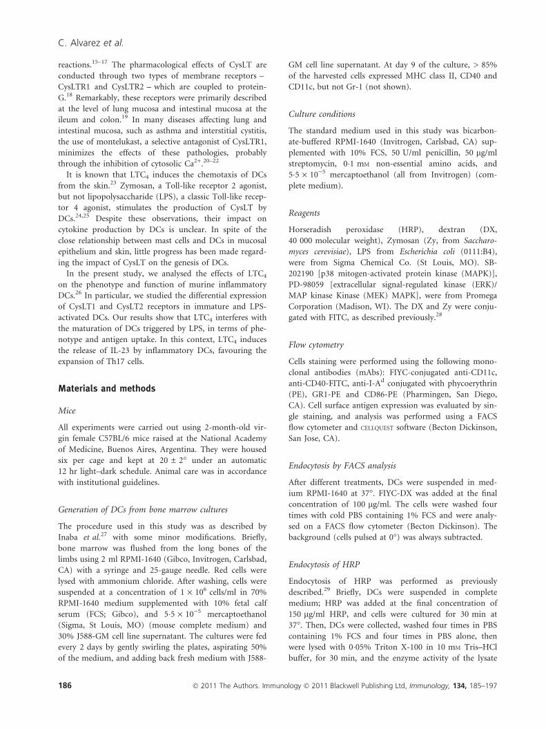
Fig. 2(a), we demonstrated that LTC4 increased the
phagocytosis of Zy-FITC by immature DCs but had no
effect in LPS-activated DCs. In contrast, as shown in
Fig. 2(b,c), uptake of DX and HRP was increased by
LTC4 in both immature and LPS-stimulated DCs. This
result would indicate that LTC4 prevents complete mat-
uration of DCs by classical stimuli such as LPS, because
it restores their endocytic capacity.
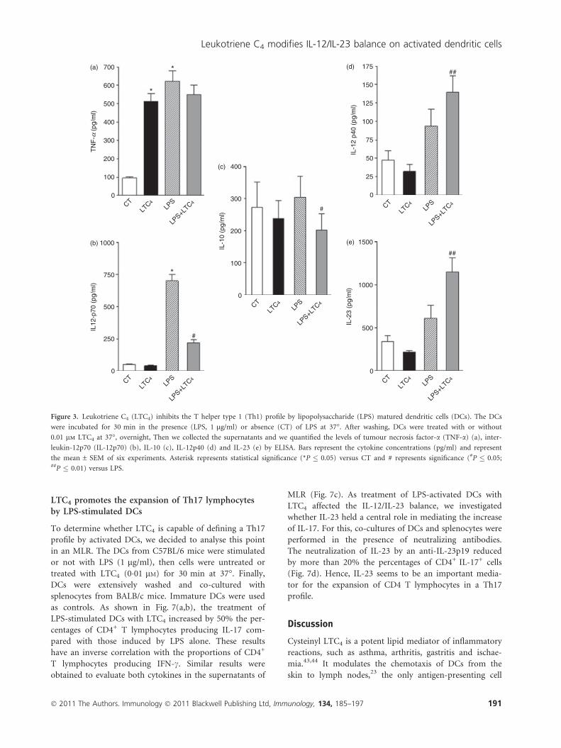
Leukotriene C4 strongly inhibits the induction of aTh1 profile and increases IL-23 production by LPS-activated DCs
Taking into account the fact that LTC4 imposes changes
in DCs that prevent their maturation we decided to eval-
uate their impact on the genesis of the adaptive response,
through the analysis of the cytokines induced. With this
aim, immature and activated DCs were cultured for 18 hr
at 37� in presence or not of LTC4 (10–8M). After incuba-
tion, culture supernatants were collected and we evaluated
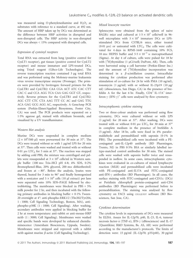
cytokines by ELISA. As shown in Fig. 3(a), LTC4
increased the production of TNF-a in immature DCs but
was unable to reverse its release induced by LPS. Interest-
ingly, LTC4 completely abolished the induction of
IL-12p70 in LPS-stimulated DCs (Fig. 3b), indicating an
antagonistic effect of LPS. Therefore, LTC4 inhibits the
induction of a Th1 profile by T CD4+ naive lymphocytes,
by acting on activated DCs.34,35
Moreover, to further investigate the effect of LTC4 we
decided to evaluate whether LTC4 could favour a tolero-
genic state;36,37 however, when we analysed the release of
IL-10 in culture supernatants, we showed inhibition of
this cytokine in LPS-treated DCs (Fig. 3c), whereas it was
not modulated on immature DCs.
188 � 2011 The Authors. Immunology � 2011 Blackwell Publishing Ltd, Immunology, 134, 185–197
C. Alvarez et al.
Finally, as demonstrated in Fig. 3(d), LTC4 significantly
stimulated the production of IL-12p40 by LPS-stimulated
DCs. Taking into account that p40 is a chain shared by
the cytokines IL-12 and IL-23 and the finding that IL-
12p70 was strongly inhibited by LTC4, we decided to
evaluate the presence of IL-23 in supernatants of DCs. As
shown in Fig. 4(e), LTC4 increased the release of IL-23 in
LPS-stimulated DCs, a cytokine associated with the main-
tenance of Th17 profiles.38,39
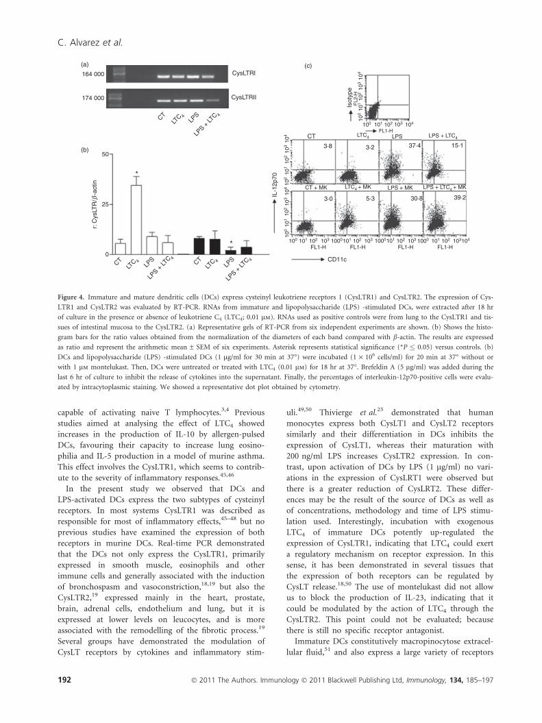
Murine DCs express CysLTR1 and CysLTR2
The CysLTs exert their effects in several tissues through
their action on CysLT1 and CysLT2 receptors.18 Expression
of CysLTR1 has been demonstrated in murine DCs.40 Our
objective was to evaluate the expression of both receptors
in immature and LPS-stimulated DCs by reverse transcrip-
tion (RT-) PCR. For that, DCs were incubated without or
with LPS (1 lg/ml) at 37�, after 30 min we added or not
10–8M LTC4 and cells were cultured overnight at 37� and
finally we analysed the expression of both receptors using
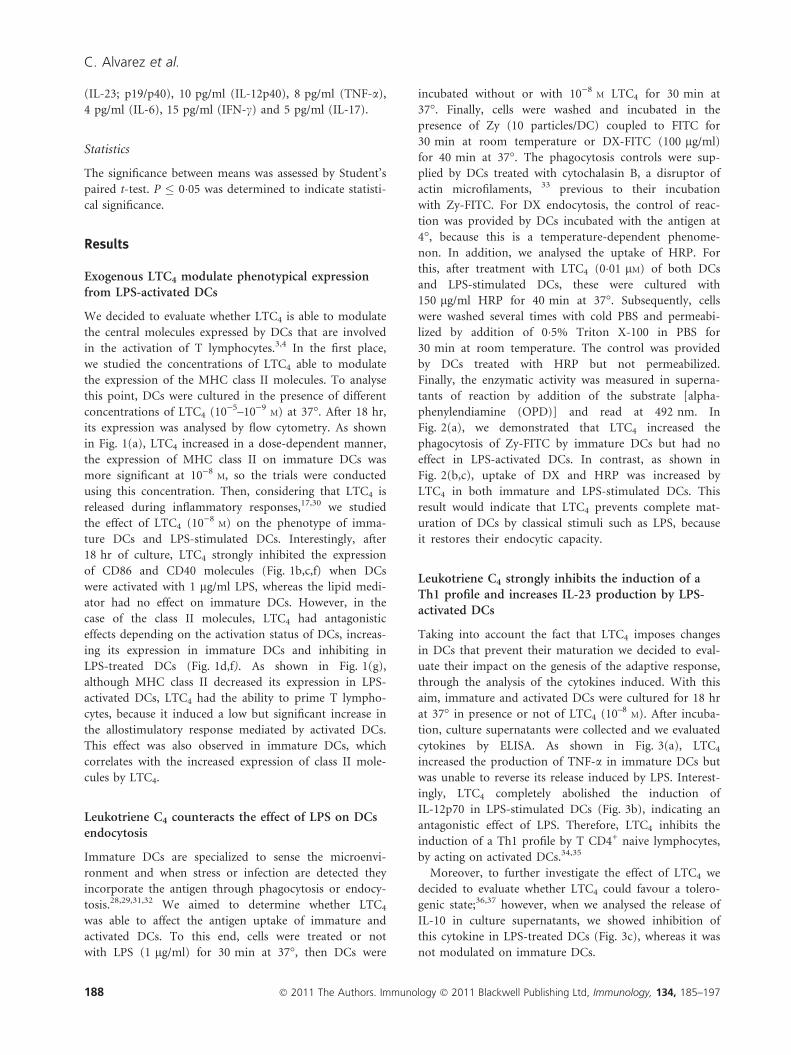
RT-PCR. The RT-PCR amplification yielded DNA frag-
ments of the expected size for both CysLTR1 and CysLTR2
(Fig. 4a). By analysis of bands compared with b-actin, we
found similar expression for both receptors in immature
and LPS-stimulated DCs (Fig. 4b), an interesting fact was
that, LTC4 treatment of immature DCs up-regulated the
expression of CysLTR1 mRNA. This could suggest that the
(a)
LTC4 (µM)
0
100
200
300
MF
I
0 0·001
**
*
0·01 0·1 1 10
(b) (c) (d)
CTLT
C4
LPS
LPS +
LTC 4
MF
I
MF
I
MF
I
**
100
75
70
60
50
40
30
20
10
0
350
300
250
200
150
100
50
0
50
25
0
##
#
#
*
*
CTLT
C4
LPS
LPS +
LTC 4 CT
LTC4
LPS
LPS +
LTC 4
(f) 25 000
20 000
15 000
10 000
5000
0
*
*
*
CTLT
C 4LP
S
LPS +
LTC 4
c.p.
m m
ean
(e)
SS
-CH
0
0
400
400
1000
1000
800
800
600
600
200
200FSC-H
Isot
ype
IAb
100
100
100
100
101
101
101
101
102
102
103
103
104
104
102
10210
310
3104
104
FL1-H CD11c
87·5%
100 101 101 101102 102 102103 103 103 104100 100
Cou
nts
040
3020
10
CD86 CD40 IAb
Isotype CTLTC4
LPSLPS + LTC4
Figure 1. Leukotriene C4 (LTC4) inhibits CD86 and MHC class II expression on lipopolysaccharide (LPS) -matured dendritic cells (DCs). (a)
DCs (1.5 · 106/ml) were cultured for 18 hr at 37� with different concentrations of LTC4. Then, MHC class II molecule expression was analysed
by flow cytometry. (b,c,d) CD40; CD86 and MHC class II expression, DCs were evaluated after the incubation for 30 min at 37� in presence
(LPS) or not (Ct) of LPS (1 lg/ml). Finally, cells were incubated overnight at 37� with or without LTC4 (0.01 lm). (e) Representative histograms
of six or seven experiments, for MHC class II, CD40 and CD86 molecules, respectively. Results are expressed as mean fluorescence intensity
(MFI) and represent the arithmetic mean ± SEM of six or seven experiments. (f) DCs (1.5 · 106/ml) were cultured with or without LPS (1 lg/
ml) for 30 min at 37�, finally cells were treated or not with LTC4 (0.01 lm, 30 min at 37�). Then, DCs were washed and co-cultured with freshly
isolated allogeneic splenocytes for 5 days at a ratio 1 : 10. Thymidine incorporation was measured on day 5 by a 16-hr pulse with [3H]thymidine
(1 lCi/well). Results are the mean ± SEM of five experiments. Asterisk represents statistical significance (*P < 0.05) versus CT; and # represents
significance (#P � 0.05; ##P � 0.01) versus LPS.
� 2011 The Authors. Immunology � 2011 Blackwell Publishing Ltd, Immunology, 134, 185–197 189
Leukotriene C4 modifies IL-12/IL-23 balance on activated dendritic cells
effects of LTC4 are mediated through the CysLTR1. How-
ever, when we analysed DX uptake and cytokine secretion
in the presence of montelukast (MK-571), an antagonist of
CysLTR1, we found that DX endocytosis only decreased
the mean fluorescence intensity in immature DCs by 25–
30% (control: 78�2 ± 8�1; LTC4: 165�5 ± 12�4 versus
MK-571: 91 ± 15�1; MK-571 + LTC4: 108 ± 21�0, mean ±
SEM, n = 3, P < 0�05). However, in LPS-stimulated DCs
the blockage with MK did not affect this function (data not
shown). Additional MK treatment significantly increased
the production of IL-12p70 by LPS-activated DCs (Fig. 4c),
suggesting a central role of CysLTR1 as inhibitor of Th1
responses.
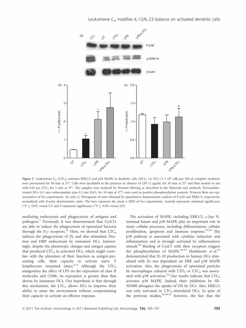
Leukotriene C4 activates MAPK depending on theactivation status of DCs
The activation of MAPK plays a central role in DCs func-
tion.39 It has been shown that LPS and CysLT induce the
activation of ERK1/2 and p38.41,42 Taking this into
account, we decided to analyse the activation of ERK1/2
and p38 MAPK. Western blots of lysates from DCs cul-
tured without or with LPS (1 lg/ml) for 30 min at 37�were incubated in the presence or not of LTC4 (10–8
M)
for 5 min and finally were probed with antibodies against
MAPK. Figure 5(a,c) illustrates that LTC4 only triggers
the activation of p38 in immature DCs; on the contrary
with LPS stimulation the lipid mediator was not able to
affect activation of this pathway induced by LPS on DCs.
Interestingly, LTC4 led to the phosphorylation of ERK1/2
MAPK on LPS-activated DCs (Fig. 5b,c) suggesting that,
these pathways would be responsible for LTC4 modula-
tion of DC function.
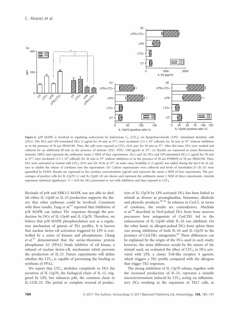
To evaluate this point, we decided to analyse DCs func-
tion in the presence of SB and PD, known inhibitors of p38
and ERK1/2 phosphorylation, respectively. For this, imma-
ture and LPS-stimulated DCs were cultured in the presence
of SB or PD (50 lM) for 20 min at 37�, after this time cells
were cultured in the presence or absence of LTC4 (10)8M)
for 30 min at 37�. Finally, we studied the endocytosis of
DX-FITC. As shown in Fig. 6(a), the blockade of p38
inhibited DX uptake in LPS-activated DCs, suggesting that
the activation of this MAPK is an essential mechanism for
LTC4-induced up-regulation of LPS endocytosis.
On the other hand, when we evaluated the effect of
these inhibitors in culture supernatants, we found that
release of IL-23 was independent of the blockade of
ERK1/2, as shown in Fig. 6(b); the presence of PD, an
antagonist of ERK1/2 MAPK, did not inhibit its produc-
tion in activated DCs, as expected because in these condi-
tions this pathway was activated by LTC4. Interestingly,
the use of SB significantly increased the release of IL-
12p70, whereas IL-12p40 was not affected (Fig. 6c,d).
These results allow us to conclude that other activation
pathways may be involved in the induction of cytokines.
However, it should be noted that, under the influence of
LTC4 impacting on activated-DCs, p38 plays an essential
role in the control of Th1 polarization.
450 300 2·5
2·0
1·5
1·0
0·5
0·0
200
*
*
# #
100
0CT
LTC4
LPS
LPS+L
TC4CTLT
C4LP
S
LPS+L
TC4 CTLT
C4LP
S
LPS+L
TC4
400
350
300
*(a) (b) (c)
250
200
150
100
50
0
MF
I
MF
I
OD
(49
2 nm
)
Figure 2. Leukotriene C4 (LTC4) differentially stimulates antigen-uptake in immature and mature dendritic cells (DCs). (a,b) The DCs
(1.5 · 106/ml) were cultured for 30 min at 37� with or without lipopolysaccharide (LPS; 1 lg/ml), then cells were stimulated in the presence or
not of 0.01 lm LTC4 for 30 min at 37�. Then, cells were incubated with Zy-FITC (a) for 40 min at room temperature or DX-FITC (b) (100 lg/
ml) for 1 hr at 37�, and antigen uptake was measured by flow cytometry. Results are expressed as mean fluorescence intensity (MFI) and repre-
sent the arithmetic mean ± SEM of six experiments. In (c), DCs or LPS-treated DCs (1.5 · 106/ml) were cultured with or without LTC4
(0.01 lm) for 30 min at 37�. Then, HRP (150 lg/ml) was added and the HRP uptake by DCs was evaluated as described in the Materials and
methods. Results are expressed as optical density (OD) at 492 nm and represent the arithmetic mean ± SEM of five experiments. The results are
expressed as MFI or OD and represent the arithmetic mean ± SEM of five or six experiments. Asterisk represents statistical significance
(*P � 0.05) versus controls and # represents significance (#P � 0.05) versus LPS.
190 � 2011 The Authors. Immunology � 2011 Blackwell Publishing Ltd, Immunology, 134, 185–197
C. Alvarez et al.
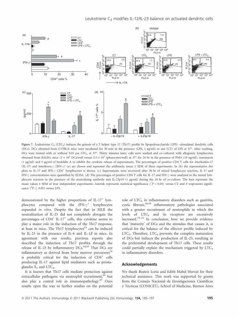
LTC4 promotes the expansion of Th17 lymphocytesby LPS-stimulated DCs
To determine whether LTC4 is capable of defining a Th17
profile by activated DCs, we decided to analyse this point
in an MLR. The DCs from C57BL/6 mice were stimulated
or not with LPS (1 lg/ml), then cells were untreated or
treated with LTC4 (0�01 lM) for 30 min at 37�. Finally,
DCs were extensively washed and co-cultured with
splenocytes from BALB/c mice. Immature DCs were used
as controls. As shown in Fig. 7(a,b), the treatment of
LPS-stimulated DCs with LTC4 increased by 50% the per-
centages of CD4+ T lymphocytes producing IL-17 com-
pared with those induced by LPS alone. These results
have an inverse correlation with the proportions of CD4+
T lymphocytes producing IFN-c. Similar results were
obtained to evaluate both cytokines in the supernatants of
MLR (Fig. 7c). As treatment of LPS-activated DCs with
LTC4 affected the IL-12/IL-23 balance, we investigated
whether IL-23 held a central role in mediating the increase
of IL-17. For this, co-cultures of DCs and splenocytes were
performed in the presence of neutralizing antibodies.
The neutralization of IL-23 by an anti-IL-23p19 reduced
by more than 20% the percentages of CD4+ IL-17+ cells
(Fig. 7d). Hence, IL-23 seems to be an important media-
tor for the expansion of CD4 T lymphocytes in a Th17
profile.
Discussion
Cysteinyl LTC4 is a potent lipid mediator of inflammatory
reactions, such as asthma, arthritis, gastritis and ischae-
mia.43,44 It modulates the chemotaxis of DCs from the
skin to lymph nodes,23 the only antigen-presenting cell
700
*
*(a)
(c)
(e) 1500
175
150
(d)
125
100
75
50
25
0
1000
500
0
(b)
600
500
400
300
200
100
400
#
##
##
#
*
1000
750
500
250
0
300
200
100
0
0
TN
F- α
(pg
/ml)
IL-1
0 (p
g/m
l)
IL-2
3 (p
g/m
l)
IL12
-p70
(pg
/ml)
IL-1
2 p4
0 (p
g/m
l)
CTLT
C4LP
S
LPS+L
TC4
CTLT
C4LP
S
LPS+L
TC4
CTLT
C4LP
S
LPS+L
TC4
CTLT
C4LP
S
LPS+L
TC4CTLT
C4LP
S
LPS+L
TC4
Figure 3. Leukotriene C4 (LTC4) inhibits the T helper type 1 (Th1) profile by lipopolysaccharide (LPS) matured dendritic cells (DCs). The DCs
were incubated for 30 min in the presence (LPS, 1 lg/ml) or absence (CT) of LPS at 37�. After washing, DCs were treated with or without
0.01 lm LTC4 at 37�, overnight, Then we collected the supernatants and we quantified the levels of tumour necrosis factor-a (TNF-a) (a), inter-
leukin-12p70 (IL-12p70) (b), IL-10 (c), IL-12p40 (d) and IL-23 (e) by ELISA. Bars represent the cytokine concentrations (pg/ml) and represent
the mean ± SEM of six experiments. Asterisk represents statistical significance (*P � 0.05) versus CT and # represents significance (#P � 0.05;##P � 0.01) versus LPS.
� 2011 The Authors. Immunology � 2011 Blackwell Publishing Ltd, Immunology, 134, 185–197 191
Leukotriene C4 modifies IL-12/IL-23 balance on activated dendritic cells
capable of activating naive T lymphocytes.3,4 Previous
studies aimed at analysing the effect of LTC4 showed
increases in the production of IL-10 by allergen-pulsed
DCs, favouring their capacity to increase lung eosino-
philia and IL-5 production in a model of murine asthma.
This effect involves the CysLTR1, which seems to contrib-
ute to the severity of inflammatory responses.45,46
In the present study we observed that DCs and
LPS-activated DCs express the two subtypes of cysteinyl
receptors. In most systems CysLTR1 was described as
responsible for most of inflammatory effects,45–48 but no
previous studies have examined the expression of both
receptors in murine DCs. Real-time PCR demonstrated
that the DCs not only express the CysLTR1, primarily
expressed in smooth muscle, eosinophils and other
immune cells and generally associated with the induction
of bronchospasm and vasoconstriction,18,19 but also the
CysLTR2,19 expressed mainly in the heart, prostate,
brain, adrenal cells, endothelium and lung, but it is
expressed at lower levels on leucocytes, and is more
associated with the remodelling of the fibrotic process.19
Several groups have demonstrated the modulation of
CysLT receptors by cytokines and inflammatory stim-
uli.49,50 Thivierge et al.25 demonstrated that human
monocytes express both CysLT1 and CysLT2 receptors
similarly and their differentiation in DCs inhibits the
expression of CysLT1, whereas their maturation with
200 ng/ml LPS increases CysLTR2 expression. In con-
trast, upon activation of DCs by LPS (1 lg/ml) no vari-
ations in the expression of CysLRT1 were observed but
there is a greater reduction of CysLRT2. These differ-
ences may be the result of the source of DCs as well as
of concentrations, methodology and time of LPS stimu-
lation used. Interestingly, incubation with exogenous
LTC4 of immature DCs potently up-regulated the
expression of CysLTR1, indicating that LTC4 could exert
a regulatory mechanism on receptor expression. In this
sense, it has been demonstrated in several tissues that
the expression of both receptors can be regulated by
CysLT release.18,50 The use of montelukast did not allow
us to block the production of IL-23, indicating that it
could be modulated by the action of LTC4 through the
CysLTR2. This point could not be evaluated; because
there is still no specific receptor antagonist.
Immature DCs constitutively macropinocytose extracel-
lular fluid,51 and also express a large variety of receptors
164 000 CysLTRI
CysLTRII174 000
CTLT
C 4LP
S
LPS +
LTC 4
*
*
50
25
0
r: C
ysLT
R/β
-act
in
CTLT
C 4LP
S
LPS +
LTC 4
CTLT
C 4LP
S
LPS +
LTC 4
100
Isot
ype
FL2
-H10
110
210
310
4
100 101 102 103 104
CD11c
CT LTC4 LPS LPS + LTC4
CT + MK LTC4 + MK LPS + MK LPS + LTC4 + MK
15·137·43·23·8
3·0
100
100
101
102
103
104
100
101
102
103
104
FL1-H
101 102 103 101 102 1031000 101 102 103101 102 103 10410001000
5·3 30·8 39·2
FL1-H FL1-H FL1-H FL1-H
IL-1
2p70
(c)
(b)
(a)
Figure 4. Immature and mature dendritic cells (DCs) express cysteinyl leukotriene receptors 1 (CysLTR1) and CysLTR2. The expression of Cys-
LTR1 and CysLTR2 was evaluated by RT-PCR. RNAs from immature and lipopolysaccharide (LPS) -stimulated DCs, were extracted after 18 hr
of culture in the presence or absence of leukotriene C4 (LTC4; 0.01 lm). RNAs used as positive controls were from lung to the CysLTR1 and tis-
sues of intestinal mucosa to the CysLTR2. (a) Representative gels of RT-PCR from six independent experiments are shown. (b) Shows the histo-
gram bars for the ratio values obtained from the normalization of the diameters of each band compared with b-actin. The results are expressed
as ratio and represent the arithmetic mean ± SEM of six experiments. Asterisk represents statistical significance (*P � 0.05) versus controls. (b)
DCs and lipopolysaccharide (LPS) -stimulated DCs (1 lg/ml for 30 min at 37�) were incubated (1 · 106 cells/ml) for 20 min at 37� without or
with 1 lm montelukast. Then, DCs were untreated or treated with LTC4 (0.01 lm) for 18 hr at 37�. Brefeldin A (5 lg/ml) was added during the
last 6 hr of culture to inhibit the release of cytokines into the supernatant. Finally, the percentages of interleukin-12p70-positive cells were evalu-
ated by intracytoplasmic staining. We showed a representative dot plot obtained by cytometry.
192 � 2011 The Authors. Immunology � 2011 Blackwell Publishing Ltd, Immunology, 134, 185–197
C. Alvarez et al.
mediating endocytosis and phagocytosis of antigens and
pathogens.5 Previously it was demonstrated that CysLTs
are able to induce the phagocytosis of opsonized bacteria
through the Fcc receptors.52 Here, we showed that LTC4
induces the phagocytosis of Zy and also stimulates Dex-
tran and HRP endocytosis by immature DCs. Interest-
ingly, despite the phenotypic changes and antigen capture
that produced LTC4 in activated DCs, which might corre-
late with the alteration of their function as antigen-pre-
senting cells, their capacity to activate naive T
lymphocytes remained intact.2–4 Although the LTC4
antagonizes the effect of LPS on the expression of class II
molecules and CD86, its expression is greater than that
shown by immature DCs. Our hypothesis is that through
this mechanism, the LTC4 allows DCs to improve their
ability to sense the environment without compromising
their capacity to activate an effector response.
The activation of MAPK, including ERK1/2, c-Jun N-
terminal kinase and p38 MAPK play an important role in
many cellular processes, including differentiation, cellular
proliferation, apoptosis and immune response.53,54 The
p38 pathway is associated with cytokine induction and
inflammation and is strongly activated by inflammatory
stimuli.54 Binding of CysLT with their receptors triggers
the phosphorylation of MAPK.18,19 Hashimoto et al.55
demonstrated that IL-10 production in human DCs stim-
ulated with Zy was dependent on ERK and p38 MAPK
activation. Also, the phagocytosis of opsonized particles
by macrophages cultured with LTD4 or LTC4 was associ-
ated with p38 activation.56 Our results indicate that LTC4
activates p38 MAPK. Indeed, their inhibition by SB-
303080 abrogates the uptake of DX by DCs. Also, ERK1/2
was only activated in LTC4-stimulated DCs. In spite of
the previous studies,18,19,52 however, the fact that the
CTCT+LT
C4
LPS
LPS+L
TC4
P-p38
p-ERK1/2
(a)
β-actin
*
*
4(b)
3
2
r =
P-p
38/ β
-act
in
1
0
CTLT
C4LP
S
LPS+L
TC4
(c)
#
2·0
1·5
1·0
0·5
0·0
r =
P-e
rk/ β
-act
in
CTLT
C4LP
S
LPS+L
TC4
Figure 5. Leukotriene C4 (LTC4) activates ERK1/2 and p38 MAPK in dendritic cells (DCs). (a) DCs (3 · 106 cells per 500 ll complete medium)
were prewarmed for 30 min at 37�. Cells were incubated in the presence or absence of LPS (1 lg/ml) for 30 min at 37� and then treated or not
with 0.01 lm LTC4 for 5 min at 37�. The samples were analysed by Western blotting as described in the Materials and methods. Pervanadate-
treated DCs (0.1 mm orthovanadate plus 0.3 mm H2O2 for 10 min at 37�) were used as positive phosphorylation controls. Western blots are rep-
resentative of five experiments. (b) and (c) Histograms of ratio obtained by quantitative densitometric analysis of P-p38 and ERK1/2; respectively,
normalized with b-actin densitometric units. The bars represent the mean ± SEM of five experiments. Asterisk represents statistical significance
(*P � 0.05) versus CT and # represents significance (#P � 0.05) versus LPS.
� 2011 The Authors. Immunology � 2011 Blackwell Publishing Ltd, Immunology, 134, 185–197 193
Leukotriene C4 modifies IL-12/IL-23 balance on activated dendritic cells
blockade of p38 and ERK1/2 MAPK was not able to abol-
ish either IL-12p40 or IL-23 production supports the the-
ory that other pathways could be involved. Consistent
with these results, Yang et al.53 reported that inhibition of
p38 MAPK can induce Th1 responses through the pro-
duction by DCs of IL-12p40 and IL-12p70. Therefore, we
believe that p38 MAPK phosphorylation acts as a regula-
tory mechanism of genesis of Th1 profiles. It is known
that nuclear factor-jB activation triggered by LPS is con-
trolled by a series of kinases and phosphatases. Chang
et al.57 demonstrated that the serine-threonine protein
phosphatase A2 (PPA2) binds inhibitor of jB kinase, a
subunit of nuclear factor-jB, mechanism which prevents
the production of IL-23. Future experiments will define
whether the LTC4 is capable of preventing the binding or
synthesis of PPA2.
We report that LTC4 abolishes completely in DCs the
secretion of IL-12p70, the biological chain of IL-12, trig-
gered by LPS, but enhances p40, the common chain to
IL-12/IL-23. The partial or complete reversal of produc-
tion of IL-12p70 by LPS-activated DCs has been linked to
stimuli as diverse as prostaglandins, histamine, alkaloids
and phenolic products.58–61 In relation to CysLT, in terms
of cytokines, the results are contradictory. Machida
et al.40 described in Derf-pulsed DCs from bone marrow
precursors how antagonists of CysLTR1 led to the
enhancement of IL-12p40 while IL-10 was inhibited. On
the other hand, in allergen-pulsed DCs from spleen there
was strong inhibition of both IL-10 and IL-12p70 in the
presence of CysLTR1 antagonists.62 These differences can
be explained by the origin of the DCs used in each study;
however, the main difference would be the nature of the
stimuli used, we evaluated the effect of LTC4 in DCs acti-
vated with LPS, a classic Toll-like receptor 4 agonist,
which triggers a Th1 profile compared with the allergens
that trigger Th2 responses.
The strong inhibition of IL-12p70 release, together with
the increased production of IL-23, represent a suitable
microenvironment induced by LTC4 acting on inflamma-
tory DCs resulting in the expansion of Th17 cells, as
500
250
0
100 1500 50 0 25 50 75 100 125
0 1000 2000
–PD+PD
LPS+LTC4
LPS
LTC4
CT
IL-12p70 (positive cells %)
–SB+SB
LPS+LTC4
LPS
LTC4
CT
IL-23 (pg/ml)
–SB+SB
MF
I
–XB+XB
CTLT
C4LP
S
LPS+L
TC4CT
LTC4
LPS
LPS+L
TC4
**
*
*
*
LPS+LTC4
LPS
LTC4
CT
IL-12p40 (positive cells %)
(a)
(b)
(c) (d)
Figure 6. p38 MAPK is involved in regulating endocytosis by leukotriene C4 (LTC4) on lipopolysaccharide (LPS) -stimulated dendritic cells
(DCs). The DCs and LPS-stimulated DCs (1 lg/ml for 30 min at 37�) were incubated (2.5 · 106 cells/ml) for 20 min at 37� without inhibitors
or in the presence of 50 lm SB202190. Then, the cells were exposed to LTC4 (0.01 lm) for 30 min at 37�. After this time, DCs were washed and
cultured for an additional 40 min in the presence of dextran (DX) -FITC (100 lg/ml) at 37�. (a) Results are expressed as mean fluorescence
intensity (MFI) and represent the arithmetic mean ± SEM of four experiments. (b,c) and (d) DCs and LPS-stimulated DCs (1 lg/ml for 30 min
at 37�) were incubated (2.5 · 106 cells/ml) for 20 min at 37� without inhibitors or in the presence of 50 lm PD98059 or 50 lm SB202190. Then,
DCs were untreated or treated with LTC4 (0.01 lm) for 18 hr at 37�, in some cases, brefeldin A (5 lg/ml) was added during the last 6 hr of cul-
ture to inhibit the release of cytokines into the supernatant. (b) Culture supernatants were collected and levels of interleukin-23 (IL-23) were
quantified by ELISA. Results are expressed as the cytokine concentrations (pg/ml) and represent the mean ± SEM of four experiments. The per-
centages of positive cells for IL-12p70 (c) and IL-12p40 (d) are shown and represent the arithmetic mean ± SEM of three experiments. Asterisk
represents statistical significance *P < 0.05 for DCs pretreated or not with inhibitors and then exposed to LTC4.
194 � 2011 The Authors. Immunology � 2011 Blackwell Publishing Ltd, Immunology, 134, 185–197
C. Alvarez et al.
demonstrated by the higher proportions of IL-17+ lym-
phocytes compared with the IFN-c+ lymphocytes
expanded in vitro. Despite the fact that in MLR the
neutralization of IL-23 did not completely abrogate the
percentages of CD4+ IL-17+ cells, this cytokine seems to
play a major role in the induction of the Th17 response,
at least in mice. The Th17 lymphocytes63 can be induced
by IL-23 in the presence of IL-6 and IL-1b in mice. In
agreement with our results, previous reports also
described the induction of Th17 profiles through the
release of IL-23 by inflammatory DCs.64,65 That DCs are
inflammatory as derived from bone marrow precursors26
is probably critical for the induction of CD4+ cells
producing IL-17 against lipid mediators such as prosta-
glandin E2 and LTC4.
It is known that Th17 cells mediate protection against
extracellular pathogens via neutrophil recruitment,66 but
also play a central role in immunopathology.67 Ours
results open the way to further studies on the potential
role of LTC4 in inflammatory disorders such as gastritis,
cystic fibrosis,68,69 inflammatory pathologies associated
with a greater recruitment of neutrophils in which the
levels of LTC4 and its receptors are excessively
increased.19–22 In conclusion, here we provide evidence
that ‘maturity’ of DCs and the stimulus that causes it, is
critical for the balance of the effector profile induced by
LTC4. Therefore, LTC4 prevents the complete maturation
of DCs but induces the production of IL-23, resulting in
the preferential development of Th17 cells. These results
could partially explain the mechanism triggered by LTC4
in inflammatory disorders.
Acknowledgements
We thank Beatriz Loria and Edith Mabel Horvat for their
technical assistance. This work was supported by grants
from the Consejo Nacional de Investigaciones Cientıficas
y Tecnicas (CONICET), School of Medicine, Buenos Aires
IL-17A
0 10 20 30 40
CD4+ cells %
CTLTC4LPSLPS + LTC4
*
#
#
IFN-γ
CT LTC4 LPS LPS + LTC4
IL-17A
CD
4
100 101 102 103
22·0 1·3
R3R2
FL3
-H
FL3
-H
FL3
-H
FL3
-HR2 R2R2 R3
23·0 0·8 22·0
R3
2·9
R3
5·832·0
10010
110
210
310
4
100 101 102 103 104
R2 R3
1·40·5
Isotype
FL3
-H
FL2-H FL2-H FL2-H FL2-H
FL2-H
104 100 101 102 103 104 100 101 102 103 104 100 101 102 103 10410010
110
210
310
4
10010
110
210
310
4
10010
110
210
310
4
10010
110
210
310
4
500
250
0IFN-g IL-17
CTLTC4LPSLPS + LTC4
(pg/
ml)
*
*
#
0
5
10
– IL-23p19 + IL23p19
CTLTC4LPSLPS + LTC4
IL-1
7+ c
ells
%
**
#
#
(a)
(c) (d)
(b)
Figure 7. Leukotriene C4 (LTC4) induces the genesis of a T helper type 17 (Th17) profile by lipopolysaccharide (LPS) -stimulated dendritic cells
(DCs). DCs obtained from C57BL/6 mice were incubated for 30 min in the presence (LPS, 1 lg/ml) or not (CT) of LPS at 37�. After washing,
DCs were treated with or without 0.01 lm LTC4 at 37�. Thirty minutes later; cells were washed and co-cultured with allogeneic lymphocytes
obtained from BALB/c mice (5 · 104 DCs/well versus 2.5 · 105 splenocytes/well) at 37� for 24 hr in the presence of PMA (10 ng/ml), ionomycin
(1 lg/ml) and 5 lg/ml of brefeldin A to inhibit the cytokine release of supernatants. The percentages of positive CD4 T cells for interleukin-17
(IL-17) and interferon-c (IFN-c) (a) are shown and represent the arithmetic mean ± SEM of three experiments. In (b) the representative dot
plots to IL-17 and IFN-c CD4+ lymphocytes is shown. (c) Supernatants were recovered after 36 hr of mixed lymphocyte reaction, IL-17 and
IFN-c concentrations were quantified by ELISA. (d) The percentages of positive CD4 T cells for IL-17 and IFN-c were analysed in the mixed lym-
phocyte reaction in the presence of the neutralizing antibody anti IL-23p19 (1 lg/ml) during the 24 hr of co-culture. The bars represent the
mean values ± SEM of four independent experiments. Asterisk represents statistical significance (*P < 0.05) versus CT and # respresents signifi-
cance (#P � 0.05) versus LPS.
� 2011 The Authors. Immunology � 2011 Blackwell Publishing Ltd, Immunology, 134, 185–197 195
Leukotriene C4 modifies IL-12/IL-23 balance on activated dendritic cells
University, and Agencia Nacional de Promocion Cientıfi-
ca y Tecnologica, Argentina.
Disclosures
The authors have no conflicts of interest.
References
1 Lutz MB, Kurts C. Induction of peripheral CD4+ T-cell tolerance and CD8+ T-cell
cross-tolerance by dendritic cells. Eur J Immunol 2009; 32:2325–30.
2 Joffre O, Nolte MA, Sporri R, Reis e Sousa C. Inflammatory signals in dendritic cell
activation and the induction of adaptive immunity. Immunol Rev 2009; 227:234–47.
3 Steinman RM, Hemmi H. Dendritic cells: translating innate to adaptive immunity.
Curr Top Microbiol Immunol 2006; 311:17–58.
4 Reis e Sousa C. Activation of dendritic cells: translating innate into adaptative immu-
nity. Curr Opin Immunol 2006; 16:21–5.
5 Sabatte J, Maggini J, Nahmod K et al. Interplay of pathogens, cytokines and other
stress signals in the regulation of dendritic cell function. Cytokine Growth Factor Rev
2007; 18:5–17.
6 Ricart BG, John B, Lee D, Hunter CA, Hammer DA. Dendritic cells distinguish
individual chemokine signals through CCR7 and CXCR4. J Immunol 2011; 186:
53–61.
7 Lombardi V, Van Overtvelt L, Horiot S, Moingeon P. Human dendritic cells stimulated
via TLR7 and/or TLR8 induce the sequential production of Il-10, IFN-gamma, and IL-
17A by naive CD4+ T cells. J Immunol 2009; 182:3372–9.
8 Paustian C, Caspell R, Johnson T, Cohen PA, Shu S, Xu S, Czerniecki BJ, Koski GK.
Effect of multiple activation stimuli on the generation of Th1-polarizing dendritic cells.
Hum Immunol 2011; 72:24–31.
9 Rossetti M, Gregori S, Roncarolo MG. Granulocyte-colony stimulating factor drives the
in vitro differentiation of human dendritic cells that induce anergy in naıve T cells. Eur
J Immunol 2010; 40:3097–106.
10 McIlroy A, Caron G, Blanchard S, Fremaux I, Duluc D, Delneste Y, Chevailler A,
Jeannin P. Histamine and prostaglandin E up-regulate the production of Th2-
attracting chemokines (CCL17 and CCL22) and down-regulate IFN-gamma-induced
CXCL10 production by immature human dendritic cells. Immunology 2006; 117:
507–16.
11 Rivas-Carvalho A, Meraz-Rıos MA, Santos-Argumedo L, Bajana S, Soldevila G, Mo-
reno-Garcıa ME, Sanchez-Torres C. CD16+ human monocyte-derived dendritic cells
matured with different and unrelated stimuli promote similar allogeneic Th2 respon-
ses: regulation by pro- and anti-inflammatory cytokines. Int Immunol 2004; 16:1251–
63.
12 Amaral MM., Davio C, Ceballos A, Salamone G, Canones C, Geffner J, Vermeulen M.
Histamine improves antigen uptake and cross-presentation by dendritic cells. J Immu-
nol 2007; 179:3425–33.
13 Rinaldo-Matthis A, Haeggstrom JZ. Structures and mechanisms of enzymes in the leu-
kotriene cascade. Biochimie 2010; 92:676–81.
14 Austen KF, Maekawa A, Kanaoka Y, Boyce JA. The leukotriene E4 puzzle: finding the
missing pieces and revealing the pathobiologic implications. J Allergy Clin Immunol
2009; 124:406–14.
15 Vaidyanathan S, Williamson P, Clearie K, Morrison A, Lipworth B. Nasal AMP and
histamine challenge within and outside the pollen season in patients with seasonal
allergic rhinitis. J Allergy Clin Immunol 2011; 127:173–8.
16 Muraki M, Imbe S, Sato R, Ikeda Y, Yamagata S, Iwanaga T, Tohda Y. Inhaled mont-
elukast inhibits cysteinyl-leukotriene-induced bronchoconstriction in ovalbumin-sensi-
tized guinea-pigs: the potential as a new asthma medication. Int Immunopharmacol
2009; 9:1337–41.
17 Poulin S, Thompson C, Thivierge M, Veronneau S, McMahon S, Dubois CM, Stankova
J, Rola-Pleszczynski M. Cysteinyl-leukotrienes induce vascular endothelial growth factor
production in human monocytes and bronchial smooth muscle cells. Clin Exp Allergy
2011; 41:204–17.
18 Singh RK, Gupta S, Dastidar S, Ray A. Cysteinyl leukotrienes and their receptors:
molecular and functional characteristics. Pharmacology 2010; 85:336–49.
19 Kanaoka Y, Boyce JA. Cysteinyl leukotrienes and their receptors: cellular distribution
and function in immune and inflammatory responses. J Immunol 2004; 173:1503–10.
20 Cho SH. Pharmacogenomic approaches to asthma treatment. Allergy Asthma Immunol
Res 2010; 2:177–82.
21 Tintinger GR, Feldman C, Theron AJ, Anderson R. Montelukast: more than a cysteinyl
leukotriene receptor antagonist? Scientific World Journal 2010; 10:2403–13.
22 Shimbori C, Shiota N, Okunishi H. Effects of montelukast, a cysteinyl-leukotriene type
1 receptor antagonist, on the pathogenesis of bleomycin-induced pulmonary fibrosis in
mice. Eur J Pharmacol 2011; 650:424–30.
23 Robbiani DF, Finch RA, Jager D, Muller WA, Sartorelli AC, Randolph GJ. The leuko-
triene C4 transporter MRP1 regulates CCL19 (MIP-3b, ELC)-dependent mobilization of
dendritic cells to lymph nodes. Cell 2000; 103:757–68.
24 Jozefowski S, Biedron R, Bobek M, Marcinkiewicz J. Leukotrienes modulate cytokine
release from dendritic cells. Immunology 2005; 116:418–28.
25 Thivierge M, Stankova J, Rola-Pleszczynski M. Cysteinyl-leukotriene receptor type 1
expression and function is down-regulated during monocyte-derived dendritic cell mat-
uration with zymosan: involvement of IL-10 and prostaglandins. J Immunol 2009;
83:6778–87.
26 Xu Y, Zhan Y, Lew AM, Naik SH, Kershaw MH. Differential development of murine
dendritic cells by GM-CSF versus Flt3 ligand has implications for inflammation and
trafficking. J Immunol 2007; 179:7577–84.
27 Inaba K, Inaba M, Romani N, Aya H, Deguchi M, Ikehara S, Muramatsu S, Steinman
R M. Generation of large numbers of dendritic cells from mouse bone marrow cultures
supplemented with granulocyte/macrophage colony-stimulating factor. J Exp Med 1992;
176:1693–7.
28 Lutz MB, Assmann CU, Girolomoni G, Ricciardi-Castagnoli P. Different cytokines reg-
ulate antigen uptake and presentation of a precursor dendritic cell line. Eur J Immunol
1996; 26:586–9.
29 Sallusto F, Cella M, Danieli C, Lanzavecchia A. Dendritic cells use macropynocitosis
and the mannose receptor to concentrate macromolecules in the major histocompati-
bility complex class II compartment: down regulation by cytokines and bacterial prod-
ucts. J Exp Med 1995; 182:389–400.
30 Ono E, Taniguchi M, Higashi N et al. Increase in salivary cysteinyl-leukotriene concen-
tration in patients with aspirin-intolerant asthma. Allergol Int 2011; 60:37–43.
31 Banchereau J, Briere F, Caux C, Davoust J, Lebecque S, Yong-Jun L, Pulendran B,
Palucka K. Immunobiology of dendritic cells. Annu Rev Immunol 2000; 18:767–
811.
32 Tanaka Y, Taneichi M, Kasai M, Kakiuchi T, Uchida T. Liposome-coupled antigens are
internalized by antigen-presenting cells via pinocytosis and cross-presented to CD8 T
cells. PLoS One 2010; 5:e15225.
33 Sahlin S, Hed J, Rundquist I. Differentiation between attached and ingested immune
complexes by a fluorescence quenching cytofluorometric assay. J Immunol Methods
1983; 60:115–24.
34 Conzelmann M, Wagner AH, Hildebrandt A et al. IFN-c activated JAK1 shifts CD40-
induced cytokine profiles in human antigen-presenting cells toward high IL-12p70 and
low IL-10 production. Biochem Pharmacol 2010; 80:2074–86.
35 Jensen SS, Gad M. Differential induction of inflammatory cytokines by dendritic cells
treated with novel TLR-agonist and cytokine based cocktails: targeting dendritic cells in
autoimmunity. J Inflamm 2010; 7:37–49.
36 Gregori S. Dendritic cells in networks of immunological tolerance. Tissue Antigens
2011; 77:89–99.
37 Saito M, Nagasawa M, Takada H et al. Defective IL-10 signaling in hyper-IgE syndrome
results in impaired generation of tolerogenic dendritic cells and induced regulatory T
cells. J Exp Med 2011; 208:235–49.
38 Doisne JM, Soulard V, Becourt C et al. Cutting edge: crucial role of IL-1 and IL-23 in
the innate IL-17 response of peripheral lymph node NK1.1– invariant NKT cells to bac-
teria. J Immunol 2011; 186:662–6.
39 Jackson AM, Mulcahy LA, Porte J et al. Role of mitogen-activated protein kinase and
PI3K pathways, in the regulation of IL-12-family cytokines in dendritic cells and the
generation of T H-responses. Eur Cytokine Netw 2010; 21:319–28.
40 Machida I, Matsuse H, Kondo Y et al. Cysteinyl leukotrienes regulate dendritic cell
functions in a murine model of asthma. J Immunol 2004; 172:1833–8.
41 Liu YY, Cai WF, Yang HZ et al. Bacillus Calmette–Guerin and TLR4 agonist prevent
cardiovascular hypertrophy and fibrosis by regulating immune microenvironment.
J Immunol 2008; 180:7349–57.
42 Yuan YM, Fang SH, Qian XD et al. Leukotriene D4 stimulates the migration but
not proliferation of endothelial cells mediated by the cysteinyl leukotriene cyslt (1)
receptor via the extracellular signal-regulated kinase pathway. J Pharmacol Sci 2009;
109:285–92.
43 Bisgaard H. Pathophysiology of the cysteinyl leukotrienes and effects of leukotriene
receptor antagonists in asthma. Allergy 2001; 66:7–11.
44 Riccioni G, Zanasi A, Vitulano N, Mancini B, D’Orazio N. Leukotrienes in atheroscle-
rosis: new target insights and future therapy perspectives. Mediators Inflamm 2009;
2009: 737282–8.
45 Ihaku D, Cameron L, Suzuki M, Molet S, Martin J, Hamid Q. Montelukast, a leukotri-
ene receptor antagonist, inhibits the late airway response to antigen, airway eosino-
philia, and IL-5-expressing cells in Brown Norway rats. J Allergy Clin Immunol 1999;
104:1147–54.
196 � 2011 The Authors. Immunology � 2011 Blackwell Publishing Ltd, Immunology, 134, 185–197
C. Alvarez et al.
46 Hisada T, Salmon M, Nasuhara Y, Chung KF. Cysteinyl-leukotrienes partly mediate
eotaxin-induced bronchial hyperresponsiveness and eosinophilia in IL-5 transgenic
mice. Am J Respir Crit Care Med 1999; 160:571–5.
47 Kiwamoto T, Ishii Y, Morishima Y et al. Blockade of cysteinyl leukotriene-1 receptors
suppresses airway remodelling in mice overexpressing GATA-3. Clin Exp Allergy 2011;
41:116–28.
48 Wu AY, Chik SC, Chan AW, Li Z, Tsang KW, Li W. Anti-inflammatory effects of
high-dose montelukast in an animal model of acute asthma. Clin Exp Allergy 2003;
33:359–66.
49 Magnusson C, Mezhybovska M, Lorinc E, Fernebro E, Nilbert M, Sjolander A. Low
expression of CysLT1R and high expression of CysLT2R mediate good prognosis in
colorectal cancer. Eur J Cancer 2010; 46:826–35.
50 Magnusson C, Ehrnstrom R, Olsen J, Sjolander A. An increased expression of cysteinyl
leukotriene 2 receptor in colorectal adenocarcinomas correlates with high differentia-
tion. Cancer Res 2007; 67:9190–8.
51 Amyere M, Mettlen M, Van Der Smissen P, Platek A, Payrastre B, Veithen A, Courtoy
PJ. Origin, originality, functions, subversions and molecular signalling of macropinocy-
tosis. Int J Med Microbiol 2002; 291:487–94.
52 Silva RC, Landgraf MA, Hiyane MI, Pacheco-Silva A, Camara NO, Landgraf RG. Leu-
kotrienes produced in allergic lung inflammation activate alveolar macrophages. Cell
Physiol Biochem 2010; 26:319–26.
53 Yang Z, Zhang X, Darrah PA, Mosser DM. The regulation of Th1 responses by the p38
MAPK. J Immunol 2010; 185:6205–13.
54 Dunn KL, Espino PS, Drobic B, He S, Davie JR. The Ras-MAPK signal transduction
pathway, cancer and chromatin remodeling. Biochem Cell Biol 2005; 83:1–14.
55 Hashimoto K, Ichiyama T, Hasegawa M, Hasegawa S, Matsubara T, Furukawa S. Cyste-
inyl leukotrienes induce monocyte chemoattractant protein-1 in human monocyte/mac-
rophages via mitogen-activated protein kinase and nuclear factor-kappaB pathways. Int
Arch Allergy Immunol 2009; 149:275–82.
56 Campos MR, Serezani CH, Peters-Golden M, Jancar S. Differential kinase requirement
for enhancement of Fc gammaR-mediated phagocytosis in alveolar macrophages by leu-
kotriene B4 vs. D4. Mol Immunol 2009; 46:1204–11.
57 Chang JH, Voorhees TJ, Liu J, Zhao Y, Chang C-H. Interleukin-23 production in den-
dritic cells is negatively regulated by protein phosphatase 2A. PNAS 2009; 107:8340–5.
58 Kim GY, Kim KH, Lee SH et al. Curcumin inhibits immunostimulatory function of
dendritic cells: MAPKs and translocation of NF-jB as potential targets. J Immunol
2005; 174:8116–24.
59 Kang BY, Chung SW, Cho D, Kim TS. Involvement of p38 mitogen-activated protein
kinase in the induction of interleukin-12 p40 production in mouse macrophages by
berberine, a benzodioxoloquinolizine alkaloid. Biochem Pharmacol 2002; 63:1901–10.
60 Kalinski P, Vieira PL, Schuitemaker JH, de Jong EC, Kapsenberg ML. Prostaglandin E2
is a selective inducer of interleukin-12 p40 (IL-12p40) production and an inhibitor of
bioactive IL-12p70 heterodimer. Blood 2001; 97:3466–9.
61 Li CY, Chao LK, Wang SC et al. Honokiol inhibits LPS-induced maturation and
inflammatory response of human monocyte-derived dendritic cells. J Cell Physiol 2011;
226:2338–49.
62 Okunishi K, Dohi M, Nakagome K, Tanaka R, Yamamoto K. A novel role of cysteinyl
leukotrienes to promote dendritic cell activation in the antigen-induced immune
responses in the lung. J Immunol 2004; 173:6393–402.
63 Kyburz D, Corr M. Th17 cells generated in the absence of TGF-b induce experimental
allergic encephalitis upon adoptive transfer. Expert Rev Clin Immunol 2011; 7:283–5.
64 Khayrullina T, Yen JH, Jing H, Ganea D. In vitro differentiation of dendritic cells in the
presence of prostaglandin E2 alters the IL-12/IL-23 balance and promotes differentia-
tion of Th17 cells. J Immunol 2008; 181:721–35.
65 Sheibanie AF, Yen JH, Khayrullina T, Emig F, Zhang M, Tuma R, Ganea D. The proin-
flammatory effect of prostaglandin E2 in experimental inflammatory bowel disease is
mediated through the IL-23–IL-17 axis. J Immunol 2007; 178:8138–47.
66 Abi Abdallah DS, Egan CE, Butcher BA, Denkers EY. Mouse neutrophils are profes-
sional antigen-presenting cells programmed to instruct Th1 and Th17 T-cell differentia-
tion. Int Immunol 2011; 23:317–26.
67 De Nitto D, Sarra M, Cupi ML, Pallone F, Monteleone G. Targeting IL-23 and Th17-
cytokines in inflammatory bowel diseases. Curr Pharm Des 2010; 16:3656–60.
68 Stelmach I, Korzeniewska A, Stelmach W, Majak P, Grzelewski T, Jerzynska J. Effects of
montelukast treatment on clinical and inflammatory variables in patients with cystic
fibrosis. Ann Allergy Asthma Immunol 2005; 95:372–80.
69 Ahmed A, Holton J, Vaira D, Smith SK, Hoult JR. Eicosanoid synthesis and Helicobact-
er pylori associated gastritis: increase in leukotriene C4 generation associated with
H. pylori colonization. Prostaglandins 1992; 44:75–86.
� 2011 The Authors. Immunology � 2011 Blackwell Publishing Ltd, Immunology, 134, 185–197 197
Leukotriene C4 modifies IL-12/IL-23 balance on activated dendritic cells


