Embed Size (px)
Citation preview
Printed by Jouve, 75001 PARIS (FR)
(19)E
P2
567
693
A1
TEPZZ 56769¥A_T(11) EP 2 567 693 A1
(12) EUROPEAN PATENT APPLICATION
(43) Date of publication: 13.03.2013 Bulletin 2013/11
(21) Application number: 12191154.9
(22) Date of filing: 16.07.2004
(51) Int Cl.:A61K 9/127 (2006.01) C12N 15/11 (2006.01)
(84) Designated Contracting States: AT BE BG CH CY CZ DE DK EE ES FI FR GB GR HU IE IT LI LU MC NL PL PT RO SE SI SK TRDesignated Extension States: AL HR LT LV MK
(30) Priority: 16.07.2003 US 488144 P15.09.2003 US 503279 P11.12.2003 US 529406 P
(62) Document number(s) of the earlier application(s) in accordance with Art. 76 EPC: 04761574.5 / 1 648 519
(71) Applicant: Protiva Biotherapeutics Inc.Burnaby, British Columbia V5J 5J8 (CA)
(72) Inventors: • MacLachlan, Ian
Vancouver, British Columbia V4S 1ES (CA)• Ambegia, Ellen Grace
Vancouver, British Columbia V6K 1R7 (CA)• Heyes, James
Vancouver, British Columbia V5Z 4B2 (CA)
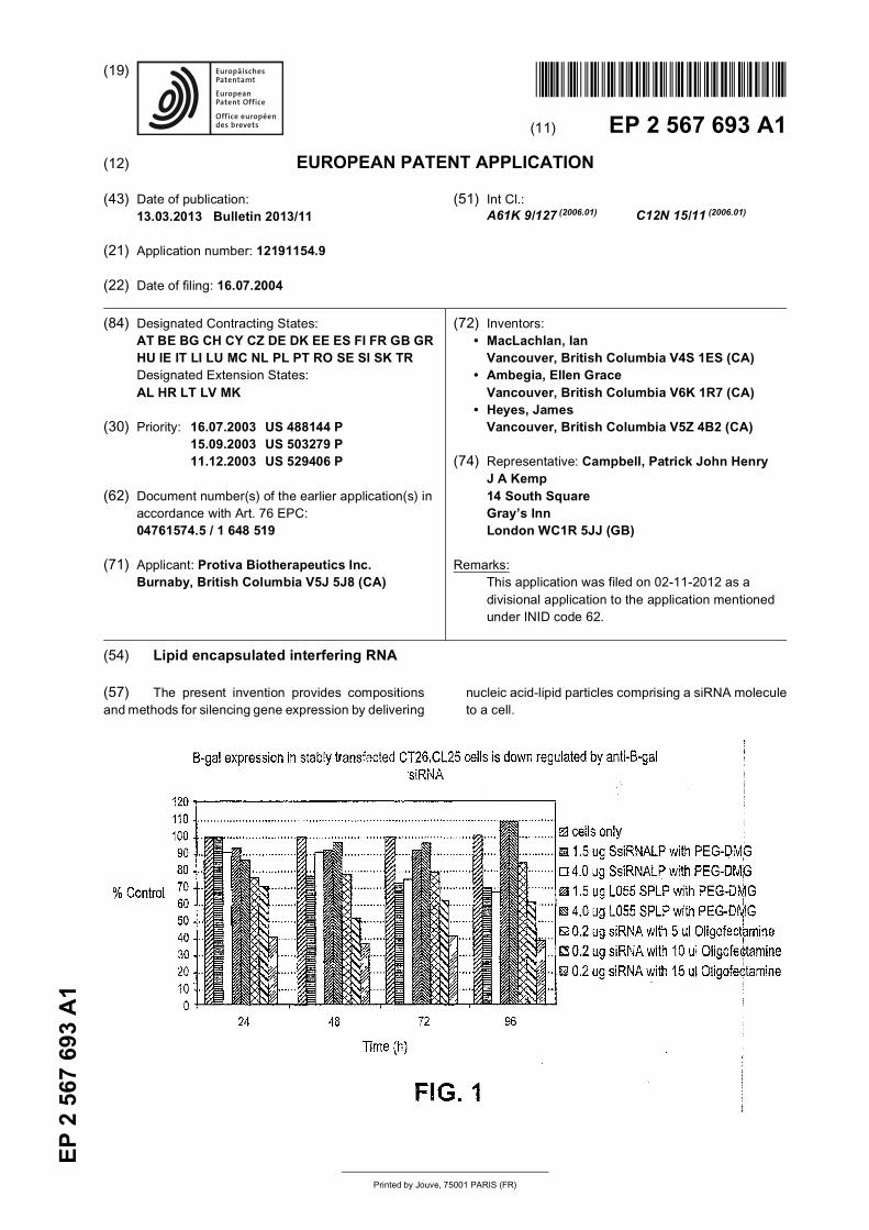
(74) Representative: Campbell, Patrick John HenryJ A Kemp 14 South Square Gray’s InnLondon WC1R 5JJ (GB)
Remarks: This application was filed on 02-11-2012 as a divisional application to the application mentioned under INID code 62.
(54) Lipid encapsulated interfering RNA
(57) The present invention provides compositionsand methods for silencing gene expression by delivering
nucleic acid-lipid particles comprising a siRNA moleculeto a cell.
EP 2 567 693 A1
2
5
10
15
20
25
30
35
40
45
50
55
Description
CROSS-REFERENCES TO RELATED APPLICATIONS
[0001] This application claims the benefit of U.S. Provisional Patent Application Nos. 60/529,406, filed December 11,2003; 60/503,279, filed September 15, 2003, and 60/488,144, filed July 16, 2003, the disclosures of each of which arehereby incorporated by reference in their entirety for all purposes.
FIELD OF THE INVENTION
[0002] The present invention relates to compositions and methods for the therapeutic delivery of a nucleic acid bydelivering a serum-stable lipid delivery vehicle encapsulating the nucleic acid to provide efficient RNA interference (RNAi)in a cell or mammal. More particularly, the present invention is directed to using a small interfering RNA (siRNA) encap-sulated in a serum-stable lipid particle having a small diameter suitable for systemic delivery.
BACKGROUND OF THE INVENTION
[0003] RNA interference (RNAi) is an evolutionarily conserved, sequence specific mechanism triggered by doublestranded RNA (dsRNA) that induces degradation of complementary target single stranded mRNA and "silencing" of thecorresponding translated sequences (McManus and Sharp, Nature Rev. Genet. 3:737 (2002)). RNAi functions by en-zymatic cleavage of longer dsRNA strands into biologically active "short-interfering RNA" (siRNA) sequences of about21-23 nucleotides in length (Elbashir, et al., Genes Dev. 15:188 (2001)). siRNA can be used downregulate or silencethe translation of a gene product of interest. For example, it is desirable to downregulate genes associated with variousdiseases and disorders.[0004] Delivery of siRNA remains problematic (see, e.g., Novina and Sharp, Nature 430::161-163 (2004); and Garber,J. Natl. Cancer Inst. 95(7):500-2 (2003)). An effective and safe nucleic acid delivery system is required for siRNA to betherapeutically useful. Naked dsRNA administered to most subjects will: (1) be degraded by endogenous nucleases;and (2) will not be able to cross cell membranes to contact and silence their target gene sequences.[0005] Viral vectors are relatively efficient gene delivery systems, but suffer from a variety of safety concerns, suchas potential for undesired immune responses. Furthermore, viral systems are rapidly cleared from the circulation, limitingtransfection to "first-pass" organs such as the lungs, liver, and spleen. In addition, these systems induce immune re-sponses that compromise delivery with subsequent injections. As a result, nonviral gene delivery systems are receivingincreasing attention (Worgall, et al., Human Gene Therapy 8:37 (1997); Peeters, et al., Human Gene Therapy 7:1693(1996); Yei, et al., Gene Therapy 1: 192 (1994); Hope, et al., Molecular Membrane Biology 15:1 (1998)).[0006] Plasmid DNA-cationic liposome complexes are currently the most commonly employed nonviral gene deliveryvehicles (Felgner, Scientific American 276:102 (1997); Chonn, et al., Current Opinion in Biotechnology 6:698 (1995)).For instance, cationic liposome complexes made of an amphipathic compound, a neutral lipid, and a detergent fortransfecting insect cells are disclosed in U.S. Patent No. 6,458,382. Cationic liposome complexes are also disclosed inU.S. Patent Application Publication No. 2003/0073640. Cationic liposome complexes, however, are large, poorly definedsystems that are not suited for systemic applications and can elicit considerable toxic side effects (Harrison, et al.,Biotechniques 19:816 (1995); Li, et al., The Gene 4:891 (1997); Tam, et al, Gene Ther. 7:1867 (2000)). As large, positivelycharged aggregates, lipoplexes are rapidly cleared when administered in vivo, with highest expression levels observedin first-pass organs, particularly the lungs (Huang, et al., Nature Biotechnology 15:620 (1997); Templeton, et al., NatureBiotechnology 15:647 (1997); Hofland, et al., Pharmaceutical Research 14:742 (1997)).[0007] Other liposomal delivery systems include, for example, the use of reverse micelles, anionic and polymer lipo-somes as disclosed in, e.g., U.S. Patent No. 6,429,200; U.S. Patent Application No. 2003/0026831; and U.S. PatentApplication Nos. 2002/0081736 and 2003/0082103, respectively.[0008] Recent work has shown that nucleic acids can be encapsulated in small (about 70 nm diameter) "stabilizedplasmid-lipid particles" (SPLP) that consist of a single plasmid encapsulated within a bilayer lipid vesicle (Wheeler, etal., Gene Therapy 6:271 (1999)). These SPLPs typically contain the "fusogenic" lipid dioleoylphosphatidylethanolamine(DOPE), low levels of cationic lipid (i.e., 10% or less), and are stabilized in aqueous media by the presence of a poly(ethylene glycol) (PEG) coating. SPLP have systemic application as they exhibit extended circulation lifetimes followingintravenous (i.v.) injection, accumulate preferentially at distal tumor sites due to the enhanced vascular permeability insuch regions, and can mediate transgene expression at these tumor sites. The levels of transgene expression observedat the tumor site following i.v. injection of SPLP containing the luciferase marker gene are superior to the levels that canbe achieved employing plasmid DNA-cationic liposome complexes (lipoplexes) or naked DNA.[0009] However, there remains a strong need in the art for novel and more efficient methods and compositions forintroducing nucleic acids, such as siRNA, into cells. The present invention addresses this and other needs.
EP 2 567 693 A1
3
5
10
15
20
25
30
35
40
45
50
55
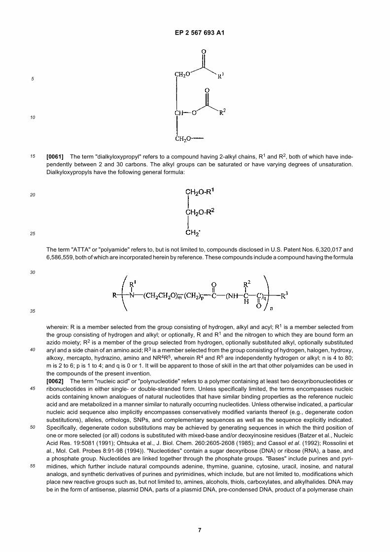
BRIEF SUMMARY OF THE INVENTION
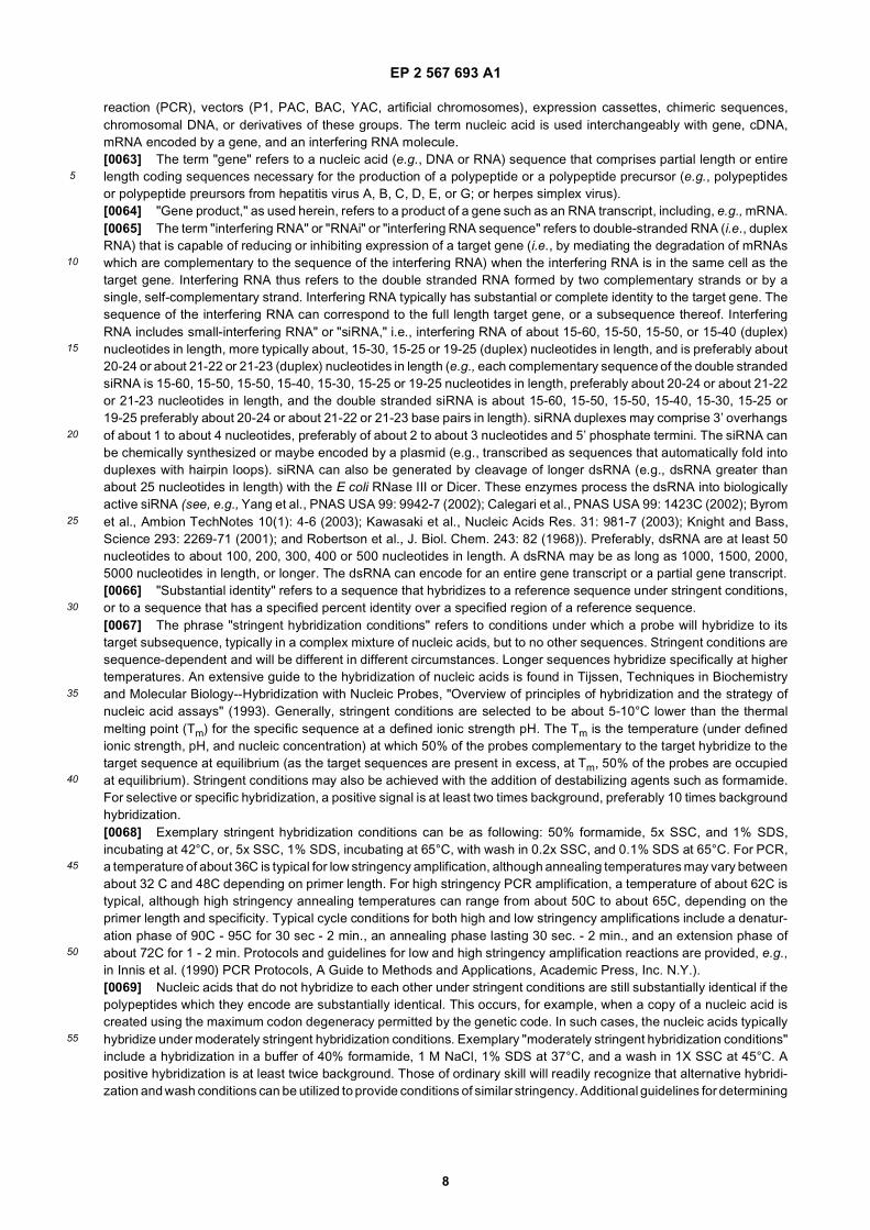
[0010] The present invention provides stable nucleic acid-lipid particles (SNALP) useful for encapsulating one or moresiRNA molecules, methods of making SNALPs comprising siRNA, SNALPs comprising siRNA and methods of deliveringand/or administering the SNALPs to a subject to silence expression of a target gene sequence.[0011] In one embodiment, the invention provide nucleic acid-lipid particles comprising: a cationic lipid, a non-cationiclipid, a conjugated lipid that inhibits aggregation of particles and a siRNA. In some embodiments, the siRNA moleculeis fully encapsulated within the lipid bilayer of the nucleic acid-lipid particle such that the nucleic acid in the nucleic acid-lipid particle is resistant in aqueous solution to degradation by a nuclease. The nucleic acid particle are substantiallynon-toxic to mammals. The siRNA molecule may comprise about 15 to about 60 nucleotides. The siRNA molecule maybe derived from a double-stranded RNA greater than about 25 nucleotides in length. In some embodiments the siRNAis transcribed from a plasmid, in particular a plasmid comprising a DNA template of a target sequence.[0012] The cationic lipid may be one or more of N,N-dioleyl-N,N-dimethylammonium chloride (DODAC), N,N-distearyl-N,N-dimethylammonium bromide (DDAB), N-(1-(2,3-dioleoyloxy)propyl)-N,N,N-trimethylammonium chloride (DOTAP),N-(1-(2,3-dioleyloxy)propyl)-N,N,N-trimethylammonium chloride (DOTMA), and N,N-dimethyl-2,3-dioleyloxy)pro-pylamine (DODMA), and a mixture thereof. The non-cationic lipid may be one or more of dioleoylphosphatidyleth-anolamine (DOPE), palmitoyloleoylphosphatidylcholine (POPC), egg phosphatidylcholine (EPC), distearoylphosphati-dylcholine (DSPC), cholesterol, and combinations thereof.[0013] The conjugated lipid that inhibits aggregation of particles may be one or more of a polyethyleneglycol (PEG)-lipidconjugate, a polyamide (ATTA)-lipid conjugate, and combinations thereof. The PEG-lipid conjugate may be one or moreof a PEG-dialkyloxypropyl (DAA), a PEG-diacylglycerol (DAG), a PEG-phospholipid, a PEG-ceramide, and combinationsthereof. The PEG-DAG conjugate may be one or more of a PEG-dilauroylglycerol (C12), a PEG-dimyristoylglycerol (C14),a PEG-dipalmitoylglycerol (C16), and a PEG-distearoylglycerol (C18), and combinations thereof. The PEG-DAA conjugatemay be one or more of a PEG-dilauryloxypropyl (C12), a PEG-dimyristyloxypropyl (C14), a PEG-dipalmityloxypropyl(C16), and a PEG-distearyloxypropyl (C18), and combinations thereof. The nucleic acid-lipid particle may further comprisea cationic polymer lipid.[0014] In some embodiments, the particles are made by providing an aqueous solution in a first reservoir and anorganic lipid solution in a second reservoir and mixing the aqueous solution with the organic lipid solution so as tosubstantially instantaneously produce a liposome encapsulating an interfering RNA. In some embodiments, the particlesare made by formation of hydrophobic intermediate complexes in either detergent-based or organic solvent-basedsystems, followed by removal of the detergent or organic solvent. Preferred embodiments are charge-neutralized.[0015] In one embodiment, the interfering RNA is transcribed from a plasmid and the plasmid is combined with cationiclipids in a detergent solution to provide a coated nucleic acid-lipid complex. The complex is then contacted with non-cationic lipids to provide a solution of detergent, a nucleic acid-lipid complex and non-cationic lipids, and the detergentis then removed to provide a solution of serum-stable nucleic acid-lipid particles, in which the plasmid comprising aninterfering RNA template is encapsulated in a lipid bilayer. The particles thus formed have a size of about 50-150 nm.[0016] In another embodiment, serum-stable nucleic acid-lipid particles are formed by preparing a mixture of cationiclipids and non-cationic lipids in an organic solvent; contacting an aqueous solution of nucleic acids comprising interferingRNA with the mixture of cationic and non-cationic lipids to provide a clear single phase; and removing the organic solventto provide a suspension of nucleic acid-lipid particles, in which the nucleic acid is encapsulated in a lipid bilayer, andthe particles are stable in serum and have a size of about 50-150 nm.[0017] The nucleic acid-lipid particles of the present invention are useful for the therapeutic delivery of nucleic acidscomprising a siRNA sequence. In particular, it is an object of this invention to provide in vitro and in vivo methods fortreatment of a disease in a mammal by downregulating or silencing the translation of a target nucleic acid sequence. Inthese methods, a siRNA molecule is formulated into a nucleic acid-lipid particle, and the particles are administered topatients requiring such treatment (e.g., a patient diagnosed with a disease or disorder associated with the expressionor overexpression of a gene comprising the target nucleic acid sequence). Alternatively, cells are removed from a patient,the siRNA is delivered in vitro, and the cells are reinjected into the patient. In one embodiment, the present inventionprovides for a method of introducing a siRNA molecule into a cell by contacting a cell with a nucleic acid-lipid particlecomprising of a cationic lipid, a non-cationic lipid, a conjugated lipid that inhibits aggregation, and a siRNA.[0018] The nucleic acid-lipid particle may be administered, e.g., intravenously, parenterally or intraperitoneally. In oneembodiment, at least about 10% of the total administered dose of the nucleic acid-lipid particles is present in plasmaabout 24, 3 6, 48, 60, 72, 84, or 96 hours after injection. In other embodiments, more than 20%, 30%, 40% and as muchas 60%, 70% or 80% of the total injected dose of the nucleic acid-lipid particles is present in plasma 24,36, 48, 60,72,84, or 96 hours after injection. In one embodiment, the presence of a siRNA in cells in a target tissue (i.e., lung, liver,tumor or at a site of inflammation) is detectable at 24, 48, 72 and 96 hours after administration. In one embodiment,downregulation of expression of the target sequence is detectable at 24, 48, 72 and 96 hours after administration. Inone embodiment, downregulation of expression of the target sequence occurs preferentially in tumor cells or in cells at
EP 2 567 693 A1
4
5
10
15
20
25
30
35
40
45
50
55
a site of inflammation. In one embodiment, the presence of a siRNA in cells at a site distal to the site of administrationis detectable at least four days after intravenous injection of the nucleic acid-lipid particle. In another embodiment, thepresence of a siRNA in of cells in t a target tissue (i.e., lung, liver, tumor or at a site of inflammation) is detectable atleast four days after injection of the nucleic acid-lipid particle.[0019] The particles are suitable for use in intravenous nucleic acid transfer as they are stable in circulation, of a sizerequired for pharmacodynamic behavior resulting in access to extravascular sites and target cell populations. The in-vention also provides for pharmaceutically acceptable compositions comprising a nucleic acid-lipid particle.[0020] Another embodiment of the present invention provides methods for in vivo delivery of siRNA. A nucleic acid-lipidparticle comprising a cationic lipid, a non-cationic lipid, a conjugated lipid that inhibits aggregation of particles, and siRNAis administered (e.g., intravenously, subcutaneously, intraperitoneally, or subdermally) to a subject (e.g., a mammalsuch as a human). In some embodiments, the invention provides methods for in vivo delivery of interfering RNA to theliver of a mammalian subject.[0021] A further embodiment of the present invention provides a method of treating a disease or disorder in a mammaliansubject. A therapeutically effective amount of a nucleic acid-lipid particle comprising a cationic lipid, a non-cationic lipid,a conjugated lipid that inhibits aggregation of particles, and siRNA is administered to the mammalian subject (e.g., arodent such as a mouse, a primate such as a human or a monkey) with the disease or disorder. In some embodiments,the disease or disorder is associated with expression and/or overexpression of a gene and expression or overexpressionof the gene is silenced by the siRNA. In some embodiments, the disease is a viral disease such as, for example, hepatitis(e.g., Hepatitis A, Hepatitis B, Hepatitis C, Hepatitis D, Hepatitis E, Hepatitis G, or a combination thereof). In someembodiment, the disease or disorder is a liver disease or disorder, such as, for example, dyslipidemia.
BRIEF DESCRIPTION OF THE DRAWINGS
[0022] Figure 1 illustrates downregulating ß-galactosidase expression in CT26.CL25 cells via in vitro delivery of en-capsulated anti-ß-galactosidase siRNA in DSPC:Cholesterol:DODMA:PEG-DMG liposomes.[0023] Figure 2 illustrates the structures of PEG-Diacylglycerols and PEG-Ceramide C20.[0024] Figure 3 illustrates that clearance studies with LUVs showed that SNALPs containing PEG-DAGs were com-parable to SNALPs containing PEG-CeramideC20.[0025] Figure 4 illustrates that SNALPs containing PEG-DAGs can be formulated via a detergent dialysis method.[0026] Figure 5 illustrates the pharmacokinetic properties of SNALPs containing PEG-DAGs.[0027] Figure 6 illustrates the biodistribution properties of SNALPs containing PEG-DAGs.[0028] Figure 7 illustrates the luciferase gene expression 24 hrs post IV administration of SPLPs containingPEG-CeramideC20 versus PEG-DAGs in Neuro-2a Tumor Bearing Male A/J Mice.[0029] Figure 8 illustrates the luciferase gene expression 48 hrs post IV administration of SPLPs containingPEG-CeramideC20 versus PEG-DAGs in Neuro-2a Tumor Bearing Male A/J Mice.[0030] Figure 9 illustrates the luciferase gene expression 72 hrs post IV administration of SPLPs containingPEG-CeramideC20 versus PEG-DAGs in Neuro-2a Tumor Bearing Male A/J Mice.[0031] Figure 10 illustrates the structures of three exemplary PEG-dialkyloxypropyl derivatives suitable for use in thepresent invention, i.e., N-(2,3-dimyristyloxypropyl) carbamate PEG2000 methyl ether (i.e., PEG-C-DMA), N-(2,3-dimyri-styloxypropyl) amide PEG2000 methyl ether (i.e., PEG-A-DMA), and N-(2,3-dimyristyloxypropyl) succinamide PEG2000methyl ether (i.e., PEG-S-DMA).[0032] Figure 11 illustrates data showing luciferase gene expression in tumors 48 hours after intravenous administrationof SPLP comprising PEG-DAA conjugates and PEG-DAG conjugates.[0033] Figure 12 illustrates data showing luciferase gene expression in liver, lung, spleen, heart, and tumor followingintravenous administration of SPLP comprising PEG-DAA conjugates and PEG-DAG conjugates.[0034] Figure 13 illustrates data from clearance studies in Neuro-2a tumor bearing male A/J mice after administrationof SPLPs comprising a PEG-DAA conjugate and containing a plasmid encoding luciferase under the control of the CMVpromoter and SNALPs comprising a PEG-DAA conjugate and containing anti-luciferase siRNA.[0035] Figure 14 illustrates data from studies of the pharmacokinetic properties of SPLPs comprising a PEG-DAAconjugate and containing a plasmid encoding luciferase under the control of the CMV promoter and SNALPs comprisinga PEG-DAA conjugate and containing anti-luciferase siRNA in Neuro-2a tumor bearing male A/J mice.[0036] Figure 15 illustrates data from clearance studies in Neuro-2a tumor bearing male A/J mice after administrationof SPLPs comprising a PEG-DAA conjugate or a PEG-DAG conjugate and containing a plasmid encoding luciferaseunder the control of the CMV promoter, pSPLPs comprising a PEG-DAG conjugate and containing a plasmid encodingluciferase under the control of the CMV promoter and SNALPs comprising a PEG-DAA conjugate and containing anti-luciferase siRNA.[0037] Figure 16 illustrates data from studies of the pharmacokinetic properties of SPLPs comprising a PEG-DAAconjugate or a PEG-DAG conjugate and containing a plasmid encoding luciferase under the control of the CMV promoter,
EP 2 567 693 A1
5
5
10
15
20
25
30
35
40
45
50
55
pSPLPs comprising a PEG-DAG conjugate and containing a plasmid encoding luciferase under the control of the CMVpromoter and SNALPs comprising a PEG-DAA conjugate and containing anti-luciferase siRNA in Neuro-2a tumor bearingmale A/J mice.[0038] Figure 17 illustrates in vitro data demonstrating silencing of luciferase expression in luciferase expressing cellstreated with SPLPs comprising a PEG-lipid conjugate and containing a plasmid encoding luciferase under the controlof the CMV promoter and SNALPs comprising a PEG-lipid conjugate conjugate and containing anti-luciferase siRNA.[0039] Figure 18 illustrates in vivo data demonstrating silencing of luciferase expression in Neuro-2a tumor bearingmale A/J mice treated with SPLPs comprising a PEG-DAA conjugate and containing a plasmid encoding luciferaseunder the control of the CMV promoter and SNALPs comprising a PEG-DAA conjugate and containing anti-luciferasesiRNA.[0040] Figure 19 illustrates in vivo data demonstrating silencing of luciferase expression in Neuro-2a tumor bearingmale A/J mice treated with SPLPs comprising a PEG-DAA conjugate and containing a plasmid encoding luciferaseunder the control of the CMV promoter and SNALPs comprising a PEG-DAA conjugate and containing anti-luciferasesiRNA.[0041] Figure 20 illustrates in vivo data demonstrating silencing ofluciferase expression in Neuro-2a tumor bearingmale A/J mice treated with SPLPs comprising a PEG-DAA conjugate and containing a plasmid encoding luciferaseunder the control of the CMV promoter and SNALPs comprising a PEG-DAA conjugate and containing anti-luciferasesiRNA.[0042] Figure 21 illustrates in vivo data demonstrating silencing af luciferase expression in Neuro-2a tumor bearingmale A/J mice treated with SPLPs comprising a PEG-DAA conjugate and containing a plasmid encoding luciferaseunder the control of the CMV promoter and SNALPs comprising a PEG-DAA conjugate and containing anti-luciferasesiRNA.[0043] Figure 22 illustrates in vivo data demonstrating silencing of luciferase expression in Neuro-2a tumor bearingmale A/J mice treated with SPLPs comprising a PEG-DAA conjugate and containing a plasmid encoding luciferaseunder the control of the CMV promoter and SNALPs comprising a PEG-DAA conjugate and containing anti-luciferasesiRNA.
DETAILED DESCRIPTION OF THE INVENTION
I. Introduction
[0044] The present invention provides stable nucleic acid-lipid particles (SNALP) useful for encapsulating one or moresiRNA molecules, methods of making SNALPs comprising siRNA, SNALPs comprising siRNA and methods of deliveringand/or administering the SNALPs to a subject to silence expression of a target gene sequence.[0045] The present invention is based on the unexpected success of encapsulating short interfering RNA (siRNA)molecules in SNALPs. Using the methods of the present invention, siRNA molecules are encapsulated in SNALPs withan efficiency greater than 70%, more usually with an efficiency greater than 80 to 90%. The SNALPs described hereincan conveniently be used in vitro and in vivo to efficiently deliver administer siRNA molecules locally or systemically tocells expressing a target gene. Once delivered, the siRNA molecules in the SNALPs silence expression of the target gene.[0046] The SNALPs described herein are typically < 150 nm diameter and remain intact in the circulation for anextended period of time in order to achieve delivery of siRNA to target tissues. The SNALPs are highly stable, serum-re-sistant nucleic acid-containing particles that does not interact with cells and other components of the vascular compart-ment. Moreover, the SNALPs also readily interact with target cells at a disease site in order to facilitate intracellulardelivery of a desired nucleic acid (e.g., a siRNA or a plasmid encoding a siRNA).
II. Definitions
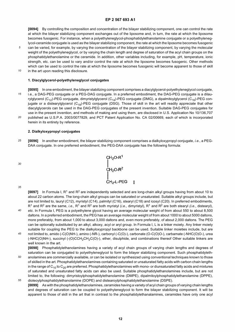
[0047] The term "lipid" refers to a group of organic compounds that include, but are not limited to, esters of fatty acidsand are characterized by being insoluble in water, but soluble in many organic solvents. They are usually divided intoat least three classes: (1) "simple lipids which include fats and oils as well as waxes; (2) "compound lipids" which includephospholipids and glycolipids; (3) "derived lipids" such as steroids.[0048] "Lipid vesicle" refers to any lipid composition that can be used to deliver a compound including, but not limitedto, liposomes, wherein an aqueous volume is encapsulated by an amphipathic lipid bilayer; or wherein the lipids coatan interior comprising a large molecular component, such as a plasmid comprising an interfering RNA sequence, witha reduced aqueous interior; or lipid aggregates or micelles, wherein the encapsulated component is contained within arelatively disordered lipid mixture.[0049] As used herein, "lipid encapsulated" can refer to a lipid formulation that provides a compound with full encap-sulation, partial encapsulation, or both. In a preferred embodiment, the nucleic acid is fully encapsulated in the lipid
EP 2 567 693 A1
6
5
10
15
20
25
30
35
40
45
50
55
formulation.[0050] As used herein, the term "SNALP" refers to a stable nucleic acid lipid particle. A SNALP represents a vesicleof lipids coating a reduced aqueous interior comprising a nucleic acid such as an interfering RNA sequence or a plasmidfrom which an interfering RNA is transcribed.[0051] The term "vesicle-forming lipid" is intended to include any amphipathic lipid having a hydrophobic moiety anda polar head group, and which by itself can form spontaneously into bilayer vesicles in water, as exemplified by mostphospholipids.[0052] The term "vesicle-adopting lipid" is intended to include any amphipathic lipid that is stably incorporated intolipid bilayers in combination with other amphipathic lipids, with its hydrophobic moiety in contact with the interior, hydro-phobic region of the bilayer membrane, and its polar head group moiety oriented toward the exterior, polar surface ofthe membrane. Vesicle-adopting lipids include lipids that on their own tend to adopt a nonlamellar phase, yet which arecapable of assuming a bilayer structure in the presence of a bilayer-stabilizing component. A typical example is DOPE(dioleoylphosphatidylethanolamine). Bilayer stabilizing components include, but are not limited to, conjugated lipids thatinhibit aggregation of the SNALPs, polyamide oligomers (e.g., ATTA-lipid derivatives), peptides, proteins, detergents,lipid-derivatives, PEG-lipid derivatives such as PEG coupled to dialkyloxypropyls, PEG coupled to diacylglycerols, PEGcoupled to phosphatidylethanolamines, and PEG conjugated to ceramides (see, U.S. Pat. No. 5,885,613, which isincorporated herein by reference).[0053] The term "amphipathic lipid" refers, in part, to any suitable material wherein the hydrophobic portion of the lipidmaterial orients into a hydrophobic phase, while the hydrophilic portion orients toward the aqueous phase. Amphipathiclipids are usually the major component of a lipid vesicle. Hydrophilic characteristics derive from the presence of polaror charged groups such as carbohydrates, phosphate, carboxylic, sulfato, amino, sulfhydryl, nitro, hydroxy and otherlike groups. Hydrophobicity can be conferred by the inclusion af apolar groups that include, but are not limited to, longchain saturated and unsaturated aliphatic hydrocarbon groups and such groups substituted by one or more aromatic,cycloaliphatic or heterocyclic group(s). Examples of amphipathic compounds include, but are not limited to, phospholipids,aminolipids and sphingolipids. Representative examples of phospholipids include, but are not limited to, phosphatidyl-choline, phosphatidylethanolamine, phosphatidylserine, phosphatidylinositol, phosphatidic acid, palmitoyloleoyl phos-phatidylcholine, lysophosphatidylcholine, lysophosphatidylethanolamine, dipalmitoylphosphatidylcholine, dioleoylphos-phatidylcholine, distearoylphosphatidylcholine or dilinoleoylphosphatidylcholine. Other compounds lacking in phospho-rus, such as sphingolipid, glycosphingolipid families, diacylglycerols and beta.-acyloxyacids, are also within the groupdesignated as amphipathic lipids. Additionally, the amphipathic lipid described above can be mixed with other lipidsincluding triglycerides and sterols.[0054] The term "neutral lipid" refers to any of a number of lipid species that exist either in an uncharged or neutralzwitterionic form at a selected pH. At physiological pH, such lipids include, for example, diacylphosphatidylcholine,diacylphosphatidylethanolamine, ceramide, sphingomyelin, cephalin, cholesterol, cerebrosides and diacylglycerols.[0055] The term "noncationic lipid" refers to any neutral lipid as described above as well as anionic lipids.[0056] The term "anionic lipid" refers to any lipid that is negatively charged at physiological pH. These lipids include,but are not limited to, phosphatidylglycerol, cardiolipin, diacylphosphatidylserine, diacylphosphatidic acid, N-dodecanoylphosphatidylethanolamines, N-succinyl phosphatidylethanolamines, N-glutarylphosphatidylethanolamines, lysylphos-phatidylglycerols, palmitoyloleyolphosphatidylglycerol (POPG), and other anionic modifying groups joined to neutrallipids.[0057] The term "cationic lipid" refers to any of a number of lipid species that carry a net positive charge at a selectedpH, such as physiological pH. Such lipids include, but are not limited to, N,N-dioleyl-N,N-dimethylammonium chloride("DODAC"); N-(2,3-dioleyloxy)propyl)-N,N,N-trimethylammonium chloride ("DOTMA"); N,N-distearyl-N,N-dimethylam-monium bromide ("DDAB"); N-(2,3-dioleoyloxy)propyl)-N,N,N-trimethylammonium chloride ("DOTAP"); 3 -(N-(N’,N’-dimethylaminoethane)-carbamoyl)cholesterol ("DC-Chol") and N-(1,2-dimyristyloxyprop-3-yl)-N,N-dimethyl-N-hydrox-yethyl ammonium bromide ("DMRIE"). The following lipids are cationic and have a positive charge at below physiologicalpH: DODAP, DODMA, DMDMA and the like.[0058] The term "hydrophobic lipid" refers to compounds having apolar groups that include, but are not limited to, longchain saturated and unsaturated aliphatic hydrocarbon groups and such groups optionally substituted by one or morearomatic, cycloaliphatic or heterocyclic group(s). Suitable examples include, but are not limited to, diacylglycerol, di-alkylglycerol, N-N-dialkylamino, 1,2-diacyloxy-3-aminopropane and 1,2-dialkyl-3-aminopropane.[0059] The term "fusogenic" refers to the ability of a liposome, an SNALP or other drug delivery system to fuse withmembranes of a cell. The membranes can be either the plasma membrane or membranes surrounding organelles, e.g.,endosome, nucleus, etc.[0060] The term "diacylglycerol" refers to a compound having 2-fatty acyl chains, R1 and R2, both of which haveindependently between 2 and 30 carbons bonded to the 1- and 2-position of glycerol by ester linkages. The acyl groupscan be saturated or have varying degrees ofunsaturation. Diacylglycerols have the following general formula:
EP 2 567 693 A1
7
5
10
15
20
25
30
35
40
45
50
55
[0061] The term "dialkyloxypropyl" refers to a compound having 2-alkyl chains, R1 and R2, both of which have inde-pendently between 2 and 30 carbons. The alkyl groups can be saturated or have varying degrees of unsaturation.Dialkyloxypropyls have the following general formula:
The term "ATTA" or "polyamide" refers to, but is not limited to, compounds disclosed in U.S. Patent Nos. 6,320,017 and6,586,559, both of which are incorporated herein by reference. These compounds include a compound having the formula
wherein: R is a member selected from the group consisting of hydrogen, alkyl and acyl; R1 is a member selected fromthe group consisting of hydrogen and alkyl; or optionally, R and R1 and the nitrogen to which they are bound form anazido moiety; R2 is a member of the group selected from hydrogen, optionally substituted alkyl, optionally substitutedaryl and a side chain of an amino acid; R3 is a member selected from the group consisting of hydrogen, halogen, hydroxy,alkoxy, mercapto, hydrazino, amino and NR4R5, wherein R4 and R5 are independently hydrogen or alkyl; n is 4 to 80;m is 2 to 6; p is 1 to 4; and q is 0 or 1. It will be apparent to those of skill in the art that other polyamides can be used inthe compounds of the present invention.[0062] The term "nucleic acid" or "polynucleotide" refers to a polymer containing at least two deoxyribonucleotides orribonucleotides in either single- or double-stranded form. Unless specifically limited, the terms encompasses nucleicacids containing known analogues of natural nucleotides that have similar binding properties as the reference nucleicacid and are metabolized in a manner similar to naturally occurring nucleotides. Unless otherwise indicated, a particularnucleic acid sequence also implicitly encompasses conservatively modified variants thereof (e.g., degenerate codonsubstitutions), alleles, orthologs, SNPs, and complementary sequences as well as the sequence explicitly indicated.Specifically, degenerate codon substitutions may be achieved by generating sequences in which the third position ofone or more selected (or all) codons is substituted with mixed-base and/or deoxyinosine residues (Batzer et al., NucleicAcid Res. 19:5081 (1991); Ohtsuka et al., J. Biol. Chem. 260:2605-2608 (1985); and Cassol et al. (1992); Rossolini etal., Mol. Cell. Probes 8:91-98 (1994)). "Nucleotides" contain a sugar deoxyribose (DNA) or ribose (RNA), a base, anda phosphate group. Nucleotides are linked together through the phosphate groups. "Bases" include purines and pyri-midines, which further include natural compounds adenine, thymine, guanine, cytosine, uracil, inosine, and naturalanalogs, and synthetic derivatives of purines and pyrimidines, which include, but are not limited to, modifications whichplace new reactive groups such as, but not limited to, amines, alcohols, thiols, carboxylates, and alkylhalides. DNA maybe in the form of antisense, plasmid DNA, parts of a plasmid DNA, pre-condensed DNA, product of a polymerase chain
EP 2 567 693 A1
8
5
10
15
20
25
30
35
40
45
50
55
reaction (PCR), vectors (P1, PAC, BAC, YAC, artificial chromosomes), expression cassettes, chimeric sequences,chromosomal DNA, or derivatives of these groups. The term nucleic acid is used interchangeably with gene, cDNA,mRNA encoded by a gene, and an interfering RNA molecule.[0063] The term "gene" refers to a nucleic acid (e.g., DNA or RNA) sequence that comprises partial length or entirelength coding sequences necessary for the production of a polypeptide or a polypeptide precursor (e.g., polypeptidesor polypeptide preursors from hepatitis virus A, B, C, D, E, or G; or herpes simplex virus).[0064] "Gene product," as used herein, refers to a product of a gene such as an RNA transcript, including, e.g., mRNA.[0065] The term "interfering RNA" or "RNAi" or "interfering RNA sequence" refers to double-stranded RNA (i.e., duplexRNA) that is capable of reducing or inhibiting expression of a target gene (i.e., by mediating the degradation of mRNAswhich are complementary to the sequence of the interfering RNA) when the interfering RNA is in the same cell as thetarget gene. Interfering RNA thus refers to the double stranded RNA formed by two complementary strands or by asingle, self-complementary strand. Interfering RNA typically has substantial or complete identity to the target gene. Thesequence of the interfering RNA can correspond to the full length target gene, or a subsequence thereof. InterferingRNA includes small-interfering RNA" or "siRNA," i.e., interfering RNA of about 15-60, 15-50, 15-50, or 15-40 (duplex)nucleotides in length, more typically about, 15-30, 15-25 or 19-25 (duplex) nucleotides in length, and is preferably about20-24 or about 21-22 or 21-23 (duplex) nucleotides in length (e.g., each complementary sequence of the double strandedsiRNA is 15-60, 15-50, 15-50, 15-40, 15-30, 15-25 or 19-25 nucleotides in length, preferably about 20-24 or about 21-22or 21-23 nucleotides in length, and the double stranded siRNA is about 15-60, 15-50, 15-50, 15-40, 15-30, 15-25 or19-25 preferably about 20-24 or about 21-22 or 21-23 base pairs in length). siRNA duplexes may comprise 3’ overhangsof about 1 to about 4 nucleotides, preferably of about 2 to about 3 nucleotides and 5’ phosphate termini. The siRNA canbe chemically synthesized or maybe encoded by a plasmid (e.g., transcribed as sequences that automatically fold intoduplexes with hairpin loops). siRNA can also be generated by cleavage of longer dsRNA (e.g., dsRNA greater thanabout 25 nucleotides in length) with the E coli RNase III or Dicer. These enzymes process the dsRNA into biologicallyactive siRNA (see, e.g., Yang et al., PNAS USA 99: 9942-7 (2002); Calegari et al., PNAS USA 99: 1423C (2002); Byromet al., Ambion TechNotes 10(1): 4-6 (2003); Kawasaki et al., Nucleic Acids Res. 31: 981-7 (2003); Knight and Bass,Science 293: 2269-71 (2001); and Robertson et al., J. Biol. Chem. 243: 82 (1968)). Preferably, dsRNA are at least 50nucleotides to about 100, 200, 300, 400 or 500 nucleotides in length. A dsRNA may be as long as 1000, 1500, 2000,5000 nucleotides in length, or longer. The dsRNA can encode for an entire gene transcript or a partial gene transcript.[0066] "Substantial identity" refers to a sequence that hybridizes to a reference sequence under stringent conditions,or to a sequence that has a specified percent identity over a specified region of a reference sequence.[0067] The phrase "stringent hybridization conditions" refers to conditions under which a probe will hybridize to itstarget subsequence, typically in a complex mixture of nucleic acids, but to no other sequences. Stringent conditions aresequence-dependent and will be different in different circumstances. Longer sequences hybridize specifically at highertemperatures. An extensive guide to the hybridization of nucleic acids is found in Tijssen, Techniques in Biochemistryand Molecular Biology--Hybridization with Nucleic Probes, "Overview of principles of hybridization and the strategy ofnucleic acid assays" (1993). Generally, stringent conditions are selected to be about 5-10°C lower than the thermalmelting point (Tm) for the specific sequence at a defined ionic strength pH. The Tm is the temperature (under definedionic strength, pH, and nucleic concentration) at which 50% of the probes complementary to the target hybridize to thetarget sequence at equilibrium (as the target sequences are present in excess, at Tm, 50% of the probes are occupiedat equilibrium). Stringent conditions may also be achieved with the addition of destabilizing agents such as formamide.For selective or specific hybridization, a positive signal is at least two times background, preferably 10 times backgroundhybridization.[0068] Exemplary stringent hybridization conditions can be as following: 50% formamide, 5x SSC, and 1% SDS,incubating at 42°C, or, 5x SSC, 1% SDS, incubating at 65°C, with wash in 0.2x SSC, and 0.1% SDS at 65°C. For PCR,a temperature of about 36C is typical for low stringency amplification, although annealing temperatures may vary betweenabout 32 C and 48C depending on primer length. For high stringency PCR amplification, a temperature of about 62C istypical, although high stringency annealing temperatures can range from about 50C to about 65C, depending on theprimer length and specificity. Typical cycle conditions for both high and low stringency amplifications include a denatur-ation phase of 90C - 95C for 30 sec - 2 min., an annealing phase lasting 30 sec. - 2 min., and an extension phase ofabout 72C for 1 - 2 min. Protocols and guidelines for low and high stringency amplification reactions are provided, e.g.,in Innis et al. (1990) PCR Protocols, A Guide to Methods and Applications, Academic Press, Inc. N.Y.).[0069] Nucleic acids that do not hybridize to each other under stringent conditions are still substantially identical if thepolypeptides which they encode are substantially identical. This occurs, for example, when a copy of a nucleic acid iscreated using the maximum codon degeneracy permitted by the genetic code. In such cases, the nucleic acids typicallyhybridize under moderately stringent hybridization conditions. Exemplary "moderately stringent hybridization conditions"include a hybridization in a buffer of 40% formamide, 1 M NaCl, 1% SDS at 37°C, and a wash in 1X SSC at 45°C. Apositive hybridization is at least twice background. Those of ordinary skill will readily recognize that alternative hybridi-zation and wash conditions can be utilized to provide conditions of similar stringency. Additional guidelines for determining
EP 2 567 693 A1
9
5
10
15
20
25
30
35
40
45
50
55
hybridization parameters are provided in numerous reference, e.g., and Current Protocols in Molecular Biology, ed.Ausubel, et al.[0070] The terms "substantially identical" or "substantial identity," in the context of two or more nucleic acids, refer totwo or more sequences or subsequences that are the same or have a specified percentage of nucleotides that are thesame (i.e., at least about 60%, preferably 65%, 70%, 75%, preferably 80%, 85%, 90%, or 95% identity over a specifiedregion), when compared and aligned for maximum correspondence over a comparison window, or designated regionas measured using one of the following sequence comparison algorithms or by manual alignment and visual inspection.This definition, when the context indicates, also refers analogously to the complement of a sequence. Preferably, thesubstantial identity exists over a region that is at least about 5, 10,15, 20, 25, 30, 35, 40, 45, 50, 75, or 100 nucleotidesin length.[0071] For sequence comparison, typically one sequence acts as a reference sequence, to which test sequences arecompared. When using a sequence comparison algorithm, test and reference sequences are entered into a computer,subsequence coordinates are designated, if necessary, and sequence algorithm program parameters are designated.Default program parameters can be used, or alternative parameters can be designated. The sequence comparisonalgorithm then calculates the percent sequence identities for the test sequences relative to the reference sequence,based on the program parameters.[0072] A "comparison window", as used herein, includes reference to a segment of any one of the number of contiguouspositions selected from the group consisting of from 20 to 600, usually about 50 to about 200, more usually about 100to about 150 in which a sequence may be compared to a reference sequence of the same number of contiguous positionsafter the two sequences are optimally aligned. Methods of alignment of sequences for comparison are well-known inthe art. Optimal alignment of sequences for comparison can be conducted, e.g., by the local homology algorithm ofSmith & Waterman, Adv. Appl. Math. 2:482 (1981), by the homology alignment algorithm of Needleman & Wunsch, J.Mol. Biol. 48:443 (1970), by the search for similarity method of Pearson & Lipman, Proc. Nat’l. Acad. Sci. USA 85:2444(1988), by computerized implementations of these algorithms (GAP, BESTFIT, FASTA, and TFASTA in the WisconsinGenetics Software Package, Genetics Computer Group, 575 Science Dr., Madison, WI), or by manual alignment andvisual inspection (see, e.g., Current Protocols in Molecular Biology (Ausubel et al., eds. 1995 supplement)).[0073] A preferred example of algorithm that is suitable for determining percent sequence identity and sequencesimilarity are the BLAST and BLAST 2.0 algorithms, which are described in Altschul et al., Nuc. Acids Res. 25:3389-3402(1977) and Altschul et al., J. Mol. Biol. 215:403-410 (1990), respectively. BLAST and BLAST 2.0 are used, with theparameters described herein, to determine percent sequence identity for the nucleic acids and proteins of the invention.Software for performing BLAST analyses is publicly available through the National Center for Biotechnology Information(http://www.ncbi.nlm.nih.gov/). This algorithm involves first identifying high scoring sequence pairs (HSPs) by identifyingshort words of length W in the query sequence, which either match or satisfy some positive-valued threshold score Twhen aligned with a word of the same length in a database sequence. T is referred to as the neighborhood word scorethreshold (Altschul et al., supra). These initial neighborhood word hits act as seeds for initiating searches to find longerHSPs containing them. The word hits are extended in both directions along each sequence for as far as the cumulativealignment score can be increased. Cumulative scores are calculated using, for nucleotide sequences, the parametersM (reward score for a pair of matching residues; always > 0) and N (penalty score for mismatching residues; always <0). For amino acid sequences, a scoring matrix is used to calculate the cumulative score. Extension of the word hits ineach direction are halted when: the cumulative alignment score falls off by the quantity X from its maximum achievedvalue; the cumulative score goes to zero or below, due to the accumulation of one or more negative-scoring residuealignments; or the end of either sequence is reached. The BLAST algorithm parameters W, T, and X determine thesensitivity and speed of the alignment. The BLASTN program (for nucleotide sequences) uses as defaults a wordlength(W) of 11, an expectation (E) or 10, M=5, N=-4 and a comparison of both strands. For amino acid sequences, the BLASTPprogram uses as defaults a wordlength of 3, and expectation (E) of 10, and the BLOSUM62 scoring matrix (see Henikoff& Henikoff, Proc. Natl. Acad. Sci. USA 89:10915 (1989)) alignments (B) of 50, expectation (E) of 10, M=5, N=-4, and acomparison of both strands.[0074] The BLAST algorithm also performs a statistical analysis of the similarity between two sequences (see, e.g.,Karlin & Altschul, Proc. Nat’l. Acad. Sci. USA 90:5873-5787 (1993)). One measure of similarity provided by the BLASTalgorithm is the smallest sum probability (P(N)), which provides an indication of the probability by which a match betweentwo nucleotide or amino acid sequences would occur by chance. For example, a nucleic acid is considered similar to areference sequence if the smallest sum probability in a comparison of the test nucleic acid to the reference nucleic acidis less than about 0.2, more preferably less than about 0.01, and most preferably less than about 0.001.[0075] The phrase "inhibiting expression of a target gene" refers to the ability of a siRNA of the invention to initiategene silencing of the target gene. To examine the extent of gene silencing, samples or assays of the organism of interestor cells in culture expressing a particular construct are compared to control samples lacking expression of the construct.Control samples (lacking construct expression) are assigned a relative value of 100%. Inhibition of expression of a targetgene is achieved when the test value relative to the control is about 90%, preferably 50%, more preferably 25-0%.
EP 2 567 693 A1
10
5
10
15
20
25
30
35
40
45
50
55
Suitable assays include, e.g., examination of protein or mRNA levels using techniques known to those of skill in the artsuch as dot blots, northern blots, in situ hybridization, ELISA, immunoprecipitation, enzyme function, as well as phenotypicassays known to those of skill in the art.[0076] "Nucleic acid" refers to deoxyribonucleotides or ribonucleotides and polymers thereof in single- or double-stranded form. The term encompasses nucleic acids containing known nucleotide analogs or modified backbone residuesor linkages, which are synthetic, naturally occurring, and non-naturally occurring, which have similar binding propertiesas the reference nucleic acid, and which are metabolized in a manner similar to the reference nucleotides. Examples ofsuch analogs include, without limitation, phosphorothioates, phosphoramidates, methyl phosphonates, chiral-methylphosphonates, 2-O-methyl ribonucleotides, peptide-nucleic acids (PNAs).[0077] By "silencing" or "downregulation" of a gene or nucleic acid is intended to mean a detectable decrease oftranslation of a target nucleic acid sequence, i. e., the sequence targeted by the RNAi, or a decrease in the amount oractivity of the target sequence or protein, in comparison to the level that is detected in the absence of the interferingRNA or other nucleic acid sequence. A detectable decrease can be as small as about 5% or 10%, or as great as about80%, 90% or 100%. More typically, a detectable decrease is about 20%, 30%, 40%, 50%, 60%, or 70%.[0078] A "therapeutically effective amount" or an "effective amount" of a siRNA is an amount sufficient to produce thedesired effect, e.g., a decrease in the expression of a target sequence in comparison to the normal expression leveldetected in the absence of the siRNA.[0079] As used herein, the term "aqueous solution" refers to a composition comprising in whole, or in part, water.[0080] As used herein, the term "organic lipid solution" refers to a composition comprising in whole, or in part, anorganic solvent having a lipid.[0081] "Distal site," as used herein, refers to a physically separated site, which is not limited to an adjacent capillarybed, but includes sites broadly distributed throughout an organism. In some embodiments, distal site refers to a sitephysically separated from a disease site (e.g., the site of a tumor, the site of inflammation, or the site of an infection).[0082] "Serum-stable" in relation to nucleic acid-lipid particles means that the particle is not significantly degradedafter exposure to a serum or nuclease assay that would significantly degrade free DNA. Suitable assays include, forexample, a standard serum assay or a DNAse assay such as those described in the Examples below.[0083] "Systemic delivery," as used herein, refers to delivery that leads to a broad biodistribution of a compound withinan organism. Some techniques of administration can lead to the systemic delivery of certain compounds, but not others.Systemic delivery means that a useful, preferably therapeutic, amount of a compound is exposed to most parts of thebody. To obtain broad biodistribution generally requires a blood lifetime such that the compound is not rapidly degradedor cleared (such as by first pass organs (liver, lung, etc.) or by rapid, nonspecific cell binding) before reaching a diseasesite distal to the site of administration. Systemic delivery of nucleic acid-lipid particules can be by any means known inthe art including, for example, intravenous, subcutaneous, intraperitoneal, In a preferred embodiment, systemic deliveryof nucleic acid-lipid particles is by intravenous delivery.[0084] "Local delivery" as used herein refers to delivery of a compound directly to a target site within an organism.For example, a compound can be locally delivered by direct injection into a disease site such as a tumor or other targetsite such as a site of inflammation or a target organ such as the liver, heart, pancreas, kidney, and the like,
III. Stable Nucleic Acid-Lipid Particles
[0085] The stable nucleic acid-lipid particles (SNALPs) described herein typically comprise a nucleic acid (e.g., asiRNA sequence or a DNA sequence encoding a siRNA sequence), a cationic lipid, a noncationic lipid and a bilayerstabilizing component such as, e.g., a conjugated lipid that inhibits aggregation of the SNALPs. The SNALPs of thepresent invention have a mean diameter of less than about 150 nm and are substantially nontoxic. In addition, nucleicacids encapsulated in the SNALPs of the present invention are resistant in aqueous solution to degradation with anuclease.
A. Cationic Lipids
[0086] Various suitable cationic lipids may be used in the SNALPs described herein, either alone or in combinationwith one or more other cationic lipid species or neutral lipid species.[0087] Cationic lipids which are useful in the present invention can be any of a number of lipid species which carry anet positive charge at physiological pH, for example: DODAC, DOTMA, DDAB, DOTAP, DOSPA, DOGS, DC.-Chol andDMRIE, or combinations thereof. A number of these lipids and related analogs, which are also useful in the presentinvention, have been described in U.S. Patent Nos. 5,208,036, 5,264,618, 5,279,833, 5,283,185, 5,753,613 and5,785,992, the disclosures of each of which are incorporated herein by reference.[0088] The cationic lipid typically comprises from about 2% to about 60% of the total lipid present in said particle,preferably from about 5% to about 45% of the total lipid present in said particle. In certain preferred embodiments, the
EP 2 567 693 A1
11
5
10
15
20
25
30
35
40
45
50
55
cationic lipid comprises from about 5% to about 15% of the total lipid present in said particle. In other preferred embod-iments, the cationic lipid comprises from about 40% to about 50% of the total lipid present in said particle. Dependingon the intended use of the nucleic acid-lipid particles, the proportions of the components are varied and the deliveryefficiency of a particular formulation can be measured using an endosomal release parameter (ERP) assay. For example,for systemic delivery, the cationic lipid may comprise from about 5% to about 15% of the total lipid present in said particleand for local or regional delivery, the cationic lipid comprises from about 40% to about 50% of the total lipid present insaid particle.
B. Noncationic Lipids
[0089] The noncationic lipid component of the SNALPs described herein can be any of a variety of neutral uncharged,zwitterionic or anionic lipids capable of producing a stable complex. They are preferably neutral, although they canalternatively be positively or negatively charged. Examples of noncationic lipids useful in the present invention include:phospholipid-related materials, such as lecithin, phosphatidylethanolamine, lysolecithin, lysophosphatidylethanolamine,phosphatidylserine, phosphatidylinositol, sphingomyelin, cephalin, cardiolipin, phosphatidic acid, cerebrosides, dicetyl-phosphate, distearoylphosphatidylcholine (DSPC), dioleoylphosphatidylcholine (DOPC), dipalmitoylphosphatidylcholine(DPPC), dioleoylphosphatidylglycerol (DOPG), palmitoyloleyolphosphatidylglycerol (POPG), dipalmitoylphosphatidylg-lycerol (DPPG), dioleoyl-phosphatidylethanolamine (DOPE), palmitoyloleoylphosphatidylcholine (POPC), palmitoylole-oyl- phosphatidylethanolamine (POPE) and dialeayl-phosphatidylethanolamine 4-(N-maleimidomethyl)-cyclobexane-1-carboxylate (DOPE-mal). Noncationic lipids or sterols such as cholesterol may be present. Additional nonphosphorouscontaining lipids are, e.g., stearylamine, dodecylamine, hexadecylamine, acetyl palmitate, glycerotricinoleate, hexadecylstereate, isopropyl myristate, amphoteric acrylic polymers, triethanolamine-lauryl sulfate, alkyl-aryl sulfate polyethyloxy-lated fatty acid amides, dioctadecyldimethyl ammonium bromide and the like, diacylphosphatidylcholine, diacylphos-phatidylethanolamine, ceramide, sphingomyelin, cephalin, and cerebrosides. Other lipids such as lysophosphatidylcho-line and lysophosphatidylethanolamine may be present. Noncationic lipids also include polyethylene glycol-based pol-ymers such as PEG 2000, PEG 5000 and polyethylene glycol conjugated to phospholipids or to ceramides (referred toas PEG-Cer), as described in U.S. Patent No. 5,820,873, incorporated herein by reference.[0090] In preferred embodiments, the noncationic lipids are diacylphosphatidylcholine (e.g., distearoylphosphatidyl-choline, dioleoylphasphatidylcholine, dipalmitoylphosphatidylcholine and dilinoleoylphosphatidylcholine), diacylphos-phatidylethanolamine (e.g., dioleoylphosphatidylethanolamine and palmitoyloleoylphosphatidylethanolamine), ceramideor sphingomyelin. The acyl groups in these lipids are preferably acyl groups derived from fatty acids having C10-C24carbon chains. More preferably the acyl groups are lauroyl, myristoyl, palmitoyl, stearoyl or oleoyl. In particularly preferredembodiments, the noncationic lipid will include one or more of cholesterol, 1,2-sn-dioleoylphosphatidylethanolamine, oregg sphingomyelin (ESM).[0091] The non-cationic lipid typically comprises from about 5% to about 90% of the total lipid present in said particle,preferably from about 20% to about 85% of the total lipid present in said particle. The PEG-DAG conjugate typicallycomprises from 1% to about 20% of the total lipid present in said particle, preferably from 4% to about 15% of the totallipid present in said particle. The nucleic acid-lipid particles of the present invention may further comprise cholesterol.If present, the cholesterol typically comprises from about 14% to about 60% of the total lipid present in said particle,preferably the cholesterol comprises from about 20% to about 45% of the total lipid present in said particle.
C. Bilayer Stabilizing Component
[0092] In one embodiment, the SNALP further comprises a bilayer stabilizing component (BSC). Suitable BSCs include,but are not limited to, polyamide oligomers, peptides, proteins, detergents, lipid-derivatives, PEG-lipids, such as PEGcoupled to dialkyloxypropyls (PEG-DAA), PEG coupled to diacylglycerol (PEG-DAG), PEG coupled to phosphatidyleth-anolamine (PE) (PEG-PE), or PEG conjugated to ceramides, or a mixture thereof (see, U.S. Patent No. 5,885,613, whichis incorporated herein by reference). In one embodiment, the bilayer stabilizing component is a PEG-lipid, or an ATTA-lipid. In one preferred embodiment, the BSC is a conjugated lipid that inhibits aggregation of the SNALPs. Suitableconjugated lipids include, but are not limited to PEG-lipid conjugates, ATTA-lipid conjugates, cationic-polymer-lipidconjugates (CPLs) or mixtures thereof. In one preferred embodiment, the SNALPs comprise either a PEG-lipid conjugateor an ATTA-lipid conjugate together with a CPL.[0093] Typically, the bilayer stabilizing component is present ranging from about 0.5% to about 50% of the total lipidpresent in the particle. In a preferred embodiment, the bilayer stabilizing component is present from about 0.5% to about25% of the total lipid in the particle. In other preferred embodiments, the bilayer stabilizing component is present fromabout 1% to about 20%, or about 3% to about 15% or about 4% to about 10% of the total lipid in the particle. One ofordinary skill in the art will appreciate that the concentration of the bilayer stabilizing component can be varied dependingon the bilayer stabilizing component employed and the rate at which the liposome is to become fusogenic.
EP 2 567 693 A1
12
5
10
15
20
25
30
35
40
45
50
55
[0094] By controlling the composition and concentration of the bilayer stabilizing component, one can control the rateat which the bilayer stabilizing component exchanges out of the liposome and, in turn, the rate at which the liposomebecomes fusogenic. For instance, when a polyethyleneglycol-phosphatidylethanolamine conjugate or a polyethyleneg-lycol-ceramide conjugate is used as the bilayer stabilizing component, the rate at which the liposome becomes fusogeniccan be varied, for example, by varying the concentration of the bilayer stabilizing component, by varying the molecularweight of the polyethyleneglycol, or by varying the chain length and degree of saturation of the acyl chain groups on thephosphatidylethanolamine or the ceramide. In addition, other variables including, for example, pH, temperature, ionicstrength, etc. can be used to vary and/or control the rate at which the liposome becomes fusogenic. Other methodswhich can be used to control the rate at which the liposome becomes fusogenic will become apparent to those of skillin the art upon reading this disclosure.
1. Diacylglycerol-polyethyleneglycol conjugates
[0095] In one embodiment, the bilayer stabilizing component comprises a diacylglycerol-polyethyleneglycol conjugate,i.e., a DAG-PEG conjugate or a PEG-DAG conjugate. In a preferred embodiment, the DAG-PEG conjugate is a dilau-rylglycerol (C12)-PEG conjugate, dimyristylglycerol (C14)-PEG conjugate (DMG), a dipalmitoylglycerol (C16)-PEG con-jugate or a distearylglycerol (C18)-PEG conjugate (DSG). Those of skill in the art will readily appreciate that otherdiacylglycerols can be used in the DAG-PEG conjugates of the present invention. Suitable DAG-PEG conjugates foruse in the present invention, and methods of making and using them, are disclosed in U.S. Application No 10/136,707published as U.S.P.A. 2003/0077829, and PCT Patent Application No. CA 02/00669, each of which is incorporatedherein in its entirety by reference.
2. Dialkyloxypropyl conjugates
[0096] In another embodiment, the bilayer stabilizing component comprises a dialkyloxypropyl conjugate, i.e., a PEG-DAA conjugate. In one preferred embodiment, the PEG-DAA conjugate has the following formula:
[0097] In Formula I, R1 and R2 are independently selected and are long-chain alkyl groups having from about 10 toabout 22 carbon atoms. The long-chain alkyl groups can be saturated or unsaturated. Suitable alkyl groups include, butare not limited to, lauryl (C12), myristyl (C14), palmityl (C16), stearyl (C18) and icosyl (C20). In preferred embodiments,R1 and R2 are the same, i.e., R1 and R2 are both myristyl (i.e., dimyristyl), R1 and R2 are both stearyl (i.e., distearyl),etc. In Formula I, PEG is a polyethylene glycol having an average molecular weight of from about 550 to about 8,500daltons. In a preferred embodiment, the PEG has an average molecular weight of from about 1000 to about 5000 daltons,more preferably, from about 1,000 to about 3,000 daltons and, even more preferably, of about 2,000 daltons. The PEGcan be optionally substituted by an alkyl, alkoxy, acyl or aryl group. In Formula I, L is a linker moiety. Any linker moietysuitable for coupling the PEG to the dialkyloxypropyl backbone can be used. Suitable linker moieties include, but arenot limited to, amido (-C(O)NH-), amino (-NR-), carbonyl (-C(O)-), carbonate (O-C(O)O-), carbamate (-NHC(O)O-), urea(-NHC(O)NH-), succinyl (-(O)CCH2CH2C(O)-), ether, disulphide, and combinations thereof Other suitable linkers arewell known in the art.[0098] Phosphatidylethanolamines having a variety of acyl chain groups of varying chain lengths and degrees ofsaturation can be conjugated to polyethyleneglycol to form the bilayer stabilizing component. Such phosphatidyleth-anolamines are commercially available, or can be isolated or synthesized using conventional techniques known to thoseof skilled in the art. Phosphatidylethanolamines containing saturated or unsaturated fatty acids with carbon chain lengthsin the range of C10 to C20 are preferred. Phosphatidylethanolamines with mono- or diunsaturated fatty acids and mixturesof saturated and unsaturated fatty acids can also be used. Suitable phosphatidylethanolamines include, but are notlimited to, the following: dimyristoylphosphatidylethanolamine (DMPE), dipalmitoylphosphatidylethanolamine (DPPE),dioleoylphosphatidylethanolamine (DOPE) and distearoylphosphatidylethanolamine (DSPE).[0099] As with the phosphatidylethanolamines, ceramides having a variety of acyl chain groups of varying chain lengthsand degrees of saturation can be coupled to polyethyleneglycol to form the bilayer stabilizing component. It will beapparent to those of skill in the art that in contrast to the phosphatidylethanalamines, ceramides have only one acyl
EP 2 567 693 A1
13
5
10
15
20
25
30
35
40
45
50
55
group which can be readily varied in terms of its chain length and degree of saturation. Ceramides suitable for use inaccordance with the present invention are commercially available. In addition, ceramides can be isolated, for example,from egg or brain using well-known isolation techniques or, alternatively, they can be synthesized using the methodsand techniques disclosed in U.S. Patent No. 5,820,873, which is incorporated herein by reference. Using the syntheticroutes set forth in the foregoing application, ceramides having saturated or unsaturated fatty acids with carbon chainlengths in the range of C2 to C31 can be prepared.
3. Cationic polymer lipids
[0100] Cationic polymer lipids (CPLs) can also be used in the SNALPS described herein. Suitable CPL typically havethe following architectural features: (1) a lipid anchor, such as a hydrophobic lipid, for incorporating the CPLs into thelipid bilayer; (2) a hydrophilic spacer, such as a polyethylene glycol, for linking the lipid anchor to a cationic head group;and (3) a polycationic moiety, such as a naturally occurring amino acid, to produce a protonizable cationic head group.Suitable SNALPs and SNALP-CPLs for use in the present invention, and methods of making and using SNALPs andSNALP-CPLs, are disclosed, e.g., in U.S. Application Nos. 09/553,639 and 09/839,707 (published as U.S.P.A.2002/0072121) and PCT Patent Application No. CA 00/00451 (published as WO 00/62813), each of which is incorporatedherein in its entirety by reference.[0101] Briefly, the present invention provides a compound of Formula II:
A-W-Y I
wherein A, W and Y are as follows.[0102] With reference to Formula II, "A" is a lipid moiety such as an amphipathic lipid, a neutral lipid or a hydrophobiclipid that acts as a lipid anchor. Suitable lipid examples include vesicle-forming lipids or vesicle adopting lipids andinclude, but are not limited to, diacylglycerolyls, dialkylglycerolyls, N-N-dialkylaminos, 1,2-diacyloxy-3-aminopropanesand 1,2-dialkyl-3-aminopropanes.[0103] "W" is a polymer or an oligomer, such as a hydrophilic polymer or oligomer. Preferably, the hydrophilic polymeris a biocompatable polymer that is nonimmunogenic or possesses low inherent immunogenicity. Alternatively, the hy-drophilic polymer can be weakly antigenic if used with appropriate adjuvants. Suitable nonimmunogenic polymers include,but are not limited to, PEG, polyamides, polylactic acid, polyglycolic acid, polylactic acid/polyglycolic acid copolymersand combinations thereof. In a preferred embodiment, the polymer has a molecular weight of about 250 to about 7000daltons.[0104] "Y" is a polycationic moiety. The term polycationic moiety refers to a compound, derivative, or functional grouphaving a positive charge, preferably at least 2 positive charges at a selected pH, preferably physiological pH. Suitablepolycationic moieties include basic amino acids and their derivatives such as arginine, asparagine, glutamine, lysineand histidine; spermine; spermidine; cationic dendrimers; polyamines; polyamine sugars; and amino polysaccharides.The polycationic moieties can be linear, such as linear tetralysine, branched or dendrimeric in structure. Polycationicmoieties have between about 2 to about 15 positive charges, preferably between about 2 to about 12 positive charges,and more preferably between about 2 to about 8 positive charges at selected pH values. The selection of which polycationicmoiety to employ may be determined by the type of liposome application which is desired.[0105] The charges on the polycationic moieties can be either distributed around the entire liposome moiety, or alter-natively, they can be a discrete concentration of charge density in one particular area of the liposome moiety e.g., acharge spike. If the charge density is distributed on the liposome, the charge density can be equally distributed orunequally distributed. All variations of charge distribution of the polycationic moiety are encompassed by the presentinvention.[0106] The lipid "A," and the nonimmunogenic polymer "W," can be attached by various methods and preferably, bycovalent attachment. Methods known to those of skill in the art can be used for the covalent attachment of "A" and "W."Suitable linkages include, but are not limited to, amide, amine, carboxyl, carbonate, carbamate, ester and hydrazonelinkages. It will be apparent to those skilled in the art that "A" and "W" must have complementary functional groups toeffectuate the linkage. The reaction of these two groups, one on the lipid and the other on the polymer, will provide thedesired linkage. For example, when the lipid is a diacylglycerol and the terminal hydroxyl is activated, for instance withNHS and DCC, to form an active ester, and is then reacted with a polymer which contains an amino group, such as witha polyamide (see, U.S. Patent Nos. 6,320,017 and 6,586,559, both of which are incorporated herein by reference), anamide bond will form between the two groups.[0107] In certain instances, the polycationic moiety can have a ligand attached, such as a targeting ligand or a chelatingmoiety for complexing calcium. Preferably, after the ligand is attached, the cationic moiety maintains a positive charge.In certain instances, the ligand that is attached has a positive charge. Suitable ligands include, but are not limited to, acompound or device with a reactive functional group and include lipids, amphipathic lipids, carrier compounds, bioaffinity
EP 2 567 693 A1
14
5
10
15
20
25
30
35
40
45
50
55
compounds, biomaterials, biopolymers, biomedical devices, analytically detectable compounds, therapeutically activecompounds, enzymes, peptides, proteins, antibodies, immune stimulators, radiolabels, fluorogens, biotin, drugs, haptens,DNA, RNA, polysaccharides, liposomes, virosomes, micelles, immunoglobulins, functional groups, other targeting moi-eties, or toxins.
D. siRNA
[0108] The nucleic acid component of the SNALPs typically comprise an interfering RNA (i.e., siRNA), which can beprovided in several forms including, e.g. as one or more isolated small-interfering RNA (siRNA) duplexes, longer double-stranded RNA (dsRNA) or as siRNA or dsRNA transcribed from a transcriptional cassette in a DNA plasmid.[0109] An RNA population can be used to provide long precursor RNAs, or long precursor RNAs that have substantialor complete identity to a selected target sequence can be used to make the siRNA. The RNAs can be isolated from cellsor tissue, synthesized, and/or cloned according to methods well known to those of skill in the art. The RNA can be amixed population (obtained from cells or tissue, transcribed from cDNA, subtrated, selected etc.), or can represent asingle target sequence. RNA can be naturally occurring, e.g., isolated from tissue or cell samples, synthesized in vitro,e.g., using T7 or SP6 polymerase and PCR products or a cloned cDNA; or chemically synthesized.[0110] To form a long dsRNA, for synthetic RNAs, the complement is also transcribed in vitro and hybridized to forma ds RNA. If a naturally occuring RNA population is used, the RNA complements are also provided (e,g., to form dsRNAfor digestion by E. coli RNAse III or Dicer), e.g., by transcribing cDNAs corresponding to the RNA population, or by usingRNA polymerases. The precursor RNAs are then hybridized to form double stranded RNAs for digestion. The dsRNAscan be directlu emcapsulated in the SNALPs or can be digested in vitro prior to encapsulation.[0111] Alternatively, one or more DNA plasmids encoding one or more siRNA templates are encapsulated in a nucleicacid-lipid particle. siRNA can be transcribed as sequences that automatically fold into duplexes with hairpin loops fromDNA templates in plasmids having RNA polymerase III transcriptional units, for example, based on the naturally occurringtranscription units for small nuclear RNA U6 or human RNase P RNA H1 (see, Brummelkamp, et al., Science 296:550(2002); Donzé, et al., Nucleic Acids Res. 30:e46 (2002); Paddison, et al., Genes Dev. 16:948 (2002); Yu, et al., Proc.Natl. Acad. Sci. 99:6047 (2002); Lee, et al., Nat. Biotech. 20:500 (2002); Miyagishi, et al., Nat. Biotech. 20:497 (2002);Paul, et al., Nat. Biotech. 20:505 (2002); and Sui, et al., Proc. Natl. Acad. Sci. 99:5515 (2002)). Typically, a transcriptionalunit or cassette will contain an RNA transcript promoter sequence, such as an H1-RNA or a U6 promoter, operablylinked to a template for transcription of a desired siRNA sequence and a termination sequence, comprised of 2-3 uridineresidues and a polythymidine (T5) sequence (polyadenylation signal) (Brummelkamp, Science, supra). The selectedpromoter can provide for constitutive or inducible transcription. Compositions and methods for DNA-directed transcriptionof RNA interference molecules is described in detail in U.S. Patent No. 6,573,099, incorporated herein by reference.Preferably, the synthesized or transcribed siRNA have 3’ overhangs of about 1-4 nucleotides, preferably of about 2-3nucleotides and 5’ phosphate termini (Elbashir, et al., Genes Dev. 15:188 (2001); Nykänen, et al., Cell 107:309 (2001)).The transcriptional unit is incorporated into a plasmid or DNA vector from which the interfering RNA is transcribed.Plasmids suitable for in vivo delivery of genetic material for therapeutic purposes are described in detail in U.S. PatentNos. 5,962,428 and 5,910,488, both of which are incorporated herein by reference. The selected plasmid can providefor transient or stable delivery of a target cell. It will be apparent to those of skill in the art that plasmids originally designedto express desired gene sequences can be modified to contain a transcriptional unit cassette for transcription of siRNA.[0112] Methods for isolating RNA, synthesizing RNA, hybridizing nucleic acids, making and screening cDNA libraries,and performing PCR are well known in the art (see, e.g., Gubler & Hoffman, Gene 25:263-269 (1983); Sambrook et al.,supra; Ausubel et al., supra), as are PCR methods (see U.S. Patents 4,683,195 and 4,683,202; PCR Protocols: A Guideto Methods and Applications (Innis et al., eds, 1990)). Expression libraries are also well known to those of skill in theart. Additional basic texts disclosing the general methods of use in this invention include Sambrook et al., MolecularCloning, A Laboratory Manual (2nd ed. 1989); Kriegler, Gene Transfer and Expression: A Laboratory Manual (1990);and Current Protocols in Molecular Biology (Ausubel et al., eds., 1994)).
1. Target Genes
[0113] Generally, it is desired to deliver the SNALPS to downregulate or silence the translation (i.e., expression) of agene product of interest. Suitable classes of gene products include, but are not limited to, genes associated with viralinfection and survival, genes associated with metabolic dieases and disorders (e.g., diseases and disorders in whichthe liver is the target, and liver diseases and disorders) and disorders, genes associated with tumorigenesis and celltransformation, angiogenic genes, immunomodulator genes, such as those associated with inflammatory and autoim-mune responses, ligand receptor genes, and genes associated with neurodegenerative disorders.[0114] Genes associated with viral infection and survival include those expressed by a virus in order to bind, enterand replicate in a cell. Of particular interest are viral sequences associated with chronic viral diseases. Viral sequences
EP 2 567 693 A1
15
5
10
15
20
25
30
35
40
45
50
55
of particular interest include sequences of Hepatitis viruses (Hamasaki, et al., FEBS Lett. 543:51 (2003); Yokota, et al.,EMBO Rep. 4:602 (2003); Schlomai, et al., Hepatology 37:764 (2003); Wilson, et al., Proc. Natl. Acad. Sci. 100:2783(2003); Kapadia, et al., Proc. Natl. Acad. Sci. 100:2014 (2003); and FIELDS VIROLOGY (Knipe et al. eds. 2001)), HumanImmunodeficiency Virus (HIV) (Banerjea, et al., Mol. Ther. 8:62 (2003); Song, et al., J. Virol, 77:7174 (2003); StephensonJAMA 289:1494 (2003); Qin, et al., Proc. Natl. Acad. Sci. 100:183 (2003)), Herpes viruses (Jia, et al., J. Virol. 77:3301(2003)), and Human Papilloma Viruses (HPV) (Hall, et a/., J. Virol. 77:6066 (2003); Jiang, et al., Oncogene 21:6041(2002)). Examplary hepatitis viral nucleic acid sequences that can be silenced include but are not limited to: nucleic acidsequences involved in transcription and translation (e.g., En1, En2, X, P), nucleic acid sequences encoding structuralproteins (e.g., core proteins including C and C-related proteins; capsid and envelope proteins including S, M, and/or Lproteins, or fragments thereof) (see, e.g., FIELDS VIROLOGY, 2001, supra). Exemplary Hepatits C nucleic acid se-quences that can be silenced include but are not limited to: serine proteases (e.g., NS3/NS4), helicases (e.g. NS3),polymerases (e.g., NS5B), and envelope proteins (e.g., E1, E2, and p7). Hepatitis A nucleic acid sequences are setforth in e.g., Genbank Accession No. NC_001489 ; Hepatitis B nucleic acid sequences are set forth in, e.g., GenbankAccession No. NC_003977; Hepatitis C nucleic acid sequences are set forth in, e.g., Genbank Accession No. NC_004102; Hepatitis D nucleic acid sequence are set forth in, e.g., Genbank Accession No. NC_001653; Hepatitis E nucleicacid sequences are set forth in e.g., Genbank Accession No. NC_001434;.and Hepatitis G nucleic acid sequences areset forth in e.g., Genbank Accession No. NC_001710. Silencing of sequences that encode genes associated with viralinfection and survival can conveniently be used in combination with the administration of conventional agents used totreat the viral condition.[0115] Genes associated with metabolic diseases and disorders (e.g., disorders in which the liver is the target andliver diseases and disorders) include, for example genes expressed in, for example, dyslipidemia (e.g., liver X receptors(e.g., LXRα and LXRβ Genback Accession No. NM_007121), famesoid X receptors (FXR) (Genbank Accession No.NM_005123), sterol-regulatory element binding protein (SREBP), Site-1 protease (S1P), 3-hydroxy-3-methylglutarylcoenzyme-A reductase (HMG coenzyme-A reductase), Apolipoprotein (ApoB), and Apolipoprotein (ApoE)) and diabetes(e.g., Glucose 6-phosphatase) (see, e.g., Forman et al., Cell 81:687 (1995); Seol et al., Mol. Endocrinol. 9:72 (1995),Zavacki et al., PNAS USA 94:7909 (1997); Sakai, et al., Cell 85:1037-1046 (1996); Duncan, et al., J. Biol. Chem. 272:12778-12785 (1997);, Willy, et al., Genes Dev. 9(9):1033-45 (1995); Lehmann, et al., J. Biol. Chem. 272(6):3137-3140(1997); Janowski, et al., Nature 383:728-731 (1996); Peet, et al., Cell 93:693-704 (1998)). One of skill in the art willappreciate that genes associated with metabolic diseases and disorders (e.g., diseases and disorders in which the liveris a target and liver diseases and disorders) include genes that are expressed in the liver itself as well as and genesexpressed in other organs and tissues. Silencing of sequences that encode genes associated with metabolic diseasesand disorders can conveniently be used in combination with the administration of conventional agents used to treat thedisease or disorder.[0116] Examples of gene sequences associated with tumorigenesis and cell transformation include translocationsequences such as MLL fusion genes, BCR-ABL (Wilda, et al., Oncogene, 21:5716 (2002); Scherr, et al., Blood 101:1566), TEL-AML1, EWS-FLI1, TLS-FUS, PAX3-FKHR, BCL-2, AML1-ETO and AML1-MTG8 (Heidenreich, et al., Blood141:3157 (2003)); overexpressed sequences such as multidrug resistance genes (Nieth, et al., FEBS Lett. 545:144(2003); Wu, et al, Cancer Res. 63:1515 (2003)), cyclins (Li, et al., Cancer Res. 63:3593 (2003); Zou, et al., Genes Dev.16:2923 (2002)), beta-Catenin (Verma, et al., Clin Cancer Res. 9:1291 (2003)), telomerase genes (Kosciolek, et al., MolCancer Ther. 2:209 (2003)), c-MYC, N-MYC, BCL-2, ERBB1 and ERBB2 (Nagy, et al. Exp. Cell Res. 285:39 (2003));and mutated sequences such as RAS (reviewed in Tuschl and Borkhardt, Mol. Interventions, 2:158 (2002)). Silencingof sequences that encode DNA repair enzymes find use in combination with the administration of chemotherapeuticagents (Collis, et al., Cancer Res. 63:1550 (2003)). Genes encoding proteins associated with tumor migration are alsotarget sequences of interest, for example, integrins, selectins and metalloproteinases. The foregoing examples are notexclusive. Any whole or partial gene sequence that facilitates or promotes tumorigenesis or cell transformation, tumorgrowth or tumor migration can be included as a template sequence[0117] Angiogenic genes are able to promote the formation of new vessels. Of particular interest is Vascular EndothelialGrowth Factor (VEGF) (Reich, et al., Mol. Vis. 9:210 (2003)).[0118] Immunomodulator genes are genes that modulate one or more immune responses. Examples of immunomod-ulator genes include cytokines such as growth factors (e.g., TGF-α., TGF.-β, EGF, FGF, IGF, NGF, PDGF, CGF, GM-CSF, SCF, etc.), interleukins (e.g., IL-2, IL-4, IL-12 (Hill, et al., J. Immunol. 171:691 (2003)), IL-15, IL-18, IL-20, etc.),interferons (e.g., IFN-α, IFN-β, IFN-γ, etc.) and TNF. Fas and Fas Ligand genes are also immunomodulator targetsequences of interest (Song, et al., Nat. Med. 9:347 (2003)). Genes encoding secondary signaling molecules in hemat-opoietic and lymphoid cells are also included in the present invention, for example, Tec family kinases, such as Bruton’styrosine kinase (Btk) (Heinonen, et al., FEBS Lett. 527:274 (2002)).[0119] Cell receptor ligands include ligands that are able to bind to cell surface receptors (e.g., insulin receptor, EPOreceptor, G-protein coupled receptors, receptors with tyrosine kinase activity, cytokine receptors, growth factor receptors,etc.), to modulate (e.g,. inhibit, activate, etc.) the physiological pathway that the receptor is involved in (e.g., glucose
EP 2 567 693 A1
16
5
10
15
20
25
30
35
40
45
50
55
level modulation, blood cell development, mitogenesis, etc.). Examples of cell receptor ligands include cytokines, growthfactors, interleukins, interferons, erythropoietin (EPO), insulin, glucagon, G-protein coupled receptor ligands, etc.). Tem-plates coding for an expansion of trinucleotide repeats (e.g., CAG repeats), find use in silencing pathogenic sequencesin neurodegenerative disorders caused by the expansion of trinucleotide repeats, such as spinobulbular muscular atrophyand Huntington’s Disease (Caplen, et al., Hum. Mol. Genet. 11:175 (2002)).
IV. Preparation of SNALPs
[0120] The present invention provides methods for preparing serum-stable nucleic acid-lipid particles such that thenucleic acid (e.g., siRNA or plasmid encoding siRNA) is encapsulated in a lipid bilayer and is protected from degradation.The SNALPs made by the methods of this invention are typically about 50 to about 150 nm in diameter. They generallyhave a median diameter of less than about 150 nm, more typically a diameter of less than about 100 nm, with a majorityof the particles having a median diameter of about 65 to 85 nm. The particles can be formed by using any method knownin the art including, e.g., a detergent dialysis method or by modification of a reverse-phase method which utilizes organicsolvents to provide a single phase during mixing of the components. Without intending to be bound by any particularmechanism of formation, a plasmid or other nucleic acid (i.e., siRNA) is contacted with a detergent solution of cationiclipids to form a coated nucleic acid complex. These coated nucleic acids can aggregate and precipitate. However, thepresence of a detergent reduces this aggregation and allows the coated nucleic acids to react with excess lipids (typically,noncationic lipids) to form particles in which the plasmid or other nucleic acid is encapsulated in a lipid bilayer. Themethods described below for the formation of nucleic acid-lipid particles using organic solvents follow a similar scheme.Exemplary methods of making SNALPS are disclosed in U.S. Pat. Nos. 5,705,385; 5,981,501; 5,976,567; 6,586,410;6,534,484; U.S. application Ser. No. 09/553,639; U.S.P.A. Publication Nos. 2002/0072121 and 2003/0077829)WO96/40964; and WO 00/62813.[0121] In one embodiment, the present invention provides nucleic acid-lipid particles produced via a process thatincludes providing an aqueous solution in a first reservoir, and providing an organic lipid solution in a second reservoir,and mixing the aqueous solution with the organic lipid solution such that the organic lipid solution mixes with the aqueoussolution so as to substantially instantaneously produce a liposome encapsulating the nucleic acid. This process and theapparatus for carrying this process is described in detail in U.S. Patent Application No. 10/611, 274 filed June 30, 2003,which is incorporated herein by reference.[0122] The action of continuously introducing lipid and buffer solutions into a mixing environment, such as in a mixingchamber, causes a continuous dilution of the lipid solution with the buffer solution, thereby producing a liposome sub-stantially instantaneously upon mixing. As used herein, the phrase "continuously diluting a lipid solution with a buffersolution" (and variations) generally means that the lipid solution is diluted sufficiently rapidly in an hydration process withsufficient force to effectuate vesicle generation. By mixing the aqueous solution with the organic lipid solution, the organiclipid solution undergoes a continuous stepwise dilution in the presence of the buffer (aqueous) solution to produce aliposome.[0123] In some embodiments, the particles are formed using detergent dialysis. Thus, the present invention providesa method for the preparation of scrum-stable nucleic acid-lipid particles, comprising:
(a) combining a nucleic acid with cationic lipids in a detergent solution to form a coated nucleic acid-lipid complex;(b) contacting noncationic lipids with the coated nucleic acid-lipid complex to form a detergent solution comprisinga nucleic acid-lipid complex and noncationic lipids; and(c) dialyzing the detergent solution of step (b) to provide a solution of serum-stable nucleic acid-lipid particles,wherein the nucleic acid is encapsulated in a lipid bilayer and the particles are serum-stable and have a size of fromabout 50 to about 150 nm.
[0124] An initial solution of coated nucleic acid-lipid complexes is formed by combining the nucleic acid with the cationiclipids in a detergent solution.[0125] In these embodiments, the detergent solution is preferably an aqueous solution of a neutral detergent havinga critical micelle concentration of 15-300 mM, more preferably 20-50 mM. Examples of suitable detergents include, forexample, N,N’-((octanoylimino)-bis-(trimethylene))-bis-(D-gluconamide) (BIGCHAP); BRIJ 35; Deoxy-BIGCHAP; do-decylpoly(ethylene glycol) ether; Tween 20; Tween 40; Tween 60; Tween 80; Tween 85; Mega 8; Mega 9; Zwittergent®3-08; Zwittergent® 3-10; Triton X-405; hexyl-, heptyl-, octyl- and nonyl-β-D-glucopyranoside; and heptylthioglucopyra-noside; with octyl β-D-glucopyranoside and Tween-20 being the most preferred. The concentration of detergent in thedetergent solution is typically about 100 mM to about 2 M, preferably from about 200 mM to about 1.5 M.[0126] The cationic lipids and nucleic acids will typically be combined to produce a charge ratio (+/-) of about 1:1 toabout 20:1, preferably in a ratio of about 1:1 to about 12:1, and more preferably in a ratio of about 2:1 to about 6:1.Additionally, the overall concentration of nucleic acid in solution will typically be from about 25 mg/mL to about 1 mg/mL,
EP 2 567 693 A1
17
5
10
15
20
25
30
35
40
45
50
55
preferably from about 25 mg/mL to about 200 mg/mL, and more preferably from about 50 mg/mL to about 100 mg/mL.The combination of nucleic acids and cationic lipids in detergent solution is kept, typically at room temperature, for aperiod of time which is sufficient for the coated complexes to form. Alternatively, the nucleic acids and cationic lipids canbe combined in the detergent solution and warmed to temperatures of up to about 37°C. For nucleic acids which areparticularly sensitive to temperature, the coated complexes can be formed at lower temperatures, typically down to about4°C.[0127] In a preferred embodiment, the nucleic acid to lipid ratios (mass/mass ratios) in a formed SNALP will rangefrom about 0.01 to about 0.08. The ratio of the starting materials also falls within this range because the purification steptypically removes the unencapsulated nucleic acid as well as the empty liposomes. In another preferred embodiment,the SNALP preparation uses about 400 mg nucleic acid per 10 mg total lipid or a nucleic acid to lipid ratio of about 0.01to about 0.08 and, more preferably, about 0.04, which corresponds to 1.25 mg of total lipid per 50 mg of nucleic acid.[0128] The detergent solution of the coated nucleic acid-lipid complexes is then contacted with non-cationic lipids toprovide a detergent solution of nucleic acid-lipid complexes and non-cationic lipids. The non-cationic lipids which areuseful in this step include, diacylphosphatidylcholine, diacylphosphatidylethanolamine, ceramide, sphingomyelin, ce-phalin, cardiolipin, and cerebrosides. In preferred embodiments, the non-cationic lipids are diacylphosphatidylcholine,diacylphosphatidylethanolamine, ceramide or sphingomyelin. The acyl groups in these lipids are preferably acyl groupsderived from fatty acids having C10-C24 carbon chains. More preferably the acyl groups are lauroyl, myristoyl, palmitoyl,stearoyl or oleayl. In particularly preferred embodiments, the non-cationic lipid will be 1,2-sn-dioleoylphosphatidyleth-anolamine (DOPE), palmitoyl oleoyl phosphatidylcholine (POPC), egg phosphatidylcholine (EPC), distearoylphosphati-dylcholine (DSPC), cholesterol, or a mixture thereof. In the most preferred embodiments, the nucleic acid-lipid particleswill be fusogenic particles with enhanced properties in vivo and the non-cationic lipid will be DSPC or DOPE. In addition,the nucleic acid-lipid particles of the present invention may further comprise cholesterol. In other preferred embodiments,the noncationic lipids will further comprise polyethylene glycol-based polymers such as PEG 2000, PEG 5000 andpolyethylene glycol conjugated to a diacylglycerol, a ceramide or a phospholipid, as described in U.S. Patent No.5,820,873 and in co-pending Patent Application No. 10/136,707 (published as U.S.P.A. Publication No. 2003/0077829),both of which are incorporated herein by reference. In further preferred embodiments, the noncationic lipids will furthercomprise polyethylene glycol-based polymers such as PEG 2000, PEG 5000 and polyethylene glycol conjugated to adialkyloxypropyl.[0129] The amount of noncationic lipid which is used in the present methods is typically about 2 to about 20 mg oftotal lipids to 50 mg of nucleic acid. Preferably the amount of total lipid is from about 5 to about 10 mg per 50 mg ofnucleic acid.[0130] Following formation of the detergent solution of nucleic acid-lipid complexes and non-cationic lipids, the deter-gent is removed, preferably by dialysis. The removal of the detergent results in the formation of a lipid-bilayer whichsurrounds the nucleic acid providing serum-stable nucleic acid-lipid particles which have a size of from about 50 nm toabout 150 nm. The particles thus formed do not aggregate and are optionally sized to achieve a uniform particle size.[0131] The serum-stable nucleic acid-lipid particles can be sized by any of the methods available for sizing liposomes.The sizing may be conducted in order to achieve a desired size range and relatively narrow distribution of particle sizes.[0132] Several techniques are available for sizing the particles to a desired size. One sizing method, used for liposomesand equally applicable to the present particles is described in U.S. Patent No. 4,737,323, incorporated herein by reference.Sonicating a particle suspension either by bath or probe sonication produces a progressive size reduction down toparticles of less than about 50 nm in size. Homogenization is another method which relies on shearing energy to fragmentlarger particles into smaller ones. In a typical homogenization procedure, particles are recirculated through a standardemulsion homogenizer until selected particle sizes, typically between about 60 and 80 nm, are observed. In both methods,the particle size distribution can be monitored by conventional laser-beam particle size discrimination, or QELS.[0133] Extrusion of the particles through a small-pore polycarbonate membrane or an asymmetric ceramic membraneis also an effective method for reducing particle sizes to a relatively well-defined size distribution. Typically, the suspensionis cycled through the membrane one or more times until the desired particle size distribution is achieved. The particlesmay be extruded through successively smaller-pore membranes, to achieve a gradual reduction in size.[0134] In another group of embodiments, the present invention provides a method for the preparation of serum-stablenucleic acid-lipid particles, comprising:
(a) preparing a mixture comprising cationic lipids and noncationic lipids in an organic solvent;(b) contacting an aqueous solution of nucleic acid with said mixture in step (a) to provide a clear single phase; and(c) removing said organic solvent to provide a suspension of nucleic acid-lipid particles, wherein said nucleic acidis encapsulated in a lipid bilayer, and said particles are stable in serum and have a size of from about 50 to about150 nm.
[0135] The nucleic acids (or plasmids), cationic lipids and noncationic lipids which are useful in this group of embod-
EP 2 567 693 A1
18
5
10
15
20
25
30
35
40
45
50
55
iments are as described for the detergent dialysis methods above.[0136] The selection of an organic solvent will typically involve consideration of solvent polarity and the ease withwhich the solvent can be removed at the later stages of particle formation. The organic solvent, which is also used asa solubilizing agent, is in an amount sufficient to provide a clear single phase mixture of nucleic acid and lipids. Suitablesolvents include, but are not limited to, chloroform, dichloromethane, diethylether, cyclohexane, cyclopentane, benzene,toluene, methanol, or other aliphatic alcohols such as propanol, isopropanol, butanol, tert-butanol, iso-butanol, pentanoland hexanol. Combinations of two or more solvents may also be used in the present invention.[0137] Contacting the nucleic acid with the organic solution of cationic and non-cationic lipids is accomplished bymixing together a first solution of nucleic acid, which is typically an aqueous solution, and a second organic solution ofthe lipids. One of skill in the art will understand that this mixing can take place by any number of methods, for exampleby mechanical means such as by using vortex mixers.[0138] After the nucleic acid has been contacted with the organic solution of lipids, the organic solvent is removed,thus forming an aqueous suspension of serum-stable nucleic acid-lipid particles. The methods used to remove theorganic solvent will typically involve evaporation at reduced pressures or blowing a stream of inert gas (e.g., nitrogenor argon) across the mixture.[0139] The serum-stable nucleic acid-lipid particles thus formed will typically be sized from about 50 nm to 150 nm.To achieve further size reduction or homogeneity of size in the particles, sizing can be conducted as described above.[0140] In other embodiments, the methods will further comprise adding nonlipid polycations which are useful to effectthe delivery to cells using the present compositions. Examples of suitable nonlipid polycations include, but are limitedto, hexadimethrine bromide (sold under the brandname POLYBRENE®, from Aldrich Chemical Co., Milwaukee, Wis-consin, USA) or other salts af heaxadimethrine. Other suitable polycations include, for example, salts of poly-L-ornithine,poly-L-arginine, poly-L-lysine, poly-D-lysine, polyallylamine and polyethyleneimine.[0141] In certain embodiments, the formation of the nucleic acid-lipid particles can be carried out either in a mono-phasesystem (e.g., a Bligh and Dyer monophase or similar mixture of aqueous and organic solvents) or in a two-phase systemwith suitable mixing.[0142] When formation of the complexes is carried out in a mono-phase system, the cationic lipids and nucleic acidsare each dissolved in a volume of the mono-phase mixture. Combination of the two solutions provides a single mixturein which the complexes form. Alternatively, the complexes can form in two-phase mixtures in which the cationic lipidsbind to the nucleic acid (which is present in the aqueous phase), and "pull" it into the organic phase.[0143] In another embodiment, the present invention provides a method for the preparation of nucleic acid-lipid particles,comprising:
(a) contacting nucleic acids with a solution comprising noncationic lipids and a detergent to form a nucleic acid-lipidmixture;
(b) contacting cationic lipids with the nucleic acid-lipid mixture to neutralize a portion of the negative charge of thenucleic acids and form a charge-neutralized mixture of nucleic acids and lipids; and
(c) removing the detergent from the charge-neutralized mixture to provide the nucleic acid-lipid particles in whichthe nucleic acids are protected from degradation.
[0144] In one group of embodiments, the solution of non-cationic lipids and detergent is an aqueous solution. Contactingthe nucleic acids with the solution of non-cationic lipids and detergent is typically accomplished by mixing together afirst solution of nucleic acids and a second solution of the lipids and detergent. One of skill in the art will understand thatthis mixing can take place by any number of methods, for example, by mechanical means such as by using vortexmixers. Preferably, the nucleic acid solution is also a detergent solution. The amount of non-cationic lipid which is usedin the present method is typically determined based on the amount of cationic lipid used, and is typically of from about0.2 to 5 times the amount of cationic lipid, preferably from about 0.5 to about 2 times the amount of cationic lipid used.[0145] In some embodiments, the nucleic acids are precondensed as described in, e.g., U.S. Patent Application No.09/744,103.[0146] The nucleic acid-lipid mixture thus formed is contacted with cationic lipids to neutralize a portion of the negativecharge which is associated with the nucleic acids (or other polyanionic materials) present. The amount of cationic lipidsused will typically be sufficient to neutralize at least 50% of the negative charge of the nucleic acid. Preferably, thenegative charge will be at least 70% neutralized, more preferably at least 90% neutralized. Cationic lipids which areuseful in the present invention, include, for example, DODAC, DOTMA, DDAB, DOTAP, DC-Chol and DMRIE. Theselipids and related analogs have been described in U.S. Patent Nos. 5,208,036, 5,264,618, 5,279,833, 5,283,185,5,753,613 and 5,785,992, the disclosures of each of which are incorporated herein by reference.[0147] Contacting the cationic lipids with the nucleic acid-lipid mixture can be accomplished by any of a number of
EP 2 567 693 A1
19
5
10
15
20
25
30
35
40
45
50
55
techniques, preferably by mixing together a solution of the cationic lipid and a solution containing the nucleic acid lipidmixture. Upon mixing the two solutions (or contacting in any other manner), a portion of the negative charge associatedwith the nucleic acid is neutralized. Nevertheless, the nucleic acid remains in an uncondensed state and acquireshydrophilic characteristics.[0148] After the cationic lipids have been contacted with the nucleic acid-lipid mixture, the detergent (or combinationof detergent and organic solvent) is removed, thus forming the nucleic acid-lipid particles. The methods used to removethe detergent will typically involve dialysis. When organic solvents are present, removal is typically accomplished byevaporation at reduced pressures or by blowing a stream of inert gas (e.g., nitrogen or argon) across the mixture.[0149] The particles thus formed will typically be sized from about 100 nm to several microns. To achieve further sizereduction or homogeneity of size in the particles, the nucleic acid-lipid particles can be sonicated, filtered or subjectedto other sizing techniques which are used in liposomal formulations and are known to those of skill in the art.[0150] In other embodiments, the methods will further comprise adding nonlipid polycations which are useful to effectthe lipofection of cells using the present compositions. Examples of suitable nonlipid polycations include, hexadimethrinebromide (sold under the brandname POLYBRENE®, from Aldrich Chemical Co., Milwaukee, Wisconsin, USA) or othersalts of hexadimethrine. Other suitable polycations include, for example, salts of poly-L-ornithine, poly-L-arginine, poly-L-lysine, poly-D-lysine, polyallylamine and polyethyleneimine. Addition of these salts is preferably after the particleshave been formed.[0151] In another aspect, the present invention provides methods for the preparation of nucleic acid-lipid particles,comprising:
(a) contacting an amount of cationic lipids with nucleic acids in a solution; the solution comprising from about 15-35%water and about 65-85% organic solvent and the amount of cationic lipids being sufficient to produce a +/- chargeratio of from about 0.85 to about 2.0, to provide a hydrophobic nucleic acid-lipid complex;(b)contacting the hydrophobic, nucleic acid-lipid complex in solution with non-cationic lipids, to provide a nucleicacid-lipid mixture; and(c)removing the organic solvents from the nucleic acid-lipid mixture to provide nucleic acid-lipid particles in whichthe nucleic acids are protected from degradation.
[0152] The nucleic acids, non-cationic lipids, cationic lipids and organic solvents which are useful in this aspect af theinvention are the same as those described for the methods above which used detergents. In one group of embodiments,the solution of step (a) is a mono-phase. In another group of embodiments, the solution of step (a) is two-phase.[0153] In preferred embodiments, the cationic lipids are DODAC, DDAB, DOTMA, DOSPA, DMRIE, DOGS or com-binations thereof. In other preferred embodiments, the noncationic lipids are ESM, DOPE, DOPC, DSPC, polyethyleneglycol-based polymers (e.g., PEG 2000, PEG 5000, PEG-modified diacylglycerols, or PEG-modified dialkyloxypropyls),distearoylphosphatidylcholine (DSPC), cholesterol, or combinations thereof. In still other preferred embodiments, theorganic solvents are methanol, chloroform, methylene chloride, ethanol, diethyl ether or combinations thereof.[0154] In one embodiment, the nucleic acid is a plasmid from which an interfering RNA is transcribed; the cationiclipid is DODAC, DDAB, DOTMA, DOSPA, DMRIE, DOGS or combinations thereof; the noncationic lipid is ESM, DOPE,DAG-PEGs, distearoylphosphatidylcholine (DSPC), cholesterol, or combinations thereof (e.g. DSPC and DAG-PEGs);and the organic solvent is methanol, chloroform, methylene chloride, ethanol, diethyl ether or combinations thereof.[0155] As above, contacting the nucleic acids with the cationic lipids is typically accomplished by mixing together afirst solution of nucleic acids and a second solution of the lipids, preferably by mechanical means such as by using vortexmixers. The resulting mixture contains complexes as described above. These complexes are then converted to particlesby the addition of non-cationic lipids and the removal of the organic solvent. The addition of the non-cationic lipids istypically accomplished by simply adding a solution of the non-cationic lipids to the mixture containing the complexes. Areverse addition can also be used. Subsequent removal of organic solvents can be accomplished by methods knownto those of skill in the art and also described above.[0156] The amount of non-cationic lipids which is used in this aspect of the invention is typically an amount of fromabout 0.2 to about 15 times the amount (on a mole basis) of cationic lipids which was used to provide the charge-neutralizednucleic acid-lipid complex. Preferably, the amount is from about 0.5 to about 9 times the amount of cationic lipids used.[0157] In yet another aspect, the present invention provides nucleic acid-lipid particles which are prepared by themethods described above. In these embodiments, the nucleic acid-lipid particles are either net charge neutral or carryan overall charge which provides the particles with greater gene lipofection activity. Preferably, the nucleic acid componentof the particles is a nucleic acid which interferes with the production of an undesired protein. In a preferred embodiment,the nucleic acid comprises an interfering RNA, the noncationic lipid is egg sphingomyelin and the cationic lipid is DODAC.In a preferred embodiment, the nucleic acid comprises an interfering RNA, the noncationic lipid is a mixture of DSPCand cholesterol, and the cationic lipid is DOTMA. In other preferred embodiments, the noncationic lipid may furthercomprise cholesterol.
EP 2 567 693 A1
20
5
10
15
20
25
30
35
40
45
50
55
[0158] A variety of general methods for making SNALP-CPLs (CPL-containing SNALPs) are discussed herein. Twogeneral techniques include "post-insertion" technique, that is, insertion of a CPL into for example, a pre-formed SNALP,and the "standard" technique, wherein the CPL is included in the lipid mixture during for example, the SNALP formationsteps. The post-insertion technique results in SNALPs having CPLs mainly in the external face of the SNALP bilayermembrane, whereas standard techniques provide SNALPs having CPLs on both internal and external faces. The methodis especially useful for vesicles made from phospholipids (which can contain cholesterol) and also for vesicles containingPEG-lipids (such as PEG-DAGs). Methods of making SNALP-CPL, are taught, for example, in U.S. Patent Nos. 5,705,385,6,586,410, 5,981,501 and 6,534,484; in U.S. application Ser. Nos. 09/553,639 and 09/839,707 (published as U.S.P.A.Publication No. 2002/0072121), as well as in PCT International Application PCT/CA00/00451 (published as WO00/62813), each of which is incorporated herein by reference.[0159] The nucleic acid-lipid particles of the present invention can be administered either alone or in mixture with aphysiologically-acceptable carrier (such as physiological saline or phosphate buffer) selected in accordance with theroute of administration and standard pharmaceutical practice. Generally, normal saline will be employed as the phar-maceutically acceptable carrier. Other suitable carriers include, e.g., water, buffered water, 0.4% saline, 0.3% glycine,and the like, including glycoproteins for enhanced stability, such as albumin, lipoprotein, globulin, etc.[0160] The pharmaceutical carrier is generally added following particle formation. Thus, after the particle is formed,the particle can be diluted into pharmaceutically acceptable carriers such as normal saline.[0161] The concentration of particles in the pharmaceutical formulations can vary widely, i.e., from less than about0.05%, usually at or at least about 2.5% to as much as 10 to 30% by weight and will be selected primarily by fluidvolumes, viscosities, etc., in accordance with the particular mode of administration selected. For example, the concen-tration may be increased to lower the fluid load associated with treatment. This may be particularly desirable in patientshaving atherosclerosis associated congestive heart failure or severe hypertension. Alternatively, particles composed ofirritating lipids may be diluted to low concentrations to lessen inflammation at the site of administration.[0162] As described above, in some embodiments of the present invention the nucleic acid-lipid particles compriseDAG-PEG conjugates. In other embodiments, of the invention, the nucleic acid-lipid particles comprise PEG-dialkyloxy-propyl conjugates. It is often desirable to include other components that act in a manner similar to the DAG-PEGconjugates or PEG-dialkyloxypropyl conjugates and that serve to prevent particle aggregation and to provide a meansfor increasing circulation lifetime and increasing the delivery of the nucleic acid-lipid particles to the target tissues. Suchcomponents include, but are not limited to, PEG-lipid conjugates, such as PEG-ceramides or PEG-phospholipids (suchas PEG-PE), ganglioside GM1-modified lipids or ATTA-lipids to the particles. Typically, the concentration of the com-ponent in the particle will be about 1-20% and, more preferably from about 3-10%.[0163] The pharmaceutical compositions of the present invention may be sterilized by conventional, well known ster-ilization techniques. Aqueous solutions can be packaged for use or filtered under aseptic conditions and lyophilized, thelyophilized preparation being combined with a sterile aqueous solution prior to administration. The compositions cancontain pharmaceutically acceptable auxiliary substances as required to approximate physiological conditions, such aspH adjusting and buffering agents, tonicity adjusting agents and the like, for example, sodium acetate, sodium lactate,sodium chloride, potassium chloride, and calcium chloride. Additionally, the particle suspension may include lipid-pro-tective agents which protect lipids against free-radical and lipid-peroxidative damages on storage. Lipophilic free-radicalquenchers, such as alphatocopherol and water-soluble iron-specific chelators, such as ferrioxamine, are suitable.
V. Administration of SNALPs
[0164] The SNALPs of the present invention can conveniently be used to introduce nucleic acids into cells (e.g., totreat or prevent a disease or disorder associated with expression of a target gene). Accordingly, the present inventionalso provides methods for introducing a nucleic acid (e.g., an interfering RNA) into a cell. The methods are carried outin vitro or in vivo by first forming the particles as described above, then contacting the particles with the cells for a periodof time sufficient for delivery of siRNA to occur.[0165] The SNALPs of the present invention can be adsorbed to almost any cell type with which they are mixed orcontacted. Once adsorbed, the particles can either be endocytosed by a portion of the cells, exchange lipids with cellmembranes, or fuse with the cells. Transfer or incorporation of the nucleic acid portion of the particle can take place viaany one of these pathways. In particular, when fusion takes place, the particle membrane is integrated into the cellmembrane and the contents of the particle combine with the intracellular fluid.[0166] Among the cell types most often targeted for intracellular delivery of a siRNA are neoplastic cells (e.g. tumorcells) and hepatocytes. Other cells that can be targeted, e.g., hematopoietic precursor (stem) cells, fibroblasts, kerati-nocytes, hepatocytes, endothelial cells, skeletal and smooth muscle cells, osteoblasts, neurons, quiescent lymphocytes,terminally differentiated cells, slow or noncycling primary cells, parenchymal cells, lymphoid cells, epithelial cells, bonecells, and the like. In a preferred embodiment, hepatocytes are targeted.[0167] To the extent that tissue culture of cells may be required, it is well known in the art. Freshney (1994) (Culture
EP 2 567 693 A1
21
5
10
15
20
25
30
35
40
45
50
55
of Animal Cells, a Manual of Basic Technique, third edition Wiley-Liss, New York), Kuchler et al. (1977) BiochemicalMethods in Cell Culture and Virology, Kuchler, R.J., Dowden, Hutchinson and Ross, Inc., and the references cited thereinprovides a general guide to the culture of cells. Cultured cell systems often will be in the form of monolayers of cells,although cell suspensions are also used.
A. In vitro gene transfer
[0168] For in vitro applications, the delivery of siRNA can be to any cell grown in culture, whether of plant or animalorigin, vertebrate or invertebrate, and of any tissue or type. In preferred embodiments, the cells will be animal cells, morepreferably mammalian cells, and most preferably human cells.[0169] Contact between the cells and the lipid nucleic acid particles, when carried out in vitro, takes place in a biologicallycompatible medium. The concentration of particles varies widely depending on the particular application, but is generallybetween about 1 mmol and about 10 mmol. Treatment of the cells with the nucleic acid-lipid particles is generally carriedout at physiological temperatures (about 37°C) for periods of time of from about 1 to 48 hours, preferably of from about2 to 4 hours.[0170] In one group of preferred embodiments, a lipid nucleic acid particle suspension is added to 60-80% confluentplated cells having a cell density of from about 103 to about 105 cells/mL, more preferably about 2 x 10⁴ cells/mL. Theconcentration of the suspension added to the cells is preferably of from about 0.01 to 0.2 mg/mL, more preferably about0.1 mg/mL.[0171] Using an Endosomal Release Parameter (ERP) assay, the delivery efficiency of the SNALP or other lipid-basedcarrier system can be optimized. An ERP assay is described in detail in U.S. Patent Application No. 10/136,707 (publishedas U.S.P.A. Publication No. 2003/0077829), incorporated herein by reference. More particularly, the purpose of an ERPassay is to distinguish the effect of various cationic lipids and helper lipid components of SNALPs based on their relativeeffect on binding/uptake or fusion with/destabilization of the endosomal membrane. This assay allows one to determinequantitatively how each component of the SNALP or other lipid-based carrier system effects delivery efficiency, therebyoptimizing the SNALPs or other lipid-based carrier systems. Usually, an ERP assay measures expression of a reporterprotein (e.g., luciferase, β-galactosidase, green fluorescent protein, etc.), and in some instances, a SNALP formulationoptimized for an expression plasmid will also be appropriate for encapsulating an interfering RNA. In other instances,an ERP assay can be adapted to measure downregulation of translation of a target sequence in the presence or absenceof an interfering RNA. By comparing the ERPs for each of the various SNALPs or other lipid-based formulations, onecan readily determine the optimized system, e.g.,the SNALP or other lipid-based formulation that has the greatest uptakein the cell.[0172] Suitable labels for carrying out the ERP assay of the present invention include, but are not limited to, spectrallabels, such as fluorescent dyes (e.g., fluorescein and derivatives, such as fluorescein isothiocyanate (FITC) and OregonGreen™; rhodamine and derivatives, such Texas red, tetrarhodimine isothiocynate (TRITC), etc., digoxigenin, biotin,phycoerythrin, AMCA, CyDyes™, and the like; radiolabels, such as 3H, 125I, 35S, 14C, 32P, 33P, etc.; enzymes, such ashorse radish peroxidase, alkaline phosphatase, etc.; spectral colorimetric labels, such as colloidal gold or colored glassor plastic beads, such as polystyrene, polypropylene, latex, etc. The label can be coupled directly or indirectly to acomponent of the SNALP or other lipid-based carrier system using methods well known in the art. As indicated above,a wide variety of labels can be used, with the choice of label depending on sensitivity required, ease of conjugation withthe SNALP component, stability requirements, and available instrumentation and disposal provisions.
B. In vivo gene transfer
[0173] In some embodiments, the SNALPs can be used for in vivo delivery of siRNA to a wide variety of vertebrates,including mammals such as canines, felines, equines, bovines, ovines, caprines, rodents, lagomorphs, swines, primates,including, e.g., humans. In vivo delivery of the SNALPs may be local (i.e., directly to the site of interest) or systemic (i.e.,distal from the site of interest).[0174] Systemic delivery for in vivo gene therapy, i.e., delivery of a therapeutic nucleic acid to a distal target cell viabody systems such as the circulation, has been achieved using nucleic acid-lipid particles such as those disclosed inpublished PCT Patent Application WO 96/40964, U.S. Patent Nos. 5,705,385, 5,976,567, 5,981,501, and 6,410,328,each of which are incorporated herein by reference. This latter format provides a fully encapsulated nucleic acid-lipidparticle that protects the nucleic acid from nuclease degradation in serum, is nonimmunogenic, is small in size and issuitable for repeat dosing.[0175] The SNALPs of the present invention can be administered via any route known in the art including, e.g.,intravenously, intramuscularly, subcutaneously, intradermally, intraperitoneally, orally, intranasally, or topically. For ex-ample, Zhu, et al., Science 261:209 (1993) describes the intravenous delivery of plasmid-cationic lipid complexes; Hyde,et al., Nature 362:250 (1993) describes intranasal delivery of plasmid-liposome complexes (i.e., lipoplexes); and Brigham,
EP 2 567 693 A1
22
5
10
15
20
25
30
35
40
45
50
55
et al., Am, J. Med. Sci. 298:278 (1989), describes intravenous and intratracheal delivery of plasmid-cationic lipid com-plexes. The SNALPs may be administered alone or in mixture with a physiologically-acceptable carrier (such as phys-iological saline or phosphate buffer) selected in accordance with the route of administration and standard pharmaceuticalpractice.[0176] When preparing pharmaceutical preparations of the nucleic acid-lipid particles of the invention, it is preferableto use quantities of the nucleic acid-lipid particles which have been purified to reduce or eliminate empty lipid particlesor particles with nucleic acid portion associated with the external surface. The pharmaceutical carrier is generally addedfollowing particle formation. Thus, after the particle is formed, the particle can be diluted into pharmaceutically acceptablecarriers.[0177] The concentration of particles in the pharmaceutical formulations can vary widely, i.e., from less than about0.05 %, usually at or at least about 2.5 % to as much as 10 to 30 % by weight and will be selected primarily by fluidvolumes, viscosities, etc., in accordance with the particular mode of administration selected.[0178] The amount of particles administered will depend upon the ratio of nucleic acid to lipid; the particular nucleicacid used, the disease state being diagnosed; the age, weight, and condition of the patient and the judgment of theclinician; but will generally be between about 0.01 and about 50 mg per kilogram of body weight; preferably betweenabout 0.1 and about 5 mg/kg of body weight or about 108-1010 particles per injection.
1. Injectable Delivery
[0179] In certain circumstances it will be desirable to deliver the SNALPs disclosed herein parenterally, intravenously,intramuscularly, subcutaneously, intradermally, or intraperitoneally as described in U.S. Patent 5,543,158; U.S. Patent5,641,515 and U.S. Patent 5,399,363. The SNALPs may be locally injected to the site of interest (e.g., a site of diseasesuch as inflammation or neoplasia or to a target organ or tissue) or systemically injected for broad distribution throughoutthe organism. Solutions of the SNALPs may be prepared in water suitably mixed with a surfactant. Dispersions mayalso be prepared in glycerol, liquid polyethylene glycols, and mixtures thereof and in oils. Typically, these preparationscontain a preservative to prevent the growth of microorganisms. Generally, when administered intravenously, the nucleicacid-lipid particles formulations are formulated with a suitable pharmaceutical carrier. Generally, normal buffered saline(135-150 mM NaCl) will be employed as the pharmaceutically acceptable carrier, but other suitable carriers will suffice.Additional suitable carriers are described in, e.g., REMINGTON’S PHARMACEUTICAL SCIENCES, Mack PublishingCompany, Philadelphia, PA, 17th ed. (1985). As used herein, "carrier" includes any and all solvents, dispersion media,vehicles, coatings, diluents, antibacterial and antifungal agents, isotonic and absorption delaying agents, buffers, carriersolutions, suspensions, colloids, and the like. The phrase "pharmaceutically-acceptable" refers to molecular entities andcompositions that do not produce an allergic or similar untoward reaction when administered to a human. The preparationof an aqueous composition that contains a protein as an active ingredient is well understood in the art. Typically, suchcompositions are prepared as injectables, either as liquid solutions or suspensions; solid forms suitable for solution in,or suspension in, liquid prior to injection can also be prepared. The preparation can also be emulsified.[0180] These compositions can be sterilized by conventional liposomal sterilization techniques, such as filtration. Thecompositions may contain pharmaceutically acceptable auxiliary substances as required to approximate physiologicalconditions, such as pH adjusting and buffering agents, tonicity adjusting agents, wetting agents and the like. Thesecompositions can be sterilized using the techniques referred to above or, alternatively, they can be produced understerile conditions. The resulting aqueous solutions may be packaged for use or filtered under aseptic conditions andlyophilized, the lyophilized preparation being combined with a sterile aqueous solution prior to administration.
2. Oral Delivery
[0181] In certain applications, the SNALPs disclosed herein may be delivered via oral administration to the individual.The active compounds may even be incorporated with excipients and used in the form of ingestible tablets, buccaltablets, troches, capsules, elixirs, mouthwash, suspensions, oral sprays, syrups, wafers, and the like (Mathiowitz et al.,1997; Hwang et al., 1998; U.S. Patent 5,641,515; U.S. Patent 5,580,579 and U.S. Patent 5,792,451). The tablets, troches,pills, capsules and the like may also contain the following: binders, gelatin; excipients, lubricants, or flavoring agents.When the dosage unit form is a capsule, it may contain, in addition to materials of the above type, a liquid carrier. Variousother materials may be present as coatings or to otherwise modify the physical form of the dosage unit. Of course, anymaterial used in preparing any dosage unit form should be pharmaceutically pure and substantially non-toxic in theamounts employed.[0182] Typically, these formulations may contain at least about 0.1 % of the active compound or more, although thepercentage of the active ingredient(s) may, of course, be varied and may conveniently be between about 1 or 2% andabout 60% or 70% or more of the weight or volume of the total formulation. Naturally, the amount of active compound(s) in each therapeutically useful composition may be prepared is such a way that a suitable dosage will be obtained in
EP 2 567 693 A1
23
5
10
15
20
25
30
35
40
45
50
55
any given unit dose of the compound. Factors such as solubility, bioavailability, biological half-life, route of administration,product shelf life, as well as other pharmacological considerations will be contemplated by one skilled in the art ofpreparing such pharmaceutical formulations, and as such, a variety of dosages and treatment regimens may be desirable.
3. Nasal Delivery
[0183] In certain embodiments, the pharmaceutical compositions may be delivered by intranasal sprays, inhalation,and/or other aerosol delivery vehicles. Methods for delivering nucleic acid compositions directly to the lungs via nasalaerosol sprays has been described e.g., in U. S. Patent 5,756,353 and U. S. Patent 5,804,212. Likewise, the deliveryof drugs using intranasal microparticle resins (Takenaga et al., 1998) and lysophosphatidyl-glycerol compounds (U. S.Patent 5,725,871) are also well-known in the pharmaceutical arts. Likewise, transmucosal drug delivery in the form ofa polytetrafluoroetheylene support matrix is described in U. S. Patent 5,780,045.
4. Topical Delivery
[0184] In another example of their use, nucleic acid-lipid particles can be incorporated into a broad range of topicaldosage forms including, but not limited to, gels, oils, emulsions and the like. For instance, the suspension containingthe nucleic acid-lipid particles can be formulated and administered as topical creams, pastes, ointments, gels, lotionsand the like.
C. Prophylactic and Therapeutic Treatment
[0185] In some embodiments, theSNALPs can be used for prophylactic or therapeutic treatment of subjects (e.g.,mammalian subjects) with a disease or disorder associated with expression or overexpression of a target sequence.The SNALPs are administered to the subject in an amount sufficient to elicit a therapeutic response in the patient. Anamount adequate to accomplish this is defined as "therapeutically effective dose or amount" or "effective dose or amount."In determining the effective amount of the SNALP to be administered in the treatment or prophylaxis of conditions owingto expression or overexpression of the target gene, the physician evaluates circulating plasma levels of the SNALPs,SNALP toxicities, and progression of the disease associated with expression or overexpression of the target gene.Administration can be accomplished via single or divided doses.[0186] For example, the SNALP can be administered to a subject infected with, or at risk of being infected with apathogenic microorganism. The siRNA should preferably correspond to a sequence that plays an essential role in thelifecycle of the microorganism, and should also be unique to the microorganism (or at least absent from the genome ofthe natural genome of a patient undergoing therapy). The nucleic acid-lipid complex is introduced into target cells, tissue,or organs, either ex vivo or by intravenous injection in a therapeutically effective dose. Silencing of sequences thatencode genes associated with pathogenic infection can conveniently be used in combination with the administration ofconventional agents used to treat the pathogenic condition. The treatment can be administered prophylactically topersons at risk of being infected with the pathogenic microorganism or to persons already infected with the pathogenicmicroorganism.[0187] In a preferred embodiment, the compositions and methods of the invention can conveniently be used to treatliver disorders such as, for example, hepatitis. For example, suitable sites for inhibition on the Hepatitis B virus includenucleic acids sequences encoding S, C, P, and X proteins, PRE, EnI, and EnII (see, e.g., FIELDS VIROLOGY, 2001,supra).) One of skill in the art will appreciate that silencing of genes associated with hepatitis infection can be combinedwith conventional treatments for hepatitis such as, for example, immune globulin, interferon (e.g., pegylated and unpe-gylated interferon α) (see, e.g., Medina et al., Antiviral Res.60(2):135-143 (2003); ribavirin (see, e.g., Hugle and Cerny,Rev. Med. Virol. 13(6):361-71(2003); adefovir and lamivudine (see, e.g. ,Kock et al., Hepatology 38(6):1410-8 (2003);prenylation inhibitors (see, e.g., Bordier et al., J. Clin. Invest. 112(3): 407-414 (2003)); famciclovir (see, e.g., Yurdaydinet al., J Hepatol. 37(2):266-71 (2002); and saikosaponins c and d (see, e.g., Chiang et al., Planta Med. 69(8):705-9 (2003).[0188] In another exemplary embodiment, the pathogenic microorganism is HIV. For example, suitable sites for inhi-bition on the HIV virus include TAR, REV or nef (Chatterjee et al., Science 258:1485-1488 (1992)). Rev is a regulatoryRNA binding protein that facilitates the export of unspliced HIV pre-mRNA from the nucleus. Malim et al., Nature 338:254 (1989). Tat is thought to be a transcriptional activator that functions by binding a recognition sequence in 5’ flankingmRNA. Karn & Graeble, Trends Genet. 8:365 (1992). The nucleic acid-lipid complex is introduced into leukocytes orhemopoietic stem cells, either ex vivo or by intravenous injection in a therapeutically effective dose. The treatment canbe administered prophylactically to persons at risk of being infected with HIV, or to persons already infected with HIV.Analogous methods are used for suppressing expression of endogenous recipient cell genes encoding adhesion proteins.[0189] In another embodiment, the compositions and methods of the invention can conveniently be used to treatdiseases and disorders characterized by expression or overexpression of a gene or group of genes. In some aspects,
EP 2 567 693 A1
24
5
10
15
20
25
30
35
40
45
50
55
the compositions and methods of the invention can be used to treat metabolic diseases and disorders (e.g., diseasesand disorders in which the liver is a target and liver diseases and disorders) such as, for example, dyslipidemia anddiabetes. One of skill in the art will appreciate that silencing of genes associated with metabolic diseases and disorderscan be combined with conventional treatments for these disorders. For example, silencing of genes involved in dyslip-idemia can be combined treatment with, for example, statins, bile acid sequestrants/resins and cholesterol absorptioninhibitors such as ezetimibe, plant stanols/sterols, polyphenols, as well as nutraceuticals such as oat bran, psyllium andsoy proteins, phytostanol analogues, squalene synthase inhibitors, bile acid transport inhibitors SREBP cleavage-acti-vating protein (SCAP) activating ligands, nicotinic acid (niacin), acipimox, high-dose fish oils, antioxidants and policosanol,microsomal triglyceride transfer protein (MTP) inhibitors, acylcoenzyme A: cholesterol acyltransferase (ACAT) inhibitors,gemcabene, lifibrol, pantothenic acid analogues, nicotinic acid-receptor agonists, anti-inflammatory agents (such as Lp-PLA(2) antagonists and AGI1067) functional oils, PPAR- -α, -γ, -δ agonists, as well as dual PPAR-α,/γ and ’pan’ PPAR-α /γ,/δ agonists, cholesteryl ester transfer protein (CETP) inhibitors (such as torcetrapib), CETP vaccines, upregulatorsof ATP-binding cassette transporter (ABC) A1, lecithin cholesterol acyltransferase (LCAT) and scavenger receptor classB Type 1 (SRB1), as well as synthetic apolipoprotein (Apo)E-related peptides, extended-release niacin/lovastatin, ator-vastatin/amlodipine, ezetimibe/simvastatin, atorvastatin/CETP inhibitor, statin/PPAR agonist, extended-release niacin/simvastatin and pravastatin/aspirin are under development, and anti-obesity agents, (see, e.g., Bays and Stein, ExpertOpin. Pharmacother. 4(11):1901-38 (2003)). Likewise, silencing of genes involved in diabetes can be combined withtreatment with insulin as well as diet modification and exercise.[0190] Analogous methods are used for suppressing expression of endogenous recipient cell genes associated withtumorigenesis and cell transformation, tumor growth, and tumor migration; angiogenic genes; immunomodulator genes,such as those associated with inflammatory and autoimmune responses; ligand receptor genes; genes associated withneurodegenerative disorders; and additional genes associated with viral infection and survival. Target gene sequencesof particular interest are described supra.
D. Detection of SNALPs
[0191] In some embodiments, the nucleic acid-lipid particles are detectable in the plasma and/or cells of the subject8, 12, 24, 36, 48, 60, 72, 84, or 96 hours after administration of the particles. The presence of the particles can bedetected by any means known in the art including, for example, direct detection of the particles, detection of the interferingRNA sequence, detection of the target sequence of interest (i.e., by detecting expression or reduced expression of thesequence of interest), or a combination thereof.
1. Detection of Particles
[0192] Nucleic acid-lipid particles are detected herein using any methods known in the art. For example, a label canbe coupled directly or indirectly to a component of the SNALP or other lipid-based carrier system using methods wellknown in the art. A wide variety of labels can be used, with the choice of label depending on sensitivity required, easeof conjugation with the SNALP component, stability requirements, and available instrumentation and disposal provisions.Suitable labels include, but are not limited to, spectral labels, such as fluorescent dyes (e.g., fluorescein and derivatives,such as fluorescein isothiocyanate (FITC) and Oregon Green™; rhodamine and derivatives, such Texas red, tetrarhodi-mine isothiocynate (TRITC), etc., digoxigenin, biotin, phycoerythrin, AMCA, CyDyes™, and the like; radiolabels, suchas 3H, 125I, 35S, 14C, 32P, 33P etc.; enzymes, such as horse radish peroxidase, alkaline phosphatase, etc.; spectral color-imetric labels, such as colloidal gold or colored glass or plastic beads, such as polystyrene, polypropylene, latex, etc.The label can be detected using any means known in the art.
2. Detection of Nucleic Acids
[0193] Nucleic acids are detected and quantified herein by any of a number of means well known to those of skill inthe art. The detection of nucleic acids proceeds by well known methods such as Southern analysis, northern analysis,gel electrophoresis, PCR, radiolabeling, scintillation counting, and affinity chromatography. Additional analytic biochem-ical methods such as spectrophotometry, radiography, electrophoresis, capillary electrophoresis, high performance liquidchromatography (HPLC), thin layer chromatography (TLC), hyperdiffusion chromatography, may also be employed[0194] The selection of a nucleic acid hybridization format is not critical. A variety of nucleic acid hybridization formatsare known to those skilled in the art. For example, common formats include sandwich assays and competition or dis-placement assays. Hybridization techniques are generally described in "Nucleic Acid Hybridization, A Practical Ap-proach," Ed. Hames, B.D. and Higgins, S.J., IRL Press, 1985.[0195] The sensitivity of the hybridization assays may be enhanced through use of a nucleic acid amplification systemwhich multiplies the target nucleic acid being detected. In vitro amplification techniques suitable for amplifying sequences
EP 2 567 693 A1
25
5
10
15
20
25
30
35
40
45
50
55
for use as molecular probes or for generating nucleic acid fragments for subsequent subcloning are known. Examplesof techniques sufficient to direct persons of skill through such in vitro amplification methods, including the polymerasechain reaction (PCR) the ligase chain reaction (LCR), Qβ-replicase amplification and other RNA polymerase mediatedtechniques (e.g., NASBA™) are found in Sambrook, et al., In Molecular Cloning: A Laboratory Manual, Cold SpringHarbor Laboratory Press, 2000, and Ausubel et al., SHORT PROTOCOLS IN MOLECULAR BIOLOGY, eds., CurrentProtocols, a joint venture between Greene Publishing Associates, Inc. and John Wiley & Sons, Inc., (2002), as well asMullis et al. (1987), U.S. Patent No. 4,683,202; PCR Protocols A Guide to Methods and Applications (Innis et al. eds)Academic Press Inc. San Diego, CA (1990) (Innis); Arnheim & Levinson (October 1, 1990), C&EN 36; The Journal OfNIH Research , 3:81 (1991); (Kwoh et al., Proc. Natl. Acad. Sci. USA, 86:1173 (1989); Guatelli et al., Proc. Natl. Acad.Sci. USA, 87:1874 (1990); Lomell et al., J. Clin. Chem., 35:1826 (1989); Landegren et al., Science, 241:1077 (1988);Van Brunt, Biotechnology, 8:291 (1990); Wu and Wallace, Gene, 4:560 (1989); Barringer et al., Gene, 89:117 (1990),and Sooknanan and Malek, Biotechnology, 13:563 (1995). Improved methods of cloning in vitro amplified nucleic acidsare described in Wallace et al., U.S. Pat. No. 5,426,039. Other methods described in the art are the nucleic acid sequencebased amplification (NASBA™, Cangene, Mississauga, Ontario) and Q Beta Replicase systems.[0196] Oligonucleotides for use as probes, e.g., in in vitro amplification methods, for use as gene probes, or as inhibitorcomponents are typically synthesized chemically according to the solid phase phosphoramidite triester method describedby Beaucage and Caruthers, Tetrahedron Letts., 22(20):1859 1862 (1981), e.g., using an automated synthesizer, asdescribed in Needham VanDevanter et al., Nucleic Acids Res., 12:6159 (1984). Purification of oligonucleotides, wherenecessary, is typically performed by either native acrylamide gel electrophoresis or by anion exchange HPLC as describedin Pearson and Regnier, J. Chrom., 255:137 149 (1983). The sequence of the synthetic oligonucleotides can be verifiedusing the chemical degradation method of Maxam and Gilbert (1980) in Grossman and Moldave (eds.) Academic Press,New York, Methods in Enzymology, 65:499.[0197] An alternative means for determining the level of translation is in situ hybridization. In situ hybridization assaysare well known and are generally described in Angerer et al., Methods Enzymol., 152:649 (1987). In an in situ hybridizationassay cells are fixed to a solid support, typically a glass slide. If DNA is to be probed, the cells are denatured with heator alkali. The cells are then contacted with a hybridization solution at a moderate temperature to permit annealing ofspecific probes that are labeled. The probes are preferably labeled with radioisotopes or fluorescent reporters.
VI. Kits
[0198] The present invention also provides nucleic acid-lipid particles in kit form. The kit will typically be comprised ofa container which is compartmentalized for holding the various elements of the nucleic acid-lipid particles and theendosomal membrane destabilizer (e.g., calcium ions). The kit will contain the compositions of the present inventions,preferably in dehydrated form, with instructions for their rehydration and administration. In still other embodiments, theparticles and/or compositions comprising the particles will have a targeting moiety attached to the surface of the particle.Methods of attaching targeting moieties (e.g., antibodies, proteins) to lipids (such as those used in the present particles)are known to those of skill in the art.
EXAMPLES
[0199] The following examples are offered to illustrate, but not to limit the claimed invention. Those of skill in the artwill readily recognize a variety of noncritical parameters which can be changed or modified to yield essentially the sameresults.
Example 1. SNALP formulations encapsulating siRNA
[0200] This example demonstrates encapsulating siRNA in SNALP formulated with either short- or long-chainPEG-DAG and produced by continuously mixing organic lipid and aqueous buffer solutions. PEG-DAG lipids employedwere PEG-dimyristylglycerol (C14) (PEG-DMG) and PEG-distearylglycerol (C18) (PEG-DSG). Anti-β-galactosidase (β-gal) siRNA encapsulated in DSPC:Cholesterol:DODMA:PEG-DMG/PEG-DSG SNALP by this method resulted in ≥ 90%encapsulation (Ribogreen Assay) and~120 nm particle size (Malvern sizer). The preparations had the following charac-teristics:
4 ml prep: anti-B-gal siRNA in DSPC:Chol:DODMA:PEG-DMG liposomesInitial mix = 94% encapsulationPost dilution mix = 98% encapsulationPost incubation mix = 97% encapsulationPost overnight dialysis = 96% encapsulation
EP 2 567 693 A1
26
5
10
15
20
25
30
35
40
45
50
55
Particle size =109.7 nmPolydispersity = 0.14
8 ml prep: anti-B-gal siRNA in DSPC:Chol:DODMA:PEG-DMG liposomesPost dilution & incubated mix = 89%Post overnight dialysis = 91%Particle size = 127.5 nmPolydispersity = 0.11
8 ml prep: anti-B-gal siRNA in DSPC: Chol:DODMA:PEG-DSG liposomesPost dilution & incubated mix = 90%Post overnight dialysis = 90%Post sterile-filter = 90%Particle size = 111.6 nmPolydispersity = 0.24
Example 2. Downregulation of intracellular expression in cells by delivering in vitro an SNALP formulation encapsulating siRNA.
[0201] This example demonstrates downregulation of β-Gal expression in CT26.CL25 cells delivered in vitro DSPC:Cholesterol:DODMA:PEG-DMG liposomes encapsulating anti-β-Gal siRNA. The results are depicted in Figure 1.[0202] In vitro delivery of 0.2 mg Oligofectamine-encapsulated anti-β-Gal siRNA decreased β-Gal activity by about60% in comparison to unexposed control cells. Encapsulating 1.5 mg anti-β-Gal siRNA in DSPC:Cholesterol:DODMA:PEG-DMG liposomes decreased β-Gal activity by about 30% in comparison to unexposed control cells.
Example 3. Assays for Serum Stability
[0203] Lipid/therapeutic nucleic acid particles formulated according to the above noted techniques can be assayedfor serum stability by a variety of methods.[0204] For instance, in a typical DNase 1 digestion, 1 mg of DNA encapsulated in the particle of interest is incubatedin a total volume of 100 mL of 5 mM HEPES, 150 mM NaCl, 10.0 mM MgCl2 pH 7.4. DNase treated samples are treatedwith either 100 or 10 U of DNase I (Gibco - BRL). 1.0% Triton X-100 can be added in control experiments to ensure thatlipid formulations are not directly inactivating the enzyme. Samples are incubated at 37°C for 30 min after which timethe DNA is isolated by addition of 500 mL of DNAZOL followed by 1.0 mL of ethanol. The samples are centrifuged for30 min at 15,000 rpm in a tabletop microfuge. The supernatant is decanted and the resulting DNA pellet is washed twicewith 80% ethanol and dried. This DNA is resuspended in 30 mL of TE buffer. 20 mL of this sample is loaded on a 1.0%agarose gel and subjected to electrophoresis in TAE buffer.[0205] In a typical serum assay, 50 mg of DNA in free, encapsulated, or encapsulated + 0.5% Triton X100 was aliquotedinto 1.5 mL Eppendorf tubes. To the tubes were added 45 ml normal murine or human serum, dH2O (to make finalvolume 50 mL). The tubes were sealed with parafilm and incubated at 37°C. A sample of the free, encapsulated, orencapsulated + 0.5% Triton X100 not digested by nuclease (standard) was frozen in liquid nitrogen in an Eppendorftube and stored at -20°C. Aliquots were taken at various time points, added to GDP buffer containing proteinase K (133mg/mL) and immediately frozen in liquid nitrogen to stop the reaction. Once all of the time points were collected, thesamples were incubated at 55°C in a waterbath to activate proteinase K enabling it to denature any remaining exonu-clease. Proteinase K digested samples were applied to polyacrylamide gels to assess levels of exonuclease degradation.[0206] Particles disclosed above demonstrate serum stability by showing less than 5% and preferably undetectableamounts of DNA degradation (partial or total) as a result of such treatment, even in the presence of 100 U DNase 1.This compares favorably to free DNA, which is completely degraded, and plasmid/lipid complexes (such as DOTMA orDODAC:DOPE complexes), wherein DNA is substantially (i.e., greater than 20%, often 80%) degraded after such treat-ment.
Example 4. Characterization of SNALPs.
[0207] This example describes disease site targeting and gene expression resulting from intravenous administrationof SNALP in tumor bearing mice. In this example, the encapsulated nucleic acid is a plasmid.[0208] The SNALP method resulted in the encapsulation of plasmid DNA in small (diameter ~70 nm) "stabilized nucleicacid-lipid particles" (SNALP). SNALP consist of one plasmid per particle, encapsulated within a lipid bilayer stabilizedby the presence of a bilayer stabilizing component, such as a poly(ethyleneglycol) (PEG) coating. SNALP exhibited
EP 2 567 693 A1
27
5
10
15
20
25
30
35
40
45
50
55
extended circulation lifetimes following intravenous administration and promoted delivery of intact plasmid to distal tumorsites resulting in reporter gene expression at the disease site.[0209] SNALP with long circulation times accumulated to levels corresponding to five to ten percent of the total injecteddose per gram of tumor or greater than 1000 copies of plasmid DNA per cell, giving rise to levels of gene expressionthat were more than two orders of magnitude greater than those observed in any other tissue. Interestingly, althoughthe liver accumulated 20-30% of the total injected dose, very low levels of gene expression were observed in the liver.This is thought to be due to the limited hepatocellular uptake of the PEG-ylated SNALP. See, Figures 7-9.[0210] The in vivo delivery and transfection potential of nucleic acid-lipid particles containing a bilayer stabilizingcomponent was further enhanced through the incorporation of a cationic PEG lipid (CPL) consisting of a DSPE anchor,PEG3400 spacer chain and a cationic head group. When CPL were incorporated into SNALP at concentrations of 2 to4 mol% the resulting CPL-SNALP were of a similar size and stability as native SNALP. Incorporation of CPL resulted ina dramatic increase in intracellular delivery and a concomitant increase in transfection activity measured both in vitroand in vivo. Specifically, CPL-SNALP yielded 105-fold more in vitro gene expression than native SNALP. When CPL-SNALP were administered intravenously they yielded a substantial (250 fold) increase in hepatic gene expressioncompared to native SNALP. The increase in CPL-SNALP potency was specific to the liver. The levels of gene expressionmeasured in the lung, kidney, spleen or heart remained unchanged, contributing to more than two orders of magnitudedifferential in the gene expression measured in the liver vs. other organs.[0211] These results illustrate the potential for modulating the delivery properties of PEG-lipid containing systemswhile retaining the stability and small uniform size required to achieve systemic gene delivery. In particular they dem-onstrate that disease site targeting and tissue specific gene expression can be re-programmed by altering the lipidcomposition of non-viral gene delivery systems.
Example 5. SNALPs containin PEG-DAG conjugates
[0212] This example demonstrates the preparation of a series of PEG-diacylglycerol lipids (PEG-DAG) SNALPs. Inthis example, the encapsulated nucleic acid is a plasmid.[0213] PEG-DAG SNALP were prepared incorporating 10 mol percent PEG-dilaurylglycerol (C12), PEG-dimyristylg-lycerol (C14), PEG-dipalmitoylglycerol (C16) or PEG-disterylglycerol (C18) and evaluated for in vitro transfection activity,pharmacokinetics and the biodistribution of gene expression resulting from systemic administration in tumor bearingmice. PEG-DAG lipid containing SNALP demonstrated a similar relationship between acyl chain length and in vitrotransfection activity to those containing PEG-ceramides. Shorter acyl chain anchors (dimyristyl (C14) and dipalmitoyl(C16)) resulted in SNALP particles that were less stable but have higher transfection activity in vitro than those incorpo-rating longer acyl chain anchors (disteryl (C18)). Evaluation of the pharmacokinetics of PEG-DAG containing SNALPconfirmed a correlation between the stability of the PEG lipid component and the circulation lifetime of SNALP. SNALPcontaining PEG-dimyristylglycerol (C14), PEG-dipalmitoylglycerol (C16) and PEG-disterylglycerol (C18) demonstratedcirculation half-lives of 0.75, 7 and 15 hours respectively. Extended circulation lifetime in turn correlates with an increasein tumor delivery and concomitant gene expression.[0214] Upon intravenous administration, PEG-disterylglycerol (C18) containing SNALP bypass so-called ’first pass’organs, including the lung, and elicited gene expression in distal tumor tissue. The level of reporter gene expressionobserved in tumors represents a 100 to 1000-fold differential over that observed in any other tissue. This compared wellwith the behavior af SNALP containing PEG-ceramide C20. The incorporation of PEG-DAG in SNALP confirmed thatsmall size, low surface charge and extended circulation lifetimes are prerequisite to the passive disease site targetingleading to accumulation of plasmid DNA and gene expression in tumors following systemic administration of non-viraltransfection systems. See, Figures 3-9.
MATERIALS AND METHODS
Materials
[0215] DOPE and DSPC were obtained from Northern Lipids (Vancouver, BC). DODAC and the PEG-diacylglycerolswere manufactured by Inex Pharmaceuticals (Burnaby, BC). The other materials, HEPES, OGP and 3H-cholesterylhexadecyl ether, were obtained from a number of different commercial sources.[0216] DOPE:DODAC:PEG-Diacylglycerols (82.5:7.5:10) large unilamellar vesicles were prepared via detergent di-alysis in Hepes Buffered Saline (150 mM NaCl and 10 mM HEPES) for 48 hours. Lipid stock solutions were preparedin ethanol and then dried down to create a lipid film which was reconstituted in final 200mM OGP. LUVs were labeledwith 3H-cholesteryl hexadecyl ether at 1uCi/1mg lipid. Particle sizes were determined by nicomp analysis. Radioactivitywas determined by scintillation counting with Picofluor20.[0217] SNALP containing PEG-Diacyglycerols were formulated via detergent dialysis by varying the salt concentration
EP 2 567 693 A1
28
5
10
15
20
25
30
35
40
45
50
55
to maximize the percent of DNA encapsulation. Optimal salt concentration was chosen for the 48 hour detergent dialysis.Empty vesicles were removed by one step sucrose centrifugation. 3.5% sucrose was used to separate out the emptyparticles from the plasmid-containing PEG-Diacylglycerol formulations except for PEG-Dimyristylglycerol containingSNALP which used 5.0% sucrose. Empty vesicles migrated to the top of the tube which were fractioned out and removed.
In vitro Transfection
[0218] 5 x 10⁴ cells/ml were plated onto 24-well plates (1 ml). Cells were left to grow for 24 hours. 500 ml of transfectionmedia (2.5mg/well) was added and then incubated for stated timepoints. Transfection media was aspirated after timepointand then exposed to complete media for another 24 hours at 37°C in 5.0% CO2. Complete media was removed. Cellswere washed with PBS twice and stored at -70°C until day of experiment. Cells were lysed with 150 ml of 1x CCLRcontaining protease inhibitors. Plates were shaken for 5 minutes. 20 ml of each sample were assayed in duplicate on a96-well luminescence plate for luciferase activity.
Pharmacokinetics, Biodistribution, and in vivo Gene Expression
[0219] Pharmacokinetics and biodistribution were all determined by normalizing the data to the quantity of radioactivitypresent. Approximately 500 ml of blood was obtained by cardiac puncture. Red blood cells and plasma were separatedby centrifugation (4°C, 3000 rpm, 10 minutes) and 100 ml of plasma was used to determine radioactive counts. Organswere harvested at specified timepoints and homogenized in lysing matrix tubes (Fast Prep , 2 x 15 seconds, 4.5 intensity)to assay a portion of the mixture.[0220] Gene expression was determined by luciferase assay. Organs were harvested, homogenized, and kept on icethroughout the experiment. Lysates were centrifuged (10,000 rpm, 5 minutes) and 20 ml of supernatant were assayedin duplicate on a 96-well luminescence plate for luciferase activity. The results are depicted in Figures 6-9.
In vitro Gene Silencing
[0221] Cells were transfected with SPLP comprising PEG-lipid conjugates and containing a plasmid encoding luciferaseunder the control of the CMV promoter and SNALPs containing anti-luciferase siRNA, according to the methods describedabove. Gene expression was determined by luciferase assay. The results are depicted in Figure 17.
Example 6. Expression of nucleic acids encapsulated in SPLP comprising PEG-dialkyloxypropyl conjugates
[0222] This examples describes experiments comparing expression of nucleic acids encapsulated in SPLP comprisingPEG-dialkyloxypropyl conjugates. All SPLP formulations comprise a plasmid encoding luiferase under the control of theCMV promoter (pLO55)
[0223] The lipids (DSPC:CHOL:DODMA:PEG-Lipid) were present in the SPLP in the following molar ratios (20:55:15:10). The following formulations were made:
A: PBS sterile filtered, 5 mL.
B: pL055-SPLP with PEG-DSG, 2 mL at 0.50 mg/mL.
Group # Mice Tumor Route Treatment Route # Doses Timepoint ASSAY***
A 4 Neuro-2a SC PBS IV 1 48 hrs
Body weights, Blood
analyses, Luciferase
activity
B5
Neuro-2a SCSPLP
PEG-DSGIV 1
48 hrs
C5
Neuro-2a SCSPLP
PEG-A-DSAIV 1
48 hrs
D5
Neuro-2a SCSPLP
PEG-A-DPAIV 1
48 hrs
E5
Neuro-2a SCSPLP
PEG-A-DMAIV 1
48 hrs
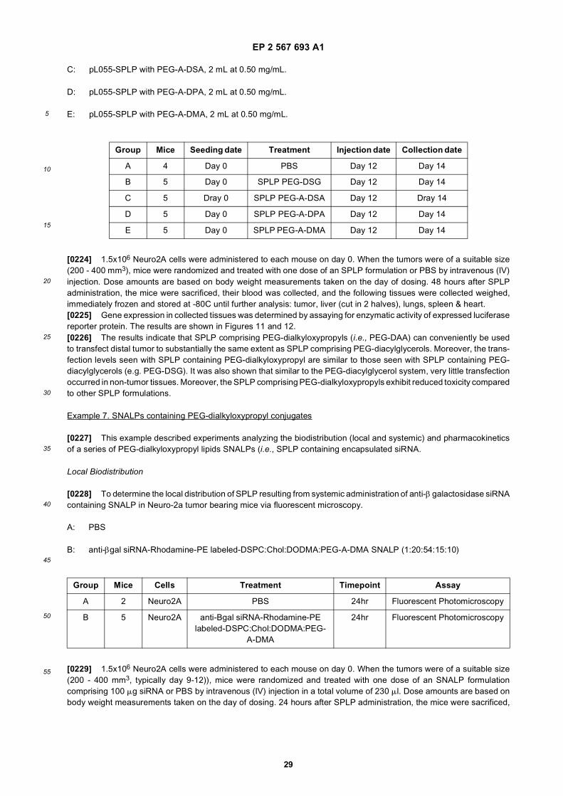
EP 2 567 693 A1
29
5
10
15
20
25
30
35
40
45
50
55
C: pL055-SPLP with PEG-A-DSA, 2 mL at 0.50 mg/mL.
D: pL055-SPLP with PEG-A-DPA, 2 mL at 0.50 mg/mL.
E: pL055-SPLP with PEG-A-DMA, 2 mL at 0.50 mg/mL.
[0224] 1.5x10⁶ Neuro2A cells were administered to each mouse on day 0. When the tumors were of a suitable size(200 - 400 mm3), mice were randomized and treated with one dose of an SPLP formulation or PBS by intravenous (IV)injection. Dose amounts are based on body weight measurements taken on the day of dosing. 48 hours after SPLPadministration, the mice were sacrificed, their blood was collected, and the following tissues were collected weighed,immediately frozen and stored at -80C until further analysis: tumor, liver (cut in 2 halves), lungs, spleen & heart.[0225] Gene expression in collected tissues was determined by assaying for enzymatic activity of expressed luciferasereporter protein. The results are shown in Figures 11 and 12.[0226] The results indicate that SPLP comprising PEG-dialkyloxypropyls (i.e., PEG-DAA) can conveniently be usedto transfect distal tumor to substantially the same extent as SPLP comprising PEG-diacylglycerols. Moreover, the trans-fection levels seen with SPLP containing PEG-dialkyloxypropyl are similar to those seen with SPLP containing PEG-diacylglycerols (e.g. PEG-DSG). It was also shown that similar to the PEG-diacylglycerol system, very little transfectionoccurred in non-tumor tissues. Moreover, the SPLP comprising PEG-dialkyloxypropyls exhibit reduced toxicity comparedto other SPLP formulations.
Example 7. SNALPs containing PEG-dialkyloxypropyl conjugates
[0227] This example described experiments analyzing the biodistribution (local and systemic) and pharmacokineticsof a series of PEG-dialkyloxypropyl lipids SNALPs (i.e., SPLP containing encapsulated siRNA.
Local Biodistribution
[0228] To determine the local distribution of SPLP resulting from systemic administration of anti-β galactosidase siRNAcontaining SNALP in Neuro-2a tumor bearing mice via fluorescent microscopy.
A: PBS
B: anti-βgal siRNA-Rhodamine-PE labeled-DSPC:Chol:DODMA:PEG-A-DMA SNALP (1:20:54:15:10)
[0229] 1.5x10⁶ Neuro2A cells were administered to each mouse on day 0. When the tumors were of a suitable size(200 - 400 mm3, typically day 9-12)), mice were randomized and treated with one dose of an SNALP formulationcomprising 100 mg siRNA or PBS by intravenous (IV) injection in a total volume of 230 ml. Dose amounts are based onbody weight measurements taken on the day of dosing. 24 hours after SPLP administration, the mice were sacrificed,
Group Mice Seeding date Treatment Injection date Collection date
A 4 Day 0 PBS Day 12 Day 14
B 5 Day 0 SPLP PEG-DSG Day 12 Day 14
C 5 Dray 0 SPLP PEG-A-DSA Day 12 Dray 14
D 5 Day 0 SPLP PEG-A-DPA Day 12 Day 14
E 5 Day 0 SPLP PEG-A-DMA Day 12 Day 14
Group Mice Cells Treatment Timepoint Assay
A 2 Neuro2A PBS 24hr Fluorescent Photomicroscopy
B 5 Neuro2A anti-Bgal siRNA-Rhodamine-PE labeled-DSPC:Chol:DODMA:PEG-
A-DMA
24hr Fluorescent Photomicroscopy
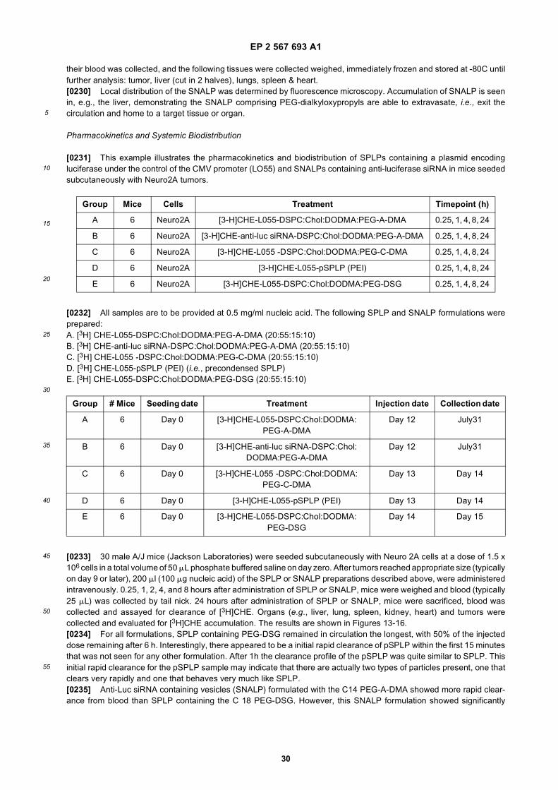
EP 2 567 693 A1
30
5
10
15
20
25
30
35
40
45
50
55
their blood was collected, and the following tissues were collected weighed, immediately frozen and stored at -80C untilfurther analysis: tumor, liver (cut in 2 halves), lungs, spleen & heart.[0230] Local distribution of the SNALP was determined by fluorescence microscopy. Accumulation of SNALP is seenin, e.g., the liver, demonstrating the SNALP comprising PEG-dialkyloxypropyls are able to extravasate, i.e., exit thecirculation and home to a target tissue or organ.
Pharmacokinetics and Systemic Biodistribution
[0231] This example illustrates the pharmacokinetics and biodistribution of SPLPs containing a plasmid encodingluciferase under the control of the CMV promoter (LO55) and SNALPs containing anti-luciferase siRNA in mice seededsubcutaneously with Neuro2A tumors.
[0232] All samples are to be provided at 0.5 mg/ml nucleic acid. The following SPLP and SNALP formulations wereprepared:A. [3H] CHE-L055-DSPC:Chol:DODMA:PEG-A-DMA (20:55:15:10)B. [3H] CHE-anti-luc siRNA-DSPC:Chol:DODMA:PEG-A-DMA (20:55:15:10)C. [3H] CHE-L055 -DSPC:Chol:DODMA:PEG-C-DMA (20:55:15:10)D. [3H] CHE-L055-pSPLP (PEI) (i.e., precondensed SPLP)E. [3H] CHE-L055-DSPC:Chol:DODMA:PEG-DSG (20:55:15:10)
[0233] 30 male A/J mice (Jackson Laboratories) were seeded subcutaneously with Neuro 2A cells at a dose of 1.5 x10⁶ cells in a total volume of 50 mL phosphate buffered saline on day zero. After tumors reached appropriate size (typicallyon day 9 or later), 200 ml (100 mg nucleic acid) of the SPLP or SNALP preparations described above, were administeredintravenously. 0.25, 1, 2, 4, and 8 hours after administration of SPLP or SNALP, mice were weighed and blood (typically25 mL) was collected by tail nick. 24 hours after administration of SPLP or SNALP, mice were sacrificed, blood wascollected and assayed for clearance of [3H]CHE. Organs (e.g., liver, lung, spleen, kidney, heart) and tumors werecollected and evaluated for [3H]CHE accumulation. The results are shown in Figures 13-16.[0234] For all formulations, SPLP containing PEG-DSG remained in circulation the longest, with 50% of the injecteddose remaining after 6 h. Interestingly, there appeared to be a initial rapid clearance of pSPLP within the first 15 minutesthat was not seen for any other formulation. After 1h the clearance profile of the pSPLP was quite similar to SPLP. Thisinitial rapid clearance for the pSPLP sample may indicate that there are actually two types of particles present, one thatclears very rapidly and one that behaves very much like SPLP.[0235] Anti-Luc siRNA containing vesicles (SNALP) formulated with the C14 PEG-A-DMA showed more rapid clear-ance from blood than SPLP containing the C 18 PEG-DSG. However, this SNALP formulation showed significantly
Group Mice Cells Treatment Timepoint (h)
A 6 Neuro2A [3-H]CHE-L055-DSPC:Chol:DODMA:PEG-A-DMA 0.25, 1, 4, 8, 24
B 6 Neuro2A [3-H]CHE-anti-luc siRNA-DSPC:Chol:DODMA:PEG-A-DMA 0.25, 1, 4, 8, 24
C 6 Neuro2A [3-H]CHE-L055 -DSPC:Chol:DODMA:PEG-C-DMA 0.25, 1, 4, 8, 24
D 6 Neuro2A [3-H]CHE-L055-pSPLP (PEI) 0.25, 1, 4, 8, 24
E 6 Neuro2A [3-H]CHE-L055-DSPC:Chol:DODMA:PEG-DSG 0.25, 1, 4, 8, 24
Group # Mice Seeding date Treatment Injection date Collection date
A 6 Day 0 [3-H]CHE-L055-DSPC:Chol:DODMA:PEG-A-DMA
Day 12 July31
B 6 Day 0 [3-H]CHE-anti-luc siRNA-DSPC:Chol:DODMA:PEG-A-DMA
Day 12 July31
C 6 Day 0 [3-H]CHE-L055 -DSPC:Chol:DODMA:PEG-C-DMA
Day 13 Day 14
D 6 Day 0 [3-H]CHE-L055-pSPLP (PEI) Day 13 Day 14
E 6 Day 0 [3-H]CHE-L055-DSPC:Chol:DODMA:PEG-DSG
Day 14 Day 15
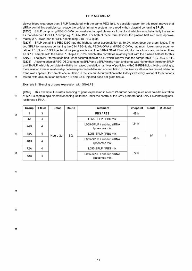
EP 2 567 693 A1
31
5
10
15
20
25
30
35
40
45
50
55
slower blood clearance than SPLP formulated with the same PEG lipid. A possible reason for this result maybe thatsiRNA containing particles can evade the cellular immune system more readily than plasmid containing SPLP.[0236] SPLP comprising PEG-C-DMA demonstrated a rapid clearance from blood, which was substantially the sameas that observed for SPLP comprisig PEG-A-DMA. For both of these formulations, the plasma half lives were approxi-mately 2 h, lower than for SPLP containing C18 PEG-lipids.[0237] SPLP containing PEG-DSG had the highest tumor accumulation at 10.9% inject dose per gram tissue. Thetwo SPLP formulations containing the C14 PEG-lipids, PEG-A-DMA and PEG-C-DMA, had much lower tumor accumu-lation of 6.1% and 5.9% injected dose per gram tissue. The SiRNA SNALP had slightly more tumor accumulation thanan SPLP sample with the same PEG-lipid at 7.3%, which also correlates relatively well with the plasma half-life for thisSNALP. The pSPLP formulation had tumor accumulation at 7.5%, which is lower than the comparable PEG-DSG SPLP.[0238] Accumulation of PEG-DSG containing SPLP and pSPLP in the heart and lungs was higher than the other SPLPand SNALP, which is consistent with the increased circulation half lives of particles with C18 PEG-lipids. Not surprisingly,there was an inverse relationship between plasma half-life and accumulation in the liver for all samples tested, while notrend was apparent for sample accumulation in the spleen. Accumulation in the kidneys was very low for all formulationstested, with accumulation between 1.2 and 2.4% injected dose per gram tissue.
Example 8: Silencing of gene expression with SNALPS
[0239] This example illustrates silencing of gene expression in Neuro 2A tumor bearing mice after co-administrationof SPLPs containing a plasmid encoding luciferase under the control of the CMV promoter and SNALPs containing anti-luciferase siRNA.
Group # Mice Tumor Route Treatment Timepoint Route # Doses
1 3
Neuro-2a SQ
PBS / PBS 48 h
IV I
4A 4 L055-SPLP / PBS mix
24 h24B 4
L055-SPLP / anti-luc siRNA liposomes mix
48A 4 L055-SPLP / PBS mix
48 h48B 4
L055-SPLP / anti-luc siRNA liposomes mix
72A 4 L055-SPLP / PBS mix
72 h72B 4
L055-SPLP / anti-luc siRNA liposomes mix
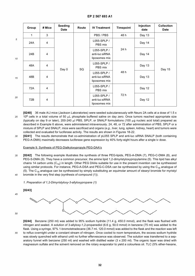
EP 2 567 693 A1
32
5
10
15
20
25
30
35
40
45
50
55
[0240] 36 male A/J mice (Jackson Laboratories) were seeded subcutaneously with Neuro 2A cells at a dose of 1.5 x10⁶ cells in a total volume of 50 mL phosphate buffered saline on day zero. Once tumors reached appropriate size(typically on day 9 or later), 200-240 ml PBS, SPLP, or SNALP formulations (100 mg nucleic acid total) prepared asdescribed in Example 6 above, were administered intravenously. 24, 48, or 72 after administration of PBS, SPLP or amixture of SPLP and SNALP, mice were sacrificed and organs (e.g., liver, lung, spleen, kidney, heart) and tumors werecollected and evaluated for luciferase activity. The results are shown in Figures 18-22.[0241] The results demonstrate that co-administration of pL055 SPLP and anti-luc siRNA SNALP (both containingPEG-A-DMA) maximally decreases luciferase gene expression by 40% forty-eight hours after a single iv dose.
Example 9. Synthesis of PEG-Dialkyloxypropyls PEG-DAA’s
[0242] The following example illustrates the synthesis of three PEG-lipids, PEG-A-DMA (7), PEG-C-DMA (8), andPEG-S-DMA (9). They have a common precursor, the amine lipid 1,2-dimyristyloxypropylamine (5). This lipid has alkylchains 14 carbon units (C14) in length. Other PEG DAAs suitable for use in the present invention can be synthesizedusing similar protocols. For instance, PEG-A-DSA and PEG-C-DSA can be synthesized by using the C18 analogue of(5). The C18 analogue can be synthesized by simply substituting an equimolar amount of stearyl bromide for myristylbromide in the very first step (synthesis of compound (1)).
1. Preparation of 1,2-Dimyristyloxy-3-allyloxypropane (1)
[0243]
[0244] Benzene (250 ml) was added to 95% sodium hydride (11.4 g, 450.0 mmol), and the flask was flushed withnitrogen and sealed. A solution of 3-allyloxy-1,2-propanediol (6.6 g, 50.0 mmol) in benzene (75 ml) was added to theflask. Using a syringe, 97% 1-bromotetradecane (36.7 ml, 120.0 mmol) was added to the flask and the reaction was leftto reflux overnight under a constant stream of nitrogen. Once cooled to room temperature, the excess sodium hydridewas slowly quenched with ethanol until no further effervescence was observed. The solution was transferred to a sep-aratory funnel with benzene (250 ml) and washed with distilled water (3 x 200 ml). The organic layer was dried withmagnesium sulfate and the solvent removed on the rotary evaporator to yield a colourless oil. TLC (5% ether-hexane,
Group # MiceSeeding
DateRoute IV Treatment Timepoint
Injection date
Collection Date
1 3
Day 0 SQ
PBS / PBS 48 h Day 13
Day 15
24A 4L055-SPLP /
PBS mix
24 h
Day 14
24B 4L055-SPLP /
anti-luc siRNA liposomes mix
Day 14
48A 4L055-SPLP /
PBS mix
48 h
Day 13
48B 4L055-SPLP /
anti-luc siRNA liposomes mix
Day 13
72A 4L055-SPLP /
PBS mix
72 h
Day 12
72B 4L055-SPLP /
anti-luc siRNA liposomes mix
Day 12
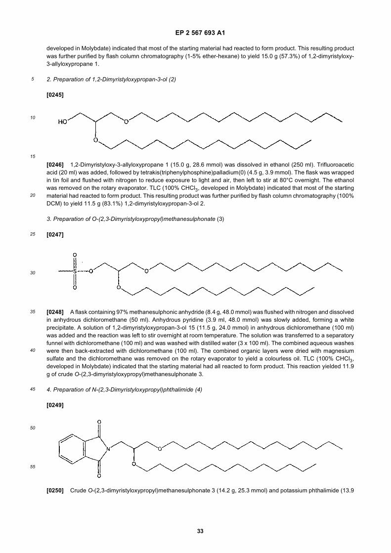
EP 2 567 693 A1
33
5
10
15
20
25
30
35
40
45
50
55
developed in Molybdate) indicated that most of the starting material had reacted to form product. This resulting productwas further purified by flash column chromatography (1-5% ether-hexane) to yield 15.0 g (57.3%) of 1,2-dimyristyloxy-3-allyloxypropane 1.
2. Preparation of 1,2-Dimyristyloxypropan-3-ol (2)
[0245]
[0246] 1,2-Dimyristyloxy-3-allyloxypropane 1 (15.0 g, 28.6 mmol) was dissolved in ethanol (250 ml). Trifluoroaceticacid (20 ml) was added, followed by tetrakis(triphenylphosphine)palladium(0) (4.5 g, 3.9 mmol). The flask was wrappedin tin foil and flushed with nitrogen to reduce exposure to light and air, then left to stir at 80°C overnight. The ethanolwas removed on the rotary evaporator. TLC (100% CHCl3, developed in Molybdate) indicated that most of the startingmaterial had reacted to form product. This resulting product was further purified by flash column chromatography (100%DCM) to yield 11.5 g (83.1%) 1,2-dimyristyloxypropan-3-ol 2.
3. Preparation of O-(2,3-Dimyristyloxypropyl)methanesulphonate (3)
[0247]
[0248] A flask containing 97% methanesulphonic anhydride (8.4 g, 48.0 mmol) was flushed with nitrogen and dissolvedin anhydrous dichloromethane (50 ml). Anhydrous pyridine (3.9 ml, 48.0 mmol) was slowly added, forming a whiteprecipitate. A solution of 1,2-dimyristyloxypropan-3-ol 15 (11.5 g, 24.0 mmol) in anhydrous dichloromethane (100 ml)was added and the reaction was left to stir overnight at room temperature. The solution was transferred to a separatoryfunnel with dichloromethane (100 ml) and was washed with distilled water (3 x 100 ml). The combined aqueous washeswere then back-extracted with dichloromethane (100 ml). The combined organic layers were dried with magnesiumsulfate and the dichloromethane was removed on the rotary evaporator to yield a colourless oil. TLC (100% CHCl3,developed in Molybdate) indicated that the starting material had all reacted to form product. This reaction yielded 11.9g of crude O-(2,3-dimyristyloxypropyl)methanesulphonate 3.
4. Preparation of N-(2,3-Dimyristyloxypropyl)phthalimide (4)
[0249]
[0250] Crude O-(2,3-dimyristyloxypropyl)methanesulphonate 3 (14.2 g, 25.3 mmol) and potassium phthalimide (13.9
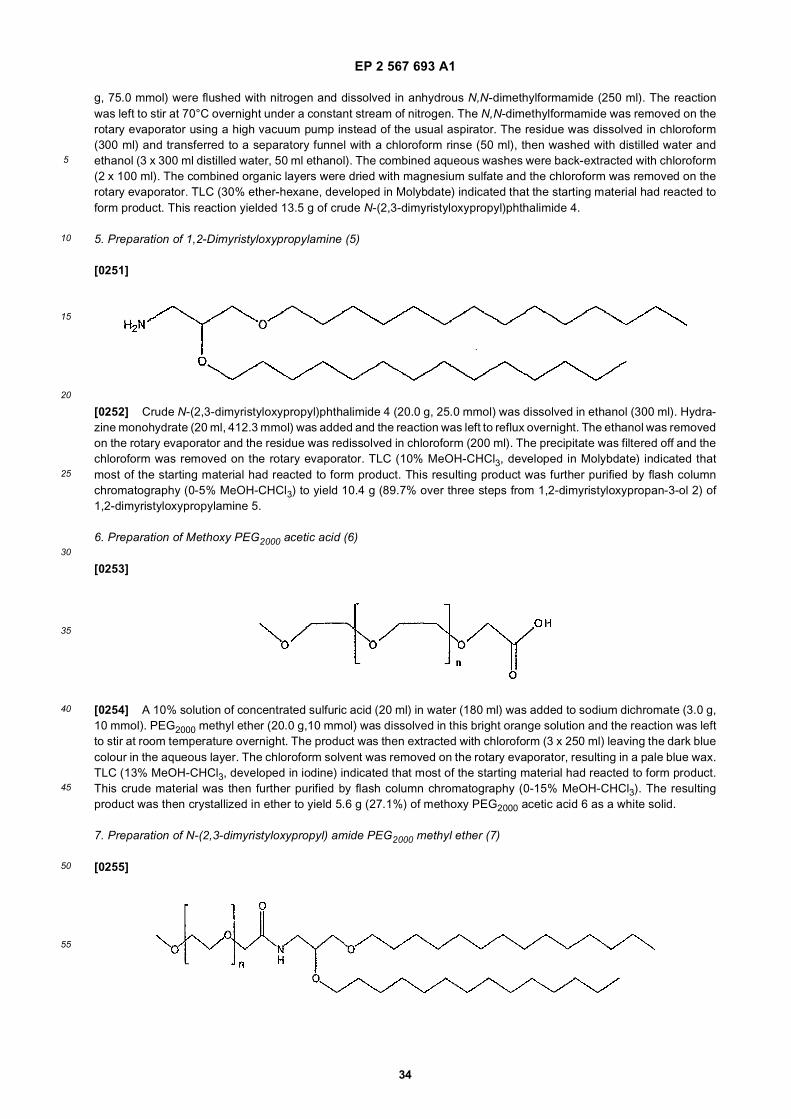
EP 2 567 693 A1
34
5
10
15
20
25
30
35
40
45
50
55
g, 75.0 mmol) were flushed with nitrogen and dissolved in anhydrous N,N-dimethylformamide (250 ml). The reactionwas left to stir at 70°C overnight under a constant stream of nitrogen. The N,N-dimethylformamide was removed on therotary evaporator using a high vacuum pump instead of the usual aspirator. The residue was dissolved in chloroform(300 ml) and transferred to a separatory funnel with a chloroform rinse (50 ml), then washed with distilled water andethanol (3 x 300 ml distilled water, 50 ml ethanol). The combined aqueous washes were back-extracted with chloroform(2 x 100 ml). The combined organic layers were dried with magnesium sulfate and the chloroform was removed on therotary evaporator. TLC (30% ether-hexane, developed in Molybdate) indicated that the starting material had reacted toform product. This reaction yielded 13.5 g of crude N-(2,3-dimyristyloxypropyl)phthalimide 4.
5. Preparation of 1,2-Dimyristyloxypropylamine (5)
[0251]
[0252] Crude N-(2,3-dimyristyloxypropyl)phthalimide 4 (20.0 g, 25.0 mmol) was dissolved in ethanol (300 ml). Hydra-zine monohydrate (20 ml, 412.3 mmol) was added and the reaction was left to reflux overnight. The ethanol was removedon the rotary evaporator and the residue was redissolved in chloroform (200 ml). The precipitate was filtered off and thechloroform was removed on the rotary evaporator. TLC (10% MeOH-CHCl3, developed in Molybdate) indicated thatmost of the starting material had reacted to form product. This resulting product was further purified by flash columnchromatography (0-5% MeOH-CHCl3) to yield 10.4 g (89.7% over three steps from 1,2-dimyristyloxypropan-3-ol 2) of1,2-dimyristyloxypropylamine 5.
6. Preparation of Methoxy PEG2000 acetic acid (6)
[0253]
[0254] A 10% solution of concentrated sulfuric acid (20 ml) in water (180 ml) was added to sodium dichromate (3.0 g,10 mmol). PEG2000 methyl ether (20.0 g,10 mmol) was dissolved in this bright orange solution and the reaction was leftto stir at room temperature overnight. The product was then extracted with chloroform (3 x 250 ml) leaving the dark bluecolour in the aqueous layer. The chloroform solvent was removed on the rotary evaporator, resulting in a pale blue wax.TLC (13% MeOH-CHCl3, developed in iodine) indicated that most of the starting material had reacted to form product.This crude material was then further purified by flash column chromatography (0-15% MeOH-CHCl3). The resultingproduct was then crystallized in ether to yield 5.6 g (27.1%) of methoxy PEG2000 acetic acid 6 as a white solid.
7. Preparation of N-(2,3-dimyristyloxypropyl) amide PEG2000 methyl ether (7)
[0255]
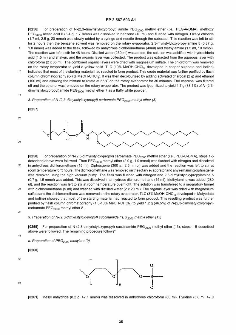
EP 2 567 693 A1
35
5
10
15
20
25
30
35
40
45
50
55
[0256] For preparation of N-(2,3-dimyristyloxypropyl) amide PEG2000 methyl ether (i.e., PEG-A-DMA), methoxyPEG2000 acetic acid 6 (3.4 g, 1.7 mmol) was dissolved in benzene (40 ml) and flushed with nitrogen. Oxalyl chloride(1.7 ml, 2.5 g, 20 mmol) was slowly added by a syringe and needle through the subaseal. This reaction was left to stirfor 2 hours then the benzene solvent was removed on the rotary evaporator. 2,3-myristylyloxypropylamine 5 (0.87 g,1.8 mmol) was added to the flask, followed by anhydrous dichloromethane (40ml) and triethylamine (1.5 ml, 10 mmol).The reaction was left to stir for 48 hours. Distilled water (250 ml) was added, the solution was acidified with hydrochloricacid (1.5 ml) and shaken, and the organic layer was collected. The product was extracted from the aqueous layer withchloroform (2 x 65 ml). The combined organic layers were dried with magnesium sulfate. The chloroform was removedon the rotary evaporator to yield a yellow solid. TLC (10% MeOH-CHCl3, developed in copper sulphate and iodine)indicated that most of the starting material had reacted to form product. This crude material was further purified by flashcolumn chromatography (0-7% MeOH-CHCl3). It was then decolourized by adding activated charcoal (2 g) and ethanol(100 ml) and allowing the mixture to rotate at 55°C on the rotary evaporator for 30 minutes. The charcoal was filteredoff and the ethanol was removed on the rotary evaporator. The product was lyophilized to yield 1.7 g (38.1%) of N-(2,3-dimyristyloxypropyl)amide PEG2000 methyl ether 7 as a fluffy white powder.
8. Preparation of N-(2,3-dimyristyloxypropyl) carbamate PEG2000 methyl ether (8)
[0257]
[0258] For preparation of N-(2,3-dimyristyloxypropyl) carbamate PEG2000 methyl ether (i.e., PEG-C-DMA), steps 1-5described above were followed. Then PEG2000 methyl ether (2.0 g, 1.0 mmol) was flushed with nitrogen and dissolvedin anhydrous dichloromethane (15 ml). Diphosgene (300 ml, 2.5 mmol) was added and the reaction was left to stir atroom temperature for 3 hours. The dichloromethane was removed on the rotary evaporator and any remaining diphosgenewas removed using the high vacuum pump. The flask was flushed with nitrogen and 2,3-dimyristyloxypropylamine 5(0.7 g, 1.5 mmol) was added. This was dissolved in anhydrous dichloromethane (15 ml), triethylamine was added (280ul), and the reaction was left to stir at room temperature overnight. The solution was transferred to a separatory funnelwith dichloromethane (5 ml) and washed with distilled water (2 x 20 ml). The organic layer was dried with magnesiumsulfate and the dichloromethane was removed on the rotary evaporator. TLC (3% MeOH-CHCl3 developed in Molybdateand iodine) showed that most of the starting material had reacted to form product. This resulting product was furtherpurified by flash column chromatography (1.5-10% MeOH-CHCl3) to yield 1.2 g (46.5%) of N-(2,3-dimyristyloxypropyl)carbamate PEG2000 methyl ether 8.
9. Preparation of N-(2,3-dimyristyloxypropyl) succinamide PEG2000 methyl ether (13)
[0259] For preparation of N-(2,3-dimyristyloxypropyl) succinamide PEG2000 methyl ether (13), steps 1-5 describedabove were followed. The remaining procedure follows"
a. Preparation of PEG2000 mesylate (9)
[0260]
[0261] Mesyl anhydride (8.2 g, 47.1 mmol) was dissolved in anhydrous chloroform (80 ml). Pyridine (3.8 ml, 47.0
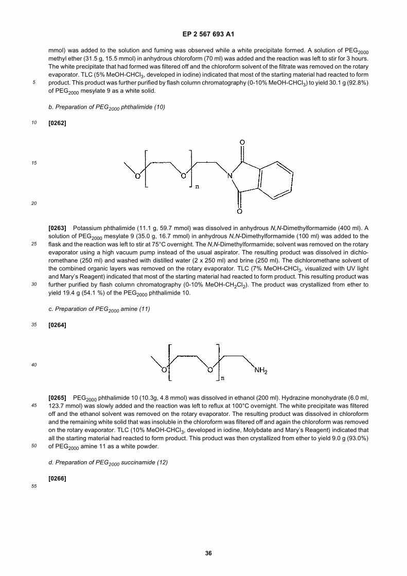
EP 2 567 693 A1
36
5
10
15
20
25
30
35
40
45
50
55
mmol) was added to the solution and fuming was observed while a white precipitate formed. A solution of PEG2000methyl ether (31.5 g, 15.5 mmol) in anhydrous chloroform (70 ml) was added and the reaction was left to stir for 3 hours.The white precipitate that had formed was filtered off and the chloroform solvent of the filtrate was removed on the rotaryevaporator. TLC (5% MeOH-CHCl3, developed in iodine) indicated that most of the starting material had reacted to formproduct. This product was further purified by flash column chromatography (0-10% MeOH-CHCl3) to yield 30.1 g (92.8%)of PEG2000 mesylate 9 as a white solid.
b. Preparation of PEG2000 phthalimide (10)
[0262]
[0263] Potassium phthalimide (11.1 g, 59.7 mmol) was dissolved in anhydrous N,N-Dimethylformamide (400 ml). Asolution of PEG2000 mesylate 9 (35.0 g, 16.7 mmol) in anhydrous N,N-Dimethylformamide (100 ml) was added to theflask and the reaction was left to stir at 75°C overnight. The N,N-Dimethylformamide; solvent was removed on the rotaryevaporator using a high vacuum pump instead of the usual aspirator. The resulting product was dissolved in dichlo-romethane (250 ml) and washed with distilled water (2 x 250 ml) and brine (250 ml). The dichloromethane solvent ofthe combined organic layers was removed on the rotary evaporator. TLC (7% MeOH-CHCl3, visualized with UV lightand Mary’s Reagent) indicated that most of the starting material had reacted to form product. This resulting product wasfurther purified by flash column chromatography (0-10% MeOH-CH2Cl2). The product was crystallized from ether toyield 19.4 g (54.1 %) of the PEG2000 phthalimide 10.
c. Preparation of PEG2000 amine (11)
[0264]
[0265] PEG2000 phthalimide 10 (10.3g, 4.8 mmol) was dissolved in ethanol (200 ml). Hydrazine monohydrate (6.0 ml,123.7 mmol) was slowly added and the reaction was left to reflux at 100°C overnight. The white precipitate was filteredoff and the ethanol solvent was removed on the rotary evaporator. The resulting product was dissolved in chloroformand the remaining white solid that was insoluble in the chloroform was filtered off and again the chloroform was removedon the rotary evaporator. TLC (10% MeOH-CHCl3, developed in iodine, Molybdate and Mary’s Reagent) indicated thatall the starting material had reacted to form product. This product was then crystallized from ether to yield 9.0 g (93.0%)of PEG2000 amine 11 as a white powder.
d. Preparation of PEG2000 succinamide (12)
[0266]
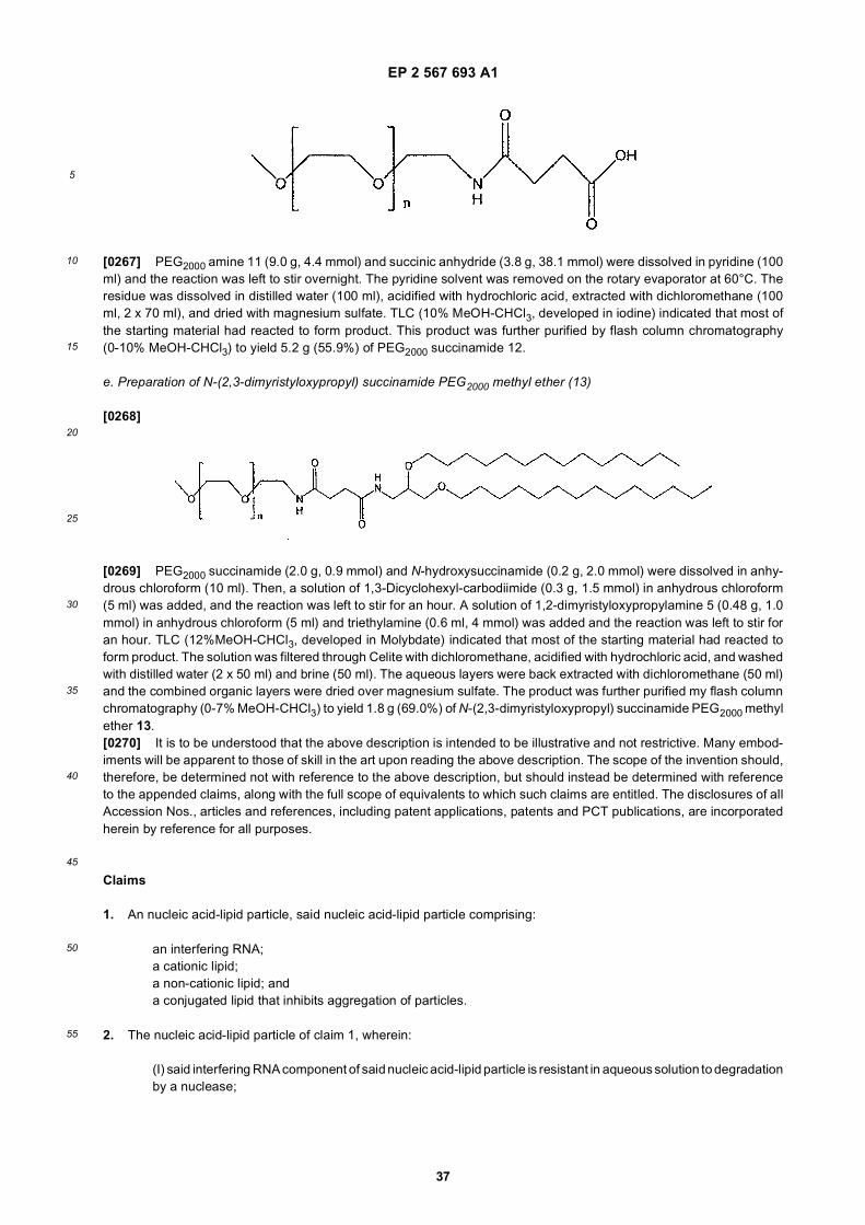
EP 2 567 693 A1
37
5
10
15
20
25
30
35
40
45
50
55
[0267] PEG2000 amine 11 (9.0 g, 4.4 mmol) and succinic anhydride (3.8 g, 38.1 mmol) were dissolved in pyridine (100ml) and the reaction was left to stir overnight. The pyridine solvent was removed on the rotary evaporator at 60°C. Theresidue was dissolved in distilled water (100 ml), acidified with hydrochloric acid, extracted with dichloromethane (100ml, 2 x 70 ml), and dried with magnesium sulfate. TLC (10% MeOH-CHCl3, developed in iodine) indicated that most ofthe starting material had reacted to form product. This product was further purified by flash column chromatography(0-10% MeOH-CHCl3) to yield 5.2 g (55.9%) of PEG2000 succinamide 12.
e. Preparation of N-(2,3-dimyristyloxypropyl) succinamide PEG2000 methyl ether (13)
[0268]
[0269] PEG2000 succinamide (2.0 g, 0.9 mmol) and N-hydroxysuccinamide (0.2 g, 2.0 mmol) were dissolved in anhy-drous chloroform (10 ml). Then, a solution of 1,3-Dicyclohexyl-carbodiimide (0.3 g, 1.5 mmol) in anhydrous chloroform(5 ml) was added, and the reaction was left to stir for an hour. A solution of 1,2-dimyristyloxypropylamine 5 (0.48 g, 1.0mmol) in anhydrous chloroform (5 ml) and triethylamine (0.6 ml, 4 mmol) was added and the reaction was left to stir foran hour. TLC (12%MeOH-CHCl3, developed in Molybdate) indicated that most of the starting material had reacted toform product. The solution was filtered through Celite with dichloromethane, acidified with hydrochloric acid, and washedwith distilled water (2 x 50 ml) and brine (50 ml). The aqueous layers were back extracted with dichloromethane (50 ml)and the combined organic layers were dried over magnesium sulfate. The product was further purified my flash columnchromatography (0-7% MeOH-CHCl3) to yield 1.8 g (69.0%) of N-(2,3-dimyristyloxypropyl) succinamide PEG2000 methylether 13.[0270] It is to be understood that the above description is intended to be illustrative and not restrictive. Many embod-iments will be apparent to those of skill in the art upon reading the above description. The scope of the invention should,therefore, be determined not with reference to the above description, but should instead be determined with referenceto the appended claims, along with the full scope of equivalents to which such claims are entitled. The disclosures of allAccession Nos., articles and references, including patent applications, patents and PCT publications, are incorporatedherein by reference for all purposes.
Claims
1. An nucleic acid-lipid particle, said nucleic acid-lipid particle comprising:
an interfering RNA;a cationic lipid;a non-cationic lipid; anda conjugated lipid that inhibits aggregation of particles.
2. The nucleic acid-lipid particle of claim 1, wherein:
(I) said interfering RNA component of said nucleic acid-lipid particle is resistant in aqueous solution to degradationby a nuclease;
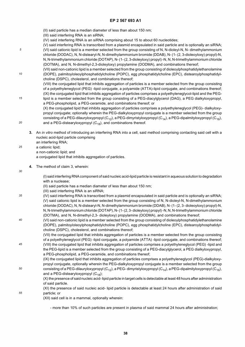
EP 2 567 693 A1
38
5
10
15
20
25
30
35
40
45
50
55
(II) said particle has a median diameter of less than about 150 nm;(III) said interfering RNA is an siRNA;(IV) said interfering RNA is an siRNA comprising about 15 to about 60 nucleotides;(V) said interfering RNA is transcribed from a plasmid encapsulated in said particle and is optionally an siRNA;(VI) said cationic lipid is a member selected from the group consisting of N, N-dioleyl-N, N- dimethylammoniumchloride (DODAC), N, N-distearyl-N, N-dimethylammonium bromide (DDAB), N- (1- (2, 3-dioleoyloxy) propyl)-N,N, N-trimethylammonium chloride (DOTAP), N- (1- (2, 3-dioleyloxy) propyl) -N, N, N-trimethylammonium chloride(DOTMA), and N, N-dimethyl-2,3-dioleyloxy) propylamine (DODMA), and combinations thereof;(VII) said non-cationic lipid is a member selected from the group consisting of dioleoylphosphatidylethanolamine(DOPE), palmitoyloleoylphosphatidylcholine (POPC), egg phosphatidylcholine (EPC), distearoylphosphatidyl-choline (DSPC), cholesterol, and combinations thereof;(VIII) the conjugated lipid that inhibits aggregation of particles is a member selected from the group consistingof a polyethyleneglycol (PEG) -lipid conjugate, a polyamide (ATTA)-lipid conjugate, and combinations thereof;(IX) the conjugated lipid that inhibits aggregation of particles comprises a polyethyleneglycol-lipid and the PEG-lipid is a member selected from the group consisting of a PEG-diacylglycerol (DAG), a PEG dialkyloxypropyl,a PEG-phospholipid, a PEG-ceramide, and combinations thereof; or(X) the conjugated lipid that inhibits aggregation of particles comprises a polyethyleneglycol (PEG)- dialkyloxy-propyl conjugate; optionally wherein the PEG-dialkyloxypropyl conjugate is a member selected from the groupconsisting of a PEG-dilauryloxypropyl (C12), a PEG-dimyristyloxypropyl (C14), a PEG-dipalmityloxypropyl (C16),and a PEG-distearyloxypropyl (C18), and combinations thereof.
3. An in vitro method of introducing an interfering RNA into a cell, said method comprising contacting said cell with anucleic acid-lipid particle comprisingan interfering RNA;a cationic lipid;a non-cationic lipid; anda conjugated lipid that inhibits aggregation of particles.
4. The method of claim 3, wherein:
(I) said interfering RNA component of said nucleic acid-lipid particle is resistant in aqueous solution to degradationwith a nuclease;(II) said particle has a median diameter of less than about 150 nm;(III) said interfering RNA is an siRNA;(IV) said interfering RNA is transcribed from a plasmid encapsulated in said particle and is optionally an siRNA;(V) said cationic lipid is a member selected from the group consisting of N, N-dioleyl-N, N-dimethylammoniumchloride (DODAC), N, N-distearyl-N, N-dimethylammonium bromide (DDAB), N- (1- (2, 3- dioleoyloxy) propyl)-N,N, N-trimethylammonium chloride (DOTAP), N- (1- (2, 3- dioleyloxy) propyl) -N, N, N-trimethylammonium chloride(DOTMA), and N, N-dimethyl-2,3- dioleyloxy) propylamine (DODMA), and combinations thereof;(VI) said non-cationic lipid is a member selected from the group consisting of dioleoylphosphatidylethanolamine(DOPE), palmitoyloleoylphosphatidylcholine (POPC), egg phosphatidylcholine (EPC), distearoylphosphatidyl-choline (DSPC), cholesterol, and combinations thereof;(VII) the conjugated lipid that inhibits aggregation of particles is a member selected from the group consistingof a polyethyleneglycol (PEG) -lipid conjugate, a polyamide (ATTA) -lipid conjugate, and combinations thereof;(VIII) the conjugated lipid that inhibits aggregation of particles comprises a polyethyleneglycol (PEG) -lipid andthe PEG-lipid is a member selected from the group consisting of a PEG-diacylglycerol, a PEG dialkyloxypropyl,a PEG-phospholipid, a PEG-ceramide, and combinations thereof;(IX) the conjugated lipid that inhibits aggregation of particles comprises a polyethyleneglycol (PEG)-dialkyloxy-propyl conjugate, optionally wherein the PEG-dialkyloxypropyl conjugate is a member selected from the groupconsisting of a PEG-dilauryloxypropyl (C12), a PEG- dimyristyloxypropyl (C14), a PEG-dipalmityloxypropyl (C16),and a PEG-distearyloxypropyl (C18);(X) the presence of said nucleic acid- lipid particle in target cells is detectable at least 48 hours after administrationof said particle.(XI) the presence of said nucleic acid- lipid particle is detectable at least 24 hours after administration of saidparticle; or(XII) said cell is in a mammal, optionally wherein:
- more than 10% of such particles are present in plasma of said mammal 24 hours after administration;
EP 2 567 693 A1
39
5
10
15
20
25
30
35
40
45
50
55
- said interfering RNA is an siRNA and presence of a siRNA at a site distal to the site of administration isdetectable for at least 48 hours after administration of said particle; or- said interfering RNA is an siRNA and presence of a siRNA at a site distal to the site of administration isdetectable for at least 24 hours after administration of said particle, optionally wherein the mammal is ahuman.
5. A nucleic acid-lipid particle comprising:
an interfering RNA;a cationic lipid;a non-cationic lipid; anda conjugated lipid that inhibits aggregation of particles,for use in a method for silencing expression of a target sequence, said method comprising administering saidnucleic acid-lipid particle to a mammalian subject in a therapeutically effective amount.
6. The nucleic acid-lipid particle for use according to claim 5, wherein:
(I) said administration is intravenous; or(II) said mammal is a human, optionally wherein said human has a disease or disorder associated with expressionof a gene comprising a target sequence for said interfering RNA, such as wherein said disease or disorder ishepatitis; or(III) said interfering RNA is an siRNA.
7. A nucleic acid-lipid particle comprising:
an interfering RNA;a cationic lipid;a non-cationic lipid; anda conjugated lipid that inhibits aggregation of particles,for use in a method for in vivo delivery of an interfering RNA, said method comprising administering said nucleicacid-lipid particle to a mammalian subject.
8. The nucleic acid-lipid particle for use according to claim 7, wherein:
(I) said administration is intravenous;(II) said mammal is a human;(III) said mammal is a human having a disease or disorder associated with expression of a gene comprising atarget sequence for said interfering RNA, optionally wherein said disease or disorder is associated with over-expression of said gene, or wherein said disease or disorder is hepatitis;(IV) said interfering RNA is an siRNA; or(V) said method is for in vivo delivery of an interfering RNA, such as an siRNA, to a liver cell.
9. A nucleic acid-lipid particle comprising:
an interfering RNA;a cationic lipid;a non-cationic lipid; anda conjugated lipid that inhibits aggregation of particles,for use in a method of treating a disease in a mammalian subject, said method comprising administering saidnucleic acid-lipid particle to said subject in a therapeutically effective amount, wherein said disease is associatedwith expression of a gene comprising a target sequence for said interfering RNA.
10. The nucleic acid-lipid particle for use according to claim 9, wherein:
(I) said mammal is a human;(II) said disease is hepatitis, optionally wherein said hepatitis is Hepatitis B or Hepatitis C;(III) said administration is intravenous; or(IV) said interfering RNA is an siRNA.
EP 2 567 693 A1
40
5
10
15
20
25
30
35
40
45
50
55
11. The use of a nucleic acid-lipid particle to formulate a composition for introducing an interfering RNA into a cell,wherein said nucleic acid-lipid particle comprises:
an interfering RNA;a cationic lipid;a non-cationic lipid; anda conjugated lipid that inhibits aggregation of particles.
12. The use of claim 11, wherein:
(I) said interfering RNA component of said nucleic acid-lipid particle is resistant in aqueous solution to degradationwith a nuclease;(II) said particle has a median diameter of less than about 150 nm;(III) said interfering RNA is an siRNA;(IV) said interfering RNA is transcribed from a plasmid encapsulated in said particle, and is optionally an siRNA;(V) said cationic lipid is a member selected from the group consisting of N, N-dioleyl-N, N-dimethylammoniumchloride (DODAC), N, N- distearyl-N, N-dimethylammonium bromide (DDAB), N- (1- (2, 3-dioleoyloxy) propyl)- N, N, N-trimethylammonium chloride (DOTAP), N- (1- (2, 3-dioleyloxy) propyl) -N, N, N- trimethylammoniumchloride (DOTMA), and N, N-dimethyl-2,3-dioleyloxy) propylamine (DODMA), and combinations thereof;(VI) said non-cationic lipid is a member selected from the group consisting of dioleoylphosphatidylethanolamine(DOPE), palmitoyloleoylphosphatidylcholine (POPC), egg phosphatidylcholine (EPC), distearoylphosphatidyl-choline (DSPC), cholesterol, and combinations thereof;(VII) the conjugated lipid that inhibits aggregation of particles is a member selected from the group consistingof a polyethyleneglycol (PEG) -lipid conjugate, a polyamide (ATTA) -lipid conjugate, and combinations thereof;(VIII) the conjugated lipid that inhibits aggregation of particles comprises a polyethyleneglycol (PEG) -lipid andthe PEG-lipid is a member selected from the group consisting of a PEG-diacylglycerol, a PEG dialkyloxypropyl,a PEG-phospholipid, a PEG-ceramide, and combinations thereof;(IX) the conjugated lipid that inhibits aggregation of particles comprises a polyethyleneglycol (PEG)-dialkyloxy-propyl conjugate, optionally wherein the PEG-dialkyloxypropyl conjugate is a member selected from the groupconsisting of a PEG-dilauryloxypropyl (C12), a PEG- dimyristyloxypropyl (C14), a PEG-dipalmityloxypropyl (C16),and a PEG-distearyloxypropyl (C18);(X) the presence of said nucleic acid-lipid particle in target cells is detectable at least 48 hours after administrationof said particle;(XI) the presence of said nucleic acid-lipid particle is detectable at least 24 hours after administration of saidparticle; or(XII) said cell is in a mammal, optionally wherein:
- more than 10% of such particles are present in plasma of said mammal 24 hours after administration;- said interfering RNA is an siRNA and presence of a siRNA at a site distal to the site of administration isdetectable for at least 48 hours after administration of said particle; or- said interfering RNA is an siRNA and presence of a siRNA at a site distal to the site of administration isdetectable for at least 24 hours after administration of said particle, optionally wherein the mammal is ahuman.
13. The use of a nucleic acid-lipid particle to formulate a medicament for:
(I) silencing expression of a target sequence in a mammalian subject,(II) in vivo delivery of an interfering RNA, or(III) treating a disease in a mammalian subject,wherein said nucleic acid- lipid particle comprises:
an interfering RNA;a cationic lipid;a non-cationic lipid; anda conjugated lipid that inhibits aggregation of particles.
14. The use of claim 13(I), (II) or (III), wherein:
EP 2 567 693 A1
41
5
10
15
20
25
30
35
40
45
50
55
(a) said nucleic acid-lipid particle is formulated for intravenous administration;(b) said mammal is a human, optionally wherein said human has a disease or disorder associated with expres-sion, such as overexpression, of a gene comprising a target sequence for said interfering RNA, or wherein saiddisease or disorder is hepatitis;(c) said interfering RNA is an siRNA; or(d) said use is to formulate a medicament for in vivo delivery of an interfering RNA, such as an siRNA, to a liver cell.
15. The use of claim 13(III), wherein:
- said disease is hepatitis, such as Hepatitis B, or Hepatitis C; or- said medicament is adapted for intravenous administration.
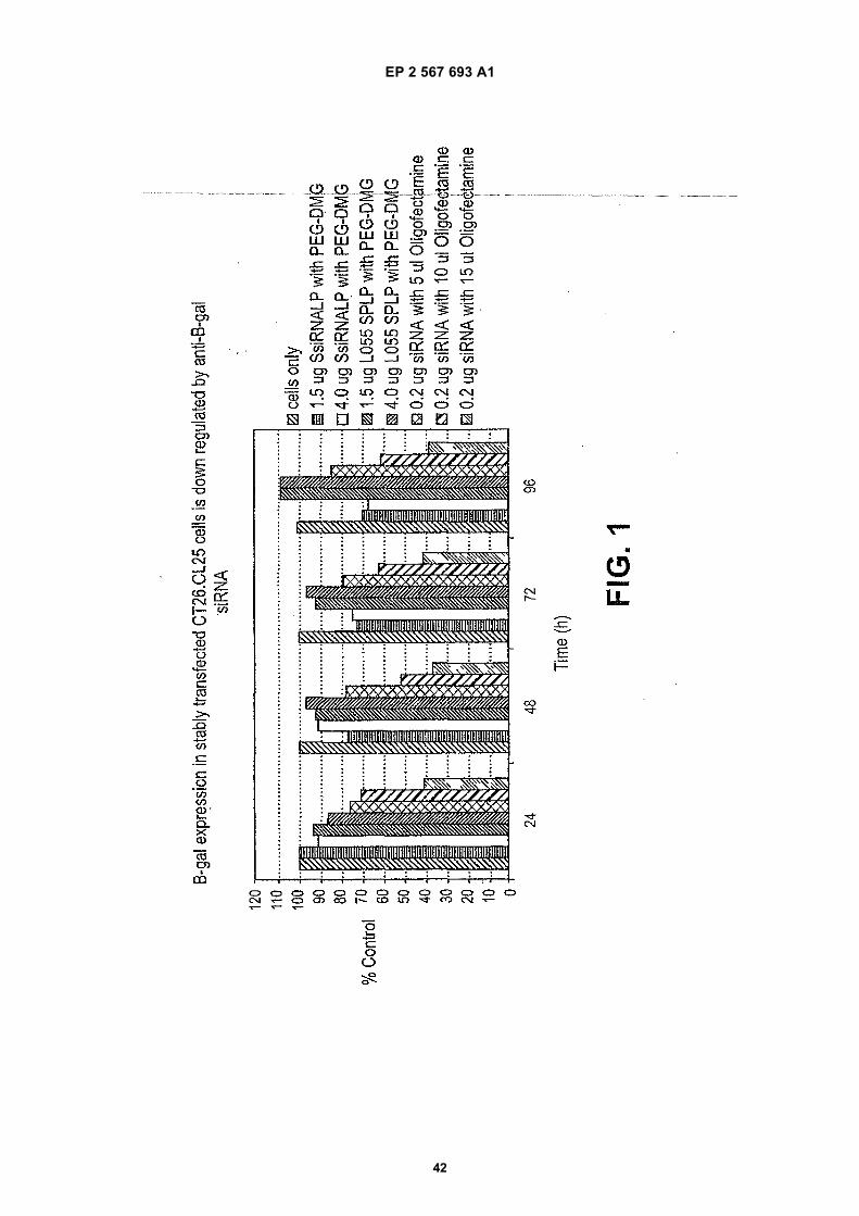
EP 2 567 693 A1
42
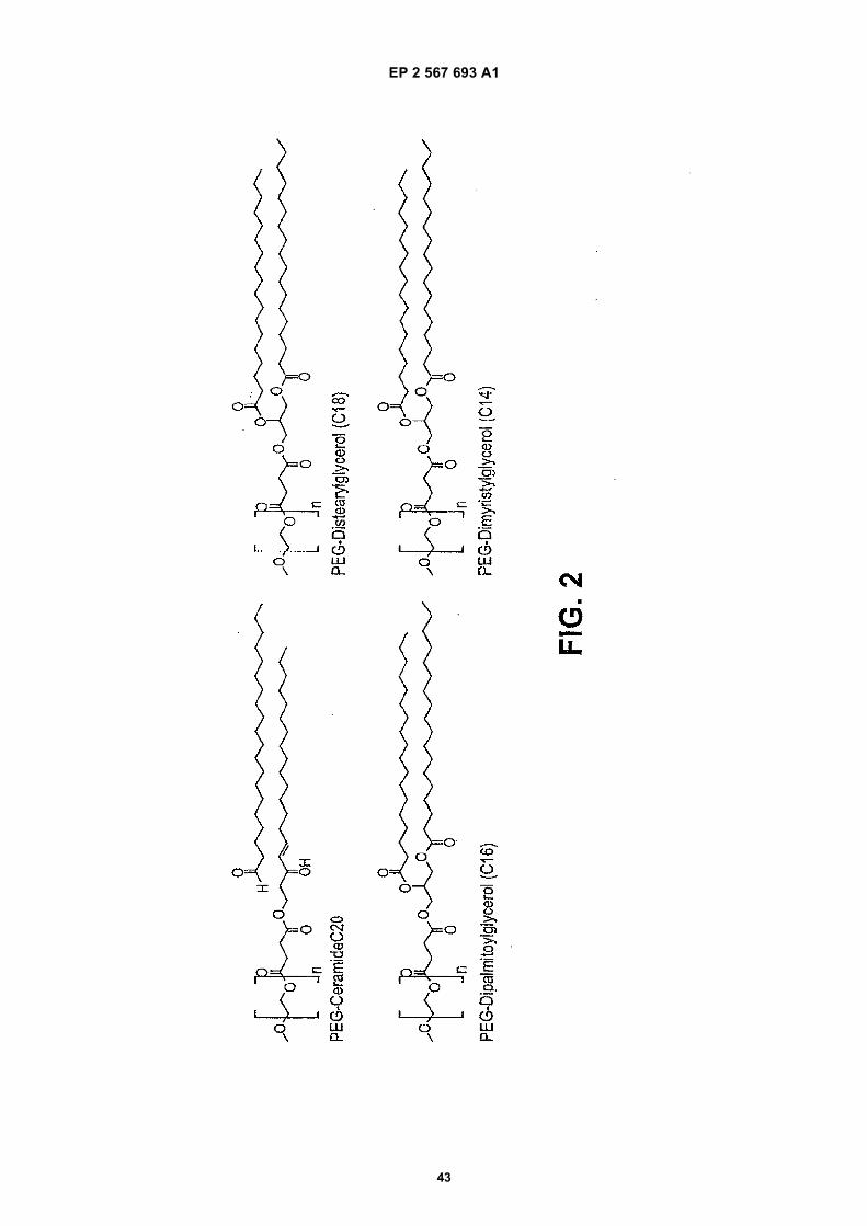
EP 2 567 693 A1
43
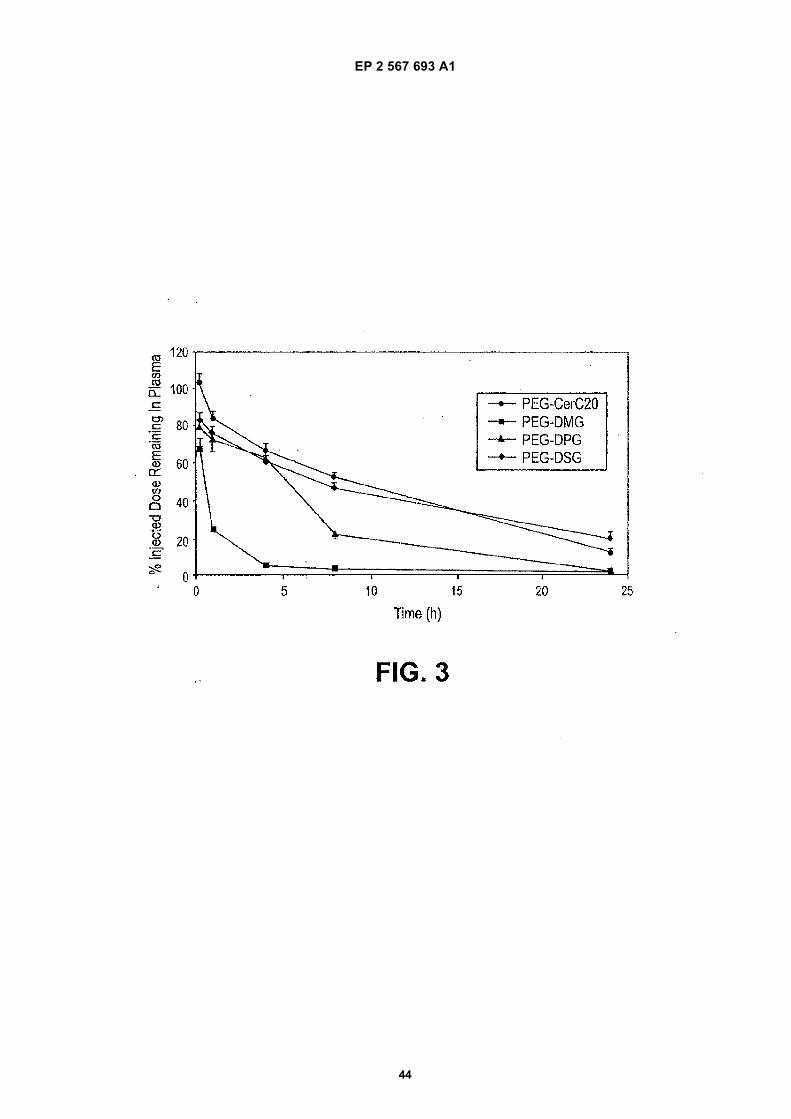
EP 2 567 693 A1
44
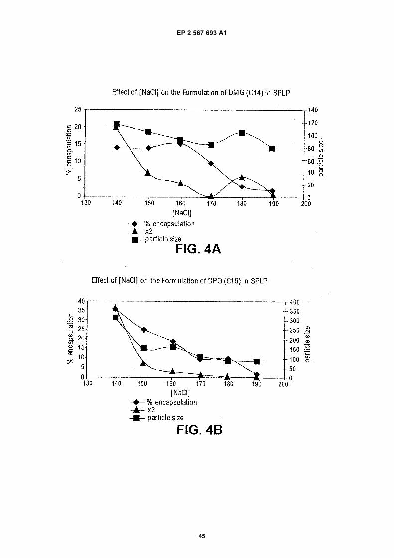
EP 2 567 693 A1
45
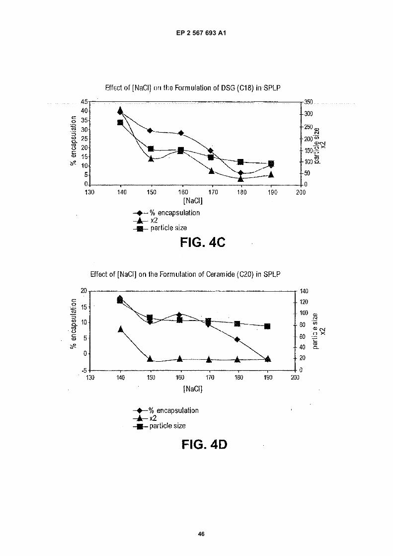
EP 2 567 693 A1
46
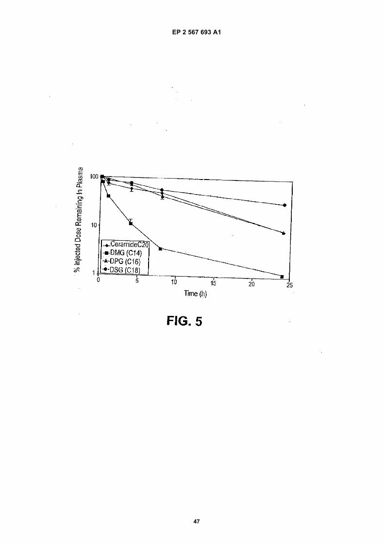
EP 2 567 693 A1
47
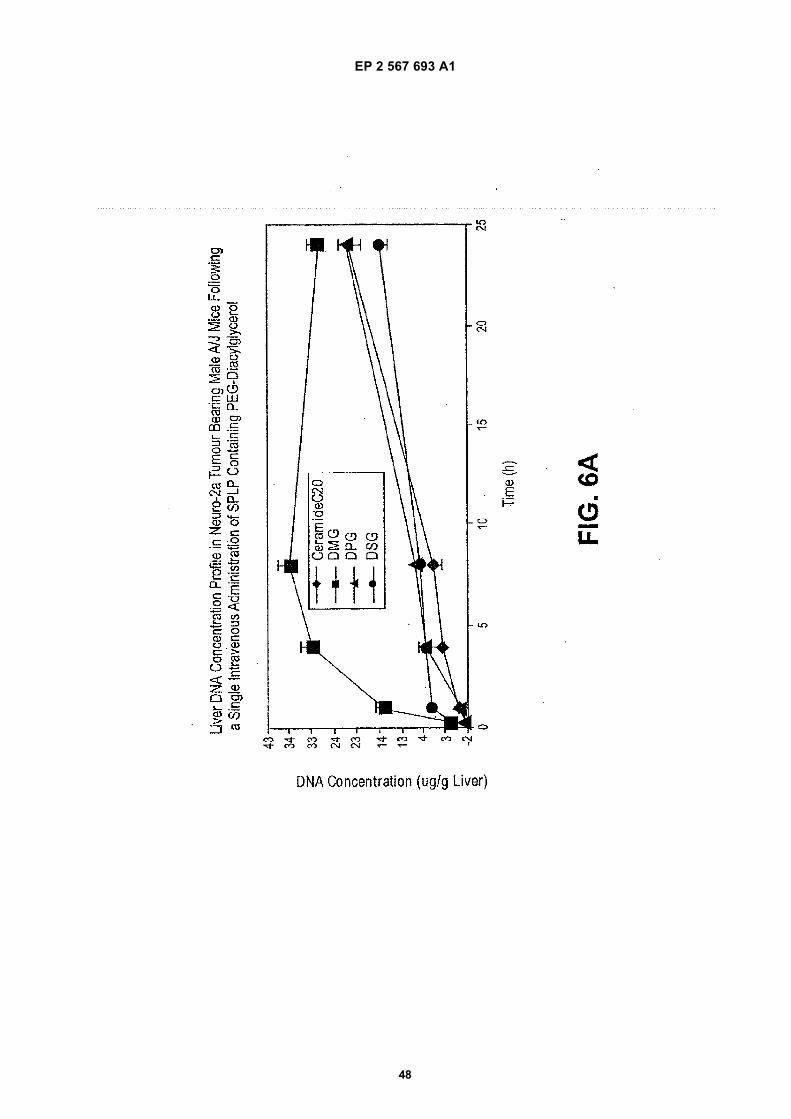
EP 2 567 693 A1
48
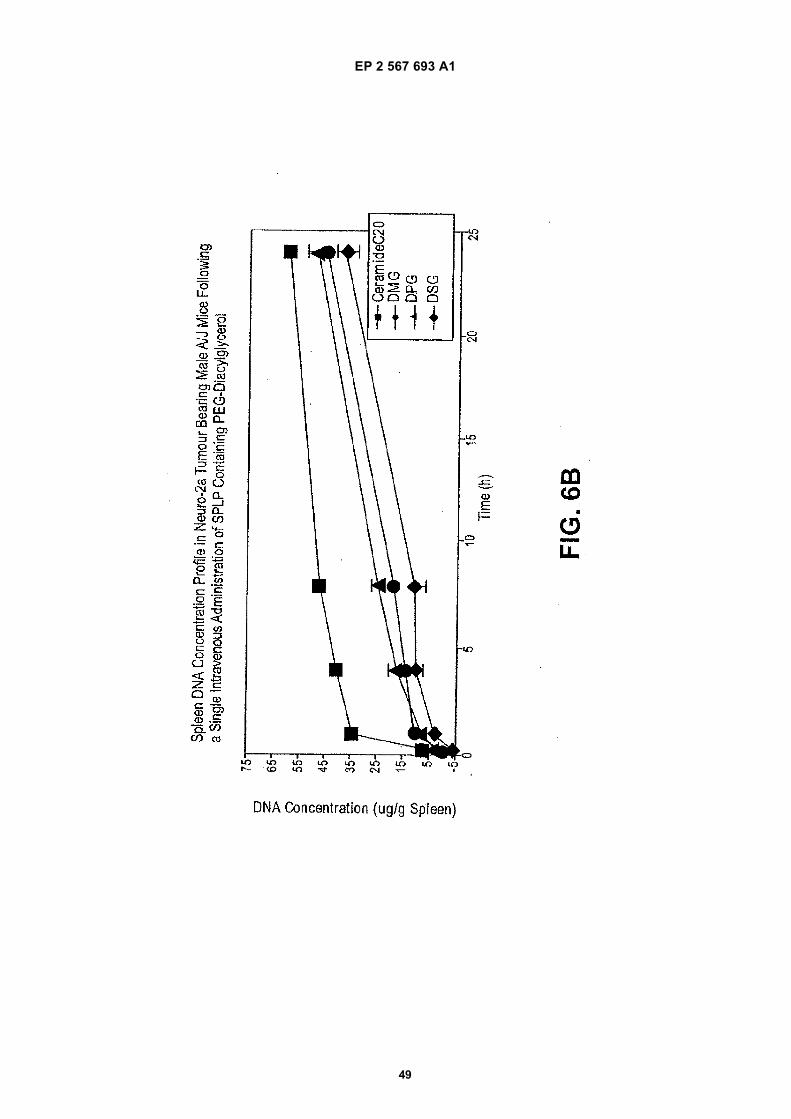
EP 2 567 693 A1
49
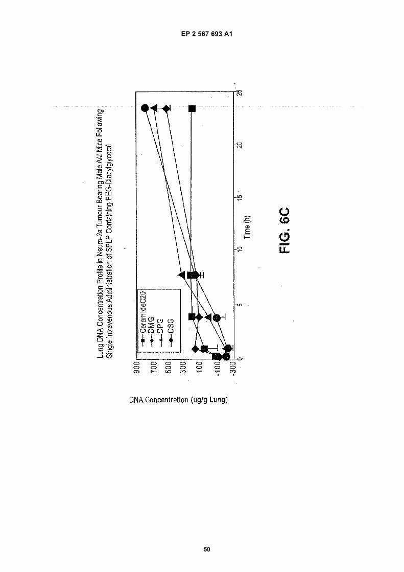
EP 2 567 693 A1
50
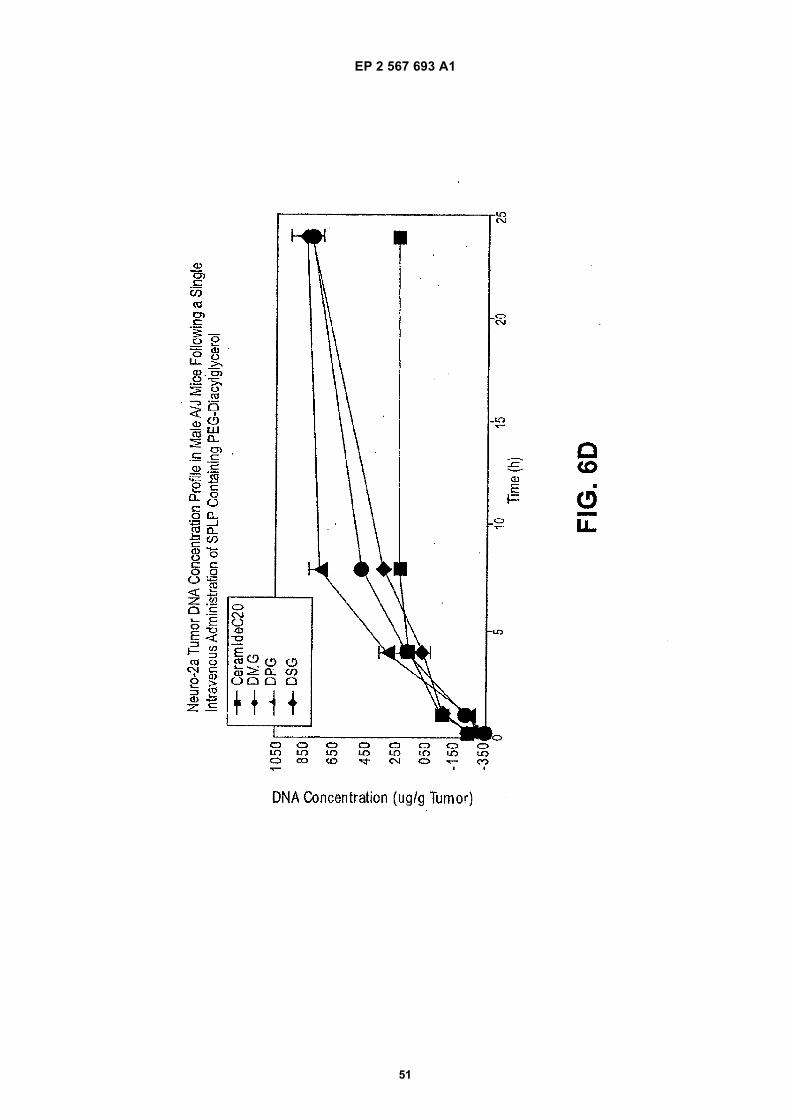
EP 2 567 693 A1
51
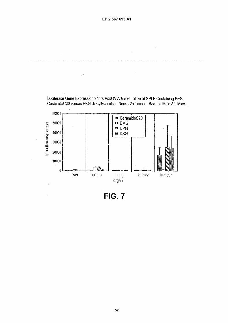
EP 2 567 693 A1
52
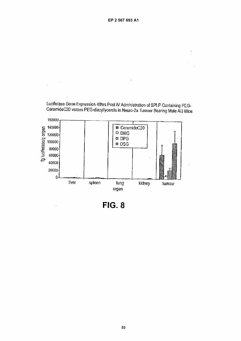
EP 2 567 693 A1
53
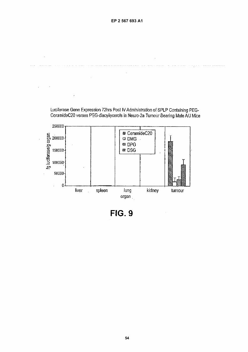
EP 2 567 693 A1
54
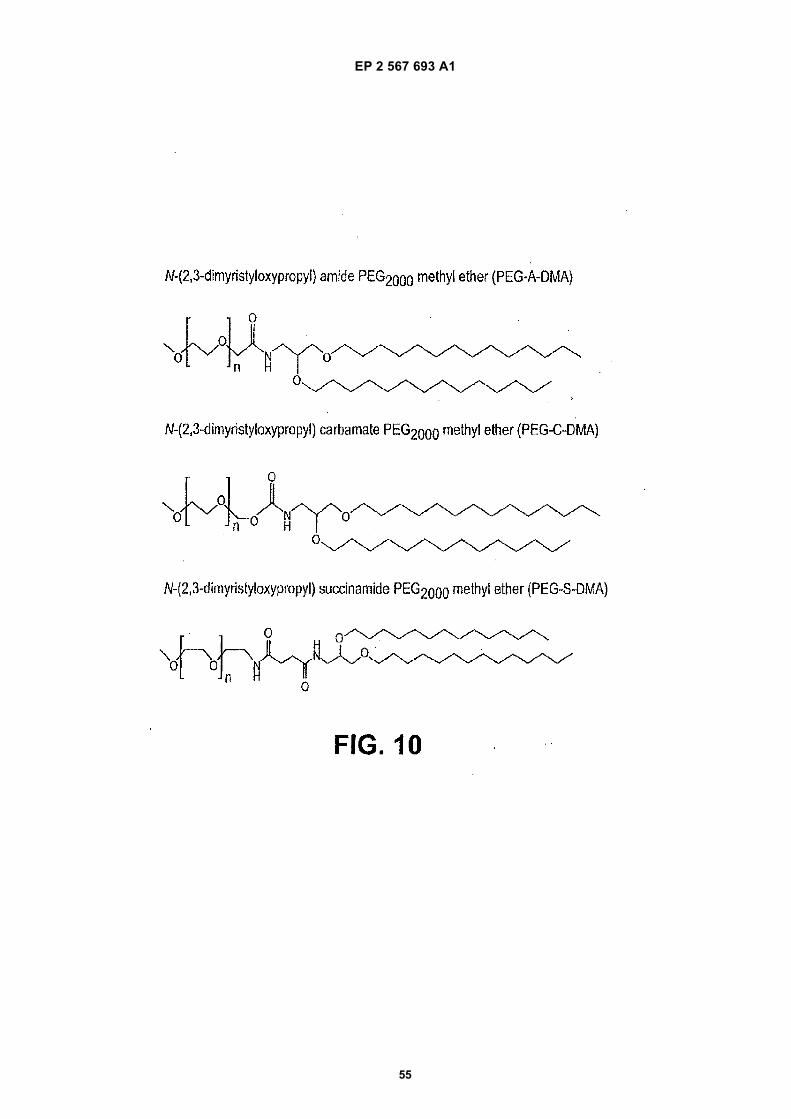
EP 2 567 693 A1
55
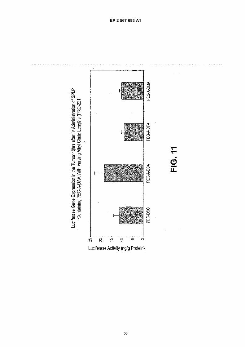
EP 2 567 693 A1
56
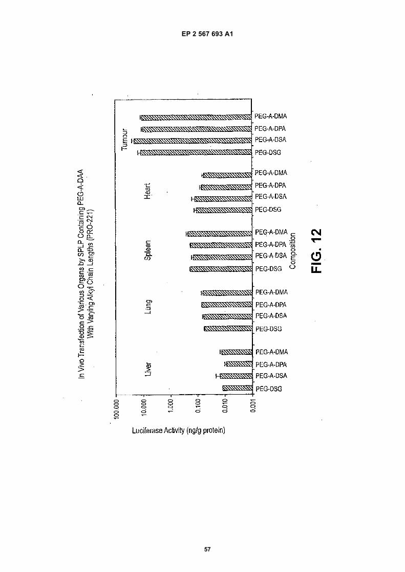
EP 2 567 693 A1
57
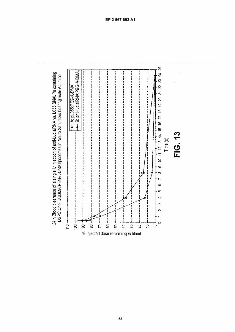
EP 2 567 693 A1
58
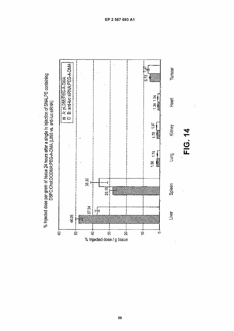
EP 2 567 693 A1
59
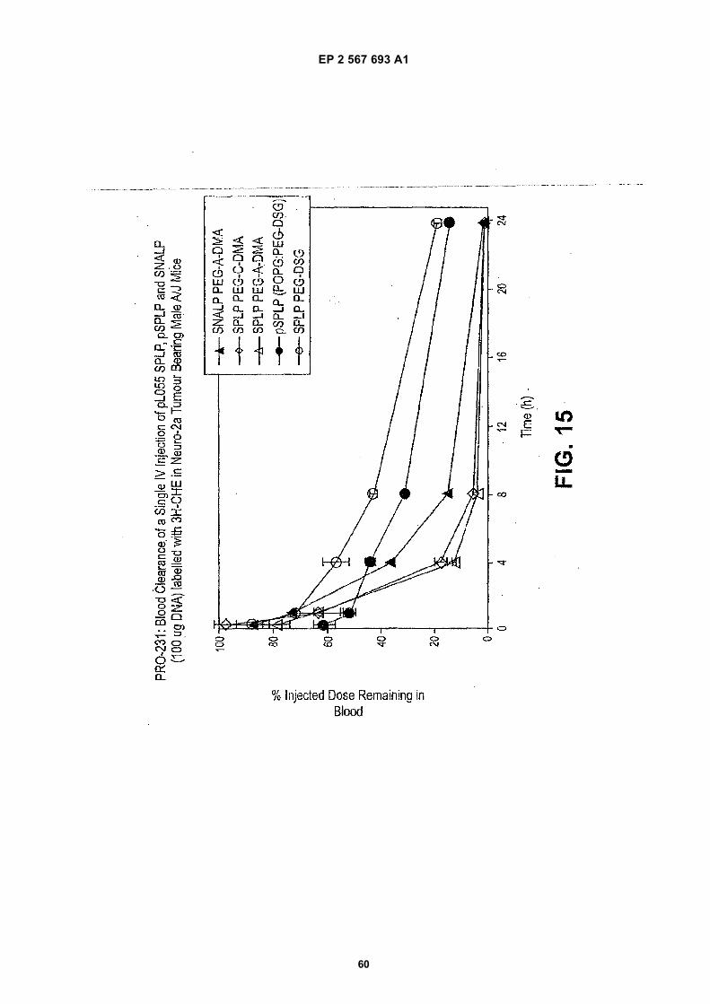
EP 2 567 693 A1
60
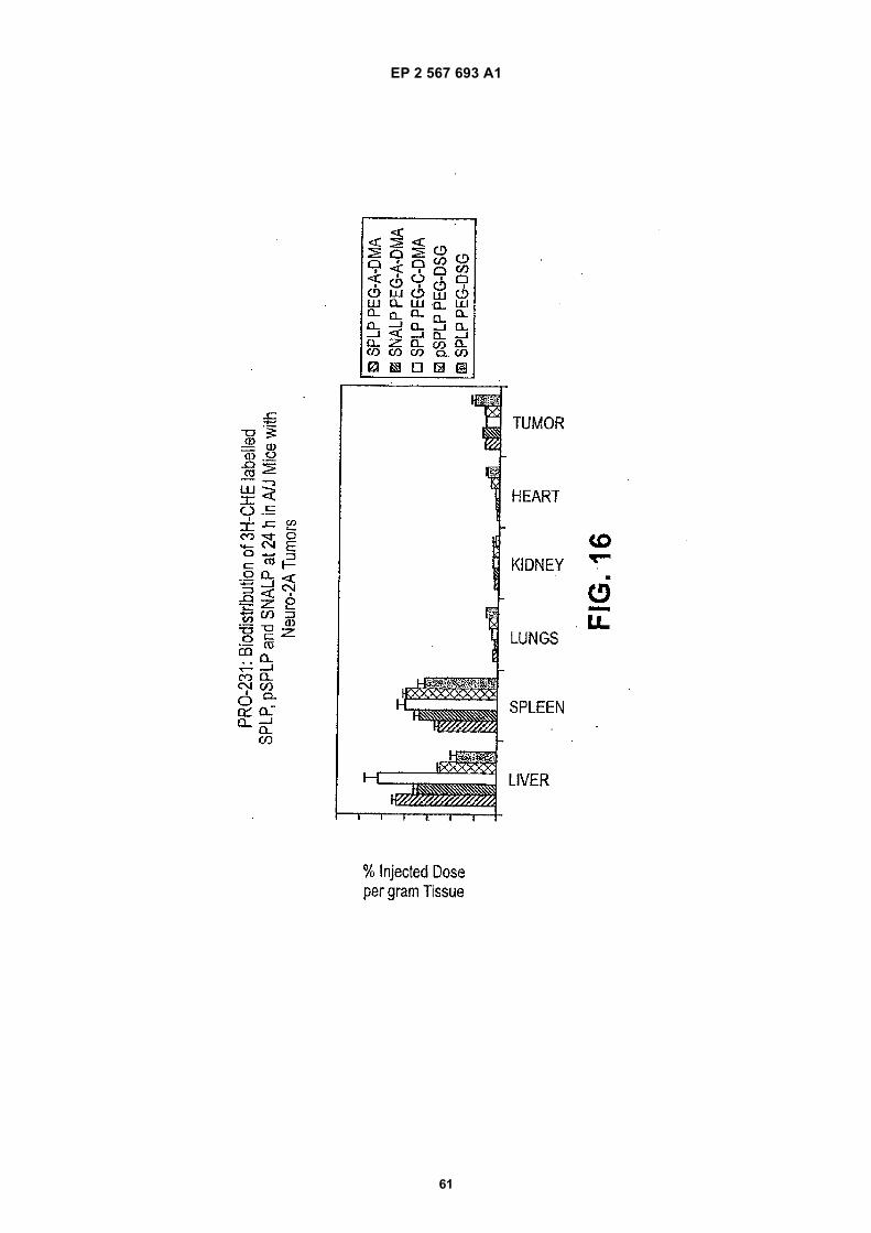
EP 2 567 693 A1
61
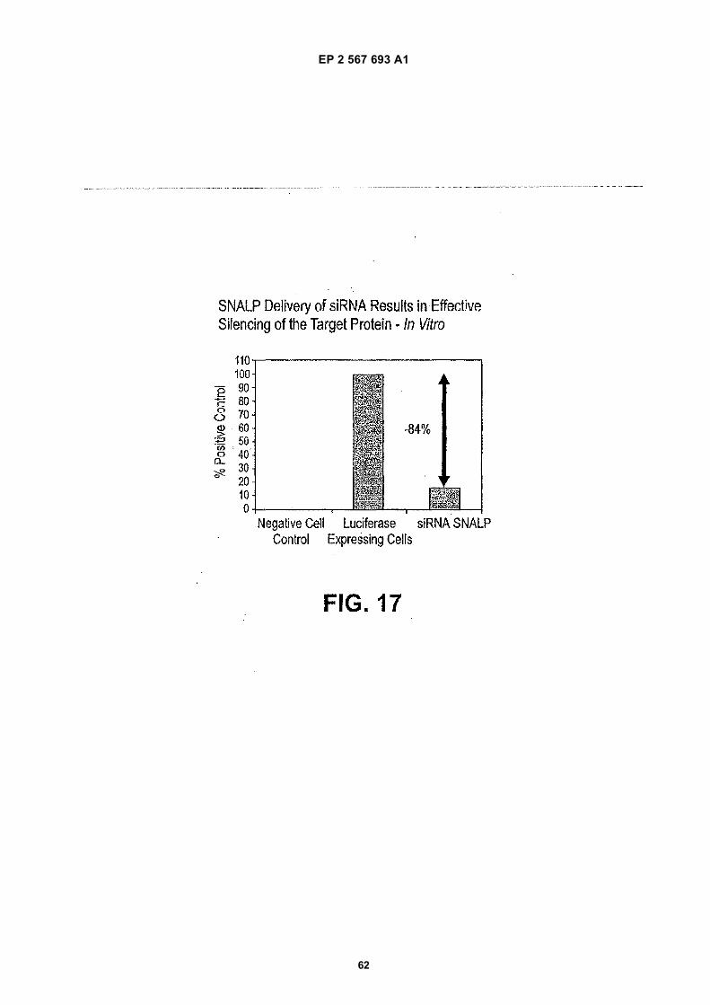
EP 2 567 693 A1
62
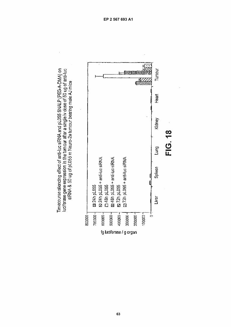
EP 2 567 693 A1
63
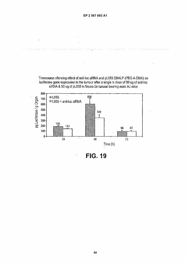
EP 2 567 693 A1
64
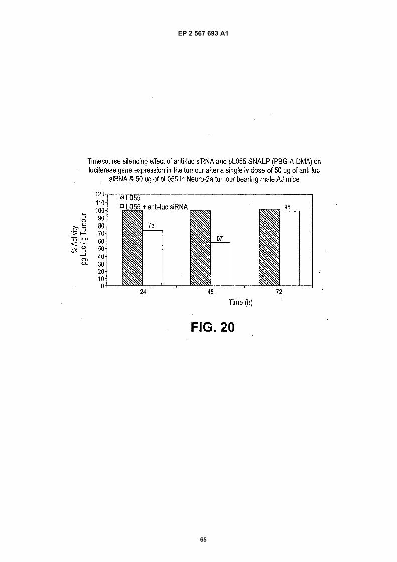
EP 2 567 693 A1
65
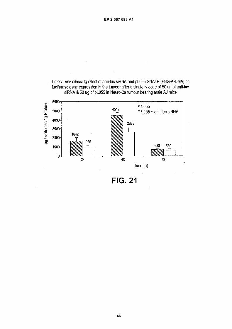
EP 2 567 693 A1
66
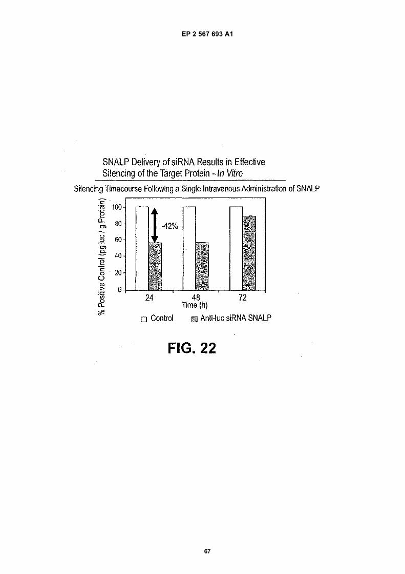
EP 2 567 693 A1
67
EP 2 567 693 A1
68
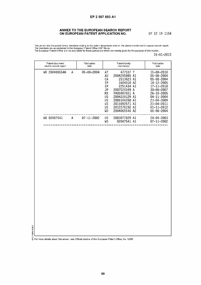
EP 2 567 693 A1
69
EP 2 567 693 A1
70
REFERENCES CITED IN THE DESCRIPTION
This list of references cited by the applicant is for the reader’s convenience only. It does not form part of the Europeanpatent document. Even though great care has been taken in compiling the references, errors or omissions cannot beexcluded and the EPO disclaims all liability in this regard.
Patent documents cited in the description
• US 52940603 P [0001]• US 60503279 P [0001]• US 60488144 P [0001]• US 6458382 B [0006]• US 20030073640 A [0006]• US 6429200 B [0007]• US 20030026831 A [0007]• US 20020081736 A [0007]• US 20030082103 A [0007]• US 5885613 A [0052] [0092]• US 6320017 B [0061] [0106]• US 6586559 B [0061] [0106]• US 5208036 A [0087] [0146]• US 5264618 A [0087] [0146]• US 5279833 A [0087] [0146]• US 5283185 A [0087] [0146]• US 5753613 A [0087] [0146]• US 5785992 A [0087] [0146]• US 5820873 A [0089] [0099] [0128]• US 136707 A [0095] [0171]• US PA20030077829 A [0095]• CA 0200669 W [0095]• US 553639 A [0100] [0120] [0158]• US 09839707 A [0100]• US PA20020072121 A [0100]• CA 0000451 W [0100] [0158]• WO 0062813 A [0100] [0120] [0158]• US 6573099 B [0111]• US 5962428 A [0111]
• US 5910488 A [0111]• US 4683195 A [0112]• US 4683202 A [0112] [0195]• US 5705385 A [0120] [0158] [0174]• US 5981501 A [0120] [0158] [0174]• US 5976567 A [0120] [0174]• US 6586410 A [0120]• US 6534484 A [0120]• US 20020072121 A [0120] [0158]• US 20030077829 A [0120] [0128] [0171]• WO 9640964 A [0120] [0174]• US 61127403 A [0121]• US 10136707 B [0128]• US 4737323 A [0132]• US 744103 A [0145]• US 6586410 B [0158]• US 6534484 B [0158]• US 09839707 B [0158]• US 6410328 B [0174]• US 5543158 A [0179]• US 5641515 A [0179] [0181]• US 5399363 A [0179]• US 5580579 A [0181]• US 5792451 A [0181]• US 5756353 A [0183]• US 5804212 A [0183]• US 5725871 A [0183]• US 5780045 A [0183]• US 5426039 A, Wallace [0195]
Non-patent literature cited in the description
• MCMANUS ; SHARP. Nature Rev. Genet., 2002, vol.3, 737 [0003]
• ELBASHIR et al. Genes Dev., 2001, vol. 15, 188[0003] [0111]
• NOVINA ; SHARP. Nature, 2004, vol. 430, 161-163[0004]
• GARBER. J. Natl. Cancer Inst., 2003, vol. 95 (7),500-2 [0004]
• WORGALL et al. Human Gene Therapy, 1997, vol.8, 37 [0005]
• PEETERS et al. Human Gene Therapy, 1996, vol. 7,1693 [0005]
• YEI et al. Gene Therapy, 1994, vol. 1, 192 [0005]• HOPE et al. Molecular Membrane Biology, 1998, vol.
15, 1 [0005]
• FELGNER. Scientific American, 1997, vol. 276, 102[0006]
• CHONN et al. Current Opinion in Biotechnology,1995, vol. 6, 698 [0006]
• HARRISON et al. Biotechniques, 1995, vol. 19, 816[0006]
• LI et al. The Gene, 1997, vol. 4, 891 [0006]• TAM et al. Gene Ther., 2000, vol. 7, 1867 [0006]• HUANG et al. Nature Biotechnology, 1997, vol. 15,
620 [0006]• TEMPLETON et al. Nature Biotechnology, 1997, vol.
15, 647 [0006]• HOFLAND et al. Pharmaceutical Research, 1997,
vol. 14, 742 [0006]• WHEELER et al. Gene Therapy, 1999, vol. 6, 271
[0008]
EP 2 567 693 A1
71
• BATZER et al. Nucleic Acid Res., 1991, vol. 19, 5081[0062]
• OHTSUKA et al. J. Biol. Chem., 1985, vol. 260,2605-2608 [0062]
• ROSSOLINI et al. Mol. Cell. Probes, 1994, vol. 8,91-98 [0062]
• YANG et al. PNAS USA, 2002, vol. 99, 9942-7 [0065]• CALEGARI et al. PNAS USA, 2002, vol. 99, 1423C
[0065]• BYROM et al. Ambion TechNotes, 2003, vol. 10 (1),
4-6 [0065]• KAWASAKI et al. Nucleic Acids Res., 2003, vol. 31,
981-7 [0065]• KNIGHT ; BASS. Science, 2001, vol. 293, 2269-71
[0065]• ROBERTSON et al. J. Biol. Chem., 1968, vol. 243,
82 [0065]• TIJSSEN. Overview of principles of hybridization and
the strategy of nucleic acid assays. Techniques inBiochemistry and Molecular Biology--Hybridizationwith Nucleic Probes, 1993 [0067]
• INNIS et al. PCR Protocols, A Guide to Methods andApplications. Academic Press, Inc, 1990 [0068]
• Current Protocols in Molecular Biology [0069]• SMITH ; WATERMAN. Adv. Appl. Math., 1981, vol.
2, 482 [0072]• NEEDLEMAN ; WUNSCH. J. Mol. Biol., 1970, vol.
48, 443 [0072]• PEARSON ; LIPMAN. Proc. Nat’l. Acad. Sci. USA,
1988, vol. 85, 2444 [0072]• Current Protocols in Molecular Biology. 1995 [0072]• ALTSCHUL et al. Nuc. Acids Res., 1977, vol. 25,
3389-3402 [0073]• ALTSCHUL et al. J. Mol. Biol., 1990, vol. 215,
403-410 [0073]• HENIKOFF ; HENIKOFF. Proc. Natl. Acad. Sci. USA,
1989, vol. 89, 10915 [0073]• KARLIN ; ALTSCHUL. Proc. Nat’l. Acad. Sci. USA,
1993, vol. 90, 5873-5787 [0074]• BRUMMELKAMP et al. Science, 2002, vol. 296, 550
[0111]• DONZÉ et al. Nucleic Acids Res., 2002, vol. 30, e46
[0111]• PADDISON et al. Genes Dev., 2002, vol. 16, 948
[0111]• YU et al. Proc. Natl. Acad. Sci., 2002, vol. 99, 6047
[0111]• LEE et al. Nat. Biotech., 2002, vol. 20, 500 [0111]• MIYAGISHI et al. Nat. Biotech., 2002, vol. 20, 497
[0111]• PAUL et al. Nat. Biotech., 2002, vol. 20, 505 [0111]• SUI et al. Proc. Natl. Acad. Sci., 2002, vol. 99, 5515
[0111]• NYKÄNEN et al. Cell, 2001, vol. 107, 309 [0111]• GUBLER ; HOFFMAN. Gene, 1983, vol. 25, 263-269
[0112]• PCR Protocols: A Guide to Methods and Applica-
tions. 1990 [0112]
• SAMBROOK et al. Molecular Cloning, A LaboratoryManual. 1989 [0112]
• KRIEGLER. Gene Transfer and Expression: A Lab-oratory Manual. 1990 [0112]
• Current Protocols in Molecular Biology. 1994 [0112]• HAMASAKI et al. FEBS Lett., 2003, vol. 543, 51
[0114]• YOKOTA et al. EMBO Rep., 2003, vol. 4, 602 [0114]• SCHLOMAI et al. Hepatology, 2003, vol. 37, 764
[0114]• WILSON et al. Proc. Natl. Acad. Sci., 2003, vol. 100,
2783 [0114]• KAPADIA et al. Proc. Natl. Acad. Sci., 2003, vol. 100,
2014 [0114]• FIELDS VIROLOGY. 2001 [0114]• BANERJEA et al. Mol. Ther., 2003, vol. 8, 62 [0114]• SONG et al. J. Virol, 2003, vol. 77, 7174 [0114]• STEPHENSON. JAMA, 2003, vol. 289, 1494 [0114]• QIN et al. Proc. Natl. Acad. Sci., 2003, vol. 100, 183
[0114]• JIA et al. J. Virol., 2003, vol. 77, 3301 [0114]• HALL. J. Virol., 2003, vol. 77, 6066 [0114]• JIANG et al. Oncogene, 2002, vol. 21, 6041 [0114]• FIELDS VIROLOGY, 2001 [0114] [0187]• FORMAN et al. Cell, 1995, vol. 81, 687 [0115]• SEOL et al. Mol. Endocrinol., 1995, vol. 9, 72 [0115]• ZAVACKI et al. PNAS USA, 1997, vol. 94, 7909
[0115]• SAKAI et al. Cell, 1996, vol. 85, 1037-1046 [0115]• DUNCAN et al. J. Biol. Chem., 1997, vol. 272,
12778-12785 [0115]• WILLY et al. Genes Dev., 1995, vol. 9 (9), 1033-45
[0115]• LEHMANN et al. J. Biol. Chem., 1997, vol. 272 (6),
3137-3140 [0115]• JANOWSKI et al. Nature, 1996, vol. 383, 728-731
[0115]• PEET et al. Cell, 1998, vol. 93, 693-704 [0115]• WILDA et al. Oncogene, 2002, vol. 21, 5716 [0116]• SCHERR et al. Blood, vol. 101, 1566 [0116]• HEIDENREICH et al. Blood, 2003, vol. 141, 3157
[0116]• NIETH et al. FEBS Lett., 2003, vol. 545, 144 [0116]• WU et al. Cancer Res., 2003, vol. 63, 1515 [0116]• LI et al. Cancer Res., 2003, vol. 63, 3593 [0116]• ZOU et al. Genes Dev., 2002, vol. 16, 2923 [0116]• VERMA et al. Clin Cancer Res., 2003, vol. 9, 1291
[0116]• KOSCIOLEK et al. Mol Cancer Ther., 2003, vol. 2,
209 [0116]• NAGY et al. Exp. Cell Res., 2003, vol. 285, 39 [0116]• TUSCHL ; BORKHARDT. Mol. Interventions, 2002,
vol. 2, 158 [0116]• COLLIS et al. Cancer Res., 2003, vol. 63, 1550
[0116]• REICH et al. Mol. Vis., 2003, vol. 9, 210 [0117]• HILL et al. J. Immunol., 2003, vol. 171, 691 [0118]• SONG et al. Nat. Med., 2003, vol. 9, 347 [0118]
EP 2 567 693 A1
72
• HEINONEN et al. FEBS Lett., 2002, vol. 527, 274[0118]
• CAPLEN et al. Hum. Mol. Genet., 2002, vol. 11, 175[0119]
• FRESHNEY. Culture of Animal Cells, a Manual ofBasic Technique. Wiley-Liss, 1994 [0167]
• KUCHLER et al. Biochemical Methods in Cell Cultureand Virology. Hutchinson and Ross, Inc, 1977 [0167]
• ZHU et al. Science, 1993, vol. 261, 209 [0175]• HYDE et al. Nature, 1993, vol. 362, 250 [0175]• BRIGHAM et al. Am, J. Med. Sci., 1989, vol. 298,
278 [0175]• REMINGTON’S PHARMACEUTICAL SCIENCES.
Mack Publishing Company, 1985 [0179]• MEDINA et al. Antiviral Res., 2003, vol. 60 (2),
135-143 [0187]• HUGLE ; CERNY. Rev. Med. Virol., 2003, vol. 13 (6),
361-71 [0187]• KOCK et al. Hepatology, 2003, vol. 38 (6), 1410-8
[0187]• BORDIER et al. J. Clin. Invest., 2003, vol. 112 (3),
407-414 [0187]• YURDAYDIN et al. J Hepatol., 2002, vol. 37 (2),
266-71 [0187]• CHIANG et al. Planta Med., 2003, vol. 69 (8), 705-9
[0187]• CHATTERJEE et al. Science, 1992, vol. 258,
1485-1488 [0188]• MALIM et al. Nature, 1989, vol. 338, 254 [0188]• KARN ; GRAEBLE. Trends Genet., 1992, vol. 8, 365
[0188]• BAYS ; STEIN. Expert Opin. Pharmacother., 2003,
vol. 4 (11), 1901-38 [0189]• Nucleic Acid Hybridization, A Practical Approach. IRL
Press, 1985 [0194]
• SAMBROOK et al. Molecular Cloning: A LaboratoryManual. Cold Spring Harbor Laboratory Press, 2000[0195]
• AUSUBEL et al. SHORT PROTOCOLS IN MOLEC-ULAR BIOLOGY. Current Protocols, 2002 [0195]
• PCR Protocols A Guide to Methods and Applications.Academic Press Inc, 1990 [0195]
• ARNHEIM ; LEVINSON. C&EN, 01 October 1990,36 [0195]
• The Journal Of NIH Research, 1991, vol. 3, 81 [0195]• KWOH et al. Proc. Natl. Acad. Sci. USA, 1989, vol.
86, 1173 [0195]• GUATELLI et al. Proc. Natl. Acad. Sci. USA, 1990,
vol. 87, 1874 [0195]• LOMELL et al. J. Clin. Chem., 1989, vol. 35, 1826
[0195]• LANDEGREN et al. Science, 1988, vol. 241, 1077
[0195]• VAN BRUNT. Biotechnology, 1990, vol. 8, 291
[0195]• WU ; WALLACE. Gene, 1989, vol. 4, 560 [0195]• BARRINGER et al. Gene, 1990, vol. 89, 117 [0195]• SOOKNANAN ; MALEK. Biotechnology, 1995, vol.
13, 563 [0195]• BEAUCAGE ; CARUTHERS. Tetrahedron Letts.,
1981, vol. 22 (20), 1859-1862 [0196]• NEEDHAM VANDEVANTER et al. Nucleic Acids
Res., 1984, vol. 12, 6159 [0196]• PEARSON ; REGNIER. J. Chrom., 1983, vol. 255,
137-149 [0196]• MAXAM ; GILBERT. Methods in Enzymology. Aca-
demic Press, 1980, vol. 65, 499 [0196]• ANGERER et al. Methods Enzymol., 1987, vol. 152,
649 [0197]


![United States Patent [19] [11] Patent Number: 6,021,398](https://img.pdfslide.net/doc/110x75/6336422ee687b37e4f0610b8/united-states-patent-19-11-patent-number-6021398.jpg)


