Embed Size (px)
Citation preview

Citation: Mbongue, J.C.; Vanterpool,
E.; Firek, A.; Langridge, W.H.R.
Lipopolysaccharide-Induced
Immunological Tolerance in
Monocyte-Derived Dendritic Cells.
Immuno 2022, 2, 482–500. https://
doi.org/10.3390/immuno2030030
Academic Editor: Alessandro
Perrella
Received: 12 July 2022
Accepted: 11 August 2022
Published: 15 August 2022
Publisher’s Note: MDPI stays neutral
with regard to jurisdictional claims in
published maps and institutional affil-
iations.
Copyright: © 2022 by the authors.
Licensee MDPI, Basel, Switzerland.
This article is an open access article
distributed under the terms and
conditions of the Creative Commons
Attribution (CC BY) license (https://
creativecommons.org/licenses/by/
4.0/).
Review
Lipopolysaccharide-Induced Immunological Tolerance inMonocyte-Derived Dendritic CellsJacques C. Mbongue 1,* , Elaine Vanterpool 1 , Anthony Firek 2,3 and William H. R. Langridge 3
1 Department of Biological Sciences, Oakwood University, Huntsville, AL 35896, USA2 Comparative Effectiveness and Clinical Outcomes Research Center (CECORC), Riverside University Health
System, Moreno Valley, CA 92555, USA3 Center for Health Disparities and Molecular Medicine, 11085 Campus Street, Mortensen Hall, Department of
Basic Sciences, Loma Linda University School of Medicine, Loma Linda, CA 92354, USA* Correspondence: [email protected]
Abstract: Bacterial lipopolysaccharides (LPS), also referred to as endotoxins, are major outer surfacemembrane components present on almost all Gram-negative bacteria and are major determinantsof sepsis-related clinical complications including septic shock. LPS acts as a strong stimulator ofinnate or natural immunity in a wide variety of eukaryotic species ranging from insects to humansincluding specific effects on the adaptive immune system. However, following immune stimulation,lipopolysaccharide can induce tolerance which is an essential immune-homeostatic response thatprevents overactivation of the inflammatory response. The tolerance induced by LPS is a state ofreduced immune responsiveness due to persistent and repeated challenges, resulting in decreasedexpression of pro-inflammatory modulators and up-regulation of antimicrobials and other mediatorsthat promote a reduction of inflammation. The presence of environmental-derived LPS may play akey role in decreasing autoimmune diseases and gut tolerance to the plethora of ingested antigens.The use of LPS may be an important immune adjuvant as demonstrated by the promotion of IDO1increase when present in the fusion protein complex of CTB-INS (a chimera of the cholera toxin Bsubunit linked to proinsulin) that inhibits human monocyte-derived DC (moDC) activation, whichmay act through an IDO1-dependent pathway. The resultant state of DC tolerance can be furtherenhanced by the presence of residual E. coli lipopolysaccharide (LPS) which is almost always presentin partially purified CTB-INS preparations. The approach to using an adjuvant with an autoantigenin immunotherapy promises effective treatment for devastating tissue-specific autoimmune diseaseslike multiple sclerosis (MS) and type 1 diabetes (T1D).
Keywords: immunological tolerance; LPS; cholera toxin B; indoleamine 2,3 dioxygenase
1. Introduction
Understanding the role of LPS in both the toxicity of bacterial infections and effectson immune regulation has emerged as a critical objective due to the global importance ofsepsis. The LPS bacterial surface molecule is produced by most Gram-negative bacteria.The attention LPS received in the early 20th century was due to its ability to stimulatethe immune system, and was known as endotoxin glycolipids [1]. It was subsequentlydiscovered that LPS created a permeable barrier on the cell surface and was the maindriver of the innate resistance of Gram-negative bacteria to many antibacterial agents [1–4].Unsurprisingly, these important properties of LPS have provided a vast and extensiveliterature for over 100 years.
The innate immune system’s detection of microorganisms or microbial components ismediated by a special set of proteins called pattern recognition receptors (PRRs). One ofthe best studied PRRs is the bacterial LPS receptor, Toll-like receptor 4 (TLR4) [5,6]. TLR4 isan important driver of the immune response to bacterial infections and its dysregulationis thought to promote abnormal cytokine production, leading to bacterial sepsis [3,4,7,8].
Immuno 2022, 2, 482–500. https://doi.org/10.3390/immuno2030030 https://www.mdpi.com/journal/immuno

Immuno 2022, 2 483
Because sepsis remains one of the major conditions leading to acute morbidity and mortality,understanding the nature of TLR4 signaling will direct efforts towards understanding thebasic mechanisms underlying inflammation and can lead to improved clinical outcome.Bacterial LPS is widely used in inflammation models because it induces many inflammatoryeffects by promoting the production and release of pro-inflammatory cytokines such asTNF-α, IL-1β, and IL-6 [8–10].
LPS signaling by PRR leads to the activation of intracellular signaling networks thatpromote the expression of inflammatory genes which stimulate the acute and sustained defenseof the host [6,7]. TLR4 first encounters LPS in extracellular space when interacting with intactbacteria or when exposed to soluble LPS aggregates. Upon binding to LPS, TLR4 rapidlyinduces the assembly of supramolecular organization centers (SMOC) called myddosomes [11].
The intermediate consists of the MyD88 adapter protein and several serine-threoninekinases from the TIRAP and IRAK families [5–8]. This hub-like organizing center is amajor subcellular site where TLR4 signaling activates the NF-κB and AP-1 pathways topromote the expression of inflammatory genes [12–14]. Subsequently, TLR4 is taken up byendosomes and promotes the production of IRF3-dependent interferon type I (IFN) viaTRAM and TRIF adaptor proteins [15–17].
LPS acts as a proto-endotoxin and contributes to the inflammatory cascade becauseit binds to the CD14/TLR4/MD2 receptor complex in many cell types, but primarily tomonocytes, dendritic cells, macrophages, and B cells. In these cell types, LPS stimulatesthe secretion of proinflammatory cytokines, eicosanoids, and nitric oxide [1,7]. Due to itsrole in the activation of several transcription factors, LPS activity has been experimentallyinvestigated for many years [1,2,7,18].
Humans are more sensitive to LPS than other animals (e.g., mice). An LPS doseof 1 mcg/kg causes shock in humans, but mice can tolerate doses up to 1000 times thatamount [19]. For this reason, LPS levels in pharmaceutical products and medical devicesmust be strictly monitored using the limulus amebocyte lysate (LAL) assay. This require-ment may be due to differences in the amount of circulating natural anti-LPS antibodiesbetween the two species [20,21].
Multiple pathways may be activated on engagement with LPS because it has beenshown that LPS has both an immunostimulatory as well as immunosuppressive rolesto play in immune activation of immune cells [22,23]. Consequently, despite LPS beingrecognized as a classical immune stimulating factor best characterized in bacterial infection,there is clear evidence of a far more complex role for LPS in the immune cascade.
Dendritic cells (DCs), considered the most potent APCs, are critical gateways of theimmune system and have the unique ability to synthesize a wide range of input signalsand transmit them to naive lymphocytes, thus directing immunization, or suppression ofpathogenic microorganisms and tumors [24]. When confronted with pathogen-associatedmolecular structures, DCs “mature” by upregulating the expression of MHC class II re-ceptors that exhibit antigen, cofactors, and processed cytokines and chemokines [25]. DCscontain TLRs which are major pattern recognition receptors that initiate and regulate im-mune responses via various signaling pathways [26]. Thus, the application and targetedregulation of DCs to control cancer and infectious diseases is being pursued in the develop-ment of clinical therapeutics [27]. However, DCs are also involved in the pathogenesis ofdiseases caused by immune cell dysfunction, such as chronic inflammation, autoimmunity,and cancer development and progression [28,29]. Uptake processing and presentationof self-antigens as foreign proteins is considered fundamental to the development of au-toimmune conditions such as type 1 diabetes. Thus, targeting the downregulation of DCactivation may be a useful strategy for treatment of these diseases.
Monocytes are also important in the early acute inflammatory phase of the immuneresponse to an infectious agent because they can stimulate and modulate the adaptive immunesystem by inducing cytokine secretion and antigen presentation to T cells [30,31]. The adaptiveimmune response is complimentary to the innate immune response and these two processescan simultaneously eliminate pathogens. In most cases, monocytes initiate and enhance
Immuno 2022, 2 484
the immune response. However, LPS activation of monocytes has been shown to suppressthe T cell immune response and induce the expression of FOXP3 regulatory T cell functionmodulators in resting CD4 + CD25 T cells via a PGE2-dependent mechanism [32].
LPS also plays an immunosuppressive role in autoimmunity [22]. For example, repeatedexposure to LPS causes a state of endotoxin tolerance that, in part, contributes to the well-recognized state of immunosuppression seen in sepsis [33,34]. This effect is particularlyimportant in antigen presenting cells such as monocytes [35] and dendritic cells [27,36,37].
Furthermore, exposure of bone marrow-derived dendritic cells (BMDC) to high dosesof pure lipopolysaccharide for 24 h (LPS-primed BMDC) increases their potency in theprevention of inter-photoreceptor retinoid binding protein in Freund’s adjuvant-inducedexperimental autoimmune uveoretinitis (EAU) [38].
The concept that exposure to LPS is important for interacting with the immune systemto prevent allergic and autoimmune diseases has a long history. Strachan proposed thehygiene hypothesis in 1989, and since then there have been many studies indicatingdifferences in endotoxin levels in different habitats. These studies showed an associationbetween LPS in house dust and the incidence of asthma. Children in rural areas havesignificantly fewer autoimmune diseases, such as type 1 diabetes, asthma, allergies, andgeneralized atopy, compared to children in urban environments, but these observationsare largely descriptive [39]. Children who grew up on farms had lower rates of allergiesand asthma, and dust-contaminated bedding and mattresses in their homes containedhigher levels of LPS [39], a result suggesting that chronic environmental LPS exposure canpromote immunotolerance to environmental antigens.
Early studies in animal models showed that the increased sensitivity of C3H/HeJmice to food allergens was due to an inability to signal through TLR4 [40]. Neonataladministration of a cocktail of broad-spectrum antibiotics induced a food allergy responsein TLR4-sufficient mice—similar to those seen in TLR4-deficient mice—which identifiesthe intestinal microbiota as a source of TLR4 ligand [40]. The authors of this new studyconfirm that several mouse model studies have shown that NOD mice that are sensitive toT1D are protected against the disease by oral or intraperitoneal administration of LPS [41].Recent developments have revealed the mechanisms underlying adjuvant stimulatedfusion protein vaccines such as the cholera toxin B subunit adjuvant linked to autoantigenslike proinsulin (CTB-INS) for the protection against autoimmunity. It was shown thatCTB-INS impedes human monocyte-derived DC (moDC) activation through stimulationof indoleamine 2,3 dioxygenase (IDO1) biosynthesis [42,43]. The resulting state of DCtolerance was enhanced by the residual presence of E. coli lipopolysaccharide (LPS) inpartially purified CTB-INS preparations [37]. This adjuvant-like action for LPS is nowrecognized in vaccine development. However, the toxicity of LPS may limit its use [44,45].Later in this review we examine in a detailed fashion, the role of LPS in the suppression ofCTB-INS-induced DC activation in the context of autoimmunity.
2. Function of LPS2.1. Virulence and Toxicity
Lipid A, which is the toxic component of LPS, and polysaccharide side chains, whichare considered the non-toxic but immunogenic part of LPS, act as virulence determinantsin Gram-negative bacteria [46–48]. O antigens have adhesive properties, phagocyte re-sistance, antigen protection, and antigen mutation properties [47,49]. Lipid A acts as animmunostimulant that induces biological responses to a specific organism [50–52].
2.2. Biological Activity of Lipopolysaccharide
An animal’s biological immune responses can be analyzed using various parameters,such as an injection of live or killed Gram-negative cells or purified LPS in laboratoryanimals, which causes a broad spectrum of pathophysiological responses, such as fever,changes in blood counts, disseminated intravascular coagulation white blood cells, hy-potension, and shock resulting in death. Injecting very small doses of endotoxin can cause

Immuno 2022, 2 485
death in most mammals. The sequence of events follows a regular pattern: (1) latencyperiod; (2) physiological stress (diarrhea, exhaustion, shock); and (3) death. The rate atwhich death occurs depends on the dose of the endotoxin, the route of administration ofthe toxin, and the animal species.
3. Lipopolysaccharide Signaling and Immune Activation Mechanisms inHigher Organisms3.1. Lipopolysaccharide Detoxification Mechanisms in Higher Animals
The defense against infection in vertebrates is mediated by two interdependent armsof the immune system, known as innate and adaptive portions of the immune system. Theinnate immune system, consisting of antigen presenting cells, recognizes a diverse array ofnon-self-antigens and if overwhelmed, can signal and activate the adaptive immune systemthrough well-established signaling pathways to stimulate an array of T-cells and B-cellsto overcome the pathogen [53]. As LPS can have significant adverse effects on animalsand humans, a process to detoxify LPS has been developed [54]. The detoxification mech-anism of LPS occurs through enzymatic degradation or through complement-mediateddetoxification, which leads to the breakdown of LPS.
3.2. Host-Microbe Interactions (Lipopolysaccharide Activity) in Invertebrates—Insects
The innate immune system of insects plays an important role in the developmentof immunity [55]. In recent years, arthropods and insects have become the most usefulmodels for describing the molecular regulation of the innate immune response [56]. Insectshave highly effective defense mechanisms against invasive microorganisms, which includeGram-negative and Gram-positive molecules, LPS, and peptidoglycan [55–57].
These insect defense mechanisms include cellular and humoral responses. Cellularresponses include phagocytosis and/or encapsulation of large parasites by bacterial nodulesand blood cells [58]. In addition, the humoral response uses various antimicrobial peptideswhich are synthesized in the adipose body and some hemocytes after induction by septiclesions and which are then secreted into the hemolymph [59–61]. The insect defense systemagainst LPS pathogens results in a transient increase in antimicrobial activity in the acellu-lar hemolymph, including phagocytosis and encapsulation of invaders by blood cells andsubsequent production of antimicrobial proteins (mainly in the insect’s adipose tissue) [62].Strong immunoreactivity was found in the interaction between Galleria mellonella (large waxmoth) and LPS. The high tolerance of LPS to insects can be explained by an extremely effectivedetoxification mechanism involving the binding of LPS to hemolymph lipophorins [63]. Thisobservation suggests that LPS has the potential to induce immune activation.
Activation of the proteolytic cascade and coagulation cascade using LPS triggersthe limulus hemocyte to act as a signaling mechanism [64–66]. In addition, a blood cellmembrane receptor for LPS has been isolated from Bombyx mori silkworm that can transmitan activation signal for the synthesis of the antibacterial peptide cecropin B [67–69].
3.3. Expression of Genes and Signaling Action Induced by Lipopolysaccharide in Vertebratesand Invertebrates
It is a general phenomenon that antibacterial protein gene expression culminates afew hours after bacterial infection and decreases over time in vertebrates. This reductionin antibacterial protein gene expression has been shown to correlate with LPS depriva-tion [70,71]. TLRs, a class of pattern recognition receptors (PRRs) found in vertebrates, playan important role not only in initiating innate immunity, but also in activating adaptiveimmunity (Figure 1).
Immuno 2022, 2 486
Immuno 2022, 2, FOR PEER REVIEW 5
3.3. Expression of Genes and Signaling Action Induced by Lipopolysaccharide in Vertebrates and
Invertebrates
It is a general phenomenon that antibacterial protein gene expression culminates a
few hours after bacterial infection and decreases over time in vertebrates. This reduction
in antibacterial protein gene expression has been shown to correlate with LPS deprivation.
[70,71]. TLRs, a class of pattern recognition receptors (PRRs) found in vertebrates, play an
important role not only in initiating innate immunity, but also in activating adaptive im-
munity (Figure 1).
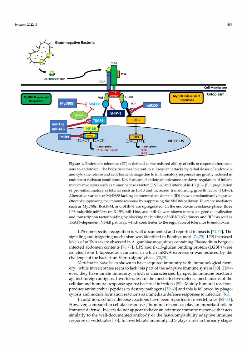
Figure 1. Endotoxin tolerance (ET) is defined as the reduced ability of cells to respond after exposure
to endotoxin. The body becomes tolerant to subsequent attacks by lethal doses of endotoxin, and
cytokine release and cell/tissue damage due to inflammatory responses are greatly reduced in en-
dotoxin-resistant conditions. Key features of endotoxin tolerance are down-regulation of inflamma-
tory mediators such as tumor necrosis factor (TNF-α) and interleukin-1β (IL-1β), upregulation of
pro-inflammatory cytokines such as IL-10 and increased transforming growth factor (TGF-β). Alter-
native variants of MyD88S lacking an intermediate domain (ID) show a predominantly negative
effect of suppressing the immune response by suppressing the MyD88 pathway. Tolerance media-
tors such as MyD88s, IRAK-M, and SHIP-1 are upregulated. In the endotoxin resistance phase, three
LPS-inducible miRNAs (miR-155, miR-146α, and miR-9), were shown to mediate gene colocaliza-
tion and transcription factor binding by blocking the binding of NF-kB p50 dimers and IRF3 as well
as TRAF6-dependent NF-kB pathway, which contributes to the regulation of tolerance to endotox-
ins.
LPS non-specific recognition is well documented and reported in insects [72,73]. The
signaling and triggering mechanism was identified in Bombyx mori [74,75]. LPS-increased
levels of mRNAs were observed in A. gambiae mosquitoes containing Plasmodium
berguei-infected abdomen contents [76,77]. LPS and β-1,3-glucan binding protein (LGBP)
were isolated from Litopenaeus vannamei in which mRNA expression was induced by
the challenge of the bacterium Vibrio alginolyticus [78,79].
Vertebrates have been shown to have acquired immunity with 'immunological
memory', while invertebrates seem to lack this part of the adaptive immune system [80].
However, they have innate immunity, which is characterized by specific immune
Figure 1. Endotoxin tolerance (ET) is defined as the reduced ability of cells to respond after expo-sure to endotoxin. The body becomes tolerant to subsequent attacks by lethal doses of endotoxin,and cytokine release and cell/tissue damage due to inflammatory responses are greatly reduced inendotoxin-resistant conditions. Key features of endotoxin tolerance are down-regulation of inflam-matory mediators such as tumor necrosis factor (TNF-α) and interleukin-1β (IL-1β), upregulationof pro-inflammatory cytokines such as IL-10 and increased transforming growth factor (TGF-β).Alternative variants of MyD88S lacking an intermediate domain (ID) show a predominantly negativeeffect of suppressing the immune response by suppressing the MyD88 pathway. Tolerance mediatorssuch as MyD88s, IRAK-M, and SHIP-1 are upregulated. In the endotoxin resistance phase, threeLPS-inducible miRNAs (miR-155, miR-146α, and miR-9), were shown to mediate gene colocalizationand transcription factor binding by blocking the binding of NF-kB p50 dimers and IRF3 as well asTRAF6-dependent NF-kB pathway, which contributes to the regulation of tolerance to endotoxins.
LPS non-specific recognition is well documented and reported in insects [72,73]. Thesignaling and triggering mechanism was identified in Bombyx mori [74,75]. LPS-increasedlevels of mRNAs were observed in A. gambiae mosquitoes containing Plasmodium berguei-infected abdomen contents [76,77]. LPS and β-1,3-glucan binding protein (LGBP) wereisolated from Litopenaeus vannamei in which mRNA expression was induced by thechallenge of the bacterium Vibrio alginolyticus [78,79].
Vertebrates have been shown to have acquired immunity with ‘immunological mem-ory’, while invertebrates seem to lack this part of the adaptive immune system [80]. How-ever, they have innate immunity, which is characterized by specific immune reactionsagainst foreign antigens. Invertebrates are the most effective defense mechanisms of thecellular and humoral response against bacterial infections [57]. Mainly humoral reactionsproduce antimicrobial peptides to destroy pathogens [59,60] and this is followed by phago-cytosis and nodule formation reactions as immediate defense responses to infection [81].
In addition, cellular defense reactions have been reported in invertebrates [82–84].However, compared to cellular responses, humoral responses play an important role inimmune defense. Insects do not appear to have an adaptive immune response that actssimilarly to the well-documented antibody or the histocompatibility adaptive immuneresponse of vertebrates [55]. In invertebrate immunity, LPS plays a role in the early stages

Immuno 2022, 2 487
of signaling that activates acute phase protein genes. In particular, future research onimmune surveillance and purification of pathogens in vertebrates and invertebrates coulddemonstrate the efficacy of innate immune systems based on bacterial endotoxins.
4. LPS in Inflammatory-Mediated Pathogenesis4.1. Role of LPS from the Gut Microbiome in Inflammatory Conditions
The composition of the gut microbiome plays a critical role in maintaining local gutintegrity, systemic homeostasis, and in regulating local and systemic immune responses.The microbiota can play a major role in immune regulation as well as the developmentof pathologies such as obesity, diabetes, fatty liver disease, carcinoma, and autoimmunediseases [85]. Incidences of esophageal adenocarcinoma have been increasing in recentyears and it has been proposed to be linked to the resident local microbiota [86]. Recognitionof the potential role of LPS was observed when mice fed a high-fat diet had higher blood LPSlevels than normal chow-fed mice, resulting in inflammation of the liver and adipose tissue,which led to the development of insulin resistance, defining this condition as metabolicendotoxemia [87,88]. This low level of systemic LPS is related to an increased permeabilityor “leaky gut” phenomenon promoting inflammation in various tissue sites [89].
Subsequent studies on metabolic endotoxemia have been conducted for a varietyof diseases. It has been reported that blood LPS levels are higher in humans with obe-sity [90,91]. The role of locally produced LPS has emerged as an important humoral factorthat may have a key role in local and systemic inflammation and immune regulation [92].
The role that changes in the microbiome have on LPS production suggests that dietand the types of foods may both promote adverse immune–metabolic changes and canalso serve as a remedy as demonstrated by the beneficial effects of green tea, a plant-based vegetarian diet [93], the Dietary Approaches to Stop Hypertension (DASH), andMediterranean diets [94,95].
4.2. Bacterial LPS-Induced Lung Injury and Pathologies
As previously mentioned, inflammatory events can be regulated by NF-kB. TLR-4activation by LPS can induce injury in areas where TLR-4 is activated [96]. Acute lunginjury and acute respiratory distress syndrome are a result of strong inflammation that canpotentially lead to extreme severe consequence including severe pulmonary edema anddamage potentially triggering respiratory failure [96].
Non-typable Hemophilus influenzae (NTHi) causes lower respiratory tract infections,especially in patients with chronic obstructive pulmonary disease (COPD). Generally, NTHiinfection is cleared by alveolar macrophages, but in patients with COPD it may not bereadily cleared from the airway [97]. NTHi infections result in a TLR-dependent immuneresponse in alveolar macrophages, eliciting the release of pro-inflammatory cytokinesrecruiting neutrophils to the lungs [98–101]. Proteins including Pellino-1 are E3 ubiquitinligases that play a role in TLR-4 signaling in monocytes [102]. Expression levels of Pellino-1are associated with persistent bacterial infections. Studies show that macrophages canup-regulate Pellino-1 in response to LPS and NTHi through TLR4 mechanisms [102,103]. Inthe Hughes et al. study, the absence of Pellino-1 gene in mice led to development of airwayinflammation and resulted in recruitment of neutrophils. Another study shows that IL-5expression in human peripheral blood mononuclear cells was induced by H. influenzaeLPS. This increase in IL-5 expression is suggested to influence eosinophilic inflammation inpatients with COPD [104].
4.3. LPS-induced Meningococcal Inflammatory Disorders
Neisseria meningitidis is a Gram-negative coccus that is implicated in meningitis andfulminant meningococcemia or meningococcal septicemia. The presence of N. meningitidisin the subarachnoid space can result in meningitis and while the organism continues tocirculate and disseminate, it can lead to septicemia. N. meningitidis LPS cell activationrequires an interaction with lipid A. Studies show that patients with fulminant septicemia

Immuno 2022, 2 488
have high levels of LPS and CXCL10 while patients that manifest clinical symptoms ofmeningitis have low levels of LPS. Additional studies propose that meningococcal LPS leadsto early expression of IFN-B through TLR-MyD88-independent pathway. It is suggestedthat IFN-B could activate the JAK/STAT signaling pathway [105].
4.4. LPS-induced Periodontal Inflammatory-Mediated Pathologies
Periodontitis is a chronic inflammation of the gum tissues that support the teeth.Gram-negative anaerobic and microaerophilic organisms including Porhyromonas gingivalis,Treponema denticola, Tannerella forsythia, and Aggregatibacter actinomycetescomitans are com-mon etiological agents in periodontal disease. These bacteria are significant activators ofinflammation in cells and tissues through increased secretion of inflammatory cytokinesIL-B1, IL-6, and TNFα, among others. Studies report that hypoxia in the gingival region iscommonly associated with inflammation [106]. The state of hypoxia can lead to activationof caspase-1 resulting in stimulating activation of IL-1B and IL-18 [106]. Gingival fibroblastsunder hypoxia stimulated with P. gingivalis LPS resulted in the activation of caspase-1 andcaspase 11 as well as IL-1B maturation [107]. Under normoxic conditions, in this study,there was a down-regulation of NLRP3, IL-1B precursor, and mature IL-1B resulting in adecrease in inflammation.
Autophagy is another important mechanism that stimulates inflammation. It has beenshown that P. gingivalis LPS can stimulate reactive oxygen species-mediated autophagy.Further studies also showed that P. gingivalis LPS induced autophagy in human gingivalfibroblasts [108]. Autophagy is generally down-regulated by the PI3K/Akt/mTOR path-way [108]. Suppression of these pathways are associated with autophagy. Human gingivalfibroblast cells stimulated by P. gingvalis LPS showed suppression of PI3K/Akt/mTORactivity implicating autophagy [108].
In periodontal disease pathogenesis, LPS contributes to cellular senescence due toinflammation and remodeling of the extracellular matrix [109]. Studies show that exposureto Gram-negative bacteria components can facilitate cellular aging or senescence [110,111].Further, prolonged exposure to LPS was shown to be proinflammatory and genotoxic onperiodontal associated cells [112,113]. An example of such mechanisms can be seen inperiodontitis, infection of gum tissues. Periodontitis is associated with alveolar bone loss.Studies show that bacterial LPS exposure in alveolar osteocytes can trigger prematurealveolar osteocyte senescence leading to alveolar bone loss [109].
4.5. LPS and Neuroinflammation
There is evidence correlating inflammation with dementia and nerve injury. LPS fromGram-negative bacteria modulate TLR 2 and 4 as well as cytokine expression [114]. Studiesstrongly correlate chronic periodontitis with dementia [115–117]. These inflammatory re-sponses can lead to neuronal loss and apoptosis. Additional studies show that P. gingivalisand Escherichia coli LPS can induce inflammatory changes within the nervous system [115].Behavioral studies indicated that P. gingivalis LPS can induce cognitive impairment. WhileLPS from both P. gingivalis and E. coli impaired spatial learning and memory, P. gingivalisLPS was shown to activate microglia and astrocyte immune cells in the cortex and hip-pocampus [115]. Expression analysis of mice cortex stimulated with P. gingivalis showedan upregulation in proinflammatory cytokines TNF-α, IL-1β, IL-6, and IL-8 [115,118]. Mi-croglia can produce potent proinflammatory cytokines inducing neuroinflammation [119].Inflammation normally precedes the production of amyloid plaque development in braintissue. In addition, LPS activated TLR4 leading to stimulation of the NF-kB signalingpathway, resulting in upregulation of TLR4, CD14, IRAK1, and p-p65 [115].
4.6. LPS Aggravates Inherited Retinal Dystrophy
Activated microglia release inflammatory mediators contributing to neurodegener-ative diseases including retinal neurodegenerative diseases. Studies have shown thatsystemic administration of LPS activate retinal microglia [120]. Experiments with Sprague

Immuno 2022, 2 489
Dawley rats show there are no significant morphological changes that alter retinal func-tion [121]. Using a retinal dystrophy rat model, P23H, studies show that LPS exposureincreases the degeneration of the P23H retina in comparison with untreated retina [122]. Toevaluate changes on a molecular level, mRNA analyses show that LPS injection in SpragueDawley rats caused upregulation in retinal expression of apoptotic genes including caspase8, Bad, Bax, and Bcl-2 among others [121]. There were no inflammation genes that wereupregulated in response to LPS. In P23H rats, the retina did show upregulation of severalinflammation genes including TNF-α and IL-1β cytokines. Genetic analysis of apoptosis inp23H rat retinas treated with LPS showed upregulation of pro-apoptotic factors includingBcl-2, Bax, Bad, and Apaf-1 [121].
4.7. LPS-Induced Perinatal Impact of Maternal Inflammation
Chronic inflammatory responses are implicated in pre-term births, perinatal morbidity,and mortality. Pregnant mice challenged with E. coli LPS were likely to go into preterm labor.Further LPS exposure was able to cause neutrophilic infiltrates in fetal renal and pulmonarytissues [123,124]. When LPS was injected mid-or full gestation into pregnant sheep, therewere observable injuries to the fetal brain, particularly in the white matter [125–128].Intracerebral LPS exposure showed increased pro-inflammatory markers and caused a lossof oligodendroglial precursors. Intracerebral LPS exposure was also implicated in loss ofmyelination and bilateral ventricular dilation [129–131]. In addition, Oskvig et al. showedthat pregnant Sprague Dawley rats exposed or challenged with LPS showed an increasein maternal serum cytokines [132]. The amniotic fluid and fetal brains of LPS challengedrats also showed increases in pro-inflammatory cytokine levels. The marked increase in thematernal immune response was shown to dysregulate gene expression in the fetal brain.
5. The Function of LPS in Immune Suppression5.1. Endotoxin Tolerance
Endotoxin tolerance (ET) is an important immune homeostatic response that pre-vents unnecessary overactivation of the inflammatory response. It impairs the response torepeated attacks against LPS, leading to decreased expression of anti-inflammatory modu-lators, antimicrobial up-regulation, and other mediators that contribute to the inhibition ofinflammation. If the reaction is not controlled, endotoxic shock can occur, which can leadto death. Though the mechanisms associated with ET are still largely under investigation,several studies have given us some insight.
The aryl hydrocarbon receptor (AhR) participates in protection against LPS-mediatedtissue damage through ET, as it plays a necessary role in restraining the proinflammatoryaction of IL-1β and TNF-α while fostering the expression of protective TGF-β [133]. TGF-β,in turn, promotes durable expression of the tolerogenic enzyme indoleamine 2,3-dioxygenase1 (IDO1) [42]. In the same vein, it has been shown TLR4 ligation in LPS-primed DCs inducedhigher levels of IDO isoforms together with the transcription factor aryl-hydrocarbon receptor(AhR) compared to unprimed controls [134]. These data could potentially explain whyMbongue et al. observed higher levels of IDO1 in human monocyte-derived DCs co-culturedwith CTB-INS fusion protein [42] later found to contain significant amounts of LPS [37].
Upon binding to TLR4, LPS stimulates two signaling pathways—a gene-dependent path-way of the primary myeloid differentiation response 88 (My-D88) and a pathway independentof MyD88—both of which lead to NF-κB activation. ET is linked to overexpression of p50NF-κB homodimers and decreased levels of the active p65/p50 NF-κB heterodimers [33,135].Recent evidence has also shown RelB plays an important role in gene silencing in resistance.TLR4-induced RelB activation, together with G9a methyltransferase and histone H3 demethy-lation, induces a histone code switch from active transcription to attenuated transcription at theIL-1β promoter [136]. It is not surprising that Kim et al. have shown that a CTB-INS vaccine(later shown to have significant amount of endotoxin) [37] inoculated with moDCs showedby chromatin immunoprecipitation (ChIP) analysis that RelB bound to NF-κB consensussequences in the IDO1 promoter and was crucial in its biosynthesis [43].

Immuno 2022, 2 490
As Lopez-Collazo et al. expertly reviewed, extensive studies on the development ofET in genetically deficient mice have analyzed the participation of intracellular moleculesin this process and established the roles of interleukin-1 receptor-associated kinase (IRAK)family (IRAK-M) and SRC-homology-2-domain-containing inositol-5-phosphatase (SHIP-1)occasionally observed in various models [34,137]. Since IRAK-M pseudokinase is one of thegenes consistently induced in ET, it can be considered an important regulator of ET [138,139].This was corroborated by a study conducted by Kobayashi and colleagues [140] thatreported the first link between ET and IRAK-M when they described IRAK-M-deficientmice as unable to develop ET in vivo [34].
Cross-tolerance between TNF-α and LPS, as well as IL-1 and LPS, can also be inducedin vivo and in vitro, although treatment with high doses of these cytokines is required [141].Monocytes from patients with sepsis exhibit numerous characteristics of ET. For example,after an ex vivo challenge with LPS, it was shown that monocytes failed to produceproinflammatory cytokines, such as TNF-α, IL-12, IL-23, and IL-6 [142] in comparison withmonocytes from healthy subjects. Down-regulation of major histocompatibility (MHC)class II, costimulatory factor CD86 has also been observed in circulating cells from patientswith sepsis [34,143] which further decreases any T-cell interaction.
Based on studies showing that NF-κB activation is impaired in ET and that SHIP-1inhibits the NF-κB pathway in bone marrow-derived mast cells stimulated with IgE plusantigen, SHIP has been identified as a potential target in understanding endotoxin toler-ance. Further, SHIP-deficient mice have been shown to be more susceptible to LPS-inducedtoxicity than wild-type mice. SHIP-deficient mice produced significantly increased levelsof pro-inflammatory cytokines and nitric oxide, suggesting that SHIP is a negative reg-ulator of LPS-induced inflammatory mediator production. Unlike wild-type cells, bonemarrow-derived monocytes and mast cells from SHIP-deficient mice showed no endotoxintolerance [144,145]. In the latter study, TLR4 levels were similar for SHIP-deficient andSHIP-sufficient cells stimulated with a tolerizing dose of LPS. By contrast, SHIP protein ex-pression was markedly upregulated after low-level stimulation with LPS in cells that wereSHIP deficient. This suggests that the lack of induction of SHIP expression is the reasonfor the inability to generate endotoxin tolerance. Since microRNAs (miRNAs) can regulategene expression at the transcriptional level, these factors have been studied in the field ofET. Several authors have described the activation of several miRNAs during ET, includingmiR-98, miR-221, miR-155, miR-125b, miR-579, let-7e, and miR-146a [146,147]. The expres-sion of miR-146a involved in LPS-induced cross-tolerance acts as a negative regulatoryfeedback mechanism (or optimization mechanism) to prevent the destructive consequencesof uncontrolled inflammatory responses caused by overactivation of TLR signaling [148].Additionally, miR-98-mediated post-transcriptional control has been shown to be involvedin fine tuning the critical level of IL-10 production in endotoxin tolerance [149]. miR-579,miR-221, and miR-125b were increased significantly in LPS-tolerized THP1 cells comparedwith naive cells and a second LPS stimulus in tolerized cells significantly increased theirexpression [150]. As Akt1−/− mice do not develop endotoxin tolerance in vivo, overexpres-sion of let-7e and suppression of miR-155 in Akt1−/− APCs have been shown to restoreLPS tolerance [151].
Gene-specific regulation in macrophages is mediated by modification of chromatinto silence a subset of TLR-inducible genes. Silencing is achieved by acquisition of non-permissive histone modifications and a block in nucleosome remodeling. This blocks theaccessibility of gene loci to transcription factors [152,153]. For example, histone H3K4trimethylation is induced in the promoter in response to LPS stimulation [154]. However,during resistance, H3K4 trimethylation is no longer induced at the promoter of a repressedgene, such as IL-6, but only at the promoter of a nonresistance gene [152]. In this studyit was shown that treatment with the H3K4-demethylase-lysine-specific demethylase 1Ainhibitor pargylin can restore the induction of H3K4 trimethylation at the IL-6 promoterand reduce IL-6 repression during resistance.

Immuno 2022, 2 491
5.2. Role of LPS from the Gut Microbiome as an Anti-Inflammatory Agent
A group of LPS molecules mainly produced by certain microbiota bacteria such asBacteroidetes show a decreased or even antagonistic activity in initiating pro-inflammatoryresponses (anti-inflammatory LPS, abbreviated as A-LPS and P-LPS standing for pro-inflammatory LPS) [155]. In a recent study, computational and experimental analysesof healthy human fecal samples on the TLR4 signaling capacity of the gut microbiota,revealed significant immunoinhibitory activity of LPS suggesting possible implications forprevention of autoimmunity. Comparative analysis of metagenomic data from the HumanMicrobiome Project and healthy-donor samples indicates that immune silencing via LPSis a microbe-intrinsic feature in all healthy adults [41,156,157]. Metagenomic sequencingdelineated strain level contributions to the gut LPS pool and found that bacteria acrossthe members of the order Bacteroidales produce forms of LPS (A-LPS), that drive immunesilencing for the entire microbial community.
Chilton et al. established that the natural heterogeneity observed in the lipid A structureportion of LPS may produce differential modulatory effects on immune responses [45,155]and the immune silencing mechanism of A-LPS has been found to be closely related to lipidA acylation in contrast to P-LPS, where hypoacylation is frequently observed [157,158].
5.3. Role of LPS in CTB-INS-mediated Tolerance5.3.1. CTB-INS Vaccines
Parenteral vaccination is widely considered to be the most effective treatment for theprevention of infectious diseases. Recently, combinatorial vaccination strategies have beendeveloped to bind immunostimulatory molecules to antigens for mucosal vaccination andto increase vaccine efficacy. Important among the strategies to improve the immune systemare bacterial toxins A and B, including subunits A (CTA) and B (CTB) of cholera toxin.Unlike the toxic CTA subunit, the non-toxic CTB subunit has transport and immunostimu-latory properties [159,160]. When bound to a non-self-pathogen expressed antigen, CTBcan confer immunostimulatory properties characteristic of the bound antigen. Vaccinationstrategies have been expanded to include “self” proteins used for immunological suppres-sion of autoimmunity [159]. For example, in type 1 diabetes and many other tissue-specificautoimmune diseases, self-proteins are highly immunosuppressive. Interestingly, bindingof CTBs to tumor-associated antigens can induce strong anti-inflammatory responses andis being developed for cancer immunotherapy.
To investigate this strategy, a type 1 diabetes vaccine was constructed linking CTB, aknown mucosal immune adjuvant, to proinsulin as the autoantigen. The fusion protein usesa DNA sequence that codes for 258 bp of the human proinsulin gene (INS M12913.1) linkedto the carboxy terminus of the DNA fragment (309 bp) that codes for the cholera toxin Bsubunit gene (CTB U25679.1) to create the CTB-INS fusion gene. Four GpGp sequenceswere inserted between the two genes to increase the flexibility of the molecule [42,161].The CTB-INS fusion gene was then cloned into the E. coli PBR-322 expression vector andthe amplified plasmid into the E. coli HB101 strain [162]. To obtain high levels of trans-gene expression, CTB-INS gene fusion was subcloned into the E. coli pRSET-A expressionvector under the control of the T7 bacteriophage promoter. The resulting bacterial expres-sion vector (pRSET-CTB-INS) contains oligonucleotides encoding six adjacent histidinesimmediately upstream of CTB-INS to allow isolation of recombinant fusion proteins ona nickel affinity column. The expression vector pRSET-CTB-INS was transformed withE. coli-producing strain BL21 (DE3) pLysS for the production and isolation of milligramsof CTB-INS protein [42,43,161–164]. The preparation of CTB-INS protein from E. coli wasshown to lead to significant levels of endotoxin contamination largely responsible for theresults obtained in vitro [37,42,43].
5.3.2. CTB-INS Vaccine Stimulation of Dendritic Cell Maturation
Tissue-specific autoimmune diseases with impaired metabolism, such as type 1 diabetes(T1D), are prone to serious medical conditions that shorten overall life expectancy [165,166].

Immuno 2022, 2 492
Treatments that prevent or reverse the progression of T1D autoimmunity can have asignificant impact on prolonging patients’ rapidly increasing life expectancy. An importantcomponent of immune cells considered to be the key to autoimmune pathogenesis aredendritic cells (DC) [24].
Current immune strategies aim to inhibit DC induction of anti-inflammatory effectorT cell differentiation by reducing inflammation that causes autoimmune diseases throughfunctionally stable toxic resistance. The combination of proinsulin with the adjuvant of the Bsubunit of the cholera toxin expressed in plants (CTB-INS) has been shown to prevent insulitisand hyperglycemia in obese prediabetic mice (NODs) [167–169] and cell culture experimentshave shown that DC stimulates autoimmunity in mice [36,170,171]. Conversely, induction ofCTB-INS tolerance in mouse DCs was found to prevent T1D autoimmunity. Together, theseexperiments suggest that CTB-INS prevention of T1D is related to treatments that generateDC tolerance. Further studies suggest that CTB autoantigen fusion proteins induce toleranceby expanding Foxp3 (+) regulatory T-cell populations [172–174].
Additionally, stimulation of human DCs with CTB-INS has been shown to increasethe biosynthesis of indoleamine-2,3-dioxigenase (IDO1), a key regulatory enzyme in thetryptophan degradation pathway known to induce a functional tolerance state in DC [42].Moreover, the upregulation of IDO1 in DC takes place by activating the non-canonicalNF-kB signaling pathway, but the receptors involved in CTB-INS signaling are still un-known [37]. It has previously been shown that DCs isolated from the blood of healthysubjects when stimulated with CTB-INS positively regulate the anti-inflammatory cytokinesTGF-β and IL-10, which antagonize proinflammatory T cells and promote immune toler-ance [163,164,175]. DC-T cell co-culture experiments have shown that DCs treated withCTB-INS inhibit the proliferation of anti-inflammatory T cells [163,164]. Furthermore, it hasbeen shown that upregulation of IDO1 and its tryptophan degradation product (kynure-nine) can induce a functional state of DC resistance that promotes DC tolerance and furtherinhibits DC activation by recruiting Tregs [42,43].
In this study, monocyte-derived dendritic cells (MoDCs) were prepared from freshlycollected human peripheral blood cells isolated from aphaeresis filter cones obtained froma local blood bank. In the protocol outlined in [42], we had cultured CD14 monocytes withGMCSF and IL-10 until their differentiation into CD11c + dendritic cells prior to treatmentwith CTB-INS in vitro. Monocyte-derived dendritic cells (Mo-DCs) are a distinct subset of DCsinvolved in inflammation and infection, they originate from monocytes during circulatorystimulation, and their activation and function may differ in autoimmune diseases [176,177].It is in this specific DC subset that the immunosuppressive effect of CTB-INS was observed.The authors have not observed this effect in macrophages but have not tested it in other DCsubsets such as plasmacytoid DCs (pDC) or conventional DCs (cDC).
5.3.3. The Role of LPS in CTB-INS-Mediated Tolerance
Recent studies have shown that LPS residues present in CTB-INS fusion proteins usedto treat healthy human DCs increase the regulation of IDO1 by CTB-INS [37]. The presenceof residual LPS in CTB-INS-treated DCs activated the CD80 and CD86 co-stimulatingfactors, but did not stimulate the up-regulation of CD83 maturation factor, which mayleave CTB-INS-treated DCs semi-activated [37].
Salazar et al. showed that TLR4 ligation induced higher levels of IDO isoforms, suchas aryl hydrocarbon receptor (AhR) transcription factor, in DCs primed with LPS comparedto untreated controls. Moreover, LPS has been shown to induce an anti-inflammatoryphenotype in DC, as evidenced by up-regulation of IL-10 and increased expression ofthe programmed death ligands PD-L1 and PD-L2, which are dependent on IDO1 [178].In addition, it was demonstrated that the aryl hydrocarbon pathway (AhR-IDO) maybe responsible for the preferential activation of the LPS-regulated non-canonical NF-kBpathway in DC [134]. Taken together, these data suggest that LPS stimulates CTB-INS-induced DC synthesis of the immuno-inhibitory enzyme IDO1, which stimulates the releaseof kynurenines, tryptophan degradation products that initiate a state of functional tolerance

Immuno 2022, 2 493
in human moDCs. Application of this experimental strategy could lead to the cessationof DC-mediated pro-inflammatory T cell responses important in the development of T1Dautoimmunity. Furthermore, our data suggest that the presence of LPS in the CTB-INSfusion protein could significantly increase DC-mediated tolerance: (1) by inhibiting iDCactivation (maturation), (2) through enzymatically active IDO1 levels, (3) through theproduction of functional kynurenines, and (4) through the increased secretion of anti-inflammatory IL-10 by DCs [10,37,43].
6. Future Directions
New antibiotics, vaccines and anti-inflammatory drugs may be the result of a deeperunderstanding of LPS-protein interactions at the molecular level. The application of struc-tural biology, bacterial genomics, and animal knockout models to LPS biology is still underdevelopment. These new strategies need to be combined with older approaches, such as en-zymology, carbohydrate chemistry, and membrane biochemistry to gain more informationabout the effects of LPS on the immune response. In the discussion of LPS biosynthesis, adetailed mechanical understanding of the biosynthesis and function of the base regionsand O-polysaccharides is behind the understanding of lipid A function. However, genomicand other sequence-based studies have shown that the common biosynthetic pathway ofLPS in different bacterial strains is evolutionarily conserved.
Funding: This work was funded in part by NIH award DK-99-013 to W.H.R.L. and 5P20MD006988to the Loma Linda University Center for Health Disparities and Molecular Medicine and to the LomaLinda University Research Fund to A.F.F.
Institutional Review Board Statement: Experiments on monocyte-derived DCs were performedex-vivo, with aphaeresis blood provided by the Life Stream Blood Bank (San Bernardino, CA, USA)with Loma Linda University IRB and blood donor consent. Blood donor information was anonymizedand de-identified prior to the study.
Informed Consent Statement: Not applicable.
Conflicts of Interest: The authors declare no conflict of interest.
References1. Bertani, B.; Ruiz, N. Function and Biogenesis of Lipopolysaccharides. EcoSal Plus 2018, 8. [CrossRef]2. Alexander, T.E.; Smith, I.M.; Lipsky, Z.W.; Lozeau, L.D.; Camesano, T.A. Role of lipopolysaccharides and lipoteichoic acids on
C-Chrysophsin-1 interactions with model Gram-positive and Gram-negative bacterial membranes. Biointerphases 2020, 15, 031007.[CrossRef] [PubMed]
3. Brandenburg, K.; Schromm, A.B.; Weindl, G.; Heinbockel, L.; Correa, W.; Mauss, K.; de Tejada, G.M.; Garidel, P. An updateon endotoxin neutralization strategies in Gram-negative bacterial infections. Expert Rev. Anti-Infect. Ther. 2020, 19, 495–517.[CrossRef] [PubMed]
4. Giordano, N.P.; Cian, M.B.; Dalebroux, Z.D. Outer Membrane Lipid Secretion and the Innate Immune Response to Gram-NegativeBacteria. Infect. Immun. 2020, 88, e00920-19. [CrossRef] [PubMed]
5. Poltorak, A.; He, X.; Smirnova, I.; Liu, M.-Y.; Van Huffel, C.; Du, X.; Birdwell, D.; Alejos, E.; Silva, M.; Galanos, C.; et al. DefectiveLPS signaling in C3H/HeJ and C57BL/10ScCr mice: Mutations in Tlr4 gene. Science 1998, 282, 2085–2088. [CrossRef]
6. Iwasaki, A.; Medzhitov, R. Toll-like receptor control of the adaptive immune responses. Nat. Immunol. 2004, 5, 987–995. [CrossRef]7. Rosadini, C.V.; Kagan, J.C. Early innate immune responses to bacterial LPS. Curr. Opin. Immunol. 2016, 44, 14–19. [CrossRef]
[PubMed]8. Tan, Y.; Kagan, J.C. A Cross-Disciplinary perspective on the innate immune responses to bacterial lipopolysaccharide. Mol. Cell
2014, 54, 212–223. [CrossRef]9. Kent, L.W.; Rahemtulla, F.; Hockett, R.D.; Gilleland, R.C.; Michalek, S.M. Effect of lipopolysaccharide and inflammatory cytokines
on Interleukin-6 production by healthy human gingival fibroblasts. Infect. Immun. 1998, 66, 608–614. [CrossRef]10. Choi, J.; Moon, S.; Bae, H.; Kim, Y.-W.; Lee, D.; Kim, S.; Seo, Y.; Wang, H.S.; Choi, Y.W.; Lee, M.W.; et al. Alnus Sibirica Extracts
Suppress the Expression of Inflammatory Cytokines Induced by Lipopolysaccharides, Tumor Necrosis Factor-α, and Interferon-γin Human Dermal Fibroblasts. Molecules 2019, 24, 2883. [CrossRef]
11. Bonham, K.; Orzalli, M.H.; Hayashi, K.; Wolf, A.I.; Glanemann, C.; Weninger, W.; Iwasaki, A.; Knipe, D.M.; Kagan, J.C. Apromiscuous lipid-binding protein diversifies the subcellular sites of toll-like receptor signal transduction. Cell 2014, 156, 705–716.[CrossRef] [PubMed]

Immuno 2022, 2 494
12. Lai, J.; Ge, M.; Shen, S.; Yang, L.; Jin, T.; Cao, D.; Xu, H.; Zheng, X.; Qiu, S.; Wang, K.; et al. Activation of NFKB-JMJD3 signalingpromotes bladder fibrosis via boosting bladder smooth muscle cell proliferation and collagen accumulation. Biochim. Biophys.Acta (BBA) Mol. Basis Dis. 2019, 1865, 2403–2410. [CrossRef] [PubMed]
13. Min, Y.; Kim, M.-J.; Lee, S.; Chun, E.; Lee, K.-Y. Inhibition of TRAF6 ubiquitin-ligase activity by PRDX1 leads to inhibition ofNFKB activation and autophagy activation. Autophagy 2018, 14, 1347–1358. [CrossRef] [PubMed]
14. Wang, P.; Zhou, S.; Ge, Y.; Lu, M.; Liu, Z.; Gong, R. Valproate hampers podocyte acquisition of immune phenotypes viaintercepting the GSK3β facilitated NFkB activation. Oncotarget 2017, 8, 88332–88344. [CrossRef]
15. Van Acker, T.; Eyckerman, S.; Walle, L.V.; Gerlo, S.; Goethals, M.; Lamkanfi, M.; Bovijn, C.; Tavernier, J.; Peelman, F. The smallGTPase Arf6 is essential for the Tram/Trif pathway in TLR4 signaling. J. Biol. Chem. 2014, 289, 1364–1376. [CrossRef]
16. Wang, Y.; Yang, Y.; Liu, X.; Wang, N.; Cao, H.; Lu, Y.; Zhou, H.; Zheng, J. Inhibition of clathrin/dynamin-dependent internalizationinterferes with LPS-mediated TRAM–TRIF-dependent signaling pathway. Cell. Immunol. 2012, 274, 121–129. [CrossRef]
17. Zhang, S.; Yuquan, W.; Guo, Q.; Li, R.; Li, G.-B.; Tan, S.; Li, X.; Wei, Y.; Wu, M. Annexin A2 binds to endosomes and negativelyregulates TLR4-triggered inflammatory responses via the TRAM-TRIF pathway. Sci. Rep. 2015, 5, 15859. [CrossRef]
18. Hilliard, A.; Mendonca, P.; Soliman, K.F. Involvement of NFÎB and MAPK signaling pathways in the preventive effects ofGanoderma lucidum on the inflammation of BV-2 microglial cells induced by LPS. J. Neuroimmunol. 2020, 345, 577269. [CrossRef]
19. Warren, H.S.; Fitting, C.; Hoff, E.; Adib-Conquy, M.; Beasley-Topliffe, L.; Tesini, B.; Liang, X.; Valentine, C.; Hellman, J.;Hayden, D.; et al. Resilience to bacterial infection: Difference between species could be due to proteins in serum. J. Infect. Dis.2010, 201, 223–232. [CrossRef]
20. Reid, R.R.; Prodeus, A.P.; Khan, W.; Hsu, T.; Rosen, F.S.; Carroll, M.C. Endotoxin shock in antibody-deficient mice: Unraveling therole of natural antibody and complement in the clearance of lipopolysaccharide. J. Immunol. 1997, 159, 970–975.
21. Boes, M.; Prodeus, A.P.; Schmidt, T.; Carroll, M.C.; Chen, J. A critical role of natural immunoglobulin M in immediate defenseagainst systemic bacterial infection. J. Exp. Med. 1998, 188, 2381–2386. [CrossRef]
22. Zhou, F.; Zhang, G.-X.; Rostami, A. LPS-treated bone marrow-derived dendritic cells induce immune tolerance through modulat-ing differentiation of CD4+ regulatory T cell subpopulations mediated by 3G11 and CD127. Immunol. Res. 2016, 65, 630–638.[CrossRef]
23. Hayashi, T.; Gray, C.S.; Chan, M.; Tawatao, R.I.; Ronacher, L.; McGargill, M.A.; Datta, S.K.; Carson, D.A.; Corr, M. Prevention ofautoimmune disease by induction of tolerance to Toll-like receptor 7. Proc. Natl. Acad. Sci. USA 2009, 106, 2764–2769. [CrossRef]
24. Mbongue, J.; Nicholas, D.; Firek, A.; Langridge, W. The role of dendritic cells in tissue-specific autoimmunity. J. Immunol. Res.2014, 2014. [CrossRef]
25. Joffre, O.; Nolte, M.A.; Spörri, R.; Sousa, C.R.E. Inflammatory signals in dendritic cell activation and the induction of adaptiveimmunity. Immunol. Rev. 2009, 227, 234–247. [CrossRef]
26. Kumar, H.; Kawai, T.; Akira, S. Toll-like receptors and innate immunity. Biochem. Biophys. Res. Commun. 2009, 388, 621–625.[CrossRef]
27. Steinman, R.M.; Banchereau, J. Taking dendritic cells into medicine. Nature 2007, 449, 419–426. [CrossRef]28. Galkina, E.; Ley, K. Immune and Inflammatory Mechanisms of Atherosclerosis. Annu. Rev. Immunol. 2009, 27, 165–197. [CrossRef]29. Oyoshi, M.K.; He, R.; Kumar, L.; Yoon, J.; Geha, R.S. Cellular and molecular mechanisms in atopic dermatitis. Adv. Immunol. 2009,
102, 135–226.30. Said, E.A.; Dupuy, F.P.; Trautmann, L.; Zhang, Y.; Shi, Y.; El-Far, M.; Hill, B.J.; Noto, A.; Ancuta, P.; Peretz, Y.; et al. Programmed
death-1-induced interleukin-10 production by monocytes impairs CD4+ T cell activation during HIV infection. Nat. Med. 2010,16, 452–459. [CrossRef]
31. Agarwal, S.; Piesco, N.; Johns, L.; Riccelli, A. Differential Expression of IL-1β, TNF-α, IL-6, and IL-8 in Human Monocytes inResponse to Lipopolysaccharides from Different Microbes. J. Dent. Res. 1995, 74, 1057–1065. [CrossRef]
32. Bryn, T.; Yaqub, S.; Mahic, M.; Henjum, K.; Aandahl, E.M.; Taskén, K. LPS-activated monocytes suppress T-cell immune responsesand induce FOXP3+ T cells through a COX-2-PGE2-dependent mechanism. Int. Immunol. 2008, 20, 235–245. [CrossRef]
33. Adib-Conquy, M.; Adrie, C.; Moine, P.; Asehnoune, K.; Fitting, C.; Pinsky, M.R.; Dhainaut, J.F.; Cavaillon, J.M. NF-kappaBexpression in mononuclear cells of patients with sepsis resembles that observed in lipopolysaccharide tolerance. Am. J. Respir.Crit. Care Med. 2000, 162, 1877–1883. [CrossRef]
34. López-Collazo, E.; del Fresno, C. Pathophysiology of endotoxin tolerance: Mechanisms and clinical consequences. Crit. Care 2013,17, 242. [CrossRef]
35. Randolph, G.J.; Jakubzick, C.; Qu, C. Antigen presentation by monocytes and monocyte-derived cells. Curr. Opin. Immunol. 2008,20, 52–60. [CrossRef]
36. Morin, J.; Faideau, B.; Gagnerault, M.; Lepault, F.; Boitard, C.; Boudaly, S. Passive transfer of flt-3L-derived dendritic cells delaysdiabetes development in NOD mice and associates with early production of interleukin (IL)-4 and IL-10 in the spleen of recipientmice. Clin. Exp. Immunol. 2003, 134, 388–395. [CrossRef]
37. Kim, N.-S.; Torrez, T.; Langridge, W. LPS enhances CTB-INSULIN induction of IDO1 and IL-10 synthesis in human dendritic cells.Cell. Immunol. 2019, 338, 32–42. [CrossRef]
38. Klaska, I.P.; Muckersie, E.; Martin-Granados, C.; Christofi, M.; Forrester, J.V. Lipopolysaccharide-primed heterotolerant dendriticcells suppress experimental autoimmune uveoretinitis by multiple mechanisms. Immunology 2016, 150, 364–377. [CrossRef]
Immuno 2022, 2 495
39. Braun-Fahrländer, C.; Riedler, J.; Herz, U.; Eder, W.; Waser, M.; Grize, L.; Maisch, S.; Carr, D.; Gerlach, F.; Bufe, A.; et al.Environmental exposure to endotoxin and its relation to asthma in school-age children. N. Engl. J. Med. 2002, 347, 869–877.[CrossRef]
40. Bashir, M.E.H.; Louie, S.; Shi, H.N.; Nagler-Anderson, C. Toll-Like receptor 4 signaling by intestinal microbes influencessusceptibility to food allergy. J. Immunol. 2004, 172, 6978–6987. [CrossRef]
41. Vatanen, T.; Kostic, A.D.; d’Hennezel, E.; Siljander, H.; Franzosa, E.A.; Yassour, M.; Kolde, R.; Vlamakis, H.; Arthur, T.D.;Hämäläinen, A.M.; et al. Variation in Microbiome LPS Immunogenicity Contributes to Autoimmunity in Humans. Cell 2016,165, 1551. [CrossRef]
42. Mbongue, J.C.; Nicholas, D.A.; Zhang, K.; Kim, N.S.; Hamilton, B.N.; Larios, M.; Zhang, G.; Umezawa, K.; Firek, A.F.;Langridge, W.H. Induction of indoleamine 2, 3-dioxygenase in human dendritic cells by a cholera toxin B subunit-proinsulinvaccine. PLoS ONE 2015, 10, e0118562. [CrossRef]
43. Kim, N.-S.; Mbongue, J.C.; Nicholas, D.A.; Esebanmen, G.E.; Unternaehrer, J.J.; Firek, A.F.; Langridge, W.H.R. Chimeric VaccineStimulation of Human Dendritic Cell Indoleamine 2, 3-Dioxygenase Occurs via the Non-Canonical NF-κB Pathway. PLoS ONE2016, 11, e0147509. [CrossRef]
44. Zariri, A.; van der Ley, P. Biosynthetically engineered lipopolysaccharide as vaccine adjuvant. Expert Rev. Vaccines 2015,14, 861–876. [CrossRef]
45. Chilton, P.M.; Hadel, D.M.; To, T.T.; Mitchell, T.C.; Darveau, R.P. Adjuvant activity of naturally occurring monophosphoryllipopolysaccharide preparations from mucosa-associated bacteria. Infect. Immun. 2013, 81, 3317–3325. [CrossRef]
46. Raetz, C.R.; Whitfield, C. Lipopolysaccharide endotoxins. Annu. Rev. Biochem. 2002, 71, 635–700. [CrossRef]47. Gaspar, J.A.; Thomas, J.A.; Marolda, C.L.; Valvano, M.A. Surface expression of O-specific lipopolysaccharide in Escherichia coli
requires the function of the TolA protein. Mol. Microbiol. 2000, 38, 262–275. [CrossRef]48. Whitfield, C. Biosynthesis and Assembly of Capsular Polysaccharides in Escherichia coli. Annu. Rev. Biochem. 2006, 75, 39–68.
[CrossRef]49. Vinés, E.D.; Marolda, C.L.; Balachandran, A.; Valvano, M.A. Defective O-Antigen polymerization in tolA and pal mutants of
Escherichia coli in response to Extracytoplasmic stress. J. Bacteriol. 2005, 187, 3359–3368. [CrossRef] [PubMed]50. Hancock, R.E.; Diamond, G. The role of cationic antimicrobial peptides in innate host defences. Trends Microbiol. 2000, 8, 402–410.
[CrossRef]51. Papo, N.; Shai, Y. A molecular mechanism for lipopolysaccharide protection of gram-negative bacteria from antimicrobial
peptides. J. Biol. Chem. 2005, 280, 10378–10387. [CrossRef]52. Surapaneni, K.M.; Vishnu Priya, V.; Mallika, J. Effect of pioglitazone, quercetin, and hydroxy citric acid on vascular endothelial
growth factor messenger RNA (VEGF mRNA) expression in experimentally induced nonalcoholic steatohepatitis (NASH). Turk.J. Med. Sci. 2015, 45, 542–546. [PubMed]
53. Nitkin, C.R.; Xia, S.; Menden, H.; Yu, W.; Xiong, M.; Heruth, D.P.; Ye, S.Q.; Sampath, V. FOSL1 is a novel mediator ofendotoxin/lipopolysaccharide-induced pulmonary angiogenic signaling. Sci. Rep. 2020, 10, 1–14. [CrossRef]
54. Mamat, U.; Wilke, K.; Bramhill, D.; Schromm, A.B.; Lindner, B.; Kohl, T.A.; Corchero, J.L.; Villaverde, A.; Schaffer, L.;Head, S.R.; et al. Detoxifying Escherichia coli for endotoxin-free production of recombinant proteins. Microb. Cell Factories2015, 14, 57. [CrossRef] [PubMed]
55. Hoffmann, J.A. The immune response of Drosophila. Nature 2003, 426, 33–38. [CrossRef]56. Royet, J. Drosophila melanogaster innate immunity: An emerging role for peptidoglycan recognition proteins in bacteria detection.
Experientia 2004, 61, 537–546. [CrossRef]57. Hultmark, D. Immune reactions in Drosophila and other insects: A model for innate immunity. Trends Genet. 1993, 9, 178–183.
[CrossRef]58. Lackie, A. Immune mechanisms in insects. Parasitol. Today 1988, 4, 98–105. [CrossRef]59. Cociancich, S.; Bulet, P.; Hetru, C.; Hoffmann, J. The inducible antibacterial peptides of insects. Parasitol. Today 1994, 10, 132–139.
[CrossRef]60. Cociancich, S.; Dupont, A.; Hegy, G.; Lanot, R.; Holder, F.; Hetru, C.; Hoffmann, J.A.; Bulet, P. Novel inducible antibacterial
peptides from a hemipteran insect, the sap-sucking bug Pyrrhocoris apterus. Biochem. J. 1994, 300, 567–575. [CrossRef] [PubMed]61. Koizumi, N.; Imamura, M.; Kadotani, T.; Yaoi, K.; Iwahana, H.; Sato, R. The lipopolysaccharide-binding protein participating in
hemocyte nodule formation in the silkworm Bombyx mori is a novel member of the C-type lectin superfamily with two differenttandem carbohydrate-recognition domains. FEBS Lett. 1999, 443, 139–143. [CrossRef]
62. Wittwer, D.; Weise, C.; Götz, P.; Wiesner, A. LPS (Lipopolysaccharide)-activated immune responses in a hemocyte cell line fromEstigmene acraea (Lepidoptera). Dev. Comp. Immunol. 1997, 21, 323–336. [CrossRef]
63. Kato, Y.; Motoi, Y.; Taniai, K.; Kadono-Okuda, K.; Yamamoto, M.; Higashino, Y.; Shimabukuro, M.; Chowdhury, S.; Xu, J.;Sugiyama, M.; et al. Lipopolysaccharide-lipophorin complex formation in insect hemolymph: A common pathway of lipopolysac-charide detoxification both in insects and in mammals. Insect Biochem. Mol. Biol. 1994, 24, 547–555. [CrossRef]
64. Kawabata, S.-I.; Nagayama, R.; Hirata, M.; Shigenaga, T.; Agarwala, K.L.; Saito, T.; Cho, J.; Nakajima, H.; Takagi, T.; Iwanaga, S.Tachycitin, a Small Granular Component in Horseshoe Crab Hemocytes, Is an Antimicrobial Protein with Chitin-Binding Activity.J. Biochem. 1996, 120, 1253–1260. [CrossRef]

Immuno 2022, 2 496
65. Kawabata, S.-I.; Saeki, K.; Iwanaga, S. Limulus kexin: A new type of Kex2-like endoprotease specifically expressed in hemocytesof the horseshoe crab. FEBS Lett. 1996, 386, 201–204. [CrossRef]
66. Kawabata, S.-I.; Tokunaga, F.; Kugi, Y.; Motoyama, S.; Miura, Y.; Hirata, M.; Iwanaga, S. Limulus factor D, a 43-kDa proteinisolated from horseshoe crab hemocytes, is a serine protease homologue with antimicrobial activity. FEBS Lett. 1996, 398, 146–150.[CrossRef]
67. Xu, W.-H.; Sato, Y.; Ikeda, M.; Yamashita, O. Molecular characterization of the gene encoding the precursor protein of diapausehormone and pheromone biosynthesis activating neuropeptide (DH-PBAN) of the silkworm, Bombyx mori and its distribution insome insects. Biochim. Biophys. Acta (BBA) Gene Struct. Expr. 1995, 1261, 83–89. [CrossRef]
68. Xu, W.-H.; Sato, Y.; Ikeda, M.; Yamashita, O. Stage-dependent and temperature-controlled expression of the gene encoding theprecursor protein of diapause hormone and pheromone biosynthesis activating neuropeptide in the silkworm, bombyx mori.J. Biol. Chem. 1995, 270, 3804–3808. [CrossRef]
69. Sugiyama, M.; Kuniyoshi, H.; Kotani, E.; Taniai, K.; Kadono-Okuda, K.; Kato, Y.; Yamamoto, M.; Shimabukuro, M.; Chowdhury, S.;Xu, J.; et al. Characterization of a Bombyx mori cDNA encoding a novel member of the attacin family of insect antibacterialproteins. Insect Biochem. Mol. Biol. 1995, 25, 385–392. [CrossRef]
70. Takeda, K.; Akira, S. Toll-like receptors in innate immunity. Int. Immunol. 2005, 17, 1–14. [CrossRef]71. Kang, D.; Liu, G.; Lundström, A.; Gelius, E.; Steiner, H. A peptidoglycan recognition protein in innate immunity conserved from
insects to humans. Proc. Natl. Acad. Sci. USA 1998, 95, 10078–10082. [CrossRef] [PubMed]72. Fabrick, J.; Baker, J.; Kanost, M. cDNA cloning, purification, properties, and function of a β-1,3-glucan recognition protein from a
pyralid moth, Plodiainterpunctella. Insect Biochem. Mol. Biol. 2003, 33, 579–594. [CrossRef]73. Ma, C.; Kanost, M. A β1,3-Glucan Recognition protein from an insect, manduca sexta, agglutinates microorganisms and activates
the Phenoloxidase cascade. J. Biol. Chem. 2000, 275, 7505–7514. [CrossRef] [PubMed]74. Ochiai, M.; Ashida, M. Purification of a beta-1,3-glucan recognition protein in the prophenoloxidase activating system from
hemolymph of the silkworm, Bombyx mori. J. Biol. Chem. 1988, 263, 12056–12062. [CrossRef]75. Ashida, M.; Ochiai, M.; Niki, T. Immunolocalization of prophenoloxidase among hemocytes of the silkworm, Bombyx mori.
Tissue Cell 1988, 20, 599–610. [CrossRef]76. Dimopoulos, G.; Richman, A.; Müller, H.M.; Kafatos, F.C. Molecular immune responses of the mosquito Anopheles gambiae to
bacteria and malaria parasites. Proc. Natl. Acad. Sci. USA 1997, 94, 11508–11513. [CrossRef]77. Dimopoulos, G.; Seeley, D.; Wolf, A.; Kafatos, F.C. Malaria infection of the mosquito Anopheles gambiae activates immune-
responsive genes during critical transition stages of the parasite life cycle. EMBO J. 1998, 17, 6115–6123. [CrossRef]78. Yeh, M.-S.; Lai, C.-Y.; Liu, C.-H.; Kuo, C.-M.; Cheng, W. A second proPO present in white shrimp Litopenaeus vannamei and
expression of the proPOs during a Vibrio alginolyticus injection, molt stage, and oral sodium alginate ingestion. Fish ShellfishImmunol. 2009, 26, 49–55. [CrossRef]
79. Yeh, M.-S.; Liu, C.-H.; Hung, C.-W.; Cheng, W. cDNA cloning, identification, tissue localisation, and transcription profile of atransglutaminase from white shrimp, Litopenaeus vannamei, after infection by Vibrio alginolyticus. Fish Shellfish Immunol. 2009,27, 748–756. [CrossRef] [PubMed]
80. Litman, G.W.; Rast, J.P.; Fugmann, S.D. The origins of vertebrate adaptive immunity. Nat. Rev. Immunol. 2010, 10, 543–553.[CrossRef]
81. Miller, J.S.; Nguyen, T.; Stanley-Samuelson, D.W. Eicosanoids mediate insect nodulation responses to bacterial infections. Proc.Natl. Acad. Sci. USA 1994, 91, 12418–12422. [CrossRef] [PubMed]
82. Jomori, T.; Natori, S. Function of the lipopolysaccharide-binding protein of Periplaneta americana as an opsonin. FEBS Lett 1992,296, 283–286. [CrossRef]
83. Shigenaga, T.; Takayenoki, Y.; Kawasaki, S.; Seki, N.; Muta, T.; Toh, Y.; Ito, A.; Iwanaga, S. Separation of large and small granulesfrom horseshoe crab (Tachypleus tridentatus) hemocytes and characterization of their components1. J. Biochem. 1993, 114, 307–316.[CrossRef]
84. Marmaras, V.J.; Charalambidis, N.D.; Zervas, C. Immune response in insects: The role of phenoloxidase in defense reactions inrelation to melanization and sclerotization. Arch. Insect Biochem. Physiol. 1996, 31, 119–133. [CrossRef]
85. Schwabe, R.F.; Jobin, C. The microbiome and cancer. Nat. Rev. Cancer 2013, 13, 800–812. [CrossRef]86. Amir, I.; Konikoff, F.M.; Oppenheim, M.; Gophna, U.; Half, E.E. Gastric microbiota is altered in oesophagitis and Barrett’s
oesophagus and further modified by proton pump inhibitors. Env. Microbiol. 2014, 16, 2905–2914. [CrossRef]87. Cani, P.D.; Amar, J.; Iglesias, M.A.; Poggi, M.; Knauf, C.; Bastelica, D.; Neyrinck, A.M.; Fava, F.; Tuohy, K.M.; Chabo, C.; et al.
Metabolic endotoxemia initiates obesity and insulin resistance. Diabetes 2007, 56, 1761–1772. [CrossRef] [PubMed]88. Pussinen, P.J.; Havulinna, A.S.; Lehto, M.; Sundvall, J.; Salomaa, V. Endotoxemia is associated with an increased risk of incident
diabetes. Diabetes Care 2011, 34, 392–397. [CrossRef] [PubMed]89. Candelli, M.; Franza, L.; Pignataro, G.; Ojetti, V.; Covino, M.; Piccioni, A.; Gasbarrini, A.; Franceschi, F. Interaction between
Lipopolysaccharide and Gut Microbiota in Inflammatory Bowel Diseases. Int. J. Mol. Sci. 2021, 22, 6242. [CrossRef] [PubMed]90. Liang, H.; Hussey, S.E.; Sanchez-Avila, A.; Tantiwong, P.; Musi, N. Effect of lipopolysaccharide on inflammation and insulin
action in human muscle. PLoS ONE 2013, 8, e63983. [CrossRef] [PubMed]91. Jin, R.; Willment, A.; Patel, S.S.; Sun, X.; Song, M.; Mannery, Y.O.; Kosters, A.; McClain, C.J.; Vos, M.B. Fructose induced
endotoxemia in pediatric nonalcoholic fatty liver disease. Int. J. Hepatol. 2014, 2014, 1–8. [CrossRef] [PubMed]
Immuno 2022, 2 497
92. Ahola, A.J.; Lassenius, M.I.; Forsblom, C.; Harjutsalo, V.; Lehto, M.; Groop, P.-H. Dietary patterns reflecting healthy food choicesare associated with lower serum LPS activity. Sci. Rep. 2017, 7, 6511. [CrossRef] [PubMed]
93. Fraser, G.E.; Cosgrove, C.M.; Mashchak, A.; Orlich, M.J.; Altekruse, S.F.; Mph, C.M.C.; Ms, A.D.M. Lower rates of cancer andall-cause mortality in an Adventist cohort compared with a US Census population. Cancer 2019, 126, 1102–1111. [CrossRef]
94. David, L.A.; Maurice, C.F.; Carmody, R.N.; Gootenberg, D.B.; Button, J.E.; Wolfe, B.E.; Ling, A.V.; Devlin, A.S.; Varma, Y.;Fischbach, M.A.; et al. Diet rapidly and reproducibly alters the human gut microbiome. Nature 2014, 505, 559–563. [CrossRef][PubMed]
95. Bailey, M.A.; Holscher, H.D. Microbiome-Mediated Effects of the Mediterranean Diet on Inflammation. Adv. Nutr. 2018, 9, 193–206.[CrossRef]
96. Dong, Z.; Yuan, Y. Accelerated inflammation and oxidative stress induced by LPS in acute lung injury: Inhibition by ST1926. Int.J. Mol. Med. 2018, 41, 3405–3421. [CrossRef]
97. Martí-Lliteras, P.; Regueiro, V.; Morey, P.; Hood, D.W.; Saus, C.; Sauleda, J.; Agustí, A.G.N.; Bengoechea, J.A.; Garmendia, J.Nontypeable Haemophilus influenzae Clearance by Alveolar Macrophages Is Impaired by Exposure to Cigarette Smoke. Infect.Immun. 2009, 77, 4232–4242. [CrossRef]
98. Lugade, A.A.; Bogner, P.N.; Murphy, T.F.; Ethanavala, Y. The Role of TLR2 and Bacterial Lipoprotein in Enhancing AirwayInflammation and Immunity. Front. Immunol. 2011, 2, 10. [CrossRef]
99. McClure, R.; Massari, P. TLR-Dependent Human Mucosal Epithelial Cell Responses to Microbial Pathogens. Front. Immunol.2014, 5, 386. [CrossRef]
100. Su, Y.-C.; Jalalvand, F.; Thegerström, J.; Riesbeck, K. The Interplay between Immune Response and Bacterial Infection in COPD:Focus upon Non-typeable Haemophilus influenzae. Front. Immunol. 2018, 9, 2530. [CrossRef]
101. Leiva-Juárez, M.M.; Kolls, J.K.; Evans, S.E. Lung epithelial cells: Therapeutically inducible effectors of antimicrobial defense.Mucosal Immunol. 2017, 11, 21–34. [CrossRef]
102. Hughes, B.M.; Burton, C.S.; Reese, A.; Jabeen, M.F.; Wright, C.; Willis, J.; Khoshaein, N.; Marsh, E.K.; Peachell, P.; Sun, S.C.; et al.Pellino-1 Regulates Immune Responses to Haemophilus influenzae in Models of Inflammatory Lung Disease. Front. Immunol.2019, 10, 1721. [CrossRef]
103. Rasaei, R.; Sarodaya, N.; Kim, K.-S.; Ramakrishna, S.; Hong, S.-H. Importance of Deubiquitination in Macrophage-Mediated ViralResponse and Inflammation. Int. J. Mol. Sci. 2020, 21, 8090. [CrossRef]
104. Matsui, K.; Tanaka, N.; Nishikawa, A. Lipopolysaccharide of Haemophilus influenzae induces Interleukin-5 mRNA expression inhuman peripheral blood mononuclear cells. J. Interf. Cytokine Res. 2001, 21, 439–443. [CrossRef]
105. Øvstebø, R.; Olstad, O.K.; Brusletto, B.; Møller, A.S.; Aase, A.; Haug, K.B.F.; Brandtzaeg, P.; Kierulf, P. Identification of genesparticularly sensitive to lipopolysaccharide (LPS) in human monocytes induced by wild-type versus LPS-deficient Neisseriameningitidis strains. Infect. Immun. 2008, 76, 2685–2695. [CrossRef]
106. Cheng, R.; Liu, W.; Zhang, R.; Feng, Y.; Bhowmick, N.A.; Hu, T. Porphyromonas gingivalis-Derived Lipopolysaccharide CombinesHypoxia to Induce Caspase-1 Activation in Periodontitis. Front. Cell. Infect. Microbiol. 2017, 7, 474. [CrossRef]
107. Hagar, J.A.; Powell, D.A.; Aachoui, Y.; Ernst, R.K.; Miao, E.A. Cytoplasmic LPS activates caspase-11: Implications in TLR4-independent endotoxic shock. Science 2013, 341, 1250–1253. [CrossRef]
108. Liu, J.; Wang, X.; Zheng, M.; Luan, Q. Lipopolysaccharide from Porphyromonas gingivalis promotes autophagy of humangingival fibroblasts through the PI3K/Akt/mTOR signaling pathway. Life Sci. 2018, 211, 133–139. [CrossRef]
109. Aquino-Martinez, R.; Rowsey, J.L.; Fraser, D.G.; Eckhardt, B.A.; Khosla, S.; Farr, J.N.; Monroe, D.G. LPS-induced prematureosteocyte senescence: Implications in inflammatory alveolar bone loss and periodontal disease pathogenesis. Bone 2020,132, 115220. [CrossRef]
110. Blazkova, H.; Krejcikova, K.; Moudry, P.; Frisan, T.; Hodny, Z.; Bartek, J. Bacterial intoxication evokes cellular senescence withpersistent DNA damage and cytokine signalling. J. Cell. Mol. Med. 2009, 14, 357–367. [CrossRef]
111. Feng, X.; Feng, G.; Xing, J.; Shen, B.; Tan, W.; Huang, D.; Lu, X.; Tao, T.; Zhang, J.; Li, L.; et al. Repeated lipopolysaccharidestimulation promotes cellular senescence in human dental pulp stem cells (DPSCs). Cell Tissue Res. 2014, 356, 369–380. [CrossRef]
112. Kim, C.O.; Huh, A.J.; Han, S.H.; Kim, J.M. Analysis of cellular senescence induced by lipopolysaccharide in pulmonary alveolarepithelial cells. Arch. Gerontol. Geriatr. 2012, 54, e35–e41. [CrossRef]
113. Guerra, L.; Guidi, R.; Frisan, T. Do bacterial genotoxins contribute to chronic inflammation, genomic instability and tumorprogression? FEBS J. 2011, 278, 4577–4588. [CrossRef]
114. Martin, M.; Katz, J.; Vogel, S.N.; Michalek, S.M. Differential induction of endotoxin tolerance by lipopolysaccharides derivedfrom Porphyromonas gingivalis and Escherichia coli. J. Immunol. 2001, 167, 5278–5285. [CrossRef]
115. Zhang, J.; Yu, C.; Zhang, X.; Chen, H.; Dong, J.; Lu, W.; Song, Z.; Zhou, W. Porphyromonas gingivalis lipopolysaccharide inducescognitive dysfunction, mediated by neuronal inflammation via activation of the TLR4 signaling pathway in C57BL/6 mice.J. Neuroinflamm. 2018, 15, 1–14. [CrossRef]
116. Sell, K.M.; Crowe, S.F.; Kent, S. Lipopolysaccharide induces memory-processing deficits in day-old chicks. Pharmacol. Biochem.Behav. 2001, 68, 497–502. [CrossRef]
117. Sell, K.M.; Crowe, S.F.; Kent, S. Lipopolysaccharide induces biochemical alterations in chicks trained on the passive avoidancelearning task. Physiol. Behav. 2003, 78, 679–688. [CrossRef]

Immuno 2022, 2 498
118. Charoensaensuk, V.; Chen, Y.C.; Lin, Y.H.; Ou, K.L.; Yang, L.Y.; Lu, D.Y. Induces Proinflammatory Cytokine Expression Leadingto Apoptotic Death through the Oxidative Stress/NF-κB Pathway in Brain Endothelial Cells. Cells 2021, 10, 3033. [CrossRef]
119. Wang, W.-Y.; Tan, M.-S.; Yu, J.-T.; Tan, L. Role of pro-inflammatory cytokines released from microglia in Alzheimer’s disease. Ann.Transl. Med. 2015, 3, 136.
120. Tremblay, S.; Miloudi, K.; Chaychi, S.; Favret, S.; Binet, F.; Polosa, A.; Lachapelle, P.; Chemtob, S.; Sapieha, P. Systemic InflammationPerturbs Developmental Retinal Angiogenesis and Neuroretinal Function. Investig. Opthalmology Vis. Sci. 2013, 54, 8125–8139.[CrossRef]
121. Noailles, A.; Maneu, V.; Campello, L.; Lax, P.; Cuenca, N. Systemic inflammation induced by lipopolysaccharide aggravatesinherited retinal dystrophy. Cell Death Dis. 2018, 9, 1–18. [CrossRef]
122. Fernández-Sánchez, L.; Esquiva, G.; Pinilla, I.; Lax, P.; Cuenca, N. Retinal Vascular Degeneration in the Transgenic P23H RatModel of Retinitis Pigmentosa. Front. Neuroanat. 2018, 12, 55. [CrossRef]
123. Jackson, C.M.; Mukherjee, S.; Wilburn, A.N.; Cates, C.; Lewkowich, I.P.; Deshmukh, H.; Zacharias, W.J.; Chougnet, C.A. PulmonaryConsequences of Prenatal Inflammatory Exposures: Clinical Perspective and Review of Basic Immunological Mechanisms. Front.Immunol. 2020, 11, 1285. [CrossRef]
124. Muk, T.; Jiang, P.-P.; Stensballe, A.; Skovgaard, K.; Sangild, P.T.; Nguyen, D.N. Prenatal Endotoxin Exposure Induces Fetaland Neonatal Renal Inflammation via Innate and Th1 Immune Activation in Preterm Pigs. Front. Immunol. 2020, 11, 565484.[CrossRef]
125. Duncan, J.R.; Cock, M.L.; Scheerlinck, J.P.Y.; Westcott, K.T.; McLean, C.; Harding, R.; Rees, S.M. White matter injury after repeatedendotoxin exposure in the preterm ovine fetus. Pediatr. Res. 2002, 52, 941–949. [CrossRef]
126. Mallard, C.; Welin, A.-K.; Peebles, D.; Hagberg, H.; Kjellmer, I. White matter injury following systemic endotoxemia or asphyxiain the fetal sheep. Neurochem. Res. 2003, 28, 215–223. [CrossRef]
127. Garnier, Y.; Berger, R.; Alm, S.; von Duering, M.U.; Coumans, A.B.; Michetti, F.; Bruschettini, M.; Lituania, M.; Hasaart, T.H.;Gazzolo, D. Systemic endotoxin administration results in increased S100B protein blood levels and periventricular brain whitematter injury in the preterm fetal sheep. Eur. J. Obstet. Gynecol. Reprod. Biol. 2006, 124, 15–22. [CrossRef]
128. Garnier, Y.; Frigiola, A.; Volti, G.L.; Florio, P.; Frulio, R.; Berger, R.; Alm, S.; von Duering, M.U.; Coumans, A.B.C.; Reis, F.M.; et al.Increased maternal/fetal blood S100B levels following systemic endotoxin administration and periventricular white matter injuryin preterm fetal sheep. Reprod. Sci. 2009, 16, 758–766. [CrossRef]
129. Coumans, A.B.C.; Middelanis, J.; Garnier, Y.; Vaihinger, H.-M.; Leib, S.L.; Von Duering, M.U.; Hasaart, T.H.M.; Jensen, A.;Berger, R. Intracisternal application of endotoxin enhances the susceptibility to subsequent hypoxic-ischemic brain damage inneonatal rats. Pediatr. Res. 2003, 53, 770–775. [CrossRef]
130. Lehnardt, S.; Lachance, C.; Patrizi, S.; Lefebvre, S.; Follett, P.L.; Jensen, F.E.; Rosenberg, P.; Volpe, J.J.; Vartanian, T. The toll-likereceptor TLR4 is necessary for lipopolysaccharide-induced oligodendrocyte injury in the CNS. J. Neurosci. 2002, 22, 2478–2486.[CrossRef]
131. Cai, Z.; Pang, Y.; Lin, S.; Rhodes, P.G. Differential roles of tumor necrosis factor-alpha and interleukin-1 beta in lipopolysaccharide-induced brain injury in the neonatal rat. Brain Res. 2003, 975, 37–47. [CrossRef]
132. Oskvig, D.B.; Elkahloun, A.G.; Johnson, K.R.; Phillips, T.M.; Herkenham, M. Maternal immune activation by LPS selectively altersspecific gene expression profiles of interneuron migration and oxidative stress in the fetus without triggering a fetal immuneresponse. Brain Behav. Immun. 2012, 26, 623–634. [CrossRef] [PubMed]
133. Manni, G.; Mondanelli, G.; Scalisi, G.; Pallotta, M.T.; Nardi, D.; Padiglioni, E.; Romani, R.; Talesa, V.N.; Puccetti, P.;Fallarino, F.; et al. Pharmacologic Induction of Endotoxin Tolerance in Dendritic Cells by L-Kynurenine. Front. Immunol. 2020,11, 292. [CrossRef] [PubMed]
134. Salazar, F.; Awuah, D.; Negm, O.H.; Shakib, F.; Ghaemmaghami, A. The role of indoleamine 2,3-dioxygenase-aryl hydrocarbonreceptor pathway in the TLR4-induced tolerogenic phenotype in human DCs. Sci. Rep. 2017, 7, srep43337. [CrossRef]
135. Ziegler-Heitbrock, L. The p50-homodimer mechanism in tolerance to LPS. J. Endotoxin Res. 2001, 7, 219–222. [CrossRef]136. Chen, X.; El Gazzar, M.; Yoza, B.K.; McCall, C.E. The NF-kappaB factor RelB and histone H3 lysine methyltransferase G9a directly
interact to generate epigenetic silencing in endotoxin tolerance. J. Biol. Chem. 2009, 284, 27857–27865. [CrossRef]137. Xiong, Y.; Medvedev, A.E. Induction of endotoxin tolerance in vivo inhibits activation of IRAK4 and increases negative regulators
IRAK-M, SHIP-1, and A20. J. Leukoc. Biol. 2011, 90, 1141–1148. [CrossRef]138. Van‘t Veer, C.; van den Pangaart, P.S.; van Zoelen, M.A.; de Kruif, M.; Birjmohun, R.S.; Stroes, E.S. Induction of IRAK-M is
associated with lipopolysaccharide tolerance in a human endotoxemia model. J. Immunol. 2007, 179, 7110–7120. [CrossRef]139. Wiersinga, W.J.; Veer, C.V.; Pangaart, P.S.V.D.; Dondorp, A.M.; Day, N.P.; Peacock, S.J.; Van Der Poll, T. Immunosuppression
associated with interleukin-1R-associated-kinase-M upregulation predicts mortality in Gram-negative sepsis (melioidosis). Crit.Care Med. 2009, 37, 569–576. [CrossRef]
140. Kobayashi, K.; Hernandez, L.D.; Galán, J.E.; Janeway, C.A.; Medzhitov, R.; Flavell, R.A. IRAK-M Is a negative regulator of toll-likereceptor signaling. Cell 2002, 110, 191–202. [CrossRef]
141. Seeley, J.J.; Ghosh, S. Molecular mechanisms of innate memory and tolerance to LPS. J. Leukoc. Biol. 2016, 101, 107–119. [CrossRef]142. Draisma, A.; Pickkers, P.; Bouw, M.P.; van der Hoeven, J.G. Development of endotoxin tolerance in humans in vivo. Crit. Care
Med. 2009, 37, 1261–1267. [CrossRef]

Immuno 2022, 2 499
143. Pachot, A.; Lepape, A.; Vey, S.; Bienvenu, J.; Mougin, B.; Monneret, G. Systemic transcriptional analysis in survivor andnon-survivor septic shock patients: A preliminary study. Immunol. Lett. 2006, 106, 63–71. [CrossRef] [PubMed]
144. Sly, L.M.; Rauh, M.J.; Kalesnikoff, J.; Song, C.H.; Krystal, G. LPS-Induced upregulation of SHIP is essential for endotoxin tolerance.Immunity 2004, 21, 227–239. [CrossRef] [PubMed]
145. Rauh, M.J.; Ho, V.; Pereira, C.; Sham, A.; Sly, L.M.; Lam, V.; Huxham, L.; Minchinton, A.I.; Mui, A.; Krystal, G. SHIP represses thegeneration of alternatively activated macrophages. Immunity 2005, 23, 361–374. [CrossRef] [PubMed]
146. Esaba, R.; Sorensen, D.L.; Booth, S.A. MicroRNA-146a: A Dominant, Negative Regulator of the Innate Immune Response. Front.Immunol. 2014, 5, 578. [CrossRef]
147. Quinn, E.M.; Wang, J.; Redmond, H.P. The emerging role of microRNA in regulation of endotoxin tolerance. J. Leukoc. Biol. 2012,91, 721–727. [CrossRef]
148. Nahid, M.A.; Satoh, M.; Chan, E.K.L. Mechanistic role of MicroRNA-146a in endotoxin-induced differential cross-regulation ofTLR signaling. J. Immunol. 2010, 186, 1723–1734. [CrossRef] [PubMed]
149. Liu, Y.; Chen, Q.; Song, Y.; Lai, L.; Wang, J.; Yu, H.; Cao, X.; Wang, Q. MicroRNA-98 negatively regulates IL-10 production andendotoxin tolerance in macrophages after LPS stimulation. FEBS Lett. 2011, 585, 1963–1968. [CrossRef]
150. El Gazzar, M.; McCall, C.E. MicroRNAs distinguish translational from transcriptional silencing during endotoxin tolerance.J. Biol. Chem. 2010, 285, 20940–20951. [CrossRef] [PubMed]
151. Androulidaki, A.; Iliopoulos, D.; Arranz, A.; Doxaki, C.; Schworer, S.; Zacharioudaki, V.; Margioris, A.N.; Tsichlis, P.N.;Tsatsanis, C. The Kinase Akt1 Controls Macrophage Response to Lipopolysaccharide by Regulating MicroRNAs. Immunity 2009,31, 220–231. [CrossRef] [PubMed]
152. Foster, S.L.; Hargreaves, D.C.; Medzhitov, R. Gene-specific control of inflammation by TLR-induced chromatin modifications.Nature 2007, 447, 972–978. [CrossRef] [PubMed]
153. El Gazzar, M.; Yoza, B.K.; Hu, J.Y.-Q.; Cousart, S.L.; McCall, C.E. Epigenetic Silencing of tumor necrosis factor α during endotoxintolerance. J. Biol. Chem. 2007, 282, 26857–26864. [CrossRef] [PubMed]
154. Zhao, S.; Zhong, Y.; Fu, X.; Wang, Y.; Ye, P.; Cai, J.; Liu, Y.; Sun, J.; Mei, Z.; Jiang, Y.; et al. H3K4 Methylation Regulates LPS-InducedProinflammatory Cytokine Expression and Release in Macrophages. Shock 2019, 51, 401–406. [CrossRef] [PubMed]
155. Lin, T.-L.; Shu, C.-C.; Chen, Y.-M.; Lu, J.-J.; Wu, T.-S.; Lai, W.-F.; Tzeng, C.-M.; Lai, H.-C.; Lu, C.-C. Like Cures Like: PharmacologicalActivity of Anti-Inflammatory Lipopolysaccharides from Gut Microbiome. Front. Pharmacol. 2020, 11. [CrossRef]
156. Qin, J.; Li, R.; Raes, J.; Arumugam, M.; Burgdorf, K.S.; Manichanh, C.; Nielsen, T.; Pons, N.; Levenez, F.; Yamada, T.; et al. Ahuman gut microbial gene catalogue established by metagenomic sequencing. Nature 2010, 464, 59–65. [CrossRef] [PubMed]
157. D’Hennezel, E.; Abubucker, S.; Murphy, L.O.; Cullen, T.W. Total Lipopolysaccharide from the Human Gut Microbiome SilencesToll-Like Receptor Signaling. mSystems 2017, 2, e00046-17. [CrossRef] [PubMed]
158. Coats, S.R.; Do, C.T.; Karimi-Naser, L.M.; Braham, P.H.; Darveau, R.P. Antagonistic lipopolysaccharides block E. coli lipopolysac-charide function at human TLR4 via interaction with the human MD-2 lipopolysaccharide binding site. Cell Microbiol. 2007,9, 1191–1202. [CrossRef] [PubMed]
159. Langridge, W.; Dénes, B.; Fodor, I. Cholera toxin B subunit modulation of mucosal vaccines for infectious and autoimmunediseases. Curr. Opin. Investig. Drugs 2010, 11, 919–928.
160. Lavelle, E.C.; Ward, R.W. Mucosal vaccines—fortifying the frontiers. Nat. Rev. Immunol. 2021, 22, 236–250. [CrossRef]161. Arakawa, T.; Chong, D.K.; Langridge, W.H.R. Efficacy of a food plant-based oral cholera toxin B subunit vaccine. Nat. Biotechnol.
1998, 16, 292–297. [CrossRef] [PubMed]162. Carter, J.E.; Yu, J.; Choi, N.-W.; Hough, J.; Henderson, D.; He, D.; Langridge, W.H.R. Bacterial and plant enterotoxin B subunit–
autoantigen fusion proteins suppress diabetes insulitis. Mol. Biotechnol. 2006, 32, 001–016. [CrossRef]163. Odumosu, O.; Nicholas, D.; Payne, K.; Langridge, W. Cholera toxin B subunit linked to glutamic acid decarboxylase suppresses
dendritic cell maturation and function. Vaccine 2011, 29, 8451–8458. [CrossRef]164. Odumosu, O.; Payne, K.; Baez, I.; Jutzy, J.; Wall, N.; Langridge, W. Suppression of dendritic cell activation by diabetes autoantigens
linked to the cholera toxin B subunit. Immunobiology 2011, 216, 447–456. [CrossRef]165. Atkinson, M.A. Losing a grip on the notion of β-Cell specificity for immune responses in type 1 diabetes: Can we handle the
truth? Diabetes 2014, 63, 3572–3574. [CrossRef] [PubMed]166. Atkinson, M.A.; Eisenbarth, G.S.; Michels, A.W. Type 1 diabetes. Lancet 2014, 383, 69–82. [CrossRef]167. Aspord, C.; Thivolet, C. Nasal administration of CTB-insulin induces active tolerance against autoimmune diabetes in non-obese
diabetic (NOD) mice. Clin. Exp. Immunol. 2002, 130, 204–211. [CrossRef]168. Dénes, B.; Fodor, I.; Langridge, W.H. Autoantigens plus interleukin-10 suppress diabetes autoimmunity. Diabetes Technol. Ther.
2010, 12, 649–661. [CrossRef] [PubMed]169. Ploix, C.; Bergerot, I.; Durand, A.; Czerkinsky, C.; Holmgren, J.; Thivolet, C. Oral administration of cholera toxin B-insulin
conjugates protects NOD mice from autoimmune diabetes by inducing CD4+ regulatory T-cells. Diabetes 1999, 48, 2150–2156.[CrossRef] [PubMed]
170. Ferris, S.T.; Carrero, J.A.; Mohan, J.F.; Calderon, B.; Murphy, K.M.; Unanue, E.R. A minor subset of batf3-dependent antigen-presenting cells in islets of langerhans is essential for the development of autoimmune diabetes. Immunity 2014, 41, 657–669.[CrossRef] [PubMed]

Immuno 2022, 2 500
171. Chilton, P.M.; Rezzoug, F.; Fugier-Vivier, I.; Weeter, L.A.; Xu, H.; Huang, Y.; Ray, M.B.; Ildstad, S.T. Flt3-Ligand treatment preventsdiabetes in NOD mice. Diabetes 2004, 53, 1995–2002. [CrossRef]
172. Sun, J.-B.; Czerkinsky, C.; Holmgren, J. Mucosally induced immunological tolerance, regulatory T cells and the adjuvant effect bycholera toxin B subunit. Scand. J. Immunol. 2010, 71, 1–11. [CrossRef]
173. Shinomiya, M.; Nadano, S.; Shinomiya, H.; Onji, M. In situ characterization of dendritic cells occurring in the islets of nonobesediabetic mice during the development of insulitis. Pancreas 2000, 20, 290–296. [CrossRef] [PubMed]
174. Holmgren, J.; Harandi, A.M.; Czerkinsky, C. Mucosal adjuvants and anti-infection and anti-immunopathology vaccines based oncholera toxin, cholera toxin B subunit and CpG DNA. Expert Rev. Vaccines 2003, 2, 205–217. [CrossRef] [PubMed]
175. Esebanmen, G.E.; Langridge, W.H.R. Mechanism of chimeric vaccine stimulation of indoleamine 2,3-dioxygenase biosynthesis inhuman dendritic cells is independent of TGF-β signaling. Cell Immunol. 2017, 319, 43–52. [CrossRef] [PubMed]
176. León, B.; López-Bravo, M.; Ardavín, C. Monocyte-derived dendritic cells. Semin. Immunol. 2005, 17, 313–318. [CrossRef]177. León, B.; Ardavín, C. Monocyte-derived dendritic cells in innate and adaptive immunity. Immunol. Cell Biol. 2008, 86, 320–324.
[CrossRef]178. Ludovini, V.; Bianconi, F.; Siggillino, A.; Vannucci, J.; Baglivo, S.; Berti, V.; Tofanetti, F.R.; Reda, M.S.; Bellezza, G.; Mandarano, M.
High PD-L1/IDO-2 and PD-L2/IDO-1 Co-Expression Levels Are Associated with Worse Overall Survival in Resected Non-SmallCell Lung Cancer Patients. Genes 2021, 12, 273. [CrossRef]


