Embed Size (px)
Citation preview
Immunological Methods 133
3.5 Immunological Methods Gabriel Reboux, Nadine Magy, and Jean-Charles Dalphin
G. Reboux, PhDDepartment of Mycology and Parasitology, Hôpital Jean Minjoz, Boulevard Fleming, 25030 Besançon cedex, FranceN. Magy, MD, PhDDepartment of Internal Medicine, Hôpital Jean Minjoz, Boulevard Fleming, 25030 Besançon cedex, FranceJ.-C. Dalphin, MDProfessor, Department of Respiratory Disease, Hôpital Jean Minjoz, Boulevard Fleming, 25030 Besançon cedex, France
Immunological methods for diagnosis of occupa-tional and environmental infiltrative lung diseases mainly concern hypersensitivity pneumonitis and berylliosis. Collagen – vascular disorders, which fre-quently involve lungs, must also been considered as differential diagnoses. Therefore, useful immu-nological diagnostic tools in these three situations will be described.
3.5.1 Immunological Diagnosis of Hypersensitivity Pneumonitis
Diagnosis of hypersensitivity pneumonitis (HP), or extrinsic allergic alveolitis, has most often relied
CONTENTS
3.5.1 Immunological Diagnosis of Hypersensitivity Pneumonitis 1333.5.1.1 Antigen 1393.5.1.1.1 Antigen Type 1393.5.1.1.2 Nature of Microbe Antigens 1403.5.1.1.3 Nature of Avian Antigens 1403.5.1.2 Choice of Micro-Organisms Involved in HP for Producing Antigens 1413.5.1.3 Serological Methods 1423.5.1.3.1 Methods Demonstrate Presence or Absence of Precipitins (Semi-Quantitative Method) 1423.5.1.3.2 Methods Produce Results as a Level of Antibodies (Quantitative Methods) 1443.5.1.3.3 Methods Revealing Bands from Several Immunoglobulins 1443.5.1.4 Limits and Diagnostic Value of the Tests 1453.5.1.4.1 Diagnostic Value of the Tests 1453.5.1.4.2 Durability of Antibody Response 1463.5.1.5 Technical Considerations and Choices 1463.5.1.5.1 Fine or Crude Antigens? 1463.5.1.5.2 Spore or Mycelium Antigens? 1463.5.1.5.3 Accuracy of Techniques 1463.5.1.5.4 Choice of Isotypes 1473.5.1.6 Conclusion 1473.5.2 Immunological Diagnosis of Berylliosis 1483.5.2.1 General Considerations 1483.5.2.2 Immunological Diagnosis 1493.5.2.2.1 Development of Laboratory Methods and Current Status of the BeLPT 1493.5.2.2.2 Genetic Testing and Future Directions 1503.5.3 Immunological Diagnosis for Interstitial Lung Diseases Associated with Collagen-Vascular Disorders 1513.5.3.1 Systemic Sclerosis 1513.5.3.1.1 Definition (Gilliland 2001a) 1513.5.3.1.2 Pathophysiology 1513.5.3.1.3 Pulmonary Involvement 1513.5.3.1.4 Immunological Diagnosis 1513.5.3.1.5 Association Between SSc and Silica Dust 152
3.5.3.2 Rheumatoid Arthritis 1523.5.3.2.1 Definition (Lipsky 2001) 1523.5.3.2.2 Pathophysiology 1523.5.3.2.3 Pulmonary Involvement 1523.5.3.2.4 Immunological Diagnosis (Schumacher et al. 2003) 1523.5.3.2.5 Association Between Rheumatoid Arthritis and Silicosis 1523.5.3.3 Mixed Connective Tissue Disease 1523.5.3.3.1 Definition (Sharp 1994) 1523.5.3.3.2 Pathophysiology 1523.5.3.3.3 Pulmonary Involvement 1533.5.3.3.4 Immunological Diagnosis 1533.5.3.4 Sjögren’s Syndrome 1533.5.3.4.1 Definition (Moutsopoulos 2001) 1533.5.3.4.2 Pathophysiology 1533.5.3.4.3 Pulmonary Involvement 1533.5.3.4.4 Immunological Diagnosis 1533.5.3.5 Churg and Strauss Syndrome 1533.5.3.5.1 Definition (Fauci 2001) 1533.5.3.5.2 Pathophysiology 1543.5.3.5.3 Pulmonary Involvement 1543.5.3.5.4 Immunological Diagnosis (Abril et al. 2003) 1543.5.3.6 Other Auto-Immune Diseases 1543.5.3.6.1 Polymyositis and Dermatopolymyositis (Dalakas 2001) 1543.5.3.6.2 Systemic Lupus Erythematosus (Hahn 2001) 1543.5.3.6.3 Relapsing Polychondritis (Gilliland 2001b) 1543.6 Conclusion 154 References 155
134 G. Reboux et al.
Tabl
e 3.
5.1.
Fou
r de
cade
s of
far
mer
’s lu
ng a
ntib
odie
s de
tect
ion
(196
2–20
04).
Subj
ects
: FLD
far
mer
’s lu
ng d
isea
se c
erta
in, F
LDp
farm
er’s
lung
dis
ease
pos
sibl
e, B
FL b
ird
fanc
ier
lung
, H
EF h
ealth
y ex
pose
d fa
rmer
, Ctr
l une
xpos
ed s
ubje
ct, P
ul p
atie
nts
with
pul
mon
ary
dise
ase,
Ast
h as
thm
atic
pat
ient
s, N
ERC
non
-exp
osed
rur
al c
ontr
ols.
Met
hods
: DD
dou
ble
diff
usio
n or
Ouc
hter
lony
test
, IEP
imm
unoe
lect
roph
ores
is, E
S el
ectr
osyn
eres
is, B
IDI b
i-di
men
sion
nal e
lect
roph
ores
is, E
LIED
A e
lect
rosy
nere
sis
reve
aled
with
Ig/p
erox
ydas
e, E
LISA
enz
yme
linke
d im
mun
osor
bent
ass
ay, E
LIFA
enz
yme
linke
d im
mun
osor
bent
filtr
atio
n as
say,
WB
wes
tern
-blo
t. A
ntig
ens
(Ag)
: Mf M
icro
poly
spor
a fa
eni =
Sac
char
opol
yspo
ra r
ectiv
irgu
la, T
v Th
erm
oact
i-no
myc
es v
ulga
ris,
Tc T
herm
oact
inom
yces
can
didu
s, Ts
The
rmoa
ctin
omyc
es s
acch
ari;
Tt T
herm
oact
inom
yces
tal
ophi
lus,
Sv S
acch
arom
onos
pora
vir
idis
, St
Stre
ptom
yces
« m
esop
hilic
», S
tt
Stre
ptom
yces
« th
erm
ophi
lic »
, Af A
sper
gillu
s fu
mig
atus
, Au
Asp
ergi
llus
umbr
osus
, Ea
Euro
tium
am
stel
odam
i, A
sp o
ther
Asp
ergi
llus,
Rh
Rhi
zopu
s, M
u M
ucor
, Ac
Abi
sdia
cor
ymbi
fera
, Pae
Pa
ecilo
myc
es, P
en P
enic
illiu
m, C
la C
lado
spor
ium
, Hum
Hum
icol
a gr
isea
, Ws
Wal
lem
ia s
ebi,
Fus
Fusa
rium
sp.
, Rho
Rho
doto
rula
, Fu
n ot
her
fung
i, H
E ha
y ex
trac
t, SE
sila
ge e
xtra
ct
Ref
eren
ces
Subj
ects
Test
sA
gC
oncl
usio
ns
Aia
che
et a
l. (1
976)
Pat
hol
Biol
3 FL
D, 7
BFL
, 20
HEF
BID
I IE
PM
f H
EBI
DI
bett
er th
an I
EP a
nd D
D to
dis
ting
uish
FLD
fro
m h
ealth
y fa
rmer
s
Azn
ar e
t al
. (19
88)
J C
lin
Mic
robi
ol10
FLD
, 10
Ctr
lES
WB
Mf
28 K
d, 4
9 K
d ba
nds
IgA
and
IgM
cor
rela
ted
wit
h di
seas
e
Bam
dad
(19
80)
Clin
Alle
rgy
88 F
LD, 8
0 H
EF, 6
1 C
trl
ELIS
A D
DM
fEL
ISA
is s
peci
fic a
nd s
ensi
tive
and
allo
ws
diff
eren
tiat
ion
of F
LD f
rom
HEF
exc
ept
for
10
from
80
HEF
(fa
lse
posi
tive
)
Bru
mm
un
d e
t al
. (19
88)
J A
llerg
y C
lin I
mm
unol
4 FL
D, 3
HEF
(sa
me
fam
ily)
ELIS
A, I
gG, A
, M, D
, E,
BID
IM
f, Tv
, Tc,
Sv
, Af
Hig
h le
vel o
f al
l Ig
for
all F
LD w
ith
expo
sure
up
to 1
yea
r. Se
rolo
gica
l tec
hniq
ue d
isti
n-gu
ishe
s FL
D f
rom
HEF
Cor
mie
r et
al.
(198
5) T
hora
x,
Cor
mie
r et
al.
(198
9) T
hora
x88
8 H
EF, 4
45 H
EFD
DM
f, A
f, Tv
The
re w
ere
8.4%
pos
itiv
e pr
ecip
itin
s. In
5 y
ears
, 34
rem
ain
posi
tive
, 14
beca
me
nega
tive
. A
sin
gle
prec
ipit
in-p
osit
ive
farm
er c
orre
late
d w
ith
the
dise
ase.
Of
the
farm
ers,
28 in
itia
lly
nega
tive
bec
ame
posi
tive
. In
all,
13.9
% s
ubje
cts
chan
ged
prec
ipit
in s
tatu
s. A
utho
rs c
on-
clud
ed a
fluc
tuat
ion
of p
reci
piti
n ov
er t
ime
on an array of non-specific clinical symptoms and signs developed in an appropriate setting, with the demonstration of interstitial marking on chest X-rays, lymphocytic alveolitis on bronchoalveolar lavage (BAL), serum precipitins against offending antigens (Ag) and/or a granulomatous reaction on lung biopsies (American Thoracic Society 1998; Patel et al. 2001).
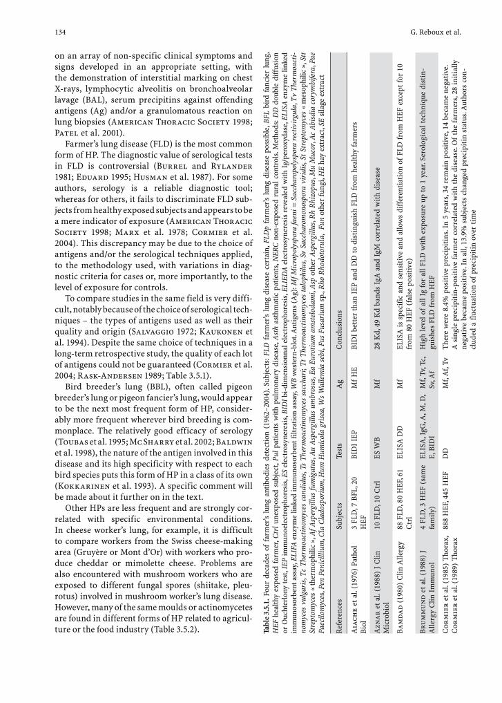
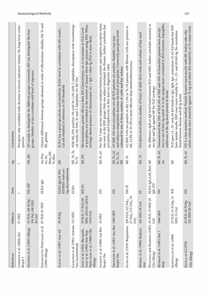
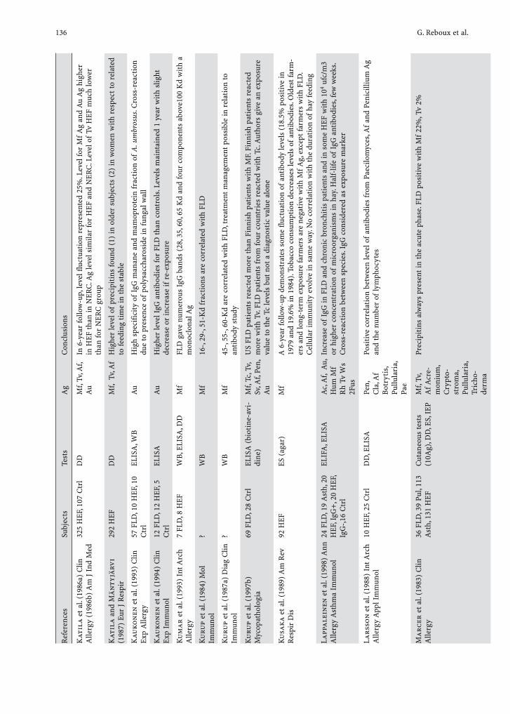
Farmer’s lung disease (FLD) is the most common form of HP. The diagnostic value of serological tests in FLD is controversial (Burrel and Rylander 1981; Eduard 1995; Husman et al. 1987). For some authors, serology is a reliable diagnostic tool; whereas for others, it fails to discriminate FLD sub-jects from healthy exposed subjects and appears to be a mere indicator of exposure (American Thoracic Society 1998; Marx et al. 1978; Cormier et al. 2004). This discrepancy may be due to the choice of antigens and/or the serological techniques applied, to the methodology used, with variations in diag-nostic criteria for cases or, more importantly, to the level of exposure for controls.
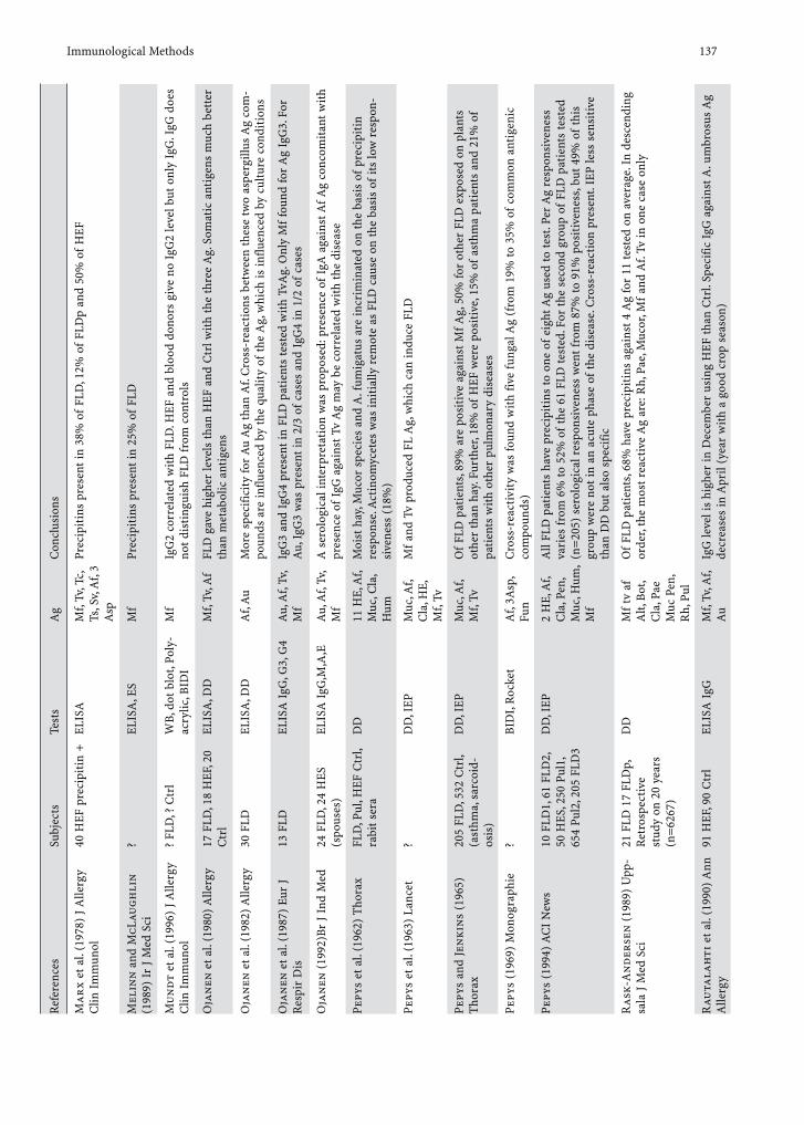
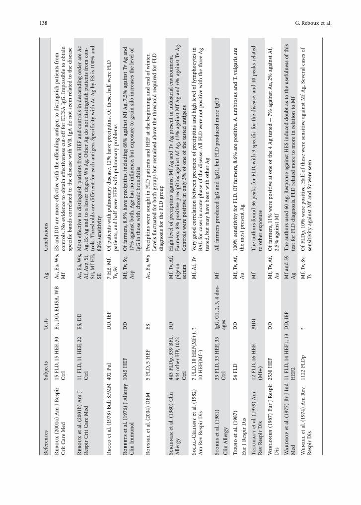
To compare studies in the same field is very diffi-cult, notably because of the choice of serological tech-niques – the types of antigens used as well as their quality and origin (Salvaggio 1972; Kaukonen et al. 1994). Despite the same choice of techniques in a long-term retrospective study, the quality of each lot of antigens could not be guaranteed (Cormier et al. 2004; Rask-Andersen 1989; Table 3.5.1).
Bird breeder’s lung (BBL), often called pigeon breeder’s lung or pigeon fancier’s lung, would appear to be the next most frequent form of HP, consider-ably more frequent wherever bird breeding is com-monplace. The relatively good efficacy of serology (Toubas et al. 1995; Mc Sharry et al. 2002; Baldwin et al. 1998), the nature of the antigen involved in this disease and its high specificity with respect to each bird species puts this form of HP in a class of its own (Kokkarinen et al. 1993). A specific comment will be made about it further on in the text.
Other HPs are less frequent and are strongly cor-related with specific environmental conditions. In cheese worker’s lung, for example, it is difficult to compare workers from the Swiss cheese-making area (Gruyère or Mont d’Or) with workers who pro-duce cheddar or mimolette cheese. Problems are also encountered with mushroom workers who are exposed to different fungal spores (shiitake, pleu-rotus) involved in mushroom worker’s lung disease. However, many of the same moulds or actinomycetes are found in different forms of HP related to agricul-ture or the food industry (Table 3.5.2).
Immunological Methods 135R
efer
ence
sSu
bjec
tsTe
sts
Ag
Con
clus
ions
Cor
mie
r et
al.
(200
4) E
ur
Res
pir
J27
HEF
??
Prec
ipit
in o
nly
corr
elat
ed w
ith
decr
ease
in fo
rced
exp
irat
ory
volu
me.
No
long
-ter
m c
onse
-qu
ence
s
Da
lph
in e
t al.
(199
4) A
llerg
y35
FLD
, 140
FLD
p,
376
Pul,
100
HEF
, 20
Ctr
l
DD
, IEP
HE,
Mf
DD
una
ble
to d
isti
ngui
sh t
he d
iffer
ent
grou
ps. C
onve
rsel
y, IE
P ca
n di
stin
guis
h th
e fo
ur
grou
ps. N
umbe
r of
arc
s co
rrel
ates
wit
h le
vel o
f ex
posu
re
Erk
inju
ntt
i-Pe
kka
nen
et
al.
(199
9) A
llerg
y87
FLD
, 81
HEF
ELIS
A I
gGA
u, P
en,
Ac,
Af,
Hum
, Mf,
Tv, R
ho
Cle
ares
t di
ffer
ence
s be
twee
n gr
oups
obt
aine
d, in
ord
er o
f ef
fect
iven
ess:
Abs
idia
, Mf,
Tv a
nd
Rh.
glu
tini
s
Fasa
ni
et a
l. (1
987)
Ann
All
58 F
LDp
ELIS
A I
gG,M
DD
/IE
P/BI
DI
isof
ocus
-in
g el
ectr
opho
resi
s
Mf
ELIS
A h
as a
goo
d sp
ecifi
city
and
sen
siti
vity
. ELI
SA le
vel i
n co
rrel
atio
n w
ith
DD
res
ults
. C
ut-o
ff o
btai
ned
in r
efer
ence
to D
D th
resh
old
Ga
ng
wa
r et
al.
(199
1) A
nton
ie
Van
Leeu
wen
hoek
112
HEF
ELIS
AM
f, S
v, Ts
, Tv
, Tt
Wit
h M
f, 25
% p
osit
ive;
3%
wit
h Sv
, 1.5
% w
ith
T. t
alop
hilu
s: d
iscr
epan
cy w
ith
mic
robi
olog
i-ca
l res
ults
(Sv
56%
, Tv
and
T. T
alop
hilu
s 5%
Mf
5%)
Gar
i et
al.
(198
2) M
ycop
ath,
G
ari
et a
l. (1
984)
Sem
Hop
Pa
ris,
Gar
i et
al.
(198
6) C
lin
Alle
rgy
10 F
LD, 1
7 FL
D, 6
0 H
EF, 1
85C
trl,
24FL
D,
1376
Ctr
l
IEP,
ES
Mf,
HE
Elec
tros
yner
esis
: gre
ater
eff
icac
y th
an I
EP. C
hym
otry
psic
arc
is in
cons
tant
in F
LD. L
evel
of
pre
cipi
tins
not
cor
rela
ted
to t
he s
ever
ity
of d
isea
se. M
ore
IgG
pre
sent
usi
ng H
ES. W
hen
sero
logy
sho
ws
pres
ence
of
chym
otry
psic
arc
+ I
gG +
oth
er I
g, F
LD is
mor
e lik
ely
Gar
iepy
et a
l. (1
989)
Am
Rev
R
espi
r D
is33
HEF
DD
Mf,
Tv, A
fFo
r th
is 7
-yea
r su
rvey
, num
erou
s va
riat
ions
bet
wee
n po
siti
ve a
nd n
egat
ive
prec
ipit
in s
tatu
s w
as s
een
in t
he H
EF g
roup
. Onl
y on
e su
bjec
t de
velo
ped
the
dise
ase.
Aut
hor
conc
lude
s th
at
prec
ipit
ins
and
lym
phoc
ytes
in B
AL
have
no
diag
nost
ic v
alue
Gru
chow
et a
l. (1
981)
Am
Rev
R
espi
r D
is14
00 H
EFD
DM
f, Tv
, Af,
Sv, T
c, T
s, H
E
Of
HEF
, 10%
hav
e pr
ecip
itin
s; n
ot a
ll FL
D p
atie
nts
are
posi
tive
. Rai
nfal
l, cr
op t
ype
and
toba
cco
com
posi
tion
dec
reas
e th
e le
vel o
f pr
ecip
itin
s. C
onve
rsel
y, pr
ecip
itin
leve
ls
enha
nced
by
size
of
farm
, num
ber
of c
attle
and
hay
vol
ume
Gu
mp
et a
l. (1
979)
Res
pira
tion
258
FLD
, 125
Pul
1, 27
Pul
2, 11
5 C
trl 1
, 76
Ctr
l 2
DD
, IF
Mf,
TvO
f H
EF, 5
.4%
are
pos
itiv
e to
Mf,
1.2%
to T
v. O
f pa
tien
ts w
ith
fibro
sis,
5.6%
are
pos
itiv
e to
M
f, 14
.4%
to T
v. D
D is
mor
e ef
fect
ive
than
imm
unof
luor
esce
nce
Hom
ma
et a
l. (1
986)
Res
pira
-ti
on44
2 H
EF, 9
4 C
trl
ESM
f, T
vPr
ecip
itin
leve
l ref
lect
s nu
mbe
r of
hou
rs o
f da
ily fa
rm w
ork
and
volu
me
of h
ay u
sed
Hu
izin
ga a
nd B
erre
ns
(198
5)
Clin
Alle
rgy
18 F
LD, 1
9 H
EF, 2
9 C
trl
ELIS
A I
gG,A
,M, R
ast
IgE,
DD
Mf
No
diff
eren
ce I
gM o
r Ig
E le
vel f
ound
bet
wee
n FL
D a
nd H
EF. A
utho
r co
nclu
des
nece
ssit
y to
us
e Ig
G a
nd I
gA a
gain
st M
f fo
r FL
D s
erol
ogy
Hu
sman
et
al. (
1987
) Eu
r J
Res
p24
40 H
EFD
DM
f, Tv
, Af,
Au
Dem
onst
rate
d th
at 1
8% o
f FL
D, 8
% o
f H
EF a
nd 1
0% o
f br
onch
itis
pat
ient
s ha
ve p
reci
pi-
tins
to o
ne o
f fo
ur A
g te
sted
. Tv
is t
he a
ntig
en m
ost
corr
elat
ed to
FLD
, how
ever
, Asp
ergi
llus
umbr
osus
is t
he A
g m
ost
pres
ent
in fa
rms
Iran
ital
ab e
t al
. (19
89)
Alle
rgy
25 F
LD, 3
1 FL
Dp,
14
HEF
, 13
Ctr
lW
BM
fFL
D g
ave
12-,
25-
and,
60-
Kd
band
s in
IgG
, IgM
, IgA
, som
etim
es 1
5–62
Kd
band
s bu
t H
EF
have
sim
ilar
resu
lts. H
EF r
eact
mor
e va
riab
ly to
11-
, 25-
and
60-
Kd
Ag.
No
corr
elat
ion
foun
d be
twee
n di
seas
e an
d al
l Ig
test
ed
Kat
ila
et a
l.(19
78)
Clin
Alle
rgy
22 F
LD, 2
6 FL
Dp,
32
5 H
EF, 8
0 C
trl
DD
Mf,
Tv, A
fD
D is
a le
ss s
ensi
tive
met
hod.
Fin
nish
farm
ers
reac
ts p
oorl
y w
ith
Mf.
Aut
hor
asks
why
ur
ban
cont
rols
rea
ct m
assi
vely
aga
inst
Tv
Ag
and
whe
re t
his
sens
itiv
ity
to T
v co
mes
fro
m
136 G. Reboux et al.
Ref
eren
ces
Subj
ects
Test
sA
gC
oncl
usio
ns
Kat
ila
et a
l. (1
986a
) C
lin
Alle
rgy
(198
6b)
Am
J I
nd M
ed32
5 H
EF, 1
07 C
trl
DD
Mf,
Tv, A
f, A
uIn
6-y
ear
follo
w-u
p, le
vel f
luct
uati
on r
epre
sent
ed 2
5%. L
evel
for
Mf A
g an
d A
u A
g hi
gher
in
HEF
tha
n in
NER
C. A
g le
vel s
imila
r fo
r H
EF a
nd N
ERC
. Lev
el o
f Tv
HEF
muc
h lo
wer
th
an fo
r N
ERC
gro
up
Kat
ila
and
Män
tyjä
rvi
(198
7) E
ur J
Res
pir
292
HEF
DD
Mf,
Tv,
Af
Hig
her
leve
l of
prec
ipit
ins
foun
d (1
) in
old
er s
ubje
cts
(2)
in w
omen
wit
h re
spec
t to
rela
ted
to fe
edin
g ti
me
in t
he s
tabl
e
Kau
kon
en e
t al
. (19
93)
Clin
Ex
p A
llerg
y57
FLD
, 10
HEF
, 10
Ctr
lEL
ISA
, WB
Au
Hig
h sp
ecifi
city
of
IgG
man
ane
and
mam
opro
tein
fra
ctio
n of
A. u
mbr
osus
. Cro
ss-r
eact
ion
due
to p
rese
nce
of p
olys
acch
aros
ide
in f
unga
l wal
l
Kau
kon
en e
t al
. (19
94)
Clin
Ex
p Im
mun
ol12
FLD
, 12
HEF
, 5
Ctr
lEL
ISA
Au
Hig
her
leve
l IgG
ant
ibod
ies
for
FLD
tha
n co
ntro
ls. L
evel
s m
aint
aine
d 1
year
wit
h sl
ight
de
crea
se o
r in
crea
se if
re-
expo
sure
Ku
mar
et a
l. (1
993)
Int
Arc
h A
llerg
y7
FLD
, 8 H
EFW
B, E
LISA
, DD
Mf
FLD
gav
e nu
mer
ous
IgG
ban
ds (
28, 3
5, 6
0, 6
5 K
d an
d fo
ur c
ompo
nent
s ab
ove1
00 K
d w
ith
a m
onoc
lona
l Ag
Ku
rup
et a
l. (1
984)
Mol
Im
mun
ol?
WB
Mf
16-,
29-,
51-K
d fr
acti
ons
are
corr
elat
ed w
ith
FLD
Ku
rup
et a
l. (1
987a
) D
iag
Clin
Im
mun
ol?
WB
Mf
45-,
55-,
60-K
d ar
e co
rrel
ated
wit
h FL
D, t
reat
men
t m
anag
emen
t po
ssib
le in
rel
atio
n to
an
tibo
dy s
tudy
Ku
rup
et a
l. (1
997b
) M
ycop
atho
logi
a69
FLD
, 28
Ctr
lEL
ISA
(bi
otin
e-av
i-di
ne)
Mf,
Tc, T
v, Sv
, Af,
Pen,
A
u
US
FLD
pat
ient
s re
acte
d m
ore
than
Fin
nish
pat
ient
s w
ith
MF.
Fin
nish
pat
ient
s re
acte
d m
ore
wit
h Tv
. FLD
pat
ient
s fr
om fo
ur c
ount
ries
rea
cted
wit
h Tc
. Aut
hors
giv
e an
exp
osur
e va
lue
to t
he T
c le
vels
but
not
a d
iagn
osti
c va
lue
alon
e
Ku
saka
et a
l. (1
989)
Am
Rev
R
espi
r D
is92
HEF
ES (
agar
)M
fA
6-y
ear
follo
w-u
p de
mon
stra
tes
som
e flu
ctua
tion
of
anti
body
leve
ls (
18.5
% p
osit
ive
in
1979
and
19.
6% in
198
4). T
obac
co c
onsu
mpt
ion
decr
ease
s le
vels
of
anti
bodi
es. O
ldes
t far
m-
ers
and
long
-ter
m e
xpos
ure
farm
ers
are
nega
tive
with
Mf A
g, e
xcep
t far
mer
s w
ith F
LD.
Cel
lula
r im
mun
ity
evol
ve in
sam
e w
ay. N
o co
rrel
atio
n w
ith th
e du
rati
on o
f ha
y fe
edin
g
Lapp
ale
inen
et a
l. (1
998)
Ann
A
llerg
y A
sthm
a Im
mun
ol24
FLD
, 19
Ast
h, 2
0 H
EF, I
gG+
, 20
HEF
, Ig
G-,
16 C
trl
ELIF
A, E
LISA
Ac,
Af,
Au,
H
um M
f R
h Tv
Ws
2Fus
Incr
ease
of
IgG
in F
LD a
nd c
hron
ic b
ronc
hiti
s pa
tien
ts a
nd in
som
e H
EF w
ith
104 u
fc/m
3 or
hig
her
conc
entr
atio
n of
mic
roor
gani
sms
in h
ay. H
alf-
life
of I
gG a
ntib
odie
s, fe
w w
eeks
. C
ross
-rea
ctio
n be
twee
n sp
ecie
s. Ig
G c
onsi
dere
d as
exp
osur
e m
arke
r
Lars
son
et a
l. (1
988)
Int
Arc
h A
llerg
y A
ppl I
mm
unol
10 H
EF, 2
5 C
trl
DD
, ELI
SAPe
n,
Cla
, Af
Bot
ryti
s, Pu
llula
ria,
Pa
e
Posi
tive
cor
rela
tion
bet
wee
n le
vel o
f an
tibo
dies
fro
m P
aeci
lom
yces
, Af
and
Peni
cilli
um A
g an
d th
e nu
mbe
r of
lym
phoc
ytes
Ma
rcer
et
al. (
1983
) C
lin
Alle
rgy
36 F
LD, 3
9 Pu
l, 11
3 A
sth,
131
HEF
Cut
aneo
us te
sts
(10A
g), D
D, E
S, I
EPM
f, Tv
, A
f Acr
e-m
oniu
m,
Cry
pto-
stro
ma,
Pu
llula
ria,
Tr
icho
-de
rma
Prec
ipit
ins
alw
ays
pres
ent
in t
he a
cute
pha
se. F
LD p
osit
ive
wit
h M
f 22
%, T
v 2%
Immunological Methods 137R
efer
ence
sSu
bjec
tsTe
sts
Ag
Con
clus
ions
Mar
x et
al.
(197
8) J
Alle
rgy
Clin
Im
mun
ol40
HEF
pre
cipi
tin
+EL
ISA
Mf,
Tv, T
c,
Ts, S
v, A
f, 3
Asp
Prec
ipit
ins
pres
ent
in 3
8% o
f FL
D, 1
2% o
f FL
Dp
and
50%
of
HEF
Mel
inn
and
McL
augh
lin
(1
989)
Ir
J M
ed S
ci?
ELIS
A, E
SM
fPr
ecip
itin
s pr
esen
t in
25%
of
FLD
Mu
ndt
et
al. (
1996
) J A
llerg
y C
lin I
mm
unol
? FL
D, ?
Ctr
lW
B, d
ot b
lot,
Poly
-ac
rylic
, BID
IM
fIg
G2
corr
elat
ed w
ith
FLD
. HEF
and
blo
od d
onor
s gi
ve n
o Ig
G2
leve
l but
onl
y Ig
G. I
gG d
oes
not d
isti
ngui
sh F
LD f
rom
con
trol
s
Oja
nen
et a
l. (1
980)
Alle
rgy
17 F
LD, 1
8 H
EF, 2
0 C
trl
ELIS
A, D
DM
f, Tv
, Af
FLD
gav
e hi
gher
leve
ls t
han
HEF
and
Ctr
l wit
h th
e th
ree
Ag.
Som
atic
ant
igen
s m
uch
bett
er
than
met
abol
ic a
ntig
ens
Oja
nen
et a
l. (1
982)
Alle
rgy
30 F
LDEL
ISA
, DD
Af,
Au
Mor
e sp
ecifi
city
for
Au
Ag
than
Af.
Cro
ss-r
eact
ions
bet
wee
n th
ese
two
aspe
rgill
us A
g co
m-
poun
ds a
re in
fluen
ced
by t
he q
ualit
y of
the
Ag,
whi
ch is
influ
ence
d by
cul
ture
con
diti
ons
Oja
nen
et
al. (
1987
) Eu
r J
Res
pir
Dis
13 F
LDEL
ISA
IgG
, G3,
G4
Au,
Af,
Tv,
Mf
IgG
3 an
d Ig
G4
pres
ent
in F
LD p
atie
nts
test
ed w
ith
TvA
g. O
nly
Mf
foun
d fo
r A
g Ig
G3.
For
A
u, I
gG3
was
pre
sent
in 2
/3 o
f ca
ses
and
IgG
4 in
1/2
of
case
s
Oja
nen
(19
92)B
r J
Ind
Med
24 F
LD, 2
4 H
ES
(spo
uses
)EL
ISA
IgG
,M,A
,EA
u, A
f, Tv
, M
fA
ser
olog
ical
inte
rpre
tati
on w
as p
ropo
sed:
pre
senc
e of
IgA
aga
inst
Af A
g co
ncom
itan
t w
ith
pres
ence
of
IgG
aga
inst
Tv
Ag
may
be
corr
elat
ed w
ith
the
dise
ase
Pepy
s et
al.
(196
2) T
hora
xFL
D, P
ul, H
EF C
trl,
rabi
t se
raD
D11
HE,
Af,
Muc
, Cla
, H
um
Moi
st h
ay, M
ucor
spe
cies
and
A. f
umig
atus
are
incr
imin
ated
on
the
basi
s of
pre
cipi
tin
resp
onse
. Act
inom
ycet
es w
as in
itia
lly r
emot
e as
FLD
cau
se o
n th
e ba
sis
of it
s lo
w r
espo
n-si
vene
ss (
18%
)
Pepy
s et
al.
(196
3) L
ance
t?
DD
, IEP
Muc
, Af,
Cla
, HE,
M
f, Tv
Mf
and
Tv p
rodu
ced
FL A
g, w
hich
can
indu
ce F
LD
Pepy
s an
d Je
nki
ns
(196
5)
Tho
rax
205
FLD
, 532
Ctr
l, (a
sthm
a, s
arco
id-
osis
)
DD
, IEP
Muc
, Af,
Mf,
TvO
f FL
D p
atie
nts,
89%
are
pos
itiv
e ag
ains
t M
f Ag,
50%
for
othe
r FL
D e
xpos
ed o
n pl
ants
ot
her
than
hay
. Fur
ther
, 18%
of
HEF
wer
e po
siti
ve, 1
5% o
f as
thm
a pa
tien
ts a
nd 2
1% o
f pa
tien
ts w
ith
othe
r pu
lmon
ary
dise
ases
Pepy
s (1
969)
Mon
ogra
phie
?BI
DI,
Roc
ket
Af,
3Asp
, Fu
nC
ross
-rea
ctiv
ity
was
foun
d w
ith
five
fung
al A
g (f
rom
19%
to 3
5% o
f co
mm
on a
ntig
enic
co
mpo
unds
)
Pepy
s (1
994)
AC
I N
ews
10 F
LD1,
61
FLD
2,
50 H
ES, 2
50 P
ul1,
65
4 Pu
l2, 2
05 F
LD3
DD
, IEP
2 H
E, A
f, C
la, P
en,
Muc
, Hum
, M
f
All
FLD
pat
ient
s ha
ve p
reci
piti
ns to
one
of
eigh
t Ag
used
to te
st. P
er A
g re
spon
sive
ness
va
ries
fro
m 6
% to
52%
of
the
61 F
LD te
sted
. For
the
sec
ond
grou
p of
FLD
pat
ient
s te
sted
(n
=20
5) s
erol
ogic
al r
espo
nsiv
enes
s w
ent
from
87%
to 9
1% p
osit
iven
ess,
but
49%
of
this
gr
oup
wer
e no
t in
an
acut
e ph
ase
of t
he d
isea
se. C
ross
-rea
ctio
n pr
esen
t. IE
P le
ss s
ensi
tive
th
an D
D b
ut a
lso
spec
ific
Ras
k-A
nd
erse
n (
1989
) U
pp-
sala
J M
ed S
ci21
FLD
17
FLD
p,
Ret
rosp
ecti
ve
stud
y on
20
year
s (n
=62
67)
DD
Mf
tv a
f A
lt, B
ot,
Cla
, Pae
M
uc P
en,
Rh,
Pul
Of
FLD
pat
ient
s, 68
% h
ave
prec
ipit
ins
agai
nst
4 A
g fo
r 11
test
ed o
n av
erag
e. I
n de
scen
ding
or
der,
the
mos
t re
acti
ve A
g ar
e: R
h, P
ae, M
ucor
, Mf
and
Af.
Tv in
one
cas
e on
ly
Rau
tala
hti
et a
l. (1
990)
Ann
A
llerg
y91
HEF
, 90
Ctr
lEL
ISA
IgG
Mf,
Tv, A
f, A
uIg
G le
vel i
s hi
gher
in D
ecem
ber
usin
g H
EF t
han
Ctr
l. Sp
ecifi
c Ig
G a
gain
st A
. um
bros
us A
g de
crea
ses
in A
pril
(yea
r w
ith
a go
od c
rop
seas
on)
138 G. Reboux et al.
Ref
eren
ces
Subj
ects
Test
sA
gC
oncl
usio
ns
Reb
oux
(200
1a)
Am
J R
espi
r C
rit
Car
e M
ed15
FLD
, 15
HEF
, 30
Ctr
lEs
, DD
, ELI
SA, W
BA
c, E
a, W
s, M
fES
and
DD
are
mor
e ef
fect
ive
wit
h th
e of
fend
ing
anti
gen
to d
isti
ngui
sh p
atie
nts
from
co
ntro
ls. N
o ev
iden
ce to
obt
ain
effe
ctiv
enes
s cu
t-of
f fo
r EL
ISA
IgG
. Im
poss
ible
to o
btai
n sp
ecifi
c ba
nds
corr
elat
ed to
the
dis
ease
wit
h W
B. I
gA d
o no
t se
em r
elat
ed to
the
dis
ease
Reb
oux
et a
l. (2
001b
) A
m J
R
espi
r C
rit C
are
Med
11 F
LD, 1
1 H
EF, 2
2 C
trl
ES, D
DA
c, E
a, W
s, A
f, A
sp, S
t, St
t, M
f H
E,
SE
Mos
t eff
ecti
ve to
dis
ting
uish
pat
ient
s fr
om H
EF a
nd c
ontr
ols
in d
esce
ndin
g or
der
are
Ac
Ag,
Ec
Ag
and
to a
less
er d
egre
e W
s A
g. O
ther
Ag
do n
ot d
isti
ngui
sh p
atie
nts
from
con
-tr
ols.
Thr
esho
lds
are
diff
eren
t for
eac
h an
tige
n. S
peci
ficit
y w
ith
Ac
Ag
by E
S is
100
% a
nd
80%
sen
siti
vity
Rec
co e
t al
. (19
78)
Bull
SFM
M40
2 Pu
lD
D, I
EP7
HE,
Mf,
Tv, S
vO
f pa
tien
ts w
ith
pulm
onar
y di
seas
e, 1
2% h
ave
prec
ipit
ins.
Of
thes
e, h
alf
wer
e FL
D
pati
ents
, and
hal
f w
ere
HEF
wit
h pu
lmon
ary
prob
lem
s
Rob
erts
et
al. (
1976
) J A
llerg
y C
lin I
mm
unol
1045
HEF
DD
Mf,
Tv, S
v, A
spO
f fa
rmer
s, 8.
9% h
ave
prec
ipit
ins,
incl
udin
g 68
% a
gain
st M
f Ag,
7.5
% a
gain
st T
v A
g an
d 17
% a
gain
st b
oth.
Age
has
no
influ
ence
, but
exp
osur
e to
gra
in s
ilo in
crea
ses
the
leve
l of
IgG
in th
ose
wit
h ch
roni
c br
onch
itis
Rou
ssel
et a
l. (2
004)
OEM
5 FL
D, 5
HEF
ESA
c, E
a, W
sPr
ecip
itin
s w
ere
soug
ht in
FLD
pat
ient
s an
d H
EF a
t the
beg
inni
ng a
nd e
nd o
f w
inte
r. Le
vels
fluc
tuat
ed fo
r bo
th g
roup
s bu
t re
mai
ned
abov
e th
e th
resh
old
requ
ired
for
FLD
di
agno
sis
for
the
FLD
gro
up
Scri
bner
et
al. (
1980
) C
lin
Alle
rgy
443
FLD
p, 3
39 B
FL,
944
othe
r H
P, 1
072
Ctr
l
DD
Mf,
Tv, A
f, pi
geon
se
rum
Hig
h le
vel o
f pr
ecip
itin
s ag
ains
t M
f Ag
and
Tv A
g pr
esen
t in
indu
stri
al e
nvir
onm
ent.
Farm
ers:
8%
pos
itiv
e pr
ecip
itin
s ag
ains
t Af A
g, 1
3% a
gain
st M
f Ag
and
6% a
gain
st T
v A
g.
Con
trol
s w
ere
posi
tive
in o
nly
3% o
f on
e of
the
test
ed a
ntig
ens
Sola
l-C
élig
ny e
t al
. (19
82)
Am
Rev
Res
pir
Dis
7 FL
D, 1
0 H
EF(M
f+),
10 H
EF(M
f–)
?M
f, A
f, Tv
Very
goo
d co
rrel
atio
n be
twee
n pr
esen
ce o
f pr
ecip
itin
s an
d hi
gh le
vel o
f ly
mph
ocyt
es in
BA
L fo
r ca
ses
in a
cute
pha
se o
f th
e di
seas
e. A
ll FL
D w
ere
not
posi
tive
wit
h th
e th
ree
Ag
test
ed, b
ut m
ay h
ave
been
wit
h ot
her
Ag
Stok
es e
t al
. (19
81)
Clin
Alle
rgy
33 F
LD, 3
3 H
EF, 3
3 C
trl
IgG
, G1,
2, 3
, 4 d
os-
ages
Mf
All
farm
ers
prod
uced
IgG
and
IgG
1, b
ut F
LD p
rodu
ced
mor
e Ig
G3
Terh
o et
al.
(198
7)
Eur
J R
espi
r D
is
54 F
LDD
DM
f, Tv
, Af,
Au
100%
sen
siti
vity
for
FLD
. Of
farm
ers,
8.6%
are
pos
itiv
e. A
. um
bros
us a
nd T
. vul
gari
s ar
e th
e m
ost
pres
ent A
g
Treu
haf
t et
al.
(197
9) A
m
Rev
Res
pir
Dis
12 F
LD, 1
6 H
EF,
(Mf+
)BI
DI
Mf
The
aut
hors
foun
d 36
pea
ks fo
r FL
D, w
ith
3 sp
ecifi
c fo
r th
e di
seas
e, a
nd 1
0 pe
aks
rela
ted
to o
ther
exp
osur
e
Voh
lon
en (
1987
) Eu
r J
Res
pir
Dis
2530
HEF
D
DM
f, Tv
, Af,
Au
Of
farm
ers,
11%
wer
e po
siti
ve a
t one
of
the
4 A
g te
sted
--
7% a
gain
st A
u, 2
% a
gain
st A
f, 2.
5% a
gain
st M
f
War
dro
p et
al.
(197
7) B
r J
Ind
Med
11 F
LD, 1
4 H
EF1,
13
HEF
2D
D, I
EPM
f an
d 59
A
gT
he a
utho
rs u
sed
60 A
g. R
espo
nse
agai
nst
HES
indu
ced
doub
t as
to t
he u
sefu
lnes
s of
thi
s te
st fo
r FL
D d
iagn
osis
. FLD
rel
ated
mor
e to
mor
e in
rel
atio
n to
Mf
Wen
zel
et a
l. (1
974)
Am
Rev
R
espi
r D
is11
22 F
LDp
?M
f, Tv
, Sv,
TsO
f FL
Dp,
10%
wer
e po
siti
ve. H
alf
of t
hese
wer
e se
nsit
ive
agai
nst
Mf A
g. S
ever
al c
ases
of
sens
itiv
ity
agai
nst
Mf
and
Sv w
ere
seen
Immunological Methods 139
Fungi are clearly involved in domestic-related HP. Moreover, it should be mentioned that these HPs, in which the role of bacterial antigens other than acti-nomycetes (Klebsiella or Mycobacteria) is probably underestimated, need to be investigated in future environmental studies.
3.5.1.1 Antigen
3.5.1.1.1 Antigen Type
The importance of antigens in the diagnosis of HP is frequently underestimated. The quality of the antigen preparation might have a significant effect on the specificity and sensitivity of diagnostic
tests (Kim et al. 1979). Varying degrees of cross-reactivity have been shown among micro-organ-ism species (Sa lvaggio 1991, 1997). Conversely, in numerous studies comparing microbiological envi-ronmental data to the immunological response for exposed workers, not all antigens react favourably ( Dutkiewicz et al. 2001, 2002; Moran et al. 2002; Miyazaki et al. 2004; Flandes et al. 2004). In addi-tion, an increasing number of potentially new anti-gen sources for which commercial extracts are not available are being identified (Lappalainen et al. 1998; Dalphin et al. 2000). There are few standard-ised antigen products available because of the dif-ficulties inherent to manufacturing and standardis-ing fungal and bacterial extracts (Ramasamy et al. 1987; Reese et al. 1989; Melinn and McLaughlin 1992; Horner et al. 1995; Mundt et al. 1996). In fact, only crude antigens for infrequent or new aetiolo-
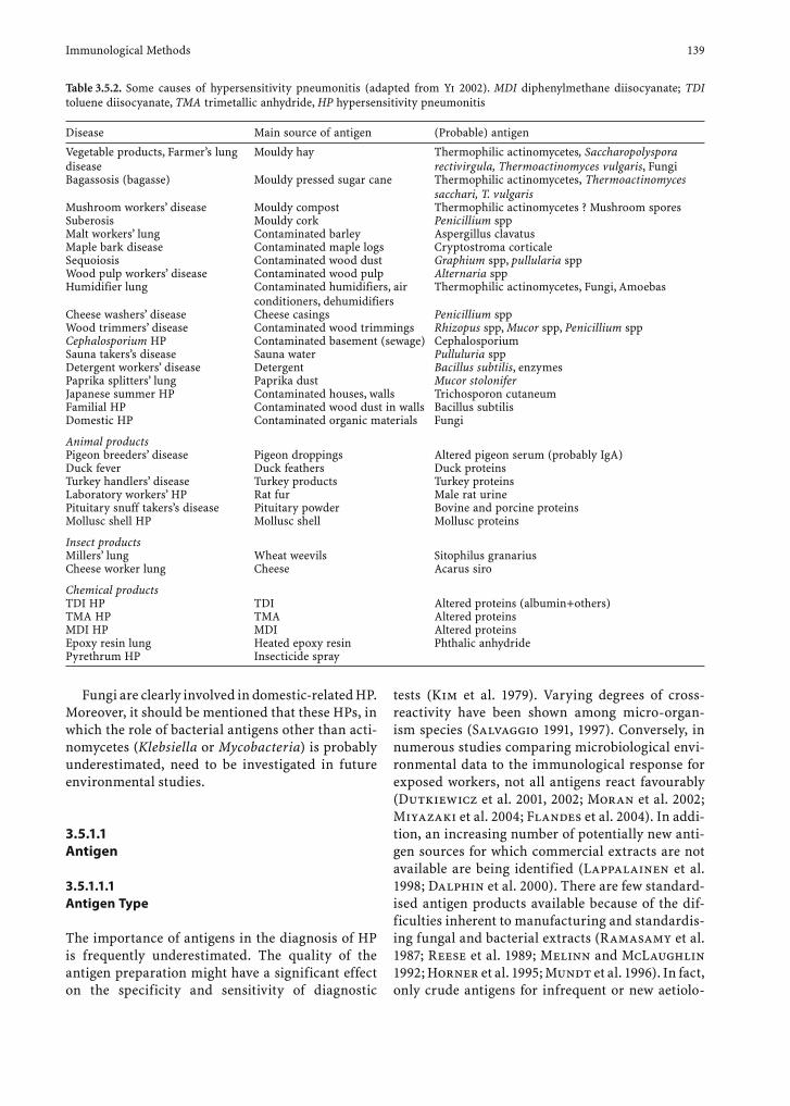
Table 3.5.2. Some causes of hypersensitivity pneumonitis (adapted from Yi 2002). MDI diphenylmethane diisocyanate; TDI toluene diisocyanate, TMA trimetallic anhydride, HP hypersensitivity pneumonitis
Disease Main source of antigen (Probable) antigen
Vegetable products, Farmer’s lung disease
Mouldy hay Thermophilic actinomycetes, Saccharopolyspora rectivirgula, Thermoactinomyces vulgaris, Fungi
Bagassosis (bagasse) Mouldy pressed sugar cane Thermophilic actinomycetes, Thermoactinomyces sacchari, T. vulgaris
Mushroom workers’ disease Mouldy compost Thermophilic actinomycetes ? Mushroom sporesSuberosis Mouldy cork Penicillium sppMalt workers’ lung Contaminated barley Aspergillus clavatusMaple bark disease Contaminated maple logs Cryptostroma corticaleSequoiosis Contaminated wood dust Graphium spp, pullularia sppWood pulp workers’ disease Contaminated wood pulp Alternaria sppHumidifier lung Contaminated humidifiers, air
conditioners, dehumidifiersThermophilic actinomycetes, Fungi, Amoebas
Cheese washers’ disease Cheese casings Penicillium sppWood trimmers’ disease Contaminated wood trimmings Rhizopus spp, Mucor spp, Penicillium sppCephalosporium HP Contaminated basement (sewage) CephalosporiumSauna takers’s disease Sauna water Pulluluria sppDetergent workers’ disease Detergent Bacillus subtilis, enzymesPaprika splitters’ lung Paprika dust Mucor stoloniferJapanese summer HP Contaminated houses, walls Trichosporon cutaneumFamilial HP Contaminated wood dust in walls Bacillus subtilisDomestic HP Contaminated organic materials Fungi
Animal productsPigeon breeders’ disease Pigeon droppings Altered pigeon serum (probably IgA)Duck fever Duck feathers Duck proteinsTurkey handlers’ disease Turkey products Turkey proteinsLaboratory workers’ HP Rat fur Male rat urinePituitary snuff takers’s disease Pituitary powder Bovine and porcine proteinsMollusc shell HP Mollusc shell Mollusc proteins
Insect productsMillers’ lung Wheat weevils Sitophilus granariusCheese worker lung Cheese Acarus siro
Chemical productsTDI HP TDI Altered proteins (albumin+others)TMA HP TMA Altered proteinsMDI HP MDI Altered proteinsEpoxy resin lung Heated epoxy resin Phthalic anhydridePyrethrum HP Insecticide spray
140 G. Reboux et al.
gies are produced by diagnostic centres themselves and used for HP diagnosis (Esch 2004).
The three types of crude antigens are:• Total crude extracts from crude sample materi-
als (hay, corn, water from humidifi ers, oil from metalworking, food products such as cheese and salami, glass fi ber, dust mites, rat excrement, chemical products). For BBL, droppings and feathers are either produced by each centre or, for common antigens, purchased from a manufac-turer (e.g. FSK Avian Immunodiffusion system, Microgen Bioproducts, Camberley, UK). Pure or diluted bird sera are also used for BBL diagnosis without transformation.
• Somatic antigens (also called “surface antigens” or “soluble antigens”) from bacterial, actinomy-cetes or pure fungal culture. A crude antigen including the micro-organism itself is obtained by cultivation on solid media on Petri dishes (spores present) or on shacked liquid broth (mycelium predominant).
• Metabolic antigens from the broth of bacterial, actinomycetes or pure fungal culture without micro-organisms.
Somatic and metabolic extracts are formed by a patchwork of numerous individual antigens (approx-imately 60 for some yeasts or moulds). Ojanen and colleagues found that the somatic antigens of fungi gave more positive results than the metabolic anti-gens. The opposite was true for actinomycetes. These authors recommend using either a somatic antigen or a combination of somatic and metabolic antigens for routine enzyme-linked immunosorbent assay (ELISA) tests (Ojanen et al. 1980).
The development of recombinant antigens con-cerns allergy tests, and, to a lesser extent, HP diag-nosis.• As we know, only Aspergillus and Saccharopolys-
pora recombinant purifi ed antigens have been used to diagnose HP (Kumar et al. 1993; Kaukonen et al. 1993) and allergic bronchopulmonary aspergil-losis (ABPA) (Moss 2002). No particular fraction has been correlated with HP diseases; hence, the development of small antigen fractions does not appear useful to diagnose HP. Further investiga-tion may well change this notion in the future.
3.5.1.1.2 Nature of Microbe Antigens
The nature of microbe antigen compounds is rarely given. Some enzymatic functions are characterised as protease, oxidase, catalase or chymotrypsinase in some species (Aspergillus fumigatus or Alter-naria) (Roberts et al. 1976; Latge 1999). The bio-chemical nature of antigens is defined as either a glycoprotein or a polysaccharidic compound. Hence, in summer-type HP fever in Japan, a high-weight polysaccharidic compound, extracted from a Trichosporon cutaneum yeast, may be responsible for immunological response (Trentin et al. 1988). It is true that certain glycoproteic complex anti-gen compounds are partially or totally shared by numerous mould antigens. Up to now, their pres-ence has always been assessed by rough precipitin, double immuno-diffusion (DD) or electrophoresis techniques: precipitin arcs indicate totally or par-tially immunological identity (either a continuous line between two arcs indicating total identity or the presence of a forked arc indicating partial iden-tity). However, this compound is not characterised or matched with a protein control with a known molecular weight. Using the Western blot (WB) technique, Ag characteristics are often only sum-marised as a fraction with a molecular weight in kilodaltons and not compared together (Edwards 1972).
3.5.1.1.3 Nature of Avian Antigens
In pigeon droppings, numerous Ag have been found – essentially gamma globulins, which are very antigenic. Hydrolytic and esterolytic enzymes Ag properties have also been found; they may play an important role in the pathogenesis of the disease by increasing the inf lammatory proc-ess on the alveoli wall. However, intestinal mucin seems to be the principal antigenic compound of BBL (Baldwin et al. 1998). This substance is a high-molecular-weight glycoprotein, only slightly biodegradable (Boyd et al. 1982) and assumed to be the best antigen for BBL serology (Todd et al. 1993).
Immunological Methods 141
3.5.1.2 Choice of Micro-Organisms Involved in HP for Producing Antigens
Pepys opened Pandora’s box four decades ago (Pepys 1994). Numerous microorganisms, if the length of their spore or bacteria is less than 4 µm, are able to induce HP. Their immunological capacity to induce disease and antibody reactions is probably widely shared by all micro-organism species. Some publications give cumulative lists of aetiological agents (Table 3.5.2) without taking long-term validity or the modification of environmental circumstances into account. Certain professional and personal settings or practices, such as round baling crops, air-conditioned dwellings, Jacuzzis and do-it-yourself wood joinery, are modifying expo-sure in terms of the level and kind of microorganisms involved in HP. This evolution implies the need for regular re-evaluation of those micro-organisms that may be useful for serology diagnosis.
In the past decade, new aetiologies of HP have been described in the field of agriculture. Although the relative humidity of hay or grain crops has been seen to decrease with better agricultural practice and education about biohazards, this change, para-doxically, does not imply a decrease in the frequency
of the disease but the discovery of new species that require less humidity.
As the microbiological flora of mouldy hay varies widely from one country to another, the antigens for serological studies should be selected on the basis of specific local species (Kurup et al. 1987).
Despite recent reports describing “new” antigens in FLD [Absidia corymbifera, Humicola grisea, Peni-cillium brevicompactum and Rhodotorula glutinis (Erkinjutti-Pekkanen et al. 1999); A. corymbifera, Eurotium amstelodami and Wallemia sebi (Reboux et al. 2001a,b)], it is not always possible to perform envi-ronmental microbiological analyses prior to selecting the appropriate antigens to use in serological tests for each patient. What is needed in routine serology is a standard panel of Ag for each professional or domes-tic activity. The choices of today are not universal and should evolve to take into account human activity, professional practice and the geographic area where patients come from. Proposals from three centres – the National Jewish Medical & Research Centre (http://nationaljewish.org), the Occupational Health Centre Kuopio (Dr. M. Reiman) and the experience of our department (University Hospital of Besançon, Parasitology-Mycology Laboratory, Besançon) – are given in Table 3.5.3.
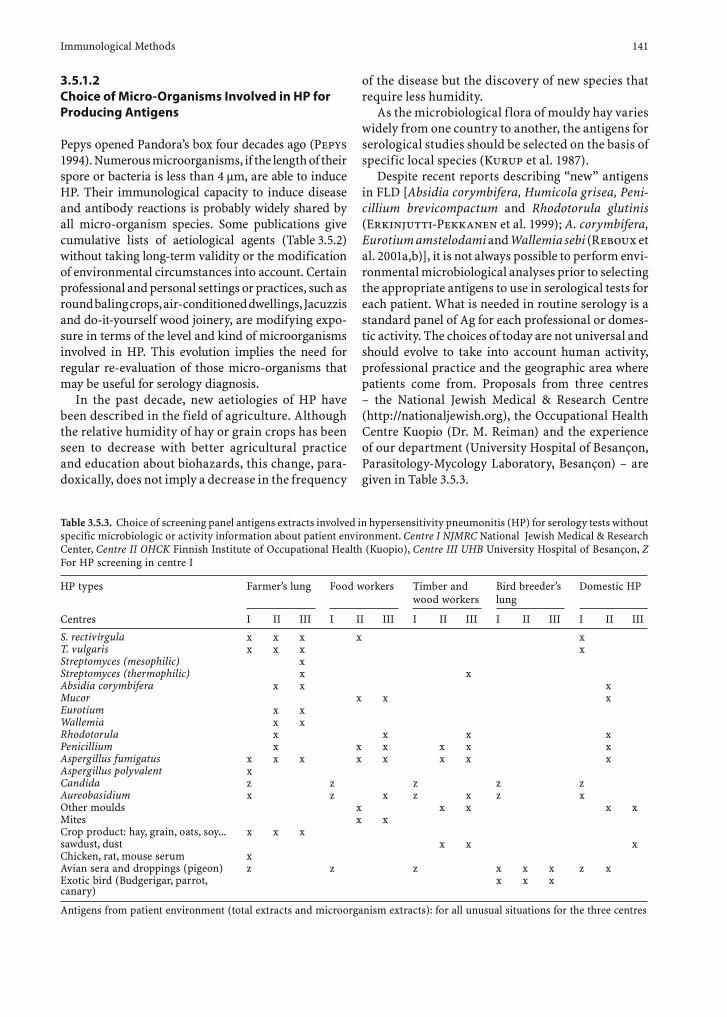
Table 3.5.3. Choice of screening panel antigens extracts involved in hypersensitivity pneumonitis (HP) for serology tests without specific microbiologic or activity information about patient environment. Centre I NJMRC National Jewish Medical & Research Center, Centre II OHCK Finnish Institute of Occupational Health (Kuopio), Centre III UHB University Hospital of Besançon, Z For HP screening in centre I
HP types Farmer’s lung Food workers Timber and wood workers
Bird breeder’s lung
Domestic HP
Centres I II III I II III I II III I II III I II III
S. rectivirgula x x x x xT. vulgaris x x x xStreptomyces (mesophilic) xStreptomyces (thermophilic) x xAbsidia corymbifera x x xMucor x x xEurotium x xWallemia x xRhodotorula x x x xPenicillium x x x x x xAspergillus fumigatus x x x x x x x xAspergillus polyvalent xCandida z z z z zAureobasidium x z x z x z xOther moulds x x x x xMites x xCrop product: hay, grain, oats, soy... x x xsawdust, dust x x xChicken, rat, mouse serum xAvian sera and droppings (pigeon) z z z x x x z xExotic bird (Budgerigar, parrot, canary)
x x x
Antigens from patient environment (total extracts and microorganism extracts): for all unusual situations for the three centres
142 G. Reboux et al.
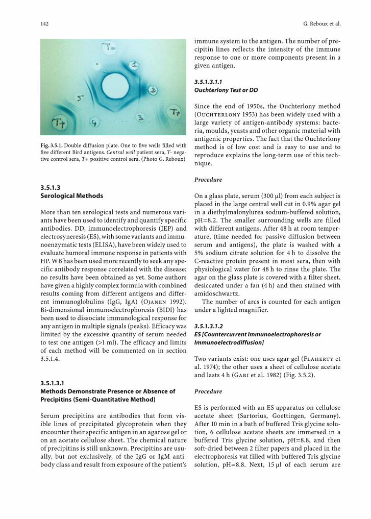
Fig. 3.5.1. Double diffusion plate. One to fi ve wells fi lled with fi ve different Bird antigens. Central well patient sera, T- nega-tive control sera, T+ positive control sera. (Photo G. Reboux)
3.5.1.3 Serological Methods
More than ten serological tests and numerous vari-ants have been used to identify and quantify specific antibodies. DD, immunoelectrophoresis (IEP) and electrosyneresis (ES), with some variants and immu-noenzymatic tests (ELISA), have been widely used to evaluate humoral immune response in patients with HP. WB has been used more recently to seek any spe-cific antibody response correlated with the disease; no results have been obtained as yet. Some authors have given a highly complex formula with combined results coming from different antigens and differ-ent immunoglobulins (IgG, IgA) (Ojanen 1992). Bi-dimensional immunoelectrophoresis (BIDI) has been used to dissociate immunological response for any antigen in multiple signals (peaks). Efficacy was limited by the excessive quantity of serum needed to test one antigen (>1 ml). The efficacy and limits of each method will be commented on in section 3.5.1.4.
3.5.1.3.1 Methods Demonstrate Presence or Absence of Precipitins (Semi-Quantitative Method)
Serum precipitins are antibodies that form vis-ible lines of precipitated glycoprotein when they encounter their specific antigen in an agarose gel or on an acetate cellulose sheet. The chemical nature of precipitins is still unknown. Precipitins are usu-ally, but not exclusively, of the IgG or IgM anti-body class and result from exposure of the patient’s
immune system to the antigen. The number of pre-cipitin lines reflects the intensity of the immune response to one or more components present in a given antigen.
3.5.1.3.1.1 Ouchterlony Test or DD
Since the end of 1950s, the Ouchterlony method (Ouchterlony 1953) has been widely used with a large variety of antigen-antibody systems: bacte-ria, moulds, yeasts and other organic material with antigenic properties. The fact that the Ouchterlony method is of low cost and is easy to use and to reproduce explains the long-term use of this tech-nique.
Procedure
On a glass plate, serum (300 µl) from each subject is placed in the large central well cut in 0.9% agar gel in a diethylmalonylurea sodium-buffered solution, pH=8.2. The smaller surrounding wells are filled with different antigens. After 48 h at room temper-ature, (time needed for passive diffusion between serum and antigens), the plate is washed with a 5% sodium citrate solution for 4 h to dissolve the C-reactive protein present in most sera, then with physiological water for 48 h to rinse the plate. The agar on the glass plate is covered with a filter sheet, desiccated under a fan (4 h) and then stained with amidoschwartz.
The number of arcs is counted for each antigen under a lighted magnifier.
3.5.1.3.1.2 ES [Countercurrent Immunoelectrophoresis or Immunoelectrodiffusion]
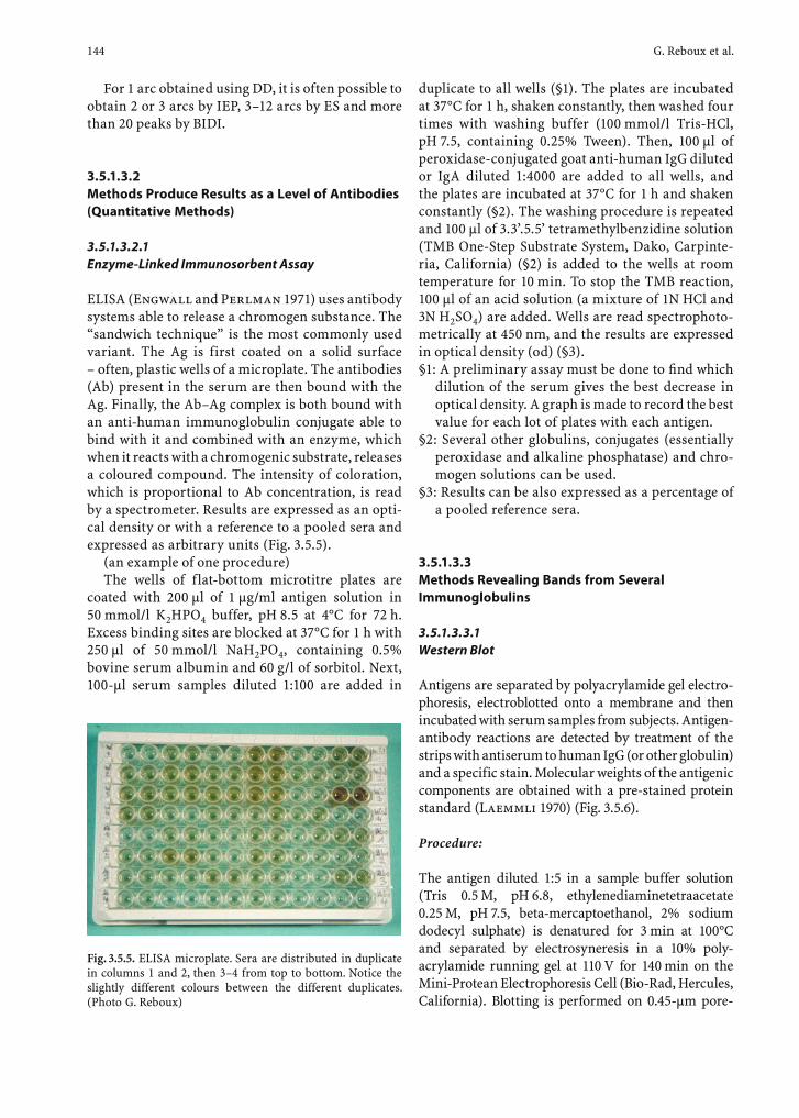
Two variants exist: one uses agar gel (Flaherty et al. 1974); the other uses a sheet of cellulose acetate and lasts 4 h (Gari et al. 1982) (Fig. 3.5.2).
Procedure
ES is performed with an ES apparatus on cellulose acetate sheet (Sartorius, Goettingen, Germany). After 10 min in a bath of buffered Tris glycine solu-tion, 6 cellulose acetate sheets are immersed in a buffered Tris glycine solution, pH=8.8, and then soft-dried between 2 filter papers and placed in the electrophoresis vat filled with buffered Tris glycine solution, pH=8.8. Next, 15 µl of each serum are
Immunological Methods 143
placed on three spots on the anode side, and a 15-µl line of antigen is placed on the cathode side. A 110-V current is applied for 2 h 15 min. After washing, the cellulose acetate sheets are stained with Coomassie blue.
3.5.1.3.1.3 Enzyme-linked Immunoelectrodiffusion Assay
Enzyme-linked immunoelectrodiffusion assay (ELIEDA) is a variant that combines ES and immu-noblotting. This technique consists of revealing bands obtained using ES on an acetate cellulose membrane, with several anti-human globulins (IgG, IgM, IgA or IgE) (Aznar et al. 1988).
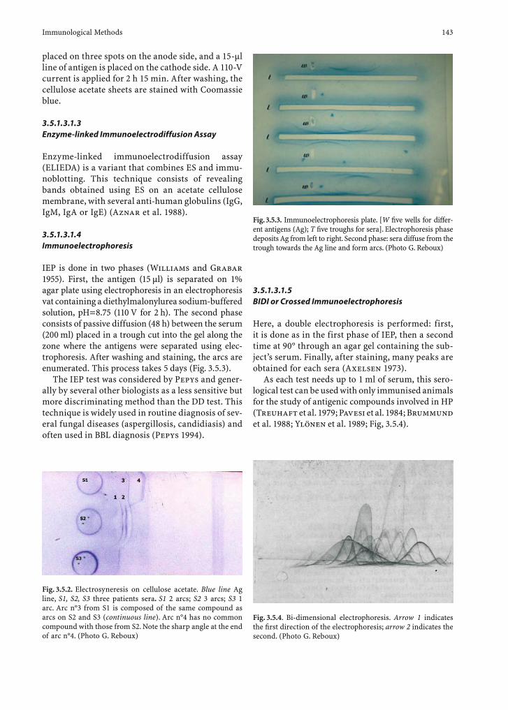
3.5.1.3.1.4 Immunoelectrophoresis
IEP is done in two phases (Williams and Grabar 1955). First, the antigen (15 µl) is separated on 1% agar plate using electrophoresis in an electrophoresis vat containing a diethylmalonylurea sodium-buffered solution, pH=8.75 (110 V for 2 h). The second phase consists of passive diffusion (48 h) between the serum (200 ml) placed in a trough cut into the gel along the zone where the antigens were separated using elec-trophoresis. After washing and staining, the arcs are enumerated. This process takes 5 days (Fig. 3.5.3).
The IEP test was considered by Pepys and gener-ally by several other biologists as a less sensitive but more discriminating method than the DD test. This technique is widely used in routine diagnosis of sev-eral fungal diseases (aspergillosis, candidiasis) and often used in BBL diagnosis (Pepys 1994).
3.5.1.3.1.5 BIDI or Crossed Immunoelectrophoresis
Here, a double electrophoresis is performed: first, it is done as in the first phase of IEP, then a second time at 90° through an agar gel containing the sub-ject’s serum. Finally, after staining, many peaks are obtained for each sera (Axelsen 1973).
As each test needs up to 1 ml of serum, this sero-logical test can be used with only immunised animals for the study of antigenic compounds involved in HP (Treuhaft et al. 1979; Pavesi et al. 1984; Bru mmund et al. 1988; Ylönen et al. 1989; Fig, 3.5.4).
Fig. 3.5.2. Electrosyneresis on cellulose acetate. Blue line Ag line, S1, S2, S3 three patients sera. S1 2 arcs; S2 3 arcs; S3 1 arc. Arc n°3 from S1 is composed of the same compound as arcs on S2 and S3 (continuous line). Arc n°4 has no common compound with those from S2. Note the sharp angle at the end of arc n°4. (Photo G. Reboux)
Fig. 3.5.3. Immunoelectrophoresis plate. [W fi ve wells for differ-ent antigens (Ag); T fi ve troughs for sera]. Electrophoresis phase deposits Ag from left to right. Second phase: sera diffuse from the trough towards the Ag line and form arcs. (Photo G. Reboux)
Fig. 3.5.4. Bi-dimensional electrophoresis. Arrow 1 indicates the fi rst direction of the electrophoresis; arrow 2 indicates the second. (Photo G. Reboux)
144 G. Reboux et al.
For 1 arc obtained using DD, it is often possible to obtain 2 or 3 arcs by IEP, 3–12 arcs by ES and more than 20 peaks by BIDI.
3.5.1.3.2 Methods Produce Results as a Level of Antibodies (Quantitative Methods)
3.5.1.3.2.1 Enzyme-Linked Immunosorbent Assay
ELISA (Engwall and Perlman 1971) uses antibody systems able to release a chromogen substance. The “sandwich technique” is the most commonly used variant. The Ag is first coated on a solid surface – often, plastic wells of a microplate. The antibodies (Ab) present in the serum are then bound with the Ag. Finally, the Ab–Ag complex is both bound with an anti-human immunoglobulin conjugate able to bind with it and combined with an enzyme, which when it reacts with a chromogenic substrate, releases a coloured compound. The intensity of coloration, which is proportional to Ab concentration, is read by a spectrometer. Results are expressed as an opti-cal density or with a reference to a pooled sera and expressed as arbitrary units (Fig. 3.5.5).
(an example of one procedure)The wells of flat-bottom microtitre plates are
coated with 200 µl of 1 µg/ml antigen solution in 50 mmol/l K2HPO4 buffer, pH 8.5 at 4°C for 72 h. Excess binding sites are blocked at 37°C for 1 h with 250 µl of 50 mmol/l NaH2PO4, containing 0.5% bovine serum albumin and 60 g/l of sorbitol. Next, 100-µl serum samples diluted 1:100 are added in
Fig. 3.5.5. ELISA microplate. Sera are distributed in duplicate in columns 1 and 2, then 3–4 from top to bottom. Notice the slightly different colours between the different duplicates. (Photo G. Reboux)
duplicate to all wells (§1). The plates are incubated at 37°C for 1 h, shaken constantly, then washed four times with washing buffer (100 mmol/l Tris-HCl, pH 7.5, containing 0.25% Tween). Then, 100 µl of peroxidase-conjugated goat anti-human IgG diluted or IgA diluted 1:4000 are added to all wells, and the plates are incubated at 37°C for 1 h and shaken constantly (§2). The washing procedure is repeated and 100 µl of 3.3’.5.5’ tetramethylbenzidine solution (TMB One-Step Substrate System, Dako, Carpinte-ria, California) (§2) is added to the wells at room temperature for 10 min. To stop the TMB reaction, 100 µl of an acid solution (a mixture of 1N HCl and 3N H2SO4) are added. Wells are read spectrophoto-metrically at 450 nm, and the results are expressed in optical density (od) (§3).§1: A preliminary assay must be done to fi nd which
dilution of the serum gives the best decrease in optical density. A graph is made to record the best value for each lot of plates with each antigen.
§2: Several other globulins, conjugates (essentially peroxidase and alkaline phosphatase) and chro-mogen solutions can be used.
§3: Results can be also expressed as a percentage of a pooled reference sera.
3.5.1.3.3 Methods Revealing Bands from Several Immunoglobulins
3.5.1.3.3.1 Western Blot
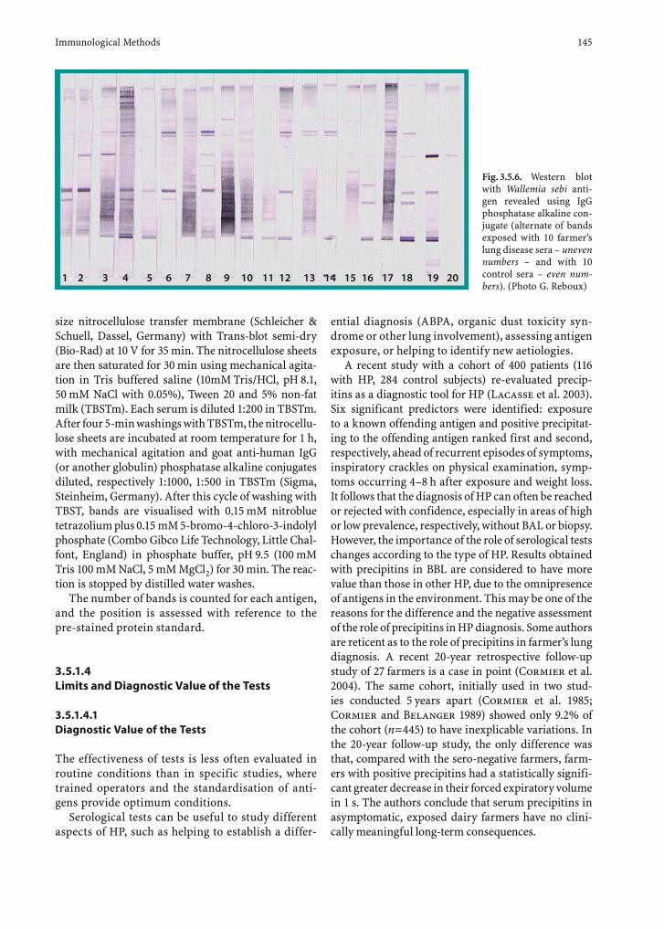
Antigens are separated by polyacrylamide gel electro-phoresis, electroblotted onto a membrane and then incubated with serum samples from subjects. Antigen-antibody reactions are detected by treatment of the strips with antiserum to human IgG (or other globulin) and a specific stain. Molecular weights of the antigenic components are obtained with a pre-stained protein standard (Laemmli 1970) (Fig. 3.5.6).
Procedure:
The antigen diluted 1:5 in a sample buffer solution (Tris 0.5 M, pH 6.8, ethylenediaminetetraacetate 0.25 M, pH 7.5, beta-mercaptoethanol, 2% sodium dodecyl sulphate) is denatured for 3 min at 100°C and separated by electrosyneresis in a 10% poly-acrylamide running gel at 110 V for 140 min on the Mini-Protean Electrophoresis Cell (Bio-Rad, Hercules, California). Blotting is performed on 0.45-µm pore-
Immunological Methods 145
size nitrocellulose transfer membrane (Schleicher & Schuell, Dassel, Germany) with Trans-blot semi-dry (Bio-Rad) at 10 V for 35 min. The nitrocellulose sheets are then saturated for 30 min using mechanical agita-tion in Tris buffered saline (10mM Tris/HCl, pH 8.1, 50 mM NaCl with 0.05%), Tween 20 and 5% non-fat milk (TBSTm). Each serum is diluted 1:200 in TBSTm. After four 5-min washings with TBSTm, the nitrocellu-lose sheets are incubated at room temperature for 1 h, with mechanical agitation and goat anti-human IgG (or another globulin) phosphatase alkaline conjugates diluted, respectively 1:1000, 1:500 in TBSTm (Sigma, Steinheim, Germany). After this cycle of washing with TBST, bands are visualised with 0.15 mM nitroblue tetrazolium plus 0.15 mM 5-bromo-4-chloro-3-indolyl phosphate (Combo Gibco Life Technology, Little Chal-font, England) in phosphate buffer, pH 9.5 (100 mM Tris 100 mM NaCl, 5 mM MgCl2) for 30 min. The reac-tion is stopped by distilled water washes.
The number of bands is counted for each antigen, and the position is assessed with reference to the pre-stained protein standard.
3.5.1.4 Limits and Diagnostic Value of the Tests
3.5.1.4.1 Diagnostic Value of the Tests
The effectiveness of tests is less often evaluated in routine conditions than in specific studies, where trained operators and the standardisation of anti-gens provide optimum conditions.
Serological tests can be useful to study different aspects of HP, such as helping to establish a differ-
ential diagnosis (ABPA, organic dust toxicity syn-drome or other lung involvement), assessing antigen exposure, or helping to identify new aetiologies.
A recent study with a cohort of 400 patients (116 with HP, 284 control subjects) re-evaluated precip-itins as a diagnostic tool for HP (Lacasse et al. 2003). Six significant predictors were identified: exposure to a known offending antigen and positive precipitat-ing to the offending antigen ranked first and second, respectively, ahead of recurrent episodes of symptoms, inspiratory crackles on physical examination, symp-toms occurring 4–8 h after exposure and weight loss. It follows that the diagnosis of HP can often be reached or rejected with confidence, especially in areas of high or low prevalence, respectively, without BAL or biopsy. However, the importance of the role of serological tests changes according to the type of HP. Results obtained with precipitins in BBL are considered to have more value than those in other HP, due to the omnipresence of antigens in the environment. This may be one of the reasons for the difference and the negative assessment of the role of precipitins in HP diagnosis. Some authors are reticent as to the role of precipitins in farmer’s lung diagnosis. A recent 20-year retrospective follow-up study of 27 farmers is a case in point (Cormier et al. 2004). The same cohort, initially used in two stud-ies conducted 5 years apart (Cormier et al. 1985; Cormier and Belanger 1989) showed only 9.2% of the cohort (n=445) to have inexplicable variations. In the 20-year follow-up study, the only difference was that, compared with the sero-negative farmers, farm-ers with positive precipitins had a statistically signifi-cant greater decrease in their forced expiratory volume in 1 s. The authors conclude that serum precipitins in asymptomatic, exposed dairy farmers have no clini-cally meaningful long-term consequences.
Fig. 3.5.6. Western blot with Wallemia sebi anti-gen revealed using IgG phosphatase alkaline con-jugate (alternate of bands exposed with 10 farmer’s lung disease sera – uneven numbers – and with 10 control sera – even num-bers). (Photo G. Reboux)
1 2 3 4 5 6 7 8 9 10 11 12 13 14 15 16 17 18 19 20
146 G. Reboux et al.
These two studies by the same team are represent-ative of the limits and diagnostic value of this test.
3.5.1.4.2 Durability of Antibody Response
Some authors have evaluated the stability of antibody response at a few weeks from exposure (Spiegelberg 1974). Other authors have evaluated this stability at more than 5 years in pensioner farmers. The high level of antibodies in the latter study correlated more with the disease than with former exposure (Katila and Mäntyjärvi 1987). For some authors, antibody response is the result of only a few months of exposure (Eduard 1995; Homma et al. 1986; Kusaka et al. 1989). Kaukonen and colleagues used ELISA to measure the antigen-binding avidity of A. umbrosus-specific IgG antibodies in FLD, in healthy farmers and urban con-trols during a 1-year follow-up. During the first acute phase, FLD patients with long exposure (>25 years) exhibited a high avidity of A. umbrosus-specific IgG antibodies that remained high throughout the 1-year follow-up, although the titre did decrease (Kaukonen et al. 1994). We had similar results with the measure-ment of A. corymbifera precipitins using ES before and after the winter period with FLD and healthy farmers. FLD precipitins fluctuate: either they decrease slightly if the patient is asymptomatic or increase if the patient has had a relapse. In all FLD cases, the precipitin titre remained over the threshold; whereas, in controls, it remained under the threshold required to classify the patient as FLD (Roussel et al. 2004). In our experi-ence, low fluctuation was detected between autumn and spring in serology from FLD and exposed control subjects and did not modify the ability of the test to dis-tinguish FLD subjects from controls (Roussel 2004).
3.5.1.5 Technical Considerations and Choices
3.5.1.5.1 Fine or Crude Antigens?
Contrary to what has been described in ABPA, where IgE serological response to recombinant purified aller-gen from A. fumigatus showed better sensitivity and specificity than with the crude antigen (Kn utsen et al. 2004), crude antigen and rough serological meth-ods with precipitin antibodies gave better results than sophisticated antigens and methods for diagnosis of HP (Reboux et al. 2001a). In some cases, total crude
extracts (humidifier water, fiberglass dust) are more effective than antigens taken from the culture of envi-ronmental sample (Baur et al. 1992; Nolard et al. 1994). This can be explained by the fact that some microorganisms or parasites present in the sample are not cultivated (Eduard 1997).
3.5.1.5.2 Spore or Mycelium Antigens?
One classic question related to fungi antigens is whether spores or mycelium constitute the better source of antigens. The allergenic composition of spores and mycelium is obviously different (Ho ffman et al. 1981). For some subunits, spore extract are more active than those from mycelium (Hoffman et al. 1981; Rosen et al. 1999). For other subunits, located in the cytoplasm, antigens are found both in spores and in mycelium (Paris et al. 1990). The spiculose part of spores contains more antigen compounds than smooth part does (Kaukonen et al. 1993).
3.5.1.5.3 Accuracy of Techniques
Precipitin techniques have shown reproducibility to be 89% for double diffusion (n=108) and 78% for electrosyneresis (n=202), with results showing +1 arc considered to be the same (Reboux et al. 2001b).
The ELISA is considered by several authors to be a sensitive, specific and quantitative test, which is readily available and widely applicable to HP diagno-sis. FLD commonly have significantly higher values in the ELISA than exposed but asymptomatic farm-ers. Bamdad concluded that ELISA (IgG) correlated better than DD with clinical diagnosis (Bamdad 1980). In this paper and in others, the difficulty lies in establishing the best cut-off to distinguish FLD patients from exposed asymptomatic farmer.
In a recent study concerning the diagnosis of pigeon breeder’s disease, specific measurement to avian antigens using the ELISA method showed greater effectiveness (10–10,000 ng/ml) than clas-sic precipitating methods. Significant differences were found between non-exposed controls and the asymptomatic fancier control group, but no signifi-cant differences were found between asymptomatic and symptomatic patients (Rodrigo et al. 2000). The greater technical effectiveness of this method is fre-
Immunological Methods 147
quently misunderstood to be the higher sensitivity of the diagnostic tool. Another widely accepted opinion states the advantage of automatic lecture methods (Spectrometer), due to less operator influence. This is certainly true. However, the other phases of the proc-ess, which require producing each reaction in dupli-cate or, better yet, in triplicate are equally as meticu-lous and often more difficult to manipulate precipitin methods. An internal reference sera is absolutely nec-essary to obtain a useful reference value between the batches of microtitre plates or as an arbitrary unit, in absence of a standard. Therefore, a cut-off is not easy to give in routine practice.
For other sophisticated techniques, with numer-ous bands (WB) or peaks (BIDI), interpretation is difficult due to excessive number of signals without specific bands or peaks correlated with the disease.
3.5.1.5.4 Choice of Isotypes
Patients in the acute phase of farmer’s lung often have antibodies of several Ig classes against several microbes, in contrast with controls, who have only IgG antibodies against one or two microbes. In the acute phase of HP, total IgG is elevated. IgG1 levels determine the levels of total IgG found both in non-exposed individuals and in farmers. IgE concentra-tions are generally normal or low, with no diagnos-tic value (Kaukonen et al. 1997). Conversely, IgG bound with the causative agent are elevated in acute reactions (Patel et al. 2001). Mundt and colleagues demonstrated specific binding of FLD patients’ IgG2 to S. rectivirgula antigens but found no such anti-bodies in exposed, unaffected subjects. Thus, IgG2 antibodies reacting with S. rectivirgula antigens are useful for serological diagnosis of patients with FLD (Mundt et al. 1996). Conversely, a study by Stoke and colleagues showed no difference between patient and exposed controls in level of IgG, IgG1, IgG2 and IgG4 but only with IgG3. They explain that FLD patients are separated from exposed popula-tion on the basis of their propensity to produce IgG3 in response to certain types of antigens stimulus (Stokes et al. 1981).
IgA antibodies against fungus have been found in patients with FLD and healthy exposed farmers. Except for Ylönen and colleagues, who assess that the presence of IgA is correlated with the disease, or for Ojanen, who highlights the value of the pres-ence of IgA from A. fumigatus in the diagnosis of FLD, no other authors have given a special value to
the presence of IgA in FLD. Exposed controls often have either IgG or IgA antibodies (Ojanen 1992). Our experience demonstrates that the highest levels of IgA were found using ELISA in healthy exposed farmers (personal unpublished data).
In BBL serodiagnosis, Martinez-Cordero showed positive IgA anti-avian antibodies to be the most fre-quent abnormalities, with values above those of the reference groups. A relationship of IgM-positive tests, using avian-pooled serum as antigen for subjects with a recent history of avian antigen exposure, was found to be reliable in the acute stage of the disease ( Martinez-Cordero et al. 2000). Baldwin et al. (1998) found positive differences in the composition of subclasses against mucin antigen with significantly higher IgG1 titres in the patients than in the controls. Similar results were found by Rodrigo in a group of patients with HP caused by inhalation of mollusc shell dust (Rodrigo et al. 2000).
3.5.1.6 Conclusion
The presence of seric precipitins is one of the diag-nostic criteria for HP and is important in epide-miological studies and research on new aetiologies. However, the diagnostic value of precipitins remains controversial. The sensitivity of tests varies accord-ing to the study, from 18% to 91%. Although some studies show what is often a considerable number of false negative subjects, the number of false positives is more problematic in that they may lead authors to consider precipitins as markers of exposure rather than of disease. Therefore, the presence of precip-itins in an exposed asymptomatic subject does not predict an increased risk of developing the disease. Differences of results in the literature are due to several factors, which stem, on one hand, from the variety of techniques and antigens used and, on the other, from the misunderstanding of the nature and precise meaning of precipitins in HP. Consequently, it is important to recall that:• Aetiological agents are varied, and the realisation
of serological tests with inappropriate antigens can lead to false negatives and to results that do not refl ect clinical features. Antigens related to domestic forms of HP are little known. We have attempted to solve this diffi culty by producing custom-made antigens that allow identifi cation of the aetiological agents at the origin of HP. This practice consists of taking environmental samples (air and surfaces), analysing them using
148 G. Reboux et al.
classic methods of culture to determine the microorganisms with which the patient has been in contact and then producing somatic antigens, useful in serological tests, from pure cultures of microorganisms.
• Detection of precipitins uses different methods, depending on the study. Besides the intrinsic problems inherent with each technique (standar-disation, reproducibility, repeatability), this vari-ety makes comparison between studies diffi cult. Rough methods seem preferable to sophisticated uninterpretable methods with several unspecifi c signals.
• Methods of preparing antigens are not stan-dardised. They may be metabolic antigens, somatic antigens, a mixture of both, total extracts or purifi ed antigenic fractions. Culture medium (solid or liquid), length and temperature for incubation, growth stage of fungi, extract and purifi cation method are also factors liable to vary antigen quality. The reactivity of each extract needs to be evaluated by WB with reference sera to be sure that the extract reveals the maximum number of antigen compounds.
• Clinical criteria for inclusion in a study are vari-able, as are the criteria for constituting control groups. Some authors have said that the test sensitivity of precipitins research borders on 100% in the acute stage of the disease. For all new antigens, an evaluation with a large healthy exposed group is necessary to provide a robust, well-founded interpretation.
• Some factors cause antibody level to vary: smok-ing decreases precipitin production; dairy size and weekly duration of exposure increases seric immunoglobulin rates. Immunoglobulin rates seem to fl uctuate over the seasons or one’s life-time; although, these studies show variations for approximately 10% of subjects only.
Despite the aforementioned difficulties, precipi-tin research contributes to HP diagnosis by demon-strating exposure to antigens liable to provoke the disease. Recently, the diagnostic value of precipitins was re-evaluated in an international multi-centric study: 78% of patients with HP showed positive serology against 31% for control subjects. Presence of seric precipitins was one of the six significant pre-dictors for diagnosis of HP.
3.5.2 Immunological Diagnosis of Berylliosis
Owing to its material properties – very light and very strong – beryllium is an important industrial metal. Generally alloyed with other metals such as copper, it is a key component of materials used in the aerospace and electronics industries. In addition, beryllium has a small neutron cross-section, which makes it useful in the production of nuclear weapons and in sealed neutron sources (Taylor et al. 2003). Unfortunately, beryllium is responsible for a severe lung disease, chronic beryllium disease (CBD) or berylliosis and, in addition, was listed in 1993 as a Class A human carcinogen by the International Agency for Research on Cancer (1993).
In this sub-chapter, we first present generalities concerning this disease: sources of exposure, fre-quency, bases for prevention and methods of diag-nosis. Second, we will develop data on the beryllium lymphocyte-proliferation test (BeLPT), which is considered as a relevant immunological diagnostic test, given its high level for sensitivity and, to a lesser degree, for positive predictive value. Finally, a few data on genetic tests for individual susceptibility will be described.
3.5.2.1 General Considerations
Because of its properties, beryllium is now widely used in a variety of industrial applications, includ-ing inertial guidance systems, turbine rotor blades, laser tubes, rocket engine liners, springs, aircraft brakes and landing gear, ball bearings, injection and blow mould tooling, electrical contacts, automo-tive electronics, X-ray tubes windows, spark plugs, electrical components, ceramic applications, gears, aircraft engines, oil and gas industries, welding electrodes, computer electronics and golf clubs. It is extremely light, with a high modulus of elastic-ity (stiffness), a low thermal expansion coefficient, high thermal and electrical conductivities and a high melting point. Pure beryllium metal is used in the nuclear industry as a moderator to slow down neutrons, increasing the effectiveness of fission. More frequently, it is used as an oxide or an alloy with copper, aluminium, magnesium or nickel. The
Immunological Methods 149
beryllium content and its attendant hazards may not be obvious to workers generating dust and fumes (Barna et al. 2003; Infante and Newman 2004).
Occupations with the highest risk involve proc-esses that generate particles, especially metal produc-tion and machining. In general, the risk of disease is proportional to the intensity and duration of expo-sure to beryllium (Kreiss et al. 1993a, 1996, 1997). However, well-documented cases have occurred in susceptible individuals with brief and trivial expo-sures: people residing near beryllium processing plants or secretaries working in offices attached to factories (Barna et al. 2003). Beryllium particles may also be liberated into the air in unexpected set-tings or circumstances, such as after the collapse of the World Trade Center (Lioy et al. 2002).
CBD can be identified in both current and former beryllium workers with a well-established blood assay, the BeLPT. This test of T-lymphocyte activa-tion identifies individuals with beryllium sensitisa-tion. Individuals are generally sensitised to beryl-lium before CBD develops. The latency period for sensitisation and CBD is very variable. Although CBD can take more than 30 years to develop, beryl-lium sensitisation can occur within 2 months and CBD within 3 months of initial exposure (Kelleher et al. 2001).
Prevalence of sensitisation and CBD depend mainly on the level and duration of exposure. The 8-h time-weighted-averages permissible exposure limits (PELs) for beryllium are generally 1–2 µg/m² (Infante and Newman 2004). In plants complying with these PELs, the prevalence of CBD has ranged from 2% to 15%, depending on the jobs being sur-veyed (Infante and Newman 2004). However, CBD can occur in workers whose average beryllium expo-sure levels are 20–100 times lower than the current PELs (Kelleher et al. 2001; Kolanz 2001). There is growing evidence that particle size and chemical forms may be important factors that influence the risk of developing CBD (Kolanz 2001).
CBD is an irreversible disease that mainly involves the respiratory tract. Common manifestations include the insidious onset of exertional dyspnoea, non-productive cough, fatigue, arthralgias and chest pain (Infante and Newman 2004; Rossman 1996). Non-pulmonary organs, including the skin, liver, spleen, myocardium, kidneys, salivary glands and bones, may also be affected (Stoeckle et al. 1969). Lung function test may demonstrate restrictive, obstructive or mixed defect, usually with a decreased pulmonary diffusing capacity. Chest radiographs are often normal in early disease; with progression, dif-
fuse infiltrates typically occur. In absence of medi-cal treatment and in cases of continuing exposure, severe lung fibrosis is a common outcome. Radio-graphic and physiological abnormalities are similar to those in sarcoidosis. A positive BeLPT, although non-specific, is indicative of CBD in a patient who presents the previous clinical and paraclinical signs. A diagnosis of CBD requires a bronchoscopy with BAL and occasionally, transbronchial biopsy. BAL fluid typically reveals a lymphocytosis in individu-als with both beryllium sensitisation and CBD. The percentage of BAL lymphocytes is higher in CBD patients than in sensitised patients. Lymphocytes are CD4-positive T-lymphocytes. The percentage of BAL lymphocytes may correlate with a severity of physiological and radiographic disease (Newman et al. 1994). Biopsy reveals evidence of non-necro-tising granulomas and lymphocytic interstitial infiltration indistinguishable from those due to other granulomatous disorders, such as sarcoidosis. Prolonged treatment with corticosteroids is usually necessary for patients with significant symptoms or physiological abnormalities. Although response to therapy is generally excellent, patients can progress to end-stage lung disease.
3.5.2.2 Immunological Diagnosis
3.5.2.2.1 Development of Laboratory Methods and Current Status of the BeLPT
In the late 1960s, the advent of in vitro techniques for studying cellular immune responses led to the development of laboratory methods for detecting beryllium sensitivity (Deodhar and Barna 1991). In 1973, Deodhar and co-workers reported a cor-relation between the clinical status of CBD and in vitro responses to beryllium in a lymphocyte prolif-eration test (Deodhar et al. 1973). In 1982, the test was adapted to use tritiated thymidine uptake as an indicator of response (Williams and Williams 1982). In these developments, elevated responses to beryllium were observed in patients with clini-cally verified CBD. Slightly elevated responses were also observed in healthy beryllium-exposed sub-jects, suggesting sensitisation to the metal but also demonstrating the lack of specificity of the method. Later, it was suggested that the BeLPT using BAL fluid cells was more sensitive and specific than that with blood (Rossman et al. 1988). However, these
150 G. Reboux et al.
findings have not been confirmed to date, and BAL BeLPT is not routinely used.
Kreiss and co-workers in 1989 and 1993 carried out two studies that suggested that the test might be helpful in preventing clinical CBD by allowing early diagnosis of subclinical disease (Kreiss et al. 1989, 1993b). However, more recent studies have not demonstrated this to be a consistent finding, and we continue to think that a positive test may be only a biological manifestation of exposure, which does not necessarily lead to a clinical or even histological disease.
A major concern encountered with the test, in addition to its lack of specificity, is a high-level of variability, which also contributes to the lack of specificity. Results from a larger industrial study reviewing the use of BeLPT as a surveillance tool, including 5483 records representing 3 080 samples from 1510 subjects, showed a single abnormal blood BeLPT to have a positive predictive value of only 39% and a first-time double abnormal test pf 49%, with very high intra- and inter-laboratory variabil-ity (Deubner et al. 2001).
In the U.S., laboratories performing the test formed a working group, the Committee to Accredit Beryllium Sensitivity Testing, to address technical issues (Barna et al. 2003).
The consensus BeLPT method uses challenge of peripheral blood mononuclear cells with three incremental doses of beryllium sulphate solution at two different culture periods, thus providing six different responses for analysis. Tritiated thymi-dine is added to cultures 1 day prior to harvest, and uptake of radio-labelled thymidine is measured as counts per minute with a beta counter. Final results are expressed as a stimulation index (SI), where the mean counts per minute of control (beryllium-free) cultures are divided into the mean counts per minute of each beryllium-exposed culture. The test is inter-preted as abnormal if an SI of 3.0 or higher occurs in two of six beryllium-exposed cultures. An SI of 3.0 or higher in one of six cultures is considered border-line, and a SI of 3.0 or higher in zero of six cultures is considered negative. The least-absolute-values method of statistical analysis has been proposed as a means to improve identification of borderline and abnormal BeLPT results (Frome et al. 2003).
This new statistical-biological positive method reflects the clinical judgement that: (1) at least two SIs show a “positive” response to beryllium and (2) the maximum of the six SIs must exceed a cut-point that is determined from a reference data set of normal individuals whose blood has been tested by
the same method in the same serum. This statistical method applied to the Y-12 National Security Com-plex in Oak Ridge, consisting of 1080 workers and 33 non-exposed control BeLPTs (all tested in the same serum), showed sensitivity to be 86% and specificity 97% (Frome et al. 2003).
To discriminate beryllium sensitisation (BeS) from CBD, some authors have questioned whether Be-stimulated neopterin production by peripheral blood cells in vitro might be useful in the diagno-sis of CBD (Maier et al. 2003). CBD, BeS and Be-exposed workers without disease normal controls and sarcoidosis subjects were compared. CBD patients produced higher levels of neopterin in both unstimulated and Be-stimulated conditions than all other subjects. Using a neopterin concentration of 2.5 ng/ml as a cut-off, Be-stimulated neopterin had an 80% sensitivity and 100% specificity for CBD and was able to differentiate CBD from BeS. These results, nevertheless, have to be confirmed before this test could be used in workplace screening.
3.5.2.2.2 Genetic Testing and Future Directions
The human leukocyte antigen (HLA) complex is a series of genes located on chromosome 6 that are important in normal immune function. Suscep-tibility to CBD is modified by genetic variants of the HLA-DP sub-region. Evaluation of HLA-DPB1 sequence motifs in current and former beryllium workers implicated a glutamic acid residue at posi-tion 69 [HLA-DPB1(Glu69)] in CBD.
Studies have demonstrated an association of CBD with HLA-DPB1 (Glu69) alleles in up to 97% of subjects with CBD, but also 30–40% of controls. Two copies of the Glu69 gene may be a disease-spe-cific genetic risk factor. The TNF-alpha-308 A vari-ant is associated with beryllium-stimulated TNF-alpha production, which, in turn, is associated with more severe CBD. Whether the TNF-alpha-308 A is a genetic risk factor in CBD, sensitisation or more severe disease still needs to be determined. It is likely that sensitisation and CBD are multigenetic proc-esses and that these genes interact with exposure to determine risk of a disease (Maier 2002; Maier et al. 2003). A close association of HLA-DR Arg 74 with beryllium sensitisation but not with CBD has also been noted (Saltini et al. 2001). Testing for carrier status in a pre-employment population is, however, problematic, not only because of ethical or financial issues but because the HLA-DPB1 (Glu69) is a hap-
Immunological Methods 151
lotype, frequently found within the general popula-tion, that leads to a low positive predictive value of this test (Deodhar and Barna 1991).
3.5.3 Immunological Diagnosis for Interstitial Lung Diseases Associated with Collagen-Vascular Disorders
Interstitial lung diseases related to environmental factors have many immunological differential diag-noses. Actually, many auto-immune diseases present with pulmonary involvement and should be properly identified. The diagnosis of those auto-immune dis-eases is mostly clinical and immunological. The most frequent differential diagnoses and their clinical and immunological features are presented below.
3.5.3.1 Systemic Sclerosis
3.5.3.1.1 Definition (Gilliland 2001a)
Systemic sclerosis (SSc), also named scleroderma, is a systemic disease characterised by thickening of the skin due to an accumulation of connective tissue and by the involvement of various organs such as the lungs, gastrointestinal tract, heart and kidneys. The annual incidence has been estimated to be 19 cases per million population, and the reported prevalence of SSc is between 19 and 75 per 100,000 persons.
3.5.3.1.2 Pathophysiology
The main feature of SSc is overproduction and accu-mulation of collagen and some other extracellular matrix proteins in the skin and other organs. The fibrosis is usually preceded by vascular injury involv-ing small arteries, arterioles and capillaries in the skin, lungs, gastrointestinal tract, kidneys and heart.
3.5.3.1.3 Pulmonary Involvement
Pulmonary involvement concerns at least two-thirds of patients with SSc. The most common symptom
is exertional dyspnea associated with dry cough. The symptoms are not directly related to pulmo-nary fibrosis. Approximately 10% of SSc patients will develop pulmonary hypertension, which is a very severe complication related to obliteration of pulmonary arteries and arterioles. The mean dura-tion of survival for SSc patients with pulmonary hypertension is approximately 2 years.
3.5.3.1.4 Immunological Diagnosis
In SSc patients, different autoantibodies could be detected, some of which are unspecific, while others are highly specific for SSc.
3.5.3.1.4.1 Unspecific Autoantibodies (Khalaley 1991)
In SSC, antinuclear antibodies detected using immun-ofluorescence are present in 95% of patients.
A rheumatoid factor (IgG anti-IgM), detected using the Waaler–Rose latex test and usually associated with rheumatoid arthritis, is present in 25% of patients.
High titres of anti-ribonucleoprotein (RNP), detected using an ELISA, are present in patients with features of mixed connective tissue disease.
3.5.3.1.4.2 Specific Autoantibodies (Dick et al. 2002)
Soluble antinuclear antibodies that have high speci-ficity for SSc are:• Anti-topoisomerase 1 (originally called anti-Scl-
70 for scleroderma 70), anti-nucleolar and anti-centromere. Anti-topoisomerase (anti-Scl-70) recognises the nuclear enzyme DNA topoisomer-ase 1. These antibodies are detected in 20% of SSc patients and are associated with generalised skin involvement and interstitial pulmonary disease.
• Antinucleolar antibodies are relatively specifi c for SSc and are present in 20% of patients. Differ-ent anti-nucleolar antibodies has been identifi ed in SSc patients: anti-RNA polymerase 1 found in patients with diffuse cutaneous SSc are associ-ated with a high prevalence of renal and cardiac involvement, anti-PM-Scl (formerly anti-PM1) are found SSc patients with polymyositis and renal involvement. Anti-U3 nucleolar RNP are highly specifi c for SSc, more common in African-Ameri-can patients and associated with skeletal muscle disease and pulmonary arterial hypertension.
152 G. Reboux et al.
• Anti-centromere antibodies react with antigen located in the kinetochore region of chromosomes and are strongly associated with CREST syndrome (i.e. calcinosis + Raynaud phenomenon + oesoph-ageal hypomotility + skin abnormalities + telangi-ectasia). Those antibodies are only found in 10% of patients with diffuse disease.
3.5.3.1.5 Association Between SSc and Silica Dust
Coal and gold miners developed SSc more frequently than other workers. A relationship was established between pulmonary disease induced by silica and the development of SSc, and the association between silicosis and SSc is called the Erasmus syndrome.
3.5.3.2 Rheumatoid Arthritis
3.5.3.2.1 Definition (Lipsky 2001)
Rheumatoid arthritis (RA) is also a multi-system dis-ease of unknown cause. The characteristic feature is a persistent inflammatory synovitis concerning periph-eral joints with a symmetric distribution. The preva-lence of RA is approximately 1% of the population.
3.5.3.2.2 Pathophysiology
Microvascular injury and an increase in the number of synovial-limited cells appear to be the earliest lesion in rheumatoid synovitis. Family studies indicate a genetic predisposition. There is a strong association between RA and the class II major histo-compatibility complex gene product HLA-DR4 (and especially DR 1̂-0404).
3.5.3.2.3 Pulmonary Involvement
The pulmonary involvement in more common in men. Pulmonary manifestations include interstitial fibrosis, pleuropulmonary nodules (single or clus-ters), pneumonitis and arteritis. Rarely, pulmonary hypertension secondary to obliteration of the pul-monary vasculature occurs.
3.5.3.2.4 Immunological Diagnosis (Schumacher et al. 2003)
Rheumatoid factors: autoantibodies reactive with the Fc portion of IgG are found in more than two-thirds of RA patients. The tests (latex and Waaler–Rose) detect IgM rheumatoid factors. Its presence is not specific to RA, it is found in 5% of healthy persons (and in 10–20% of persons over 65 years old). Patients with high titres of rheumatoid factors tend to have more severe and progressive disease with extra-articular manifestations.
A number of additional autoantibodies may be found in patients with RA, including antibodies to filaggrin, citrulline, calpastatin, components of the spliceosome (RA-33). Some of these are useful in diagnosis because they may occur in the disease before a rheumatoid factor or may be associated with severe disease.
3.5.3.2.5 Association Between Rheumatoid Arthritis and Silicosis
When the rheumatoid pulmonary nodules appear in patients with pneumoconiosis, a diffuse nodu-lar fibrotic process, called Caplan’s syndrome, may develop.
3.5.3.3 Mixed Connective Tissue Disease
3.5.3.3.1 Definition (Sharp 1994)
Mixed connective tissue disease (MCTD) is an over-lap syndrome characterised by a combination of clinical features similar to those of systemic lupus erythematosus, scleroderma, polymyositis, rheuma-toid arthritis and unusually high titres of circulating antibody to RNP antigen.
3.5.3.3.2 Pathophysiology
Characteristic immune findings include immu-noregulatory abnormalities (high titres of anti-RNP), a marked polyclonal hypergammaglob-ulinaemia indicating B-cell hyperactivity and circulating immune complexes during active dis-
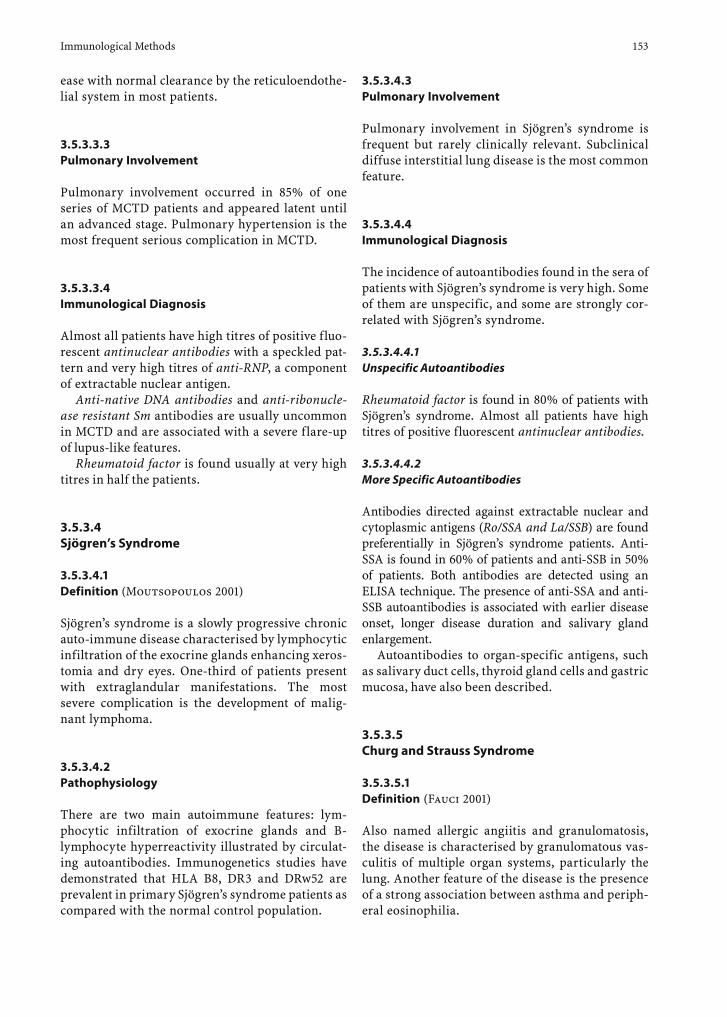
Immunological Methods 153
ease with normal clearance by the reticuloendothe-lial system in most patients.
3.5.3.3.3 Pulmonary Involvement
Pulmonary involvement occurred in 85% of one series of MCTD patients and appeared latent until an advanced stage. Pulmonary hypertension is the most frequent serious complication in MCTD.
3.5.3.3.4 Immunological Diagnosis
Almost all patients have high titres of positive fluo-rescent antinuclear antibodies with a speckled pat-tern and very high titres of anti-RNP, a component of extractable nuclear antigen.
Anti-native DNA antibodies and anti-ribonucle-ase resistant Sm antibodies are usually uncommon in MCTD and are associated with a severe flare-up of lupus-like features.
Rheumatoid factor is found usually at very high titres in half the patients.
3.5.3.4 Sjögren’s Syndrome
3.5.3.4.1 Definition (Moutsopoulos 2001)
Sjögren’s syndrome is a slowly progressive chronic auto-immune disease characterised by lymphocytic infiltration of the exocrine glands enhancing xeros-tomia and dry eyes. One-third of patients present with extraglandular manifestations. The most severe complication is the development of malig-nant lymphoma.
3.5.3.4.2 Pathophysiology
There are two main autoimmune features: lym-phocytic infiltration of exocrine glands and B-lymphocyte hyperreactivity illustrated by circulat-ing autoantibodies. Immunogenetics studies have demonstrated that HLA B8, DR3 and DRw52 are prevalent in primary Sjögren’s syndrome patients as compared with the normal control population.
3.5.3.4.3 Pulmonary Involvement
Pulmonary involvement in Sjögren’s syndrome is frequent but rarely clinically relevant. Subclinical diffuse interstitial lung disease is the most common feature.
3.5.3.4.4 Immunological Diagnosis
The incidence of autoantibodies found in the sera of patients with Sjögren’s syndrome is very high. Some of them are unspecific, and some are strongly cor-related with Sjögren’s syndrome.
3.5.3.4.4.1 Unspecific Autoantibodies
Rheumatoid factor is found in 80% of patients with Sjögren’s syndrome. Almost all patients have high titres of positive fluorescent antinuclear antibodies.
3.5.3.4.4.2 More Specific Autoantibodies
Antibodies directed against extractable nuclear and cytoplasmic antigens (Ro/SSA and La/SSB) are found preferentially in Sjögren’s syndrome patients. Anti-SSA is found in 60% of patients and anti-SSB in 50% of patients. Both antibodies are detected using an ELISA technique. The presence of anti-SSA and anti-SSB autoantibodies is associated with earlier disease onset, longer disease duration and salivary gland enlargement.
Autoantibodies to organ-specific antigens, such as salivary duct cells, thyroid gland cells and gastric mucosa, have also been described.
3.5.3.5 Churg and Strauss Syndrome
3.5.3.5.1 Definition (Fauci 2001)
Also named allergic angiitis and granulomatosis, the disease is characterised by granulomatous vas-culitis of multiple organ systems, particularly the lung. Another feature of the disease is the presence of a strong association between asthma and periph-eral eosinophilia.
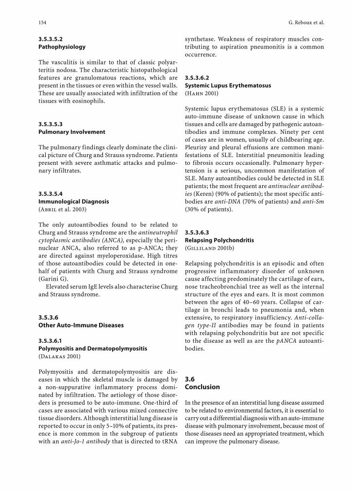
154 G. Reboux et al.
3.5.3.5.2 Pathophysiology
The vasculitis is similar to that of classic polyar-teritis nodosa. The characteristic histopathological features are granulomatous reactions, which are present in the tissues or even within the vessel walls. These are usually associated with infiltration of the tissues with eosinophils.
3.5.3.5.3 Pulmonary Involvement
The pulmonary findings clearly dominate the clini-cal picture of Churg and Strauss syndrome. Patients present with severe asthmatic attacks and pulmo-nary infiltrates.
3.5.3.5.4 Immunological Diagnosis (Abril et al. 2003)
The only autoantibodies found to be related to Churg and Strauss syndrome are the antineutrophil cytoplasmic antibodies (ANCA), especially the peri-nuclear ANCA, also referred to as p-ANCA; they are directed against myeloperoxidase. High titres of those autoantibodies could be detected in one-half of patients with Churg and Strauss syndrome (Garini G).
Elevated serum IgE levels also characterise Churg and Strauss syndrome.
3.5.3.6 Other Auto-Immune Diseases
3.5.3.6.1 Polymyositis and Dermatopolymyositis (Dalakas 2001)
Polymyositis and dermatopolymyositis are dis-eases in which the skeletal muscle is damaged by a non-suppurative inflammatory process domi-nated by infiltration. The aetiology of those disor-ders is presumed to be auto-immune. One-third of cases are associated with various mixed connective tissue disorders. Although interstitial lung disease is reported to occur in only 5–10% of patients, its pres-ence is more common in the subgroup of patients with an anti-Jo-1 antibody that is directed to tRNA
synthetase. Weakness of respiratory muscles con-tributing to aspiration pneumonitis is a common occurrence.
3.5.3.6.2 Systemic Lupus Erythematosus (Hahn 2001)
Systemic lupus erythematosus (SLE) is a systemic auto-immune disease of unknown cause in which tissues and cells are damaged by pathogenic autoan-tibodies and immune complexes. Ninety per cent of cases are in women, usually of childbearing age. Pleurisy and pleural effusions are common mani-festations of SLE. Interstitial pneumonitis leading to fibrosis occurs occasionally. Pulmonary hyper-tension is a serious, uncommon manifestation of SLE. Many autoantibodies could be detected in SLE patients; the most frequent are antinuclear antibod-ies (Keren) (90% of patients); the most specific anti-bodies are anti-DNA (70% of patients) and anti-Sm (30% of patients).
3.5.3.6.3 Relapsing Polychondritis (Gilliland 2001b)
Relapsing polychondritis is an episodic and often progressive inflammatory disorder of unknown cause affecting predominately the cartilage of ears, nose tracheobronchial tree as well as the internal structure of the eyes and ears. It is most common between the ages of 40–60 years. Collapse of car-tilage in bronchi leads to pneumonia and, when extensive, to respiratory insufficiency. Anti-colla-gen type-II antibodies may be found in patients with relapsing polychondritis but are not specific to the disease as well as are the pANCA autoanti-bodies.
3.6 Conclusion
In the presence of an interstitial lung disease assumed to be related to environmental factors, it is essential to carry out a differential diagnosis with an auto-immune disease with pulmonary involvement, because most of those diseases need an appropriated treatment, which can improve the pulmonary disease.
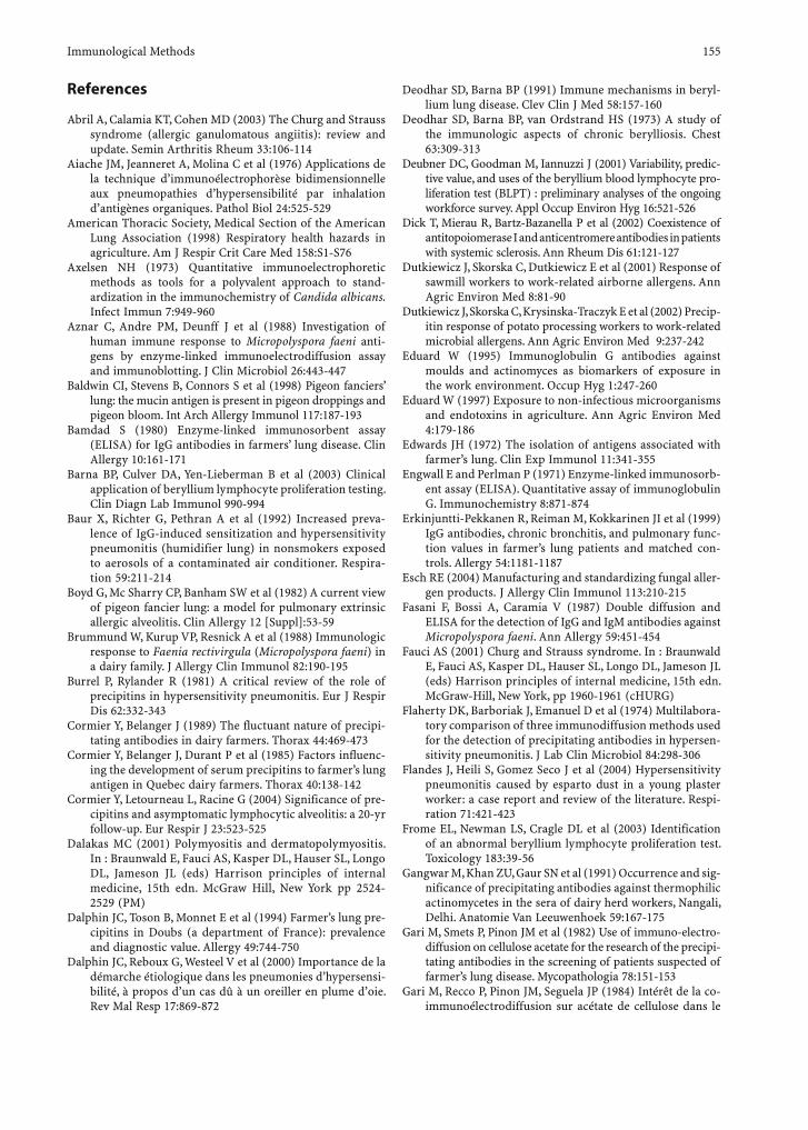
Immunological Methods 155
References
Abril A, Calamia KT, Cohen MD (2003) The Churg and Strauss syndrome (allergic ganulomatous angiitis): review and update. Semin Arthritis Rheum 33:106-114
Aiache JM, Jeanneret A, Molina C et al (1976) Applications de la technique d’immunoélectrophorèse bidimensionnelle aux pneumopathies d’hypersensibilité par inhalation d’antigènes organiques. Pathol Biol 24:525-529
American Thoracic Society, Medical Section of the American Lung Association (1998) Respiratory health hazards in agriculture. Am J Respir Crit Care Med 158:S1-S76
Axelsen NH (1973) Quantitative immunoelectrophoretic methods as tools for a polyvalent approach to stand-ardization in the immunochemistry of Candida albicans. Infect Immun 7:949-960
Aznar C, Andre PM, Deunff J et al (1988) Investigation of human immune response to Micropolyspora faeni anti-gens by enzyme-linked immunoelectrodiffusion assay and immunoblotting. J Clin Microbiol 26:443-447
Baldwin CI, Stevens B, Connors S et al (1998) Pigeon fanciers’ lung: the mucin antigen is present in pigeon droppings and pigeon bloom. Int Arch Allergy Immunol 117:187-193
Bamdad S (1980) Enzyme-linked immunosorbent assay (ELISA) for IgG antibodies in farmers’ lung disease. Clin Allergy 10:161-171
Barna BP, Culver DA, Yen-Lieberman B et al (2003) Clinical application of beryllium lymphocyte proliferation testing. Clin Diagn Lab Immunol 990-994
Baur X, Richter G, Pethran A et al (1992) Increased preva-lence of IgG-induced sensitization and hypersensitivity pneumonitis (humidifier lung) in nonsmokers exposed to aerosols of a contaminated air conditioner. Respira-tion 59:211-214
Boyd G, Mc Sharry CP, Banham SW et al (1982) A current view of pigeon fancier lung: a model for pulmonary extrinsic allergic alveolitis. Clin Allergy 12 [Suppl]:53-59
Brummund W, Kurup VP, Resnick A et al (1988) Immunologic response to Faenia rectivirgula (Micropolyspora faeni) in a dairy family. J Allergy Clin Immunol 82:190-195
Burrel P, Rylander R (1981) A critical review of the role of precipitins in hypersensitivity pneumonitis. Eur J Respir Dis 62:332-343
Cormier Y, Belanger J (1989) The fluctuant nature of precipi-tating antibodies in dairy farmers. Thorax 44:469-473
Cormier Y, Belanger J, Durant P et al (1985) Factors influenc-ing the development of serum precipitins to farmer’s lung antigen in Quebec dairy farmers. Thorax 40:138-142
Cormier Y, Letourneau L, Racine G (2004) Significance of pre-cipitins and asymptomatic lymphocytic alveolitis: a 20-yr follow-up. Eur Respir J 23:523-525
Dalakas MC (2001) Polymyositis and dermatopolymyositis. In : Braunwald E, Fauci AS, Kasper DL, Hauser SL, Longo DL, Jameson JL (eds) Harrison principles of internal medicine, 15th edn. McGraw Hill, New York pp 2524-2529 (PM)
Dalphin JC, Toson B, Monnet E et al (1994) Farmer’s lung pre-cipitins in Doubs (a department of France): prevalence and diagnostic value. Allergy 49:744-750
Dalphin JC, Reboux G, Westeel V et al (2000) Importance de la démarche étiologique dans les pneumonies d’hypersensi-bilité, à propos d’un cas dû à un oreiller en plume d’oie. Rev Mal Resp 17:869-872
Deodhar SD, Barna BP (1991) Immune mechanisms in beryl-lium lung disease. Clev Clin J Med 58:157-160
Deodhar SD, Barna BP, van Ordstrand HS (1973) A study of the immunologic aspects of chronic berylliosis. Chest 63:309-313
Deubner DC, Goodman M, Iannuzzi J (2001) Variability, predic-tive value, and uses of the beryllium blood lymphocyte pro-liferation test (BLPT) : preliminary analyses of the ongoing workforce survey. Appl Occup Environ Hyg 16:521-526
Dick T, Mierau R, Bartz-Bazanella P et al (2002) Coexistence of antitopoiomerase I and anticentromere antibodies in patients with systemic sclerosis. Ann Rheum Dis 61:121-127
Dutkiewicz J, Skorska C, Dutkiewicz E et al (2001) Response of sawmill workers to work-related airborne allergens. Ann Agric Environ Med 8:81-90
Dutkiewicz J, Skorska C, Krysinska-Traczyk E et al (2002) Precip-itin response of potato processing workers to work-related microbial allergens. Ann Agric Environ Med 9:237-242
Eduard W (1995) Immunoglobulin G antibodies against moulds and actinomyces as biomarkers of exposure in the work environment. Occup Hyg 1:247-260
Eduard W (1997) Exposure to non-infectious microorganisms and endotoxins in agriculture. Ann Agric Environ Med 4:179-186
Edwards JH (1972) The isolation of antigens associated with farmer’s lung. Clin Exp Immunol 11:341-355
Engwall E and Perlman P (1971) Enzyme-linked immunosorb-ent assay (ELISA). Quantitative assay of immunoglobulin G. Immunochemistry 8:871-874
Erkinjuntti-Pekkanen R, Reiman M, Kokkarinen JI et al (1999) IgG antibodies, chronic bronchitis, and pulmonary func-tion values in farmer’s lung patients and matched con-trols. Allergy 54:1181-1187
Esch RE (2004) Manufacturing and standardizing fungal aller-gen products. J Allergy Clin Immunol 113:210-215
Fasani F, Bossi A, Caramia V (1987) Double diffusion and ELISA for the detection of IgG and IgM antibodies against Micropolyspora faeni. Ann Allergy 59:451-454
Fauci AS (2001) Churg and Strauss syndrome. In : Braunwald E, Fauci AS, Kasper DL, Hauser SL, Longo DL, Jameson JL (eds) Harrison principles of internal medicine, 15th edn. McGraw-Hill, New York, pp 1960-1961 (cHURG)
Flaherty DK, Barboriak J, Emanuel D et al (1974) Multilabora-tory comparison of three immunodiffusion methods used for the detection of precipitating antibodies in hypersen-sitivity pneumonitis. J Lab Clin Microbiol 84:298-306
Flandes J, Heili S, Gomez Seco J et al (2004) Hypersensitivity pneumonitis caused by esparto dust in a young plaster worker: a case report and review of the literature. Respi-ration 71:421-423
Frome EL, Newman LS, Cragle DL et al (2003) Identification of an abnormal beryllium lymphocyte proliferation test. Toxicology 183:39-56
Gangwar M, Khan ZU, Gaur SN et al (1991) Occurrence and sig-nificance of precipitating antibodies against thermophilic actinomycetes in the sera of dairy herd workers, Nangali, Delhi. Anatomie Van Leeuwenhoek 59:167-175
Gari M, Smets P, Pinon JM et al (1982) Use of immuno-electro-diffusion on cellulose acetate for the research of the precipi-tating antibodies in the screening of patients suspected of farmer’s lung disease. Mycopathologia 78:151-153
Gari M, Recco P, Pinon JM, Seguela JP (1984) Intérêt de la co-immunoélectrodiffusion sur acétate de cellulose dans le
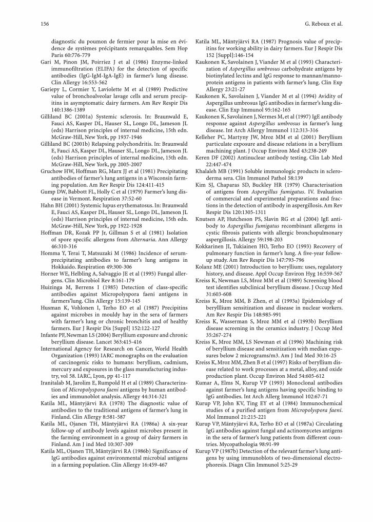
156 G. Reboux et al.
diagnostic du poumon de fermier pour la mise en évi-dence de systèmes précipitants remarquables. Sem Hop Paris 60:776-779
Gari M, Pinon JM, Poirriez J et al (1986) Enzyme-linked immunofiltration (ELIFA) for the detection of specific antibodies (IgG-IgM-IgA-IgE) in farmer’s lung disease. Clin Allergy 16:553-562
Gariepy L, Cormier Y, Laviolette M et al (1989) Predictive value of bronchoalveolar lavage cells and serum precip-itins in asymptomatic dairy farmers. Am Rev Respir Dis 140:1386-1389
Gilliland BC (2001a) Systemic sclerosis. In: Braunwald E, Fauci AS, Kasper DL, Hauser SL, Longo DL, Jameson JL (eds) Harrison principles of internal medicine, 15th edn. McGraw-Hill, New York, pp 1937-1946
Gilliland BC (2001b) Relapsing polychondritis. In: Braunwald E, Fauci AS, Kasper DL, Hauser SL, Longo DL, Jameson JL (eds) Harrison principles of internal medicine, 15th edn. McGraw-Hill, New York, pp 2005-2007
Gruchow HW, Hoffman RG, Marx JJ et al (1981) Precipitating antibodies of farmer’s lung antigens in a Wisconsin farm-ing population. Am Rev Respir Dis 124:411-415
Gump DW, Babbott FL, Holly C et al (1979) Farmer’s lung dis-ease in Vermont. Respiration 37:52-60
Hahn BH (2001) Systemic lupus erythematosus. In: Braunwald E, Fauci AS, Kasper DL, Hauser SL, Longo DL, Jameson JL (eds) Harrison principles of internal medicine, 15th edn. McGraw-Hill, New York, pp 1922-1928
Hoffman DR, Kozak PP Jr, Gillman S et al (1981) Isolation of spore specific allergens from Alternaria. Ann Allergy 46:310-316
Homma Y, Terai T, Matsuzaki M (1986) Incidence of serum-precipitating antibodies to farmer’s lung antigens in Hokkaido. Respiration 49:300-306
Horner WE, Helbling A, Salvaggio JE et al (1995) Fungal aller-gens. Clin Microbiol Rev 8:161-179
Huizinga M, Berrens I (1985) Detection of class-specific antibodies against Micropolyspora faeni antigens in farmers’lung. Clin Allergy 15:139-145
Husman K, Vohlonen I, Terho EO et al (1987) Precipitins against microbes in mouldy hay in the sera of farmers with farmer’s lung or chronic bronchitis and of healthy farmers. Eur J Respir Dis [Suppl] 152:122-127
Infante PF, Newman LS (2004) Beryllium exposure and chronic beryllium disease. Lancet 363:415-416
International Agency for Research on Cancer, World Health Organization (1993) IARC monographs on the evaluation of carcinogenic risks to humans: beryllium, cadmium, mercury and exposures in the glass manufacturing indus-try, vol 58. IARC, Lyon, pp 41-117
Iranitalab M, Jarolim E, Rumpold H et al (1989) Characteriza-tion of Micropolyspora faeni antigens by human antibod-ies and immunoblot analysis. Allergy 44:314-321
Katila ML, Mäntyjärvi RA (1978) The diagnostic value of antibodies to the traditional antigens of farmer’s lung in Finland. Clin Allergy 8:581-587
Katila ML, Ojanen TH, Mäntyjärvi RA (1986a) A six-year follow-up of antibody levels against microbes present in the farming environment in a group of dairy farmers in Finland. Am J ind Med 10:307-309
Katila ML, Ojanen TH, Mäntyjärvi RA (1986b) Significance of IgG antibodies against environmental microbial antigens in a farming population. Clin Allergy 16:459-467
Katila ML, Mäntyjärvi RA (1987) Prognosis value of precip-itins for working ability in dairy farmers. Eur J Respir Dis 152 [Suppl]:146-154
Kaukonen K, Savolainen J, Viander M et al (1993) Characteri-zation of Aspergillus umbrosus carbohydrate antigens by biotinylated lectins and IgG response to mannan/manno-protein antigens in patients with farmer’s lung. Clin Exp Allergy 23:21-27
Kaukonen K, Savolainen J, Viander M et al (1994) Avidity of Aspergillus umbrosus IgG antibodies in farmer’s lung dis-ease. Clin Exp Immunol 95:162-165
Kaukonen K, Savolainen J, Nermes M, et al (1997) IgE antibody response against Aspergillus umbrosus in farmer’s lung disease. Int Arch Allergy Immunol 112:313-316
Kelleher PC, Martyny JW, Mroz MM et al (2001) Beryllium particulate exposure and disease relations in a beryllium machining plant. J Occup Environ Med 43:238-249
Keren DF (2002) Antinuclear antibody testing. Clin Lab Med 22:447-474
Khalaleh MB (1991) Soluble immunologic products in sclero-derma sera. Clin Immunol Pathol 58:139
Kim SJ, Chaparas SD, Buckley HR (1979) Characterisation of antigens from Aspergillus fumigatus. IV. Evaluation of commercial and experimental preparations and frac-tions in the detection of antibody in aspergillosis. Am Rev Respir Dis 120:1305-1311
Knutsen AP, Hutcheson PS, Slavin RG et al (2004) IgE anti-body to Aspergillus fumigatus recombinant allergens in cystic fibrosis patients with allergic bronchopulmonary aspergillosis. Allergy 59:198-203
Kokkarinen JI, Tukiainen HO, Terho EO (1993) Recovery of pulmonary function in farmer’s lung. A five-year follow-up study. Am Rev Respir Dis 147:793-796
Kolanz ME (2001) Introduction to beryllium: uses, regulatory history, and disease. Appl Occup Environ Hyg 16:559-567
Kreiss K, Newman LS, Mroz MM et al (1989) Screening blood test identifies subclinical beryllium disease. J Occup Med 31:603-608
Kreiss K, Mroz MM, B Zhen, et al (1993a) Epidemiology of berylllium sensitization and disease in nuclear workers. Am Rev Respir Dis 148:985-991
Kreiss K, Wasserman S, Mroz MM et al (1993b) Beryllium disease screening in the ceramics industry. J Occup Med 35:267-274
Kreiss K, Mroz MM, LS Newman et al (1996) Machining risk of beryllium disease and sensitization with median expo-sures below 2 micrograms/m3. Am J Ind Med 30:16-25
Kreiss K, Mroz MM, Zhen B et al (1997) Risks of beryllium dis-ease related to work processes at a metal, alloy, and oxide production plant. Occup Environ Med 54:605-612
Kumar A, Elms N, Kurup VP (1993) Monoclonal antibodies against farmer’s lung antigens having specific binding to IgG antibodies. Int Arch Allerg Immunol 102:67-71
Kurup VP, John KV, Ting EY et al (1984) Immunochemical studies of a purified antigen from Micropolyspora faeni. Mol Immunol 21:215-221
Kurup VP, Mäntyjärvi RA, Terho EO et al (1987a) Circulating IgG antibodies against fungal and actinomycetes antigens in the sera of farmer’s lung patients from different coun-tries. Mycopathologia 98:91-99
Kurup VP (1987b) Detection of the relevant farmer’s lung anti-gens by using immunoblots of two-dimensional electro-phoresis. Diagn Clin Immunol 5:25-29
Immunological Methods 157
Kusaka H, Homma Y, Ogasawara H et al (1989) Five-year follow-up of Micropolyspora faeni antibody in smoking and nonsmoking farmers. Am Rev Respir Dis 140:695-699
Lacasse Y, Selman M, Costabel U et al (2003) Clinical diagnosis of hypersensitivity pneumonitis. Am J Respir Crit Care Med 168:952-958
Laemmli UK (1970) Cleavage of structural proteins during the assembly of the head of bacteriophage T4. Nature 227:680-685
Lappalainen S, Pasanen AL, Reiman M et al (1998) Serum IgG antibodies against Wallemia sebi and Fusarium species in Finnish farmers. Ann Allergy Asthma Immunol 81:585-592
Larsson K, Malmberg P, Eklund A et al (1988) Exposure to microorganisms, airway inflammatory changes and immune reactions in asymptomatic dairy farmers. Int Arch Allergy Appl Immunol 87:127-133
Latge JP(1999) Aspergillus fumigatus and aspergillosis. Clin Microbiol Rev 12:310-350
Lioy PJ, Weisel CP, Millette JR et al (2002) Characterization of the dust/smoke aerosol that settled east of the World Trade Center (WTC) in lower Manhattan after the col-lapse of the WTC 11 September 2001. Environ Health Perspect 110:703-714
Lipsky PE (2001) Rheumatoid arthritis. In : Braunwald E, Fauci AS, Kasper DL, Hauser SL, Longo DL, Jameson JL (eds) Harrison principles of internal medicine, 15th edn. McGraw-Hill, New York, pp 1928-1937
Maier LA (2002) Genetic and exposure risks for chronic beryl-lium disease. Clin Chest Med 23:827-839
Maier LA, Kittle LA, Mroz MM et al (2003) Beryllium-stimu-lated neopterin as a diagnostic adjunct in chronic beryl-lium disease. Am J Ind Med 43:592-601
Marcer G, Simioni L, Saia B et al (1983) Study of immunologi-cal parameters in farmer’s lung. Clin Allergy 13:443-449
Martinez-Cordero E, Aguilar Leon DE, Retana VN (2000) IgM antiavian antibodies in sera from patients with pigeon breeder’s disease. J Clin Lab Anal 14:201-207
Marx JJ, Emanuel DA, Dovenbarger WV et al (1978) Farmer’s lung disease among farmers with precipitating antibodies to the thermophilic actinomycetes: a clinical and immu-nologic study. J Allergy Clin Immunol 62:185-189
McSharry C, Anderson K, Bourke SJ et al (2002) Takes your breath away - the immunology of allergic alveolitis. Clin Exp Immunol 128:3-9
Melinn M, McLaughlin H (1989) Farmer‘s lung : a three year survey and comparison of ELISA and CIEP technique in antibody detection. Ir J Med Sci 15:173-174
Melinn M, McLaughlin H (1992) Comparison of methods of production of farmer’s lung antigens. Int Arch Allergy Immunol 99:56-62
Miyazaki H, Gemma H, Uemura K, et al (2004) Hypersensi-tivity pneumonitis induced by Aspergillus niger – a case report. Nihon Kokyuki Gakkai Zasshi 42:676-681
Moran JV, Greenberger PA, Patterson R (2002) Long-term evaluation of hypersensitivity pneumonitis: a case study follow-up and literature review. Allergy Asthma Proc 23:265-270
Moss RB (2002) Allergic bronchopulmonary aspergillosis. Clin Rev Allergy Immunol 23:87-104
Moutsopoulos HM (2001) Sjögren’s syndrome. In : Braunwald E, Fauci AS, Kasper DL, Hauser SL, Longo DL, Jameson JL
(eds) Harrison principles of internal medicine, 15th edn. McGraw-Hill, New York, pp 1947-1949
Mundt C, Becker WM, Schlaak M (1996) Farmer’s lung: patients’ IgG2 antibodies specifically recognize Saccha-ropolyspora rectivirgula proteins and carbohydrate struc-tures. J Allergy Clin Immunol 98:441-450
Newman LS, Bobka C, Schumacher B et al (1994) Compart-mentalized immune response reflects clinical severity of beryllium disease. Am J Respir Crit Care Med 150:135-142
Nolard N, Symoens F, Beguin H (1994) Mycological surveys in dwellings and factories: application to diagnosis of extrinsic allergic alveolitis. In: Samson RA, Flannigan B, Flannigan ME et al (eds) Air quality monographs. Health implication in indoor environments. Elsevier, Amster-dam, pp 201-209
Ouchterlony O (1953) Antigen-antibody reactions in gels. Acta Pathol Microbiol Scand 32:231-240
Ojanen T (1992) Class specific antibodies in serodiagnosis of farmer’s lung. Br J Ind Med 49:332-336
Ojanen TH, Katila ML, Mäntyjärvi RA (1980) The use of enzyme-linked immunosorbent assay (ELISA) in the diagnosis of farmer’s lung. Allergy 35:537-542
Ojanen TH, Terho EO, Mäntyjärvi RA (1982) Comparison of Aspergillus fumigatus and Aspergillus umbrosus antigens in serological tests of farmer’s lung. Allergy 37:297-301
Ojanen TH, Terho EO, Mantyjarvi RA (1987) IgG subclasses in farmer’s lung, Eur J Respir Dis [Suppl] 154:145-150.
Paris S, Fitting C, Latge JP et al (1990) Comparison of conid-ial and mycelial allergens of Alternaria alternata. Arch Allergy Appl Immunol 92:1-8
Patel AM, Ryu JH, Reed CE (2001) Hypersensitivity pneumoni-tis: current concepts and future questions. J Allergy Clin Immunol 108:661-670
Pavesi F, Fasani F, Rancati E et al (1984) Micropolyspora faeni antigens of three commercial extracts. Mycopathologia 84:165-169
Pepys J, Riddell RW, Citron KM et al (1962) Precipitins against extracts of hay and moulds in the serum of patients with farmer’s lung, aspergillosis, asthma and sarcoidosis. Thorax 17: 366-374
Pepys J, Jenkins PA, Festenstein GN et al (1963) Farmer’s lung, thermophilic actinomycetes as a source of “farmer’s lung hay” antigen. Lancet 41:607-611
Pepys J, Jenkins PA (1965) Precipitin (FLH) test farmer’s lung. Thorax 20:21-35
Pepys J (1969) Hypersensitivity disease of the lung due to fungi and other organic dusts. Karger, Basel (Monographs in allergy, vol 4)
Pepys J (1994) Farmer’s lung. A needle in a haystack and Pan-dora’s box. ACI News 6:63-72
Ramasamy M, Khan ZU, Kurup VP (1987) A partially puri-fied antigen from Faenia rectivirgula in the diagnosis of farmer’s lung disease. Microbios 49:171-182
Rask-Andersen A (1989) Allergic alveolitis in Swedish farmers. Upsala J Med Sci 94:271-285
Rautalahti M, Terho EO, Ojanen T (1990) Changes in the titers of IgG antibodies against farmer’s lung antigens in the sera of healthy dairy farmers. Ann Allergy 64:455-458
Reboux G, Piarroux R, Bardonnet K et al (2001a) Comparison of four serological techniques in immunological diagno-sis of farmer’s lung disease. Am J Respir Crit Care Med 163:A747 (abstract)
158 G. Reboux et al.
Reboux G, Piarroux R, Mauny F et al (2001b) Role of molds in farmer’s lung disease in Eastern France. Am J Respir Crit Care Med 163:1534-1539
Recco P, Pourciel D, Borderies M et al (1978) Poumon de fer-mier, poumon des éleveurs d’oiseaux notre expérience en midi-pyrénées. Bull Soc Fr Mycol Méd 7:281-284
Reese G, Trompelt J, Becker WM et al (1989) Extrinsic aller-gic alveolitis: IgG subclass reactivities and monoclonal antibodies against Micropolyspora faeni and Aspergillus fumigatus. Immun Infekt 17:165-168
Roberts RC, Wenzel FJ, Emanuel DA (1976) Precipitating anti-bodies in a Midwest dairy farming population toward the antigens associated with farmer’s lung disease. J Allergy Clin Immunol 57:518-524
Rodrigo MJ, Benavent MI, Cruz MJ et al (2000) Detection of specific antibodies to pigeon serum and bloom antigens by enzyme linked immunosorbent assay in pigeon breed-er’s disease. Occup Environ Med 57:159-164
Rosen S, Yu JH, Adams TH (1999) The Aspergillus nidulans sfaD gene encodes a G protein beta subunit that is required for normal growth and repression of sporulation. EMBO J 18:5592-5600
Rossman MD (1996) Chronic beryllium disease: diagnosis and management. Environ Health Perspect 104S:945-947
Rossman MD, Kern JA, Elias JA et al (1988) Proliferative response of bronchoalveolar lymphocytes to beryllium. Ann Intern Med 108:687-693
Roussel S (2004) Influence des pratiques agricoles sur la flore microbiologique des fourrages et sur le risque de maladie du poumon de fermier. Thesis, Besançon
Roussel S, Reboux G, Dalphin JC et al (2004) Microbiologi-cal evolution of hay and relapse in patients with farmer’s lung. Occup Environ Med 61:e3
Saltini C, Richeldi L, Losi M et al (2001) Major histocompat-ibility locus genetic markers of beryllium sensitization and disease. Eur Respir J 18:1-8
Salvaggio JE (1972) Diagnostic significance of serum precip-itins in hypersensitivity pneumonitis. Chest 62 242-252
Salvaggio JE (1991) Immune reactions in allergic alveolitis. Eur Respir J 4 [Suppl 13]:47s-59s
Salvaggio JE (1997) Extrinsic allergic alveolitis (hypersensi-tivity pneumonitis): past, present and future. Clin Exp Allergy 27 [Suppl 1]:18-25
Schumacher HR, Pessler F, Chen LX (2003) Diagnosing early rheumatoid arthritis (RA). What are the problems and opportunities? Clin Exp Rheumatol 21 [Suppl 31]:S15-S19
Scribner GH, Barboriak JJ, Fink JN (1980) Prevalence of pre-cipitins in group at risk of developing hypersensitivity pneumonitis. Clin Allergy 10:91-95
Sharp GC (1994) Mixed connective tissue disease arthritis. In: Isselbacher KJ, Braunwald E, Wilson JD, Martin JB, Fauci AS, Kasper DL (eds) Harrison principles of internal medi-cine, 13th edn. McGraw-Hill, New York, pp 1660-1661
Solal-Céligny PH, Laviolette M, Hébert J et al (1982) Immune reactions in the lungs of asymptomatic dairy farmers. Am Rev Respir Dis 126:964-967
Spiegelberg HL (1974) Biological activities of immunoglobu-lins of different classes and subclasses. Adv Immunol 19:259-294
Stoeckle JD, Hardy HL, Weber AL (1969) Chronic beryllium disease. Long-term follow-up of sixty cases and selective review of the literature. Am J Med 46:545-561
Stokes TC, Turton CWG, Turner-Warwick M (1981) A study of immunoglobulin G subclasses in patients with farmer’s lung. Clin Allergy 11:201-207
Taylor TP, Ding M, Ehler DS et al (2003) Beryllium in the environment: a review. J Environ Sci Health A Tox Hazard Subst Environ Eng 38:439-469
Terho EO, Husman K, Vohlonen I et al (1987) Serum precip-itins against microbes in mouldy hay with respect to age, sex, atopy and smoking of farmers. Eur J Respir Dis 152 [Suppl]:115-121
Todd A, Coan R, Allen A (1993) Pigeon breeders’ lung: IgG subclasses to pigeon intestinal mucin and IgA antigens. Clin Exp Immunol 92:494-499
Toubas D, Prevost A, Deschamps F et al (1995) Les alvéolites allergiques extrinsèques d’origine professionnelle. Presse Med 24:1391-1396
Trentin L, Marcer G, Chilosi M et al (1988) Longitudinal study of alveolitis in hypersensitivity pneumonitis patients: an immunologic evaluation. J Allergy Clin Immunol 82:577-585
Treuhaft MW, Roberts RC, Hackbarth C et al (1979) Charac-terization of precipitin response to Micropolyspora faeni in farmer’s lung disease by quantitative immunoelectro-phoresis. Am Rev Respir Dis 119:571-578
Vohlonen I, Husman K, Terho EO et al (1987) Prevalence of serum precipitins against microbes in mouldy hay, and of chronic bronchitis and farmer’s lung with respect to farmer’s occupational health hazards. Eur J Respir Dis 152 [Suppl]:139-145
Wardrop VE, Blyth W, Grant WB (1977) Farmer’s lung in a group of Scottish dairy farms. Br J Ind Med 34:186-195
Wenzel F, Gray R, Roberts RC et al (1974) Serologic studies in farmer’s lung. Am Rev Respir Dis 109:464-468
Williams CA Jr, Grabar PJ (1955) Immunoelectrophoretic stud-ies on serum proteins. I. The antigens of human serum. Immunology 74:158-168
Williams WR, Williams WJ (1982) Development of beryllium lymphocyte transformation in chronic beryllium disease. Int Arch Allergy Appl Immunol 67:175-180
Yi ES (2002) Hypersensitivity pneumonitis. Crit Rev Clin Lab Sci 39:581-629
Ylönen JK, Ojanen TH, Jägerroos HJW et al (1989) Compara-tive immunochemical analysis of five Thermoactinomyces strains. Int Arch Allergy Appl Immunol 90:405-410


