Embed Size (px)
Citation preview

J. exp. Btol. 112, 169-178 (1984)Printed in Great Britain © The Company of Biologists Limited 1984
LOCAL CIRCUIT INTERACTIONS INSYNCHRONIZATION OF CORTICAL NEURONES
BY R. K. S. WONG, R. MILES AND R. D. TRAUB*
Department of Physiology and Biophysics, University of Texas MedicalBranch, Galveston, TX 77550, U.SA. and *IBM T.J. Watson ResearchCenter, Yorktovm Heights, NY 10598, U.SA. and Neurological Institute,
New York, NY 10032, U.SA.
SUMMARY
Under certain circumstances large numbers of neurones in the mammaliancentral nervous system (CNS) can discharge simultaneously. An exampleof such activity is recorded from a hippocampal slice in the presence ofagents which block synaptic inhibition. This synchronized discharge occursspontaneously in a rhythmic fashion or may be triggered by stimulation ofany afferent pathway. Its generation appears to involve local circuit inter-actions. The favourable conditions offered by an in vitro preparation haveallowed the cellular events during this activity to be examined in some detail.Three factors appear to be critically involved in the synchronization process.Firstly, the intrinsic ability of neurones to generate bursts, secondly, theexistence of powerful recurrent excitatory connections, and thirdly the ab-sence of inhibition which normally prevents the spread of bursting activitythrough the recurrent connections. Computer simulations show that in asparsely connected network of bursting neurones activity initiated in a fewcells may spread through recurrent connections until eventually the wholepopulation discharges simultaneously. Rhythmic discharges similar to thosedescribed here also underly various CNS functions including centrally-originating motor patterns. It remains to be determined whether neuronalproperties and connectivity found to be important in this hippocampalrhythm may also play a role in the generation of other rhythmic activities inthe mammalian CNS.
INTRODUCTION
The synchronized firing of neuronal populations is frequently observed in themammalian central nervous system. Motor output for locomotion (Grillner, 1975)and respiration (Wyman, 1977) involves the simultaneous activation of manyneurones, and synchronized firing also underlies prominent electroencephalographicwaves such as the alpha (Andersen & Andersson, 1968) and theta rhythms (Fujita &Sato, 1964). At present, our knowledge of the synaptic and cellular processes leadingto neuronal synchrony for these normal activities is extremely limited.
The brain slice preparation has been very useful for studying the properties ofindividual cortical neurones and the synaptic processes underlying their collective
Key words: Hippocampus, synchronization, neuronal oscillation.

170 R. K. S. WONG, R. MILES AND R. D. TRAUB
behaviour. One of the most interesting observations from hippocampal slices is thaigroups of pyramidal cells when disinhibited can undergo spontaneous synchronizedoscillations (Schwartzkroin & Prince, 1978; Wong & Traub, 1983). These eventshave stereotyped patterns that vary little from preparation to preparation and they caneven be recorded from segments of a slice containing as few as 1000 pyramidal cells(Miles, Wong & Traub, 1984). The small size of the neuronal population involved haspermitted detailed investigation of the cellular mechanisms underlying the generationof this activity. The data show that the initiation of synchronized discharge criticallydepends on the three following properties of the hippocampal network. (1) Pyramidalcells can generate intrinsic bursts. (2) These cells are interconnected by a sparselydistributed yet powerful network of excitatory synapses. (3) Synaptic inhibition playsan important regulatory function. The incorporation of these experimentally observedproperties of hippocampal neurones into a computer model has allowed the formula-tion of a plausible mechanism for synchronization. In this review we will illustrate theoperation of each of these processes and their involvement in the initiation of popula-tion discharge.
CHARACTERISTICS OF SYNCHRONIZED EVENTS
Spontaneous and evoked synchronized activity of hippocampal neurones is consis-tently observed in the presence of agents which block gamma-aminobutyric acid(GABA) mediated inhibition (Dichter & Spencer, 1969; Dingledine & Gjerstad,1980; Schwartzkroin & Prince, 1978; Wong & Prince, 1979; Alger & Nicoll, 1980).The occurrence of these events is indicated by a comb-shaped field potential inextracellular recordings. Characteristically spontaneous events occur rhythmically atintervals of 5-10s (Fig. 1). Intracellular recordings suggest that all neurones
A B
10s 40msFig. 1. Characteristics of synchronized activities. (A) Spontaneous synchronized discharge occursrhythmically in the presence of GABA blockers. Top trace, intracellular recording; lower trace,extracellular recording. Deflections observed in the extracellular trace depend on a large populationof neurones discharging simultaneously. (B) Synchronous discharge elicited by afferent stimulation.Top traces, intracellular recording; lower trace, extracellular recording.
Neuronal synchronization 171
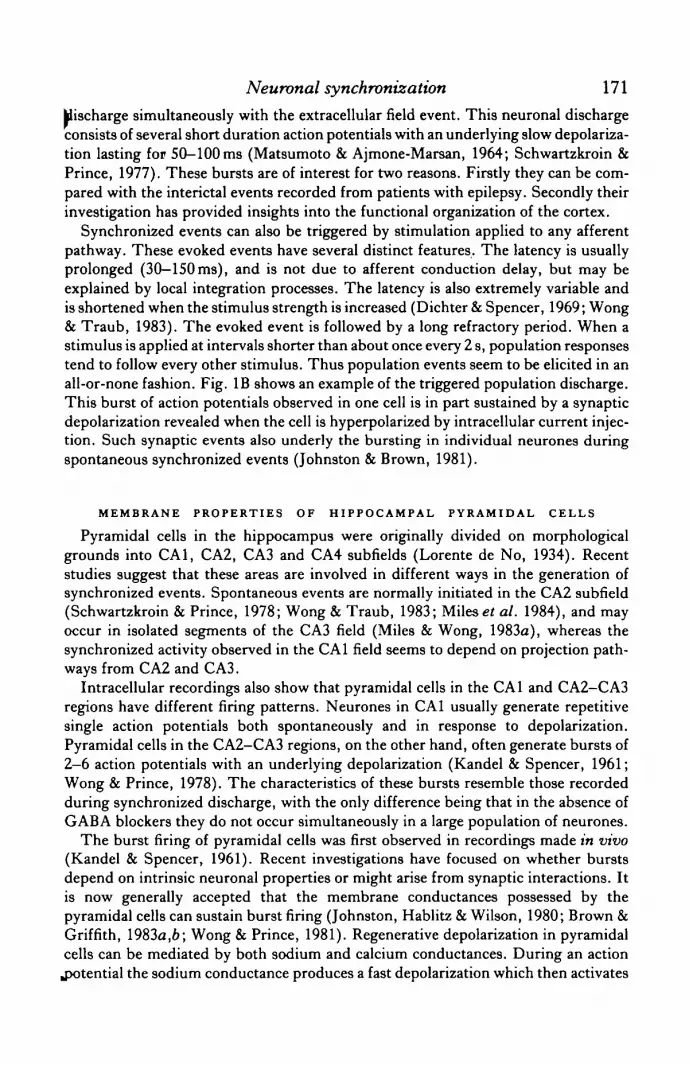
Hischarge simultaneously with the extracellular field event. This neuronal dischargeconsists of several short duration action potentials with an underlying slow depolariza-tion lasting for 50-100ms (Matsumoto & Ajmone-Marsan, 1964; Schwartzkroin &Prince, 1977). These bursts are of interest for two reasons. Firstly they can be com-pared with the interictal events recorded from patients with epilepsy. Secondly theirinvestigation has provided insights into the functional organization of the cortex.
Synchronized events can also be triggered by stimulation applied to any afferentpathway. These evoked events have several distinct features. The latency is usuallyprolonged (30-150ms), and is not due to afferent conduction delay, but may beexplained by local integration processes. The latency is also extremely variable andis shortened when the stimulus strength is increased (Dichter & Spencer, 1969; Wong& Traub, 1983). The evoked event is followed by a long refractory period. When astimulus is applied at intervals shorter than about once every 2 s, population responsestend to follow every other stimulus. Thus population events seem to be elicited in anall-or-none fashion. Fig. IB shows an example of the triggered population discharge.This burst of action potentials observed in one cell is in part sustained by a synapticdepolarization revealed when the cell is hyperpolarized by intracellular current injec-tion. Such synaptic events also underly the bursting in individual neurones duringspontaneous synchronized events (Johnston & Brown, 1981).
MEMBRANE PROPERTIES OF HIPPOCAMPAL PYRAMIDAL CELLS
Pyramidal cells in the hippocampus were originally divided on morphologicalgrounds into CA1, CA2, CA3 and CA4 subfields (Lorente de No, 1934). Recentstudies suggest that these areas are involved in different ways in the generation ofsynchronized events. Spontaneous events are normally initiated in the CA2 subfield(Schwartzkroin & Prince, 1978; Wong & Traub, 1983; Miles et al. 1984), and mayoccur in isolated segments of the CA3 field (Miles & Wong, 1983a), whereas thesynchronized activity observed in the CA1 field seems to depend on projection path-ways from CA2 and CA3.
Intracellular recordings also show that pyramidal cells in the CA1 and CA2-CA3regions have different firing patterns. Neurones in CA1 usually generate repetitivesingle action potentials both spontaneously and in response to depolarization.Pyramidal cells in the CA2-CA3 regions, on the other hand, often generate bursts of2—6 action potentials with an underlying depolarization (Kandel & Spencer, 1961;Wong & Prince, 1978). The characteristics of these bursts resemble those recordedduring synchronized discharge, with the only difference being that in the absence ofGABA blockers they do not occur simultaneously in a large population of neurones.
The burst firing of pyramidal cells was first observed in recordings made in vivo(Kandel & Spencer, 1961). Recent investigations have focused on whether burstsdepend on intrinsic neuronal properties or might arise from synaptic interactions. Itis now generally accepted that the membrane conductances possessed by thepyramidal cells can sustain burst firing (Johnston, Hablitz & Wilson, 1980; Brown &Griffith, 1983a,b; Wong & Prince, 1981). Regenerative depolarization in pyramidalcells can be mediated by both sodium and calcium conductances. During an action.potential the sodium conductance produces a fast depolarization which then activates

172 R. K. S. WONG, R. MILES AND R. D. TRAUB
a calcium conductance. This conductance deactivates slowly, resulting in a depolarizing afterpotential (DAP). The DAP can reach threshold to generate another actionpotential and so result in a burst. It now seems that calcium entry or its intracellularaccumulation during depolarization activates a potassium conductance (Hotson &Prince, 1980; Brown & Griffith, 19836; Alger & Nicoll, 1980) which produces amembrane hyperpolarization lasting for up to 2 s. It is this hyperpolarization thatdetermines the rhythm of spontaneous bursting.
Recordings from acutely isolated pyramidal cells from adult guinea pig hippo-campus have directly demonstrated the intrinsic bursting capability of these cells(Fig. 2). Isolated cells have 'clean' membrane surfaces allowing intracellular record-ings to be made with low resistance electrodes using the suction method (Hamill etal. 1981). In Fig. 2B a short depolarizing pulse applied intracellularly elicited a burstof action potentials. Furthermore isolated neurones can also generate pacemakerbursting activity (Fig. 2C).
EXCITATORY CONNECTIONS BETWEEN PYRAMIDAL CELLS
Synaptic excitation between pyramidal cells has long been considered probable.The search for its existence began in the deafferented hippocampus (Lebovitz, Dichter& Spencer, 1971) and has been continued in studies using the hippocampal slice(MacVicar & Dudek, 1980; Knowles & Schwartzkroin, 1981). More recentlymonosynaptic excitatory connections between pyramidal cells have been demon-strated directly (Fig. 3). The connection between these cells was considered to bemonosynaptic since each presynaptic action potential elicited an EPSP. Furthermorethe latency of the EPSP was short and varied little. A crucial observation was that abarrage of EPSPs elicited by bursting in the presynaptic cell summed temporally toreach threshold and initiated a burst in the postsynaptic cell. Three factors contributeto the functional strength of this excitatory coupling. Firstly, these EPSPs facilitateat short intervals similar to those between action potentials in a presynaptic burst(Miles & Wong, 19836). Secondly, the slow time course of the EPSP means thattemporal summation occurs near the peak of each one of the group of synaptic eventselicited by a burst of spikes. Thirdly, multiple and independent sites of burst genera-tion are present on the soma-dendritic membrane of pyramidal cells (Wong, Prince& Basbaum, 1979). These factors ensure that activity in pyramidal cells may propa-gate throughout the population via the excitatory interconnections.
In simultaneous intracellular recordings, monosynaptically connected pairs of cellsare rarely encountered, indicating that their distribution is sparse. Nevertheless, aswill become apparent, this feature of local connectivity is crucially involved in shapingthe synchronization.
CELLULAR MECHANISM FOR SYNCHRONIZATION
Computer simulations of hippocampal neurone networks incorporating an intrinsicbursting capability and powerful recurrent connections will generate synchronizeddischarge (Traub & Wong, 1982, 1983). Our model consists of a population of 100neurones each able to generate bursts and interconnected randomly by a sparse.

Journal of Experimental Biology, Vol. 112 Fig. 2
JlOmV10 pA
20 ms
50 ms
Fig. 2. Neurones may be acutely isolated from the hippocampus of adult guinea pigs. (A) Pyramidal-type cell with a long primary apical dendrite and three basilar dendrites. Calibration bar, 10fan.(B), (C) 1 ntraccllular activity recorded from this type of neurone (upper traces). Lower traces monitorcurrent applied intraccllularly.
-R. K. S. WONG, R. MILES AND R. D. TRAUB (Facing p. 172)


Neuronal synchronization 173
O-©
CeUT
Cell 2 w
20 mV
SraV
40 ms
Fig. 3. Recurrent excitatory connection between pyramidal cells. Each action potential in cell 1triggered an EPSP in cell 2 eventually leading to a burst of action potentials. The recording of cell2 is shown at two different gains.
network of excitatory synapses (Fig. 4). There are two critical assumptions. Firstlyeach cell is connected to more than one follower cell and, secondly synaptic connec-tions are sufficiently strong that a burst in one cell may evoke a burst in the followercells. In this way a localized stimulus applied to a few cells (1-4) will excite a largepopulation of follower cells. As this process is repeated recruitment becomes in-creasingly rapid leading to a synchronized population event (Fig. 5). This hypothesis
Jor synchronization provides explanations for several experimental observations. The
R. K. S. WONG, R. MILES AND R. D. TRAUB
25mV —¥1 Y Y Y Y Y Y
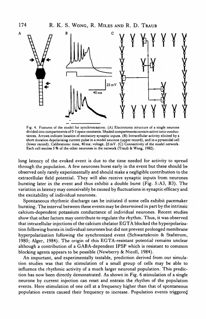
Fig. 4. Features of the model for synchronization. (A) Electrotonic structure of a single neuronedivided into compartments of 0-1 space constants. Shaded compartments contain active ionic conduc-tances. Arrows indicate location of excitatory synaptic inputs. (B) Intracellular activity elicited by ashort duration depolarizing current pulse in a model neurone (upper record), and in a pyramidal cell(lower record). Calibrations: time, 40 ms; voltage, 25 mV. (C) Connectivity of the model network.Each cell excites 5 % of the other neurones in the network (Traub & Wong, 1982).
long latency of the evoked event is due to the time needed for activity to spreadthrough the population. A few neurones burst early in the event but these should beobserved only rarely experimentally and should make a negligible contribution to theextracellular field potential. They will also receive synaptic inputs from neuronesbursting later in the event and thus exhibit a double burst (Fig. 5:A3, B3). Thevariation in latency may conceivably be caused by fluctuations in synaptic efficacy andthe excitability of individual neurones.
Spontaneous rhythmic discharge can be initiated if some cells exhibit pacemakerbursting. The interval between these events may be determined in part by the intrinsiccalcium-dependent potassium conductance of individual neurones. Recent studiesshow that other factors may contribute to regulate the rhythm. Thus, it was observedthat intracellular injections of the calcium chelator EGTA blocked the hyperpolariza-tion following bursts in individual neurones but did not prevent prolonged membranehyperpolarization following the synchronized event (Schwartzkroin & Stafstrom,1980; Alger, 1984). The origin of this EGTA-resistant potential remains unclearalthough a contribution of a GABA-dependent IPSP which is resistant to commonblocking agents appears to be possible (Newberry & Nicoll, 1984).
An important, and experimentally testable, prediction derived from our simula-tion studies was that the stimulation of a small group of cells may be able toinfluence the rhythmic activity of a much larger neuronal population. This predic-tion has now been directly demonstrated. As shown in Fig. 6 stimulation of a singleneurone by current injection can reset and entrain the rhythm of the populationevents. Here stimulation of one cell at a frequency higher than that of spontaneouspopulation events caused their frequency to increase. Population events triggered.

Neuronal synchronization 175
by activity in a single cell tended to occur with latency of 50—200 ms. These resultsclearly show that synchronized activity is initiated by local neuronal interaction.This process whereby activity in a single neurone may activate an entire neuronal
Simulation Experimental
Fig. 5. Comparison of synchronized activity from simulation (A) and experimental (B) studies. (1)Percentage of neurones bursting with time during a synchronized event. (2) Extracellular activity.(3), (4), (5) Intracellular activity (Traub & Wong, 1982). Calibrations, time: 50ms (A), 60 ms (B);amplitude: 4mV (A2.B2), 25mV (A3,4,5), 20mV (B3,4,5).

176A
R. K. S. WONG, R. MILES AND R. D. TRAUB
|20mV
|SmV
10-SnA
Control
100 m.0 4 8
Inter-burst intervals (s)
12
Fig. 6. Entrainment of synchronized population activity by a single neurone. (A),(B),(C) Uppertraces, intracellular record; middle traces, extracellular record; lower traces, intracellular currentinjection. (D) Distribution of intervals between synchronized bursts preceding (control) and duringstimulation (Stim). The control interval was 6 2 ± 2-2 s (mean ± s.D., N= 36). During stimulationat once every 4 s the interval changed to 4 3 ± 1-1 s (./V= 36), significantly (/»> 0-01) lower thancontrol (Miles & Wong, 1983a).
population can be viewed as an extremely powerful mechanism for signal ampli-fication.
SYNAPTIC INHIBITION
The synchronization of hippocampal neurones described here occurs only whenGABA-dependent inhibition is blocked. How do inhibitory processes normallyprevent the development of synchronization?
Inhibition in the hippocampus is mediated through both feedback (Andersen,Eccles & Loyning, 1963) and feedforward (Alger & Nicoll, 1982) circuits. In theformer, axon collaterals from pyramidal cells activate the inhibitory cells, whereas inthe latter, inhibitory neurones are excited directly by afferent fibres. These circuitsunderly the EPSP-IPSP sequence commonly observed following the activation ofafferent fibres. The latency of the IPSP is such that only single action potentials areelicited in neurones which possess intrinsic bursting capability (Wong & Prince,1979). As can be observed in Fig. 3, bursting ensures the spread of excitation betweenpyramidal cells which is the basis for synchronized discharge.
The way synaptic inhibition is recruited through feedback circuits by the

Neuronal synchronization 111
Celll
Cell 2
50 ms
Fig. 7. Recurrent IPSPs may be initiated by activity in a single pyramidal cell. Action potentials incell 1 elicited IPSPs in cell 2. An intercalated inhibitory neurone is assumed since inhibitory eventsdid not follow each action potential and varied considerably in latency.
spontaneous activity of pyramidal cells is less clear. Our recent studies suggest thatthe influence of a pyramidal cell on inhibitory neurones in the CA3 region may be sopowerful that a single pyramidal cell can activate an inhibitory neurone (Fig. 7).Inhibitory neurones appear to possess widely arborizing axonal collaterals. Thus onceactivated they will exert an influence over a large number of pyramidal cells loweringtheir excitability and tending to prevent the spread of activity through the recurrentnetwork.
Supported in part by NIH Grant NS 18464 and the Klingenstein Foundation.
R E F E R E N C E S
ALGER, B. E. (1984). Characteristics of a slow hyperpolarizing synaptic potential in rat hippocampal pyramidalcells. J. Neurophysiol. (in press).
ALGER, B. E. & NICOLL, R. A. (1980). Epileptiform burst afterhyperpolarization: calcium-dependentpotassium potential in hippocampal CA1 pyramidal cells. Science, N.Y. 210, 1122-1124.
ALGER, B. E. & NICOLL, R. A. (1982). Feedforward dendntic inhibition in rat hippocampal pyramidal cellsstudied in vitro. J. Physiol., Land. 328, 105-123.
ANDERSEN, P. & ANDERSSON, S. A. (1968). Physiological Basis of the Alpha Rhythm. New York: Appleton-Cenrury Crofts.
ANDERSEN, P., ECCLES, J. C. & LOYNING, Y. (1963). Recurrent inhibition in the hippocampus with identifica-tion of the inhibitory cell and its synapses. Nature, hand. 198, 540—542.

178 R. K. S. WONG, R. MILES AND R. D. TRAUB
BROWN, D. A. & GRIFFITH, W. H. (1983a). Calcium activated outward current in voltage-clamped hippo*campal neurones of the guinea-pig. J. Pkysiol., Land. 337, 287-301.
BROWN, D. A. & GRIFFITH, W. H. (19836). Persistent slow inward calcium current in voltage-clampedhippocampal neurones of the guinea-pig. J . Physiol., hand. 337, 303-320.
DICHTER, M. & SPENCER, W. A. (1969). Penicillin induced interictal discharges from the cat hippocampus. I.Characteristics and topographical features. J. Neurophysiol. 32, 649-662.
DINCLEDINE, R. & GJERSTAD, L. (1980). Reduced inhibition during epileptiform activity in the in vitrohippocampal slice. J. Physiol., Land. 305, 297-313.
FUJITA, Y. & SATO, T. (1964). Intracellular records from hippocampal pyramidal cells in rabbit during thetarhythm activity. .7. Neurophysiol. Zl, 1011-1025.
GRILLNER, S. (1975). Locomotion in vertebrates: central mechanisms and reflex interactions. Physiol. Rev. 55,247-304.
HAJIIIX, O. P., MARTY, A., NEHER, E., SAKMANN, B. & SIGWORTH, F. J. (1981). Improved patch clamp
techniques for high resolution current recording from cells and cell-free membrane patches. Pflug. Arch. ges.Physiol. 391, 85-100.
HOTSON, J. R. & PRINCE, D. A. (1980). A calcium-activated hyperpolarization follows repetitive firing inhippocampal neurones. J. Neurophysiol. 43, 409—419.
JOHNSTON, D. & BROWN, T. H. (1981). Giant synaptic potential hypothesis for epileptiform activity. Science,N.Y. 211, 294-297.
JOHNSTON, D., HABLJTZ, J. J. & WILSON, W. A. (1980). Voltage clamp discloses a slow inward current inhippocampal burst-firing neurones. Nature, Land. 286, 391—393.
KANDEL, E. R. & SPENCER, W. A. (1961). Electrophysiology of hippocampal neurons. II. Afterpotentials andrepetitive firing. J. Neurophysiol. 24, 243-259.
KNOWLES, W. D. & SCHWARTZKROIN, P. A. (1981). Local circuit synaptic interactions in hippocampal brainslices. J.Neurosd. 1, 318-322.
LEBOVTTZ, R. M., DICHTER, M. & SPENCER, W. A. (1971). Recurrent excitation in the CA3 region of cathippocampus. Int.J. Neumsci. 2, 99-108.
LORENTE DE No, R. (1934). Studies on the structure of the cerebral cortex. II . Continuation of the structureof the ammonic system. J- Psych. Neurol. 46, 225-242.
MACVICAR, B. A. & DUDEK, F. E. (1980). Local synaptic circuits in rat hippocampus: interactions betweenpyramidal cells. Brain Res. 184, 220-223.
MATSUMOTO, H. & AJMONE-MARSAN, C. (1964). Cortical cellular phenomena in experimental epilepsy:interictal manifestations. Exp. Neurol. 9, 286-304.
MILES, R. & WONG, R. K. S. (1983a). Single neurones can initiate synchronized population discharge in thehippocampus. Nature, Land. 306, 371—373.
MILES, R. & WONG, R. K. S. (19836). Properties of recurrent excitation in theCA3 region of the hippocampus.Neurosd. Abstr. 9, 909.
MILES, R., WONG, R. K. S. & TRAUB, R. D. (1984). Synchronized after-discharges in the hippocampus:contribution of local synaptic interactions. Neumsci. (in press).
NEWBERRY, N. R. & NICOLL, R. A. (1984). Baclofen directly hyperpolarizes hippocampal pyramidal cells.Nature, Land, (in press).
SCHWARTZKROIN, P. A. & PRINCE, D. A. (1977). Penicillin-induced epileptiform activity in the hippocampalin vitro preparation. A. Rev. Neurol. 1, 463—469.
SCHWARTZKJIOIN, P. A. & PRINCE, D. A. (1978). Cellular and field potential properties of epileptogenichippocampal slices. Brain. Res. 147, 117-130.
SCHWARTZKROIN, P. A. & STAFSTROM, C. E. (1980). The effects of EGTA on the calcium-activated afterhyper-polarizarJon in the hippocampal CA3 cells. Science, N.Y. 210, 1125—1126.
TRAUB, R. D. & WONG, R. K. S. (1982). Cellular mechanism of neuronal synchronization in epilepsy. Science,N.Y. 216, 745-747.
TRAUB, R. D. & WONG, R. K. S. (1983). Synchronized burst discharge in the disinhibited hippocampal slice.II . Model of cellular mechanism. J. Neurophysiol. 49, 459—471.
WONG, R. K. S. & PRINCE, D. A. (1978). Participation of calcium spikes during intrinsic burst firing inhippocampal neurons. Brain Res. 159, 385—390.
WONG, R. K. S. & PRINCE, D. A. (1979). Dendritic mechanism underlying penicillin induced epileptiformactivity. Science, N.Y. 204, 1228-1231.
WONG, R. K. S. & PRINCE, D. A. (1981). Afterpotential generation in hippocampal pyramidal cells. J. Neuro-physiol. 45, 86-97.
WONG, R. K. S., PRINCE, D. A. & BASBAUM, A. I. (1979). Intradendritic recordings from hippocampalneurons. Proc. natn. Acad. Set. U.SA. 76, 986-990.
WONG, R. K. S. & TRAUB, R. D. (1983). Synchronized burst discharge in the disinhibited hippocampal slice.I. Initiation in CA2-CA3 region. J. Neurophysiol. 49, 442-458.
WYMAN, R. J. (1977). Neural generation of the breathing rhythm. A. Rev. Physiol. 39, 417-448.


