Embed Size (px)
Citation preview

117 Copyright 2004 Psychonomic Society, Inc.
Cognitive, Affective, & Behavioral Neuroscience2004, 4 (2), 117-126
Understanding how the cerebral cortex processes in-formation is a major aim of neurobiology today, with im-portant implications for disciplines ranging from psy-chiatry to the designing of living machines. Numerousinvestigative techniques at different levels are used tothis end, including functional brain imaging, single-unitrecording, and anatomy. These techniques rapidly con-verge at the level of the cortical area, where specificphysiological functions can be localized and each areaexhibits distinct patterns of connectivity. The concertedaction of multiple areas is thought to underlie sensory pro-cesses and cognitive functions. This has led to a majorfield of research that attempts to determine the position ofindividual areas in the overall cortical organization of in-formation flow. Work in the visual system has been par-ticularly fruitful in this respect. Here, Hubel and Wieselin the 1970s built on a tradition going back to HughlingsJackson, which conceived that the brain was structuredalong hierarchical principals. In Hubel and Wiesel’s work,hierarchical relations were explored, and it was shown,for example, that as one progresses from the primary area(area V1) to successively higher areas (V2, V3, V4, etc.)there is a progressive increase in both the dimensions and
the complexity of receptive fields. This approach led tothe concept that different attributes of the retinal image,such as color and movement, are emphasized in differentareas (Zeki, 1978). In this way, information is thought tobe extracted at the successive levels going from lower tohigher visual areas according to a feedforward direction.
Physiological investigation of the cortex has recentlyput into question this feedforward hypothesis of informa-tion flow, and it is becoming increasingly clear that feed-back processes need to be considered as well. One majorissue is that the timing of activation of successive levelssimply does not fit with the concept of strictly sequentialactivation (Petroni, Panzeri, Hilgetag, Kotter, & Young,2001). Intracranial recording in the monkey has shownthat there is considerable overlap in the timing of the sig-nal response at successive hierarchical stages (Nowak,Munk, James, Girard, & Bullier, 1999; Raiguel, Lagae,Gulyas, & Orban, 1989; Schmolesky et al., 1998; Zipser,Lamme, & Schiller, 1996). Furthermore, there is experi-mental evidence of rapid widespread activation of the sen-sory parietal cortex within 45 msec and of prefrontal areaswithin 90 msec (Foxe & Simpson, 2002; Juan & Walsh,2003). Together, these studies have shown that fast activa-tion of higher areas, coupled with late activation of areaV1, means that V1 can have a higher area function, whichin turn could generate the known attentional processes inearly visual areas including the primary visual cortex (All-man, Miezin, & McGuinness, 1985; Kastner & Ungerlei-der, 2000; Mehta, Ulbert, & Schroeder, 2000a, 2000b;Motter, 1993; Somers, Dale, Seiffert, & Tootell, 1999;Vanduffel, Tootell, & Orban, 2000). Temporal, but alsospatial, attributes of activity in response to a stimulus pointto the higher function of area V1. There is increasing evi-dence that area V1 neurons are influenced by stimuli out-side of the classical receptive field, leading to complex
This work was supported by EU Grant “Nets and Representations”QLG3-1999-0106, Human Frontier Science Program Grant RG0133/2000-B, and European Community FP5 Quality of Life Grant QLG3-1999-01064, A.F. was supported by the Fondation pour la RechercheMédicale. We thank Colette Dehay, Marie-Thérèse Perenin, and CharlieSchroeder for their critical comments on an earlier version of the arti-cle. We are grateful to Souhila Benkerri and Delphine Autran for theirhistological processing and to Delphine Autran for data analysis. Cor-respondence concerning this article should be addressed to H. Kennedy,INSERM U371, Cerveau et Vision, IFR 19 Institut Fédératif des Neuro-sciences de Lyon, INSERM U371, 18 Avenue Doyen Lépine, 69675 BronCedex, France (e-mail: [email protected]).
Long-distance feedback projections to area V1:Implications for multisensory integration, spatial
awareness, and visual consciousness
SIMON CLAVAGNIER, ARNAUD FALCHIER, and HENRY KENNEDYUniversité Claude Bernard Lyon 1, Bron, France
It is generally agreed that information flow through the cortex is constrained by a hierarchical archi-tecture. Recent experimental evidence suggests that projections descending the hierarchy and target-ing the primary visual cortex (area V1) may play an essential role in perceptual processes. We have,therefore, reexamined feedback projections to area V1, using retrograde tracer injections in this area.In addition to well-known areas, quantification of labeling in higher cortical areas reveals a number ofhitherto unknown long-distance feedback connections originating from auditory (A1), multisensory(STP) cortices, but also from a perirhinal area (36). These feedback projections from advanced corticalstations, a global feature shared by areas that belong to the ventral visual stream, could play an impor-tant role in early multisensory integration and spatial awareness and could provide the physical sub-strate for the involvement of area V1 in visual consciousness.

118 CLAVAGNIER, FALCHIER, AND KENNEDY
contextual responses (Nothdurft, Gallant, & Van Essen,1999; Zipser et al., 1996). These long-range interactionsbeyond the receptive field clearly fit with the high di-vergence of feedback projections from higher areas (An-gelucci et al., 2002; Kennedy & Bullier, 1985; Salin, Gi-rard, Kennedy, & Bullier, 1992). Furthermore, these feed-back projections originating from sensory nonvisualareas provide the basis for multisensory integration(Falchier, Clavagnier, Barone, & Kennedy, 2002).
Multisensory integration of sensory inputs has beenshown to occur at multiple sites, including midbrain andcortical levels (Stein & Meredith, 1993). Multisensoryconvergence is not a sufficient condition for multisen-sory integration. At the level of a single neuron, the con-vergence of inputs from different sensory modalitiesoften, but not always (Wallace & Stein, 1994), results inthe integration of concurrent signals, so that responsesare significantly different from those obtained with ei-ther of the modality-specific components alone (Stein &Meredith, 1993). Classical descriptions of the visual cor-tex predict an integration of visual and nonvisual stimuli atlater stages of cortical processing (Calvert, Brammer, &Iversen, 1998; Hikosaka, Iwai, Saito, & Tanaka, 1988).Most anatomical and electrophysiological studies havepointed to multisensory convergence in areas located in thetemporal, parietal, and frontal cortex (Benevento, Fallon,Davis, & Rezak, 1977; Goldman-Rakic, 1988). Howeverthere is recent electrophysiological and brain-imaging ev-idence that visual, auditory, and somatosensory integra-tion occurs at relatively early stages of the visual corti-cal pathways (Giard & Peronnet, 1999; Macaluso, Frith,& Driver, 2000). Recent imaging studies performed onhumans have shown cortical multisensory integration sitesearly in visual pathways and especially around the lingualgyrus, where early visual areas are located (Macalusoet al., 2000). This result is also supported by fMRI stud-ies showing multisensory integration in the region of thesuperior occipital gyrus (Calvert, Hansen, Iversen, &Brammer, 2001). These findings in favor of early multi-sensory integration were unexpected because anatomicalstudies of the monkey brain failed until recently to detectafferent connections to the primary visual cortex fromareas processing modalities other than vision. This standsin contrast with evidence that congenital blindness leadsto the responding by early visual cortical areas in thedorsal stream to nonvisual stimuli, including auditorystimuli (Weeks et al., 2000) and tactile stimuli in a dis-crimination task (Buchel, Price, Frackowiak, & Friston,1998; Cohen et al., 1999; Sadato et al., 1996). It is thoughtthat these nonvisual responses are being relayed back toearly visual cortical stages via feedback projections fromthe parietal cortex (Pons, 1996). However, we need to keepin mind that nonvisual information may have access tothe visual areas through rewiring of subthalamic pathwaysduring development (Angelucci, Clasca, & Sur, 1998).
The primary visual area V1 of the primate contains afine-grain map of the visual world and receives the bulk of
ascending visual pathways (Zeki, 1993). It also receives alarge number of cortico-cortical connections, which havebeen described as largely from visual cortical areas(Barone, Batardière, Knoblauch, & Kennedy, 2000; Felle-man & Van Essen, 1991; Perkel, Bullier, & Kennedy,1986). Weak afferent projections to V1 have been de-scribed from distant areas in the ventral (TEO, TE) anddorsal (MST, LIP) pathways (Barone et al., 2000; Felleman& Van Essen, 1991; Perkel, et al., 1986; Rockland, 1994).All these areas respond principally to visual and oculomo-tor stimuli, except for the LIP, where auditory-related ac-tivity has been reported (Linden, Grunewald, & Andersen,1999).
Jones and Powell (1970) have postulated that the con-nectivity of primary sensory areas is restricted to thoseareas of the same modality. The evidence in favor ofmultisensory convergence at the first cortical stage of in-formation processing has prompted us to challenge theconcept put forward by Jones and Powell concerning thedefinition of a primary area.
METHOD
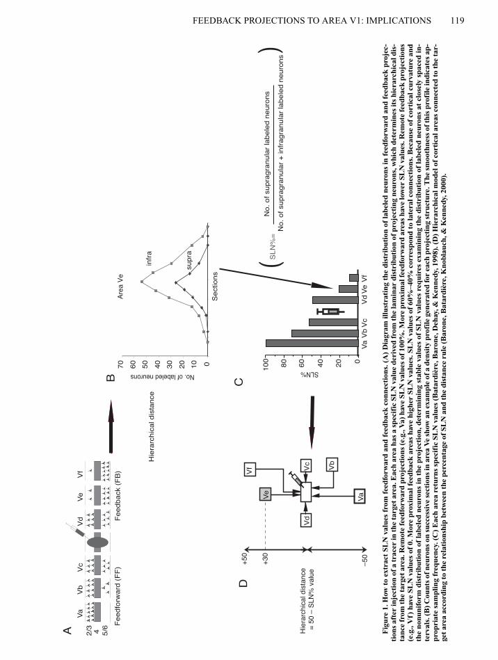
In order to establish the complete pattern of cortical connectiv-ity of area V1, we implemented a quantitative analysis using trac-ers of optimal sensitivity. We made injections of the retrograde trac-ers fast blue and diamidino yellow in area V1 subserving centraland peripheral visual fields. A major property of these tracers isthat they give very clear injection sites, with highly restricted pick-up zones (Bullier, Kennedy, & Salinger, 1984). These tracers areextremely sensitive, and counting the number of neurons in differ-ent areas and layers makes it possible to determine two parameters(Figures 1 and 2): (1) the FLN which refers to the fraction of labeledneurons in each area and indicates the relative contribution of agiven connection (Falchier et al., 2002; Vezoli et al., in press,); and(2) the SLN, which indicates the hierarchical distance separatingthe labeled area from the injected area (Barone et al., 2000). Fourretrograde tracing experiments were carried out on 3 cynomolgusmonkeys (Macaca fascicularis). Some of the results from these in-jections have already been reported in another article (Falchieret al., 2002). Central area 17 injections were in the cortex subserv-ing 0º–2º in the lower visual field (M85RHDY and M85RHFsB).Injections aimed at the peripheral representation were made in thehead and stem of the calcarine sulcus by means of vertical penetra-tions, using stereotaxic coordinates. Two different eccentricities canbe distinguished in peripheral area 17 injections. The M76LH in-jection was located in the dorsal leaf of the calcarine cortex sub-serving 10º–12º eccentricity (Falchier et al., 2002). The other pe-ripheral injection (M81RH) was located further ventrally, near thejunction of the dorsal leaf with the calcarine stem, and were in thecortex subserving 15º–20º. A detailed explanation of the histolog-ical procedures has been reported elsewhere (Falchier et al., 2002).
Immunohistochemical and myelin staining criteria made it possi-ble to localize labeled neurons in MT, MST, FST (STS complex),STP, and auditory areas (Cusick, 1997; Morel, Garraghty, & Kaas,1993). The auditory area A1, located in the posterior bank of the lat-eral sulcus, is distinguished by using acetylcholynesterase and cy-tochrome oxidase reactivity (Morel et al., 1993) and by the presenceof a strong parvalbumin staining in Layers 2–4 (Kosaki, Hashikawa,He, & Jones, 1997). The perirhinal and parahippocampal corticeswere delimitated using SMI-32 staining and the Nissl method for cy-toarchitectonic analysis (Suzuki & Amaral, 2003).
FEEDBACK PROJECTIONS TO AREA V1: IMPLICATIONS 119
Vb
Vc
Vf
Vd
Ve
Vd
Ve
Vf
2/3
4 5/6
Vc
Vb
Va
Va
A
SLN%
020406080100
Va
Vb
Vc
Vd
Ve
Vf
010203040506070
supr
a
infr
a
No. of labeled neurons
Sec
tions
D
Fee
dfor
war
d (F
F)
Fee
dbac
k (F
B)
Hie
rarc
hica
l dis
tanc
e
SLN
%=
No.
of s
upra
gran
ular
labe
led
neur
ons
B C
No.
of s
upra
gran
ular
+ in
frag
ranu
lar
labe
led
neur
ons
Hie
rarc
hica
l dis
tanc
e =
50
– S
LN%
val
ue
–50
+50
Are
a V
e
()
+30
Fig
ure
1. H
ow t
o ex
trac
t SL
N v
alue
s fr
om f
eedf
orw
ard
and
feed
back
con
nect
ions
. (A
)D
iagr
am il
lust
rati
ng t
he d
istr
ibut
ion
of la
bele
d ne
uron
s in
fee
dfor
war
d an
d fe
edba
ck p
roje
c-ti
ons
afte
r in
ject
ion
of a
trac
er in
the
targ
et a
rea.
Eac
h ar
ea h
as a
spe
cifi
c SL
N v
alue
der
ived
from
the
lam
inar
dis
trib
utio
n of
pro
ject
ing
neur
ons,
whi
ch d
eter
min
es it
s hi
erar
chic
al d
is-
tanc
e fr
om th
e ta
rget
are
a. R
emot
e fe
edfo
rwar
d pr
ojec
tion
s (e
.g.,
Va)
hav
e SL
N v
alue
s of
100
%. M
ore
prox
imal
feed
forw
ard
area
s ha
ve lo
wer
SL
N v
alue
s. R
emot
e fe
edba
ck p
roje
ctio
ns(e
.g.,
Vf)
hav
e SL
N v
alue
s of
0. M
ore
prox
imal
fee
dbac
k ar
eas
have
hig
her
SLN
val
ues.
SL
N v
alue
s of
60%
–40%
cor
resp
ond
to la
tera
l con
nect
ions
. Bec
ause
of
cort
ical
cur
vatu
re a
ndth
e no
nuni
form
dis
trib
utio
n of
labe
led
neur
ons
in t
he p
roje
ctio
n, d
eter
min
ing
stab
le v
alue
s of
SL
N v
alue
s re
quir
es e
xam
inin
g th
e di
stri
buti
on o
f la
bele
d ne
uron
s at
clo
sely
spa
ced
in-
terv
als.
(B) C
ount
s of
neu
rons
on
succ
essi
ve s
ecti
ons
in a
rea
Ve
show
an
exam
ple
of a
den
sity
pro
file
gen
erat
ed fo
r ea
ch p
roje
ctin
g st
ruct
ure.
The
sm
ooth
ness
of t
his
prof
ile in
dica
tes
ap-
prop
riat
e sa
mpl
ing
freq
uenc
y. (
C)
Eac
h ar
ea r
etur
ns s
peci
fic
SLN
val
ues
(Bat
ardi
ère,
Bar
one,
Deh
ay, &
Ken
nedy
, 199
8). (
D)
Hie
rarc
hica
l mod
el o
f co
rtic
al a
reas
con
nect
ed t
o th
e ta
r-ge
t ar
ea a
ccor
ding
to
the
rela
tion
ship
bet
wee
n th
e pe
rcen
tage
of
SLN
and
the
dis
tanc
e ru
le (
Bar
one,
Bat
ardi
ère,
Kno
blau
ch, &
Ken
nedy
, 200
0).

120 CLAVAGNIER, FALCHIER, AND KENNEDY
RESULTS
Labeled neurons were found in the 13 cortical areas thatare known to provide projections to area V1, including the
recently described projection from STP and A1 (Falchieret al., 2002; Rockland & Ojima, 2003). Labeled neuronswere found in the medial temporal lobe in accordancewith earlier reports (Doty, 1983; Kennedy & Bullier,
Figure 2. Extraction of FLN values from feedforward and feedback connections. (A) and (B) same as those in Figure 1. (C) Adaptedsampling frequency allows us to calculate precisely the total number of projecting neurons for each area. This makes it possible to de-termine the relative contribution of each area to the total afferent connectivity of the target area.
C
Va Vb Vc Vd Ve Vf
10
20
30
0
FLN
%
FLN% value area VbNo. of neurons in area Vb projecting to target area
Total No. of neurons projecting on target area=
Vd Ve Vf2/345/6
VcVbVa
A
Feedforward (FF) Feedback (FB)
0
10
20
30
40
50
60
70
supra
infra
No.
of l
abel
ed n
euro
ns
Sections
B
Area Vb
( )
Figure 3. Labeling in area 36 after V1 injections. (A) Lateral view ofthe brain and vertical sections showing levels of sections c and d. (B) In-dividual sections showing labeling in the temporal cortex. Shaded re-gions represent the extent of the cortex in which labeled neurons wereconsidered to be in area 36 between the amt sulcus (amt) and the rhinalfissure (rf).

FEEDBACK PROJECTIONS TO AREA V1: IMPLICATIONS 121
1985; Lyon & Kaas, 2002; Rockland & Van Hoesen,1994). An unexpected finding was the presence of la-beled neurons in area 36 (Figure 3). Preliminary FLNdata suggest that projections from TH and TF to area V1are comparable in strength to the projections originatingfrom STP and A1.
We have used the SLN values to construct a hierar-chical model of the relationships of the areas showinglong distance projections to area V1 (Figure 4).
We have also indicated, on the Felleman and Van Essen(1991) model of the visual cortex, those areas that showlong-distance feedback connections to area V1 (Figure 5).This reveals that all of the so-called ventral stream areashave long-distance connections with area V1.
DISCUSSION
The present results show that area V1 receives projec-tions from a number of areas that have classically notbeen considered to be part of the visual system. Aftermaking interspecies comparisons, we shall consider thefeedback projections from STP and the auditory corticeswith respect to multisensory convergence and spatialawareness. Our results also reveal long-distance feedbackprojections to area V1 from areas located at the top of theFelleman and Van Essen (1991) hierarchical model (area36, TH, and TF). These pathways are discussed with re-gard to their possible involvement in visual imagery.
Interspecies ComparisonAlthough sparse projections from the primary auditory cor-
tex to area V2 in adult rodents and cats have been reported(Innocenti, Berbel, & Clarke, 1988; Miller & Vogt, 1984),such projections to area V1 are thought to be absent in these
species (Dehay, Kennedy, & Bullier, 1988; Montero, 1993;Sanderson, Dreher, & Gayer, 1991; Symonds & Rosenquist,1984). Although we cannot exclude the possibility that someof the studies above might have missed an auditory projectionto peripheral area V1, Montero and colleagues have specifi-cally addressed this issue and have found no such projection(Montero, 1993). Furthermore, although a direct projectionfrom the auditory cortex to area V1 has been reported in theopossum, it is considerably weaker than that to area V2 (Mar-tinich, Pontes, & Rocha-Miranda, 2000). These interspeciescomparisons suggest that the projection of the auditory cortexand the STP to peripheral area V1 might be a unique primatefeature. However it needs to be remembered that homologiesof the STP in nonprimates are difficult to identify.
Feedback Projections andMultisensory Integration
Schroeder has argued that multisensory convergenceat early stages indicates a high-order function at low-level sensory cortices, which is compatible with reversehierarchy theory (Cauller, 1995; Hochstein & Ahissar,2002; Schroeder et al., 2003). The auditory system is or-ganized in parallel streams. The caudal parabelt (whichcontains 70% of the neurons of the auditory cortices pro-jecting to area V1) is part of the dorsal auditory pathwayspecialized in spatial information processing, includingsound source localization (Kaas & Hackett, 2000; Recan-zone, 2000; Tian, Reser, Durham, Kustov, & Rauschecker,2001). Overall, the auditory cortex plays an important rolein sound localization, and receptive fields are large andextend behind the pinna axis (Barone, Clarey, Irons, &Imig, 1996). Similarly, auditory receptive fields in theSTP are large and expand in the peripheral visual field(Hikosaka et al., 1988). Hence, there is a certain degree of
Hie
rarc
hica
l lev
el
(2)
(3)
(5)
(7)
(8)
(10)
(9)
(1)
(4)
(6)
Auditory
V2
V3
FEFEF
MT
FSTTEOTEOEOTEO
TETETE
TH / TFTH / TH / TFTH / TF
V1
V4V4V4
STPLIPLIP
FSTMSMST
36
Figure 4. Direct feedback connections to area V1. The hierarchical distanceis calculated using the SLN values (Barone, Batardière, Knoblauch, &Kennedy, 2000). The thickness of the lines indicates the strength of the con-nection obtained by FLN values (Falchier, Clavagnier, Barone, & Kennedy,2002; Vezoli et al., in press). Areas in black, dorsal stream; gray, ventral stream.

122 CLAVAGNIER, FALCHIER, AND KENNEDY
spatial congruence in the auditory properties of A1/STPand the visual representation of the part of area V1 towhich these areas project.
Multisensory integration serves to enhance perceptualcapacities (Stein & Meredith, 1993), so that, for example,the addition of an auditory signal to a visual stimulusmight lead to improved detection and reduced delay and,therefore, an improved orientation response, as comparedwith visual stimuli alone (Giard & Peronnet, 1999;Goldring, Dorris, Corneil, Ballantyne, & Munoz, 1996;McDonald, Teder-Salejarvi, & Hillyard, 2000). This isrelevant to our findings because the short latencies to au-ditory stimulus in A1 and the STP (Bruce, Desimone, &Gross, 1981; Recanzone, Guard, & Phan, 2000; Schroeder& Foxe, 2002) mean that the projections from these areasto V1 could participate in a foveation mechanism towarda peripheral sound source (Heffner & Heffner, 1992). Re-cently, a new form of multisensory convergence has beenreported (Dehner, Keniston, Clemo, & Meredith, 2004).The authors provided evidence of auditory suppression ofsomatosensory responses in area SIV in the cat. Such amechanism may play a role in cross-modal selective at-tention, whereby attention to one modality broadly sup-
presses activity in another. Since no auditory responseshave yet been reported in V1 neurons, one can speculatethat such a type of interaction might exist in the primaryvisual area.
Multisensory integration can play an important role inthe functional reorganization of the cortex following sen-sory deprivation. This could be made possible by corticalpathways such as the auditory/STP projection to periph-eral area V1 (Pons, 1996). In patients suffering from earlyblindness, early visual areas, including the primary visualcortex, are responsive to tactile (Buchel et al., 1998; Cohenet al., 1999; Sadato et al., 1996) and auditory (Weeks et al.,2000) stimuli. The functional reorganization in the multi-sensory area observed after early visual deprivation(Rauschecker, 1995; Wanet-Defalque et al., 1988; Yaka,Yinon, & Wollberg, 1999) might be coupled to a functionalstrengthening of the auditory and STP connections to V1by overtraining in the nonaffected modalities.
Peripheral visual deafferentation in the newborn catinduces an improvement in spatial auditory localizationperformances later in life (Korte & Rauschecker, 1993;Rauschecker & Kniepert, 1994) that has been explainedby cortical compensatory mechanisms in the multisensory
Figure 5. The modified hierarchical model of Felleman and Van Essen (1991). Areas beyond areaV3A showing long-distance feedback projections to area V1 are outlined.
MT
Auditory areas
TEOTEOEO
TETE
V4

FEEDBACK PROJECTIONS TO AREA V1: IMPLICATIONS 123
areas (Rauschecker & Korte, 1993). This could have im-portant behavioral consequences, such as the improved ca-pacity of the blind to localize sound in peripheral auditoryspace (Roder et al., 1999). A recent PET study also hasshown auditory activation of V1 and V2 in postlinguallydeaf subjects implanted with cochlear implants, listeningto sounds with closed eyes (Giraud, Price, Graham, Truy,& Frackowiak, 2001). These authors have suggested thatthe activation of the visual cortex is similar to the re-cruitment of the auditory cortex during speech reading(Calvert et al., 1997). One can assume that cross-modalcooperation could compensate for the subthreshold stim-ulation by processing complementary information fromanother sensory modality. Hence, anatomical, electro-physiological, and brain-imaging studies support a modelof cross-modal integration within a distributed networkof cortical areas in which the primary visual area couldparticipate in the initial integration of sensory informa-tion (Calvert, Campbell, & Brammer, 2000; Stein, 1998).Cross-modal integration has a particular importance fortheories of visual consciousness, which we shall examinein the next section.
Feedback Projections and Visual ConsciousnessCortico-cortical feedforward projections going from
lower to higher hierarchical levels are thought to elabo-rate receptive field responses (Bullier, Girard, & Salin,1994; Vanduffel, Payne, Lomber, & Orban, 1997; Zeki,1993). Feedback mechanisms play a role in integratinginformation outside of the classical receptive field andfigure–ground discrimination (Hupé et al., 1998; Lamme& Roelfsema, 2000). Such top-down influences couldprovide the substrate of high-level effects, such as selec-tive attention in area V1 (Kastner & Ungerleider, 2000;Lamme & Roelfsema, 2000; Mehta et al., 2000a, 2000b).Recent experiments have directly addressed the possiblerole of feedback projections in the visual consciousness.Pascual-Leone and Walsh (2001) showed that stimulationof V5 elicits moving phosphenes, which disappear duringinactivation of area V1. The work of Logothetis has ex-amined neuronal response coding perception and re-sponses to the retinal image at different cortical stages.Using binocular rivalry and an adaptation paradigm, thiswork has shown that at early cortical stations, there aresome neurons that change activity according to the percept,and not the retinal image, and hence, strongly supports theidea of top-down processes (Leopold & Logothetis, 1996;Tolias, Smirnakis, Augath, Trinath, & Logothetis, 2001).
Feedback Projections and Visual ImageryFeedback pathways have also been implicated in visual
imagery (Miyashita, 1995). There is a large body of evi-dence that visual imagery activates early cortical stages,including area V1, again indicating top-down processes(Thompson & Kosslyn, 2000). Cross-modal activity canbe associated with the mental image of the stimuli and hasbeen implicated in the activation of the calcarine cortex inhumans (Klein, Paradis, Poline, Kosslyn, & Le Bihan,2000). Similarly, in humans performing a tactile object
recognition task, tactile-specific activity has been ob-served in the calcarine visual cortex in the absence of vi-sual information (Deibert, Kraut, Kremen, & Hart, 1999).
It is still debated whether mental images are funda-mentally different from verbal thoughts, whether theyshare common mechanisms with visual perception, andwhether information in images is represented in a map-like, spatial format. But most neuropsychological, psycho-physical, and neuroimaging studies have indicated thatvisual perception and mental imagery share commonmechanisms (Farah, 1995; Ishai & Sagi, 1995; Miyashita,1995; but see Bartolomeo, 2002).
Furthermore, visual imagery activates several visualregions, including area V1 (for a review, see Kosslyn,Ganis, & Thompson, 2001). By recording single neuronsin the human medial temporal lobe while subjects wereasked to imagine previously viewed images, Kreiman,Koch, and Fried (2000) showed that single neurons in thehippocampus, amygdala, entorhinal cortex, and parahip-pocampal gyrus selectively altered their firing rates, de-pending on the nature of the imaginary stimulus. In thisrespect, the connections we report between parahip-pocampal areas TH/TF and perirhinal area 36 and V1, aswell as the connections from the amygdala to V1 (Ama-ral, Behniea, & Kelly, 2003), could constitute the path-way that activates and modulates area V1 during visualrecall (memory and imagery). According to the theoriesof reverse hierarchy and visual imagery, the activation ofarea V1 is specifically required for fine-grain visual im-ages (Hochstein & Ahissar, 2002; Kosslyn et al., 2001).
Figure 6. Unilateral neglect in the human and the monkey. Thisfigure shows the regions in the human and the monkey brainwhere lesions result in unilateral neglect. In the monkey brain,the parabelt area is in gray, and the STP is in black. The sulci areunfolded in the monkey brain.
1 cm
Parabelt area
STP
STG

124 CLAVAGNIER, FALCHIER, AND KENNEDY
Feedback Projections and Spatial AwarenessLesions of the STP and surrounding areas in the su-
perior temporal gyrus (presumably, the parabelt) in themonkey were found to induce deficits strongly resem-bling unilateral neglect in human (Luh, Butter, & Buch-tel, 1986; Watson, Valenstein, Day, & Heilman, 1994).Although there is still some debate about the exact siteof the lesion that generates this syndrome in the human,a recent study has put forward a strong argument in favorof the STG (Karnath, Ferber, & Himmelbach, 2001; seeFigure 6). Patients with unilateral neglect typically be-have as if the left part of their visual field or even of theirown body no longer exists (Bisiach & Vallar, 1988). Thissyndrome can be defined as a spatial awareness disorderbecause it is not restricted to the visual modality but in-cludes tactile and auditory modalities (Karnath & Perenin,1998; Pavani, Làdavas, & Driver, 2003). Could it be thatthe involvement of the STP in the monkey in spatialawareness is related to the fact that the STP, a multisen-sory cortical area (Cusick, 1997), has direct feedback pro-jections to area V1?
CONCLUSION
In the present article, we have developed the proposi-tion that feedback projections to area V1 play an impor-tant role in spatial and visual cognition. An important as-pect of this concept, which needs to be further explored,is that the cognitive function of area V1 is linked in someway to the multisensory convergence that occurs at thislevel. Furthermore, multisensory convergence at each hi-erarchical step of each sensory system may be a necessaryfeature for building a unified conscious system. Futurework is required to explore whether a similar anatomicalbasis exist for multisensory integration in the primary cor-tices of other sensory modalities.
REFERENCES
Allman, J., Miezin, R., & McGuinness, E. (1985). Stimulus specificresponses from beyond the classical receptive field: Neurophysio-logical mechanisms for local–global comparisons in visual neurons.Annual Review of Neuroscience, 8, 407-430.
Amaral, D. G., Behniea, H., & Kelly, J. L. (2003). Topographic or-ganization of projections from the amygdala to the visual cortex in themacaque monkey. Neuroscience, 118, 1099-1120.
Angelucci, A., Clasca, F., & Sur, M. (1998). Brainstem inputs to theferret medial geniculate nucleus and the effect of early deafferenta-tion on novel retinal projections to the auditory thalamus. Journal ofComparative Neurology, 400, 417-439.
Angelucci, A., Levitt, J. B., Walton, E. J., Hupe, J. M., Bullier, J.,& Lund, J. S. (2002). Circuits for local and global signal integrationin primary visual cortex. Journal of Neuroscience, 22, 8633-8646.
Barone, P., Batardière, A., Knoblauch, K., & Kennedy, H. (2000).Laminar distribution of neurons in extrastriate areas projecting to vi-sual areas V1 and V4 correlates with the hierarchical rank and indi-cates the operation of a distance rule. Journal of Neuroscience, 20,3263-3281.
Barone, P., Clarey, J. C., Irons, W. A., & Imig, T. J. (1996). Corticalsynthesis of azimuth-sensitive single-unit responses with nonmonot-onic level tuning: A thalamocortical comparison in the cat. Journalof Neurophysiology, 75, 1206-1220.
Bartolomeo, P. (2002). The relationship between visual perceptionand visual mental imagery: A reappraisal of the neuropsychologicalevidence. Cortex, 38, 357-378.
Batardière, A., Barone, P., Dehay, C., & Kennedy, H. (1998). Area-specific laminar distribution of cortical feedback neurons projectingto cat area 17: Quantitative analysis in the adult and during ontogeny.Journal of Comparative Neurology, 396, 493-510.
Benevento, L. A., Fallon, J., Davis, B. J., & Rezak, M. (1977).Auditory–visual interaction in single cells in the cortex of the supe-rior temporal sulcus and the orbital frontal cortex of the macaquemonkey. Experimental Neurology, 57, 849-872.
Bisiach, E. V., & Vallar, G. (1988). Hemineglect in humans. InF. Boller & J. Grafman (Eds.), Handbook of Neuropsychology (Vol.1, pp. 195-222). Amsterdam: Elsevier.
Bruce, C., Desimone, R., & Gross, C. G. (1981). Visual properties ofneurons in a polysensory area in superior temporal sulcus of themacaque. Journal of Neurophysiology, 46, 369-384.
Buchel, C., Price, C., Frackowiak, R. S., & Friston, K. (1998). Dif-ferent activation patterns in the visual cortex of late and congenitallyblind subjects. Brain, 121 (Pt. 3), 409-419.
Bullier, J., Girard, P., & Salin, P. A. (1994). The role of area 17 inthe transfer of information to extrastriate visual cortex. In A. Peters& K. S. Rockland (Eds.), Primary visual cortex in primates (Vol. 10,pp. 301-330). New York: Plenum.
Bullier, J., Kennedy, H., & Salinger, W. (1984). Branching andlaminar origin of projections between visual cortical areas in the cat.Journal of Comparative Neurology, 228, 329-341.
Calvert, G. A., Brammer, M. J., & Iversen, S. D. (1998). Crossmodalidentification. Trends in Cognitive Sciences, 7, 287-253.
Calvert, G. A., Bullmore, E. T., Brammer, M. J., Campbell, R.,Williams, S. C. R., McGuire, P. K.,Woodruff, P. W. R., Iversen,S. D., & David, A. S. (1997). Activation of auditory cortex duringsilent lipreading. Science, 276, 593-596.
Calvert, G. A., Campbell, R., & Brammer, M. J. (2000). Evidencefrom functional magnetic resonance imaging of crossmodal bindingin the human heteromodal cortex. Current Biology, 10, 649-657.
Calvert, G. A., Hansen, P. C., Iversen, S. D., & Brammer, M. J.(2001). Detection of audio-visual integration sites in humans by ap-plication of electrophysiological criteria to the BOLD effect. Neuro-Image, 14, 427-438.
Cauller, L. (1995). Layer I of primary sensory neocortex: Where top-down converges upon bottom-up. Behavioural Brain Research, 71,163-170.
Cohen, L. G., Weeks, R. A., Sadato, N., Celnik, P., Ishii, K., & Hal-lett, M. (1999). Period of susceptibility for cross-modal plasticity inthe blind. Annals of Neurology, 45, 451-460.
Cusick, C. G. (1997). The superior temporal polysensory region inmonkeys. In E. G. Jones & A. Peters (Eds.), Cerebral cortex (Vol. 12,pp. 435-468). New York: Plenum.
Dehay, C., Kennedy, H., & Bullier, J. (1988). Characterization oftransient cortical projections from auditory, somatosensory, andmotor cortices to visual areas 17, 18, and 19 in the kitten. Journal ofComparative Neurology, 272, 68-89.
Dehner, L. R., Keniston, L. P., Clemo, H. R., & Meredith, M. A.(2004). Cross-modal circuitry between auditory and somatosensoryareas of the cat anterior ectosylvian sulcal cortex: A “new” inhibitoryform of multisensory convergence. Cerebral Cortex, 14, 387-403.
Deibert, E., Kraut, M., Kremen, S., & Hart, J., Jr. (1999). Neuralpathways in tactile object recognition. Neurology, 52, 1413-1417.
Doty, R. W. (1983). Nongeniculate afferents to striate cortex in maca-ques. Journal of Comparative Neurology, 218, 159-173.
Falchier, A., Clavagnier, S., Barone, P., & Kennedy, H. (2002).Anatomical evidence of multimodal integration in primate striatecortex. Journal of Neuroscience, 22, 5749-5759.
Farah, J. M. (1995). The neural bases of mental imagery. In M. S. Gaz-zaniga (Ed.), The cognitive neurosciences (pp. 963-974). Cambridge,MA: MIT Press.
Felleman, D. J., & Van Essen, D. C. (1991). Distributed hierarchicalprocessing in the primate cerebral cortex. Cerebral Cortex, 1, 1-47.
Foxe, J. J., & Simpson, G. V. (2002). Flow of activation from V1 to

FEEDBACK PROJECTIONS TO AREA V1: IMPLICATIONS 125
frontal cortex in humans: A framework for defining “early” visualprocessing. Experimental Brain Research, 142, 139-150.
Giard, M. H., & Peronnet, F. (1999). Auditory–visual integration dur-ing multimodal object recognition in humans: A behavioral and elec-trophysiological study. Journal of Cognitive Neuroscience, 11, 473-490.
Giraud, A. L., Price, C. J., Graham, J. M., Truy, E., & Frackow-iak, R. S. (2001). Cross-modal plasticity underpins language recov-ery after cochlear implantation. Neuron, 30, 657-663.
Goldman-Rakic, P. S. (1988). Topography of cognition: Parallel dis-tributed networks in primate association cortex. Annual Review ofNeuroscience, 11, 137-156.
Goldring, J. E., Dorris, M. C., Corneil, B. D., Ballantyne, P. A.,& Munoz, D. P. (1996). Combined eye–head gaze shifts to visualand auditory targets in humans. Experimental Brain Research, 111,68-78.
Heffner, R. S., & Heffner, H. E. (1992). Visual factors in sound lo-calization in mammals. Journal of Comparative Neurology, 317,219-232.
Hikosaka, K., Iwai, E., Saito, H., & Tanaka, K. (1988). Polysensoryproperties of neurons in the anterior bank of the caudal superior tem-poral sulcus of the macaque monkey. Journal of Neurophysiology,60, 1615-1637.
Hochstein, S., & Ahissar, M. (2002). View from the top: Hierarchiesand reverse hierarchies in the visual system. Neuron, 36, 791-804.
Hupé, J. M., James, A. C., Payne, B. R., Lomber, S. G., Girard, P., &Bullier, J. (1998). Cortical feedback improves discrimination be-tween figure and background by V1, V2 and V3 neurons. Nature,394, 784-787.
Innocenti, G. M., Berbel, P., & Clarke, S. (1988). Development ofprojections from auditory to visual areas in the cat. Journal of Com-parative Neurology, 272, 242-259.
Ishai, A., & Sagi, D. (1995). Common mechanisms of visual imageryand perception. Science, 268, 1772-1774.
Jones, E. G., & Powell, T. P. (1970). An anatomical study of converg-ing sensory pathways within the cerebral cortex of the monkey.Brain, 93, 793-820.
Juan, C. H., & Walsh, V. (2003). Feedback to V1: A reverse hierarchyin vision. Experimental Brain Research, 150, 259-263.
Kaas, J. H., & Hackett, T. A. (2000). Subdivisions of auditory cortexand processing streams in primates. Proceedings of the NationalAcademy of Sciences, 97, 11793-11799.
Karnath, H. O., Ferber, S., & Himmelbach, M. (2001). Spatial aware-ness is a function of the temporal not the posterior parietal lobe. Na-ture, 411, 950-953.
Karnath, H. O., & Perenin, M. T. (1998). Tactile exploration ofperipersonal space in patients with neglect. NeuroReport, 9, 2273-2277.
Kastner, S., & Ungerleider, L. G. (2000). Mechanisms of visual at-tention in the human cortex. Annual Review of Neuroscience, 23,315-341.
Kennedy, H., & Bullier, J. (1985). A double-labeling investigation ofthe afferent connectivity to cortical areas V1 and V2 of the macaquemonkey. Journal of Neuroscience, 5, 2815-2830.
Klein, I., Paradis, A. L., Poline, J. B., Kosslyn, S. M., & Le Bihan, D.(2000). Transient activity in the human calcarine cortex during visual-mental imagery: An event-related fMRI study. Journal of CognitiveNeuroscience, 12(Suppl. 2), 15-23.
Korte, M., & Rauschecker, J. P. (1993). Auditory spatial tuning ofcortical neurons is sharpened in cats with early blindness. Journal ofNeurophysiology, 70, 1717-1721.
Kosaki, H., Hashikawa, T., He, J., & Jones, E. G. (1997). Tonotopicorganization of auditory cortical fields delineated by parvalbuminimmunoreactivity in macaque monkeys. Journal of ComparativeNeurology, 386, 304-316.
Kosslyn, S. M., Ganis, G., & Thompson, W. L. (2001). Neural foun-dations of imagery. Nature Reviews Neuroscience, 2, 635-642.
Kreiman, G., Koch, C., & Fried, I. (2000). Imagery neurons in thehuman brain. Nature, 408, 357-361.
Lamme, V. A., & Roelfsema, P. R. (2000). The distinct modes of visionoffered by feedforward and recurrent processing. Trends in Neuro-sciences, 23, 571-579.
Leopold, D. A., & Logothetis, N. K. (1996). Activity changes in earlyvisual cortex reflect monkeys’ percepts during binocular rivalry. Na-ture, 379, 549-553.
Linden, J. F., Grunewald, A., & Andersen, R. A. (1999). Responsesto auditory stimuli in macaque lateral intraparietal area:. II. Behav-ioral modulation. Journal of Neurophysiology, 82, 343-358.
Luh, K. E., Butter, C. M., & Buchtel, H. A. (1986). Impairments inorienting to visual stimuli in monkeys following unilateral lesions ofthe superior sulcal polysensory cortex. Neuropsychologia, 24, 461-470.
Lyon, D. C., & Kaas, J. H. (2002). Evidence from V1 connections forboth dorsal and ventral subdivisions of V3 in three species of NewWorld monkeys. Journal of Comparative Neurology, 449, 281-297.
Macaluso, E., Frith, C. D., & Driver, J. (2000). Modulation of humanvisual cortex by crossmodal spatial attention. Science, 289, 1206-1208.
Martinich, S., Pontes, M. N., & Rocha-Miranda, C. E. (2000). Pat-terns of corticocortical, corticotectal, and commissural connectionsin the opossum visual cortex. Journal of Comparative Neurology,416, 224-244.
McDonald, J. J., Teder-Salejarvi, W. A., & Hillyard, S. A. (2000).Involuntary orienting to sound improves visual perception. Nature,407, 906-908.
Mehta, A. D., Ulbert, I., & Schroeder, C. E. (2000a). Intermodal se-lective attention in monkeys: I: Distribution and timing of effectsacross visual areas. Cerebral Cortex, 10, 343-358.
Mehta, A. D., Ulbert, I., & Schroeder, C. E. (2000b). Intermodalselective attention in monkeys: II: Physiological mechanisms of mod-ulation. Cerebral Cortex, 10, 359-370.
Miller, M. W., & Vogt, B. A. (1984). Direct connections of rat visualcortex with sensory, motor, and association cortices. Journal of Com-parative Neurology, 226, 184-202.
Miyashita, Y. (1995). How the brain creates imagery: Projection to pri-mary visual cortex. Science, 268, 1719-1720.
Montero, V. M. (1993). Retinotopy of cortical connections betweenthe striate cortex and extrastriate visual areas in the rat. Experimen-tal Brain Research, 94, 1-15.
Morel, A., Garraghty, P. E., & Kaas, J. H. (1993). Tonotopic orga-nization, architectonic fields, and connections of auditory cortex inmacaque monkeys. Journal of Comparative Neurology, 335, 437-459.
Motter, B. C. (1993). Focal attention produces spatially selective pro-cessing in visual cortical areas V1, V2, and V4 in the presence ofcompeting stimuli. Journal of Neurophysiology, 70, 909-919.
Nothdurft, H. C., Gallant, J. L., & Van Essen, D. C. (1999). Re-sponse modulation by texture surround in primate area V1: Corre-lates of “popout” under anesthesia. Visual Neuroscience, 16, 15-34.
Nowak, L. G., Munk, M. H., James, A. C., Girard, P., & Bullier, J.(1999). Cross-correlation study of the temporal interactions betweenareas V1 and V2 of the macaque monkey. Journal of Neurophysiol-ogy, 81, 1057-1074.
Pascual-Leone, A., & Walsh, V. (2001). Fast backprojections fromthe motion to the primary visual area necessary for visual awareness.Science, 292, 510-512.
Pavani, F., Làdavas, E., & Driver, J. (2003). Auditory and multisen-sory aspects of visuospatial neglect. Trends in Cognitive Sciences, 7,407-414.
Perkel, D. J., Bullier, J., & Kennedy, H. (1986). Topography of theafferent connectivity of area 17 in the macaque monkey: A double-labeling study. Journal of Comparative Neurology, 253, 374-402.
Petroni, F., Panzeri, S., Hilgetag, C. C., Kotter, R., & Young, M. P.(2001). Simultaneity of responses in a hierarchical visual network.NeuroReport, 12, 2753-2759.
Pons, T. (1996). Novel sensations in the congenitally blind. Nature,380, 479-480.
Raiguel, S. E., Lagae, L., Gulyas, B., & Orban, G. A. (1989). Re-sponse latencies of visual cells in macaque areas V1, V2 and V5.Brain Research, 493, 155-159.
Rauschecker, J. P. (1995). Compensatory plasticity and sensory sub-stitution in the cerebral cortex. Trends in Neurosciences, 18, 36-43.
Rauschecker, J. P., & Kniepert, U. (1994). Auditory localization be-

126 CLAVAGNIER, FALCHIER, AND KENNEDY
haviour in visually deprived cats. European Journal of Neuroscience,6, 149-160.
Rauschecker, J. P., & Korte, M. (1993). Auditory compensation forearly blindness in cat cerebral cortex. Journal of Neuroscience, 13,4538-4548.
Recanzone, G. H. (2000). Spatial processing in the auditory cortex ofthe macaque monkey. Proceedings of the National Academy of Sci-ences, 97, 11829-11835.
Recanzone, G. H., Guard, D. C., & Phan, M. L. (2000). Frequencyand intensity response properties of single neurons in the auditorycortex of the behaving macaque monkey. Journal of Neurophysiol-ogy, 83, 2315-2331.
Rockland, K. S. (1994). The organization of feedback connectionsfrom area V2 (18) to V1 (17). In A. Peters & K. S. Rockland (Eds.),Primary visual cortex in primates (Vol. 10, pp. 261-299). New York:Plenum.
Rockland, K. S., & Ojima, H. (2003). Multisensory convergence incalcarine visual areas in macaque monkey. International Journal ofPsychophysiology, 50, 19-26.
Rockland, K. S., & Van Hoesen, G. W. (1994). Direct temporal-occipital feedback connections to striate cortex (V1) in the macaquemonkey. Cerebral Cortex, 4, 300-313.
Roder, B., Teder-Salejarvi, W., Sterr, A., Rosler, F., Hillyard,S. A., & Neville, H. J. (1999). Improved auditory spatial tuning inblind humans. Nature, 400, 162-166.
Sadato, N., Pascual-Leone, A., Grafman, J., Ibanez, V., Deiber,M. P., Dold, G., & Hallett, M. (1996). Activation of the primaryvisual cortex by Braille reading in blind subjects. Nature, 380, 526-528.
Salin, P. A., Girard, P., Kennedy, H., & Bullier, J. (1992). Visuo-topic organization of corticocortical connections in the visual systemof the cat. Journal of Comparative Neurology, 320, 415-434.
Sanderson, K. J., Dreher, B., & Gayer, N. (1991). Prosencephalicconnections of striate and extrastriate areas of rat visual cortex. Ex-perimental Brain Research, 85, 324-334.
Schmolesky, M. T., Wang, Y., Hanes, D. P., Thompson, K. G., Leut-geb, S., Schall, J. D., & Leventhal, A. G. (1998). Signal timingacross the macaque visual system. Journal of Neurophysiology, 79,3272-3278.
Schroeder, C. E., & Foxe, J. J. (2002). The timing and laminar profileof converging inputs to multisensory areas of the macaque neocortex.Cognitive Brain Research, 14, 187-198.
Schroeder, C. E., Smiley, J., Fu, K. G., McGinnis, T., O’Con-nell, M. N., & Hackett, T. A. (2003). Anatomical mechanisms andfunctional implications of multisensory convergence in early corticalprocessing. International Journal of Psychophysiology, 50, 5-17.
Somers, D. C., Dale, A. M., Seiffert, A. E., & Tootell, R. B. (1999).Functional MRI reveals spatially specific attentional modulation inhuman primary visual cortex. Proceedings of the National Academyof Sciences, 96, 1663-1668.
Stein, B. E. (1998). Neural mechanisms for synthesizing sensory in-formation and producing adaptive behaviors. Experimental Brain Re-search, 123, 124-135.
Stein, B. E., & Meredith, M. A. (1993). The merging of the senses.Cambridge, MA: MIT Press.
Suzuki, W. A., & Amaral, D. G. (2003). Perirhinal and parahip-pocampal cortices of the macaque monkey: Cytoarchitectonic andchemoarchitectonic organization. Journal of Comparative Neurol-ogy, 463, 67-91.
Symonds, L. L., & Rosenquist, A. C. (1984). Laminar origins of vi-sual corticocortical connections in the cat. Journal of ComparativeNeurology, 229, 39-47.
Thompson, W. L., & Kosslyn, S. M. (2000). Neural systems activatedduring visual memory imagery: A review and meta-analyses. InA. W. Toga & J. C. Mazziotta (Eds.), Brain mapping: II. The systems(pp. 535-560). San Diego: Academic Press.
Tian, B., Reser, D., Durham, A., Kustov, A., & Rauschecker, J. P.(2001). Functional specialization in rhesus monkey auditory cortex.Science, 292, 290-293.
Tolias, A. S., Smirnakis, S. M., Augath, M. A., Trinath, T., & Lo-gothetis, N. K. (2001). Motion processing in the macaque: Revis-ited with functional magnetic resonance imaging. Journal of Neuro-science, 21, 8594-8601.
Vanduffel, W., Payne, B. R., Lomber, S. G., & Orban, G. A. (1997).Functional impact of cerebral connections. Proceedings of the Na-tional Academy of Sciences, 94, 7617-7620.
Vanduffel, W., Tootell, R. B., & Orban, G. A. (2000). Attention-dependent suppression of metabolic activity in the early stages of themacaque visual system. Cerebral Cortex, 10, 109-126.
Vezoli, J., Falchier, A., Jouve, B., Knoblauch, K., Young, M. A.,& Kennedy, H. (in press). Quantitative analysis of connectivity inthe visual cortex: Extracting structure from function. The Neuro-scientist.
Wallace, M. T., & Stein, B. E. (1994). Cross-modal synthesis in themidbrain depends on input from cortex. Journal in Neurophysiology,71, 429-432.
Wanet-Defalque, M. C., Veraart, C., De Volder, A., Metz, R.,Michel, C., Dooms, G., & Goffinet, A. (1988). High metabolic ac-tivity in the visual cortex of early blind human subjects. Brain Re-search, 446, 369-373.
Watson, R. T., Valenstein, E., Day, A., & Heilman, K. M. (1994).Posterior neocortical systems subserving awareness and neglect: Ne-glect associated with superior temporal sulcus but not area 7 lesions.Archives of Neurology, 51, 1014-1021.
Weeks, R., Horwitz, B., Aziz-Sultan, A., Tian, B., Wessinger,C. M., Cohen, L. G., et al. (2000). A positron emission tomographicstudy of auditory localization in the congenitally blind. Journal ofNeuroscience, 20, 2664-2672.
Yaka, R., Yinon, U., & Wollberg, Z. (1999). Auditory activation ofcortical visual areas in cats after early visual deprivation. EuropeanJournal of Neuroscience, 11, 1301-1312.
Zeki, S. (1978). Functional specialisation in the visual cortex of the rhe-sus monkey. Nature, 274, 423-428.
Zeki, S. (1993). A vision of the brain. Oxford: Blackwell.Zipser, K., Lamme, V. A., & Schiller, P. H. (1996). Contextual mod-
ulation in primary visual cortex. Journal of Neuroscience, 16, 7376-7389.
(Manuscript received October 15, 2003;revision accepted for publication May 7, 2004.)


