Embed Size (px)
Citation preview
HAL Id: hal-01770253https://hal-amu.archives-ouvertes.fr/hal-01770253
Submitted on 18 Apr 2018
HAL is a multi-disciplinary open accessarchive for the deposit and dissemination of sci-entific research documents, whether they are pub-lished or not. The documents may come fromteaching and research institutions in France orabroad, or from public or private research centers.
L’archive ouverte pluridisciplinaire HAL, estdestinée au dépôt et à la diffusion de documentsscientifiques de niveau recherche, publiés ou non,émanant des établissements d’enseignement et derecherche français ou étrangers, des laboratoirespublics ou privés.
M-CSF improves protection against bacterial and fungalinfections after hematopoietic stem/progenitor cell
transplantationPrashanth Kandalla, Sandrine Sarrazin, Kaaweh Molawi, Carole Berruyer,David Redelberger, Anne Favel, Christophe Bordi, Sophie de Bentzmann,
Michael Sieweke
To cite this version:Prashanth Kandalla, Sandrine Sarrazin, Kaaweh Molawi, Carole Berruyer, David Redelberger,et al.. M-CSF improves protection against bacterial and fungal infections after hematopoieticstem/progenitor cell transplantation. Journal of Experimental Medicine, Rockefeller University Press,2016, 213 (11), pp.2269-2279. �10.1084/jem.20151975�. �hal-01770253�
Br ief Definit ive Repor t
The Rockefeller University Press $30.00J. Exp. Med. 2016 Vol. 213 No. 11 2269–2279https://doi.org/10.1084/jem.20151975
2269
IntroductIonSevere myeloid cytopenia is a serious complication of radia-tion or chemotherapy and leads to profound susceptibility to a variety of opportunistic infections. Myeloablative treatments are also required for autologous or allogeneic hematopoietic cell transplantation (HCT), a common medical procedure to treat hematological disorders. Hematopoiesis from the trans-planted hematopoietic stem cells (HSCs) and progenitor cells (HS/PCs) occurs with a significant lag phase, in which the patient shows severe myeloid cytopenia and is vulnerable to severe and potentially lethal infections, for example by the clinically important gram-negative bacterium Pseudomo-nas aeruginosa or the fungus Aspergillus fumigatus. Despite improved conditioning regimens, better supportive care, and new antimicrobial and antifungal agents, infection remains a cause of death in 8% of autologous HCT and 17–20% of allogeneic HCT recipients (Tomblyn et al., 2009). Antibiotic and antifungal prophylaxis during HCT can also be asso-
ciated with significant side effects and does not offer com-plete protection. These concerns are likely to increase with the mounting incidence of multidrug-resistant nosocomial strains (To et al., 1997).
Therefore, there is a clear need for alternative treatments that could offer protection against infections in the most vul-nerable early neutropenic phase after HCT. Adoptive transfer protocols of myeloid progenitors have been proposed to im-prove recovery and resulted in protection against lethal infec-tions of A. fumigatus and P. aeruginosa in a preclinical model of HCT (BitMansour et al., 2002, 2005). However, cell ther-apy approaches require complex logistics and rigorous quality control, and their effectiveness and tolerance might be patient specific. Cytokines, such as GM-CSF and G-CSF, have also been considered as a simple and effective way to shorten my-eloid cytopenia by enhancing the production and function of immune effector cells (Heuser et al., 2007), but meta- analyses of clinical studies suggest limited benefit against bacte-rial or fungal infections (Dekker et al., 2006; Sung et al., 2007;
Myeloablative treatment preceding hematopoietic stem cell (HSc) and progenitor cell (HS/Pc) transplantation results in severe myeloid cytopenia and susceptibility to infections in the lag period before hematopoietic recovery. We have previously shown that macrophage colony-stimulating factor (cSF-1; M-cSF) directly instructed myeloid commitment in HScs. In this study, we tested whether this effect had therapeutic benefit in improving protection against pathogens after HS/Pc transplantation. M-cSF treatment resulted in an increased production of mature myeloid donor cells and an increased survival of recipient mice infected with lethal doses of clinically relevant opportunistic pathogens, namely the bacteria Pseudomonas aeruginosa and the fungus Aspergillus fumigatus. M-cSF treatment during engraftment or after infection efficiently protected from these patho-gens as early as 3 days after transplantation and was effective as a single dose. It was more efficient than granulocyte cSF (G-cSF), a common treatment of severe neutropenia, which showed no protective effect under the tested conditions. M-cSF treatment showed no adverse effect on long-term lineage contribution or stem cell activity and, unlike G-cSF, did not impede recovery of HS/Pcs, thrombocyte numbers, or glucose metabolism. these results encourage potential clinical applications of M-cSF to prevent severe infections after HS/Pc transplantation.
M-CSF improves protection against bacterial and fungal infections after hematopoietic stem/progenitor cell transplantation
Prashanth K. Kandalla,1 Sandrine Sarrazin,1 Kaaweh Molawi,1,4 Carole Berruyer,1 David Redelberger,3 Anne Favel,2 Christophe Bordi,3 Sophie de Bentzmann,3 and Michael H. Sieweke1,4
1Centre National de la Recherche Scientifique, Institut National de la Santé et de la Recherche Médicale, Centre d'Immunologie de Marseille-Luminy, Aix Marseille Université, 13288 Marseille, France
2Institute National de la Recherche Agronomique, Unite Mixte de Recherche 1163 BBF, Aix Marseille Université, 13288 Marseille, France3Centre National de la Recherche Scientifique, Laboratoire d’ Ingenierie des Systemes Macromoleculaires, Institut de Microbiologie de la Mediterranee, Aix Marseille Université, 13402 Marseille, France
4Max-Delbrück-Centrum für Molekulare Medizin in der Helmholtzgemeinschaft, 13125 Berlin, Germany
© 2016 Kandalla et al. This article is distributed under the terms of an Attribution–Noncommercial–Share Alike–No Mirror Sites license for the first six months after the publication date (see http ://www .rupress .org /terms). After six months it is available under a Creative Commons License (Attribution–Noncommercial–Share Alike 3.0 Unported license, as described at http ://creativecommons .org /licenses /by -nc -sa /3 .0 /).
Correspondence to Michael H. Sieweke: [email protected]
Abbreviations used: HCT, hematopoietic cell transplantation; HSC, hematopoietic stem cell; HS/PC, HSC and progenitor cell; KSL, Kit+ Sca-1+ Lineage−; rhG-CSF, recom-binant human G-CSF; rhM-CSF, recombinant human M-CSF; rmM-CSF, recombinant mouse M-CSF.
The
Journ
al o
f Exp
erim
enta
l M
edic
ine
on April 18, 2018jem.rupress.org Downloaded from http://doi.org/10.1084/jem.20151975Published Online: 10 October, 2016 | Supp Info:
M-CSF protects from infection after HSC transplant | Kandalla et al.2270
De Santa et al., 2009; Tomblyn et al., 2009). In addition, G-CSF can result in a loss of long-term repopulating activity (de Haan et al., 1995; Bodine et al., 1996; Winkler et al., 2012; Schuettpelz et al., 2014).
M-CSF (or CSF-1) is another important myeloid cy-tokine with multiple roles in myeloid cell biology (Pixley and Stanley, 2004). It has found surprisingly little clinical applications so far (Metcalf, 2008) but recently gained at-tention as a potential therapeutic agent (Hume and Mac-Donald, 2012). We demonstrated before that M-CSF, but not GM-CSF or G-CSF, transiently increased myeloid dif-ferentiation of HSCs by direct activation of the myeloid transcription factor PU.1 but did not compromise long-term stem cell activity (Mossadegh-Keller et al., 2013). We therefore reasoned that M-CSF might be effective in ameliorating myeloid cytopenias and in protecting patients from infection after HCT. To test this hypothesis, we used a preclinical mouse model of HS/PC transplantation and in-fection with lethal doses of the clinically relevant pathogens P. aeruginosa or A. fumigatus. We demonstrate that M-CSF treatment during transplantation or after infection increased myelopoiesis and resulted in substantially improved survival and decreased pathogen load, even for early infections and single-dose treatments. In contrast, G-CSF showed less ef-ficient cell recovery and no protective activity against the tested pathogens. Our results demonstrate that M-CSF– induced myeloid commitment of HSCs rapidly protects HCT recipients from lethal infection. They also suggest that prophylactic or therapeutic M-CSF treatment during HCT could be both more efficient and have less side effects than commonly used G-CSF and could complement common antifungal or antibacterial drugs.
reSultS And dIScuSSIonTo study whether M-CSF treatment during HCT stimulated myelopoiesis and had a beneficial effect against infections, we used intraperitoneal injections of P. aeruginosa or intranasal instillation with A. fumigatus in a preclinical mouse model that mimics potential infection routes in the clinic such as common infections via the gastrointestinal tract or i.v. cathe-ters or inhalation of fungal spores (Latgé, 1999), respectively.
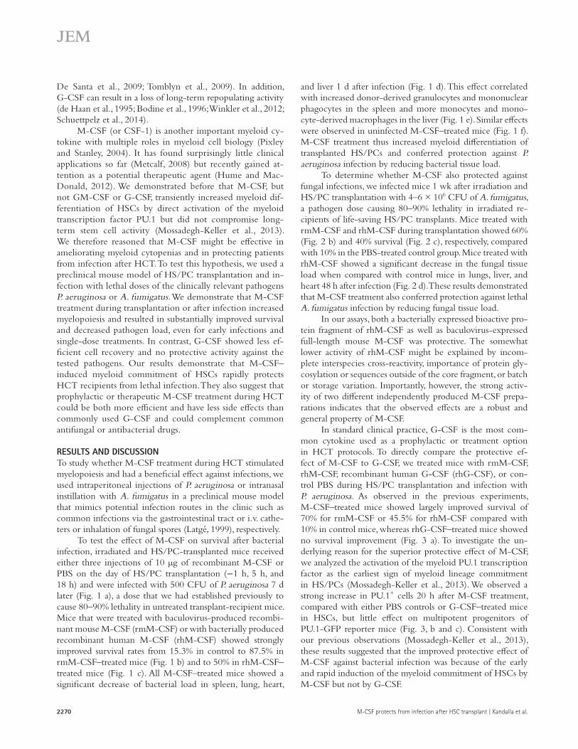
To test the effect of M-CSF on survival after bacterial infection, irradiated and HS/PC-transplanted mice received either three injections of 10 µg of recombinant M-CSF or PBS on the day of HS/PC transplantation (−1 h, 5 h, and 18 h) and were infected with 500 CFU of P. aeruginosa 7 d later (Fig. 1 a), a dose that we had established previously to cause 80–90% lethality in untreated transplant-recipient mice. Mice that were treated with baculovirus-produced recombi-nant mouse M-CSF (rmM-CSF) or with bacterially produced recombinant human M-CSF (rhM-CSF) showed strongly improved survival rates from 15.3% in control to 87.5% in rmM-CSF–treated mice (Fig. 1 b) and to 50% in rhM-CSF–treated mice (Fig. 1 c). All M-CSF–treated mice showed a significant decrease of bacterial load in spleen, lung, heart,
and liver 1 d after infection (Fig. 1 d). This effect correlated with increased donor-derived granulocytes and mononuclear phagocytes in the spleen and more monocytes and mono-cyte-derived macrophages in the liver (Fig. 1 e). Similar effects were observed in uninfected M-CSF–treated mice (Fig. 1 f). M-CSF treatment thus increased myeloid differentiation of transplanted HS/PCs and conferred protection against P. aeruginosa infection by reducing bacterial tissue load.
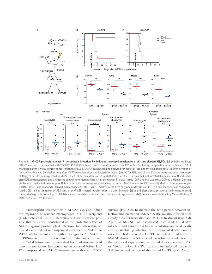
To determine whether M-CSF also protected against fungal infections, we infected mice 1 wk after irradiation and HS/PC transplantation with 4–6 × 10⁶ CFU of A. fumigatus, a pathogen dose causing 80–90% lethality in irradiated re-cipients of life-saving HS/PC transplants. Mice treated with rmM-CSF and rhM-CSF during transplantation showed 60% (Fig. 2 b) and 40% survival (Fig. 2 c), respectively, compared with 10% in the PBS-treated control group. Mice treated with rhM-CSF showed a significant decrease in the fungal tissue load when compared with control mice in lungs, liver, and heart 48 h after infection (Fig. 2 d). These results demonstrated that M-CSF treatment also conferred protection against lethal A. fumigatus infection by reducing fungal tissue load.
In our assays, both a bacterially expressed bioactive pro-tein fragment of rhM-CSF as well as baculovirus-expressed full-length mouse M-CSF was protective. The somewhat lower activity of rhM-CSF might be explained by incom-plete interspecies cross-reactivity, importance of protein gly-cosylation or sequences outside of the core fragment, or batch or storage variation. Importantly, however, the strong activ-ity of two different independently produced M-CSF prepa-rations indicates that the observed effects are a robust and general property of M-CSF.
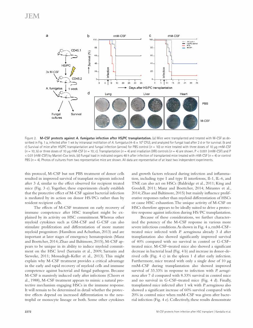
In standard clinical practice, G-CSF is the most com-mon cytokine used as a prophylactic or treatment option in HCT protocols. To directly compare the protective ef-fect of M-CSF to G-CSF, we treated mice with rmM-CSF, rhM-CSF, recombinant human G-CSF (rhG-CSF), or con-trol PBS during HS/PC transplantation and infection with P. aeruginosa. As observed in the previous experiments, M-CSF–treated mice showed largely improved survival of 70% for rmM-CSF or 45.5% for rhM-CSF compared with 10% in control mice, whereas rhG-CSF–treated mice showed no survival improvement (Fig. 3 a). To investigate the un-derlying reason for the superior protective effect of M-CSF, we analyzed the activation of the myeloid PU.1 transcription factor as the earliest sign of myeloid lineage commitment in HS/PCs (Mossadegh-Keller et al., 2013). We observed a strong increase in PU.1+ cells 20 h after M-CSF treatment, compared with either PBS controls or G-CSF–treated mice in HSCs, but little effect on multipotent progenitors of PU.1-GFP reporter mice (Fig. 3, b and c). Consistent with our previous observations (Mossadegh-Keller et al., 2013), these results suggested that the improved protective effect of M-CSF against bacterial infection was because of the early and rapid induction of the myeloid commitment of HSCs by M-CSF but not by G-CSF.
2271JEM Vol. 213, No. 11
Pretransplant treatment with M-CSF can also induce the expansion of resident macrophages in HCT recipients (Hashimoto et al., 2011). Theoretically, it was therefore pos-sible that this effect contributed to the protective effect of M-CSF against posttransplant infection. To address this, we treated irradiated but untransplanted mice with rmM-CSF or PBS 1 wk before infection with P. aeruginosa. All M-CSF– or PBS-treated mice died within 1–3 d after infection and, thus, 2–3 d before control mice died from radiation-induced bone marrow failure. In contrast and as observed before, HS/PC-transplanted and M-CSF–treated mice showed 83.33%
survival (Fig. 3 c). To increase the time period between in-fection and irradiation-induced death, we also infected mice already 3 d after irradiation and M-CSF treatment (Fig. 3 d). Again, all M-CSF– or PBS-treated mice died 1–3 d after infection and thus 8–9 d before irradiation-induced death, clearly establishing infection as the cause of death. Control mice that had received a HS/PC transplant in addition to M-CSF showed 37.5% survival even for early infection. In the reciprocal experiment, we treated donor mice with PBS or M-CSF before HS/PC isolation and infected recipients 3 d after transplantation of the treated HS/PC graft. Also, in
Figure 1. M-cSF protects against P. aeruginosa infection by inducing increased myelopoiesis of transplanted HS/Pcs. (a) Lethally irradiated CD45.2 mice were transplanted with 2,500 CD45.1 HS/PCs, treated with three doses of control PBS or M-CSF during transplantation (−1 h, 5 h, and 18 h), challenged after 1 wk by intraperitoneal injection of 500 CFU of P. aeruginosa, and analyzed for bacterial load and myeloid donor cells 1 d after infection or for survival. (b and c) Survival of mice after HS/PC transplantation and bacterial infection (arrow) for PBS control (n = 13) or mice treated with three doses of 10 µg of baculovirus-expressed rmM-CSF (n = 8; b) or three doses of 10 µg rhM-CSF (n = 10; c). Transplanted, not infected (black line; n = 4) and irradi-ated (XR), untransplanted and uninfected control mice (dashed line; n = 6) are shown. P < 0.001 (rmM-CSF) and P < 0.05 (rhM-CSF) by a Mantel-Cox test. (d) Bacterial load in indicated organs 18 h after infection of transplanted mice treated with rhM-CSF or control PBS. (e and f) Median of donor monocytes (CD11b+, Ly6C+) and monocyte-derived macrophages (CD11b+, Ly6C+, F4/80int) in the liver or granulocytes (Ly6G+, CD11b+) and mononuclear phagocytes (Ly6G−, CD11b+) in the spleen of PBS control or M-CSF–treated recipient mice 1 d after infection (e) or 9 d after transplantation of uninfected mice (f). Gating strategy is shown in Fig. S1. All data are representative of at least two independent experiments. (d–f) P-values were obtained by Mann-Whitney U tests. **, P < 0.01; ***, P < 0.001.
M-CSF protects from infection after HSC transplant | Kandalla et al.2272
this protocol, M-CSF but not PBS treatment of donor cells resulted in improved survival of transplant recipients infected after 3 d, similar to the effect observed for recipient treated mice (Fig. 3 e). Together, these experiments clearly establish that the protective effect of M-CSF against bacterial infection is mediated by its action on donor HS/PCs rather than by resident recipient cells.
The effects of M-CSF treatment on early recovery of immune competence after HSC transplant might be ex-plained by its activity on HSC commitment. Whereas other myeloid cytokines such as GM-CSF and G-CSF can also stimulate proliferation and differentiation of more mature myeloid progenitors (Hamilton and Achuthan, 2013) and are important at later stages of emergency hematopoiesis (Manz and Boettcher, 2014; Zhao and Baltimore, 2015), M-CSF ap-pears to be unique in its ability to induce myeloid commit-ment on the HSC level (Sarrazin et al., 2009; Sarrazin and Sieweke, 2011; Mossadegh-Keller et al., 2013). This might explain why M-CSF treatment provides a critical advantage in the early and rapid recovery of myeloid cells and immune competence against bacterial and fungal pathogens. Because M-CSF is massively induced early after infections (Cheers et al., 1988), M-CSF treatment appears to mimic a natural pro-tective mechanism engaging HSCs in the immune response. It will remain to be determined in detail whether the protec-tive effects depend on increased differentiation to the neu-trophil or monocyte lineage or both. Some other cytokines
and growth factors released during infection and inflamma-tion, including type I and type II interferons, Il-1, IL-6, and TNF, can also act on HSCs (Baldridge et al., 2011; King and Goodell, 2011; Manz and Boettcher, 2014; Mirantes et al., 2014; Zhao and Baltimore, 2015) but mainly influence prolif-erative responses rather than myeloid differentiation of HSCs or cause HSC exhaustion. The unique activity of M-CSF on HSCs therefore appears to be ideally suited to drive a protec-tive response against infection during HS/PC transplantation.
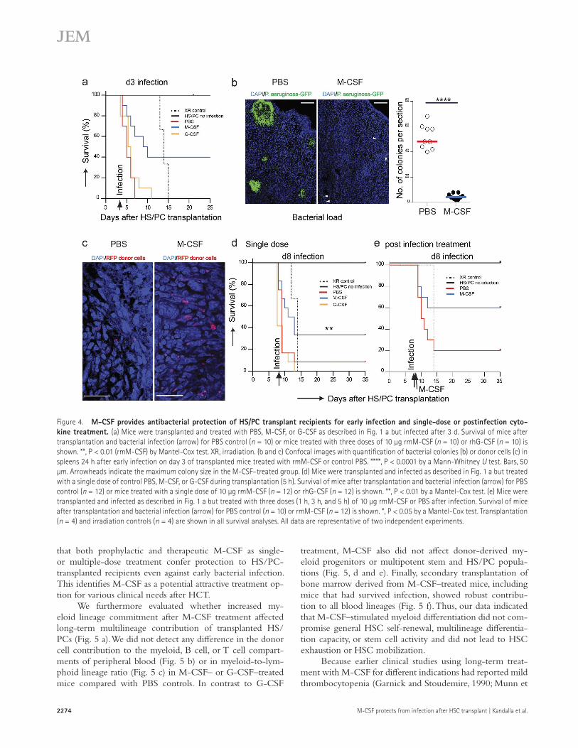
Because of these considerations, we further character-ized the potency of the M-CSF response in various more severe infectious conditions. As shown in Fig. 4 a, rmM-CSF–treated mice infected with P. aeruginosa already 3 d after transplantation also showed significantly improved survival of 40% compared with no survival in control or G-CSF–treated mice. M-CSF–treated mice also showed a significant decrease in bacterial load (Fig. 4 b) and increase in donor-de-rived cells (Fig. 4 c) in the spleen 1 d after early infection. Furthermore, mice treated with only a single dose of 10 µg rmM-CSF during transplantation also showed improved survival of 33.33% in response to infection with P. aerugi-nosa after 7 d compared with 8.33% survival in control mice and no survival in G-CSF–treated mice (Fig. 4 d). Finally, transplanted mice infected after 1 wk with P. aeruginosa also showed a significant increase of 60% survival compared with 20% in control mice when rmM-CSF was given after bacte-rial infection (Fig. 4 e). Collectively, these results demonstrate
Figure 2. M-cSF protects against A. fumigatus infection after HS/Pc transplantation. (a) Mice were transplanted and treated with M-CSF as de-scribed in Fig. 1 a, infected after 1 wk by intranasal instillation of A. fumigatus (4–6 × 10⁶ CFU), and analyzed for fungal load after 2 d or for survival. (b and c) Survival of mice after HS/PC transplantation and fungal infection (arrow) for PBS control (n = 10) or mice treated with three doses of 10 µg rmM-CSF (n = 10; b) or three doses of 10 µg rhM-CSF (n = 10; c). Transplantation (n = 4) and irradiation (XR) controls (n = 4) are shown. P < 0.001 (rmM-CSF) and P < 0.01 (rhM-CSF) by Mantel-Cox tests. (d) Fungal load in indicated organs 48 h after infection of transplanted mice treated with rhM-CSF (n = 4) or control PBS (n = 4). Photos of cultures from two representative mice are shown. All data are representative of at least two independent experiments.
2273JEM Vol. 213, No. 11
Figure 3. Specific M-cSF effect on early HSc commitment correlates with better antibacterial protection than G-cSF during HS/Pc trans-plantation. (a) Mice were transplanted, treated, and infected as described in Fig. 1 a. Survival of mice after transplantation and bacterial infection (arrow) for PBS control (n = 10) or mice treated with rmM-CSF (n = 10), rhM-CSF (n = 11), or rhG-CSF (n = 10) is shown. P < 0.05 (rmM-CSF or rhM-CSF) by a Mantel-Cox test. XR, irradiation. (b) Representative FACS profiles and median of GFP expression in HSCs (KSL CD34− Flt3−) and multipotent progenitors (MPP; KSL CD34+ Flt3+) of PU.1-GFP reporter mice 20 h after control PBS, rhM-CSF, or rhG-CSF injection. **, P < 0.01 by a Mann-Whitney U test. (c and d) Survival of nontransplanted lethally irradiated mice treated with three doses of control PBS (n = 6) or 10 µg rmM-CSF (n = 6) after irradiation (6 h, 12 h, and 24 h) and challenged after 8 (c) or 3 (d) d (arrow) by i.p. injection of 500 CFU of P. aeruginosa. Control mice were treated identically but transplanted with 2,500 HS/PCs (n = 6). ****, P < 0.0001 (rmM-CSF + HS/PC) by a Mantel-Cox test. (e) Survival of mice transplanted with donor HS/PCs from mice treated with three doses (−5 h, −3 h, and −1 h) of 10 µg rmM-CSF (n = 10) or PBS (n = 8) and infected after 3 d (arrow) with 500 CFU of P. aeruginosa. Control recipient mice were treated with three doses of 10 µg rmM-CSF (n = 8) during transplantation. ***, P < 0.001 (rmM-CSF–treated donor HS/PCs) by a Mantel-Cox test. Transplantation (n = 4) and irradiation controls (n = 4) are shown in all survival analyses. All data are representative of two independent experiments.
M-CSF protects from infection after HSC transplant | Kandalla et al.2274
that both prophylactic and therapeutic M-CSF as single- or multiple-dose treatment confer protection to HS/PC- transplanted recipients even against early bacterial infection. This identifies M-CSF as a potential attractive treatment op-tion for various clinical needs after HCT.
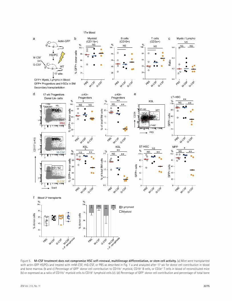
We furthermore evaluated whether increased my-eloid lineage commitment after M-CSF treatment affected long-term multilineage contribution of transplanted HS/PCs (Fig. 5 a). We did not detect any difference in the donor cell contribution to the myeloid, B cell, or T cell compart-ments of peripheral blood (Fig. 5 b) or in myeloid-to-lym-phoid lineage ratio (Fig. 5 c) in M-CSF– or G-CSF–treated mice compared with PBS controls. In contrast to G-CSF
treatment, M-CSF also did not affect donor-derived my-eloid progenitors or multipotent stem and HS/PC popula-tions (Fig. 5, d and e). Finally, secondary transplantation of bone marrow derived from M-CSF–treated mice, including mice that had survived infection, showed robust contribu-tion to all blood lineages (Fig. 5 f). Thus, our data indicated that M-CSF–stimulated myeloid differentiation did not com-promise general HSC self-renewal, multilineage differentia-tion capacity, or stem cell activity and did not lead to HSC exhaustion or HSC mobilization.
Because earlier clinical studies using long-term treat-ment with M-CSF for different indications had reported mild thrombocytopenia (Garnick and Stoudemire, 1990; Munn et
Figure 4. M-cSF provides antibacterial protection of HS/Pc transplant recipients for early infection and single-dose or postinfection cyto-kine treatment. (a) Mice were transplanted and treated with PBS, M-CSF, or G-CSF as described in Fig. 1 a but infected after 3 d. Survival of mice after transplantation and bacterial infection (arrow) for PBS control (n = 10) or mice treated with three doses of 10 µg rmM-CSF (n = 10) or rhG-CSF (n = 10) is shown. **, P < 0.01 (rmM-CSF) by Mantel-Cox test. XR, irradiation. (b and c) Confocal images with quantification of bacterial colonies (b) or donor cells (c) in spleens 24 h after early infection on day 3 of transplanted mice treated with rmM-CSF or control PBS. ****, P < 0.0001 by a Mann-Whitney U test. Bars, 50 µm. Arrowheads indicate the maximum colony size in the M-CSF–treated group. (d) Mice were transplanted and infected as described in Fig. 1 a but treated with a single dose of control PBS, M-CSF, or G-CSF during transplantation (5 h). Survival of mice after transplantation and bacterial infection (arrow) for PBS control (n = 12) or mice treated with a single dose of 10 µg rmM-CSF (n = 12) or rhG-CSF (n = 12) is shown. **, P < 0.01 by a Mantel-Cox test. (e) Mice were transplanted and infected as described in Fig. 1 a but treated with three doses (1 h, 3 h, and 5 h) of 10 µg rmM-CSF or PBS after infection. Survival of mice after transplantation and bacterial infection (arrow) for PBS control (n = 10) or rmM-CSF (n = 12) is shown. *, P < 0.05 by a Mantel-Cox test. Transplantation (n = 4) and irradiation controls (n = 4) are shown in all survival analyses. All data are representative of two independent experiments.
2275JEM Vol. 213, No. 11
Figure 5. M-cSF treatment does not compromise HSc self-renewal, multilineage differentiation, or stem cell activity. (a) Mice were transplanted with actin-GFP HS/PCs and treated with rmM-CSF, rhG-CSF, or PBS as described in Fig. 1 a and analyzed after 17 wk for donor cell contribution in blood and bone marrow. (b and c) Percentage of GFP+ donor cell contribution to CD11b+ myeloid, CD19+ B cells, or CD3e+ T cells in blood of reconstituted mice (b) or expressed as a ratio of CD11b+ myeloid cells to CD19+ lymphoid cells (c). (d) Percentage of GFP+ donor cell contribution and percentage of total bone
M-CSF protects from infection after HSC transplant | Kandalla et al.2276
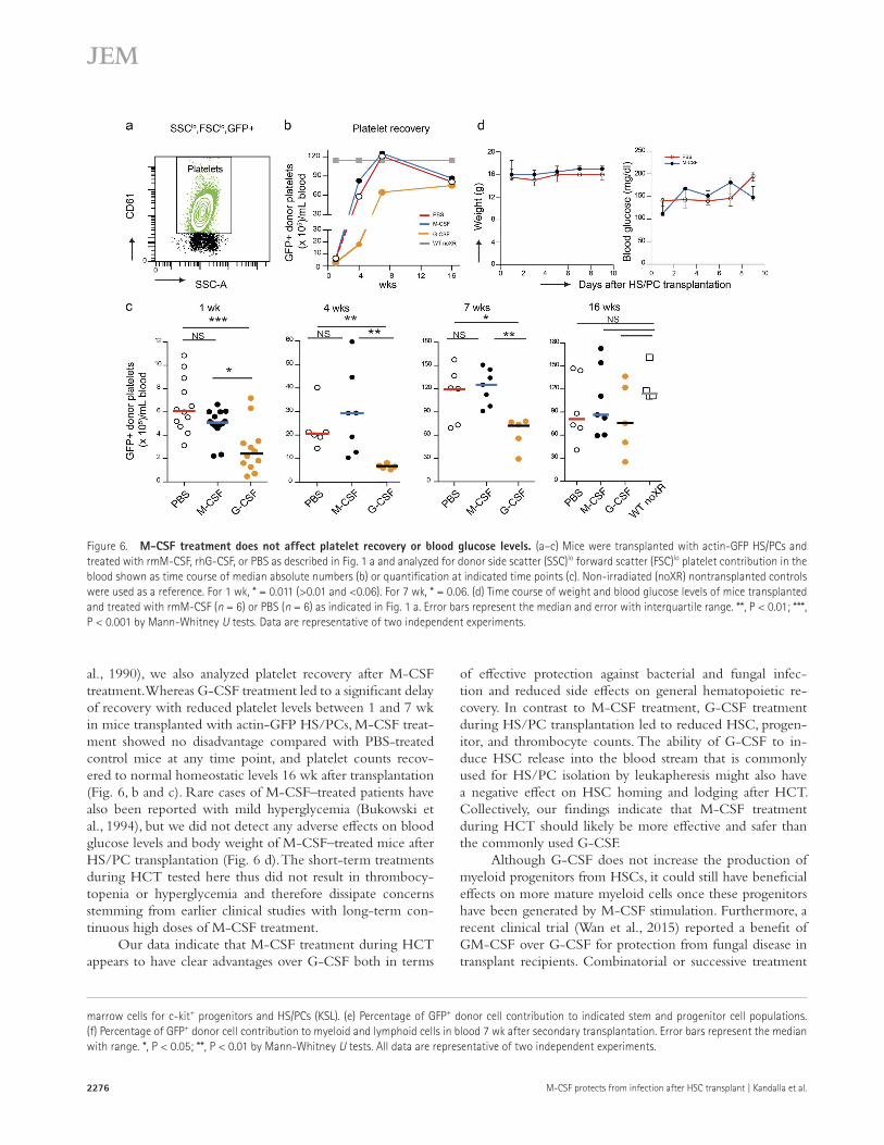
al., 1990), we also analyzed platelet recovery after M-CSF treatment. Whereas G-CSF treatment led to a significant delay of recovery with reduced platelet levels between 1 and 7 wk in mice transplanted with actin-GFP HS/PCs, M-CSF treat-ment showed no disadvantage compared with PBS-treated control mice at any time point, and platelet counts recov-ered to normal homeostatic levels 16 wk after transplantation (Fig. 6, b and c). Rare cases of M-CSF–treated patients have also been reported with mild hyperglycemia (Bukowski et al., 1994), but we did not detect any adverse effects on blood glucose levels and body weight of M-CSF–treated mice after HS/PC transplantation (Fig. 6 d). The short-term treatments during HCT tested here thus did not result in thrombocy-topenia or hyperglycemia and therefore dissipate concerns stemming from earlier clinical studies with long-term con-tinuous high doses of M-CSF treatment.
Our data indicate that M-CSF treatment during HCT appears to have clear advantages over G-CSF both in terms
of effective protection against bacterial and fungal infec-tion and reduced side effects on general hematopoietic re-covery. In contrast to M-CSF treatment, G-CSF treatment during HS/PC transplantation led to reduced HSC, progen-itor, and thrombocyte counts. The ability of G-CSF to in-duce HSC release into the blood stream that is commonly used for HS/PC isolation by leukapheresis might also have a negative effect on HSC homing and lodging after HCT. Collectively, our findings indicate that M-CSF treatment during HCT should likely be more effective and safer than the commonly used G-CSF.
Although G-CSF does not increase the production of myeloid progenitors from HSCs, it could still have beneficial effects on more mature myeloid cells once these progenitors have been generated by M-CSF stimulation. Furthermore, a recent clinical trial (Wan et al., 2015) reported a benefit of GM-CSF over G-CSF for protection from fungal disease in transplant recipients. Combinatorial or successive treatment
marrow cells for c-kit+ progenitors and HS/PCs (KSL). (e) Percentage of GFP+ donor cell contribution to indicated stem and progenitor cell populations. (f) Percentage of GFP+ donor cell contribution to myeloid and lymphoid cells in blood 7 wk after secondary transplantation. Error bars represent the median with range. *, P < 0.05; **, P < 0.01 by Mann-Whitney U tests. All data are representative of two independent experiments.
Figure 6. M-cSF treatment does not affect platelet recovery or blood glucose levels. (a–c) Mice were transplanted with actin-GFP HS/PCs and treated with rmM-CSF, rhG-CSF, or PBS as described in Fig. 1 a and analyzed for donor side scatter (SSC)lo forward scatter (FSC)lo platelet contribution in the blood shown as time course of median absolute numbers (b) or quantification at indicated time points (c). Non-irradiated (noXR) nontransplanted controls were used as a reference. For 1 wk, * = 0.011 (>0.01 and <0.06). For 7 wk, * = 0.06. (d) Time course of weight and blood glucose levels of mice transplanted and treated with rmM-CSF (n = 6) or PBS (n = 6) as indicated in Fig. 1 a. Error bars represent the median and error with interquartile range. **, P < 0.01; ***, P < 0.001 by Mann-Whitney U tests. Data are representative of two independent experiments.
2277JEM Vol. 213, No. 11
protocols of M-CSF with G-CSF or GM-CSF might there-fore offer synergistic effects.
Because M-CSF treatment reduces graft-versus-host dis-ease in allogeneic HCT (Hashimoto et al., 2011; Kimura et al., 2012), it will be important to determine whether it also has a protective effect against infection in this setting, as suggested by earlier retrospective clinical observations (Nemunaitis et al., 1993). In this context, the influence of M-CSF treatment on graft-versus-leukemia effects and on residual malignant cells will also require further investigation. In summary, the clear multiple beneficial effects of M-CSF treatment during HCT on improved graft-versus-host disease and protection against clin-ically important and often lethal fungal and bacterial infections, combined with the absence of evident negative side effects on reconstitution capacity and hematopoietic differentiation, merits consideration of clinical trials of this cytokine in HCT.
MAterIAlS And MetHodSMiceCD45.1 and C57BL/6 mice were obtained from Charles River. 10–14-wk-old sex-matched CD45.2 recipients were reconstituted as previously described (Sarrazin et al., 2009; Mossadegh-Keller et al., 2013) with bone marrow– derived KSL (c-Kit [CD117]+, Sca1+, Lin−) HS/PCs iso-lated from 6–8-wk-old CD45.1. For in vivo injections, the indicated concentrations of M-CSF and/or sorted cells were injected in 100–200 µl PBS into the retroorbital sinus. For HS/PC transplantation, 2,500 KLS HS/PCs were sorted from CD45.1 mice and mixed with 200,000 cKit− CD45.2 or cKit− Terr119+ carrier CD45.2 cells before injection into lethally irradiated (160 kV, 25 mA, and 6.9 Gy) CD45.2 re-cipient mice. For secondary transplantations, two million donor-derived bone marrow cells were injected per lethally irradiated recipient. After irradiation, all mice were given an-tibiotics (Bactrim) in drinking water to reduce the chance of opportunistic infection with other pathogens. All mouse experiments were performed under specific pathogen–free conditions. Animals were handled according to protocols ap-proved by the animal ethics committee of Marseille, France, and were performed in accordance to international, national, and institutional regulations.
Isolation of HS/Pcs, cKit−, and cKit− ter119+ cellsTotal bone marrow cells were depleted of mature cells by staining with biotinylated rat anti–mouse lineage antibody cocktail, followed by streptavidin immunomagnetic microbe-ads (Miltenyi Biotec). Lineage-negative cells were stained with HS/PC markers: anti-CD117–APC-H7 (clone 2B8; BD), anti–Sca-1–PE-Cy5 (clone D7; BioLegend), strepta-vidin-APC (eBioscience), and LIVE/DEAD Fixable Vio-let Dead cell dye (Invitrogen) as viability marker. HS/PCs were sorted using FAC SAriaIII equipment (BD). For isolat-ing cKit− carrier cells, whole bone marrow cells were de-pleted of cKit+ cells by staining with biotinylated anti–mouse CD117 (clone 2B8; BioLegend), followed by streptavidin
immunomagnetic microbeads (Miltenyi Biotec). For isolat-ing cKit− Ter119+ carrier cells, first, cKit− cells were selected by depleting cKit+ cells, stained with biotinylated anti–mouse Ter119 (clone TER-119; BD), and followed by streptavidin immunomagnetic beads.
M-cSF treatmentEach mouse received three i.v. injections of 10 µg of each cy-tokine 1 h before HS/PC transplantation and 5–6 h and 18 h after transplantation. rhM-CSF (Novartis), rmM-CSF ex-pressed in baculovirus, and human G-CSF (Neulasta) were used for the study. We thank G. Mouchiroud (Université Claude Bernard Lyon I, Villeurbanne, France) for baculovirus- produced rmM-CSF.
Infection with P. aeruginosaThe P. aeruginosa PA14 strain was tagged with GFP, as described previously (Koch et al., 2001; Giraud et al., 2011). The GFP-tagged PA14 strain was cultured overnight at 37°C in Luria broth medium, diluted 1:100 in Luria broth medium, and grown for 3 h to reach bacterial exponential phase (3–4 OD600nm). A volume of 100 µl of a bacterial solution of 5 × 10³ CFU/ml diluted in PBS was further used for infection studies. 1 wk after HS/PC transplantation, mice were challenged by intraperitoneal inoculation of 500 CFU of bacteria in 100 µl of sterile PBS.
Bacterial tissue load quantificationThe infected mice were killed, and the organs (spleen, lungs, liver, and heart) were harvested and weighed. To determine CFU per gram of tissue, serial dilutions of tissue homoge-nates were prepared in PBS and plated on Pseudomonas Isolation agar (DIF CO), supplemented with appropriate antibiotics, and incubated overnight at 37°C. The colonies were counted after 16–24 h.
Infection with A. fumigatusA. fumigatus (FGSC 1100) was provided by the Centre In-ternational de Ressources Microbiennes– Champignons Fila-menteux. For each experiment, cultures were grown on malt agar medium (2% malt extract and 2% bacto-agar; DIF CO) for 5 d at 25°C. Conidial suspension was prepared in sterile saline according to a study by BitMansour et al. (2002). 1 wk after HS/PC transplantation, mice were infected by intranasal inoculation of conidia in 20–40 µl of sterile PBS.
Fungal culture of infected organsThe organs (lungs, liver, heart, and spleen) were harvested and weighed, and tissue homogenates were prepared in PBS. The homogenates were serially diluted, and 200 µl of each dilution was plated on Sabouraud dextrose agar (DIF CO). The plates were incubated at 25°C, and pictures were taken after 3–5 d.
Flow cytometryFor FACS sorting and analysis, we used previously described staining protocols (Mossadegh-Keller et al., 2013), published
M-CSF protects from infection after HSC transplant | Kandalla et al.2278
stem and progenitor cell definitions (Bryder et al., 2006), FAC SCanto, LSR II, and FAC SAriaIII equipment, and DIVA software (BD), analyzing only populations with at least 200 events. The following antibodies were used for staining cells: anti-CD117 (clone 2B8; BD), anti–Sca-1 (clone D7; BioLeg-end), anti-CD34 (clone RAM34; BD), anti-CD16/32 (clone 2.4G2; BD), anti-CD11b (clone M1/70; BD), anti-CD19 (clone 1D3; BD), anti-CD3e (clone 145-2C11; BioLegend), anti-Ly6G (clone 1A8; BioLegend), anti-Ly6C (clone HK1.4; BioLegend), anti-CD115 (clone AFS98; eBioscience), anti- CD45.2 (clone 104; BD), anti-CD45.1 (clone A20; BD), anti-B220 (clone RA3-6B2; eBioscience), anti-Ter119 (clone TER-119; eBioscience), and anti-CD71 (clone R17217; eBioscience). LIVE/DEAD Fixable Violet Dead cell dye (Invitrogen) was used as a viability marker. Information de-scribing gating strategies is shown in Figs. S1 and S2.
Immunofluorescence and confocal imagingResected spleens were fixed in Antigen-Fix reagent (Diapath) for 2 h at 4°C, washed in phosphate buffer, and dehydrated in a 30% sucrose solution overnight at 4°C. Samples were embed-ded in TissueTek medium (Sakura), frozen at −80°C, cut into 8-µm–thick sections using a cryostat, and stained overnight at 4°C. The following antibodies were used: chicken anti-GFP (Aves laboratories) detected with donkey anti–chicken–Alexa Fluor 488 (Jackson ImmunoResearch Laboratories, Inc.). Confocal microscopy acquisition was performed on a ZEI SS microscope (LSM780) at room temperature.
Glycemia testBlood glucose was monitored every 2 d with a glucometer (Performa ACCU-CHEK; Roche).
online supplemental materialFig. S1 shows gating strategy for donor-derived granulocytes and mononuclear phagocytes from spleens and gating strategy for donor-derived monocytes and monocyte-derived macro-phages from livers. Fig. S2 shows gating strategy for quantifi-cation of actin-GFP+ HS/PC-derived platelets.
AcKnoWledGMentSWe thank L. Chasson for histology, L. Razafindramanana for animal handling, M. Barad, S. Bigot, and A. Zouine for cytometry support, Guy Mouchiroud for baculovi-rus-produced rmM-CSF, and Eric Solary and Didier Blaise for critical reading of the manuscript.
We acknowledge financial support from France Bio Imaging (n° ANR-10-INBS-04-01). This work was supported by grants to M.H. Sieweke from Fondation pour la Recherche Médicale (DEQ. 20110421320), the Agence Nationale de la Recher-che (ANR-11-BSV3-026-01), and the Institut National Du Cancer (13-10/405/AB-LC-HS). M.H. Sieweke is a BIH-Einstein fellow and Institut National de la Santé et de la Recherche Médicale-Helmholtz group leader.
The authors declare no competing financial interests.
Submitted: 18 December 2015
Accepted: 1 September 2016
reFerenceSBaldridge, M.T., K.Y. King, and M.A. Goodell. 2011. Inflammatory signals
regulate hematopoietic stem cells. Trends Immunol. 32:57–65. http ://dx .doi .org /10 .1016 /j .it .2010 .12 .003
BitMansour, A., S.M. Burns, D. Traver, K. Akashi, C.H. Contag, I.L. Weissman, and J.M. Brown. 2002. Myeloid progenitors protect against invasive aspergillosis and Pseudomonas aeruginosa infection following hematopoietic stem cell transplantation. Blood. 100:4660–4667. http ://dx .doi .org /10 .1182 /blood -2002 -05 -1552
BitMansour, A., T.M. Cao, S. Chao, S. Shashidhar, and J.M. Brown. 2005. Single infusion of myeloid progenitors reduces death from Aspergillus fumigatus following chemotherapy-induced neutropenia. Blood. 105:3535–3537. http ://dx .doi .org /10 .1182 /blood -2004 -07 -2676
Bodine, D.M., N.E. Seidel, and D. Orlic. 1996. Bone marrow collected 14 days after in vivo administration of granulocyte colony-stimulating fac-tor and stem cell factor to mice has 10-fold more repopulating ability than untreated bone marrow. Blood. 88:89–97.
Bryder, D., D.J. Rossi, and I.L. Weissman. 2006. Hematopoietic stem cells: the paradigmatic tissue-specific stem cell. Am. J. Pathol. 169:338–346. http ://dx .doi .org /10 .2353 /ajpath .2006 .060312
Bukowski, R.M., G.T. Budd, J.A. Gibbons, R.J. Bauer, A. Childs, J. Antal, J. Finke, L. Tuason, V. Lorenzi, D. McLain, et al. 1994. Phase I trial of subcu-taneous recombinant macrophage colony-stimulating factor: clinical and immunomodulatory effects. J. Clin. Oncol. 12:97–106.
Cheers, C., A.M. Haigh, A. Kelso, D. Metcalf, E.R. Stanley, and A.M. Young. 1988. Production of colony-stimulating factors (CSFs) during infec-tion: separate determinations of macrophage-, granulocyte-, granulo-cyte-macrophage-, and multi-CSFs. Infect. Immun. 56:247–251.
de Haan, G., B. Dontje, C. Engel, M. Loeffler, and W. Nijhof. 1995. The kinet-ics of murine hematopoietic stem cells in vivo in response to prolonged increased mature blood cell production induced by granulocyte colo-ny-stimulating factor. Blood. 86:2986–2992.
Dekker, A., S. Bulley, J. Beyene, L.L. Dupuis, J.J. Doyle, and L. Sung. 2006. Meta-analysis of randomized controlled trials of prophylactic granulocyte colony-stimulating factor and granulocyte-macrophage colony-stimulating factor after autologous and allogeneic stem cell transplantation. J. Clin. Oncol. 24:5207–5215. http ://dx .doi .org /10 .1200 /JCO .2006 .06 .1663
De Santa, F., V. Narang, Z.H. Yap, B.K. Tusi, T. Burgold, L. Austenaa, G. Bucci, M. Caganova, S. Notarbartolo, S. Casola, et al. 2009. Jmjd3 contributes to the control of gene expression in LPS-activated macrophages. EMBO J. 28:3341–3352. http ://dx .doi .org /10 .1038 /emboj .2009 .271
Garnick, M.B., and J.B. Stoudemire. 1990. Preclinical and clinical evaluation of recombinant human macrophage colony-stimulating factor (rhM-CSF). Int. J. Cell Cloning. 8:356–373. http ://dx .doi .org /10 .1002 /stem .5530080733
Giraud, C., C.S. Bernard, V. Calderon, L. Yang, A. Filloux, S. Molin, G. Fichant, C. Bordi, and S. de Bentzmann. 2011. The PprA-PprB two-component system activates CupE, the first non-archetypal Pseudomonas aeruginosa chaperone-usher pathway system assembling fimbriae. Environ. Microbiol. 13:666–683. http ://dx .doi .org /10 .1111 /j .1462 -2920 .2010 .02372 .x
Hamilton, J.A., and A. Achuthan. 2013. Colony stimulating factors and myeloid cell biology in health and disease. Trends Immunol. 34:81–89. http ://dx .doi .org /10 .1016 /j .it .2012 .08 .006
Hashimoto, D., A. Chow, M. Greter, Y. Saenger, W.H. Kwan, M. Leboeuf, F. Ginhoux, J.C. Ochando, Y. Kunisaki, N. van Rooijen, et al. 2011. Pretransplant CSF-1 therapy expands recipient macrophages and ameliorates GVHD after allogeneic hematopoietic cell transplantation. J. Exp. Med. 208:1069–1082. http ://dx .doi .org /10 .1084 /jem .20101709
Heuser, M., A. Ganser, C. Bokemeyer. American Society of Clinical Oncology, National Comprehensive Cancer Network, and European Organization for Research and Treatment of Cancer. 2007. Use of colony-stimulating factors for chemotherapy-associated neutropenia: review of current
2279JEM Vol. 213, No. 11
guidelines. Semin. Hematol. 44:148–156. http ://dx .doi .org /10 .1053 /j .seminhematol .2007 .04 .002
Hume, D.A., and K.P. MacDonald. 2012. Therapeutic applications of macrophage colony-stimulating factor-1 (CSF-1) and antagonists of CSF-1 receptor (CSF-1R) signaling. Blood. 119:1810–1820. http ://dx .doi .org /10 .1182 /blood -2011 -09 -379214
Kimura, F., K. Sato, H. Akiyama, H. Sao, S. Okamoto, N. Kobayashi, M. Hara, K. Kawa, and K. Motoyoshi. Japan Marrow Donor Program. 2012. M-CSF attenuates severity of chronic GVHD after unrelated BMT. Bone Marrow Transplant. 47:426–429. http ://dx .doi .org /10 .1038 /bmt .2011 .90
King, K.Y., and M.A. Goodell. 2011. Inflammatory modulation of HSCs: viewing the HSC as a foundation for the immune response. Nat. Rev. Immunol. 11:685–692. http ://dx .doi .org /10 .1038 /nri3062
Koch, B., L.E. Jensen, and O. Nybroe. 2001. A panel of Tn7-based vectors for insertion of the gfp marker gene or for delivery of cloned DNA into Gram-negative bacteria at a neutral chromosomal site. J. Microbiol. Methods. 45:187–195. http ://dx .doi .org /10 .1016 /S0167 -7012(01)00246 -9
Latgé, J.P. 1999. Aspergillus fumigatus and aspergillosis. Clin. Microbiol. Rev. 12:310–350.
Manz, M.G., and S. Boettcher. 2014. Emergency granulopoiesis. Nat. Rev. Immunol. 14:302–314. http ://dx .doi .org /10 .1038 /nri3660
Metcalf, D. 2008. Hematopoietic cytokines. Blood. 111:485–491. http ://dx .doi .org /10 .1182 /blood -2007 -03 -079681
Mirantes, C., E. Passegué, and E.M. Pietras. 2014. Pro-inflammatory cytokines: emerging players regulating HSC function in normal and diseased hematopoiesis. Exp. Cell Res. 329:248–254. http ://dx .doi .org /10 .1016 /j .yexcr .2014 .08 .017
Mossadegh-Keller, N., S. Sarrazin, P.K. Kandalla, L. Espinosa, E.R. Stanley, S.L. Nutt, J. Moore, and M.H. Sieweke. 2013. M-CSF instructs myeloid lineage fate in single haematopoietic stem cells. Nature. 497:239–243. http ://dx .doi .org /10 .1038 /nature12026
Munn, D.H., M.B. Garnick, and N.K. Cheung. 1990. Effects of parenteral recombinant human macrophage colony-stimulating factor on monocyte number, phenotype, and antitumor cytotoxicity in nonhuman primates. Blood. 75:2042–2048.
Nemunaitis, J., K. Shannon-Dorcy, F.R. Appelbaum, J. Meyers, A. Owens, R. Day, D. Ando, C. O’Neill, D. Buckner, and J. Singer. 1993. Long-term follow-up of patients with invasive fungal disease who received adjunctive therapy with recombinant human macrophage colony-stimulating factor. Blood. 82:1422–1427.
Pixley, F.J., and E.R. Stanley. 2004. CSF-1 regulation of the wandering macrophage: complexity in action. Trends Cell Biol. 14:628–638. http ://dx .doi .org /10 .1016 /j .tcb .2004 .09 .016
Sarrazin, S., and M. Sieweke. 2011. Integration of cytokine and transcription factor signals in hematopoietic stem cell commitment. Semin. Immunol. 23:326–334. http ://dx .doi .org /10 .1016 /j .smim .2011 .08 .011
Sarrazin, S., N. Mossadegh-Keller, T. Fukao, A. Aziz, F. Mourcin, L. Vanhille, L. Kelly Modis, P. Kastner, S. Chan, E. Duprez, et al. 2009. MafB restricts M-CSF-dependent myeloid commitment divisions of hematopoietic stem cells. Cell. 138:300–313. http ://dx .doi .org /10 .1016 /j .cell .2009 .04 .057
Schuettpelz, L.G., J.N. Borgerding, M.J. Christopher, P.K. Gopalan, M.P. Romine, A.C. Herman, J.R. Woloszynek, A.M. Greenbaum, and D.C. Link. 2014. G-CSF regulates hematopoietic stem cell activity, in part, through activation of Toll-like receptor signaling. Leukemia. 28:1851–1860. http ://dx .doi .org /10 .1038 /leu .2014 .68
Sung, L., P.C. Nathan, S.M. Alibhai, G.A. Tomlinson, and J. Beyene. 2007. Meta-analysis: effect of prophylactic hematopoietic colony-stimulating factors on mortality and outcomes of infection. Ann. Intern. Med. 147:400–411. http ://dx .doi .org /10 .7326 /0003 -4819 -147 -6 -200709180 -00010
To, L.B., D.N. Haylock, P.J. Simmons, and C.A. Juttner. 1997. The biology and clinical uses of blood stem cells. Blood. 89:2233–2258.
Tomblyn, M., T. Chiller, H. Einsele, R. Gress, K. Sepkowitz, J. Storek, J.R. Wingard, J.A. Young, M.J. Boeckh. Center for International Blood and Marrow Research and Centers for Disease Control and Prevention. 2009. Guidelines for preventing infectious complications among hematopoietic cell transplantation recipients: a global perspective. Biol. Blood Marrow Transplant. 15:1143–1238. http ://dx .doi .org /10 .1016 /j .bbmt .2009 .06 .019
Wan, L., Y. Zhang, Y. Lai, M. Jiang, Y. Song, J. Zhou, Z. Zhang, X. Duan, Y. Fu, L. Liao, and C. Wang. 2015. Effect of granulocyte-macrophage colony-stimulating factor on prevention and treatment of invasive fungal disease in recipients of allogeneic stem-cell transplantation: A prospective multicenter randomized phase IV trial. J. Clin. Oncol. 33:3999–4006. http ://dx .doi .org /10 .1200 /JCO .2014 .60 .5121
Winkler, I.G., A.R. Pettit, L.J. Raggatt, R.N. Jacobsen, C.E. Forristal, V. Barbier, B. Nowlan, A. Cisterne, L.J. Bendall, N.A. Sims, and J.P. Lévesque. 2012. Hematopoietic stem cell mobilizing agents G-CSF, cyclophosphamide or AMD3100 have distinct mechanisms of action on bone marrow HSC niches and bone formation. Leukemia. 26:1594–1601. http ://dx .doi .org /10 .1038 /leu .2012 .17
Zhao, J.L., and D. Baltimore. 2015. Regulation of stress-induced hematopoi-esis. Curr. Opin. Hematol. 22:286–292.