Embed Size (px)
Citation preview
Fungal Systematics and Evolution (ISSN: 2589-3823, E-ISSN: 2589-3831)
About Fungal Systematics and Evolution
Fungal Systematics and Evolution has an OPEN ACCESS publishing policy. All manuscripts will undergo peer review before acceptance, and will be published as quickly as possible following acceptance. There are no page charges or length restrictions, or fees for colour plates. The official journal language is English. All content submitted to Fungal Systematics and Evolution is checked for plagiarism.
Fungal Systematics and Evolution is licensed under a Creative Commons Attribution-NonCommercial-ShareAlike 4.0 International License
For more information and ordering of other books and publications, see www.westerdijkinstitute.nl and www.fuse-org.com.
About the Westerdijk Fungal Biodiversity Institute
The Westerdijk Fungal Biodiversity Institute is the largest expertise centre for microfungi in the world. Our renowned researchers dedicate themselves to enriching and expanding our Fungal Biobank. We explore the fungal kingdom to gain a better understanding of their fungal characteristics and their potential applications, especially in the fields of industry, agriculture and healthcare.
The Westerdijk Fungal Biodiversity Institute is part of the Royal Netherlands Academy of Arts and Sciences (KNAW).
Explore - study - preserve
Editor-in-ChiefProf. dr P.W. Crous, Westerdijk Fungal Biodiversity Institute, P.O. Box 85167, 3508 AD Utrecht, The Netherlands.E-mail:[email protected]
Editor-in-ChiefProf. dr P.W. Crous, Westerdijk Fungal Biodiversity Institute, P.O. Box 85167, 3508 AD Utrecht, The Netherlands.E-mail:[email protected]
Fungal Systematics and Evolution
Fungal Systematics and Evolution
1
Volume 1, June 2018
© 2018 Westerdijk Fungal Biodiversity Institute
Editor-in-ChiefProf. dr P.W. Crous, Westerdijk Fungal Biodiversity Institute, P.O. Box 85167, 3508 AD Utrecht, The Netherlands.E-mail:[email protected]
EDITORIAL BOARD
Editor-in-Chief
Prof. dr P.W. Crous, Westerdijk Fungal Biodiversity Institute, P.O. Box 85167, 3508 AD Utrecht, The Netherlands. E-mail: [email protected]
Senior Editors
Prof. dr U. Braun, Martin-Luther-Universität, Institut für Biologie, Geobotanik und Botanischer Garten, Herbarium, Neuwerk 21, D-06099 Halle, Germany; e-mail: [email protected]
Dr J.Z. Groenewald, Westerdijk Fungal Biodiversity Institute, P.O. Box 85167, 3508 AD Utrecht, The Netherlands; e-mail: [email protected]
Layout Editors
M.J. van den Hoeven-Verweij, Westerdijk Fungal Biodiversity Institute, P.O. Box 85167, 3508 AD Utrecht, The Netherlands; e-mail: [email protected]
M. Vermaas, Westerdijk Fungal Biodiversity Institute, P.O. Box 85167, 3508 AD Utrecht, The Netherlands; e-mail: [email protected]
Associate Editors
Prof. dr A.M. Al-Sadi, Department of Crop Sciences, College of Agricultural and Marine Sciences, Sultan Qaboos University, P.O. Box 34, AlKhoud 123, Oman; e-mail: [email protected] in Oman
Prof. dr M. Arzanlou, Plant Protection Department, Agriculture Faculty, University of Tabriz, P.O. Box: 5166614766, Tabriz, Iran; e-mail: [email protected]
Prof. dr T. Burgess, School of Biological Sciences and Biotechnology, Murdoch University, Perth, 6150, Australia; e-mail: [email protected]. dr L. Cai, State Key Laboratory of Mycology, Institute of Microbiology, Chinese Academy of Sciences, No. 3 Park 1, Beichen West Road, Chaoyang
District, Beijing, 100101, China; e-mail: [email protected]. dr L. Carris, Associate Professor, Department of Plant Pathology, Washington State University, Pullman, WA 99164-6340, USA; e-mail: carris@
mail.wsu.eduDr C. Decock, MUCL, Croix du Sud 2 bte L7.05.06, B-1348 Louvain-la-Neuve, Belgium; e-mail: [email protected] X.L. Fan, The Key Laboratory for Silviculture and Conservation of Ministry of Education, Beijing Forestry University, Beijing 100083, China; e-mail:
[email protected] L. Fang, State Key Laboratory of Mycology, Institute of Microbiology, Chinese Academy of Sciences, No. 3 Park 1, Beichen West Road, Chaoyang
District, Beijing, 100101, China; [email protected] D. Gramaje, Instituto de Ciencias de la Vid y del Vino (ICVV), Consejo Superior de Investigaciones Científicas - Universidad de la Rioja - Gobierno
de La Rioja, Ctra. Mendavia-Logroño NA 134, Km. 90, 26071 Logroño, Spain; e-mail: [email protected] V. Guarnaccia, Westerdijk Fungal Biodiversity Institute, P.O. Box 85167, 3508 AD Utrecht, The Netherlands; e-mail: [email protected] H.D.T. Nguyen, Agriculture and Agri-Food Canada, K.W. Neatby Building, 960 Carling Ave., Ottawa, ON, Canada, K1A 0C9; e-mail: Hai.Nguyen@agr.
gc.caDr R. Jeewon, Department of Health Sciences, Faculty of Science, University of Mauritius, Reduit, Mauritius; e-mail: [email protected] L. Mostert, Department of Plant Pathology, University of Stellenbosch, P. Bag X1, Matieland 7602, South Africa; e-mail: [email protected]. dr A.J.L. Phillips, University of Lisbon, Faculty of Sciences, Biosystems and Integrative Sciences Institute (BioISI), Campo Grande, 1749-016
Lisbon, Portugal; e-mail: [email protected] M. Piątek, Department of Mycology, W. Szafer Institute of Botany, Polish Academy of Sciences, Lubicz 46, PL-31-512 Kraków, Poland; e-mail:
[email protected]. dr H.-D. Shin, Division of Environmental Science and Ecological Engineering, Korea University, Seoul 02841, Korea; e-mail: [email protected]. dr B. Summerell, Royal Botanic Gardens and Domain Trust, Mrs. Macquaries Road, Sydney, NSW 2000, Australia; e-mail: Brett.Summerell@
rbgsyd.nsw.gov.auDr J.B. Tanney, Institut de Biologie Intégrative et des Systèmes, Charles-Eugène-Marchand Pavilion, 1030 Avenue de la Médecine, Québec City, QC
G1V 0A6, Canada; e-mail [email protected]. dr P. Taylor, Faculty of Veterinary and Agricultural Sciences, University of Melbourne, VIC, 3010, Australia; e-mail: [email protected]. dr M. Thines, Goethe University Frankfurt am Main, Faculty of Biosciences, Institute of Ecology, Evolution and Diversity, Max-von-Laue-Str. 9,
D-60438 Frankfurt am Main, Germany; e-mail: [email protected] A.D. van Diepeningen, Business Unit Biointeractions and Plant Health, Wageningen University and Research, Wageningen, the Netherlands;
e-mail: [email protected]. dr M.J. Wingfield, Forestry and Agricultural Research Institute (FABI), University of Pretoria, Pretoria 0002, South Africa; e-mail: mike.wingfield@
fabi.up.ac.zaProf. dr R. Zare, Agricultural Research, Education and Extension Organization, Tehrãn, Iran; e-mail: [email protected]
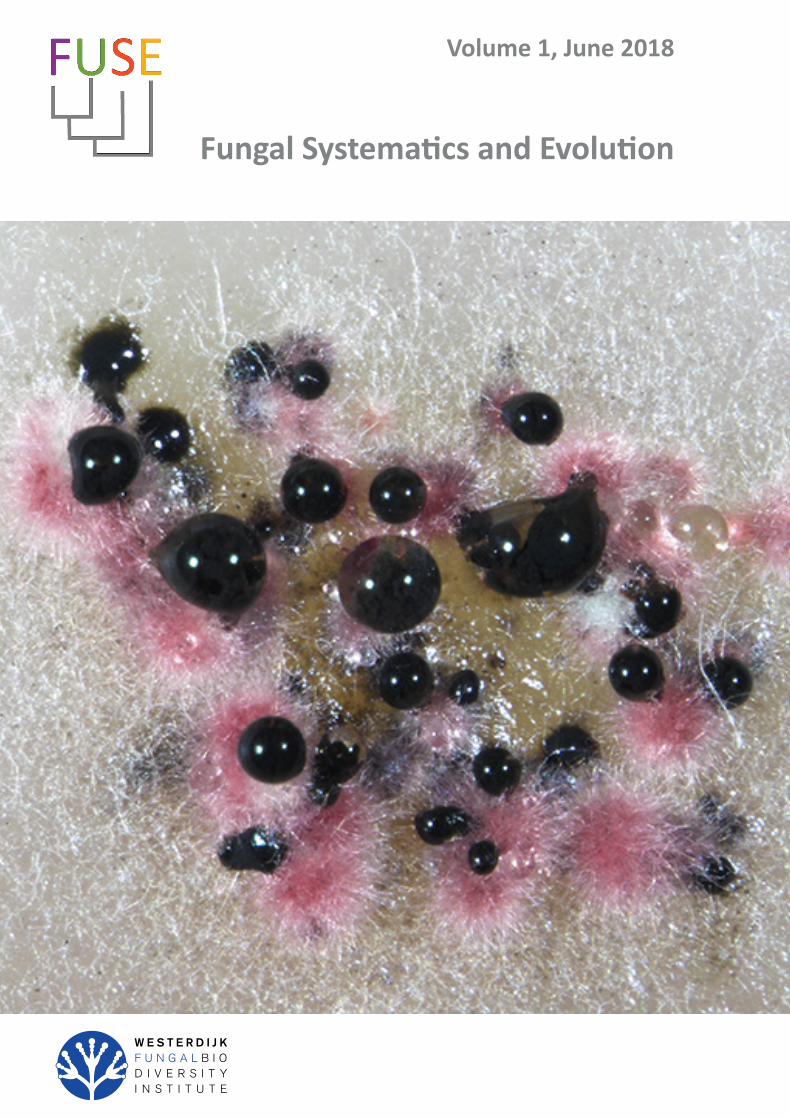
Cover: Culture of Cyclothyriella rubronotata (CBS 144201) sporulating on oatmeal agar.
© 2018 Westerdijk Fungal Biodiversity Institute
Editor-in-ChiefProf. dr P.W. Crous, Westerdijk Fungal Biodiversity Institute, P.O. Box 85167, 3508 AD Utrecht, The Netherlands.E-mail:[email protected]
SCOPE AND AIMS
SCOPEAll aspects of systematics and evolution of fungi.
AIMSFungal Systematics and Evolution is an international, peer-reviewed, open-access, full colour, fast-track journal. Papers will include reviews, research articles, methodology papers, taxonomic monographs, and the description of fungi. The journal strongly supports good practice policies, and requires voucher specimens to be deposited in a fungarium, cultures in long-term genetic resource collection, sequences in GenBank, alignments in TreeBASE, and taxonomic novelties in MycoBank.
ABOUT FUNGAL SYSTEMATICS AND EVOLUTIONFungal Systematics and Evolution has an OPEN ACCESS publishing policy. All manuscripts will undergo peer review before acceptance, and will be published as quickly as possible following acceptance. There are no page charges or length restrictions, or fees for colour plates. The official journal language is English. All content submitted to Fungal Systematics and Evolution is checked for plagiarism.
Fungal Systematics and Evolution is licensed under a Creative Commons Attribution-NonCommercial-ShareAlike 4.0 International License.
PUBLISHED BYWesterdijk Fungal Biodiversity Institute, P.O. Box 85167, 3508 AD Utrecht, The Netherlands.
All articles are copyright of Westerdijk Fungal Biodiversity Institute.
FREQUENCYTwice per year (June and December).
ISSN: 2589-3823E-ISSN: 2589-3831
© 2018 Westerdijk Fungal Biodiversity Institute
Editor-in-ChiefProf. dr P.W. Crous, Westerdijk Fungal Biodiversity Institute, P.O. Box 85167, 3508 AD Utrecht, The Netherlands.E-mail:[email protected]
CONTENTS
Research papers
M.A. Castellano, T.F. Elliott, J.M. Trappe. Three new black Elaphomyces species (Elaphomycetaceae, Eurotiales, Ascomycota) from eastern North America with notes on selected European species ........................................................................................................................................ 1
H.C. Evans, J.P.M. Araújo, V.R. Halfeld, D.P. Hughes. Epitypification and re-description of the zombie-ant fungus, Ophiocordyceps unilateralis (Ophiocordycipitaceae) .................................................................................................................................................................................. 13
T. Aoki, M.T. Kasson, M.C. Berger, S. Freeman, D.M. Geiser, K. O’Donnell. Fusarium oligoseptatum sp. nov., a mycosymbiont of the ambrosia beetle Euwallacea validus in the Eastern U.S. and typification of F. ambrosium ..................................................................................................... 23
U. Braun, C. Nakashima, P.W. Crous, J.Z. Groenewald, O. Moreno-Rico, S. Rooney-Latham, C.L. Blomquist, J. Haas, J. Marmolejo. Phylogeny and taxonomy of the genus Tubakia s. lat. ............................................................................................................................................................ 4
O. Miettinen, J. Vlasák, B. Rivoire, V. Spirin. Postia caesia complex (Polyporales, Basidiomycota) in temperate Northern Hemisphere .................. 101
V. Guarnaccia, M. Sandoval-Denis, D. Aiello, G. Polizzi, P.W. Crous. Neocosmospora perseae sp. nov., causing trunk cankers on avocado in Italy ...................................................................................................................................................................................................................... 131
E. Bovio, L. Garzoli, A. Poli, V. Prigione, D. Firsova, G.P. McCormack, G.C. Varese. The culturable mycobiota associated with three Atlantic sponges, including two new species: Thelebolus balaustiformis and T. spongiae ..................................................................................................... 141
P.W. Crous, R.K. Schumacher, M.J. Wingfield, A. Akulov, S. Denman, J. Roux, U. Braun, T.I. Burgess, A.J. Carnegie, K.Z. Váczy, E. Guatimosim, P.B. Schwartsburd, R.W. Barreto, M. Hernández-Restrepo, L. Lombard, J.Z. Groenewald. New and Interesting Fungi. 1 ........................... 169
H. Sundberg, Å. Kruys, J. Bergsten, S. Ekman. Position specificity in the genus Coreomyces (Laboulbeniomycetes, Ascomycota) ............................ 217
T.F. Elliott, J.M. Trappe. A worldwide nomenclature revision of sequestrate Russula species ................................................................................... 229

© 2018 Westerdijk Fungal Biodiversity Institute
Editor-in-ChiefProf. dr P.W. Crous, Westerdijk Fungal Biodiversity Institute, P.O. Box 85167, 3508 AD Utrecht, The Netherlands.E-mail:[email protected]
Fungal Systematics and Evolution is licensed under a Creative Commons Attribution-NonCommercial-ShareAlike 4.0 International License
© 2018 Westerdijk Fungal Biodiversity Institute 1
Editor-in-ChiefProf. dr P.W. Crous, Westerdijk Fungal Biodiversity Institute, P.O. Box 85167, 3508 AD Utrecht, The Netherlands.E-mail:[email protected]
Fungal Systematics and Evolution
doi.org/10.3114/fuse.2018.01.01
VOLUME 1JUNE 2018PAGES 1–12
INTRODUCTION
The hypogeous, sequestrate genus Elaphomyces (Elapho-mycetaceae, Eurotiales, Ascomycota) forms ectomycorrhizal associations with roots of diverse gymnosperm and angiosperm trees and shrubs around the world (Miller & Miller 1984, Trappe et al. 2009, Castellano et al. 2016). Species of Elaphomyces release aromas that are detected by numerous mammal species that dig them up and utilize them as an important food, thereby dispersing the spores across the landscape (Boudier 1876, Fogel & Trappe 1978, Vogt et al. 1981, Genov 1982, Cork & Kenagy 1989, Vernes et al. 2004, Vernes & Poirier 2007). The leathery peridium is eaten by the animals and some of the powdery mass of hydrophobic spores within the fruiting bodies are ingested, some released to the air, and some left on the ground, logs, or stumps.
Elaphomyces species have been reported from every continent except Antarctica, and occur in diverse forest habitats ranging from temperate and subarctic conifer forests to lowland tropics (Corner & Hawker 1953, Trappe & Kimbrough 1972, Zhang & Minter 1989, Castellano et al. 2011, Reynolds 2011, Castellano et al. 2012a, c, 2016, Paz et al. 2012, Castellano & Stephens 2017). Recently Paz et al. (2017) revised the systematics of the European Elaphomyces species. They present all known species from Europe and provide an updated structure to the sections and subsections within Elaphomyces.
Our recent work on the genus indicates that eastern North America is likely the epicentre of Elaphomyces biodiversity with approximately 30 to 40 species, many of which are still undescribed (Castellano, unpubl. data). Historically in North America, most Elaphomyces spp. were assigned European names due to the difficulty in distinguishing species based on
published descriptions that were often sparse in details (Trappe & Guzman 1971, Miller & Miller 1984, Luoma & Frenkel 1991, Gomez-Reyes et al. 2012). Most North American Elaphomyces with a black or dark coloured peridial surface were assigned the name E. anthracinus, and others with brown peridial surfaces were named E. granulatus or E. muricatus, depending on structure of the inner peridium. We now know that these species do not occur in North America. In eastern North America (from Québec south to the Gulf of Mexico), there are currently eight described Elaphomyces species, including E. americanus, E. appalachiensis, E. bartlettii, E. macrosporus, E. oreoides, E. remickii, E. spinoreticulatus, E. verruculosus, and E. viridiseptum (Linder 1939, Trappe & Kimbrough 1972, Zhang & Minter 1989, Castellano et al. 2012b, Castellano & Stephens 2017). The aim of the present study was thus to characterise several Elaphomyces spp. collected in the USA.
MATERIALS AND METHODS
Species of Elaphomyces typically develop below ground, so ascomata were collected by raking away the leaf and upper soil layers in suitable habitats, observing and excavating the area where animals had previously dug, or by looking for Tolypocladium species that fruit aboveground while parasitizing the ascomata of Elaphomyces species (Castellano et al. 2004). Occasionally, specimens had partially emerged from the soil in eroded or disturbed environments like road banks, campgrounds, or trail edges.
Descriptions of macro-morphological characters are based on fresh material. Colours are described in general terms based
Three new black Elaphomyces species (Elaphomycetaceae, Eurotiales, Ascomycota) from eastern North America with notes on selected European species
M.A. Castellano1*, T.F. Elliott2, J.M. Trappe3,4
1US Department of Agriculture, Forest Service, Northern Research Station, 3200 Jefferson Way, Corvallis, OR 97331, USA 2Department of Integrative Studies, Warren Wilson College, Asheville, NC 28815, USA3Department of Forest Ecosystems and Society, Oregon State University, Corvallis, OR 97331-5752, USA4US Forest Service, Pacific Northwest Research Station, 3200 Jefferson Way, Corvallis, OR 97331, USA
*Corresponding author: [email protected]
Abstract: We describe three new species of Elaphomyces from eastern North America. Of the three, Elaphomyces loebiae is the rarest, known only from North Carolina and South Carolina, and appears to associate primarily with ectomycorrhizal hardwoods but possibly also with conifers. Elaphomyces cibulae is widely distributed but disjunct from Florida, Mississippi, and North Carolina. Elaphomyces cibulae seems to primarily associate with Quercus species. Elaphomyces mitchelliae has the widest distribution of the three species, from Florida, Maryland, North Carolina, Virginia, and West Virginia, and appears to associate with either ectomycorrhizal hardwoods and/or conifers. In the course of comparing our new Elaphomyces species to previously described European species we discovered that E. persoonii var. minor is conspecific in all essential details with and thus a synonym of E. cyanosporus.
Key words: ectomycorrhizahypogeous funginew taxasequestrate fungi
Published online: 1 December 2017.
© 2018 Westerdijk Fungal Biodiversity Institute
Castellano et al.
Editor-in-ChiefProf. dr P.W. Crous, Westerdijk Fungal Biodiversity Institute, P.O. Box 85167, 3508 AD Utrecht, The Netherlands.E-mail:[email protected]
2
on the observations of the authors and collectors. Tissues and spores from dried specimens were rehydrated and examined in 3 % KOH, Melzer’s reagent, or cotton blue for study of microscopic characters. Neither tissues nor spores reacted distinctively to Melzer’s reagent. Microscopic characters of ascoma tissues and spores were described from 3 % KOH mounts unless otherwise specified. Spore dimensions include ornamentation and are from twenty randomly selected ascospores measured from the holotype collections. Asci of Elaphomyces spp. generally disintegrate with maturation, so their features often cannot be recorded. Dried ascospores were mounted on aluminum pegs with double-sided tape and coated with gold for scanning electron microscopy (SEM) with a Quanta 600 FEG scanning electron microscope. Specimens are deposited in the following herbaria as noted for each collection: FLAS, OSC, RMS, ITCV, ISC (Index Herbariorum, referenced May 2017).
TAXONOMY
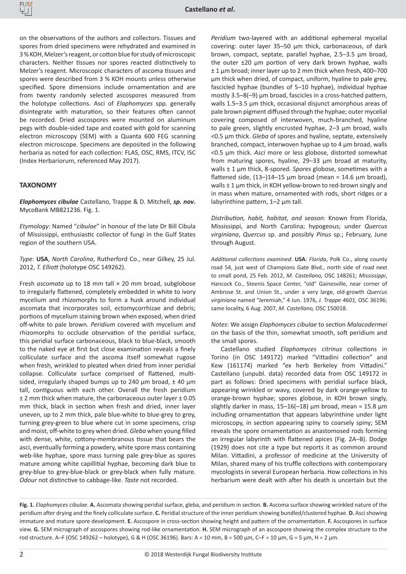
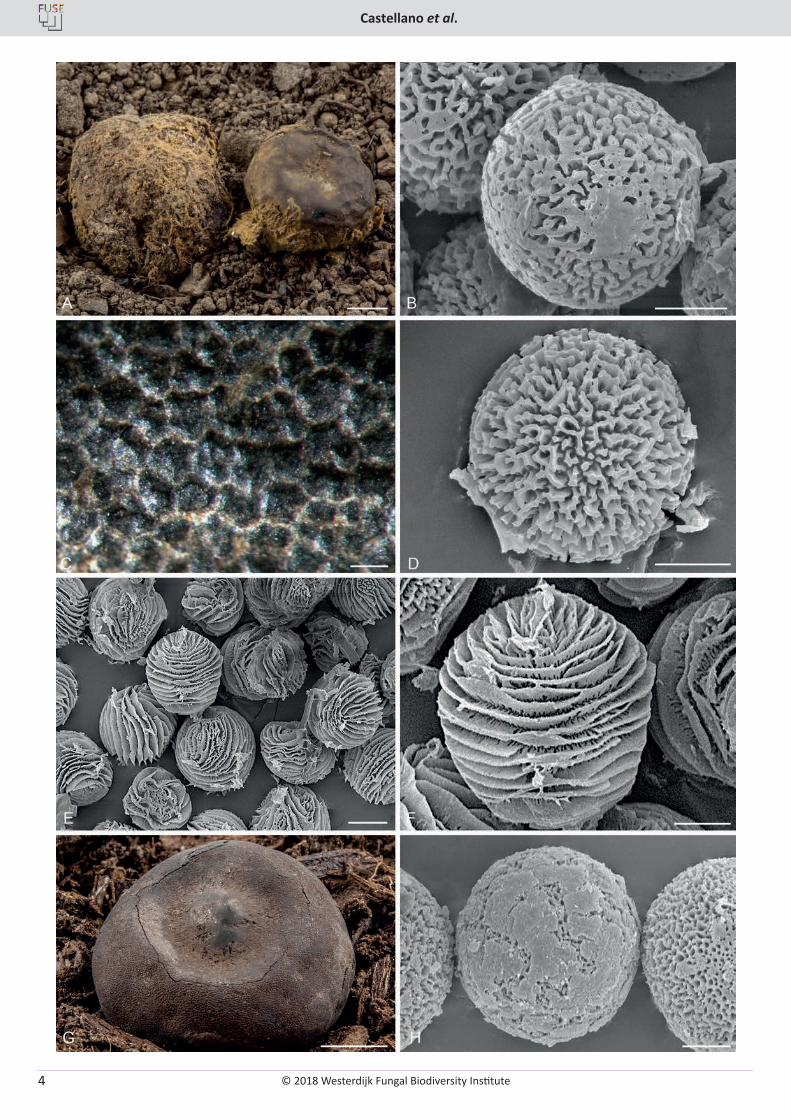
Elaphomyces cibulae Castellano, Trappe & D. Mitchell, sp. nov. MycoBank MB821236. Fig. 1.
Etymology: Named “cibulae” in honour of the late Dr Bill Cibula of Mississippi, enthusiastic collector of fungi in the Gulf States region of the southern USA.
Type: USA, North Carolina, Rutherford Co., near Gilkey, 25 Jul. 2012, T. Elliott (holotype OSC 149262).
Fresh ascomata up to 18 mm tall × 20 mm broad, subglobose to irregularly flattened, completely embedded in white to ivory mycelium and rhizomorphs to form a husk around individual ascomata that incorporates soil, ectomycorrhizae and debris; portions of mycelium staining brown when exposed, when dried off-white to pale brown. Peridium covered with mycelium and rhizomorphs to occlude observation of the peridial surface, this peridial surface carbonaceous, black to blue-black, smooth to the naked eye at first but close examination reveals a finely colliculate surface and the ascoma itself somewhat rugose when fresh, wrinkled to pleated when dried from inner peridial collapse. Colliculate surface comprised of flattened, multi-sided, irregularly shaped bumps up to 240 µm broad, ± 40 µm tall, contiguous with each other. Overall the fresh peridium ± 2 mm thick when mature, the carbonaceous outer layer ± 0.05 mm thick, black in section when fresh and dried, inner layer uneven, up to 2 mm thick, pale blue-white to blue-grey to grey, turning grey-green to blue where cut in some specimens, crisp and moist, off-white to grey when dried. Gleba when young filled with dense, white, cottony-membranous tissue that bears the asci, eventually forming a powdery, white spore mass containing web-like hyphae, spore mass turning pale grey-blue as spores mature among white capillitial hyphae, becoming dark blue to grey-blue to grey-blue-black or grey-black when fully mature. Odour not distinctive to cabbage-like. Taste not recorded.
Peridium two-layered with an additional ephemeral mycelial covering: outer layer 35–50 µm thick, carbonaceous, of dark brown, compact, septate, parallel hyphae, 2.5–3.5 µm broad, the outer ±20 µm portion of very dark brown hyphae, walls ± 1 µm broad; inner layer up to 2 mm thick when fresh, 400–700 µm thick when dried, of compact, uniform, hyaline to pale grey, fascicled hyphae (bundles of 5–10 hyphae), individual hyphae mostly 3.5–8(–9) µm broad, fascicles in a cross-hatched pattern, walls 1.5–3.5 µm thick, occasional disjunct amorphous areas of pale brown pigment diffused through the hyphae; outer mycelial covering composed of interwoven, much-branched, hyaline to pale green, slightly encrusted hyphae, 2–3 µm broad, walls <0.5 µm thick. Gleba of spores and hyaline, septate, extensively branched, compact, interwoven hyphae up to 4 µm broad, walls <0.5 µm thick. Asci more or less globose, distorted somewhat from maturing spores, hyaline, 29–33 µm broad at maturity, walls ± 1 µm thick, 8-spored. Spores globose, sometimes with a flattened side, (13–)14–15 µm broad (mean = 14.6 mm broad), walls ± 1 µm thick, in KOH yellow-brown to red-brown singly and in mass when mature, ornamented with rods, short ridges or a labyrinthine pattern, 1–2 µm tall. Distribution, habit, habitat, and season: Known from Florida, Mississippi, and North Carolina; hypogeous; under Quercus virginiana, Quercus sp. and possibly Pinus sp.; February, June through August.
Additional collections examined: USA: Florida, Polk Co., along county road 54, just west of Champions Gate Blvd., north side of road next to small pond, 25 Feb. 2012, M. Castellano, OSC 148261; Mississippi, Hancock Co., Steenis Space Center, “old” Gainesville, near corner of Ambrose St. and Union St., under a very large, old-growth Quercus virginiana named “Jeremiah,” 4 Jun. 1976, J. Trappe 4601, OSC 36196; same locality, 6 Aug. 2007, M. Castellano, OSC 150018.
Notes: We assign Elaphomyces cibulae to section Malacodermei on the basis of the thin, somewhat smooth, soft peridium and the small spores.
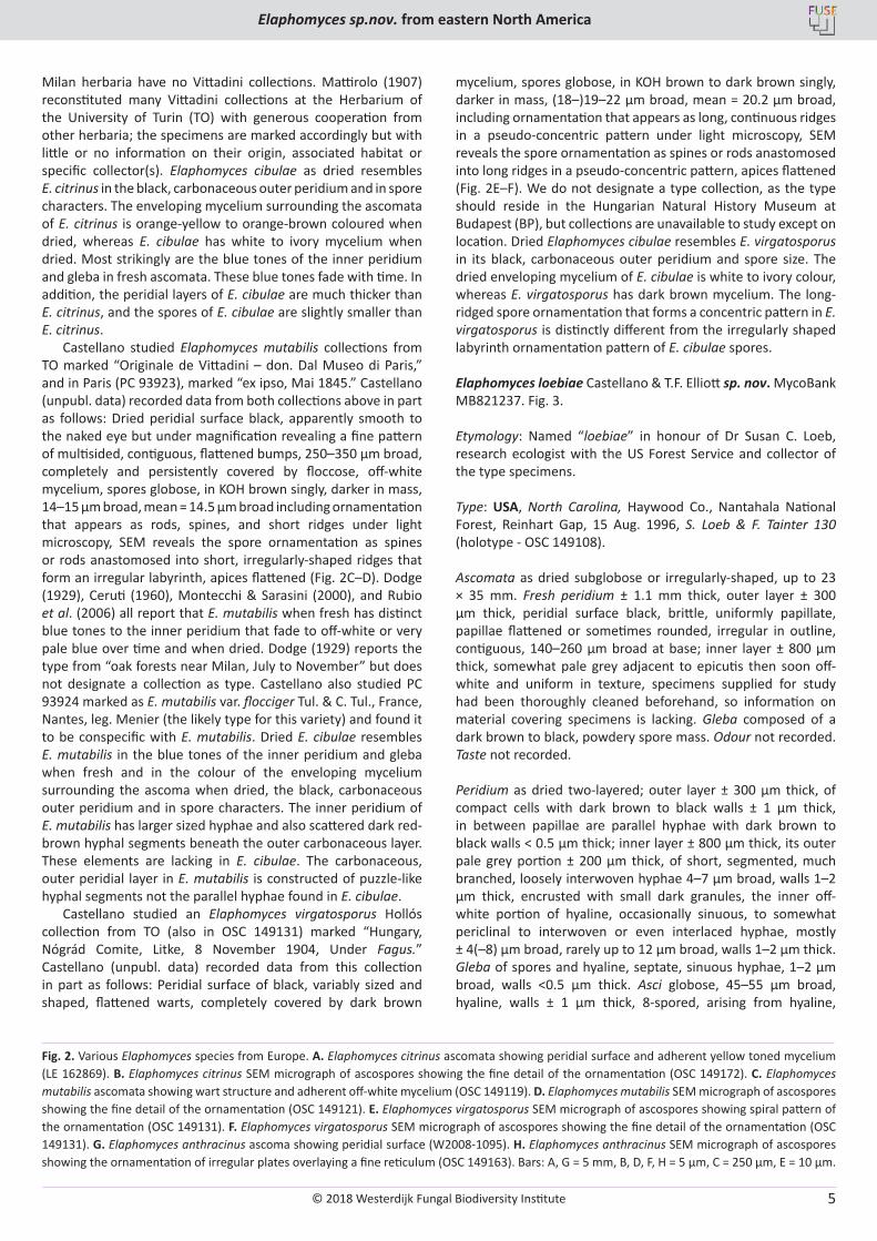
Castellano studied Elaphomyces citrinus collections in Torino (in OSC 149172) marked “Vittadini collection” and Kew (161174) marked “ex herb Berkeley from Vittadini.” Castellano (unpubl. data) recorded data from OSC 149172 in part as follows: Dried specimens with peridial surface black, appearing wrinkled or wavy, covered by dark orange-yellow to orange-brown hyphae; spores globose, in KOH brown singly, slightly darker in mass, 15–16(–18) µm broad, mean = 15.8 µm including ornamentation that appears labyrinthine under light microscopy, in section appearing spiny to coarsely spiny; SEM reveals the spore ornamentation as anastomosed rods forming an irregular labyrinth with flattened apices (Fig. 2A–B). Dodge (1929) does not cite a type but reports it as common around Milan. Vittadini, a professor of medicine at the University of Milan, shared many of his truffle collections with contemporary mycologists in several European herbaria. How collections in his herbarium were dealt with after his death is uncertain but the
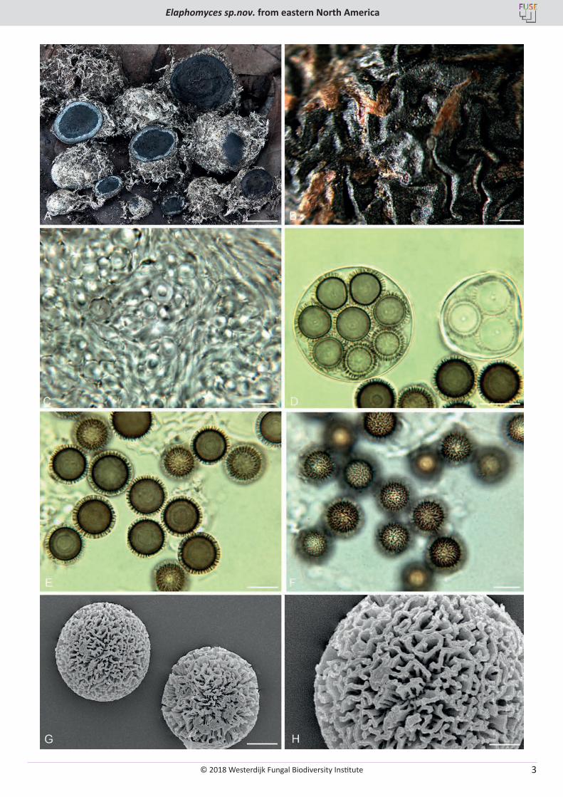
Fig. 1. Elaphomyces cibulae. A. Ascomata showing peridial surface, gleba, and peridium in section. B. Ascoma surface showing wrinkled nature of the peridium after drying and the finely colliculate surface. C. Peridial structure of the inner peridium showing bundled/clustered hyphae. D. Asci showing immature and mature spore development. E. Ascospore in cross-section showing height and pattern of the ornamentation. F. Ascospores in surface view. G. SEM micrograph of ascospores showing rod-like ornamentation. H. SEM micrograph of an ascospore showing the complex structure to the rod structure. A–F (OSC 149262 – holotype), G & H (OSC 36196). Bars: A = 10 mm, B = 500 µm, C–F = 10 µm, G = 5 µm, H = 2 µm.
© 2018 Westerdijk Fungal Biodiversity Institute
Elaphomyces sp.nov. from eastern North America
Editor-in-ChiefProf. dr P.W. Crous, Westerdijk Fungal Biodiversity Institute, P.O. Box 85167, 3508 AD Utrecht, The Netherlands.E-mail:[email protected]
3
© 2018 Westerdijk Fungal Biodiversity Institute
Castellano et al.
Editor-in-ChiefProf. dr P.W. Crous, Westerdijk Fungal Biodiversity Institute, P.O. Box 85167, 3508 AD Utrecht, The Netherlands.E-mail:[email protected]
4
© 2018 Westerdijk Fungal Biodiversity Institute
Elaphomyces sp.nov. from eastern North America
Editor-in-ChiefProf. dr P.W. Crous, Westerdijk Fungal Biodiversity Institute, P.O. Box 85167, 3508 AD Utrecht, The Netherlands.E-mail:[email protected]
5
Milan herbaria have no Vittadini collections. Mattirolo (1907) reconstituted many Vittadini collections at the Herbarium of the University of Turin (TO) with generous cooperation from other herbaria; the specimens are marked accordingly but with little or no information on their origin, associated habitat or specific collector(s). Elaphomyces cibulae as dried resembles E. citrinus in the black, carbonaceous outer peridium and in spore characters. The enveloping mycelium surrounding the ascomata of E. citrinus is orange-yellow to orange-brown coloured when dried, whereas E. cibulae has white to ivory mycelium when dried. Most strikingly are the blue tones of the inner peridium and gleba in fresh ascomata. These blue tones fade with time. In addition, the peridial layers of E. cibulae are much thicker than E. citrinus, and the spores of E. cibulae are slightly smaller than E. citrinus.
Castellano studied Elaphomyces mutabilis collections from TO marked “Originale de Vittadini – don. Dal Museo di Paris,” and in Paris (PC 93923), marked “ex ipso, Mai 1845.” Castellano (unpubl. data) recorded data from both collections above in part as follows: Dried peridial surface black, apparently smooth to the naked eye but under magnification revealing a fine pattern of multisided, contiguous, flattened bumps, 250–350 µm broad, completely and persistently covered by floccose, off-white mycelium, spores globose, in KOH brown singly, darker in mass, 14–15 µm broad, mean = 14.5 µm broad including ornamentation that appears as rods, spines, and short ridges under light microscopy, SEM reveals the spore ornamentation as spines or rods anastomosed into short, irregularly-shaped ridges that form an irregular labyrinth, apices flattened (Fig. 2C–D). Dodge (1929), Ceruti (1960), Montecchi & Sarasini (2000), and Rubio et al. (2006) all report that E. mutabilis when fresh has distinct blue tones to the inner peridium that fade to off-white or very pale blue over time and when dried. Dodge (1929) reports the type from “oak forests near Milan, July to November” but does not designate a collection as type. Castellano also studied PC 93924 marked as E. mutabilis var. flocciger Tul. & C. Tul., France, Nantes, leg. Menier (the likely type for this variety) and found it to be conspecific with E. mutabilis. Dried E. cibulae resembles E. mutabilis in the blue tones of the inner peridium and gleba when fresh and in the colour of the enveloping mycelium surrounding the ascoma when dried, the black, carbonaceous outer peridium and in spore characters. The inner peridium of E. mutabilis has larger sized hyphae and also scattered dark red-brown hyphal segments beneath the outer carbonaceous layer. These elements are lacking in E. cibulae. The carbonaceous, outer peridial layer in E. mutabilis is constructed of puzzle-like hyphal segments not the parallel hyphae found in E. cibulae.
Castellano studied an Elaphomyces virgatosporus Hollós collection from TO (also in OSC 149131) marked “Hungary, Nógrád Comite, Litke, 8 November 1904, Under Fagus.” Castellano (unpubl. data) recorded data from this collection in part as follows: Peridial surface of black, variably sized and shaped, flattened warts, completely covered by dark brown
mycelium, spores globose, in KOH brown to dark brown singly, darker in mass, (18–)19–22 µm broad, mean = 20.2 µm broad, including ornamentation that appears as long, continuous ridges in a pseudo-concentric pattern under light microscopy, SEM reveals the spore ornamentation as spines or rods anastomosed into long ridges in a pseudo-concentric pattern, apices flattened (Fig. 2E–F). We do not designate a type collection, as the type should reside in the Hungarian Natural History Museum at Budapest (BP), but collections are unavailable to study except on location. Dried Elaphomyces cibulae resembles E. virgatosporus in its black, carbonaceous outer peridium and spore size. The dried enveloping mycelium of E. cibulae is white to ivory colour, whereas E. virgatosporus has dark brown mycelium. The long-ridged spore ornamentation that forms a concentric pattern in E. virgatosporus is distinctly different from the irregularly shaped labyrinth ornamentation pattern of E. cibulae spores.
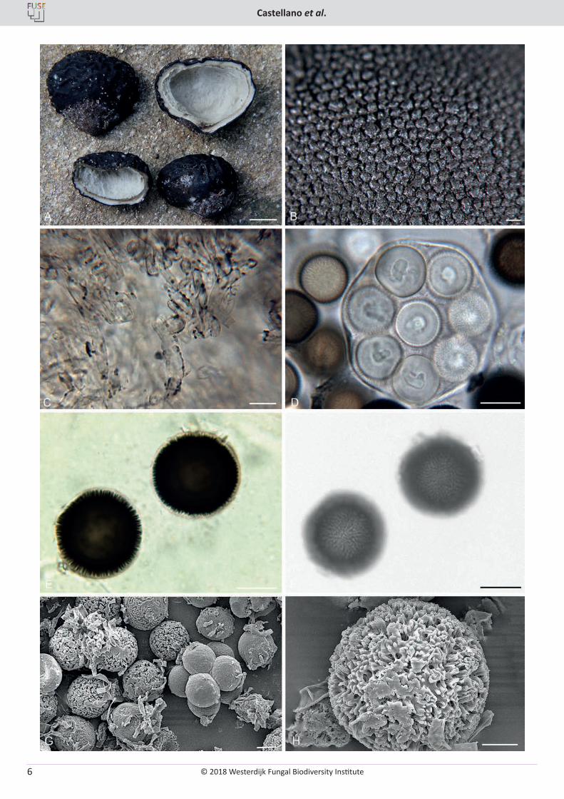
Elaphomyces loebiae Castellano & T.F. Elliott sp. nov. MycoBank MB821237. Fig. 3.
Etymology: Named “loebiae” in honour of Dr Susan C. Loeb, research ecologist with the US Forest Service and collector of the type specimens.
Type: USA, North Carolina, Haywood Co., Nantahala National Forest, Reinhart Gap, 15 Aug. 1996, S. Loeb & F. Tainter 130 (holotype - OSC 149108).
Ascomata as dried subglobose or irregularly-shaped, up to 23 × 35 mm. Fresh peridium ± 1.1 mm thick, outer layer ± 300 µm thick, peridial surface black, brittle, uniformly papillate, papillae flattened or sometimes rounded, irregular in outline, contiguous, 140–260 µm broad at base; inner layer ± 800 µm thick, somewhat pale grey adjacent to epicutis then soon off-white and uniform in texture, specimens supplied for study had been thoroughly cleaned beforehand, so information on material covering specimens is lacking. Gleba composed of a dark brown to black, powdery spore mass. Odour not recorded. Taste not recorded.
Peridium as dried two-layered; outer layer ± 300 µm thick, of compact cells with dark brown to black walls ± 1 µm thick, in between papillae are parallel hyphae with dark brown to black walls < 0.5 µm thick; inner layer ± 800 µm thick, its outer pale grey portion ± 200 µm thick, of short, segmented, much branched, loosely interwoven hyphae 4–7 µm broad, walls 1–2 µm thick, encrusted with small dark granules, the inner off-white portion of hyaline, occasionally sinuous, to somewhat periclinal to interwoven or even interlaced hyphae, mostly ± 4(–8) µm broad, rarely up to 12 µm broad, walls 1–2 µm thick. Gleba of spores and hyaline, septate, sinuous hyphae, 1–2 µm broad, walls <0.5 µm thick. Asci globose, 45–55 µm broad, hyaline, walls ± 1 µm thick, 8-spored, arising from hyaline,
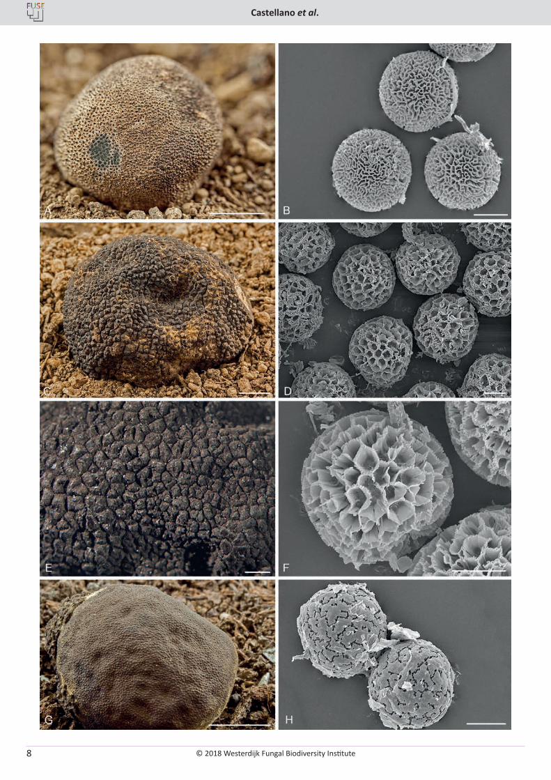
Fig. 2. Various Elaphomyces species from Europe. A. Elaphomyces citrinus ascomata showing peridial surface and adherent yellow toned mycelium (LE 162869). B. Elaphomyces citrinus SEM micrograph of ascospores showing the fine detail of the ornamentation (OSC 149172). C. Elaphomyces mutabilis ascomata showing wart structure and adherent off-white mycelium (OSC 149119). D. Elaphomyces mutabilis SEM micrograph of ascospores showing the fine detail of the ornamentation (OSC 149121). E. Elaphomyces virgatosporus SEM micrograph of ascospores showing spiral pattern of the ornamentation (OSC 149131). F. Elaphomyces virgatosporus SEM micrograph of ascospores showing the fine detail of the ornamentation (OSC 149131). G. Elaphomyces anthracinus ascoma showing peridial surface (W2008-1095). H. Elaphomyces anthracinus SEM micrograph of ascospores showing the ornamentation of irregular plates overlaying a fine reticulum (OSC 149163). Bars: A, G = 5 mm, B, D, F, H = 5 µm, C = 250 µm, E = 10 µm.
© 2018 Westerdijk Fungal Biodiversity Institute
Castellano et al.
Editor-in-ChiefProf. dr P.W. Crous, Westerdijk Fungal Biodiversity Institute, P.O. Box 85167, 3508 AD Utrecht, The Netherlands.E-mail:[email protected]
6
© 2018 Westerdijk Fungal Biodiversity Institute
Elaphomyces sp.nov. from eastern North America
Editor-in-ChiefProf. dr P.W. Crous, Westerdijk Fungal Biodiversity Institute, P.O. Box 85167, 3508 AD Utrecht, The Netherlands.E-mail:[email protected]
7
clustered, contorted, short segmented hyphae ± 6 µm broad. Spores globose, 21–22(–23) µm broad (mean = 21.7 µm), walls ± 1 µm thick, in KOH singly and in mass very dark red-brown to nearly black when mature, ornamentation opaque at maturity, consisting of rods or spines anastomosed at their apices to form a fine, irregular labyrinthine-like surface, 1–2 µm tall.
Distribution, habit, habitat, and season: Known only from North Carolina and South Carolina; hypogeous; under Betula alleghaniensis, Fagus grandifolia, Picea rubens, Quercus rubra, and Tsuga canadensis; March, May, July, and August.
Additional collections examined: USA, North Carolina, Haywood Co., Nantahala National Forest, Haywood Gap, 10 Jul. 1996, S. Loeb & F. Tainter 106 (OSC 149107); Rutherford Co., Union Mills, along Painters Gap Rd., 22 Mar. 2007, T. Elliott (OSC 149237); South Carolina, Horry Co., Huntington State Park, 25 May 2012, T. Elliott (OSC 149109).
Notes: We assign Elaphomyces loebiae to section Ceratogaster, subsection Sclerodermei based on the absence of mycelial patches on the peridial surface.
Castellano studied an Elaphomyces aculeatus collection in TO (in FH 258126) marked “Originale di Vittadini dono al Museo di Paris.” Castellano (unpubl. data) recorded data from this collection in part is as follows: Peridial surface appearing off-white to brown because of overlaying hyphae. Actual peridial surface is black, carbonaceous and warty, and the thick layer of overlying hyphae can be rubbed off to expose the apices of the warts and give the surface a black-spotted appearance against a brown background. Spores globose, in KOH brown to dark brown singly, slightly darker in mass, 21–24 µm broad, mean = 22.5 µm broad, including ornamentation that appears as irregular, short ridges under light microscopy, in section appearing warty in outline, SEM reveals the spore ornamentation as a fine, complete reticulum without overlying material (Fig. 4A–B). The somewhat smooth peridial surface and spore ornamentation of E. loebiae differs significantly from E. aculeatus. Castellano also studied a collection marked “Elaphomyces rubescens, 2 Aug. 1890, Eisenkaute, Herb Hesse” (OSC 158064) and found it to be conspecific with E. aculeatus.
Castellano studied an Elaphomyces anthracinus collection in TO (in OSC 149163) marked “Esemplare originale di Vittadini da Museo di Paris.” Castellano (unpubl. data) recorded data from this collection in part as follows: Peridial surface black, appearing smooth to the naked eye, close examination reveals a distinct pattern of low, circular, rounded papillae, not warty or angular, ascoma base with a peduncle. Spores globose, in KOH brown to dark brown singly, slightly darker in mass, 20–24 µm broad, mean = 21.9 µm broad, including ornamentation that appears punctate or dimpled under light microscopy, in section appearing with some flattened sides in outline to the spore, SEM reveals the spore ornamentation as a fine, complete reticulum overlain with over-lapping, plate-like material partially or nearly completely (Fig. 2G–H). We agree with Dodge (1929) that E. pyriformis Vittad. (Castellano studied authentic Vittadini
material in FH marked “ex ipso auctore”) and E. plumbeus Hesse (Castellano studied authentic material in Marburg marked “Laurasen, Germany, April 1890”) are conspecific. Dried Elaphomyces loebiae resembles dried E. anthracinus in its black, carbonaceous peridial surface covered with minute to small papillae and in having spores that are similarly sized, both approx. 21.5–22 µm broad with ornamentation. The spore ornamentation of E. loebiae is characterised by rods or spines anastomosed at their apices to form a fine, irregular labyrinth, whereas the spore ornamentation of E. anthracinus appears as a fine, complete reticulum overlain with plate-like material that is partially or nearly completely overlapping.
Elaphomyces loebiae resembles E. cantabricus in spore size and ornamentation and its black, carbonaceous peridial surface, but E. cantabricus has distinct sharp warts on the peridial surface in contrast to the papillae on the peridial surface of E. loebiae.
Elaphomyces loebiae resembles E. spirosporus in spore size, but the spore ornamentation of E. spirosporus appears as clearly spiraled ridges compared to the irregularly, labyrinthine-like spore ornamentation of E. loebiae.
Elaphomyces mitchelliae Castellano & T.F. Elliott, sp. nov. MycoBank MB821238. Fig. 5.
Etymology: Named “mitchelliae” (iae – after) in honour and recognition of Donna Mitchell of West Virginia, accomplished collector of sequestrate taxa.
Type: USA, Florida, Alachua Co., along State Route 325, ± 1/2 mile north of junction with SR346, 10 Aug. 1985, M. Castellano & S. Miller (holotype - OSC 149206; isotype - RMS).
Dried ascomata subglobose to somewhat turbinate, 11–18 × 14–40 mm, completely embedded in a yellow to green-yellow mycelial mat which forms a husk around individual ascomata and incorporates much sand, ectomycorrhizae and debris, the mycelium is sparse on the upper part of the ascomata (found particularly in between the warts), more dense near base but above the stipitate basal projection; the hyphae with heavy deposits of amorphous yellow material which, along with the hyphae, turns orange to magenta with KOH and releases a red pigment, under magnification the mycelial covering immediately adjacent to the surface is actually brown and gives rise to the yellowish hyphae; KOH on ascoma surface instantly red, soon brown-black, ETOH instantly black (from show-through of peridium). Peridium 3–4 mm thick when fresh, 2.5–3 mm when dried, outer layer 500–900 µm thick when fresh, carbonaceous, black, brittle, with a surface of rounded to angled, irregularly shaped warts, individual warts constructed in a compound, stellate pattern, warts 250–300 µm tall; inner layer a composite of several layers, 2.5–3 mm thick when fresh, leathery, uniform to banded, pale brown to pale yellow-brown to dark grey-brown (near gleba), KOH on darkening but not distinctive; at the base of the ascoma the carbonaceous layer is thickened as a dense, black hyphal mass incorporating roots and debris, often forming
Fig. 3. Elaphomyces loebiae. A. Ascomata showing peridial surface, immature gleba, and peridium in section (OSC 149109). B. Ascoma showing small rounded warts on peridial surface. C. Inner layer (2nd) of the peridium with encrusted hyphae. D. Asci showing 8 immature spores. E. Ascospore in cross-section showing height and pattern of the ornamentation. F. Ascospores in surface view showing the ornamentation pattern. G. SEM micrograph of ascospores showing the labyrinthine pattern to the clumpy ornamentation. H. SEM micrograph showing the fine detail of the ornamentation. B–H (OSC 149108 – holotype). Bars: A = 10 mm, B = 500 µm, C = 10 µm, D = 15 µm, E–G = 10 µm, H = 5 µm.
© 2018 Westerdijk Fungal Biodiversity Institute
Castellano et al.
Editor-in-ChiefProf. dr P.W. Crous, Westerdijk Fungal Biodiversity Institute, P.O. Box 85167, 3508 AD Utrecht, The Netherlands.E-mail:[email protected]
8

© 2018 Westerdijk Fungal Biodiversity Institute
Elaphomyces sp.nov. from eastern North America
Editor-in-ChiefProf. dr P.W. Crous, Westerdijk Fungal Biodiversity Institute, P.O. Box 85167, 3508 AD Utrecht, The Netherlands.E-mail:[email protected]
9
a projection up to 11 mm long × 8 mm broad (usually 3 × 3 mm or less). Gleba stuffed with bright white mycelium when young, when mature developing a powdery spore mass that is dark green-blue, dark grey-blue to dark grey-brown, finally grey-black, with concolourous web-like hyphae. Odour not distinctive. Taste not recorded.
Peridium 7-layered, 1st layer carbonaceous, ± 200 µm thick, of golden yellow to dark brown to black, multi-sided plates or warts with slightly raised edges, many hyphae between plates and also on the centre of the plate where it has a depression, in profile the plates wart-like with depression between the warts; 2nd layer 200–230 µm thick, of dark brown, irregularly inflated cells up to 10 × 15 µm, walls 0.5 µm thick; 3rd layer 120–140 µm thick, of hyaline to pale tan, compact, interwoven hyphae, 4–5 µm broad, walls 0.5 µm thick; 4th layer 1–1.2 mm thick, of ectomycorrhizas that are enveloped with dark brown-black hyphae, this layer irregularly structured with more ectomycorrhizas found near the ascomata base and none found at top of ascomata; 5th layer 200–250 µm thick, hyaline to pale tan, loosely interwoven, parallel to somewhat bundled hyphae, 4–5 µm broad; 6th layer 800–900 µm thick, of hyaline to pale tan, distinctly bundled, cord-like, compactly parallel hyphae, 5–7 µm broad, bundles slightly wavy or sinuous; 7th layer 200–275 µm thick, of hyaline to pale tan, short-segmented, contorted, irregularly shaped, compact, interwoven hyphae or cells, 4–5(–9) µm broad. Gleba composed of spores and hyphae that are hyaline, septate, somewhat branched, sinuous, loosely interwoven, 3–5 µm broad, walls <0.5 µm thick. Asci globose, 65–70 µm broad, hyaline, walls 2–3 µm thick, 4- or 8-spored, arising from knots of short, irregularly curved or contorted clustered hyphae, up to 14 µm broad; occasionally forming asci up to 140 µm broad filled with 11 or 12 spores. Spores globose, (23–)24–27(–28) µm broad (mean = 25.5 µm broad); walls 2–3 µm thick, in KOH dark grey-brown to olive singly and in mass when mature, ornamentation a partial to complete reticulum, alveoli 4–5 sided, (3–)5 µm broad, ± 3 µm tall.
Distribution, habit, habitat, and season: Known from Florida, Maryland, North Carolina, Virginia, and West Virginia; hypogeous; under Fagus grandifolia, Pinus serotina, P. taeda, Quercus alba, Q. coccinea, Q. laurifolia, Q. montana, Q. nigra, Q. rubra, or Q. virginiana; February, April through May, September, and November.
Additional collections examined: USA, Florida, Alachua Co., Cross Creek, 17 Sep. 1980, J. Trappe 5951 (OSC 149202) and 5955 (OSC 40154; PERTH); Newman Lake, Owens-Illinois County Park, 3 May 2006, M. Castellano (OSC 149210; same locality, 11 Aug. 1985, M. Castellano & S. Miller (OSC 149209, RMS); same locality, 6 May 1987, M. Castellano, D. Luoma, & T. O’Dell (OSC 149103); same locality, 14 Jun. 2012, M. Smith 602 (OSC 149216, FLAS); same locality, 25 Feb. 2012, M. Castellano & M. Smith (OSC 149215 & OSC 149213); same locality, 11 Aug. 2007, M. Castellano (OSC 149212); same locality, 23 Nov. 1981, Col. E. Dickstein
(FLAS 55379); ±13 miles southeast of Gainesville, 5.3 miles north of Cross Creek, 24 Aug. 1979, G. Benny, J. Kimbrough, L. Jacobs, & J. Gibson (FLAS 52090, OSC 39528); Polk Co., Lake Kissimmee State Park, picnic area, 17 Aug. 2007, M. Castellano (OSC 149211); Maryland, Anne Arundel Co., Patuxent Research Refuge, Laurie-Bowie Rd., 20 Apr. 1966, F.A. Uecker, O.K. Miller, & L. Stevens (OSC 149887); North Carolina, Brunswick Co., Wilmington, across from Belk-Beery, 8 Sept. 1984, S. Miller 806 (RMS, OSC 150029); Rutherford Co., Painters Gap Rd., 3 miles east of Cove Rd., 13 Jul. 2011, T. Elliott (OSC 149214); Virginia, Fairfax Co., no locality, 8 May 1926, E.G. Artzberger (OSC 149888); Accotink, 30 Jun. 1968, unknown collector (OSC 149101); West Virginia, Barbour Co. northwest portion of county, 18 Apr. 1992, D. Mitchell (OSC 149203).
Notes: We assign Elaphomyces mitchelliae to section Asco-scleroderma within Elaphomyces based on its distinct base.
Samuelson et al. (1987) provide an ultrastructural study of spore ornamentation of the holotype collection of this species (as E. persoonia).
Castellano studied an Elaphomyces cyanosporus Tul. & C. Tul. collection in Kew (K161175). Tulasne (1841) lists specimens from Meudon, Clamart, and Chaville in the area surrounding Paris. Castellano could not locate any Tulasne material of this species in Paris (PC or FH). Castellano (unpubl. data) recorded data from K161175 in part as follows: Dried peridium ornamented with flat, coarse, irregularly shaped black warts, peridial surface black, base subturbinate, spores globose, in KOH dark brown singly, slightly darker in mass, 27–30 µm broad, mean = 28.0 µm broad, including ornamentation that is a complete reticulum with alveoli 3–4 µm broad × 2–3 µm tall under light microscopy, SEM reveals the spore ornamentation as a complete reticulum with coarse ridges (Fig. 4E–F). The spores of E. cyanosporus are slightly larger than E. mitchelliae, and the alveoli are smaller. In addition, the peridial surface of E. cyanosporus consists of flat, irregularly shaped warts, whereas the peridial surface of E. mitchelliae has distinct, rounded to angled warts in a compound stellate pattern.
Castellano studied an Elaphomyces persoonii var. minor Tul. & C. Tul. collection in Kew (K161168). The Tulasne brothers (1841) list specimens from Meudon, Clamart, and Chaville in the area surrounding Paris but do not designate a type. Castellano could not locate any Tulasne material of this species in Paris (PC or FH). Castellano (unpubl. data) recorded data from K161175 in part as follows: Peridial surface of black, flat, coarse, irregularly shaped warts, ascoma base subturbinate, spores globose, in KOH dark brown singly, slightly darker in mass, 27–30 µm broad, mean = 28.0 µm broad including ornamentation that is a complete reticulum, alveoli 3–4 µm broad × 2–3 µm tall under light microscopy, SEM reveals the spore ornamentation as a complete reticulum with coarse-looking ridges. Elaphomyces persoonii var. minor is conspecific in all essential details with E. cyanosporus.
Castellano studied an Elaphomyces leveillei Tul. & C. Tul. collection in Kew herbarium (K162150). Tulasne (1841) lists specimens from Meudon, Clamart, and Chaville in the area
Fig. 4. Additional Elaphomyces species from Europe. A. Elaphomyces aculeatus ascomata showing peridial surface and the black warts overlain by yellow-brown hyphae (LE 162850). B. Elaphomyces aculeatus SEM micrograph of ascospores showing the labyrinthine-like ornamentation (OSC 149159). C. Elaphomyces persoonii ascomata showing wart structure and adherent yellow toned mycelium (LE 162885). D. Elaphomyces persoonii SEM micrograph of ascospores showing reticulate ornamentation (OSC 149365). E. Elaphomyces cyanosporus ascomata showing wart structure. F. Elaphomyces cyanosporus SEM micrograph of ascospores showing the reticulate ornamentation (OSC 149176). G. Elaphomyces leveillei ascomata showing papillate peridial surface and some adherent yellow toned mycelium (W2000-805). H. Elaphomyces leveillei SEM micrograph of ascospores showing the ornamentation of irregular plates (OSC 149116). Bars: A, G = 1 cm, B, D, F, H = 10 µm, C = 5 mm, E = 500 µm.
© 2018 Westerdijk Fungal Biodiversity Institute
Castellano et al.
Editor-in-ChiefProf. dr P.W. Crous, Westerdijk Fungal Biodiversity Institute, P.O. Box 85167, 3508 AD Utrecht, The Netherlands.E-mail:[email protected]
10
© 2018 Westerdijk Fungal Biodiversity Institute
Elaphomyces sp.nov. from eastern North America
Editor-in-ChiefProf. dr P.W. Crous, Westerdijk Fungal Biodiversity Institute, P.O. Box 85167, 3508 AD Utrecht, The Netherlands.E-mail:[email protected]
11
surrounding Paris but does not designate a type. Castellano could not locate any Tulasne material of this species in Paris (PC). Castellano (unpubl. data) recorded data from K162150 in part as follows: Peridial surface of black, pusticulate (bumpy) to tuberculate, partially covered by pale tan, pale yellow-brown to yellow hyphae, ascoma base indented, spores globose, in KOH dark brown singly, slightly darker in mass, 26–28 µm broad, mean = 26.6 µm broad including ornamentation that appears pusticulate under light microscopy, in section appearing with flattening of spore outline at least on a portion of numerous spores, SEM reveals the spore ornamentation as spines or rods overlain with amorphous, small, irregular plates to form a discontinuous surface, plate surface slightly roughened (Fig. 4G–H). The spores of E. leveillei are similar in size (26–28 µm broad, mean = 26.6 µm broad) but have an ornamentation of irregularly shaped, discontinuous plates compared to the distinct reticulum of E. mitchelliae. In addition, the peridial surface of E. leveillei consists of bump-like features compared to the distinct rounded to angled warts in a compound stellate pattern of E. mitchelliae.
Castellano studied Elaphomyces persoonii Vittad. collections from TO - OSC 149124, Wien - W2008-1079 and Kew - K162166 marked as Vittadini. Castellano (unpubl. data) recorded data from Trappe 1470 in part as follows: Ascoma subturbinate to turbinate, peridial surface of large black warts, with dark brown hyphae seen between warts, spores globose, in KOH brown to dark brown singly, slightly darker in mass, 29–33(–35) µm broad, mean = 31.3 µm broad including ornamentation that is a complete reticulum, up to 5 µm tall, alveoli irregular, up to 5–7 µm across under light microscopy, SEM reveals the spore ornamentation as a complete reticulum with the digitate edges along the alveoli (Fig. 4C–D). The spores of E. persoonii are larger (29–33 µm broad, mean = 31.3 µm broad) and have taller alveoli walls (up to 5 µm tall) compared to the spores of E. mitchelliae. In addition, the peridial surface of E. persoonii has flat warts compared to the distinct rounded to angled warts in a compound stellate pattern of E. mitchelliae.
ACKNOWLEDGEMENTS
We thank the following colleagues for sharing Elaphomyces collections included in this study: S. Loeb, S. Miller, D. Mitchell, F. Tainter. We also appreciate the opportunity to study specimens from the following herbaria: FH, FLAS, K, MB, PC, TO, and W.
REFERENCES
Boudier M (1876). Du Parasitisme Probable De Quelques Espèces Du Genre Elaphomyces Et De La Recherche De Ces Tubéracés. Bulletin de la Société Botanique de France 23: 115–119.
Castellano MA, Beever RE, Trappe JM (2012a). Sequestrate fungi of New Zealand: Elaphomyces (Ascomycota, Eurotiales, Elaphomycetaceae). New Zealand Journal of Botany 50: 423–433.
Castellano MA, Dentinger BTM, Séné O, et al. (2016). New species of Elaphomyces (Elaphomycetaceae, Eurotiales, Ascomycota) from tropical rainforests of Cameroon and Guyana. IMA Fungus 7: 59–73.
Castellano MA, Guerrero GG, Jiménez, JG, et al. (2012b). Elaphomyces appalachiensis and E. verruculosus sp. nov. (Elaphomycetaceae, Eurotiales, Ascomycota) from eastern North America. Revista Mexicana de Micologia 35: 17–22.
Castellano MA, Henkel TW, Miller SL, et al. (2012c). New Elaphomyces species (Elaphomycetaceae, Eurotiales, Ascomycota) from Guyana. Mycologia 104: 1244–1249.
Castellano, MA, Stephens RB (2017). Elaphomyces species (Elapho-mycetaceae, Eurotiales, Ascomycota) from Bartlett Experimental Forest, New Hampshire, USA. IMA Fungus 8: 49–63.
Castellano MA, Trappe JM, Luoma DL (2004). Sequestrate Fungi. In: Biodiversity of Fungi: Inventory and Monitoring methods (Mueller GM, Bills GE, Foster MS, eds.). Measuring and Monitoring Biological Diversity: Standard Methods for Fungi: 197–21. Academic Press, NY.
Castellano MA, Trappe JM, Vernes K (2011). Australian species of Elaphomyces (Elaphomycetaceae, Eurotiales, Ascomycota). Australian Systematic Botany 24: 32–57.
Ceruti A (1960). Elaphomycetales et Tuberales. In: Iconographia Mycologica di Bresadola Suppl. II, Trento.
Cork SJ, Kenagy GJ (1989). Nutritional value of hypogeous fungus for a forest-dwelling ground squirrel. Ecology 70: 577–586.
Corner EJH, Hawker LE (1953). Hypogeous fungi from Malaya. Transactions of the British Mycological Society 36: 125–137.
Dodge CW (1929). The higher Plectascales. Annales Mycologia 27: 145–184.
Genov P (1982). Fructification of Elaphomyces granulatus Fr. are food for boars. Acta Mycologica 18: 123–125.
Gómez-Reyes VM, Hernández-Salmerón IR, Terrón-Alfonso A, et al. (2012). Estudio taxonómico de Elaphomyces spp. (Ascomycota, Eurotiales, Elaphomycetaceae) de Michoacán, México. Revista Mexicana de Micología 36: 57–82.
Fogel R, Trappe JM (1978). Fungus consumption (mycophagy) by small animals. Northwest Science 52: 1–31.
Linder DH (1939). A new species of Elaphomyces from the Great Smoky Mountains National Park. Journal of the Elisha Mitchell Scientific Society 55: 131–133.
Luoma DL, Frenkel RE (1991). Fruiting of hypogeous fungi in Oregon Douglas-fir forests: seasonal and habitat variation. Mycologia 83: 335–353.
Mattirolo O (1907). Gli autoptici di Carlo Vittadini e la loro importanza nello studio della idnologia. Societá Italiana di Scienze Naturali: 1–7.
Monteechi A, Sarasini M (2000). Fungi Ipogei d’Europa. AMB Fondazione Centro Studi Micologici, Trento.
Miller SL, Miller Jr, OK (1984). Synthesis of Elaphomyces muricatus + Pinus sylvestris ectomycorrhizae. Canadian Journal of Botany 62: 2363–2369.
Paz AC, Alvarez JIG (2008). Elaphomyces cantabricus, una nueva especie de ascomiceto en contrada en Espana. Boletín Associació Micológica Font i Quer 6: 4–7.
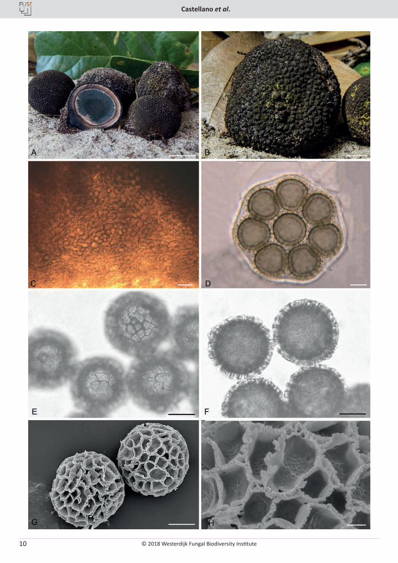
Fig. 5. Elaphomyces mitchelliae. A. Ascomata showing peridial surface, gleba, and peridium in section (OSC 149215). B. Ascoma showing peridial warts with some adherent yellow mycelium (OSC 149213). C. Outer peridium showing the pale, stacked hyphae between the darker wart tissues. D. Asci showing 8 immature spores. E. Ascospores in surface view showing the ornamentation pattern. F. Ascospores in cross-sectional view showing the height and pattern of the ornamentation. G. SEM micrograph of ascospores showing the distinct reticulate pattern. H. SEM micrograph of ascospores showing the structure of the alveoli. C, E–F (OSC 40154); D (OSC 40154); G–H (OSC 149103). Bars: A = 20 mm, B = 5 mm, C = 30 µm, D–G = 10 µm, H = 2.5 µm.
© 2018 Westerdijk Fungal Biodiversity Institute
Castellano et al.
Editor-in-ChiefProf. dr P.W. Crous, Westerdijk Fungal Biodiversity Institute, P.O. Box 85167, 3508 AD Utrecht, The Netherlands.E-mail:[email protected]
12
Paz A, Bellanger J-M, Lavoise C, et al. (2017). The genus Elaphomyces (Ascomycota, Eurotiales): a ribosomal DNA-based phylogeny and revised systematics of European deer truffles. Persoonia 38: 197–239.
Paz A, Lavoise C, Barrio L, et al. (2012). Propuesta de dos nuevas especies del género Elaphomyces, dos primeras citas para la Península Ibérica y una clave de identificación de las especies del género para Europa. Boletín Micológico de Federacion de Asociaciones Micologicas de Castilla y Leon 7: 85–104.
Reynolds HT (2011). Systematics, phylogeography, and ecology of Elaphomycetaceae. PhD dissertation, Department of Biology, Duke University, USA.
Rubio E, Miranda MA, Linde J, et al. (2006). Catálogo provisional de hongos hipogeos de Asturias y posibles fitobiontes ascociados. Revista Catalana de Micologia 28: 1–40.
Samuelson DA, Benny GL, Kimbrough JW (1987). Ultrastructure of ascospore ornamentation in Elaphomyces (Ascomycetes). Mycologia 79: 571–577.
Trappe JM, Molina R, Luoma DL, et al. (2009). Diversity, ecology and conservation of truffle fungi in forests of the Pacific Northwest. USDA Forest Service [General Technical Report PNW-GTR-772.]Portland: Pacific Northwest Research Station.
Trappe JM, Kimbrough JW (1972). Elaphomyces viridiseptum, a new species from Florida. Mycologia 64: 646–649.
Trappe JM, Guzmán G (1971). Notes on some hypogeous fungi from Mexico. Mycologia 63: 317–332.
Tulasne LR, Tulasne C (1841). Observations sur le genre Elaphomyces, et description de quelques especès nouvelles. Annales Science Naturale Series 2 16: 5–29.
Vernes K, Blois S, Bärlocher F (2004). Seasonal and yearly changes in consumption of hypogeous fungi by northern flying squirrels and red squirrels in old-growth forest, New Brunswick. Canadian Journal of Zoology 82: 110–117.
Vernes K, Poirier N (2007). Use of a robin’s nest as a cache site for truffles by a red squirrel. Northeastern Naturalist 14: 145–149.
Vogt KA, Edmonds RL, Grier CC (1981). Biomass and nutrient concentra-tions of sporocarps produced by mycorrhizal and decomposer fungi in Abies amabilis stands. Oecologia 50: 170–175.
Zhang BC, Minter DW (1989). Elaphomyces spinoreticulatus sp. nov., with notes on Canadian species of Elaphomyces. Canadian Journal of Botany 67: 909–914.
Fungal Systematics and Evolution is licensed under a Creative Commons Attribution-NonCommercial-ShareAlike 4.0 International License
© 2018 Westerdijk Fungal Biodiversity Institute 13
Editor-in-ChiefProf. dr P.W. Crous, Westerdijk Fungal Biodiversity Institute, P.O. Box 85167, 3508 AD Utrecht, The Netherlands.E-mail:[email protected]
Fungal Systematics and EvolutionVOLUME 1JUNE 2018
PAGES 13–22
doi.org/10.3114/fuse.2018.01.02
Epitypification and re-description of the zombie-ant fungus, Ophiocordyceps unilateralis (Ophiocordycipitaceae)
H.C. Evans1,2*, J.P.M. Araújo3, V.R. Halfeld4, D.P. Hughes3
1CAB International, UK Centre, Egham, Surrey, UK2Departamentos de Entomologia e Fitopatologia, Universidade Federal de Viçosa, Viçosa, Minas Gerais, Brazil3Departments of Entomology and Biology, Penn State University, University Park, Pennsylvania, USA4Universidade Federal de Juiz de Fora, Juiz de Fora, Minas Gerais, Brazil
*Corresponding author: [email protected]
Abstract: The type of Ophiocordyceps unilateralis (Ophiocordycipitaceae, Hypocreales, Ascomycota) is based on an immature specimen collected on an ant in Brazil. The host was identified initially as a leaf-cutting ant (Atta cephalotes, Attini, Myrmicinae). However, a critical examination of the original illustration reveals that the host is the golden carpenter ant, Camponotus sericeiventris (Camponotini, Formicinae). Because the holotype is no longer extant and the original diagnosis lacks critical taxonomic information – specifically, on ascus and ascospore morphology – a new type from Minas Gerais State of south-east Brazil is designated herein. A re-description of the fungus is provided and a new phylogenetic tree of the O. unilateralis clade is presented. It is predicted that many more species of zombie-ant fungi remain to be delimited within the O. unilateralis complex worldwide, on ants of the tribe Camponotini.
Key words: Atlantic rainforestCamponotus sericeiventriscarpenter antsepitypeOphiocordycepsphylogeny
Published online: 15 December 2017.
INTRODUCTION
Ophiocordyceps unilateralis (Ophiocordycipitaceae: Hypocrea-les) is a fungal pathogen of ants belonging to the tribe Camponotini (Formicinae: Formicidae) with a pantropical distribution (Evans 2001). The fungus alters the behaviour of the ant host causing it to move and die away from the nest, often in an exposed position and, typically, clinging onto and biting into vegetation in a “death-grip” (Hughes et al. 2011). This host manipulation by O. unilateralis is a particularly spectacular and complex example of the extended-phenotype paradigm (Dawkins 1982, Andersen et al. 2009, Hughes 2013, Hughes et al. 2016), which duly garnered the epithet, the zombie-ant fungus (Evans et al. 2011a), and spawned considerable media coverage by the popular press and scientific magazines alike (Kaplan 2011, Costandi 2012, Boddy 2014, Pennisi 2014). In addition, it stimulated on-going research on the nature of the ant-fungal association, as well as on fungal phylogeny, that has generated a wealth of information (reviewed in Hughes et al. 2016). Significant advancement has been made in understanding the mechanisms involved at the molecular level: thus, manipulation of the ant brain by the fungus has been ascribed to two candidate metabolites – guanobutyric acid and sphingosine – previously implicated in neurological diseases and cancer (de Bekker et al. 2014). Using comparative genomics and a mixed transcriptomics approach, it has also been shown that genes unique to the fungus are up-regulated that encode for proteins known to cause neurological and behavioural changes (de Bekker et al. 2015, de Bekker et al. 2017).
Contemporary studies have tended to use the over-arching term O. unilateralis sensu lato for the zombie-ant fungus since it has long been suspected, but only recently established, that
this is a species complex. In fact, morphological variations had been noted in collections from around the world from a very early stage (Petch 1924, 1931, 1933, 1935, 1937, Kobayasi 1941, Mains 1958, Evans 1974, 1982, Evans & Samson 1984), but it was concluded that “whilst it is tempting to separate geographic isolates (ecotypes), there is not enough evidence at the moment to conveniently divide the species into varietal units: more information is needed concerning host specificity and the range of variation in temperate, subtropical and tropical specimens” (Evans & Samson 1984). Some three decades later, Evans et al. (2011a) set out to uncover the taxonomic diversity of the newly-termed zombie or brain-manipulating fungus, based on an examination of fresh material collected on infected carpenter ants within a fragment of Atlantic rainforest in Brazil. Four Camponotus species were identified and, following a critical morphological comparison of the freshly-released (mature) ascospores – as well as of the germination process – and of the associated asexual morphs, four Ophiocordyceps species were delimited; leading to the supposition that “each species of the tribe Camponotini may be attacked by a distinct species of Ophiocordyceps” (Evans et al. 2011a), and “that there may be hundreds of species within the complex parasitising formicine ants worldwide” (Evans et al. 2011b). This hypothesis would appear to be holding true based on subsequent publications involving both morphological and molecular evidence, with six new species being described from Thailand (Luangsa-ard et al. 2011, Kobmoo et al. 2012, Kobmoo et al. 2015), one from Japan (Kepler et al. 2010), three from the Brazilian Amazon (Araújo et al. 2015) and another 14 in the pipeline (Araújo et al. 2018).
Significantly, however, only Kobayasi (1941) appears to have examined the type specimen – named as Torrubia unilateralis
© 2018 Westerdijk Fungal Biodiversity Institute
Evans et al.
Editor-in-ChiefProf. dr P.W. Crous, Westerdijk Fungal Biodiversity Institute, P.O. Box 85167, 3508 AD Utrecht, The Netherlands.E-mail:[email protected]
14
on the Brazilian ant Atta cephalotes (Tulasne & Tulasne 1865) – and he noted that it “is now preserved in [the] Paris Entomological Museum [and] is immature”. Unfortunately, repeated attempts to obtain the type for examination of the fungus and identification of the ant host were unsuccessful and it was concluded that the specimen was lost, leading to speculation that this may have gone missing during the Second World War (Evans et al. 2011a). From the latter study, and the confirmation that O. unilateralis represents a species complex, it became necessary to designate a new type, especially since Ophiocordyceps is the type genus of the recently-recognised family Ophiocordycipitaceae which is based on the placement of O. unilateralis within a Bayesian consensus tree (Sung et al. 2007). Ophiocordyceps is a highly diverse genus, with considerable pharmaceutical potential (Berenbaum & Eisner 2008, Paterson 2008, Molnár et al. 2010, Zhang et al. 2012) – species of which have also been identified recently as primary endosymbionts in certain insect hosts (Nishino et al. 2016, Gomez-Polo et al. 2017) – and thus O. unilateralis is central to our understanding of this medically-important group, as well as being considered as a keystone species for unravelling ecosystem functioning and biodiversity of fungi in tropical forests (Evans et al. 2011b).
In his diagnosis, Louis Tulasne described the unilateral position of the fleshy, hemispherical, fertile stroma on the stipe, but failed to provide details of the asci or ascospores, nor did these structures appear in the accompanying illustration by his brother, Charles (Tulasne & Tulasne 1865). This supports the statement of Kobayasi (1941) that the type was immature. Theoretically, the illustration could still stand as the holotype but, because there is no extant material, this would serve as the lectotype and a suitable epitype should be designated (Ariyawansa et al. 2014), not a neotype as Evans et al. (2011a) had originally and mistakenly proposed. The resultant search for a suitable epitype was based on the evidence from the illustration that the host representing the type is a Camponotus ant (Samson et al. 1982): specifically, the golden carpenter ant Camponotus sericeiventris, with its distinctive pronotal plate, and not the leafcutter Atta cephalotes, which is a myrmicine ant having no historical association with O. unilateralis (Evans & Samson 1984, Evans 2001). Cooke (1892) used the same Tulasne illustration to re-describe the so-called “one-sided ant club”, with additional information that the fungus had been “collected by Trail in Brazil”. This specimen is in the RBG Kew fungarium and was found by the English naturalist J.W.H. Trail in 1874 in the Brazilian Amazon, which was examined by Massee (1895) who reported it to be on the same ant species as the type. However, we consider that the type specimen of O. unilateralis was more likely to have originated in the Atlantic rainforest region of south-east Brazil – where several European naturalists were based in the 1860s – and from where the type of Camponotus sericeiventris was collected (Rio de Janeiro) during a series of French expeditions (Guérin-Menéville 1838); specimens from which were deposited in the Paris Entomological Museum, where the type of O. unilateralis was also deposited (Tulasne & Tulasne 1865).
Epitypification has been delayed until now because all the targeted collections of infected C. sericeiventris ants from Atlantic rainforest in south-east Brazil proved to be immature (Evans et al. 2011a). In fact, some newly-infected specimens were marked in situ – whilst others were harvested and incubated in the laboratory – to monitor progress, but none developed
to maturity. The present paper is the result of the discovery of specimens with fertile stromata, from the same region of Brazil (Zona da Mata Mineira), enabling a full description of the species, as well as a phylogenetic analysis.
MATERIALS AND METHODS
Field collection
Collecting was concentrated in a vestige of secondary Atlantic rainforest near Viçosa, Minas Gerais, in the Zona da Mata Mineira of south-east Brazil – belonging to the Universidade Federal de Viçosa (UFV) – where ad hoc surveys for zombie-ant fungi had been carried out previously (Evans et al. 2011a, b). Although Camponotus sericeiventris is relatively common in this habitat, it is confined mainly to open, heavily-disturbed areas and the incidence of infected ants was found to be low. All the initial collections proved to be immature and it was decided to follow progress in situ by flagging specimens and monitoring development of the ascostromata through weekly observation. However, none of the five tagged specimens survived, due to predation and loss through heavy rain. Subsequently, additional specimens were bagged but were spoiled by run-off water following storms. Finally, several more immature specimens were harvested together with the vegetation, transferred to a humid chamber in a greenhouse at UFV – with an 8 h misting/16 h dry regime – and monitored. Asexual morphs developed successfully but, because ascostromatal development was slow, the specimens were overgrown by opportunistic fungi before maturation was complete. The taxonomy of the asexual morphs is based on these paratype specimens. The mature epitype was collected by one of us (VRH) from another forest reserve in the Zona da Mata Mineira, some 150 km from the main study site, in the municipality of Juiz de Fora. These specimens were deposited in the fungarium of the Universidade Federal de Viçosa (VIC).
DNA extraction and PCR
We used a BLAST search in the GenBank nucleotide database to ensure the quality of the sequences generated in this study. Sequences that were identified as species not closely related to the species treated in this study were discarded and interpreted to be from a contaminant. All the sequences included here passed the above quality control checks.
The molecular studies were conducted according to Araújo et al. (2018), described below. The DNA templates were obtained directly from two specimens of O. unilateralis infecting Camponotus sericeiventris from the type locality in Minas Gerais (Brazil) that were collected in the field and dried in silica to avoid overgrowth by opportunistic fungi. For DNA extraction, the ants were dissected and the fungal contents (mummified mycelium and hyphal bodies) within their abdomens were placed in 1.5 mL Eppendorf tubes with 100–200 µL of CTAB (2 % CTAB powder, 100 mM Tris pH8, 20 mM EDTA, 1.4 M NaCl) and ground mechanically; 400 µL of CTAB were then added and the tubes were incubated at 60 °C for 20 min and centrifuged for 10 min at 14 000 rpm. The supernatant (approx. 400 µL) was transferred to a new 1.5 mL Eppendorf tube, mixed with 500 µL of 24:1 chloroform: isoamyl-alcohol (Sigma) and mixed by inverting. The mix was then centrifuged for 20 min at 14 000 rpm and the
© 2018 Westerdijk Fungal Biodiversity Institute
Epitypification of Ophiocordyceps unilateralis
Editor-in-ChiefProf. dr P.W. Crous, Westerdijk Fungal Biodiversity Institute, P.O. Box 85167, 3508 AD Utrecht, The Netherlands.E-mail:[email protected]
15
supernatant transferred to a new 1.5 mL Eppendorf tube and further cleaned using the GeneCleanIII kit (MP Biomedicals), following Araújo et al. (2018) modifications.
Four loci were used in the analyses, i.e. small subunit nuclear ribosomal DNA (SSU), large subunit nuclear ribosomal DNA (LSU), translation elongation factor 1-α (tef1) and the largest subunit of RNA polymerase II (rpb1). The final concatenated dataset consisted of 3 795 bp. The primers used were, SSU: NS1 (GTAGTCATATGCTTGTCTC) and NS4 (CTTCCGTCAATTCCTTTAAG) (White et al. 1990); LSU: LR0R (5’-ACCCGCTGAACTTAAGC-3’) and LR5 (5’-TCCTGAGGGAAACTTCG-3’) (Vilgalys & Hester 1990); tef1: EF1-983F (5’-GCYCCYGGHCAYCGTGAYTTYAT-3’) and EF1-2218R (5’- ATGACACCRACRGCRACRGTYTG-3’) (Rehner & Buckley 2005); CRPB1: (5’-CCWGGYTTYATCAAGAARGT-3’) (Castlebury et al. 2004) and RPB1Cr_oph: (5’-CTGVCCMGCRATGTCGTTGTCCAT-3’) (Araújo et al. 2018).
To amplify the target loci, each 25 µL PCR amplification mix contained 4.5 µL of buffer E (Premix E – Epicentre) 0.5 µL of each forward and reverse primers (10 mM), 1 µL of DNA template, 0.1 Platinum Taq polymerase (Invitrogen) and 18.4 µL of ultra-pure distilled water (Gibco). The amplification reactions were placed in a Biometra T300 thermocycler under the following conditions: for SSU and LSU (1) 2 min at 94 °C, (2) 4 cycles of denaturation at 94 °C for 30 s, annealing at 55 °C for 30 s, and extension at 72 °C for 2 min, followed by (3) 35 cycles of denaturation at 94 °C for 30 s, annealing at 50.5 °C for 1 min, and extension at 72 °C for 2 min and (4) 3 min at 72 °C. For tef1 and rpb1 (1) 2 min at 94 °C, (2) 10 cycles of denaturation at 94 °C for 30 s, annealing at 64 °C for 1 min, and extension at 72 °C for 1 min, followed by (3) 35 cycles of denaturation at 94 °C for 30 s, annealing at 54 °C for 1 min, and extension at 72 °C for 1 min and (4) 3 min at 72 °C. Each 25 µL amplification reaction was cleaned by adding 3.75 µL of Illustra ExoProStar enzymatic PCR clean up (1:1 mix of Exonuclease I and alkaline phosphatase, GE Healthcare Life Sciences), incubated at 37 °C for 1 h and 80 °C for 15 min in the thermocycler. The purified PCR products were sequenced by Sanger DNA sequencing [Applied Biosystems 3730xl DNA Analyzer (Life Technologies, Carlsbad, CA, USA)] at the Genomics Core Facility service at Penn State University.
Phylogenetic analyses
The raw sequence reads were edited manually using Geneious v. 8.1.8 (Kearse et al. 2012). Individual gene alignments were generated by MUSCLE (Edgar 2004), implemented in Geneious v. 8.1.8 (Kearse et al. 2012). The alignment of every gene was improved manually, annotated and concatenated into a single combined dataset using Geneious v. 8.1.8 (Kearse et al. 2012). Ambiguously aligned regions were manually excluded from phylogenetic analysis and gaps were treated as missing data. The final alignment length was 3 795 bp – 1 071 for SSU, 961 for LSU, 1 011 for tef1 and 752 for rpb1. A Maximum likelihood (ML) analysis was performed with RAxML v. 8.2.4 (Stamatakis 2006) on a concatenated dataset containing all four genes. The dataset consisted of eight data partitions. These included one each for SSU and LSU, and three for each of the three codon positions of the protein coding genes, tef1 and RPB1. The GTRGAMMA model of nucleotide substitution was employed during the generation of 1 000 bootstrap (bs) replicates. The sequences generated for this study were deposited in GenBank (Table 1).
RESULTS
Taxonomy
Ophiocordyceps unilateralis (Tul.) Petch, Trans. Br. Mycol. Soc. 16: 74.1933. emend. H.C. Evans, D.P. Hughes & Araújo. Figs 1–2. Basionym: Torrubia unilateralis Tul., Sel. Fung. Carp. III: 18. 1865.Synonym: Cordyceps unilateralis (Tul.) Sacc. Syll. Fung. 2: 570. 1883.
Description on host: External mycelium sparse, pale brown; emerging from sutures on body and legs. Clava stromatal, solitary, arising from the dorsal pronotum; cylindrical, brown and hirsute at the base. Ascostroma produced unilaterally, almost encircling the clava; hemisphaerical, 1.5–1.7 × 0.8 µm, dark brown, with roughened surface due to prominent perithecial necks. Ascomata (perithecia) partially erumpent, flask-shaped, 200–250 × 140–160 µm. Asci 8-spored, hyaline, cylindrical, (90–)95–125 × 6–8(–9) µm, swollen centrally tapering to a distinct foot and apical cap region (5–6 × 4–5 µm). Ascospores multiserriate, hyaline, thin-walled, filiform, (70–)75–85 × 2–2.5 µm, 4–5-septate; curved, tapering at both ends.
Lectotype designated here: holotype Brazil, “Atta cephalotes”, Tulasne (1865) Sel. Fung. Carp. III, plate I, fig. 3–4, MBT379723.
Epitype designated here: Brazil, Minas Gerais, Juiz de Fora, Paraibuna river (700–800 m a.s.l.), on Camponotus sericeiventris (Camponotini: Formicinae: Formicidae), on shrub leaf, 10 Aug. 2014, V.R. Halfeld, I14-1369A (epitype VIC 44303, MBT379722).
Additional materials examined: Brazil, Minas Gerais, Viçosa, Mata do Paraíso (700 m a.s.l.), on Camponotus sericeiventris, on shrub leaf, 26 Apr. 2010, H.C. Evans, MAP-61 (paratype VIC 44354); 12 Aug. 2012, H.C. Evans, MP-426 (paratype VIC 44349); 7 Feb. 2013, H.C. Evans, MP-502 (paratype VIC 44350).
Asexual morph: The asexual morph of the epitype proved to be in poor condition and the diagnosis below is based on the paratype collections.
Apical region of the stromatal clava, smooth, pinkish-brown, tapering to an acute tip; covered by a loose to compact hymenium of scattered to dense phialides. Phialides of two types: with a prominent swollen base (10–12 × 3–3.5 µm), tapering abruptly to a thin neck region (12–15 × 0.5–1 µm), producing hyaline, guttulate, limoniform conidia, 6.5–8 × 2–2.5 µm, apically (= Hirsutella A-type, Evans & Samson 1984); with a cylindrical base (14–16 × 2.5–3 µm), tapering gradually to a long neck (45–50 µm), 1 µm at the tip, producing solitary, hyaline, cylindrical-fusoid conidia, 8–11 × 2.5–3 µm, with a rounded apex and truncate base (= Hirsutella B-type). Hirsutella B-type also produced separately in loose, brown sporodochia arising from the leg joints.
Notes: Other synonyms – Torrubia formicivora, Cordyceps formicivora, C. ridleyi and C. subunilateralis – have been listed by various authors (Petch 1933, Mains 1958, Evans & Samson 1984, Sung et al. 2007): however, because the ant hosts are not identified and the collecting localities of some are outside the geographic range of Camponotus sericeiventris, these can no longer be considered to be synonymous with O. unilateralis s. str. Examination of the types, as well as identification of the
© 2018 Westerdijk Fungal Biodiversity Institute
Evans et al.
Editor-in-ChiefProf. dr P.W. Crous, Westerdijk Fungal Biodiversity Institute, P.O. Box 85167, 3508 AD Utrecht, The Netherlands.E-mail:[email protected]
16
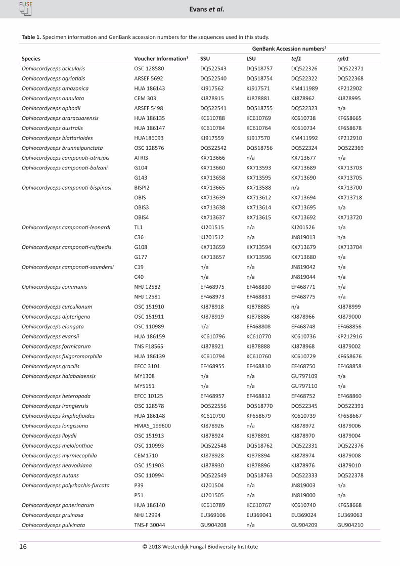
Table 1. Specimen information and GenBank accession numbers for the sequences used in this study.
Species Voucher Information1
GenBank Accession numbers2
SSU LSU tef1 rpb1
Ophiocordyceps acicularis OSC 128580 DQ522543 DQ518757 DQ522326 DQ522371
Ophiocordyceps agriotidis ARSEF 5692 DQ522540 DQ518754 DQ522322 DQ522368
Ophiocordyceps amazonica HUA 186143 KJ917562 KJ917571 KM411989 KP212902
Ophiocordyceps annulata CEM 303 KJ878915 KJ878881 KJ878962 KJ878995
Ophiocordyceps aphodii ARSEF 5498 DQ522541 DQ518755 DQ522323 n/a
Ophiocordyceps araracuarensis HUA 186135 KC610788 KC610769 KC610738 KF658665
Ophiocordyceps australis HUA 186147 KC610784 KC610764 KC610734 KF658678
Ophiocordyceps blattarioides HUA186093 KJ917559 KJ917570 KM411992 KP212910
Ophiocordyceps brunneipunctata OSC 128576 DQ522542 DQ518756 DQ522324 DQ522369
Ophiocordyceps camponoti-atricipis ATRI3 KX713666 n/a KX713677 n/a
Ophiocordyceps camponoti-balzani G104 KX713660 KX713593 KX713689 KX713703
G143 KX713658 KX713595 KX713690 KX713705
Ophiocordyceps camponoti-bispinosi BISPI2 KX713665 KX713588 n/a KX713700
OBIS KX713639 KX713612 KX713694 KX713718
OBIS3 KX713638 KX713614 KX713695 n/a
OBIS4 KX713637 KX713615 KX713692 KX713720
Ophiocordyceps camponoti-leonardi TL1 KJ201515 n/a KJ201526 n/a
C36 KJ201512 n/a JN819013 n/a
Ophiocordyceps camponoti-rufipedis G108 KX713659 KX713594 KX713679 KX713704
G177 KX713657 KX713596 KX713680 n/a
Ophiocordyceps camponoti-saundersi C19 n/a n/a JN819042 n/a
C40 n/a n/a JN819044 n/a
Ophiocordyceps communis NHJ 12582 EF468975 EF468830 EF468771 n/a
NHJ 12581 EF468973 EF468831 EF468775 n/a
Ophiocordyceps curculionum OSC 151910 KJ878918 KJ878885 n/a KJ878999
Ophiocordyceps dipterigena OSC 151911 KJ878919 KJ878886 KJ878966 KJ879000
Ophiocordyceps elongata OSC 110989 n/a EF468808 EF468748 EF468856
Ophiocordyceps evansii HUA 186159 KC610796 KC610770 KC610736 KP212916
Ophiocordyceps formicarum TNS F18565 KJ878921 KJ878888 KJ878968 KJ879002
Ophiocordyceps fulgoromorphila HUA 186139 KC610794 KC610760 KC610729 KF658676
Ophiocordyceps gracilis EFCC 3101 EF468955 EF468810 EF468750 EF468858
Ophiocordyceps halabalaensis MY1308 n/a n/a GU797109 n/a
MY5151 n/a n/a GU797110 n/a
Ophiocordyceps heteropoda EFCC 10125 EF468957 EF468812 EF468752 EF468860
Ophiocordyceps irangiensis OSC 128578 DQ522556 DQ518770 DQ522345 DQ522391
Ophiocordyceps kniphofioides HUA 186148 KC610790 KF658679 KC610739 KF658667
Ophiocordyceps longissima HMAS_199600 KJ878926 n/a KJ878972 KJ879006
Ophiocordyceps lloydii OSC 151913 KJ878924 KJ878891 KJ878970 KJ879004
Ophiocordyceps melolonthae OSC 110993 DQ522548 DQ518762 DQ522331 DQ522376
Ophiocordyceps myrmecophila CEM1710 KJ878928 KJ878894 KJ878974 KJ879008
Ophiocordyceps neovolkiana OSC 151903 KJ878930 KJ878896 KJ878976 KJ879010
Ophiocordyceps nutans OSC 110994 DQ522549 DQ518763 DQ522333 DQ522378
Ophiocordyceps polyrhachis-furcata P39 KJ201504 n/a JN819003 n/a
P51 KJ201505 n/a JN819000 n/a
Ophiocordyceps ponerinarum HUA 186140 KC610789 KC610767 KC610740 KF658668
Ophiocordyceps pruinosa NHJ 12994 EU369106 EU369041 EU369024 EU369063
Ophiocordyceps pulvinata TNS-F 30044 GU904208 n/a GU904209 GU904210
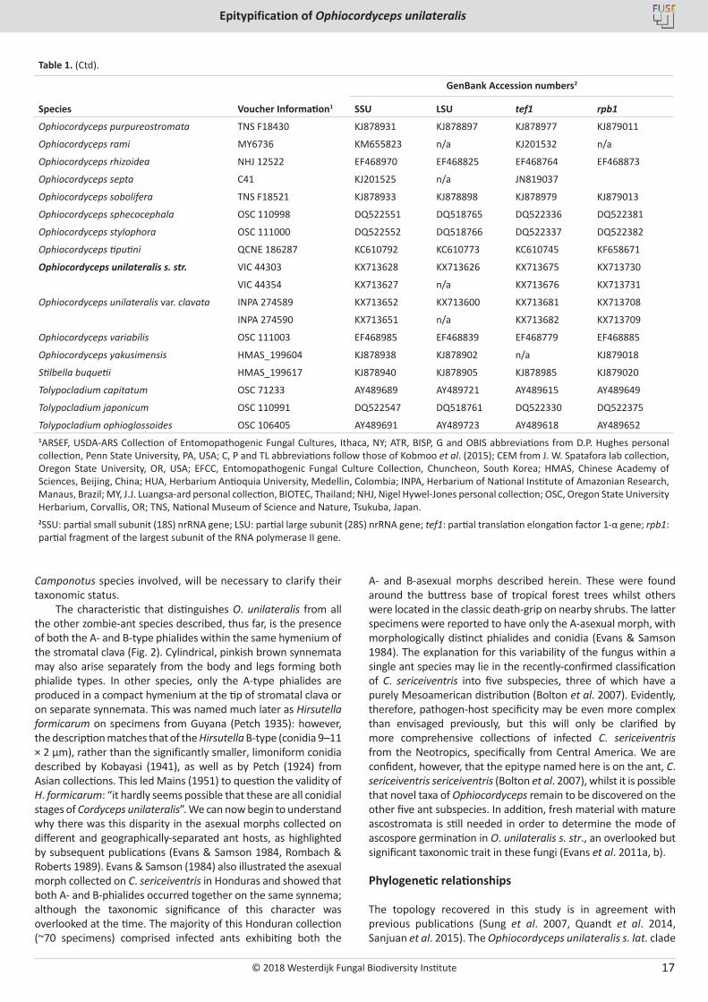
© 2018 Westerdijk Fungal Biodiversity Institute
Epitypification of Ophiocordyceps unilateralis
Editor-in-ChiefProf. dr P.W. Crous, Westerdijk Fungal Biodiversity Institute, P.O. Box 85167, 3508 AD Utrecht, The Netherlands.E-mail:[email protected]
17
Table 1. (Ctd).
Species Voucher Information1
GenBank Accession numbers2
SSU LSU tef1 rpb1
Ophiocordyceps purpureostromata TNS F18430 KJ878931 KJ878897 KJ878977 KJ879011
Ophiocordyceps rami MY6736 KM655823 n/a KJ201532 n/a
Ophiocordyceps rhizoidea NHJ 12522 EF468970 EF468825 EF468764 EF468873
Ophiocordyceps septa C41 KJ201525 n/a JN819037
Ophiocordyceps sobolifera TNS F18521 KJ878933 KJ878898 KJ878979 KJ879013
Ophiocordyceps sphecocephala OSC 110998 DQ522551 DQ518765 DQ522336 DQ522381
Ophiocordyceps stylophora OSC 111000 DQ522552 DQ518766 DQ522337 DQ522382
Ophiocordyceps tiputini QCNE 186287 KC610792 KC610773 KC610745 KF658671
Ophiocordyceps unilateralis s. str. VIC 44303 KX713628 KX713626 KX713675 KX713730
VIC 44354 KX713627 n/a KX713676 KX713731
Ophiocordyceps unilateralis var. clavata INPA 274589 KX713652 KX713600 KX713681 KX713708
INPA 274590 KX713651 n/a KX713682 KX713709
Ophiocordyceps variabilis OSC 111003 EF468985 EF468839 EF468779 EF468885
Ophiocordyceps yakusimensis HMAS_199604 KJ878938 KJ878902 n/a KJ879018
Stilbella buquetii HMAS_199617 KJ878940 KJ878905 KJ878985 KJ879020
Tolypocladium capitatum OSC 71233 AY489689 AY489721 AY489615 AY489649
Tolypocladium japonicum OSC 110991 DQ522547 DQ518761 DQ522330 DQ522375
Tolypocladium ophioglossoides OSC 106405 AY489691 AY489723 AY489618 AY489652 1ARSEF, USDA-ARS Collection of Entomopathogenic Fungal Cultures, Ithaca, NY; ATR, BISP, G and OBIS abbreviations from D.P. Hughes personal collection, Penn State University, PA, USA; C, P and TL abbreviations follow those of Kobmoo et al. (2015); CEM from J. W. Spatafora lab collection, Oregon State University, OR, USA; EFCC, Entomopathogenic Fungal Culture Collection, Chuncheon, South Korea; HMAS, Chinese Academy of Sciences, Beijing, China; HUA, Herbarium Antioquia University, Medellin, Colombia; INPA, Herbarium of National Institute of Amazonian Research, Manaus, Brazil; MY, J.J. Luangsa-ard personal collection, BIOTEC, Thailand; NHJ, Nigel Hywel-Jones personal collection; OSC, Oregon State University Herbarium, Corvallis, OR; TNS, National Museum of Science and Nature, Tsukuba, Japan. 2SSU: partial small subunit (18S) nrRNA gene; LSU: partial large subunit (28S) nrRNA gene; tef1: partial translation elongation factor 1-α gene; rpb1: partial fragment of the largest subunit of the RNA polymerase ІІ gene.
Camponotus species involved, will be necessary to clarify their taxonomic status.
The characteristic that distinguishes O. unilateralis from all the other zombie-ant species described, thus far, is the presence of both the A- and B-type phialides within the same hymenium of the stromatal clava (Fig. 2). Cylindrical, pinkish brown synnemata may also arise separately from the body and legs forming both phialide types. In other species, only the A-type phialides are produced in a compact hymenium at the tip of stromatal clava or on separate synnemata. This was named much later as Hirsutella formicarum on specimens from Guyana (Petch 1935): however, the description matches that of the Hirsutella B-type (conidia 9–11 × 2 µm), rather than the significantly smaller, limoniform conidia described by Kobayasi (1941), as well as by Petch (1924) from Asian collections. This led Mains (1951) to question the validity of H. formicarum: “it hardly seems possible that these are all conidial stages of Cordyceps unilateralis”. We can now begin to understand why there was this disparity in the asexual morphs collected on different and geographically-separated ant hosts, as highlighted by subsequent publications (Evans & Samson 1984, Rombach & Roberts 1989). Evans & Samson (1984) also illustrated the asexual morph collected on C. sericeiventris in Honduras and showed that both A- and B-phialides occurred together on the same synnema; although the taxonomic significance of this character was overlooked at the time. The majority of this Honduran collection (~70 specimens) comprised infected ants exhibiting both the
A- and B-asexual morphs described herein. These were found around the buttress base of tropical forest trees whilst others were located in the classic death-grip on nearby shrubs. The latter specimens were reported to have only the A-asexual morph, with morphologically distinct phialides and conidia (Evans & Samson 1984). The explanation for this variability of the fungus within a single ant species may lie in the recently-confirmed classification of C. sericeiventris into five subspecies, three of which have a purely Mesoamerican distribution (Bolton et al. 2007). Evidently, therefore, pathogen-host specificity may be even more complex than envisaged previously, but this will only be clarified by more comprehensive collections of infected C. sericeiventris from the Neotropics, specifically from Central America. We are confident, however, that the epitype named here is on the ant, C. sericeiventris sericeiventris (Bolton et al. 2007), whilst it is possible that novel taxa of Ophiocordyceps remain to be discovered on the other five ant subspecies. In addition, fresh material with mature ascostromata is still needed in order to determine the mode of ascospore germination in O. unilateralis s. str., an overlooked but significant taxonomic trait in these fungi (Evans et al. 2011a, b).
Phylogenetic relationships
The topology recovered in this study is in agreement with previous publications (Sung et al. 2007, Quandt et al. 2014, Sanjuan et al. 2015). The Ophiocordyceps unilateralis s. lat. clade
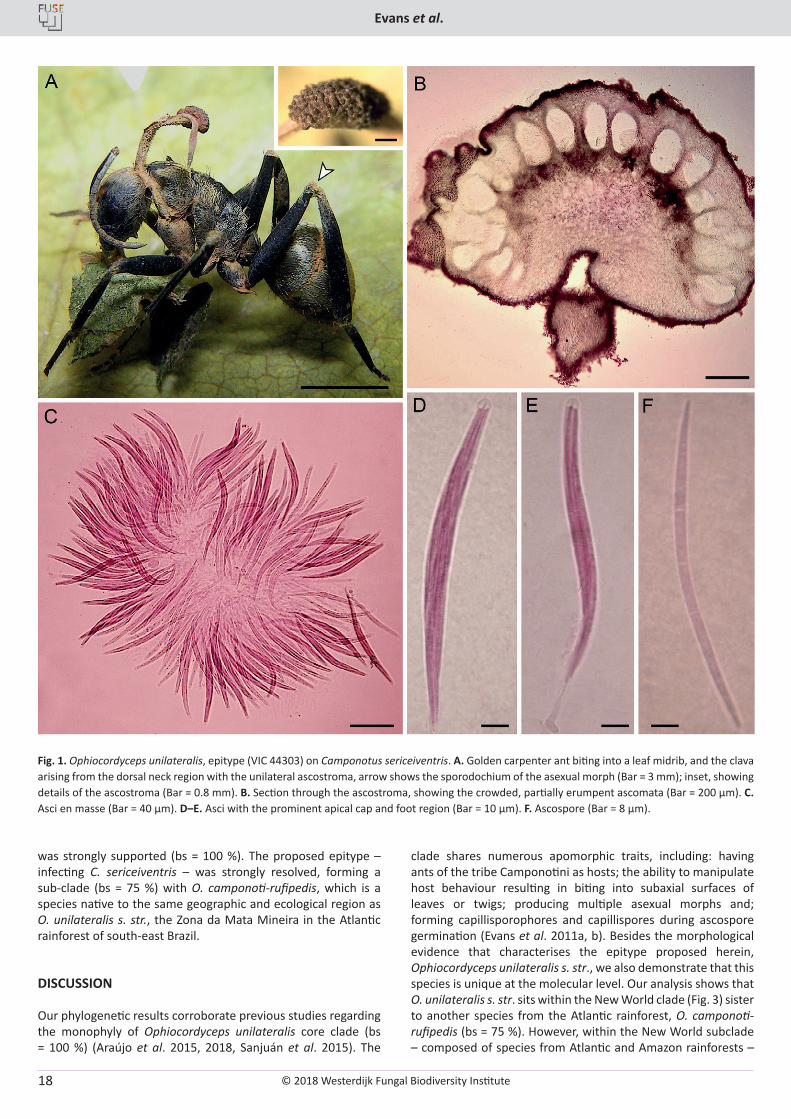
© 2018 Westerdijk Fungal Biodiversity Institute
Evans et al.
Editor-in-ChiefProf. dr P.W. Crous, Westerdijk Fungal Biodiversity Institute, P.O. Box 85167, 3508 AD Utrecht, The Netherlands.E-mail:[email protected]
18
was strongly supported (bs = 100 %). The proposed epitype – infecting C. sericeiventris – was strongly resolved, forming a sub-clade (bs = 75 %) with O. camponoti-rufipedis, which is a species native to the same geographic and ecological region as O. unilateralis s. str., the Zona da Mata Mineira in the Atlantic rainforest of south-east Brazil.
DISCUSSION
Our phylogenetic results corroborate previous studies regarding the monophyly of Ophiocordyceps unilateralis core clade (bs = 100 %) (Araújo et al. 2015, 2018, Sanjuán et al. 2015). The
clade shares numerous apomorphic traits, including: having ants of the tribe Camponotini as hosts; the ability to manipulate host behaviour resulting in biting into subaxial surfaces of leaves or twigs; producing multiple asexual morphs and; forming capillisporophores and capillispores during ascospore germination (Evans et al. 2011a, b). Besides the morphological evidence that characterises the epitype proposed herein, Ophiocordyceps unilateralis s. str., we also demonstrate that this species is unique at the molecular level. Our analysis shows that O. unilateralis s. str. sits within the New World clade (Fig. 3) sister to another species from the Atlantic rainforest, O. camponoti-rufipedis (bs = 75 %). However, within the New World subclade – composed of species from Atlantic and Amazon rainforests –
Fig. 1. Ophiocordyceps unilateralis, epitype (VIC 44303) on Camponotus sericeiventris. A. Golden carpenter ant biting into a leaf midrib, and the clava arising from the dorsal neck region with the unilateral ascostroma, arrow shows the sporodochium of the asexual morph (Bar = 3 mm); inset, showing details of the ascostroma (Bar = 0.8 mm). B. Section through the ascostroma, showing the crowded, partially erumpent ascomata (Bar = 200 µm). C. Asci en masse (Bar = 40 µm). D–E. Asci with the prominent apical cap and foot region (Bar = 10 µm). F. Ascospore (Bar = 8 µm).
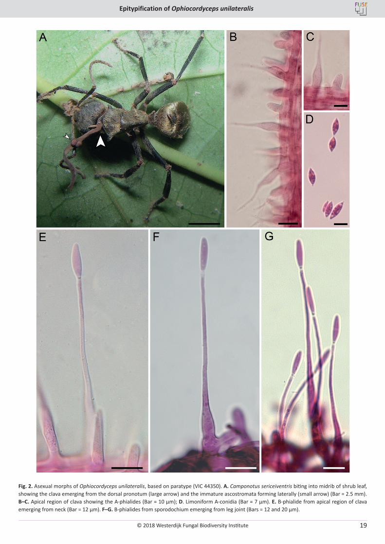
© 2018 Westerdijk Fungal Biodiversity Institute
Epitypification of Ophiocordyceps unilateralis
Editor-in-ChiefProf. dr P.W. Crous, Westerdijk Fungal Biodiversity Institute, P.O. Box 85167, 3508 AD Utrecht, The Netherlands.E-mail:[email protected]
19
Fig. 2. Asexual morphs of Ophiocordyceps unilateralis, based on paratype (VIC 44350). A. Camponotus sericeiventris biting into midrib of shrub leaf, showing the clava emerging from the dorsal pronotum (large arrow) and the immature ascostromata forming laterally (small arrow) (Bar = 2.5 mm). B–C. Apical region of clava showing the A-phialides (Bar = 10 µm); D. Limoniform A-conidia (Bar = 7 µm). E. B-phialide from apical region of clava emerging from neck (Bar = 12 µm). F–G. B-phialides from sporodochium emerging from leg joint (Bars = 12 and 20 µm).
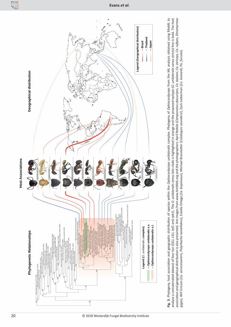
© 2018 Westerdijk Fungal Biodiversity Institute
Evans et al.
Editor-in-ChiefProf. dr P.W. Crous, Westerdijk Fungal Biodiversity Institute, P.O. Box 85167, 3508 AD Utrecht, The Netherlands.E-mail:[email protected]
20
Toly
pocl
adiu
m ja
poni
cum
OSC
110
991
Toly
pocl
adiu
m o
phio
glos
soid
es O
SC 1
0640
5 To
lypo
clad
ium
cap
itatu
m O
SC 7
1233
O
phio
cord
ycep
s va
riabi
lis O
SC 1
1100
3O
phio
cord
ycep
s he
tero
poda
EFC
C 1
0125
Oph
ioco
rdyc
eps
amaz
onic
a H
UA
1861
43
Oph
ioco
rdyc
eps
grac
ilis E
FCC
310
1O
phio
cord
ycep
s bl
atta
rioid
es H
UA1
8609
3 O
phio
cord
ycep
s ne
ovol
kian
a O
SC 1
5190
3O
phio
cord
ycep
s m
elol
onth
ae O
SC 1
1099
3O
phio
cord
ycep
s pu
rpur
eost
rom
ata
TNS
F184
30
Oph
ioco
rdyc
eps
brun
neip
unct
ata
OSC
128
576
Oph
ioco
rdyc
eps
annu
lata
CEM
303
O
phio
cord
ycep
s ap
hodi
i AR
SEF
5498
Oph
ioco
rdyc
eps
arar
acua
rens
is H
UA
1861
35
Oph
ioco
rdyc
eps
yaku
sim
ensi
s H
MAS
_199
604
Oph
ioco
rdyc
eps
long
issi
ma
HM
AS_1
9960
0O
phio
cord
ycep
s so
bolif
era
TNS
F185
21
Oph
ioco
rdyc
eps
acic
ular
is O
SC 1
2858
0O
phio
cord
ycep
s pr
uino
sa N
HJ
1299
4O
phio
cord
ycep
s st
ylop
hora
OSC
111
000
Oph
ioco
rdyc
eps
agrio
tidis
AR
SEF
5692
Oph
ioco
rdyc
eps
com
mun
is N
HJ
1258
2O
phio
cord
ycep
s co
mm
unis
NH
J 12
581
Oph
ioco
rdyc
eps
rhiz
oide
a N
HJ
1252
2 O
phio
cord
ycep
s el
onga
ta O
SC 1
1098
9O
phio
cord
ycep
s tip
utin
i QC
NE
1862
87
Oph
ioco
rdyc
eps
pone
rinar
um H
UA
1861
40
Oph
ioco
rdyc
eps
knip
hofio
ides
HU
A 18
6148
O
phio
cord
ycep
s pu
lvin
ata
TNS-
F 30
044
Oph
ioco
rdyc
eps
cam
pono
ti-ba
lzan
i G10
4O
phio
cord
ycep
s ca
mpo
noti-
balz
ani G
143
Oph
ioco
rdyc
eps
cam
pono
ti-at
ricip
is A
TRI3
Oph
ioco
rdyc
eps
unila
tera
lis s
ensu
stri
cto
VIC
443
03O
phio
cord
ycep
s un
ilate
ralis
sen
su s
trict
o VI
C 4
4354
Oph
ioco
rdyc
eps
cam
pono
ti-ru
fiped
is G
155
Oph
ioco
rdyc
eps
cam
pono
ti-ru
fiped
is G
177
Oph
ioco
rdyc
eps
cam
pono
ti-ru
fiped
is G
108
Oph
ioco
rdyc
eps
cam
pono
ti-bi
spin
osi O
BIS
Oph
ioco
rdyc
eps
cam
pono
ti-bi
spin
osi B
ISPI
2O
phio
cord
ycep
s ca
mpo
noti-
bisp
inos
i OBI
S4O
phio
cord
ycep
s ca
mpo
noti-
bisp
inos
i OBI
S3O
phio
cord
ycep
s ca
mpo
noti-
saun
ders
i C19
Oph
ioco
rdyc
eps
cam
pono
ti-sa
unde
rsi C
40O
phio
cord
ycep
s ha
laba
laen
sis
MY1
308
Oph
ioco
rdyc
eps
haab
alae
nsis
MY5
151
Oph
ioco
rdyc
eps
cam
pono
ti-le
onar
di C
36O
phio
cord
ycep
s ca
mpo
noti-
leon
ardi
TL1
Oph
ioco
rdyc
eps
sato
i J14
Oph
ioco
rdyc
eps
sato
i J13
Oph
ioco
rdyc
eps
poly
rhac
his-
furc
ata
P39
Oph
ioco
rdyc
eps
poly
rhac
his-
furc
ata
P51
Oph
ioco
rdyc
eps
sept
a* A
86-2
-001
+ C
41
Oph
ioco
rdyc
eps
ram
i* M
Y673
6O
phio
cord
ycep
s di
pter
igen
a O
SC 1
5191
1O
phio
cord
ycep
s sp
heco
ceph
ala
OSC
110
998
Oph
ioco
rdyc
eps
myr
mec
ophi
la C
EM17
10O
phio
cord
ycep
s ira
ngie
nsis
OSC
128
578
Oph
ioco
rdyc
eps
fulg
orom
orph
ila H
UA
1861
39
Oph
ioco
rdyc
eps
nuta
ns O
SC 1
1099
4O
phio
cord
ycep
s fo
rmic
arum
TN
S F1
8565
Oph
ioco
rdyc
eps
evan
sii H
UA
1861
59
Oph
ioco
rdyc
eps
curc
ulio
num
OSC
151
910
Oph
ioco
rdyc
eps
aust
ralis
926
Stilb
ella
buq
uetii
HU
A 18
6147
O
phio
cord
ycep
s llo
ydii
OSC
151
913
100
100
91
7194
99
100
9490
100
10010
010
0
7598
100
100
100
81
100
70
100 10
010
0
7584 84
100
95
100 10
0
89 100
73
97
100
100
8372
96
Cam
pono
tus
obsc
urip
es
Cam
pono
tus
balz
ani
Cam
pono
tus
atric
eps
Cam
pono
tus
seric
eive
ntris
Cam
pono
tus
rufip
es
Cam
pono
tus
bisp
inos
us
Col
obop
sis
saun
ders
i
Col
obop
sis
leon
ardi
Poly
rhac
his
lam
ellid
ens
Poly
rhac
his
furc
ata
Phyl
ogen
etic
Rel
atio
nshi
ps
Hos
t Ass
ocia
tions
Geo
grap
hica
l dis
trib
utio
n
– Ja
pan
– B
razi
l–
Thai
land
Lege
nd (G
eogr
aphi
cal d
istr
ibut
ion)
Lege
nd (O
. uni
late
ralis
com
plex
)
– O
phio
cord
ycep
s un
ilate
ralis
s.s
.–
Oph
ioco
rdyc
eps
unila
tera
lis s
.l.
Din
omyr
mex
gig
as
Fig.
3. P
hylo
geny
, hos
t as
soci
ation
and
geo
grap
hic
dist
ributi
on o
f spe
cies
with
in t
he O
phio
cord
ycep
s un
ilate
ralis
com
plex
. Phy
loge
ny o
f O
phio
cord
ycep
s fr
om t
he M
L an
alys
is ob
tain
ed u
sing
RAxM
L to
an
alys
e a
conc
aten
ated
dat
aset
of f
our l
oci (
SSU,
LSU
, tef
1 an
d rp
b1).
The
O. u
nila
tera
lis se
nsu
lato
cla
de is
hig
hlig
hted
in o
rang
e an
d th
e pr
opos
ed e
pity
pe (O
. uni
late
ralis
sens
u st
ricto
) lin
e is
bold
. The
hos
t as
soci
ation
and
geo
grap
hica
l dist
ributi
on is
also
pre
sent
ed. A
nt im
ages
from
ww
w.A
ntW
eb.o
rg a
nd th
e ph
otog
raph
ers:
Apr
il N
obile
(Cam
pono
tus o
bscu
ripes
, Ca.
bal
zani
, Ca.
atr
icep
s, C
a. ru
fipes
, Din
omyr
mex
gi
gas)
, Will
Eric
son
(Ca.
seriv
ceiv
entr
is, P
olyr
hach
is la
mel
liden
s), E
stel
la O
rteg
a (C
a. b
ispin
osus
), M
icha
el B
rans
tette
r (Co
lobo
psis
saun
ders
i), Z
ach
Lieb
erm
an (C
o. le
onar
di, P
o. fu
rcat
a).
© 2018 Westerdijk Fungal Biodiversity Institute
Epitypification of Ophiocordyceps unilateralis
Editor-in-ChiefProf. dr P.W. Crous, Westerdijk Fungal Biodiversity Institute, P.O. Box 85167, 3508 AD Utrecht, The Netherlands.E-mail:[email protected]
21
there is no clustering of species according to the region. Further studies, including more species from different continents, are helping to resolve the relationships within this clade (Araújo et al. 2018).
With the selection and re-description of the epitype of Ophiocordyceps unilateralis, it is now possible to construct a more meaningful phylogenetic tree for the O. unilateralis clade. Previously, trees were constructed using a sequence of the fungus from an unidentified ant in the herbarium of the Oregon State University (OSC 128574) (Sung et al. 2007, Kepler et al. 2010, Araújo et al. 2015, Kobmoo et al. 2015). This will be critical as more new species are identified within the O. unilateralis complex and we begin to understand more about the intricacies of the pathogen-host relationship. None more so than within the type of O. unilateralis on Camponotus sericeiventris, in which the evidence from Honduran collections suggests that different subspecies of the ant occur within the same forest habitat and that this is reflected in different death positions of the infected ants, as well as in morphological variation within the fungal pathogen. In order to coexist, the ant subspecies must occupy different niches within this ecosystem and, therefore, the fungus may also have evolved at the subspecies level with different morphological (spore forms) and physiological (neurotoxins) traits to maximize infection.
ACKNOWLEDGEMENTS
HCE and JPMA acknowledge financial support from the Brazilian Conselho Nacional de Desenvolvimento Cientifico e Tecnológia (CNPq) and the Coordenação de Aperfeiçoamento de Pessoal de Nível Superior (CAPES). We would like to thank the anonymous reviewers for their comments and recommendations.
REFERENCES
Andersen SB, Gerritsma S, Yusah KM, et al. (2009). The life of a dead ant: the expression of an adaptive extended phenotype. American Naturalist 174: 424–433.
Araújo JPM, Evans HC, Geiser DM, et al. (2015). Unravelling the diversity behind the Ophiocordyceps unilateralis (Ophiocordycipitaceae) complex: three new species of zombie-ant fungi from the Brazilian Amazon. Phytotaxa 220: 224–238.
Araújo JPM, Kepler R, Evans HC, et al. (2018). Zombie-ant fungus across continents: 14 new species and new combinations within Ophiocordyceps. I. Myrmecophilous hirsutelloid species. Studies in Mycology 90: in press.
Ariyawansa HA, Hawksworth DL, Hyde KD, et al. (2014). Epitypification and neotypification: guidelines with appropriate and inappropriate examples. Fungal Diversity 69: 57–91.
Berenbaum MR, Eisner T (2008). Bugs’ bugs. Science 322: 52–53.Boddy L (2014). Soils of war. New Scientist 2999: 43–45.Bolton B, Alpert G, Ward PS, et al. (2007). Bolton’s Catalogue of Ants of
the World. Harvard University Press, Cambridge, MA.Castlebury LA, Rossman AY, Sung G-H, et al. (2004). Multigene phylogeny
reveals new lineage for Stachybotrys chartarum, the indoor air fungus. Mycological Research 108: 864–872.
Cooke MC (1892). Vegetable Wasps and Plant Worms. Society for Promoting Christian Knowledge, London.
Costandi M (2012). Zombie-ant parasitic fungus kept in check by hyperparasitic fungus. The Guardian (Neurophilosophy),
04/05/2012. Dawkins R (1982). The extended phenotype. Oxford University Press,
Oxford.de Bekker C, Quevillon LE, Smith PB, et al. (2014). Species-specific
ant brain manipulation by a specialized fungal parasite. BMC Evolutionary Biology 14: 166.
de Bekker C, Ohm RA, Loreto RG, et al. (2015). Gene expression during zombie ant biting behavior reflects the complexity underlying fungal parasitic behavioral manipulation. BMC Genomics 16: 620.
de Bekker C, Ohm RA, Evans HC, et al. (2017). Ant-infecting Ophiocordyceps genomes reveal a high diversity of potential behavioral manipulation genes and a possible major role for enterotoxins. Scientific Reports (Nature) 7: 12508.
Edgar RC (2004). MUSCLE: multiple sequence alignment with high accuracy and high throughput. Nucleic Acids Research 32: 1792–1797.
Evans HC (1974). Natural control of arthropods, with special reference to ants (Formicidae), by fungi in the tropical high forest of Ghana. Journal of Applied Ecology 11: 37–49.
Evans HC (1982). Entomogenous fungi in tropical forest ecosystems: an appraisal. Ecological Entomology 7: 47–60.
Evans HC (2001). Entomopathogenic fungi associated with ants (Formicidae): A review. In: Trichomycetes and other fungal groups (Misra JK, Horn BW, eds). Science Publishers, Enfield, USA: 119–144.
Evans HC, Samson RA (1984). Cordyceps species and their anamorphs pathogenic on ants (Formicidae) in tropical forest ecosystems. II. The Camponotus (Formicinae) complex. Transactions of the British Mycological Society 82: 127–150.
Evans HC, Elliot SL, Hughes DP (2011a). Hidden diversity behind the zombie-ant fungus Ophiocordyceps unilateralis: four new species described from carpenter ants in Minas Gerais, Brazil. PLoS ONE 6: e17024.
Evans HC, Elliot SL, Hughes DP (2011b). Ophiocordyceps unilateralis: a keystone species for unraveling ecosystem functioning and biodiversity of fungi? Communicative & Integrative Biology 4: 5.
Gomez-Polo P, Ballinger MJ, Lalzar M, et al. (2017). An exceptional family: Ophiocordyceps-allied fungus dominates the microbiome of soft scale insects (Hemiptera: Sternorrhyncha: Coccidae). Molecular Ecology 26: 5855–5868.
Guérin-Méneville FE (1838). Première division. Crustacés, arachnides et insectes. In: Voyage Autour du Monde, executé par ordre du Roi, sur la corvette de sa Majesté, La Coquille, pendant les années (Duperrey LI, ed), Zoologie. Tome Deuxième, Part 2. H. Bertrand, Paris. 1822, 1823, 1824 et 1825: 9–320.
Hughes DP (2013). Pathways to understanding the extended phenotype of parasites in their hosts. Journal of Experimental Biology 216: 142–147.
Hughes DP, Wappler T, Labandeira C (2011). Ancient death-grip leaf scars reveal ant-fungal parasitism. Biology Letters 7: 67–70.
Hughes DP, Araújo JPM, Loreto RG, et al. (2016). From so simple a beginning: the evolution of behavioral manipulation by fungi. Advances in Genetics 94: 437–469.
Kaplan M (2011). Befriending the body snatchers. New Scientist 2827: 37–41.
Kearse M, Moir R, Wilson A, et al. (2012). Geneious Basic: an integrated and extendable desktop software platform for the organization and analysis of sequence data. Bioinformatics 28: 1647–1649.
Kepler RM, Kaitsu Y, Tanaka E, et al. (2010). Ophiocordyceps pulvinata sp. nov., a pathogen of ants with a reduced stroma. Mycoscience 52: 39–47.
Kobayasi Y (1941). The genus Cordyceps and its allies. Science Reports, Tokyo Bunrika Daigaku, Sect. B 5: 53–260.
© 2018 Westerdijk Fungal Biodiversity Institute
Evans et al.
Editor-in-ChiefProf. dr P.W. Crous, Westerdijk Fungal Biodiversity Institute, P.O. Box 85167, 3508 AD Utrecht, The Netherlands.E-mail:[email protected]
22
Kobmoo N, Mongkolsamrit S, Tasanathai K, et al. (2012). Molecular phylogenies reveal host-specific divergence of Ophiocordyceps unilateralis sensu lato following its host ants. Molecular Ecology 21: 3022–3031.
Kobmoo N, Mongkolsamrit S, Wutikhun T, et al. (2015). New species of Ophiocordyceps unilateralis, an ubiquitous pathogen of ants from Thailand. Fungal Biology 119: 44–52.
Luangsa-ard JJ, Ridkaew R, Tasanathai K, et al. (2011). Ophiocordyceps halabalaensis: a new species of Ophiocordyceps pathogenic to Camponotus gigas in Hala Bala Wildlife Sanctuary, Southern Thailand. Fungal Biology 115: 608–614.
Mains EB (1951). Entomogenous species of Hirsutella, Tilachlidium and Synnematium. Mycologia 43: 691–718.
Mains EB (1958). North American entomogenous species of Cordyceps. Mycologia 50: 169–222.
Massee G (1895). A revision of the genus Cordyceps. Annals of Botany 9: 1–44.
Molnár I, Gibson DM, Krasnoff SB (2010). Secondary metabolites from entomopathogenic Hypocrealean fungi. Natural Products Reports 27: 1233–1372.
Nishino T, Tanahashi M, Lin C-P, et al. (2016). Fungal and bacterial endosymbionts of eared leafhoppers of the subfamily Ledrinae (Hemiptera: Cicadellidae). Applied Entomology & Zoology 51: 465–477.
Paterson RRM (2008). Cordyceps – a traditional Chinese medicine and another fungal therapeutic biofactory? Phytochemistry 69: 1469–1495.
Pennisi E (2014). Parasitic puppeteers begin to yield their secrets. Science 343: 239.
Petch T (1924). Studies in entomogenous fungi. IV. Some Ceylon Cordyceps. Transactions of the British Mycological Society 10: 28–45.
Petch T (1931). Notes on entomogenous fungi. Transactions of the British Mycological Society 16: 55–75.
Petch T (1933). Notes on entomogenous fungi. Transactions of the British Mycological Society 18: 48–75.
Petch T (1935). Notes on entomogenous fungi. Transactions of the British Mycological Society 19: 161–194.
Petch T (1937). Notes on entomogenous fungi. Transactions of the British Mycological Society 21: 34–67.
Quandt CA, Kepler RM, Gams W, et al. (2014). Phylogenetic-based nomenclatural proposals for Ophiocordycipitaceae (Hypocreales) with new combinations in Tolypocladium. IMA Fungus 1: 121–134.
Rehner SA, Buckley E (2005). A Beauveria phylogeny inferred from nuclear ITS and EF1-α sequences: evidence for cryptic diversification and links to Cordyceps teleomorphs. Mycologia 97: 84–98.
Rombach MC, Roberts DW (1989). Hirsutella species (Deuteromycotina; Hyphomycetes) on Philippine insects. Philippine Entomologist 7: 491–518.
Samson RA, Evans HC, Hoekstra ES (1982). Notes on entomogenous fungi from Ghana. VI. The genus Cordyceps. Proceedings of the Koninklijke Nederlandse Akademie van Wetenschappen, Series C 85: 589–605.
Sanjuán TI, Franco-Molano AE, Kepler RM, et al. (2015). Five new species of entomopathogenic fungi from the Amazon and evolution of neotropical Ophiocordyceps. Fungal Biology 119: 901–916.
Stamatakis A (2006). RAxML-VI-HPC: maximum likelihood-based phylogenetic analyses with thousands of taxa and mixed models. Bioinformatics 22: 2688–2690.
Sung G-H, Hywel-Jones NL, Sung J-M, et al. (2007). Phylogenetic classification of Cordyceps and the clavicipitaceous fungi. Studies in Mycology 57: 1–63.
Tulasne LR, Tulasne C (1865). Selecta Fungorum Carpologia III. Imperial Press, Paris.
Vilgalys R, Hester M (1990). Rapid Genetic Identification and Mapping of Enzymatically Amplified Ribosomal DNA from Several Cryptococcus Species. Journal of Bacteriology 172: 4238–4246.
White TJ, Bruns T, Lee S, et al. (1990). Amplification and direct sequencing of fungal ribosomal RNA genes for phylogenetics. In: PCR Protocols: A Guide to Methods and Applications (Innis MA, Gelfand DH, Sninsky JJ, et al., eds). Academic Press, New York: 315–322.
Zhang Y, Li E, Wang C, et al. (2012). Ophiocordyceps sinensis, the flagship fungus of China: terminology, life strategy and ecology. Mycology 3: 2–10.
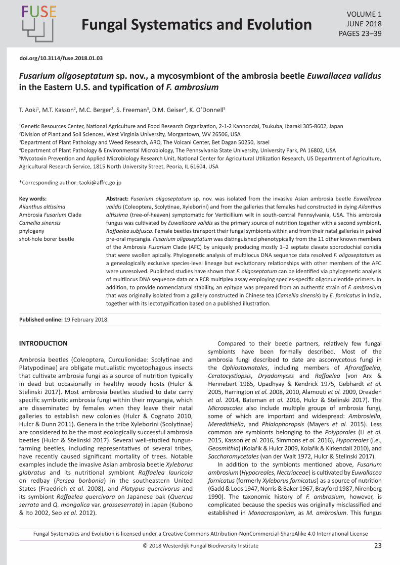
Fungal Systematics and Evolution is licensed under a Creative Commons Attribution-NonCommercial-ShareAlike 4.0 International License
© 2018 Westerdijk Fungal Biodiversity Institute 23
Editor-in-ChiefProf. dr P.W. Crous, Westerdijk Fungal Biodiversity Institute, P.O. Box 85167, 3508 AD Utrecht, The Netherlands.E-mail:[email protected]
Fungal Systematics and EvolutionVOLUME 1JUNE 2018
PAGES 23–39
doi.org/10.3114/fuse.2018.01.03
Fusarium oligoseptatum sp. nov., a mycosymbiont of the ambrosia beetle Euwallacea validus in the Eastern U.S. and typification of F. ambrosium
T. Aoki1, M.T. Kasson2, M.C. Berger2, S. Freeman3, D.M. Geiser4, K. O’Donnell5
1Genetic Resources Center, National Agriculture and Food Research Organization, 2-1-2 Kannondai, Tsukuba, Ibaraki 305-8602, Japan2Division of Plant and Soil Sciences, West Virginia University, Morgantown, WV 26506, USA3Department of Plant Pathology and Weed Research, ARO, The Volcani Center, Bet Dagan 50250, Israel4Department of Plant Pathology & Environmental Microbiology, The Pennsylvania State University, University Park, PA 16802, USA5Mycotoxin Prevention and Applied Microbiology Research Unit, National Center for Agricultural Utilization Research, US Department of Agriculture, Agricultural Research Service, 1815 North University Street, Peoria, IL 61604, USA
*Corresponding author: [email protected]
Abstract: Fusarium oligoseptatum sp. nov. was isolated from the invasive Asian ambrosia beetle Euwallacea validis (Coleoptera, Scolytinae, Xyleborini) and from the galleries that females had constructed in dying Ailanthus altissima (tree-of-heaven) symptomatic for Verticillium wilt in south-central Pennsylvania, USA. This ambrosia fungus was cultivated by Euwallacea validis as the primary source of nutrition together with a second symbiont, Raffaelea subfusca. Female beetles transport their fungal symbionts within and from their natal galleries in paired pre-oral mycangia. Fusarium oligoseptatum was distinguished phenotypically from the 11 other known members of the Ambrosia Fusarium Clade (AFC) by uniquely producing mostly 1–2 septate clavate sporodochial conidia that were swollen apically. Phylogenetic analysis of multilocus DNA sequence data resolved F. oligoseptatum as a genealogically exclusive species-level lineage but evolutionary relationships with other members of the AFC were unresolved. Published studies have shown that F. oligoseptatum can be identified via phylogenetic analysis of multilocus DNA sequence data or a PCR multiplex assay employing species-specific oligonucleotide primers. In addition, to provide nomenclatural stability, an epitype was prepared from an authentic strain of F. ambrosium that was originally isolated from a gallery constructed in Chinese tea (Camellia sinensis) by E. fornicatus in India, together with its lectotypification based on a published illustration.
Key words: Ailanthus altissimaAmbrosia Fusarium CladeCamellia sinensisphylogenyshot-hole borer beetle
Published online: 19 February 2018.
INTRODUCTION
Ambrosia beetles (Coleoptera, Curculionidae: Scolytinae and Platypodinae) are obligate mutualistic mycetophagous insects that cultivate ambrosia fungi as a source of nutrition typically in dead but occasionally in healthy woody hosts (Hulcr & Stelinski 2017). Most ambrosia beetles studied to date carry specific symbiotic ambrosia fungi within their mycangia, which are disseminated by females when they leave their natal galleries to establish new colonies (Hulcr & Cognato 2010, Hulcr & Dunn 2011). Genera in the tribe Xyleborini (Scolytinae) are considered to be the most ecologically successful ambrosia beetles (Hulcr & Stelinski 2017). Several well-studied fungus-farming beetles, including representatives of several tribes, have recently caused significant mortality of trees. Notable examples include the invasive Asian ambrosia beetle Xyleborus glabratus and its nutritional symbiont Raffaelea lauricola on redbay (Persea borbonia) in the southeastern United States (Fraedrich et al. 2008), and Platypus quercivorus and its symbiont Raffaelea quercivora on Japanese oak (Quercus serrata and Q. mongolica var. grosseserrata) in Japan (Kubono & Ito 2002, Seo et al. 2012).
Compared to their beetle partners, relatively few fungal symbionts have been formally described. Most of the ambrosia fungi described to date are ascomycetous fungi in the Ophiostomatales, including members of Afroraffaelea, Ceratocystiopsis, Dryadomyces and Raffaelea (von Arx & Hennebert 1965, Upadhyay & Kendrick 1975, Gebhardt et al. 2005, Harrington et al. 2008, 2010, Alamouti et al. 2009, Dreaden et al. 2014, Bateman et al. 2016, Hulcr & Stelinski 2017). The Microascales also include multiple groups of ambrosia fungi, some of which are important and widespread: Ambrosiella, Meredithiella, and Phialophoropsis (Mayers et al. 2015). Less common are symbionts belonging to the Polyporales (Li et al. 2015, Kasson et al. 2016, Simmons et al. 2016), Hypocreales (i.e., Geosmithia) (Kolařik & Hulcr 2009, Kolařik & Kirkendall 2010), and Saccharomycetales (van der Walt 1972, Hulcr & Stelinski 2017).
In addition to the symbionts mentioned above, Fusarium ambrosium (Hypocreales, Nectriaceae) is cultivated by Euwallacea fornicatus (formerly Xyleborus fornicatus) as a source of nutrition (Gadd & Loos 1947, Norris & Baker 1967, Brayford 1987, Nirenberg 1990). The taxonomic history of F. ambrosium, however, is complicated because the species was originally misclassified and established in Monacrosporium, as M. ambrosium. This fungus
© 2018 Westerdijk Fungal Biodiversity Institute
Aoki et al.
Editor-in-ChiefProf. dr P.W. Crous, Westerdijk Fungal Biodiversity Institute, P.O. Box 85167, 3508 AD Utrecht, The Netherlands.E-mail:[email protected]
24
was isolated originally and described from galleries of the tea shot-hole borer, E. fornicatus, in Camellia sinensis (Chinese tea) and Ricinus communis (caster-oil tree) stems in Sri Lanka (Gadd & Loos 1947). Subsequently, F. bugnicourtii was described based on collections from galleries in Chinese tea in India, borer-damaged Hevea brasiliensis (rubber tree) and Theoborma cacao (cacao) in Sabah, Malaysia (Brayford 1987). Nirenberg (1990) synonymized F. bugnicourtii with M. ambrosium and recombined the latter as F. ambrosium based on nomenclatural priority. Brayford (1987) considered F. bugnicourtii to be conspecific with F. tumidum var. coeruleum (Bugnicourt 1939), but distinct from F. tumidum. Although the type of F. tumidum var. coeruleum based on a collection from H. brasiliensis appears to be phylogenetically distinct from F. bugnicourtii, the holotype of F. bugnicourtii selected by Brayford (IMI 296597 = NRRL 20438) is conspecific with F. ambrosium (Kasson et al. 2013).
Kasson et al. (2013) conducted an extensive multilocus molecular phylogenetic study on the ambrosial fusaria, based on isolates from beetles, their galleries, or from trees showing extensive borer damage and dieback. These included Camellia sinensis, Persea americana (avocado), Ailanthus altissima (tree-of-heaven), Acer negundo (box elder), and Hevea brasiliensis from natural and cultivated ecosystems, and avocado in the United States, Israel and Australia. Seven different Fusarium species lineages were reported to be associated with Euwallacea ambrosia beetles within the Ambrosia Fusarium Clade (AFC) and one other species (i.e., Fusarium sp. AF-9) with Xyleborus ferrugineus in Costa Rica. The monophyletic AFC is nested within Clade 3 of the F. solani species complex (FSSC; O’Donnell 2000), which contains 60 plus phylogenetic species based on genealogical concordance phylogenetic species recognition (GCPSR; Taylor et al. 2000). The AFC comprises two strongly supported clades: the four species within Clade A typically produce curved fusiform septate macroconidia, which are typical of Fusarium, whereas nine of the 10 species within Clade B produce clavate macroconidia (Kasson et al. 2013, Aoki et al. unpubl.), described as ‘dolphin-shaped’ by Brayford (1987). O’Donnell et al. (2015) conducted a multilocus phylogenetic analysis of the AFC and Euwallacea and found evidence of repeated host shifts rather than strict co-evolution of this mutualism.
Freeman et al. (2013) described a new species, F. euwallaceae, based on isolates corresponding to the ambrosia species symbiotic with the Euwallacea sp. #1 sensu O’Donnell et al. (2015), which causes serious damage to avocado production in Israel and California, USA (Mendel et al. 2012, Eskalen et al. 2013). Fusarium euwallaceae is closely related morphologically to F. ambrosium, but it can be distinguished from the latter by the abundant production of bluish to brownish sporodochial conidia that form greenish masses on PDA after 1 mo in culture, together with hyaline conidia. To date only three of the 16 species within the AFC have been described formally (Kasson et al. 2013, O’Donnell et al. 2015, Na et al. 2018). Similar to E. validus, Euwallacea sp. #1 also carries additional symbiotic fungi, Graphium euwallaceae and Paracremonium pembeum (Freeman et al. 2016, Lynch et al. 2015). Recently, PCR multiplexes were developed to discriminate Fusarium symbionts of invasive Euwallacea ambrosia beetles that inflict damage on numerous tree species throughout the United States, including F. euwallaceae and F. kuroshium along with four unnamed AFC species-level lineages: AF-3, AF-4, AF-6 and AF-8 (Short et al. 2017). One of the undescribed species, which was informally referred to as Fusarium sp. AF-4, is cultivated by the ambrosia
beetle E. validus primarily in Verticillium wilt-stressed and dying stands of A. altissima, as well as from Verticillium wilt-stressed Acer pensylvanicum (striped maple), Aralia spinosa (devils walkingstick) and Rhus typhina (staghorn sumac) in south-central Pennsylvania, USA (Schall & Davis 2009, Kasson et al. 2013, 2015). In the present study, this species is described as F. oligoseptatum sp. nov. based on a comparison with F. ambrosium (AF-1) and F. euwallaceae (AF-2) (Kasson et al. 2013, Freeman et al. 2013). In addition, because type material for F. ambrosium was not designated (Gadd & Loos 1947), or appears to have been lost (Nirenberg 1990), a line-drawing of a clavate conidium of the species from Gadd & Loos (1947) was selected as the lectotype. Furthermore, an epitype was prepared from an authentic strain of this species to stabilize its taxonomy, according to the International Code of Nomenclature for algae, fungi and plants (ICN, the Melbourne Code; McNeill et al. 2012).
MATERIALS AND METHODS
Fungal isolates and type specimens
Fusarium strains examined in this study (Table 1) are stored in the Agriculture Research Service Culture Collection (NRRL), National Center for Agricultural Utilization Research (NCAUR), U.S. Department of Agriculture in Peoria, Illinois, USA. These strains were originally isolated from Euwallacea ambrosia beetles and their galleries, or from host trees showing extensive borer damage (Kasson et al. 2013). The Pennsylvanian strains of F. oligoseptatum were isolated from E. validus ambrosia beetles that had colonized A. altissima. Beetles were surface disinfested for 15 s in 70 % ethanol and then washed three times in sterile deionized water. Whole beetles or their heads were macerated using sterile Tenbroek homogenizers (Pyrex, Corning, NY), or pellet pestles (Fisher Scientific, Hampton, NH), suspensions were diluted 1:10 and 1:100, and then spread evenly over half-strength Potato Dextrose Agar (PDA, BD-DifcoTM, Thermo Fisher Scientific, Waltham, MA) amended with 100 ppm streptomycin sulfate (Sigma-Aldrich, St. Louis, MO) as described in Kasson et al. (2013). Other related ambrosia fusaria or close relatives within the F. solani species complex (O’Donnell et al. 2008) were obtained from culture collections (Table 1). Isolates used in this study are available upon request from NRRL (http://nrrl.ncaur.usda.gov/cgi-bin/usda/), NARO Genebank, Microorganisms Section (MAFF), Genetic Resources Center, National Agriculture and Food Research Organization, Tsukuba, Ibaraki, Japan (http://www.gene.affrc.go.jp/about-micro_en.php), and the Westerdijk Institute (formerly CBS-KNAW Fungal Biodiversity Center), Utrecht, the Netherlands (http://www.westerdijkinstitute.nl/). Isolates of four novel Taiwanese AFC species discovered very recently, i.e. AF-13 to AF-16 (Na et al. 2018), were not included in this study.
Holotype and epitype specimens newly prepared from the selected strains were deposited in BPI, US National Fungus Collection (https://nt.ars-grin.gov/fungaldatabases/specimens/specimens.cfm).
Incidence of Fusarium oligoseptatum and other fungi from Euwallacea validus mycangia across recently confirmed tree hosts
Mycangial fungal communities were characterized as previously described by Kasson et al. (2013) for adult female
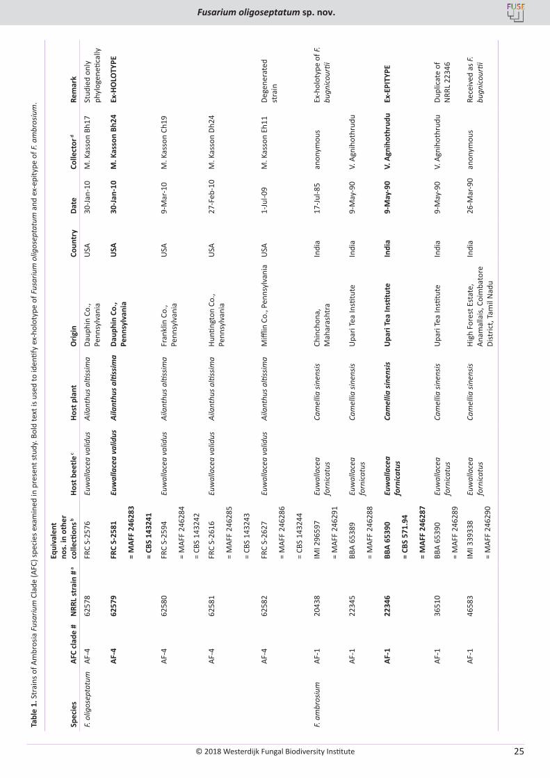
© 2018 Westerdijk Fungal Biodiversity Institute
Fusarium oligoseptatum sp. nov.
Editor-in-ChiefProf. dr P.W. Crous, Westerdijk Fungal Biodiversity Institute, P.O. Box 85167, 3508 AD Utrecht, The Netherlands.E-mail:[email protected]
25
Tabl
e 1.
Str
ains
of A
mbr
osia
Fus
ariu
m C
lade
(AFC
) spe
cies
exa
min
ed in
pre
sent
stud
y. B
old
text
is u
sed
to id
entif
y ex
-hol
otyp
e of
Fus
ariu
m o
ligos
epta
tum
and
ex-
epity
pe o
f F. a
mbr
osiu
m.
AFC
clad
e #
NRR
L st
rain
# a
Equi
vale
nt
nos.
in o
ther
co
llecti
ons b
Spec
ies
Host
bee
tle c
Host
pla
ntO
rigin
Coun
try
Date
Colle
ctor
dRe
mar
k
F. ol
igos
epta
tum
AF-4
6257
8FR
C S-
2576
Euw
alla
cea
valid
usAi
lant
hus a
ltiss
ima
Daup
hin
Co.,
Penn
sylv
ania
USA
30-Ja
n-10
M. K
asso
n Bh
17St
udie
d on
ly
phyl
ogen
etica
lly
AF-4
6257
9FR
C S-
2581
Euw
alla
cea
valid
usAi
lant
hus a
ltiss
ima
Daup
hin
Co.,
Penn
sylv
ania
USA
30-J
an-1
0M
. Kas
son
Bh24
Ex-H
OLO
TYPE
= M
AFF
2462
83
= CB
S 14
3241
AF-4
6258
0FR
C S-
2594
Euw
alla
cea
valid
usAi
lant
hus a
ltiss
ima
Fran
klin
Co.
, Pe
nnsy
lvan
iaU
SA9-
Mar
-10
M. K
asso
n Ch
19
= M
AFF
2462
84
= CB
S 14
3242
AF-4
6258
1FR
C S-
2616
Euw
alla
cea
valid
usAi
lant
hus a
ltiss
ima
Hunti
ngto
n Co
., Pe
nnsy
lvan
iaU
SA27
-Feb
-10
M. K
asso
n Dh
24
= M
AFF
2462
85
= CB
S 14
3243
AF-4
6258
2FR
C S-
2627
Euw
alla
cea
valid
usAi
lant
hus a
ltiss
ima
Miffl
in C
o., P
enns
ylva
nia
USA
1-Ju
l-09
M. K
asso
n Eh
11De
gene
rate
d st
rain
= M
AFF
2462
86
= CB
S 14
3244
F. am
bros
ium
AF-1
2043
8IM
I 296
597
Euw
alla
cea
forn
icat
usCa
mel
lia si
nens
isCh
inch
ona,
M
ahar
asht
raIn
dia
17-Ju
l-85
anon
ymou
sEx
-hol
otyp
e of
F.
bugn
icou
rtii
= M
AFF
2462
91
AF-1
2234
5BB
A 65
389
Euw
alla
cea
forn
icat
usCa
mel
lia si
nens
isU
pari
Tea
Insti
tute
Indi
a9-
May
-90
V. A
gnih
othr
udu
= M
AFF
2462
88
AF-1
2234
6BB
A 65
390
Euw
alla
cea
forn
icat
usCa
mel
lia si
nens
isU
pari
Tea
Insti
tute
Indi
a9-
May
-90
V. A
gnih
othr
udu
Ex-E
PITY
PE
= CB
S 57
1.94
= M
AFF
2462
87
AF-1
3651
0BB
A 65
390
Euw
alla
cea
forn
icat
usCa
mel
lia si
nens
isU
pari
Tea
Insti
tute
Indi
a9-
May
-90
V. A
gnih
othr
udu
Dupl
icat
e of
N
RRL
2234
6=
MAF
F 24
6289
AF-1
4658
3IM
I 339
338
Euw
alla
cea
forn
icat
usCa
mel
lia si
nens
isHi
gh F
ores
t Est
ate,
An
amal
lais,
Coi
mba
tore
Di
stric
t, Ta
mil
Nad
u
Indi
a26
-Mar
-90
anon
ymou
sRe
ceiv
ed a
s F.
bugn
icou
rtii
= M
AFF
2462
90
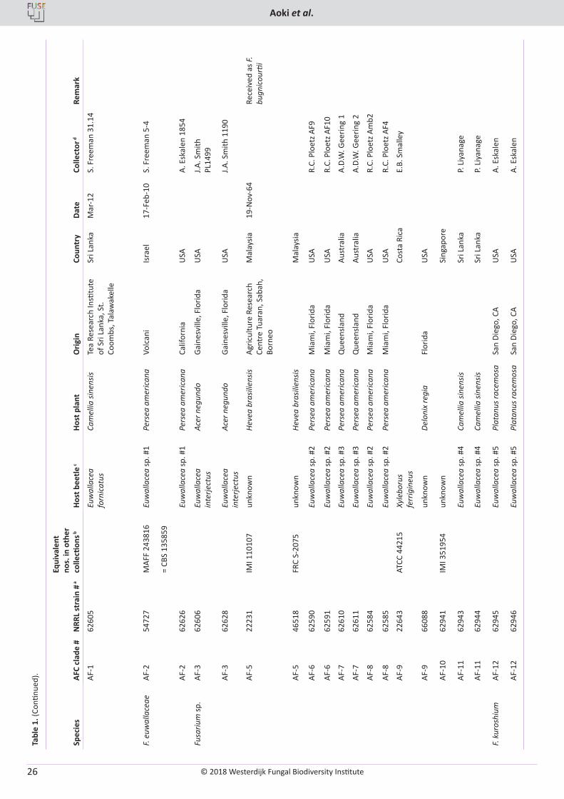
© 2018 Westerdijk Fungal Biodiversity Institute
Aoki et al.
Editor-in-ChiefProf. dr P.W. Crous, Westerdijk Fungal Biodiversity Institute, P.O. Box 85167, 3508 AD Utrecht, The Netherlands.E-mail:[email protected]
26
Tabl
e 1.
(Con
tinue
d). AFC
clad
e #
NRR
L st
rain
# a
Equi
vale
nt
nos.
in o
ther
co
llecti
ons b
Spec
ies
Host
bee
tle c
Host
pla
ntO
rigin
Coun
try
Date
Colle
ctor
dRe
mar
k
AF-1
6260
5Eu
wal
lace
a fo
rnic
atus
Cam
ellia
sine
nsis
Tea
Rese
arch
Insti
tute
of
Sri
Lank
a, S
t. Co
ombs
, Tal
awak
elle
Sri L
anka
Mar
-12
S. F
reem
an 3
1.14
F. eu
wal
lace
aeAF
-254
727
MAF
F 24
3816
Euw
alla
cea
sp. #
1Pe
rsea
am
eric
ana
Volc
ani
Isra
el17
-Feb
-10
S. F
reem
an 5
-4
= CB
S 13
5859
AF-2
6262
6Eu
wal
lace
a sp
. #1
Pers
ea a
mer
ican
aCa
lifor
nia
USA
A. E
skal
en 1
854
Fusa
rium
sp.
AF-3
6260
6Eu
wal
lace
a in
terje
ctus
Acer
neg
undo
Gain
esvi
lle, F
lorid
aU
SAJ.A
. Sm
ith
PL14
99
AF-3
6262
8Eu
wal
lace
a in
terje
ctus
Ac
er n
egun
doGa
ines
ville
, Flo
rida
USA
J.A. S
mith
119
0
AF-5
2223
1IM
I 110
107
unkn
own
Heve
a br
asili
ensis
Agric
ultu
re R
esea
rch
Cent
re T
uara
n, S
abah
, Bo
rneo
Mal
aysia
19-N
ov-6
4Re
ceiv
ed a
s F.
bugn
icou
rtii
AF-5
4651
8FR
C S-
2075
unkn
own
Heve
a br
asili
ensis
Mal
aysia
AF-6
6259
0Eu
wal
lace
a sp
. #2
Pers
ea a
mer
ican
aM
iam
i, Fl
orid
aU
SAR.
C. P
loet
z AF9
AF-6
6259
1Eu
wal
lace
a sp
. #2
Pers
ea a
mer
ican
aM
iam
i, Fl
orid
aU
SAR.
C. P
loet
z AF1
0
AF-7
6261
0Eu
wal
lace
a sp
. #3
Pers
ea a
mer
ican
aQ
ueen
sland
Aust
ralia
A.D.
W. G
eerin
g 1
AF-7
6261
1Eu
wal
lace
a sp
. #3
Pers
ea a
mer
ican
aQ
ueen
sland
Aust
ralia
A.D.
W. G
eerin
g 2
AF-8
6258
4Eu
wal
lace
a sp
. #2
Pers
ea a
mer
ican
aM
iam
i, Fl
orid
aU
SAR.
C. P
loet
z Am
b2
AF-8
6258
5Eu
wal
lace
a sp
. #2
Pers
ea a
mer
ican
aM
iam
i, Fl
orid
aU
SAR.
C. P
loet
z AF4
AF-9
2264
3AT
CC 4
4215
Xyle
boru
s fe
rrig
ineu
sCo
sta
Rica
E.B.
Sm
alle
y
AF-9
6608
8un
know
nDe
loni
x re
gia
Flor
ida
USA
AF-1
062
941
IMI 3
5195
4un
know
nSi
ngap
ore
AF-1
162
943
Euw
alla
cea
sp. #
4Ca
mel
lia si
nens
isSr
i Lan
kaP.
Liy
anag
e
AF-1
162
944
Euw
alla
cea
sp. #
4Ca
mel
lia si
nens
isSr
i Lan
kaP.
Liy
anag
e
F. ku
rosh
ium
AF-1
262
945
Euw
alla
cea
sp. #
5Pl
atan
us ra
cem
osa
San
Dieg
o, C
AU
SAA.
Esk
alen
AF-1
262
946
Euw
alla
cea
sp. #
5Pl
atan
us ra
cem
osa
San
Dieg
o, C
AU
SAA.
Esk
alen
© 2018 Westerdijk Fungal Biodiversity Institute
Fusarium oligoseptatum sp. nov.
Editor-in-ChiefProf. dr P.W. Crous, Westerdijk Fungal Biodiversity Institute, P.O. Box 85167, 3508 AD Utrecht, The Netherlands.E-mail:[email protected]
27
Tabl
e 1.
(Con
tinue
d). AFC
clad
e #
NRR
L st
rain
# a
Equi
vale
nt
nos.
in o
ther
co
llecti
ons b
Spec
ies
Host
bee
tle c
Host
pla
ntO
rigin
Coun
try
Date
Colle
ctor
dRe
mar
ka N
RRL:
ARS
Cul
ture
Col
lecti
on, N
CAU
R-AR
S-U
SDA,
Peo
ria, I
L, U
SA.
b ATC
C: A
mer
ican
Typ
e Cu
lture
Col
lecti
on, M
anas
sas,
VA,
USA
; BBA
: Bio
logi
sche
Bun
desa
nsta
lt fü
r Lan
d- u
nd F
orst
wirt
scha
ft, In
stitu
t für
Mik
robi
olog
ie (c
urre
ntly
Juliu
s-Kü
hn-In
stitu
t), B
erlin
, Ger
man
y;
CBS:
Wes
terd
ijk In
stitu
te (f
orm
erly
CBS
-KN
AW F
unga
l Bio
dive
rsity
Cen
ter)
, Utr
echt
, the
Net
herla
nds;
FRC
: Fus
ariu
m R
esea
rch
Cent
er, T
he P
enns
ylva
nia
Stat
e U
nive
rsity
, Sta
te C
olle
ge, P
A, U
SA; I
MI:
CABI
Bi
osci
ence
s, U
K Ce
ntre
, Egh
am, S
urre
y, U
K; M
AFF:
NAR
O G
eneb
ank,
Mic
roor
gani
sms S
ectio
n, G
eneti
c Re
sour
ces C
ente
r, N
ation
al A
gric
ultu
re a
nd F
ood
Rese
arch
Org
aniza
tion,
Tsu
kuba
, Iba
raki
, Jap
an.
c Th
e fiv
e un
nam
ed E
uwal
lace
a sp
p. a
re d
isting
uish
ed b
y #1
‒#5
(O’D
onne
ll et
al.
2015
). AF
-12
was
late
r des
crib
ed a
s F. k
uros
hium
(Na
et a
l. 20
18).
d V. A
gnih
othr
udu,
Upa
ri Te
a In
stitu
te, I
ndia
; A. E
skal
en, D
epar
tmen
t of P
lant
Pat
holo
gy a
nd M
icro
biol
ogy,
Uni
vers
ity o
f Cal
iforn
ia, R
iver
side,
CA,
USA
; S. F
reem
an, D
epar
tmen
t of P
lant
Pat
holo
gy a
nd
Wee
d Re
sear
ch, A
gric
ultu
ral R
esea
rch
Org
aniza
tion,
The
Vol
cani
Cen
ter,
Bet D
agan
, Isr
ael;
A. G
eerin
g, U
nive
rsity
of Q
ueen
sland
, Bris
bane
, Aus
tral
ia; M
. Kas
son,
Div
ision
of P
lant
and
Soi
l Sci
ence
s, W
est
Virg
inia
Uni
vers
ity, M
orga
ntow
n, W
V, U
SA; R
. C. P
loet
z, U
nive
rsity
of F
lorid
a, H
omes
tead
, FL,
USA
; J. A
. Sm
ith, U
nive
rsity
of F
lorid
a, G
aine
svill
e, F
L, U
SA.
beetles that harbor paired pre-oral mycangia. In addition to F. oligoseptatum, described in this paper, previous studies revealed that E. validus harbors a second less abundant symbiont, Raffaelea subfusca (Kasson et al. 2013). To determine if these trends held across other tree hosts and geographic locations, E. validus from Ailanthus and 16 other confirmed tree hosts across seven states were sampled. When available, a minimum of 10 adult females were included. Log-extracted females were processed as previously described (Kasson et al. 2013). Following serial dilution plating of head macerates, individual fungal colony forming units (CFUs) were quantified by morphotype and representatives of each morphotype retained for molecular characterization using the ITS barcoding gene. Unlike the other fungal morphotypes, representative fusaria were subjected to an AF-3 / Fusarium oligoseptatum (AF-4) multiplex PCR recently developed by Short et al. (2017) to discriminate known Fusarium symbionts of E. interjectus and E. validus in the eastern U.S.
When comparing the CFUs recovered from individual beetle heads between the primary symbionts of E. validus, F. oligoseptatum and R. subfusca, a chi-squared test was performed across all tree species. To examine if there were differences in the relative amount of colony forming units (CFUs) recovered from individual beetle heads between the primary symbionts of E. validus, F. oligoseptatum and R. subfusca within individual species, a second chi-squared test was performed for each individual species. Results of the tests were deemed significant if p < 0.05.
Molecular systematics and biology
Methods for culturing mycelium, DNA extraction, PCR amplification, DNA sequencing and phylogenetic analyses followed published protocols (Kasson et al. 2013, O’Donnell et al. 2015). DNA sequence data included in this study were deposited in GenBank as JQ038007–JQ038034.
Phenotypic characterization
Strains were grown on PDA and synthetic low-nutrient agar (SNA; Nirenberg 1990, Nirenberg & O’Donnell 1998) in the dark, under continuous black light (Black light blue fluorescent tubes, FL8BL-B 8W/08, Panasonic, Osaka, Japan), or under an ambient daylight photoperiod. Strains were cultured on PDA in 9 cm Petri dishes at 20 °C in the dark to characterize colony color, odor and morphology. Kornerup & Wanscher (1978) was used as the color standard. PDA cultures were also used for determining mycelial growth rates in the dark at eight temperatures (5–40 °C) at 5 °C increments (Aoki et al. 2015). Culture plates were examined at 1 and 4 d post inoculation, and radial growth was calculated as arithmetic mean values per day by measuring 16 radii around the colony. Measurements of growth rate at different temperatures were replicated twice, and the data averaged for each strain. Cultures on SNA were used for examination of microscopic characters as described by Aoki et al. (2015). Conidia and conidiophores were examined in water mounts after culturing on SNA under continuous black light. Phenotypic characters were compared with data from the related AFC species, F. ambrosium (published as Monacrosporium ambrosium; Gadd & Loos 1947), F. bugnicourtii (synonymized as F. ambrosium; Brayford 1987, Nirenberg 1990), and F. euwallaceae (Freeman
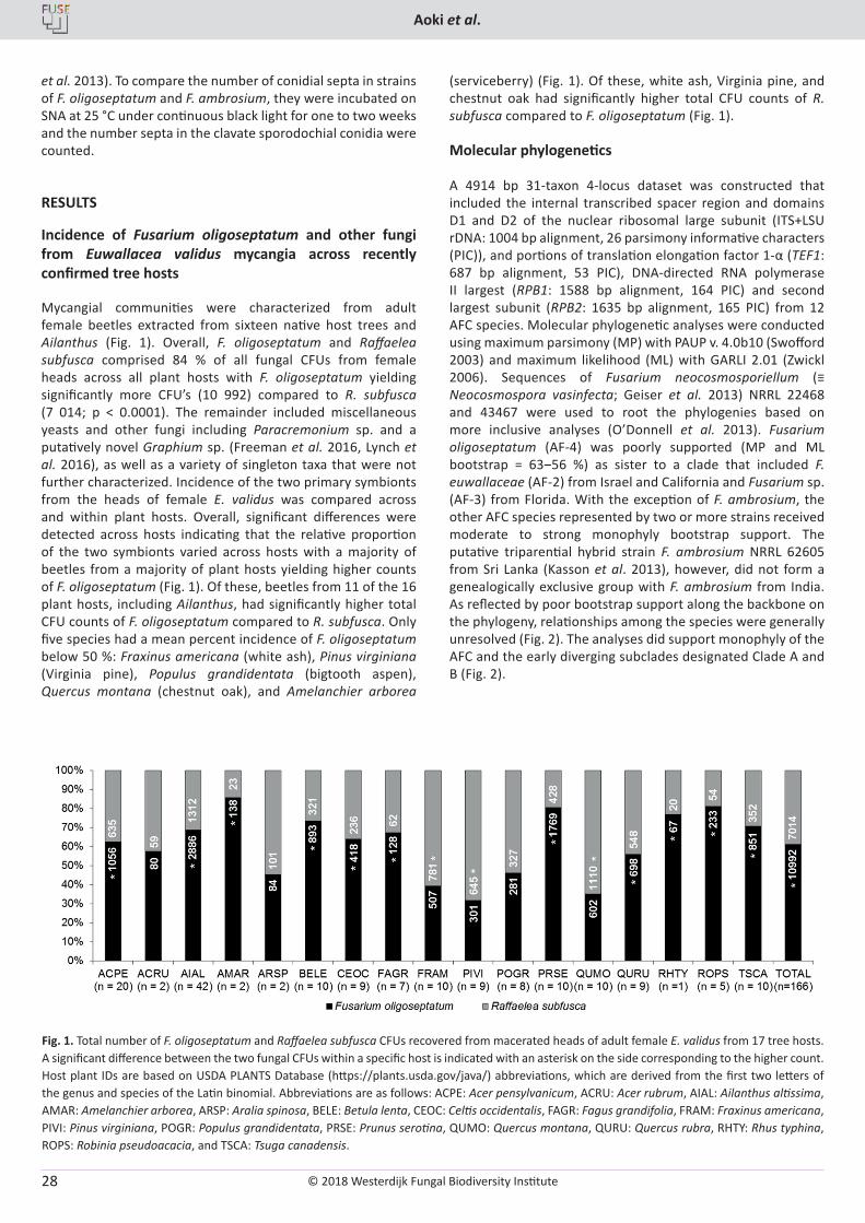
© 2018 Westerdijk Fungal Biodiversity Institute
Aoki et al.
Editor-in-ChiefProf. dr P.W. Crous, Westerdijk Fungal Biodiversity Institute, P.O. Box 85167, 3508 AD Utrecht, The Netherlands.E-mail:[email protected]
28
et al. 2013). To compare the number of conidial septa in strains of F. oligoseptatum and F. ambrosium, they were incubated on SNA at 25 °C under continuous black light for one to two weeks and the number septa in the clavate sporodochial conidia were counted.
RESULTS
Incidence of Fusarium oligoseptatum and other fungi from Euwallacea validus mycangia across recently confirmed tree hosts
Mycangial communities were characterized from adult female beetles extracted from sixteen native host trees and Ailanthus (Fig. 1). Overall, F. oligoseptatum and Raffaelea subfusca comprised 84 % of all fungal CFUs from female heads across all plant hosts with F. oligoseptatum yielding significantly more CFU’s (10 992) compared to R. subfusca (7 014; p < 0.0001). The remainder included miscellaneous yeasts and other fungi including Paracremonium sp. and a putatively novel Graphium sp. (Freeman et al. 2016, Lynch et al. 2016), as well as a variety of singleton taxa that were not further characterized. Incidence of the two primary symbionts from the heads of female E. validus was compared across and within plant hosts. Overall, significant differences were detected across hosts indicating that the relative proportion of the two symbionts varied across hosts with a majority of beetles from a majority of plant hosts yielding higher counts of F. oligoseptatum (Fig. 1). Of these, beetles from 11 of the 16 plant hosts, including Ailanthus, had significantly higher total CFU counts of F. oligoseptatum compared to R. subfusca. Only five species had a mean percent incidence of F. oligoseptatum below 50 %: Fraxinus americana (white ash), Pinus virginiana (Virginia pine), Populus grandidentata (bigtooth aspen), Quercus montana (chestnut oak), and Amelanchier arborea
(serviceberry) (Fig. 1). Of these, white ash, Virginia pine, and chestnut oak had significantly higher total CFU counts of R. subfusca compared to F. oligoseptatum (Fig. 1).
Molecular phylogenetics
A 4914 bp 31-taxon 4-locus dataset was constructed that included the internal transcribed spacer region and domains D1 and D2 of the nuclear ribosomal large subunit (ITS+LSU rDNA: 1004 bp alignment, 26 parsimony informative characters (PIC)), and portions of translation elongation factor 1-α (TEF1: 687 bp alignment, 53 PIC), DNA-directed RNA polymerase II largest (RPB1: 1588 bp alignment, 164 PIC) and second largest subunit (RPB2: 1635 bp alignment, 165 PIC) from 12 AFC species. Molecular phylogenetic analyses were conducted using maximum parsimony (MP) with PAUP v. 4.0b10 (Swofford 2003) and maximum likelihood (ML) with GARLI 2.01 (Zwickl 2006). Sequences of Fusarium neocosmosporiellum (≡ Neocosmospora vasinfecta; Geiser et al. 2013) NRRL 22468 and 43467 were used to root the phylogenies based on more inclusive analyses (O’Donnell et al. 2013). Fusarium oligoseptatum (AF-4) was poorly supported (MP and ML bootstrap = 63‒56 %) as sister to a clade that included F. euwallaceae (AF-2) from Israel and California and Fusarium sp. (AF-3) from Florida. With the exception of F. ambrosium, the other AFC species represented by two or more strains received moderate to strong monophyly bootstrap support. The putative triparential hybrid strain F. ambrosium NRRL 62605 from Sri Lanka (Kasson et al. 2013), however, did not form a genealogically exclusive group with F. ambrosium from India. As reflected by poor bootstrap support along the backbone on the phylogeny, relationships among the species were generally unresolved (Fig. 2). The analyses did support monophyly of the AFC and the early diverging subclades designated Clade A and B (Fig. 2).
Fig. 1. Total number of F. oligoseptatum and Raffaelea subfusca CFUs recovered from macerated heads of adult female E. validus from 17 tree hosts. A significant difference between the two fungal CFUs within a specific host is indicated with an asterisk on the side corresponding to the higher count. Host plant IDs are based on USDA PLANTS Database (https://plants.usda.gov/java/) abbreviations, which are derived from the first two letters of the genus and species of the Latin binomial. Abbreviations are as follows: ACPE: Acer pensylvanicum, ACRU: Acer rubrum, AIAL: Ailanthus altissima, AMAR: Amelanchier arborea, ARSP: Aralia spinosa, BELE: Betula lenta, CEOC: Celtis occidentalis, FAGR: Fagus grandifolia, FRAM: Fraxinus americana, PIVI: Pinus virginiana, POGR: Populus grandidentata, PRSE: Prunus serotina, QUMO: Quercus montana, QURU: Quercus rubra, RHTY: Rhus typhina, ROPS: Robinia pseudoacacia, and TSCA: Tsuga canadensis.
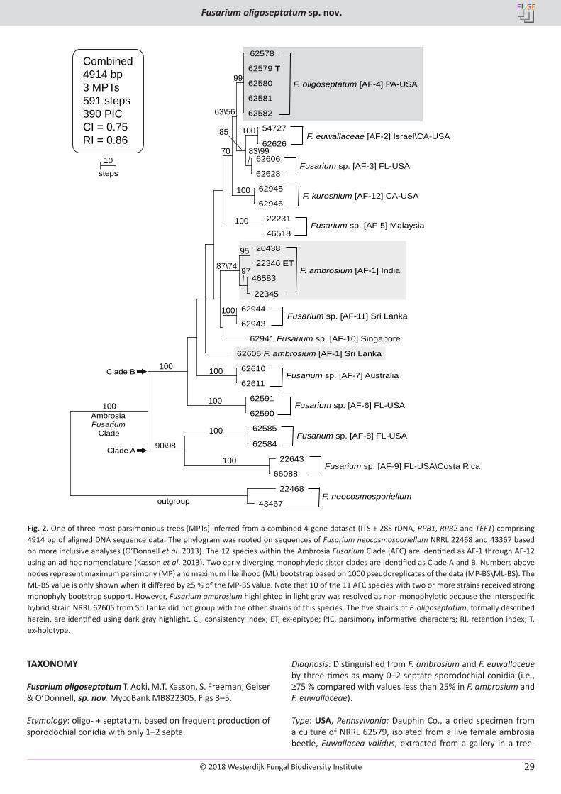
© 2018 Westerdijk Fungal Biodiversity Institute
Fusarium oligoseptatum sp. nov.
Editor-in-ChiefProf. dr P.W. Crous, Westerdijk Fungal Biodiversity Institute, P.O. Box 85167, 3508 AD Utrecht, The Netherlands.E-mail:[email protected]
29
TAXONOMY
Fusarium oligoseptatum T. Aoki, M.T. Kasson, S. Freeman, Geiser & O’Donnell, sp. nov. MycoBank MB822305. Figs 3–5.
Etymology: oligo- + septatum, based on frequent production of sporodochial conidia with only 1–2 septa.
Diagnosis: Distinguished from F. ambrosium and F. euwallaceae by three times as many 0–2-septate sporodochial conidia (i.e., ≥75 % compared with values less than 25% in F. ambrosium and F. euwallaceae).
Type: USA, Pennsylvania: Dauphin Co., a dried specimen from a culture of NRRL 62579, isolated from a live female ambrosia beetle, Euwallacea validus, extracted from a gallery in a tree-
Fusarium sp. [AF-3] FL-USA
62578
62579 T
62580
62581
62582
54727
62626
62606
62628
62945
62946
22231
46518
20438
22346 ET
46583
22345
62944
62943
62941 Fusarium sp. [AF-10] Singapore
62605 F. ambrosium [AF-1] Sri Lanka
62610
62611
62591
62590
62585
62584
22643
66088
22468
43467
F. oligoseptatum [AF-4] PA-USA
F. ambrosium [AF-1] India
Fusarium sp. [AF-11] Sri Lanka
F. euwallaceae [AF-2] Israel\CA-USA
F. kuroshium [AF-12] CA-USA
Fusarium sp. [AF-5] Malaysia
Fusarium sp. [AF-7] Australia
Fusarium sp. [AF-6] FL-USA
Fusarium sp. [AF-8] FL-USA
Fusarium sp. [AF-9] FL-USA\Costa Rica
F. neocosmosporiellum
Combined4914 bp3 MPTs591 steps390 PICCI = 0.75RI = 0.86
99
63\56
100
100
100
100
100
100
100
100
100
100
87\74
83\9970
90\98
85
95
97
outgroup
10steps
Ambrosia Fusarium Clade
Clade B
Clade A
Fig. 2.
Fig. 2. One of three most-parsimonious trees (MPTs) inferred from a combined 4-gene dataset (ITS + 28S rDNA, RPB1, RPB2 and TEF1) comprising 4914 bp of aligned DNA sequence data. The phylogram was rooted on sequences of Fusarium neocosmosporiellum NRRL 22468 and 43367 based on more inclusive analyses (O’Donnell et al. 2013). The 12 species within the Ambrosia Fusarium Clade (AFC) are identified as AF-1 through AF-12 using an ad hoc nomenclature (Kasson et al. 2013). Two early diverging monophyletic sister clades are identified as Clade A and B. Numbers above nodes represent maximum parsimony (MP) and maximum likelihood (ML) bootstrap based on 1000 pseudoreplicates of the data (MP-BS\ML-BS). The ML-BS value is only shown when it differed by ≥5 % of the MP-BS value. Note that 10 of the 11 AFC species with two or more strains received strong monophyly bootstrap support. However, Fusarium ambrosium highlighted in light gray was resolved as non-monophyletic because the interspecific hybrid strain NRRL 62605 from Sri Lanka did not group with the other strains of this species. The five strains of F. oligoseptatum, formally described herein, are identified using dark gray highlight. CI, consistency index; ET, ex-epitype; PIC, parsimony informative characters; RI, retention index; T, ex-holotype.
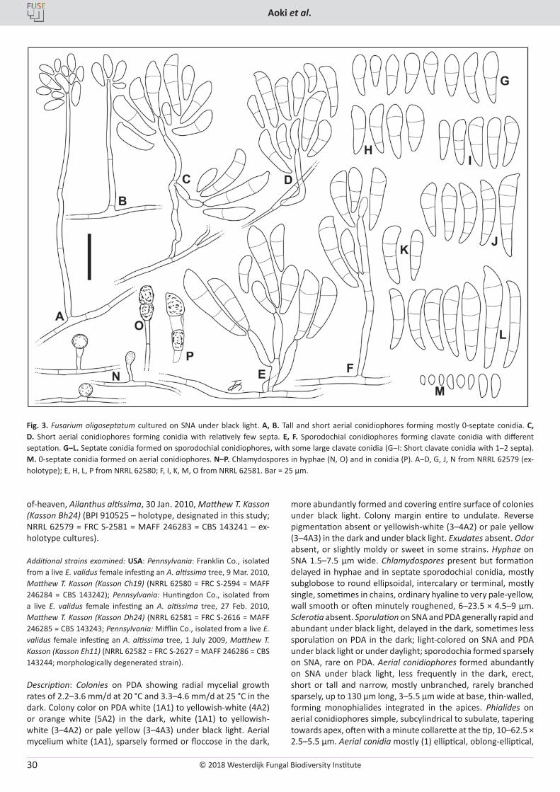
© 2018 Westerdijk Fungal Biodiversity Institute
Aoki et al.
Editor-in-ChiefProf. dr P.W. Crous, Westerdijk Fungal Biodiversity Institute, P.O. Box 85167, 3508 AD Utrecht, The Netherlands.E-mail:[email protected]
30
of-heaven, Ailanthus altissima, 30 Jan. 2010, Matthew T. Kasson (Kasson Bh24) (BPI 910525 – holotype, designated in this study; NRRL 62579 = FRC S-2581 = MAFF 246283 = CBS 143241 – ex-holotype cultures).
Additional strains examined: USA: Pennsylvania: Franklin Co., isolated from a live E. validus female infesting an A. altissima tree, 9 Mar. 2010, Matthew T. Kasson (Kasson Ch19) (NRRL 62580 = FRC S-2594 = MAFF 246284 = CBS 143242); Pennsylvania: Huntingdon Co., isolated from a live E. validus female infesting an A. altissima tree, 27 Feb. 2010, Matthew T. Kasson (Kasson Dh24) (NRRL 62581 = FRC S-2616 = MAFF 246285 = CBS 143243; Pennsylvania: Mifflin Co., isolated from a live E. validus female infesting an A. altissima tree, 1 July 2009, Matthew T. Kasson (Kasson Eh11) (NRRL 62582 = FRC S-2627 = MAFF 246286 = CBS 143244; morphologically degenerated strain).
Description: Colonies on PDA showing radial mycelial growth rates of 2.2–3.6 mm/d at 20 °C and 3.3–4.6 mm/d at 25 °C in the dark. Colony color on PDA white (1A1) to yellowish-white (4A2) or orange white (5A2) in the dark, white (1A1) to yellowish-white (3–4A2) or pale yellow (3–4A3) under black light. Aerial mycelium white (1A1), sparsely formed or floccose in the dark,
more abundantly formed and covering entire surface of colonies under black light. Colony margin entire to undulate. Reverse pigmentation absent or yellowish-white (3–4A2) or pale yellow (3–4A3) in the dark and under black light. Exudates absent. Odor absent, or slightly moldy or sweet in some strains. Hyphae on SNA 1.5–7.5 µm wide. Chlamydospores present but formation delayed in hyphae and in septate sporodochial conidia, mostly subglobose to round ellipsoidal, intercalary or terminal, mostly single, sometimes in chains, ordinary hyaline to very pale-yellow, wall smooth or often minutely roughened, 6–23.5 × 4.5–9 µm. Sclerotia absent. Sporulation on SNA and PDA generally rapid and abundant under black light, delayed in the dark, sometimes less sporulation on PDA in the dark; light-colored on SNA and PDA under black light or under daylight; sporodochia formed sparsely on SNA, rare on PDA. Aerial conidiophores formed abundantly on SNA under black light, less frequently in the dark, erect, short or tall and narrow, mostly unbranched, rarely branched sparsely, up to 130 µm long, 3–5.5 µm wide at base, thin-walled, forming monophialides integrated in the apices. Phialides on aerial conidiophores simple, subcylindrical to subulate, tapering towards apex, often with a minute collarette at the tip, 10–62.5 × 2.5–5.5 µm. Aerial conidia mostly (1) elliptical, oblong-elliptical,
Fig. 3. Fusarium oligoseptatum cultured on SNA under black light. A, B. Tall and short aerial conidiophores forming mostly 0-septate conidia. C, D. Short aerial conidiophores forming conidia with relatively few septa. E, F. Sporodochial conidiophores forming clavate conidia with different septation. G‒L. Septate conidia formed on sporodochial conidiophores, with some large clavate conidia (G‒I: Short clavate conidia with 1‒2 septa). M. 0-septate conidia formed on aerial conidiophores. N‒P. Chlamydospores in hyphae (N, O) and in conidia (P). A‒D, G, J, N from NRRL 62579 (ex-holotype); E, H, L, P from NRRL 62580; F, I, K, M, O from NRRL 62581. Bar = 25 µm.
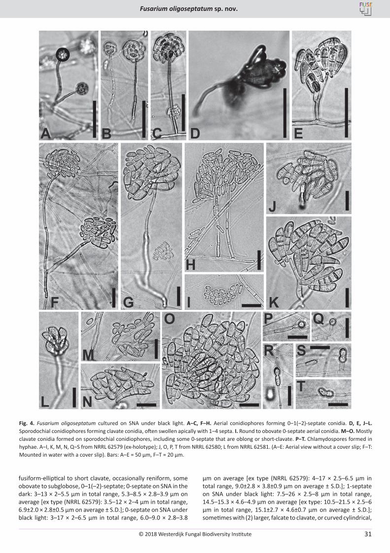
© 2018 Westerdijk Fungal Biodiversity Institute
Fusarium oligoseptatum sp. nov.
Editor-in-ChiefProf. dr P.W. Crous, Westerdijk Fungal Biodiversity Institute, P.O. Box 85167, 3508 AD Utrecht, The Netherlands.E-mail:[email protected]
31
fusiform-elliptical to short clavate, occasionally reniform, some obovate to subglobose, 0–1(–2)-septate; 0-septate on SNA in the dark: 3–13 × 2–5.5 µm in total range, 5.3–8.5 × 2.8–3.9 µm on average [ex type (NRRL 62579): 3.5–12 × 2–4 µm in total range, 6.9±2.0 × 2.8±0.5 µm on average ± S.D.]; 0-septate on SNA under black light: 3–17 × 2–6.5 µm in total range, 6.0–9.0 × 2.8–3.8
µm on average [ex type (NRRL 62579): 4–17 × 2.5–6.5 µm in total range, 9.0±2.8 × 3.8±0.9 µm on average ± S.D.]; 1-septate on SNA under black light: 7.5–26 × 2.5–8 µm in total range, 14.5–15.3 × 4.6–4.9 µm on average [ex type: 10.5–21.5 × 2.5–6 µm in total range, 15.1±2.7 × 4.6±0.7 µm on average ± S.D.]; sometimes with (2) larger, falcate to clavate, or curved cylindrical,
Fig. 4. Fusarium oligoseptatum cultured on SNA under black light. A‒C, F‒H. Aerial conidiophores forming 0‒1(‒2)-septate conidia. D, E, J‒L. Sporodochial conidiophores forming clavate conidia, often swollen apically with 1‒4 septa. I. Round to obovate 0-septate aerial conidia. M‒O. Mostly clavate conidia formed on sporodochial conidiophores, including some 0-septate that are oblong or short-clavate. P‒T. Chlamydospores formed in hyphae. A‒I, K, M, N, Q‒S from NRRL 62579 (ex-holotype); J, O, P, T from NRRL 62580; L from NRRL 62581. (A‒E: Aerial view without a cover slip; F‒T: Mounted in water with a cover slip). Bars: A‒E = 50 μm, F‒T = 20 μm.
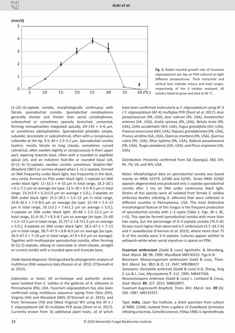
© 2018 Westerdijk Fungal Biodiversity Institute
Aoki et al.
Editor-in-ChiefProf. dr P.W. Crous, Westerdijk Fungal Biodiversity Institute, P.O. Box 85167, 3508 AD Utrecht, The Netherlands.E-mail:[email protected]
32
(1–)2(–3)-septate conidia, morphologically continuous with falcate sporodochial conidia. Sporodochial conidiophores generally shorter and thicker than aerial conidiophores, unbranched or sometimes sparsely branched, contorted, forming monophialides integrated apically, 20–145 × 3–6 µm, or sometimes adelophialides. Sporodochial phialides simple, subulate, lanceolate or subcylindrical, often with a conspicuous collarette at the tip, 9.5–44 × 2.5–5.5 µm. Sporodochial conidia hyaline, mostly falcate to long clavate, sometimes curved cylindrical, often swollen slightly or conspicuously in their upper part, tapering towards base, often with a rounded or papillate apical cell, and an indistinct foot-like or rounded basal cell, (0–)1–3(–5)-septate; swollen conidia sometimes ‘dolphin-like’ (Brayford 1987) or comma-shaped when 1- to 2-septate, formed on SNA frequently under black light, less frequently in the dark, very rarely formed on PDA under black light; 1-septate on SNA under black light: 11–32.5 × 4–10 µm in total range, 18.2–20.1 × 6.1–7.2 µm on average [ex type: 13.5–30 × 4.5–8.5 µm in total range, 19.3±3.9 × 6.2±1.0 µm on average ± S.D.]; 2-septate on SNA under black light: 15.5–39.5 × 5.5–12 µm in total range, 24.8–26.5 × 7.4–8.0 µm on average [ex type: 22–34 × 5.5–12 µm in total range, 26.1±3.2 × 7.4±1.2 µm on average ± S.D.]; 3-septate on SNA under black light: 20–60 × 5.5–12.5 µm in total range, 31.4–35.7 × 8.3–8.7 µm on average [ex type: 23–60 × 5.5–12.5 µm in total range, 35.7±7.2 × 8.7±1.3 µm on average ± S.D.]; 4-septate on SNA under black light: 28.5–67.5 × 7–11 µm in total range, 38.7–47.9 × 8.8–8.9 µm on average [ex type: 28.5–67.5 × 7–10 µm in total range, 47.9 × 8.9 µm on average]. Together with multiseptate sporodochial conidia, often forming (0-)1(-2)-septate, oblong to naviculate or short-clavate, straight or curved conidia with a rounded apex and truncate base.
Clade-based diagnosis: Distinguished by phylogenetic analysis of multilocus DNA sequence data (Kasson et al. 2013, O’Donnell et al. 2015).
Substrates or hosts: All ex-holotype and authentic strains were isolated from E. validus in the galleries of A. altissima in Pennsylvania (PA), USA. Fusarium oligoseptatum has also been confirmed using multilocus sequence typing from Ohio (OH), Virginia (VA) and Maryland (MD) (O’Donnell et al. 2015), and from Tennessee (TN) and (West Virginia) WV using the AF-3 / F. oligoseptatum (AF-4) multiplex PCR assay (Short et al. 2017). Currently known from 16 additional plant hosts, all of which
have been confirmed molecularly as F. oligoseptatum using AF-3 / F. oligoseptatum (AF-4) multiplex PCR (Short et al. 2017): Acer pensylvanicum (PA, USA), Acer rubrum (PA, USA), Amelanchier arborea (VA, USA), Aralia spinosa (PA, USA), Betula lenta (PA, USA), Celtis occidentalis (WV, USA), Fagus grandifolia (OH, USA), Fraxinus americana (WV, USA), Populus grandidentata (PA, USA), Prunus serotina (GA, USA), Quercus montana (PA, USA), Quercus rubra (PA, USA), Rhus typhina (PA, USA), Robinia pseudoacacia (PA, USA), Tsuga canadensis (OH, USA), and Pinus virginiana (VA, USA).
Distribution: Presently confirmed from GA (Georgia), MD, OH, PA, TN, VA, and WV, USA.
Notes: Morphological data on sporodochial conidia was based mainly on NRRL 62579, 62580 and 62581. Strain NRRL 62582 appears degenerated and produced only 1-septate sporodochial conidia after 1 mo on SNA under continuous black light. Strains of this species were all isolated from female E. validus ambrosia beetles infesting A. altissima that were collected in different counties in Pennsylvania, USA. The most distinctive morphological feature of this fungus is the frequent production of sporodochial conidia with 1–2 septa (Table 2, Figs. 3E–I, 4E, J–O). This species formed sporodochial conidia with more than two septa, but the percentage of 0–2-septate conidia (76.5–81 %) was much higher than observed in F. ambrosium (3.7–24.5 %) and F. euwallaceae (Freeman et al. 2013), where more than 75 % of the conidia were 3–5-septate. Cultures appear whitish to yellowish-white when aerial mycelium is sparse on PDA.
Fusarium ambrosium (Gadd & Loos) Agnihothr. & Nirenberg, Stud. Mycol. 32: 98. 1990. MycoBank MB130225. Figs 6–8.Basionym: Monacrosporium ambrosium Gadd & Loos, Trans. Brit. Mycol. Soc. 31(1 & 2): 13. 1947. MB288427. Synonyms: Dactylella ambrosia (Gadd & Loos) K.Q. Zhang, Xing Z. Liu & L. Cao, Mycosystema 7: 112. 1995. MB447506.Neocosmospora ambrosia (Gadd & Loos) L. Lombard & Crous, Stud. Mycol. 80: 227. 2015. MB810957.Fusarium bugnicourtii Brayford, Trans. Brit. Mycol. Soc. 89 (3): 350. 1987. MB133337.
Type: India, Upari Tea Institute, a dried specimen from culture of NRRL 22346, isolated from a gallery of Euwallacea fornicatus infesting a tea tree, Camellia sinensis, 9 May 1990, V. Agnihothrudu
Fig. 5. Radial mycelial growth rate of Fusarium oligoseptatum per day on PDA cultured at eight different temperatures. Thick horizontal and vertical bars indicate means and total ranges, respectively, of the 4 isolates analyzed. All isolates failed to grow and died at 40 °C.
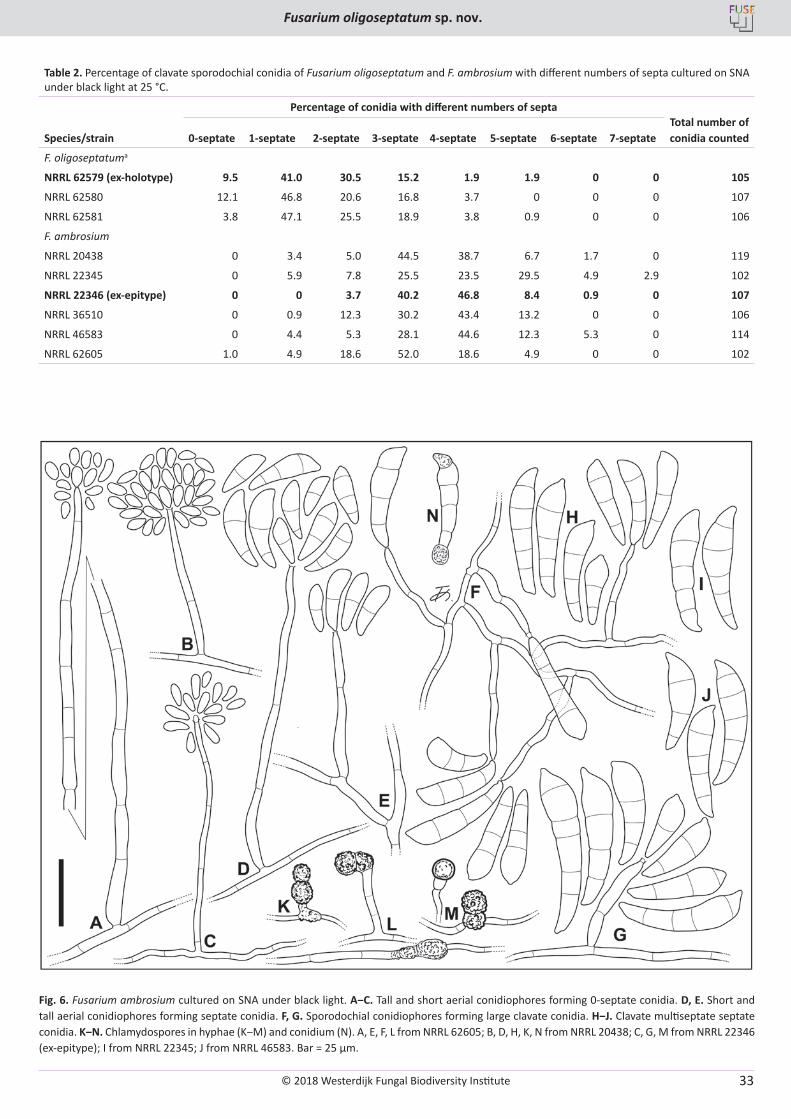
© 2018 Westerdijk Fungal Biodiversity Institute
Fusarium oligoseptatum sp. nov.
Editor-in-ChiefProf. dr P.W. Crous, Westerdijk Fungal Biodiversity Institute, P.O. Box 85167, 3508 AD Utrecht, The Netherlands.E-mail:[email protected]
33
Table 2. Percentage of clavate sporodochial conidia of Fusarium oligoseptatum and F. ambrosium with different numbers of septa cultured on SNA under black light at 25 °C.
Percentage of conidia with different numbers of septa Total number of conidia countedSpecies/strain 0-septate 1-septate 2-septate 3-septate 4-septate 5-septate 6-septate 7-septate
F. oligoseptatuma
NRRL 62579 (ex-holotype) 9.5 41.0 30.5 15.2 1.9 1.9 0 0 105
NRRL 62580 12.1 46.8 20.6 16.8 3.7 0 0 0 107
NRRL 62581 3.8 47.1 25.5 18.9 3.8 0.9 0 0 106
F. ambrosium
NRRL 20438 0 3.4 5.0 44.5 38.7 6.7 1.7 0 119
NRRL 22345 0 5.9 7.8 25.5 23.5 29.5 4.9 2.9 102
NRRL 22346 (ex-epitype) 0 0 3.7 40.2 46.8 8.4 0.9 0 107
NRRL 36510 0 0.9 12.3 30.2 43.4 13.2 0 0 106
NRRL 46583 0 4.4 5.3 28.1 44.6 12.3 5.3 0 114
NRRL 62605 1.0 4.9 18.6 52.0 18.6 4.9 0 0 102
Fig. 6. Fusarium ambrosium cultured on SNA under black light. A‒C. Tall and short aerial conidiophores forming 0-septate conidia. D, E. Short and tall aerial conidiophores forming septate conidia. F, G. Sporodochial conidiophores forming large clavate conidia. H‒J. Clavate multiseptate septate conidia. K‒N. Chlamydospores in hyphae (K‒M) and conidium (N). A, E, F, L from NRRL 62605; B, D, H, K, N from NRRL 20438; C, G, M from NRRL 22346 (ex-epitype); I from NRRL 22345; J from NRRL 46583. Bar = 25 µm.
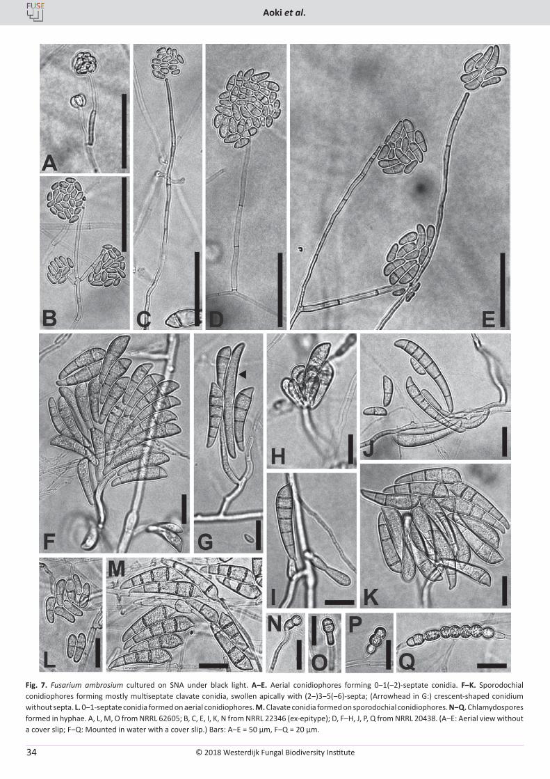
© 2018 Westerdijk Fungal Biodiversity Institute
Aoki et al.
Editor-in-ChiefProf. dr P.W. Crous, Westerdijk Fungal Biodiversity Institute, P.O. Box 85167, 3508 AD Utrecht, The Netherlands.E-mail:[email protected]
34
Fig. 7. Fusarium ambrosium cultured on SNA under black light. A‒E. Aerial conidiophores forming 0‒1(‒2)-septate conidia. F‒K. Sporodochial conidiophores forming mostly multiseptate clavate conidia, swollen apically with (2‒)3‒5(‒6)-septa; (Arrowhead in G:) crescent-shaped conidium without septa. L. 0‒1-septate conidia formed on aerial conidiophores. M. Clavate conidia formed on sporodochial conidiophores. N‒Q. Chlamydospores formed in hyphae. A, L, M, O from NRRL 62605; B, C, E, I, K, N from NRRL 22346 (ex-epitype); D, F‒H, J, P, Q from NRRL 20438. (A‒E: Aerial view without a cover slip; F‒Q: Mounted in water with a cover slip.) Bars: A‒E = 50 µm, F‒Q = 20 µm.
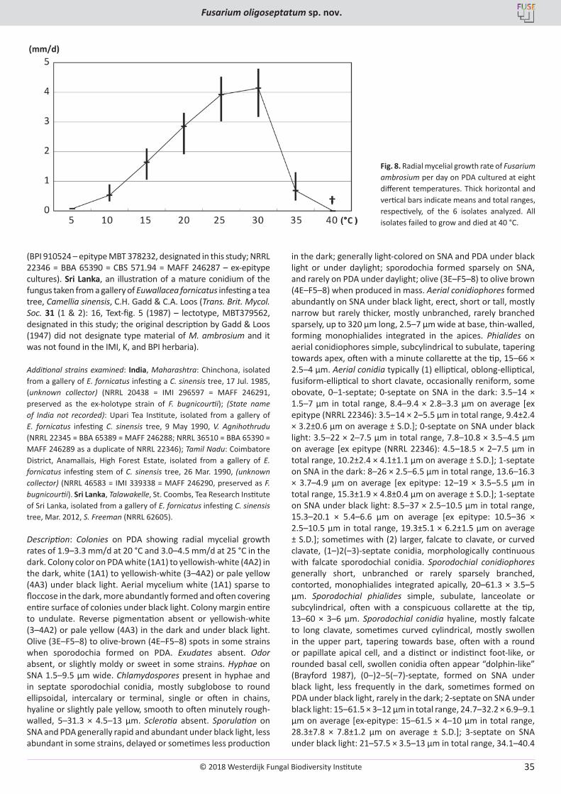
© 2018 Westerdijk Fungal Biodiversity Institute
Fusarium oligoseptatum sp. nov.
Editor-in-ChiefProf. dr P.W. Crous, Westerdijk Fungal Biodiversity Institute, P.O. Box 85167, 3508 AD Utrecht, The Netherlands.E-mail:[email protected]
35
(BPI 910524 – epitype MBT 378232, designated in this study; NRRL 22346 = BBA 65390 = CBS 571.94 = MAFF 246287 – ex-epitype cultures). Sri Lanka, an illustration of a mature conidium of the fungus taken from a gallery of Euwallacea fornicatus infesting a tea tree, Camellia sinensis, C.H. Gadd & C.A. Loos (Trans. Brit. Mycol. Soc. 31 (1 & 2): 16, Text-fig. 5 (1987) – lectotype, MBT379562, designated in this study; the original description by Gadd & Loos (1947) did not designate type material of M. ambrosium and it was not found in the IMI, K, and BPI herbaria).
Additional strains examined: India, Maharashtra: Chinchona, isolated from a gallery of E. fornicatus infesting a C. sinensis tree, 17 Jul. 1985, (unknown collector) (NRRL 20438 = IMI 296597 = MAFF 246291, preserved as the ex-holotype strain of F. bugnicourtii); (State name of India not recorded): Upari Tea Institute, isolated from a gallery of E. fornicatus infesting C. sinensis tree, 9 May 1990, V. Agnihothrudu (NRRL 22345 = BBA 65389 = MAFF 246288; NRRL 36510 = BBA 65390 = MAFF 246289 as a duplicate of NRRL 22346); Tamil Nadu: Coimbatore District, Anamallais, High Forest Estate, isolated from a gallery of E. fornicatus infesting stem of C. sinensis tree, 26 Mar. 1990, (unknown collector) (NRRL 46583 = IMI 339338 = MAFF 246290, preserved as F. bugnicourtii). Sri Lanka, Talawakelle, St. Coombs, Tea Research Institute of Sri Lanka, isolated from a gallery of E. fornicatus infesting C. sinensis tree, Mar. 2012, S. Freeman (NRRL 62605).
Description: Colonies on PDA showing radial mycelial growth rates of 1.9–3.3 mm/d at 20 °C and 3.0–4.5 mm/d at 25 °C in the dark. Colony color on PDA white (1A1) to yellowish-white (4A2) in the dark, white (1A1) to yellowish-white (3–4A2) or pale yellow (4A3) under black light. Aerial mycelium white (1A1) sparse to floccose in the dark, more abundantly formed and often covering entire surface of colonies under black light. Colony margin entire to undulate. Reverse pigmentation absent or yellowish-white (3–4A2) or pale yellow (4A3) in the dark and under black light. Olive (3E–F5–8) to olive-brown (4E–F5–8) spots in some strains when sporodochia formed on PDA. Exudates absent. Odor absent, or slightly moldy or sweet in some strains. Hyphae on SNA 1.5–9.5 µm wide. Chlamydospores present in hyphae and in septate sporodochial conidia, mostly subglobose to round ellipsoidal, intercalary or terminal, single or often in chains, hyaline or slightly pale yellow, smooth to often minutely rough-walled, 5–31.3 × 4.5–13 µm. Sclerotia absent. Sporulation on SNA and PDA generally rapid and abundant under black light, less abundant in some strains, delayed or sometimes less production
in the dark; generally light-colored on SNA and PDA under black light or under daylight; sporodochia formed sparsely on SNA, and rarely on PDA under daylight; olive (3E–F5–8) to olive brown (4E–F5–8) when produced in mass. Aerial conidiophores formed abundantly on SNA under black light, erect, short or tall, mostly narrow but rarely thicker, mostly unbranched, rarely branched sparsely, up to 320 µm long, 2.5–7 µm wide at base, thin-walled, forming monophialides integrated in the apices. Phialides on aerial conidiophores simple, subcylindrical to subulate, tapering towards apex, often with a minute collarette at the tip, 15–66 × 2.5–4 µm. Aerial conidia typically (1) elliptical, oblong-elliptical, fusiform-elliptical to short clavate, occasionally reniform, some obovate, 0–1-septate; 0-septate on SNA in the dark: 3.5–14 × 1.5–7 µm in total range, 8.4–9.4 × 2.8–3.3 µm on average [ex epitype (NRRL 22346): 3.5–14 × 2–5.5 µm in total range, 9.4±2.4 × 3.2±0.6 µm on average ± S.D.]; 0-septate on SNA under black light: 3.5–22 × 2–7.5 µm in total range, 7.8–10.8 × 3.5–4.5 µm on average [ex epitype (NRRL 22346): 4.5–18.5 × 2–7.5 µm in total range, 10.2±2.4 × 4.1±1.1 µm on average ± S.D.]; 1-septate on SNA in the dark: 8–26 × 2.5–6.5 µm in total range, 13.6–16.3 × 3.7–4.9 µm on average [ex epitype: 12–19 × 3.5–5.5 µm in total range, 15.3±1.9 × 4.8±0.4 µm on average ± S.D.]; 1-septate on SNA under black light: 8.5–37 × 2.5–10.5 µm in total range, 15.3–20.1 × 5.4–6.6 µm on average [ex epitype: 10.5–36 × 2.5–10.5 µm in total range, 19.3±5.1 × 6.2±1.5 µm on average ± S.D.]; sometimes with (2) larger, falcate to clavate, or curved clavate, (1–)2(–3)-septate conidia, morphologically continuous with falcate sporodochial conidia. Sporodochial conidiophores generally short, unbranched or rarely sparsely branched, contorted, monophialides integrated apically, 20–61.3 × 3.5–5 µm. Sporodochial phialides simple, subulate, lanceolate or subcylindrical, often with a conspicuous collarette at the tip, 13–60 × 3–6 µm. Sporodochial conidia hyaline, mostly falcate to long clavate, sometimes curved cylindrical, mostly swollen in the upper part, tapering towards base, often with a round or papillate apical cell, and a distinct or indistinct foot-like, or rounded basal cell, swollen conidia often appear “dolphin-like” (Brayford 1987), (0–)2–5(–7)-septate, formed on SNA under black light, less frequently in the dark, sometimes formed on PDA under black light, rarely in the dark; 2-septate on SNA under black light: 15–61.5 × 3–12 µm in total range, 24.7–32.2 × 6.9–9.1 µm on average [ex-epitype: 15–61.5 × 4–10 µm in total range, 28.3±7.8 × 7.8±1.2 µm on average ± S.D.]; 3-septate on SNA under black light: 21–57.5 × 3.5–13 µm in total range, 34.1–40.4
Fig. 8. Radial mycelial growth rate of Fusarium ambrosium per day on PDA cultured at eight different temperatures. Thick horizontal and vertical bars indicate means and total ranges, respectively, of the 6 isolates analyzed. All isolates failed to grow and died at 40 °C.
© 2018 Westerdijk Fungal Biodiversity Institute
Aoki et al.
Editor-in-ChiefProf. dr P.W. Crous, Westerdijk Fungal Biodiversity Institute, P.O. Box 85167, 3508 AD Utrecht, The Netherlands.E-mail:[email protected]
36
× 8.3–10.0 µm on average [ex epitype: 30–57 × 7.5–12.5 µm in total range, 40.4±4.9 × 8.8±1.1 µm on average ± S.D.]; 4-septate on SNA under black light: 25.5–78.5 × 6–12.5 µm in total range, 40.7–45.6 × 8.8–10.4 µm on average [ex epitype: 33–78.5 × 7.5–11.5 µm in total range, 45.3±6.1 × 8.8±0.9 µm on average ± S.D.]; 5-septate on SNA under black light: 30–64 × 7–12.5 µm in total range, 42.9–52.1 × 8.8–10.3 µm on average [ex epitype: 37–51.5 × 7.5–12 µm in total range, 45.6±3.7 × 8.8±1.1 µm on average ± S.D.]. Together with multiseptate sporodochial conidia, often forming (0-)1(-2)-septate, oblong to naviculate or short-clavate, straight or curved conidia, with a rounded apex and a truncate base.
Notes: Strain NRRL 62605 did not form conidia on SNA or PDA in the dark. Therefore, the description of conidia in the dark was based on the three other strains examined in this study. All of the strains studied were isolated from galleries of E. fornicatus infesting C. sinensis trees in India and Sri Lanka. Fusarium ambrosium was originally isolated from E. (Xyleborus) fornicatus galleries in stems of Chinese tea and caster-oil trees in Sri Lanka, and was described as a new species of Monacrosporium, i.e., M. ambrosium by Gadd & Loos (1947). Forty years later, it was redescribed by Brayford (1987) as F. bugnicourtii based on collections from beetle galleries in Chinese tea in India and borer-damaged Hevea brasiliensis and Theoborma cacao in Malaysia. Brayford (1987) considered F. bugnicourtii to be conspecific with F. tumidum var. coeruleum (Bugnicourt 1939) isolated from H. brasiliensis, but distinct from F. tumidum. Fusarium bugnicourtii was recognized as conspecific with Gadd and Loos’ species, M. ambrosium from the shot-hole borer on tea, and synonymized under the new combination, F. ambrosium based on its nomenclatural priority (Nirenberg 1990). Because type material of M. ambrosium (≡ F. ambrosium) was not designated in the original description by Gadd & Loos (1947) and not found in the IMI, K, and BPI herbaria, we selected a line-drawing (illustration) of a conidium from Gadd & Loos (1947) as the lectotype, according to Art. 9.2 and 9.3 of the ICNafp (McNeill et al. 2012). To supplement the lectotype, BPI 910524, a dried culture of NRRL 22346 (= BBA 65390 = CBS 571.94), isolated from a gallery of E. fornicatus infesting C. sinensis in India by V. Agnihothrudu, was selected as the epitype according to Art. 9.8 of the code. Because an authentic strain of F. ambrosium, IMI 296597 (= NRRL 20438), isolated from E. (Xyleborus) fornicatus galleries in tea tree from India in 1985, was designated as the holotype of F. bugnicourtii by Brayford (1987), this material was not selected for the epitype, per Art. 52.1 and 52.2 of the ICN (McNeill et al. 2012). Although NRRL 62605 was isolated from an E. fornicatus gallery in Sri Lanka from the same host and type locality, it was not selected as the epitype because it appears to be an interspecific hybrid that contains alleles from what appear to be two other AFC species (Kasson et al. 2013). The present epitypification was prepared to stabilize the taxonomy of this species.
Fusarium ambrosium is most similar morphologically to F. euwallaceae (Freeman et al. 2013, Kasson et al. 2013). Fusarium ambrosium and F. euwallaceae produce very similar falcate to clavate, septate sporodochial conidia that are swollen in their upper half, together with ovoid to ellipsoid, 0-septate aerial conidia. The conidial sizes and number of septa of these two fusaria are almost identical (Table 2, Figs 6, 7; Freeman et al. 2013). However, F. ambrosium and F. euwallaceae can be distinguished by the production of hyaline or olive to olive-
brown conidia when old in the former and bluish to greenish conidia in the latter when produced in mass on PDA after 1 mo (Freeman et al. 2013). Production of sporodochial conidia in F. euwallaceae is easily observed on SNA and PDA in the dark and under black light, but in F. ambrosium it is often delayed or limited without black light illumination, even if cultured on SNA.
A preliminary morphological comparison of the sporodochial conidia for 10 of the 12 AFC species has been conducted (T. Aoki et al. unpubl.). Ten of the species produced clavate sporodochial conidia that were swollen apically, and two, including AF-6 associated with Euwallacea sp. #2 in avocado in the Miami-Dade area of southern Florida, USA and AF-9 from Xyleborus ferrugineus in Costa Rica and Delonix regia (royal poinciana) in southern Florida only produced curved fusiform, septate sporodochial conidia in culture, as commonly observed in typical members of the F. solani species complex. Three novel AFC species that were reported recently (O’Donnell et al. 2015, Na et al. 2018), including AF-10 from Singapore, AF-11 from Sri Lanka, and AF-12 (= F. kuroshium) from San Diego County, California, produced clavate sporodochial conidia in culture.
The available data suggests that most of the AFC species might possess diagnostic phenotypic characters. For example, AF-3 ex Euwallacea interjectus infesting Acer negundo in Gainesville, Florida produced sporodochial conidia that were variable in size and shape; AF-5 from Malaysia ex Hevea brasiliensis produced the shortest sporodochial conidia when comparing those with the same number of septa produced by the members of the AFC; AF-7 from Euwallacea sp. #3 ex Persea americana in Australia produced sporodochial conidia that were densely and/or obliquely septate; AF-8 from Euwallacea sp. #2 ex avocado in the Miami-Dade area of southern Florida appeared to be unique in that it is the only AFC species that produced swollen clavate and curved fusiform sporodochial conidia (Kasson et al. 2013); AF-10 from Singapore produced clavate sporodochial conidia that were narrower than conidia of other AFC species with the same number of septa; AF-11 ex E. fornicatus in Chinese tea from Sri Lanka produced sporodochial conidia that were frequently pointed and curved or hooked to one side; F. kuroshium AF-12 ex Euwallacea sp. #5 from Platanus racemosa (California sycamore) and several other woody hosts in San Diego County, California was distinguished by the production of clavate sporodochial conidia together with crescent- or comma-shaped conidia (T. Aoki et al. unpubl.). By way of contrast, AF-6 ex Euwallacea sp. #2 in avocado in the Miami-Dade area of Florida and AF-9 from Costa Rica and the Miami-Dade area of Florida only produced curved cylindrical multiseptate conidia and elliptical to oblong aerial conidia (Kasson et al. 2013). However, the sporodochial conidia formed by AF-9 are atypical of the F. solani species complex because they were pointed at both ends, terminating in a short apical beak and a conspicuous foot-like basal cell. Detailed studies will be required to assess whether AF-6 and AF-9 possess morphological characters that distinguish them from other members of the F. solani species complex.
DISCUSSION
The AFC symbiont cultivated by the ambrosia beetle Euwallacea validus in Ailanthus altissima in eastern U.S. is formally described herein as Fusarium oligoseptatum. Sampling across 7 eastern U.S. states and 17 tree hosts confirmed that F. oligoseptatum is the
© 2018 Westerdijk Fungal Biodiversity Institute
Fusarium oligoseptatum sp. nov.
Editor-in-ChiefProf. dr P.W. Crous, Westerdijk Fungal Biodiversity Institute, P.O. Box 85167, 3508 AD Utrecht, The Netherlands.E-mail:[email protected]
37
primary symbiont of E. validus and dominant, regardless of plant host with few exceptions. This species can be distinguished from the 11 other known AFC species by producing significantly more 0–2-septate clavate sporodochial conidia that are swollen apically and via multilocus molecular phylogenetics where it was strongly supported as a genealogically exclusive species-level lineage in the analyses reported here and in previous studies (Kasson et al. 2013, O’Donnell et al. 2015, Na et al. 2018). Fusarium oligoseptatum was strongly supported as a reciprocally monophyletic sister to F. euwallaceae + Fusarium sp. (AF-3) in Kasson et al. (2013), but the sister group relationship of F. oligoseptatum was unresolved in analyses that included the closely related F. kuroshium (AF-12) from San Diego, California (O’Donnell et al. 2015, and present study). Efforts to develop a robust hypothesis of evolutionary relationships among these four AFC species, which are estimated to have shared a most recent common ancestor approximately 1.6 Mya (O’Donnell et al. 2015), might benefit from the comparative phylogenomic analyses that are currently underway (Stajich et al., pers. comm.).
Herein, an epitype of F. ambrosium was designated based on material originally isolated from a gallery of E. fornicatus infesting Chinese tea in India to provide nomenclatural stability for this species. AFC species have been collected in eight different countries, including Sri Lanka (F. ambrosium AF-1 and Fusarium sp. AF-11), India (F. ambrosium AF-1), Malaysia (Fusarium sp. AF-5), Singapore (Fusarium sp. AF-10), Australia (Fusarium sp. AF-7), Israel (F. euwallaceae AF-2), Costa Rica (Fusarium sp. AF-9) and the United States (F. euwallaceae AF-2, Fusarium sp. AF-3, F. oligoseptatum AF-4, Fusarium spp. AF-6, AF-8, AF-9 and F. kuroshium AF-12)(Brayford 1987, Nirenberg 1990, Freeman et al. 2013, Kasson et al. 2013, O’Donnell et al. 2015, Short et al. 2017, Na et al. 2018). To date only three species within the AFC have been described formally, i.e., F. ambrosium (AF-1; Gadd & Loos 1947, Nirenberg 1990), F. euwallaceae (AF-2; Freeman et al. 2013) and F. oligoseptatum (AF-4; in this study). Although nine of the AFC species are currently unnamed, the prospects for naming them are excellent because most of them appear to possess unique phenotypic/morphological features. Delimitations of such features may, in time, help to uncover the mechanisms underlying the production of clavate conidia, a posited adaptation for the Euwallacea − Fusarium symbiosis (Kasson et al. 2013). Indeed, analogous adaptations in agaricalean fungi (i.e., gongylidia) farmed by higher and occasionally lower attine ants (Schultz & Brady 2008, Masiulionis et al. 2014) also appear to exhibit variation among closely related lineages. However, quality of the substrate, pH, and temperature have also been shown to affect the growth and size of gongylidia in some higher attine ant cultivars when cultivated under lab conditions (Powell & Stradling 1986).
It remains unclear whether F. ambrosium, F. euwallaceae, or F. oligoseptatum are each farmed by a single Euwallacea species, including within their native range, where evidence of hybridization and co-cultivation with other closely related AFC members have been reported (O’Donnell et al. 2015). However, it has been shown that F. euwallaceae from avocado is obligately required for the survival and development of Euwallacea sp. #1 sensu O’Donnell et al. (2015) currently occurring in Israel, whereas F. ambrosium does not support development of this beetle species (Freeman et al. 2012). Likewise, specificity exists for F. ambrosium and its beetle host. Future studies focused on vector specificity could help clarify the threats these beetle-fungus consortia pose to our
native ecosystems. This is especially important given that some AFC members such as F. euwallaceae have caused significant damage to orchard, landscape and forest trees and threaten avocado production worldwide (Mendel et al. 2012, Eskalen et al. 2013, Kasson et al. 2013), while other AFC members such as F. oligoseptatum appear to be quite innocuous when challenged against numerous plant species (Berger 2017).
The FSSC includes over 60 species (Zhang et al. 2006, O’Donnell et al. 2008, Short et al. 2013), a majority of which lack formal Latin binomials thus making it difficult to link specific plant diseases with specific phylogenetic species within the FSSC, including the AFC (Montecchio et al. 2015). The designation of formal Latin binomials for a majority of phylogenetic species within the FSSC coupled with recent abolishment of the dual system of fungal nomenclature will likely reduce confusion surrounding molecular identification of taxa within this large species complex. Nevertheless, the use of multilocus phylogenetic studies will remain the gold standard to discriminate closely related members in the FSSC.
Another avenue to further resolve these closely related phylogenetic species is to examine functional differences among closely related AFC. A recent study by Kasson et al. (2016) assessed the enzyme activity and wood degrading capacity of F. oligoseptatum and R. subfusca, the two known symbionts of E. validus in the eastern U.S. Polyphenol oxidase production was detected from F. oligoseptatum but not R. subfusca. An earlier study by Norris (1980) on AFC member Fusarium sp. AF-9 revealed this fungus was capable of degrading lignin. Further enzymatic studies among closely related AFC may compliment morphological and phylogenetic studies within the Euwallacea − Fusarium mutualism, revealing significant differences in enzyme activity. This is particularly important given recent studies by Aylward et al. (2015) that showed a diverse but consistent set of enzymes present in gongylidia, which are essential for initial degradation of plant substrates in the leaf-cutter ant-Leucoagaricus mutualism.
The results of this study suggest that many of the unnamed AFC species like F. oligoseptatum possess unique phenotypic/morphological features, which will facilitate formal description of these economically important pathogens. Phenotypic/morphological studies on the four additional AFC species from Tawain (Na et al. 2018) are, therefore, also fully expected. Our ongoing research is focused on advancing the systematics of the AFC to promote accurate communication within the global scientific community.
ACKNOWLEDGMENTS
We are pleased to acknowledge the skilled technical assistance of Gail Doehring, Stacy Sink and Nathane Orwig in generating the DNA sequence data reported in this study. Dr. Konstanze Bensch, MycoBank, is thanked for her helpful advice concerning typification of F. ambrosium.
DISCLAMER
Mention of trade names or commercial products in this publication is solely for the purpose of providing specific information and does not imply recommendation or endorsement by the U.S. Department of Agriculture. USDA is an equal opportunity provider and employer.
© 2018 Westerdijk Fungal Biodiversity Institute
Aoki et al.
Editor-in-ChiefProf. dr P.W. Crous, Westerdijk Fungal Biodiversity Institute, P.O. Box 85167, 3508 AD Utrecht, The Netherlands.E-mail:[email protected]
38
REFERENCES
Alamouti S, Tsui C, Breuil C (2009). Multigene phylogeny of filamentous ambrosia fungi associated with ambrosia and bark beetles. Mycological Research 113: 822–835.
Aoki T, Vaughan MM, McCormick SP, et al. (2015). Fusarium dactylidis sp. nov., a novel nivalenol toxin-producing species sister to F. pseudograminearum isolated from orchard grass (Dactylis glomerata) in Oregon and New Zealand. Mycologia 107: 409–418.
Aylward FO, Khadempour L, Tremmel DM, et al. (2015). Enrichment and broad representation of plant biomass-degrading enzymes in the specialized hyphal swellings of Leucoagaricus gongylophorus, the fungal symbiont of leaf-cutter ants. PloS ONE 10: e0134752.
Bateman CC, Huang Y-T, Simmons D, et al. (2016). Ambrosia beetle Premnobius cavipennis (Scolytinae: Ipini) carries highly divergent ascomycotan ambrosia fungus, Afroraffaelea ambrosiae gen. nov. et sp. nov. (Ophiostomatales). Fungal Ecology 25: 41–49.
Berger MC (2017). Interactions between Euwallacea ambrosia beetles, their fungal symbionts and the native trees - They attack in the Eastern United States (Master Thesis, West Virginia University).
Brayford D (1987). Fusarium bugincourtii sp. nov., and its relationship to F. tumidum and F. tumidum var. coeruleum. Transactions of the British Mycological Society 89: 347–351.
Bugnicourt F (1939). Les Fusarium et Cylindrocarpon de l’Indochine. Encyclopedie Mycologique 11: 83–87 + plate II.
Dreaden TJ, Davis JM, de Beer ZW, et al. (2014). Phylogeny of ambrosia beetle symbionts in the genus Raffaelea. Fungal Biology 118: 970–978.
Eskalen A, Stouthamer R, Lynch SC, et al. (2013). Host range of Fusarium dieback and its ambrosia beetle (Coleoptera: Scolytinae) vector in southern California. Plant Disease 97: 938–951.
Fraedrich SW, Harrington TC, Rabaglia RJ, et al. (2008). A fungal symbiont of the redbay ambrosia beetle causes a lethal wilt in redbay and other Lauraceae in the southeastern United States. Plant Disease 92: 215–224.
Freeman S, Protasov A, Sharon M, et al. (2012). Obligate feed requirement of Fusarium sp. nov., an avocado wilting agent, by the ambrosia beetle Euwallacea aff. fornicata. Symbiosis 58: 245–251.
Freeman S, Sharon M, Dori-Bachash M, et al. (2016). Symbiotic association of three fungal species throughout the life cycle of the ambrosia beetle Euwallacea nr. fornicatus. Symbiosis 68: 115–128.
Freeman S, Sharon M, Maymon M, et al. (2013). Fusarium euwallaceae sp. nov. - a symbiotic fungus of Euwallacea sp., an invasive ambrosia beetle in Israel and California. Mycologia 105: 1595–1606.
Gadd CH, Loos CA (1947). The ambrosia fungus of Xyleborus fornicatus Eich. Transactions of the British Mycological Society 30: 13–18 + plate I.
Gebhardt H, Weiss M, Oberwinkler F (2005). Dryadomyces amasae: a nutritional fungus associated with ambrosia beetles of the genus Amasa (Coleoptera: Curculionidae, Scolytinae). Mycological Research 109: 687–696.
Geiser DM, Aoki T, Bacon CW, et al. (2013). One fungus, one name: defining the genus Fusarium in a scientifically robust way that preserves longstanding use. Phytopathology 103: 400–408.
Harrington TC, Aghayeva DN, Fraedrich SW (2010). New combinations in Raffaelea, Ambrosiella, and Hyalorhinocladiella, and four new species from the Redbay ambrosia beetle, Xyleborus glabratus. Mycotaxon 111: 337–361.
Harrington TC, Fraedrich SW, Aghayeva DN (2008). Raffaelea lauricola, a new ambrosia beetle symbiont and pathogen on the Lauraceae. Mycotaxon 104: 399–404.
Hulcr J, Cognato AI (2010). Repeated evolution of crop theft in fungus-
farming ambrosia beetles. Evolution 64: 3205–3212. Hulcr J, Dunn RR (2011). The sudden emergence of pathogenicity
in insect-fungus symbioses threatens naive forest ecosystems. Proceeding of the Royal Society B 278: 2866–2873.
Hulcr J, Stelinski LL (2017). The ambrosia symbiosis: from evolutionary ecology to practical management. Annual Review of Entomology 62: 285–303.
Kasson MT, O’Donnell K, Rooney AP, et al. (2013). An inordinate fondness for Fusarium: phylogenetic diversity of fusaria cultivated by ambrosia beetles in the genus Euwallacea on avocado and other plant hosts. Fungal Genetics and Biology 56: 147–157.
Kasson MT, O’Neal ES, Davis DD (2015). Expanded host range testing for Verticillium nonalfalfae: potential biocontrol agent against the invasive Ailanthus altissima. Plant Disease 99: 823–835.
Kasson MT, Wickert KL, Stauder CM, et al. (2016). Mutualism with aggressive wood-degrading Flavodon ambrosius (Polyporales) facilitates niche expansion and communal social structure in Ambrosiophilus ambrosia beetles. Fungal Ecology 23: 86–96.
Kolařik M, Hulcr J (2009). Mycobiota associated with the ambrosia beetle Scolytodes unipunctatus (Coleoptera: Curculionidae, Scolytinae). Mycological Research 113: 44–60.
Kolařik M, Kirkendall LR (2010). Evidence for a new lineage of primary ambrosia fungi in Geosmithia Pitt (Ascomycota: Hypocreales). Fungal Biology 114: 676–689.
Kornerup A, Wanscher JH (1978). Methuen handbook of colour. London; Eyre Methuen.
Kubono T, Ito S (2002). Raffaelea quercivora sp. nov. associated with mass mortality of Japanese oak, and the ambrosia beetle (Platypus quercivorus). Mycoscience 43: 255–260.
Li Y, Simmons DR, Bateman CC, et al. (2015). New fungus-insect symbiosis: culturing, molecular, and histological methods determine saprophytic Polyporales mutualists of Ambrosiodmus ambrosia beetles. PLoS ONE 10: e0137689.
Lynch SC, Twizeyimana M, Mayorquin JS, et al. (2016). Identification, pathogenicity and abundance of Paracremonium pembeum sp. nov. and Graphium euwallaceae sp. nov.—two newly discovered mycangial associates of the polyphagous shot hole borer (Euwallacea sp.) in California. Mycologia 108: 313–329.
Masiulionis VE, Rabeling C, Henrik H, et al. (2014). A Brazilian population of the asexual fungus-growing ant Mycocepurus smithii (Formicidae, Myrmicinae, Attini) cultivates fungal symbionts with gongylidia-like structures. PloS ONE 9: e103800.
Mayers CG, McNew DL, Harrington TC, et al. (2015). Three genera in the Ceratocystidaceae are the respective symbionts of three independent lineages of ambrosia beetles with large, complex mycangia. Fungal Biology 119: 1075–1092.
McNeill J, Barrie FR, Buck WR, et al. (eds.) (2012). International Code of Nomenclature for algae, fungi, and plants (Melbourne Code). [Regnum Vegetabile no. 154.] Königstein: Koeltz Scientific Books.
Mendel Z, Protasov A, Sharon M, et al. (2012). An Asian ambrosia beetle Euwallacea fornicatus and its novel symbiotic fungus Fusarium sp. pose a serious threat to the Israeli avocado industry. Phytoparasitica 40: 235–238.
Montecchio L, Faccoli M, Short DPG, et al. (2015). First report of Fusarium solani phylogenetic species 25 associated with early stages of Thousand Cankers Disease on Juglans nigra and Juglans regia in Italy. Plant Disease 99: 1183.
Na F, Carrillo JD, Mayorquin JS, et al. (2017). Two novel fungal symbionts Fusarium kuroshium sp. nov. and Graphium kuroshium sp. nov. of Kuroshio shot hole borer (Euwallacea sp. nr. fornicatus) cause Fusarium dieback on woody host species in California. Plant Disease (ja). (in press) https://doi.org/10.1094/PDIS-07-17-1042-RE
© 2018 Westerdijk Fungal Biodiversity Institute
Fusarium oligoseptatum sp. nov.
Editor-in-ChiefProf. dr P.W. Crous, Westerdijk Fungal Biodiversity Institute, P.O. Box 85167, 3508 AD Utrecht, The Netherlands.E-mail:[email protected]
39
Nirenberg HI (1990). Recent advances in Fusarium taxonomy. Studies in Mycology 32: 91–101.
Nirenberg HI, O’Donnell K (1998). New Fusarium species and combinations within the Gibberella fujikuroi species complex. Mycologia 90: 434–458.
Norris DM (1980). Degradation of 14C-labeled lignins and 14C-labeled aromatic acids by Fusarium solani. Applied and Environmental Microbiology 40: 376–380.
Norris DM, Baker JK (1967). Symbiosis: effects of a mutualistic fungus upon the growth and reproduction of Xyleborus ferrugineus. Science 156: 1120–1122.
O’Donnell K (2000). Molecular phylogeny of the Nectria haematococca–Fusarium solani species complex. Mycologia 92: 919–938.
O’Donnell K, Rooney AP, Proctor RH, et al. (2013). Phylogenetic analyses of RPB1 and RPB2 support a middle Cretaceous origin for a clade comprising all agriculturally and medically important fusaria. Fungal Genetics and Biology 52: 20–31.
O’Donnell K, Sink S, Libeskind-Hadas R, et al. (2015). Discordant phylogenies suggest repeated host shifts in the Fusarium-Euwallacea ambrosia beetle mutualism. Fungal Genetics and Biology 82: 277–290.
O’Donnell K, Sutton DA, Fothergill A, et al. (2008). Molecular phylogenetic diversity, multilocus haplotype nomenclature, and in vitro antifungal resistance within the Fusarium solani species complex. Journal of Clinical Microbiology 46: 2477–2490.
Powell RJ, Stradling DJ (1986). Factors influencing the growth of Attamyces bromatificus, a symbiont of attine ants. Transactions of the British Mycological Society 87: 205–213.
Schall MJ, Davis DD (2009). Ailanthus altissima wilt and mortality: Etiology. Plant Disease 93: 747–751.
Schultz TR, Brady SG (2008). Major evolutionary transitions in ant agriculture. Proceedings of the National Academy of Science of the United States of America 105: 5435–5440.
Seo MY, Matsuda Y, Nakashima C, et al. (2012). Taxonomic reevaluation of Raffaelea quercivora isolates collected from mass mortality of oak trees in Japan. Mycoscience 53: 211–219.
Short DPG, O’Donnell K, Stajich JE, et al. (2017). PCR multiplexes discriminate Fusarium symbionts of invasive Euwallacea ambrosia beetles that inflict damage on numerous tree species throughout the United States. Plant Disease 101: 233–240.
Simmons DR, Li Y, Bateman CC, et al. (2016). Flavodon ambrosius sp. nov., a basidiomycetous mycosymbiont of Ambrosiodmus ambrosia beetles. Mycotaxon 131: 277–285.
Swofford DL (2003). PAUP*: Phylogenetic analysis using parsimony (and other methods), version 4.0b10. Sunderland, MA: Sinauer Associates.
Upadhyay HP, Kendrick WB (1975). Prodromus for a revision of Ceratocystis (Microascales, Ascomycetes) and its conidial states. Mycologia 67: 798–805.
van der Walt JP (1972). The yeast genus Ambrosiozyma gen. nov. (Ascomycetes). Mycopathologia et Mycologia Applicata 46: 305–316.
von Arx JA, Hennebert GL (1965). Deux champignons ambrosia. Mycopathologia et Mycologia Applicata 25: 309–315.
Zhang N, O’Donnell K, Sutton DA, et al. (2006). Members of the Fusarium solani species complex that cause infections in both humans and plants are common in the environment. Journal of Clinical Microbiology 44: 2186–2190.
Zwickl DJ (2006). Genetic algorithm approaches for the phylogenetic analysis of large biological sequence data sets under the maximum likelihood criterion. Ph.D. dissertation, The University of Texas, Austin.

© 2018 Westerdijk Fungal Biodiversity Institute
Editor-in-ChiefProf. dr P.W. Crous, Westerdijk Fungal Biodiversity Institute, P.O. Box 85167, 3508 AD Utrecht, The Netherlands.E-mail:[email protected]
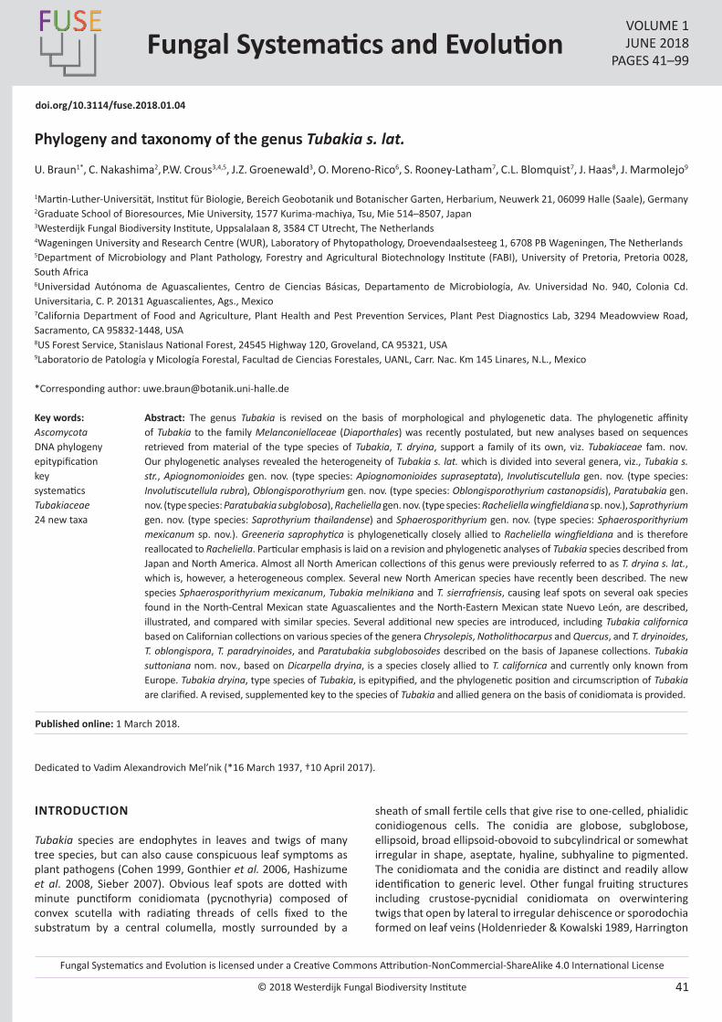
Fungal Systematics and Evolution is licensed under a Creative Commons Attribution-NonCommercial-ShareAlike 4.0 International License
© 2018 Westerdijk Fungal Biodiversity Institute 41
Editor-in-ChiefProf. dr P.W. Crous, Westerdijk Fungal Biodiversity Institute, P.O. Box 85167, 3508 AD Utrecht, The Netherlands.E-mail:[email protected]
Fungal Systematics and EvolutionVOLUME 1JUNE 2018
PAGES 41–99
doi.org/10.3114/fuse.2018.01.04
Phylogeny and taxonomy of the genus Tubakia s. lat.
U. Braun1*, C. Nakashima2, P.W. Crous3,4,5, J.Z. Groenewald3, O. Moreno-Rico6, S. Rooney-Latham7, C.L. Blomquist7, J. Haas8, J. Marmolejo9
1Martin-Luther-Universität, Institut für Biologie, Bereich Geobotanik und Botanischer Garten, Herbarium, Neuwerk 21, 06099 Halle (Saale), Germany2Graduate School of Bioresources, Mie University, 1577 Kurima-machiya, Tsu, Mie 514–8507, Japan3Westerdijk Fungal Biodiversity Institute, Uppsalalaan 8, 3584 CT Utrecht, The Netherlands4Wageningen University and Research Centre (WUR), Laboratory of Phytopathology, Droevendaalsesteeg 1, 6708 PB Wageningen, The Netherlands5Department of Microbiology and Plant Pathology, Forestry and Agricultural Biotechnology Institute (FABI), University of Pretoria, Pretoria 0028, South Africa6Universidad Autónoma de Aguascalientes, Centro de Ciencias Básicas, Departamento de Microbiología, Av. Universidad No. 940, Colonia Cd. Universitaria, C. P. 20131 Aguascalientes, Ags., Mexico7California Department of Food and Agriculture, Plant Health and Pest Prevention Services, Plant Pest Diagnostics Lab, 3294 Meadowview Road, Sacramento, CA 95832-1448, USA8US Forest Service, Stanislaus National Forest, 24545 Highway 120, Groveland, CA 95321, USA9Laboratorio de Patología y Micología Forestal, Facultad de Ciencias Forestales, UANL, Carr. Nac. Km 145 Linares, N.L., Mexico
*Corresponding author: [email protected]
Abstract: The genus Tubakia is revised on the basis of morphological and phylogenetic data. The phylogenetic affinity of Tubakia to the family Melanconiellaceae (Diaporthales) was recently postulated, but new analyses based on sequences retrieved from material of the type species of Tubakia, T. dryina, support a family of its own, viz. Tubakiaceae fam. nov. Our phylogenetic analyses revealed the heterogeneity of Tubakia s. lat. which is divided into several genera, viz., Tubakia s. str., Apiognomonioides gen. nov. (type species: Apiognomonioides supraseptata), Involutiscutellula gen. nov. (type species: Involutiscutellula rubra), Oblongisporothyrium gen. nov. (type species: Oblongisporothyrium castanopsidis), Paratubakia gen. nov. (type species: Paratubakia subglobosa), Racheliella gen. nov. (type species: Racheliella wingfieldiana sp. nov.), Saprothyrium gen. nov. (type species: Saprothyrium thailandense) and Sphaerosporithyrium gen. nov. (type species: Sphaerosporithyrium mexicanum sp. nov.). Greeneria saprophytica is phylogenetically closely allied to Racheliella wingfieldiana and is therefore reallocated to Racheliella. Particular emphasis is laid on a revision and phylogenetic analyses of Tubakia species described from Japan and North America. Almost all North American collections of this genus were previously referred to as T. dryina s. lat., which is, however, a heterogeneous complex. Several new North American species have recently been described. The new species Sphaerosporithyrium mexicanum, Tubakia melnikiana and T. sierrafriensis, causing leaf spots on several oak species found in the North-Central Mexican state Aguascalientes and the North-Eastern Mexican state Nuevo León, are described, illustrated, and compared with similar species. Several additional new species are introduced, including Tubakia californica based on Californian collections on various species of the genera Chrysolepis, Notholithocarpus and Quercus, and T. dryinoides, T. oblongispora, T. paradryinoides, and Paratubakia subglobosoides described on the basis of Japanese collections. Tubakia suttoniana nom. nov., based on Dicarpella dryina, is a species closely allied to T. californica and currently only known from Europe. Tubakia dryina, type species of Tubakia, is epitypified, and the phylogenetic position and circumscription of Tubakia are clarified. A revised, supplemented key to the species of Tubakia and allied genera on the basis of conidiomata is provided.
Key words: AscomycotaDNA phylogenyepitypificationkeysystematicsTubakiaceae24 new taxa
Published online: 1 March 2018.
Dedicated to Vadim Alexandrovich Mel’nik (*16 March 1937, †10 April 2017).
INTRODUCTION
Tubakia species are endophytes in leaves and twigs of many tree species, but can also cause conspicuous leaf symptoms as plant pathogens (Cohen 1999, Gonthier et al. 2006, Hashizume et al. 2008, Sieber 2007). Obvious leaf spots are dotted with minute punctiform conidiomata (pycnothyria) composed of convex scutella with radiating threads of cells fixed to the substratum by a central columella, mostly surrounded by a
sheath of small fertile cells that give rise to one-celled, phialidic conidiogenous cells. The conidia are globose, subglobose, ellipsoid, broad ellipsoid-obovoid to subcylindrical or somewhat irregular in shape, aseptate, hyaline, subhyaline to pigmented. The conidiomata and the conidia are distinct and readily allow identification to generic level. Other fungal fruiting structures including crustose-pycnidial conidiomata on overwintering twigs that open by lateral to irregular dehiscence or sporodochia formed on leaf veins (Holdenrieder & Kowalski 1989, Harrington
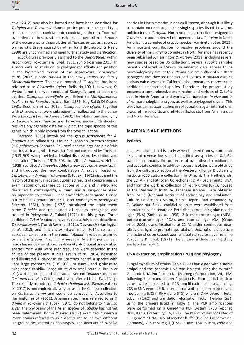
© 2018 Westerdijk Fungal Biodiversity Institute
Braun et al.
Editor-in-ChiefProf. dr P.W. Crous, Westerdijk Fungal Biodiversity Institute, P.O. Box 85167, 3508 AD Utrecht, The Netherlands.E-mail:[email protected]
42
et al. 2012) may also be formed and have been described for T. dryina and T. iowensis. Some species produce a second type of much smaller conidia (microconidia), either in “normal” pycnothyria or in separate, mostly smaller pycnothyria. Reports of the occurrence and sporulation of Tubakia dryina as a saprobe on necrotic tissue caused by other fungi (Munkvold & Neely 1990) are unconfirmed and need further study and clarification.
Tubakia was previously assigned to the Diaporthales within Ascomycota (Yokoyama & Tubaki 1971, Yun & Rossman 2011). In a more detailed study on its phylogenetic affinity and position in the hierarchical system of the Ascomycota, Senanayake et al. (2017) placed Tubakia in the newly introduced family Melanconiellaceae. The sexual morph of “T. dryina” has been referred to as Dicarpella dryina (Belisario 1991). However, D. dryina is not the type species of Dicarpella, and at least one species, Dicarpella quercifolia was linked to Mastigosporella hyalina (≡ Harknessia hyalina; Barr 1979, Nag Raj & Di Cosmo 1981, Rossman et al. 2015). Dicarpella quercifolia, together with D. georgiana, were subsequently reallocated to the genus Wuestneiopsis (Reid & Dawsett 1990). The relation and synonymy of Dicarpella and Tubakia are, however, unclear. Clarification requires phylogenetic data for D. bina, the type species of this genus, which is only known from the type collection.
Saccardo (1913) introduced the genus Actinopelte for A. japonica, a scutellate fungus found in Japan on Castanea crenata (= C. pubinervis). Saccardo (l.c.) confused the large conidia of this species with asci, which was clarified and corrected by Theissen (1913: 509) who provided a detailed discussion, description, and illustration (Theissen 1913: 508, fig. VI) of A. japonica. Höhnel (1925) revisited Actinopelte, added a new species, A. americana, and introduced the new combination A. dryina, based on Leptothyrium dryinum. Yokoyama & Tubaki (1971) discussed the history of this genus in detail, published results of comprehensive examinations of Japanese collections in vivo and in vitro, and described A. castanopsidis, A. rubra, and A. subglobosa based on Japanese collections. Since Saccardo’s Actinopelte turned out to be illegitimate (Art. 53.1, later homonym of Actinopelte Stitzenb. 1861), Sutton (1973) introduced the replacement name Tubakia and reallocated all species recognised and treated in Yokoyama & Tubaki (1971) to this genus. Three additional Tubakia species have subsequently been described: T. seoraksanensis (Yun & Rossman 2011), T. iowensis (Harrington et al. 2012), and T. chinensis (Braun et al. 2014). So far, all European collections in the genus Tubakia have been assigned to a single species, T. dryina, whereas in Asia this genus has a much higher degree of species diversity. Additional undescribed species from Asia were predicted, and were detected in the course of the present studies. Braun et al. (2014) described and illustrated T. chinensis on Castanea henryi, a species with very large pycnothyria (135–200 µm diam), and globose to subglobose conidia. Based on its very small scutella, Braun et al. (2014) described and illustrated a second Tubakia species on Castanea henryi in China, tentatively referred to as Tubakia sp. The recently introduced Tubakia thailandensis (Senanayake et al. 2017) is morphologically very close to the Chinese collection on Castanea henryi and could be conspecific. According to Harrington et al. (2012), Japanese specimens referred to as T. dryina in Yokoyama & Tubaki (1971) do not belong to T. dryina s. str. The phylogeny of the Asian species of Tubakia has not yet been determined. Boroń & Grad (2017) examined numerous Polish strains referred to as T. dryina and found two different ITS groups designated as haplotypes. The diversity of Tubakia
species in North America is not well known, although it is likely to contain more than just the single species listed in various publications as T. dryina. North American collections assigned to T. dryina are undoubtedly heterogeneous, i.e., T. dryina in North America is a complex of cryptic species (Harrington et al. 2012). An important contribution to resolve problems around the diversity of the T. dryina complex in North America has recently been published by Harrington & McNew (2018), including several new species based on US collections. Several Tubakia samples recently collected in Mexico on endemic oaks appear to be morphologically similar to T. dryina but are sufficiently distinct to suggest that they are undescribed species. A Tubakia causing serious oak diseases in California also appears to represent an additional undescribed species. Therefore, the present study presents a comprehensive examination and revision of Tubakia spp. in North America and worldwide, based on in vivo and in vitro morphological analyses as well as phylogenetic data. This work has been accomplished in collaboration by an international group of mycologists and phytopathologists from Asia, Europe and North America.
MATERIALS AND METHODS
Isolates
Isolates included in this study were obtained from symptomatic leaves of diverse hosts, and identified as species of Tubakia based on primarily the presence of pycnothyrial conidiomata and aseptate conidia. In addition, several isolates were obtained from the culture collection of the Westerdijk Fungal Biodiversity Institute (CBS culture collection), in Utrecht, The Netherlands, the CDFA Plant Pathogen Collections (CDFA), Sacramento, USA, and from the working collection of Pedro Crous (CPC), housed at the Westerdijk Institute. Japanese isolates were obtained from NBRC (National Institute of Technology and Evaluation, Culture Collection Division, Chiba, Japan) and examined by C. Nakashima. Single conidial colonies were established from sporulating conidiomata on Petri dishes containing pine needle agar (PNA) (Smith et al. 1996), 2 % malt extract agar (MEA), potato-dextrose agar (PDA), and oatmeal agar (OA) (Crous et al. 2009b), and incubated at 22 °C under continuous near-ultraviolet light to promote sporulation. Descriptions of culture characteristics on Czapek agar and potato sucrose agar refer to Yokoyama & Tubaki (1971). The cultures included in this study are listed in Table 1.
DNA extraction, amplification (PCR) and phylogeny
Fungal mycelium of strains (Table 1) was harvested with a sterile scalpel and the genomic DNA was isolated using the Wizard® Genomic DNA Purification Kit (Promega Corporation, WI, USA) following the manufacturers’ protocols. Four partial nuclear genes were subjected to PCR amplification and sequencing: 28S nrRNA gene (LSU), internal transcribed spacer regions and intervening 5.8S nrRNA gene (ITS) of the nrDNA operon, beta-tubulin (tub2) and translation elongation factor 1-alpha (tef1) using the primers listed in Table 2. The PCR amplifications were performed on a GeneAmp PCR System 9700 (Applied Biosystems, Foster City, CA, USA). The PCR mixtures consisted of 1 µL genomic DNA, 1× NH4 reaction buffer (Bioline, Luckenwalde, Germany), 2–5 mM MgCl2 (ITS: 2.5 mM, LSU: 5 mM, rpb2 and
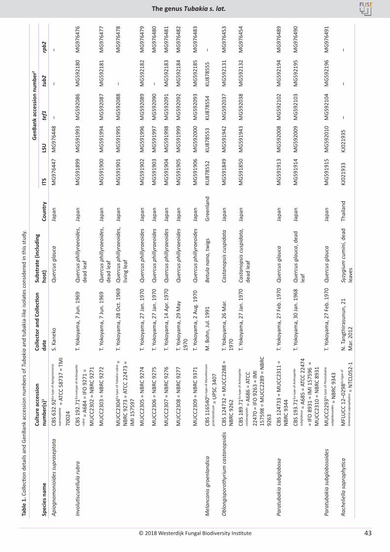
© 2018 Westerdijk Fungal Biodiversity Institute
The genus Tubakia s. lat.
Editor-in-ChiefProf. dr P.W. Crous, Westerdijk Fungal Biodiversity Institute, P.O. Box 85167, 3508 AD Utrecht, The Netherlands.E-mail:[email protected]
43
Tabl
e 1.
Col
lecti
on d
etai
ls an
d Ge
nBan
k ac
cess
ion
num
bers
of T
ubak
ia a
nd tu
baki
a-lik
e iso
late
s con
sider
ed in
this
stud
y.
Spec
ies n
ame
Cultu
re a
cces
sion
nu
mbe
r(s)
1Co
llect
or a
nd C
olle
ction
da
teSu
bstr
ate
(incl
udin
g ho
st)
Coun
try
Gen
Bank
acc
essi
on n
umbe
r2
ITS
LSU
tef1
tub2
rpb2
Apio
gnom
onio
ides
supr
asep
tata
CBS
632.
92Ex
-typ
e of
Api
ogno
mon
ia
supr
asep
tata
= A
TCC
5873
7 =
TMI
7002
4
S. K
anek
oQ
uerc
us g
lauc
aJa
pan
MG9
7644
7M
G976
448
‒ ‒
‒
Invo
lutis
cute
llula
rubr
aCB
S 19
2.71
Ex-is
otyp
e of
Acti
nope
lte
rubr
a = A
684
= IF
O 9
271
= M
UCC
2302
= N
BRC
9271
T. Y
okoy
ama,
7 Ju
n. 1
969
Que
rcus
phi
llyra
eoid
es,
dead
leaf
Japa
nM
G591
899
MG5
9199
3M
G592
086
MG5
9218
0M
G976
476
MU
CC23
03 =
NBR
C 92
72T.
Yok
oyam
a, 7
Jun.
196
9Q
uerc
us p
hilly
raeo
ides
, de
ad le
afJa
pan
MG5
9190
0M
G591
994
MG5
9208
7M
G592
181
MG9
7647
7
MU
CC23
04Ex
-typ
e of
Tub
akia
rubr
a =
NBR
C 92
73 =
ATC
C 22
473
= IM
I 157
597
T. Y
okoy
ama,
28
Oct
. 196
9Q
uerc
us p
hilly
raeo
ides
, liv
ing
leaf
Japa
nM
G591
901
MG5
9199
5M
G592
088
‒M
G976
478
MU
CC23
05 =
NBR
C 92
74T.
Yok
oyam
a, 2
7 Ja
n. 1
970
Que
rcus
phi
llyra
eoid
es
Japa
nM
G591
902
MG5
9199
6M
G592
089
MG5
9218
2M
G976
479
MU
CC23
06 =
NBR
C 92
75T.
Yok
oyam
a, 2
7 Ja
n. 1
970
Que
rcus
phi
llyra
eoid
esJa
pan
MG5
9190
3M
G591
997
MG5
9209
0 ‒
MG9
7648
0
MU
CC23
07 =
NBR
C 92
76T.
Yok
oyam
a, 1
4 Ap
r. 19
70Q
uerc
us p
hilly
raeo
ides
Japa
nM
G591
904
MG5
9199
8M
G592
091
MG5
9218
3M
G976
481
MU
CC23
08 =
NBR
C 92
77T.
Yok
oyam
a, 2
9 M
ay
1970
Que
rcus
phi
llyra
eoid
es
Japa
nM
G591
905
MG5
9199
9M
G592
092
MG5
9218
4M
G976
482
MU
CC23
09 =
NBR
C 93
71T.
Yok
oyam
a, 2
Aug
. 197
0Q
uerc
us p
hilly
raeo
ides
Ja
pan
MG5
9190
6M
G592
000
MG5
9209
3M
G592
185
MG9
7648
3
Mel
anco
nis g
roen
land
ica
CBS
1165
40Ex
-typ
e of
Myr
othe
cium
groe
nlan
dicu
m =
UPS
C 34
07M
. Boh
n, Ju
l. 19
91Be
tula
nan
a, tw
igs
Gree
nlan
dKU
8785
52KU
8785
53KU
8785
54KU
8785
55 ‒
Obl
ongi
spor
othy
rium
cas
tano
psid
isCB
S 12
4732
= M
UCC
2288
=
NBR
C 92
62T.
Yok
oyam
a, 2
6 M
ar.
1970
Cast
anop
sis c
uspi
data
Japa
nM
G591
849
MG5
9194
2M
G592
037
MG5
9213
1M
G976
453
CBS
189.
71Ex
-typ
e of
Acti
nope
lte
cast
anop
sidis =
A68
6 =
ATCC
22
470
= IF
O 9
263
= IM
I 15
7598
= M
UCC
2289
= N
BRC
9263
T. Y
okoy
ama,
27
Jan.
197
0Ca
stan
opsis
cus
pida
ta,
dead
leaf
Japa
nM
G591
850
MG5
9194
3M
G592
038
MG5
9213
2M
G976
454
Para
tuba
kia
subg
lobo
saCB
S 12
4733
= M
UCC
2311
=
NBR
C 93
44T.
Yok
oyam
a, 2
7 Fe
b. 1
970
Que
rcus
gla
uca
Japa
nM
G591
913
MG5
9200
8M
G592
102
MG5
9219
4M
G976
489
CBS
193.
71Ex
-typ
e of
Acti
nope
lte
subg
lobo
sa =
A68
5 =
ATCC
224
74
= IF
O 8
931
= IM
I 157
596
=
MU
CC23
10 =
NBR
C 89
31
T. Y
okoy
ama,
30
Jan.
196
8Q
uerc
us g
lauc
a, d
ead
leaf
Japa
nM
G591
914
MG5
9200
9M
G592
103
MG5
9219
5M
G976
490
Para
tuba
kia
subg
lobo
soid
esM
UCC
2293
Ex-t
ype
of P
arat
ubak
ia
subg
lobo
soid
es =
NBR
C 93
43T.
Yok
oyam
a, 2
7 Fe
b. 1
970
Que
rcus
gla
uca
Japa
nM
G591
915
MG5
9201
0M
G592
104
MG5
9219
6M
G976
491
Rach
elie
lla sa
prop
hytic
aM
FLU
CC 1
2–02
98Ex
-typ
e of
Gree
neria
sapr
ophy
tica =
NTC
L052
-1N
. Tan
gthi
rasu
nun,
21
Mar
. 201
2Sy
zygi
um c
umin
i, de
ad
leav
esTh
aila
ndKJ
0219
33KJ
0219
35 ‒
‒ ‒
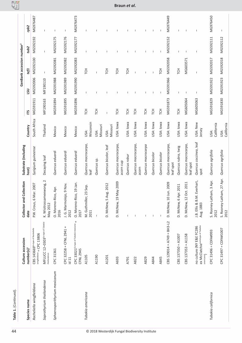
© 2018 Westerdijk Fungal Biodiversity Institute
Braun et al.
Editor-in-ChiefProf. dr P.W. Crous, Westerdijk Fungal Biodiversity Institute, P.O. Box 85167, 3508 AD Utrecht, The Netherlands.E-mail:[email protected]
44
Tabl
e 1.
(Con
tinue
d).
Spec
ies n
ame
Cultu
re a
cces
sion
nu
mbe
r(s)
1Co
llect
or a
nd C
olle
ction
da
teSu
bstr
ate
(incl
udin
g ho
st)
Coun
try
Gen
Bank
acc
essi
on n
umbe
r2
ITS
LSU
tef1
tub2
rpb2
Rach
elie
lla w
ingfi
eldi
ana
CBS
1436
69Ex
-typ
e of
Rac
helie
lla
win
gfiel
dian
a = C
PC 1
3806
P.W. C
rous
, 6 M
ar. 2
007
Syzig
ium
gui
neen
seSo
uth
Afric
aM
G591
911
MG5
9200
6M
G592
100
MG5
9219
2M
G976
487
Sapr
othy
rium
thai
land
ense
MFL
UCC
12–
0303
Ex-t
ype
of T
ubak
ia
thai
land
ensis
K. W
isitr
assa
mee
won
g, 2
M
ay 2
012
Deca
ying
leaf
Thai
land
MF1
9016
3M
F190
110
‒ ‒
‒
Spha
eros
porit
hyriu
m m
exic
anum
CPC
3136
1O
. Mor
eno-
Rico
, Apr
. 20
16Q
uerc
us e
duar
diM
exic
oM
G591
894
MG5
9198
8M
G592
081
MG5
9217
5 ‒
CPC
3225
8 =
CFN
L 29
41 =
id
11
J. G.
Mar
mol
ejo,
9 N
ov.
2012
Que
rcus
edu
ardi
Mex
ico
MG5
9189
5M
G591
989
MG5
9208
2M
G592
176
‒
CPC
3302
1Ex-t
ype
of T
ubak
ia m
exic
ana =
CF
NL
2945
O. M
oren
o-Ri
co, 1
9 Ja
n.
2017
Que
rcus
edu
ardi
Mex
ico
MG5
9189
6M
G591
990
MG5
9208
3M
G592
177
MG9
7647
3
Tuba
kia
amer
ican
aA1
105
M. G
uthm
iller
, 23
Sep.
20
11Q
uerc
us m
acro
carp
a,
leaf
USA
: W
iscon
sinTC
H ‒
TCH
‒ ‒
A119
0 ‒
Que
rcus
sp.
USA
: M
issou
riTC
H ‒
‒ ‒
‒
A120
1D.
McN
ew, 5
Aug
. 201
2Q
uerc
us b
icol
or, l
eaf
USA
: M
issou
ri ‒
‒TC
H ‒
‒
A693
D. M
cNew
, 19
May
200
9Q
uerc
us m
acro
carp
a,
acor
n cu
pU
SA: I
owa
TCH
‒TC
H ‒
‒
A791
‒Q
uerc
us ro
bur
USA
: Iow
aTC
H ‒
TCH
‒ ‒
A822
‒Q
uerc
us m
acro
carp
a,
acor
nU
SA: I
owa
TCH
‒ ‒
‒ ‒
A829
‒Q
uerc
us m
acro
carp
aU
SA: I
owa
TCH
‒ ‒
‒ ‒
A844
‒Q
uerc
us b
icol
orU
SA: I
owa
TCH
‒ ‒
‒ ‒
A845
‒Q
uerc
us b
icol
orU
SA: I
owa
TCH
‒TC
H ‒
‒
CBS
1290
14 =
A74
9 =
BH3-
L2D.
McN
ew, 1
0 Ju
n. 2
009
Que
rcus
mac
roca
rpa,
le
af sp
otU
SA: I
owa
MG5
9187
3M
G591
966
MG5
9205
8M
G592
152
MG9
7644
9
CBS
1373
50 =
A10
07D.
McN
ew, 6
Apr
. 201
1Q
uerc
us ru
bra,
twig
USA
: Iow
aTC
H ‒
TCH
‒ ‒
CBS
1373
53 =
A11
58D.
McN
ew, 1
2 O
ct. 2
011
Que
rcus
mac
roca
rpa,
le
af sp
otU
SA: I
owa
MG6
0506
4 ‒
MG6
0357
1 ‒
‒
no c
ultu
re (N
Y E&
E FC
286
ex M
cCla
tchi
eLect
otyp
e of
Tub
akia
amer
ican
a )
J.B. E
llis &
B.M
. Eve
rhar
t, Au
g. 1
883
Que
rcus
coc
cine
a, le
af
spot
USA
: New
Je
rsey
MG6
0506
3 ‒
‒ ‒
‒
Tuba
kia
calif
orni
caCP
C 31
496
= CD
FA#9
93S.
Roo
ney-
Lath
am, 3
Apr
. 20
12Q
uerc
us a
grifo
liaU
SA:
Calif
orni
aM
G591
829
MG5
9192
2M
G592
017
MG5
9211
1M
G976
450
CPC
3149
7 =
CDFA
#100
7S.
Roo
ney-
Lath
am, 2
7 Ap
r. 20
12Q
uerc
us a
grifo
liaU
SA:
Calif
orni
aM
G591
830
MG5
9192
3M
G592
018
MG5
9211
2 ‒
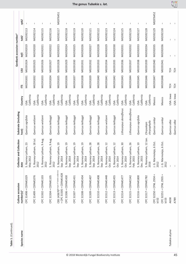
© 2018 Westerdijk Fungal Biodiversity Institute
The genus Tubakia s. lat.
Editor-in-ChiefProf. dr P.W. Crous, Westerdijk Fungal Biodiversity Institute, P.O. Box 85167, 3508 AD Utrecht, The Netherlands.E-mail:[email protected]
45
Tabl
e 1.
(Con
tinue
d).
Spec
ies n
ame
Cultu
re a
cces
sion
nu
mbe
r(s)
1Co
llect
or a
nd C
olle
ction
da
teSu
bstr
ate
(incl
udin
g ho
st)
Coun
try
Gen
Bank
acc
essi
on n
umbe
r2
ITS
LSU
tef1
tub2
rpb2
CPC
3149
8 =
CDFA
#102
9S.
Roo
ney-
Lath
am, 2
3 M
ay 2
012
Que
rcus
agr
ifolia
USA
: Ca
lifor
nia
MG5
9183
1M
G591
924
MG5
9201
9M
G592
113
‒
CPC
3149
9 =
CDFA
#107
6S.
Roo
ney-
Lath
am, 1
8 Ju
l. 20
12Q
uerc
us w
islize
niU
SA:
Calif
orni
aM
G591
832
MG5
9192
5M
G592
020
MG5
9211
4 ‒
CPC
3150
2 =
CDFA
#110
3S.
Roo
ney-
Lath
am, 9
Aug
. 20
12Q
uerc
us w
islize
niU
SA:
Calif
orni
aM
G591
833
MG5
9192
6M
G592
021
MG5
9211
5 ‒
CPC
3150
4 =
CDFA
#110
5S.
Roo
ney-
Lath
am, 9
Aug
. 20
12Q
uerc
us k
ello
ggii
USA
: Ca
lifor
nia
MG5
9183
4M
G591
927
MG5
9202
2M
G592
116
‒
CBS
1436
70Ex
-typ
e of
Tub
akia
cal
iforn
ica
= CP
C 31
505
= CD
FA#1
428
S. R
oone
y-La
tham
, 19
Sep.
201
4Q
uerc
us k
ello
ggii
USA
: Ca
lifor
nia
MG5
9183
5M
G591
928
MG5
9202
3M
G592
117
MG9
7645
1
CPC
3150
6 =
CDFA
#143
0S.
Roo
ney-
Lath
am, 1
9 Se
p. 2
014
Que
rcus
kel
logg
iiU
SA:
Calif
orni
aM
G591
836
MG5
9192
9M
G592
024
MG5
9211
8 ‒
CPC
3150
7 =
CDFA
#143
1S.
Roo
ney-
Lath
am, 1
9 Se
p. 2
014
Que
rcus
kel
logg
iiU
SA:
Calif
orni
aM
G591
837
MG5
9193
0M
G592
025
MG5
9211
9 ‒
CPC
3150
8 =
CDFA
#143
4S.
Roo
ney-
Lath
am, 1
9 Se
p. 2
014
Que
rcus
kel
logg
iiU
SA:
Calif
orni
aM
G591
838
MG5
9193
1M
G592
026
MG5
9212
0 ‒
CPC
3150
9 =
CDFA
#140
7S.
Roo
ney-
Lath
am, 2
8 Se
p. 2
014
Que
rcus
kel
logg
iiU
SA:
Calif
orni
aM
G591
839
MG5
9193
2M
G592
027
MG5
9212
1 ‒
CPC
3151
0 =
CDFA
#140
8S.
Roo
ney-
Lath
am, 2
8 Se
p. 2
014
Que
rcus
kel
logg
iiU
SA:
Calif
orni
aM
G591
840
MG5
9193
3M
G592
028
MG5
9212
2 ‒
CPC
3151
2 =
CDFA
#144
8S.
Roo
ney-
Lath
am, 1
2 O
ct. 2
014
Que
rcus
wisl
izeni
USA
: Ca
lifor
nia
MG5
9184
1M
G591
934
MG5
9202
9M
G592
123
‒
CPC
3151
3 =
CDFA
#145
5S.
Roo
ney-
Lath
am, 1
2 O
ct. 2
014
Que
rcus
kel
logg
iiU
SA:
Calif
orni
aM
G591
842
MG5
9193
5M
G592
030
MG5
9212
4 ‒
CPC
3151
4 =
CDFA
#147
7S.
Roo
ney-
Lath
am, 3
0 O
ct. 2
014
Lith
ocar
pus d
ensifl
orus
USA
: Ca
lifor
nia
MG5
9184
3M
G591
936
MG5
9203
1M
G592
125
‒
CPC
3151
5 =
CDFA
#154
2S.
Roo
ney-
Lath
am, 2
6 Fe
b. 2
015
Lith
ocar
pus d
ensifl
orus
USA
: Ca
lifor
nia
MG5
9184
4M
G591
937
MG5
9203
2M
G592
126
‒
CPC
3151
6 =
CDFA
#180
0S.
Roo
ney-
Lath
am, 1
0 De
c. 2
015
Que
rcus
agr
ifolia
USA
: Ca
lifor
nia
MG5
9184
5M
G591
938
MG5
9203
3M
G592
127
‒
CPC
3151
7 =
CDFA
#178
2S.
Roo
ney-
Lath
am, 1
2 Ja
n.
2016
Chry
sole
pis
chry
soph
ylla
USA
: Ca
lifor
nia
MG5
9184
6M
G591
939
MG5
9203
4M
G592
128
‒
CPC
3225
0 =
CFN
L 29
34 =
id
02
J. G.
Mar
mol
ejo,
6 O
ct.
2012
Que
rcus
can
byi
Mex
ico
MG5
9184
7M
G591
940
MG5
9203
5M
G592
129
MG9
7645
2
CPC
3225
1 =
CFN
L 29
35 =
id
03
J. G.
Mar
mol
ejo,
6 O
ct.
2012
Que
rcus
can
byi
Mex
ico
MG5
9184
8M
G591
941
MG5
9203
6M
G592
130
‒
Tuba
kia
dryi
naA6
58 ‒
Que
rcus
alb
aU
SA: I
owa
TCH
TCH
‒ ‒
‒
A789
‒Q
uerc
us a
lba
USA
: Iow
aTC
HTC
H ‒
‒ ‒
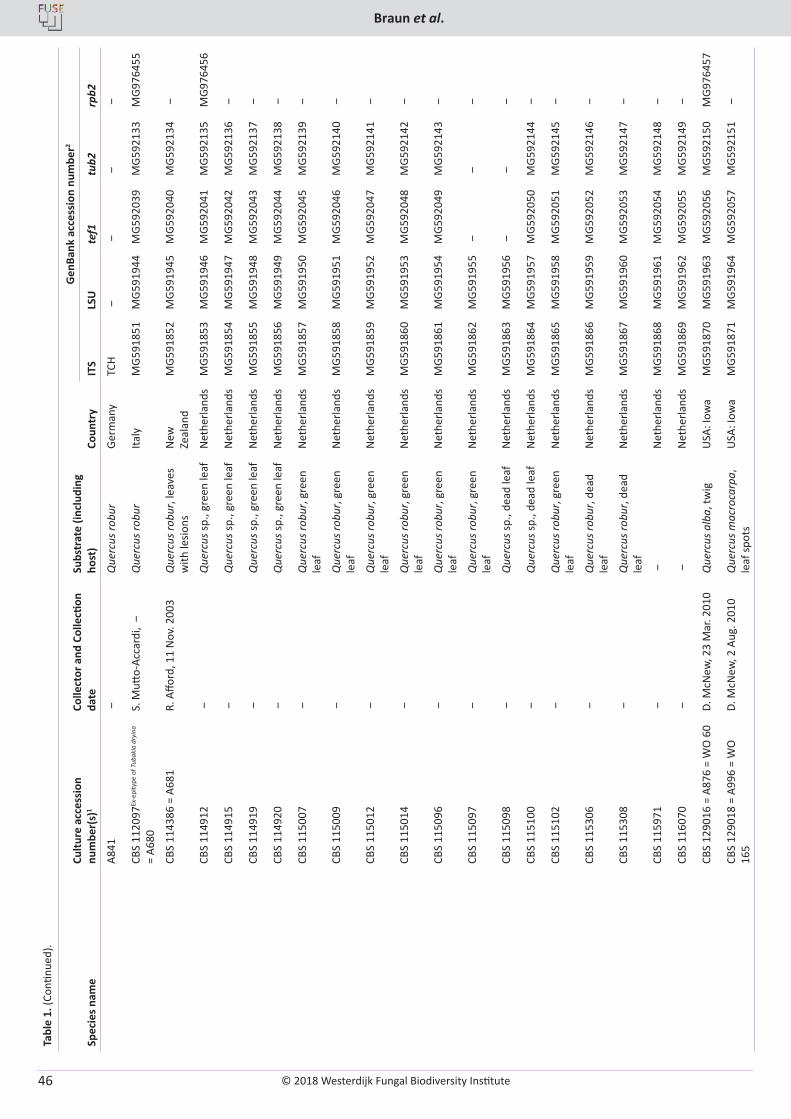
© 2018 Westerdijk Fungal Biodiversity Institute
Braun et al.
Editor-in-ChiefProf. dr P.W. Crous, Westerdijk Fungal Biodiversity Institute, P.O. Box 85167, 3508 AD Utrecht, The Netherlands.E-mail:[email protected]
46
Tabl
e 1.
(Con
tinue
d).
Spec
ies n
ame
Cultu
re a
cces
sion
nu
mbe
r(s)
1Co
llect
or a
nd C
olle
ction
da
teSu
bstr
ate
(incl
udin
g ho
st)
Coun
try
Gen
Bank
acc
essi
on n
umbe
r2
ITS
LSU
tef1
tub2
rpb2
A841
‒Q
uerc
us ro
bur
Germ
any
TCH
‒ ‒
‒ ‒
CBS
1120
97Ex
-epi
type
of T
ubak
ia d
ryin
a =
A680
S. M
utto-
Acca
rdi,
‒Q
uerc
us ro
bur
Italy
MG5
9185
1M
G591
944
MG5
9203
9M
G592
133
MG9
7645
5
CBS
1143
86 =
A68
1R.
Affo
rd, 1
1 N
ov. 2
003
Que
rcus
robu
r, le
aves
w
ith le
sions
New
Ze
alan
dM
G591
852
MG5
9194
5M
G592
040
MG5
9213
4 ‒
CBS
1149
12 ‒
Que
rcus
sp.,
gree
n le
afN
ethe
rland
sM
G591
853
MG5
9194
6M
G592
041
MG5
9213
5M
G976
456
CBS
1149
15 ‒
Que
rcus
sp.,
gree
n le
afN
ethe
rland
sM
G591
854
MG5
9194
7M
G592
042
MG5
9213
6 ‒
CBS
1149
19 ‒
Que
rcus
sp.,
gree
n le
afN
ethe
rland
sM
G591
855
MG5
9194
8M
G592
043
MG5
9213
7 ‒
CBS
1149
20 ‒
Que
rcus
sp.,
gree
n le
afN
ethe
rland
sM
G591
856
MG5
9194
9M
G592
044
MG5
9213
8 ‒
CBS
1150
07 ‒
Que
rcus
robu
r, gr
een
leaf
Net
herla
nds
MG5
9185
7M
G591
950
MG5
9204
5M
G592
139
‒
CBS
1150
09 ‒
Que
rcus
robu
r, gr
een
leaf
Net
herla
nds
MG5
9185
8M
G591
951
MG5
9204
6M
G592
140
‒
CBS
1150
12 ‒
Que
rcus
robu
r, gr
een
leaf
Net
herla
nds
MG5
9185
9M
G591
952
MG5
9204
7M
G592
141
‒
CBS
1150
14 ‒
Que
rcus
robu
r, gr
een
leaf
Net
herla
nds
MG5
9186
0M
G591
953
MG5
9204
8M
G592
142
‒
CBS
1150
96 ‒
Que
rcus
robu
r, gr
een
leaf
Net
herla
nds
MG5
9186
1M
G591
954
MG5
9204
9M
G592
143
‒
CBS
1150
97 ‒
Que
rcus
robu
r, gr
een
leaf
Net
herla
nds
MG5
9186
2M
G591
955
‒ ‒
‒
CBS
1150
98 ‒
Que
rcus
sp.,
dead
leaf
Net
herla
nds
MG5
9186
3M
G591
956
‒ ‒
‒
CBS
1151
00 ‒
Que
rcus
sp.,
dead
leaf
Net
herla
nds
MG5
9186
4M
G591
957
MG5
9205
0M
G592
144
‒
CBS
1151
02 ‒
Que
rcus
robu
r, gr
een
leaf
Net
herla
nds
MG5
9186
5M
G591
958
MG5
9205
1M
G592
145
‒
CBS
1153
06 ‒
Que
rcus
robu
r, de
ad
leaf
Net
herla
nds
MG5
9186
6M
G591
959
MG5
9205
2M
G592
146
‒
CBS
1153
08 ‒
Que
rcus
robu
r, de
ad
leaf
Net
herla
nds
MG5
9186
7M
G591
960
MG5
9205
3M
G592
147
‒
CBS
1159
71 ‒
‒N
ethe
rland
sM
G591
868
MG5
9196
1M
G592
054
MG5
9214
8 ‒
CBS
1160
70 ‒
‒N
ethe
rland
sM
G591
869
MG5
9196
2M
G592
055
MG5
9214
9 ‒
CBS
1290
16 =
A87
6 =
WO
60
D. M
cNew
, 23
Mar
. 201
0Q
uerc
us a
lba,
twig
USA
: Iow
aM
G591
870
MG5
9196
3M
G592
056
MG5
9215
0M
G976
457
CBS
1290
18 =
A99
6 =
WO
16
5D.
McN
ew, 2
Aug
. 201
0Q
uerc
us m
acro
carp
a,
leaf
spot
sU
SA: I
owa
MG5
9187
1M
G591
964
MG5
9205
7M
G592
151
‒
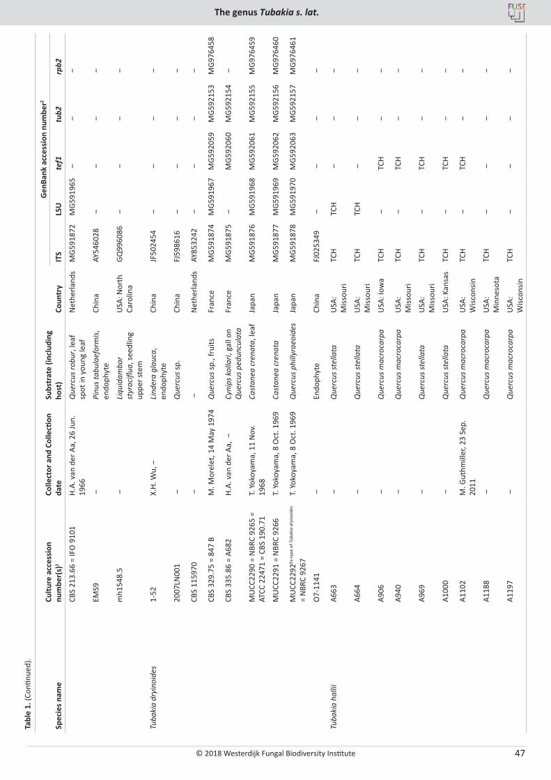
© 2018 Westerdijk Fungal Biodiversity Institute
The genus Tubakia s. lat.
Editor-in-ChiefProf. dr P.W. Crous, Westerdijk Fungal Biodiversity Institute, P.O. Box 85167, 3508 AD Utrecht, The Netherlands.E-mail:[email protected]
47
Tabl
e 1.
(Con
tinue
d).
Spec
ies n
ame
Cultu
re a
cces
sion
nu
mbe
r(s)
1Co
llect
or a
nd C
olle
ction
da
teSu
bstr
ate
(incl
udin
g ho
st)
Coun
try
Gen
Bank
acc
essi
on n
umbe
r2
ITS
LSU
tef1
tub2
rpb2
CBS
213.
66 =
IFO
910
1H.
A. v
an d
er A
a, 2
6 Ju
n.
1966
Que
rcus
robu
r, le
af
spot
in y
oung
leaf
Net
herla
nds
MG5
9187
2M
G591
965
‒ ‒
‒
EMS9
‒Pi
nus t
abul
aefo
rmis,
en
doph
yte
Chin
aAY
5460
28 ‒
‒ ‒
‒
mh1
548.
5 ‒
Liqu
idam
bar
styr
acifl
ua, s
eedl
ing
uppe
r ste
m
USA
: Nor
th
Caro
lina
GQ99
6086
‒ ‒
‒ ‒
Tuba
kia
dryi
noid
es1-
52X.
H. W
u, ‒
Lind
era
glau
ca,
endo
phyt
eCh
ina
JF50
2454
‒ ‒
‒ ‒
2007
LN00
1 ‒
Que
rcus
sp.
Chin
aFJ
5986
16 ‒
‒ ‒
‒
CBS
1159
70 ‒
‒N
ethe
rland
sAY
8532
42 ‒
‒ ‒
‒
CBS
329.
75 =
847
BM
. Mor
elet
, 14
May
197
4Q
uerc
us sp
., fr
uits
Fran
ceM
G591
874
MG5
9196
7M
G592
059
MG5
9215
3M
G976
458
CBS
335.
86 =
A68
2H.
A. v
an d
er A
a, ‒
Cyni
ps k
olla
ri, g
all o
n Q
uerc
us p
edun
cula
taFr
ance
MG5
9187
5 ‒
MG5
9206
0M
G592
154
‒
MU
CC22
90 =
NBR
C 92
65 =
AT
CC 2
2471
= C
BS 1
90.7
1 T.
Yok
oyam
a, 1
1 N
ov.
1968
Cast
anea
cre
nata
, lea
fJa
pan
MG5
9187
6M
G591
968
MG5
9206
1M
G592
155
MG9
7645
9
MU
CC22
91 =
NBR
C 92
66T.
Yok
oyam
a, 8
Oct
. 196
9Ca
stan
ea c
rena
taJa
pan
MG5
9187
7M
G591
969
MG5
9206
2M
G592
156
MG9
7646
0
MU
CC22
92Ex
-typ
e of
Tub
akia
dry
inoi
des
= N
BRC
9267
T. Y
okoy
ama,
8 O
ct. 1
969
Que
rcus
phi
llyra
eoid
es
Japa
nM
G591
878
MG5
9197
0M
G592
063
MG5
9215
7M
G976
461
O7-
1141
‒En
doph
yte
Chin
aFJ
0253
49 ‒
‒ ‒
‒
Tuba
kia
halli
iA6
63 ‒
Que
rcus
stel
lata
USA
: M
issou
riTC
HTC
H ‒
‒ ‒
A664
‒Q
uerc
us st
ella
taU
SA:
Miss
ouri
TCH
TCH
‒ ‒
‒
A906
‒Q
uerc
us m
acro
carp
aU
SA: I
owa
TCH
‒TC
H ‒
‒
A940
‒Q
uerc
us m
acro
carp
aU
SA:
Miss
ouri
TCH
‒TC
H ‒
‒
A969
‒Q
uerc
us st
ella
taU
SA:
Miss
ouri
TCH
‒TC
H ‒
‒
A100
0 ‒
Que
rcus
stel
lata
USA
: Kan
sas
TCH
‒TC
H ‒
‒
A110
2M
. Gut
hmill
er, 2
3 Se
p.
2011
Que
rcus
mac
roca
rpa
USA
: W
iscon
sinTC
H ‒
TCH
‒ ‒
A118
8 ‒
Que
rcus
mac
roca
rpa
USA
: M
inne
sota
TCH
‒ ‒
‒ ‒
A119
7 ‒
Que
rcus
mac
roca
rpa
USA
: W
iscon
sinTC
H ‒
‒ ‒
‒
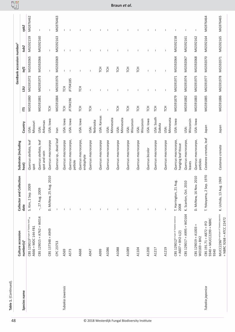
© 2018 Westerdijk Fungal Biodiversity Institute
Braun et al.
Editor-in-ChiefProf. dr P.W. Crous, Westerdijk Fungal Biodiversity Institute, P.O. Box 85167, 3508 AD Utrecht, The Netherlands.E-mail:[email protected]
48
Tabl
e 1.
(Con
tinue
d).
Spec
ies n
ame
Cultu
re a
cces
sion
nu
mbe
r(s)
1Co
llect
or a
nd C
olle
ction
da
teSu
bstr
ate
(incl
udin
g ho
st)
Coun
try
Gen
Bank
acc
essi
on n
umbe
r2
ITS
LSU
tef1
tub2
rpb2
CBS
1290
13Ex
-typ
e of
Tub
akia
hal
lii =
A666
= M
DC 1
44-F
B1S.
Kim
, 2 S
ep. 2
009
Que
rcus
stel
lata
, lea
f sp
otU
SA:
Miss
ouri
MG5
9188
0M
G591
972
MG5
9206
5M
G592
159
MG9
7646
2
CBS
1290
15 =
A76
2 =
W01
4 ‒
, 27
Aug.
200
9Q
uerc
us st
ella
ta, l
eaf
spot
and
vei
nU
SA:
Arka
nsas
MG5
9188
1M
G591
973
MG5
9206
6M
G592
160
‒
CBS
1373
48 =
A94
9D.
McN
ew, 2
5 Au
g. 2
010
Que
rcus
mac
roca
rpa
USA
: Iow
aTC
H ‒
‒ ‒
‒
CPC
2375
3 ‒
Que
rcus
sp.,
dead
leaf
Iran
MG5
9188
4M
G591
976
MG5
9206
9M
G592
163
MG9
7646
3
Tuba
kia
iow
ensis
A569
‒Q
uerc
us m
acro
carp
aU
SA: I
owa
TCH
TCH
‒ ‒
‒
A573
‒Q
uerc
us m
acro
carp
a,
petio
leU
SA: I
owa
JF70
4196
JF70
4185
‒ ‒
‒
A668
‒Q
uerc
us m
acro
carp
a,
endo
phyt
eU
SA: I
owa
TCH
TCH
‒ ‒
‒
A947
‒Q
uerc
us m
acro
carp
aU
SA:
Neb
rask
aTC
H ‒
‒ ‒
‒
A999
‒Q
uerc
us m
acro
carp
aU
SA: K
ansa
s ‒
‒TC
H ‒
‒
A108
6 ‒
Que
rcus
mac
roca
rpa
USA
: M
inne
sota
‒ ‒
TCH
‒ ‒
A108
8 ‒
Que
rcus
mac
roca
rpa
USA
: M
inne
sota
TCH
‒TC
H ‒
‒
A108
9 ‒
Que
rcus
mac
roca
rpa
USA
: W
iscon
sinTC
H ‒
TCH
‒ ‒
A110
4 ‒
Que
rcus
mac
roca
rpa
USA
: W
iscon
sinTC
H ‒
TCH
‒ ‒
A120
0 ‒
Que
rcus
bic
olor
USA
: Iow
aTC
H ‒
‒ ‒
‒
A121
7 ‒
Que
rcus
mac
roca
rpa
USA
: Sou
th
Dako
taTC
H ‒
‒ ‒
‒
A121
9 ‒
Que
rcus
mac
roca
rpa
USA
: M
inne
sota
TCH
‒ ‒
‒ ‒
CBS
1290
12Ex
-typ
e of
Tub
akia
iow
ensis
=
A607
= B
H2-L
(2)
T. H
arrin
gton
, 21
Aug.
20
08Q
uerc
us m
acro
carp
a,
hang
ing
leaf
tiss
ueU
SA: I
owa
MG5
9187
9M
G591
971
MG5
9206
4M
G592
158
‒
CBS
1290
17 =
A99
5 =
WO
164
K. S
canl
on, O
ct. 2
010
Que
rcus
mac
roca
rpa,
le
aves
USA
: W
iscon
sinM
G591
882
MG5
9197
4M
G592
067
MG5
9216
1 ‒
CBS
1290
19 =
A10
03 =
W
O16
9 =
BH2
D. M
cNew
, 16
Nov
. 201
0Q
uerc
us m
acro
carp
a,
petio
les
USA
: Iow
aM
G591
883
MG5
9197
5M
G592
068
MG5
9216
2 ‒
Tuba
kia
japo
nica
CBS
191.
71 =
A87
2 =
IFO
93
40 =
MU
CC22
99 =
NBR
C 93
40
T. Y
okoy
ama,
2 S
ep. 1
970
Cast
anea
cre
nata
, lea
fJa
pan
MG5
9188
5M
G591
977
MG5
9207
0M
G592
164
MG9
7646
4
MU
CC22
96Ex
-epi
type
of T
ubak
ia ja
poni
ca
= N
BRC
9268
= A
TCC
2247
2K.
Uch
ida,
13
Aug.
196
9Ca
stan
ea c
rena
taJa
pan
MG5
9188
6M
G591
978
MG5
9207
1M
G592
165
MG9
7646
5
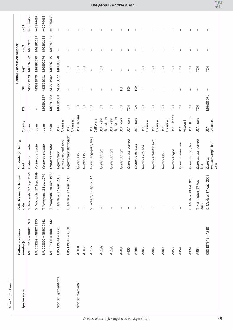
© 2018 Westerdijk Fungal Biodiversity Institute
The genus Tubakia s. lat.
Editor-in-ChiefProf. dr P.W. Crous, Westerdijk Fungal Biodiversity Institute, P.O. Box 85167, 3508 AD Utrecht, The Netherlands.E-mail:[email protected]
49
Tabl
e 1.
(Con
tinue
d).
Spec
ies n
ame
Cultu
re a
cces
sion
nu
mbe
r(s)
1Co
llect
or a
nd C
olle
ction
da
teSu
bstr
ate
(incl
udin
g ho
st)
Coun
try
Gen
Bank
acc
essi
on n
umbe
r2
ITS
LSU
tef1
tub2
rpb2
MU
CC22
97 =
NBR
C 92
69Y.
Koba
yash
i, 17
Sep
. 196
9Ca
stan
ea c
rena
taJa
pan
‒M
G591
979
MG5
9207
2M
G592
166
MG9
7646
6
MU
CC22
98 =
NBR
C 92
70Y.
Koba
yash
i, 17
Sep
. 196
9Ca
stan
ea c
rena
taJa
pan
‒M
G591
980
MG5
9207
3M
G592
167
MG9
7646
7
MU
CC23
00 =
NBR
C 93
41T.
Yok
oyam
a, 2
Sep
. 197
0Ca
stan
ea c
rena
taJa
pan
MG5
9188
7M
G591
981
MG5
9207
4M
G592
168
MG9
7646
8
MU
CC23
01 =
NBR
C 93
42T.
Yok
oyam
a, 3
0 O
ct. 1
970
Cast
anea
cre
nata
Japa
nM
G591
888
MG5
9198
2M
G592
075
MG5
9216
9M
G976
469
Tuba
kia
liqui
dam
baris
CBS
1397
44 =
A77
1D.
McN
ew, 2
7 Au
g. 2
009
Liqu
idam
bar
styr
acifl
ua, l
eaf s
pot
USA
: Ar
kans
asM
G605
068
MG6
0507
7M
G603
578
‒ ‒
CBS
1397
45 =
A83
0D.
McN
ew, 2
7 Au
g. 2
009
Liqu
idam
bar s
tyra
ciflu
aU
SA:
Arka
nsas
TCH
‒TC
H ‒
‒
Tuba
kia
mac
nabb
iiA1
001
‒Q
uerc
us sp
.U
SA: K
ansa
sTC
H ‒
TCH
‒ ‒
A103
9 ‒
Que
rcus
sp.
‒TC
H ‒
TCH
‒ ‒
A117
7S.
Lat
ham
, 27
Apr.
2012
Que
rcus
agr
ifolia
, tw
igU
SA:
Calif
orni
aTC
H ‒
‒ ‒
‒
A119
2 ‒
Que
rcus
rubr
aU
SA: N
ew
Ham
pshi
reTC
H ‒
TCH
‒ ‒
A119
3 ‒
Que
rcus
rubr
aU
SA: N
ew
Ham
pshi
reTC
H ‒
‒ ‒
‒
A608
‒Q
uerc
us ru
bra
USA
: Iow
aTC
HTC
H ‒
‒ ‒
A655
‒Q
uerc
us m
acro
carp
aU
SA: I
owa
TCH
TCH
‒ ‒
‒
A766
‒Ca
stan
ea d
enta
taU
SA: I
owa
TCH
TCH
‒ ‒
‒
A805
‒Q
uerc
us v
eluti
naU
SA:
Arka
nsas
TCH
‒TC
H ‒
‒
A806
‒Q
uerc
us m
arila
ndic
aU
SA:
Arka
nsas
TCH
‒TC
H ‒
‒
A809
‒Q
uerc
us sp
.U
SA:
Arka
nsas
TCH
‒TC
H ‒
‒
A853
‒Q
uerc
us n
igra
USA
: Flo
rida
TCH
‒TC
H ‒
‒
A859
‒Q
uerc
us im
bric
aria
USA
: M
issou
riTC
H ‒
TCH
‒ ‒
A929
D. M
cNew
, 28
Jul.
2010
Que
rcus
rubr
a, le
afU
SA: I
llino
isTC
H ‒
TCH
‒ ‒
A954
T. H
arrin
gton
, 27
Aug.
20
10Q
uerc
us m
acro
carp
a,
leaf
USA
: Iow
aTC
H ‒
TCH
‒ ‒
CBS
1373
46 =
A81
0D.
McN
ew, 2
7 Au
g. 2
009
Que
rcus
m
uehl
enbe
rgii,
leaf
ve
in
USA
: Ar
kans
asM
G605
071
‒TC
H ‒
‒
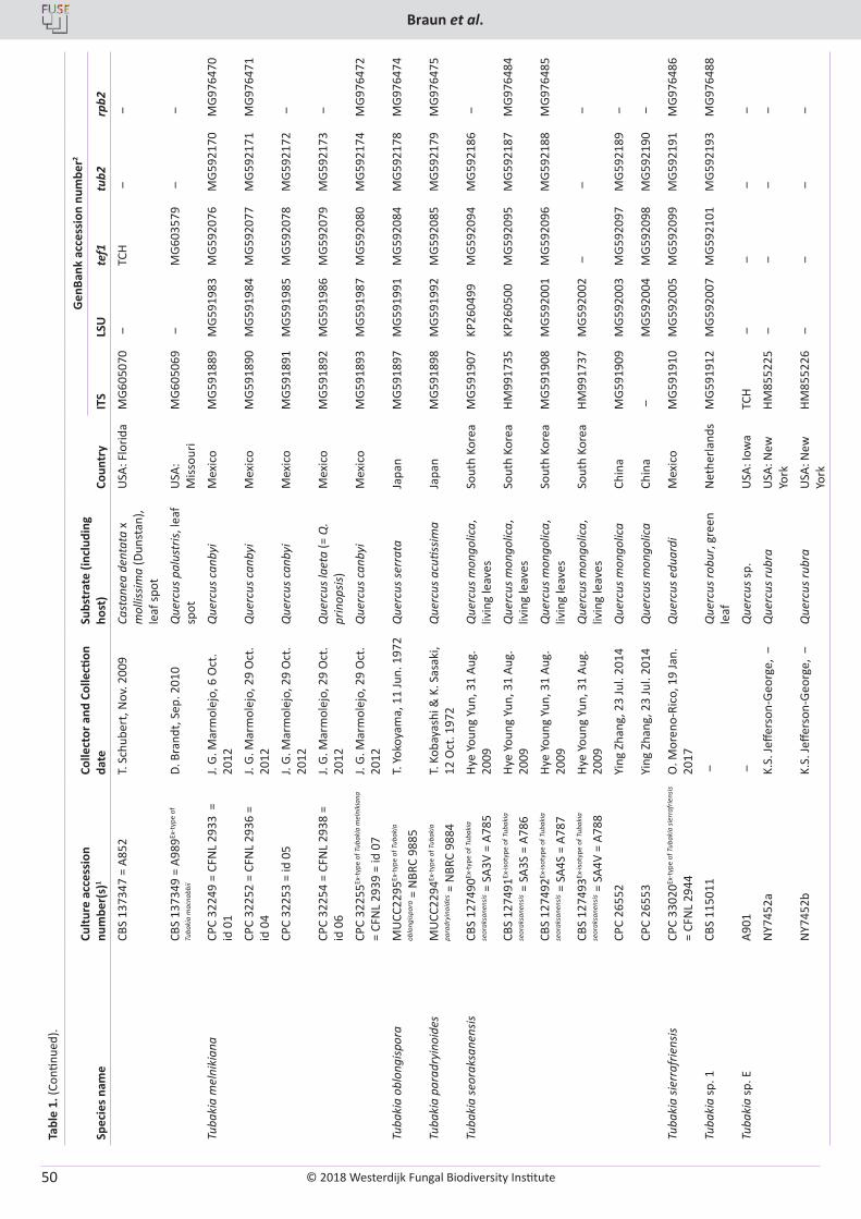
© 2018 Westerdijk Fungal Biodiversity Institute
Braun et al.
Editor-in-ChiefProf. dr P.W. Crous, Westerdijk Fungal Biodiversity Institute, P.O. Box 85167, 3508 AD Utrecht, The Netherlands.E-mail:[email protected]
50
Tabl
e 1.
(Con
tinue
d).
Spec
ies n
ame
Cultu
re a
cces
sion
nu
mbe
r(s)
1Co
llect
or a
nd C
olle
ction
da
teSu
bstr
ate
(incl
udin
g ho
st)
Coun
try
Gen
Bank
acc
essi
on n
umbe
r2
ITS
LSU
tef1
tub2
rpb2
CBS
1373
47 =
A85
2T.
Sch
uber
t, N
ov. 2
009
Cast
anea
den
tata
x
mol
lissim
a (D
unst
an),
leaf
spot
USA
: Flo
rida
MG6
0507
0 ‒
TCH
‒ ‒
CBS
1373
49 =
A98
9Ex-t
ype
of
Tuba
kia
mac
nabb
iiD.
Bra
ndt,
Sep.
201
0Q
uerc
us p
alus
tris,
leaf
sp
otU
SA:
Miss
ouri
MG6
0506
9 ‒
MG6
0357
9 ‒
‒
Tuba
kia
mel
niki
ana
CPC
3224
9 =
CFN
L 29
33 =
id
01
J. G.
Mar
mol
ejo,
6 O
ct.
2012
Que
rcus
can
byi
Mex
ico
MG5
9188
9M
G591
983
MG5
9207
6M
G592
170
MG9
7647
0
CPC
3225
2 =
CFN
L 29
36 =
id
04
J. G.
Mar
mol
ejo,
29
Oct
. 20
12Q
uerc
us c
anby
iM
exic
oM
G591
890
MG5
9198
4M
G592
077
MG5
9217
1M
G976
471
CPC
3225
3 =
id 0
5J.
G. M
arm
olej
o, 2
9 O
ct.
2012
Que
rcus
can
byi
Mex
ico
MG5
9189
1M
G591
985
MG5
9207
8M
G592
172
‒
CPC
3225
4 =
CFN
L 29
38 =
id
06
J. G.
Mar
mol
ejo,
29
Oct
. 20
12Q
uerc
us la
eta
(= Q
. pr
inop
sis)
Mex
ico
MG5
9189
2M
G591
986
MG5
9207
9M
G592
173
‒
CPC
3225
5Ex-t
ype
of T
ubak
ia m
elni
kian
a =
CFN
L 29
39 =
id 0
7J.
G. M
arm
olej
o, 2
9 O
ct.
2012
Que
rcus
can
byi
Mex
ico
MG5
9189
3M
G591
987
MG5
9208
0M
G592
174
MG9
7647
2
Tuba
kia
oblo
ngisp
ora
MU
CC22
95Ex
-typ
e of
Tub
akia
oblo
ngisp
ora =
NBR
C 98
85T.
Yok
oyam
a, 1
1 Ju
n. 1
972
Que
rcus
serr
ata
Japa
nM
G591
897
MG5
9199
1M
G592
084
MG5
9217
8M
G976
474
Tuba
kia
para
dryi
noid
esM
UCC
2294
Ex-t
ype
of T
ubak
ia
para
dryi
noid
es =
NBR
C 98
84T.
Kob
ayas
hi &
K. S
asak
i, 12
Oct
. 197
2Q
uerc
us a
cutis
sima
Japa
nM
G591
898
MG5
9199
2M
G592
085
MG5
9217
9M
G976
475
Tuba
kia
seor
aksa
nens
isCB
S 12
7490
Ex-t
ype
of T
ubak
ia
seor
aksa
nens
is = S
A3V
= A7
85Hy
e Yo
ung
Yun,
31
Aug.
20
09Q
uerc
us m
ongo
lica,
liv
ing
leav
esSo
uth
Kore
aM
G591
907
KP26
0499
MG5
9209
4M
G592
186
‒
CBS
1274
91Ex
-isot
ype
of T
ubak
ia
seor
aksa
nens
is = S
A3S
= A7
86Hy
e Yo
ung
Yun,
31
Aug.
20
09Q
uerc
us m
ongo
lica,
liv
ing
leav
esSo
uth
Kore
aHM
9917
35KP
2605
00M
G592
095
MG5
9218
7M
G976
484
CBS
1274
92Ex
-isot
ype
of T
ubak
ia
seor
aksa
nens
is = S
A4S
= A7
87Hy
e Yo
ung
Yun,
31
Aug.
20
09Q
uerc
us m
ongo
lica,
liv
ing
leav
esSo
uth
Kore
aM
G591
908
MG5
9200
1M
G592
096
MG5
9218
8M
G976
485
CBS
1274
93Ex
-isot
ype
of T
ubak
ia
seor
aksa
nens
is = S
A4V
= A7
88Hy
e Yo
ung
Yun,
31
Aug.
20
09Q
uerc
us m
ongo
lica,
liv
ing
leav
esSo
uth
Kore
aHM
9917
37M
G592
002
‒ ‒
‒
CPC
2655
2Yi
ng Z
hang
, 23
Jul.
2014
Que
rcus
mon
golic
aCh
ina
MG5
9190
9M
G592
003
MG5
9209
7M
G592
189
‒
CPC
2655
3Yi
ng Z
hang
, 23
Jul.
2014
Que
rcus
mon
golic
aCh
ina
‒M
G592
004
MG5
9209
8M
G592
190
‒
Tuba
kia
sierr
afrie
nsis
CPC
3302
0Ex-t
ype
of T
ubak
ia si
erra
frie
nsis
= CF
NL
2944
O. M
oren
o-Ri
co, 1
9 Ja
n.
2017
Que
rcus
edu
ardi
Mex
ico
MG5
9191
0M
G592
005
MG5
9209
9M
G592
191
MG9
7648
6
Tuba
kia
sp. 1
CBS
1150
11 ‒
Que
rcus
robu
r, gr
een
leaf
Net
herla
nds
MG5
9191
2M
G592
007
MG5
9210
1M
G592
193
MG9
7648
8
Tuba
kia
sp. E
A901
‒Q
uerc
us sp
.U
SA: I
owa
TCH
‒ ‒
‒ ‒
NY7
452a
K.S.
Jeffe
rson
-Geo
rge,
‒Q
uerc
us ru
bra
USA
: New
Yo
rkHM
8552
25 ‒
‒ ‒
‒
NY7
452b
K.S.
Jeffe
rson
-Geo
rge,
‒Q
uerc
us ru
bra
USA
: New
Yo
rkHM
8552
26 ‒
‒ ‒
‒
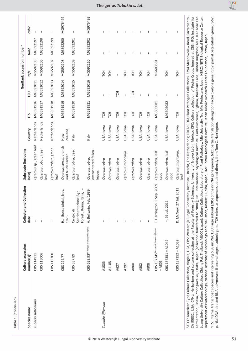
© 2018 Westerdijk Fungal Biodiversity Institute
The genus Tubakia s. lat.
Editor-in-ChiefProf. dr P.W. Crous, Westerdijk Fungal Biodiversity Institute, P.O. Box 85167, 3508 AD Utrecht, The Netherlands.E-mail:[email protected]
51
Tabl
e 1.
(Con
tinue
d).
Spec
ies n
ame
Cultu
re a
cces
sion
nu
mbe
r(s)
1Co
llect
or a
nd C
olle
ction
da
teSu
bstr
ate
(incl
udin
g ho
st)
Coun
try
Gen
Bank
acc
essi
on n
umbe
r2
ITS
LSU
tef1
tub2
rpb2
Tuba
kia
sutto
nian
aCB
S 11
4911
‒Q
uerc
us sp
., gr
een
leaf
Net
herla
nds
MG5
9191
6M
G592
011
MG5
9210
5M
G592
197
‒
CBS
1150
06 ‒
Que
rcus
robu
r, gr
een
leaf
Net
herla
nds
MG5
9191
7M
G592
012
MG5
9210
6M
G592
198
‒
CBS
1153
00 ‒
Que
rcus
robu
r, gr
een
leaf
Net
herla
nds
MG5
9191
8M
G592
013
MG5
9210
7M
G592
199
‒
CBS
229.
77H.
J. Bo
esew
inke
l, N
ov.
1975
Que
rcus
cer
ris, b
ranc
h an
d tr
unk
cank
erN
ew
Zeal
and
MG5
9191
9M
G592
014
MG5
9210
8M
G592
200
MG9
7649
2
CBS
387.
89Ce
ntro
di
Sper
imen
tazio
ne A
gr.
Fore
st.,
Rom
a, It
alia
, ‒
Que
rcus
rubr
a, d
ead
leaf
Italy
MG5
9192
0M
G592
015
MG5
9210
9M
G592
201
‒
CBS
639.
93Ex
-isot
ype
of D
icar
pella
dry
ina
A. B
elisa
rio, F
eb. 1
989
Que
rcus
rubr
a,
over
win
tere
d fa
llen
leav
es
Italy
MG5
9192
1M
G592
016
MG5
9211
0M
G592
202
MG9
7649
3
Tuba
kia
tiffan
yae
A103
5 ‒
Que
rcus
rubr
aU
SA: I
owa
TCH
‒TC
H ‒
‒
A110
8 ‒
Que
rcus
rubr
aU
SA: I
owa
TCH
‒TC
H ‒
‒
A627
‒Q
uerc
us ru
bra
USA
: Iow
aTC
HTC
H ‒
‒ ‒
A792
‒Q
uerc
us im
bric
aria
USA
: Iow
aTC
H ‒
TCH
‒ ‒
A800
‒Q
uerc
us ru
bra
USA
: Iow
aTC
HTC
HTC
H ‒
‒
A802
‒Q
uerc
us ru
bra
USA
: Iow
aTC
H ‒
TCH
‒ ‒
A808
‒Q
uerc
us ru
bra
USA
: Iow
aTC
H ‒
TCH
‒ ‒
CBS
1373
45Ex
-typ
e of
Tub
akia
tiffa
nyae
=
A803
T. H
arrin
gton
, 5 S
ep. 2
009
Que
rcus
rubr
a, le
af
spot
USA
: Iow
aM
G605
081
‒M
G603
581
‒ ‒
CBS
1373
51 =
A10
42 ‒
, 29
Jul.
2011
Que
rcus
rubr
a, le
af
spot
USA
: Iow
aM
G605
082
‒TC
H ‒
‒
CBS
1373
52 =
A10
52D.
McN
ew, 2
7 Ju
l. 20
11Q
uerc
us im
bric
aria
, le
afU
SA: I
owa
TCH
‒TC
H ‒
‒
1 AT
CC: A
mer
ican
Typ
e Cu
lture
Col
lecti
on, V
irgin
ia, U
SA; C
BS: W
este
rdijk
Fun
gal B
iodi
vers
ity In
stitu
te, U
trec
ht, T
he N
ethe
rland
s; C
DFA:
CDF
A Pl
ant P
atho
gen
Colle
ction
s, 3
294
Mea
dow
view
Roa
d, S
acra
men
to,
CA 9
5832
, USA
; CFN
L: H
erba
rium
and
cul
ture
col
lecti
on a
t th
e Fa
culty
of
Fore
stry
Sci
ence
s, U
nive
rsity
of
Nue
vo L
eón,
Méx
ico;
CPC
: Cul
ture
col
lecti
on o
f Pe
dro
Crou
s, h
ouse
d at
CBS
; IFO
: Ins
titut
e fo
r Fe
rmen
tatio
n, O
saka
, Yod
ogaw
a-ku
, Osa
ka, J
apan
(co
llecti
on t
rans
ferr
ed t
o N
BRC)
; IM
I: In
tern
ation
al M
ycol
ogic
al In
stitu
te, C
ABI-B
iosc
ienc
e, E
gham
, Bak
eham
Lan
e, U
nite
d Ki
ngdo
m; M
FLU
CC: M
ae F
ah
Luan
g U
nive
rsity
Cul
ture
Col
lecti
on, C
hian
g Ra
i, Th
aila
nd; M
UCC
(Jap
an):
Cultu
re C
olle
ction
, Lab
orat
ory
of P
lant
Pat
holo
gy, M
ie U
nive
rsity
, Tsu
, Mie
Pre
fect
ure,
Japa
n; N
BRC:
NIT
E Bi
olog
ical
Res
ourc
e Ce
nter
, De
part
men
t of B
iote
chno
logy
, Nati
onal
Insti
tute
of T
echn
olog
y an
d Ev
alua
tion,
Kisa
razu
, Chi
ba, J
apan
, TM
I: To
ttori
Myc
olog
ical
Insti
tute
, Jap
an K
inok
o Re
sear
ch C
ente
r Fou
ndati
on, T
ottor
i, Ja
pan.
2 IT
S: in
tern
al tr
ansc
ribed
spac
ers a
nd in
terv
enin
g 5.
8S n
rDN
A; LS
U: l
arge
subu
nit (
28S)
of t
he n
rRN
A ge
ne o
pero
n; te
f1: p
artia
l tra
nsla
tion
elon
gatio
n fa
ctor
1-a
lpha
gen
e; tu
b2: p
artia
l bet
a-tu
bulin
gen
e; rp
b2:
parti
al D
NA-
dire
cted
RN
A po
lym
eras
e II
seco
nd la
rges
t sub
unit
gene
. TCH
refe
rs to
sequ
ence
s obt
aine
d di
rect
ly fr
om To
m C
. Har
ringt
on.
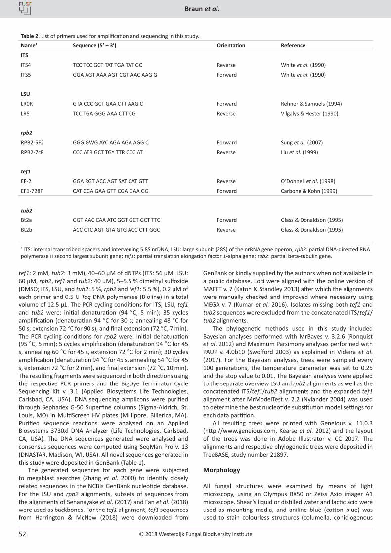
© 2018 Westerdijk Fungal Biodiversity Institute
Braun et al.
Editor-in-ChiefProf. dr P.W. Crous, Westerdijk Fungal Biodiversity Institute, P.O. Box 85167, 3508 AD Utrecht, The Netherlands.E-mail:[email protected]
52
tef1: 2 mM, tub2: 3 mM), 40–60 µM of dNTPs (ITS: 56 µM, LSU: 60 µM, rpb2, tef1 and tub2: 40 µM), 5–5.5 % dimethyl sulfoxide (DMSO; ITS, LSU, and tub2: 5 %, rpb2 and tef1: 5.5 %), 0.2 µM of each primer and 0.5 U Taq DNA polymerase (Bioline) in a total volume of 12.5 µL. The PCR cycling conditions for ITS, LSU, tef1 and tub2 were: initial denaturation (94 °C, 5 min); 35 cycles amplification (denaturation 94 °C for 30 s; annealing 48 °C for 50 s; extension 72 °C for 90 s), and final extension (72 °C, 7 min). The PCR cycling conditions for rpb2 were: initial denaturation (95 °C, 5 min); 5 cycles amplification (denaturation 94 °C for 45 s, annealing 60 °C for 45 s, extension 72 °C for 2 min); 30 cycles amplification (denaturation 94 °C for 45 s, annealing 54 °C for 45 s, extension 72 °C for 2 min), and final extension (72 °C, 10 min). The resulting fragments were sequenced in both directions using the respective PCR primers and the BigDye Terminator Cycle Sequencing Kit v. 3.1 (Applied Biosystems Life Technologies, Carlsbad, CA, USA). DNA sequencing amplicons were purified through Sephadex G-50 Superfine columns (Sigma-Aldrich, St. Louis, MO) in MultiScreen HV plates (Millipore, Billerica, MA). Purified sequence reactions were analysed on an Applied Biosystems 3730xl DNA Analyzer (Life Technologies, Carlsbad, CA, USA). The DNA sequences generated were analysed and consensus sequences were computed using SeqMan Pro v. 13 (DNASTAR, Madison, WI, USA). All novel sequences generated in this study were deposited in GenBank (Table 1).
The generated sequences for each gene were subjected to megablast searches (Zhang et al. 2000) to identify closely related sequences in the NCBIs GenBank nucleotide database. For the LSU and rpb2 alignments, subsets of sequences from the alignments of Senanayake et al. (2017) and Fan et al. (2018) were used as backbones. For the tef1 alignment, tef1 sequences from Harrington & McNew (2018) were downloaded from
GenBank or kindly supplied by the authors when not available in a public database. Loci were aligned with the online version of MAFFT v. 7 (Katoh & Standley 2013) after which the alignments were manually checked and improved where necessary using MEGA v. 7 (Kumar et al. 2016). Isolates missing both tef1 and tub2 sequences were excluded from the concatenated ITS/tef1/tub2 alignments.
The phylogenetic methods used in this study included Bayesian analyses performed with MrBayes v. 3.2.6 (Ronquist et al. 2012) and Maximum Parsimony analyses performed with PAUP v. 4.0b10 (Swofford 2003) as explained in Videira et al. (2017). For the Bayesian analyses, trees were sampled every 100 generations, the temperature parameter was set to 0.25 and the stop value to 0.01. The Bayesian analyses were applied to the separate overview LSU and rpb2 alignments as well as the concatenated ITS/tef1/tub2 alignments and the expanded tef1 alignment after MrModelTest v. 2.2 (Nylander 2004) was used to determine the best nucleotide substitution model settings for each data partition.
All resulting trees were printed with Geneious v. 11.0.3 (http://www.geneious.com, Kearse et al. 2012) and the layout of the trees was done in Adobe Illustrator v. CC 2017. The alignments and respective phylogenetic trees were deposited in TreeBASE, study number 21897.
Morphology
All fungal structures were examined by means of light microscopy, using an Olympus BX50 or Zeiss Axio imager A1 microscope. Shear’s liquid or distilled water and lactic acid were used as mounting media, and aniline blue (cotton blue) was used to stain colourless structures (columella, conidiogenous
Table 2. List of primers used for amplification and sequencing in this study.
Name1 Sequence (5’ – 3’) Orientation Reference
ITS
ITS4 TCC TCC GCT TAT TGA TAT GC Reverse White et al. (1990)
ITS5 GGA AGT AAA AGT CGT AAC AAG G Forward White et al. (1990)
LSU
LR0R GTA CCC GCT GAA CTT AAG C Forward Rehner & Samuels (1994)
LR5 TCC TGA GGG AAA CTT CG Reverse Vilgalys & Hester (1990)
rpb2
RPB2-5F2 GGG GWG AYC AGA AGA AGG C Forward Sung et al. (2007)
RPB2-7cR CCC ATR GCT TGY TTR CCC AT Reverse Liu et al. (1999)
tef1
EF-2 GGA RGT ACC AGT SAT CAT GTT Reverse O’Donnell et al. (1998)
EF1-728F CAT CGA GAA GTT CGA GAA GG Forward Carbone & Kohn (1999)
tub2
Bt2a GGT AAC CAA ATC GGT GCT GCT TTC Forward Glass & Donaldson (1995)
Bt2b ACC CTC AGT GTA GTG ACC CTT GGC Reverse Glass & Donaldson (1995)
1 ITS: internal transcribed spacers and intervening 5.8S nrDNA; LSU: large subunit (28S) of the nrRNA gene operon; rpb2: partial DNA-directed RNA polymerase II second largest subunit gene; tef1: partial translation elongation factor 1-alpha gene; tub2: partial beta-tubulin gene.
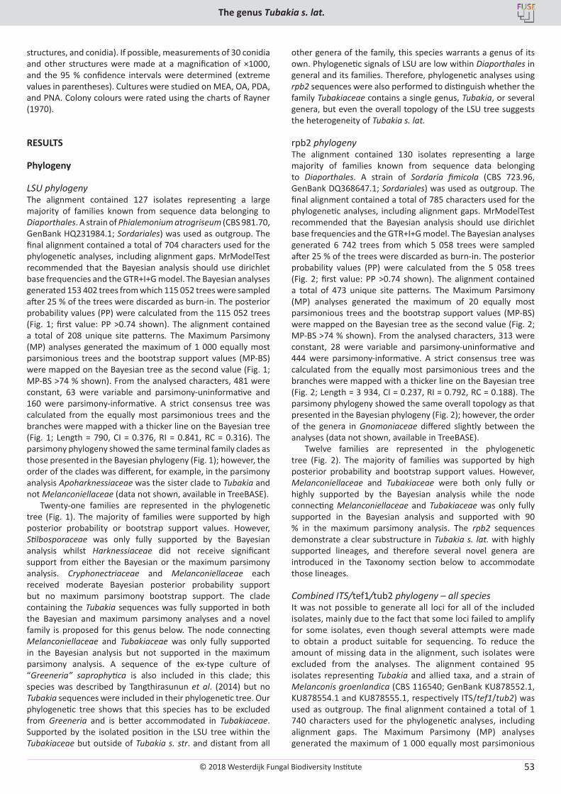
© 2018 Westerdijk Fungal Biodiversity Institute
The genus Tubakia s. lat.
Editor-in-ChiefProf. dr P.W. Crous, Westerdijk Fungal Biodiversity Institute, P.O. Box 85167, 3508 AD Utrecht, The Netherlands.E-mail:[email protected]
53
structures, and conidia). If possible, measurements of 30 conidia and other structures were made at a magnification of ×1000, and the 95 % confidence intervals were determined (extreme values in parentheses). Cultures were studied on MEA, OA, PDA, and PNA. Colony colours were rated using the charts of Rayner (1970).
RESULTS
Phylogeny
LSU phylogenyThe alignment contained 127 isolates representing a large majority of families known from sequence data belonging to Diaporthales. A strain of Phialemonium atrogriseum (CBS 981.70, GenBank HQ231984.1; Sordariales) was used as outgroup. The final alignment contained a total of 704 characters used for the phylogenetic analyses, including alignment gaps. MrModelTest recommended that the Bayesian analysis should use dirichlet base frequencies and the GTR+I+G model. The Bayesian analyses generated 153 402 trees from which 115 052 trees were sampled after 25 % of the trees were discarded as burn-in. The posterior probability values (PP) were calculated from the 115 052 trees (Fig. 1; first value: PP >0.74 shown). The alignment contained a total of 208 unique site patterns. The Maximum Parsimony (MP) analyses generated the maximum of 1 000 equally most parsimonious trees and the bootstrap support values (MP-BS) were mapped on the Bayesian tree as the second value (Fig. 1; MP-BS >74 % shown). From the analysed characters, 481 were constant, 63 were variable and parsimony-uninformative and 160 were parsimony-informative. A strict consensus tree was calculated from the equally most parsimonious trees and the branches were mapped with a thicker line on the Bayesian tree (Fig. 1; Length = 790, CI = 0.376, RI = 0.841, RC = 0.316). The parsimony phylogeny showed the same terminal family clades as those presented in the Bayesian phylogeny (Fig. 1); however, the order of the clades was different, for example, in the parsimony analysis Apoharknessiaceae was the sister clade to Tubakia and not Melanconiellaceae (data not shown, available in TreeBASE).
Twenty-one families are represented in the phylogenetic tree (Fig. 1). The majority of families were supported by high posterior probability or bootstrap support values. However, Stilbosporaceae was only fully supported by the Bayesian analysis whilst Harknessiaceae did not receive significant support from either the Bayesian or the maximum parsimony analysis. Cryphonectriaceae and Melanconiellaceae each received moderate Bayesian posterior probability support but no maximum parsimony bootstrap support. The clade containing the Tubakia sequences was fully supported in both the Bayesian and maximum parsimony analyses and a novel family is proposed for this genus below. The node connecting Melanconiellaceae and Tubakiaceae was only fully supported in the Bayesian analysis but not supported in the maximum parsimony analysis. A sequence of the ex-type culture of “Greeneria” saprophytica is also included in this clade; this species was described by Tangthirasunun et al. (2014) but no Tubakia sequences were included in their phylogenetic tree. Our phylogenetic tree shows that this species has to be excluded from Greeneria and is better accommodated in Tubakiaceae. Supported by the isolated position in the LSU tree within the Tubakiaceae but outside of Tubakia s. str. and distant from all
other genera of the family, this species warrants a genus of its own. Phylogenetic signals of LSU are low within Diaporthales in general and its families. Therefore, phylogenetic analyses using rpb2 sequences were also performed to distinguish whether the family Tubakiaceae contains a single genus, Tubakia, or several genera, but even the overall topology of the LSU tree suggests the heterogeneity of Tubakia s. lat.
rpb2 phylogenyThe alignment contained 130 isolates representing a large majority of families known from sequence data belonging to Diaporthales. A strain of Sordaria fimicola (CBS 723.96, GenBank DQ368647.1; Sordariales) was used as outgroup. The final alignment contained a total of 785 characters used for the phylogenetic analyses, including alignment gaps. MrModelTest recommended that the Bayesian analysis should use dirichlet base frequencies and the GTR+I+G model. The Bayesian analyses generated 6 742 trees from which 5 058 trees were sampled after 25 % of the trees were discarded as burn-in. The posterior probability values (PP) were calculated from the 5 058 trees (Fig. 2; first value: PP >0.74 shown). The alignment contained a total of 473 unique site patterns. The Maximum Parsimony (MP) analyses generated the maximum of 20 equally most parsimonious trees and the bootstrap support values (MP-BS) were mapped on the Bayesian tree as the second value (Fig. 2; MP-BS >74 % shown). From the analysed characters, 313 were constant, 28 were variable and parsimony-uninformative and 444 were parsimony-informative. A strict consensus tree was calculated from the equally most parsimonious trees and the branches were mapped with a thicker line on the Bayesian tree (Fig. 2; Length = 3 934, CI = 0.237, RI = 0.792, RC = 0.188). The parsimony phylogeny showed the same overall topology as that presented in the Bayesian phylogeny (Fig. 2); however, the order of the genera in Gnomoniaceae differed slightly between the analyses (data not shown, available in TreeBASE).
Twelve families are represented in the phylogenetic tree (Fig. 2). The majority of families was supported by high posterior probability and bootstrap support values. However, Melanconiellaceae and Tubakiaceae were both only fully or highly supported by the Bayesian analysis while the node connecting Melanconiellaceae and Tubakiaceae was only fully supported in the Bayesian analysis and supported with 90 % in the maximum parsimony analysis. The rpb2 sequences demonstrate a clear substructure in Tubakia s. lat. with highly supported lineages, and therefore several novel genera are introduced in the Taxonomy section below to accommodate those lineages.
Combined ITS/tef1/tub2 phylogeny – all speciesIt was not possible to generate all loci for all of the included isolates, mainly due to the fact that some loci failed to amplify for some isolates, even though several attempts were made to obtain a product suitable for sequencing. To reduce the amount of missing data in the alignment, such isolates were excluded from the analyses. The alignment contained 95 isolates representing Tubakia and allied taxa, and a strain of Melanconis groenlandica (CBS 116540; GenBank KU878552.1, KU878554.1 and KU878555.1, respectively ITS/tef1/tub2) was used as outgroup. The final alignment contained a total of 1 740 characters used for the phylogenetic analyses, including alignment gaps. The Maximum Parsimony (MP) analyses generated the maximum of 1 000 equally most parsimonious
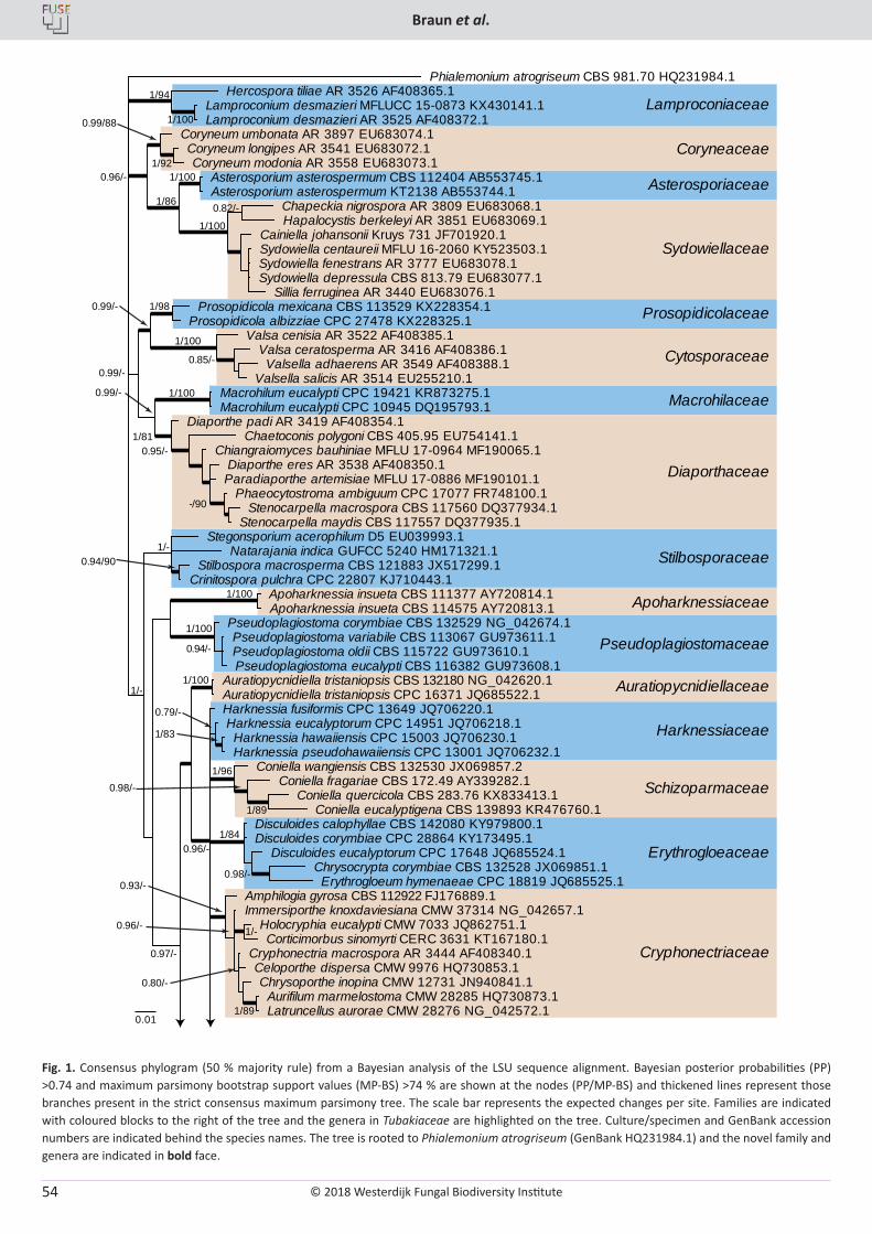
© 2018 Westerdijk Fungal Biodiversity Institute
Braun et al.
Editor-in-ChiefProf. dr P.W. Crous, Westerdijk Fungal Biodiversity Institute, P.O. Box 85167, 3508 AD Utrecht, The Netherlands.E-mail:[email protected]
54
Fig. 1. Consensus phylogram (50 % majority rule) from a Bayesian analysis of the LSU sequence alignment. Bayesian posterior probabilities (PP) >0.74 and maximum parsimony bootstrap support values (MP-BS) >74 % are shown at the nodes (PP/MP-BS) and thickened lines represent those branches present in the strict consensus maximum parsimony tree. The scale bar represents the expected changes per site. Families are indicated with coloured blocks to the right of the tree and the genera in Tubakiaceae are highlighted on the tree. Culture/specimen and GenBank accession numbers are indicated behind the species names. The tree is rooted to Phialemonium atrogriseum (GenBank HQ231984.1) and the novel family and genera are indicated in bold face.
1/100
1/94
1/84
1/100
1/100
1/98
0.79/-
1/89
1/83
1/96
1/89
1/100
1/100
1/100
0.94/90 1/-
1/100
1/100
1/86
0.94/-
Lamproconiaceae
Coryneaceae
Asterosporiaceae
Sydowiellaceae
Prosopidicolaceae
Cytosporaceae
Macrohilaceae
Diaporthaceae
Stilbosporaceae
Apoharknessiaceae
Pseudoplagiostomaceae
Auratiopycnidiellaceae
Harknessiaceae
Schizoparmaceae
Erythrogloeaceae
0.01
0.80/-
1/- 0.96/-
0.93/-
0.98/-
0.98/-
0.96/-
0.97/-
1/-
-/90
0.95/- 1/81
0.85/-
0.99/-
0.99/-
0.99/-
1/92
0.99/88
0.82/-
0.96/-
Phialemonium atrogriseum CBS 981.70 HQ231984.1
Lamproconium desmazieri MFLUCC 15-0873 KX430141.1 Lamproconium desmazieri AR 3525 AF408372.1
Hercospora tiliae AR 3526 AF408365.1
Cryphonectria macrospora AR 3444 AF408340.1 Celoporthe dispersa CMW 9976 HQ730853.1
Aurifilum marmelostoma CMW 28285 HQ730873.1 Latruncellus aurorae CMW 28276 NG_042572.1
Chrysoporthe inopina CMW 12731 JN940841.1
Holocryphia eucalypti CMW 7033 JQ862751.1 Corticimorbus sinomyrti CERC 3631 KT167180.1
Immersiporthe knoxdaviesiana CMW 37314 NG_042657.1 Amphilogia gyrosa CBS 112922 FJ176889.1
Harknessia eucalyptorum CPC 14951 JQ706218.1 Harknessia hawaiiensis CPC 15003 JQ706230.1 Harknessia pseudohawaiiensis CPC 13001 JQ706232.1
Harknessia fusiformis CPC 13649 JQ706220.1
Coniella wangiensis CBS 132530 JX069857.2 Coniella fragariae CBS 172.49 AY339282.1
Coniella quercicola CBS 283.76 KX833413.1 Coniella eucalyptigena CBS 139893 KR476760.1
Disculoides calophyllae CBS 142080 KY979800.1 Disculoides corymbiae CPC 28864 KY173495.1
Disculoides eucalyptorum CPC 17648 JQ685524.1 Chrysocrypta corymbiae CBS 132528 JX069851.1 Erythrogloeum hymenaeae CPC 18819 JQ685525.1
Auratiopycnidiella tristaniopsis CBS 132180 NG_042620.1 Auratiopycnidiella tristaniopsis CPC 16371 JQ685522.1
Pseudoplagiostoma corymbiae CBS 132529 NG_042674.1 Pseudoplagiostoma variabile CBS 113067 GU973611.1 Pseudoplagiostoma oldii CBS 115722 GU973610.1 Pseudoplagiostoma eucalypti CBS 116382 GU973608.1
Apoharknessia insueta CBS 111377 AY720814.1 Apoharknessia insueta CBS 114575 AY720813.1
Stegonsporium acerophilum D5 EU039993.1
Stilbospora macrosperma CBS 121883 JX517299.1 Crinitospora pulchra CPC 22807 KJ710443.1
Natarajania indica GUFCC 5240 HM171321.1
Diaporthe eres AR 3538 AF408350.1
Phaeocytostroma ambiguum CPC 17077 FR748100.1 Stenocarpella macrospora CBS 117560 DQ377934.1
Stenocarpella maydis CBS 117557 DQ377935.1
Paradiaporthe artemisiae MFLU 17-0886 MF190101.1
Chiangraiomyces bauhiniae MFLU 17-0964 MF190065.1 Chaetoconis polygoni CBS 405.95 EU754141.1
Diaporthe padi AR 3419 AF408354.1
Macrohilum eucalypti CPC 19421 KR873275.1 Macrohilum eucalypti CPC 10945 DQ195793.1
Valsa cenisia AR 3522 AF408385.1 Valsa ceratosperma AR 3416 AF408386.1 Valsella adhaerens AR 3549 AF408388.1
Valsella salicis AR 3514 EU255210.1
Prosopidicola mexicana CBS 113529 KX228354.1 Prosopidicola albizziae CPC 27478 KX228325.1
Coryneum longipes AR 3541 EU683072.1 Coryneum modonia AR 3558 EU683073.1
Coryneum umbonata AR 3897 EU683074.1
Sydowiella centaureii MFLU 16-2060 KY523503.1 Sydowiella fenestrans AR 3777 EU683078.1 Sydowiella depressula CBS 813.79 EU683077.1
Sillia ferruginea AR 3440 EU683076.1
Cainiella johansonii Kruys 731 JF701920.1
Chapeckia nigrospora AR 3809 EU683068.1 Hapalocystis berkeleyi AR 3851 EU683069.1
Asterosporium asterospermum CBS 112404 AB553745.1 Asterosporium asterospermum KT2138 AB553744.1
Cryphonectriaceae
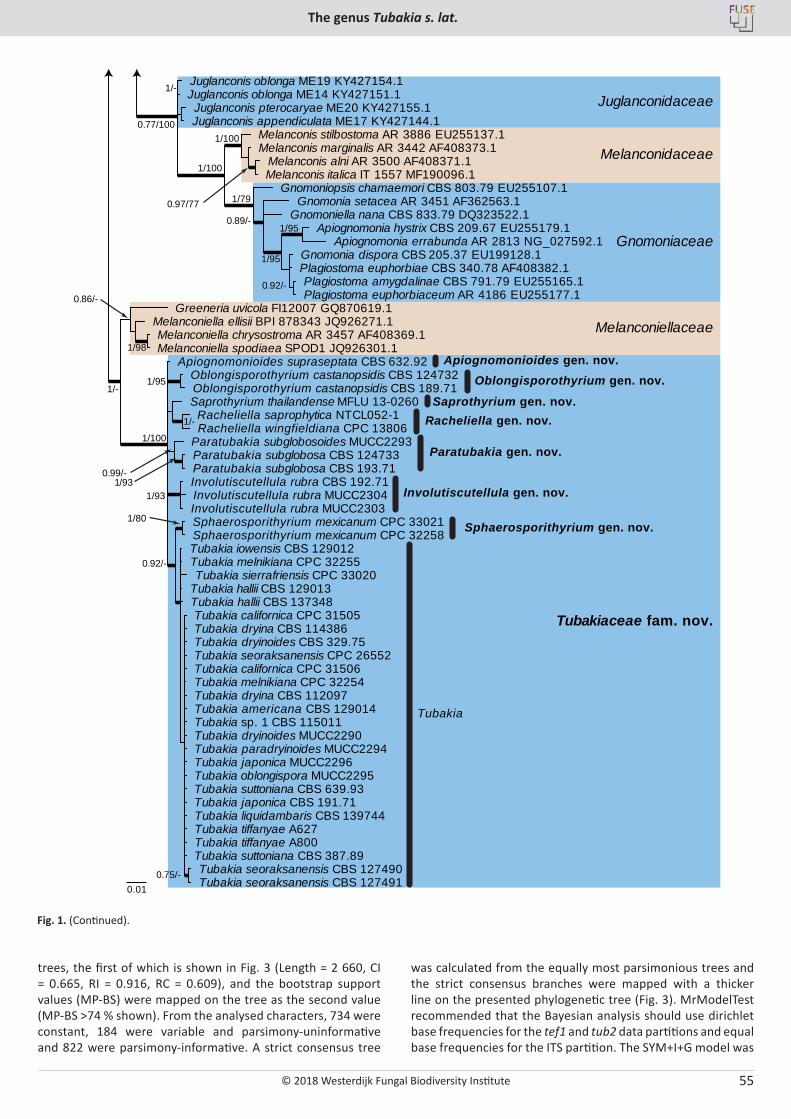
© 2018 Westerdijk Fungal Biodiversity Institute
The genus Tubakia s. lat.
Editor-in-ChiefProf. dr P.W. Crous, Westerdijk Fungal Biodiversity Institute, P.O. Box 85167, 3508 AD Utrecht, The Netherlands.E-mail:[email protected]
55
Fig. 1. (Continued).
1/98
1/100
1/100
1/93
1/95
1/93
Juglanconidaceae
Melanconidaceae
Gnomoniaceae
Melanconiellaceae
Tubakiaceae fam. nov.
0.92/-
1/95
1/95
0.89/-
1/79 0.97/77
0.77/100
0.86/-
1/-
0.75/-
1/80
0.92/-
0.99/-
1/100
1/-
1/-
Gnomonia setacea AR 3451 AF362563.1 Gnomoniella nana CBS 833.79 DQ323522.1
Gnomonia dispora CBS 205.37 EU199128.1
Plagiostoma amygdalinae CBS 791.79 EU255165.1 Plagiostoma euphorbiaceum AR 4186 EU255177.1
Plagiostoma euphorbiae CBS 340.78 AF408382.1
Apiognomonia hystrix CBS 209.67 EU255179.1 Apiognomonia errabunda AR 2813 NG_027592.1
Gnomoniopsis chamaemori CBS 803.79 EU255107.1
Melanconis stilbostoma AR 3886 EU255137.1 Melanconis marginalis AR 3442 AF408373.1
Melanconis alni AR 3500 AF408371.1 Melanconis italica IT 1557 MF190096.1
Juglanconis pterocaryae ME20 KY427155.1 Juglanconis appendiculata ME17 KY427144.1
Juglanconis oblonga ME19 KY427154.1 Juglanconis oblonga ME14 KY427151.1
Melanconiella chrysostroma AR 3457 AF408369.1 Melanconiella spodiaea SPOD1 JQ926301.1
Melanconiella ellisii BPI 878343 JQ926271.1 Greeneria uvicola FI12007 GQ870619.1
Racheliella saprophytica NTCL052-1 Racheliella wingfieldiana CPC 13806
Saprothyrium thailandense MFLU 13-0260
Tubakia californica CPC 31505 Tubakia dryina CBS 114386 Tubakia dryinoides CBS 329.75 Tubakia seoraksanensis CPC 26552 Tubakia californica CPC 31506 Tubakia melnikiana CPC 32254 Tubakia dryina CBS 112097 Tubakia americana CBS 129014 Tubakia sp. 1 CBS 115011 Tubakia dryinoides MUCC2290 Tubakia paradryinoides MUCC2294
Tubakia seoraksanensis CBS 127490 Tubakia seoraksanensis CBS 127491
Tubakia japonica MUCC2296 Tubakia oblongispora MUCC2295 Tubakia suttoniana CBS 639.93 Tubakia japonica CBS 191.71 Tubakia liquidambaris CBS 139744 Tubakia tiffanyae A627 Tubakia tiffanyae A800 Tubakia suttoniana CBS 387.89
Tubakia iowensis CBS 129012 Tubakia melnikiana CPC 32255 Tubakia sierrafriensis CPC 33020
Tubakia hallii CBS 129013 Tubakia hallii CBS 137348
Sphaerosporithyrium mexicanum CPC 33021 Sphaerosporithyrium mexicanum CPC 32258
Oblongisporothyrium castanopsidis CBS 124732 Oblongisporothyrium castanopsidis CBS 189.71
Paratubakia subglobosa CBS 124733 Paratubakia subglobosa CBS 193.71
Paratubakia subglobosoides MUCC2293
Involutiscutellula rubra CBS 192.71 Involutiscutellula rubra MUCC2304 Involutiscutellula rubra MUCC2303
Apiognomonioides supraseptata CBS 632.92
0.01
Oblongisporothyrium gen. nov.Apiognomonioides gen. nov.
Paratubakia gen. nov.
Involutiscutellula gen. nov.
Tubakia
Saprothyrium gen. nov.
Sphaerosporithyrium gen. nov.
Racheliella gen. nov.
trees, the first of which is shown in Fig. 3 (Length = 2 660, CI = 0.665, RI = 0.916, RC = 0.609), and the bootstrap support values (MP-BS) were mapped on the tree as the second value (MP-BS >74 % shown). From the analysed characters, 734 were constant, 184 were variable and parsimony-uninformative and 822 were parsimony-informative. A strict consensus tree
was calculated from the equally most parsimonious trees and the strict consensus branches were mapped with a thicker line on the presented phylogenetic tree (Fig. 3). MrModelTest recommended that the Bayesian analysis should use dirichlet base frequencies for the tef1 and tub2 data partitions and equal base frequencies for the ITS partition. The SYM+I+G model was
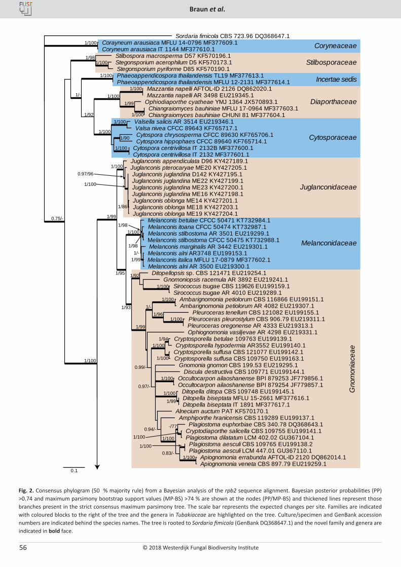
© 2018 Westerdijk Fungal Biodiversity Institute
Braun et al.
Editor-in-ChiefProf. dr P.W. Crous, Westerdijk Fungal Biodiversity Institute, P.O. Box 85167, 3508 AD Utrecht, The Netherlands.E-mail:[email protected]
56
Fig. 2. Consensus phylogram (50 % majority rule) from a Bayesian analysis of the rpb2 sequence alignment. Bayesian posterior probabilities (PP) >0.74 and maximum parsimony bootstrap support values (MP-BS) >74 % are shown at the nodes (PP/MP-BS) and thickened lines represent those branches present in the strict consensus maximum parsimony tree. The scale bar represents the expected changes per site. Families are indicated with coloured blocks to the right of the tree and the genera in Tubakiaceae are highlighted on the tree. Culture/specimen and GenBank accession numbers are indicated behind the species names. The tree is rooted to Sordaria fimicola (GenBank DQ368647.1) and the novel family and genera are indicated in bold face.
Coryneaceae
Cytosporaceae
Diaporthaceae
Stilbosporaceae
Incertae sedis
Juglanconidaceae
Melanconidaceae
Gno
mon
iace
ae
0.1
1/100
1/100
1/100
1/100 0.83/-
1/100
0.94/-
1/99
-/77
1/100
1/100
0.97/-
1/94
1/100
1/100
0.99/-
1/100
1/100 1/96
1/-
1/99
1/100
1/92
1/93
1/-
1/100 1/98
1/98
1/95
1/86
1/100
0.97/96 1/100
1/99
1/100
1/100
1/100
1/100 1/99
1/100
1/90
1/100
1/100
1/92
1/100
1/-
1/100 1/98
0.75/-
1/99
Sordaria fimicola CBS 723.96 DQ368647.1 Corayneum arausiaca MFLU 14-0796 MF377609.1 Coryneum arausiaca IT 1144 MF377610.1
Plagiostoma euphorbiae CBS 340.78 DQ368643.1 Cryptodiaporthe salicella CBS 109755 EU199141.1 Plagiostoma dilatatum LCM 402.02 GU367104.1 Plagiostoma aesculi CBS 109765 EU199138.2 Plagiostoma aesculi LCM 447.01 GU367110.1
Apiognomonia errabunda AFTOL-ID 2120 DQ862014.1 Apiognomonia veneta CBS 897.79 EU219259.1
Amphiporthe hranicensis CBS 119289 EU199137.1 Alnecium auctum PAT KF570170.1
Gnomonia gnomon CBS 199.53 EU219295.1
Ditopella ditopa CBS 109748 EU199145.1 Ditopella biseptata MFLU 15-2661 MF377616.1 Ditopella biseptata IT 1891 MF377617.1
Occultocarpon ailaoshanense BPI 879253 JF779856.1 Occultocarpon ailaoshanense BPI 879254 JF779857.1
Discula destructiva CBS 109771 EU199144.1
Cryptosporella betulae 109763 EU199139.1 Cryptosporella hypodermia AR3552 EU199140.1
Cryptosporella suffusa CBS 121077 EU199142.1 Cryptosporella suffusa CBS 109750 EU199163.1
Ophiognomonia vasiljevae AR 4298 EU219331.1
Ambarignomonia petiolorum CBS 116866 EU199151.1 Ambarignomonia petiolorum AR 4082 EU219307.1
Pleuroceras tenellum CBS 121082 EU199155.1 Pleuroceras pleurostylum CBS 906.79 EU219311.1 Pleuroceras oregonense AR 4333 EU219313.1
Sirococcus tsugae CBS 119626 EU199159.1 Sirococcus tsugae AR 4010 EU219289.1
Gnomoniopsis racemula AR 3892 EU219241.1 Ditopellopsis sp. CBS 121471 EU219254.1
Melanconis alni AR3748 EU199153.1 Melanconis italica MFLU 17-0879 MF377602.1 Melanconis alni AR 3500 EU219300.1
Melanconis marginalis AR 3442 EU219301.1
Melanconis stilbostoma AR 3501 EU219299.1 Melanconis stilbostoma CFCC 50475 KT732988.1
Melanconis itoana CFCC 50474 KT732987.1 Melanconis betulae CFCC 50471 KT732984.1
Juglanconis appendiculata D96 KY427189.1
Juglanconis juglandina D142 KY427195.1 Juglanconis juglandina ME22 KY427199.1 Juglanconis juglandina ME23 KY427200.1 Juglanconis juglandina ME16 KY427198.1 Juglanconis oblonga ME14 KY427201.1 Juglanconis oblonga ME18 KY427203.1 Juglanconis oblonga ME19 KY427204.1
Juglanconis pterocaryae ME20 KY427205.1
Mazzantia napelli AFTOL-ID 2126 DQ862020.1 Mazzantia napelli AR 3498 EU219345.1
Ophiodiaporthe cyatheae YMJ 1364 JX570893.1 Chiangraiomyces bauhiniae MFLU 17-0964 MF377603.1 Chiangraiomyces bauhiniae CHUNI 81 MF377604.1
Valsella salicis AR 3514 EU219346.1 Valsa nivea CFCC 89643 KF765717.1 Cytospora chrysosperma CFCC 89630 KF765706.1 Cytospora hippophaes CFCC 89640 KF765714.1
Cytospora centrivillosa IT 2132B MF377600.1 Cytospora centrivillosa IT 2132 MF377601.1
Phaeoappendicospora thailandensis TL19 MF377613.1 Phaeoappendicospora thailandensis MFLU 12-2131 MF377614.1
Stegonsporium acerophilum D5 KF570173.1 Stegonsporium pyriforme D85 KF570190.1
Stilbospora macrosperma D57 KF570196.1
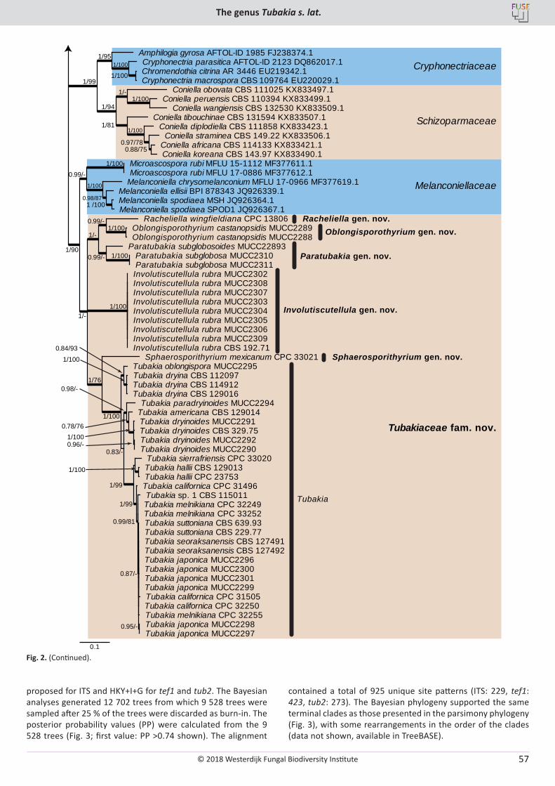
© 2018 Westerdijk Fungal Biodiversity Institute
The genus Tubakia s. lat.
Editor-in-ChiefProf. dr P.W. Crous, Westerdijk Fungal Biodiversity Institute, P.O. Box 85167, 3508 AD Utrecht, The Netherlands.E-mail:[email protected]
57
Schizoparmaceae
Cryphonectriaceae
Melanconiellaceae
Tubakiaceae fam. nov.
Oblongisporothyrium gen. nov.
Paratubakia gen. nov.
Racheliella gen. nov.
Involutiscutellula gen. nov.
Tubakia
Sphaerosporithyrium gen. nov.
0.1
1/100 0.99/-
1/100 0.99/-
1/-
0.95/-
0.87/-
0.99/81
1/99
1/100
1/99
0.96/- 1/100
0.78/76
0.98/-
0.83/-
1/100
0.84/93
1/100
1/76
1/100 1/-
1 /100 0.98/87
1/100
1/100 0.99/-
1/90
1/100
1/100 1/95
0.88/75 0.97/78
1/100 1/81
1/100 1/-
1/94
1/99
Racheliella wingfieldiana CPC 13806 Oblongisporothyrium castanopsidis MUCC2289 Oblongisporothyrium castanopsidis MUCC2288
Paratubakia subglobosoides MUCC22893 Paratubakia subglobosa MUCC2310 Paratubakia subglobosa MUCC2311
Sphaerosporithyrium mexicanum CPC 33021
Tubakia suttoniana CBS 639.93 Tubakia suttoniana CBS 229.77 Tubakia seoraksanensis CBS 127491 Tubakia seoraksanensis CBS 127492 Tubakia japonica MUCC2296 Tubakia japonica MUCC2300 Tubakia japonica MUCC2301 Tubakia japonica MUCC2299
Tubakia japonica MUCC2298 Tubakia japonica MUCC2297
Tubakia californica CPC 31505 Tubakia californica CPC 32250 Tubakia melnikiana CPC 32255
Tubakia sp. 1 CBS 115011 Tubakia melnikiana CPC 32249 Tubakia melnikiana CPC 33252
Tubakia californica CPC 31496
Tubakia hallii CBS 129013 Tubakia hallii CPC 23753
Tubakia sierrafriensis CPC 33020
Tubakia americana CBS 129014
Tubakia dryinoides MUCC2292 Tubakia dryinoides MUCC2290
Tubakia dryinoides MUCC2291 Tubakia dryinoides CBS 329.75
Tubakia paradryinoides MUCC2294
Tubakia dryina CBS 112097 Tubakia dryina CBS 114912 Tubakia dryina CBS 129016
Tubakia oblongispora MUCC2295
Involutiscutellula rubra MUCC2302 Involutiscutellula rubra MUCC2308 Involutiscutellula rubra MUCC2307 Involutiscutellula rubra MUCC2303 Involutiscutellula rubra MUCC2304 Involutiscutellula rubra MUCC2305 Involutiscutellula rubra MUCC2306 Involutiscutellula rubra MUCC2309 Involutiscutellula rubra CBS 192.71
Melanconiella ellisii BPI 878343 JQ926339.1 Melanconiella spodiaea MSH JQ926364.1 Melanconiella spodiaea SPOD1 JQ926367.1
Melanconiella chrysomelanconium MFLU 17-0966 MF377619.1
Microascospora rubi MFLU 15-1112 MF377611.1 Microascospora rubi MFLU 17-0886 MF377612.1
Cryphonectria parasitica AFTOL-ID 2123 DQ862017.1 Chromendothia citrina AR 3446 EU219342.1 Cryphonectria macrospora CBS 109764 EU220029.1
Amphilogia gyrosa AFTOL-ID 1985 FJ238374.1
Coniella africana CBS 114133 KX833421.1 Coniella koreana CBS 143.97 KX833490.1
Coniella straminea CBS 149.22 KX833506.1 Coniella diplodiella CBS 111858 KX833423.1
Coniella tibouchinae CBS 131594 KX833507.1
Coniella obovata CBS 111025 KX833497.1 Coniella peruensis CBS 110394 KX833499.1
Coniella wangiensis CBS 132530 KX833509.1
Fig. 2. (Continued).
proposed for ITS and HKY+I+G for tef1 and tub2. The Bayesian analyses generated 12 702 trees from which 9 528 trees were sampled after 25 % of the trees were discarded as burn-in. The posterior probability values (PP) were calculated from the 9 528 trees (Fig. 3; first value: PP >0.74 shown). The alignment
contained a total of 925 unique site patterns (ITS: 229, tef1: 423, tub2: 273). The Bayesian phylogeny supported the same terminal clades as those presented in the parsimony phylogeny (Fig. 3), with some rearrangements in the order of the clades (data not shown, available in TreeBASE).
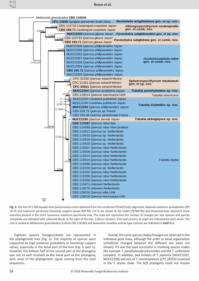
© 2018 Westerdijk Fungal Biodiversity Institute
Braun et al.
Editor-in-ChiefProf. dr P.W. Crous, Westerdijk Fungal Biodiversity Institute, P.O. Box 85167, 3508 AD Utrecht, The Netherlands.E-mail:[email protected]
58
Eighteen species lineages/clades are represented in the phylogenetic tree (Fig. 3). The majority of species were supported by high posterior probability or bootstrap support values, especially in the basal part of the tree (Fig. 3, part 1). However, the bottom half of the second part of the phylogeny was not as well resolved as the basal part of the phylogeny, with most of the phylogenetic signal coming from the tub2 sequences.
Overall, the same species clades/lineages are observed in the individual gene trees, although the order or basal organisation sometimes changed between the different loci (data not shown). ITS was the least successful in resolving species clades (for example T. paradryinoides/dryinoides and the T. suttoniana complex). In addition, two isolates of T. japonica (MUCC2297, MUCC2298) and one of T. seoraksanensis (CPC 26553) clustered in the T. dryina clade. The tef1 phylogeny could not resolve
Fig. 3. The first of 1 000 equally most parsimonious trees obtained from the combined ITS/tef1/tub2 alignment. Bayesian posterior probabilities (PP) >0.74 and maximum parsimony bootstrap support values (MP-BS) >74 % are shown at the nodes (PP/MP-BS) and thickened lines represent those branches present in the strict consensus maximum parsimony tree. The scale bar represents the number of changes per site. Species and species complexes are indicated with coloured blocks to the right of the tree. Culture numbers, host and country of origin are indicated for each strain. The tree is rooted to Melanconis groenlandica (culture CBS 116540) and taxonomic novelties and ex-type cultures are indicated in bold face.
50
CBS 112097 Quercus robur Italy CBS 114386 Quercus robur New Zealand CBS 114912 Quercus sp. Netherlands CBS 114915 Quercus sp. Netherlands CBS 114919 Quercus sp. Netherlands CBS 114920 Quercus sp. Netherlands CBS 115007 Quercus robur Netherlands CBS 115009 Quercus robur Netherlands CBS 115012 Quercus robur Netherlands CBS 115014 Quercus robur Netherlands CBS 115096 Quercus robur Netherlands CBS 115100 Quercus sp. Netherlands CBS 115102 Quercus robur Netherlands CBS 115306 Quercus robur Netherlands CBS 115308 Quercus robur Netherlands CBS 115971 Unknown Netherlands CBS 116070 Unknown Netherlands CBS 129016 Quercus alba USA CBS 129018 Quercus macrocarpa USA
MUCC2295 Quercus serrata Japan
CBS 129014 Quercus macrocarpa USA
CBS 329.75 Quercus sp. France CBS 335.86 Quercus pedunculata France
MUCC2292 Quercus phillyraeoides Japan
MUCC2291 Castanea pubinervis Japan MUCC2290 Castanea pubinervis Japan
MUCC2294 Quercus acutissima Japan
CPC 31361 Quercus eduardi Mexico CPC 33021 Quercus eduardi Mexico
CPC 32258 Quercus eduardi Mexico
CBS 192.71 Quercus phillyraeoides Japan MUCC2305 Quercus phillyraeoides Japan
MUCC2304 Quercus phillyraeoides Japan
MUCC2303 Quercus phillyraeoides Japan MUCC2307 Quercus phillyraeoides Japan MUCC2309 Quercus phillyraeoides Japan
MUCC2306 Quercus phillyraeoides Japan MUCC2308 Quercus phillyraeoides Japan
CBS 124732 Castanopsis cuspidata Japan CBS 189.71 Castanopsis cuspidata Japan
CBS 124733 Quercus glauca Japan CBS 193.71 Quercus glauca Japan
MUCC2293 Quercus glauca Japan
CPC 13806 Syzigium guineense South Africa Melanconis groenlandica CBS 116540
Racheliella wingfieldiana gen. et sp. nov.Oblongisporothyrium castanopsidisgen. et comb. nov.
Paratubakia subglobosoides gen. et sp. nov.
Paratubakia subglobosa gen. et comb. nov.
0.96/92
1/100
-/100
1/1001/100
1/100
0.91/100
1/100
1/100
1/-1/-
1/100
0.99/100
1/100
1/100
1/100
0.99/--/79
0.79/99
-/99
Involutiscutellula rubragen. et comb. nov.
Tubakia paradryinoides sp. nov.
Tubakia dryinoides sp. nov.
Tubakia oblongispora sp. nov.
Tubakia dryina
Tubakia americana
Sphaerosporithyrium mexicanumgen. et sp. nov.
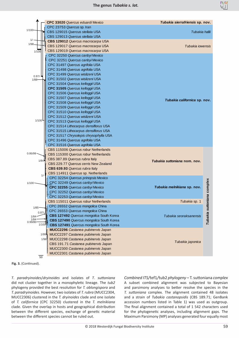
© 2018 Westerdijk Fungal Biodiversity Institute
The genus Tubakia s. lat.
Editor-in-ChiefProf. dr P.W. Crous, Westerdijk Fungal Biodiversity Institute, P.O. Box 85167, 3508 AD Utrecht, The Netherlands.E-mail:[email protected]
59
T. paradryinoides/dryinoides and isolates of T. suttoniana did not cluster together in a monophyletic lineage. The tub2 phylogeny provided the best resolution for T. oblongispora and T. paradryinoides. However, two isolates of T. rubra (MUCC2304, MUCC2306) clustered in the T. dryinoides clade and one isolate of T. californica (CPC 32250) clustered in the T. melnikiana clade. Given the overlap in hosts and geographical distribution between the different species, exchange of genetic material between the different species cannot be ruled out.
Combined ITS/tef1/tub2 phylogeny – T. suttoniana complexA subset combined alignment was subjected to Bayesian and parsimony analyses to better resolve the species in the T. suttoniana complex. The alignment contained 48 isolates and a strain of Tubakia castanopsidis (CBS 189.71; GenBank accession numbers listed in Table 1) was used as outgroup. The final alignment contained a total of 1 542 characters used for the phylogenetic analyses, including alignment gaps. The Maximum Parsimony (MP) analyses generated four equally most
50
CPC 32249 Quercus canbyi Mexico CPC 32255 Quercus canbyi Mexico CPC 32252 Quercus canbyi Mexico CPC 32253 Quercus canbyi Mexico
CPC 32254 Quercus prinopsis Mexico CBS 114911 Quercus sp. Netherlands
CBS 115011 Quercus robur Netherlands
CBS 127490 Quercus mongolica South Korea CBS 127491 Quercus mongolica South Korea
CBS 127492 Quercus mongolica South Korea
CPC 26552 Quercus mongolica China CPC 26553 Quercus mongolica China
CBS 191.71 Castanea pubinervis Japan MUCC2300 Castanea pubinervis Japan MUCC2301 Castanea pubinervis Japan
MUCC2298 Castanea pubinervis Japan MUCC2297 Castanea pubinervis Japan MUCC2296 Castanea pubinervis Japan
CBS 639.93 Quercus rubra Italy CBS 229.77 Quercus cerris New Zealand
CBS 115300 Quercus robur Netherlands CBS 387.89 Quercus rubra Italy
CBS 115006 Quercus robur Netherlands
CPC 31496 Quercus agrifolia USA CPC 31516 Quercus agrifolia USA
CPC 31497 Quercus agrifolia USA CPC 31498 Quercus agrifolia USA CPC 31499 Quercus wislizeni USA CPC 31502 Quercus wislizeni USA CPC 31504 Quercus kelloggii USA CPC 31505 Quercus kelloggii USA CPC 31506 Quercus kelloggii USA CPC 31507 Quercus kelloggii USA CPC 31508 Quercus kelloggii USA CPC 31509 Quercus kelloggii USA CPC 31510 Quercus kelloggii USA CPC 31512 Quercus wislizeni USA CPC 31513 Quercus kelloggii USA CPC 31514 Lithocarpus densiflorus USA CPC 31515 Lithocarpus densiflorus USA CPC 31517 Chrysolepis chrysophylla USA
CPC 32250 Quercus canbyi Mexico CPC 32251 Quercus canbyi Mexico
CBS 129012 Quercus macrocarpa USA CBS 129017 Quercus macrocarpa USA CBS 129019 Quercus macrocarpa USA
CBS 129013 Quercus stellata USA CBS 129015 Quercus stellata USA CPC 23753 Quercus sp. Iran CPC 33020 Quercus eduardi Mexico
1/100
1/100
1/98
1/88
1/-
1/100
1/84
1/100
1/-
1/76
1/85
1/841/84
1/100
1/99
1/96
0.96/-
0.87/-
0.99/80
0.95/99
0.97/-
Tubakia sierrafriensis sp. nov.
Tubakia iowensis
Tubakia californica sp. nov.
Tubakia suttoniana nom. nov.
Tubakia melnikiana sp. nov.
Tubakia sp. 1
Tubakia seoraksanensis
Tubakia japonica
Tuba
kia
sutto
nian
a co
mpl
ex
Tubakia hallii
Fig. 3. (Continued).
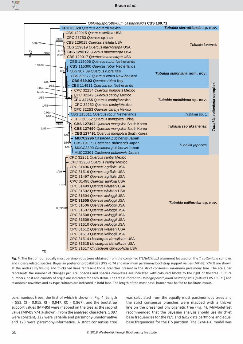
© 2018 Westerdijk Fungal Biodiversity Institute
Braun et al.
Editor-in-ChiefProf. dr P.W. Crous, Westerdijk Fungal Biodiversity Institute, P.O. Box 85167, 3508 AD Utrecht, The Netherlands.E-mail:[email protected]
60
parsimonious trees, the first of which is shown in Fig. 4 (Length = 553, CI = 0.915, RI = 0.947, RC = 0.867), and the bootstrap support values (MP-BS) were mapped on the tree as the second value (MP-BS >74 % shown). From the analysed characters, 1 097 were constant, 322 were variable and parsimony-uninformative and 123 were parsimony-informative. A strict consensus tree
was calculated from the equally most parsimonious trees and the strict consensus branches were mapped with a thicker line on the presented phylogenetic tree (Fig. 4). MrModelTest recommended that the Bayesian analysis should use dirichlet base frequencies for the tef1 and tub2 data partitions and equal base frequencies for the ITS partition. The SYM+I+G model was
-/100
1/90
1/100
1/100
1/100
0.99/75
1/78
1/99
1/99
1/99
1/99
0.69/98
1/87
1/96
1/94
1/95
0.94/99
0.95/-
0.99/-
0.82/-
0.91/-
1/83
1/85
1/78
0.99/-
Tubakia sierrafriensis sp. nov.
Tubakia iowensis
Tubakia californica sp. nov.
Tubakia suttoniana nom. nov.
Tubakia melnikiana sp. nov.
Tubakia sp. 1
Tubakia seoraksanensis
Tubakia japonica
CPC 32249 Quercus canbyi Mexico CPC 32255 Quercus canbyi Mexico CPC 32252 Quercus canbyi Mexico CPC 32253 Quercus canbyi Mexico
CPC 32254 Quercus prinopsis Mexico CBS 114911 Quercus sp. Netherlands
CBS 115011 Quercus robur Netherlands
CBS 127490 Quercus mongolica South Korea CBS 127491 Quercus mongolica South Korea
CBS 127492 Quercus mongolica South Korea CPC 26552 Quercus mongolica China
CBS 191.71 Castanea pubinervis Japan MUCC2300 Castanea pubinervis Japan MUCC2301 Castanea pubinervis Japan
MUCC2296 Castanea pubinervis Japan
CBS 639.93 Quercus rubra Italy CBS 229.77 Quercus cerris New Zealand
CBS 115300 Quercus robur Netherlands CBS 387.89 Quercus rubra Italy
CBS 115006 Quercus robur Netherlands
CPC 31496 Quercus agrifolia USA CPC 31516 Quercus agrifolia USA CPC 31497 Quercus agrifolia USA CPC 31498 Quercus agrifolia USA CPC 31499 Quercus wislizeni USA CPC 31502 Quercus wislizeni USA CPC 31504 Quercus kelloggii USA CPC 31505 Quercus kelloggii USA CPC 31506 Quercus kelloggii USA CPC 31507 Quercus kelloggii USA CPC 31508 Quercus kelloggii USA CPC 31509 Quercus kelloggii USA CPC 31510 Quercus kelloggii USA CPC 31512 Quercus wislizeni USA CPC 31513 Quercus kelloggii USA CPC 31514 Lithocarpus densiflorus USA CPC 31515 Lithocarpus densiflorus USA CPC 31517 Chrysolepis chrysophylla USA
CPC 32250 Quercus canbyi Mexico CPC 32251 Quercus canbyi Mexico
CBS 129012 Quercus macrocarpa USA CBS 129017 Quercus macrocarpa USA
CBS 129019 Quercus macrocarpa USA CBS 129013 Quercus stellata USA
CBS 129015 Quercus stellata USA CPC 23753 Quercus sp. Iran
CPC 33020 Quercus eduardi Mexico Oblongisporothyrium castanopsidis CBS 189.71
10
2x
Tuba
kia
sutto
nian
a co
mpl
ex
Fig. 4. The first of four equally most parsimonious trees obtained from the combined ITS/tef1/tub2 alignment focused on the T. suttoniana complex and closely related species. Bayesian posterior probabilities (PP) >0.74 and maximum parsimony bootstrap support values (MP-BS) >74 % are shown at the nodes (PP/MP-BS) and thickened lines represent those branches present in the strict consensus maximum parsimony tree. The scale bar represents the number of changes per site. Species and species complexes are indicated with coloured blocks to the right of the tree. Culture numbers, host and country of origin are indicated for each strain. The tree is rooted to Oblongisporothyrium castanopsidis (culture CBS 189.71) and taxonomic novelties and ex-type cultures are indicated in bold face. The length of the most basal branch was halfed to facilitate layout.
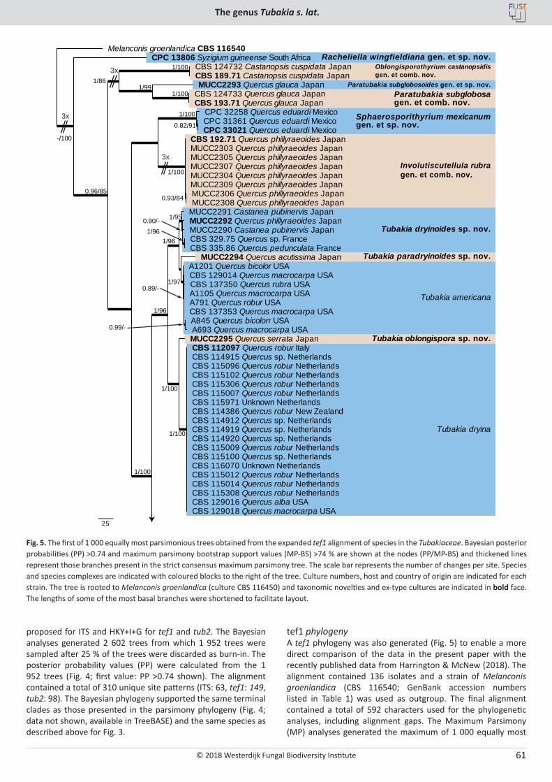
© 2018 Westerdijk Fungal Biodiversity Institute
The genus Tubakia s. lat.
Editor-in-ChiefProf. dr P.W. Crous, Westerdijk Fungal Biodiversity Institute, P.O. Box 85167, 3508 AD Utrecht, The Netherlands.E-mail:[email protected]
61
Paratubakia subglobosagen. et comb. nov.
Sphaerosporithyrium mexicanumgen. et sp. nov.
Tubakia paradryinoides sp. nov.
Tubakia dryinoides sp. nov.
Tubakia oblongispora sp. nov.
25
CBS 112097 Quercus robur Italy CBS 114915 Quercus sp. Netherlands CBS 115096 Quercus robur Netherlands CBS 115102 Quercus robur Netherlands CBS 115306 Quercus robur Netherlands CBS 115007 Quercus robur Netherlands CBS 115971 Unknown Netherlands CBS 114386 Quercus robur New Zealand CBS 114912 Quercus sp. Netherlands CBS 114919 Quercus sp. Netherlands CBS 114920 Quercus sp. Netherlands CBS 115009 Quercus robur Netherlands CBS 115100 Quercus sp. Netherlands CBS 116070 Unknown Netherlands CBS 115012 Quercus robur Netherlands CBS 115014 Quercus robur Netherlands CBS 115308 Quercus robur Netherlands CBS 129016 Quercus alba USA CBS 129018 Quercus macrocarpa USA
MUCC2295 Quercus serrata Japan
CBS 129014 Quercus macrocarpa USA CBS 137350 Quercus rubra USA A1105 Quercus macrocarpa USA A791 Quercus robur USA CBS 137353 Quercus macrocarpa USA A845 Quercus bicolorr USA A693 Quercus macrocarpa USA
A1201 Quercus bicolor USA MUCC2294 Quercus acutissima Japan
CBS 329.75 Quercus sp. France CBS 335.86 Quercus pedunculata France
MUCC2292 Quercus phillyraeoides Japan MUCC2290 Castanea pubinervis Japan
MUCC2291 Castanea pubinervis Japan
CPC 31361 Quercus eduardi Mexico CPC 33021 Quercus eduardi Mexico
CPC 32258 Quercus eduardi Mexico
CBS 192.71 Quercus phillyraeoides Japan MUCC2303 Quercus phillyraeoides Japan MUCC2305 Quercus phillyraeoides Japan MUCC2307 Quercus phillyraeoides Japan MUCC2304 Quercus phillyraeoides Japan MUCC2309 Quercus phillyraeoides Japan MUCC2306 Quercus phillyraeoides Japan MUCC2308 Quercus phillyraeoides Japan
CBS 124733 Quercus glauca Japan CBS 193.71 Quercus glauca Japan
MUCC2293 Quercus glauca Japan
CBS 124732 Castanopsis cuspidata Japan CBS 189.71 Castanopsis cuspidata Japan
CPC 13806 Syzigium guineense South Africa Melanconis groenlandica CBS 116540
-/100
1/100
1/100
1/100
1/100
1/100
1/100
1/1001/99
0.82/91
1/96
0.90/-
0.89/-
0.99/-
1/961/96
1/97
1/95
0.96/85
1/86
0.93/84
Racheliella wingfieldiana gen. et sp. nov.Oblongisporothyrium castanopsidisgen. et comb. nov.
Paratubakia subglobosoides gen. et sp. nov.
Involutiscutellula rubragen. et comb. nov.
Tubakia americana
Tubakia dryina
3x
3x
3x
Fig. 5. The first of 1 000 equally most parsimonious trees obtained from the expanded tef1 alignment of species in the Tubakiaceae. Bayesian posterior probabilities (PP) >0.74 and maximum parsimony bootstrap support values (MP-BS) >74 % are shown at the nodes (PP/MP-BS) and thickened lines represent those branches present in the strict consensus maximum parsimony tree. The scale bar represents the number of changes per site. Species and species complexes are indicated with coloured blocks to the right of the tree. Culture numbers, host and country of origin are indicated for each strain. The tree is rooted to Melanconis groenlandica (culture CBS 116450) and taxonomic novelties and ex-type cultures are indicated in bold face. The lengths of some of the most basal branches were shortened to facilitate layout.
proposed for ITS and HKY+I+G for tef1 and tub2. The Bayesian analyses generated 2 602 trees from which 1 952 trees were sampled after 25 % of the trees were discarded as burn-in. The posterior probability values (PP) were calculated from the 1 952 trees (Fig. 4; first value: PP >0.74 shown). The alignment contained a total of 310 unique site patterns (ITS: 63, tef1: 149, tub2: 98). The Bayesian phylogeny supported the same terminal clades as those presented in the parsimony phylogeny (Fig. 4; data not shown, available in TreeBASE) and the same species as described above for Fig. 3.
tef1 phylogenyA tef1 phylogeny was also generated (Fig. 5) to enable a more direct comparison of the data in the present paper with the recently published data from Harrington & McNew (2018). The alignment contained 136 isolates and a strain of Melanconis groenlandica (CBS 116540; GenBank accession numbers listed in Table 1) was used as outgroup. The final alignment contained a total of 592 characters used for the phylogenetic analyses, including alignment gaps. The Maximum Parsimony (MP) analyses generated the maximum of 1 000 equally most
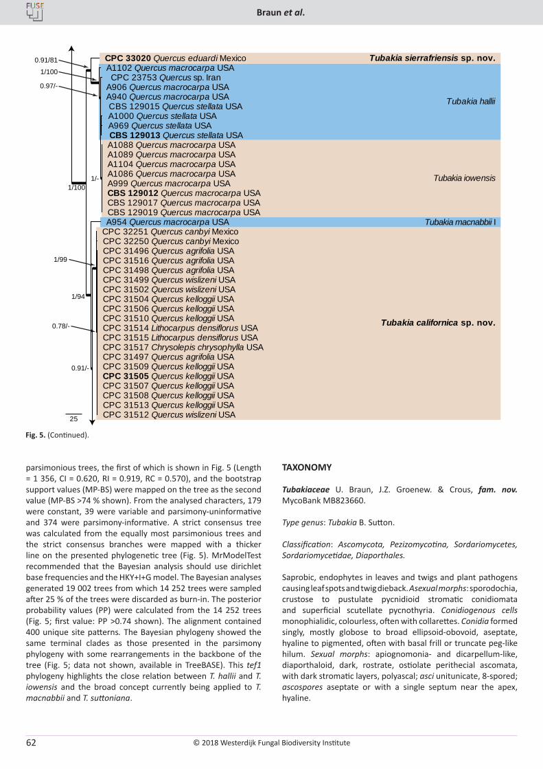
© 2018 Westerdijk Fungal Biodiversity Institute
Braun et al.
Editor-in-ChiefProf. dr P.W. Crous, Westerdijk Fungal Biodiversity Institute, P.O. Box 85167, 3508 AD Utrecht, The Netherlands.E-mail:[email protected]
62
parsimonious trees, the first of which is shown in Fig. 5 (Length = 1 356, CI = 0.620, RI = 0.919, RC = 0.570), and the bootstrap support values (MP-BS) were mapped on the tree as the second value (MP-BS >74 % shown). From the analysed characters, 179 were constant, 39 were variable and parsimony-uninformative and 374 were parsimony-informative. A strict consensus tree was calculated from the equally most parsimonious trees and the strict consensus branches were mapped with a thicker line on the presented phylogenetic tree (Fig. 5). MrModelTest recommended that the Bayesian analysis should use dirichlet base frequencies and the HKY+I+G model. The Bayesian analyses generated 19 002 trees from which 14 252 trees were sampled after 25 % of the trees were discarded as burn-in. The posterior probability values (PP) were calculated from the 14 252 trees (Fig. 5; first value: PP >0.74 shown). The alignment contained 400 unique site patterns. The Bayesian phylogeny showed the same terminal clades as those presented in the parsimony phylogeny with some rearrangements in the backbone of the tree (Fig. 5; data not shown, available in TreeBASE). This tef1 phylogeny highlights the close relation between T. hallii and T. iowensis and the broad concept currently being applied to T. macnabbii and T. suttoniana.
TAXONOMY
Tubakiaceae U. Braun, J.Z. Groenew. & Crous, fam. nov. MycoBank MB823660.
Type genus: Tubakia B. Sutton.
Classification: Ascomycota, Pezizomycotina, Sordariomycetes, Sordariomycetidae, Diaporthales.
Saprobic, endophytes in leaves and twigs and plant pathogens causing leaf spots and twig dieback. Asexual morphs: sporodochia, crustose to pustulate pycnidioid stromatic conidiomata and superficial scutellate pycnothyria. Conidiogenous cells monophialidic, colourless, often with collarettes. Conidia formed singly, mostly globose to broad ellipsoid-obovoid, aseptate, hyaline to pigmented, often with basal frill or truncate peg-like hilum. Sexual morphs: apiognomonia- and dicarpellum-like, diaporthaloid, dark, rostrate, ostiolate perithecial ascomata, with dark stromatic layers, polyascal; asci unitunicate, 8-spored; ascospores aseptate or with a single septum near the apex, hyaline.
Fig. 5. (Continued).
25
CPC 31496 Quercus agrifolia USA CPC 31516 Quercus agrifolia USA CPC 31498 Quercus agrifolia USA CPC 31499 Quercus wislizeni USA CPC 31502 Quercus wislizeni USA CPC 31504 Quercus kelloggii USA CPC 31506 Quercus kelloggii USA CPC 31510 Quercus kelloggii USA CPC 31514 Lithocarpus densiflorus USA CPC 31515 Lithocarpus densiflorus USA CPC 31517 Chrysolepis chrysophylla USA CPC 31497 Quercus agrifolia USA CPC 31509 Quercus kelloggii USA CPC 31505 Quercus kelloggii USA CPC 31507 Quercus kelloggii USA CPC 31508 Quercus kelloggii USA CPC 31513 Quercus kelloggii USA CPC 31512 Quercus wislizeni USA
CPC 32251 Quercus canbyi Mexico CPC 32250 Quercus canbyi Mexico
A954 Quercus macrocarpa USA
A1000 Quercus stellata USA A969 Quercus stellata USA CBS 129013 Quercus stellata USA
A906 Quercus macrocarpa USA A940 Quercus macrocarpa USA
A1088 Quercus macrocarpa USA A1089 Quercus macrocarpa USA A1104 Quercus macrocarpa USA A1086 Quercus macrocarpa USA A999 Quercus macrocarpa USA CBS 129012 Quercus macrocarpa USA CBS 129017 Quercus macrocarpa USA CBS 129019 Quercus macrocarpa USA
CBS 129015 Quercus stellata USA
CPC 23753 Quercus sp. Iran A1102 Quercus macrocarpa USA CPC 33020 Quercus eduardi Mexico
1/100
1/94
1/99
0.78/-
0.91/-
1/100
1/-
0.91/81
0.97/-
Tubakia sierrafriensis sp. nov.
Tubakia hallii
Tubakia iowensis
Tubakia macnabbii I
Tubakia californica sp. nov.
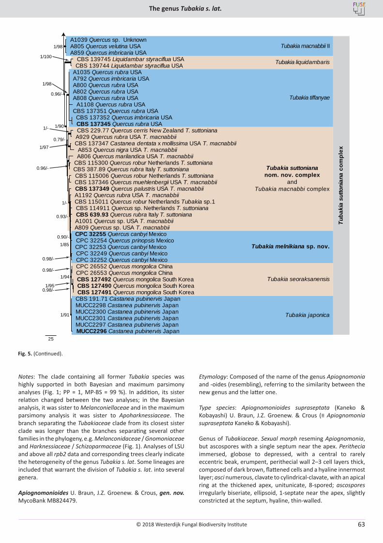
© 2018 Westerdijk Fungal Biodiversity Institute
The genus Tubakia s. lat.
Editor-in-ChiefProf. dr P.W. Crous, Westerdijk Fungal Biodiversity Institute, P.O. Box 85167, 3508 AD Utrecht, The Netherlands.E-mail:[email protected]
63
Notes: The clade containing all former Tubakia species was highly supported in both Bayesian and maximum parsimony analyses (Fig. 1; PP = 1, MP-BS = 99 %). In addition, its sister relation changed between the two analyses; in the Bayesian analysis, it was sister to Melanconiellaceae and in the maximum parsimony analysis it was sister to Apoharknessiaceae. The branch separating the Tubakiaceae clade from its closest sister clade was longer than the branches separating several other families in the phylogeny, e.g. Melanconidaceae / Gnomoniaceae and Harknessiaceae / Schizoparmaceae (Fig. 1). Analyses of LSU and above all rpb2 data and corresponding trees clearly indicate the heterogeneity of the genus Tubakia s. lat. Some lineages are included that warrant the division of Tubakia s. lat. into several genera.
Apiognomonioides U. Braun, J.Z. Groenew. & Crous, gen. nov. MycoBank MB824479.
Etymology: Composed of the name of the genus Apiognomonia and -oides (resembling), referring to the similarity between the new genus and the latter one.
Type species: Apiognomonioides supraseptata (Kaneko & Kobayashi) U. Braun, J.Z. Groenew. & Crous (≡ Apiognomonia supraseptata Kaneko & Kobayashi).
Genus of Tubakiaceae. Sexual morph reseming Apiognomonia, but ascospores with a single septum near the apex. Perithecia immersed, globose to depressed, with a central to rarely eccentric beak, erumpent, perithecial wall 2–3 cell layers thick, composed of dark brown, flattened cells and a hyaline innermost layer; asci numerous, clavate to cylindrical-clavate, with an apical ring at the thickened apex, unitunicate, 8-spored; ascospores irregularly biseriate, ellipsoid, 1-septate near the apex, slightly constricted at the septum, hyaline, thin-walled.
25
CPC 32249 Quercus canbyi Mexico CPC 32252 Quercus canbyi Mexico
CPC 32255 Quercus canbyi Mexico CPC 32254 Quercus prinopsis Mexico CPC 32253 Quercus canbyi Mexico
CBS 115011 Quercus robur Netherlands Tubakia sp.1
CBS 191.71 Castanea pubinervis Japan MUCC2298 Castanea pubinervis Japan MUCC2300 Castanea pubinervis Japan MUCC2301 Castanea pubinervis Japan MUCC2297 Castanea pubinervis Japan MUCC2296 Castanea pubinervis Japan
CBS 114911 Quercus sp. Netherlands T. suttoniana
CBS 127490 Quercus mongolica South Korea CBS 127491 Quercus mongolica South Korea
CBS 127492 Quercus mongolica South Korea
CPC 26552 Quercus mongolica China CPC 26553 Quercus mongolica China
CBS 639.93 Quercus rubra Italy T. suttoniana A1001 Quercus sp. USA T. macnabbii A809 Quercus sp. USA T. macnabbii
A1192 Quercus rubra USA T. macnabbii CBS 137349 Quercus palustris USA T. macnabbii
CBS 115006 Quercus robur Netherlands T. suttoniana CBS 137346 Quercus muehlenbergii USA T. macnabbii
CBS 115300 Quercus robur Netherlands T. suttoniana CBS 387.89 Quercus rubra Italy T. suttoniana
A806 Quercus marilandica USA T. macnabbii
CBS 229.77 Quercus cerris New Zealand T. suttoniana A929 Quercus rubra USA T. macnabbii CBS 137347 Castanea dentata x mollissima USA T. macnabbii A853 Quercus nigra USA T. macnabbii
A1035 Quercus rubra USA A792 Quercus imbricaria USA A800 Quercus rubra USA A802 Quercus rubra USA A808 Quercus rubra USA
A1108 Quercus rubra USA
CBS 137352 Quercus imbricaria USA CBS 137345 Quercus rubra USA
CBS 137351 Quercus rubra USA
CBS 139745 Liquidambar styraciflua USA CBS 139744 Liquidambar styraciflua USA
A1039 Quercus sp. Unknown A805 Quercus velutina USA A859 Quercus imbricaria USA
0.96/-
0.96/-
0.98/-
0.98/-
0.98/-
0.79/-
0.93/-
0.90/-
1/98
1/100
1/90
1/85
1/94
1/95
1/91
1/98
1/97
1/-
1/-
Tubakia macnabbii II
Tubakia liquidambaris
Tubakia tiffanyae
Tubakia suttoniananom. nov. complex
andTubakia macnabbi complex
Tubakia melnikiana sp. nov.
Tubakia seoraksanensis
Tubakia japonica
Tuba
kia
sutto
nian
a co
mpl
ex
Fig. 5. (Continued).
© 2018 Westerdijk Fungal Biodiversity Institute
Braun et al.
Editor-in-ChiefProf. dr P.W. Crous, Westerdijk Fungal Biodiversity Institute, P.O. Box 85167, 3508 AD Utrecht, The Netherlands.E-mail:[email protected]
64
Notes: Kaneko & Kobayashi (1984) introduced the name Apiognomonia supraseptata on the basis of ascomata formed on leaves of Quercus glauca incubated in a humid petri dish. Conidia were not formed. An ex-type strain (ATTC 58737) was used to retrieve a LSU rDNA sequence (GenBank AF277127, Zhang & Blackwell 2001) which was used by Harrington & McNew (2018) as basis to transfer A. supraseptata to Tubakia. They classified the mycelial colonies in culture to be tubakia-like. According to Kaneko & Kobayashi (1984), Apiognomonia has two-celled ascospores in which the top cell is larger than the bottom cell, but in A. supraseptata the top cell is smaller. Harrington & McNew (2018) postulated that A. supraseptata represents the only clearly demonstrated sexual morph of Tubakia. The allocation of Apiognomonia supraseptata to Tubakiaceae is reasonable and could be confirmed in our own analyses (Fig. 1). Apiognomonia supraseptata is not included in the rpb2 tree due to lack of sequence data, but in the LSU (Fig. 1) and ITS trees (not shown) this species clusters distantly from all other lineages outside of the Tubakia s. str. clade, i.e., this species cannot be maintained in Tubakia s. str. Owing to its isolated position in the LSU tree within the Tubakiaceae, but distant from all other genera of this family, Apiognomonia supraseptata is best accommodated in a genus of its own. A. supraseptata and the sexual morph of Tubakia suttoniana have various morphological characters in common (rostrate perithecia, unitunicate 8-spored asci, colourless conidia), but A. supraseptata differs in forming uniseptate ascospores with a septum near the apex.
Apiognomonioides supraseptata (Kaneko & Kobayashi) U. Braun, J.Z. Groenew. & Crous, comb. nov. MycoBank MB824480.Basionym: Apiognomonia supraseptata Kaneko & Kobayashi, Trans. Mycol. Soc. Japan 25: 11. 1984.Synonym: Tubakia supraseptata (Kaneko & Kobayashi) T.C. Harr. & McNew, Antonie van Leeuwenhoek J. Microbiol. Serol. doi.org/10.1007/s10482-017-1001-9 [16]. 2017.
Illustrations: Kaneko & Kobayashi (1984: 13, figs 1–10).
Description in vivo: Perithecia formed in lesions on leaves after incubation in a humid petri dish, black, immersed, globose or depressed at the base, 130–220 µm diam, 80–180 µm high, with a central to rarely eccentric beak, erumpent, mostly hypophyllous, to 300 µm long and 50–75 µm wide at the base, perithecial wall 2–3 cell layers thick, composed of dark brown, flattened cells and a hyaline innermost layer. Asci numerous, clavate to cylindrical-clavate, tapering towards the base, with an apical ring at the thickened apex, unitunicate, 50–70 × 10–12.5 µm, 8-spored. Ascospores irregularly biseriate, ellipsoid, 1-septate near the apex, slightly constricted at the septum, 11–15 × 4.5–6 µm, hyaline, thin-walled.
In vitro: On PSA rapidly growing at 25 °C, attaining 35–45 mm diam after 10 d, more or less zonate, wooly, white, reverse white to pale luteous, colony colour deeper at temperatures higher than 27 °C; aerial mycelium composed of branched, hyaline, septate hyphae, 1.5–2 µm wide; immersed hyphae 2–5 µm wide; development of perithecia in culture 14 d after incubation at 20 °C with continuous fluorescent light, scattered or in concentric circles, somewhat immersed (ascomata formed in culture 250–280 µm diam; asci 65–78 × 10–12.5 µm, basal part more elongated than on the host, ascospores 11.5–15 × 4.5–6.5 µm).
Type: Japan, Tottori Pref., Tottori City, Ohchidani Park, on leaves of Quercus glauca [incubated in a humid petri dish for 28 d after collection], 11 Feb. 1982, S. Kaneko (TMI 7647 – holotype; TFM : FPT5447; ATTC 58737 = CBS 632.92 = TMI cult. 70024 – ex-type strains).
Involutiscutellula U. Braun & C. Nakash., gen. nov. MycoBank MB824481.
Etymology: Composed of “involutus” (involute), “scutellulum” (referring to the pycnothyrial scutella), and -ula (diminutive) = minute scutellum with involute margin.
Type species: Actinopelte rubra T. Yokoy. & Tubaki [≡ Involutiscu-tellula rubra (T. Yokoy. & Tubaki) U. Braun & C. Nakash.].
Genus of Tubakiaceae. Living as endophytes in leaves. Colonies in vitro finally turning reddish brown. Mycelium internal, hyaline, and external, pigmented; hyphae in culture finally turning reddish brown. Asexual morphs forming crustose conidiomata on shed leaves (litter) and superficial pycnothyria on symptomless leaves, occasionally on reddish discolorations. Pycnothyria subcircular when viewed from above, superficial, easily removable, scutellate, fixed to the leaf by a central columella; scutellum convex to flattened, membranous, compact, neither loose nor splitting, outline regular, circular-subcircular, margin continuous, more or less undulate, distinctly involute; conidiophores reduced to conidiogenous cells, conical, cylindrical, ampulliform, pale orange yellowish to yellowish brown, arising from the underside of the scutella, around the columella, conidiogenous cells phialidic; conidia formed singly, globose, subglobose to broad ellipsoid-obovoid, smooth; microconidia oblong-bacilliform, cylindrical, straight to curved-sigmoid, very narrow, only 1 µm wide.
Notes: In all of the phylogenetic trees, especially the LSU and rpb2 phylogenies, Actinopelte rubra takes an isolated basal position and forms a single species lineage that warrants a classification as a genus of its own (Figs 1, 2). A. rubra is morphologically readily distinguishable from all other tubakia-like species by colonies and hyphae turning reddish brown with age and distinctive pycnothyria with continuous, more or less undulate, distinctly involute margin and very narrow oblong-bacilliform to cylindrical microconidia.
Involutiscutellula rubra (T. Yokoy. & Tubaki) U. Braun & C. Nakash., comb. nov. MycoBank MB824482. Fig. 6.Basionym: Actinopelte rubra T. Yokoy. & Tubaki, Res. Commun. Inst. Ferment. Osaka 5: 47. 1971.Synonym: Tubakia rubra (T. Yokoy. & Tubaki) B. Sutton, Trans. Brit. Mycol. Soc. 60: 165. 1973.
Illustrations: Yokoyama & Tubaki (1971: 65, pl. 1F, 67, pl. 2D; 69, pl. 3D; 72, pl. 6A–G; 76, pl. 10A–H).
Description in vivo: Living as endophyte in leaves, forming crustose conidiomata on the surface of shed leaves (litter), usually without distinct symptoms (lesions), sometimes causing a reddish tinge on the leaf surface. Mycelium internal and external, forming hyaline, branched intra- and intercellular external hyphae, external hyphae observed on the upper leaf surface, pale brown, branched. Conidiomata (pycnothyria) amphigenous,
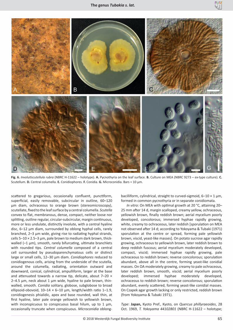
© 2018 Westerdijk Fungal Biodiversity Institute
The genus Tubakia s. lat.
Editor-in-ChiefProf. dr P.W. Crous, Westerdijk Fungal Biodiversity Institute, P.O. Box 85167, 3508 AD Utrecht, The Netherlands.E-mail:[email protected]
65
scattered to gregarious, occasionally confluent, punctiform, superficial, easily removable, subcircular in outline, 60–120 µm diam, ochraceous to orange brown (stereomicroscopy), scutellate, fixed to the leaf surface by a central columella. Scutella convex to flat, membranous, dense, compact, neither loose nor splitting, outline regular, circular-subcircular, margin continuous, more or less undulate, distinctly involute, with a central hyaline disc, 6–12 µm diam, surrounded by oblong hyphal cells, rarely branched, 2–3 µm wide, giving rise to radiating hyphal strands, cells 5–10 × 2.5–3 µm, pale brown to medium dark brown, thick-walled (–1 µm), smooth, rarely bifurcating, ultimate branchlets with rounded tips. Central columella composed of a central cell surrounded by pseudoparenchymatous cells or distinct large or small cells, 12–30 µm diam. Conidiophores reduced to conidiogenous cells, arising from the underside of the scutella, around the columella, radiating, orientation outward and downward, conical, cylindrical, ampulliform, larger at the base and attenuated towards a narrow tip, delicate, about 7–20 × 2–4.5 µm, neck about 1 µm wide, hyaline to pale brown, thin-walled, smooth. Conidia solitary, globose, subglobose to broad ellipsoid-obovoid, 10–14 × 6–10 µm, length/width ratio 1–1.9, conidiogenesis phialidic, apex and base rounded, wall thin, at first hyaline, later pale orange yellowish to yellowish brown, with inconspicuous to conspicuous basal hilum, up to 1 µm, occasionally truncate when conspicuous. Microconidia oblong-
bacilliform, cylindrical, straight to curved-sigmoid, 6–10 × 1 µm, formed in common pycnothyria or in separate conidiomata.
In vitro: On MEA with optimal growth at 20 °C, attaining 20–25 mm after 14 d, margin scalloped, creamy yellow, ochraceous, yellowish brown, finally reddish brown; aerial mycelium poorly developed, concolorous; immersed hyphae rapidly growing, white, creamy to ochraceous, later reddish [sporulation on MEA not observed after 14 d; according to Yokoyama & Tubaki (1971) sporulation at the centre or spread, forming pale yellowish brown, viscid, yeast-like masses]. On potato sucrose agar rapidly growing, ochraceous to yellowish brown, later reddish brown to deep reddish fuscous; aerial mycelium moderately developed, compact, viscid; immersed hyphae rapidly growing, pale ochraceous to reddish brown; reverse concolorous; sporulation abundant, above all in the centre, forming yeast-like conidial masses. On OA moderately growing, creamy to pale ochraceous, later reddish brown, smooth, viscid; aerial mycelium poorly developed; immersed hyphae moderately developed, ochraceous to reddish brown; reverse concolorous; sporulation abundant, evenly scattered, forming yeast-like conidial masses. On Czapek agar growth lacking or only restricted, reddish brown (from Yokoyama & Tubaki 1971).
Type: Japan, Kyoto Pref., Kyoto, on Quercus phillyraeoides, 28 Oct. 1969, T. Yokoyama 44102801 (NBRC H-11622 – holotype;
Fig. 6. Involutiscutellula rubra (NBRC H-11622 – holotype). A. Pycnothyria on the leaf surface. B. Culture on MEA (NBRC 9273 – ex-type culture). C. Scutellum. D. Central columella. E. Conidiophores. F. Conidia. G. Microconidia. Bars = 10 µm.
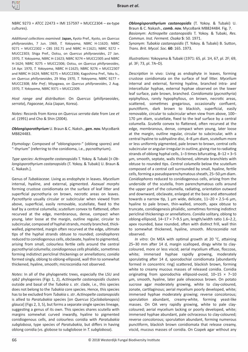
© 2018 Westerdijk Fungal Biodiversity Institute
Braun et al.
Editor-in-ChiefProf. dr P.W. Crous, Westerdijk Fungal Biodiversity Institute, P.O. Box 85167, 3508 AD Utrecht, The Netherlands.E-mail:[email protected]
66
NBRC 9273 = ATCC 22473 = IMI 157597 = MUCC2304 – ex-type cultures).
Additional collections examined: Japan, Kyoto Pref., Kyoto, on Quercus phillyraeoides, 7 Jun. 1969, T. Yokoyama, NBRC H-11620; NBRC 9271 = MUCC2302 = CBS 192.71 and NBRC H-11621; NBRC 9272 = MUCC2303; Shiga Pref., Ootsu, on Quercus phillyraeoides, 27 Jan. 1970, T. Yokoyama, NBRC H-11623; NBRC 9274 = MUCC2305 and NBRC H-1624; NBRC 9275 = MUCC2306; Ootsu, on Quercus phillyraeoides, 14 Apr. 1970, T. Yokoyama, NBRC H-11625; NBRC 9276 = MUCC2307 and NBRC H-1624; NBRC 9275 = MUCC2306; Kagoshima Pref., Yaku Is., on Quercus phillyraeoides, 29 May 1970, T. Yokoyama, NBRC 9277 = MUCC2308; Mie Pref.; Miyagawa, on Quercus phillyraeoides, 2 Aug. 1970, T. Yokoyama, NBRC 9371 = MUCC2309.
Host range and distribution: On Quercus (phillyraeoides, serrata), Fagaceae, Asia (Japan, Korea).
Notes: Records from Korea on Quercus serrata date from Lee et al. (1991) and Cho & Shin (2004).
Oblongisporothyrium U. Braun & C. Naksh., gen. nov. MycoBank MB824483.
Etymology: Composed of “oblongisporo-“ (oblong spores) and “-thyrium” (referring to the conidioma, i.e., pycnothyrium).
Type species: Actinopelte castanopsidis T. Yokoy. & Tubaki [≡ Ob-longisporothyrium castanopsidis (T. Yokoy. & Tubaki) U. Braun & C. Nakash.].
Genus of Tubakiaceae. Living as endophyte in leaves. Mycelium internal, hyaline, and external, pigmented. Asexual morphs forming crustose conidiomata on the surface of leaf litter and superficial pycnothyria on brown, necrotic areas on leaves. Pycnothyria usually circular or subcircular when viewed from above, superficial, easily removable, scutellate, fixed to the leaf by a central columella; scutellum convex to flattened, often recurved at the edge, membranous, dense, compact when young, later loose at the margin, outline regular, circular to subcircular, composed of hyphal strands, mostly branched, thick-walled, pigmented, margin often recurved at the edge, ultimate tips of the hyphal strands obtuse to rounded; conidiophores reduced to conidiogenous cells, obclavate, hyaline to pigmented, arising from small, colourless fertile cells around the central pycnothyrial columella; conidiogenous cells phialidic, sometimes forming indistinct periclinal thickenings or annellations; conidia formed singly, oblong to oblong-ellipsoid, wall thin to somewhat thickened, hyaline, smooth; microconidia not observed.
Notes: In all of the phylogenetic trees, especially the LSU and rpb2 phylogenies (Figs 1, 2), Actinopelte castanopsidis clusters outside and basal of the Tubakia s. str. clade, i.e., this species does not belong to the Tubakia core species. Hence, this species has to be excluded from Tubakia s. str. Actinopelte castanopsidis is allied to Paratubakia species [on Quercus (Cyclobalanopsis) glauca] (Figs 2, 3, 5), but forms a separate single species lineage, suggesting a genus of its own. This species shares scutella with margins somewhat curved inwardly, hyaline to pigmented conidiogenous cells, and colourless conidia with Paratubakia subglobosa, type species of Paratubakia, but differs in having oblong conidia (vs. globose to subglobose in T. subglobosa).
Oblongisporothyrium castanopsidis (T. Yokoy. & Tubaki) U. Braun & C. Nakash., comb. nov. MycoBank MB824484. Fig. 7.Basionym: Actinopelte castanopsidis T. Yokoy. & Tubaki, Res. Commun. Inst. Ferment. Osaka 5: 50. 1971.Synonym: Tubakia castanopsidis (T. Yokoy. & Tubaki) B. Sutton, Trans. Brit. Mycol. Soc. 60: 165. 1973.
Illustrations: Yokoyama & Tubaki (1971: 65, pl. 1H, 67, pl. 2F; 69, pl. 3F; 73, pl. 7A–D).
Description in vivo: Living as endophyte in leaves, forming crustose conidiomata on the surface of leaf litter. Mycelium internal and external, forming hyaline, branched intra- and intercellular hyphae, external hyphae observed on the lower leaf surface, pale brown, branched. Conidiomata (pycnothyria) epiphyllous, rarely hypophyllous, on brown, necrotic areas, scattered, sometimes gregarious, occasionally confluent, punctiform, dark brown to blackish, superficial, easily removable, circular to subcircular when view from above, 100–170 µm diam, scutellate, fixed to the leaf surface by a central columella. Scutella convex to flattened, often recurved at the edge, membranous, dense, compact when young, later loose at the margin, outline regular, circular to subcircular, with a central hyaline to subhyaline disc, 4–8 µm diam, scutellum more or less uniformly pigmented, pale brown to brown, central cells subcircular or angular-irregular in outline, giving rise to radiating strands of oblong hyphal cells, 1–3 times bifurcating, 8–12 × 3–5 µm, smooth, septate, walls thickened, ultimate branchlets with obtuse to rounded tips. Central columella below the scutellum composed of a central cell surrounded by small, hyaline, fertile cells, forming a pseudoparenchymatous sheath, 25–50 µm diam. Conidiophores reduced to conidiogenous cells, arising from the underside of the scutella, from parenchymatous cells around the upper part of the columella, radiating, orientation outward and downward, obclavate, enlarged at the base and attenuated towards a narrow tip, 1 µm wide, delicate, 11–20 × 2.5–6 µm, hyaline to pale brown, thin-walled, smooth, apex obtuse to truncate, conidiogenesis phialidic, sometimes forming indistinct periclinal thickenings or annellations. Conidia solitary, oblong to oblong-ellipsoid, 14–17 × 7–9.5 µm, length/width ratio 1.6–2.2, apex rounded, base rounded, often with distinct frill, wall thin to somewhat thickened, hyaline, smooth. Microconidia not observed. In vitro: On MEA with optimal growth at 20 °C, attaining 25–30 mm after 14 d, margin scalloped, dingy white to clay-coloured, more or less viscid; aerial mycelium effuse, floccose, white; immersed hyphae rapidly growing, moderately sporulating after 14 d, sporodochial conidiomata [abundantly formed in concentric ring] scattered, blackish brown, forming white to creamy mucous masses of released conidia. Conidia originating from sporodochia ellipsoid-ovoid, 10–15 × 7–10 µm, smooth, hyaline, later pale olivaceous brown. On potato sucrose agar moderately growing, white to clay-coloured, zonate, cartilaginous; aerial mycelium poorly developed, white; immersed hyphae moderately growing; reverse concolorous; sporulation abundant, creamy-white, forming yeast-like masses. On OA very rapidly growing, white to pale clay-coloured; aerial mycelium lacking or poorly developed, white; immersed hyphae abundant, pale ochraceous to clay-coloured; reverse concolorous; sporulation abundant, forming numerous punctiform, blackish brown conidiomata that release creamy, viscid, mucous masses of conidia. On Czapek agar without any
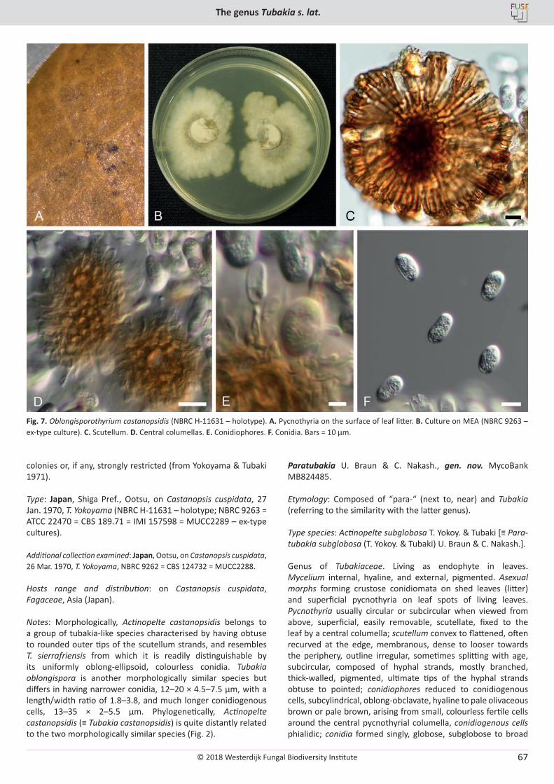
© 2018 Westerdijk Fungal Biodiversity Institute
The genus Tubakia s. lat.
Editor-in-ChiefProf. dr P.W. Crous, Westerdijk Fungal Biodiversity Institute, P.O. Box 85167, 3508 AD Utrecht, The Netherlands.E-mail:[email protected]
67
colonies or, if any, strongly restricted (from Yokoyama & Tubaki 1971).
Type: Japan, Shiga Pref., Ootsu, on Castanopsis cuspidata, 27 Jan. 1970, T. Yokoyama (NBRC H-11631 – holotype; NBRC 9263 = ATCC 22470 = CBS 189.71 = IMI 157598 = MUCC2289 – ex-type cultures).
Additional collection examined: Japan, Ootsu, on Castanopsis cuspidata, 26 Mar. 1970, T. Yokoyama, NBRC 9262 = CBS 124732 = MUCC2288.
Hosts range and distribution: on Castanopsis cuspidata, Fagaceae, Asia (Japan).
Notes: Morphologically, Actinopelte castanopsidis belongs to a group of tubakia-like species characterised by having obtuse to rounded outer tips of the scutellum strands, and resembles T. sierrafriensis from which it is readily distinguishable by its uniformly oblong-ellipsoid, colourless conidia. Tubakia oblongispora is another morphologically similar species but differs in having narrower conidia, 12–20 × 4.5–7.5 µm, with a length/width ratio of 1.8–3.8, and much longer conidiogenous cells, 13–35 × 2–5.5 µm. Phylogenetically, Actinopelte castanopsidis (≡ Tubakia castanopsidis) is quite distantly related to the two morphologically similar species (Fig. 2).
Paratubakia U. Braun & C. Nakash., gen. nov. MycoBank MB824485.
Etymology: Composed of “para-“ (next to, near) and Tubakia (referring to the similarity with the latter genus).
Type species: Actinopelte subglobosa T. Yokoy. & Tubaki [≡ Para-tubakia subglobosa (T. Yokoy. & Tubaki) U. Braun & C. Nakash.].
Genus of Tubakiaceae. Living as endophyte in leaves. Mycelium internal, hyaline, and external, pigmented. Asexual morphs forming crustose conidiomata on shed leaves (litter) and superficial pycnothyria on leaf spots of living leaves. Pycnothyria usually circular or subcircular when viewed from above, superficial, easily removable, scutellate, fixed to the leaf by a central columella; scutellum convex to flattened, often recurved at the edge, membranous, dense to looser towards the periphery, outline irregular, sometimes splitting with age, subcircular, composed of hyphal strands, mostly branched, thick-walled, pigmented, ultimate tips of the hyphal strands obtuse to pointed; conidiophores reduced to conidiogenous cells, subcylindrical, oblong-obclavate, hyaline to pale olivaceous brown or pale brown, arising from small, colourless fertile cells around the central pycnothyrial columella, conidiogenous cells phialidic; conidia formed singly, globose, subglobose to broad
Fig. 7. Oblongisporothyrium castanopsidis (NBRC H-11631 – holotype). A. Pycnothyria on the surface of leaf litter. B. Culture on MEA (NBRC 9263 – ex-type culture). C. Scutellum. D. Central columellas. E. Conidiophores. F. Conidia. Bars = 10 µm.
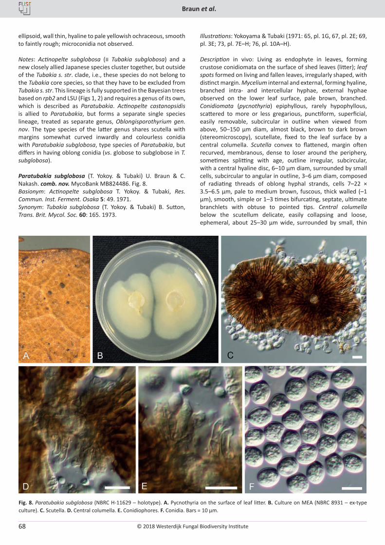
© 2018 Westerdijk Fungal Biodiversity Institute
Braun et al.
Editor-in-ChiefProf. dr P.W. Crous, Westerdijk Fungal Biodiversity Institute, P.O. Box 85167, 3508 AD Utrecht, The Netherlands.E-mail:[email protected]
68
ellipsoid, wall thin, hyaline to pale yellowish ochraceous, smooth to faintly rough; microconidia not observed.
Notes: Actinopelte subglobosa (≡ Tubakia subglobosa) and a new closely allied Japanese species cluster together, but outside of the Tubakia s. str. clade, i.e., these species do not belong to the Tubakia core species, so that they have to be excluded from Tubakia s. str. This lineage is fully supported in the Bayesian trees based on rpb2 and LSU (Figs 1, 2) and requires a genus of its own, which is described as Paratubakia. Actinopelte castanopsidis is allied to Paratubakia, but forms a separate single species lineage, treated as separate genus, Oblongisporothyrium gen. nov. The type species of the latter genus shares scutella with margins somewhat curved inwardly and colourless conidia with Paratubakia subglobosa, type species of Paratubakia, but differs in having oblong conidia (vs. globose to subglobose in T. subglobosa).
Paratubakia subglobosa (T. Yokoy. & Tubaki) U. Braun & C. Nakash. comb. nov. MycoBank MB824486. Fig. 8.Basionym: Actinopelte subglobosa T. Yokoy. & Tubaki, Res. Commun. Inst. Ferment. Osaka 5: 49. 1971.Synonym: Tubakia subglobosa (T. Yokoy. & Tubaki) B. Sutton, Trans. Brit. Mycol. Soc. 60: 165. 1973.
Illustrations: Yokoyama & Tubaki (1971: 65, pl. 1G, 67, pl. 2E; 69, pl. 3E; 73, pl. 7E–H; 76, pl. 10A–H).
Description in vivo: Living as endophyte in leaves, forming crustose conidiomata on the surface of shed leaves (litter); leaf spots formed on living and fallen leaves, irregularly shaped, with distinct margin. Mycelium internal and external, forming hyaline, branched intra- and intercellular hyphae, external hyphae observed on the lower leaf surface, pale brown, branched. Conidiomata (pycnothyria) epiphyllous, rarely hypophyllous, scattered to more or less gregarious, punctiform, superficial, easily removable, subcircular in outline when viewed from above, 50–150 µm diam, almost black, brown to dark brown (stereomicroscopy), scutellate, fixed to the leaf surface by a central columella. Scutella convex to flattened, margin often recurved, membranous, dense to loser around the periphery, sometimes splitting with age, outline irregular, subcircular, with a central hyaline disc, 6–10 µm diam, surrounded by small cells, subcircular to angular in outline, 3–6 µm diam, composed of radiating threads of oblong hyphal strands, cells 7–22 × 3.5–6.5 µm, pale to medium brown, fuscous, thick walled (–1 µm), smooth, simple or 1–3 times bifurcating, septate, ultimate branchlets with obtuse to pointed tips. Central columella below the scutellum delicate, easily collapsing and loose, ephemeral, about 25–30 µm wide, surrounded by small, thin
Fig. 8. Paratubakia subglobosa (NBRC H-11629 – holotype). A. Pycnothyria on the surface of leaf litter. B. Culture on MEA (NBRC 8931 – ex-type culture). C. Scutella. D. Central columella. E. Conidiophores. F. Conidia. Bars = 10 µm.
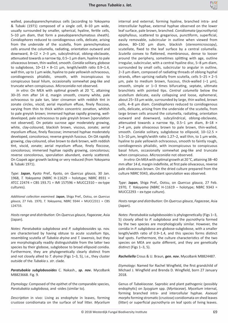
© 2018 Westerdijk Fungal Biodiversity Institute
The genus Tubakia s. lat.
Editor-in-ChiefProf. dr P.W. Crous, Westerdijk Fungal Biodiversity Institute, P.O. Box 85167, 3508 AD Utrecht, The Netherlands.E-mail:[email protected]
69
walled, pseudoparenchymatous cells [according to Yokoyama & Tubaki (1971) composed of a single cell, 8–10 µm wide, usually surrounded by smaller, spherical, hyaline, fertile cells, 5–10 µm diam, that form a pseudoparenchymatous sheath]. Conidiophores reduced to conidiogenous cells, delicate, arising from the underside of the scutella, from parenchymatous cells around the columella, radiating, orientation outward and downward, 8–12 × 2–3 µm, subcylindrical, oblong-obclavate, attenuated towards a narrow tip, 0.5–1 µm diam, hyaline to pale olivaceous brown, thin-walled, smooth. Conidia solitary, globose to subglobose, 10–13 × 8–11 µm, length/width ratio 0.9–1.4, wall thin, up to 1 µm wide, hyaline to pale yellowish ochraceous, conidiogenesis phialidic, smooth, with inconspicuous to conspicuous basal hilum, occasionally somewhat peg-like and truncate when conspicuous. Microconidia not observed.
In vitro: On MEA with optimal growth at 20 °C, attaining 30–35 mm after 14 d, margin smooth, creamy white, pale ochraceous to pale tan, later cinnamon with reddish tint in zonate circles, viscid; aerial mycelium effuse, finely floccose, varying from thin to thick within concentric zonation, whitish to pale greyish brown; immersed hyphae rapidly growing, well-developed, pale ochraceous to pale greyish brown [sporulation not observed]. On potato sucrose agar moderately growing, white, clay-coloured, blackish brown, viscous, zonate; aerial mycelium effuse, finely floccose; immersed hyphae moderately developed, concolorous; reverse greyish fuscous. On OA rapidly growing, clay-coloured, olive brown to dark brown, with reddish tint, viscid, zonate; aerial mycelium effuse, finely floccose, concolorous; immersed hyphae rapidly growing, concolorous; reverse concolorous, sporulation abundant, evenly scattered. On Czapek agar growth lacking or very reduced (from Yokoyama & Tubaki 1971).
Type: Japan, Kyoto Pref., Kyoto, on Quercus glauca, 30 Jan. 1968, T. Yokoyama (NBRC H-11629 – holotype; NBRC 8931 = ATCC 22474 = CBS 193.71 = IMI 157596 = MUCC2310 – ex-type cultures).
Additional collection examined: Japan, Shiga Pref., Ootsu, on Quercus glauca, 27 Feb. 1970, T. Yokoyama, NBRC 9344 = MUCC2311 = CBS 124733.
Hosts range and distribution: On Quercus glauca, Fagaceae, Asia (Japan).
Notes: Paratubakia subglobosa and P. subglobosoides sp. nov. are characterised by having obtuse to acute scutellum tips, resembling scutella of Tubakia dryina and T. iowensis, but they are morphologically readily distinguishable from the latter two species by their globose, subglobose to broad ellipsoid conidia. Furthermore, they are phylogenetically clearly distinct from and not closely allied to T. dryina (Figs 1–3, 5), i.e., they cluster outside of the Tubakia s. str. clade.
Paratubakia subglobosoides C. Nakash., sp. nov. MycoBank MB823668. Fig. 9.
Etymology: Composed of the epithet of the comparable species, Paratubakia subglobosa, and -oides (similar to).
Description in vivo: Living as endophyte in leaves, forming crustose conidiomata on the surface of leaf litter. Mycelium
internal and external, forming hyaline, branched intra- and intercellular hyphae, external hyphae observed on the lower leaf surface, pale brown, branched. Conidiomata (pycnothyria) epiphyllous, scattered to gregarious, punctiform, superficial, easily removable, subcircular in outline when viewed from above, 80–130 µm diam, blackish (stereomicroscopy), scutellate, fixed to the leaf surface by a central columella. Scutella convex to flattened, membranous, dense to looser around the periphery, sometimes splitting with age, outline irregular, subcircular, with a central hyaline disc, 5–8 µm diam, surrounded by small cells, subcircular to angular in outline, 2–3 µm diam, composed of radiating threads of oblong hyphal strands, often uprising radially from scutella, cells 5–25 × 2–5 µm, pale to medium brown, fuscous, thick-walled (–1 µm), smooth, simple or 1–3 times bifurcating, septate, ultimate branchlets with pointed tips. Central columella below the scutellum delicate, easily collapsing and loose, ephemeral, about 25–33 µm wide, surrounded by large, thin walled, brown cells, 4–8 µm diam. Conidiophores reduced to conidiogenous cells, delicate, arising from the underside of the scutella, from large brown cells around the columella, radiating, orientation outward and downward, subcylindrical, oblong-obclavate, attenuated towards a narrow tip, 0.5–1 µm diam, 8–18 × 2–4 µm, pale olivaceous brown to pale brown, thin-walled, smooth. Conidia solitary, subglobose to ellipsoid, 10–12.5 × 5.5–10 µm, length/width ratio 1.27–2, wall thin, to 1 µm wide, hyaline to pale yellowish ochraceous, smooth to faintly rough, conidiogenesis phialidic, with inconspicuous to conspicuous basal hilum, occasionally somewhat peg-like and truncate when conspicuous. Microconidia not observed.
In vitro: On MEA with optimal growth at 20 °C, attaining 38–40 mm after 14 d, margin indefinite, at first pale olivaceous, reverse pale olivaceous brown. On the dried culture prepared from the culture NBRC 9343, abundant sporulation was observed.
Type: Japan, Shiga Pref., Ootsu, on Quercus glauca, 27 Feb. 1970, T. Yokoyama (NBRC H-11619 – holotype; NBRC 9343 = MUCC2293 – ex-type culture).
Hosts range and distribution: On Quercus glauca, Fagaceae, Asia (Japan).
Notes: Paratubakia subglobosoides is phylogenetically (Figs 1–3, 5) closely allied to P. subglobosa and the pycnothyria formed by the two species are morphologically similar. However, the conidia in P. subglobosa are globose-subglobose, with a smaller length/width ratio of 0.9–1.4, and this species forms distinct leaf spots. Furthermore, the culture characteristics of the two species on MEA are quite different, and they are genetically distinct (Figs 1–3, 5).
Racheliella Crous & U. Braun, gen. nov. MycoBank MB824487.
Etymology: Named for Rachel Wingfield, the first grandchild of Michael J. Wingfield and Brenda D. Wingfield, born 27 January 2018.
Genus of Tubakiaceae. Saprobic and plant pathogenic (possibly endophytic) on Syzygium spp. (Myrtaceae). Mycelium internal, forming branched intra- and intercellular hyphae. Asexual morphs forming stromatic (crustose) conidiomata on shed leaves (litter) or superficial pycnothyria on leaf spots of living leaves.
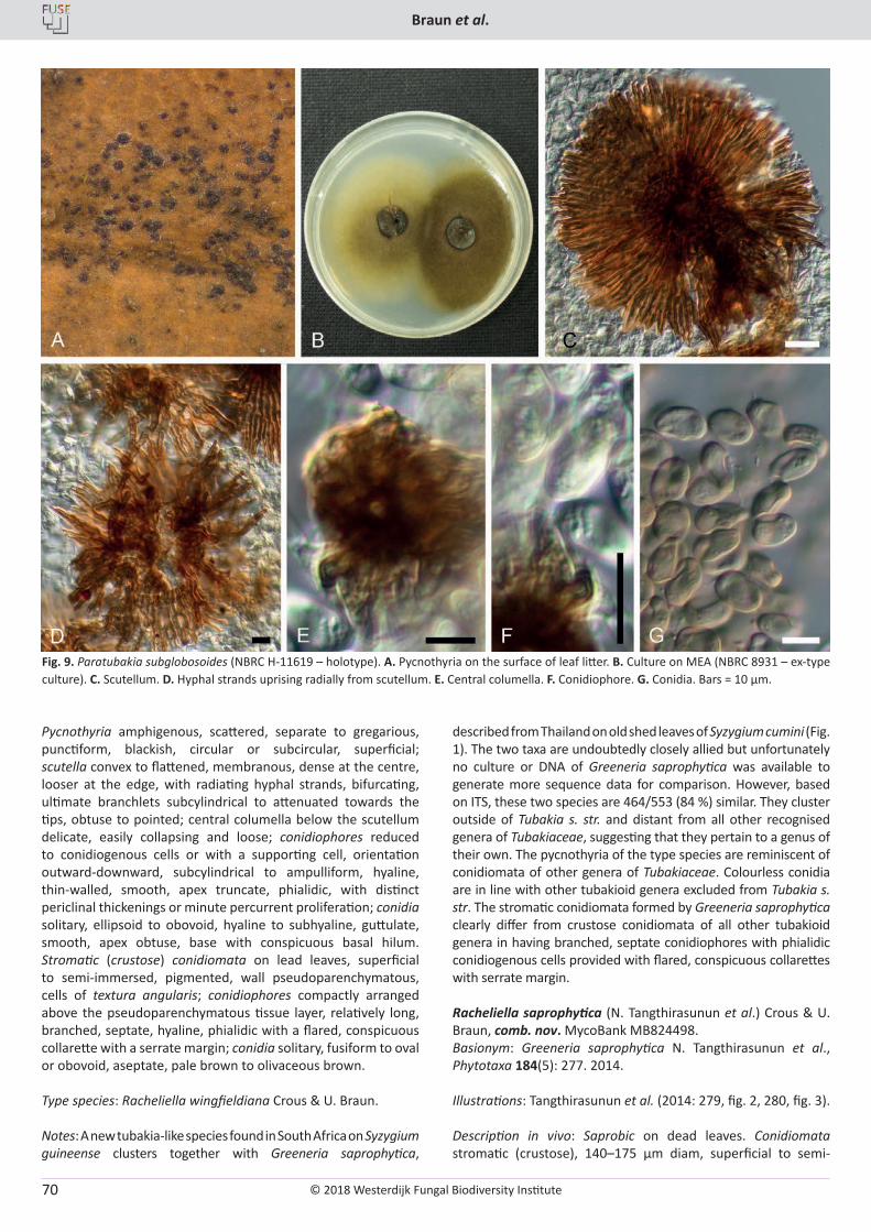
© 2018 Westerdijk Fungal Biodiversity Institute
Braun et al.
Editor-in-ChiefProf. dr P.W. Crous, Westerdijk Fungal Biodiversity Institute, P.O. Box 85167, 3508 AD Utrecht, The Netherlands.E-mail:[email protected]
70
Pycnothyria amphigenous, scattered, separate to gregarious, punctiform, blackish, circular or subcircular, superficial; scutella convex to flattened, membranous, dense at the centre, looser at the edge, with radiating hyphal strands, bifurcating, ultimate branchlets subcylindrical to attenuated towards the tips, obtuse to pointed; central columella below the scutellum delicate, easily collapsing and loose; conidiophores reduced to conidiogenous cells or with a supporting cell, orientation outward-downward, subcylindrical to ampulliform, hyaline, thin-walled, smooth, apex truncate, phialidic, with distinct periclinal thickenings or minute percurrent proliferation; conidia solitary, ellipsoid to obovoid, hyaline to subhyaline, guttulate, smooth, apex obtuse, base with conspicuous basal hilum. Stromatic (crustose) conidiomata on lead leaves, superficial to semi-immersed, pigmented, wall pseudoparenchymatous, cells of textura angularis; conidiophores compactly arranged above the pseudoparenchymatous tissue layer, relatively long, branched, septate, hyaline, phialidic with a flared, conspicuous collarette with a serrate margin; conidia solitary, fusiform to oval or obovoid, aseptate, pale brown to olivaceous brown.
Type species: Racheliella wingfieldiana Crous & U. Braun.
Notes: A new tubakia-like species found in South Africa on Syzygium guineense clusters together with Greeneria saprophytica,
described from Thailand on old shed leaves of Syzygium cumini (Fig. 1). The two taxa are undoubtedly closely allied but unfortunately no culture or DNA of Greeneria saprophytica was available to generate more sequence data for comparison. However, based on ITS, these two species are 464/553 (84 %) similar. They cluster outside of Tubakia s. str. and distant from all other recognised genera of Tubakiaceae, suggesting that they pertain to a genus of their own. The pycnothyria of the type species are reminiscent of conidiomata of other genera of Tubakiaceae. Colourless conidia are in line with other tubakioid genera excluded from Tubakia s. str. The stromatic conidiomata formed by Greeneria saprophytica clearly differ from crustose conidiomata of all other tubakioid genera in having branched, septate conidiophores with phialidic conidiogenous cells provided with flared, conspicuous collarettes with serrate margin.
Racheliella saprophytica (N. Tangthirasunun et al.) Crous & U. Braun, comb. nov. MycoBank MB824498. Basionym: Greeneria saprophytica N. Tangthirasunun et al., Phytotaxa 184(5): 277. 2014.
Illustrations: Tangthirasunun et al. (2014: 279, fig. 2, 280, fig. 3).
Description in vivo: Saprobic on dead leaves. Conidiomata stromatic (crustose), 140–175 μm diam, superficial to semi-
Fig. 9. Paratubakia subglobosoides (NBRC H-11619 – holotype). A. Pycnothyria on the surface of leaf litter. B. Culture on MEA (NBRC 8931 – ex-type culture). C. Scutellum. D. Hyphal strands uprising radially from scutellum. E. Central columella. F. Conidiophore. G. Conidia. Bars = 10 µm.
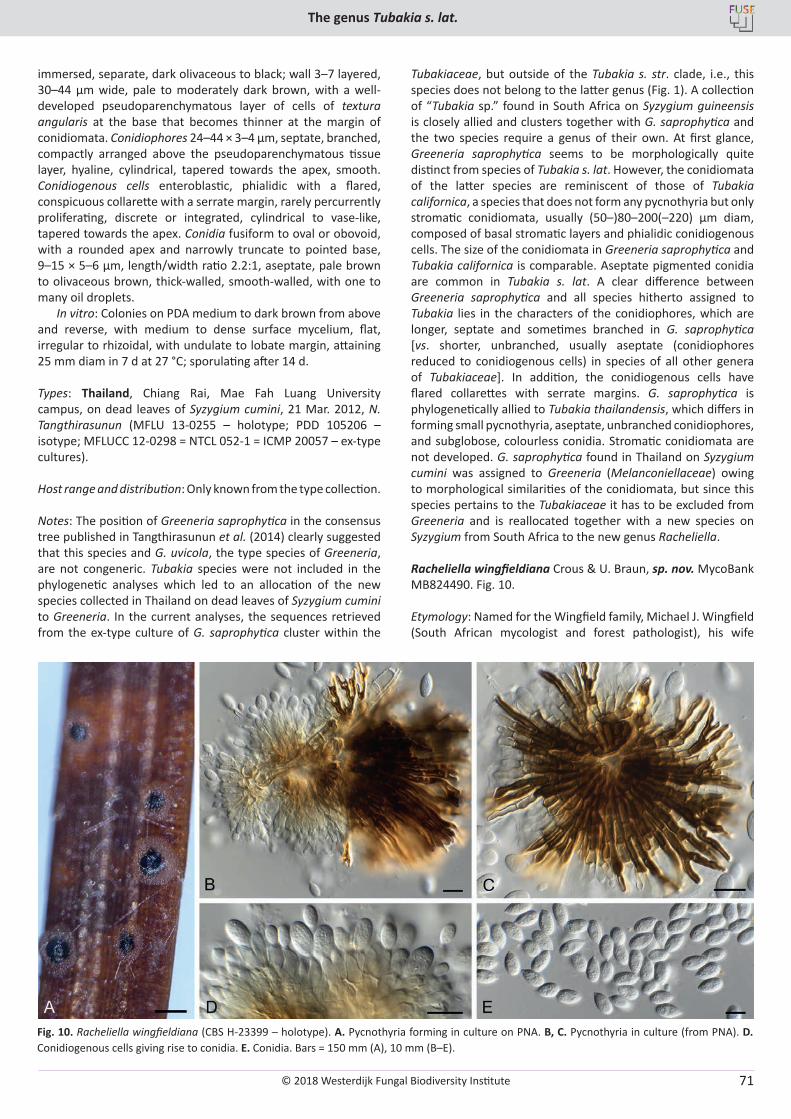
© 2018 Westerdijk Fungal Biodiversity Institute
The genus Tubakia s. lat.
Editor-in-ChiefProf. dr P.W. Crous, Westerdijk Fungal Biodiversity Institute, P.O. Box 85167, 3508 AD Utrecht, The Netherlands.E-mail:[email protected]
71
immersed, separate, dark olivaceous to black; wall 3–7 layered, 30–44 μm wide, pale to moderately dark brown, with a well-developed pseudoparenchymatous layer of cells of textura angularis at the base that becomes thinner at the margin of conidiomata. Conidiophores 24–44 × 3–4 μm, septate, branched, compactly arranged above the pseudoparenchymatous tissue layer, hyaline, cylindrical, tapered towards the apex, smooth. Conidiogenous cells enteroblastic, phialidic with a flared, conspicuous collarette with a serrate margin, rarely percurrently proliferating, discrete or integrated, cylindrical to vase-like, tapered towards the apex. Conidia fusiform to oval or obovoid, with a rounded apex and narrowly truncate to pointed base, 9–15 × 5–6 μm, length/width ratio 2.2:1, aseptate, pale brown to olivaceous brown, thick-walled, smooth-walled, with one to many oil droplets.
In vitro: Colonies on PDA medium to dark brown from above and reverse, with medium to dense surface mycelium, flat, irregular to rhizoidal, with undulate to lobate margin, attaining 25 mm diam in 7 d at 27 °C; sporulating after 14 d.
Types: Thailand, Chiang Rai, Mae Fah Luang University campus, on dead leaves of Syzygium cumini, 21 Mar. 2012, N. Tangthirasunun (MFLU 13-0255 – holotype; PDD 105206 – isotype; MFLUCC 12-0298 = NTCL 052-1 = ICMP 20057 – ex-type cultures).
Host range and distribution: Only known from the type collection.
Notes: The position of Greeneria saprophytica in the consensus tree published in Tangthirasunun et al. (2014) clearly suggested that this species and G. uvicola, the type species of Greeneria, are not congeneric. Tubakia species were not included in the phylogenetic analyses which led to an allocation of the new species collected in Thailand on dead leaves of Syzygium cumini to Greeneria. In the current analyses, the sequences retrieved from the ex-type culture of G. saprophytica cluster within the
Tubakiaceae, but outside of the Tubakia s. str. clade, i.e., this species does not belong to the latter genus (Fig. 1). A collection of “Tubakia sp.” found in South Africa on Syzygium guineensis is closely allied and clusters together with G. saprophytica and the two species require a genus of their own. At first glance, Greeneria saprophytica seems to be morphologically quite distinct from species of Tubakia s. lat. However, the conidiomata of the latter species are reminiscent of those of Tubakia californica, a species that does not form any pycnothyria but only stromatic conidiomata, usually (50–)80–200(–220) µm diam, composed of basal stromatic layers and phialidic conidiogenous cells. The size of the conidiomata in Greeneria saprophytica and Tubakia californica is comparable. Aseptate pigmented conidia are common in Tubakia s. lat. A clear difference between Greeneria saprophytica and all species hitherto assigned to Tubakia lies in the characters of the conidiophores, which are longer, septate and sometimes branched in G. saprophytica [vs. shorter, unbranched, usually aseptate (conidiophores reduced to conidiogenous cells) in species of all other genera of Tubakiaceae]. In addition, the conidiogenous cells have flared collarettes with serrate margins. G. saprophytica is phylogenetically allied to Tubakia thailandensis, which differs in forming small pycnothyria, aseptate, unbranched conidiophores, and subglobose, colourless conidia. Stromatic conidiomata are not developed. G. saprophytica found in Thailand on Syzygium cumini was assigned to Greeneria (Melanconiellaceae) owing to morphological similarities of the conidiomata, but since this species pertains to the Tubakiaceae it has to be excluded from Greeneria and is reallocated together with a new species on Syzygium from South Africa to the new genus Racheliella.
Racheliella wingfieldiana Crous & U. Braun, sp. nov. MycoBank MB824490. Fig. 10.
Etymology: Named for the Wingfield family, Michael J. Wingfield (South African mycologist and forest pathologist), his wife
Fig. 10. Racheliella wingfieldiana (CBS H-23399 – holotype). A. Pycnothyria forming in culture on PNA. B, C. Pycnothyria in culture (from PNA). D. Conidiogenous cells giving rise to conidia. E. Conidia. Bars = 150 mm (A), 10 mm (B–E).
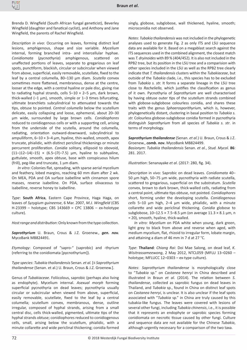
© 2018 Westerdijk Fungal Biodiversity Institute
Braun et al.
Editor-in-ChiefProf. dr P.W. Crous, Westerdijk Fungal Biodiversity Institute, P.O. Box 85167, 3508 AD Utrecht, The Netherlands.E-mail:[email protected]
72
Brenda D. Wingfield (South African fungal geneticist), Beverley Wingfield (daughter and fanatical cyclist), and Anthony and Jane Wingfield, the parents of Rachel Wingfield.
Description in vivo: Occurring on leaves, forming distinct leaf lesions, amphigenous, shape and size variable. Mycelium internal, forming branched intra- and intercellular hyphae. Conidiomata (pycnothyria) amphigenous, scattered on unaffected portions of leaves, separate to gregarious on leaf spots, punctiform, blackish, circular or subcircular when viewed from above, superficial, easily removable, scutellate, fixed to the leaf by a central columella, 80–130 µm diam. Scutella convex sometimes more flattened, membranous, dense at the centre, looser at the edge, with a central hyaline or pale disc, giving rise to radiating hyphal strands, cells 5–10 × 2–5 µm, dark brown, thick-walled (–1 µm), smooth, simple or 1–3 times bifurcating, ultimate branchlets subcylindrical to attenuated towards the tips, obtuse to pointed. Central columella below the scutellum delicate, easily collapsing and loose, ephemeral, about 20–30 µm wide, surrounded by large brown cells. Conidiophores reduced to conidiogenous cells or with a supporting cell, arising from the underside of the scutella, around the columella, radiating, orientation outward-downward, subcylindrical to ampulliform, 6–10 × 3–4 µm, hyaline, thin-walled, smooth, apex truncate, phialidic, with distinct periclinal thickenings or minute percurrent proliferation. Conidia solitary, ellipsoid to obovoid, (11–)12–14(–15) × (6.5–)7(–7.5) µm, hyaline to subhyaline, guttulate, smooth, apex obtuse, base with conspicuous hilum (frill), peg-like and truncate, 1 mm diam.
In vitro: Colonies flat, spreading, with sparse aerial mycelium and feathery, lobed margins, reaching 60 mm diam after 2 wk. On MEA, PDA and OA surface isabelline with cinnamon spore masses, reverse isabelline. On PDA, surface olivaceous to isabelline, reverse honey to isabelline.
Type: South Africa, Eastern Cape Province, Haga Haga, on leaves of Syzygium guineense, 6 Mar. 2007, M.J. Wingfield (CBS H-23399 – holotype; CBS 143669 = CPC 13806 – ex-holotype culture).
Host range and distribution: Only known from the type collection.
Saprothyrium U. Braun, Crous & J.Z. Groenew., gen. nov. MycoBank MB824491.
Etymology: Composed of “sapro-“ (saprobic) and -thyrium (referring to the conidiomata [pycnothyrium]).
Type species: Tubakia thailandensis Senan. et al. [≡ Saprothyrium thailandense (Senan. et al.) U. Braun, Crous & J.Z. Groenew.].
Genus of Tubakiaceae. Foliicolous, saprobic (perhaps also living as endophyte). Mycelium internal. Asexual morph forming superficial pycnothyria on dead leaves; pycnothyria usually circular or subcircular when viewed from above, superficial, easily removable, scutellate, fixed to the leaf by a central columella; scutellum convex, membranous, dense, outline irregular, composed of hyphal strands, arising from a small central disc, cells thick-walled, pigmented, ultimate tips of the hyphal strands obtuse; conidiophores reduced to conidiogenous cells, small, arising below the scutellum, phialidic, with a minute collarette and wide periclinal thickening; conidia formed
singly, globose, subglobose, wall thickened, hyaline, smooth; microconidia not observed.
Notes: Tubakia thailandensis was not included in the phylogenetic analyses used to generate Fig. 2 as only ITS and LSU sequence data are available for it. Based on a megablast search against the ITS sequences used in the combined alignment, the closest match was T. dryinoides with 89 % (404/452). It is also not included in the RPB2 tree, but its position in the LSU tree and a comparision with other species included in the LSU as well as the RPB2 tree clearly indicate that T. thailandensis clusters within the Tubakiaceae, but outside of the Tubakia clade, i.e., this species has to be excluded from Tubakia s. str. It forms a separate lineage in the LSU tree close to Racheliella, which justifies the classification as genus of it own. Pycnothyria of Saprothyrium are well characterised by having obtuse outer tips of the scutellum strands combined with globose-subglobose colourless conidia, and shares these traits with the genus Sphaerosporithyrium, which is, however, phylogenetically distant, clustering in basal position to Tubakia s. str. Colourless globose-subglobose conidia formed in pycnothyria distinguish Saprothyrium from all species of Tubakia s. str. in terms of morphology.
Saprothyrium thailandense (Senan. et al.) U. Braun, Crous & J.Z. Groenew., comb. nov. MycoBank MB824499.Basionym: Tubakia thailandensis Senan. et al., Stud. Mycol. 86: 281. 2017.
Illustration: Senanayake et al. (2017: 280, fig. 34).
Description in vivo: Saprobic on dead leaves. Conidiomata 40–50 µm high, 50–75 µm wide, pycnothyria with radiate scutella, scattered to gregarious, superficial on the substratum. Scutella convex, brown to dark brown, thick-walled cells, radiating from a central point, ultimate tips obtuse, not pointed. Conidiophores short, forming under the developing scutella. Conidiogenous cells 5–10 µm high, 2–4 µm wide, phialidic, with a minute collarette and wide periclinal thickening. Conidia globose to subglobose, 10–12.5 × 7.5–8.5 µm (on average 11.3 × 8.1 µm, n = 20), smooth, hyaline, thick-walled.
In vitro: Mycelium on PDA white when young, dark green, light grey to black from above and reverse when aged, with medium mycelium, flat, rhizoid to irregular form, lobate margin, and attaining a diam of 46 mm in 7 d at 27 °C. Type: Thailand: Chiang Rai: Doi Mae Salong, on dead leaf, K. Wisitrassameewong, 2 May 2012, NTCL059 (MFLU 13–0260 – holotype; MFLUCC 12–0303 – ex-type culture). Notes: Saprothyrium thailandense is morphologically close to “Tubakia sp.” on Castanea henryi in China described and illustrated in Braun et al. (2014). The relation between S. thailandense, collected as saprobic fungus on dead leaves in Thailand, and Tubakia sp., found in China on distinct leaf spots on Castanea henryi, is unclear. It is also unclear if the leaf spots associated with “Tubakia sp.” in China are truly caused by this tubakia-like fungus. The leaves were covered with lesions of several other fungi, including Tubakia chinensis, i.e., it is possible that it represents an endophyte or saprobic species forming conidiomata on necrotic tissue caused by other fungi. Culture and sequence data are not available for the Chinese Tubakia, although urgently necessary for a comparison of the two taxa.
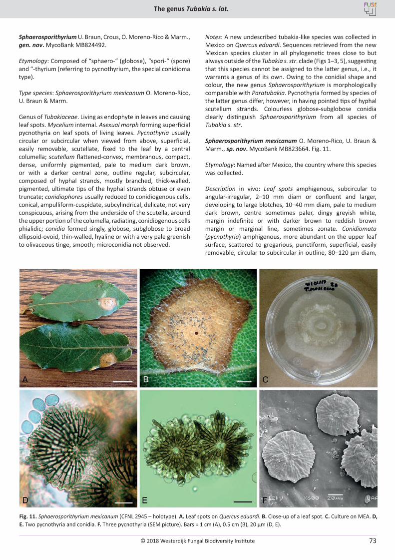
© 2018 Westerdijk Fungal Biodiversity Institute
The genus Tubakia s. lat.
Editor-in-ChiefProf. dr P.W. Crous, Westerdijk Fungal Biodiversity Institute, P.O. Box 85167, 3508 AD Utrecht, The Netherlands.E-mail:[email protected]
73
Sphaerosporithyrium U. Braun, Crous, O. Moreno-Rico & Marm., gen. nov. MycoBank MB824492.
Etymology: Composed of “sphaero-“ (globose), “spori-“ (spore) and “-thyrium (referring to pycnothyrium, the special conidioma type).
Type species: Sphaerosporithyrium mexicanum O. Moreno-Rico, U. Braun & Marm.
Genus of Tubakiaceae. Living as endophyte in leaves and causing leaf spots. Mycelium internal. Asexual morph forming superficial pycnothyria on leaf spots of living leaves. Pycnothyria usually circular or subcircular when viewed from above, superficial, easily removable, scutellate, fixed to the leaf by a central columella; scutellum flattened-convex, membranous, compact, dense, uniformly pigmented, pale to medium dark brown, or with a darker central zone, outline regular, subcircular, composed of hyphal strands, mostly branched, thick-walled, pigmented, ultimate tips of the hyphal strands obtuse or even truncate; conidiophores usually reduced to conidiogenous cells, conical, ampulliform-cuspidate, subcylindrical, delicate, not very conspicuous, arising from the underside of the scutella, around the upper portion of the columella, radiating, conidiogenous cells phialidic; conidia formed singly, globose, subglobose to broad ellipsoid-ovoid, thin-walled, hyaline or with a very pale greenish to olivaceous tinge, smooth; microconidia not observed.
Notes: A new undescribed tubakia-like species was collected in Mexico on Quercus eduardi. Sequences retrieved from the new Mexican species cluster in all phylogenetic trees close to but always outside of the Tubakia s. str. clade (Figs 1–3, 5), suggesting that this species cannot be assigned to the latter genus, i.e., it warrants a genus of its own. Owing to the conidial shape and colour, the new genus Sphaerosporithyrium is morphologically comparable with Paratubakia. Pycnothyria formed by species of the latter genus differ, however, in having pointed tips of hyphal scutellum strands. Colourless globose-subglobose conidia clearly distinguish Sphaerosporithyrium from all species of Tubakia s. str.
Sphaerosporithyrium mexicanum O. Moreno-Rico, U. Braun & Marm., sp. nov. MycoBank MB823664. Fig. 11.
Etymology: Named after Mexico, the country where this species was collected.
Description in vivo: Leaf spots amphigenous, subcircular to angular-irregular, 2–10 mm diam or confluent and larger, developing to large blotches, 10–40 mm diam, pale to medium dark brown, centre sometimes paler, dingy greyish white, margin indefinite or with darker brown to reddish brown margin or marginal line, sometimes zonate. Conidiomata (pycnothyria) amphigenous, more abundant on the upper leaf surface, scattered to gregarious, punctiform, superficial, easily removable, circular to subcircular in outline, 80–120 µm diam,
Fig. 11. Sphaerosporithyrium mexicanum (CFNL 2945 – holotype). A. Leaf spots on Quercus eduardi. B. Close-up of a leaf spot. C. Culture on MEA. D, E. Two pycnothyria and conidia. F. Three pycnothyria (SEM picture). Bars = 1 cm (A), 0.5 cm (B), 20 µm (D, E).
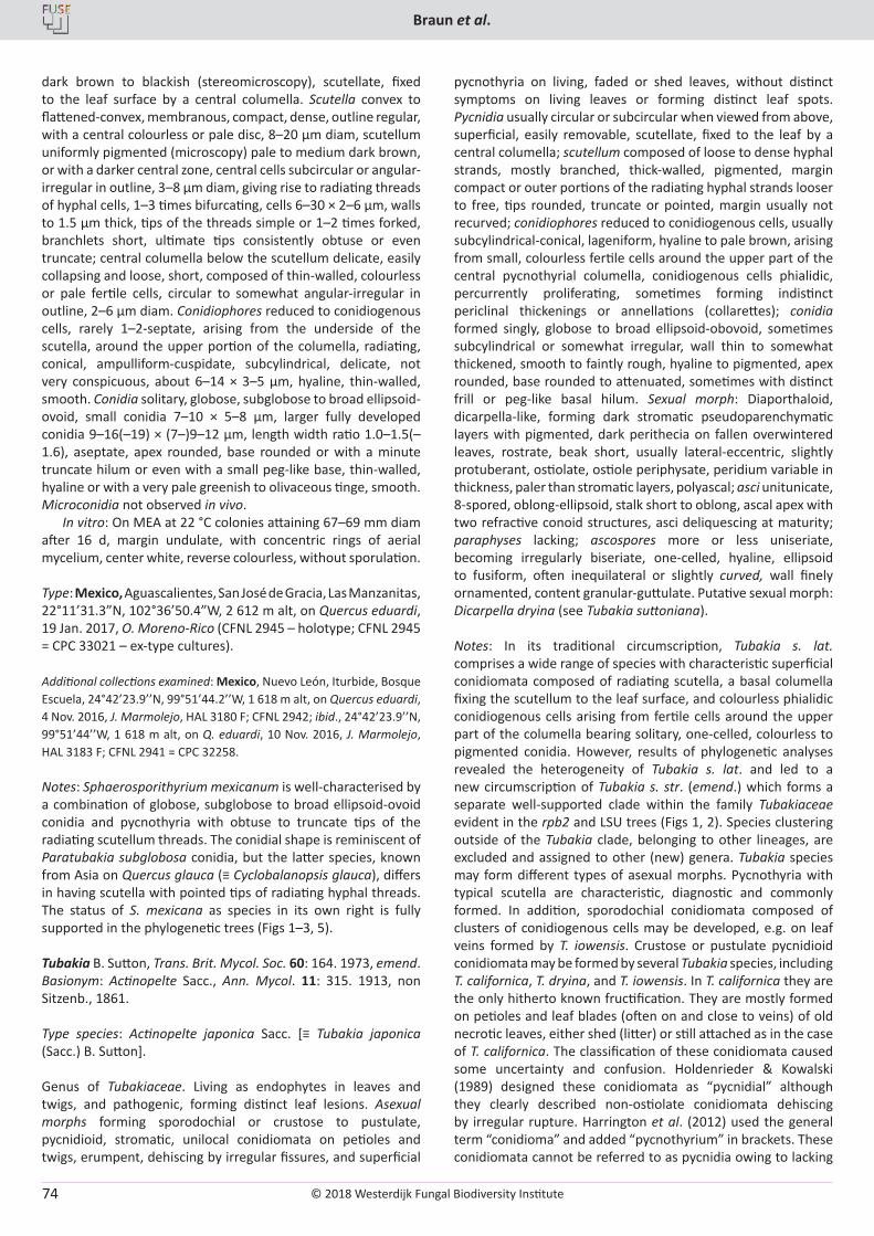
© 2018 Westerdijk Fungal Biodiversity Institute
Braun et al.
Editor-in-ChiefProf. dr P.W. Crous, Westerdijk Fungal Biodiversity Institute, P.O. Box 85167, 3508 AD Utrecht, The Netherlands.E-mail:[email protected]
74
dark brown to blackish (stereomicroscopy), scutellate, fixed to the leaf surface by a central columella. Scutella convex to flattened-convex, membranous, compact, dense, outline regular, with a central colourless or pale disc, 8–20 µm diam, scutellum uniformly pigmented (microscopy) pale to medium dark brown, or with a darker central zone, central cells subcircular or angular-irregular in outline, 3–8 µm diam, giving rise to radiating threads of hyphal cells, 1–3 times bifurcating, cells 6–30 × 2–6 µm, walls to 1.5 µm thick, tips of the threads simple or 1–2 times forked, branchlets short, ultimate tips consistently obtuse or even truncate; central columella below the scutellum delicate, easily collapsing and loose, short, composed of thin-walled, colourless or pale fertile cells, circular to somewhat angular-irregular in outline, 2–6 µm diam. Conidiophores reduced to conidiogenous cells, rarely 1–2-septate, arising from the underside of the scutella, around the upper portion of the columella, radiating, conical, ampulliform-cuspidate, subcylindrical, delicate, not very conspicuous, about 6–14 × 3–5 µm, hyaline, thin-walled, smooth. Conidia solitary, globose, subglobose to broad ellipsoid-ovoid, small conidia 7–10 × 5–8 µm, larger fully developed conidia 9–16(–19) × (7–)9–12 µm, length width ratio 1.0–1.5(–1.6), aseptate, apex rounded, base rounded or with a minute truncate hilum or even with a small peg-like base, thin-walled, hyaline or with a very pale greenish to olivaceous tinge, smooth. Microconidia not observed in vivo.
In vitro: On MEA at 22 °C colonies attaining 67–69 mm diam after 16 d, margin undulate, with concentric rings of aerial mycelium, center white, reverse colourless, without sporulation.
Type: Mexico, Aguascalientes, San José de Gracia, Las Manzanitas, 22°11’31.3”N, 102°36’50.4”W, 2 612 m alt, on Quercus eduardi, 19 Jan. 2017, O. Moreno-Rico (CFNL 2945 – holotype; CFNL 2945 = CPC 33021 – ex-type cultures).
Additional collections examined: Mexico, Nuevo León, Iturbide, Bosque Escuela, 24°42’23.9’’N, 99°51’44.2’’W, 1 618 m alt, on Quercus eduardi, 4 Nov. 2016, J. Marmolejo, HAL 3180 F; CFNL 2942; ibid., 24°42’23.9’’N, 99°51’44’’W, 1 618 m alt, on Q. eduardi, 10 Nov. 2016, J. Marmolejo, HAL 3183 F; CFNL 2941 = CPC 32258.
Notes: Sphaerosporithyrium mexicanum is well-characterised by a combination of globose, subglobose to broad ellipsoid-ovoid conidia and pycnothyria with obtuse to truncate tips of the radiating scutellum threads. The conidial shape is reminiscent of Paratubakia subglobosa conidia, but the latter species, known from Asia on Quercus glauca (≡ Cyclobalanopsis glauca), differs in having scutella with pointed tips of radiating hyphal threads. The status of S. mexicana as species in its own right is fully supported in the phylogenetic trees (Figs 1–3, 5).
Tubakia B. Sutton, Trans. Brit. Mycol. Soc. 60: 164. 1973, emend.Basionym: Actinopelte Sacc., Ann. Mycol. 11: 315. 1913, non Sitzenb., 1861.
Type species: Actinopelte japonica Sacc. [≡ Tubakia japonica (Sacc.) B. Sutton].
Genus of Tubakiaceae. Living as endophytes in leaves and twigs, and pathogenic, forming distinct leaf lesions. Asexual morphs forming sporodochial or crustose to pustulate, pycnidioid, stromatic, unilocal conidiomata on petioles and twigs, erumpent, dehiscing by irregular fissures, and superficial
pycnothyria on living, faded or shed leaves, without distinct symptoms on living leaves or forming distinct leaf spots. Pycnidia usually circular or subcircular when viewed from above, superficial, easily removable, scutellate, fixed to the leaf by a central columella; scutellum composed of loose to dense hyphal strands, mostly branched, thick-walled, pigmented, margin compact or outer portions of the radiating hyphal strands looser to free, tips rounded, truncate or pointed, margin usually not recurved; conidiophores reduced to conidiogenous cells, usually subcylindrical-conical, lageniform, hyaline to pale brown, arising from small, colourless fertile cells around the upper part of the central pycnothyrial columella, conidiogenous cells phialidic, percurrently proliferating, sometimes forming indistinct periclinal thickenings or annellations (collarettes); conidia formed singly, globose to broad ellipsoid-obovoid, sometimes subcylindrical or somewhat irregular, wall thin to somewhat thickened, smooth to faintly rough, hyaline to pigmented, apex rounded, base rounded to attenuated, sometimes with distinct frill or peg-like basal hilum. Sexual morph: Diaporthaloid, dicarpella-like, forming dark stromatic pseudoparenchymatic layers with pigmented, dark perithecia on fallen overwintered leaves, rostrate, beak short, usually lateral-eccentric, slightly protuberant, ostiolate, ostiole periphysate, peridium variable in thickness, paler than stromatic layers, polyascal; asci unitunicate, 8-spored, oblong-ellipsoid, stalk short to oblong, ascal apex with two refractive conoid structures, asci deliquescing at maturity; paraphyses lacking; ascospores more or less uniseriate, becoming irregularly biseriate, one-celled, hyaline, ellipsoid to fusiform, often inequilateral or slightly curved, wall finely ornamented, content granular-guttulate. Putative sexual morph: Dicarpella dryina (see Tubakia suttoniana).
Notes: In its traditional circumscription, Tubakia s. lat. comprises a wide range of species with characteristic superficial conidiomata composed of radiating scutella, a basal columella fixing the scutellum to the leaf surface, and colourless phialidic conidiogenous cells arising from fertile cells around the upper part of the columella bearing solitary, one-celled, colourless to pigmented conidia. However, results of phylogenetic analyses revealed the heterogeneity of Tubakia s. lat. and led to a new circumscription of Tubakia s. str. (emend.) which forms a separate well-supported clade within the family Tubakiaceae evident in the rpb2 and LSU trees (Figs 1, 2). Species clustering outside of the Tubakia clade, belonging to other lineages, are excluded and assigned to other (new) genera. Tubakia species may form different types of asexual morphs. Pycnothyria with typical scutella are characteristic, diagnostic and commonly formed. In addition, sporodochial conidiomata composed of clusters of conidiogenous cells may be developed, e.g. on leaf veins formed by T. iowensis. Crustose or pustulate pycnidioid conidiomata may be formed by several Tubakia species, including T. californica, T. dryina, and T. iowensis. In T. californica they are the only hitherto known fructification. They are mostly formed on petioles and leaf blades (often on and close to veins) of old necrotic leaves, either shed (litter) or still attached as in the case of T. californica. The classification of these conidiomata caused some uncertainty and confusion. Holdenrieder & Kowalski (1989) designed these conidiomata as “pycnidial” although they clearly described non-ostiolate conidiomata dehiscing by irregular rupture. Harrington et al. (2012) used the general term “conidioma” and added “pycnothyrium” in brackets. These conidiomata cannot be referred to as pycnidia owing to lacking
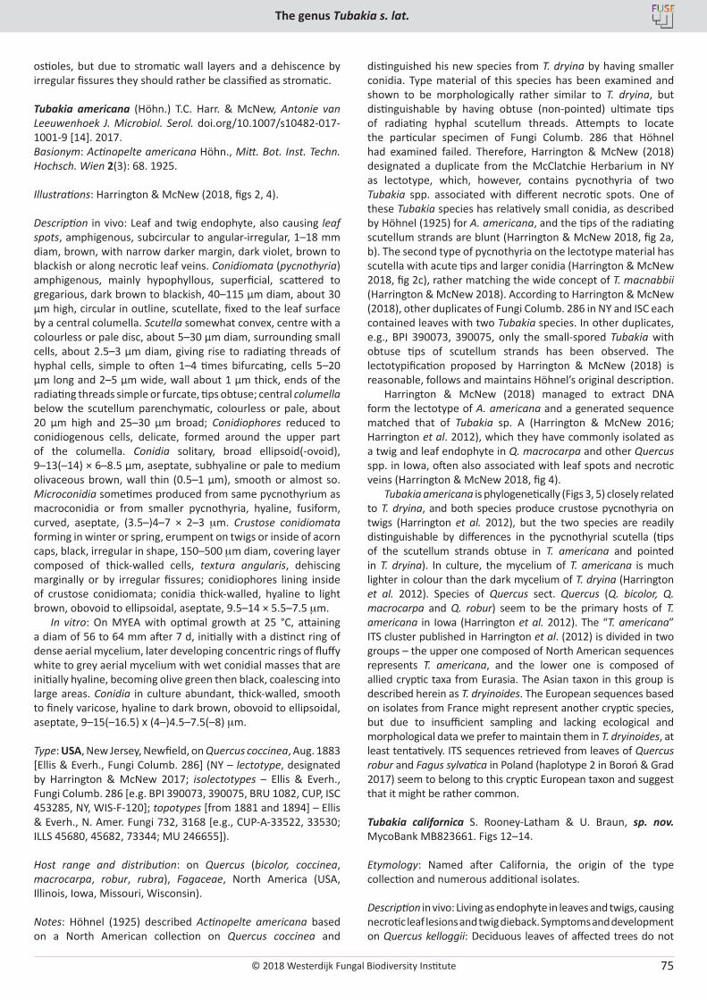
© 2018 Westerdijk Fungal Biodiversity Institute
The genus Tubakia s. lat.
Editor-in-ChiefProf. dr P.W. Crous, Westerdijk Fungal Biodiversity Institute, P.O. Box 85167, 3508 AD Utrecht, The Netherlands.E-mail:[email protected]
75
ostioles, but due to stromatic wall layers and a dehiscence by irregular fissures they should rather be classified as stromatic.
Tubakia americana (Höhn.) T.C. Harr. & McNew, Antonie van Leeuwenhoek J. Microbiol. Serol. doi.org/10.1007/s10482-017-1001-9 [14]. 2017.Basionym: Actinopelte americana Höhn., Mitt. Bot. Inst. Techn. Hochsch. Wien 2(3): 68. 1925.
Illustrations: Harrington & McNew (2018, figs 2, 4).
Description in vivo: Leaf and twig endophyte, also causing leaf spots, amphigenous, subcircular to angular-irregular, 1–18 mm diam, brown, with narrow darker margin, dark violet, brown to blackish or along necrotic leaf veins. Conidiomata (pycnothyria) amphigenous, mainly hypophyllous, superficial, scattered to gregarious, dark brown to blackish, 40–115 µm diam, about 30 µm high, circular in outline, scutellate, fixed to the leaf surface by a central columella. Scutella somewhat convex, centre with a colourless or pale disc, about 5–30 µm diam, surrounding small cells, about 2.5–3 µm diam, giving rise to radiating threads of hyphal cells, simple to often 1–4 times bifurcating, cells 5–20 µm long and 2–5 µm wide, wall about 1 µm thick, ends of the radiating threads simple or furcate, tips obtuse; central columella below the scutellum parenchymatic, colourless or pale, about 20 µm high and 25–30 µm broad; Conidiophores reduced to conidiogenous cells, delicate, formed around the upper part of the columella. Conidia solitary, broad ellipsoid(-ovoid), 9–13(–14) × 6–8.5 µm, aseptate, subhyaline or pale to medium olivaceous brown, wall thin (0.5–1 µm), smooth or almost so. Microconidia sometimes produced from same pycnothyrium as macroconidia or from smaller pycnothyria, hyaline, fusiform, curved, aseptate, (3.5–)4–7 × 2–3 mm. Crustose conidiomata forming in winter or spring, erumpent on twigs or inside of acorn caps, black, irregular in shape, 150–500 mm diam, covering layer composed of thick-walled cells, textura angularis, dehiscing marginally or by irregular fissures; conidiophores lining inside of crustose conidiomata; conidia thick-walled, hyaline to light brown, obovoid to ellipsoidal, aseptate, 9.5–14 × 5.5–7.5 mm.
In vitro: On MYEA with optimal growth at 25 °C, attaining a diam of 56 to 64 mm after 7 d, initially with a distinct ring of dense aerial mycelium, later developing concentric rings of fluffy white to grey aerial mycelium with wet conidial masses that are initially hyaline, becoming olive green then black, coalescing into large areas. Conidia in culture abundant, thick-walled, smooth to finely varicose, hyaline to dark brown, obovoid to ellipsoidal, aseptate, 9–15(–16.5) X (4–)4.5–7.5(–8) mm.
Type: USA, New Jersey, Newfield, on Quercus coccinea, Aug. 1883 [Ellis & Everh., Fungi Columb. 286] (NY – lectotype, designated by Harrington & McNew 2017; isolectotypes – Ellis & Everh., Fungi Columb. 286 [e.g. BPI 390073, 390075, BRU 1082, CUP, ISC 453285, NY, WIS-F-120]; topotypes [from 1881 and 1894] – Ellis & Everh., N. Amer. Fungi 732, 3168 [e.g., CUP-A-33522, 33530; ILLS 45680, 45682, 73344; MU 246655]).
Host range and distribution: on Quercus (bicolor, coccinea, macrocarpa, robur, rubra), Fagaceae, North America (USA, Illinois, Iowa, Missouri, Wisconsin).
Notes: Höhnel (1925) described Actinopelte americana based on a North American collection on Quercus coccinea and
distinguished his new species from T. dryina by having smaller conidia. Type material of this species has been examined and shown to be morphologically rather similar to T. dryina, but distinguishable by having obtuse (non-pointed) ultimate tips of radiating hyphal scutellum threads. Attempts to locate the particular specimen of Fungi Columb. 286 that Höhnel had examined failed. Therefore, Harrington & McNew (2018) designated a duplicate from the McClatchie Herbarium in NY as lectotype, which, however, contains pycnothyria of two Tubakia spp. associated with different necrotic spots. One of these Tubakia species has relatively small conidia, as described by Höhnel (1925) for A. americana, and the tips of the radiating scutellum strands are blunt (Harrington & McNew 2018, fig 2a, b). The second type of pycnothyria on the lectotype material has scutella with acute tips and larger conidia (Harrington & McNew 2018, fig 2c), rather matching the wide concept of T. macnabbii (Harrington & McNew 2018). According to Harrington & McNew (2018), other duplicates of Fungi Columb. 286 in NY and ISC each contained leaves with two Tubakia species. In other duplicates, e.g., BPI 390073, 390075, only the small-spored Tubakia with obtuse tips of scutellum strands has been observed. The lectotypification proposed by Harrington & McNew (2018) is reasonable, follows and maintains Höhnel’s original description.
Harrington & McNew (2018) managed to extract DNA form the lectotype of A. americana and a generated sequence matched that of Tubakia sp. A (Harrington & McNew 2016; Harrington et al. 2012), which they have commonly isolated as a twig and leaf endophyte in Q. macrocarpa and other Quercus spp. in Iowa, often also associated with leaf spots and necrotic veins (Harrington & McNew 2018, fig 4).
Tubakia americana is phylogenetically (Figs 3, 5) closely related to T. dryina, and both species produce crustose pycnothyria on twigs (Harrington et al. 2012), but the two species are readily distinguishable by differences in the pycnothyrial scutella (tips of the scutellum strands obtuse in T. americana and pointed in T. dryina). In culture, the mycelium of T. americana is much lighter in colour than the dark mycelium of T. dryina (Harrington et al. 2012). Species of Quercus sect. Quercus (Q. bicolor, Q. macrocarpa and Q. robur) seem to be the primary hosts of T. americana in Iowa (Harrington et al. 2012). The “T. americana” ITS cluster published in Harrington et al. (2012) is divided in two groups – the upper one composed of North American sequences represents T. americana, and the lower one is composed of allied cryptic taxa from Eurasia. The Asian taxon in this group is described herein as T. dryinoides. The European sequences based on isolates from France might represent another cryptic species, but due to insufficient sampling and lacking ecological and morphological data we prefer to maintain them in T. dryinoides, at least tentatively. ITS sequences retrieved from leaves of Quercus robur and Fagus sylvatica in Poland (haplotype 2 in Boroń & Grad 2017) seem to belong to this cryptic European taxon and suggest that it might be rather common.
Tubakia californica S. Rooney-Latham & U. Braun, sp. nov. MycoBank MB823661. Figs 12–14.
Etymology: Named after California, the origin of the type collection and numerous additional isolates.
Description in vivo: Living as endophyte in leaves and twigs, causing necrotic leaf lesions and twig dieback. Symptoms and development on Quercus kelloggii: Deciduous leaves of affected trees do not
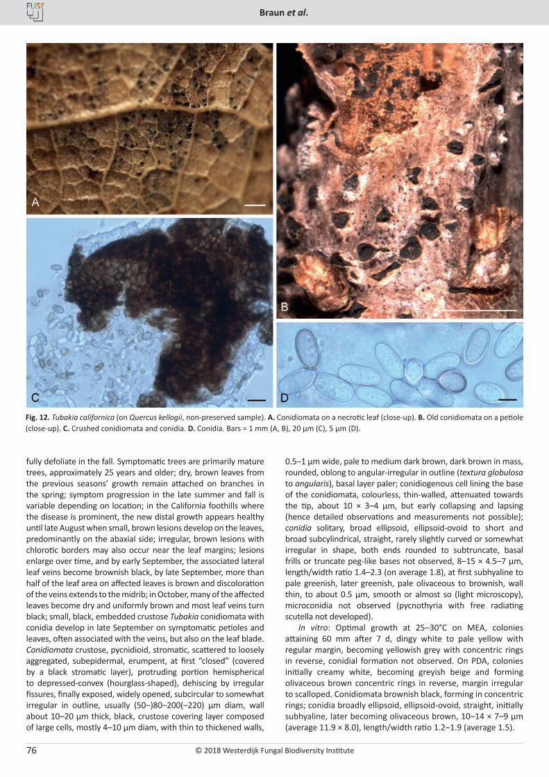
© 2018 Westerdijk Fungal Biodiversity Institute
Braun et al.
Editor-in-ChiefProf. dr P.W. Crous, Westerdijk Fungal Biodiversity Institute, P.O. Box 85167, 3508 AD Utrecht, The Netherlands.E-mail:[email protected]
76
fully defoliate in the fall. Symptomatic trees are primarily mature trees, approximately 25 years and older; dry, brown leaves from the previous seasons’ growth remain attached on branches in the spring; symptom progression in the late summer and fall is variable depending on location; in the California foothills where the disease is prominent, the new distal growth appears healthy until late August when small, brown lesions develop on the leaves, predominantly on the abaxial side; irregular, brown lesions with chlorotic borders may also occur near the leaf margins; lesions enlarge over time, and by early September, the associated lateral leaf veins become brownish black, by late September, more than half of the leaf area on affected leaves is brown and discoloration of the veins extends to the midrib; in October, many of the affected leaves become dry and uniformly brown and most leaf veins turn black; small, black, embedded crustose Tubakia conidiomata with conidia develop in late September on symptomatic petioles and leaves, often associated with the veins, but also on the leaf blade. Conidiomata crustose, pycnidioid, stromatic, scattered to loosely aggregated, subepidermal, erumpent, at first “closed” (covered by a black stromatic layer), protruding portion hemispherical to depressed-convex (hourglass-shaped), dehiscing by irregular fissures, finally exposed, widely opened, subcircular to somewhat irregular in outline, usually (50–)80–200(–220) µm diam, wall about 10–20 µm thick, black, crustose covering layer composed of large cells, mostly 4–10 µm diam, with thin to thickened walls,
0.5–1 µm wide, pale to medium dark brown, dark brown in mass, rounded, oblong to angular-irregular in outline (textura globulosa to angularis), basal layer paler; conidiogenous cell lining the base of the conidiomata, colourless, thin-walled, attenuated towards the tip, about 10 × 3–4 µm, but early collapsing and lapsing (hence detailed observations and measurements not possible); conidia solitary, broad ellipsoid, ellipsoid-ovoid to short and broad subcylindrical, straight, rarely slightly curved or somewhat irregular in shape, both ends rounded to subtruncate, basal frills or truncate peg-like bases not observed, 8–15 × 4.5–7 µm, length/width ratio 1.4–2.3 (on average 1.8), at first subhyaline to pale greenish, later greenish, pale olivaceous to brownish, wall thin, to about 0.5 µm, smooth or almost so (light microscopy), microconidia not observed (pycnothyria with free radiating scutella not developed).
In vitro: Optimal growth at 25–30°C on MEA, colonies attaining 60 mm after 7 d, dingy white to pale yellow with regular margin, becoming yellowish grey with concentric rings in reverse, conidial formation not observed. On PDA, colonies initially creamy white, becoming greyish beige and forming olivaceous brown concentric rings in reverse, margin irregular to scalloped. Conidiomata brownish black, forming in concentric rings; conidia broadly ellipsoid, ellipsoid-ovoid, straight, initially subhyaline, later becoming olivaceous brown, 10–14 × 7–9 µm (average 11.9 × 8.0), length/width ratio 1.2–1.9 (average 1.5).
Fig. 12. Tubakia californica (on Quercus kellogii, non-preserved sample). A. Conidiomata on a necrotic leaf (close-up). B. Old conidiomata on a petiole (close-up). C. Crushed conidiomata and conidia. D. Conidia. Bars = 1 mm (A, B), 20 µm (C), 5 µm (D).
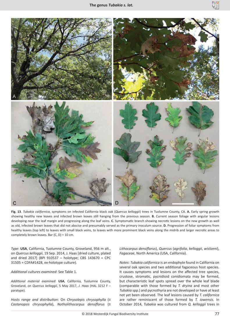
© 2018 Westerdijk Fungal Biodiversity Institute
The genus Tubakia s. lat.
Editor-in-ChiefProf. dr P.W. Crous, Westerdijk Fungal Biodiversity Institute, P.O. Box 85167, 3508 AD Utrecht, The Netherlands.E-mail:[email protected]
77
Type: USA, California, Tuolumne County, Groveland, 956 m alt., on Quercus kelloggii, 19 Sep. 2014, J. Haas [dried culture, plated and dried 2017] (BPI 910537 – holotype; CBS 143670 = CPC 31505 = CDFA#1428, ex-holotype culture).
Additional cultures examined: See Table 1.
Additional material examined: USA, California, Tuolumne County, Groveland, on Quercus kelloggii, 5 May 2017, J. Haas (HAL 3212 F – paratype).
Hosts range and distribution: On Chrysolepis chrysophylla (≡ Castanopsis chrysophylla), Notholithocarpus densiflorus (≡
Lithocarpus densiflorus), Quercus (agrifolia, kelloggii, wislizeni), Fagaceae, North America (USA, California).
Notes: Tubakia californica is an endophyte found in California on several oak species and two additional fagaceous host species. It causes symptoms and lesions on the affected tree species, crustose, stromatic, pycnidioid conidiomata may be formed, but characteristic leaf spots spread over the whole leaf blade (comparable with those formed by T. dryina and most other Tubakia spp.) and pycnothyria are not developed or have at least not yet been observed. The leaf lesions caused by T. californica are rather reminiscent of those formed by T. iowensis. In October 2014, Tubakia was cultured from Q. kelloggii trees in
Fig. 13. Tubakia californica, symptoms on infected California black oak (Quercus kelloggii) trees in Tuolumne County, CA. A. Early spring growth showing healthy new leaves and infected brown leaves still hanging from the previous season. B. Current season foliage with angular lesions developing near the leaf margin and progressing along the leaf veins. C. Symptomatic branch showing necrotic lesions on the new growth as well as old, infected brown leaves that did not abscise and presumably served as the primary inoculum source. D. Progression of foliar symptoms from healthy leaves (top left) to leaves with small black veins, to leaves with more prominent black veins along the midrib and larger necrotic areas to completely brown leaves. Bar (C, D) = 10 cm.
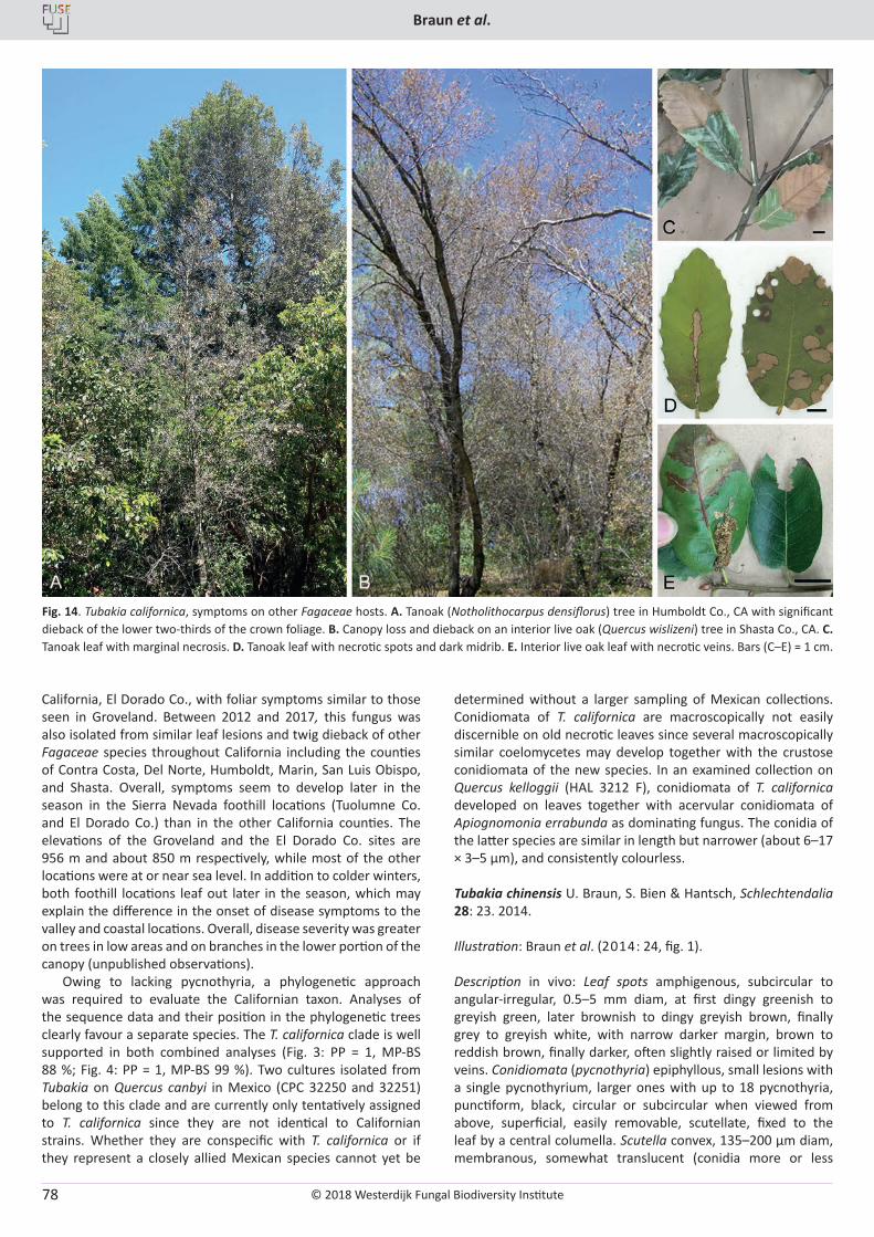
© 2018 Westerdijk Fungal Biodiversity Institute
Braun et al.
Editor-in-ChiefProf. dr P.W. Crous, Westerdijk Fungal Biodiversity Institute, P.O. Box 85167, 3508 AD Utrecht, The Netherlands.E-mail:[email protected]
78
California, El Dorado Co., with foliar symptoms similar to those seen in Groveland. Between 2012 and 2017, this fungus was also isolated from similar leaf lesions and twig dieback of other Fagaceae species throughout California including the counties of Contra Costa, Del Norte, Humboldt, Marin, San Luis Obispo, and Shasta. Overall, symptoms seem to develop later in the season in the Sierra Nevada foothill locations (Tuolumne Co. and El Dorado Co.) than in the other California counties. The elevations of the Groveland and the El Dorado Co. sites are 956 m and about 850 m respectively, while most of the other locations were at or near sea level. In addition to colder winters, both foothill locations leaf out later in the season, which may explain the difference in the onset of disease symptoms to the valley and coastal locations. Overall, disease severity was greater on trees in low areas and on branches in the lower portion of the canopy (unpublished observations).
Owing to lacking pycnothyria, a phylogenetic approach was required to evaluate the Californian taxon. Analyses of the sequence data and their position in the phylogenetic trees clearly favour a separate species. The T. californica clade is well supported in both combined analyses (Fig. 3: PP = 1, MP-BS 88 %; Fig. 4: PP = 1, MP-BS 99 %). Two cultures isolated from Tubakia on Quercus canbyi in Mexico (CPC 32250 and 32251) belong to this clade and are currently only tentatively assigned to T. californica since they are not identical to Californian strains. Whether they are conspecific with T. californica or if they represent a closely allied Mexican species cannot yet be
determined without a larger sampling of Mexican collections. Conidiomata of T. californica are macroscopically not easily discernible on old necrotic leaves since several macroscopically similar coelomycetes may develop together with the crustose conidiomata of the new species. In an examined collection on Quercus kelloggii (HAL 3212 F), conidiomata of T. californica developed on leaves together with acervular conidiomata of Apiognomonia errabunda as dominating fungus. The conidia of the latter species are similar in length but narrower (about 6–17 × 3–5 µm), and consistently colourless.
Tubakia chinensis U. Braun, S. Bien & Hantsch, Schlechtendalia 28: 23. 2014.
Illustration: Braun et al. (2014: 24, fig. 1).
Description in vivo: Leaf spots amphigenous, subcircular to angular-irregular, 0.5–5 mm diam, at first dingy greenish to greyish green, later brownish to dingy greyish brown, finally grey to greyish white, with narrow darker margin, brown to reddish brown, finally darker, often slightly raised or limited by veins. Conidiomata (pycnothyria) epiphyllous, small lesions with a single pycnothyrium, larger ones with up to 18 pycnothyria, punctiform, black, circular or subcircular when viewed from above, superficial, easily removable, scutellate, fixed to the leaf by a central columella. Scutella convex, 135–200 µm diam, membranous, somewhat translucent (conidia more or less
Fig. 14. Tubakia californica, symptoms on other Fagaceae hosts. A. Tanoak (Notholithocarpus densiflorus) tree in Humboldt Co., CA with significant dieback of the lower two-thirds of the crown foliage. B. Canopy loss and dieback on an interior live oak (Quercus wislizeni) tree in Shasta Co., CA. C. Tanoak leaf with marginal necrosis. D. Tanoak leaf with necrotic spots and dark midrib. E. Interior live oak leaf with necrotic veins. Bars (C–E) = 1 cm.
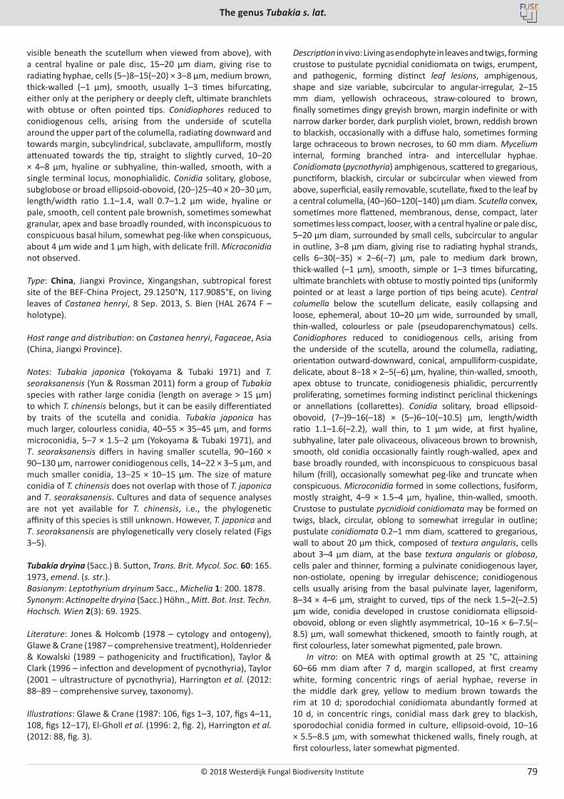
© 2018 Westerdijk Fungal Biodiversity Institute
The genus Tubakia s. lat.
Editor-in-ChiefProf. dr P.W. Crous, Westerdijk Fungal Biodiversity Institute, P.O. Box 85167, 3508 AD Utrecht, The Netherlands.E-mail:[email protected]
79
visible beneath the scutellum when viewed from above), with a central hyaline or pale disc, 15–20 µm diam, giving rise to radiating hyphae, cells (5–)8–15(–20) × 3–8 µm, medium brown, thick-walled (–1 µm), smooth, usually 1–3 times bifurcating, either only at the periphery or deeply cleft, ultimate branchlets with obtuse or often pointed tips. Conidiophores reduced to conidiogenous cells, arising from the underside of scutella around the upper part of the columella, radiating downward and towards margin, subcylindrical, subclavate, ampulliform, mostly attenuated towards the tip, straight to slightly curved, 10–20 × 4–8 µm, hyaline or subhyaline, thin-walled, smooth, with a single terminal locus, monophialidic. Conidia solitary, globose, subglobose or broad ellipsoid-obovoid, (20–)25–40 × 20–30 µm, length/width ratio 1.1–1.4, wall 0.7–1.2 µm wide, hyaline or pale, smooth, cell content pale brownish, sometimes somewhat granular, apex and base broadly rounded, with inconspicuous to conspicuous basal hilum, somewhat peg-like when conspicuous, about 4 µm wide and 1 µm high, with delicate frill. Microconidia not observed.
Type: China, Jiangxi Province, Xingangshan, subtropical forest site of the BEF-China Project, 29.1250°N, 117.9085°E, on living leaves of Castanea henryi, 8 Sep. 2013, S. Bien (HAL 2674 F – holotype).
Host range and distribution: on Castanea henryi, Fagaceae, Asia (China, Jiangxi Province).
Notes: Tubakia japonica (Yokoyama & Tubaki 1971) and T. seoraksanensis (Yun & Rossman 2011) form a group of Tubakia species with rather large conidia (length on average > 15 µm) to which T. chinensis belongs, but it can be easily differentiated by traits of the scutella and conidia. Tubakia japonica has much larger, colourless conidia, 40–55 × 35–45 µm, and forms microconidia, 5–7 × 1.5–2 µm (Yokoyama & Tubaki 1971), and T. seoraksanensis differs in having smaller scutella, 90–160 × 90–130 µm, narrower conidiogenous cells, 14–22 × 3–5 µm, and much smaller conidia, 13–25 × 10–15 µm. The size of mature conidia of T. chinensis does not overlap with those of T. japonica and T. seoraksanensis. Cultures and data of sequence analyses are not yet available for T. chinensis, i.e., the phylogenetic affinity of this species is still unknown. However, T. japonica and T. seoraksanensis are phylogenetically very closely related (Figs 3–5).
Tubakia dryina (Sacc.) B. Sutton, Trans. Brit. Mycol. Soc. 60: 165. 1973, emend. (s. str.).Basionym: Leptothyrium dryinum Sacc., Michelia 1: 200. 1878.Synonym: Actinopelte dryina (Sacc.) Höhn., Mitt. Bot. Inst. Techn. Hochsch. Wien 2(3): 69. 1925.
Literature: Jones & Holcomb (1978 – cytology and ontogeny), Glawe & Crane (1987 – comprehensive treatment), Holdenrieder & Kowalski (1989 – pathogenicity and fructification), Taylor & Clark (1996 – infection and development of pycnothyria), Taylor (2001 – ultrastructure of pycnothyria), Harrington et al. (2012: 88–89 – comprehensive survey, taxonomy).
Illustrations: Glawe & Crane (1987: 106, figs 1–3, 107, figs 4–11, 108, figs 12–17), El-Gholl et al. (1996: 2, fig. 2), Harrington et al. (2012: 88, fig. 3).
Description in vivo: Living as endophyte in leaves and twigs, forming crustose to pustulate pycnidial conidiomata on twigs, erumpent, and pathogenic, forming distinct leaf lesions, amphigenous, shape and size variable, subcircular to angular-irregular, 2–15 mm diam, yellowish ochraceous, straw-coloured to brown, finally sometimes dingy greyish brown, margin indefinite or with narrow darker border, dark purplish violet, brown, reddish brown to blackish, occasionally with a diffuse halo, sometimes forming large ochraceous to brown necroses, to 60 mm diam. Mycelium internal, forming branched intra- and intercellular hyphae. Conidiomata (pycnothyria) amphigenous, scattered to gregarious, punctiform, blackish, circular or subcircular when viewed from above, superficial, easily removable, scutellate, fixed to the leaf by a central columella, (40–)60–120(–140) µm diam. Scutella convex, sometimes more flattened, membranous, dense, compact, later sometimes less compact, looser, with a central hyaline or pale disc, 5–20 µm diam, surrounded by small cells, subcircular to angular in outline, 3–8 µm diam, giving rise to radiating hyphal strands, cells 6–30(–35) × 2–6(–7) µm, pale to medium dark brown, thick-walled (–1 µm), smooth, simple or 1–3 times bifurcating, ultimate branchlets with obtuse to mostly pointed tips (uniformly pointed or at least a large portion of tips being acute). Central columella below the scutellum delicate, easily collapsing and loose, ephemeral, about 10–20 µm wide, surrounded by small, thin-walled, colourless or pale (pseudoparenchymatous) cells. Conidiophores reduced to conidiogenous cells, arising from the underside of the scutella, around the columella, radiating, orientation outward-downward, conical, ampulliform-cuspidate, delicate, about 8–18 × 2–5(–6) µm, hyaline, thin-walled, smooth, apex obtuse to truncate, conidiogenesis phialidic, percurrently proliferating, sometimes forming indistinct periclinal thickenings or annellations (collarettes). Conidia solitary, broad ellipsoid-obovoid, (7–)9–16(–18) × (5–)6–10(–10.5) µm, length/width ratio 1.1–1.6(–2.2), wall thin, to 1 µm wide, at first hyaline, subhyaline, later pale olivaceous, olivaceous brown to brownish, smooth, old conidia occasionally faintly rough-walled, apex and base broadly rounded, with inconspicuous to conspicuous basal hilum (frill), occasionally somewhat peg-like and truncate when conspicuous. Microconidia formed in some collections, fusiform, mostly straight, 4–9 × 1.5–4 µm, hyaline, thin-walled, smooth. Crustose to pustulate pycnidioid conidiomata may be formed on twigs, black, circular, oblong to somewhat irregular in outline; pustulate conidiomata 0.2–1 mm diam, scattered to gregarious, wall to about 20 µm thick, composed of textura angularis, cells about 3–4 µm diam, at the base textura angularis or globosa, cells paler and thinner, forming a pulvinate conidiogenous layer, non-ostiolate, opening by irregular dehiscence; conidiogenous cells usually arising from the basal pulvinate layer, lageniform, 8–34 × 4–6 µm, straight to curved, tips of the neck 1.5–2(–2.5) µm wide, conidia developed in crustose conidiomata ellipsoid-obovoid, oblong or even slightly asymmetrical, 10–16 × 6–7.5(–8.5) µm, wall somewhat thickened, smooth to faintly rough, at first colourless, later somewhat pigmented, pale brown.
In vitro: on MEA with optimal growth at 25 °C, attaining 60–66 mm diam after 7 d, margin scalloped, at first creamy white, forming concentric rings of aerial hyphae, reverse in the middle dark grey, yellow to medium brown towards the rim at 10 d; sporodochial conidiomata abundantly formed at 10 d, in concentric rings, conidial mass dark grey to blackish, sporodochial conidia formed in culture, ellipsoid-ovoid, 10–16 × 5.5–8.5 µm, with somewhat thickened walls, finely rough, at first colourless, later somewhat pigmented.
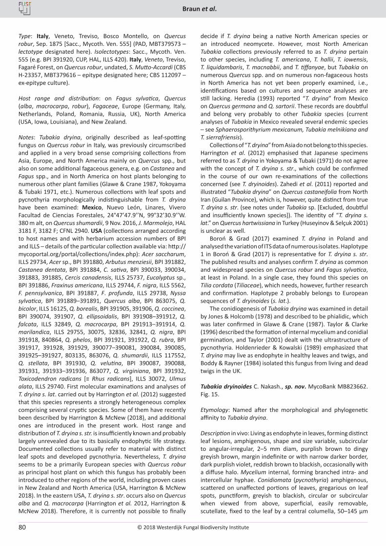
© 2018 Westerdijk Fungal Biodiversity Institute
Braun et al.
Editor-in-ChiefProf. dr P.W. Crous, Westerdijk Fungal Biodiversity Institute, P.O. Box 85167, 3508 AD Utrecht, The Netherlands.E-mail:[email protected]
80
Type: Italy, Veneto, Treviso, Bosco Montello, on Quercus robur, Sep. 1875 [Sacc., Mycoth. Ven. 555] (PAD, MBT379573 – lectotype designated here). Isolectotypes: Sacc., Mycoth. Ven. 555 (e.g. BPI 391920, CUP, HAL, ILLS 420). Italy, Veneto, Treviso, Fagaré Forest, on Quercus robur, undated, S. Mutto-Accardi (CBS H-23357, MBT379616 – epitype designated here; CBS 112097 – ex-epitype culture).
Host range and distribution: on Fagus sylvatica, Quercus (alba, macrocarpa, robur), Fagaceae, Europe (Germany, Italy, Netherlands, Poland, Romania, Russia, UK), North America (USA, Iowa, Louisiana), and New Zealand.
Notes: Tubakia dryina, originally described as leaf-spotting fungus on Quercus robur in Italy, was previously circumscribed and applied in a very broad sense comprising collections from Asia, Europe, and North America mainly on Quercus spp., but also on some additional fagaceous genera, e.g. on Castanea and Fagus spp., and in North America on host plants belonging to numerous other plant families (Glawe & Crane 1987, Yokoyama & Tubaki 1971, etc.). Numerous collections with leaf spots and pycnothyria morphologically indistinguishable from T. dryina have been examined: Mexico, Nuevo León, Linares, Vivero Facultad de Ciencias Forestales, 24°47’47.9’’N, 99°32’30.9’’W. 380 m alt, on Quercus shumardii, 9 Nov. 2016, J. Marmolejo, HAL 3181 F, 3182 F; CFNL 2940. USA (collections arranged according to host names and with herbarium accession numbers of BPI and ILLS – details of the particular collection available via: http://mycoportal.org/portal/collections/index.php): Acer saccharum, ILLS 29734, Acer sp., BPI 391880, Arbutus menziesii, BPI 391882, Castanea dentata, BPI 391884, C. sativa, BPI 390033, 390034, 391883, 391885, Cercis canadensis, ILLS 25737, Eucalyptus sp., BPI 391886, Fraxinus americana, ILLS 29744, F. nigra, ILLS 5562, F. pennsylvanica, BPI 391887, F. profunda, ILLS 29738, Nyssa sylvatica, BPI 391889–391891, Quercus alba, BPI 863075, Q. bicolor, ILLS 16125, Q. borealis, BPI 391905, 391906, Q. coccinea, BPI 390074, 391907, Q. ellipsoidalis, BPI 391908–391912, Q. falcata, ILLS 32849, Q. macrocarpa, BPI 291913–391914, Q. marilandica, ILLS 29755, 30075, 32836, 32841, Q. nigra, BPI 391918, 840864, Q. phelos, BPI 391921, 391922, Q. rubra, BPI 391917, 391928, 391929, 390077–390081, 390084, 390085, 391925–391927, 803135, 863076, Q. shumardii, ILLS 117552, Q. stellata, BPI 391930, Q. velutina, BPI 390087, 390088, 391931, 391933–391936, 863077, Q. virginiana, BPI 391932, Toxicodendron radicans [≡ Rhus radicans], ILLS 30072, Ulmus alata, ILLS 29740. First molecular examinations and analyses of T. dryina s. lat. carried out by Harrington et al. (2012) suggested that this species represents a strongly heterogeneous complex comprising several cryptic species. Some of them have recently been described by Harrington & McNew (2018), and additional ones are introduced in the present work. Host range and distribution of T. dryina s. str. is insufficiently known and probably largely unrevealed due to its basically endophytic life strategy. Documented collections usually refer to material with distinct leaf spots and developed pycnothyria. Nevertheless, T. dryina seems to be a primarily European species with Quercus robur as principal host plant on which this fungus has probably been introduced to other regions of the world, including proven cases in New Zealand and North America (USA, Harrington & McNew 2018). In the eastern USA, T. dryina s. str. occurs also on Quercus alba and Q. macrocarpa (Harrington et al. 2012, Harrington & McNew 2018). Therefore, it is currently not possible to finally
decide if T. dryina being a native North American species or an introduced neomycete. However, most North American Tubakia collections previously referred to as T. dryina pertain to other species, including T. americana, T. hallii, T. iowensis, T. liquidambaris, T. macnabbii, and T. tiffanyae, but Tubakia on numerous Quercus spp. and on numerous non-fagaceous hosts in North America has not yet been properly examined, i.e., identifications based on cultures and sequence analyses are still lacking. Heredia (1993) reported “T. dryina” from Mexico on Quercus germana and Q. sartorii. These records are doubtful and belong very probably to other Tubakia species (current analyses of Tubakia in Mexico revealed several endemic species – see Sphaerosporithyrium mexicanum, Tubakia melnikiana and T. sierrafriensis).
Collections of “T. dryina” from Asia do not belong to this species. Harrington et al. (2012) emphasised that Japanese specimens referred to as T. dryina in Yokoyama & Tubaki (1971) do not agree with the concept of T. dryina s. str., which could be confirmed in the course of our own re-examinations of the collections concerned (see T. dryinoides). Zahedi et al. (2011) reported and illustrated “Tubakia dryina” on Quercus castaneifolia from North Iran (Guilan Province), which is, however, quite distinct from true T. dryina s. str. (see notes under Tubakia sp. [Excluded, doubtful and insufficiently known species]). The identity of “T. dryina s. lat.” on Quercus hartwissiana in Turkey (Huseyinov & Selçuk 2001) is unclear as well.
Boroń & Grad (2017) examined T. dryina in Poland and analysed the variation of ITS data of numerous isolates. Haplotype 1 in Boroń & Grad (2017) is representative for T. dryina s. str. The published results and analyses confirm T. dryina as common and widespread species on Quercus robur and Fagus sylvatica, at least in Poland. In a single case, they found this species on Tilia cordata (Tiliaceae), which needs, however, further research and confirmation. Haplotype 2 probably belongs to European sequences of T. dryinoides (s. lat.).
The conidiogenesis of Tubakia dryina was examined in detail by Jones & Holcomb (1978) and described to be phialidic, which was later confirmed in Glawe & Crane (1987). Taylor & Clarke (1996) described the formation of internal mycelium and conidial germination, and Taylor (2001) dealt with the ultrastructure of pycnothyria. Holdenrieder & Kowalski (1989) emphasized that T. dryina may live as endophyte in healthy leaves and twigs, and Boddy & Rayner (1984) isolated this fungus from living and dead twigs in the UK.
Tubakia dryinoides C. Nakash., sp. nov. MycoBank MB823662. Fig. 15.
Etymology: Named after the morphological and phylogenetic affinity to Tubakia dryina.
Description in vivo: Living as endophyte in leaves, forming distinct leaf lesions, amphigenous, shape and size variable, subcircular to angular-irregular, 2–5 mm diam, purplish brown to dingy greyish brown, margin indefinite or with narrow darker border, dark purplish violet, reddish brown to blackish, occasionally with a diffuse halo. Mycelium internal, forming branched intra- and intercellular hyphae. Conidiomata (pycnothyria) amphigenous, scattered on unaffected portions of leaves, gregarious on leaf spots, punctiform, greyish to blackish, circular or subcircular when viewed from above, superficial, easily removable, scutellate, fixed to the leaf by a central columella, 50–145 µm

© 2018 Westerdijk Fungal Biodiversity Institute
The genus Tubakia s. lat.
Editor-in-ChiefProf. dr P.W. Crous, Westerdijk Fungal Biodiversity Institute, P.O. Box 85167, 3508 AD Utrecht, The Netherlands.E-mail:[email protected]
81
diam. Scutella convex sometimes more flattened, membranous, dense at the centre, looser at the edge, with a central hyaline or pale disc, 4–6 µm diam, surrounded by cells subcircular to angular in outline and 2–6 µm diam, giving rise to radiating hyphal strands, cells 7–25 × 2–5 µm, pale to dark brown, thick-walled (–1 µm), smooth, simple or 1–3 times bifurcating, ultimate branchlets with obtuse to mostly pointed tips. Central columella below the scutellum delicate, easily collapsing and loose, ephemeral, about 13–37 µm wide, surrounded by large brown cells. Conidiophores reduced to conidiogenous cells, arising from the underside of the scutella, around the columella, radiating, orientation outward-downward, cylindrical, conical, delicate, about 8–10 × 2–7 µm, hyaline, thin-walled, smooth, apex obtuse to truncate, conidiogenesis phialidic, sometimes forming indistinct periclinal thickenings. Conidia solitary, ellipsoid to obovoid, 8.5–14.5 × 5.5–8.5(–10) µm, length/width ratio 1.3–2.3, wall thin, up to 1 µm, hyaline to subhyaline, smooth, apex and base broadly rounded, with inconspicuous to conspicuous basal hilum (frill), occasionally somewhat peg-like and truncate when conspicuous. Microconidia narrowly ellipsoid-ovoid, fusiform, mostly straight, 3–10 × 1.5–2.5 µm, hyaline, thin-walled, smooth.
In vitro: on MEA with optimal growth at 20° C, attaining 30–40 mm diam after 14 d, margin scalloped, at first creamy white, forming concentric rings of olivaceous mycelium, reverse greyish
white, with olivaceous edge. Conidial formation not observed. Type: Japan, Osaka, Suita, on Quercus phillyraeoides, 8 Oct. 1969, T. Yokoyama (NBRC H-11618 – holotype; NBRC 9267 = MUCC2292 – ex-type cultures).
Hosts range and distribution: On Castanea crenata and Quercus phillyraeoides, Fagaceae, Asia (Japan) [(?) on Fagus sylvatica and Quercus robur, Europe (France, Poland)].
Additional collections examined: Japan, Osaka, Ikeda, on Castanea crenata, 11 Nov. 1968, T. Yokoyama, NBRC H-11616; NBRC 9265 = MUCC2290; Minoo, on Castanea crenata, 8 Oct. 1969, T. Yokoyama, NBRC H-11617; cultures NBRC 9266 = MUCC2291.
Notes: Tubakia dryinoides is morphologically similar to T. dryina, at least in terms of characters of the pycnothyria, and was referred to as T. dryina in Yokoyama & Tubaki (1971). In contrast to genuine T. dryina collections, the conidia in T. dryinoides remain hyaline or subhyaline, at least until germination (see Yokoyama & Tubaki 1971). The phylogenetic analyses corroborated a close affinity of the Japanese collections to T. dryina but clearly suggested a separate species (Figs 3–5), which confirms the doubts upon the correct assignment of Japanese collections to T. dryina by Harrington et al. (2012). Tubakia dryinoides belongs to a cluster undoubtedly
Fig. 15. Tubakia dryinoides (NBRC H-11618 – holotype). A. Pycnothyria on the surface of leaf litter. B. Culture on MEA (NBRC 9267 – ex-type culture). C. Scutellum. D. Central columella. E. Conidiophores. F. Conidia. G. Microconidia. Bars = 10 µm.
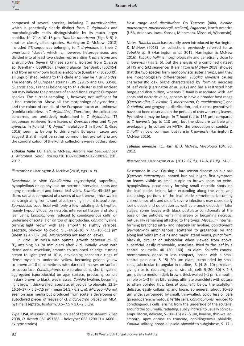
© 2018 Westerdijk Fungal Biodiversity Institute
Braun et al.
Editor-in-ChiefProf. dr P.W. Crous, Westerdijk Fungal Biodiversity Institute, P.O. Box 85167, 3508 AD Utrecht, The Netherlands.E-mail:[email protected]
82
composed of several species, including T. paradryinoides, which is genetically clearly distinct from T. dryinoides and morphologically easily distinguishable by its much larger conidia, 14–21 × 10–15 µm. Tubakia americana (Figs 3–5) is another closely allied species. Harrington & McNew (2018) included ITS sequences belonging to T. dryinoides in their T. americana “clade”, which is, however, heterogeneous and divided into at least two clades representing T. americana and T. dryinoides. Several Chinese strains, isolated from Quercus sp. (GenBank FJ598616), Linderia glauca (GenBank JF502454) and from an unknown host as endophyte (GenBank FJ025349), all unpublished, belong to this clade and may be T. dryinoides. The identity of European strains (CBS 329.75 and CPC 33586, Quercus spp., France) belonging to this cluster is still unclear, but may indicate the presence of an additional cryptic European species. The current sampling is, however, not sufficient for a final conclusion. Above all, the morphology of pycnothyria and the colour of conidia of the European taxon are unknown (conidia colourless in T. dryinoides). Therefore, the collections concerned are tentatively maintained in T. dryinoides. ITS sequences retrieved from leaves of Quercus robur and Fagus sylvatica in Poland (“T. dryina” haplotype 2 in Boroń & Grad 2016) seem to belong to this cryptic European taxon and suggest that it might be rather common, but pycnothyria and the conidial colour of the Polish collections were not described.
Tubakia hallii T.C. Harr. & McNew, Antonie van Leeuwenhoek J. Microbiol. Serol. doi.org/10.1007/s10482-017-1001-9 [10]. 2017.
Illustrations: Harrington & McNew (2018, figs 1a–c).
Description in vivo: Conidiomata (pycnothyria) superficial, hypophyllous or epiphyllous on necrotic interveinal spots and along necrotic mid and lateral leaf veins. Scutella 45–155 µm diam, radiate, composed of a series of dark brown, thick-walled cells originating from a central cell, ending in blunt to acute tips. Sporodochia superficial with only a few radiating dark hyphae, mainly hypophyllous, on necrotic interveinal tissues and along leaf veins. Conidiophores reduced to conidiogenous cells, on underside of scutella or on top of sporodochia. Conidia hyaline, turning light brown with age, smooth to slightly varicose, aseptate, obovoid to ovoid, 9.5–14.5(–16) × 7.5–10(–11) µm (mean 12.4 × 8.7 µm). Microconidia not seen on leaves.
In vitro: On MYEA with optimal growth between 25–30 °C, attaining 50–70 mm diam after 7 d, initially white with dense aerial mycelium, smooth to scalloped at edge, turning cream to light grey at 10 d, developing concentric rings of dense mycelium, underside yellow, becoming golden yellow to brown at 10 d, sometimes with dark cell masses on surface or subsurface. Conidiophores rare to abundant, short, hyaline, aggregated (sporodochia) on agar surface, producing conidia in dark brown to black, wet masses. Conidia hyaline, becoming light brown, thick-walled, aseptate, ellipsoidal to obovate, 12.5–16.5(–17) × 5.3–7.5 µm (mean 14.5 × 6.2 µm). Microconidia not seen on agar media but produced from scutella developing on autoclaved pieces of leaves of Q. macrocarpa placed on MEA, hyaline, aseptate, fusiform, 3.5–7.5 × 1.0–2.5 µm.
Type: USA, Missouri, Kirbyville, on leaf of Quercus stellata, 2 Sep 2008, D. Brandt (ISC 453286 – holotype; CBS 129013 = A666 – ex-type strains).
Host range and distribution: On Quercus (alba, bicolor, macrocarpa, muehlenbergii, stellata), Fagaceae, North America (USA, Arkansas, Iowa, Kansas, Minnesota, Missouri, Wisconsin).
Notes: Tubakia hallii has recently been introduced by Harrington & McNew (2018) for collections previously referred to as Tubakia sp. B (Harrington et al. 2012, Harrington & McNew 2016). Tubakia hallii is morphologically and genetically close to T. iowensis (Figs 3, 5), but the analysis of a combined dataset of ITS and tef1 sequences (Harrington & McNew 2018) showed that the two species form monophyletic sister groups, and they are morphologically differentiated. Tubakia iowensis causes characteristic oak blight characterised by forming necroses of leaf veins (Harrington et al. 2012) and has a restricted host range and distribution, whereas T. hallii is associated with leaf spots and necrotic veins, appears to have a broader host range (Quercus alba, Q. bicolor, Q. macrocarpa, Q. muehlenbergii, and Q. stellata) and geographic distribution, and crustose pycnothyria have not been observed on the petioles of overwintering leaves. Pycnothyria may be larger in T. hallii (up to 155 µm) compared to T. iowensis (up to 110 µm), but the sizes are variable and overlapping. In culture on MYEA, the production of conidia in T. hallii is not uncommon, but rare in T. iowensis (Harrington & McNew 2016).
Tubakia iowensis T.C. Harr. & D. McNew, Mycologia 104: 86. 2012.
Illustrations: Harrington et al. (2012: 82, fig. 1A–N, 87, fig. 2A–L).
Description in vivo: Causing a late-season disease on bur oak (Quercus macrocarpa), named bur oak blight, first symptom on leaves visible as small purple to brown spots on veins, hypophyllous, occasionally forming small necrotic spots on the leaf blade, lesions later expanding along the veins and coalescing, portions of the leaf blade sometimes becoming chlorotic-necrotic and die off; severe infections may cause early leaf dieback and defoliation as well as branch dieback in later stages; some leaves with black pustulate conidiomata at the base of the petioles, remaining green or becoming necrotic, but usually remaining attached to the twigs. Mycelium internal, forming branched intra- and intercellular hyphae. Conidiomata (pycnothyria) amphigenous, scattered to gregarious on and along leaf veins (midveins and major lateral veins), punctiform, blackish, circular or subcircular when viewed from above, superficial, easily removable, scutellate, fixed to the leaf by a central columella, 40–120(–160) µm diam. Scutella convex, membranous, dense to less compact, looser, with a small central pale disc, 5–15(–20) µm diam, surrounded by small cells, subcircular to angular in outline, (3–)4–8(–10) µm diam, giving rise to radiating hyphal strands, cells 5–20(–30) × 2–8 µm, pale to medium dark brown, thick-walled (–1 µm), smooth, simple or 1–3 times bifurcating, ultimate branchlets with obtuse to often pointed tips. Central columella below the scutellum delicate, easily collapsing and loose, ephemeral, about 10–20 µm wide, surrounded by small, thin-walled, colourless or pale (pseudoparenchymatous) fertile cells. Conidiophores reduced to conidiogenous cells, arising from the underside of the scutella, around the columella, radiating, subcylindrical to usually conical-ampulliform, delicate, 5–10(–15) × 2–5 µm, hyaline, thin-walled, smooth, apex obtuse to truncate, conidiogenesis phialidic. Conidia solitary, broad ellipsoid-obovoid to subglobose, 9–17 ×
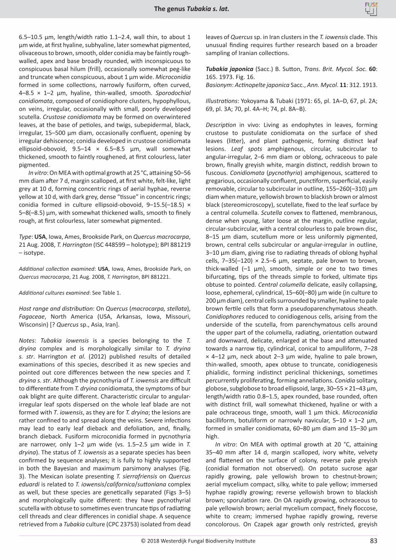
© 2018 Westerdijk Fungal Biodiversity Institute
The genus Tubakia s. lat.
Editor-in-ChiefProf. dr P.W. Crous, Westerdijk Fungal Biodiversity Institute, P.O. Box 85167, 3508 AD Utrecht, The Netherlands.E-mail:[email protected]
83
6.5–10.5 µm, length/width ratio 1.1–2.4, wall thin, to about 1 µm wide, at first hyaline, subhyaline, later somewhat pigmented, olivaceous to brown, smooth, older conidia may be faintly rough-walled, apex and base broadly rounded, with inconspicuous to conspicuous basal hilum (frill), occasionally somewhat peg-like and truncate when conspicuous, about 1 µm wide. Microconidia formed in some collections, narrowly fusiform, often curved, 4–8.5 × 1–2 µm, hyaline, thin-walled, smooth. Sporodochial conidiomata, composed of conidiophore clusters, hypophyllous, on veins, irregular, occasionally with small, poorly developed scutella. Crustose conidiomata may be formed on overwintered leaves, at the base of petioles, and twigs, subepidermal, black, irregular, 15–500 µm diam, occasionally confluent, opening by irregular dehiscence; conidia developed in crustose conidiomata ellipsoid-obovoid, 9.5–14 × 6.5–8.5 µm, wall somewhat thickened, smooth to faintly roughened, at first colourless, later pigmented.
In vitro: On MEA with optimal growth at 25 °C, attaining 50–56 mm diam after 7 d, margin scalloped, at first white, felt-like, light grey at 10 d, forming concentric rings of aerial hyphae, reverse yellow at 10 d, with dark grey, dense “tissue” in concentric rings; conidia formed in culture ellipsoid-obovoid, 9–15.5(–18.5) × 5–8(–8.5) µm, with somewhat thickened walls, smooth to finely rough, at first colourless, later somewhat pigmented.
Type: USA, Iowa, Ames, Brookside Park, on Quercus macrocarpa, 21 Aug. 2008, T. Harrington (ISC 448599 – holotype); BPI 881219 – isotype.
Additional collection examined: USA, Iowa, Ames, Brookside Park, on Quercus macrocarpa, 21 Aug. 2008, T. Harrington, BPI 881221.
Additional cultures examined: See Table 1.
Host range and distribution: On Quercus (macrocarpa, stellata), Fagaceae, North America (USA, Arkansas, Iowa, Missouri, Wisconsin) [? Quercus sp., Asia, Iran].
Notes: Tubakia iowensis is a species belonging to the T. dryina complex and is morphologically similar to T. dryina s. str. Harrington et al. (2012) published results of detailed examinations of this species, described it as new species and pointed out core differences between the new species and T. dryina s. str. Although the pycnothyria of T. iowensis are difficult to differentiate from T. dryina conidiomata, the symptoms of bur oak blight are quite different. Characteristic circular to angular-irregular leaf spots dispersed on the whole leaf blade are not formed with T. iowensis, as they are for T. dryina; the lesions are rather confined to and spread along the veins. Severe infections may lead to early leaf dieback and defoliation, and, finally, branch dieback. Fusiform microconidia formed in pycnothyria are narrower, only 1–2 µm wide (vs. 1.5–2.5 µm wide in T. dryina). The status of T. iowensis as a separate species has been confirmed by sequence analyses; it is fully to highly supported in both the Bayesian and maximum parsimony analyses (Fig. 3). The Mexican isolate presenting T. sierrafriensis on Quercus eduardi is related to T. iowensis/californica/suttoniana complex as well, but these species are genetically separated (Figs 3–5) and morphologically quite different: they have pycnothyrial scutella with obtuse to sometimes even truncate tips of radiating cell threads and clear differences in conidial shape. A sequence retrieved from a Tubakia culture (CPC 23753) isolated from dead
leaves of Quercus sp. in Iran clusters in the T. iowensis clade. This unusual finding requires further research based on a broader sampling of Iranian collections.
Tubakia japonica (Sacc.) B. Sutton, Trans. Brit. Mycol. Soc. 60: 165. 1973. Fig. 16.Basionym: Actinopelte japonica Sacc., Ann. Mycol. 11: 312. 1913.
Illustrations: Yokoyama & Tubaki (1971: 65, pl. 1A–D, 67, pl. 2A; 69, pl. 3A; 70, pl. 4A–H; 74, pl. 8A–B).
Description in vivo: Living as endophytes in leaves, forming crustose to pustulate conidiomata on the surface of shed leaves (litter), and plant pathogenic, forming distinct leaf lesions. Leaf spots amphigenous, circular, subcircular to angular-irregular, 2–6 mm diam or oblong, ochraceous to pale brown, finally greyish white, margin distinct, reddish brown to fuscous. Conidiomata (pycnothyria) amphigenous, scattered to gregarious, occasionally confluent, punctiform, superficial, easily removable, circular to subcircular in outline, 155–260(–310) µm diam when mature, yellowish brown to blackish brown or almost black (stereomicroscopy), scutellate, fixed to the leaf surface by a central columella. Scutella convex to flattened, membranous, dense when young, later loose at the margin, outline regular, circular-subcircular, with a central colourless to pale brown disc, 8–15 µm diam, scutellum more or less uniformly pigmented, brown, central cells subcircular or angular-irregular in outline, 3–10 µm diam, giving rise to radiating threads of oblong hyphal cells, 7–35(–120) × 2.5–6 µm, septate, pale brown to brown, thick-walled (–1 µm), smooth, simple or one to two times bifurcating, tips of the threads simple to forked, ultimate tips obtuse to pointed. Central columella delicate, easily collapsing, loose, ephemeral, cylindrical, 15–60(–80) µm wide (in culture to 200 µm diam), central cells surrounded by smaller, hyaline to pale brown fertile cells that form a pseudoparenchymatous sheath. Conidiophores reduced to conidiogenous cells, arising from the underside of the scutella, from parenchymatous cells around the upper part of the columella, radiating, orientation outward and downward, delicate, enlarged at the base and attenuated towards a narrow tip, cylindrical, conical to ampulliform, 7–28 × 4–12 µm, neck about 2–3 µm wide, hyaline to pale brown, thin-walled, smooth, apex obtuse to truncate, conidiogenesis phialidic, forming indistinct periclinal thickenings, sometimes percurrently proliferating, forming annellations. Conidia solitary, globose, subglobose to broad ellipsoid, large, 30–55 × 21–43 µm, length/width ratio 0.8–1.5, apex rounded, base rounded, often with distinct frill, wall somewhat thickened, hyaline or with a pale ochraceous tinge, smooth, wall 1 µm thick. Microconidia bacilliform, botuliform or narrowly navicular, 5–10 × 1–2 µm, formed in smaller conidiomata, 60–80 µm diam and 15–30 µm high.
In vitro: On MEA with optimal growth at 20 °C, attaining 35–40 mm after 14 d, margin scalloped, ivory white, velvety and flattened on the surface of colony, reverse pale greyish (conidial formation not observed). On potato sucrose agar rapidly growing, pale yellowish brown to chestnut-brown; aerial mycelium compact, silky, white to pale yellow; immersed hyphae rapidly growing; reverse yellowish brown to blackish brown; sporulation rare. On OA rapidly growing, ochraceous to pale yellowish brown; aerial mycelium compact, finely floccose, white to cream; immersed hyphae rapidly growing, reverse concolorous. On Czapek agar growth only restricted, greyish
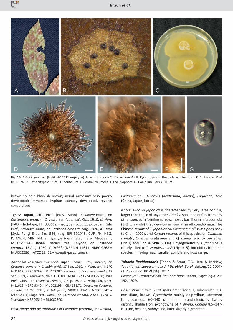
© 2018 Westerdijk Fungal Biodiversity Institute
Braun et al.
Editor-in-ChiefProf. dr P.W. Crous, Westerdijk Fungal Biodiversity Institute, P.O. Box 85167, 3508 AD Utrecht, The Netherlands.E-mail:[email protected]
84
brown to pale blackish brown; aerial mycelium very poorly developed; immersed hyphae scarcely developed, reverse concolorous.
Types: Japan, Gifu Pref. (Prov. Mino), Kawauye-mura, on Castanea crenata (= C. vesca var. japonica), Oct. 1910, K. Hara (PAD – holotype; FH 888612 – isotype). Topotypes: Japan, Gifu Pref., Kawauye-mura, on Castanea crenata, Aug. 1920, K. Hara [Syd., Fungi Exot. Exs. 526] (e.g. BPI 391948, CUP, FH, HBG, K, MICH, MIN, PH, S). Epitype (designated here, MycoBank, MBT379574): Japan, Ibaraki Pref., Chiyoda, on Castanea crenata, 13 Aug. 1969, K. Uchida (NBRC H-11611; NBRC 9268 = MUCC2296 = ATCC 22472 – ex-epitype cultures).
Additional collection examined: Japan, Ibaraki Pref., Kasama, on Castanea crenata (= C. pubinervis), 17 Sep. 1969, Y. Kobayashi, NBRC H-11612; NBRC 9269 = MUCC2297; Kasama, on Castanea crenata, 17 Sep. 1969, Y. Kobayashi, NBRC H-11883; NBRC 9270 = MUCC2298; Shiga Pref., Ootsu, on Castanea crenata, 2 Sep. 1970, T. Yokoyama, NBRC H-11613; NBRC 9340 = MUCC2299 = CBS 191.71; Ootsu, on Castanea crenata, 30 Oct. 1970, T. Yokoyama, NBRC H-11615; NBRC 9342 = MUCC2301; Shiga Pref., Ootsu, on Castanea crenata, 2 Sep. 1970, T. Yokoyama, NBRC9341 = MUCC2300.
Host range and distribution: On Castanea (crenata, mollissima,
Castanea sp.), Quercus (acutissima, aliena), Fagaceae, Asia (China, Japan, Korea).
Notes: Tubakia japonica is characterised by very large conidia, larger than those of any other Tubakia spp., and differs from any other species in forming narrow, mostly bacilliform microconidia (1–2 µm wide) that develop in special small conidiomata. The Chinese report of T. japonica on Castanea mollissima goes back to Chen (2002), and Korean records of this species on Castanea crenata, Quercus acutissima and Q. aliena refer to Lee et al. (1991) and Cho & Shin (2004). Phylogenetically T. japonica is closely allied to T. seoraksanensis (Figs 3–5), but differs from this species in having much smaller conidia and host range.
Tubakia liquidambaris (Tehon & Stout) T.C. Harr. & McNew, Antonie van Leeuwenhoek J. Microbiol. Serol. doi.org/10.1007/s10482-017-1001-9 [16]. 2017.Basionym: Leptothyriella liquidambaris Tehon, Mycologia 21: 192. 1929.
Description in vivo: Leaf spots amphigenous, subcircular, 1–6 mm diam, brown. Pycnothyria mainly epiphyllous, scattered to gregarious, 60–140 µm diam, morphologically barely distinguishable from pycnothyria of T. dryina. Conidia 8.5–14 × 6–9 µm, hyaline, subhyaline, later slightly pigmented.
Fig. 16. Tubakia japonica (NBRC H-11611 – epitype). A. Symptoms on Castanea crenata. B. Pycnothyria on the surface of leaf spot. C. Culture on MEA (NBRC 9268 – ex-epitype culture). D. Scutellum. E. Central columella. F. Conidiophore. G. Conidium. Bars = 10 µm.
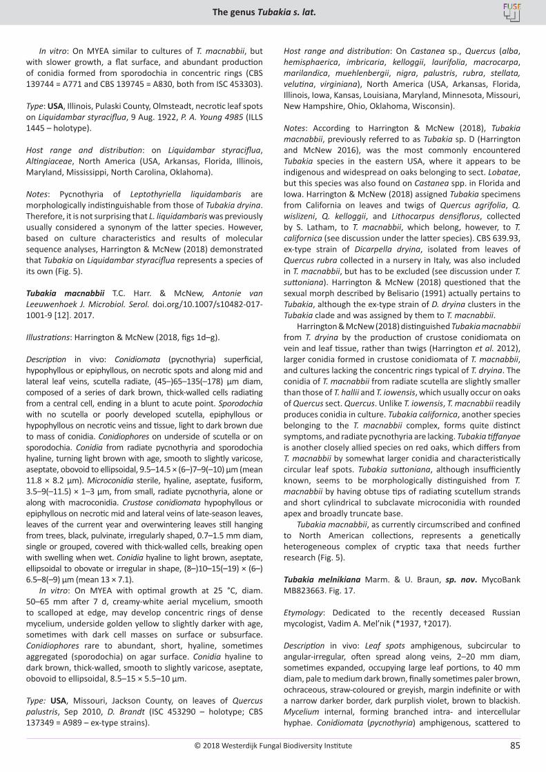
© 2018 Westerdijk Fungal Biodiversity Institute
The genus Tubakia s. lat.
Editor-in-ChiefProf. dr P.W. Crous, Westerdijk Fungal Biodiversity Institute, P.O. Box 85167, 3508 AD Utrecht, The Netherlands.E-mail:[email protected]
85
In vitro: On MYEA similar to cultures of T. macnabbii, but with slower growth, a flat surface, and abundant production of conidia formed from sporodochia in concentric rings (CBS 139744 = A771 and CBS 139745 = A830, both from ISC 453303).
Type: USA, Illinois, Pulaski County, Olmsteadt, necrotic leaf spots on Liquidambar styraciflua, 9 Aug. 1922, P. A. Young 4985 (ILLS 1445 – holotype).
Host range and distribution: on Liquidambar styraciflua, Altingiaceae, North America (USA, Arkansas, Florida, Illinois, Maryland, Mississippi, North Carolina, Oklahoma).
Notes: Pycnothyria of Leptothyriella liquidambaris are morphologically indistinguishable from those of Tubakia dryina. Therefore, it is not surprising that L. liquidambaris was previously usually considered a synonym of the latter species. However, based on culture characteristics and results of molecular sequence analyses, Harrington & McNew (2018) demonstrated that Tubakia on Liquidambar styraciflua represents a species of its own (Fig. 5).
Tubakia macnabbii T.C. Harr. & McNew, Antonie van Leeuwenhoek J. Microbiol. Serol. doi.org/10.1007/s10482-017-1001-9 [12]. 2017.
Illustrations: Harrington & McNew (2018, figs 1d–g).
Description in vivo: Conidiomata (pycnothyria) superficial, hypophyllous or epiphyllous, on necrotic spots and along mid and lateral leaf veins, scutella radiate, (45–)65–135(–178) µm diam, composed of a series of dark brown, thick-walled cells radiating from a central cell, ending in a blunt to acute point. Sporodochia with no scutella or poorly developed scutella, epiphyllous or hypophyllous on necrotic veins and tissue, light to dark brown due to mass of conidia. Conidiophores on underside of scutella or on sporodochia. Conidia from radiate pycnothyria and sporodochia hyaline, turning light brown with age, smooth to slightly varicose, aseptate, obovoid to ellipsoidal, 9.5–14.5 × (6–)7–9(–10) µm (mean 11.8 × 8.2 µm). Microconidia sterile, hyaline, aseptate, fusiform, 3.5–9(–11.5) × 1–3 µm, from small, radiate pycnothyria, alone or along with macroconidia. Crustose conidiomata hypophyllous or epiphyllous on necrotic mid and lateral veins of late-season leaves, leaves of the current year and overwintering leaves still hanging from trees, black, pulvinate, irregularly shaped, 0.7–1.5 mm diam, single or grouped, covered with thick-walled cells, breaking open with swelling when wet. Conidia hyaline to light brown, aseptate, ellipsoidal to obovate or irregular in shape, (8–)10–15(–19) × (6–) 6.5–8(–9) µm (mean 13 × 7.1).
In vitro: On MYEA with optimal growth at 25 °C, diam. 50–65 mm after 7 d, creamy-white aerial mycelium, smooth to scalloped at edge, may develop concentric rings of dense mycelium, underside golden yellow to slightly darker with age, sometimes with dark cell masses on surface or subsurface. Conidiophores rare to abundant, short, hyaline, sometimes aggregated (sporodochia) on agar surface. Conidia hyaline to dark brown, thick-walled, smooth to slightly varicose, aseptate, obovoid to ellipsoidal, 8.5–15 × 5.5–10 µm.
Type: USA, Missouri, Jackson County, on leaves of Quercus palustris, Sep 2010, D. Brandt (ISC 453290 – holotype; CBS 137349 = A989 – ex-type strains).
Host range and distribution: On Castanea sp., Quercus (alba, hemisphaerica, imbricaria, kelloggii, laurifolia, macrocarpa, marilandica, muehlenbergii, nigra, palustris, rubra, stellata, velutina, virginiana), North America (USA, Arkansas, Florida, Illinois, Iowa, Kansas, Louisiana, Maryland, Minnesota, Missouri, New Hampshire, Ohio, Oklahoma, Wisconsin).
Notes: According to Harrington & McNew (2018), Tubakia macnabbii, previously referred to as Tubakia sp. D (Harrington and McNew 2016), was the most commonly encountered Tubakia species in the eastern USA, where it appears to be indigenous and widespread on oaks belonging to sect. Lobatae, but this species was also found on Castanea spp. in Florida and Iowa. Harrington & McNew (2018) assigned Tubakia specimens from California on leaves and twigs of Quercus agrifolia, Q. wislizeni, Q. kelloggii, and Lithocarpus densiflorus, collected by S. Latham, to T. macnabbii, which belong, however, to T. californica (see discussion under the latter species). CBS 639.93, ex-type strain of Dicarpella dryina, isolated from leaves of Quercus rubra collected in a nursery in Italy, was also included in T. macnabbii, but has to be excluded (see discussion under T. suttoniana). Harrington & McNew (2018) questioned that the sexual morph described by Belisario (1991) actually pertains to Tubakia, although the ex-type strain of D. dryina clusters in the Tubakia clade and was assigned by them to T. macnabbii.
Harrington & McNew (2018) distinguished Tubakia macnabbii from T. dryina by the production of crustose conidiomata on vein and leaf tissue, rather than twigs (Harrington et al. 2012), larger conidia formed in crustose conidiomata of T. macnabbii, and cultures lacking the concentric rings typical of T. dryina. The conidia of T. macnabbii from radiate scutella are slightly smaller than those of T. hallii and T. iowensis, which usually occur on oaks of Quercus sect. Quercus. Unlike T. iowensis, T. macnabbii readily produces conidia in culture. Tubakia californica, another species belonging to the T. macnabbii complex, forms quite distinct symptoms, and radiate pycnothyria are lacking. Tubakia tiffanyae is another closely allied species on red oaks, which differs from T. macnabbii by somewhat larger conidia and characteristically circular leaf spots. Tubakia suttoniana, although insufficiently known, seems to be morphologically distinguished from T. macnabbii by having obtuse tips of radiating scutellum strands and short cylindrical to subclavate microconidia with rounded apex and broadly truncate base.
Tubakia macnabbii, as currently circumscribed and confined to North American collections, represents a genetically heterogeneous complex of cryptic taxa that needs further research (Fig. 5).
Tubakia melnikiana Marm. & U. Braun, sp. nov. MycoBank MB823663. Fig. 17.
Etymology: Dedicated to the recently deceased Russian mycologist, Vadim A. Mel’nik (*1937, †2017).
Description in vivo: Leaf spots amphigenous, subcircular to angular-irregular, often spread along veins, 2–20 mm diam, sometimes expanded, occupying large leaf portions, to 40 mm diam, pale to medium dark brown, finally sometimes paler brown, ochraceous, straw-coloured or greyish, margin indefinite or with a narrow darker border, dark purplish violet, brown to blackish. Mycelium internal, forming branched intra- and intercellular hyphae. Conidiomata (pycnothyria) amphigenous, scattered to
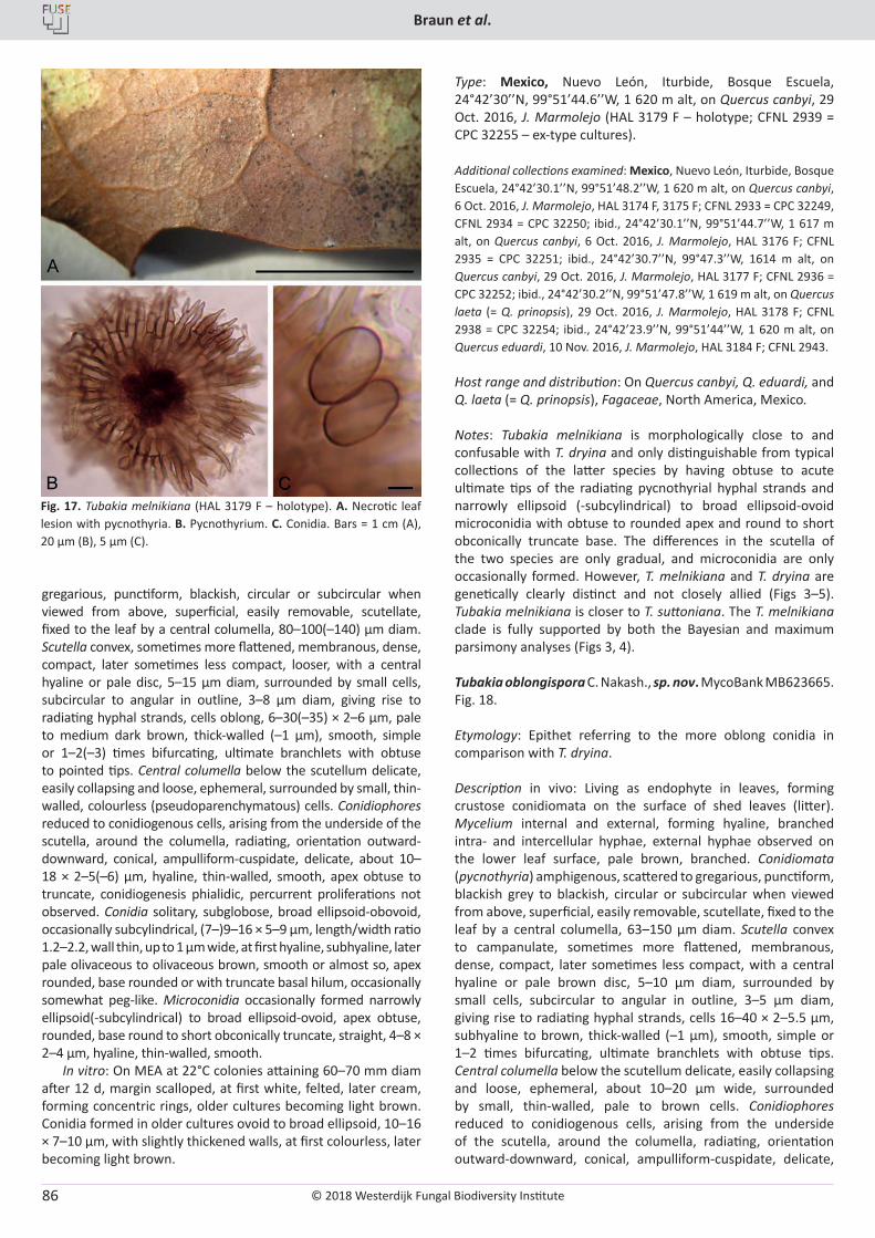
© 2018 Westerdijk Fungal Biodiversity Institute
Braun et al.
Editor-in-ChiefProf. dr P.W. Crous, Westerdijk Fungal Biodiversity Institute, P.O. Box 85167, 3508 AD Utrecht, The Netherlands.E-mail:[email protected]
86
gregarious, punctiform, blackish, circular or subcircular when viewed from above, superficial, easily removable, scutellate, fixed to the leaf by a central columella, 80–100(–140) µm diam. Scutella convex, sometimes more flattened, membranous, dense, compact, later sometimes less compact, looser, with a central hyaline or pale disc, 5–15 µm diam, surrounded by small cells, subcircular to angular in outline, 3–8 µm diam, giving rise to radiating hyphal strands, cells oblong, 6–30(–35) × 2–6 µm, pale to medium dark brown, thick-walled (–1 µm), smooth, simple or 1–2(–3) times bifurcating, ultimate branchlets with obtuse to pointed tips. Central columella below the scutellum delicate, easily collapsing and loose, ephemeral, surrounded by small, thin-walled, colourless (pseudoparenchymatous) cells. Conidiophores reduced to conidiogenous cells, arising from the underside of the scutella, around the columella, radiating, orientation outward-downward, conical, ampulliform-cuspidate, delicate, about 10–18 × 2–5(–6) µm, hyaline, thin-walled, smooth, apex obtuse to truncate, conidiogenesis phialidic, percurrent proliferations not observed. Conidia solitary, subglobose, broad ellipsoid-obovoid, occasionally subcylindrical, (7–)9–16 × 5–9 µm, length/width ratio 1.2–2.2, wall thin, up to 1 µm wide, at first hyaline, subhyaline, later pale olivaceous to olivaceous brown, smooth or almost so, apex rounded, base rounded or with truncate basal hilum, occasionally somewhat peg-like. Microconidia occasionally formed narrowly ellipsoid(-subcylindrical) to broad ellipsoid-ovoid, apex obtuse, rounded, base round to short obconically truncate, straight, 4–8 × 2–4 µm, hyaline, thin-walled, smooth.
In vitro: On MEA at 22°C colonies attaining 60–70 mm diam after 12 d, margin scalloped, at first white, felted, later cream, forming concentric rings, older cultures becoming light brown. Conidia formed in older cultures ovoid to broad ellipsoid, 10–16 × 7–10 µm, with slightly thickened walls, at first colourless, later becoming light brown.
Type: Mexico, Nuevo León, Iturbide, Bosque Escuela, 24°42’30’’N, 99°51’44.6’’W, 1 620 m alt, on Quercus canbyi, 29 Oct. 2016, J. Marmolejo (HAL 3179 F – holotype; CFNL 2939 = CPC 32255 – ex-type cultures).
Additional collections examined: Mexico, Nuevo León, Iturbide, Bosque Escuela, 24°42’30.1’’N, 99°51’48.2’’W, 1 620 m alt, on Quercus canbyi, 6 Oct. 2016, J. Marmolejo, HAL 3174 F, 3175 F; CFNL 2933 = CPC 32249, CFNL 2934 = CPC 32250; ibid., 24°42’30.1’’N, 99°51’44.7’’W, 1 617 m alt, on Quercus canbyi, 6 Oct. 2016, J. Marmolejo, HAL 3176 F; CFNL 2935 = CPC 32251; ibid., 24°42’30.7’’N, 99°47.3’’W, 1614 m alt, on Quercus canbyi, 29 Oct. 2016, J. Marmolejo, HAL 3177 F; CFNL 2936 = CPC 32252; ibid., 24°42’30.2’’N, 99°51’47.8’’W, 1 619 m alt, on Quercus laeta (= Q. prinopsis), 29 Oct. 2016, J. Marmolejo, HAL 3178 F; CFNL 2938 = CPC 32254; ibid., 24°42’23.9’’N, 99°51’44’’W, 1 620 m alt, on Quercus eduardi, 10 Nov. 2016, J. Marmolejo, HAL 3184 F; CFNL 2943.
Host range and distribution: On Quercus canbyi, Q. eduardi, and Q. laeta (= Q. prinopsis), Fagaceae, North America, Mexico.
Notes: Tubakia melnikiana is morphologically close to and confusable with T. dryina and only distinguishable from typical collections of the latter species by having obtuse to acute ultimate tips of the radiating pycnothyrial hyphal strands and narrowly ellipsoid (-subcylindrical) to broad ellipsoid-ovoid microconidia with obtuse to rounded apex and round to short obconically truncate base. The differences in the scutella of the two species are only gradual, and microconidia are only occasionally formed. However, T. melnikiana and T. dryina are genetically clearly distinct and not closely allied (Figs 3–5). Tubakia melnikiana is closer to T. suttoniana. The T. melnikiana clade is fully supported by both the Bayesian and maximum parsimony analyses (Figs 3, 4).
Tubakia oblongispora C. Nakash., sp. nov. MycoBank MB623665. Fig. 18.
Etymology: Epithet referring to the more oblong conidia in comparison with T. dryina.
Description in vivo: Living as endophyte in leaves, forming crustose conidiomata on the surface of shed leaves (litter). Mycelium internal and external, forming hyaline, branched intra- and intercellular hyphae, external hyphae observed on the lower leaf surface, pale brown, branched. Conidiomata (pycnothyria) amphigenous, scattered to gregarious, punctiform, blackish grey to blackish, circular or subcircular when viewed from above, superficial, easily removable, scutellate, fixed to the leaf by a central columella, 63–150 µm diam. Scutella convex to campanulate, sometimes more flattened, membranous, dense, compact, later sometimes less compact, with a central hyaline or pale brown disc, 5–10 µm diam, surrounded by small cells, subcircular to angular in outline, 3–5 µm diam, giving rise to radiating hyphal strands, cells 16–40 × 2–5.5 µm, subhyaline to brown, thick-walled (–1 µm), smooth, simple or 1–2 times bifurcating, ultimate branchlets with obtuse tips. Central columella below the scutellum delicate, easily collapsing and loose, ephemeral, about 10–20 µm wide, surrounded by small, thin-walled, pale to brown cells. Conidiophores reduced to conidiogenous cells, arising from the underside of the scutella, around the columella, radiating, orientation outward-downward, conical, ampulliform-cuspidate, delicate,
Fig. 17. Tubakia melnikiana (HAL 3179 F – holotype). A. Necrotic leaf lesion with pycnothyria. B. Pycnothyrium. C. Conidia. Bars = 1 cm (A), 20 µm (B), 5 µm (C).
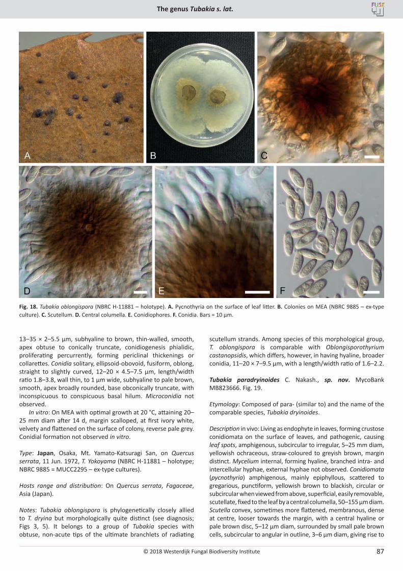
© 2018 Westerdijk Fungal Biodiversity Institute
The genus Tubakia s. lat.
Editor-in-ChiefProf. dr P.W. Crous, Westerdijk Fungal Biodiversity Institute, P.O. Box 85167, 3508 AD Utrecht, The Netherlands.E-mail:[email protected]
87
13–35 × 2–5.5 µm, subhyaline to brown, thin-walled, smooth, apex obtuse to conically truncate, conidiogenesis phialidic, proliferating percurrently, forming periclinal thickenings or collarettes. Conidia solitary, ellipsoid-obovoid, fusiform, oblong, straight to slightly curved, 12–20 × 4.5–7.5 µm, length/width ratio 1.8–3.8, wall thin, to 1 µm wide, subhyaline to pale brown, smooth, apex broadly rounded, base obconically truncate, with inconspicuous to conspicuous basal hilum. Microconidia not observed.
In vitro: On MEA with optimal growth at 20 °C, attaining 20–25 mm diam after 14 d, margin scalloped, at first ivory white, velvety and flattened on the surface of colony, reverse pale grey. Conidial formation not observed in vitro.
Type: Japan, Osaka, Mt. Yamato-Katsuragi San, on Quercus serrata, 11 Jun. 1972, T. Yokoyama (NBRC H-11881 – holotype; NBRC 9885 = MUCC2295 – ex-type cultures).
Hosts range and distribution: On Quercus serrata, Fagaceae, Asia (Japan).
Notes: Tubakia oblongispora is phylogenetically closely allied to T. dryina but morphologically quite distinct (see diagnosis; Figs 3, 5). It belongs to a group of Tubakia species with obtuse, non-acute tips of the ultimate branchlets of radiating
scutellum strands. Among species of this morphological group, T. oblongispora is comparable with Oblongisporothyrium castanopsidis, which differs, however, in having hyaline, broader conidia, 11–20 × 7–9.5 µm, with a length/width ratio of 1.6–2.2.
Tubakia paradryinoides C. Nakash., sp. nov. MycoBank MB823666. Fig. 19.
Etymology: Composed of para- (similar to) and the name of the comparable species, Tubakia dryinoides.
Description in vivo: Living as endophyte in leaves, forming crustose conidiomata on the surface of leaves, and pathogenic, causing leaf spots, amphigenous, subcircular to irregular, 5–25 mm diam, yellowish ochraceous, straw-coloured to greyish brown, margin distinct. Mycelium internal, forming hyaline, branched intra- and intercellular hyphae, external hyphae not observed. Conidiomata (pycnothyria) amphigenous, mainly epiphyllous, scattered to gregarious, punctiform, yellowish brown to blackish, circular or subcircular when viewed from above, superficial, easily removable, scutellate, fixed to the leaf by a central columella, 50–155 µm diam. Scutella convex, sometimes more flattened, membranous, dense at centre, looser towards the margin, with a central hyaline or pale brown disc, 5–12 µm diam, surrounded by small pale brown cells, subcircular to angular in outline, 3–6 µm diam, giving rise to
Fig. 18. Tubakia oblongispora (NBRC H-11881 – holotype). A. Pycnothyria on the surface of leaf litter. B. Colonies on MEA (NBRC 9885 – ex-type culture). C. Scutellum. D. Central columella. E. Conidiophores. F. Conidia. Bars = 10 µm.
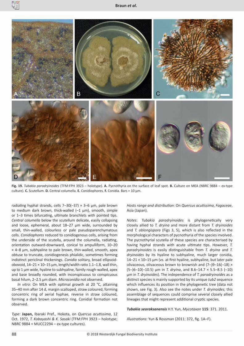
© 2018 Westerdijk Fungal Biodiversity Institute
Braun et al.
Editor-in-ChiefProf. dr P.W. Crous, Westerdijk Fungal Biodiversity Institute, P.O. Box 85167, 3508 AD Utrecht, The Netherlands.E-mail:[email protected]
88
radiating hyphal strands, cells 7–30(–37) × 3–6 µm, pale brown to medium dark brown, thick-walled (–1 µm), smooth, simple or 1–3 times bifurcating, ultimate branchlets with pointed tips. Central columella below the scutellum delicate, easily collapsing and loose, ephemeral, about 18–27 µm wide, surrounded by small, thin-walled, colourless or pale pseudoparenchymatous cells. Conidiophores reduced to conidiogenous cells, arising from the underside of the scutella, around the columella, radiating, orientation outward-downward, conical to ampulliform, 10–20 × 4–8 µm, subhyaline to pale brown, thin-walled, smooth, apex obtuse to truncate, conidiogenesis phialidic, sometimes forming indistinct periclinal thickenings. Conidia solitary, broad ellipsoid-obovoid, 14–21 × 10–15 µm, length/width ratio 1.1–1.8, wall thin, up to 1 µm wide, hyaline to subhyaline, faintly rough-walled, apex and base broadly rounded, with inconspicuous to conspicuous basal hilum, 2–2.5 µm diam. Microconidia not observed.
In vitro: On MEA with optimal growth at 20 °C, attaining 35–40 mm after 14 d, margin scalloped, straw coloured, forming concentric ring of aerial hyphae, reverse in straw coloured, forming a dark brown concentric ring. Conidial formation not observed.
Type: Japan, Ibaraki Pref., Hokota, on Quercus acutissima, 12 Oct. 1972, T. Kobayashi & K. Sasaki (TFM:FPH 3923 – holotype; NBRC 9884 = MUCC2294 – ex-type cultures).
Hosts range and distribution: On Quercus acutissima, Fagaceae, Asia (Japan).
Notes: Tubakia paradryinoides is phylogenetically very closely allied to T. dryina and more distant from T. dryinoides and T. oblongispora (Figs 3, 5), which is also reflected in the morphological characters of pycnothyria of the species involved. The pycnothyrial scutella of these species are characterised by having hyphal strands with acute ultimate tips. However, T. paradryinoides is easily distinguishable from T. dryina and T. dryinoides by its hyaline to subhyaline, much larger conidia, 14–21 × 10–15 µm (vs. at first hyaline, subhyaline, but later pale olivaceous, olivaceous brown to brownish and (7–)9–16(–18) × (5–)6–10(–10.5) µm in T. dryina, and 8.6–14.7 × 5.5–8.5 (–10) µm in T. dryinoides). The independence of T. paradryinoides as a distinct species is mainly supported by its unique tub2 sequence which influences its position in the phylogenetic tree (data not shown, see Fig. 3). Also see the notes under T. dryinoides; this assemblage of sequences could comprise several closely allied lineages that might represent additional cryptic species.
Tubakia seoraksanensis H.Y. Yun, Mycotaxon 115: 371. 2011.
Illustrations: Yun & Rossman (2011: 372, fig. 1A–F).
Fig. 19. Tubakia paradryinoides (TFM:FPH 3923 – holotype). A. Pycnothyria on the surface of leaf spot. B. Culture on MEA (NBRC 9884 – ex-type culture). C. Scutellum. D. Central columella. E. Conidiophores. F. Conidia. Bars = 10 µm.
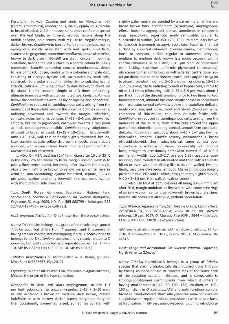
© 2018 Westerdijk Fungal Biodiversity Institute
The genus Tubakia s. lat.
Editor-in-ChiefProf. dr P.W. Crous, Westerdijk Fungal Biodiversity Institute, P.O. Box 85167, 3508 AD Utrecht, The Netherlands.E-mail:[email protected]
89
Description in vivo: Causing leaf spots on Mongolian oak (Quercus mongolica), amphigenous, mainly epiphyllous, circular to broad elliptical, 2–10 mm diam, sometimes confluent, spread over the leaf blade, or forming necrotic lesions along the midrib or veins, pale brown, with regular to irregular margin, darker brown. Conidiomata (pycnothyria) amphigenous, mainly epiphyllous, mostly associated with leaf spots, superficial, scattered to gregarious, sometimes confluent, above all at veins, brown to dark brown, 90–160 µm diam, circular in outline, scutellate, fixed to the leaf surface by a central columella, easily removable. Scutella somewhat convex, membranous, dense to less compact, looser, centre with a colourless or pale disc, consisting of a single hyaline cell, surrounded by small cells, subcircular to angular in outline, giving rise to radiating hyphal strands, cells 4–6 µm wide, brown to dark brown, thick-walled (to about 1 µm), smooth, simple or 1–3 times bifurcating, ultimate branchlets with acute, cornuted tips. Central columella below the scutellum delicate, easily collapsing and ephemeral. Conidiophores reduced to conidiogenous cells, arising from the underside of the scutella, around the upper part of the columella, radiating downward and towards the margin, cylindrical, conical-clavate, fusiform, delicate, 14–22 × 3–5 µm, thin-walled, smooth, hyaline to pigmented, narrowed towards a thin point at neck, conidiogenesis phialidic. Conidia solitary, subglobose, ellipsoid to broad ellipsoid, 13–25 × 10–15 µm, length/width ratio 1.15–1.56, wall thin or finally slightly thickened, hyaline, later sometimes pale yellowish brown, smooth, apex broadly rounded, with a conspicuous basal hilum and prominent frill. Microconidia not observed.
In vitro: On MEA reaching 32–44 mm diam after 10 d at 25 °C in the dark, low velutinous to fuzzy, margin uneven, whitish to pale yellow, centre darker, becoming paler towards the margin, olive brown, light olive brown to yellow, margin white, reverse wrinkled, non-sporulating; hyphae branched, septate, 3.2–4.8 µm wide, hyaline to slightly brownish in mass, some hyphae with short coils on side branches.
Type: South Korea, Gangwon, Seorakasan National Park, Seorak-dong, Sokcho-si, Gangwon-do, on Quercus mongolica, Fagaceae, 31 Aug. 2009, H.Y. Yun (BPI 880799 – holotype; CBS 127490–127493 – ex-type cultures).
Host range and distribution: Only known from the type collection.
Notes: This species belongs to a group of relatively large-spored Tubakia spp., but differs from T. japonica and T. chinensis in having smaller conidia, not overlapping in size. T. seoraksanensis belongs to the T. suttoniana complex and is closely related to T. japonica, but well supported as a separate species (Fig. 3: PP = 1.0, MP-BS = 84 %; Figs 4, 5: PP = 1.0, MP-BS = 94 %).
Tubakia sierrafriensis O. Moreno-Rico & U. Braun, sp. nov. MycoBank MB823667. Figs 20, 21.
Etymology: Named after Sierra Fría, mountain in Aguascalientes, México, the origin of the type collection.
Description in vivo: Leaf spots amphigenous, usually 1–3 per leaf, subcircular to angular-irregular, 2–15 × 2–10 mm, usually ochraceous brown to medium dark brown, margin indefinite or with narrow darker brown margin or marginal line, occasionally somewhat raised, sometimes zonate, with
slightly paler centre surrounded by a darker marginal line and broad brown halo. Conidiomata (pycnothyria) amphigenous, effuse, loose to aggregated, dense, sometimes in concentric rings, punctiform, superficial, easily removable, circular to subcircular in outline, (40–)50–120(–135) µm diam, dark brown to blackish (stereomicroscopy), scutellate, fixed to the leaf surface by a central columella. Scutella convex, membranous, loose to compact, outline regular to somewhat irregular, medium to medium dark brown (stereomicroscopy), with a central colourless or pale disc, 5–15 µm diam or sometimes even lacking, scutellum uniformly pigmented (microscopy), olivaceous to medium brown, or with a darker central zone, 20–60 µm diam, and paler periphery, central cells angular-irregular to almost rounded in outline, 2–10 µm diam, or oblong, 10–15 × 2–7 µm, giving rise to radiating threads of hyphal cells, simple to often 1–3 times bifurcating, cells 4–25 × 2–5 µm, walls about 1 µm thick, tips of the threads simple or once to two times forked, branchlets short, ultimate tips consistently obtuse or sometimes even truncate; central columella below the scutellum delicate, easily collapsing and loose, short and about 25–30 µm wide, composed of thin-walled, colourless or pale fertile cells. Conidiophores reduced to conidiogenous cells, arising from the underside of the scutella, from fertile cells around the upper part of the columella, radiating, conical, ampulliform-cuspidate, delicate, not very conspicuous, about 5–15 × 2–4 µm, hyaline, thin-walled, smooth. Conidia solitary, polymorphous, broad ellipsoid-obovoid, short subcylindrical, some conidia even subglobose or irregular in shape, occasionally with oblique base, straight to occasionally somewhat curved, 9–18 × 5–9 µm (length/width ratio 1.3–2.7, average 1.95), aseptate, apex rounded, base rounded to attenuated and then with a truncate hilum or even with a small peg-like base, thin-walled, hyaline, finally very pale olivaceous, smooth. Microconidia occasionally present in vivo, ellipsoid-fusiform, straight, rarely slightly curved, 5–10 × 2–3.5 µm, thin-walled, hyaline, smooth.
In vitro: On MEA at 22 °C colonies attaining 80–82 mm diam after 20 d, margin undulate, at first white, with concentric rings of aerial mycelium, center green olive with brown hyphal stripes, reverse still colourless after 20 d, without sporulation.
Type: México, Aguascalientes, San José de Gracia, Laguna Seca, 22°10’40.61’’N, 102°38’36.48’’W, 2.662 m alt, on Quercus eduardi, 19 Jan. 2017, O. Moreno-Rico (CFNL 2944 – holotype; CFNL 2944 = CPC 33020 – ex-type culture).
Additional collections examined: ibid., on Quercus eduardi, 21 Apr. 2016, O. Moreno-Rico, HAL 3163 F; 22 Nov. 2016, O. Moreno-Rico, HAL 3173 F.
Hosts range and distribution: On Quercus eduardi, Fagaceae, North America (Mexico).
Notes: Tubakia sierrafriensis belongs to a group of Tubakia species that are morphologically distinguished from T. dryina by having rounded-obtuse to truncate tips of the outer ends of the radiating scutellum threads, and is comparable to Oblongisporothyrium castanopsidis from which it differs in having smaller scutella [(40–)50–120(–135) µm diam, vs. 100–150 µm diam in O. castanopsidis] and polymorphous conidia, broad ellipsoid-obovoid, short subcylindrical, some conidia even subglobose or irregular in shape, occasionally with oblique base, at first hyaline, finally very pale olivaceous (vs. uniformly oblong-
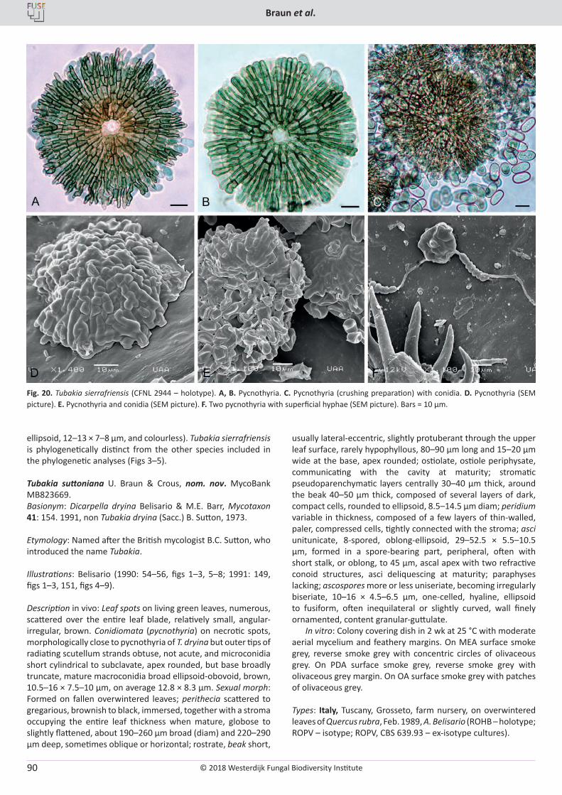
© 2018 Westerdijk Fungal Biodiversity Institute
Braun et al.
Editor-in-ChiefProf. dr P.W. Crous, Westerdijk Fungal Biodiversity Institute, P.O. Box 85167, 3508 AD Utrecht, The Netherlands.E-mail:[email protected]
90
ellipsoid, 12–13 × 7–8 µm, and colourless). Tubakia sierrafriensis is phylogenetically distinct from the other species included in the phylogenetic analyses (Figs 3–5).
Tubakia suttoniana U. Braun & Crous, nom. nov. MycoBank MB823669.Basionym: Dicarpella dryina Belisario & M.E. Barr, Mycotaxon 41: 154. 1991, non Tubakia dryina (Sacc.) B. Sutton, 1973.
Etymology: Named after the British mycologist B.C. Sutton, who introduced the name Tubakia.
Illustrations: Belisario (1990: 54–56, figs 1–3, 5–8; 1991: 149, figs 1–3, 151, figs 4–9).
Description in vivo: Leaf spots on living green leaves, numerous, scattered over the entire leaf blade, relatively small, angular-irregular, brown. Conidiomata (pycnothyria) on necrotic spots, morphologically close to pycnothyria of T. dryina but outer tips of radiating scutellum strands obtuse, not acute, and microconidia short cylindrical to subclavate, apex rounded, but base broadly truncate, mature macroconidia broad ellipsoid-obovoid, brown, 10.5–16 × 7.5–10 µm, on average 12.8 × 8.3 µm. Sexual morph: Formed on fallen overwintered leaves; perithecia scattered to gregarious, brownish to black, immersed, together with a stroma occupying the entire leaf thickness when mature, globose to slightly flattened, about 190–260 µm broad (diam) and 220–290 µm deep, sometimes oblique or horizontal; rostrate, beak short,
usually lateral-eccentric, slightly protuberant through the upper leaf surface, rarely hypophyllous, 80–90 µm long and 15–20 µm wide at the base, apex rounded; ostiolate, ostiole periphysate, communicating with the cavity at maturity; stromatic pseudoparenchymatic layers centrally 30–40 µm thick, around the beak 40–50 µm thick, composed of several layers of dark, compact cells, rounded to ellipsoid, 8.5–14.5 µm diam; peridium variable in thickness, composed of a few layers of thin-walled, paler, compressed cells, tightly connected with the stroma; asci unitunicate, 8-spored, oblong-ellipsoid, 29–52.5 × 5.5–10.5 µm, formed in a spore-bearing part, peripheral, often with short stalk, or oblong, to 45 µm, ascal apex with two refractive conoid structures, asci deliquescing at maturity; paraphyses lacking; ascospores more or less uniseriate, becoming irregularly biseriate, 10–16 × 4.5–6.5 µm, one-celled, hyaline, ellipsoid to fusiform, often inequilateral or slightly curved, wall finely ornamented, content granular-guttulate.
In vitro: Colony covering dish in 2 wk at 25 °C with moderate aerial mycelium and feathery margins. On MEA surface smoke grey, reverse smoke grey with concentric circles of olivaceous grey. On PDA surface smoke grey, reverse smoke grey with olivaceous grey margin. On OA surface smoke grey with patches of olivaceous grey.
Types: Italy, Tuscany, Grosseto, farm nursery, on overwintered leaves of Quercus rubra, Feb. 1989, A. Belisario (ROHB – holotype; ROPV – isotype; ROPV, CBS 639.93 – ex-isotype cultures).
Fig. 20. Tubakia sierrafriensis (CFNL 2944 – holotype). A, B. Pycnothyria. C. Pycnothyria (crushing preparation) with conidia. D. Pycnothyria (SEM picture). E. Pycnothyria and conidia (SEM picture). F. Two pycnothyria with superficial hyphae (SEM picture). Bars = 10 µm.
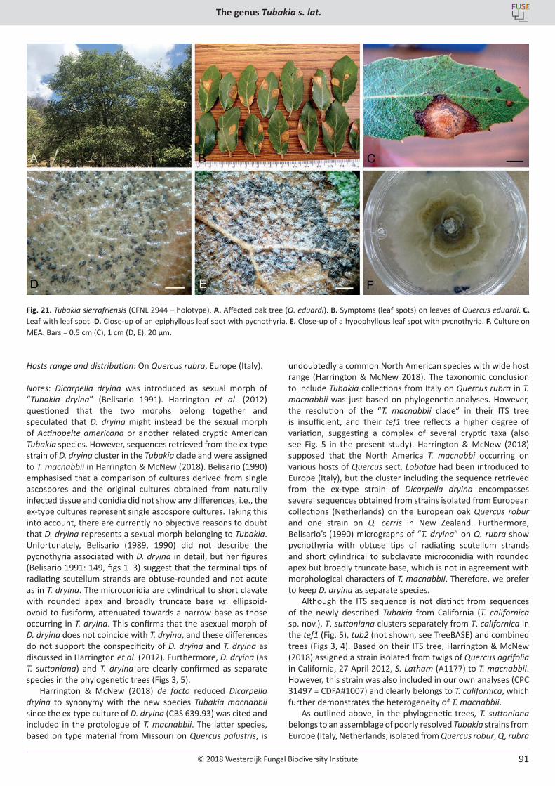
© 2018 Westerdijk Fungal Biodiversity Institute
The genus Tubakia s. lat.
Editor-in-ChiefProf. dr P.W. Crous, Westerdijk Fungal Biodiversity Institute, P.O. Box 85167, 3508 AD Utrecht, The Netherlands.E-mail:[email protected]
91
Hosts range and distribution: On Quercus rubra, Europe (Italy).
Notes: Dicarpella dryina was introduced as sexual morph of “Tubakia dryina” (Belisario 1991). Harrington et al. (2012) questioned that the two morphs belong together and speculated that D. dryina might instead be the sexual morph of Actinopelte americana or another related cryptic American Tubakia species. However, sequences retrieved from the ex-type strain of D. dryina cluster in the Tubakia clade and were assigned to T. macnabbii in Harrington & McNew (2018). Belisario (1990) emphasised that a comparison of cultures derived from single ascospores and the original cultures obtained from naturally infected tissue and conidia did not show any differences, i.e., the ex-type cultures represent single ascospore cultures. Taking this into account, there are currently no objective reasons to doubt that D. dryina represents a sexual morph belonging to Tubakia. Unfortunately, Belisario (1989, 1990) did not describe the pycnothyria associated with D. dryina in detail, but her figures (Belisario 1991: 149, figs 1–3) suggest that the terminal tips of radiating scutellum strands are obtuse-rounded and not acute as in T. dryina. The microconidia are cylindrical to short clavate with rounded apex and broadly truncate base vs. ellipsoid-ovoid to fusiform, attenuated towards a narrow base as those occurring in T. dryina. This confirms that the asexual morph of D. dryina does not coincide with T. dryina, and these differences do not support the conspecificity of D. dryina and T. dryina as discussed in Harrington et al. (2012). Furthermore, D. dryina (as T. suttoniana) and T. dryina are clearly confirmed as separate species in the phylogenetic trees (Figs 3, 5).
Harrington & McNew (2018) de facto reduced Dicarpella dryina to synonymy with the new species Tubakia macnabbii since the ex-type culture of D. dryina (CBS 639.93) was cited and included in the protologue of T. macnabbii. The latter species, based on type material from Missouri on Quercus palustris, is
undoubtedly a common North American species with wide host range (Harrington & McNew 2018). The taxonomic conclusion to include Tubakia collections from Italy on Quercus rubra in T. macnabbii was just based on phylogenetic analyses. However, the resolution of the “T. macnabbii clade” in their ITS tree is insufficient, and their tef1 tree reflects a higher degree of variation, suggesting a complex of several cryptic taxa (also see Fig. 5 in the present study). Harrington & McNew (2018) supposed that the North America T. macnabbi occurring on various hosts of Quercus sect. Lobatae had been introduced to Europe (Italy), but the cluster including the sequence retrieved from the ex-type strain of Dicarpella dryina encompasses several sequences obtained from strains isolated from European collections (Netherlands) on the European oak Quercus robur and one strain on Q. cerris in New Zealand. Furthermore, Belisario’s (1990) micrographs of “T. dryina” on Q. rubra show pycnothyria with obtuse tips of radiating scutellum strands and short cylindrical to subclavate microconidia with rounded apex but broadly truncate base, which is not in agreement with morphological characters of T. macnabbii. Therefore, we prefer to keep D. dryina as separate species.
Although the ITS sequence is not distinct from sequences of the newly described Tubakia from California (T. californica sp. nov.), T. suttoniana clusters separately from T. californica in the tef1 (Fig. 5), tub2 (not shown, see TreeBASE) and combined trees (Figs 3, 4). Based on their ITS tree, Harrington & McNew (2018) assigned a strain isolated from twigs of Quercus agrifolia in California, 27 April 2012, S. Latham (A1177) to T. macnabbii. However, this strain was also included in our own analyses (CPC 31497 = CDFA#1007) and clearly belongs to T. californica, which further demonstrates the heterogeneity of T. macnabbii.
As outlined above, in the phylogenetic trees, T. suttoniana belongs to an assemblage of poorly resolved Tubakia strains from Europe (Italy, Netherlands, isolated from Quercus robur, Q, rubra
Fig. 21. Tubakia sierrafriensis (CFNL 2944 – holotype). A. Affected oak tree (Q. eduardi). B. Symptoms (leaf spots) on leaves of Quercus eduardi. C. Leaf with leaf spot. D. Close-up of an epiphyllous leaf spot with pycnothyria. E. Close-up of a hypophyllous leaf spot with pycnothyria. F. Culture on MEA. Bars = 0.5 cm (C), 1 cm (D, E), 20 µm.
© 2018 Westerdijk Fungal Biodiversity Institute
Braun et al.
Editor-in-ChiefProf. dr P.W. Crous, Westerdijk Fungal Biodiversity Institute, P.O. Box 85167, 3508 AD Utrecht, The Netherlands.E-mail:[email protected]
92
and Quercus sp.) and New Zealand (isolated from Quercus cerris) that neither form a distinct cluster nor a clade, but rather reflect different lineages which might be different species. “Tubakia dryina” formed on leaf spots on Quercus cerris (Belisario 1993) and isolated as endophyte from buds and shoots of Q. cerris (Gennario et al. 2001) was also reported from Italy. Tubakia macnabbii is also part of this complex. The sample numbers are currently too small to resolve this assemblage of some taxa and more isolates are needed. Tubakia suttoniana represents a cryptic species morphologically and is phylogenetically distinct from T. dryina, but currently only known with certainty from its type material. In addition, it forms a complex of closely related species which also includes T. japonica, T. melnikiana, T. seoraksanensis and “Tubakia sp. nov. II” (Figs 3–5).
According to Art. 55.1, the name D. dryina is legitimate and applicable as basionym although published under the illegitimate genus name Dicarpella Syd. & P. Syd. (younger homonym, Art. 53.1). In the type paragraph, Belisario (1990) cited two different collections, one from Grosseto, Tuscany, the other one from Rome, and specified ROHB as herbarium in which the holotype was deposited [as “Belisario (RHOB)”], but without clear indication which of the two cited collections represents the genuine holotype. Due to this imprecise, confusing citation, the name D. dryina would be invalid according to Art. 40.1, 40.6. However, on page 148 it was mentioned that she (Belisario) first found this fungus in the Grosseto farm nursery in February 1989. Later the same fungus was collected on dead leaves in a nursery in Rome. The reference to “Belisario” may be accepted as reference to the first collection from “Grosseto” as holotype so that one can consider D. dryina to be validly published.
Tubakia tiffanyae T.C. Harr. & McNew, Antonie van Leeuwenhoek J. Microbiol. Serol. doi.org/10.1007/s10482-017-1001-9 [13]. 2017.
Illustrations: Harrington & McNew (2018, figs 1h–m).
Description in vivo: Conidiomata (pycnothyria) superficial, hypophyllous or epiphyllous, on circular leaf spots or along necrotic leaf veins. Scutella radiate, (40–)60–150 µm diam, composed of a series of dark brown, thick-walled cells originating from a central cell, ending in blunt to acute tips. Sporodochia with no scutella or poorly developed scutella epiphyllous or hypophyllous on necrotic vein tissue, light to dark brown due to masses of conidia. Conidiophores on underside of scutella or on sporodochia. Conidia hyaline, turning light brown with age, smooth to slightly varicose, aseptate, obovoid to ovoid (9–)10–15.5 × (8–)8.5–11.5(–12) µm (mean 12.9 × 9.6 µm). Microconidia sterile, hyaline, aseptate, fusiform 3–8 × 1–2.5 µm may develop from small pycnothyria, alone or along with macroconidia. Crustose conidiomata developing in late summer to fall on underside of necrotic veins and found on overwintering leaves still hanging from twigs, erumpent, brown to black, irregularly shaped, 80–580 µm diam, single to grouped, covered with dark, thick-walled cells that break open in fissures due to swelling when wet. Conidia hyaline to brown, ellipsoidal to obovate to irregularly shaped, aseptate, (11–)13–18(–20) × (6–)7–9.5 µm (mean 15.2 × 7.9 µm).
In vitro: On MYEA with optimal growth at 25 C, 40–58 mm diam after 7 d, creamy white aerial mycelium, smooth to scalloped at edge, developing concentric rings of dense mycelium, underside golden yellow to slightly darker with age,
sometimes with dark cell masses on surface or subsurface. Conidiophores rare to abundant, short, hyaline, sometimes aggregated (sporodochia) on agar surface. Conidia hyaline to dark brown, thick-walled, smooth to slightly varicose, aseptate, obovoid to ellipsoidal, 10–16 × 7–11 µm.
Type: USA, Iowa, Ames, N42° 04’ 4’’ W93° 65’ 22’’, on leaf of Quercus rubra, 5 Sep. 2009, T. Harrington (ISC 453296 – holotype; ex-type strains CBS 137345 = A803).
Host range and distribution: On Quercus (ellipsoidalis, imbricaria, rubra), Fagaceae, North America (USA, Iowa, Minnesota).
Notes: This species was recently introduced by Harrington & McNew (2018) for Tubakia sp. C (Harrington & McNew 2016) which is closely allied to T. macnabbii but genetically separated (Fig. 5) and morphologically distinguished by having wider conidia and causing characteristic circular leaf spots with light-coloured centres (see Harrington & McNew 2018: fig 1h) in addition to the vein necrosis.
Excluded, doubtful and insufficiently known species
Actinopelte acnisti (Syd.) Toro, Bol. Acad. Ci. Fís. 2(7): 217. 1935, nom. inval. (Art. 36.1, c).Basionym: Calopeltis acnisti Syd., Ann. Mycol. 23: 393. 1915.
Notes: Calopeltis is a recognised genus belonging to the Microthyriaceae. Syntypes of this species are distributed in several herbaria and include “Syd., Fungi Exot. Exs. 689” (BPI 645025–6425029, CUP, ILL 10525–10528, LSU 156932, MICH 13880, S-F10919,10920, 194870, 194872, 194873).
Actinopelte psychotriae I. Hino & Katum., Bull. Fac. Agric. Yamaguchi Univ. 15: 506. 1964.
Notes: This species, described by Hino & Katumoto (1964) on leaves of Psychotria serpens from Japan, is unrelated to Tubakia. In the original description and illustration (Hino & Katumoto 1964: 506, fig. 1), this species was described to form pycnothyria with scattered globose ostiolate locules and small “pycnospores” (6–8 × 2.5–3 µm), which is quite distinct from pycnothyria of Tubakia.
Actinopelte stellata M.L. Farr, Mycopathol. Mycol. Appl. 31: 63. 1967.Synonym: Tubakia stellata (M.L. Farr) T.C. Harr. & McNew, Antonie van Leeuwenhoek J. Microbiol. Serol. doi.org/10.1007/s10482-017-1001-9 [16]. 2017.
Notes: The generic affinity of this species is unclear. Cultures and results of molecular sequence analyses are not yet available. This species is only known from its type collection (Brazil, Para, Belem, on leaves of Byrsonima coriacea, 3 Feb. 1963, F.C. Albuquerque, BPI 391941) and has not yet been revised and reassessed. However, the described and illustrated characteristics of A. stellata do not fit into the generic concept of Tubakia. Farr (1967) described a basal membrane, unknown in true Tubakia species, a one-celled columella, and conidiogenous cells arising from the underside of the scutellum. In Tubakia spp., the columella is composed of several cells or a single cell surrounded by small, fertile, parenchymatous cells giving rise to conidiogenous cells,
© 2018 Westerdijk Fungal Biodiversity Institute
The genus Tubakia s. lat.
Editor-in-ChiefProf. dr P.W. Crous, Westerdijk Fungal Biodiversity Institute, P.O. Box 85167, 3508 AD Utrecht, The Netherlands.E-mail:[email protected]
93
above all around the point of attachment of the columella at the scutellum and peripherally. Tubakia species are usually confined to fagaceous hosts in the northern hemisphere. The reallocation of A. stellata to Tubakia by Harrington & McNew (2018) was just a formal act, neither supported by culture and sequence data nor accompanied by a critical discussion of the morphology of this species.
Actinothyrium gloeosporioides Tehon, Mycologia 16: 136. 1924.Synonym: Tubakia gloeosporioides (Tehon) T.C. Harr. & McNew, Antonie van Leeuwenhoek J. Microbiol. Serol. doi.org/10.1007/s10482-017-1001-9 [15]. 2017.
Holotype: USA: Illinois, Franklin County, Christopher, on Sassafras albidum, Lauraceae, 20 Jul. 1922, P. A. Young 3311 (ILLS 2972).
Notes: Harrington & McNew (2018) examined type material of A. gloeosporioides and additional specimens on Sassafras albidum from Illinois (ILLS 2972, 3671, 29748, 17547) and New Jersey (as Leptothyrium dryinum f. sassafras). The pycnothyria were 45–132 µm diam (reported as 50–95 µm diam by Tehon 1924) with conidia 9.5–12.5 × 7–8.5 µm (reported as 11–12 × 6–7.5 µm in Tehon 1924), which are similar to pycnothyria and conidia of T. dryina s. lat. Harrington & McNew (2018) compared it with T. macnabbii and commented that the two species are indistinguishable without cultures or DNA. They introduced the new combination T. gloeosporioides just based on the assumption that the latter taxon represents a species of its own since it was described on a lauraceous host. However, it should be noted that an ITS sequence retrieved from leaves of Lindera glauca (Lauraceae) in China clusters with T. dryinoides. Hence, the status and true affinity of A. gloeosporioides remain unclear and unresolved pending cultures and results of molecular sequence analyses.
Leptothyrium castaneicola Ellis & Everh., J. Mycol. 4: 137. 1888.
Type: USA, New Jersey, on Castanea sativa [vesca], 20 Oct. 1888, without collector (NY 927812 – holotype).
Notes: This species has usually been considered a synonym of Tubakia dryina. Type material has been re-examined. Pycnothyria formed on leaf spots on Castanea sativa are morphologically barely distinguishable from those of T. dryina and allied North American species with pointed tips of radiating scutellum strands. This species may be a synonym of one of the North American Tubakia species, but it can also not be excluded that an additional cryptic species on sweet chestnut being involved, but cultures and sequence data are not yet available to be able to answer this question.
Pirostoma nyssae Tehon, Mycologia 16: 137. 1924. Synonym: Tubakia nyssae (Tehon) T.C. Harr. & McNew, Antonie van Leeuwenhoek J. Microbiol. Serol. doi.org/10.1007/s10482-017-1001-9 [16]. 2017.
Holotype: USA, Illinois, Johnson County, Tunnel Hill, on Nyssa sylvatica, 25 Jul. 1922, P. A. Young 3665 (ILLS 2940).
Notes: Collections of Pirostoma nyssae have been examined (Nyssa sylvatica, BPI 391889–391891). Pycnothyria (65–110 µm diam, tips of the radiating scutellum strands pointed) are barely
distinguishable from those of T. dryina, so that it is not surprising that P. nyssae has previously usually been considered a synonym of the latter species. Harrington & McNew (2018) compared P. nyssae rather with T. macnabbii, which is, however, genetically totally heterogeneous and undoubtedly composed of several cryptic taxa. Without any cultures and sequence data, the status and affinity of P. nyssae remain quite unclear. The reallocation to Tubakia was just based on the occurrence of this species on Nyssa sylvatica (Nyssaceae), i.e., on a non-fagaceous host, which is, however, insufficient for a final conclusion and reassessment.
Tubakia sp. (see Braun et al. 2014: 25)
Leaf spots amphigenous, subcircular to angular-irregular, 1–7 mm diam, sometimes oblong, up to 10 µm, medium brown on the upper leaf surface, paler below, finally with pale centre, pale brownish to ochraceous, margin indefinite or narrow and somewhat darker, occasionally surrounded by a narrow diffuse halo, yellowish to yellow-green. Conidiomata (pycnothyria) epiphyllous, up to about 15 per leaf spot, punctiform, scutellate, blackish. Scutella convex, 40–80 µm diam, membranous, barely translucent, with a central paler disc, 8–15 µm diam, giving rise to radiating hyphae, cells 4–15 × 2–5 µm, peripheral cells mostly somewhat broadening towards the margin, medium brown, thick-walled (–1 µm), smooth, up to three times bifurcating, either only at the periphery or deeply cleft, peripheral bifurcations mostly shallow, branchlets with obtuse to truncate tips. Central columella below the scutellum delicate, easily collapsing and ephemeral. Conidiophores reduced to conidiogenous cells, arising from the underside of scutella around the columella, radiating downward and towards margin. Conidia solitary, globose to subglobose, 9–11 × 7–9 µm, length/width ratio 1–1.2, wall thin, 0.3–0.8 µm wide, hyaline or subhyaline, very pale greenish or faintly olivaceous, smooth, apex and base broadly rounded, basal hilum inconspicuous or with minute, not very conspicuous, delicate frill or peg. Microconidia not observed.
Material examined: China, Jiangxi Province, Xingangshan, subtropical forest site of the BEF-China Project, 29.1250° N, 117.9085° E, on living leaves of Castanea henryi, Fagaceae, 8 Sep. 2013, S. Bien, HAL 2675 F.
Notes: Tubakia sp. from Castanea henryi in China belongs to an undescribed species. Cultures and results of sequence analyses are not available, and the material deposited at HAL is too meagre for a formal description of a new species. The Chinese fungus on Castanea henryi is morphologically close to, and possibly identical with, Saprothyrium thailandensis, but unproven due to lack of cultures and sequence data for comparison. The ecology of the Chinese collection on Castanea henryi is unclear. The pycnothyria were found on leaf spots of living leaves, but lesions of several fungi, including Tubakia chinensis, were developed. Thus, it cannot be excluded that the Chinese Tubakia on Castanea henryi with small pycnothyria was formed on necrotic spots caused by other fungi.
Tubakia sp. (see Zahedi et al. 2011: 67, fig. 3).
Notes: Zahedi et al. (2011) reported and illustrated “Tubakia dryina” on Quercus castaneifolia from North Iran (Guilan Province). This fungus is quite distinct from true T. dryina (s. str.) and readily distinguishable by its small pycnothyria, about 60 µm diam, blunt tips of radiating scutellum strands, and broad
© 2018 Westerdijk Fungal Biodiversity Institute
Braun et al.
Editor-in-ChiefProf. dr P.W. Crous, Westerdijk Fungal Biodiversity Institute, P.O. Box 85167, 3508 AD Utrecht, The Netherlands.E-mail:[email protected]
94
ellipsoid-ovoid, colourless or pale conidia, about 7–10 × 5–7 µm. Shape and size of the pycnothyria are reminiscent of those of Tubakia sp. on Castanea henryi in China (Braun et al. 2014: 25) and Saprothyrium thailandense, which differ, however, in having globose to subglobose conidia. There is no Tubakia species
with comparable pycnothyria. Therefore, the collection on Q. castaneifolia from Iran represents probably an undescribed species, but without cultures and sequence data the generic affinity remains unclear.
Key to species of Tubakia and allied genera (Tubakiaceae) based on conidiomatal characters
1. Pycnothyria with characteristically radiating scutella not developed; only acervuloid, crustose, pycnidioid, stromatic black conidiomata formed, (50–)80–200(–220) µm diam; on fagaceous hosts in California or Syzygium cumini (Myrtaceae) in Thailand ..................................................................................................................................................................................... 2
1* Pycnothyria developed; sporodochial and crustose, pycnidioid, stromatic conidiomata may be developed in addition to pycnothyria or may be absent ................................................................................................................................................... 3
2. Conidiomata crustose, pycnidioid, stromatic, black, dehiscing by irregular fissures, formed on petioles and leaf blades of necrotic, brown leaves, often close to veins, dry, brown leaves with conidioma from the previous seasons’ growth remain attached to many branches of affected trees until spring; conidiophores aseptate, unbranched; conidia broad ellipsoid, ellipsoid-ovoid to short and broad subcylindrical, rarely irregular in shape, 8–15 × 4.5–7 µm, length/width ratio 1.4–2.3 (on average 1.8), thin-walled, at first subhyaline to pale greenish, later greenish, pale olivaceous to brownish; on Chrysolepis chrysophylla (≡ Castanopsis chrysophylla), Notholithocarpus densiflorus (≡ Lithocarpus densiflorus), and various Quercus spp., North America, California (and perhaps Mexico) ........................................................................................... Tubakia californica
2* Conidiomata acervuloid, basistromatic; conidiophores septate, branched; 9–15 × 5–6 μm, light brown to olivaceous brown, thick-walled; on dead leaves of Syzygium cumini, Thailand .................................................................. Racheliella saprophytica
3. Conidia large, length on average > 25 µm .................................................................................................................................. 43* Conidia smaller, length on average < 25 µm .............................................................................................................................. 5
4. Conidia very large, 40–55 × 35–45 µm; microconidia present, 5–10 × 1–2 µm, formed in smaller conidiomata; on Castanea crenata, C. mollissima and Quercus acutissima, Q. aliena, Asia (China, Japan, Korea) ..................................... Tubakia japonica
4* Conidia (20–)25–40 × 20–30 µm; microconidia not formed on Castanea henryi ............................................ Tubakia chinensis
5(3) Scutella relatively small, 60–100 µm diam, margin continuous, compact, more or less undulate and distinctly involute; conidia broad ellipsoid-ovoid, 12–15 × 10–13 µm, at first hyaline, later pale yellowish brown to light orange yellow; microconidia formed, bacilliform, 8–10 × 1 µm; on Quercus phillyreoides, Q. serrata, Japan, Korea .......................... Involutiscutellula rubra
5* Scutella larger, up to 160 µm diam and/or conidia narrower (width < 10 µm), margin of the scutellum different, loose, not distinctly involved, tips either pointed or obtuse-truncate; microconidia either much broader or not developed .................. 6
6. Outer tips of the bifurcating hyphal scutellum strands obtuse, rounded or even more or less truncate, but not pointed ....... 76* Outer tips of the bifurcating hyphal scutellum strands always or predominantly pointed (or at least all or most ultimate
branchlets distinctly attenuated towards an obtuse to pointed tip) ........................................................................................ 15
7. Conidia globose or subglobose to broad ellipsoid-obovoid ....................................................................................................... 87* Conidia not consistently globose-subglobose, either consistently broad ellipsoid-obovoid or oblong to oblong-ellipsoid or
polymorphous, broad ellipsoid-obovoid, short subcylindrical, some conidia even subglobose or irregular in shape, occasionally with oblique base ..................................................................................................................................................................... 10
8. Scutella larger, 80–120 µm diam; conidia globose, subglobose to broad ellipsoid-obovoid, small conidia 7–10 × 5–8 µm, larger fully developed conidia 9–16(–19) × (7–)9–12 µm, length/width ratio 1.0–1.5(–1.6), on average 1.27, hyaline to pale greenish or olivaceous; on Quercus eduardi, Mexico ............................................................................ Sphaerosporithyrium mexicanum
8* Scutella small, 40–80 µm diam; conidia subglobose, mature conidia narrower, 7–9 µm, hyaline or subhyaline ...................... 9
9. Isolated as saprobic fungus from an unidentified leaf in Thailand .................................................. Saprothyrium thailandense9* On leaf spots on Castanea henryi, China ................................................................................................................... Tubakia sp.
10(7*) Conidia polymorphous, broad ellipsoid-obovoid, short subcylindrical, some conidia even subglobose or irregular in shape, occasionally with oblique base, 9–18 × 5–9 µm, at first hyaline, finally very pale olivaceous; on Quercus eduardi, Mexico .................................................................................................................................................................... Tubakia sierrafriensis
10* Conidia uniform, not polymorphous, not or barely curved; in Africa, Asia or Europe ............................................................. 11
11. Conidia hyaline ......................................................................................................................................................................... 1211* Conidia pigmented, at least when mature ............................................................................................................................... 13
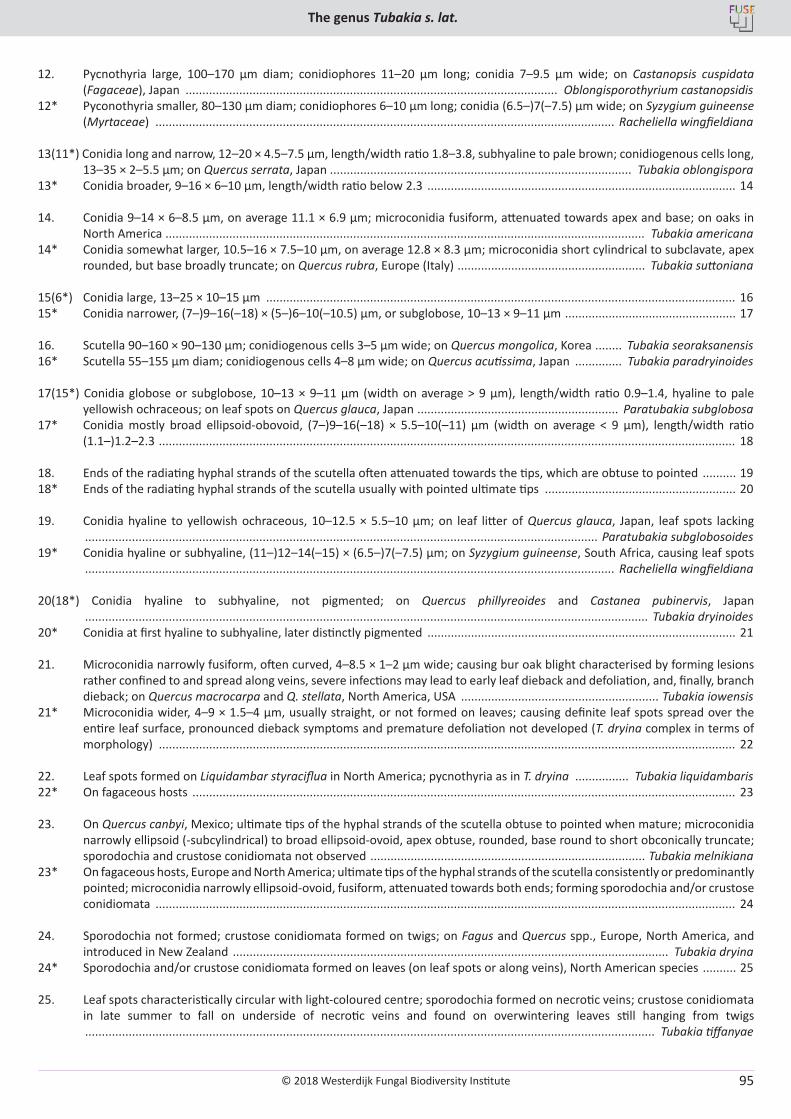
© 2018 Westerdijk Fungal Biodiversity Institute
The genus Tubakia s. lat.
Editor-in-ChiefProf. dr P.W. Crous, Westerdijk Fungal Biodiversity Institute, P.O. Box 85167, 3508 AD Utrecht, The Netherlands.E-mail:[email protected]
95
12. Pycnothyria large, 100–170 µm diam; conidiophores 11–20 µm long; conidia 7–9.5 µm wide; on Castanopsis cuspidata (Fagaceae), Japan ............................................................................................................... Oblongisporothyrium castanopsidis
12* Pyconothyria smaller, 80–130 µm diam; conidiophores 6–10 µm long; conidia (6.5–)7(–7.5) µm wide; on Syzygium guineense (Myrtaceae) ......................................................................................................................................... Racheliella wingfieldiana
13(11*) Conidia long and narrow, 12–20 × 4.5–7.5 µm, length/width ratio 1.8–3.8, subhyaline to pale brown; conidiogenous cells long, 13–35 × 2–5.5 µm; on Quercus serrata, Japan .......................................................................................... Tubakia oblongispora
13* Conidia broader, 9–16 × 6–10 µm, length/width ratio below 2.3 ............................................................................................ 14
14. Conidia 9–14 × 6–8.5 µm, on average 11.1 × 6.9 µm; microconidia fusiform, attenuated towards apex and base; on oaks in North America ............................................................................................................................................... Tubakia americana
14* Conidia somewhat larger, 10.5–16 × 7.5–10 µm, on average 12.8 × 8.3 µm; microconidia short cylindrical to subclavate, apex rounded, but base broadly truncate; on Quercus rubra, Europe (Italy) ........................................................ Tubakia suttoniana
15(6*) Conidia large, 13–25 × 10–15 µm ............................................................................................................................................ 1615* Conidia narrower, (7–)9–16(–18) × (5–)6–10(–10.5) µm, or subglobose, 10–13 × 9–11 µm ................................................... 17
16. Scutella 90–160 × 90–130 µm; conidiogenous cells 3–5 µm wide; on Quercus mongolica, Korea ........ Tubakia seoraksanensis16* Scutella 55–155 µm diam; conidiogenous cells 4–8 µm wide; on Quercus acutissima, Japan .............. Tubakia paradryinoides
17(15*) Conidia globose or subglobose, 10–13 × 9–11 µm (width on average > 9 µm), length/width ratio 0.9–1.4, hyaline to pale yellowish ochraceous; on leaf spots on Quercus glauca, Japan ............................................................ Paratubakia subglobosa
17* Conidia mostly broad ellipsoid-obovoid, (7–)9–16(–18) × 5.5–10(–11) µm (width on average < 9 µm), length/width ratio (1.1–)1.2–2.3 ............................................................................................................................................................................ 18
18. Ends of the radiating hyphal strands of the scutella often attenuated towards the tips, which are obtuse to pointed .......... 1918* Ends of the radiating hyphal strands of the scutella usually with pointed ultimate tips ......................................................... 20
19. Conidia hyaline to yellowish ochraceous, 10–12.5 × 5.5–10 µm; on leaf litter of Quercus glauca, Japan, leaf spots lacking ......................................................................................................................................................... Paratubakia subglobosoides
19* Conidia hyaline or subhyaline, (11–)12–14(–15) × (6.5–)7(–7.5) µm; on Syzygium guineense, South Africa, causing leaf spots .............................................................................................................................................................. Racheliella wingfieldiana
20(18*) Conidia hyaline to subhyaline, not pigmented; on Quercus phillyreoides and Castanea pubinervis, Japan ........................................................................................................................................................................ Tubakia dryinoides
20* Conidia at first hyaline to subhyaline, later distinctly pigmented ............................................................................................ 21
21. Microconidia narrowly fusiform, often curved, 4–8.5 × 1–2 µm wide; causing bur oak blight characterised by forming lesions rather confined to and spread along veins, severe infections may lead to early leaf dieback and defoliation, and, finally, branch dieback; on Quercus macrocarpa and Q. stellata, North America, USA ........................................................... Tubakia iowensis
21* Microconidia wider, 4–9 × 1.5–4 µm, usually straight, or not formed on leaves; causing definite leaf spots spread over the entire leaf surface, pronounced dieback symptoms and premature defoliation not developed (T. dryina complex in terms of morphology) ............................................................................................................................................................................ 22
22. Leaf spots formed on Liquidambar styraciflua in North America; pycnothyria as in T. dryina ................ Tubakia liquidambaris22* On fagaceous hosts .................................................................................................................................................................. 23
23. On Quercus canbyi, Mexico; ultimate tips of the hyphal strands of the scutella obtuse to pointed when mature; microconidia narrowly ellipsoid (-subcylindrical) to broad ellipsoid-ovoid, apex obtuse, rounded, base round to short obconically truncate; sporodochia and crustose conidiomata not observed .................................................................................. Tubakia melnikiana
23* On fagaceous hosts, Europe and North America; ultimate tips of the hyphal strands of the scutella consistently or predominantly pointed; microconidia narrowly ellipsoid-ovoid, fusiform, attenuated towards both ends; forming sporodochia and/or crustose conidiomata ............................................................................................................................................................................. 24
24. Sporodochia not formed; crustose conidiomata formed on twigs; on Fagus and Quercus spp., Europe, North America, and introduced in New Zealand .................................................................................................................................. Tubakia dryina
24* Sporodochia and/or crustose conidiomata formed on leaves (on leaf spots or along veins), North American species .......... 25
25. Leaf spots characteristically circular with light-coloured centre; sporodochia formed on necrotic veins; crustose conidiomata in late summer to fall on underside of necrotic veins and found on overwintering leaves still hanging from twigs .......................................................................................................................................................................... Tubakia tiffanyae
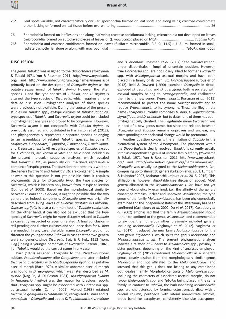
© 2018 Westerdijk Fungal Biodiversity Institute
Braun et al.
Editor-in-ChiefProf. dr P.W. Crous, Westerdijk Fungal Biodiversity Institute, P.O. Box 85167, 3508 AD Utrecht, The Netherlands.E-mail:[email protected]
96
25* Leaf spots variable, not characteristically circular; sporodochia formed on leaf spots and along veins; crustose conidiomata either lacking or formed on leaf tissue before overwintering ................................................................................................. 26
26. Sporodochia formed on leaf lesions and along leaf veins; crustose conidiomata lacking; microconidia not developed on leaves (microconidia formed on autoclaved pieces of leaves of Q. macrocarpa placed on MEA) ..................................... Tubakia hallii
26* Sporodochia and crustose conidiomata formed on leaves (fusiform microconidia, 3.5–9(–11.5) × 1–3 µm, formed in small, radiate pycnothyria, alone or along with macroconidia) ............................................................................... Tubakia macnabbii
DISCUSSION
The genus Tubakia was assigned to the Diaporthales (Yokoyama & Tubaki 1971, Yun & Rossman 2011, http://www.mycobank.org/ and http://www.indexfungorum.org/names/names.asp) primarily based on the description of Dicarpella dryina as the putative sexual morph of Tubakia dryina. However, the latter species is not the type species of Tubakia, and D. dryina is also not the type species of Dicarpella, which requires a more detailed discussion. Phylogenetic analyses of these species were previously not available. During the course of the present studies on Tubakia spp., ex-type cultures of Tubakia japonica, type species of Tubakia, and Dicarpella dryina could be included in phylogenetic analyses and proved to be congeneric. However, Dicarpella dryina is not conspecific with Tubakia dryina, as previously assumed and postulated in Harrington et al. (2012), and phylogenetically represents a separate species belonging to an assemblage of related Tubakia species, including T. californica, T. dryinoides, T. japonica, T. macnabbii, T. melnikiana, and T. seoraksanensis. All recognised species of Tubakia, except for T. chinensis, are known in vitro and have been included in the present molecular sequence analyses, which revealed that Tubakia s. lat., as previously circumscribed, represents a complex of cryptic genera. The question that remains is whether the genera Dicarpella and Tubakia s. str. are congeneric. A simple answer to this question is not yet possible since it requires phylogenetic data for Dicarpella bina, the type species of Dicarpella, which is hitherto only known from its type collection (Sognov et al. 2008). Based on the morphological similarity between D. bina and D. dryina, it might be possible that the two genera are, indeed, congeneric. Dicarpella bina was originally described from living leaves of Quercus agrifolia in California. Quercus agrifolia is also a common host of Tubakia californica. On the other hand, it can also not be excluded that the type species of Dicarpella might be more distantly related to Tubakia as currently suspected or even unrelated. A final conclusion is still pending and further cultures and sequence data for D. bina are needed. In any case, the older name Dicarpella would not threaten the younger name Tubakia in case that the two genera were congeneric, since Dicarpella Syd. & P. Syd., 1913 (nom. illeg.) being a younger homonym of Dicarpella Sitzenb., 1861, i.e., Tubakia would be the correct name in any case.
Barr (1978) assigned Dicarpella to the Pseudovalsaceae subfam. Pseudovalsoideae tribe Ditopelleae, and later included Dicarpella quercifolia with Mastigosporella hyalina as putative asexual morph (Barr 1979). A similar unnamed asexual morph was found in D. georgiana, which was later described as M. nyssae (Nag Raj & Di Cosmo 1981). Mastigosporella hyalina (≡ Harknessia hyalina) was the reason for previous reports that Dicarpella spp. might be associated with Harknessia spp. as asexual morphs (Cannon 2001). Monod (1983) retained Dicarpella georgiana in Gnomoniella, recognised D. bina and D. quercifolia in Dicarpella, and added D. liquidambaris-styracifluae
and D. orientalis. Rossman et al. (2007) cited Harknessia spp. under diaporthalean fungi of uncertain position. However, true Harknessia spp. are not closely allied to former Dicarpella spp. with Mastigosporella asexual morphs and have been placed in a family of its own, viz. Harknessiaceae (Crous et al. 2012). Reid & Dowsett (1990) examined Dicarpella in detail, excluded D. georgiana and D. quercifolia, both associated with asexual morphs belong to Mastigosporella, and reallocated them to the new genus, Wuestneiopsis. Rossman et al. (2015) recommended to protect the name Mastigosporella and to reduce Wuestneiopsis to its synonymy. Thus, the illegitimate name Dicarpella currently comprises D. bina, D. liquidambaris-styracifluae, and D. orientalis, but to date none of them has been phylogenetically clarified. The illegitimate name Dicarpella was in need of a new genus name, but since the relation between Dicarpella and Tubakia remains unproven and unclear, any corresponding nomenclatural change would be premature.
Another question concerns the affiliation of Tubakia in the hierarchical system of the Ascomycota. The placement within the Diaporthales is clearly resolved. Tubakia is currently usually listed as diaporthalean genus of unclear family affinity (Yokoyama & Tubaki 1971, Yun & Rossman 2011, http://www.mycobank.org/ and http://www.indexfungorum.org/names/names.asp). Dicarpella was usually assigned to the Melanconidaceae s. lat., comprising up to almost 30 genera (Eriksson et al. 2001, Lumbsch & Huhndorf 2007, Maharachchikumbura et al. 2015, 2016). This affiliation is, however, disputable (see discussion above). Most genera allocated to the Melanconidaceae s. lat. have not yet been phylogenetically examined, i.e., the affinity of the genera concerned remains unclear and unproven. Melanconis, the type genus of the family Melanconidaceae, has been phylogenetically examined and the independent status of the latter family has been confirmed (Castlebury et al. 2002, Du et al. 2017). Castlebury et al. (2002) emphasised that the family Melanconidaceae should rather be confined to the genus Melanconis, and recommended to exclude the numerous other previously included genera, including Melanconiella (Voglmayr et al. 2012). Voglmayr et al. (2017) introduced the new family Juglanconidaceae for the new genus Juglanconis, which splits the genus Melanconis and Melanconidaceae s. lat. The present phylogenetic analyses indicate a relation of Tubakia to Melanconiella spp., possibly in sister positions, depending on the kind of analyses employed. Voglmayr et al. (2012) confirmed Melanconiella as a separate genus, clearly distinct from the morphologically similar genus Melanconis and not affiliated to the Melanconidaceae, and stressed that this genus does not belong to any established dothidealean family. Morphological traits of Melanconiella spp., including the characters of associated asexual morphs, do not favour Melanconiella spp. and Tubakia being placed in the same family. In contrast to Tubakia, the bark-inhabiting Melanconiella spp. are characterised by forming ectostromatic discs with a central column, perithecia with lateral non-rostrate ostioles, broad band-like paraphyses, consistently bicellular ascospores,
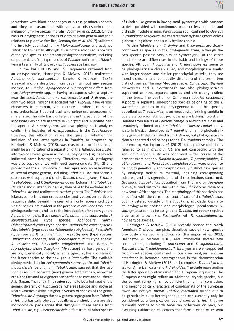
© 2018 Westerdijk Fungal Biodiversity Institute
The genus Tubakia s. lat.
Editor-in-ChiefProf. dr P.W. Crous, Westerdijk Fungal Biodiversity Institute, P.O. Box 85167, 3508 AD Utrecht, The Netherlands.E-mail:[email protected]
97
sometimes with blunt appendages or a thin gelatinous sheath, and they are associated with acervular discosporina- and melanconium-like asexual morphs (Voglmayr et al. 2012). On the basis of phylogenetic analyses of dothidealean genera and their relations to putative families, Senanayake et al. (2017) validated the invalidly published family Melanconiellaceae and assigned Tubakia to this family, although it was not based on sequence data of the type species. The present phylogenetic analyses, including sequence data of the type species of Tubakia confirm that Tubakia warrants a family of its own, viz., Tubakiaceae fam. nov.
On the basis of ITS and LSU sequences retrieved from an ex-type strain, Harrington & McNew (2018) reallocated Apiognomonia supraseptata (Kaneko & Kobayashi 1984), a sexual morph described from Japan without any asexual morphs, to Tubakia. Apiognomonia supraseptata differs from true Apiognomonia spp. in having ascospores with a septum near the apex. Apiognomonia supraseptata and D. dryina, the only two sexual morphs associated with Tubakia, have various characters in common, viz., rostrate perithecia of similar size, unitunicate 8-spored asci, and colourless ascospores of similar size. The only basic difference is in the septation of the ascospores which are aseptate in D. dryina and 1-septate near the apex in A. supraseptata. Our own phylogenetic analyses confirm the inclusion of A. supraseptata in the Tubakiaceae. However, this allocation raises the question whether the inclusion of the latter species in Tubakia, as proposed by Harrington & McNew (2018), was reasonable, or if this result might be an indication of a separation of the Tubakiaceae cluster into two or several genera as the LSU phylogeny (Fig. 1) already indicated some heterogeneity. Therefore, the LSU phylogeny was also supplemented with rpb2 sequence data (Fig. 2) and proved that the Tubakiaceae cluster represents an assemblage of several cryptic genera, including Tubakia s. str. that forms a separate, well-supported clade. Tubakia castanopsidis, T. rubra, T. subglobosa, and T. thailandensis do not belong in the Tubakia s. str. clade and cluster outside, i.e., they have to be excluded from Tubakia s. str. and reallocated to other genera. The Tubakia clade is large, comprising numerous species, and is based on multigene sequence data. Several lineages, often only represented by a single species, are evident in the portions of excluded taxa in the phylogenetic trees, and led to the introduction of the new genera Apiognomonioides (type species: Apiognomonia supraseptata), Involutiscutellula (type species: Actinopelte rubra), Oblongisporothyrium (type species: Actinopelte castanopsidis), Paratubakia (type species: Actinopelte subglobosa), Racheliella (type species: R. wingfieldiana), Saprothyrium (type species: Tubakia thailandensis) and Sphaerosporithyrium (type species S. mexicanum). Racheliella wingfieldiana and Greeneria saprophytica share Syzygium (Myrtaceae) as host genus and are phylogenetically closely allied, suggesting the allocation of the latter species to the new genus Racheliella. The available phylogenetic data for Apiognomonia supraseptata and Tubakia thailandensis, belonging in Tubakiaceae, suggest that the two species require separate (new) genera. Interestingly, almost all excluded taxa and new genera are confined to east and southeast Asia (Japan, Thailand). This region seems to be a hot spot of the generic diversity of Tubakiaceae, whereas Europe and above all North America exhibit a higher diversity of species of the genus Tubakia s. str. Although the new genera segregated from Tubakia s. lat. are basically phylogenetically established, there are also morphological peculiarities that distinguish these genera from Tubakia s. str., e.g., Involutiscutellula differs from all other species
of tubakia-like genera in having small pycnothyria with compact scutella provided with continuous, more or less undulate and distinctly involute margin. Paratubakia spp., confined to Quercus (Cyclobalanopsis) glauca, are characterised by having more or less globose-subglobose and usually hyaline conidia.
Within Tubakia s. str., T. dryina and T. iowensis, are clearly confirmed as species in the phylogenetic trees, although the two species possess very similar pycnothyria. On the other hand, there are differences in the habit and biology of these species. Although T. japonica and T. seoraksanensis seem to be phylogenetically closely allied, and morphologically similar with larger spores and similar pycnothyrial scutella, they are morphologically and genetically distinct and represent two distinct species. The new Mexican species Sphaerosporithyrium mexicanum and T. sierrafriensis are also phylogenetically supported as new, separate species and are clearly distinct in the trees. The position of Californian Tubakia sequences supports a separate, undescribed species belonging to the T. suttoniana complex in the phylogenetic trees. This species, described as T. californica, is an endophyte, forming conidia in pustulate conidiomata, but pycnothyria are lacking. Two strains isolated from leaves of Quercus canbyi in Mexico are close and tentatively included. Another Tubakia on Quercus canbyi and Q. laeta in Mexico, described as T. melnikiana, is morphologically only gradually distinguished from T. dryina, but phylogenetically clearly separated and belongs to the T. suttoniana complex. The inference by Harrington et al. (2012) that Japanese collections referred to as T. dryina s. lat. are not conspecific with the genuine T. dryina s. str. was confirmed in the course of the present examinations. Tubakia dryinoides, T. paradryinoides, T. oblongispora, and Paratubakia subglobosoides were proven to belong to genetically and morphologically distinct new species by analysing herbarium material, including corresponding cultures, and phylogenetic data of the collections concerned. Greeneria saprophytica, described from Thailand on Syzygium cumini, turned out to cluster within the Tubakiaceae, close to a new South African species. The morphology of this species is not in conflict with the current concept of taxa of the Tubakiaceae, but it clustered outside of the Tubakia s. str. clade. Owing to its phylogenetic position and morphological peculiarities, G. saprophytica cannot be assigned to Tubakia, but rather requires a genus of its own, viz., Racheliella, with R. wingfieldiana sp. nov. as type species.
Harrington & McNew (2018) recently revised the North American T. dryina complex, described several new species previously classified as Tubakia sp. (Harrington et al. 2012, Harrington & McNew 2016), and introduced several new combinations, including T. americana and T. liquidambaris. Tubakia hallii, T. liquidambaris, T. tiffanyae are well-supported recognised species confirmed in our own analyses. Tubakia americana is, however, heterogeneous in the circumscription of Harrington & McNew (2018) and comprises T. americana s. str. (on American oaks) and T. dryinoides. The clade representing the latter species contains Asian and European sequences. The European ones might reflect an additional cryptic species, but the current sampling is not sufficient for a final conclusion, and morphological characters of conidiomata of the European taxon are not yet known. Tubakia macnabbii turned out to be genetically quite heterogeneous and can currently only be considered as a complex compound species (s. lat.) that we currently confine to North American collections on oaks, but excluding Californian collections that form a clade of its own
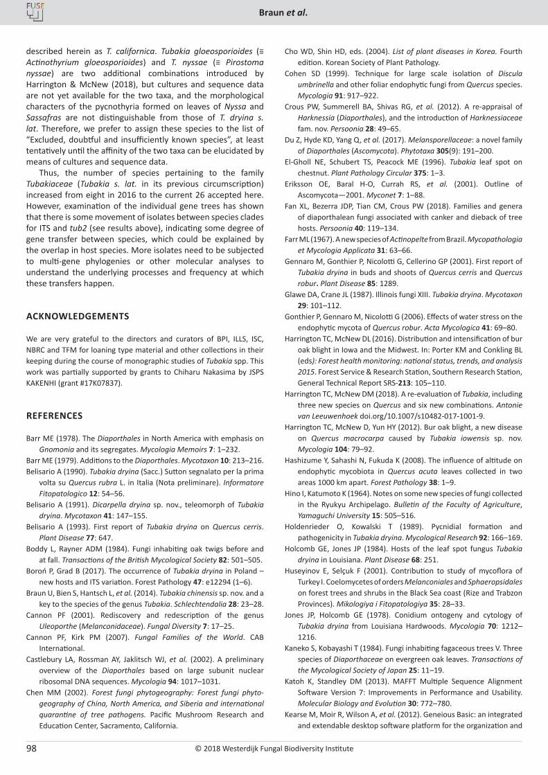
© 2018 Westerdijk Fungal Biodiversity Institute
Braun et al.
Editor-in-ChiefProf. dr P.W. Crous, Westerdijk Fungal Biodiversity Institute, P.O. Box 85167, 3508 AD Utrecht, The Netherlands.E-mail:[email protected]
98
described herein as T. californica. Tubakia gloeosporioides (≡ Actinothyrium gloeosporioides) and T. nyssae (≡ Pirostoma nyssae) are two additional combinations introduced by Harrington & McNew (2018), but cultures and sequence data are not yet available for the two taxa, and the morphological characters of the pycnothyria formed on leaves of Nyssa and Sassafras are not distinguishable from those of T. dryina s. lat. Therefore, we prefer to assign these species to the list of “Excluded, doubtful and insufficiently known species”, at least tentatively until the affinity of the two taxa can be elucidated by means of cultures and sequence data.
Thus, the number of species pertaining to the family Tubakiaceae (Tubakia s. lat. in its previous circumscription) increased from eight in 2016 to the current 26 accepted here. However, examination of the individual gene trees has shown that there is some movement of isolates between species clades for ITS and tub2 (see results above), indicating some degree of gene transfer between species, which could be explained by the overlap in host species. More isolates need to be subjected to multi-gene phylogenies or other molecular analyses to understand the underlying processes and frequency at which these transfers happen.
ACKNOWLEDGEMENTS
We are very grateful to the directors and curators of BPI, ILLS, ISC, NBRC and TFM for loaning type material and other collections in their keeping during the course of monographic studies of Tubakia spp. This work was partially supported by grants to Chiharu Nakasima by JSPS KAKENHI (grant #17K07837).
REFERENCES
Barr ME (1978). The Diaporthales in North America with emphasis on Gnomonia and its segregates. Mycologia Memoirs 7: 1–232.
Barr ME (1979). Additions to the Diaporthales. Mycotaxon 10: 213–216.Belisario A (1990). Tubakia dryina (Sacc.) Sutton segnalato per la prima
volta su Quercus rubra L. in Italia (Nota preliminare). Informatore Fitopatologico 12: 54–56.
Belisario A (1991). Dicarpella dryina sp. nov., teleomorph of Tubakia dryina. Mycotaxon 41: 147–155.
Belisario A (1993). First report of Tubakia dryina on Quercus cerris. Plant Disease 77: 647.
Boddy L, Rayner ADM (1984). Fungi inhabiting oak twigs before and at fall. Transactions of the British Mycological Society 82: 501–505.
Boroń P, Grad B (2017). The occurrence of Tubakia dryina in Poland – new hosts and ITS variation. Forest Pathology 47: e12294 (1–6).
Braun U, Bien S, Hantsch L, et al. (2014). Tubakia chinensis sp. nov. and a key to the species of the genus Tubakia. Schlechtendalia 28: 23–28.
Cannon PF (2001). Rediscovery and redescription of the genus Uleoporthe (Melanconidaceae). Fungal Diversity 7: 17–25.
Cannon PF, Kirk PM (2007). Fungal Families of the World. CAB International.
Castlebury LA, Rossman AY, Jaklitsch WJ, et al. (2002). A preliminary overview of the Diaporthales based on large subunit nuclear ribosomal DNA sequences. Mycologia 94: 1017–1031.
Chen MM (2002). Forest fungi phytogeography: Forest fungi phyto-geography of China, North America, and Siberia and international quarantine of tree pathogens. Pacific Mushroom Research and Education Center, Sacramento, California.
Cho WD, Shin HD, eds. (2004). List of plant diseases in Korea. Fourth edition. Korean Society of Plant Pathology.
Cohen SD (1999). Technique for large scale isolation of Discula umbrinella and other foliar endophytic fungi from Quercus species. Mycologia 91: 917–922.
Crous PW, Summerell BA, Shivas RG, et al. (2012). A re-appraisal of Harknessia (Diaporthales), and the introduction of Harknessiaceae fam. nov. Persoonia 28: 49–65.
Du Z, Hyde KD, Yang Q, et al. (2017). Melansporellaceae: a novel family of Diaporthales (Ascomycota). Phytotaxa 305(9): 191–200.
El-Gholl NE, Schubert TS, Peacock ME (1996). Tubakia leaf spot on chestnut. Plant Pathology Circular 375: 1–3.
Eriksson OE, Baral H-O, Currah RS, et al. (2001). Outline of Ascomycota—2001. Myconet 7: 1–88.
Fan XL, Bezerra JDP, Tian CM, Crous PW (2018). Families and genera of diaporthalean fungi associated with canker and dieback of tree hosts. Persoonia 40: 119–134.
Farr ML (1967). A new species of Actinopelte from Brazil. Mycopathologia et Mycologia Applicata 31: 63–66.
Gennaro M, Gonthier P, Nicolotti G, Cellerino GP (2001). First report of Tubakia dryina in buds and shoots of Quercus cerris and Quercus robur. Plant Disease 85: 1289.
Glawe DA, Crane JL (1987). Illinois fungi XIII. Tubakia dryina. Mycotaxon 29: 101–112.
Gonthier P, Gennaro M, Nicolotti G (2006). Effects of water stress on the endophytic mycota of Quercus robur. Acta Mycologica 41: 69–80.
Harrington TC, McNew DL (2016). Distribution and intensification of bur oak blight in Iowa and the Midwest. In: Porter KM and Conkling BL (eds): Forest health monitoring: national status, trends, and analysis 2015. Forest Service & Research Station, Southern Research Station, General Technical Report SRS-213: 105–110.
Harrington TC, McNew DM (2018). A re-evaluation of Tubakia, including three new species on Quercus and six new combinations. Antonie van Leeuwenhoek doi.org/10.1007/s10482-017-1001-9.
Harrington TC, McNew D, Yun HY (2012). Bur oak blight, a new disease on Quercus macrocarpa caused by Tubakia iowensis sp. nov. Mycologia 104: 79–92.
Hashizume Y, Sahashi N, Fukuda K (2008). The influence of altitude on endophytic mycobiota in Quercus acuta leaves collected in two areas 1000 km apart. Forest Pathology 38: 1–9.
Hino I, Katumoto K (1964). Notes on some new species of fungi collected in the Ryukyu Archipelago. Bulletin of the Faculty of Agriculture, Yamaguchi University 15: 505–516.
Holdenrieder O, Kowalski T (1989). Pycnidial formation and pathogenicity in Tubakia dryina. Mycological Research 92: 166–169.
Holcomb GE, Jones JP (1984). Hosts of the leaf spot fungus Tubakia dryina in Louisiana. Plant Disease 68: 251.
Huseyinov E, Selçuk F (2001). Contribution to study of mycoflora of Turkey I. Coelomycetes of orders Melanconiales and Sphaeropsidales on forest trees and shrubs in the Black Sea coast (Rize and Trabzon Provinces). Mikologiya i Fitopatologiya 35: 28–33.
Jones JP, Holcomb GE (1978). Conidium ontogeny and cytology of Tubakia dryina from Louisiana Hardwoods. Mycologia 70: 1212–1216.
Kaneko S, Kobayashi T (1984). Fungi inhabiting fagaceous trees V. Three species of Diaporthaceae on evergreen oak leaves. Transactions of the Mycological Society of Japan 25: 11–19.
Katoh K, Standley DM (2013). MAFFT Multiple Sequence Alignment Software Version 7: Improvements in Performance and Usability. Molecular Biology and Evolution 30: 772–780.
Kearse M, Moir R, Wilson A, et al. (2012). Geneious Basic: an integrated and extendable desktop software platform for the organization and
© 2018 Westerdijk Fungal Biodiversity Institute
The genus Tubakia s. lat.
Editor-in-ChiefProf. dr P.W. Crous, Westerdijk Fungal Biodiversity Institute, P.O. Box 85167, 3508 AD Utrecht, The Netherlands.E-mail:[email protected]
99
analysis of sequence data. Bioinformatics 28: 1647–1649.Kobayashi T (2007). Index of fungi inhabiting woody plants in Japan.
Host, Distribution and Literature. Zenkoku-Noson-Kyoiku Kyokai Publishing Co., Ltd.
Kumar S, Stecher G, Tamura K (2016). MEGA7: Molecular Evolutionary Genetics Analysis Version 7.0 for Bigger Datasets. Molecular Biology and Evolution 33: 1870–1874.
Lee YH, Cho WD, Kim WK, et al. (1991). Report on host-unrecorded diseases identified from economical crops in Korea. Research Report to Rural Development Administration (Suwon, Korea) 33: 15–19
Limber DP, Cash EK (1945). Actinopelte dryina. Mycologia 37: 129–137.Liu YJ, Whelen S, Hall BD (1999). Phylogenetic relationships among
ascomycetes: evidence from an RNA Polymerase II subunit. Molecular Biology and Evolution 16: 1799–1808.
Lumbsch HT, Huhndorf SM eds (2007). Outline of Ascomycota – 2007. Myconet 13: 1–58.
Maharachchikumbura SSN, Hyde KD, Jones EGB, et al. (2015). Towards a natural classification and backbone tree for Sordariomycetes. Fungal Diversity 72: 199–301.
Maharachchikumbura SSN, Hyde KD, Gareth Jones EB, et al. (2016). Families of Sordariomyctes. Fungal Diversity 79: 1–317.
Monod M (1983). Monographie taxonomique des Gnomoniaceae. Sydowia, Beiheft IX: 1–315.
Munkvold GP, Neely D (1990). Pathogenicity of Tubakia dryina. Plant Disease 74: 518–522
Nag Raj TR, DiCosmo F (1981). A monograph of Harknessia and Mastigosporella, with notes on associated teleomorphs. Bibliotheca Mycologica 80: 1–62.
Nylander JAA (2004). MrModeltest v2. Program distributed by the author. Evolutionary Biology Centre, Uppsala University, Sweden.
Reid J, Dowsett JA (1990). On Dicarpella, Sphaerognomonia, and Apiosporopsis. Canadian Journal of Botany 68: 2398–2407.
Ronquist F, Teslenko M, Van der Mark P, et al. (2012). MrBayes 3.2: Efficient Bayesian phylogenetic inference and model choice across a large model space. Systematic Biology 61: 539–542.
Rossman AY, Farr DF, Castlebury LA (2007). A review of the phylogeny and biology of the Diaporthales. Mycoscience 48: 135–144.
Saccardo PA (1913). Notae mycologicae. Series XVI. Annales Mycologici 11: 493–511.
Senanayake IC, Crous PW, Groenewald JZ, et al. (2017). Families of Diaporthales based on morphological and phylogenetic evidence. Studies in Mycology 86: 217–296.
Sieber TN (2007). Endophytic fungi in forest trees: Are they mutualists? Fungal Biology Reviews 21(2-3): 75–89.
Sognov MV, Castlebury LA, Meija LC, et al. (2008). Leaf-inhabiting genera of the Gnomoniaceae, Diaporthales. Studies in Mycology 62: 1–79.
Sung G-H, Sung J-M, Hywel-Jones NL, et al. (2007). A multi-gene phylogeny of Clavicipitaceae (Ascomycota, Fungi): identification of localized incongruence using a combinational bootstrap approach. Molecular Phylogenetics and Evolution 44: 1204–1223.
Sutton BC (1973). Tubakia nom. nov. Transactions of the British Mycological Society 60: 164–165.
Swofford DL (2003). PAUP*: phylogenetic analysis using parsimony. (*and other methods). Version 4.0b10. Sinauer Associates, Sunderland.
Tangthirasunun N, Silar P, Jayarama Bhat D, et al. (2014). Greeneria saprophytica sp. nov. on dead leaves of Syzygium cumini from Chiang Rai, Thailand. Phytotaxa 184: 275–282.
Taylor J (2001). Pycnothyrium ultrastucture in Tubakia dryina. Mycological Research 105: 119–121.
Taylor J, Clark S (1996). Infection and fungal development of Tubakia dryina on Sweet Gum (Liquidambar styraciflua). Mycologia 88: 613–618.
Tehon LR (1924). Notes on the parasitic fungi of Illinois. Mycologia 16: 135–142.
Tehon LR, Stout GL (1929). Notes on the parasitic fungi of Illinois: IV. Mycologia 21: 180–196.
Theissen F (1913). Ueber einige Mikrothyriaceen. Annales Mycologici 11: 493–511.
Videira SIR, Groenewald JZ, Nakashima C, et al. (2017). Mycosphaerellaceae – Chaos or clarity? Studies in Mycology 87: 257–421.
Voglmayr H, Rossman AY, Castlebury LA, et al. (2012). Multigene phylogeny and taxonomy of the genus Melanconiella (Diaporthales). Fungal Diversity 57: 1–44.
Voglmayr H, Castlebury LA, Jaklitsch WM (2017). Juglanconis gen. nov. on Juglandaceae, and the new family Juglanconidaceae (Diaporthales). Persoonia 38: 136–155.
Von Höhnel F (1925). Neue Fungi imperfecti. 5. Mitteilung. Mitteilungen des Botanischen Instituts der Technischen Hochschule Wien 2: 65–73.
Yokoyama T, Tubaki K (1971). Cultural and taxonomical studies on the genus Actinopelte. Institute of Fermentation, Osaka, Research Communication 5: 43–77.
Yun HY, Rossman AY (2011). Tubakia seoraksanensis, a new species from Korea. Mycotaxon 115: 369–373.
Zahedi M, Elahinia SA, Khodaparast SA, Bujari J (2011). Introduction of some new mitosporic fungi causing leaf spot on broad leaf trees in Guilan province (N Iran). Rostaniha 12: 63–71.
Zhang Z, Schwartz S, Wagner L, et al. (2000). A greedy algorithm for aligning DNA sequences. Journal of Computational Biology 7: 203–214.
© 2018 Westerdijk Fungal Biodiversity Institute
Castellano et al.
Editor-in-ChiefProf. dr P.W. Crous, Westerdijk Fungal Biodiversity Institute, P.O. Box 85167, 3508 AD Utrecht, The Netherlands.E-mail:[email protected]
100
Fungal Systematics and Evolution is licensed under a Creative Commons Attribution-NonCommercial-ShareAlike 4.0 International License
© 2018 Westerdijk Fungal Biodiversity Institute 101
Editor-in-ChiefProf. dr P.W. Crous, Westerdijk Fungal Biodiversity Institute, P.O. Box 85167, 3508 AD Utrecht, The Netherlands.E-mail:[email protected]
Fungal Systematics and EvolutionVOLUME 1JUNE 2018
PAGES 101–129
doi.org/10.3114/fuse.2018.01.05
Postia caesia complex (Polyporales, Basidiomycota) in temperate Northern Hemisphere
O. Miettinen1*, J. Vlasák2, B. Rivoire3, V. Spirin4
1Finnish Museum of Natural History, University of Helsinki, Finland2Biology Centre of the Czech Academy of Sciences, České Budějovice, Czech Republic3Société Linnéenne, Lyon, France4Finnish Museum of Natural History, University of Helsinki, Finland
*Corresponding author: [email protected]
Abstract: Taxonomy of the Postia caesia complex is revised based on morphology and two genetic markers, ITS and tef1. In total, we recognize 24 species, multiplying the known species diversity in the complex. We provide descriptions for 20 temperate Northern Hemisphere taxa. Identity of the core species, P. caesia, is re-established, and a neotype from the type locality is selected. Four new combinations are proposed, and 10 new species are described: P. arbuti, P. auricoma, P. bifaria, P. comata, P. cyanescens, P. glauca, P. livens, P. magna, P. populi, and P. yanae.
Key words: allopatric speciationsympatric speciationhost specificitytype studiessystematics14 new taxaDacryobolaceae
Published online: 19 March 2018.
INTRODUCTION
The Postia caesia species complex contains closely related brown-rot polypore species with blue-tinted basidiocarps making them easy to recognize. Distinct blue colors are rather rare among fungi, and among polypores only species of Skeletocutis nivea coll. develop a similar blue-tinted pore surface. For a long time, all blue-tinted Postia spp. went under the name Postia caesia (=Oligoporus caesius), described from conifers in Europe.
David (1974, 1980) first showed through mating tests and morphological analyses that two other species are present in Europe besides P. caesia, describing P. luteocaesia and P. subcaesia as new species. Jahn (1979) noted that David’s P. subcaesia comes in many forms. He then introduced P. subcaesia “f. minor”, which Niemelä and Vampola later described as Postia alni (Niemelä et al. 2001). Lastly, Pieri & Rivoire (2005) introduced the fifth European species, P. mediterraneocaesia.
Outside the Northern temperate area, a few further species have been included in the complex. Ryvarden (1983) noted that Patouillard’s Polyporus caesioflavus from Ecuador is closely related but separate from P. caesia. Ryvarden (1988a) described Oligoporus africanus from Burundi. Corner (1989) introduced two new species from Malaysian Borneo, Tyromyces amyloideus and T. coeruleivirens, confirmed by Hattori (2005) to belong to the Postia caesia species complex. Papp (2015) provided combinations of the above-mentioned species to Postia. In New Zealand, Rajchenberg (1995) noted that Postia atrostrigosa is a relative of Postia caesia.
The above-mentioned authors, however, never reviewed the availability of older names for European taxa, and no revision of the species complex in Europe or elsewhere has been attempted. Yao et al. (2005) showed that three ITS-sequence-based groups
were present in England, but they could not connect those groups to existing names. Ortiz-Santana et al. (2013) and Pildain & Rajchenberg (2013) presented genus-level phylogenies that included representatives of the Postia caesia complex. Both concluded that the Postia caesia complex belongs to the genus Postia, but did not touch upon species concepts.
In this study we have sampled specimens of the Postia caesia species complex originating from Europe, Siberia, East Asia and North America from a molecular and morphological perspective. The material is extensive, covering 146 localities from 20 countries. Our aim is to revise the species concepts within the northern temperate area. To establish a firm nomenclatural basis for species concepts in this group, we have conducted type studies of all known species names from the temperate Northern Hemisphere.
MATERIAL AND METHODS
Morphology
We studied types and specimens from the herbaria BPI, CFMR, CUP, H, K, LE, LY, MJ and O as well as from private herbarium of the author JV. Herbarium acronyms are given according to Index Herbariorum (2017). Sequenced specimens are marked with an asterisk (*).
Due to small morphological differences between P. caesia and its relatives, all specimens were examined following the same routine. Number of pores per mm was measured with a stereomicroscope targeting areas with regular pore form. When studying hyphal structure and measuring hyphae, the part of the basidiome cut may influence the outcome. Thus we studied
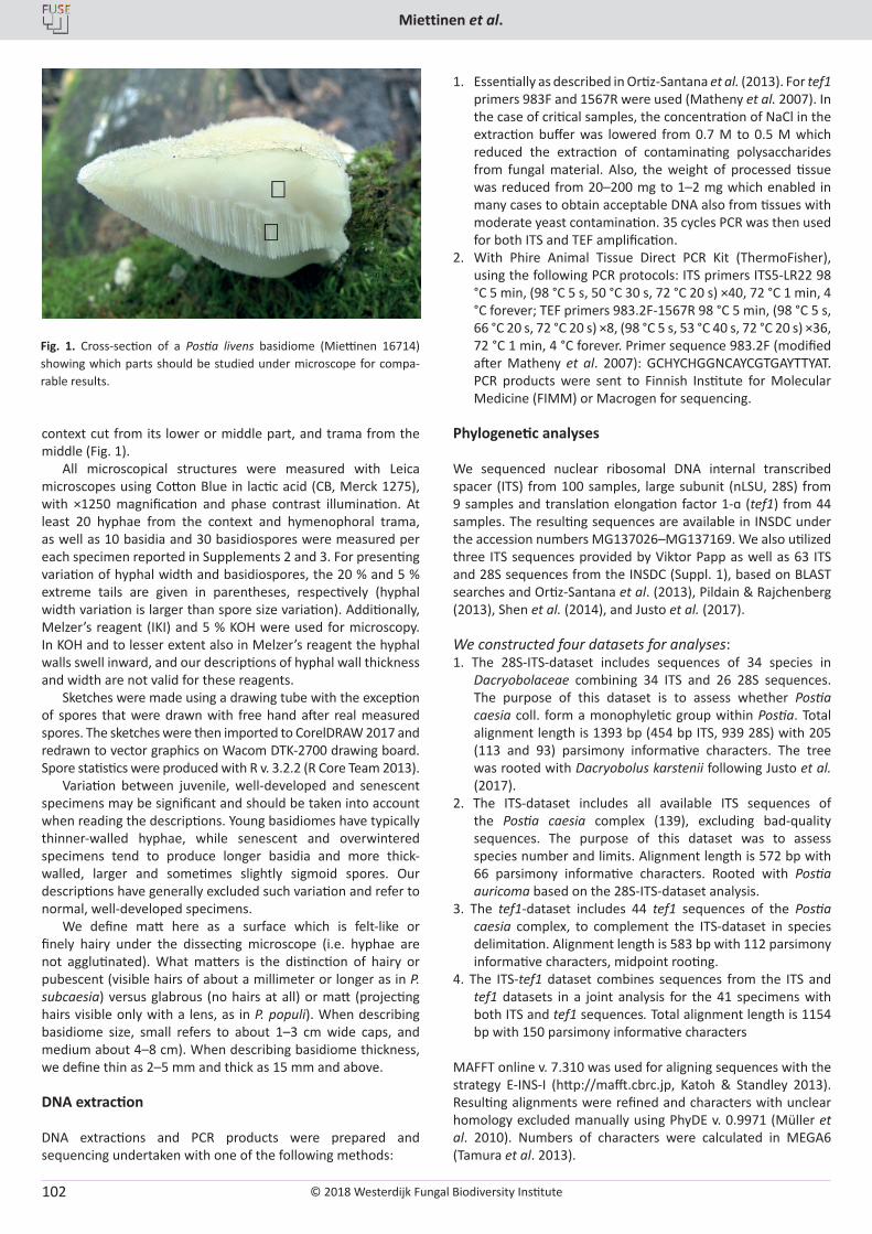
© 2018 Westerdijk Fungal Biodiversity Institute
Miettinen et al.
Editor-in-ChiefProf. dr P.W. Crous, Westerdijk Fungal Biodiversity Institute, P.O. Box 85167, 3508 AD Utrecht, The Netherlands.E-mail:[email protected]
102
context cut from its lower or middle part, and trama from the middle (Fig. 1).
All microscopical structures were measured with Leica microscopes using Cotton Blue in lactic acid (CB, Merck 1275), with ×1250 magnification and phase contrast illumination. At least 20 hyphae from the context and hymenophoral trama, as well as 10 basidia and 30 basidiospores were measured per each specimen reported in Supplements 2 and 3. For presenting variation of hyphal width and basidiospores, the 20 % and 5 % extreme tails are given in parentheses, respectively (hyphal width variation is larger than spore size variation). Additionally, Melzer’s reagent (IKI) and 5 % KOH were used for microscopy. In KOH and to lesser extent also in Melzer’s reagent the hyphal walls swell inward, and our descriptions of hyphal wall thickness and width are not valid for these reagents.
Sketches were made using a drawing tube with the exception of spores that were drawn with free hand after real measured spores. The sketches were then imported to CorelDRAW 2017 and redrawn to vector graphics on Wacom DTK-2700 drawing board. Spore statistics were produced with R v. 3.2.2 (R Core Team 2013).
Variation between juvenile, well-developed and senescent specimens may be significant and should be taken into account when reading the descriptions. Young basidiomes have typically thinner-walled hyphae, while senescent and overwintered specimens tend to produce longer basidia and more thick-walled, larger and sometimes slightly sigmoid spores. Our descriptions have generally excluded such variation and refer to normal, well-developed specimens.
We define matt here as a surface which is felt-like or finely hairy under the dissecting microscope (i.e. hyphae are not agglutinated). What matters is the distinction of hairy or pubescent (visible hairs of about a millimeter or longer as in P. subcaesia) versus glabrous (no hairs at all) or matt (projecting hairs visible only with a lens, as in P. populi). When describing basidiome size, small refers to about 1–3 cm wide caps, and medium about 4–8 cm). When describing basidiome thickness, we define thin as 2–5 mm and thick as 15 mm and above.
DNA extraction
DNA extractions and PCR products were prepared and sequencing undertaken with one of the following methods:
1. Essentially as described in Ortiz-Santana et al. (2013). For tef1 primers 983F and 1567R were used (Matheny et al. 2007). In the case of critical samples, the concentration of NaCl in the extraction buffer was lowered from 0.7 M to 0.5 M which reduced the extraction of contaminating polysaccharides from fungal material. Also, the weight of processed tissue was reduced from 20–200 mg to 1–2 mg which enabled in many cases to obtain acceptable DNA also from tissues with moderate yeast contamination. 35 cycles PCR was then used for both ITS and TEF amplification.
2. With Phire Animal Tissue Direct PCR Kit (ThermoFisher), using the following PCR protocols: ITS primers ITS5-LR22 98 °C 5 min, (98 °C 5 s, 50 °C 30 s, 72 °C 20 s) ×40, 72 °C 1 min, 4 °C forever; TEF primers 983.2F-1567R 98 °C 5 min, (98 °C 5 s, 66 °C 20 s, 72 °C 20 s) ×8, (98 °C 5 s, 53 °C 40 s, 72 °C 20 s) ×36, 72 °C 1 min, 4 °C forever. Primer sequence 983.2F (modified after Matheny et al. 2007): GCHYCHGGNCAYCGTGAYTTYAT. PCR products were sent to Finnish Institute for Molecular Medicine (FIMM) or Macrogen for sequencing.
Phylogenetic analyses
We sequenced nuclear ribosomal DNA internal transcribed spacer (ITS) from 100 samples, large subunit (nLSU, 28S) from 9 samples and translation elongation factor 1-ɑ (tef1) from 44 samples. The resulting sequences are available in INSDC under the accession numbers MG137026–MG137169. We also utilized three ITS sequences provided by Viktor Papp as well as 63 ITS and 28S sequences from the INSDC (Suppl. 1), based on BLAST searches and Ortiz-Santana et al. (2013), Pildain & Rajchenberg (2013), Shen et al. (2014), and Justo et al. (2017).
We constructed four datasets for analyses:1. The 28S-ITS-dataset includes sequences of 34 species in
Dacryobolaceae combining 34 ITS and 26 28S sequences. The purpose of this dataset is to assess whether Postia caesia coll. form a monophyletic group within Postia. Total alignment length is 1393 bp (454 bp ITS, 939 28S) with 205 (113 and 93) parsimony informative characters. The tree was rooted with Dacryobolus karstenii following Justo et al. (2017).
2. The ITS-dataset includes all available ITS sequences of the Postia caesia complex (139), excluding bad-quality sequences. The purpose of this dataset was to assess species number and limits. Alignment length is 572 bp with 66 parsimony informative characters. Rooted with Postia auricoma based on the 28S-ITS-dataset analysis.
3. The tef1-dataset includes 44 tef1 sequences of the Postia caesia complex, to complement the ITS-dataset in species delimitation. Alignment length is 583 bp with 112 parsimony informative characters, midpoint rooting.
4. The ITS-tef1 dataset combines sequences from the ITS and tef1 datasets in a joint analysis for the 41 specimens with both ITS and tef1 sequences. Total alignment length is 1154 bp with 150 parsimony informative characters
MAFFT online v. 7.310 was used for aligning sequences with the strategy E-INS-I (http://mafft.cbrc.jp, Katoh & Standley 2013). Resulting alignments were refined and characters with unclear homology excluded manually using PhyDE v. 0.9971 (Müller et al. 2010). Numbers of characters were calculated in MEGA6 (Tamura et al. 2013).
Fig. 1. Cross-section of a Postia livens basidiome (Miettinen 16714) showing which parts should be studied under microscope for compa-rable results.
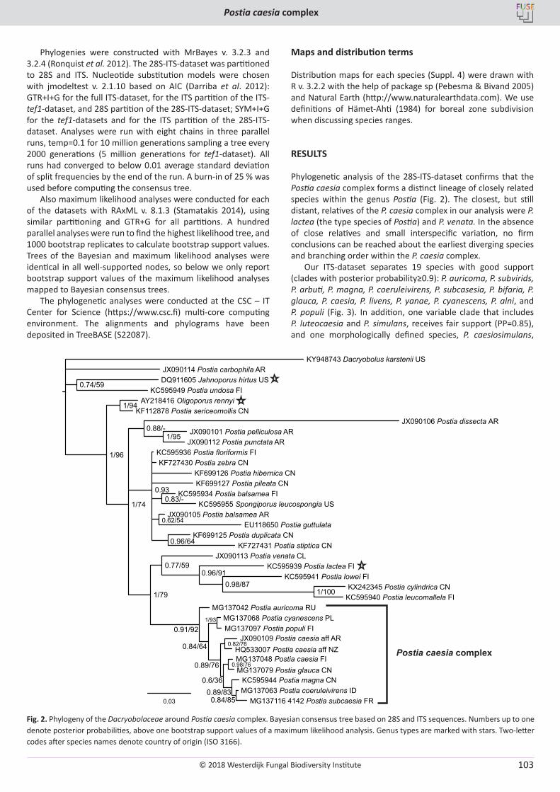
© 2018 Westerdijk Fungal Biodiversity Institute
Postia caesia complex
Editor-in-ChiefProf. dr P.W. Crous, Westerdijk Fungal Biodiversity Institute, P.O. Box 85167, 3508 AD Utrecht, The Netherlands.E-mail:[email protected]
103
Phylogenies were constructed with MrBayes v. 3.2.3 and 3.2.4 (Ronquist et al. 2012). The 28S-ITS-dataset was partitioned to 28S and ITS. Nucleotide substitution models were chosen with jmodeltest v. 2.1.10 based on AIC (Darriba et al. 2012): GTR+I+G for the full ITS-dataset, for the ITS partition of the ITS-tef1-dataset, and 28S partition of the 28S-ITS-dataset; SYM+I+G for the tef1-datasets and for the ITS partition of the 28S-ITS-dataset. Analyses were run with eight chains in three parallel runs, temp=0.1 for 10 million generations sampling a tree every 2000 generations (5 million generations for tef1-dataset). All runs had converged to below 0.01 average standard deviation of split frequencies by the end of the run. A burn-in of 25 % was used before computing the consensus tree.
Also maximum likelihood analyses were conducted for each of the datasets with RAxML v. 8.1.3 (Stamatakis 2014), using similar partitioning and GTR+G for all partitions. A hundred parallel analyses were run to find the highest likelihood tree, and 1000 bootstrap replicates to calculate bootstrap support values. Trees of the Bayesian and maximum likelihood analyses were identical in all well-supported nodes, so below we only report bootstrap support values of the maximum likelihood analyses mapped to Bayesian consensus trees.
The phylogenetic analyses were conducted at the CSC – IT Center for Science (https://www.csc.fi) multi-core computing environment. The alignments and phylograms have been deposited in TreeBASE (S22087).
Maps and distribution terms
Distribution maps for each species (Suppl. 4) were drawn with R v. 3.2.2 with the help of package sp (Pebesma & Bivand 2005) and Natural Earth (http://www.naturalearthdata.com). We use definitions of Hämet-Ahti (1984) for boreal zone subdivision when discussing species ranges.
RESULTS
Phylogenetic analysis of the 28S-ITS-dataset confirms that the Postia caesia complex forms a distinct lineage of closely related species within the genus Postia (Fig. 2). The closest, but still distant, relatives of the P. caesia complex in our analysis were P. lactea (the type species of Postia) and P. venata. In the absence of close relatives and small interspecific variation, no firm conclusions can be reached about the earliest diverging species and branching order within the P. caesia complex.
Our ITS-dataset separates 19 species with good support (clades with posterior probability≥0.9): P. auricoma, P. subvirids, P. arbuti, P. magna, P. coeruleivirens, P. subcasesia, P. bifaria, P. glauca, P. caesia, P. livens, P. yanae, P. cyanescens, P. alni, and P. populi (Fig. 3). In addition, one variable clade that includes P. luteocaesia and P. simulans, receives fair support (PP=0.85), and one morphologically defined species, P. caesiosimulans,
Fig. 2. Phylogeny of the Dacryobolaceae around Postia caesia complex. Bayesian consensus tree based on 28S and ITS sequences. Numbers up to one denote posterior probabilities, above one bootstrap support values of a maximum likelihood analysis. Genus types are marked with stars. Two-letter codes after species names denote country of origin (ISO 3166).
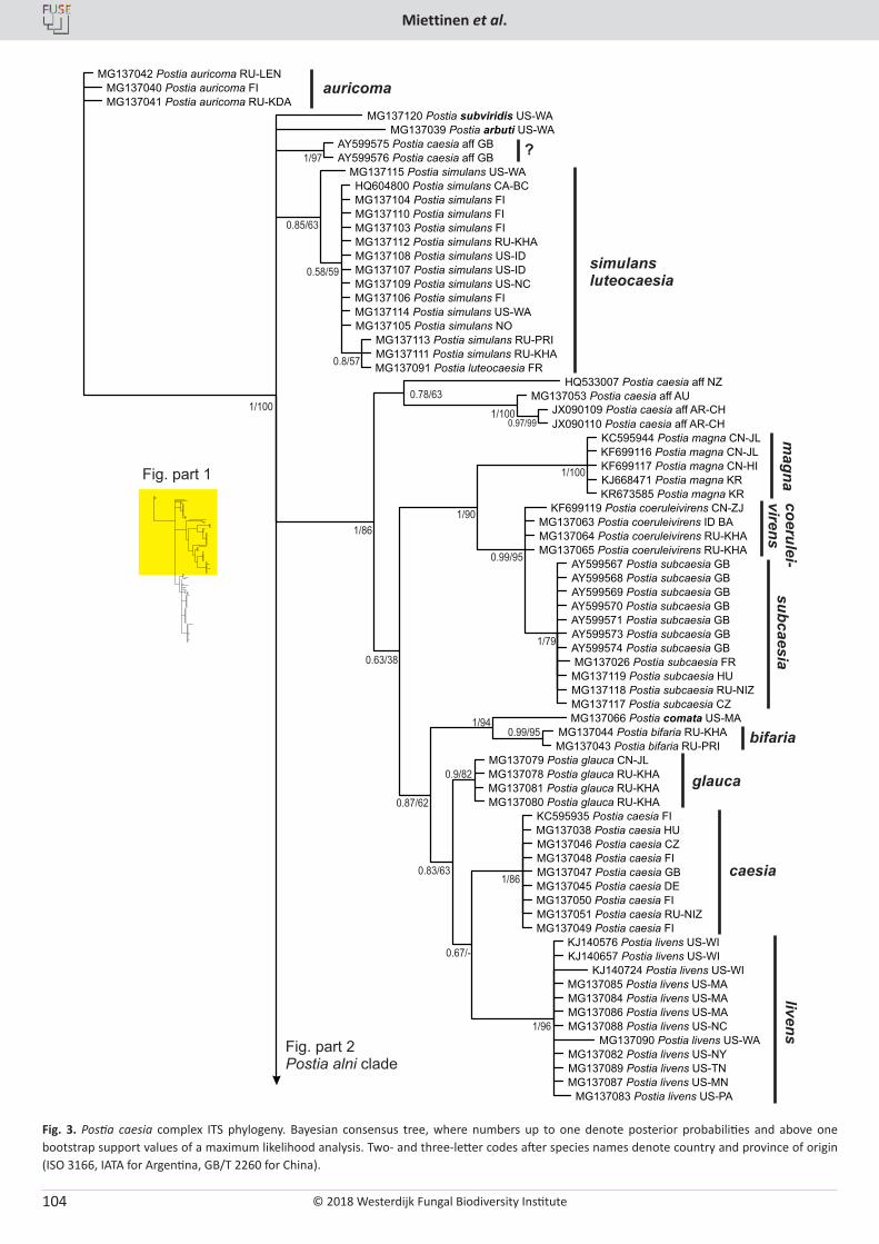
© 2018 Westerdijk Fungal Biodiversity Institute
Miettinen et al.
Editor-in-ChiefProf. dr P.W. Crous, Westerdijk Fungal Biodiversity Institute, P.O. Box 85167, 3508 AD Utrecht, The Netherlands.E-mail:[email protected]
104
Fig. 3. Postia caesia complex ITS phylogeny. Bayesian consensus tree, where numbers up to one denote posterior probabilities and above one bootstrap support values of a maximum likelihood analysis. Two- and three-letter codes after species names denote country and province of origin (ISO 3166, IATA for Argentina, GB/T 2260 for China).
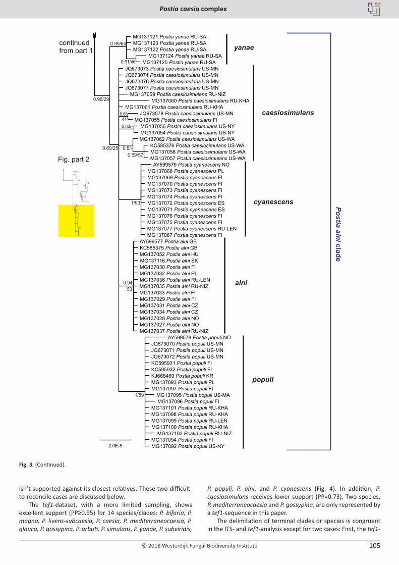
© 2018 Westerdijk Fungal Biodiversity Institute
Postia caesia complex
Editor-in-ChiefProf. dr P.W. Crous, Westerdijk Fungal Biodiversity Institute, P.O. Box 85167, 3508 AD Utrecht, The Netherlands.E-mail:[email protected]
105
isn’t supported against its closest relatives. These two difficult-to-reconcile cases are discussed below.
The tef1-dataset, with a more limited sampling, shows excellent support (PP≥0.95) for 14 species/clades: P. bifaria, P. magna, P. livens-subcaesia, P. caesia, P. mediterraneocaesia, P. glauca, P. gossypina, P. arbuti, P. simulans, P. yanae, P. subviridis,
P. populi, P. alni, and P. cyanescens (Fig. 4). In addition, P. caesiosimulans receives lower support (PP=0.73). Two species, P. mediterraneocaesia and P. gossypina, are only represented by a tef1-sequence in this paper.
The delimitation of terminal clades or species is congruent in the ITS- and tef1-analysis except for two cases: First, the tef1-
Fig. 3. (Continued).
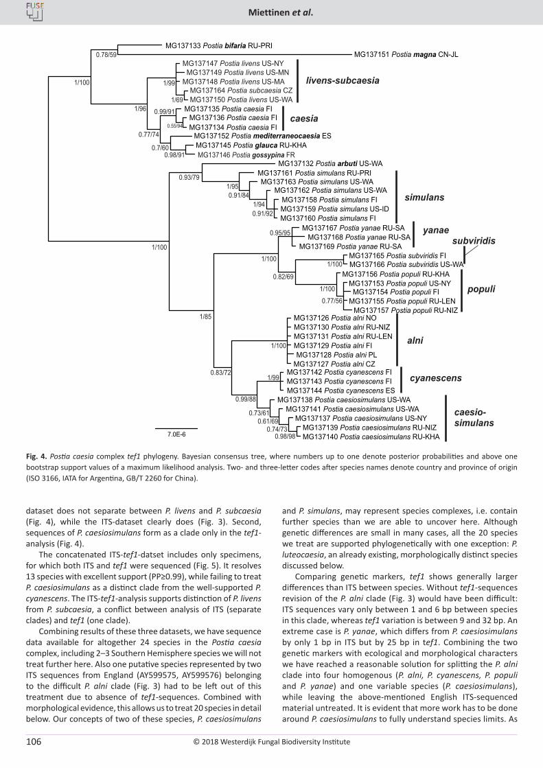
© 2018 Westerdijk Fungal Biodiversity Institute
Miettinen et al.
Editor-in-ChiefProf. dr P.W. Crous, Westerdijk Fungal Biodiversity Institute, P.O. Box 85167, 3508 AD Utrecht, The Netherlands.E-mail:[email protected]
106
dataset does not separate between P. livens and P. subcaesia (Fig. 4), while the ITS-dataset clearly does (Fig. 3). Second, sequences of P. caesiosimulans form as a clade only in the tef1-analysis (Fig. 4).
The concatenated ITS-tef1-datset includes only specimens, for which both ITS and tef1 were sequenced (Fig. 5). It resolves 13 species with excellent support (PP≥0.99), while failing to treat P. caesiosimulans as a distinct clade from the well-supported P. cyanescens. The ITS-tef1-analysis supports distinction of P. livens from P. subcaesia, a conflict between analysis of ITS (separate clades) and tef1 (one clade).
Combining results of these three datasets, we have sequence data available for altogether 24 species in the Postia caesia complex, including 2–3 Southern Hemisphere species we will not treat further here. Also one putative species represented by two ITS sequences from England (AY599575, AY599576) belonging to the difficult P. alni clade (Fig. 3) had to be left out of this treatment due to absence of tef1-sequences. Combined with morphological evidence, this allows us to treat 20 species in detail below. Our concepts of two of these species, P. caesiosimulans
and P. simulans, may represent species complexes, i.e. contain further species than we are able to uncover here. Although genetic differences are small in many cases, all the 20 species we treat are supported phylogenetically with one exception: P. luteocaesia, an already existing, morphologically distinct species discussed below.
Comparing genetic markers, tef1 shows generally larger differences than ITS between species. Without tef1-sequences revision of the P. alni clade (Fig. 3) would have been difficult: ITS sequences vary only between 1 and 6 bp between species in this clade, whereas tef1 variation is between 9 and 32 bp. An extreme case is P. yanae, which differs from P. caesiosimulans by only 1 bp in ITS but by 25 bp in tef1. Combining the two genetic markers with ecological and morphological characters we have reached a reasonable solution for splitting the P. alni clade into four homogenous (P. alni, P. cyanescens, P. populi and P. yanae) and one variable species (P. caesiosimulans), while leaving the above-mentioned English ITS-sequenced material untreated. It is evident that more work has to be done around P. caesiosimulans to fully understand species limits. As
Fig. 4. Postia caesia complex tef1 phylogeny. Bayesian consensus tree, where numbers up to one denote posterior probabilities and above one bootstrap support values of a maximum likelihood analysis. Two- and three-letter codes after species names denote country and province of origin (ISO 3166, IATA for Argentina, GB/T 2260 for China).
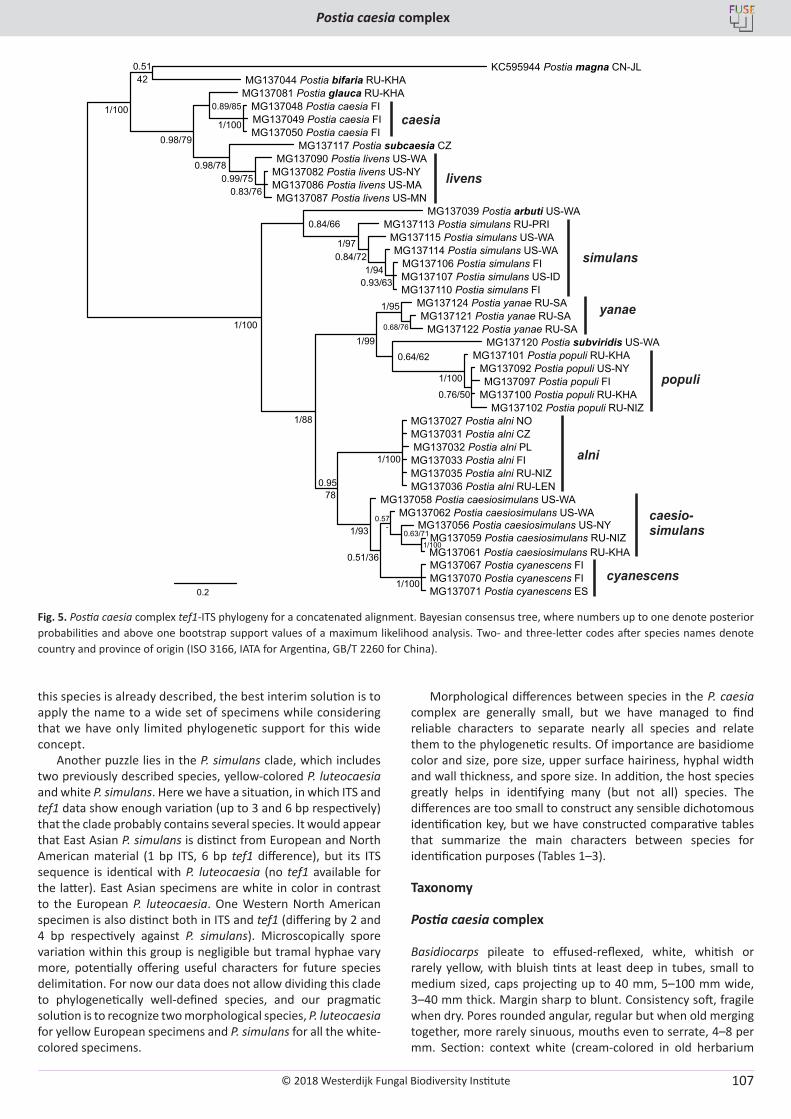
© 2018 Westerdijk Fungal Biodiversity Institute
Postia caesia complex
Editor-in-ChiefProf. dr P.W. Crous, Westerdijk Fungal Biodiversity Institute, P.O. Box 85167, 3508 AD Utrecht, The Netherlands.E-mail:[email protected]
107
this species is already described, the best interim solution is to apply the name to a wide set of specimens while considering that we have only limited phylogenetic support for this wide concept.
Another puzzle lies in the P. simulans clade, which includes two previously described species, yellow-colored P. luteocaesia and white P. simulans. Here we have a situation, in which ITS and tef1 data show enough variation (up to 3 and 6 bp respectively) that the clade probably contains several species. It would appear that East Asian P. simulans is distinct from European and North American material (1 bp ITS, 6 bp tef1 difference), but its ITS sequence is identical with P. luteocaesia (no tef1 available for the latter). East Asian specimens are white in color in contrast to the European P. luteocaesia. One Western North American specimen is also distinct both in ITS and tef1 (differing by 2 and 4 bp respectively against P. simulans). Microscopically spore variation within this group is negligible but tramal hyphae vary more, potentially offering useful characters for future species delimitation. For now our data does not allow dividing this clade to phylogenetically well-defined species, and our pragmatic solution is to recognize two morphological species, P. luteocaesia for yellow European specimens and P. simulans for all the white-colored specimens.
Morphological differences between species in the P. caesia complex are generally small, but we have managed to find reliable characters to separate nearly all species and relate them to the phylogenetic results. Of importance are basidiome color and size, pore size, upper surface hairiness, hyphal width and wall thickness, and spore size. In addition, the host species greatly helps in identifying many (but not all) species. The differences are too small to construct any sensible dichotomous identification key, but we have constructed comparative tables that summarize the main characters between species for identification purposes (Tables 1–3).
Taxonomy
Postia caesia complex
Basidiocarps pileate to effused-reflexed, white, whitish or rarely yellow, with bluish tints at least deep in tubes, small to medium sized, caps projecting up to 40 mm, 5–100 mm wide, 3–40 mm thick. Margin sharp to blunt. Consistency soft, fragile when dry. Pores rounded angular, regular but when old merging together, more rarely sinuous, mouths even to serrate, 4–8 per mm. Section: context white (cream-colored in old herbarium
Fig. 5. Postia caesia complex tef1-ITS phylogeny for a concatenated alignment. Bayesian consensus tree, where numbers up to one denote posterior probabilities and above one bootstrap support values of a maximum likelihood analysis. Two- and three-letter codes after species names denote country and province of origin (ISO 3166, IATA for Argentina, GB/T 2260 for China).
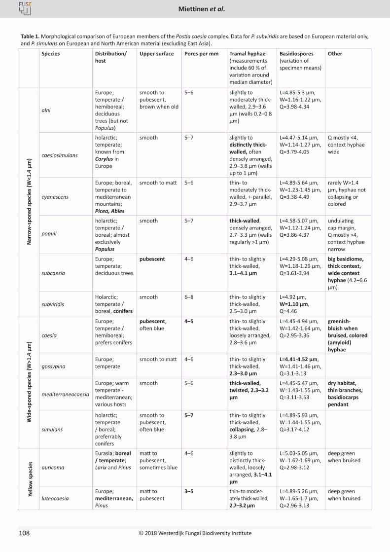
© 2018 Westerdijk Fungal Biodiversity Institute
Miettinen et al.
Editor-in-ChiefProf. dr P.W. Crous, Westerdijk Fungal Biodiversity Institute, P.O. Box 85167, 3508 AD Utrecht, The Netherlands.E-mail:[email protected]
108
Table 1. Morphological comparison of European members of the Postia caesia complex. Data for P. subviridis are based on European material only, and P. simulans on European and North American material (excluding East Asia).
Species Distribution/ host
Upper surface Pores per mm Tramal hyphae (measurements include 60 % of variation around median diameter)
Basidiospores (variation of specimen means)
Other
Nar
row
-spo
red
spec
ies (
W<1
.4 µ
m)
alni
Europe; temperate / hemiboreal; deciduous trees (but not Populus)
smooth to pubescent, brown when old
5–6 slightly to moderately thick-walled, 2.9–3.6 μm (walls 0.2–0.8 µm)
L=4.85-5.3 µm, W=1.16-1.22 µm, Q=3.98-4.34
caesiosimulans
holarctic; temperate; known from Corylus in Europe
smooth 5–7 slightly to distinctly thick-walled, often densely arranged, 2.9–3.8 μm (walls up to 1 µm)
L=4.47-5.14 µm, W=1.14-1.27 µm, Q=3.79-4.05
Q mostly <4, context hyphae wide
cyanescens
Europe; boreal, temperate to mediterranean mountains; Picea, Abies
smooth to matt 5–6 thin- to moderately thick-walled, +-parallel, 2.9–3.7 μm
L=4.89-5.64 µm, W=1.23-1.45 µm, Q=3.38-4.49
rarely W>1.4 µm, hyphae not collapsing or colored
populi
holarctic; temperate / boreal; almost exclusively Populus
smooth 5–7 thick-walled, densely arranged, 2.7–3.3 μm (walls regularly >1 µm)
L=4.58-5.07 µm, W=1.12-1.24 µm, Q=3.86-4.37
undulating cap margin, Q mostly >4, context hyphae narrow
subcaesia
Europe; temperate; deciduous trees
pubescent 4–6 thin- to slightly thick-walled, 3.1–4.1 μm
L=4.29-5.08 µm, W=1.18-1.29 µm, Q=3.61-3.94
big basidiome, thick context, wide context hyphae (4.2–6.6 µm)
subviridisHolarctic; temperate / boreal, conifers
smooth 6–8 thin- to slightly thick-walled, 2.5–3.0 µm
L=4.92 µm, W=1.10 µm, Q=4.46
Wid
e-sp
ored
spec
ies (
W>1
.4 µ
m)
caesia
Europe; temperate / hemiboreal; prefers conifers
pubescent, often blue
4–5 thin- to slightly thick-walled, loosely arranged, 2.8–3.6 μm
L=4.45-4.94 µm, W=1.42-1.64 µm, Q=2.95-3.36
greenish-bluish when bruised, colored (amyloid) hyphae
gossypinaEurope; temperate
smooth to matt 4–6 thin- to slightly thick-walled, 2.3–3.0 µm
L=4.41-4.52 µm, W=1.41-1.46 µm, Q=3.1-3.13
mediterraneocaesia
Europe; warm temperate - mediterranean; various hosts
smooth 5–6 thick-walled, twisted, 2.3–3.2 μm
L=4.45-5.47 µm, W=1.43-1.55 µm, Q=3.11-3.53
dry habitat, thin branches, basidiocarps pendant
simulans
holarctic; temperate / boreal; preferrably conifers
smooth to pubescent, often blue
5–7 thin- to slightly thick-walled, collapsing, 2.8–3.8 μm
L=4.89-5.93 µm, W=1.44-1.55 µm, Q=3.17-4.12
Yello
w sp
ecie
s
auricoma
Eurasia; boreal / temperate; Larix and Pinus
matt to pubescent, sometimes blue
4–6 slightly to distinctly thick-walled, loosely arranged, 3.1–4.1 μm
L=5.03-5.05 µm, W=1.62-1.69 µm, Q=2.98-3.12
deep green when bruised
luteocaesiaEurope; mediterranean, Pinus
matt to pubescent
3–5 thin- to moder-ately thick-walled, 2.7–3.2 μm
L=4.89-5.26 µm, W=1.65-1.7 µm, Q=2.96-3.13
deep green when bruised
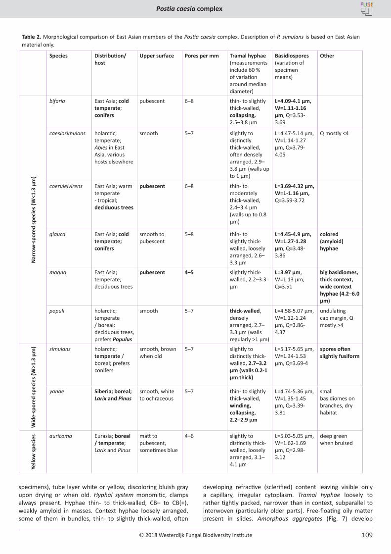
© 2018 Westerdijk Fungal Biodiversity Institute
Postia caesia complex
Editor-in-ChiefProf. dr P.W. Crous, Westerdijk Fungal Biodiversity Institute, P.O. Box 85167, 3508 AD Utrecht, The Netherlands.E-mail:[email protected]
109
specimens), tube layer white or yellow, discoloring bluish gray upon drying or when old. Hyphal system monomitic, clamps always present. Hyphae thin- to thick-walled, CB– to CB(+), weakly amyloid in masses. Context hyphae loosely arranged, some of them in bundles, thin- to slightly thick-walled, often
developing refractive (sclerified) content leaving visible only a capillary, irregular cytoplasm. Tramal hyphae loosely to rather tightly packed, narrower than in context, subparallel to interwoven (particularly older parts). Free-floating oily matter present in slides. Amorphous aggregates (Fig. 7) develop
Table 2. Morphological comparison of East Asian members of the Postia caesia complex. Description of P. simulans is based on East Asian material only.
Species Distribution/ host
Upper surface Pores per mm Tramal hyphae (measurements include 60 % of variation around median diameter)
Basidiospores (variation of specimen means)
Other
Nar
row
-spo
red
spec
ies (
W<1
.3 µ
m)
bifaria East Asia; cold temperate; conifers
pubescent 6–8 thin- to slightly thick-walled, collapsing, 2.5–3.8 μm
L=4.09-4.1 µm, W=1.11-1.16 µm, Q=3.53-3.69
caesiosimulans holarctic; temperate; Abies in East Asia, various hosts elsewhere
smooth 5–7 slightly to distinctly thick-walled, often densely arranged, 2.9–3.8 μm (walls up to 1 µm)
L=4.47-5.14 µm, W=1.14-1.27 µm, Q=3.79-4.05
Q mostly <4
coeruleivirens East Asia; warm temperate - tropical; deciduous trees
pubescent 6–8 thin- to moderately thick-walled, 2.4–3.4 μm (walls up to 0.8 µm)
L=3.69-4.32 µm, W=1-1.16 µm, Q=3.59-3.72
glauca East Asia; cold temperate; conifers
smooth to pubescent
5–8 thin- to slightly thick-walled, loosely arranged, 2.6–3.3 μm
L=4.45-4.9 µm, W=1.27-1.28 µm, Q=3.48-3.86
colored (amyloid) hyphae
magna East Asia; temperate; deciduous trees
pubescent 4–5 slightly thick-walled, 2.2–3.3 µm
L=3.97 µm, W=1.13 µm, Q=3.51
big basidiomes, thick context, wide context hyphae (4.2–6.0 µm)
populi holarctic; temperate / boreal; deciduous trees, prefers Populus
smooth 5–7 thick-walled, densely arranged, 2.7–3.3 μm (walls regularly >1 µm)
L=4.58-5.07 µm, W=1.12-1.24 µm, Q=3.86-4.37
undulating cap margin, Q mostly >4
Wid
e-sp
ored
spec
ies (
W>1
.3 µ
m) simulans holarctic;
temperate / boreal; prefers conifers
smooth, brown when old
5–7 slightly to distinctly thick-walled, 2.7–3.2 µm (walls 0.2-1 µm thick)
L=5.17-5.65 µm, W=1.34-1.53 µm, Q=3.69-4
spores often slightly fusiform
yanae Siberia; boreal; Larix and Pinus
smooth, white to ochraceous
5–7 thin- to slightly thick-walled, winding, collapsing, 2.2–2.9 μm
L=4.74-5.36 µm, W=1.35-1.45 µm, Q=3.39-3.81
small basidiomes on branches, dry habitat
Yello
w sp
ecie
s auricoma Eurasia; boreal / temperate; Larix and Pinus
matt to pubescent, sometimes blue
4–6 slightly to distinctly thick-walled, loosely arranged, 3.1–4.1 μm
L=5.03-5.05 µm, W=1.62-1.69 µm, Q=2.98-3.12
deep green when bruised
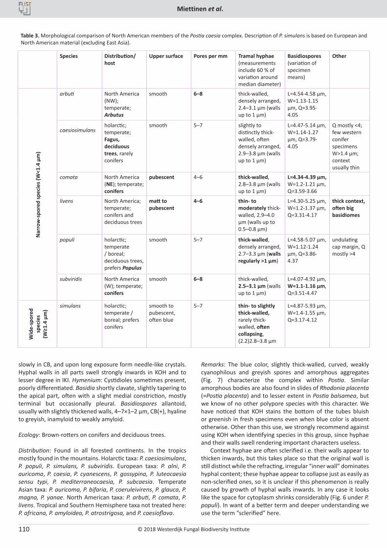
© 2018 Westerdijk Fungal Biodiversity Institute
Miettinen et al.
Editor-in-ChiefProf. dr P.W. Crous, Westerdijk Fungal Biodiversity Institute, P.O. Box 85167, 3508 AD Utrecht, The Netherlands.E-mail:[email protected]
110
slowly in CB, and upon long exposure form needle-like crystals. Hyphal walls in all parts swell strongly inwards in KOH and to lesser degree in IKI. Hymenium: Cystidioles sometimes present, poorly differentiated. Basidia shortly clavate, slightly tapering to the apical part, often with a slight medial constriction, mostly terminal but occasionally pleural. Basidiospores allantoid, usually with slightly thickened walls, 4–7×1–2 µm, CB(+), hyaline to greyish, inamyloid to weakly amyloid.
Ecology: Brown-rotters on conifers and deciduous trees.
Distribution: Found in all forested continents. In the tropics mostly found in the mountains. Holarctic taxa: P. caesiosimulans, P. populi, P. simulans, P. subviridis. European taxa: P. alni, P. auricoma, P. caesia, P. cyanescens, P. gossypina, P. luteocaesia sensu typi, P. mediterraneocaesia, P. subcaesia. Temperate Asian taxa: P. auricoma, P. bifaria, P. coeruleivirens, P. glauca, P. magna, P. yanae. North American taxa: P. arbuti, P. comata, P. livens. Tropical and Southern Hemisphere taxa not treated here: P. africana, P. amyloidea, P. atrostrigosa, and P. caesioflava.
Remarks: The blue color, slightly thick-walled, curved, weakly cyanophilous and greyish spores and amorphous aggregates (Fig. 7) characterize the complex within Postia. Similar amorphous bodies are also found in slides of Rhodonia placenta (=Postia placenta) and to lesser extent in Postia balsamea, but we know of no other polypore species with this character. We have noticed that KOH stains the bottom of the tubes bluish or greenish in fresh specimens even when blue color is absent otherwise. Other than this use, we strongly recommend against using KOH when identifying species in this group, since hyphae and their walls swell rendering important characters useless.
Context hyphae are often sclerified i.e. their walls appear to thicken inwards, but this takes place so that the original wall is still distinct while the refracting, irregular “inner wall” dominates hyphal content; these hyphae appear to collapse just as easily as non-sclerified ones, so it is unclear if this phenomenon is really caused by growth of hyphal walls inwards. In any case it looks like the space for cytoplasm shrinks considerably (Fig. 6 under P. populi). In want of a better term and deeper understanding we use the term “sclerified” here.
Table 3. Morphological comparison of North American members of the Postia caesia complex. Description of P. simulans is based on European and North American material (excluding East Asia).
Species Distribution/ host
Upper surface Pores per mm Tramal hyphae (measurements include 60 % of variation around median diameter)
Basidiospores (variation of specimen means)
Other
Nar
row
-spo
red
spec
ies (
W<1
.4 µ
m)
arbuti North America (NW); temperate; Arbutus
smooth 6–8 thick-walled, densely arranged, 2.4–3.1 μm (walls up to 1 µm)
L=4.54-4.58 µm, W=1.13-1.15 µm, Q=3.95-4.05
caesiosimulansholarctic; temperate; Fagus, deciduous trees, rarely conifers
smooth 5–7 slightly to distinctly thick-walled, often densely arranged, 2.9–3.8 μm (walls up to 1 µm)
L=4.47-5.14 µm, W=1.14-1.27 µm, Q=3.79-4.05
Q mostly <4; few western conifer specimens W>1.4 µm; context usually thin
comata North America (NE); temperate; conifers
pubescent 4–6 thick-walled, 2.8–3.8 μm (walls up to 1 µm)
L=4.34-4.39 µm, W=1.2-1.21 µm, Q=3.59-3.66
livens North America; temperate; conifers and deciduous trees
matt to pubescent
4–6 thin- to moderately thick-walled, 2.9–4.0 μm (walls up to 0.5–0.8 µm)
L=4.30-5.25 µm, W=1.2-1.37 µm, Q=3.31-4.17
thick context, often big basidiomes
populi holarctic; temperate / boreal; deciduous trees, prefers Populus
smooth 5–7 thick-walled, densely arranged, 2.7–3.3 μm (walls regularly >1 µm)
L=4.58-5.07 µm, W=1.12-1.24 µm, Q=3.86-4.37
undulating cap margin, Q mostly >4
subviridis North America (W); temperate; conifers
smooth 6–8 thick-walled, 2.5–3.1 μm (walls up to 1 µm)
L=4.07-4.92 µm, W=1.1-1.16 µm, Q=3.51-4.47
Wid
e-sp
ored
sp
ecie
s (W
≥1.4
µm
)
simulans holarctic; temperate / boreal; prefers conifers
smooth to pubescent, often blue
5–7 thin- to slightly thick-walled, rarely thick-walled, often collapsing, (2.2)2.8–3.8 μm
L=4.87-5.93 µm, W=1.4-1.55 µm, Q=3.17-4.12
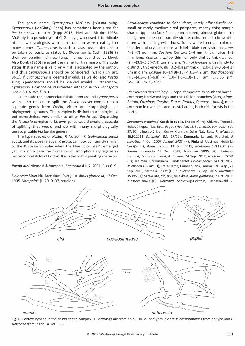
© 2018 Westerdijk Fungal Biodiversity Institute
Postia caesia complex
Editor-in-ChiefProf. dr P.W. Crous, Westerdijk Fungal Biodiversity Institute, P.O. Box 85167, 3508 AD Utrecht, The Netherlands.E-mail:[email protected]
111
The genus name Cyanosporus McGinty (=Postia subg. Cyanosporus (McGinty) Papp) has sometimes been used for Postia caesia complex (Papp 2015; Pieri and Rivoire 1998). McGinty is a pseudonym of C. G. Lloyd, who used it to ridicule his fellow mycologists who in his opinion were creating too many names. Cyanosporus is such a case, never intended to be taken seriously, as stated by Stevenson & Cash (1936) in their compendium of new fungal names published by Lloyd. Also Donk (1960) rejected the name for this reason. The code states that a name is valid only if it is accepted by the author, and thus Cyanosporus should be considered invalid (ICN art. 36.1). If Cyanosporus is deemed invalid, as we do, also Postia subg. Cyanosporus should be viewed invalid. Furthermore, Cyanosporus cannot be resurrected either due to Cyanospora Heald & F.A. Wolf 1910.
Quite aside the nomenclatural situation around Cyanosporus we see no reason to split the Postia caesia complex to a separate genus from Postia, either on morphological or phylogenetic grounds. The complex is distinct morphologically, but nevertheless very similar to other Postia spp. Separating the P. caesia complex to its own genus would create a cascade of splitting that would end up with many morphologically unrecognizable Postia-like genera.
The type species of Postia, P. lactea (=P. tephroleuca sensu auct.), and its close relative, P. grata, can look confusingly similar to the P. caesia complex when the blue color hasn’t emerged yet. In such a case the formation of amorphous aggregates in microscopical slides of Cotton Blue is the best separating character.
Postia alni Niemelä & Vampola, Karstenia 41: 7. 2001. Figs 6–9.
Holotype: Slovakia, Bratislava, Svätý Jur, Alnus glutinosa, 12 Oct. 1995, Vampola* (H 7019137, studied).
Basidiocarps conchate to flabelliform, rarely effused-reflexed, small or rarely medium-sized polypores, mostly thin; margin sharp. Upper surface first cream colored, almost glabrous to matt, then pubescent, radially striate, ochraceous to brownish, often with bluish-greyish hues. Tubes white to cream-colored, in older and dry specimens with light bluish-greyish tint; pores 4–6(–7) per mm. Section: Context 1–4 mm thick, tubes 1–6 mm long. Context hyphae thin- or only slightly thick-walled, (2.4–)3.9–5.5(–7.4) μm in diam. Tramal hyphae with slightly to distinctly thickened walls (0.2–0.8 μm thick), (2.0–)2.9–3.6(–4.3) μm in diam. Basidia 10–14.8(–16) × 3.3–4.2 μm. Basidiospores (4.1–)4.3–6.1(–6.8) × (1.0–)1.1–1.3(–1.5) µm, L=5.05 µm, W=1.20 µm, Q=4.22.
Distribution and ecology: Europe, temperate to southern boreal, common; hardwood logs and thick fallen branches (Acer, Alnus, Betula, Carpinus, Corylus, Fagus, Prunus, Quercus, Ulmus), most common in riversides and coastal areas, herb-rich forests in the north.
Specimens examined: Czech Republic, Jihočeský kraj, Chlum u Třeboně, Bukové Kopce Nat. Res., Fagus sylvatica, 18 Sep. 2010, Vampola* (MJ 27/10); Jihočeský kraj, Český Krumlov, Žofín Nat. Res., F. sylvatica, 16.IX.2012 Vampola* (MJ 17/12). Denmark, Lolland, Faursted, F. sylvatica, 4 Oct. 2007 Schigel 5425 (H). Finland, Uusimaa, Helsinki, Veräjämäki, Alnus incana, 19 Oct. 2011, Miettinen 14918.2* (H), Sorbus aucuparia, 12 Dec. 2015, Miettinen 19883 (H); Uusimaa, Helsinki, Pornaistenniemi, A. incana, 24 Sep. 2012, Miettinen 15741 (H); Uusimaa, Kirkkonummi, Sundsberget, Prunus padus, 24 Oct. 2012, Miettinen 15830* (H); Etelä-Häme, Hämeenlinna, Lammi, Betula sp., 21 Sep. 2016, Niemelä 9233* (H); S. aucuparia, 14 Sep. 2015, Miettinen 19386 (H); Satakunta, Ylöjärvi, Viljakkala, Alnus glutinosa, 2 Oct. 2011, Niemelä 8843 (H). Germany, Schleswig-Holstein, Sachsenwald, F.
Fig. 6. Context hyphae in the Postia caesia complex. All drawings are from holo-, iso- or neotypes, except P. caesiosimulans from epitype and P. subcaesia from Legon 14 Oct. 1995.
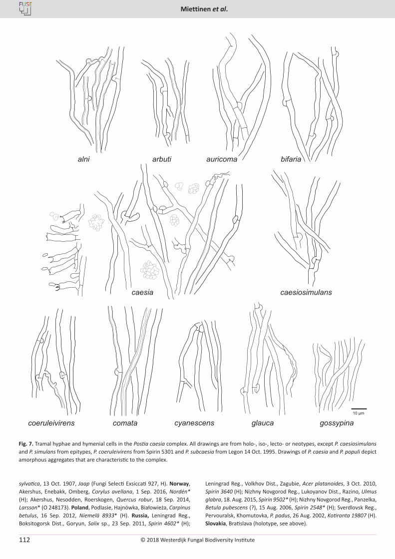
© 2018 Westerdijk Fungal Biodiversity Institute
Miettinen et al.
Editor-in-ChiefProf. dr P.W. Crous, Westerdijk Fungal Biodiversity Institute, P.O. Box 85167, 3508 AD Utrecht, The Netherlands.E-mail:[email protected]
112
Fig. 7. Tramal hyphae and hymenial cells in the Postia caesia complex. All drawings are from holo-, iso-, lecto- or neotypes, except P. caesiosimulans and P. simulans from epitypes, P. coeruleivirens from Spirin 5301 and P. subcaesia from Legon 14 Oct. 1995. Drawings of P. caesia and P. populi depict amorphous aggregates that are characteristic to the complex.
sylvatica, 13 Oct. 1907, Jaap (Fungi Selecti Exsiccati 927, H). Norway, Akershus, Enebakk, Omberg, Corylus avellana, 1 Sep. 2016, Nordén* (H); Akershus, Nesodden, Roerskogen, Quercus robur, 18 Sep. 2014, Larsson* (O 248173). Poland, Podlasie, Hajnówka, Białowieża, Carpinus betulus, 16 Sep. 2012, Niemelä 8933* (H). Russia, Leningrad Reg., Boksitogorsk Dist., Goryun, Salix sp., 23 Sep. 2011, Spirin 4602* (H);
Leningrad Reg., Volkhov Dist., Zagubie, Acer platanoides, 3 Oct. 2010, Spirin 3640 (H); Nizhny Novgorod Reg., Lukoyanov Dist., Razino, Ulmus glabra, 18. Aug. 2015, Spirin 9502* (H); Nizhny Novgorod Reg., Panzelka, Betula pubescens (?), 15 Aug. 2006, Spirin 2548* (H); Sverdlovsk Reg., Pervouralsk, Khomutovka, P. padus, 26 Aug. 2002, Kotiranta 19807 (H). Slovakia, Bratislava (holotype, see above).
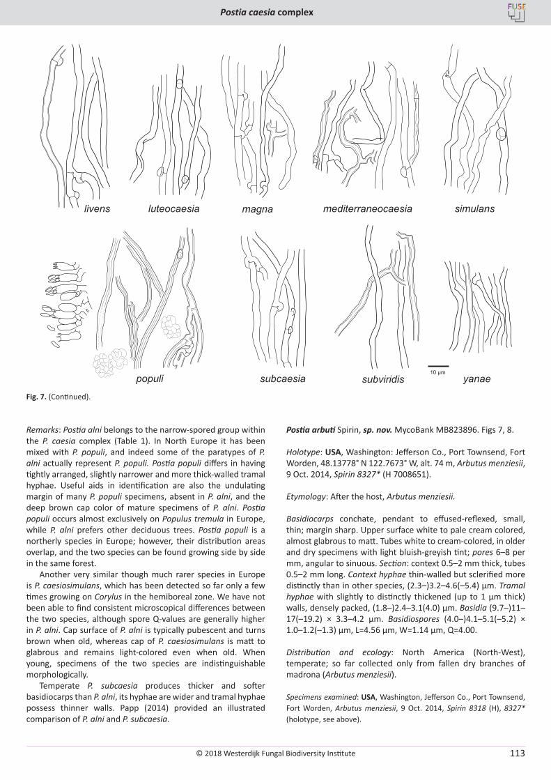
© 2018 Westerdijk Fungal Biodiversity Institute
Postia caesia complex
Editor-in-ChiefProf. dr P.W. Crous, Westerdijk Fungal Biodiversity Institute, P.O. Box 85167, 3508 AD Utrecht, The Netherlands.E-mail:[email protected]
113
Fig. 7. (Continued).
Remarks: Postia alni belongs to the narrow-spored group within the P. caesia complex (Table 1). In North Europe it has been mixed with P. populi, and indeed some of the paratypes of P. alni actually represent P. populi. Postia populi differs in having tightly arranged, slightly narrower and more thick-walled tramal hyphae. Useful aids in identification are also the undulating margin of many P. populi specimens, absent in P. alni, and the deep brown cap color of mature specimens of P. alni. Postia populi occurs almost exclusively on Populus tremula in Europe, while P. alni prefers other deciduous trees. Postia populi is a northerly species in Europe; however, their distribution areas overlap, and the two species can be found growing side by side in the same forest.
Another very similar though much rarer species in Europe is P. caesiosimulans, which has been detected so far only a few times growing on Corylus in the hemiboreal zone. We have not been able to find consistent microscopical differences between the two species, although spore Q-values are generally higher in P. alni. Cap surface of P. alni is typically pubescent and turns brown when old, whereas cap of P. caesiosimulans is matt to glabrous and remains light-colored even when old. When young, specimens of the two species are indistinguishable morphologically.
Temperate P. subcaesia produces thicker and softer basidiocarps than P. alni, its hyphae are wider and tramal hyphae possess thinner walls. Papp (2014) provided an illustrated comparison of P. alni and P. subcaesia.
Postia arbuti Spirin, sp. nov. MycoBank MB823896. Figs 7, 8.
Holotype: USA, Washington: Jefferson Co., Port Townsend, Fort Worden, 48.13778° N 122.7673° W, alt. 74 m, Arbutus menziesii, 9 Oct. 2014, Spirin 8327* (H 7008651).
Etymology: After the host, Arbutus menziesii.
Basidiocarps conchate, pendant to effused-reflexed, small, thin; margin sharp. Upper surface white to pale cream colored, almost glabrous to matt. Tubes white to cream-colored, in older and dry specimens with light bluish-greyish tint; pores 6–8 per mm, angular to sinuous. Section: context 0.5–2 mm thick, tubes 0.5–2 mm long. Context hyphae thin-walled but sclerified more distinctly than in other species, (2.3–)3.2–4.6(–5.4) μm. Tramal hyphae with slightly to distinctly thickened (up to 1 μm thick) walls, densely packed, (1.8–)2.4–3.1(4.0) μm. Basidia (9.7–)11–17(–19.2) × 3.3–4.2 μm. Basidiospores (4.0–)4.1–5.1(–5.2) × 1.0–1.2(–1.3) µm, L=4.56 µm, W=1.14 µm, Q=4.00.
Distribution and ecology: North America (North-West), temperate; so far collected only from fallen dry branches of madrona (Arbutus menziesii).
Specimens examined: USA, Washington, Jefferson Co., Port Townsend, Fort Worden, Arbutus menziesii, 9 Oct. 2014, Spirin 8318 (H), 8327* (holotype, see above).
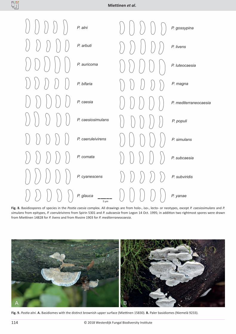
© 2018 Westerdijk Fungal Biodiversity Institute
Miettinen et al.
Editor-in-ChiefProf. dr P.W. Crous, Westerdijk Fungal Biodiversity Institute, P.O. Box 85167, 3508 AD Utrecht, The Netherlands.E-mail:[email protected]
114
Fig. 9. Postia alni. A. Basidiomes with the distinct brownish upper surface (Miettinen 15830). B. Paler basidiomes (Niemelä 9233).
Fig. 8. Basidiospores of species in the Postia caesia complex. All drawings are from holo-, iso-, lecto- or neotypes, except P. caesiosimulans and P. simulans from epitypes, P. coeruleivirens from Spirin 5301 and P. subcaesia from Legon 14 Oct. 1995; in addition two rightmost spores were drawn from Miettinen 14828 for P. livens and from Rivoire 1903 for P. mediterraneocaesia.
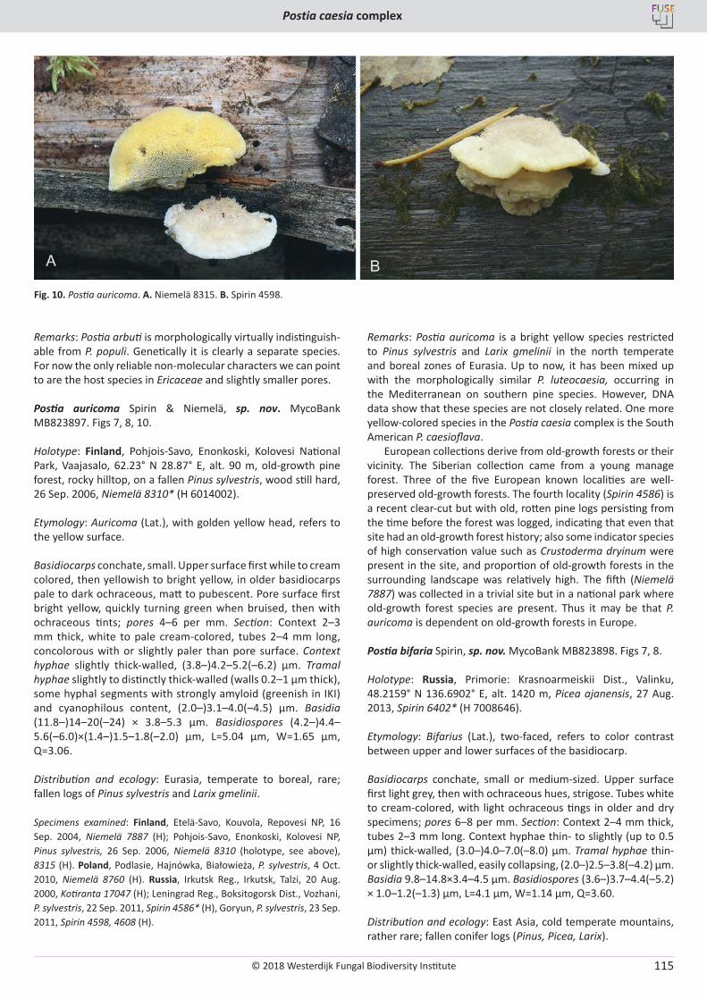
© 2018 Westerdijk Fungal Biodiversity Institute
Postia caesia complex
Editor-in-ChiefProf. dr P.W. Crous, Westerdijk Fungal Biodiversity Institute, P.O. Box 85167, 3508 AD Utrecht, The Netherlands.E-mail:[email protected]
115
Remarks: Postia arbuti is morphologically virtually indistinguish-able from P. populi. Genetically it is clearly a separate species. For now the only reliable non-molecular characters we can point to are the host species in Ericaceae and slightly smaller pores.
Postia auricoma Spirin & Niemelä, sp. nov. MycoBank MB823897. Figs 7, 8, 10.
Holotype: Finland, Pohjois-Savo, Enonkoski, Kolovesi National Park, Vaajasalo, 62.23° N 28.87° E, alt. 90 m, old-growth pine forest, rocky hilltop, on a fallen Pinus sylvestris, wood still hard, 26 Sep. 2006, Niemelä 8310* (H 6014002).
Etymology: Auricoma (Lat.), with golden yellow head, refers to the yellow surface.
Basidiocarps conchate, small. Upper surface first while to cream colored, then yellowish to bright yellow, in older basidiocarps pale to dark ochraceous, matt to pubescent. Pore surface first bright yellow, quickly turning green when bruised, then with ochraceous tints; pores 4–6 per mm. Section: Context 2–3 mm thick, white to pale cream-colored, tubes 2–4 mm long, concolorous with or slightly paler than pore surface. Context hyphae slightly thick-walled, (3.8–)4.2–5.2(–6.2) μm. Tramal hyphae slightly to distinctly thick-walled (walls 0.2–1 μm thick), some hyphal segments with strongly amyloid (greenish in IKI) and cyanophilous content, (2.0–)3.1–4.0(–4.5) μm. Basidia (11.8–)14–20(–24) × 3.8–5.3 μm. Basidiospores (4.2–)4.4–5.6(–6.0)×(1.4–)1.5–1.8(–2.0) µm, L=5.04 µm, W=1.65 µm, Q=3.06.
Distribution and ecology: Eurasia, temperate to boreal, rare; fallen logs of Pinus sylvestris and Larix gmelinii.
Specimens examined: Finland, Etelä-Savo, Kouvola, Repovesi NP, 16 Sep. 2004, Niemelä 7887 (H); Pohjois-Savo, Enonkoski, Kolovesi NP, Pinus sylvestris, 26 Sep. 2006, Niemelä 8310 (holotype, see above), 8315 (H). Poland, Podlasie, Hajnówka, Białowieża, P. sylvestris, 4 Oct. 2010, Niemelä 8760 (H). Russia, Irkutsk Reg., Irkutsk, Talzi, 20 Aug. 2000, Kotiranta 17047 (H); Leningrad Reg., Boksitogorsk Dist., Vozhani, P. sylvestris, 22 Sep. 2011, Spirin 4586* (H), Goryun, P. sylvestris, 23 Sep. 2011, Spirin 4598, 4608 (H).
Remarks: Postia auricoma is a bright yellow species restricted to Pinus sylvestris and Larix gmelinii in the north temperate and boreal zones of Eurasia. Up to now, it has been mixed up with the morphologically similar P. luteocaesia, occurring in the Mediterranean on southern pine species. However, DNA data show that these species are not closely related. One more yellow-colored species in the Postia caesia complex is the South American P. caesioflava.
European collections derive from old-growth forests or their vicinity. The Siberian collection came from a young manage forest. Three of the five European known localities are well-preserved old-growth forests. The fourth locality (Spirin 4586) is a recent clear-cut but with old, rotten pine logs persisting from the time before the forest was logged, indicating that even that site had an old-growth forest history; also some indicator species of high conservation value such as Crustoderma dryinum were present in the site, and proportion of old-growth forests in the surrounding landscape was relatively high. The fifth (Niemelä 7887) was collected in a trivial site but in a national park where old-growth forest species are present. Thus it may be that P. auricoma is dependent on old-growth forests in Europe.
Postia bifaria Spirin, sp. nov. MycoBank MB823898. Figs 7, 8.
Holotype: Russia, Primorie: Krasnoarmeiskii Dist., Valinku, 48.2159° N 136.6902° E, alt. 1420 m, Picea ajanensis, 27 Aug. 2013, Spirin 6402* (H 7008646).
Etymology: Bifarius (Lat.), two-faced, refers to color contrast between upper and lower surfaces of the basidiocarp.
Basidiocarps conchate, small or medium-sized. Upper surface first light grey, then with ochraceous hues, strigose. Tubes white to cream-colored, with light ochraceous tings in older and dry specimens; pores 6–8 per mm. Section: Context 2–4 mm thick, tubes 2–3 mm long. Context hyphae thin- to slightly (up to 0.5 µm) thick-walled, (3.0–)4.0–7.0(–8.0) μm. Tramal hyphae thin- or slightly thick-walled, easily collapsing, (2.0–)2.5–3.8(–4.2) μm. Basidia 9.8–14.8×3.4–4.5 μm. Basidiospores (3.6–)3.7–4.4(–5.2) × 1.0–1.2(–1.3) µm, L=4.1 µm, W=1.14 µm, Q=3.60.
Distribution and ecology: East Asia, cold temperate mountains, rather rare; fallen conifer logs (Pinus, Picea, Larix).
Fig. 10. Postia auricoma. A. Niemelä 8315. B. Spirin 4598.
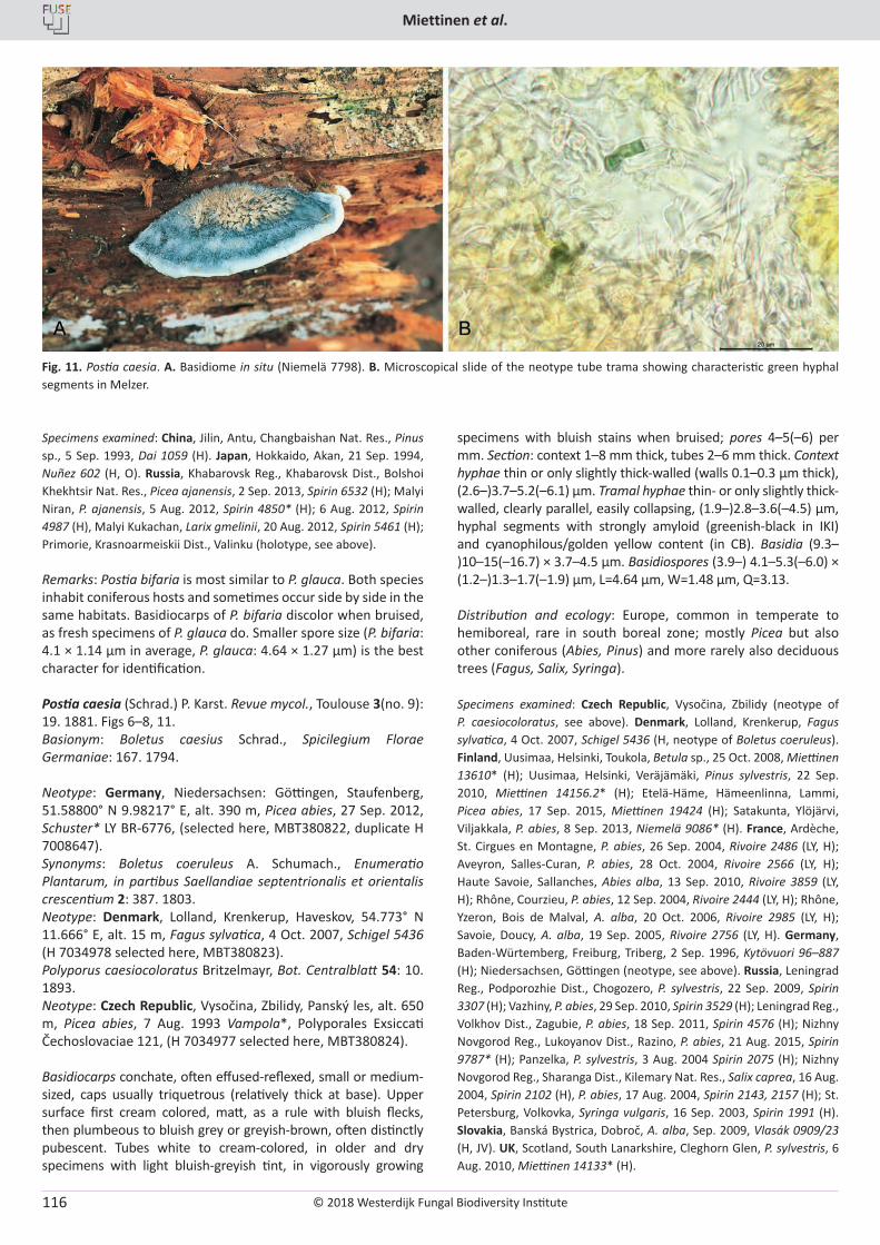
© 2018 Westerdijk Fungal Biodiversity Institute
Miettinen et al.
Editor-in-ChiefProf. dr P.W. Crous, Westerdijk Fungal Biodiversity Institute, P.O. Box 85167, 3508 AD Utrecht, The Netherlands.E-mail:[email protected]
116
Specimens examined: China, Jilin, Antu, Changbaishan Nat. Res., Pinus sp., 5 Sep. 1993, Dai 1059 (H). Japan, Hokkaido, Akan, 21 Sep. 1994, Nuñez 602 (H, O). Russia, Khabarovsk Reg., Khabarovsk Dist., Bolshoi Khekhtsir Nat. Res., Picea ajanensis, 2 Sep. 2013, Spirin 6532 (H); Malyi Niran, P. ajanensis, 5 Aug. 2012, Spirin 4850* (H); 6 Aug. 2012, Spirin 4987 (H), Malyi Kukachan, Larix gmelinii, 20 Aug. 2012, Spirin 5461 (H); Primorie, Krasnoarmeiskii Dist., Valinku (holotype, see above).
Remarks: Postia bifaria is most similar to P. glauca. Both species inhabit coniferous hosts and sometimes occur side by side in the same habitats. Basidiocarps of P. bifaria discolor when bruised, as fresh specimens of P. glauca do. Smaller spore size (P. bifaria: 4.1 × 1.14 µm in average, P. glauca: 4.64 × 1.27 µm) is the best character for identification.
Postia caesia (Schrad.) P. Karst. Revue mycol., Toulouse 3(no. 9): 19. 1881. Figs 6–8, 11.Basionym: Boletus caesius Schrad., Spicilegium Florae Germaniae: 167. 1794.
Neotype: Germany, Niedersachsen: Göttingen, Staufenberg, 51.58800° N 9.98217° E, alt. 390 m, Picea abies, 27 Sep. 2012, Schuster* LY BR-6776, (selected here, MBT380822, duplicate H 7008647). Synonyms: Boletus coeruleus A. Schumach., Enumeratio Plantarum, in partibus Saellandiae septentrionalis et orientalis crescentium 2: 387. 1803.Neotype: Denmark, Lolland, Krenkerup, Haveskov, 54.773° N 11.666° E, alt. 15 m, Fagus sylvatica, 4 Oct. 2007, Schigel 5436 (H 7034978 selected here, MBT380823).Polyporus caesiocoloratus Britzelmayr, Bot. Centralblatt 54: 10. 1893.Neotype: Czech Republic, Vysočina, Zbilidy, Panský les, alt. 650 m, Picea abies, 7 Aug. 1993 Vampola*, Polyporales Exsiccati Čechoslovaciae 121, (H 7034977 selected here, MBT380824).
Basidiocarps conchate, often effused-reflexed, small or medium-sized, caps usually triquetrous (relatively thick at base). Upper surface first cream colored, matt, as a rule with bluish flecks, then plumbeous to bluish grey or greyish-brown, often distinctly pubescent. Tubes white to cream-colored, in older and dry specimens with light bluish-greyish tint, in vigorously growing
specimens with bluish stains when bruised; pores 4–5(–6) per mm. Section: context 1–8 mm thick, tubes 2–6 mm thick. Context hyphae thin or only slightly thick-walled (walls 0.1–0.3 μm thick), (2.6–)3.7–5.2(–6.1) µm. Tramal hyphae thin- or only slightly thick-walled, clearly parallel, easily collapsing, (1.9–)2.8–3.6(–4.5) μm, hyphal segments with strongly amyloid (greenish-black in IKI) and cyanophilous/golden yellow content (in CB). Basidia (9.3–)10–15(–16.7) × 3.7–4.5 µm. Basidiospores (3.9–) 4.1–5.3(–6.0) × (1.2–)1.3–1.7(–1.9) µm, L=4.64 µm, W=1.48 µm, Q=3.13.
Distribution and ecology: Europe, common in temperate to hemiboreal, rare in south boreal zone; mostly Picea but also other coniferous (Abies, Pinus) and more rarely also deciduous trees (Fagus, Salix, Syringa).
Specimens examined: Czech Republic, Vysočina, Zbilidy (neotype of P. caesiocoloratus, see above). Denmark, Lolland, Krenkerup, Fagus sylvatica, 4 Oct. 2007, Schigel 5436 (H, neotype of Boletus coeruleus). Finland, Uusimaa, Helsinki, Toukola, Betula sp., 25 Oct. 2008, Miettinen 13610* (H); Uusimaa, Helsinki, Veräjämäki, Pinus sylvestris, 22 Sep. 2010, Miettinen 14156.2* (H); Etelä-Häme, Hämeenlinna, Lammi, Picea abies, 17 Sep. 2015, Miettinen 19424 (H); Satakunta, Ylöjärvi, Viljakkala, P. abies, 8 Sep. 2013, Niemelä 9086* (H). France, Ardèche, St. Cirgues en Montagne, P. abies, 26 Sep. 2004, Rivoire 2486 (LY, H); Aveyron, Salles-Curan, P. abies, 28 Oct. 2004, Rivoire 2566 (LY, H); Haute Savoie, Sallanches, Abies alba, 13 Sep. 2010, Rivoire 3859 (LY, H); Rhône, Courzieu, P. abies, 12 Sep. 2004, Rivoire 2444 (LY, H); Rhône, Yzeron, Bois de Malval, A. alba, 20 Oct. 2006, Rivoire 2985 (LY, H); Savoie, Doucy, A. alba, 19 Sep. 2005, Rivoire 2756 (LY, H). Germany, Baden-Würtemberg, Freiburg, Triberg, 2 Sep. 1996, Kytövuori 96–887 (H); Niedersachsen, Göttingen (neotype, see above). Russia, Leningrad Reg., Podporozhie Dist., Chogozero, P. sylvestris, 22 Sep. 2009, Spirin 3307 (H); Vazhiny, P. abies, 29 Sep. 2010, Spirin 3529 (H); Leningrad Reg., Volkhov Dist., Zagubie, P. abies, 18 Sep. 2011, Spirin 4576 (H); Nizhny Novgorod Reg., Lukoyanov Dist., Razino, P. abies, 21 Aug. 2015, Spirin 9787* (H); Panzelka, P. sylvestris, 3 Aug. 2004 Spirin 2075 (H); Nizhny Novgorod Reg., Sharanga Dist., Kilemary Nat. Res., Salix caprea, 16 Aug. 2004, Spirin 2102 (H), P. abies, 17 Aug. 2004, Spirin 2143, 2157 (H); St. Petersburg, Volkovka, Syringa vulgaris, 16 Sep. 2003, Spirin 1991 (H). Slovakia, Banská Bystrica, Dobroč, A. alba, Sep. 2009, Vlasák 0909/23 (H, JV). UK, Scotland, South Lanarkshire, Cleghorn Glen, P. sylvestris, 6 Aug. 2010, Miettinen 14133* (H).
Fig. 11. Postia caesia. A. Basidiome in situ (Niemelä 7798). B. Microscopical slide of the neotype tube trama showing characteristic green hyphal segments in Melzer.
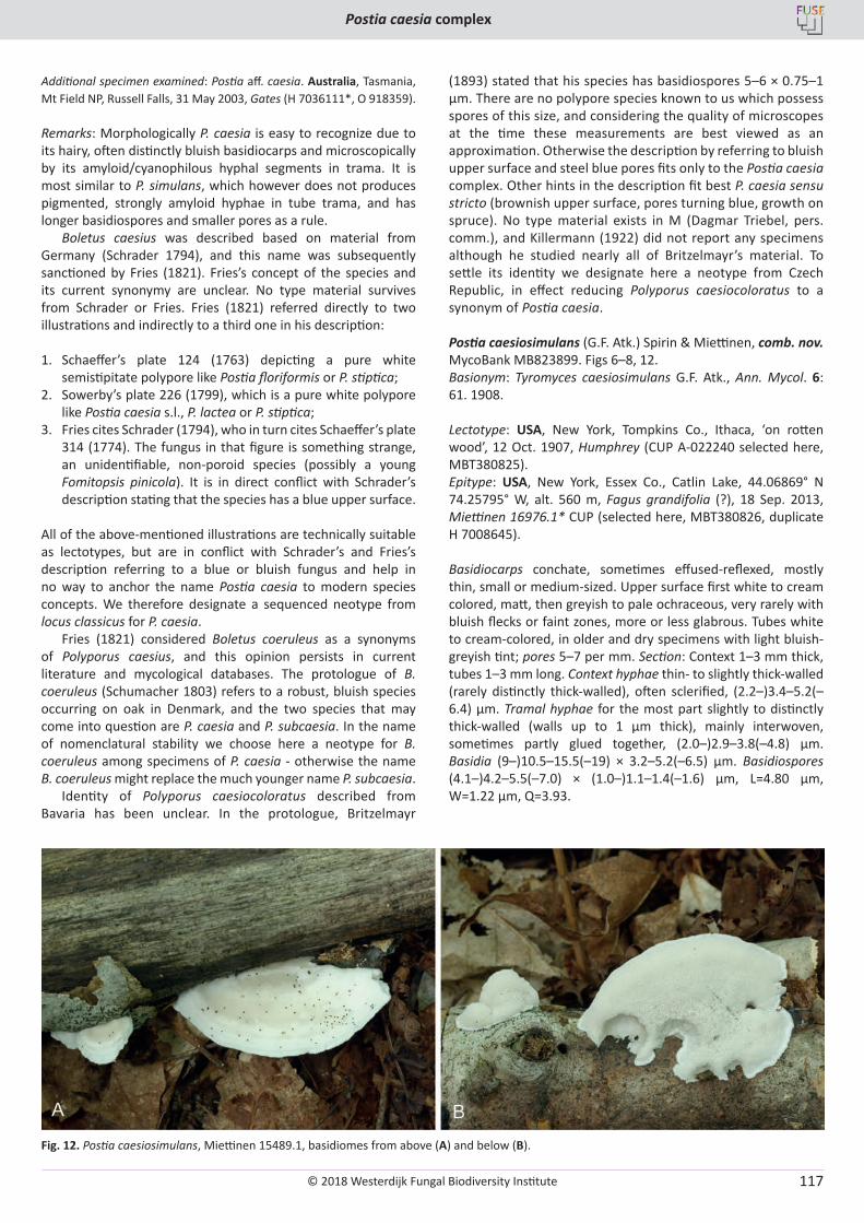
© 2018 Westerdijk Fungal Biodiversity Institute
Postia caesia complex
Editor-in-ChiefProf. dr P.W. Crous, Westerdijk Fungal Biodiversity Institute, P.O. Box 85167, 3508 AD Utrecht, The Netherlands.E-mail:[email protected]
117
Additional specimen examined: Postia aff. caesia. Australia, Tasmania, Mt Field NP, Russell Falls, 31 May 2003, Gates (H 7036111*, O 918359).
Remarks: Morphologically P. caesia is easy to recognize due to its hairy, often distinctly bluish basidiocarps and microscopically by its amyloid/cyanophilous hyphal segments in trama. It is most similar to P. simulans, which however does not produces pigmented, strongly amyloid hyphae in tube trama, and has longer basidiospores and smaller pores as a rule.
Boletus caesius was described based on material from Germany (Schrader 1794), and this name was subsequently sanctioned by Fries (1821). Fries’s concept of the species and its current synonymy are unclear. No type material survives from Schrader or Fries. Fries (1821) referred directly to two illustrations and indirectly to a third one in his description:
1. Schaeffer’s plate 124 (1763) depicting a pure white semistipitate polypore like Postia floriformis or P. stiptica;
2. Sowerby’s plate 226 (1799), which is a pure white polypore like Postia caesia s.l., P. lactea or P. stiptica;
3. Fries cites Schrader (1794), who in turn cites Schaeffer’s plate 314 (1774). The fungus in that figure is something strange, an unidentifiable, non-poroid species (possibly a young Fomitopsis pinicola). It is in direct conflict with Schrader’s description stating that the species has a blue upper surface.
All of the above-mentioned illustrations are technically suitable as lectotypes, but are in conflict with Schrader’s and Fries’s description referring to a blue or bluish fungus and help in no way to anchor the name Postia caesia to modern species concepts. We therefore designate a sequenced neotype from locus classicus for P. caesia.
Fries (1821) considered Boletus coeruleus as a synonyms of Polyporus caesius, and this opinion persists in current literature and mycological databases. The protologue of B. coeruleus (Schumacher 1803) refers to a robust, bluish species occurring on oak in Denmark, and the two species that may come into question are P. caesia and P. subcaesia. In the name of nomenclatural stability we choose here a neotype for B. coeruleus among specimens of P. caesia - otherwise the name B. coeruleus might replace the much younger name P. subcaesia.
Identity of Polyporus caesiocoloratus described from Bavaria has been unclear. In the protologue, Britzelmayr
(1893) stated that his species has basidiospores 5–6 × 0.75–1 μm. There are no polypore species known to us which possess spores of this size, and considering the quality of microscopes at the time these measurements are best viewed as an approximation. Otherwise the description by referring to bluish upper surface and steel blue pores fits only to the Postia caesia complex. Other hints in the description fit best P. caesia sensu stricto (brownish upper surface, pores turning blue, growth on spruce). No type material exists in M (Dagmar Triebel, pers. comm.), and Killermann (1922) did not report any specimens although he studied nearly all of Britzelmayr’s material. To settle its identity we designate here a neotype from Czech Republic, in effect reducing Polyporus caesiocoloratus to a synonym of Postia caesia.
Postia caesiosimulans (G.F. Atk.) Spirin & Miettinen, comb. nov. MycoBank MB823899. Figs 6–8, 12.Basionym: Tyromyces caesiosimulans G.F. Atk., Ann. Mycol. 6: 61. 1908.
Lectotype: USA, New York, Tompkins Co., Ithaca, ‘on rotten wood’, 12 Oct. 1907, Humphrey (CUP A-022240 selected here, MBT380825). Epitype: USA, New York, Essex Co., Catlin Lake, 44.06869° N 74.25795° W, alt. 560 m, Fagus grandifolia (?), 18 Sep. 2013, Miettinen 16976.1* CUP (selected here, MBT380826, duplicate H 7008645).
Basidiocarps conchate, sometimes effused-reflexed, mostly thin, small or medium-sized. Upper surface first white to cream colored, matt, then greyish to pale ochraceous, very rarely with bluish flecks or faint zones, more or less glabrous. Tubes white to cream-colored, in older and dry specimens with light bluish-greyish tint; pores 5–7 per mm. Section: Context 1–3 mm thick, tubes 1–3 mm long. Context hyphae thin- to slightly thick-walled (rarely distinctly thick-walled), often sclerified, (2.2–)3.4–5.2(–6.4) μm. Tramal hyphae for the most part slightly to distinctly thick-walled (walls up to 1 μm thick), mainly interwoven, sometimes partly glued together, (2.0–)2.9–3.8(–4.8) μm. Basidia (9–)10.5–15.5(–19) × 3.2–5.2(–6.5) μm. Basidiospores (4.1–)4.2–5.5(–7.0) × (1.0–)1.1–1.4(–1.6) µm, L=4.80 µm, W=1.22 µm, Q=3.93.
Fig. 12. Postia caesiosimulans, Miettinen 15489.1, basidiomes from above (A) and below (B).
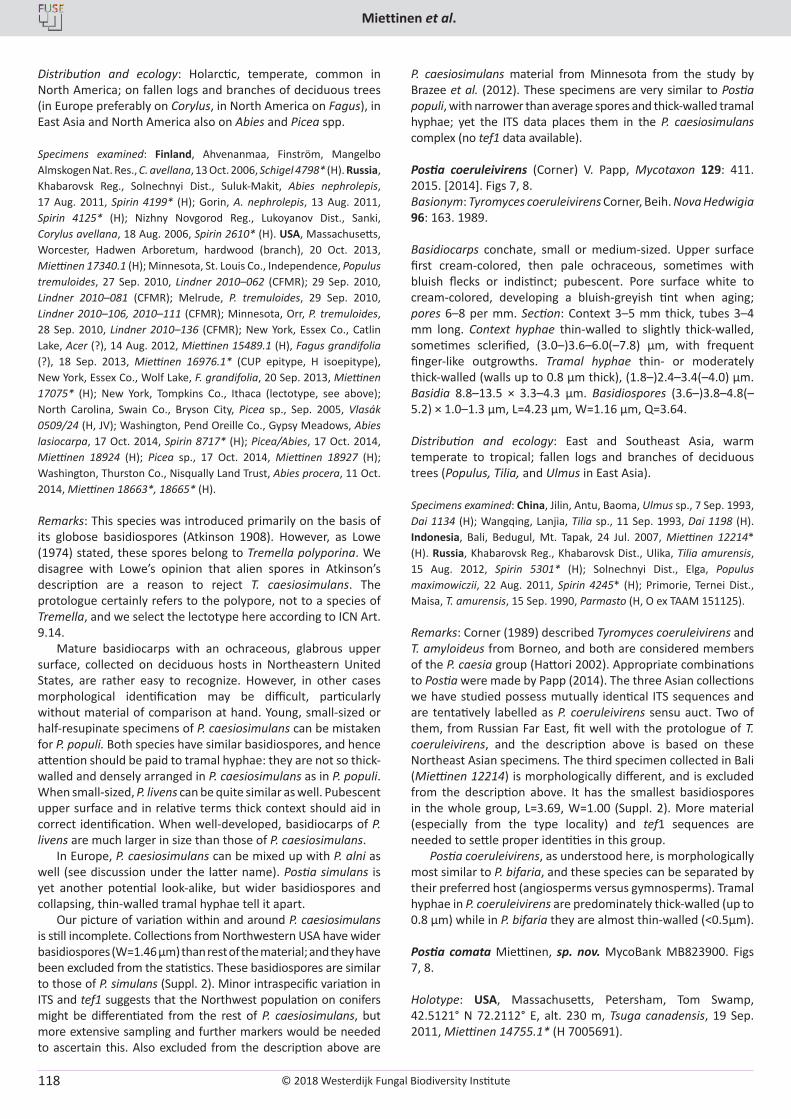
© 2018 Westerdijk Fungal Biodiversity Institute
Miettinen et al.
Editor-in-ChiefProf. dr P.W. Crous, Westerdijk Fungal Biodiversity Institute, P.O. Box 85167, 3508 AD Utrecht, The Netherlands.E-mail:[email protected]
118
Distribution and ecology: Holarctic, temperate, common in North America; on fallen logs and branches of deciduous trees (in Europe preferably on Corylus, in North America on Fagus), in East Asia and North America also on Abies and Picea spp.
Specimens examined: Finland, Ahvenanmaa, Finström, Mangelbo Almskogen Nat. Res., C. avellana, 13 Oct. 2006, Schigel 4798* (H). Russia, Khabarovsk Reg., Solnechnyi Dist., Suluk-Makit, Abies nephrolepis, 17 Aug. 2011, Spirin 4199* (H); Gorin, A. nephrolepis, 13 Aug. 2011, Spirin 4125* (H); Nizhny Novgorod Reg., Lukoyanov Dist., Sanki, Corylus avellana, 18 Aug. 2006, Spirin 2610* (H). USA, Massachusetts, Worcester, Hadwen Arboretum, hardwood (branch), 20 Oct. 2013, Miettinen 17340.1 (H); Minnesota, St. Louis Co., Independence, Populus tremuloides, 27 Sep. 2010, Lindner 2010–062 (CFMR); 29 Sep. 2010, Lindner 2010–081 (CFMR); Melrude, P. tremuloides, 29 Sep. 2010, Lindner 2010–106, 2010–111 (CFMR); Minnesota, Orr, P. tremuloides, 28 Sep. 2010, Lindner 2010–136 (CFMR); New York, Essex Co., Catlin Lake, Acer (?), 14 Aug. 2012, Miettinen 15489.1 (H), Fagus grandifolia (?), 18 Sep. 2013, Miettinen 16976.1* (CUP epitype, H isoepitype), New York, Essex Co., Wolf Lake, F. grandifolia, 20 Sep. 2013, Miettinen 17075* (H); New York, Tompkins Co., Ithaca (lectotype, see above); North Carolina, Swain Co., Bryson City, Picea sp., Sep. 2005, Vlasák 0509/24 (H, JV); Washington, Pend Oreille Co., Gypsy Meadows, Abies lasiocarpa, 17 Oct. 2014, Spirin 8717* (H); Picea/Abies, 17 Oct. 2014, Miettinen 18924 (H); Picea sp., 17 Oct. 2014, Miettinen 18927 (H); Washington, Thurston Co., Nisqually Land Trust, Abies procera, 11 Oct. 2014, Miettinen 18663*, 18665* (H).
Remarks: This species was introduced primarily on the basis of its globose basidiospores (Atkinson 1908). However, as Lowe (1974) stated, these spores belong to Tremella polyporina. We disagree with Lowe’s opinion that alien spores in Atkinson’s description are a reason to reject T. caesiosimulans. The protologue certainly refers to the polypore, not to a species of Tremella, and we select the lectotype here according to ICN Art. 9.14.
Mature basidiocarps with an ochraceous, glabrous upper surface, collected on deciduous hosts in Northeastern United States, are rather easy to recognize. However, in other cases morphological identification may be difficult, particularly without material of comparison at hand. Young, small-sized or half-resupinate specimens of P. caesiosimulans can be mistaken for P. populi. Both species have similar basidiospores, and hence attention should be paid to tramal hyphae: they are not so thick-walled and densely arranged in P. caesiosimulans as in P. populi. When small-sized, P. livens can be quite similar as well. Pubescent upper surface and in relative terms thick context should aid in correct identification. When well-developed, basidiocarps of P. livens are much larger in size than those of P. caesiosimulans.
In Europe, P. caesiosimulans can be mixed up with P. alni as well (see discussion under the latter name). Postia simulans is yet another potential look-alike, but wider basidiospores and collapsing, thin-walled tramal hyphae tell it apart.
Our picture of variation within and around P. caesiosimulans is still incomplete. Collections from Northwestern USA have wider basidiospores (W=1.46 µm) than rest of the material; and they have been excluded from the statistics. These basidiospores are similar to those of P. simulans (Suppl. 2). Minor intraspecific variation in ITS and tef1 suggests that the Northwest population on conifers might be differentiated from the rest of P. caesiosimulans, but more extensive sampling and further markers would be needed to ascertain this. Also excluded from the description above are
P. caesiosimulans material from Minnesota from the study by Brazee et al. (2012). These specimens are very similar to Postia populi, with narrower than average spores and thick-walled tramal hyphae; yet the ITS data places them in the P. caesiosimulans complex (no tef1 data available).
Postia coeruleivirens (Corner) V. Papp, Mycotaxon 129: 411. 2015. [2014]. Figs 7, 8.Basionym: Tyromyces coeruleivirens Corner, Beih. Nova Hedwigia 96: 163. 1989.
Basidiocarps conchate, small or medium-sized. Upper surface first cream-colored, then pale ochraceous, sometimes with bluish flecks or indistinct; pubescent. Pore surface white to cream-colored, developing a bluish-greyish tint when aging; pores 6–8 per mm. Section: Context 3–5 mm thick, tubes 3–4 mm long. Context hyphae thin-walled to slightly thick-walled, sometimes sclerified, (3.0–)3.6–6.0(–7.8) μm, with frequent finger-like outgrowths. Tramal hyphae thin- or moderately thick-walled (walls up to 0.8 μm thick), (1.8–)2.4–3.4(–4.0) μm. Basidia 8.8–13.5 × 3.3–4.3 μm. Basidiospores (3.6–)3.8–4.8(–5.2) × 1.0–1.3 µm, L=4.23 µm, W=1.16 µm, Q=3.64.
Distribution and ecology: East and Southeast Asia, warm temperate to tropical; fallen logs and branches of deciduous trees (Populus, Tilia, and Ulmus in East Asia).
Specimens examined: China, Jilin, Antu, Baoma, Ulmus sp., 7 Sep. 1993, Dai 1134 (H); Wangqing, Lanjia, Tilia sp., 11 Sep. 1993, Dai 1198 (H). Indonesia, Bali, Bedugul, Mt. Tapak, 24 Jul. 2007, Miettinen 12214* (H). Russia, Khabarovsk Reg., Khabarovsk Dist., Ulika, Tilia amurensis, 15 Aug. 2012, Spirin 5301* (H); Solnechnyi Dist., Elga, Populus maximowiczii, 22 Aug. 2011, Spirin 4245* (H); Primorie, Ternei Dist., Maisa, T. amurensis, 15 Sep. 1990, Parmasto (H, O ex TAAM 151125).
Remarks: Corner (1989) described Tyromyces coeruleivirens and T. amyloideus from Borneo, and both are considered members of the P. caesia group (Hattori 2002). Appropriate combinations to Postia were made by Papp (2014). The three Asian collections we have studied possess mutually identical ITS sequences and are tentatively labelled as P. coeruleivirens sensu auct. Two of them, from Russian Far East, fit well with the protologue of T. coeruleivirens, and the description above is based on these Northeast Asian specimens. The third specimen collected in Bali (Miettinen 12214) is morphologically different, and is excluded from the description above. It has the smallest basidiospores in the whole group, L=3.69, W=1.00 (Suppl. 2). More material (especially from the type locality) and tef1 sequences are needed to settle proper identities in this group.
Postia coeruleivirens, as understood here, is morphologically most similar to P. bifaria, and these species can be separated by their preferred host (angiosperms versus gymnosperms). Tramal hyphae in P. coeruleivirens are predominately thick-walled (up to 0.8 µm) while in P. bifaria they are almost thin-walled (<0.5µm).
Postia comata Miettinen, sp. nov. MycoBank MB823900. Figs 7, 8.
Holotype: USA, Massachusetts, Petersham, Tom Swamp, 42.5121° N 72.2112° E, alt. 230 m, Tsuga canadensis, 19 Sep. 2011, Miettinen 14755.1* (H 7005691).
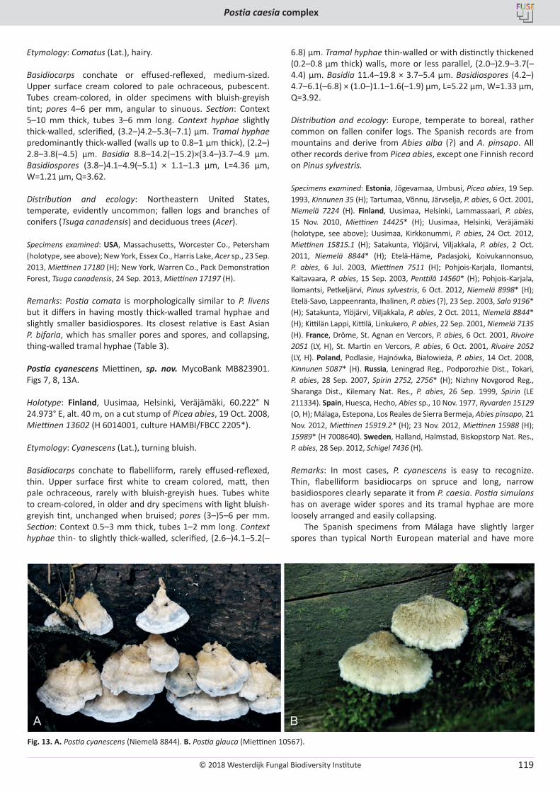
© 2018 Westerdijk Fungal Biodiversity Institute
Postia caesia complex
Editor-in-ChiefProf. dr P.W. Crous, Westerdijk Fungal Biodiversity Institute, P.O. Box 85167, 3508 AD Utrecht, The Netherlands.E-mail:[email protected]
119
Etymology: Comatus (Lat.), hairy.
Basidiocarps conchate or effused-reflexed, medium-sized. Upper surface cream colored to pale ochraceous, pubescent. Tubes cream-colored, in older specimens with bluish-greyish tint; pores 4–6 per mm, angular to sinuous. Section: Context 5–10 mm thick, tubes 3–6 mm long. Context hyphae slightly thick-walled, sclerified, (3.2–)4.2–5.3(–7.1) μm. Tramal hyphae predominantly thick-walled (walls up to 0.8–1 μm thick), (2.2–) 2.8–3.8(–4.5) μm. Basidia 8.8–14.2(–15.2)×(3.4–)3.7–4.9 μm. Basidiospores (3.8–)4.1–4.9(–5.1) × 1.1–1.3 µm, L=4.36 µm, W=1.21 µm, Q=3.62.
Distribution and ecology: Northeastern United States, temperate, evidently uncommon; fallen logs and branches of conifers (Tsuga canadensis) and deciduous trees (Acer).
Specimens examined: USA, Massachusetts, Worcester Co., Petersham (holotype, see above); New York, Essex Co., Harris Lake, Acer sp., 23 Sep. 2013, Miettinen 17180 (H); New York, Warren Co., Pack Demonstration Forest, Tsuga canadensis, 24 Sep. 2013, Miettinen 17197 (H).
Remarks: Postia comata is morphologically similar to P. livens but it differs in having mostly thick-walled tramal hyphae and slightly smaller basidiospores. Its closest relative is East Asian P. bifaria, which has smaller pores and spores, and collapsing, thing-walled tramal hyphae (Table 3).
Postia cyanescens Miettinen, sp. nov. MycoBank MB823901. Figs 7, 8, 13A.
Holotype: Finland, Uusimaa, Helsinki, Veräjämäki, 60.222° N 24.973° E, alt. 40 m, on a cut stump of Picea abies, 19 Oct. 2008, Miettinen 13602 (H 6014001, culture HAMBI/FBCC 2205*).
Etymology: Cyanescens (Lat.), turning bluish.
Basidiocarps conchate to flabelliform, rarely effused-reflexed, thin. Upper surface first white to cream colored, matt, then pale ochraceous, rarely with bluish-greyish hues. Tubes white to cream-colored, in older and dry specimens with light bluish-greyish tint, unchanged when bruised; pores (3–)5–6 per mm. Section: Context 0.5–3 mm thick, tubes 1–2 mm long. Context hyphae thin- to slightly thick-walled, sclerified, (2.6–)4.1–5.2(–
6.8) μm. Tramal hyphae thin-walled or with distinctly thickened (0.2–0.8 μm thick) walls, more or less parallel, (2.0–)2.9–3.7(–4.4) μm. Basidia 11.4–19.8 × 3.7–5.4 μm. Basidiospores (4.2–) 4.7–6.1(–6.8) × (1.0–)1.1–1.6(–1.9) µm, L=5.22 µm, W=1.33 µm, Q=3.92.
Distribution and ecology: Europe, temperate to boreal, rather common on fallen conifer logs. The Spanish records are from mountains and derive from Abies alba (?) and A. pinsapo. All other records derive from Picea abies, except one Finnish record on Pinus sylvestris.
Specimens examined: Estonia, Jõgevamaa, Umbusi, Picea abies, 19 Sep. 1993, Kinnunen 35 (H); Tartumaa, Võnnu, Järvselja, P. abies, 6 Oct. 2001, Niemelä 7224 (H). Finland, Uusimaa, Helsinki, Lammassaari, P. abies, 15 Nov. 2010, Miettinen 14425* (H); Uusimaa, Helsinki, Veräjämäki (holotype, see above); Uusimaa, Kirkkonummi, P. abies, 24 Oct. 2012, Miettinen 15815.1 (H); Satakunta, Ylöjärvi, Viljakkala, P. abies, 2 Oct. 2011, Niemelä 8844* (H); Etelä-Häme, Padasjoki, Koivukannonsuo, P. abies, 6 Jul. 2003, Miettinen 7511 (H); Pohjois-Karjala, Ilomantsi, Kaitavaara, P. abies, 15 Sep. 2003, Penttilä 14560* (H); Pohjois-Karjala, Ilomantsi, Petkeljärvi, Pinus sylvestris, 6 Oct. 2012, Niemelä 8998* (H); Etelä-Savo, Lappeenranta, Ihalinen, P. abies (?), 23 Sep. 2003, Salo 9196* (H); Satakunta, Ylöjärvi, Viljakkala, P. abies, 2 Oct. 2011, Niemelä 8844* (H); Kittilän Lappi, Kittilä, Linkukero, P. abies, 22 Sep. 2001, Niemelä 7135 (H). France, Drôme, St. Agnan en Vercors, P. abies, 6 Oct. 2001, Rivoire 2051 (LY, H), St. Martin en Vercors, P. abies, 6 Oct. 2001, Rivoire 2052 (LY, H). Poland, Podlasie, Hajnówka, Białowieża, P. abies, 14 Oct. 2008, Kinnunen 5087* (H). Russia, Leningrad Reg., Podporozhie Dist., Tokari, P. abies, 28 Sep. 2007, Spirin 2752, 2756* (H); Nizhny Novgorod Reg., Sharanga Dist., Kilemary Nat. Res., P. abies, 26 Sep. 1999, Spirin (LE 211334). Spain, Huesca, Hecho, Abies sp., 10 Nov. 1977, Ryvarden 15129 (O, H); Málaga, Estepona, Los Reales de Sierra Bermeja, Abies pinsapo, 21 Nov. 2012, Miettinen 15919.2* (H); 23 Nov. 2012, Miettinen 15988 (H); 15989* (H 7008640). Sweden, Halland, Halmstad, Biskopstorp Nat. Res., P. abies, 28 Sep. 2012, Schigel 7436 (H).
Remarks: In most cases, P. cyanescens is easy to recognize. Thin, flabelliform basidiocarps on spruce and long, narrow basidiospores clearly separate it from P. caesia. Postia simulans has on average wider spores and its tramal hyphae are more loosely arranged and easily collapsing.
The Spanish specimens from Málaga have slightly larger spores than typical North European material and have more
Fig. 13. A. Postia cyanescens (Niemelä 8844). B. Postia glauca (Miettinen 10567).
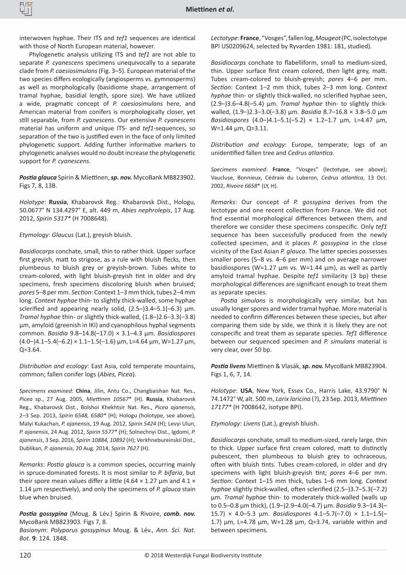
© 2018 Westerdijk Fungal Biodiversity Institute
Miettinen et al.
Editor-in-ChiefProf. dr P.W. Crous, Westerdijk Fungal Biodiversity Institute, P.O. Box 85167, 3508 AD Utrecht, The Netherlands.E-mail:[email protected]
120
interwoven hyphae. Their ITS and tef1 sequences are identical with those of North European material, however.
Phylogenetic analysis utilizing ITS and tef1 are not able to separate P. cyanescens specimens unequivocally to a separate clade from P. caesiosimulans (Fig. 3–5). European material of the two species differs ecologically (angiosperms vs. gymnosperms) as well as morphologically (basidiome shape, arrangement of tramal hyphae, basidial length, spore size). We have utilized a wide, pragmatic concept of P. caesiosimulans here, and American material from conifers is morphologically closer, yet still separable, from P. cyanescens. Our extensive P. cyanescens material has uniform and unique ITS- and tef1-sequences, so separation of the two is justified even in the face of only limited phylogenetic support. Adding further informative markers to phylogenetic analyses would no doubt increase the phylogenetic support for P. cyanescens.
Postia glauca Spirin & Miettinen, sp. nov. MycoBank MB823902. Figs 7, 8, 13B.
Holotype: Russia, Khabarovsk Reg.: Khabarovsk Dist., Hologu, 50.0677° N 134.4297° E, alt. 449 m, Abies nephrolepis, 17 Aug. 2012, Spirin 5317* (H 7008648).
Etymology: Glaucus (Lat.), greyish bluish.
Basidiocarps conchate, small, thin to rather thick. Upper surface first greyish, matt to strigose, as a rule with bluish flecks, then plumbeous to bluish grey or greyish-brown. Tubes white to cream-colored, with light bluish-greyish tint in older and dry specimens, fresh specimens discoloring bluish when bruised; pores 5–8 per mm. Section: Context 1–3 mm thick, tubes 2–4 mm long. Context hyphae thin- to slightly thick-walled, some hyphae sclerified and appearing nearly solid, (2.5–)3.4–5.1(–6.3) μm. Tramal hyphae thin- or slightly thick-walled, (1.8–)2.6–3.3(–3.8) μm, amyloid (greenish in IKI) and cyanophilous hyphal segments common. Basidia 9.8–14.8(–17.0) × 3.1–4.3 μm. Basidiospores (4.0–)4.1–5.4(–6.2) × 1.1–1.5(–1.6) µm, L=4.64 µm, W=1.27 µm, Q=3.64.
Distribution and ecology: East Asia, cold temperate mountains, common; fallen conifer logs (Abies, Picea).
Specimens examined: China, Jilin, Antu Co., Changbaishan Nat. Res., Picea sp., 27 Aug. 2005, Miettinen 10567* (H). Russia, Khabarovsk Reg., Khabarovsk Dist., Bolshoi Khekhtsir Nat. Res., Picea ajanensis, 2–3 Sep. 2013, Spirin 6548, 6580* (H); Hologu (holotype, see above), Malyi Kukachan, P. ajanensis, 19 Aug. 2012, Spirin 5424 (H); Levyi Ulun, P. ajanensis, 24 Aug. 2012, Spirin 5577* (H); Solnechnyi Dist., Igdomi, P. ajanensis, 3 Sep. 2016, Spirin 10884, 10892 (H); Verkhnebureinskii Dist., Dublikan, P. ajanensis, 20 Aug. 2014, Spirin 7627 (H).
Remarks: Postia glauca is a common species, occurring mainly in spruce-dominated forests. It is most similar to P. bifaria, but their spore mean values differ a little (4.64 × 1.27 µm and 4.1 × 1.14 µm respectively), and only the specimens of P. glauca stain blue when bruised.
Postia gossypina (Moug. & Lév.) Spirin & Rivoire, comb. nov. MycoBank MB823903. Figs 7, 8.Basionym: Polyporus gossypinus Moug. & Lév., Ann. Sci. Nat. Bot. 9: 124. 1848.
Lectotype: France, “Vosges”, fallen log, Mougeot (PC, isolectotype BPI US0209624, selected by Ryvarden 1981: 181, studied).
Basidiocarps conchate to flabelliform, small to medium-sized, thin. Upper surface first cream colored, then light grey, matt. Tubes cream-colored to bluish-greyish; pores 4–6 per mm. Section: Context 1–2 mm thick, tubes 2–3 mm long. Context hyphae thin- or slightly thick-walled, no sclerified hyphae seen, (2.9–)3.6–4.8(–5.4) μm. Tramal hyphae thin- to slightly thick-walled, (1.9–)2.3–3.0(–3.8) μm. Basidia 8.7–16.8 × 3.8–5.0 μm Basidiospores (4.0–)4.1–5.1(–5.2) × 1.2–1.7 µm, L=4.47 µm, W=1.44 µm, Q=3.11.
Distribution and ecology: Europe, temperate; logs of an unidentified fallen tree and Cedrus atlantica.
Specimens examined: France, “Vosges” (lectotype, see above); Vaucluse, Bonnieux, Cédraie du Luberon, Cedrus atlantica, 13 Oct. 2002, Rivoire 6658* (LY, H).
Remarks: Our concept of P. gossypina derives from the lectotype and one recent collection from France. We did not find essential morphological differences between them, and therefore we consider these specimens conspecific. Only tef1 sequence has been successfully produced from the newly collected specimen, and it places P. gossypina in the close vicinity of the East Asian P. glauca. The latter species possesses smaller pores (5–8 vs. 4–6 per mm) and on average narrower basidiospores (W=1.27 µm vs. W=1.44 µm), as well as partly amyloid tramal hyphae. Despite tef1 similarity (3 bp) these morphological differences are significant enough to treat them as separate species.
Postia simulans is morphologically very similar, but has usually longer spores and wider tramal hyphae. More material is needed to confirm differences between these species, but after comparing them side by side, we think it is likely they are not conspecific and treat them as separate species. Tef1 difference between our sequenced specimen and P. simulans material is very clear, over 50 bp.
Postia livens Miettinen & Vlasák, sp. nov. MycoBank MB823904. Figs 1, 6, 7, 14.
Holotype: USA, New York, Essex Co., Harris Lake, 43.9790° N 74.1472° W, alt. 500 m, Larix laricina (?), 23 Sep. 2013, Miettinen 17177* (H 7008642, isotype BPI).
Etymology: Livens (Lat.), greyish bluish.
Basidiocarps conchate, small to medium-sized, rarely large, thin to thick. Upper surface first cream colored, matt to distinctly pubescent, then plumbeous to bluish grey to ochraceous, often with bluish tints. Tubes cream-colored, in older and dry specimens with light bluish-greyish tint; pores 4–6 per mm. Section: Context 1–15 mm thick, tubes 1–6 mm long. Context hyphae slightly thick-walled, often sclerified (2.5–)3.7–5.3(–7.2) μm. Tramal hyphae thin- to moderately thick-walled (walls up to 0.5–0.8 μm thick), (1.9–)2.9–4.0(–4.7) μm. Basidia 9.3–14.3(–15.7) × 4.0–5.3 μm. Basidiospores 4.1–5.7(–7.0) × 1.1–1.5(–1.7) µm, L=4.78 µm, W=1.28 µm, Q=3.74, variable within and between specimens.
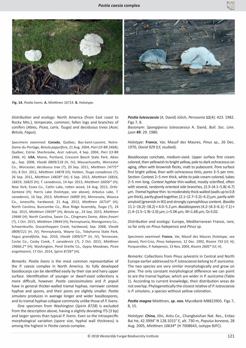
© 2018 Westerdijk Fungal Biodiversity Institute
Postia caesia complex
Editor-in-ChiefProf. dr P.W. Crous, Westerdijk Fungal Biodiversity Institute, P.O. Box 85167, 3508 AD Utrecht, The Netherlands.E-mail:[email protected]
121
Distribution and ecology: North America (from East coast to Rocky Mts.), temperate, common; fallen logs and branches of conifers (Abies, Picea, Larix, Tsuga) and deciduous trees (Acer, Betula, Fagus).
Specimens examined: Canada, Québec, Bas-Saint-Laurent, Notre-Dame-du-Portage, Betula papyrifera, 21 Aug. 2004, Pieri (LY-BR 2468); Québec, Estrie: Sherbrooke, Acer rubrum, 4 Sep. 2004, Pieri (LY-BR 2466, H). USA, Maine, Portland, Crescent Beech State Park, Abies sp., Sep. 2008, Vlasák 0809/118 (H, JV); Massachusetts, Worcester Co., Worcester, deciduous tree (?), 20 Sep. 2011, Miettinen 14775* (H); 8 Oct. 2011, Miettinen 14878 (H); Holden, Tsuga canadensis (?), 26 Sep. 2011, Miettinen 14828* (H); 6 Sep. 2013, Miettinen 16816, 16819, 16825 (H); T. canadensis, 14 Apr. 2013, Miettinen 16056* (H); New York, Essex Co., Catlin Lake, rotten wood, 14 Aug. 2012, Ortiz-Santana (H); Harris Lake (holotype, see above); Arbutus Lake, T. canadensis, 16 Sep. 2013, Miettinen 16899 (H); Minnesota, Waseca Co., Janesville, hardwood, 21 Aug. 2013, Miettinen 16714* (H); North Carolina, Buncombe Co., Blue Ridge Assembly, Tsuga (?), 24 Sep. 2015, Miettinen 19439* (H); Betula sp., 24 Sep. 2015, Miettinen 19446 (H); North Carolina, Swain Co., Clingmans Dome, Abies fraseri (?), 1 Oct. 2015, Miettinen 19644 (H); Pennsylvania, Montgomery Co., Schwenksville, Gossenhoppen Creek, hardwood, Sep. 2008, Vlasák 0809/121 (H, JV); Pennsylvania, Wayne Co., Tobyhanna State Park, Fagus grandifolia, Sep. 2010, Vlasák 1009/57* (H, JV); Tennessee, Cocke Co., Cosby Creek, T. canadensis (?), 2 Oct. 2015, Miettinen 19666.2* (H); Washington, Pend Oreille Co., Gypsy Meadows, Picea engelmannii, 17 Oct. 2014, Spirin 8728* (H).
Remarks: Postia livens is the most common representative of the P. caesia complex in North America. Its fully developed basidiocarps can be identified easily by their size and hairy upper surface. Identification of younger or dwarf-sized collections is more difficult, however. Postia caesiosimulans and P. populi have in general thicker-walled tramal hyphae, narrower context hyphae and spores, and their pores are slightly smaller. Postia simulans produces in average longer and wider basidiospores, and its tramal hyphae collapse commonly unlike those of P. livens.
One specimen from Washington (Spirin 8728) is excluded from the description above, having a slightly deviating ITS (3 bp) and larger spores than typical P. livens. Even so the intraspecific morphological variation (spore size, hyphal wall thickness) is among the highest in Postia caesia complex.
Postia luteocaesia (A. David) Jülich, Persoonia 11(4): 423. 1982. Figs 7, 8.Basionym: Spongiporus luteocaesius A. David, Bull. Soc. Linn. Lyon 49: 29. 1980.
Holotype: France, Var, Massif des Maures, Pinus sp., 26 Dec. 1970, David 929 (LY, studied).
Basidiocarps conchate, medium-sized. Upper surface first cream colored, then yellowish to bright yellow, pale to dark ochraceous on aging, often with brownish flecks, matt to pubescent. Pore surface first bright yellow, then with ochraceous tints; pores 3–5 per mm. Section: Context 2–5 mm thick, white to pale cream-colored, tubes 2–5 mm long. Context hyphae thin-walled, mostly sclerified, often with several, randomly oriented side branches, (3.3–)4.1–5.8(–6.7) μm. Tramal hyphae thin- to moderately thick-walled (walls up to 0.8 μm thick), partly glued together, (2.1–)2.7–3.2(–4.2) μm, partly with amyloid (greenish in IKI) and strongly cyanophilous content. Basidia 11.1–16.2(–18.2) × 4.0–5.2 μm. Basidiospores (4.2–)4.3–6.1(–7.1) × (1.4–)1.5–1.9(–2.0) µm, L=5.06 µm, W=1.68 µm, Q=3.02.
Distribution and ecology: Europe, Mediterranean France, rare; so far only on Pinus halepensis and Pinus sp.
Specimens examined: France, Var, Massif des Maures (holotype, see above); Port-Cros, Pinus halepensis, 12 Dec. 1992, Rivoire 733 (LY, H); Porquerolles, P. halepensis, 13 Nov. 2004, Rivoire 2605* (LY, H).
Remarks: Collections from Pinus sylvestris in Central and North Europe earlier addressed to P. luteocaesia belong to P. auricoma. The two species are very similar morphologically and grow on pine. The only constant morphological difference we can point to are the tramal hyphae, which are wider in P. auricoma (Table 1). According to current knowledge, their distribution areas do not overlap. Phylogenetically the closest relative of P. luteocaesia is P. simulans, a species without yellow coloration.
Postia magna Miettinen, sp. nov. MycoBank MB823905. Figs 7, 8, 15.
Holotype: China, Jilin, Antu Co., Changbaishan Nat. Res., Erdao Bai He, 42.3994° N 128.1015° E, alt. 730 m, Populus koreana, 28 Aug. 2005, Miettinen 10634* (H 7008643, isotype BJFC).
Fig. 14. Postia livens. A. Miettinen 16714. B. Holotype.
© 2018 Westerdijk Fungal Biodiversity Institute
Miettinen et al.
Editor-in-ChiefProf. dr P.W. Crous, Westerdijk Fungal Biodiversity Institute, P.O. Box 85167, 3508 AD Utrecht, The Netherlands.E-mail:[email protected]
122
Etymology: Magnus (Lat.), big.
Basidiocarps conchate, medium-sized (7×4 cm), thick, margin sharp. Upper surface white, drying cream colored to light greyish and ochraceous, distinctly pubescent. Tubes white, drying ochraceous with bluish tint, pores (3–)4–5 per mm. Section: Context 3–10 mm thick, white, tubes 3–6 mm long. Context hyphae thin-walled (to slightly thick-walled), (3.4–)4.2–6.0(–6.6) µm. Tramal hyphae slightly thick-walled, regularly branched, (1.5–)2.2–3.3(–3.8) µm. Basidia 10–12.5×3.2–4 μm. Basidiospores 3.6–4.4(–4.5) × 1.0–1.2 µm, L=3.97 µm, W=1.13 µm, Q=3.51.
Distribution and ecology: Temperate China and South Korea, so far known from hardwoods only.
Specimen examined: Postia magna. China, Jilin, Antu (holotype, see above).
Remarks: This species reminds P. subcaesia and P. livens with its large, hairy basidiocarps. Microscopically P. magna seems to differ from them by its smaller spores. As far as we know their distribution areas do not overlap. Although we have examined only one collection from Northeast China, available ITS sequences indicate that the species is widespread in China and Korea (Shen et al. 2014, Kim et al. 2015, Jang et al. 2016).
Postia mediterraneocaesia M. Pieri & B. Rivoire, Bull. Semestriel Féd. Assoc. Mycol. Méditerranéennes 28: 34. 2005. Figs 7, 8.
Holotype: France, Bouches du Rhône, St. Rémy de Provence, Pinus halepensis, 11 Nov. 2000, Pieri & Rivoire 1946 (LY, studied).
Basidiocarps effused-reflexed or resupinate with detaching or adnate margin, thin. Upper surface white to cream colored or pale ochraceous, matt or almost glabrous. Tubes white to cream-colored, in older and dry specimens pale ochraceous, with light bluish-greyish tint; pores (4)5–6 per mm. Section: Context 0.2–1 mm thick, tubes 0.5–2 mm long. Context hyphae thin- to slightly thick-walled, often sclerified, (2.4–)3.1–4.0(–4.8) μm. Tramal hyphae thin to distinctly thick-walled (walls 0.2–1 μm thick), densely interwoven, collapsing easily,
twisted, branching at random directions, (1.8–)2.3–3.2(–4.2) μm. Basidia (8.8–)12–18.5(–21.8) × 3.5–4.6(–5.1) μm. Basidiospores (3.9–)4.2–5.8(–6.3) × (1.2–)1.3–1.7(–1.9) µm, L=4.83 µm, W=1.48 µm, Q=3.26.
Distribution and ecology: Europe, warm temperate to Mediterranean (France, Spain), rather common; on fallen logs and dry branches of deciduous trees (Buxus, Erica, Populus, Quercus) and conifers (Cedrus, Juniperus, Pinus).
Specimens examined: France, Alpes-Maritimes, Lac de St. Cassien, Juniperus oxycedrus, 31 Oct. 1996 Rivoire 1356 (LY, H); 19. Nov. 2000, Rivoire 1903 (LY, H); Bouches du Rhône, St. Rémy de Provence (holotype, see above); Vaucluse, Goult, Quercus pubescens, 7 Nov. 2007, Rivoire 3278 (LY, H); Vaucluse, Bédouin, Pinus nigra, 22 Oct. 2000, Rivoire 1834 (LY, H). Spain, Navarra, Abaurea Alta, Juniperus communis, 28 Oct. 2001, Rivoire 2083* (LY, H); P. sylvestris, 31 Oct. 2001, Rivoire 2107 (LY, H); Garayoa, Buxus sempervirens, 30 Oct. 2001, Rivoire 2100 (LY, H).
Remarks: Postia mediterraneocaesia produces small-sized, often resupinate or pendant basidiocarps, and it usually occurs on dry branches in xerophilic habitats. Its spores are similar to those of P. caesia, P. cyanescens and P. simulans, but tramal hyphae of P. mediterraneocaesia are strikingly different, unevenly thick-walled and irregularly branched. For a photo and illustration see Pieri & Rivoire (2005).
Postia populi Miettinen, sp. nov. MycoBank MB823906. Figs 6–8, 16.
Holotype: USA, New York, Essex Co., Wolf Lake, 44.0336° N 74.2262° W, alt. 560 m, Populus tremuloides, 20 Sep. 2013, Miettinen 17043* (H 7008644, isotype BPI).
Etymology: After Populus spp., the most common host species.
Basidiocarps conchate to flabelliform, sometimes effused-reflexed, mostly thin, later fusing together, small to medium-sized; margin often undulating. Upper surface first white to cream colored, matt, then pale ochraceous to greyish, rarely with bluish flecks or indistinct zones, more or less glabrous. Tubes white to cream-colored, in older and dry specimens with light bluish-greyish tint; pores 5–7(–8) per mm, dissepiments first uneven, then strongly serrate. Section: Context 1–3 mm thick, tubes 1–5 mm long. Context hyphae slightly thick-walled, sclerified, tightly arranged, firm, (2.6–)3.2–4.8(–5.6) μm. Tramal hyphae predominantly thick- or very thick-walled (walls regularly over 1 µm thick), tightly arranged, (2.0–)2.7–3.3(–4.2) μm. Basidia (9–)10–16(–18.5) × (3.2–)3.5–4.2(–4.8) μm. Basidiospores (4.0–)4.2–5.6(–6.1) × 1.0–1.3(–1.6) µm, L=4.84 µm, W=1.17 µm, Q=4.14.
Distribution and ecology: Holarctic, boreal to temperate, common; on fallen logs and large branches of Populus spp., rarely on other deciduous trees (Acer, Alnus, Betula, Salix).
Specimens examined: China, Jilin, Antu Co., Changbaishan Nat. Res., Populus sp., 1 Sep. 1993, Dai 960 (H); Acer sp., 2 Sep. 1993, Dai 1001 (H). Finland, Uusimaa, Helsinki, Veräjämäki, P. tremula, 8 Nov. 2015, Miettinen 19821 (H); Uusimaa, Kirkkonummi, Betula sp., 24 Oct. 2012, Miettinen 15827.2* (H); Etelä-Häme, Hämeenlinna, Musta-Kotinen, Populus/Salix, 18 Sep. 2007, Niemelä 8379* (H 6007874); Pohjois-Häme, Jyväskylä,
Fig. 15. Postia magna holotype photographed in the field.
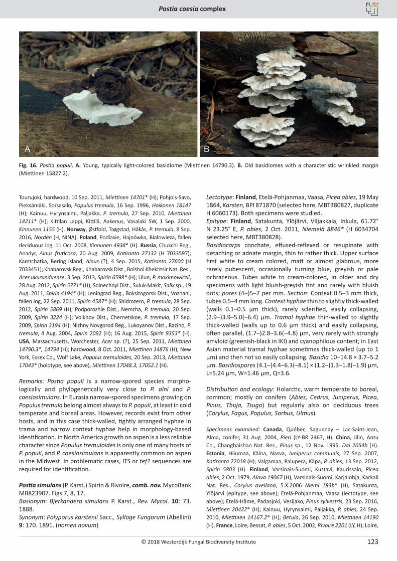
© 2018 Westerdijk Fungal Biodiversity Institute
Postia caesia complex
Editor-in-ChiefProf. dr P.W. Crous, Westerdijk Fungal Biodiversity Institute, P.O. Box 85167, 3508 AD Utrecht, The Netherlands.E-mail:[email protected]
123
Tourujoki, hardwood, 10 Sep. 2011, Miettinen 14701* (H); Pohjois-Savo, Pieksämäki, Sorsasalo, Populus tremula, 16 Sep. 1996, Haikonen 18147 (H); Kainuu, Hyrynsalmi, Paljakka, P. tremula, 27 Sep. 2010, Miettinen 14211* (H); Kittilän Lappi, Kittilä, Aakenus, Vasalaki SW, 1 Sep. 2000, Kinnunen 1155 (H). Norway, Østfold, Trøgstad, Håkås, P. tremula, 8 Sep. 2016, Nordén (H, NINA). Poland, Podlasie, Hajnówka, Białowieża, fallen deciduous log, 11 Oct. 2008, Kinnunen 4938* (H). Russia, Chukchi Reg., Anadyr, Alnus fruticosa, 20 Aug. 2009, Kotiranta 27132 (H 7033597); Kamtchatka, Bering Island, Alnus (?), 4 Sep. 2015, Kotiranta 27600 (H 7033451); Khabarovsk Reg., Khabarovsk Dist., Bolshoi Khekhtsir Nat. Res., Acer ukurunduense, 3 Sep. 2013, Spirin 6598* (H); Ulun, P. maximowiczii, 28 Aug. 2012, Spirin 5771* (H); Solnechnyi Dist., Suluk-Makit, Salix sp., 19 Aug. 2011, Spirin 4194* (H); Leningrad Reg., Boksitogorsk Dist., Vozhani, fallen log, 22 Sep. 2011, Spirin 4587* (H); Shidrozero, P. tremula, 28 Sep. 2012, Spirin 5869 (H); Podporozhie Dist., Nemzha, P. tremula, 20 Sep. 2009, Spirin 3224 (H); Volkhov Dist., Chernetskoe, P. tremula, 17 Sep. 2009, Spirin 3194 (H); Nizhny Novgorod Reg., Lukoyanov Dist., Razino, P. tremula, 4 Aug. 2004, Spirin 2092 (H); 16 Aug. 2015, Spirin 9353* (H). USA, Massachusetts, Worchester, Acer sp. (?), 25 Sep. 2011, Miettinen 14790.3*, 14794 (H); hardwood, 8 Oct. 2011, Miettinen 14876 (H); New York, Essex Co., Wolf Lake, Populus tremuloides, 20 Sep. 2013, Miettinen 17043* (holotype, see above), Miettinen 17048.3, 17052.1 (H).
Remarks: Postia populi is a narrow-spored species morpho-logically and phylogenetically very close to P. alni and P. caesiosimulans. In Eurasia narrow-spored specimens growing on Populus tremula belong almost always to P. populi, at least in cold temperate and boreal areas. However, records exist from other hosts, and in this case thick-walled, tightly arranged hyphae in trama and narrow context hyphae help in morphology-based identification. In North America growth on aspen is a less reliable character since Populus tremuloides is only one of many hosts of P. populi, and P. caesiosimulans is apparently common on aspen in the Midwest. In problematic cases, ITS or tef1 sequences are required for identification.
Postia simulans (P. Karst.) Spirin & Rivoire, comb. nov. MycoBank MB823907. Figs 7, 8, 17.Basionym: Bjerkandera simulans P. Karst., Rev. Mycol. 10: 73. 1888.Synonym: Polyporus karstenii Sacc., Sylloge Fungorum (Abellini) 9: 170. 1891. (nomen novum)
Lectotype: Finland, Etelӓ-Pohjanmaa, Vaasa, Picea abies, 19 May 1864, Karsten, BPI 871870 (selected here, MBT380827, duplicate H 6060173). Both specimens were studied.Epitype: Finland, Satakunta, Ylöjärvi, Viljakkala, Inkula, 61.72° N 23.25° E, P. abies, 2 Oct. 2011, Niemelä 8846* (H 6034704 selected here, MBT380828).Basidiocarps conchate, effused-reflexed or resupinate with detaching or adnate margin, thin to rather thick. Upper surface first white to cream colored, matt or almost glabrous, more rarely pubescent, occasionally turning blue, greyish or pale ochraceous. Tubes white to cream-colored, in older and dry specimens with light bluish-greyish tint and rarely with bluish dots; pores (4–)5–7 per mm. Section: Context 0.5–3 mm thick, tubes 0.5–4 mm long. Context hyphae thin to slightly thick-walled (walls 0.1–0.5 μm thick), rarely sclerified, easily collapsing, (2.9–)3.9–5.0(–6.4) μm. Tramal hyphae thin-walled to slightly thick-walled (walls up to 0.6 μm thick) and easily collapsing, often parallel, (1.7–)2.8–3.6(–4.8) μm, very rarely with strongly amyloid (greenish-black in IKI) and cyanophilous content; in East Asian material tramal hyphae sometimes thick-walled (up to 1 µm) and then not so easily collapsing. Basidia 10–14.8 × 3.7–5.2 µm. Basidiospores (4.1–)4.4–6.3(–8.1) × (1.2–)1.3–1.8(–1.9) µm, L=5.24 µm, W=1.46 µm, Q=3.6.
Distribution and ecology: Holarctic, warm temperate to boreal, common; mostly on conifers (Abies, Cedrus, Juniperus, Picea, Pinus, Thuja, Tsuga) but regularly also on deciduous trees (Corylus, Fagus, Populus, Sorbus, Ulmus).
Specimens examined: Canada, Québec, Saguenay – Lac-Saint-Jean, Alma, conifer, 31 Aug. 2004, Pieri (LY-BR 2467, H). China, Jilin, Antu Co., Changbaishan Nat. Res., Pinus sp., 12 Nov. 1995, Dai 2054b (H). Estonia, Hiiumaa, Käina, Nasva, Juniperus communis, 27 Sep. 2007, Kotiranta 22018 (H); Valgamaa, Palupera, Käpa, P. abies, 13 Sep. 2012, Spirin 5803 (H). Finland, Varsinais-Suomi, Kustavi, Kaurissalo, Picea abies, 2 Oct. 1979, Alava 19067 (H), Varsinais-Suomi, Karjalohja, Karkali Nat. Res., Corylus avellana, 5.X.2006 Niemi 183b* (H); Satakunta, Ylöjärvi (epitype, see above); Etelӓ-Pohjanmaa, Vaasa (lectotype, see above); Etelä-Häme, Padasjoki, Vesijako, Pinus sylvestris, 23 Sep. 2016, Miettinen 20422* (H); Kainuu, Hyrynsalmi, Paljakka, P. abies, 24 Sep. 2010, Miettinen 14167.2* (H); Betula, 26 Sep. 2010, Miettinen 14190 (H). France, Loire, Bessat, P. abies, 5 Oct. 2002, Rivoire 2201 (LY, H); Loire,
Fig. 16. Postia populi. A. Young, typically light-colored basidiome (Miettinen 14790.3). B. Old basidiomes with a characteristic wrinkled margin (Miettinen 15827.2).
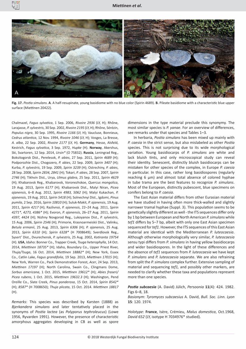
© 2018 Westerdijk Fungal Biodiversity Institute
Miettinen et al.
Editor-in-ChiefProf. dr P.W. Crous, Westerdijk Fungal Biodiversity Institute, P.O. Box 85167, 3508 AD Utrecht, The Netherlands.E-mail:[email protected]
124
Chalmazel, Fagus sylvatica, 1 Sep. 2006, Rivoire 2936 (LY, H); Rhône, Larajasse, P. sylvestris, 30 Sep. 2002, Rivoire 2195 (LY, H); Rhône, Sérézin, Populus nigra, 30 Sep. 1995, Rivoire 1166 (LY, H); Vaucluse, Bonnieux, Cedrus atlantica, 12 Nov. 1994, Rivoire 1046 (LY, H); Vosges, La Bresse, A. alba, 22 Sep. 2002, Rivoire 2177 (LY, H). Germany, Hesse, Alsfeld, Kestrich, Fagus sylvatica, 3 Sep. 1972, Hupke (H). Norway, Akershus, Ski, Svartoren, 12 Sep. 2014, Ursin* (O 75832). Russia, Leningrad Reg., Boksitogorsk Dist., Perelesok, P. abies, 27 Sep. 2011, Spirin 4689 (H); Podporozhie Dist., Chogozero, P. abies, 22 Sep. 2009, Spirin 3407 (H); Kurba, P. sylvestris, 19 Sep. 2009, Spirin 3239 (H); Ostrechiny, P. abies, 28 Sep. 2008, Spirin 2834, 2841 (H); Tokari, P. abies, 28 Sep. 2007, Spirin 2746 (H); Tikhvin Dist., Urya, Ulmus glabra, 25 Sep. 2011, Spirin 4629 (H); Khabarovsk Reg., Khabarovsk, Voronezhskoe, Quercus mongolica, 19 Aug. 2013, Spirin 6177 (H); Khabarovsk Dist., Malyi Niran, Picea ajanensis, 6–8 Aug. 2012, Spirin 4983, 5062 (H); Malyi Kukachan, P. ajanensis, 19 Aug. 2012, Spirin 5418 (H); Solnechnyi Dist., Igdomi, Pinus pumila, 2 Sep. 2016, Spirin 10810 (H); Suluk-Makit, P. ajanensis, 19 Aug. 2011, Spirin 4217 (H); Razlivnoi, P. ajanensis, 22–24 Aug. 2011, Spirin 4271*, 4273, 4386* (H); Evoron, P. ajanensis, 26–27 Aug. 2011, Spirin 4397, 4424 (H); Nizhny Novgorod Reg., Lukoyanov Dist., P. sylvestris, 15 Aug. 2006, Spirin 2546 (H); Primorie, Krasnoarmeiskii Dist., Valinku, Betula ermanii, 25 Aug. 2013, Spirin 6306 (H); P. ajanensis, 25 Aug. 2013, Spirin 6310 (H); Spirin 6328* (H 7008649); Sverdlovsk Reg., Sysert’ Dist., Dvurechensk, P. sylvestris, 25 Aug. 2002, Kotiranta 19754 (H). USA, Idaho: Bonner Co., Trapper Creek, Tsuga heterophylla, 14 Oct. 2014, Miettinen 18735* (H); Idaho, Boundary Co., Upper Priest River, Thuja/Tsuga, 16 Oct. 2014, Miettinen 18882* (H); New York, Essex Co., Catlin Lake, Fagus grandifolia, 19 Sep. 2013, Miettinen 17015 (H); New York, Warren Co., Pack Demonstration Forest, Acer, 24 Sep. 2013, Miettinen 17197 (H); North Carolina, Swain Co., Clingmans Dome, Sorbus americana, 1 Oct. 2015, Miettinen 19612* (H); Abies fraseri/Picea rubens, 1 Oct. 2015, Miettinen 19632.3 (H); Washington, Pend Oreille Co., Slate Creek, Pinus ponderosa, 15 Oct. 2014, Spirin 8542* (H); 8587* (H 7008650); Thuja plicata, 15 Oct. 2014. Miettinen 18817 (H).
Remarks: This species was described by Karsten (1888) as Bjerkandera simulans and later tentatively placed in the synonyms of Postia lactea (as Polyporus tephroleucus) (Lowe 1956, Ryvarden 1991). However, the presence of characteristic amorphous aggregates developing in CB as well as spore
dimensions in the type material preclude this synonymy. The most similar species is P. yanae. For an overview of differences, see remarks under that species and Tables 1–3.
In herbaria, Postia simulans has been mixed up mainly with P. caesia in the strict sense, but also mislabeled as other Postia species. This is not surprising due to its wide morphological variation. Young basidiocarps of P. simulans are white and lack bluish tints, and only microscopical study can reveal their identity. Senescent, distinctly bluish basidiocarps can be mistaken for other species of the complex, in Europe P. caesia in particular. In this case, rather long basidiospores (regularly reaching 6 µm) and almost total absence of colored hyphae in tube trama are the best features to recognize P. simulans. Most of the European, distinctly pubescent, blue specimens on conifers belong to P. caesia.
The East Asian material differs from other Eurasian material we have studied in having often more thick-walled and slightly narrower tramal hyphae (Suppl. 3). This population seems to be genetically slightly different as well - the ITS sequences differ only by 1 bp between European and North American P. simulans while tef1 differs by 5–7 bp, albeit with only one East Asian specimen sequenced for tef1. However, the ITS sequences of this East Asian material are identical with the Mediterranean P. luteocaesia. Although otherwise morphologically very similar, P. luteocaesia sensu typi differs from P. simulans in having yellow basidiocarps and wider basidiospores. In the light of these differences and the absence of tef1 sequences from P. luteocaesia we have kept P. simulans and P. luteocaesia separate. We are also refraining from split the P. simulans complex further. Extensive sampling of material and sequencing tef1, and possibly other markers, are needed to clarify whether these taxa and populations represent more than one species.
Postia subcaesia (A. David) Jülich, Persoonia 11(4): 424. 1982. Figs 6–8, 18.Basionym: Tyromyces subcaesius A. David, Bull. Soc. Linn. Lyon 15: 120. 1974.
Holotype: France, Isère, Crémieu, Malus domestica, Oct.1968, David 652 (LY, isotype H 7034976* studied).
Fig. 17. Postia simulans. A. A half-resupinate, young basidiome with no blue color (Spirin 4689). B. Pileate basidiome with a characteristic blue upper surface (Miettinen 20422).
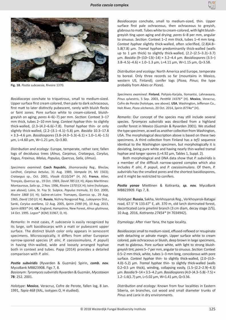
© 2018 Westerdijk Fungal Biodiversity Institute
Postia caesia complex
Editor-in-ChiefProf. dr P.W. Crous, Westerdijk Fungal Biodiversity Institute, P.O. Box 85167, 3508 AD Utrecht, The Netherlands.E-mail:[email protected]
125
Basidiocarps conchate to triquetrous, small to medium-sized. Upper surface first cream colored, then pale to dark ochraceous, first matt to later distinctly pubescent, rarely with bluish flecks or faint zones. Pore surface white to cream-colored, bluish-greyish on aging; pores 4–6(–7) per mm. Section: Context 3–17 mm thick, tubes 2–10 mm long. Context hyphae thin- to slightly thick-walled, (2.3–)4.2–6.6(–7.8). Tramal hyphae thin- or only slightly thick-walled, (2.2–)3.1–4.1(–5.8) μm. Basidia 10.3–17.8 × 3.3–4.6 μm. Basidiospores (3.8–)4.0–5.3(–6.1) × 1.0–1.4(–1.5) µm, L=4.60 µm, W=1.21 µm, Q=3.80.
Distribution and ecology: Europe, temperate, rather rare; fallen logs of deciduous trees (Alnus, Carpinus, Crataegus, Corylus, Fagus, Fraxinus, Malus, Populus, Quercus, Salix, Ulmus).
Specimens examined: Czech Republic, Jihomoravský Reg., Břeclav, Lanžhot, Carpinus betulus, 31 Aug. 1989, Vampola (H, MJ 1563); Crataegus sp., Oct. 2001, Vlasák 0110/24* (H, JV). France, Allier, Tronçais, Quercus sp., 19 Oct. 1969, David 785 (LY, H); Alpes Maritimes: Montauroux, Salix sp., 2 Nov. 1996, Rivoire 1370 (LY, H); Isère (holotype, see above); Loire, St. Foy St. Sulpice, Populus tremula, 31 Oct. 2000, Rivoire 1868 (LY, H); Saône-et-Loire: Tramayes, Quercus sp., 29 Aug. 1965, David 150 (LY, H). Russia, Nizhny Novgorod Reg., Lukoyanov Dist., Sanki, Corylus avellana, 12 Aug. 2005, Spirin 2390 (H), 10 Aug. 2013, Spirin 6083* (H). UK, England, Hampshire, New Forest, Alnus glutinosa, 14 Oct. 1995. Legon* (K(M) 31967, O, H).
Remarks: In most cases, P. subcaesia is easily recognized by its large, soft basidiocarps with a matt or pubescent upper surface. The distinct bluish color only appears in senescent specimens. Microscopically, it differs from other European narrow-spored species (P. alni, P. caesiosimulans, P. populi) in having thin-walled, wide and loosely arranged hyphae both in context and tubes. Papp (2014) provides a detailed comparison with P. alni.
Postia subviridis (Ryvarden & Guzmán) Spirin, comb. nov. MycoBank MB823908. Figs 7, 8.Basionym: Tyromyces subviridis Ryvarden & Guzmán, Mycotaxon 78: 252. 2001.
Holotype: Mexico, Veracruz, Cofre de Perote, fallen log, 8 Jan. 1991, Tapia 468 (XAL, isotypes O, H studied).
Basidiocarps conchate, small to medium-sized, thin. Upper surface first pale ochraceous, then ochraceous to greyish, glabrous to matt. Tubes white to cream-colored, with light bluish-greyish ting upon aging and drying; pores 6–8 per mm, angular to sinuous. Section: Context 1–2 mm thick, tubes 1–4 mm long. Context hyphae slightly thick-walled, often sclerified, (2.8)4.8–5.8(7.8) μm. Tramal hyphae predominantly thick-walled (walls up to 1 µm thick) to slightly thick-walled, (2.2–)2.5–3.2(–3.7) μm. Basidia (9–)10–13(–14) × 3.2–4.4 μm. Basidiospores (3.5–) 3.8–4.5(–4.6) × 1.0–1.3 µm, L=4.11 µm, W=1.15 µm, Q=3.58.
Distribution and ecology: North America and Europe, temperate to boreal. Only three records so far (mountains in Mexico, western US, Finland); conifer logs (Picea, Pinus; the type probably from Abies or Picea).
Specimens examined: Finland, Pohjois-Karjala, Ilomantsi, Lahnavaara, Pinus sylvestris, 5 Sep. 2003, Penttilä 14376* (H). Mexico, Veracruz, Cofre de Perote (holotype, see above). USA, Washington, Jefferson Co., Hoh River, Picea sitchensis, 20 Oct. 2014, Spirin 8774a* (H).
Remarks: Our concept of the species may still include several species. Tyromyces subviridis was described from a highland conifer forest in Mexico (Guzmán & Ryvarden 2001). We studied the type specimen, as well as another collection from Washington, USA. The morphological description above is based on these two specimens. A third collection from Finland has a tef1 sequence identical to the Washington specimen, but morphologically it is deviating, being pure white and having nearly thin-walled tramal hyphae and longer spores (L=4.92 µm, Tables 1, Suppl. 2).
Both morphological and DNA data show that P. subviridis is a member of the difficult narrow-spored complex which also includes P. alni, P. populi, and P. caesiosimulans. Of them, P. subviridis has the smallest pores and the shortest basidiospores, and it might be restricted to conifers.
Postia yanae Miettinen & Kotiranta, sp. nov. MycoBank MB823909. Figs 7, 8.
Holotype: Russia, Sakha, Verkhoyansk Reg., Verkhoyansk-Batagai road, 67.5° N 133.67° E, alt. 370 m, old larch dominated forest, decorticated Larix gmelinii branch (3 cm diam, decay stage 2/5), 10 Aug. 2016, Kotiranta 27454* (H 7034942).
Etymology: After river Yana, the type locality.
Basidiocarps small to medium-sized, effused-reflexed or resupinate with detaching or adnate margin. Upper surface white to cream colored, pale ochraceous or bluish, deep brown in large specimens, matt to glabrous. Pore surface white, with light to strong bluish-greyish tint; pores 5–7 per mm, angular to sinuous. Section: Context 0.5–2 mm thick, white, tubes 1–3 mm long, concolorous with pore surface. Context hyphae thin- to slightly thick-walled, (2.0–)3.0–4.0(–5.2) μm. Tramal hyphae thin- to slightly thick-walled (walls 0.2–0.5 μm thick), winding, collapsing easily, (1.5–)2.2–2.9(–4.3) μm. Basidia 9–14 × 3.5–4.2 μm. Basidiospores (4.0–)4.3–5.8(–7.5) × 1.2–1.6(1.7) µm, L=5.02 µm, W=1.41 µm, Q=3.56.
Distribution and ecology: Known from four localities in Eastern Siberia, on branches, cut wood and small diameter trunks of Pinus and Larix in dry environments.
Fig. 18. Postia subcaesia, Rivoire 1370.
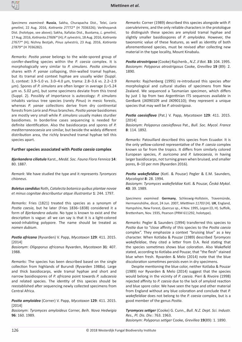
© 2018 Westerdijk Fungal Biodiversity Institute
Miettinen et al.
Editor-in-ChiefProf. dr P.W. Crous, Westerdijk Fungal Biodiversity Institute, P.O. Box 85167, 3508 AD Utrecht, The Netherlands.E-mail:[email protected]
126
Specimens examined: Russia, Sakha, Churapacha Dist., Telei, Larix gmelinii, 22 Aug. 2016, Kotiranta 27772* (H 7036326); Verkhoyansk Dist. (holotype, see above); Sakha, Xañalas Dist., Buotama, L. gmelinii, 17 Aug. 2016, Kotiranta 27606* (H); P. sylvestris, 18 Aug. 2016, Kotiranta 27677* (H); Nizhny Bestjah, Pinus sylvestris, 23 Aug. 2016, Kotiranta 27879* (H 7036282).
Remarks: Postia yanae belongs to the wide-spored group of conifer-dwelling species within the P. caesia complex. It is morphologically very similar to P. simulans. Postia simulans shares with P. yanae collapsing, thin-walled tramal hyphae, but its tramal and context hyphae are usually wider (Suppl. 3, context: 3.9–5.0 vs. 3.0–4.0 µm, trama: 2.8–3.6 vs. 2.2–2.9 µm). Spores of P. simulans are often longer in average (L=5.24 µm vs. 5.02 µm), but some specimens deviate from this trend (Suppl. 2). Possibly of importance is autecology: P. simulans inhabits various tree species (rarely Pinus) in mesic forests, whereas P. yanae collections derive from dry continental forests from Larix and Pinus branches. Postia yanae basidiomes are mostly very small while P. simulans usually makes sturdier basidiomes. In borderline cases sequencing is needed for definite identification. Also the basidiocarps and spores of P. mediterraneocaesia are similar, but beside the widely different distribution area, the richly branched tramal hyphae tell this species apart.
Further species associated with Postia caesia complex
Bjerkandera ciliatula Karst., Medd. Soc. Fauna Flora Fennica 14: 80. 1887.
Remark: We have studied the type and it represents Tyromyces chioneus.
Boletus candidus Roth, Catalecta botanica quibus plantae novae et minus cognitae describuntur atque illustrantur 1: 244. 1797.
Remarks: Fries (1821) treated this species as a synonym of Postia caesia, but he later (Fries 1836–1838) considered it a form of Bjerkandera adusta. No type is known to exist and the description is vague: all we can say is that it is a light-colored wood-inhabiting polypore. The name should be treated as nomen dubium.
Postia africana (Ryvarden) V. Papp, Mycotaxon 129: 411. 2015. [2014]Basionym: Oligoporus africanus Ryvarden, Mycotaxon 31: 407. 1988.
Remarks: The species has been described based on the single collection from highlands of Burundi (Ryvarden 1988a). Large and thick basidiocarps, wide tramal hyphae and short and narrow basidiospores of P. africana point towards P. subcaesia and related species. The identity of this species should be reestablished after sequencing newly collected specimens from Central Africa.
Postia amyloidea (Corner) V. Papp, Mycotaxon 129: 411. 2015. [2014]Basionym: Tyromyces amyloideus Corner, Beih. Nova Hedwigia 96: 160. 1989.
Remarks: Corner (1989) described this species alongside with P. coeruleivirens, and the only reliable characters in the protologue to distinguish these species are amyloid tramal hyphae and slightly smaller basidiospores of P. amyloidea. However, the taxonomic value of these features, as well as identity of both aforementioned species, must be revised after collecting new material in the type locality, Mount Kinabalu.
Postia atrostrigosa (Cooke) Rajchenb., N.Z. Jl Bot. 33: 104. 1995.Basionym: Polyporus atrostrigosus Cooke, Grevillea 19 (89): 2. 1890.
Remarks: Rajchenberg (1995) re-introduced this species after morphological and cultural studies of specimens from New Zealand. We sequenced a Tasmanian specimen, which differs by just 1 bp from two Argentinian ITS sequences available in GenBank (JX090109 and JX090110); they represent a unique species that may well be P. atrostrigosa.
Postia caesioflava (Pat.) V. Papp, Mycotaxon 129: 411. 2015. [2014]Basionym: Polyporus caesioflavus Pat., Bull. Soc. Mycol. France 8: 114. 1892.
Remarks: Patouillard described this species from Ecuador. It is the only yellow-colored representative of the P. caesia complex known so far from the tropics. It differs from similarly colored European species, P. auricoma and P. luteocaesia, in having larger basidiocarps, not turning green when bruised, and smaller pores, 8–10 per mm (Ryvarden 2016).
Postia wakefieldiae (Kotl. & Pouzar) Pegler & E.M. Saunders, Mycologist 8: 28. 1994.Basionym: Tyromyces wakefieldiae Kotl. & Pouzar, Česká Mykol. 43: 39. 1989.
Specimens examined: Germany, Schleswig-Hollstein, Travemünde, Hermannshöhe, dicot, 24 Jun. 2007, Miettinen 11703 (H). UK, England, Hampshire, New Forest, Quercus sp., 4 Nov. 1995, Legon (O, H). Suffolk, Brettenham, Nov. 1935, Pearson (PRM 611292, holotype).
Remarks: Pegler & Saunders (1994) transferred this species to Postia due to “close affinity of this species to the Postia caesia complex”. They emphasize a context “bruising blue” as a key character. When Kotlaba & Pouzar (1989) described Tyromyces wakefieldiae, they cited a letter from D.A. Reid stating that the species sometimes shows blue coloration. Also Wakefield stated, according to Kotlaba and Pouzar, that “the flesh” stained blue when fresh. Ryvarden & Melo (2014) note that the blue discoloration sometimes persists even in dry specimens.
Despite mentioning the blue color, neither Kotlaba & Pouzar (1989) nor Ryvarden & Melo (2014) suggest that the species would belong in the vicinity of P. caesia. Pieri & Rivoire (1998) rejected affinity to P. caesia due to the lack of amyloid reaction and blue spore color. We have seen the type and other material from England without any blue coloration and conclude that P. wakefieldiae does not belong to the P. caesia complex, but is a good member of the genus Postia.
Tyromyces setiger (Cooke) G. Cunn., Bull. N.Z. Dept. Sci. Industr. Res., Pl. Dis. Div.: 763. 1963.Basionym: Polyporus setiger Cooke, Grevillea 19(89): 1. 1890.
© 2018 Westerdijk Fungal Biodiversity Institute
Postia caesia complex
Editor-in-ChiefProf. dr P.W. Crous, Westerdijk Fungal Biodiversity Institute, P.O. Box 85167, 3508 AD Utrecht, The Netherlands.E-mail:[email protected]
127
Remarks: Cunningham (1965) mentions that he reported this species previously under Postia caesia, so there is some resemblance. Another Austro-American species, P. atrostrigosa, differs in having wider basidiospores. Ryvarden (1988b) also treated this species under Tyromyces. New collections are needed to settle its proper taxonomic position.
DISCUSSION
Our study has raised the species number in the Postia caesia complex from 10 to 24. The most important contribution of this paper are the rigorous type studies that allowed us to combine existing temperate names to phylogenetic species concepts and previously unaccounted diversity.
Considering that we did not revise tropical or Southern Hemisphere material, the number of species known from Northern Hemisphere more than tripled, from 6 to 20 species. Why this diversity has gone unnoticed or at least undescribed for so long is due to the combination of minute morphological differences and small interspecific differences in the most commonly used genetic marker ITS (in some cases below 1% between species). Obviously, the insight provided by DNA sequences proved fundamental in revising species concepts accurately.
ITS sequences were commonly polymorphic, exhibiting double bases and even length variation. Considering that ITS sequence differences between some species are very low (< 1 %) they may fall within error margins of current mass sequencing methods used in environmental studies. Tef1 sequences are in general more variable between species and in this sense more reliable for molecular identification, but reference sequences are available for much fewer species.
Our sampling and analyses allowed distinguishing 20 species in the temperate Northern Hemisphere. However, even in the best sampled area, Europe, we have indications that still more species are present: Postia cf. subviridis from Finland does not agree well the North American material though their tef1 sequence are identical; two English specimens collected on Acer and Fraxinus from the study of Yao et al. (2005) have distinct ITS sequences. Variation within the wide-spread P. caesiosimulans leaves open the possibility that our concept of the species encompasses more than one taxon (species or subspecies). We can also expect a good number of new species from the Southern Hemisphere, the Himalayas as well as subtropical and tropical areas.
While species differences at the extremes of the morphological variations within the complex are very clear (e.g. P. alni vs. P. auricoma), some species are very difficult to separate, even to the extent that DNA sequences are required for a reliable identification of some individuals. Such examples include P. alni vs. P. caesiosimulans in Europe, P. arbuti vs. P. populi in North America and P. simulans vs. P. yanae in Asia. Young or senescent specimens may also have to be left unnamed even after a careful microscopical examination. However, the great majority of specimens from the Postia caesia complex can be identified without DNA sequences, often in the field.
Most species of the P. caesia complex are restricted geographically to either western or eastern part of Eurasia or to North America, indicating that geographic isolation has probably played a role in speciation. Only three species (P. caesiosimulans, P. populi, P. simulans) and possibly a fourth (P. subviridis) are
distributed in both Eurasia and North America. In addition, one further species (P. auricoma) is found in Europe and East Asia. Within North America some species have been recorded only from the east (P. comata) or from the west coast (P. arbuti), but collection intensity is too low to judge whether this is just accidental or a true pattern of distribution. Continental divide and possibly ice ages - we expect that some of the species divisions such as P. alni – P. cyanescens – P. caesiosimulans – P. populi are relatively young - have thus shaped the species diversity.
Host tree, particularly division between angiosperms and gymnosperms, is an important character for identification but also when contemplating speciation patterns in this complex. Six out of 20 north temperate species grow only on angiosperms (P. arbuti, P. alni, P. coeruleivirens, P. magna, P. populi, P. subcaesia), and eight only on gymnosperms (P. auricoma, P. bifaria, P. cyanescens, P. glauca, P. gossypina, P. luteocaesia, P. subviridis, P. yanae). Six species (P. caesia, P. caesiosimulans, P. comata, P. livens, P. mediterraneocaesia, P. simulans) grow both on gymno- and angiosperms, although in the case of P. caesiosimulans the conifer records from North America may represent distinct, possibly differentiating populations.
In a few cases Postia species strongly prefer a particular tree species, at least regionally. Thus Postia cyanescens is almost always found on Picea abies in the North, while Postia populi prefers Populus tremula in Europe. P. luteocaesia have been recorded only from Pinus spp., and P. auricoma only from Pinus sylvestris in Europe.
Although host specialization clearly plays a role in the evolution of the P. caesia complex, it is an open question whether sympatric speciation has significance. We can point to one case where sympatric speciation seems likely: the spruce-inhabiting species P. cyanescens is distributed in Europe, where its closest relatives are P. alni, P. caesiosimulans and P. populi, all angiosperm-inhabiting species. Tef1 sequence places P. cyanescens in the vicinity of P. caesiosimulans, which occurs on conifers in East Asia and North America. In this case host jump as a primary speciation process would appear to be a plausible option.
Despite small ITS differences, overlapping distribution areas and similar ecology we did not observe clear cases of hybridization between species with the exception of a single individual: Collection Spirin 9353 from Nizhny Novgorod produced polymorphic ITS sequence that can be interpreted as having ITS copies from two parental species, P. alni and P. populi. Since the tef1 sequence is a typical P. populi sequence this specimen is not a first generation hybrid. It nevertheless indicates that limited gene flow may be taking place between closely related species.
A number of Postia species have been red-listed or are considered indicators of old-growth forests in Northern Europe. Most species of the P. caesia complex do not seem to be associated with endangered habitats at the continental level. It is worth mentioning that P. auricoma records derive from forests with a continuum of big pine logs, so this species may well be old-growth-forest dependent. Also all the records of P. bifaria derive from virgin or near virgin conifer forests. The number of records is so low for many species that it is premature to assess how common and ecologically specialized they are.
We hope to see other researchers continue where we have left in studying this fascinating, easily recognizable species group. While doing a decent job at delineating species, our
© 2018 Westerdijk Fungal Biodiversity Institute
Miettinen et al.
Editor-in-ChiefProf. dr P.W. Crous, Westerdijk Fungal Biodiversity Institute, P.O. Box 85167, 3508 AD Utrecht, The Netherlands.E-mail:[email protected]
128
study did not fare as well in establishing a robust phylogeny for the Postia caesia complex. For this, we need further genetic markers, whose sequencing will require cultures or recently collected specimens. Fresh collections are really needed since even ITS sequence production in this group is difficult from specimens older than a few years, and other markers are likely to be more difficult to sequence.
ACKNOWLEDGEMENTS
We are indebted to Frank Dämmrich, Ellen Larsson, Karl-Henrik Larsson, Tuomo Niemelä, Beatriz Ortiz-Santana, Viktor Papp, Dmitry Schigel, Gerhard Schuster and Petr Vampola for valuable specimens and sequences. Tuomo Niemelä also commented the manuscript. Sampsa Lommi prepared the distribution maps. Alix David provided valuable ideas for species delimitation in this complex. We are ever grateful for Yu-Cheng Dai and his students for arranging a collecting trip in China resulting in valuable collections. Part of the sequences were produced by the Finnish Barcode of Life (FinBOL) initiative. CSC – IT Center for Science (Espoo, Finland) provided computational resources. This research was made possible by the PolyPEET project led by David Hibbett (NSF grant DEB0933081) and the European Commission Marie Curie grant PIOF-GA-2011–302349.
REFERENCES
Atkinson GF (1908). Notes on some new species of fungi from the United States. Annales Mycologici 6: 54–62.
Brazee NJ, Lindner DL, Fraver S, et al. (2012). Wood-inhabiting, polyporoid fungi in aspen-dominated forests managed for biomass in the U.S. Lake States. Fungal Ecology 5: 600–609.
Britzelmayr M (1893). Materialien zur Beschreibung der Hymeno-myceten 3. Botanisches Zentralblatt 54: 97–105.
Corner EJH (1989). Ad polyporaceas V. Beihefte zur Nova Hedwigia 96: 1–218.
Cunningham GH (1965). Polyporaceae of New Zealand. Bulletin of the New Zealand Department of Industrial Research 164: 1–304.
Darriba D, Taboada GL, Doallo R, et al. (2012). jModelTest 2: more models, new heuristics and parallel computing. Nature Methods 9: 772.
David A (1974). Une nouvelle espèce de Polyporaceae: Tyromyces subcaesius. Bulletin de la Société Linnéenne de Lyon 43: 119–126.
David A (1980). Etude du genre Tyromyces sensu lato: repartition dans le genres Leptoporus, Spongiporus et Tyromyces sensu stricto. Bulletin mensuel de la Société linnéenne de Lyon 49: 6–56.
Donk MA (1960). The generic names proposed for Polyporaceae. Persoonia 1: 173–302.
Fries EM (1821). Systema mycologicum 1. Ex officina Berlingiana, Lund.Fries EM (1836–1838). Epicrisis systematis mycologici. Typographia
Academica, Uppsala.Guzmán G, Ryvarden L (2001). Studies in neotropical polypores 12. New
and noteworthy polypores from Mexico. Mycotaxon 78: 245–256.Hattori T (2002). Type studies of the polypores described by E.J.H.
Corner from Asia and West Pacific Areas. IV. Species described in Tyromyces (1). Mycoscience 43: 307–315.
Hattori T (2005). Type studies of the polypores described by E.J.H. Corner from Asia and West Pacific Areas. VII. Species described in Trametes (1). Mycoscience 46: 303–312.
Hämet-Ahti L (1984). The boreal zone and its biotic subdivision. Fennia 159: 69–75.
Index Herbariorum (2017). A global directory of public herbaria and associated staff. New York Botanical Garden’s Virtual Herbarium. http://sweetgum.nybg.org/science/ih [accessed 10.3.2017].
Jahn H (1979). Pilze die an Holz wachsen. Busse, Herford.Jang Y, Jang S, Lee J, et al. (2016). Diversity of Wood-Inhabiting
Polyporoid and Corticioid Fungi in Odaesan National Park, Korea. Mycobiology 44: 217–236.
Justo A, Miettinen O, Floudas D, et al. (2017). A revised family-level classification of the Polyporales (Basidiomycota). Fungal Biology 121: 798–824.
Karsten PA (1888). Diagnoses fungorum nonnullorum novorum, in Fennia detectorum. Revue Mycologique Toulouse 10: 73–75.
Katoh K, Standley DM (2013). MAFFT Multiple Sequence Alignment Software Version 7: Improvements in Performance and Usability. Molecular Biology and Evolution 30: 772–780.
Killermann S (1922). Pilze aus Bayern. Kritische Studien besonders zu M. Britzelmayr; Standortsangaben u. (kurtze) Bestimmunstabellen. 1. Teil: Thelephoraceen, Hydnaceen, Polyporaceen, Clariaceen un Tremellaceen. Denkschriften der Bayerischen Botanischen Gesellschaft in Regensburg 15: 1–134.
Kim CS, Jo JW, Kwag Y-N, et al. (2015). Mushroom flora of Ulleung-gun and a newly recorded Bovista species in the Republic of Korea. Mycobiology 43: 239–257.
Kotlaba F, Pouzar Z (1989). Type studies of polypores described by A. Pilát 2. Česká Mykologie 43: 36–44.
Lowe JL (1956). Type studies of the polypores described by Karsten. Mycologia 1: 99–125.
Lowe JL (1974) Solution to a mycological mystery. Bulletin de la Société Linnéenne de Lyon 43: 239–240.
Matheny PB, Wang Z, Binder M, et al. (2007). Contributions of rpb2 and tef1 to the phylogeny of mushrooms and allies (Basidiomycota, Fungi). Molecular Phylogenetics and Evolution 43: 430–451.
Niemelä T, Kinnunen J, Lindgren M, et al. (2001). Novelties and records of poroid Basidiomycetes in Finland and adjacent Russia. Karstenia 41: 1–21.
Ortiz-Santana B, Lindner DL, Miettinen O, et al. (2013). A phylogenetic overview of the antrodia clade (Basidiomycota, Polyporales). Mycologia 105: 1391–1411.
Papp V (2014). Postia alni Niemela & Vampola (Basidiomycota, Polyporales) - member of the problematic Postia caesia complex - has been found for the first time in Hungary. Biodiversity Data Journal 2: e1034.
Papp V (2015). Nomenclatural novelties in the Postia caesia complex. Mycotaxon 129: 407–413.
Pebesma EJ, Bivand RS (2005). Classes and methods for spatial data in R. R News 5: 9–13.
Pegler DN, Saunders EM (1994). British poroid species formerly placed in the genus Tyromyces (Coriolaceae). Mycologist 8: 24–31.
Pieri M, Rivoire B (1998). Postia inocybe (David & Malençon) Jülich f. inocybe et f. pileatus, f. nov. Notes nomenclaturales sur le genre Postia. Bulletin trimestriel de la Société mycologique de France 114: 19–33.
Pieri M, Rivoire B (2005). Postia mediterraneocaesia, une nouvelle espèce de polypore découverte dans le sud de l’Europe. Bulletin Semestriel de la Fédération des Associations Mycologiques Méditerranéennes 28: 33–38.
Pildain MB, Rajchenberg M (2013). The phylogenetic position of Postia s.l. (Polyporales, Basidiomycota) from Patagonia, Argentina. Mycologia 105: 357–367.
Rajchenberg M (1995). Notes on New Zealand polypores (Basidiomycetes) 2. Cultural and morphological studies of selected species. New Zealand Journal of Botany 33: 99–109.
© 2018 Westerdijk Fungal Biodiversity Institute
Postia caesia complex
Editor-in-ChiefProf. dr P.W. Crous, Westerdijk Fungal Biodiversity Institute, P.O. Box 85167, 3508 AD Utrecht, The Netherlands.E-mail:[email protected]
129
Ronquist F, Teslenko M, van der Mark P, et al. (2012). MrBayes 3.2: Efficient Bayesian Phylogenetic Inference and model choice across a large model space. Systematic Biology 61: 539–542.
Ryvarden L (1983). Type studies in the Polyporaceae 14. species described by N. Patouillard, either alone or with other mycologists. Occasional papers of the Farlow Herbarium of Cryptogamic Botany. Harvard University Herbaria, Cambridge, MA.
Ryvarden L (1988a). Two new polypores from Burundi in Africa. Mycotaxon 31: 407–409.
Ryvarden L (1988b). Type studies in the Polyporaceae 19. Species described by M.C. Cooke. Mycotaxon 31:45–58
Ryvarden L (1991). Genera of polypores. Nomenclature and taxonomy. Synopsis Fungorum 5: 1–363.
Ryvarden L (2016). Neotropical polypores 3. Polyporaceae, Obba-Wrightoporia. Synopsis Fungorum 36: 447-613.
Ryvarden L, Melo I (2014). Poroid fungi of Europe. Fungiflora, Oslo.Schaeffer JC (1763). Fungorum qui in Bavaria et Palatinatu circa
Ratisbonam nascuntur Icones, Regensburg.Schaeffer JC (1774). Fungorum qui in Bavaria et Palatinatu circa
Ratisbonam nascuntur Icones, Regensburg.Schrader HA (1794). Spicilegium Florae Germanicae. Ritscher, Hannover.Schumacher CF (1803). Enumeratio plantarum in partibus Saellandiae
septentrionalis et orientalis, pars posterior. Friedrich Brummer, Hafniae.
Shen L, Cui B, Dai Y (2014). A new species of Postia (Polyporales, Basidiomycota) from China based on morphological and molecular evidence. Phytotaxa 162: 147.
Sowerby J (1799). Coloured Figures of English fungi or mushrooms. J. Davis, London.
Stamatakis A (2014). RAxML Version 8: A tool for Phylogenetic Analysis and Post-Analysis of Large Phylogenies. Bioinformatics 30: 1312–1313.
Stevenson JA, Cash EK (1936) The new fungus names proposed by C. G. Lloyd. Bulletin of the Lloyd library and Museum 35: 1–209.
Tamura K, Stecher G, Peterson D, et al. (2013). MEGA6: Molecular Evolutionary Genetics Analysis Version 6.0. Molecular Biology and Evolution 30: 2725–2729.
Yao YJ, Pegler DN, Chase MW (2005). Molecular variation in the Postia caesia complex. FEMS Microbiology Letters 242: 109–116.
Supplement 1 - INSDC accession numbersINSDC accession numbers for DNA sequences used in this study. Specimens provided with collector and collection number information have been sequenced for this study, the rest retrieved from the INSDC database.Supplement 2 - Spore measurements in the Postia caesia complexBold-face values represent composite statistics for species. L = average of spore length, W = average of spore width, Q = L/W, and n = number of spores measured. The whole range is given in parentheses; 90% range excluding 5% extreme values from both ends of variation is given without parentheses; in case the values are identical, parentheses are omitted. Specimens marked with asterisk (*) have been excluded from the combined statistics for that species.Supplement 3 - Hyphal measurements in the Postia caesia complexBold-face values represent composite statistics for species, n = number of spores measured. The whole range is given in parentheses; 60% range excluding 20% extreme values from both ends of variation is given without parentheses; in case the values are identical, parentheses are omitted.Supplement 4 – Distribution maps of north temperate Postia caesia species

© 2018 Westerdijk Fungal Biodiversity Institute
Editor-in-ChiefProf. dr P.W. Crous, Westerdijk Fungal Biodiversity Institute, P.O. Box 85167, 3508 AD Utrecht, The Netherlands.E-mail:[email protected]
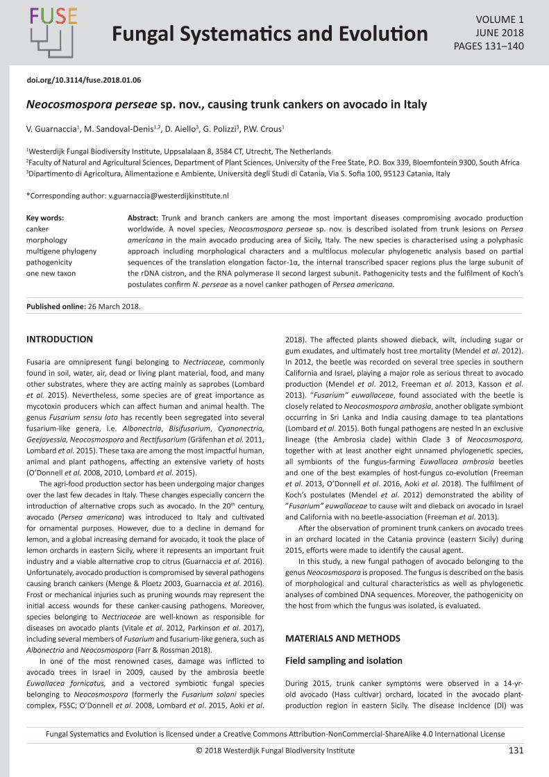
Fungal Systematics and Evolution is licensed under a Creative Commons Attribution-NonCommercial-ShareAlike 4.0 International License
© 2018 Westerdijk Fungal Biodiversity Institute 131
Editor-in-ChiefProf. dr P.W. Crous, Westerdijk Fungal Biodiversity Institute, P.O. Box 85167, 3508 AD Utrecht, The Netherlands.E-mail:[email protected]
Fungal Systematics and EvolutionVOLUME 1JUNE 2018
PAGES 131–140
doi.org/10.3114/fuse.2018.01.06
Neocosmospora perseae sp. nov., causing trunk cankers on avocado in Italy
V. Guarnaccia1, M. Sandoval-Denis1,2, D. Aiello3, G. Polizzi3, P.W. Crous1
1Westerdijk Fungal Biodiversity Institute, Uppsalalaan 8, 3584 CT, Utrecht, The Netherlands2Faculty of Natural and Agricultural Sciences, Department of Plant Sciences, University of the Free State, P.O. Box 339, Bloemfontein 9300, South Africa3Dipartimento di Agricoltura, Alimentazione e Ambiente, Università degli Studi di Catania, Via S. Sofia 100, 95123 Catania, Italy
*Corresponding author: [email protected]
Abstract: Trunk and branch cankers are among the most important diseases compromising avocado production worldwide. A novel species, Neocosmospora perseae sp. nov. is described isolated from trunk lesions on Persea americana in the main avocado producing area of Sicily, Italy. The new species is characterised using a polyphasic approach including morphological characters and a multilocus molecular phylogenetic analysis based on partial sequences of the translation elongation factor-1α, the internal transcribed spacer regions plus the large subunit of the rDNA cistron, and the RNA polymerase II second largest subunit. Pathogenicity tests and the fulfilment of Koch’s postulates confirm N. perseae as a novel canker pathogen of Persea americana.
Key words: cankermorphologymultigene phylogenypathogenicityone new taxon
Published online: 26 March 2018.
INTRODUCTION
Fusaria are omnipresent fungi belonging to Nectriaceae, commonly found in soil, water, air, dead or living plant material, food, and many other substrates, where they are acting mainly as saprobes (Lombard et al. 2015). Nevertheless, some species are of great importance as mycotoxin producers which can affect human and animal health. The genus Fusarium sensu lato has recently been segregated into several fusarium-like genera, i.e. Albonectria, Bisifusarium, Cyanonectria, Geejayessia, Neocosmospora and Rectifusarium (Gräfenhan et al. 2011, Lombard et al. 2015). These taxa are among the most impactful human, animal and plant pathogens, affecting an extensive variety of hosts (O’Donnell et al. 2008, 2010, Lombard et al. 2015).
The agri-food production sector has been undergoing major changes over the last few decades in Italy. These changes especially concern the introduction of alternative crops such as avocado. In the 20th century, avocado (Persea americana) was introduced to Italy and cultivated for ornamental purposes. However, due to a decline in demand for lemon, and a global increasing demand for avocado, it took the place of lemon orchards in eastern Sicily, where it represents an important fruit industry and a viable alternative crop to citrus (Guarnaccia et al. 2016). Unfortunately, avocado production is compromised by several pathogens causing branch cankers (Menge & Ploetz 2003, Guarnaccia et al. 2016). Frost or mechanical injuries such as pruning wounds may represent the initial access wounds for these canker-causing pathogens. Moreover, species belonging to Nectriaceae are well-known as responsible for diseases on avocado plants (Vitale et al. 2012, Parkinson et al. 2017), including several members of Fusarium and fusarium-like genera, such as Albonectria and Neocosmospora (Farr & Rossman 2018).
In one of the most renowned cases, damage was inflicted to avocado trees in Israel in 2009, caused by the ambrosia beetle Euwallacea fornicatus, and a vectored symbiotic fungal species belonging to Neocosmospora (formerly the Fusarium solani species complex, FSSC; O’Donnell et al. 2008, Lombard et al. 2015, Aoki et al.
2018). The affected plants showed dieback, wilt, including sugar or gum exudates, and ultimately host tree mortality (Mendel et al. 2012). In 2012, the beetle was recorded on several tree species in southern California and Israel, playing a major role as serious threat to avocado production (Mendel et al. 2012, Freeman et al. 2013, Kasson et al. 2013). “Fusarium” euwallaceae, found associated with the beetle is closely related to Neocosmospora ambrosia, another obligate symbiont occurring in Sri Lanka and India causing damage to tea plantations (Lombard et al. 2015). Both fungal pathogens are nested in an exclusive lineage (the Ambrosia clade) within Clade 3 of Neocosmospora, together with at least another eight unnamed phylogenetic species, all symbionts of the fungus-farming Euwallacea ambrosia beetles and one of the best examples of host-fungus co-evolution (Freeman et al. 2013, O’Donnell et al. 2016, Aoki et al. 2018). The fulfilment of Koch’s postulates (Mendel et al. 2012) demonstrated the ability of ”Fusarium” euwallaceae to cause wilt and dieback on avocado in Israel and California with no beetle-association (Freeman et al. 2013).
After the observation of prominent trunk cankers on avocado trees in an orchard located in the Catania province (eastern Sicily) during 2015, efforts were made to identify the causal agent.
In this study, a new fungal pathogen of avocado belonging to the genus Neocosmospora is proposed. The fungus is described on the basis of morphological and cultural characteristics as well as phylogenetic analyses of combined DNA sequences. Moreover, the pathogenicity on the host from which the fungus was isolated, is evaluated.
MATERIALS AND METHODS
Field sampling and isolation
During 2015, trunk canker symptoms were observed in a 14-yr-old avocado (Hass cultivar) orchard, located in the avocado plant-production region in eastern Sicily. The disease incidence (DI) was
© 2018 Westerdijk Fungal Biodiversity Institute
Guarnaccia et al.
Editor-in-ChiefProf. dr P.W. Crous, Westerdijk Fungal Biodiversity Institute, P.O. Box 85167, 3508 AD Utrecht, The Netherlands.E-mail:[email protected]
132
recorded based on the number of symptomatic plants compared to the total number present. Branch canker samples were taken from 10 plants. Fragments (5 × 5 mm) of symptomatic tissues were cut from the lesion margins, surface-sterilised in a sodium hypochlorite solution (10 %) for 20 s, followed by 70 % ethanol for 30 s, and rinsed three times in sterilised water. Tissue fragments were dried between sterilised filter papers, placed on 2 % potato dextrose agar (PDA; Difco, Leeuwarden, The Netherlands) amended with 100 μg/mL penicillin and 100 μg/mL streptomycin (PDA-PS) and incubated at 25 °C until characteristic fungal colonies were observed. Pure cultures were obtained by transferring germinating single conidia to fresh PDA plates with the aid of a Nikon SMZ1000 dissecting microscope.
Fungal isolates and morphological characterization
The cultural and micromorphological features of all the isolates included in this study were evaluated following the procedures of Aoki et al. (2003) with some modification as described previously (Sandoval-Denis et al. 2018). Colour notation followed the mycological colour charts of Rayner (1970). Micromorphological characteristics were examined and photographed using a Nikon Eclipse 80i microscope with Differential Interference Contrast (DIC) optics and a Nikon AZ100 stereomicroscope, both equipped with a Nikon DS-Ri2 high definition colour digital camera. Photographs and measurements were taken using the Nikon software NIS-elements D software v. 4.50.
DNA extraction, PCR amplification and sequencing
Fungal isolates were grown on PDA for 4–7 d at room temperature, under a natural day/night photoperiod. Total genomic DNA was extracted from fresh mycelium scraped from the colony surface using the Wizard® Genomic DNA purification Kit (Promega Corporation, Madison, WI, USA). Fragments of four nuclear loci including the translation elongation factor 1-alpha (EF-1α), the internal transcribed spacer region of the rDNA (ITS), the large subunit of the rDNA (LSU) and the RNA polymerase second largest subunit (RPB2) were PCR amplified as described previously (O’Donnell et al. 2009, 2010, Sandoval-Denis et al. 2018) and sequenced using the following primer pairs: EF-1/EF-2 for EF-1α (O’Donnell et al. 2008), ITS4/ITS5 for ITS (White et al. 1990), LR0R/LR5 for LSU (Vilgalys & Hester 1990, Vilgalys & Sun 1994) and 5f2/7cr and 7cf/11ar for RPB2 (Liu et al. 1999, Sung et al. 2007). Sequences generated in this study were uploaded to GenBank and the European Nucleotide Archive (ENA) databases (Table 1).
Phylogenetic analyses and molecular identification
Sequence alignments were performed individually for each locus using MAFFT on the European Bioinformatics Institute (EMBL-EBI) portal (http://www.ebi.ac.uk/Tools/msa/mafft/). BLASTn searches on GenBank and pairwise sequence alignments on the Fusarium MLST database of the Westerdijk Fungal Biodiversity Institute (http://www.westerdijkinstitute.nl/fusarium/) were performed using EF-1α and RPB2 sequences in order to preliminary identify the fungal isolates to generic level. Following this initial identification, a combination of DNA sequences from four loci (EF-1α, ITS, LSU and RPB2) was used for the final molecular identification and phylogenetic analyses (O’Donnell et al. 2008).
The different gene datasets were analysed independently and combined using RAxML (ML) and Bayesian methods (BI) as described previously (Sandoval-Denis et al. 2018). Evolutionary models for the four loci (GTR+I+G for ITS, LSU and RPB2; GTR+G for EF-1α) were calculated using MrModelTest v. 2.3 (Nylander 2004) selecting the best-fit model for each data partition according to the Akaike criterion.
Pathogenicity tests
Pathogenicity tests were performed on potted, healthy avocado seedlings (6-mo-old) with a subset of two representative isolates. Each experiment was conducted twice. For each experiment three replicates per isolate were used with 10 plants per replicate. Twigs were superficially wounded between two nodes forming a slit using a sterile blade. Inoculations were conducted by placing a 1-wk-old, 6-mm-diam colonised agar plug from each fungal isolate on a wound. Wounds were then wrapped with Parafilm® (American National Can, Chicago, IL, USA). Ten twigs were inoculated as described above with 6-mm-diam non-colonised MEA plugs as negative controls. The same number of wounds/plants were inoculated with sterile MEA plugs and served as controls. After inoculation, plants were covered with a plastic bag for 48 h and maintained at 25 ± 1 °C and 95 % relative humidity (RH) under a 12-h fluorescent light/dark regime. All plants were irrigated 2–3 times per week and examined weekly for disease symptom development. Disease incidence (DI) was recorded as described above.
RESULTS
Field sampling and fungal isolation
Symptoms referable to fusaria species were detected in an avocado orchard in the main avocado-producing region of Eastern Sicily, Italy (GPS coordinates: 37.687247, 15.175479). The disease was observed on established plants (14-yr-old) in an open field. Disease incidence was ascertained at 10 %. The symptoms observed on avocado plants consisted of trunk cankers. Bark appeared cracked, darkly discoloured and/or slightly sunken. Occasionally, a sugar exudate was present on the surface. Cankers were internally reddish brown in colour and variable in shape. Transverse cuts revealed a characteristic wedge-shaped canker extending deep into the xylem (Fig. 1). Only fusarium-like isolates growing in pure culture were obtained from the symptomatic avocado trees, from which five monosporic strains were retained.
Phylogenetic analyses and species identification
Pairwise sequence alignments on the Fusarium MLST database and GenBank BLASTn searches demonstrated that the five fungal isolates belonged to the genus Neocosmospora.
Subsequently, more inclusive multilocus phylogenetic analyses were performed based on EF-1α, ITS, LSU, and RPB2 sequences. A first analysis spanned the currently known phylogenetic diversity of the genus Neocosmospora, and included sequences from a total of 365 strains, based on the alignments published by O’Donnell et al. (2008). According to this analysis, the five strains from avocado formed an exclusive new linage in the genus Neocosmospora (data not shown, alignments, trees and statistics all available at TreeBASE). A second analysis was run based on a selected subset of DNA data representing most of the species of Neocosmospora currently assigned with Latin binomials, plus several yet unnamed phylogenetic clades phylogenetically related to the new lineage (Fig. 2). This final analysis included sequences from 80 strains, representing 48 taxa and a total of 2 917 character sites, of which 2 203 were conserved (EF-1α 212, ITS 372, LSU 441 and RPB2 1178), and 555 were variable and phylogenetically informative (EF-1α 69, ITS 101, LSU 35 and RPB2 350). The BI analyses identified a total of 774 unique sites (EF-1α 134, ITS 179, LSU 43 and RPB2 418) and sampled a total 315 000 trees, from which 236 250 were used to calculate the 50 % consensus tree and posterior probability (PP) values, after discarding 25 % of trees as burn-in fraction. Results from ML and BI methods showed that the
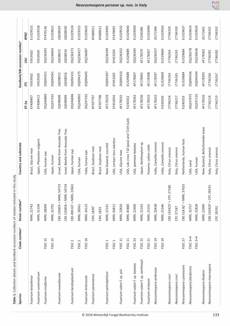
© 2018 Westerdijk Fungal Biodiversity Institute
Neocosmospora perseae sp. nov. in Italy
Editor-in-ChiefProf. dr P.W. Crous, Westerdijk Fungal Biodiversity Institute, P.O. Box 85167, 3508 AD Utrecht, The Netherlands.E-mail:[email protected]
133
Tabl
e 1.
Col
lecti
on d
etai
ls an
d Ge
nBan
k ac
cess
ion
num
bers
of i
sola
tes i
nclu
ded
in th
is st
udy.
Spec
ies
Clad
e nu
mbe
ra St
rain
num
berb
Coun
try
and
subs
trat
eG
enBa
nk/E
BI a
cces
sion
num
berc
EF-1
αIT
SLS
URP
B2
Fusa
rium
bra
silie
nse
NRR
L 22
743
Braz
il, G
lyci
ne m
axEF
4084
07FJ
9195
02FJ
9195
02EU
3295
25
Fusa
rium
cun
eiro
stru
mN
RRL
3110
4Ja
pan,
Pha
seol
us v
ulga
risEF
4084
13FJ
9195
09FJ
9195
09EU
3295
58
Fusa
rium
ens
iform
eFS
SC 1
5N
RRL
2800
9U
SA, h
uman
eye
DQ24
6869
DQ09
4351
DQ23
6393
EF47
0136
FSSC
15
NRR
L 32
792
Japa
n, h
uman
DQ24
7101
DQ09
4561
DQ23
6603
EU32
9621
Fusa
rium
euw
alla
ceae
CBS
1358
55 =
NRR
L 54
723
Isra
el, B
eetle
from
Avo
cado
Tre
eJQ
0380
08JQ
0380
15JQ
0380
15JQ
0380
29
CBS
1358
56 =
NRR
L 54
724
Isra
el, B
eetle
from
Avo
cado
Tre
eJQ
0380
09JQ
0380
16JQ
0380
16JQ
0380
30
Fusa
rium
ker
atop
lasti
cum
FSSC
2CB
S 49
0.63
T = N
RRL
2266
1Ja
pan,
hum
an e
yeDQ
2468
46DQ
0943
31DQ
2363
73EU
3295
24
FSSC
2N
RRL
2856
1U
SA, h
uman
DQ24
6902
DQ09
4375
DQ23
6417
EU32
9552
Fusa
rium
lich
enic
ola
FSSC
16
NRR
L 34
123
Indi
a, h
uman
eye
DQ24
7192
DQ09
4645
DQ23
6687
EU32
9635
Fusa
rium
par
anae
nse
CML
1830
TBr
azil,
Soy
bean
root
KF59
7797
KF68
0011
CML
1833
Braz
il, S
oybe
an ro
otKF
5977
98KF
6800
12
Fusa
rium
pet
rolip
hilu
mFS
SC 1
NRR
L 22
141
New
Zea
land
, cuc
urbi
tAF
1783
29DQ
0943
07DQ
2363
49EU
3294
91
FSSC
1N
RRL
4381
2U
SA, c
onta
ct le
ns so
lutio
nEF
4530
54EF
4532
05EF
4532
05EF
4700
93
Fusa
rium
sola
ni f.
sp. p
isiFS
SC 1
1N
RRL
2282
0U
SA, G
lyci
ne m
axAF
1783
55DQ
0943
10DQ
2363
52EU
3295
32
FSSC
11
NRR
L 45
880
USA
, Lab
cro
ss T
10 (p
ea) a
nd T
219
(soi
l)FJ
2403
52EU
3296
89EU
3296
89EU
3296
40
Fusa
rium
sola
ni f.
sp. b
atat
asFS
SC 2
3N
RRL
2240
0U
SA, I
pom
oea
bata
tas
AF17
8343
AF17
8407
DQ23
6345
EU32
9509
Fusa
rium
sola
ni f.
sp. x
anth
oxyl
iFS
SC 2
2N
RRL
2216
3Ja
pan,
Xan
thox
ylum
sp.
AF17
8336
AF17
8401
AF17
8370
FJ24
0380
Fusa
rium
stria
tum
FSSC
21
NRR
L 22
101
Pana
ma,
cott
on c
loth
AF17
8333
AF17
8398
AF17
8367
EU32
9490
Neo
cosm
ospo
ra a
mbr
osia
FSSC
19
NRR
L 20
438
Indi
a, C
amel
lia si
nens
isAF
1783
32AF
1783
97DQ
2363
57JX
1715
84
FSSC
19
NRR
L 22
346
Indi
a, C
amel
lia si
nens
isFJ
2403
50EU
3296
69EU
3296
69EU
3295
03
Neo
cosm
ospo
ra c
roci
CBS
1424
23T =
CPC
271
86Ita
ly, C
itrus
sine
nsis
LT74
6216
LT74
6264
LT74
6264
LT74
6329
Neo
cosm
ospo
ra c
roci
CPC
2718
7Ita
ly, C
itrus
sine
nsis
LT74
6217
LT74
6265
LT74
6265
LT74
6330
Neo
cosm
ospo
ra c
yane
scen
sFS
SC 2
7CB
S 51
8.82
T = N
RRL
3762
5N
ethe
rland
s, h
uman
foot
FJ24
0353
EU32
9684
EU32
9684
EU32
9637
Neo
cosm
ospo
ra fa
lcifo
rmis
FSSC
3+4
NRR
L 32
757
USA
, san
dDQ
2470
75DQ
0945
36DQ
2365
78EU
3296
14
FSSC
3+4
NRR
L 32
828
USA
, hum
anDQ
2471
35DQ
0945
94DQ
2366
36EU
3296
26
Neo
cosm
ospo
ra il
lude
nsN
RRL
2209
0N
ew Z
eala
nd, B
eilsc
hmie
dia
taw
aAF
1783
26AF
1783
93AF
1783
62JX
1716
01
Neo
cosm
ospo
ra m
acro
spor
aCB
S 14
2424
T = C
PC 2
8191
Italy,
Citr
us si
nens
isLT
7462
18LT
7462
66LT
7462
81LT
7463
31
CPC
2819
2Ita
ly, C
itrus
sine
nsis
LT74
6219
LT74
6267
LT74
6282
LT74
6332
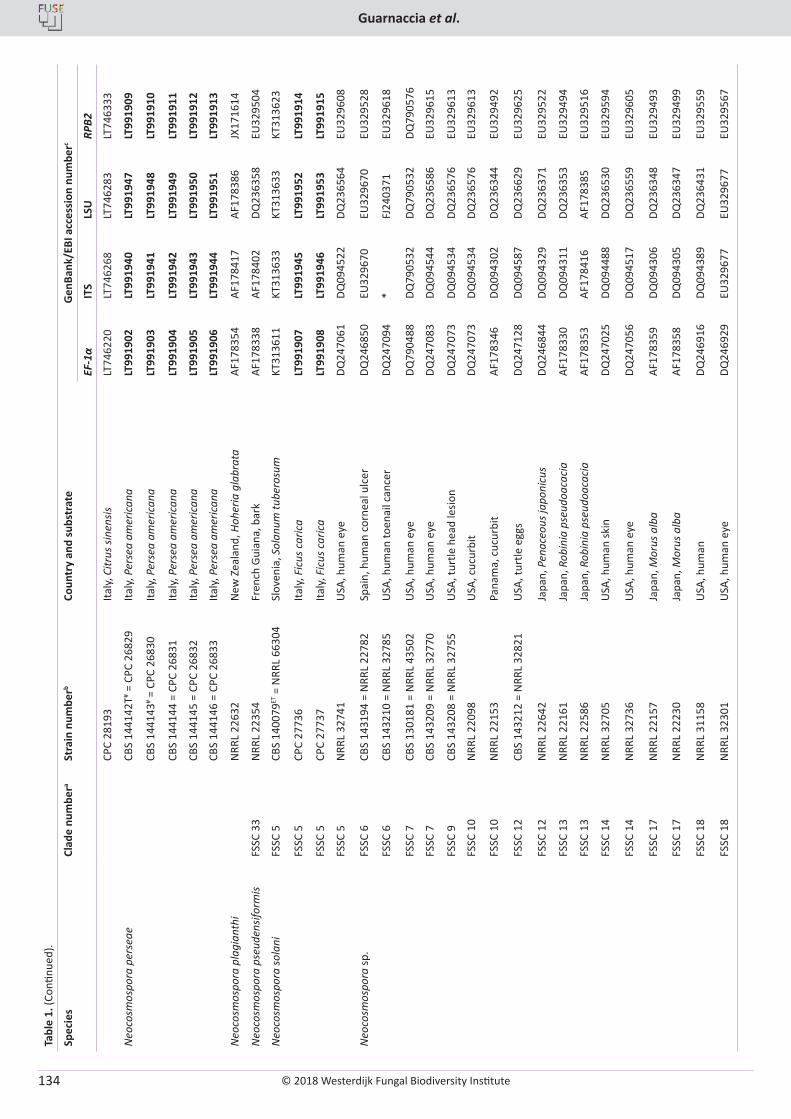
© 2018 Westerdijk Fungal Biodiversity Institute
Guarnaccia et al.
Editor-in-ChiefProf. dr P.W. Crous, Westerdijk Fungal Biodiversity Institute, P.O. Box 85167, 3508 AD Utrecht, The Netherlands.E-mail:[email protected]
134
Tabl
e 1.
(Con
tinue
d).
Spec
ies
Clad
e nu
mbe
ra St
rain
num
berb
Coun
try
and
subs
trat
eG
enBa
nk/E
BI a
cces
sion
num
berc
EF-1
αIT
SLS
URP
B2
CPC
2819
3Ita
ly, C
itrus
sine
nsis
LT74
6220
LT74
6268
LT74
6283
LT74
6333
Neo
cosm
ospo
ra p
erse
aeCB
S 14
4142
T# = C
PC 2
6829
Italy,
Per
sea
amer
ican
aLT
9919
02LT
9919
40LT
9919
47LT
9919
09
CBS
1441
43# =
CPC
268
30Ita
ly, P
erse
a am
eric
ana
LT99
1903
LT99
1941
LT99
1948
LT99
1910
CBS
1441
44 =
CPC
268
31Ita
ly, P
erse
a am
eric
ana
LT99
1904
LT99
1942
LT99
1949
LT99
1911
CBS
1441
45 =
CPC
268
32Ita
ly, P
erse
a am
eric
ana
LT99
1905
LT99
1943
LT99
1950
LT99
1912
CBS
1441
46 =
CPC
268
33Ita
ly, P
erse
a am
eric
ana
LT99
1906
LT99
1944
LT99
1951
LT99
1913
Neo
cosm
ospo
ra p
lagi
anth
iN
RRL
2263
2N
ew Z
eala
nd, H
oher
ia g
labr
ata
AF17
8354
AF17
8417
AF17
8386
JX17
1614
Neo
cosm
ospo
ra p
seud
ensif
orm
isFS
SC 3
3N
RRL
2235
4Fr
ench
Gui
ana,
bar
kAF
1783
38AF
1784
02DQ
2363
58EU
3295
04
Neo
cosm
ospo
ra so
lani
FSSC
5CB
S 14
0079
ET =
NRR
L 66
304
Slov
enia
, Sol
anum
tube
rosu
mKT
3136
11KT
3136
33KT
3136
33KT
3136
23
FSSC
5CP
C 27
736
Italy,
Fic
us c
aric
aLT
9919
07LT
9919
45LT
9919
52LT
9919
14
FSSC
5CP
C 27
737
Italy,
Fic
us c
aric
aLT
9919
08LT
9919
46LT
9919
53LT
9919
15
FSSC
5N
RRL
3274
1U
SA, h
uman
eye
DQ24
7061
DQ09
4522
DQ23
6564
EU32
9608
Neo
cosm
ospo
ra sp
.FS
SC 6
CBS
1431
94 =
NRR
L 22
782
Spai
n, h
uman
cor
neal
ulc
erDQ
2468
50EU
3296
70EU
3296
70EU
3295
28
FSSC
6CB
S 14
3210
= N
RRL
3278
5U
SA, h
uman
toen
ail c
ance
rDQ
2470
94*
FJ24
0371
EU32
9618
FSSC
7CB
S 13
0181
= N
RRL
4350
2U
SA, h
uman
eye
DQ79
0488
DQ79
0532
DQ79
0532
DQ79
0576
FSSC
7CB
S 14
3209
= N
RRL
3277
0U
SA, h
uman
eye
DQ24
7083
DQ09
4544
DQ23
6586
EU32
9615
FSSC
9CB
S 14
3208
= N
RRL
3275
5U
SA, t
urtle
hea
d le
sion
DQ24
7073
DQ09
4534
DQ23
6576
EU32
9613
FSSC
10
NRR
L 22
098
USA
, cuc
urbi
tDQ
2470
73DQ
0945
34DQ
2365
76EU
3296
13
FSSC
10
NRR
L 22
153
Pana
ma,
cuc
urbi
tAF
1783
46DQ
0943
02DQ
2363
44EU
3294
92
FSSC
12
CBS
1432
12 =
NRR
L 32
821
USA
, tur
tle e
ggs
DQ24
7128
DQ09
4587
DQ23
6629
EU32
9625
FSSC
12
NRR
L 22
642
Japa
n, P
enac
eous
japo
nicu
sDQ
2468
44DQ
0943
29DQ
2363
71EU
3295
22
FSSC
13
NRR
L 22
161
Japa
n, R
obin
ia p
seud
oaca
cia
AF17
8330
DQ09
4311
DQ23
6353
EU32
9494
FSSC
13
NRR
L 22
586
Japa
n, R
obin
ia p
seud
oaca
cia
AF17
8353
AF17
8416
AF17
8385
EU32
9516
FSSC
14
NRR
L 32
705
USA
, hum
an sk
inDQ
2470
25DQ
0944
88DQ
2365
30EU
3295
94
FSSC
14
NRR
L 32
736
USA
, hum
an e
yeDQ
2470
56DQ
0945
17DQ
2365
59EU
3296
05
FSSC
17
NRR
L 22
157
Japa
n, M
orus
alb
aAF
1783
59DQ
0943
06DQ
2363
48EU
3294
93
FSSC
17
NRR
L 22
230
Japa
n, M
orus
alb
aAF
1783
58DQ
0943
05DQ
2363
47EU
3294
99
FSSC
18
NRR
L 31
158
USA
, hum
anDQ
2469
16DQ
0943
89DQ
2364
31EU
3295
59
FSSC
18
NRR
L 32
301
USA
, hum
an e
yeDQ
2469
29EU
3296
77EU
3296
77EU
3295
67
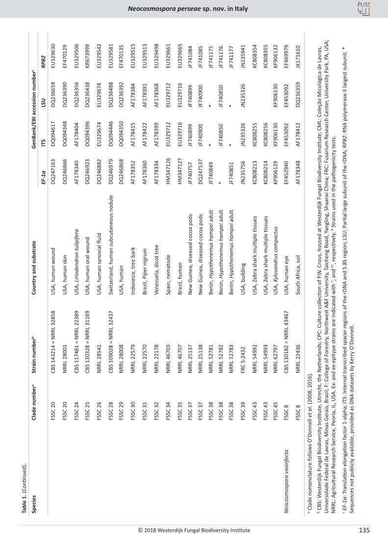
© 2018 Westerdijk Fungal Biodiversity Institute
Neocosmospora perseae sp. nov. in Italy
Editor-in-ChiefProf. dr P.W. Crous, Westerdijk Fungal Biodiversity Institute, P.O. Box 85167, 3508 AD Utrecht, The Netherlands.E-mail:[email protected]
135
Tabl
e 1.
(Con
tinue
d).
Spec
ies
Clad
e nu
mbe
ra St
rain
num
berb
Coun
try
and
subs
trat
eG
enBa
nk/E
BI a
cces
sion
num
berc
EF-1
αIT
SLS
URP
B2
FSSC
20
CBS
1432
14 =
NRR
L 32
858
USA
, hum
an w
ound
DQ24
7163
DQ09
4617
DQ23
6659
EU32
9630
FSSC
20
NRR
L 28
001
USA
, hum
an sk
inDQ
2468
66DQ
0943
48DQ
2363
90EF
4701
29
FSSC
24
CBS
1174
81 =
NRR
L 22
389
USA
, Liri
oden
dron
tulip
ifera
AF17
8340
AF17
8404
DQ23
6356
EU32
9506
FSSC
25
CBS
1303
28 =
NRR
L 31
169
USA
, hum
an o
ral w
ound
DQ24
6923
DQ09
4396
DQ23
6438
KR67
3999
FSSC
26
NRR
L 28
541
USA
, hum
an sy
novi
al fl
uid
DQ24
6882
EU32
9674
EU32
9674
EU32
9542
FSSC
28
CBS
1090
28 =
NRR
L 32
437
Switz
erla
nd, h
uman
subc
utan
eous
nod
ule
DQ24
6979
DQ09
4446
DQ23
6488
EU32
9581
FSSC
29
NRR
L 28
008
USA
, hum
anDQ
2468
68DQ
0943
50DQ
2363
92EF
4701
35
FSSC
30
NRR
L 22
579
Indo
nesia
, tre
e ba
rkAF
1783
52AF
1784
15AF
1783
84EU
3295
15
FSSC
31
NRR
L 22
570
Braz
il, P
iper
nig
rum
AF17
8360
AF17
8422
AF17
8391
EU32
9513
FSSC
32
NRR
L 22
178
Vene
zuel
a, d
icot
tree
AF17
8334
AF17
8399
AF17
8368
EU32
9498
FSSC
34
NRR
L 46
703
Spai
n, n
emat
ode
HM34
7126
EU32
9712
EU32
9712
EU32
9661
FSSC
35
NRR
L 46
707
Braz
il, h
uman
HM34
7127
EU32
9716
EU32
9716
EU32
9665
FSSC
37
NRR
L 25
137
New
Gui
nea,
dise
ased
coc
oa p
ods
JF74
0757
JF74
0899
JF74
0899
JF74
1084
FSSC
37
NRR
L 25
138
New
Gui
nea,
dise
ased
coc
oa p
ods
DQ24
7537
JF74
0900
JF74
0900
JF74
1085
FSSC
38
NRR
L 52
781
Beni
n, H
ypot
hene
mus
ham
pei a
dult
JF74
0849
**
JF74
1175
FSSC
38
NRR
L 52
782
Beni
n, H
ypot
hene
mus
ham
pei a
dult
*JF
7408
50JF
7408
50JF
7411
76
FSSC
38
NRR
L 52
783
Beni
n, H
ypot
hene
mus
ham
pei a
dult
JF74
0851
**
JF74
1177
FSSC
39
FRC
S-24
32U
SA, b
uild
ing
JN23
5756
JN23
5326
JN23
5326
JN23
5941
FSSC
43
NRR
L 54
992
USA
, Zeb
ra sh
ark
mul
tiple
tiss
ues
KC80
8213
KC80
8255
KC80
8354
FSSC
43
NRR
L 54
993
USA
, Zeb
ra sh
ark
mul
tiple
tiss
ues
KC80
8214
KC80
8256
KC80
8355
FSSC
45
NRR
L 62
797
USA
, Xyl
osan
drus
com
pact
usKF
9061
29KF
9061
30KF
9061
30KF
9061
32
Neo
cosm
ospo
ra v
asin
fect
aFS
SC 8
CBS
1301
82 =
NRR
L 43
467
USA
, hum
an e
yeEF
4529
40EF
4530
92EF
4530
92EF
4699
79
FSSC
8N
RRL
2243
6So
uth
Afric
a, so
ilAF
1783
48AF
1784
12DQ
2363
59JX
1716
10
a Cla
de n
omen
clat
ure
follo
ws O
’Don
nell
et a
l. (2
008,
201
6).
b CBS
: Wes
terd
ijk F
unga
l Bio
dive
rsity
Insti
tute
, Utr
echt
, the
Net
herla
nds;
CPC
: Cul
ture
col
lecti
on o
f P.W
. Cro
us, h
ouse
d at
Wes
terd
ijk F
unga
l Bio
dive
rsity
Insti
tute
; CM
L: C
oleç
ão M
icol
ógic
a de
Lav
ras,
U
nive
rsid
ade
Fede
ral d
e La
vras
, Min
as G
erai
s, B
razil
; F: C
olle
ge o
f For
estr
y, N
orth
wes
t A&
F U
nive
rsity
, Tai
chen
g Ro
ad, Y
angl
ing,
Sha
anxi
Chi
na; F
RC: F
usar
ium
Res
earc
h Ce
nter
, Uni
vers
ity P
ark,
PA,
USA
; N
RRL:
Agr
icul
tura
l Res
earc
h Se
rvic
e, P
eoria
, IL,
USA
. Ex-
and
ex-
epity
pe st
rain
s are
indi
cate
d w
ith T , a
nd ET
, res
pecti
vely.
# Str
ains
use
d in
the
path
ogen
icity
test
s.c E
F-1α
: Tra
nsla
tion
elon
gatio
n fa
ctor
1-a
lpha
; ITS
: Int
erna
l tra
nscr
ibed
spac
er re
gion
s of t
he rD
NA
and
5.8S
regi
on; L
SU: P
artia
l lar
ge su
buni
t of t
he rD
NA;
RPB
2: R
NA
poly
mer
ase
II la
rges
t sub
unit.
*
Sequ
ence
s not
pub
licly
ava
ilabl
e, p
rovi
ded
as D
NA
data
sets
by
Kerr
y O
’Don
nell.
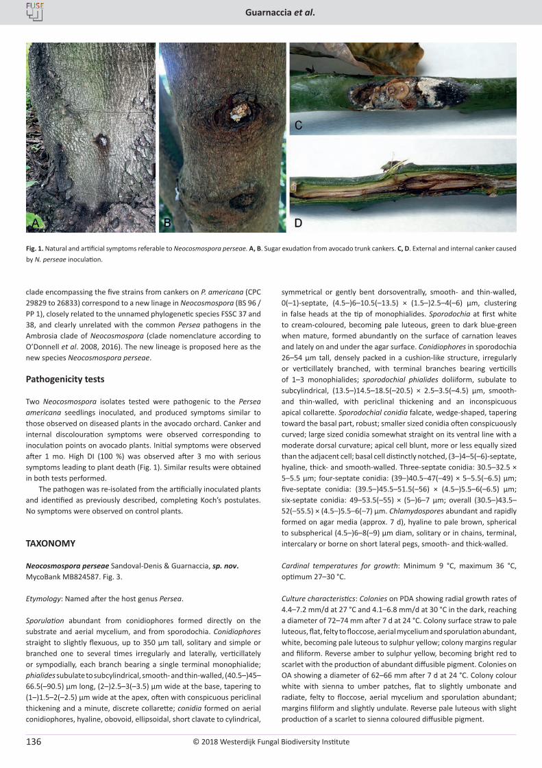
© 2018 Westerdijk Fungal Biodiversity Institute
Guarnaccia et al.
Editor-in-ChiefProf. dr P.W. Crous, Westerdijk Fungal Biodiversity Institute, P.O. Box 85167, 3508 AD Utrecht, The Netherlands.E-mail:[email protected]
136
clade encompassing the five strains from cankers on P. americana (CPC 29829 to 26833) correspond to a new linage in Neocosmospora (BS 96 / PP 1), closely related to the unnamed phylogenetic species FSSC 37 and 38, and clearly unrelated with the common Persea pathogens in the Ambrosia clade of Neocosmospora (clade nomenclature according to O’Donnell et al. 2008, 2016). The new lineage is proposed here as the new species Neocosmospora perseae.
Pathogenicity tests
Two Neocosmospora isolates tested were pathogenic to the Persea americana seedlings inoculated, and produced symptoms similar to those observed on diseased plants in the avocado orchard. Canker and internal discolouration symptoms were observed corresponding to inoculation points on avocado plants. Initial symptoms were observed after 1 mo. High DI (100 %) was observed after 3 mo with serious symptoms leading to plant death (Fig. 1). Similar results were obtained in both tests performed.
The pathogen was re-isolated from the artificially inoculated plants and identified as previously described, completing Koch’s postulates. No symptoms were observed on control plants.
TAXONOMY
Neocosmospora perseae Sandoval-Denis & Guarnaccia, sp. nov. MycoBank MB824587. Fig. 3.
Etymology: Named after the host genus Persea.
Sporulation abundant from conidiophores formed directly on the substrate and aerial mycelium, and from sporodochia. Conidiophores straight to slightly flexuous, up to 350 μm tall, solitary and simple or branched one to several times irregularly and laterally, verticillately or sympodially, each branch bearing a single terminal monophialide; phialides subulate to subcylindrical, smooth- and thin-walled, (40.5–)45–66.5(–90.5) μm long, (2–)2.5–3(–3.5) μm wide at the base, tapering to (1–)1.5–2(–2.5) μm wide at the apex, often with conspicuous periclinal thickening and a minute, discrete collarette; conidia formed on aerial conidiophores, hyaline, obovoid, ellipsoidal, short clavate to cylindrical,
symmetrical or gently bent dorsoventrally, smooth- and thin-walled, 0(–1)-septate, (4.5–)6–10.5(–13.5) × (1.5–)2.5–4(–6) μm, clustering in false heads at the tip of monophialides. Sporodochia at first white to cream-coloured, becoming pale luteous, green to dark blue-green when mature, formed abundantly on the surface of carnation leaves and lately on and under the agar surface. Conidiophores in sporodochia 26–54 μm tall, densely packed in a cushion-like structure, irregularly or verticillately branched, with terminal branches bearing verticills of 1–3 monophialides; sporodochial phialides doliiform, subulate to subcylindrical, (13.5–)14.5–18.5(–20.5) × 2.5–3.5(–4.5) μm, smooth- and thin-walled, with periclinal thickening and an inconspicuous apical collarette. Sporodochial conidia falcate, wedge-shaped, tapering toward the basal part, robust; smaller sized conidia often conspicuously curved; large sized conidia somewhat straight on its ventral line with a moderate dorsal curvature; apical cell blunt, more or less equally sized than the adjacent cell; basal cell distinctly notched, (3–)4–5(–6)-septate, hyaline, thick- and smooth-walled. Three-septate conidia: 30.5–32.5 × 5–5.5 μm; four-septate conidia: (39–)40.5–47(–49) × 5–5.5(–6.5) μm; five-septate conidia: (39.5–)45.5–51.5(–56) × (4.5–)5.5–6(–6.5) μm; six-septate conidia: 49–53.5(–55) × (5–)6–7 μm; overall (30.5–)43.5–52(–55.5) × (4.5–)5.5–6(–7) μm. Chlamydospores abundant and rapidly formed on agar media (approx. 7 d), hyaline to pale brown, spherical to subspherical (4.5–)6–8(–9) μm diam, solitary or in chains, terminal, intercalary or borne on short lateral pegs, smooth- and thick-walled.
Cardinal temperatures for growth: Minimum 9 °C, maximum 36 °C, optimum 27–30 °C.
Culture characteristics: Colonies on PDA showing radial growth rates of 4.4–7.2 mm/d at 27 °C and 4.1–6.8 mm/d at 30 °C in the dark, reaching a diameter of 72–74 mm after 7 d at 24 °C. Colony surface straw to pale luteous, flat, felty to floccose, aerial mycelium and sporulation abundant, white, becoming pale luteous to sulphur yellow; colony margins regular and filiform. Reverse amber to sulphur yellow, becoming bright red to scarlet with the production of abundant diffusible pigment. Colonies on OA showing a diameter of 62–66 mm after 7 d at 24 °C. Colony colour white with sienna to umber patches, flat to slightly umbonate and radiate, felty to floccose, aerial mycelium and sporulation abundant; margins filiform and slightly undulate. Reverse pale luteous with slight production of a scarlet to sienna coloured diffusible pigment.
Fig. 1. Natural and artificial symptoms referable to Neocosmospora perseae. A, B. Sugar exudation from avocado trunk cankers. C, D. External and internal canker caused by N. perseae inoculation.
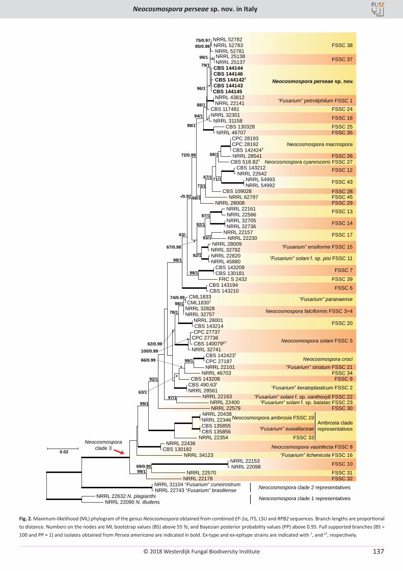
© 2018 Westerdijk Fungal Biodiversity Institute
Neocosmospora perseae sp. nov. in Italy
Editor-in-ChiefProf. dr P.W. Crous, Westerdijk Fungal Biodiversity Institute, P.O. Box 85167, 3508 AD Utrecht, The Netherlands.E-mail:[email protected]
137
0.02
CBS 140079ET
CBS 144146
CBS 518.82T
NRRL 32757
CBS 135856
NRRL 22586
NRRL 25138
CPC 28192
NRRL 20438
NRRL 46703
CML1833
NRRL 52781
NRRL 25137
NRRL 22743 “Fusarium” brasiliense
CPC 27736
NRRL 22163
NRRL 31104 “Fusarium” cuneirostrum
NRRL 32705
CBS 144143
CBS 142424T
NRRL 45880
NRRL 32828
NRRL 22642
CBS 144142T
NRRL 32736
NRRL 34123
NRRL 46707
NRRL 22400
CBS 130328
NRRL 22157
NRRL 22820
NRRL 43812
NRRL 28001
CBS 143209
CBS 144144
CBS 142423T
NRRL 62797
NRRL 22436
NRRL 22141
FRC S 2432
NRRL 22178
NRRL 32301
CBS 143210
NRRL 54992
NRRL 22570
NRRL 32741
NRRL 22090 N. illudens
CPC 27737
CBS 135855
CBS 144145
CBS 117481
NRRL 54993
NRRL 52783
CBS 143208
NRRL 22579
CBS 143194
NRRL 32792
NRRL 22101
CBS 109028
NRRL 22632 N. plagianthi
CPC 28193
CBS 130181
NRRL 22153
CBS 143212
CBS 143214
CBS 490.63T
NRRL 28008
NRRL 22346
NRRL 22354
CBS 130182
NRRL 22230
NRRL 31158
NRRL 22098
NRRL 52782
NRRL 28561
NRRL 28541
CML1830T
CPC 27187
NRRL 22161
NRRL 28009
100/0.99
66/0.99
97/1
79/1
73/1
92/1
67/0.98
98/1
99/1
96/1
99/1
92/1
69/0.90
62/0.99
98/1
72/0.99
75/0.97
98/1
92/1
87/1
63/-
94/1
88/1
99/1
67/1
68/1
68/1
71/1
93/1
63/1
74/0.99
85/0.98
78/1
98/1
99/1
FSSC 37
FSSC 38
“Fusarium” petroliphilum FSSC 1FSSC 24
FSSC 18
FSSC 25FSSC 35
Neocosmospora macrospora
FSSC 26Neocosmospora cyanescens FSSC 27
FSSC 12
FSSC 43
FSSC 28FSSC 45FSSC 29FSSC 13
FSSC 14
FSSC 17
“Fusarium” ensiforme FSSC 15
“Fusarium” solani f. sp. pisi FSSC 11
FSSC 7
FSSC 39
FSSC 6
“Fusarium” paranaense
Neocosmospora falciformis FSSC 3+4
FSSC 20
Neocosmospora solani FSSC 5
Neocosmospora croci“Fusarium” striatum FSSC 21
FSSC 34FSSC 9
“Fusarium” keratoplasticum FSSC 2
“Fusarium” solani f. sp. xanthoxyli FSSC 22“Fusarium” solani f. sp. batatas FSSC 23
FSSC 30
Neocosmospora ambrosia FSSC 19
“Fusarium” euwallaceae
FSSC 33
Neocosmospora vasinfecta FSSC 8“Fusarium” lichenicola FSSC 16
FSSC 10
FSSC 31FSSC 32
Neocosmospora clade 2 representatives
Neocosmospora clade 1 representatives
-/0.92
Neocosmosporaclade 3
Ambrosia clade representatives
Neocosmospora perseae sp. nov.
Fig. 2. Maximum-likelihood (ML) phylogram of the genus Neocosmospora obtained from combined EF-1α, ITS, LSU and RPB2 sequences. Branch lengths are proportional to distance. Numbers on the nodes are ML bootstrap values (BS) above 55 %; and Bayesian posterior probability values (PP) above 0.95. Full supported branches (BS = 100 and PP = 1) and isolates obtained from Persea americana are indicated in bold. Ex-type and ex-epitype strains are indicated with T, and ET, respectively.
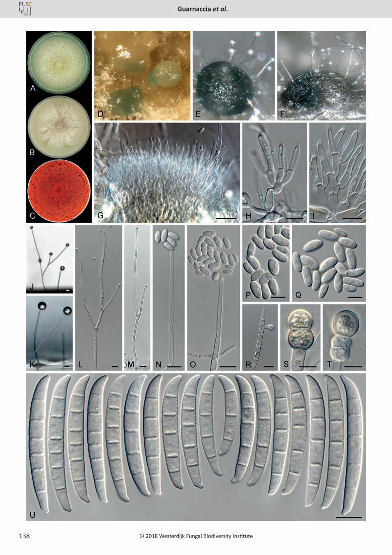
© 2018 Westerdijk Fungal Biodiversity Institute
Guarnaccia et al.
Editor-in-ChiefProf. dr P.W. Crous, Westerdijk Fungal Biodiversity Institute, P.O. Box 85167, 3508 AD Utrecht, The Netherlands.E-mail:[email protected]
138
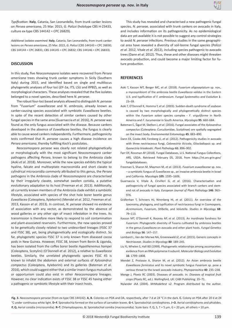
© 2018 Westerdijk Fungal Biodiversity Institute
Neocosmospora perseae sp. nov. in Italy
Editor-in-ChiefProf. dr P.W. Crous, Westerdijk Fungal Biodiversity Institute, P.O. Box 85167, 3508 AD Utrecht, The Netherlands.E-mail:[email protected]
139
Typification: Italy, Catania, San Leonardello, from trunk canker lesions on Persea americana, 25 Mar. 2015, G. Polizzi (holotype CBS H-23433, culture ex-type CBS 144142 = CPC 26829).
Additional isolates examined: Italy, Catania, San Leonardello, from trunk canker lesions on Persea americana, 25 Mar. 2015, G. Polizzi (CBS 144143 = CPC 26830; CBS 144144 = CPC 26831; CBS 144145 = CPC 26832; CBS 144146 = CPC 26833).
DISCUSSION
In this study, five Neocosmospora isolates were recovered from Persea americana trees showing trunk canker symptoms in Sicily (Southern Italy) during 2015, and identified based on single and multilocus phylogenetic analyses of four loci (EF-1α, ITS, LSU and RPB2), as well as morphological characters. These analyses revealed that the five isolates belonged to a novel species, described here N. perseae.
The robust four-loci based analysis allowed to distinguish N. perseae from “Fusarium” euwallaceae and N. ambrosia, already known as canker-causing species associated with symbiotic Euwallacea beetles. In spite of the recent detection of similar cankers caused by other fungal species in the same area (Guarnaccia et al. 2016), N. perseae was found as the only fungus associated with the disease. Because cankers developed in the absence of Euwallacea beetles, the fungus is clearly able to cause wood cankers independently. Furthermore, pathogenicity tests confirmed that N. perseae causes a high disease incidence on Persea americana, thereby fulfilling Koch’s postulates.
Neocosmospora perseae was clearly not related phylogenetically or morphologically with the most significant Neocosmospora canker pathogens affecting Persea, known to belong to the Ambrosia clade (Aoki et al. 2018). Moreover, while the new species exhibits the typical hyaline, falcate and multiseptate macroconidia and short clavate to cylindrical microconidia commonly attributed to this genus, the Persea pathogens in the Ambrosia clade of Neocosmospora are characterised by their irregularly clavate, somewhat swollen conidia, a putative evolutionary adaptation to its host (Freeman et al. 2013). Additionally, all currently known members of the Ambrosia clade exhibit a symbiotic lifestyle, associated with species of the shot hole borer beetle genus Euwallacea (Coleoptera, Xyleborini) (Mendel et al. 2012, Freeman et al. 2013, Kasson et al. 2013). In contrast, N. perseae showed no evidence of association with any vector, as demonstrated by the absence of wood galleries or any other sign of insect infestation in the trees. Its transmission is therefore more likely to respond to soil contamination and plant-associated reservoirs. Furthermore, the new species proved to be genetically closely related to two undescribed lineages (FSSC 37 and FSSC 38), yet, being phylogenetically and ecologically distinct. So far, phylogenetic species FSSC 37 is only known from diseased cocoa pods in New Guinea. However, FSSC 38, known from Benin & Uganda, has been isolated from the coffee borer beetle Hypothenemus hampei (Coleoptera, Scotylini) (O’Donnell et al. 2012), a relative to Euwallacea beetles. Similarly, the unrelated phylogenetic species FSSC 45 is known to inhabit the abdomen and external surfaces of Xylosandrus compactus (Coleoptera, Xyleborini) and its galleries (Bateman et al. 2016), which could suggest either that a similar insect-fungus mutualism or opportunism could also exist in other Neocosmospora lineages. However, no clear indication exists of FSSC 38 or FSSC 45 having either a pathogenic or symbiotic lifestyle with their insect hosts.
This study has revealed and characterised a new pathogenic fungal species, N. perseae, associated with trunk cankers on avocado in Italy, and includes information on its pathogenicity. As no epidemiological data are yet available it is not possible to suggest any control strategies to avoid N. perseae infections. Previous studies in the same geographi-cal area have revealed a diversity of soil-borne fungal species (Polizzi et al. 2012, Vitale et al. 2012), including species pathogenic to avocado trees (Dann et al. 2012). Thus, these and other diseases might threaten avocado production, and could become a major limiting factor for fu-ture production.
REFERENCES
Aoki T, Kasson MT, Berger MC, et al. (2018). Fusarium oligoseptatum sp. nov., a mycosymbiont of the ambrosia beetle Euwallacea validus in the Eastern U.S. and typification of F. ambrosium. Fungal Systematics and Evolution 1: 23–39.
Aoki T, O’Donnell K, Homma Y, et al. (2003). Sudden-death syndrome of soybean is caused by two morphologically and phylogenetically distinct species within the Fusarium solani species complex - F. virguliforme in North America and F. tucumaniae in South America. Mycologia 95: 660–684.
Bateman C, Šigut M, Skelton J, et al. (2016). Fungal associates of the Xylosandrus compactus (Coleoptera: Curculionidae, Scolytinae) are spatially segregated on the insect body. Environmental Entomology 45: 883–890.
Dann EK, Cooke AW, Forsberg LI, et al. (2012). Pathogenicity studies in avocado with three nectriaceous fungi, Calonectria ilicicola, Gliocladiopsis sp. and Ilyonectria liriodendri. Plant Pathology 61: 896–902.
Farr DF, Rossman AY (2018). Fungal Databases, U.S. National Fungus Collections, ARS, USDA. Retrieved February 05, 2018, from https://nt.ars-grin.gov/fungaldatabases.
Freeman S, Sharon M, Maymon M, et al. (2013). Fusarium euwallaceae sp. nov. – a symbiotic fungus of Euwallacea sp., an invasive ambrosia beetle in Israel and California. Mycologia 105: 1595–1606.
Guarnaccia V, Vitale A, Cirvilleri G, et al. (2016). Characterisation and pathogenicity of fungal species associated with branch cankers and stem-end rot of avocado in Italy. European Journal of Plant Pathology 146: 963–976.
Gräfenhan T, Schroers HJ, Nirenberg HI, et al. (2011). An overview of the taxonomy, phylogeny, and typification of nectriaceous fungi in Cosmospora, Acremonium, Fusarium, Stilbella, and Volutella. Studies in Mycology 68: 79–113.
Kasson MT, O’Donnell K, Rooney AP, et al. (2013). An inordinate fondness for Fusarium: Phylogenetic diversity of fusaria cultivated by ambrosia beetles in the genus Euwallacea on avocado and other plant hosts. Fungal Genetics and Biology 56: 147–157.
Lombard L, Van der Merwe NA, Groenewald JZ, et al. (2015). Generic concepts in Nectriaceae. Studies in Mycology 80: 189–245.
Liu YJ, Whelen S, Hall BD (1999). Phylogenetic relationships among ascomycetes: evidence from an RNA polymerse II subunit. Molecular Biology and Evolution 16: 1799–1808.
Mendel Z, Protasov A, Sharon M, et al. (2012). An Asian ambrosia beetle Euwallacea fornicatus and its novel symbiotic fungus Fusarium sp. pose a serious threat to the Israeli avocado industry. Phytoparasitica 40: 235–238.
Menge J, Ploetz RC (2003). Diseases of avocado. In: Diseases of tropical fruit crops (Ploetz RC, ed.). Wallingford, UK: CABI Publishing: 35–71.
Nylander JAA (2004). MrModeltest v2. Program distributed by the author.
Fig. 3. Neocosmospora perseae (from ex-type CBS 144142). A, B. Colonies on PDA and OA, respectively, after 7 d at 24 °C in the dark. C. Colony on PDA after 20 d at 24 °C under continuous white light. D–F. Sporodochia formed on the surface of carnation leaves. G–I. Sporodochial conidiophores. J–O. Aerial conidiophores and phialides. P, Q. Aerial conidia (microconidia). R–T. Chlamydospores. U. Sporodochial conidia (macroconidia). Scale bars: P, Q, S, T = 5 µm, G = 20 µm, all others = 10 µm.
© 2018 Westerdijk Fungal Biodiversity Institute
Guarnaccia et al.
Editor-in-ChiefProf. dr P.W. Crous, Westerdijk Fungal Biodiversity Institute, P.O. Box 85167, 3508 AD Utrecht, The Netherlands.E-mail:[email protected]
140
Evolutionary Biology Centre, Uppsala University.O’Donnell K, Humber RA, Geiser DM, et al. (2012) Phylogenetic diversity of
insecticolous fusaria inferred from multilocus DNA sequence data and their molecular identification via FUSARIUM-ID and Fusarium MLST. Mycologia 104: 427–445.
O'Donnell K, Gueidan C, Sink S, et al. (2009). A two-locus DNA sequence database for typing plant and human pathogens within the Fusarium oxysporum species complex. Fungal Genetics and Biology 46: 936–948.
O’Donnell K, Libeskind-Hadas R, Hulcr J, et al. (2016). Invasive Asian Fusarium – Euwallacea ambrosia beetle mutualists pose a serious threat to forests, urban landscapes and the avocado industry. Phytoparasitica 44: 435–442.
O’Donnell K, Sutton DA, Fothergill A, et al. (2008). Molecular phylogenetic diversity, multilocus haplotype nomenclature, and in vitro antifungal resistance within the Fusarium solani species complex. Journal of Clinical Microbiology 46: 2477–2490.
O’Donnell K, Sutton DA, Rinaldi MG, et al. (2010). Internet-accessible DNA sequence database for identifying fusaria from human and animal infections. Journal of Clinical Microbiology 48: 3708–3718.
Parkinson LE, Shivas RG, Dann EK (2017). Pathogenicity of nectriaceous fungi on avocado in Australia. Phytopathology 107: 1479–1485.
Polizzi G, Vitale A, Aiello D, et al. (2012). First report of Calonectria ilicicola causing a new disease on Laurus (Laurus nobilis) in Europe. Journal of Phytopathology 160: 41–44.
Rayner RW (1970). A mycological colour chart. CMI and British Mycological Society, Kew, Surrey, UK.
Sandoval-Denis M, Guarnaccia V, Polizzi G, et al. (2018). Symptomatic citrus trees reveal a new pathogenic lineage in Fusarium and two new Neocosmospora species. Persoonia 40: 1–25.
Sung GH, Sung JM, Hywel-Jones NL, et al. (2007). A multi-gene phylogeny of Clavicipitaceae (Ascomycota, Fungi): Identification of localized incongruence using a combinational bootstrap approach. Molecular phylogenetics and evolution 44: 1204–1223.
Vilgalys R, Hester M (1990). Rapid genetic identification and mapping of enzymatically amplified ribosomal DNA from several Cryptococcus species. Journal of Bacteriology 172: 4238–4246.
Vilgalys R, Sun BL (1994). Ancient and recent patterns of geographic speciation in the oyster mushroom Pleurotus revealed by phylogenetic analysis of ribosomal DNA sequences. Proceedings of the National Academy of Science USA 91: 4599–4603.
Vitale A, Aiello D, Guarnaccia V, et al. (2012). First report of root rot caused by Ilyonectria (=Neonectria) macrodidyma on avocado (Persea americana) in Italy. Journal of Phytopathology 160: 156–159.
White TJ, Bruns T, Lee S, et al. (1990). Amplification and direct sequencing of fungal ribosomal RNA genes for phylogenetics. In: PCR protocols: a guide to methods and applications (Innes MA, Gelfand DH, Sninsky JJ, White TJ, eds). USA, NY, Academic Press: 315–322.
Fungal Systematics and Evolution is licensed under a Creative Commons Attribution-NonCommercial-ShareAlike 4.0 International License
© 2018 Westerdijk Fungal Biodiversity Institute 141
Editor-in-ChiefProf. dr P.W. Crous, Westerdijk Fungal Biodiversity Institute, P.O. Box 85167, 3508 AD Utrecht, The Netherlands.E-mail:[email protected]
Fungal Systematics and EvolutionVOLUME 1JUNE 2018
PAGES 141–167
doi.org/10.3114/fuse.2018.01.07
The culturable mycobiota associated with three Atlantic sponges, including two new species: Thelebolus balaustiformis and T. spongiae
E. Bovio1,2, L. Garzoli1, A. Poli1, V. Prigione1, D. Firsova3, G.P. McCormack4, G.C. Varese1*
1Mycotheca Universitatis Taurinensis (MUT), Department of Life Sciences and Systems Biology, University of Turin, 10125 Turin, Italy2Marine Natural Products Team, CNRS, Institute of Chemistry (UMR 7272), University Nice Côte d'Azur, Nice, 06100, France3School of Chemistry, National University of Ireland Galway, Galway, Ireland4Zoology, Ryan Institute, School of Natural Sciences, National University of Ireland Galway, Galway, Ireland
*Corresponding author: [email protected]
Abstract: Covering 70 % of Earth, oceans are at the same time the most common and the environment least studied by microbiologists. Considering the large gaps in our knowledge on the presence of marine fungi in the oceans, the aim of this research was to isolate and identify the culturable fungal community within three species of sponges, namely Dysidea fragilis, Pachymatisma johnstonia and Sycon ciliatum, collected in the Atlantic Ocean and never studied for their associated mycobiota. Applying different isolation methods, incubation temperatures and media, and attempting to mimic the marine and sponge environments, were fundamental to increase the number of cultivable taxa. Fungi were identified using a polyphasic approach, by means of morpho-physiological, molecular and phylogenetic techniques. The sponges revealed an astonishing fungal diversity represented by 87 fungal taxa. Each sponge hosted a specific fungal community with more than half of the associated fungi being exclusive of each invertebrate. Several species isolated and identified in this work, already known in terrestrial environment, were first reported in marine ecosystems (21 species) and in association with sponges (49 species), including the two new species Thelebolus balaustiformis and Thelebolus spongiae, demonstrating that oceans are an untapped source of biodiversity.
Key words: Atlantic Oceanmarine fungispongessystematicstwo new taxa
Published online: 28 March 2018.
INTRODUCTION
Water covers almost 70 % of our planet. Nonetheless, its biodiversity and habitats remain largely unexplored. For the last 580 M years, oceans have been hosting the most ancient metazoans on Earth: sponges. Porifera contains more than 8 600 described species and about 15 000 species waiting to be discovered by scientists (Webster & Thomas 2016). Sponges are key components of marine ecosystems, because of their incredible ability to filter seawater: according to recent estimates, they are able to process 24 000 L/kg of seawater per day and to detain over 80 % of its particles (Taylor et al. 2007, Rozas et al. 2011).
Over the millions of years of their evolution, species of Porifera have formed a close association with a wide variety of microorganisms including bacteria, archaea, fungi, and algae (Taylor et al. 2007). This close association was described for the first time by Vacelet & Donadey (1977), who observed bacteria within sponges’ tissues. Today, it is well recognised that microorganisms can represent up to 40–60 % of the sponge biomass (Yarden 2014). The term “sponge holobiont”, is used when sponges and the associated microbial communities are considered as a whole (He et al. 2014). Different degrees of complexity characterise the interactions among sponge holobiont components, including mutualism, commensalism and parasitism (Rodríguez-Marconi 2005). Non-pathogenic
microorganisms can positively contribute to sponge metabolism, by increasing the uptake of carbon, nitrogen and sulphur. Furthermore, by the production of secondary metabolites, they could be involved in host defence systems and in the regulation of the microbial community associated with sponges (Taylor et al. 2007). Interestingly, metabolites previously ascribed to sponges were recognised to be structurally similar to those produced by the associated bacteria (Imhoff & Stöhr 2003). As a consequence, the use of microorganisms for the bio-discovery of new molecules would avoid several problems related to the use of sponges. The isolation of new molecules and their production in the required amount from sponges is always very problematical for reasons such as their rare occurrence, difficulties with sponge collections, or irreproducible production of metabolites due to specimen variability (Imhoff & Stöhr 2003). Nowadays studies on microorganisms associated with sponges are primarily focused on prokaryotic organisms while the fungal community remain less studied, despite recent results emphasizing its great biodiversity and biotechnological potential (Raghukumar 2012). Fungi represent suitable biotechnological resources but require specific expertise for the isolation and the correct identification. Many taxa already known for their bioactivity lack a precise identification and correct preservation in culture collections hampering their possible future exploitation as recently highlighted by the 2nd International Conference of Marine Fungal Natural Products (MaFNaP 2017).
© 2018 Westerdijk Fungal Biodiversity Institute
Bovio et al.
Editor-in-ChiefProf. dr P.W. Crous, Westerdijk Fungal Biodiversity Institute, P.O. Box 85167, 3508 AD Utrecht, The Netherlands.E-mail:[email protected]
142
In this study we present for the first time the mycobiota associated with Dysidea fragilis, Pachymatisma johnstonia (Demospongiae), and Sycon ciliatum (Calcarea). The two Demospongiae have been extensively examined for their production of secondary metabolites. The metabolome of D. fragilis was characterised by Yu et al. (2006), although its biological activity included only a single compound, which acted as fish feeding deterrent (Marin et al. 1998). Pachymatisma johnstonia is also well known for its production of secondary metabolites, whose anticancer (Zidane et al. 1996, Ferreira et al. 2011) and antibacterial (Warabi et al. 2004) activity has been demonstrated. On the contrary, the metabolome of S. ciliatum has never been studied.
In light of the above-mentioned considerations, it is likely that microorganisms, including fungi, could be the true producers of the bioactive molecules isolated so far but could also be a source of additional novel compounds of interest. Aiming at the biotechnological exploitation of new molecules, the scope of this study was to isolate and identify the fungal community associated with three Atlantic sponges, with particular emphasis on the proper systematic identification of taxa. Moreover, in this paper two novel Thelebolus species are described.
MATERIAL AND METHODS
Sampling sites and axenic isolation
The sponges Dysidea fragilis and Pachymatisma johnstonia (three specimens each) were collected by scuba divers in Gurraig Sound (Co. Galway, Ireland; N 53°, 18.944; W 09°, 40.140). The sampling site was at 15 m depth, characterised by fairly strong current and suspended sediments. Three specimens of Sycon ciliatum exposed to a fast water flow due to the tide going out were collected in Coranroo rapids (Co. Clare, Ireland; N 053°09.100, W 009°,00. 550).
Specimens were surface sterilised with ethanol 70 % (for 30 s) to prevent contaminants and serially washed (three times) in artificial sterile Sea Water (SW) to get rid of unrefined sediments and to wash out propagules, in order to leave only fungi actively growing on the surface or into the sponge tissues.
Working in sterile conditions, the sponge samples were divided into three parts to be used for two different fungal isolation techniques and for a taxonomic voucher of the sponge. For the first isolation method, one third of the sponge was further cut in 20 pieces of about 0.5 cm3 by means of sterile tools and directly plated in Petri dishes (six cm Ø) containing two different media: Sea Water Agar – SWA (Sea Salts 30 g, Agar 15 g, up to 1 L dH2O) and Corn Meal Agar Sea Water - CMASW (Corn Meal 2 g, Agar 15 g, Sea Salts 30 g, up to 1 L dH2O). Five replicates for each medium and incubation temperatures (15 °C and 25 °C) were performed.
Approximately 5 g of each sample were also homogenised (homogenizer blade Sterilmixer II - PBI International) and diluted 1:10 w/v in SW. One mL of suspension was included in Petri dishes (nine cm Ø) containing CMASW or Gelatin Agar Sea Water– GASW (Gelatin 20 g, Sea Salts 30 g, Agar 15 g, up to 1 L dH2O), rich in collagen and mimicking sponge tissue composition. Five replicates for each medium and incubation temperatures (15 °C and 25 °C) were performed. All media were supplemented with
an antibiotic mix (Gentamicin Sulfate 40 mg/L, Piperacillin plus Tazobactam 11 mg/L) to prevent bacterial growth. Plates were incubated in the dark and periodically checked for 30 d to isolate slow growing fungi.
Fungal identification
Fungal morphotypes were isolated in pure culture and identified by means of a polyphasic approach combining morpho-physiological and molecular features. After determination of genera via macroscopic and microscopic features (Domsch et al. 1980, Von Arx 1981, Kiffer & Morelet 1997), fungi were transferred to genus-specific media (Klich 2002, Braun et al. 2003, Samson & Frisvad 2004).
In parallel for molecular analyses, fungi were pre-grown on Malt Extract Agar - MEA (Malt Extract 20 g, Glucose 20 g, Peptone 2 g, Agar 20 g, up to 1 L dH2O) at 25 °C for 1 wk, for fast growing fungi, and from 2–4 wk for slow growing fungi. DNA was extracted using the NucleoSpin kit (Macherey Nagel GmbH, Duren, DE, USA), according to the manufacturer instructions. Based on the taxonomic assignment attributed to each fungus by morphological observations, specific primers were used for PCR as detailed in Table 1. Briefly, PCR reactions were performed in 50 μL final volumes and consisted of 0.5 μL Taq DNA Polymerase (Qiagen 5 U/μL), 10 μL PCR Buffer (10 ×), 2.5 μL dNTP Mixture (dATP, dCTP, dGTP, dTTP; 10 mM), 2 μL MgCl2 (25 mM), 2.5 μL of each primer (10 μM), 1 μL genomic DNA extract (80 ng/mL) and 34 μL distilled-deionised water. PCR products were visualised under UV light (BIO-RAD Universal Hood II) on 1.5 % agarose electrophoresis gels stained with ethidium bromide. Macrogen, Inc. (Seoul, South Korea) Europe Lab carried out the purification and sequencing of PCR products.
Taxonomic assignments were based both on high percentage homologies with sequences available in public databases (GenBank - NCBI database and CBS-KNAW Collection, Westerdijk Fungal Biodiversity Institute) and the consistency of morphological features with available literature descriptions. The taxonomic position of doubtful strains (low homologies with sequences available in public databases) or sterile mycelia (i.e. not showing morphological features useful to confirm taxonomical assignments) were inferred via molecular phylogenetic analyses based on DNA sequences from the large ribosomal subunit LSU, (Vilgalys & Hester 1990). Separate alignments were created for the orders Pleosporales, Capnodiales and Chaetothyriales and two for the classes Leotiomycetes and Sordariomycetes. Alignments were generated using MEGA v. 7.0 and manually refined. Phylogenetic analyses were performed using a Bayesian Inference (BI; MrBayes v. 3.2.2 four incrementally heated simultaneous Monte Carlo Markov Chains (MCMC), run over 10 M generations, (under GTR + Γ + I evolutionary model approach). BPP values are reported in the resulting trees. A full alignment of the dataset was submitted to TreeBASE (submission number 21746).
Representative strains of each species isolated in pure culture during this work are preserved at Mycotheca Universitatis Taurinensis (MUT- www.mut.unito.it) of the Department of Life Sciences and Systems Biology, University of Turin (Italy). The Accession numbers of the sequences deposited in GenBank are available in the supplementary material 1.
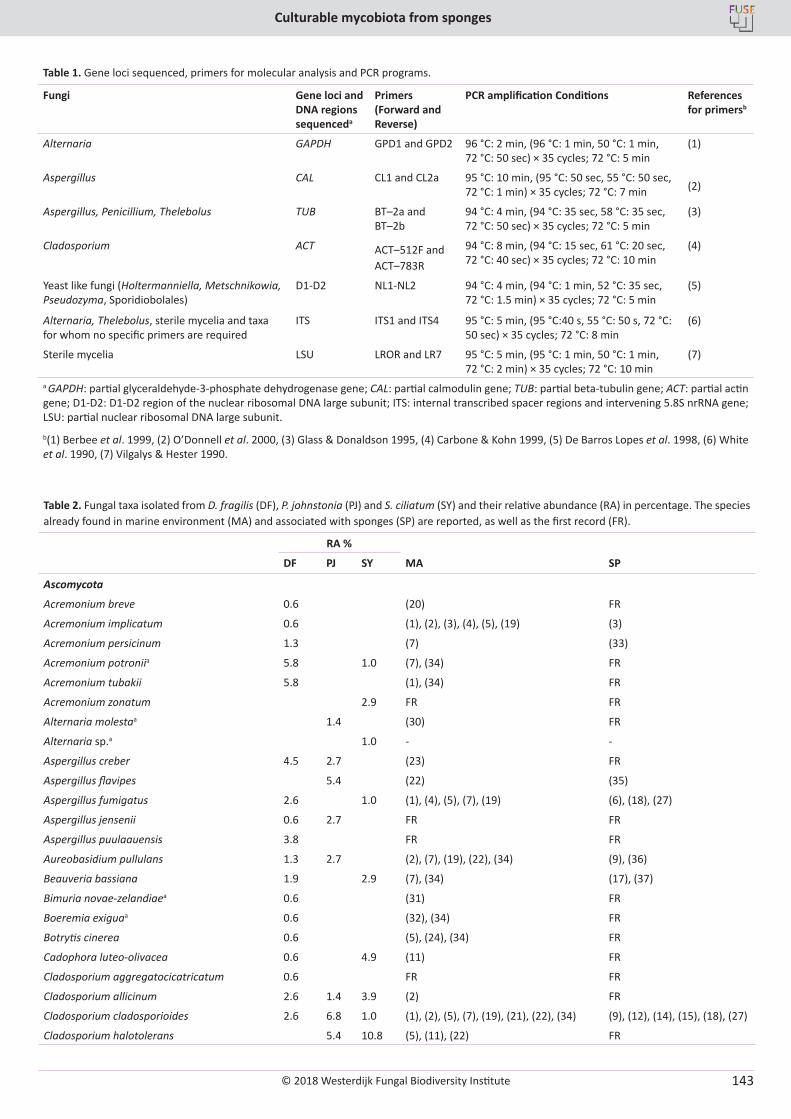
© 2018 Westerdijk Fungal Biodiversity Institute
Culturable mycobiota from sponges
Editor-in-ChiefProf. dr P.W. Crous, Westerdijk Fungal Biodiversity Institute, P.O. Box 85167, 3508 AD Utrecht, The Netherlands.E-mail:[email protected]
143
Table 2. Fungal taxa isolated from D. fragilis (DF), P. johnstonia (PJ) and S. ciliatum (SY) and their relative abundance (RA) in percentage. The species already found in marine environment (MA) and associated with sponges (SP) are reported, as well as the first record (FR).
RA %
DF PJ SY MA SP
Ascomycota
Acremonium breve 0.6 (20) FR
Acremonium implicatum 0.6 (1), (2), (3), (4), (5), (19) (3)
Acremonium persicinum 1.3 (7) (33)
Acremonium potroniia 5.8 1.0 (7), (34) FR
Acremonium tubakii 5.8 (1), (34) FR
Acremonium zonatum 2.9 FR FR
Alternaria molestaa 1.4 (30) FR
Alternaria sp.a 1.0 - -
Aspergillus creber 4.5 2.7 (23) FR
Aspergillus flavipes 5.4 (22) (35)
Aspergillus fumigatus 2.6 1.0 (1), (4), (5), (7), (19) (6), (18), (27)
Aspergillus jensenii 0.6 2.7 FR FR
Aspergillus puulaauensis 3.8 FR FR
Aureobasidium pullulans 1.3 2.7 (2), (7), (19), (22), (34) (9), (36)
Beauveria bassiana 1.9 2.9 (7), (34) (17), (37)
Bimuria novae-zelandiaea 0.6 (31) FR
Boeremia exiguaa 0.6 (32), (34) FR
Botrytis cinerea 0.6 (5), (24), (34) FR
Cadophora luteo-olivacea 0.6 4.9 (11) FR
Cladosporium aggregatocicatricatum 0.6 FR FR
Cladosporium allicinum 2.6 1.4 3.9 (2) FR
Cladosporium cladosporioides 2.6 6.8 1.0 (1), (2), (5), (7), (19), (21), (22), (34) (9), (12), (14), (15), (18), (27)
Cladosporium halotolerans 5.4 10.8 (5), (11), (22) FR
Table 1. Gene loci sequenced, primers for molecular analysis and PCR programs.
Fungi Gene loci and DNA regions sequenceda
Primers (Forward and Reverse)
PCR amplification Conditions References for primersb
Alternaria GAPDH GPD1 and GPD2 96 °C: 2 min, (96 °C: 1 min, 50 °C: 1 min, 72 °C: 50 sec) × 35 cycles; 72 °C: 5 min
(1)
Aspergillus CAL CL1 and CL2a 95 °C: 10 min, (95 °C: 50 sec, 55 °C: 50 sec, 72 °C: 1 min) × 35 cycles; 72 °C: 7 min (2)
Aspergillus, Penicillium, Thelebolus TUB BT–2a and BT–2b
94 °C: 4 min, (94 °C: 35 sec, 58 °C: 35 sec, 72 °C: 50 sec) × 35 cycles; 72 °C: 5 min
(3)
Cladosporium ACT ACT–512F and ACT–783R
94 °C: 8 min, (94 °C: 15 sec, 61 °C: 20 sec, 72 °C: 40 sec) × 35 cycles; 72 °C: 10 min
(4)
Yeast like fungi (Holtermanniella, Metschnikowia, Pseudozyma, Sporidiobolales)
D1-D2 NL1-NL2 94 °C: 4 min, (94 °C: 1 min, 52 °C: 35 sec, 72 °C: 1.5 min) × 35 cycles; 72 °C: 5 min
(5)
Alternaria, Thelebolus, sterile mycelia and taxa for whom no specific primers are required
ITS ITS1 and ITS4 95 °C: 5 min, (95 °C:40 s, 55 °C: 50 s, 72 °C: 50 sec) × 35 cycles; 72 °C: 8 min
(6)
Sterile mycelia LSU LROR and LR7 95 °C: 5 min, (95 °C: 1 min, 50 °C: 1 min, 72 °C: 2 min) × 35 cycles; 72 °C: 10 min
(7)
a GAPDH: partial glyceraldehyde-3-phosphate dehydrogenase gene; CAL: partial calmodulin gene; TUB: partial beta-tubulin gene; ACT: partial actin gene; D1-D2: D1-D2 region of the nuclear ribosomal DNA large subunit; ITS: internal transcribed spacer regions and intervening 5.8S nrRNA gene; LSU: partial nuclear ribosomal DNA large subunit.b(1) Berbee et al. 1999, (2) O’Donnell et al. 2000, (3) Glass & Donaldson 1995, (4) Carbone & Kohn 1999, (5) De Barros Lopes et al. 1998, (6) White et al. 1990, (7) Vilgalys & Hester 1990.
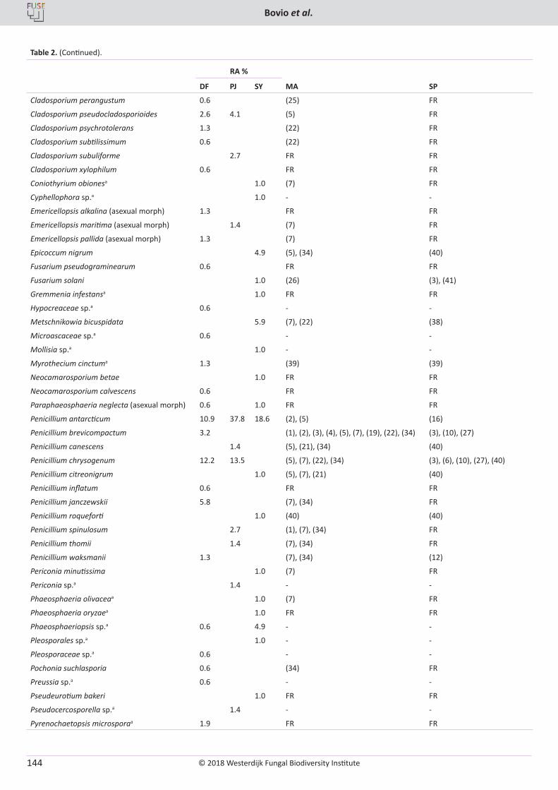
© 2018 Westerdijk Fungal Biodiversity Institute
Bovio et al.
Editor-in-ChiefProf. dr P.W. Crous, Westerdijk Fungal Biodiversity Institute, P.O. Box 85167, 3508 AD Utrecht, The Netherlands.E-mail:[email protected]
144
Table 2. (Continued).
RA %
DF PJ SY MA SP
Cladosporium perangustum 0.6 (25) FR
Cladosporium pseudocladosporioides 2.6 4.1 (5) FR
Cladosporium psychrotolerans 1.3 (22) FR
Cladosporium subtilissimum 0.6 (22) FR
Cladosporium subuliforme 2.7 FR FR
Cladosporium xylophilum 0.6 FR FR
Coniothyrium obionesa 1.0 (7) FR
Cyphellophora sp.a 1.0 - -
Emericellopsis alkalina (asexual morph) 1.3 FR FR
Emericellopsis maritima (asexual morph) 1.4 (7) FR
Emericellopsis pallida (asexual morph) 1.3 (7) FR
Epicoccum nigrum 4.9 (5), (34) (40)
Fusarium pseudograminearum 0.6 FR FR
Fusarium solani 1.0 (26) (3), (41)
Gremmenia infestansa 1.0 FR FR
Hypocreaceae sp.a 0.6 - -
Metschnikowia bicuspidata 5.9 (7), (22) (38)
Microascaceae sp.a 0.6 - -
Mollisia sp.a 1.0 - -
Myrothecium cinctuma 1.3 (39) (39)
Neocamarosporium betae 1.0 FR FR
Neocamarosporium calvescens 0.6 FR FR
Paraphaeosphaeria neglecta (asexual morph) 0.6 1.0 FR FR
Penicillium antarcticum 10.9 37.8 18.6 (2), (5) (16)
Penicillium brevicompactum 3.2 (1), (2), (3), (4), (5), (7), (19), (22), (34) (3), (10), (27)
Penicillium canescens 1.4 (5), (21), (34) (40)
Penicillium chrysogenum 12.2 13.5 (5), (7), (22), (34) (3), (6), (10), (27), (40)
Penicillium citreonigrum 1.0 (5), (7), (21) (40)
Penicillium inflatum 0.6 FR FR
Penicillium janczewskii 5.8 (7), (34) FR
Penicillium roqueforti 1.0 (40) (40)
Penicillium spinulosum 2.7 (1), (7), (34) FR
Penicillium thomii 1.4 (7), (34) FR
Penicillium waksmanii 1.3 (7), (34) (12)
Periconia minutissima 1.0 (7) FR
Periconia sp.a 1.4 - -
Phaeosphaeria olivaceaa 1.0 (7) FR
Phaeosphaeria oryzaea 1.0 FR FR
Phaeosphaeriopsis sp.a 0.6 4.9 - -
Pleosporales sp.a 1.0 - -
Pleosporaceae sp.a 0.6 - -
Pochonia suchlasporia 0.6 (34) FR
Preussia sp.a 0.6 - -
Pseudeurotium bakeri 1.0 FR FR
Pseudocercosporella sp.a 1.4 - -
Pyrenochaetopsis microsporaa 1.9 FR FR
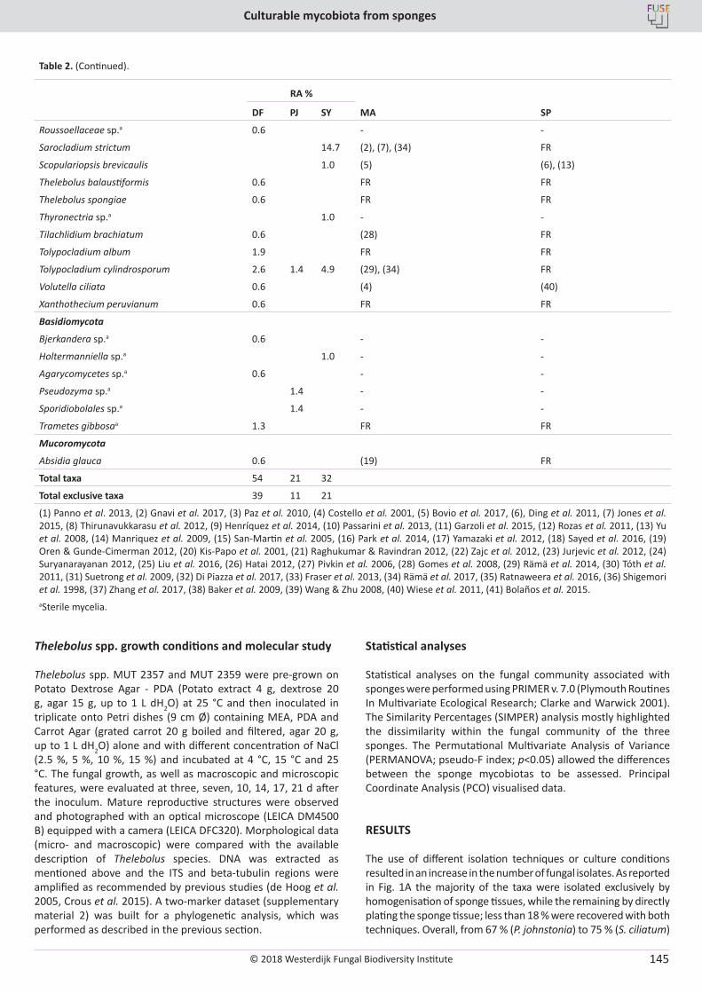
© 2018 Westerdijk Fungal Biodiversity Institute
Culturable mycobiota from sponges
Editor-in-ChiefProf. dr P.W. Crous, Westerdijk Fungal Biodiversity Institute, P.O. Box 85167, 3508 AD Utrecht, The Netherlands.E-mail:[email protected]
145
Table 2. (Continued).
RA %
DF PJ SY MA SP
Roussoellaceae sp.a 0.6 - -
Sarocladium strictum 14.7 (2), (7), (34) FR
Scopulariopsis brevicaulis 1.0 (5) (6), (13)
Thelebolus balaustiformis 0.6 FR FR
Thelebolus spongiae 0.6 FR FR
Thyronectria sp.a 1.0 - -
Tilachlidium brachiatum 0.6 (28) FR
Tolypocladium album 1.9 FR FR
Tolypocladium cylindrosporum 2.6 1.4 4.9 (29), (34) FR
Volutella ciliata 0.6 (4) (40)
Xanthothecium peruvianum 0.6 FR FR
Basidiomycota
Bjerkandera sp.a 0.6 - -
Holtermanniella sp.a 1.0 - -
Agarycomycetes sp.a 0.6 - -
Pseudozyma sp.a 1.4 - -
Sporidiobolales sp.a 1.4 - -
Trametes gibbosaa 1.3 FR FR
Mucoromycota
Absidia glauca 0.6 (19) FR
Total taxa 54 21 32
Total exclusive taxa 39 11 21
(1) Panno et al. 2013, (2) Gnavi et al. 2017, (3) Paz et al. 2010, (4) Costello et al. 2001, (5) Bovio et al. 2017, (6), Ding et al. 2011, (7) Jones et al. 2015, (8) Thirunavukkarasu et al. 2012, (9) Henríquez et al. 2014, (10) Passarini et al. 2013, (11) Garzoli et al. 2015, (12) Rozas et al. 2011, (13) Yu et al. 2008, (14) Manriquez et al. 2009, (15) San-Martin et al. 2005, (16) Park et al. 2014, (17) Yamazaki et al. 2012, (18) Sayed et al. 2016, (19) Oren & Gunde-Cimerman 2012, (20) Kis-Papo et al. 2001, (21) Raghukumar & Ravindran 2012, (22) Zajc et al. 2012, (23) Jurjevic et al. 2012, (24) Suryanarayanan 2012, (25) Liu et al. 2016, (26) Hatai 2012, (27) Pivkin et al. 2006, (28) Gomes et al. 2008, (29) Rämä et al. 2014, (30) Tóth et al. 2011, (31) Suetrong et al. 2009, (32) Di Piazza et al. 2017, (33) Fraser et al. 2013, (34) Rämä et al. 2017, (35) Ratnaweera et al. 2016, (36) Shigemori et al. 1998, (37) Zhang et al. 2017, (38) Baker et al. 2009, (39) Wang & Zhu 2008, (40) Wiese et al. 2011, (41) Bolaños et al. 2015.aSterile mycelia.
Thelebolus spp. growth conditions and molecular study
Thelebolus spp. MUT 2357 and MUT 2359 were pre-grown on Potato Dextrose Agar - PDA (Potato extract 4 g, dextrose 20 g, agar 15 g, up to 1 L dH2O) at 25 °C and then inoculated in triplicate onto Petri dishes (9 cm Ø) containing MEA, PDA and Carrot Agar (grated carrot 20 g boiled and filtered, agar 20 g, up to 1 L dH2O) alone and with different concentration of NaCl (2.5 %, 5 %, 10 %, 15 %) and incubated at 4 °C, 15 °C and 25 °C. The fungal growth, as well as macroscopic and microscopic features, were evaluated at three, seven, 10, 14, 17, 21 d after the inoculum. Mature reproductive structures were observed and photographed with an optical microscope (LEICA DM4500 B) equipped with a camera (LEICA DFC320). Morphological data (micro- and macroscopic) were compared with the available description of Thelebolus species. DNA was extracted as mentioned above and the ITS and beta-tubulin regions were amplified as recommended by previous studies (de Hoog et al. 2005, Crous et al. 2015). A two-marker dataset (supplementary material 2) was built for a phylogenetic analysis, which was performed as described in the previous section.
Statistical analyses
Statistical analyses on the fungal community associated with sponges were performed using PRIMER v. 7.0 (Plymouth Routines In Multivariate Ecological Research; Clarke and Warwick 2001). The Similarity Percentages (SIMPER) analysis mostly highlighted the dissimilarity within the fungal community of the three sponges. The Permutational Multivariate Analysis of Variance (PERMANOVA; pseudo-F index; p<0.05) allowed the differences between the sponge mycobiotas to be assessed. Principal Coordinate Analysis (PCO) visualised data.
RESULTS
The use of different isolation techniques or culture conditions resulted in an increase in the number of fungal isolates. As reported in Fig. 1A the majority of the taxa were isolated exclusively by homogenisation of sponge tissues, while the remaining by directly plating the sponge tissue; less than 18 % were recovered with both techniques. Overall, from 67 % (P. johnstonia) to 75 % (S. ciliatum)
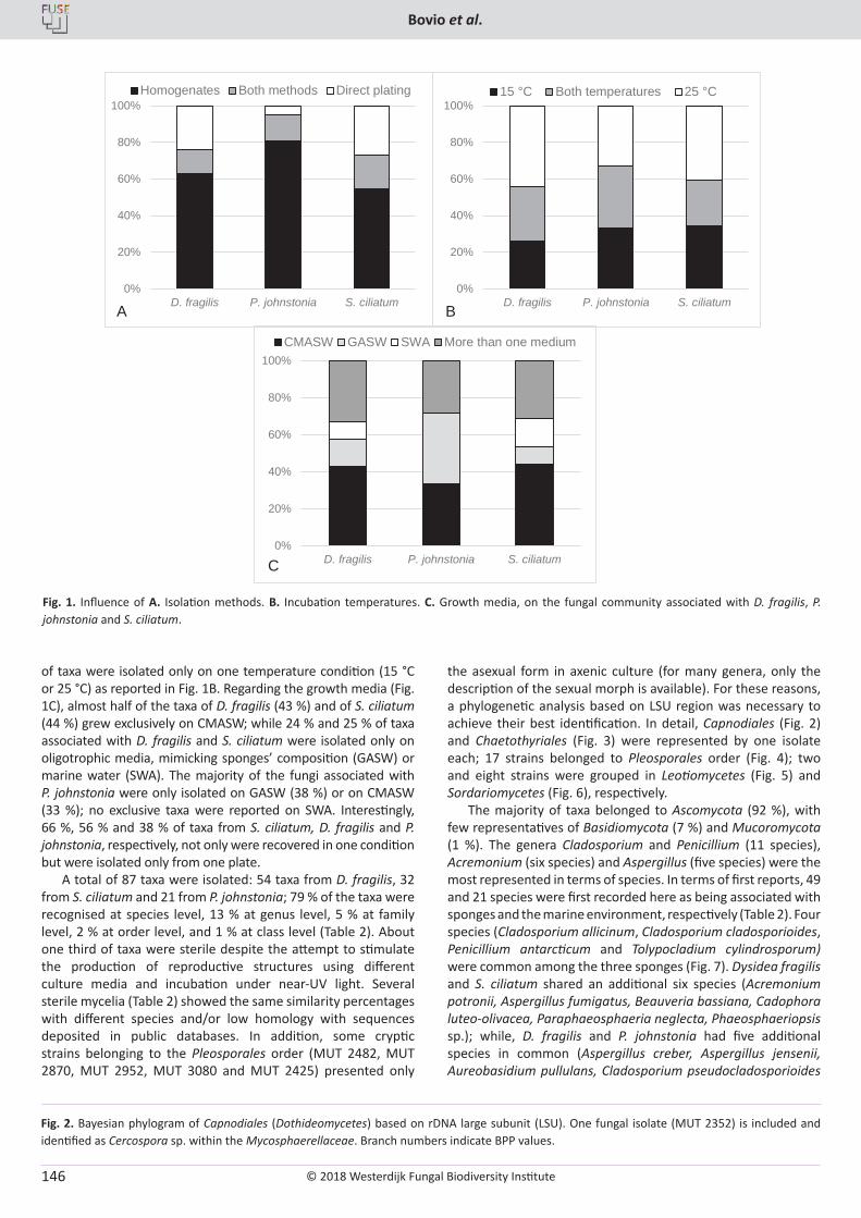
© 2018 Westerdijk Fungal Biodiversity Institute
Bovio et al.
Editor-in-ChiefProf. dr P.W. Crous, Westerdijk Fungal Biodiversity Institute, P.O. Box 85167, 3508 AD Utrecht, The Netherlands.E-mail:[email protected]
146
of taxa were isolated only on one temperature condition (15 °C or 25 °C) as reported in Fig. 1B. Regarding the growth media (Fig. 1C), almost half of the taxa of D. fragilis (43 %) and of S. ciliatum (44 %) grew exclusively on CMASW; while 24 % and 25 % of taxa associated with D. fragilis and S. ciliatum were isolated only on oligotrophic media, mimicking sponges’ composition (GASW) or marine water (SWA). The majority of the fungi associated with P. johnstonia were only isolated on GASW (38 %) or on CMASW (33 %); no exclusive taxa were reported on SWA. Interestingly, 66 %, 56 % and 38 % of taxa from S. ciliatum, D. fragilis and P. johnstonia, respectively, not only were recovered in one condition but were isolated only from one plate.
A total of 87 taxa were isolated: 54 taxa from D. fragilis, 32 from S. ciliatum and 21 from P. johnstonia; 79 % of the taxa were recognised at species level, 13 % at genus level, 5 % at family level, 2 % at order level, and 1 % at class level (Table 2). About one third of taxa were sterile despite the attempt to stimulate the production of reproductive structures using different culture media and incubation under near-UV light. Several sterile mycelia (Table 2) showed the same similarity percentages with different species and/or low homology with sequences deposited in public databases. In addition, some cryptic strains belonging to the Pleosporales order (MUT 2482, MUT 2870, MUT 2952, MUT 3080 and MUT 2425) presented only
the asexual form in axenic culture (for many genera, only the description of the sexual morph is available). For these reasons, a phylogenetic analysis based on LSU region was necessary to achieve their best identification. In detail, Capnodiales (Fig. 2) and Chaetothyriales (Fig. 3) were represented by one isolate each; 17 strains belonged to Pleosporales order (Fig. 4); two and eight strains were grouped in Leotiomycetes (Fig. 5) and Sordariomycetes (Fig. 6), respectively.
The majority of taxa belonged to Ascomycota (92 %), with few representatives of Basidiomycota (7 %) and Mucoromycota (1 %). The genera Cladosporium and Penicillium (11 species), Acremonium (six species) and Aspergillus (five species) were the most represented in terms of species. In terms of first reports, 49 and 21 species were first recorded here as being associated with sponges and the marine environment, respectively (Table 2). Four species (Cladosporium allicinum, Cladosporium cladosporioides, Penicillium antarcticum and Tolypocladium cylindrosporum) were common among the three sponges (Fig. 7). Dysidea fragilis and S. ciliatum shared an additional six species (Acremonium potronii, Aspergillus fumigatus, Beauveria bassiana, Cadophora luteo-olivacea, Paraphaeosphaeria neglecta, Phaeosphaeriopsis sp.); while, D. fragilis and P. johnstonia had five additional species in common (Aspergillus creber, Aspergillus jensenii, Aureobasidium pullulans, Cladosporium pseudocladosporioides
0%
20%
40%
60%
80%
100%
D. fragilis P. johnstonia S. ciliatum
CMASW GASW SWA More than one medium
0%
20%
40%
60%
80%
100%
D. fragilis P. johnstonia S. ciliatum
Homogenates Both methods Direct plating
0%
20%
40%
60%
80%
100%
D. fragilis P. johnstonia S. ciliatum
15 °C Both temperatures 25 °C
A B
C
Fig. 1. Influence of A. Isolation methods. B. Incubation temperatures. C. Growth media, on the fungal community associated with D. fragilis, P. johnstonia and S. ciliatum.
Fig. 2. Bayesian phylogram of Capnodiales (Dothideomycetes) based on rDNA large subunit (LSU). One fungal isolate (MUT 2352) is included and identified as Cercospora sp. within the Mycosphaerellaceae. Branch numbers indicate BPP values.
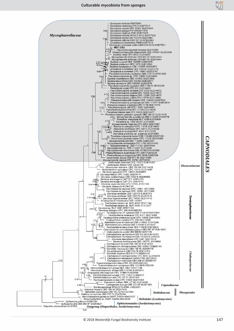
© 2018 Westerdijk Fungal Biodiversity Institute
Culturable mycobiota from sponges
Editor-in-ChiefProf. dr P.W. Crous, Westerdijk Fungal Biodiversity Institute, P.O. Box 85167, 3508 AD Utrecht, The Netherlands.E-mail:[email protected]
147
Mycosphaerellaceae
TeratosphaeriaceaeCladosporiaceae
Capnodiaceae
Dothideaceae
Helotiales (Leotiomycetes)Ophiostomatales (Sordariomycetes)
Outgroup (Diaporthales, Sordariomycetes)
Pleosporales
CAPNO
DIALESDissoconiaceae
Teratosphaeriaceae
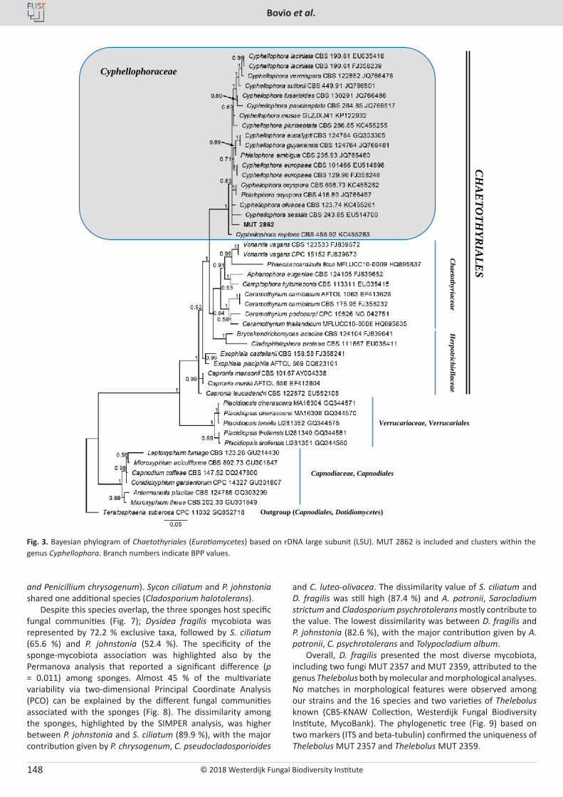
© 2018 Westerdijk Fungal Biodiversity Institute
Bovio et al.
Editor-in-ChiefProf. dr P.W. Crous, Westerdijk Fungal Biodiversity Institute, P.O. Box 85167, 3508 AD Utrecht, The Netherlands.E-mail:[email protected]
148
and Penicillium chrysogenum). Sycon ciliatum and P. johnstonia shared one additional species (Cladosporium halotolerans).
Despite this species overlap, the three sponges host specific fungal communities (Fig. 7); Dysidea fragilis mycobiota was represented by 72.2 % exclusive taxa, followed by S. ciliatum (65.6 %) and P. johnstonia (52.4 %). The specificity of the sponge-mycobiota association was highlighted also by the Permanova analysis that reported a significant difference (p = 0.011) among sponges. Almost 45 % of the multivariate variability via two-dimensional Principal Coordinate Analysis (PCO) can be explained by the different fungal communities associated with the sponges (Fig. 8). The dissimilarity among the sponges, highlighted by the SIMPER analysis, was higher between P. johnstonia and S. ciliatum (89.9 %), with the major contribution given by P. chrysogenum, C. pseudocladosporioides
and C. luteo-olivacea. The dissimilarity value of S. ciliatum and D. fragilis was still high (87.4 %) and A. potronii, Sarocladium strictum and Cladosporium psychrotolerans mostly contribute to the value. The lowest dissimilarity was between D. fragilis and P. johnstonia (82.6 %), with the major contribution given by A. potronii, C. psychrotolerans and Tolypocladium album.
Overall, D. fragilis presented the most diverse mycobiota, including two fungi MUT 2357 and MUT 2359, attributed to the genus Thelebolus both by molecular and morphological analyses. No matches in morphological features were observed among our strains and the 16 species and two varieties of Thelebolus known (CBS-KNAW Collection, Westerdijk Fungal Biodiversity Institute, MycoBank). The phylogenetic tree (Fig. 9) based on two markers (ITS and beta-tubulin) confirmed the uniqueness of Thelebolus MUT 2357 and Thelebolus MUT 2359.
Cyphellophoraceae
Chaetothyriaceae
Verrucariaceae, Verrucariales
Capnodiaceae, Capnodiales
Outgroup (Capnodiales, Dotidiomycetes)
CH
AE
TOTH
YRIA
LES
Herpotrichiellaceae
Fig. 3. Bayesian phylogram of Chaetothyriales (Eurotiomycetes) based on rDNA large subunit (LSU). MUT 2862 is included and clusters within the genus Cyphellophora. Branch numbers indicate BPP values.
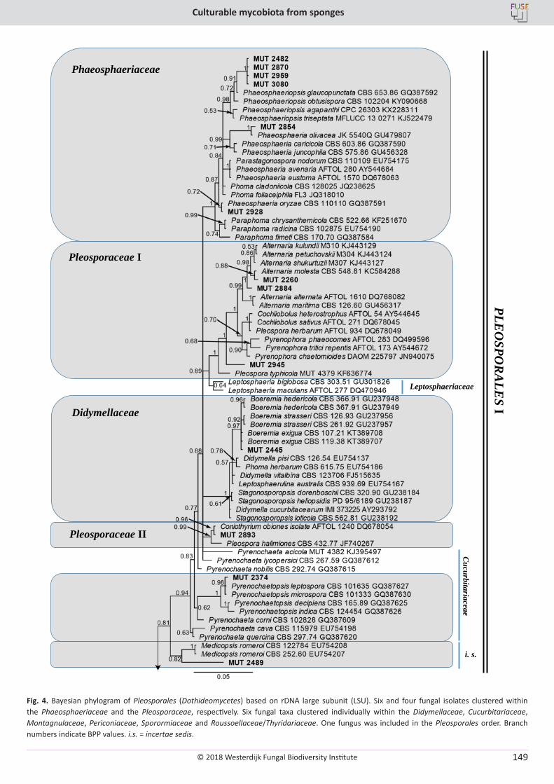
© 2018 Westerdijk Fungal Biodiversity Institute
Culturable mycobiota from sponges
Editor-in-ChiefProf. dr P.W. Crous, Westerdijk Fungal Biodiversity Institute, P.O. Box 85167, 3508 AD Utrecht, The Netherlands.E-mail:[email protected]
149
Phaeosphaeriaceae
Pleosporaceae I
Leptosphaeriaceae
Didymellaceae
Cucurbitariaceae
Pleosporaceae II
i. s.
PLEO
SPOR
ALE
SI
Fig. 4. Bayesian phylogram of Pleosporales (Dothideomycetes) based on rDNA large subunit (LSU). Six and four fungal isolates clustered within the Phaeosphaeriaceae and the Pleosporaceae, respectively. Six fungal taxa clustered individually within the Didymellaceae, Cucurbitariaceae, Montagnulaceae, Periconiaceae, Sporormiaceae and Roussoellaceae/Thyridariaceae. One fungus was included in the Pleosporales order. Branch numbers indicate BPP values. i.s. = incertae sedis.
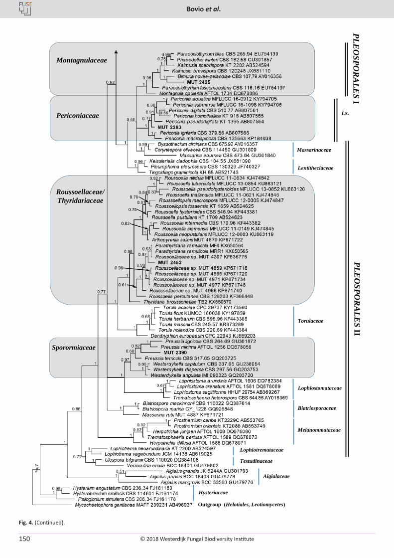
© 2018 Westerdijk Fungal Biodiversity Institute
Bovio et al.
Editor-in-ChiefProf. dr P.W. Crous, Westerdijk Fungal Biodiversity Institute, P.O. Box 85167, 3508 AD Utrecht, The Netherlands.E-mail:[email protected]
150
Montagnulaceae
Periconiaceae i.s.
PLEO
SPOR
ALE
SI
Massarinaceae
Lentitheciaceae
Roussoellaceae/Thyridariaceae
Torulaceae
Sporormiaceae
Lophiostomataceae
Aigialaceae
Testudinaceae
Lophiotremataceae
Melanommataceae
Biatriosporaceae
Hysteriaceae
Outgroup (Helotiales, Leotiomycetes)
PLEO
SPOR
ALE
SII
Fig. 4. (Continued).
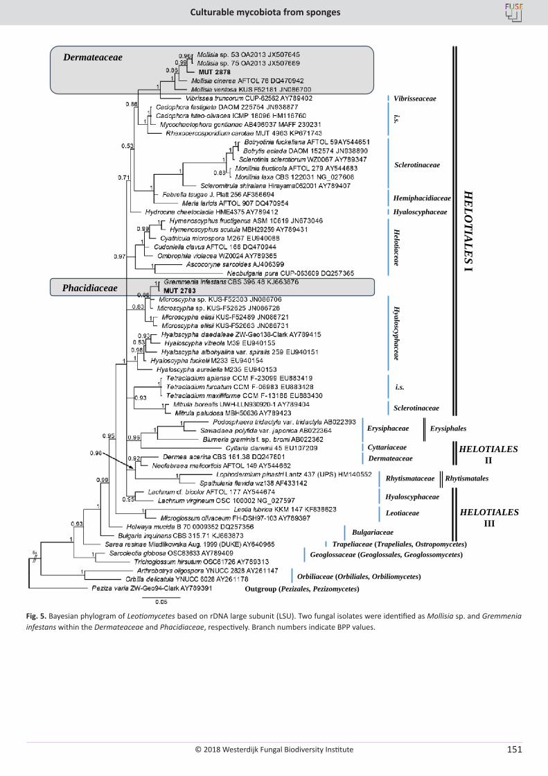
© 2018 Westerdijk Fungal Biodiversity Institute
Culturable mycobiota from sponges
Editor-in-ChiefProf. dr P.W. Crous, Westerdijk Fungal Biodiversity Institute, P.O. Box 85167, 3508 AD Utrecht, The Netherlands.E-mail:[email protected]
151
Vibrisseaceae
Dermateaceae
i.s.
Hyaloscyphaceae
Hemiphacidiaceae
Sclerotinaceae
Helotiaceae
Phacidiaceae
Hyaloscyphaceae
Sclerotinaceae
i.s.
HE
LOTIA
LES
I
Cyttariaceae
Erysiphaceae Erysiphales
Dermateaceae
Rhytismataceae Rhytismatales
HELOTIALESII
Leotiaceae
Hyaloscyphaceae
Bulgariaceae
HELOTIALESIII
Geoglossaceae (Geoglossales, Geoglossomycetes)Trapeliaceae (Trapeliales, Ostropomycetes)
Outgroup (Pezizales, Pezizomycetes)Orbiliaceae (Orbiliales, Orbiliomycetes)
Fig. 5. Bayesian phylogram of Leotiomycetes based on rDNA large subunit (LSU). Two fungal isolates were identified as Mollisia sp. and Gremmenia infestans within the Dermateaceae and Phacidiaceae, respectively. Branch numbers indicate BPP values.
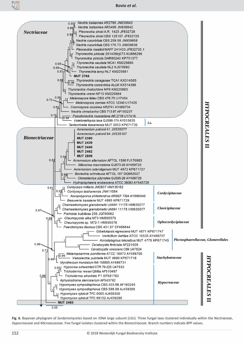
© 2018 Westerdijk Fungal Biodiversity Institute
Bovio et al.
Editor-in-ChiefProf. dr P.W. Crous, Westerdijk Fungal Biodiversity Institute, P.O. Box 85167, 3508 AD Utrecht, The Netherlands.E-mail:[email protected]
152
Bionectriaceae
Nectriaceae
Clavicipitaceae
Cordycipitaceae
i.s.
Ophocordycipitaceae
Plectosphaerellaceae, Glomerellales
HYPO
CR
EA
LES II
Hypocreaceae
Stachybotraceae
HYPO
CR
EA
LES
II
Fig. 6. Bayesian phylogram of Sordariomycetes based on rDNA large subunit (LSU). Three fungal taxa clustered individually within the Nectriaceae, Hypocreaceae and Microascaceae. Five fungal isolates clustered within the Bionectriaceae. Branch numbers indicate BPP values.
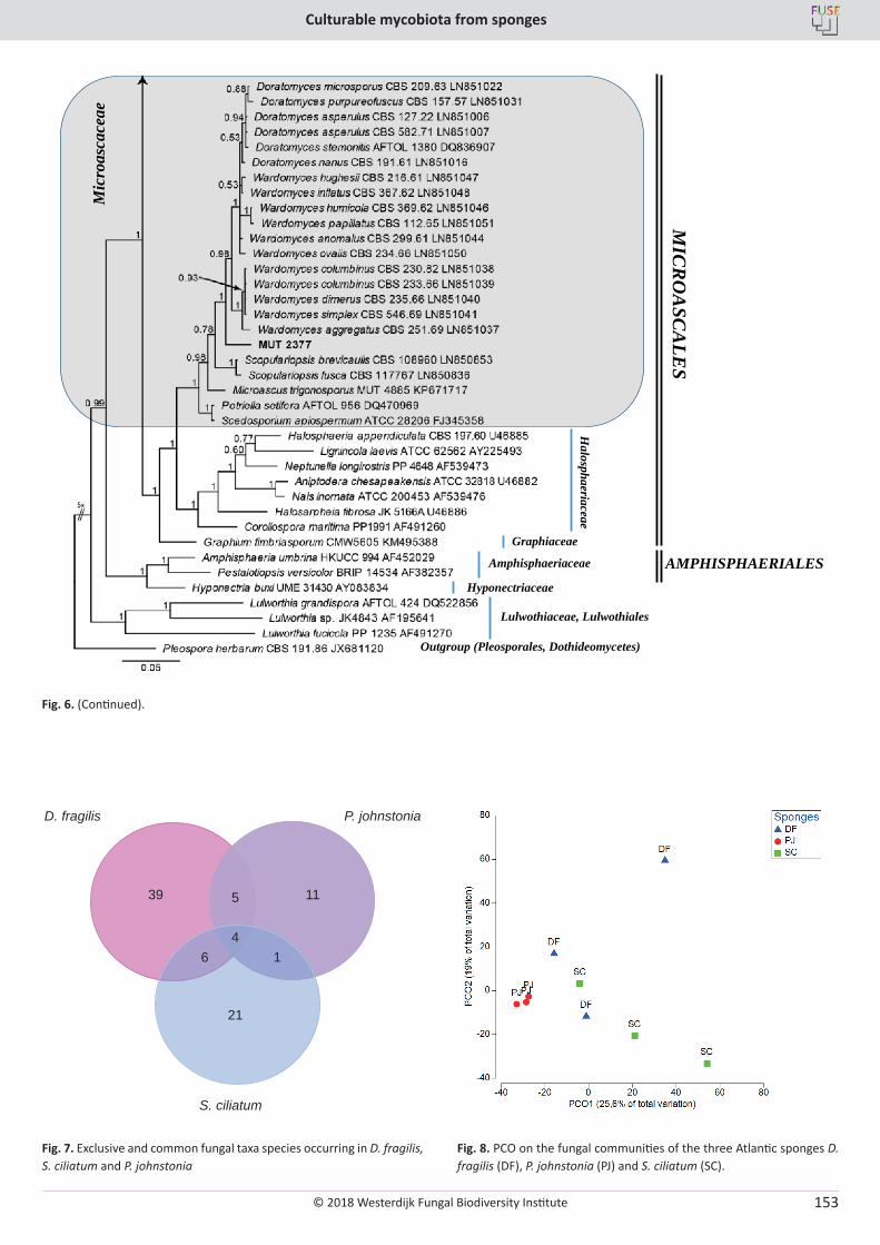
© 2018 Westerdijk Fungal Biodiversity Institute
Culturable mycobiota from sponges
Editor-in-ChiefProf. dr P.W. Crous, Westerdijk Fungal Biodiversity Institute, P.O. Box 85167, 3508 AD Utrecht, The Netherlands.E-mail:[email protected]
153
Mic
roas
cace
ae
Amphisphaeriaceae
Graphiaceae
Halosphaeriaceae
Hyponectriaceae
AMPHISPHAERIALES
Lulwothiaceae, Lulwothiales
Outgroup (Pleosporales, Dothideomycetes)
MIC
RO
ASC
ALE
S
Fig. 6. (Continued).
D. fragilis
4
39 11
21
P. johnstonia
S. ciliatum
5
6 1
Fig. 7. Exclusive and common fungal taxa species occurring in D. fragilis, S. ciliatum and P. johnstonia
Fig. 8. PCO on the fungal communities of the three Atlantic sponges D. fragilis (DF), P. johnstonia (PJ) and S. ciliatum (SC).
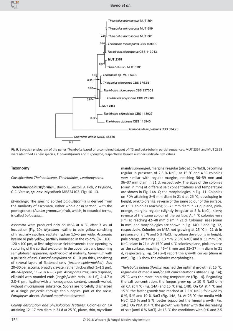
© 2018 Westerdijk Fungal Biodiversity Institute
Bovio et al.
Editor-in-ChiefProf. dr P.W. Crous, Westerdijk Fungal Biodiversity Institute, P.O. Box 85167, 3508 AD Utrecht, The Netherlands.E-mail:[email protected]
154
Fig 9. Bayesian phylogram of the genus Thelebolus based on a combined dataset of ITS and beta-tubulin partial sequences. MUT 2357 and MUT 2359 were identified as new species, T. balaustiformis and T. spongiae, respectively. Branch numbers indicate BPP values
Taxonomy
Classification: Thelebolaceae, Thelebolales, Leotiomycetes.
Thelebolus balaustiformis E. Bovio, L. Garzoli, A. Poli, V. Prigione, G.C. Varese, sp. nov. MycoBank MB824102. Figs 10–13.
Etymology: The specific epithet balaustiformis is derived from the similarity of ascomata, either whole or in section, with the pomegranate (Punica granatum) fruit, which, in botanical terms, is called balaustium.
Ascomata were produced only on MEA at 4 °C, after 3 wk of incubation (Fig. 10). Mycelium hyaline to pale yellow consisting of irregularly swollen, septate hyphae 1.5–5 µm wide. Ascomata hyaline or pale yellow, partially immersed in the colony, (87–)100–120 × 100 µm, at first subglobose cleistohymenial then opening by rupturing of the cortical excipulum in the upper part and becoming semiglobular, appearing 'apothecioid' at maturity. Hymenium with a palisade of asci. Cortical excipulum ca. 6–10 µm thick, consisting of several layers of flattened cells (textura epidermoidea). Asci 20–30 per ascoma, broadly clavate, rather thick-walled (1–1.5 µm), 48–64-spored, 11–20 × 43–57 µm. Ascospores irregularly disposed, ellipsoid with rounded ends (length/width ratio 1.4–1.6), 4–4.7 × 2.8–3 µm, hyaline with a homogenous content, smooth-walled, without mucilaginous substance. Spores are forcefully discharged as a single projectile through the subapical part of the ascus. Paraphyses absent. Asexual morph not observed.
Colony description and physiological features: Colonies on CA attaining 12–17 mm diam in 21 d at 25 °C, plane, thin, mycelium
mainly submerged, margins irregular (also at 5 % NaCl), becoming regular in presence of 2.5 % NaCl; at 15 °C and 4 °C colonies very similar with regular margins, reaching 56–59 mm and 36–37 mm diam in 21 d, respectively. The sizes of the colonies (diam in mm) at different salt concentrations and temperature are shown in Fig. 14A–C; the morphologies in Fig. 11. Colonies on PDA attaining 8–9 mm diam in 21 d at 25 °C, developing in height, pink to orange, reverse of the same colour of the surface. At 15 °C colonies reaching 65–73 mm diam in 21 d, plane, pink-orange, margins regular (slightly irregular at 5 % NaCl), slimy; reverse of the same colour of the surface. At 4 °C colonies very similar, reaching 42–48 mm diam in 21 d. Colonies’ sizes (diam in mm) and morphologies are shown in Fig. 14D–F and Fig. 12, respectively. Colonies on MEA not growing at 25 °C in 21 d; in presence of 2.5 % and 5 % NaCl, mycelium developing in height, pale orange, attaining 11–13 mm (2.5 % NaCl) and 8–11 mm (5 % NaCl) diam in 21 d. At 15 °C and 4 °C colonies plane, pink, reverse as the surface, reaching 46–48 mm and 25–27 mm diam in 21 d, respectively; Fig. 14 (G–I) report the growth curves (diam in mm); Fig. 13 show the colonies morphologies.
Thelebolus balaustiformis reached the optimal growth at 15 °C, regardless of media and/or salt concentrations utilised (Fig. 14); 25 °C was the most inhibiting temperature (Fig. 14). Regarding the salt concentration, the fungus grew up to 10 % NaCl only on CA at 4 °C (Fig. 14A) and 15 °C (Fig. 14B). On CA at 4 °C and 15 °C the faster growth was reached at 2.5 % NaCl, followed by 0 %, 5 % and 10 % NaCl (Fig. 14A, B). At 25 °C the media with NaCl (2.5 % and 5 %) better supported the fungal growth (Fig. 14C). On PDA at 4 °C the growth was faster with the decreasing of salt (until 0 % NaCl). At 15 °C the conditions with 0 % and 2.5
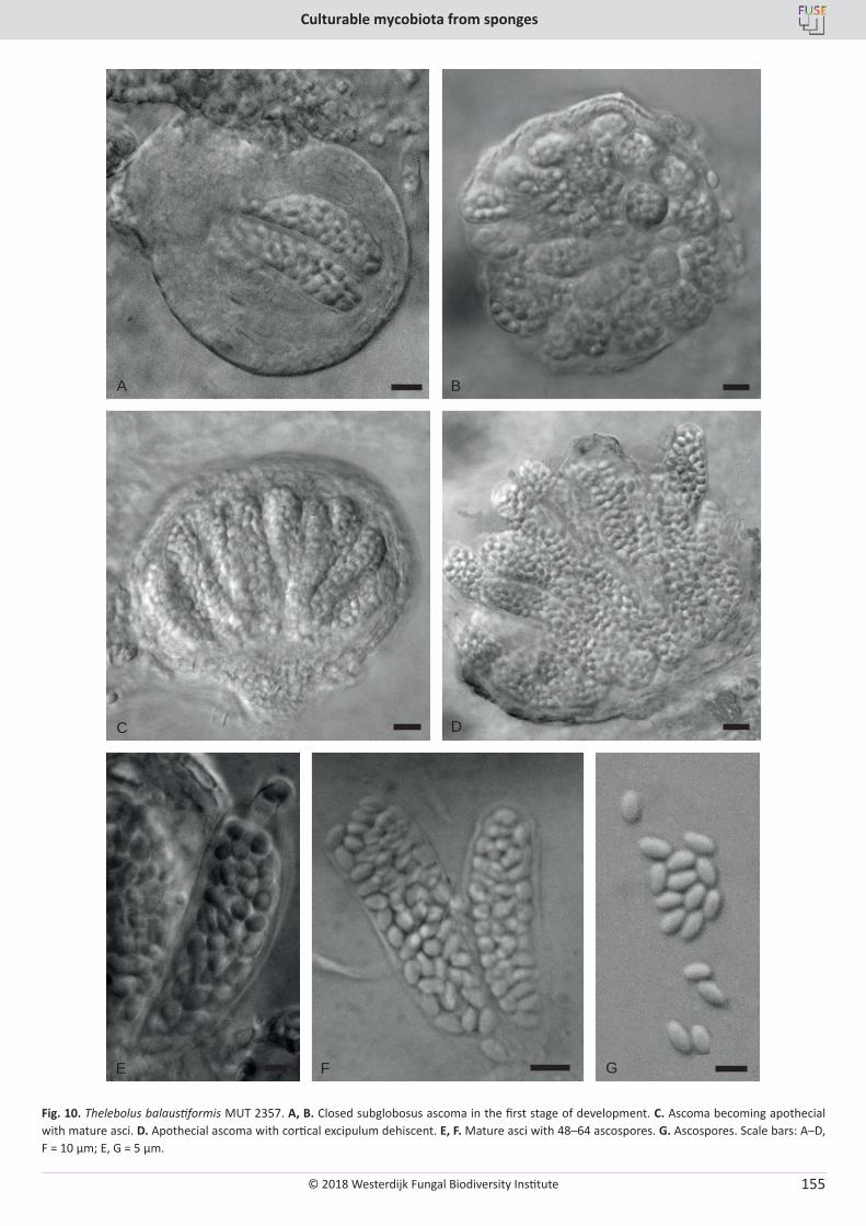
© 2018 Westerdijk Fungal Biodiversity Institute
Culturable mycobiota from sponges
Editor-in-ChiefProf. dr P.W. Crous, Westerdijk Fungal Biodiversity Institute, P.O. Box 85167, 3508 AD Utrecht, The Netherlands.E-mail:[email protected]
155
B
C D
E F G
A
Fig. 10. Thelebolus balaustiformis MUT 2357. A, B. Closed subglobosus ascoma in the first stage of development. C. Ascoma becoming apothecial with mature asci. D. Apothecial ascoma with cortical excipulum dehiscent. E, F. Mature asci with 48–64 ascospores. G. Ascospores. Scale bars: A–D, F = 10 µm; E, G = 5 µm.
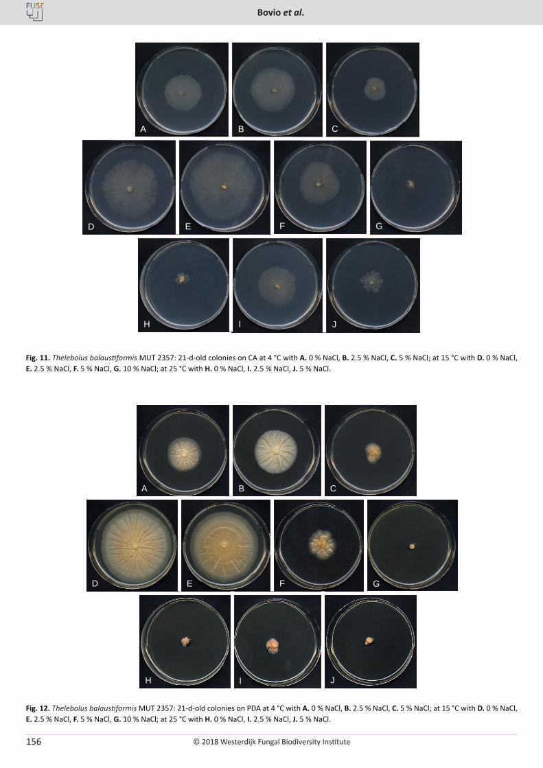
© 2018 Westerdijk Fungal Biodiversity Institute
Bovio et al.
Editor-in-ChiefProf. dr P.W. Crous, Westerdijk Fungal Biodiversity Institute, P.O. Box 85167, 3508 AD Utrecht, The Netherlands.E-mail:[email protected]
156
A B C
D E F G
H I J
Fig. 11. Thelebolus balaustiformis MUT 2357: 21-d-old colonies on CA at 4 °C with A. 0 % NaCl, B. 2.5 % NaCl, C. 5 % NaCl; at 15 °C with D. 0 % NaCl, E. 2.5 % NaCl, F. 5 % NaCl, G. 10 % NaCl; at 25 °C with H. 0 % NaCl, I. 2.5 % NaCl, J. 5 % NaCl.
Fig. 12. Thelebolus balaustiformis MUT 2357: 21-d-old colonies on PDA at 4 °C with A. 0 % NaCl, B. 2.5 % NaCl, C. 5 % NaCl; at 15 °C with D. 0 % NaCl, E. 2.5 % NaCl, F. 5 % NaCl, G. 10 % NaCl; at 25 °C with H. 0 % NaCl, I. 2.5 % NaCl, J. 5 % NaCl.
A B C
D E F G
H I J
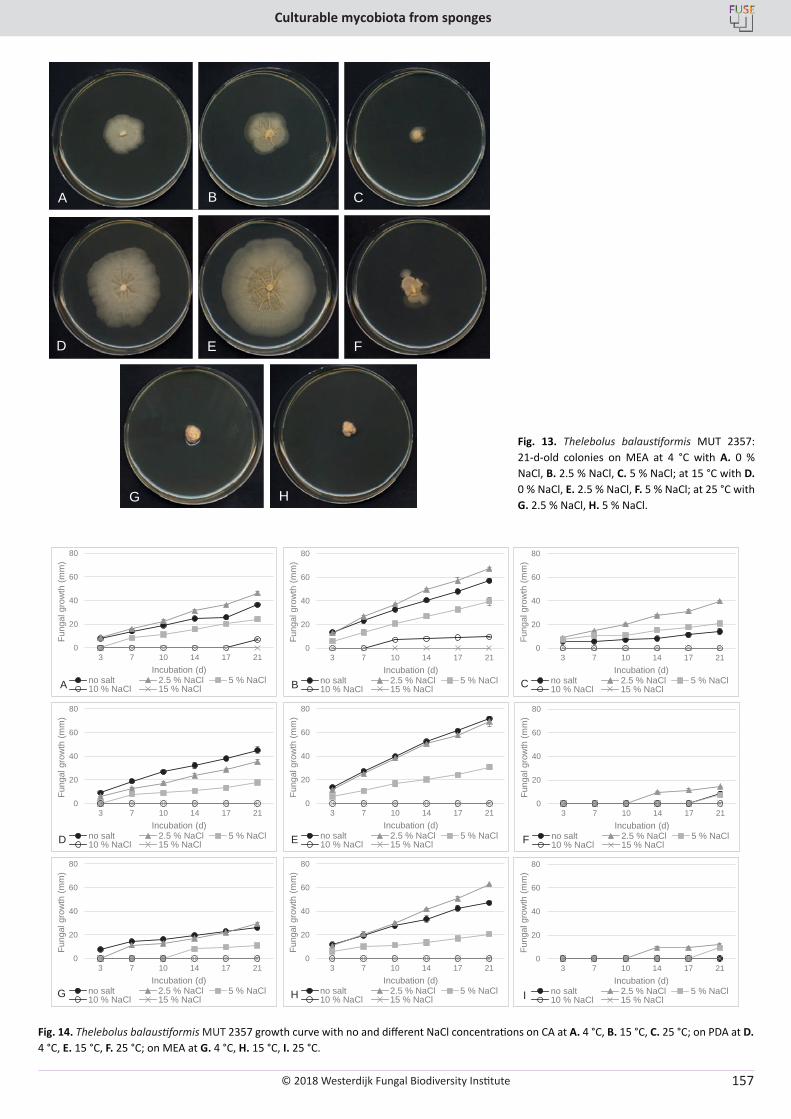
© 2018 Westerdijk Fungal Biodiversity Institute
Culturable mycobiota from sponges
Editor-in-ChiefProf. dr P.W. Crous, Westerdijk Fungal Biodiversity Institute, P.O. Box 85167, 3508 AD Utrecht, The Netherlands.E-mail:[email protected]
157
A B C
D E F
G H
Fig. 13. Thelebolus balaustiformis MUT 2357: 21-d-old colonies on MEA at 4 °C with A. 0 % NaCl, B. 2.5 % NaCl, C. 5 % NaCl; at 15 °C with D. 0 % NaCl, E. 2.5 % NaCl, F. 5 % NaCl; at 25 °C with G. 2.5 % NaCl, H. 5 % NaCl.
0
20
40
60
80
3 7 10 14 17 21
Fung
al g
row
th (m
m)
Incubation (d)no salt 2.5 % NaCl 5 % NaCl10 % NaCl 15 % NaCl
0
20
40
60
80
3 7 10 14 17 21
Fung
al g
row
th (m
m)
Incubation (d)no salt 2.5 % NaCl 5 % NaCl10 % NaCl 15 % NaCl
0
20
40
60
80
3 7 10 14 17 21
Fung
al g
row
th (m
m)
Incubation (d)no salt 2.5 % NaCl 5 % NaCl10 % NaCl 15 % NaCl
0
20
40
60
80
3 7 10 14 17 21
Fung
al g
row
th (m
m)
Incubation (d)no salt 2.5 % NaCl 5 % NaCl10 % NaCl 15 % NaCl
0
20
40
60
80
3 7 10 14 17 21
Fung
al g
row
th (m
m)
Incubation (d)no salt 2.5 % NaCl 5 % NaCl10 % NaCl 15 % NaCl
0
20
40
60
80
3 7 10 14 17 21
Fung
al g
row
th (m
m)
Incubation (d)no salt 2.5 % NaCl 5 % NaCl10 % NaCl 15 % NaCl
0
20
40
60
80
3 7 10 14 17 21
Fung
al g
row
th (m
m)
Incubation (d)no salt 2.5 % NaCl 5 % NaCl10 % NaCl 15 % NaCl
0
20
40
60
80
3 7 10 14 17 21
Fung
al g
row
th (m
m)
Incubation (d)no salt 2.5 % NaCl 5 % NaCl10 % NaCl 15 % NaCl
0
20
40
60
80
3 7 10 14 17 21
Fung
al g
row
th (m
m)
Incubation (d)no salt 2.5 % NaCl 5 % NaCl10 % NaCl 15 % NaClA B C
D
G H
E
I
F
Fig. 14. Thelebolus balaustiformis MUT 2357 growth curve with no and different NaCl concentrations on CA at A. 4 °C, B. 15 °C, C. 25 °C; on PDA at D. 4 °C, E. 15 °C, F. 25 °C; on MEA at G. 4 °C, H. 15 °C, I. 25 °C.
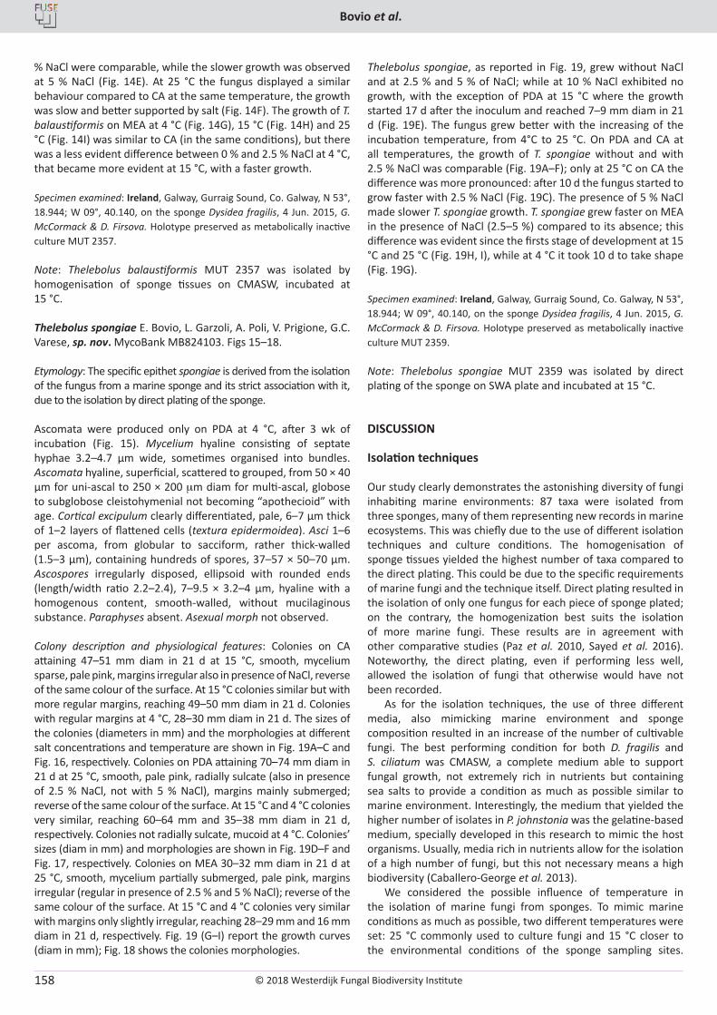
© 2018 Westerdijk Fungal Biodiversity Institute
Bovio et al.
Editor-in-ChiefProf. dr P.W. Crous, Westerdijk Fungal Biodiversity Institute, P.O. Box 85167, 3508 AD Utrecht, The Netherlands.E-mail:[email protected]
158
% NaCl were comparable, while the slower growth was observed at 5 % NaCl (Fig. 14E). At 25 °C the fungus displayed a similar behaviour compared to CA at the same temperature, the growth was slow and better supported by salt (Fig. 14F). The growth of T. balaustiformis on MEA at 4 °C (Fig. 14G), 15 °C (Fig. 14H) and 25 °C (Fig. 14I) was similar to CA (in the same conditions), but there was a less evident difference between 0 % and 2.5 % NaCl at 4 °C, that became more evident at 15 °C, with a faster growth.
Specimen examined: Ireland, Galway, Gurraig Sound, Co. Galway, N 53°, 18.944; W 09°, 40.140, on the sponge Dysidea fragilis, 4 Jun. 2015, G. McCormack & D. Firsova. Holotype preserved as metabolically inactive culture MUT 2357.
Note: Thelebolus balaustiformis MUT 2357 was isolated by homogenisation of sponge tissues on CMASW, incubated at 15 °C.
Thelebolus spongiae E. Bovio, L. Garzoli, A. Poli, V. Prigione, G.C. Varese, sp. nov. MycoBank MB824103. Figs 15–18.
Etymology: The specific epithet spongiae is derived from the isolation of the fungus from a marine sponge and its strict association with it, due to the isolation by direct plating of the sponge.
Ascomata were produced only on PDA at 4 °C, after 3 wk of incubation (Fig. 15). Mycelium hyaline consisting of septate hyphae 3.2–4.7 µm wide, sometimes organised into bundles. Ascomata hyaline, superficial, scattered to grouped, from 50 × 40 µm for uni-ascal to 250 × 200 mm diam for multi-ascal, globose to subglobose cleistohymenial not becoming “apothecioid” with age. Cortical excipulum clearly differentiated, pale, 6–7 µm thick of 1–2 layers of flattened cells (textura epidermoidea). Asci 1–6 per ascoma, from globular to sacciform, rather thick-walled (1.5–3 µm), containing hundreds of spores, 37–57 × 50–70 µm. Ascospores irregularly disposed, ellipsoid with rounded ends (length/width ratio 2.2–2.4), 7–9.5 × 3.2–4 µm, hyaline with a homogenous content, smooth-walled, without mucilaginous substance. Paraphyses absent. Asexual morph not observed.
Colony description and physiological features: Colonies on CA attaining 47–51 mm diam in 21 d at 15 °C, smooth, mycelium sparse, pale pink, margins irregular also in presence of NaCl, reverse of the same colour of the surface. At 15 °C colonies similar but with more regular margins, reaching 49–50 mm diam in 21 d. Colonies with regular margins at 4 °C, 28–30 mm diam in 21 d. The sizes of the colonies (diameters in mm) and the morphologies at different salt concentrations and temperature are shown in Fig. 19A–C and Fig. 16, respectively. Colonies on PDA attaining 70–74 mm diam in 21 d at 25 °C, smooth, pale pink, radially sulcate (also in presence of 2.5 % NaCl, not with 5 % NaCl), margins mainly submerged; reverse of the same colour of the surface. At 15 °C and 4 °C colonies very similar, reaching 60–64 mm and 35–38 mm diam in 21 d, respectively. Colonies not radially sulcate, mucoid at 4 °C. Colonies’ sizes (diam in mm) and morphologies are shown in Fig. 19D–F and Fig. 17, respectively. Colonies on MEA 30–32 mm diam in 21 d at 25 °C, smooth, mycelium partially submerged, pale pink, margins irregular (regular in presence of 2.5 % and 5 % NaCl); reverse of the same colour of the surface. At 15 °C and 4 °C colonies very similar with margins only slightly irregular, reaching 28–29 mm and 16 mm diam in 21 d, respectively. Fig. 19 (G–I) report the growth curves (diam in mm); Fig. 18 shows the colonies morphologies.
Thelebolus spongiae, as reported in Fig. 19, grew without NaCl and at 2.5 % and 5 % of NaCl; while at 10 % NaCl exhibited no growth, with the exception of PDA at 15 °C where the growth started 17 d after the inoculum and reached 7–9 mm diam in 21 d (Fig. 19E). The fungus grew better with the increasing of the incubation temperature, from 4°C to 25 °C. On PDA and CA at all temperatures, the growth of T. spongiae without and with 2.5 % NaCl was comparable (Fig. 19A–F); only at 25 °C on CA the difference was more pronounced: after 10 d the fungus started to grow faster with 2.5 % NaCl (Fig. 19C). The presence of 5 % NaCl made slower T. spongiae growth. T. spongiae grew faster on MEA in the presence of NaCl (2.5–5 %) compared to its absence; this difference was evident since the firsts stage of development at 15 °C and 25 °C (Fig. 19H, I), while at 4 °C it took 10 d to take shape (Fig. 19G).
Specimen examined: Ireland, Galway, Gurraig Sound, Co. Galway, N 53°, 18.944; W 09°, 40.140, on the sponge Dysidea fragilis, 4 Jun. 2015, G. McCormack & D. Firsova. Holotype preserved as metabolically inactive culture MUT 2359.
Note: Thelebolus spongiae MUT 2359 was isolated by direct plating of the sponge on SWA plate and incubated at 15 °C.
DISCUSSION
Isolation techniques
Our study clearly demonstrates the astonishing diversity of fungi inhabiting marine environments: 87 taxa were isolated from three sponges, many of them representing new records in marine ecosystems. This was chiefly due to the use of different isolation techniques and culture conditions. The homogenisation of sponge tissues yielded the highest number of taxa compared to the direct plating. This could be due to the specific requirements of marine fungi and the technique itself. Direct plating resulted in the isolation of only one fungus for each piece of sponge plated; on the contrary, the homogenization best suits the isolation of more marine fungi. These results are in agreement with other comparative studies (Paz et al. 2010, Sayed et al. 2016). Noteworthy, the direct plating, even if performing less well, allowed the isolation of fungi that otherwise would have not been recorded.
As for the isolation techniques, the use of three different media, also mimicking marine environment and sponge composition resulted in an increase of the number of cultivable fungi. The best performing condition for both D. fragilis and S. ciliatum was CMASW, a complete medium able to support fungal growth, not extremely rich in nutrients but containing sea salts to provide a condition as much as possible similar to marine environment. Interestingly, the medium that yielded the higher number of isolates in P. johnstonia was the gelatine-based medium, specially developed in this research to mimic the host organisms. Usually, media rich in nutrients allow for the isolation of a high number of fungi, but this not necessary means a high biodiversity (Caballero-George et al. 2013).
We considered the possible influence of temperature in the isolation of marine fungi from sponges. To mimic marine conditions as much as possible, two different temperatures were set: 25 °C commonly used to culture fungi and 15 °C closer to the environmental conditions of the sponge sampling sites.
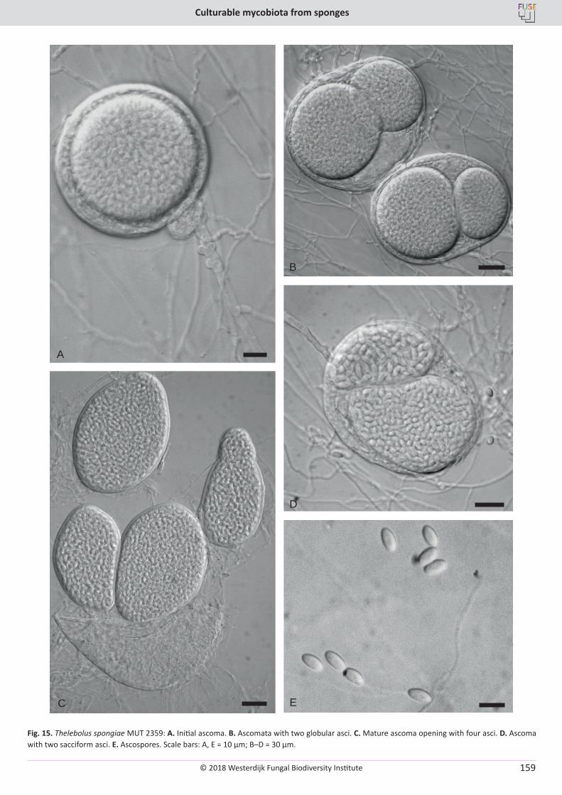
© 2018 Westerdijk Fungal Biodiversity Institute
Culturable mycobiota from sponges
Editor-in-ChiefProf. dr P.W. Crous, Westerdijk Fungal Biodiversity Institute, P.O. Box 85167, 3508 AD Utrecht, The Netherlands.E-mail:[email protected]
159
B
C
D
E
A
Fig. 15. Thelebolus spongiae MUT 2359: A. Initial ascoma. B. Ascomata with two globular asci. C. Mature ascoma opening with four asci. D. Ascoma with two sacciform asci. E. Ascospores. Scale bars: A, E = 10 µm; B–D = 30 µm.
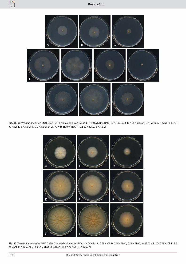
© 2018 Westerdijk Fungal Biodiversity Institute
Bovio et al.
Editor-in-ChiefProf. dr P.W. Crous, Westerdijk Fungal Biodiversity Institute, P.O. Box 85167, 3508 AD Utrecht, The Netherlands.E-mail:[email protected]
160
Fig. 16. Thelebolus spongiae MUT 2359: 21-d-old colonies on CA at 4 °C with A. 0 % NaCl, B. 2.5 % NaCl, C. 5 % NaCl; at 15 °C with D. 0 % NaCl, E. 2.5 % NaCl, F. 5 % NaCl, G. 10 % NaCl; at 25 °C with H. 0 % NaCl, I. 2.5 % NaCl, J. 5 % NaCl.
Fig. 17 Thelebolus spongiae MUT 2359: 21-d-old colonies on PDA at 4 °C with A. 0 % NaCl, B. 2.5 % NaCl, C. 5 % NaCl; at 15 °C with D. 0 % NaCl, E. 2.5 % NaCl, F. 5 % NaCl; at 25 °C with G. 0 % NaCl, H. 2.5 % NaCl, I. 5 % NaCl.
A B C
D E F G
H I J
A B C
D E F
G H I
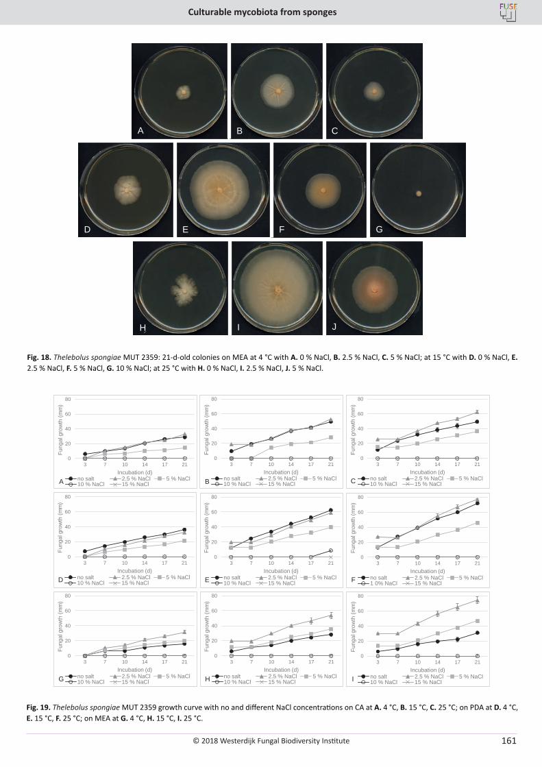
© 2018 Westerdijk Fungal Biodiversity Institute
Culturable mycobiota from sponges
Editor-in-ChiefProf. dr P.W. Crous, Westerdijk Fungal Biodiversity Institute, P.O. Box 85167, 3508 AD Utrecht, The Netherlands.E-mail:[email protected]
161
Fig. 19. Thelebolus spongiae MUT 2359 growth curve with no and different NaCl concentrations on CA at A. 4 °C, B. 15 °C, C. 25 °C; on PDA at D. 4 °C, E. 15 °C, F. 25 °C; on MEA at G. 4 °C, H. 15 °C, I. 25 °C.
Fig. 18. Thelebolus spongiae MUT 2359: 21-d-old colonies on MEA at 4 °C with A. 0 % NaCl, B. 2.5 % NaCl, C. 5 % NaCl; at 15 °C with D. 0 % NaCl, E. 2.5 % NaCl, F. 5 % NaCl, G. 10 % NaCl; at 25 °C with H. 0 % NaCl, I. 2.5 % NaCl, J. 5 % NaCl.
A B C
D E F G
H I J
0
20
40
60
80
3 7 10 14 17 21
Fung
al g
row
th (m
m)
Incubation (d)no salt 2.5 % NaCl 5 % NaCl10 % NaCl 15 % NaCl
0
20
40
60
80
3 7 10 14 17 21
Fung
al g
row
th (m
m)
Incubation (d)no salt 2.5 % NaCl 5 % NaCl10 % NaCl 15 % NaCl
0
20
40
60
80
3 7 10 14 17 21
Fung
al g
row
th (m
m)
Incubation (d)no salt 2.5 % NaCl 5 % NaCl10 % NaCl 15 % NaCl
0
20
40
60
80
3 7 10 14 17 21
Fung
al g
row
th (m
m)
Incubation (d)no salt 2.5 % NaCl 5 % NaCl10 % NaCl 15 % NaCl
0
20
40
60
80
3 7 10 14 17 21
Fung
al g
row
th (m
m)
Incubation (d)no salt 2.5 % NaCl 5 % NaCl10 % NaCl 15 % NaCl
0
20
40
60
80
3 7 10 14 17 21
Fung
al g
row
th (m
m)
Incubation (d)no salt 2.5 % NaCl 5 % NaCl1 0% NaCl 15 % NaCl
0
20
40
60
80
3 7 10 14 17 21
Fung
al g
row
th (m
m)
Incubation (d)no salt 2.5 % NaCl 5 % NaCl10 % NaCl 15 % NaCl
0
20
40
60
80
3 7 10 14 17 21
Fung
al g
row
th (m
m)
Incubation (d)no salt 2.5 % NaCl 5 % NaCl10 % NaCl 15 % NaCl
0
20
40
60
80
3 7 10 14 17 21
Fung
al g
row
th (m
m)
Incubation (d)no salt 2.5 % NaCl 5 % NaCl10 % NaCl 15 % NaClA B C
D E F
G H I
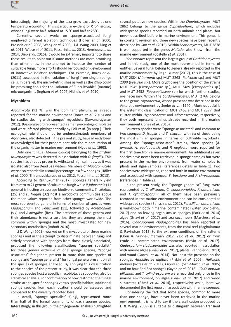
© 2018 Westerdijk Fungal Biodiversity Institute
Bovio et al.
Editor-in-ChiefProf. dr P.W. Crous, Westerdijk Fungal Biodiversity Institute, P.O. Box 85167, 3508 AD Utrecht, The Netherlands.E-mail:[email protected]
162
Interestingly, the majority of the taxa grew exclusively at one temperature condition; this is particular evident for P. johnstonia, whose fungi were half isolated at 15 °C and half at 25°C.
Currently, several works on sponge-associated fungi employed different isolation techniques (Höller et al. 2000, Proksch et al. 2008, Wang et al. 2008, Li & Wang 2009, Ding et al. 2011, Wiese et al. 2011, Passarini et al. 2013, Henríquez et al. 2014, Diep et al. 2016). It would be extremely important to share these results to point out if some methods are more promising than other ones. In the attempt to increase the number of cultivable fungi, more efforts should be focused on development of innovative isolation techniques. For example, Rozas et al. (2011) succeeded in the isolation of fungi from single sponge cells. In parallel, the micro-Petri dishes as well as the iChip could be promising tools for the isolation of “uncultivable” (marine) microorganisms (Ingham et al. 2007, Nichols et al. 2010).
Mycobiota
Ascomycota (92 %) was the dominant phylum, as already reported for the marine environment (Jones et al. 2015) and for studies dealing with sponges’ mycobiota (Suryanarayanan 2012). Basidiomycota represented a small percentage of isolates and were inferred phylogenetically by Poli et al. (in prep.). Their ecological role should not be underestimated: members of Agaricales, also detected in the present study, have already been acknowledged for their predominant role the mineralization of the organic matter in marine environment (Hyde et al. 1998).
Only one fungus (Absidia glauca) belonging to the phylum Mucoromycota was detected in association with D. fragilis. This species has already proven to withstand high salinities, as it was isolated also from Dead Sea waters. Members of Mucoromycota were also recorded in a small percentage in a few sponges (Höller et al. 2000, Thirunavukkarasu et al. 2012, Passarini et al. 2013).
According to Raghukumar (2017), sponges generally yield from zero to 21 genera of culturable fungi: while P. johnstonia (11 genera) is hosting an average biodiverse community, S. ciliatum (25) and D. fragilis (32) host a mycobiota communities above the mean values reported from other sponges worldwide. The most represented genera in terms of number of species were Cladosporium and Penicillium (11), followed by Acremonium (six) and Aspergillus (five). The presence of these genera and their abundance is not a surprise: they are among the most common within sponges and the most investigated for new secondary metabolites (Imhoff 2016).
Li & Wang (2009), worked on the mycobiota of three marine sponges and in the attempt to discriminate between fungi not strictly associated with sponges from those closely associated, proposed the following classification: “sponge specialist” for those genera exclusive of one sponge species, “sponge associates” for genera present in more than one species of sponge and “sponge generalist” for fungal genera present on all the species of sponges analysed. By applying this classification to the species of the present study, it was clear that the three sponges species host a specific mycobiota, as supported also by statistical analysis. For confirmation of how restricted the fungal strains are to specific sponges versus specific habitat, additional sponge species from each location should be assessed and compared to the diversity reported here.
In detail, “sponge specialist” fungi, represented more than half of the fungal community of each sponge species. Interestingly, in this group, the phylogenetic analysis highlighted
several putative new species. Within the Chaetothyriales, MUT 2862 belongs to the genus Cyphellophora, which includes widespread species recorded on both animals and plants, but never described before in marine environment. This genus is in constant revision and three new species have been recently described by Gao et al. (2015). Within Leotiomycetes, MUT 2878 is well supported in the genus Mollisia, also known from the marine environment (Costello et al. 2001).
Pleosporales represent the largest group of Dothideomycetes and in this study, one of the most represented in terms of entities. Several fungi belong to genera already reported in the marine environment by Raghukumar (2017), this is the case of MUT 2884 (Alternaria sp.) MUT 2263 (Periconia sp.) and MUT 2390 (Preussia sp.). More cryptic are the position of the strains MUT 2945 (Pleosporaceae sp.), MUT 2489 (Pleosporales sp.) and MUT 2452 (Roussoellaceae sp.) for which further studies, are necessary. Within the Sordariomycetes, MUT 2766 belong to the genus Thyronectria, whose presence was described in the Antarctic environment by Seeler et al. (1940). More doubtful is the systematic classification of MUT 2463 and MUT 2377, that cluster within Hypocreaceae and Microascaceae, respectively; they both represent families already recorded in the marine environment (Jones et al. 2015).
Fourteen species were “sponge-associated” and common to two sponges; D. fragilis and S. ciliatum with six of these being the most similar sponges in terms of cultivable mycobiota. Among the “sponge-associated” strains, three species (A. jensenii, A. puulaauensis and P. neglecta) were reported for the first time from a marine environment. An additional seven species have never been retrieved in sponge samples but were present in the marine environment, from water samples to plants and algae samples (References details in Table 1). Two species were widespread, reported both in marine environment and associated with sponges: B. bassiana and P. chrysogenum (references in Table 2).
In the present study, the “sponge generalist” fungi were represented by C. allicinum, C. cladosporioides, P. antarcticum and T. cylindrosporum; all of them have been previously recorded in the marine environment and can be considered as widespread species (Bensch et al. 2012). Penicillium antarcticum is well-known both in marine (contaminated) water (Bovio et al. 2017) and on leaving organisms as sponges (Park et al. 2014) algae (Gnavi et al. 2017) and sea cucumbers (Marchese et al. 2016). Cladosporium cladosporioides has been reported in several marine environments, from the coral reef (Raghukumar & Ravindran 2012) to the extreme conditions of the salterns (Oren & Gunde-Cimerman 2012, Zajc et al. 2012) or from crude oil contaminated environments (Bovio et al. 2017). Cladosporium cladosporioides was also reported in association with marine algae (Gnavi et al. 2017), plants (Panno et al. 2013) and wood (Garzoli et al. 2014). Not least the presence on the sponges Amphilectus digitata (Pivkin et al. 2006), Haliclona melana (Rozas et al. 2011), Cliona sp. (San-Martin et al. 2005) and on four Red Sea sponges (Sayed et al. 2016). Cladosporium allicinum and T. cylindrosporum were recorded only once in the marine environment, on algae (Gnavi et al. 2017) and wood substrates (Rämä et al. 2014), respectively; while, here we documented the first report in association with marine sponges.
Considering the fact that some species, common to more than one sponge, have never been retrieved in the marine environment, it is hard to say if the classification proposed by Li & Wang (2009) is suitable to distinguish between transient
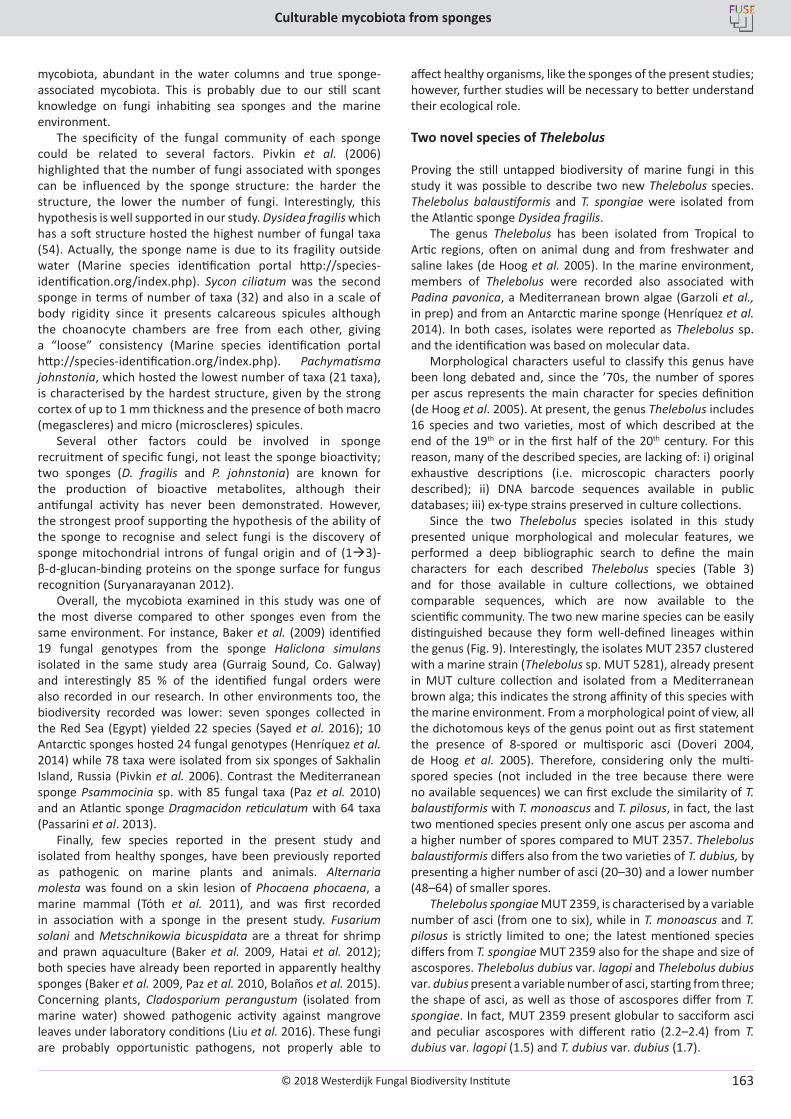
© 2018 Westerdijk Fungal Biodiversity Institute
Culturable mycobiota from sponges
Editor-in-ChiefProf. dr P.W. Crous, Westerdijk Fungal Biodiversity Institute, P.O. Box 85167, 3508 AD Utrecht, The Netherlands.E-mail:[email protected]
163
mycobiota, abundant in the water columns and true sponge-associated mycobiota. This is probably due to our still scant knowledge on fungi inhabiting sea sponges and the marine environment.
The specificity of the fungal community of each sponge could be related to several factors. Pivkin et al. (2006) highlighted that the number of fungi associated with sponges can be influenced by the sponge structure: the harder the structure, the lower the number of fungi. Interestingly, this hypothesis is well supported in our study. Dysidea fragilis which has a soft structure hosted the highest number of fungal taxa (54). Actually, the sponge name is due to its fragility outside water (Marine species identification portal http://species-identification.org/index.php). Sycon ciliatum was the second sponge in terms of number of taxa (32) and also in a scale of body rigidity since it presents calcareous spicules although the choanocyte chambers are free from each other, giving a “loose” consistency (Marine species identification portal http://species-identification.org/index.php). Pachymatisma johnstonia, which hosted the lowest number of taxa (21 taxa), is characterised by the hardest structure, given by the strong cortex of up to 1 mm thickness and the presence of both macro (megascleres) and micro (microscleres) spicules.
Several other factors could be involved in sponge recruitment of specific fungi, not least the sponge bioactivity; two sponges (D. fragilis and P. johnstonia) are known for the production of bioactive metabolites, although their antifungal activity has never been demonstrated. However, the strongest proof supporting the hypothesis of the ability of the sponge to recognise and select fungi is the discovery of sponge mitochondrial introns of fungal origin and of (1à3)-β-d-glucan-binding proteins on the sponge surface for fungus recognition (Suryanarayanan 2012).
Overall, the mycobiota examined in this study was one of the most diverse compared to other sponges even from the same environment. For instance, Baker et al. (2009) identified 19 fungal genotypes from the sponge Haliclona simulans isolated in the same study area (Gurraig Sound, Co. Galway) and interestingly 85 % of the identified fungal orders were also recorded in our research. In other environments too, the biodiversity recorded was lower: seven sponges collected in the Red Sea (Egypt) yielded 22 species (Sayed et al. 2016); 10 Antarctic sponges hosted 24 fungal genotypes (Henríquez et al. 2014) while 78 taxa were isolated from six sponges of Sakhalin Island, Russia (Pivkin et al. 2006). Contrast the Mediterranean sponge Psammocinia sp. with 85 fungal taxa (Paz et al. 2010) and an Atlantic sponge Dragmacidon reticulatum with 64 taxa (Passarini et al. 2013).
Finally, few species reported in the present study and isolated from healthy sponges, have been previously reported as pathogenic on marine plants and animals. Alternaria molesta was found on a skin lesion of Phocaena phocaena, a marine mammal (Tóth et al. 2011), and was first recorded in association with a sponge in the present study. Fusarium solani and Metschnikowia bicuspidata are a threat for shrimp and prawn aquaculture (Baker et al. 2009, Hatai et al. 2012); both species have already been reported in apparently healthy sponges (Baker et al. 2009, Paz et al. 2010, Bolaños et al. 2015). Concerning plants, Cladosporium perangustum (isolated from marine water) showed pathogenic activity against mangrove leaves under laboratory conditions (Liu et al. 2016). These fungi are probably opportunistic pathogens, not properly able to
affect healthy organisms, like the sponges of the present studies; however, further studies will be necessary to better understand their ecological role.
Two novel species of Thelebolus
Proving the still untapped biodiversity of marine fungi in this study it was possible to describe two new Thelebolus species. Thelebolus balaustiformis and T. spongiae were isolated from the Atlantic sponge Dysidea fragilis.
The genus Thelebolus has been isolated from Tropical to Artic regions, often on animal dung and from freshwater and saline lakes (de Hoog et al. 2005). In the marine environment, members of Thelebolus were recorded also associated with Padina pavonica, a Mediterranean brown algae (Garzoli et al., in prep) and from an Antarctic marine sponge (Henríquez et al. 2014). In both cases, isolates were reported as Thelebolus sp. and the identification was based on molecular data.
Morphological characters useful to classify this genus have been long debated and, since the ’70s, the number of spores per ascus represents the main character for species definition (de Hoog et al. 2005). At present, the genus Thelebolus includes 16 species and two varieties, most of which described at the end of the 19th or in the first half of the 20th century. For this reason, many of the described species, are lacking of: i) original exhaustive descriptions (i.e. microscopic characters poorly described); ii) DNA barcode sequences available in public databases; iii) ex-type strains preserved in culture collections.
Since the two Thelebolus species isolated in this study presented unique morphological and molecular features, we performed a deep bibliographic search to define the main characters for each described Thelebolus species (Table 3) and for those available in culture collections, we obtained comparable sequences, which are now available to the scientific community. The two new marine species can be easily distinguished because they form well-defined lineages within the genus (Fig. 9). Interestingly, the isolates MUT 2357 clustered with a marine strain (Thelebolus sp. MUT 5281), already present in MUT culture collection and isolated from a Mediterranean brown alga; this indicates the strong affinity of this species with the marine environment. From a morphological point of view, all the dichotomous keys of the genus point out as first statement the presence of 8-spored or multisporic asci (Doveri 2004, de Hoog et al. 2005). Therefore, considering only the multi-spored species (not included in the tree because there were no available sequences) we can first exclude the similarity of T. balaustiformis with T. monoascus and T. pilosus, in fact, the last two mentioned species present only one ascus per ascoma and a higher number of spores compared to MUT 2357. Thelebolus balaustiformis differs also from the two varieties of T. dubius, by presenting a higher number of asci (20–30) and a lower number (48–64) of smaller spores.
Thelebolus spongiae MUT 2359, is characterised by a variable number of asci (from one to six), while in T. monoascus and T. pilosus is strictly limited to one; the latest mentioned species differs from T. spongiae MUT 2359 also for the shape and size of ascospores. Thelebolus dubius var. lagopi and Thelebolus dubius var. dubius present a variable number of asci, starting from three; the shape of asci, as well as those of ascospores differ from T. spongiae. In fact, MUT 2359 present globular to sacciform asci and peculiar ascospores with different ratio (2.2–2.4) from T. dubius var. lagopi (1.5) and T. dubius var. dubius (1.7).
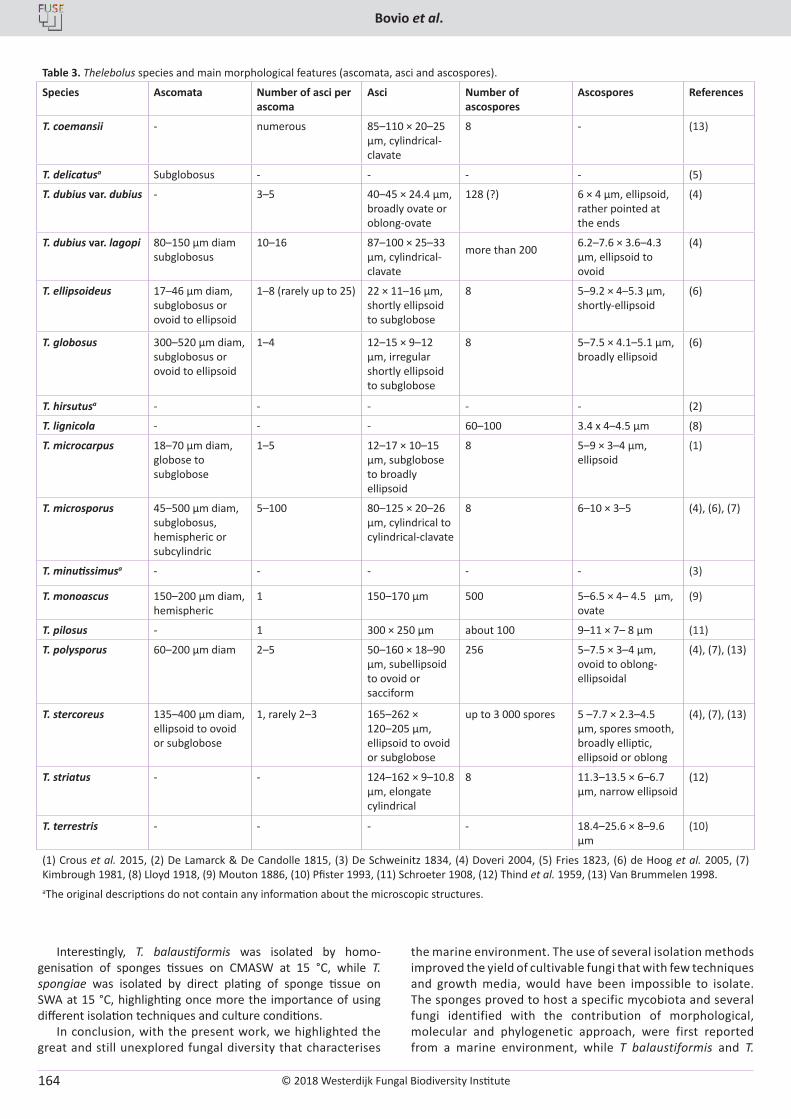
© 2018 Westerdijk Fungal Biodiversity Institute
Bovio et al.
Editor-in-ChiefProf. dr P.W. Crous, Westerdijk Fungal Biodiversity Institute, P.O. Box 85167, 3508 AD Utrecht, The Netherlands.E-mail:[email protected]
164
Interestingly, T. balaustiformis was isolated by homo-genisation of sponges tissues on CMASW at 15 °C, while T. spongiae was isolated by direct plating of sponge tissue on SWA at 15 °C, highlighting once more the importance of using different isolation techniques and culture conditions.
In conclusion, with the present work, we highlighted the great and still unexplored fungal diversity that characterises
the marine environment. The use of several isolation methods improved the yield of cultivable fungi that with few techniques and growth media, would have been impossible to isolate. The sponges proved to host a specific mycobiota and several fungi identified with the contribution of morphological, molecular and phylogenetic approach, were first reported from a marine environment, while T balaustiformis and T.
Table 3. Thelebolus species and main morphological features (ascomata, asci and ascospores).
Species Ascomata Number of asci per ascoma
Asci Number of ascospores
Ascospores References
T. coemansii - numerous 85–110 × 20–25 µm, cylindrical-clavate
8 - (13)
T. delicatusa Subglobosus - - - - (5)
T. dubius var. dubius - 3–5 40–45 × 24.4 µm, broadly ovate or oblong-ovate
128 (?) 6 × 4 µm, ellipsoid, rather pointed at the ends
(4)
T. dubius var. lagopi 80–150 µm diam subglobosus
10–16 87–100 × 25–33 µm, cylindrical-clavate
more than 200 6.2–7.6 × 3.6–4.3 µm, ellipsoid to ovoid
(4)
T. ellipsoideus 17–46 µm diam, subglobosus or ovoid to ellipsoid
1–8 (rarely up to 25) 22 × 11–16 µm, shortly ellipsoid to subglobose
8 5–9.2 × 4–5.3 µm, shortly-ellipsoid
(6)
T. globosus 300–520 µm diam, subglobosus or ovoid to ellipsoid
1–4 12–15 × 9–12 µm, irregular shortly ellipsoid to subglobose
8 5–7.5 × 4.1–5.1 µm, broadly ellipsoid
(6)
T. hirsutusa - - - - - (2)
T. lignicola - - - 60–100 3.4 x 4–4.5 µm (8)
T. microcarpus 18–70 µm diam, globose to subglobose
1–5 12–17 × 10–15 µm, subglobose to broadly ellipsoid
8 5–9 × 3–4 µm, ellipsoid
(1)
T. microsporus 45–500 µm diam, subglobosus, hemispheric or subcylindric
5–100 80–125 × 20–26 µm, cylindrical to cylindrical-clavate
8 6–10 × 3–5 (4), (6), (7)
T. minutissimusa - - - - - (3)
T. monoascus 150–200 µm diam, hemispheric
1 150–170 µm 500 5–6.5 × 4– 4.5 µm, ovate
(9)
T. pilosus - 1 300 × 250 µm about 100 9–11 × 7– 8 µm (11)
T. polysporus 60–200 µm diam 2–5 50–160 × 18–90 µm, subellipsoid to ovoid or sacciform
256 5–7.5 × 3–4 µm, ovoid to oblong-ellipsoidal
(4), (7), (13)
T. stercoreus 135–400 µm diam, ellipsoid to ovoid or subglobose
1, rarely 2–3 165–262 × 120–205 µm, ellipsoid to ovoid or subglobose
up to 3 000 spores 5 –7.7 × 2.3–4.5 µm, spores smooth, broadly elliptic, ellipsoid or oblong
(4), (7), (13)
T. striatus - - 124–162 × 9–10.8 µm, elongate cylindrical
8 11.3–13.5 × 6–6.7 µm, narrow ellipsoid
(12)
T. terrestris - - - - 18.4–25.6 × 8–9.6 µm
(10)
(1) Crous et al. 2015, (2) De Lamarck & De Candolle 1815, (3) De Schweinitz 1834, (4) Doveri 2004, (5) Fries 1823, (6) de Hoog et al. 2005, (7) Kimbrough 1981, (8) Lloyd 1918, (9) Mouton 1886, (10) Pfister 1993, (11) Schroeter 1908, (12) Thind et al. 1959, (13) Van Brummelen 1998.aThe original descriptions do not contain any information about the microscopic structures.
© 2018 Westerdijk Fungal Biodiversity Institute
Culturable mycobiota from sponges
Editor-in-ChiefProf. dr P.W. Crous, Westerdijk Fungal Biodiversity Institute, P.O. Box 85167, 3508 AD Utrecht, The Netherlands.E-mail:[email protected]
165
spongiae were here described as new. The present study again highlights the great mosaic of largely unknown marine microbial diversity.
REFERENCES
Baker PW, Kennedy J, Dobson AD, et al. (2009). Phylogenetic diversity and antimicrobial activities of fungi associated with Haliclona simulans isolated from Irish coastal waters. Marine Biotechnology 11: 540–547.
Bensch K, Braun U, Groenewald JZ, et al. (2012). The genus Cladosporium. Studies in Mycology 72: 1–401.
Berbee ML, Pirseyedi M, Hubbard S (1999). Cochliobolus phylogenetics and the origin of known, highly virulent pathogens, inferred from ITS and glyceraldehyde-3-phosphate dehydrogenase gene sequences. Mycologia 91: 964–977.
Bolaños J, De León LF, Ochoa E, et al. (2015). Phylogenetic diversity of sponge-associated fungi from the Caribbean and the Pacific of Panama and their in vitro effect on angiotensin and endothelin receptors. Marine Biotechnology 17: 533–564.
Bovio E, Gnavi G, Prigione V, et al. (2017). The culturable mycobiota of a Mediterranean marine site after an oil spill: isolation, identification and potential application in bioremediation. Science of the Total Environment 576: 310–318.
Braun U, Crous PW, Dugan F, et al. (2003). Phylogeny and taxonomy of Cladosporium-like hyphomycetes, including Davidiella gen. nov., the teleomorph of Cladosporium s. str. Mycological Progress 2: 3–18.
Caballero-George C, Bolaños J, De León LF, et al. (2013). Fungal diversity in marine sponges from highly diverse areas in the Isthmus of Panama. In: Proceedings of the 2013 AAUS/ESDP Curaçao Joint International Scientific Diving Symposium, October 24–27, 2013, Curaçao.
Carbone I, Kohn LM (1999). A method for designing primer sets for speciatioin studies in filamentous ascomycetes. Mycologia 91: 553–556.
Clarke KR, Warwick RM (2001). Change in Marine Communities: An Approach to Statistical Analysis and Interpretation. Primer-E Ltd, Plymouth, UK. http://www.primer-e.com/
Costello MJ, Emblow C, White RJ (2001). European register of marine species: a check-list of the marine species in Europe and a bibliography of guides to their identification. Collection Patrimoines Naturels, Paris.
Crous PW, Wingfield MJ, Guarro J, et al. (2015). Fungal Planet description sheets: 320–370. Persoonia 34: 167–226.
De Barros Lopes M, Soden A, Martens AL, et al. (1998). Differentiation and species identification of yeasts using PCR. International Journal of Systematic and Evolutionary Microbiology 48:279–286.
De Hoog GS, Gottlich E, Platas G, et al. (2005). Evolution, taxonomy and ecology of the genus Thelebolus in Antarctica. Studies in Mycology 51: 33–66.
De Lamarck MM, De Candolle (1815). Flore française: ou descriptions succintes de toutes les plantes qui croissent naturellement en France, disposées selon une novelle méthode d’analyse, et précédées par un exposé des principes élémentaires de la botanique (Vol. 2). 3rd edn. M. Agasse, France.
De Schweinitz LD (1834). Synopsis fungorum in America boreali media degentium. Secundum observationes. Transactions of the American Philosophical Society 4: 141–316.
Di Piazza S, Baiardo S, Cecchi G, et al. (2017). Microfungal diversity in the swash zone interstitial water (SZIW) of three Ligurian urban
beaches (NW, Italy). Italian Journal of Mycology 46: 8–20. Diep CN, Phong NT, Tam HT (2016). Phylogenetic diversity of culturable
fungi associated with sponges Leucosolenia sp. and Hexactinosa sp. In Ha Tien sea, Kien Giang, Vietnam. World Journal of Pharmacy and Pharmaceutical Sciences 5: 294–308.
Ding B, Yin Y, Zhang F, et al. (2011). Recovery and phylogenetic diversity of culturable fungi associated with marine sponges Clathrina luteoculcitella and Holoxea sp. in the South China Sea. Marine Biotechnology 13: 713–721.
Domsch KH, Gams W, Anderson TH (1980). Compendium of soil fungi. Vol. 1. Academic Press, London.
Doveri F (2004). Fungi Fimicoli Italici: A guide to the recognition of basidiomycetes and ascomycetes living on faecal material. Associazione Micologica Bresadola, Trento.
Ferreira M, Cabado AG, Chapela MJ, et al. (2011). Cytotoxic activity of extracts of marine sponges from NW Spain on a neuroblastoma cell line. Environmental Toxicology and Pharmacology 32: 430–437.
Fraser JA, Lambert LK, Pierens GK, et al. (2013). Secondary metabolites of the sponge-derived fungus Acremonium persicinum. Journal of Natural Products 76: 1432–1440.
Fries EM (1823). Systema Mycologicum, sistens fungorum ordines, genera et species. Vol. 2. Sumtibus Ernesti Mauritti, London.
Gao L, Ma Y, Zhao W, et al. (2015). Three new species of Cyphellophora (Chaetothyriales) associated with sooty blotch and flyspeck. PLoS ONE 10: 1–21.
Garzoli L, Gnavi G, Tamma F, et al. (2015). Sink or swim: Updated knowledge on marine fungi associated with wood substrates in the Mediterranean Sea and hints about their potential to remediate hydrocarbons. Progress in Oceanography 137: 140–148.
Glass NL, Donaldson GC (1995). Development of primer sets designed for use with the PCR to amplify conserved genes from filamentous ascomycetes. Applied and Environmental Microbiology 61: 1323–1330.
Gnavi G, Garzoli L, Poli A, et al. (2017). The culturable mycobiota of Flabellia petiolata: first survey of marine fungi associated to a Mediterranean green alga. PLoS ONE 12: 1–20.
Gomes D, Cavalcanti MAQ, Fernandes MJS, et al. (2008). Filamentous fungi isolated from sand and water of “Bairro Novo” and “Casa Caiada” beaches, Olinda, Pernambuco, Brazil. Brazilian Journal of Biology 68: 577–582.
Hatai K (2012). Diseases of fish and shellfish caused by marine fungi. In: Biology of marine fungi (Raghukumar ed.). Springer, Berlin Heidelberg: 15–52.
He L, Liu F, Karuppiah V, et al. (2014). Comparisons of the fungal and protistan communities among different marine sponge holobionts by pyrosequencing. Microbialecology 67: 951–961.
Henríquez M, Vergara K, Norambuena J, et al. (2014). Diversity of cultivable fungi associated with Antarctic marine sponges and screening for their antimicrobial, antitumoral and antioxidant potential. World Journal of Microbiology and Biotechnology 30: 65–76.
Höller U, Wright AD, Matthee GF, et al. (2000). Fungi from marine sponges: diversity, biological activity and secondary metabolites. Mycological Research 104: 1354–1365.
Hyde KD, Jones EG, Leaño E, et al. (1998). Role of fungi in marine ecosystems. Biodiversity and Conservation 7: 1147–1161.
Imhoff JF (2016). Natural products from marine fungi - Still an underrepresented resource. Marine Drugs 14: 1–19.
Imhoff JF, Stöhr R (2003). Sponge-associated bacteria: general overview and special aspects of bacteria associated with Halichondria panicea. In: Sponges, Porifera (Werner ed.). Springer, Berlin Heidelberg: 35–57.
© 2018 Westerdijk Fungal Biodiversity Institute
Bovio et al.
Editor-in-ChiefProf. dr P.W. Crous, Westerdijk Fungal Biodiversity Institute, P.O. Box 85167, 3508 AD Utrecht, The Netherlands.E-mail:[email protected]
166
Ingham CJ, Sprenkels A, Bomer J, et al. (2007). The micro-Petri dish, a million-well growth chip for the culture and high-throughput screening of microorganisms. Proceedings of the National Academy of Sciences USA 104: 18217–18222.
Jones EG, Suetrong S, Sakayaroj J, et al. (2015). Classification of marine ascomycota, basidiomycota, blastocladiomycota and chytridiomycota. Fungal Diversity 73: 1–72.
Jurjevic Z, Peterson SW, Horn BW (2012). Aspergillus section Versicolores: nine new species and multilocus DNA sequence based phylogeny. IMA Fungus 3: 59–79.
Kiffer E, Morelet M (1997). Les deutéromycetes: Classification et clés d’identification genérique. INRA ed, France.
Kimbrough JW (1981). Cytology, ultrastructure, and taxonomy of Thelebolus (Ascomycetes). Mycologia 73 1–27.
Kis-Papo T, Grishkan I, Oren A, et al. (2001). Spatiotemporal diversity of filamentous fungi in the hypersaline Dead Sea. Mycological Research 105: 749–756.
Klich MA (2002). Identification of common Aspergillus species. Westerdijk Fungal Biodiversity Institute, Utrecht, Netherlands.
Li Q, Wang G (2009). Diversity of fungal isolates from three Hawaiian marine sponges. Microbiological Research 164: 233–241.
Liu Y, Li Y, Lin Q, et al. (2016). Assessment of the pathogenicity of marine Cladosporium spp. towards mangroves. Forest Pathology 47: 1–5.
Lloyd CG (1918). Mycological Notes 52. Mycological Writings 5: 733–748.
Marchese P, Gnavi G, Garzoli L, et al. (2016). Marine fungi from Holothuria poli (Della Chiaje, 1823): diversity and extracts bioactivity. Biologia Marina Mediterranea 23: 290–291.
Marin A, Lopez MD, Esteban MA, et al. (1998). Anatomical and ultrastructural studies of chemical defence in the sponge Dysidea fragilis. Marine Biology 131: 639–645.
Mouton V (1886). Bulletin de la Société Royale de Botanique de Belgique. Royal Botanical Society of Belgium.
Nichols D, Cahoon N, Trakhtenberg EM, et al. (2010). Use of ichip for high-throughput in situ cultivation of “uncultivable” microbial species. Applied and Environmental Microbiology 76: 2445–2450.
O’Donnell K, Nirenberg HI, Aoki T, et al. (2000). A multigene phylogeny of the Gibberella fujikuroi species complex: detection of additional phylogenetically distinct species. Mycoscience 41: 61–78.
Oren A, Gunde-Cimerman N (2012). Fungal life in the Dead Sea. In: Biology of Marine Fungi (Raghukumar ed.). Springer, Berlin Heidelberg: 115–132.
Panno L, Bruno M, Voyron S, et al. (2013). Diversity, ecological role and potential biotechnological applications of marine fungi associated to the seagrass Posidonia oceanica. New Biotechnology 30: 685–694.
Park MS, Lee EJ, Fong JJ, et al. (2014). A new record of Penicillium antarcticum from marine environments in Korea. Mycobiology 42:109–113.
Passarini MR, Santos C, Lima N, et al. (2013). Filamentous fungi from the Atlantic marine sponge Dragmacidon reticulatum. Archives of Microbiology 195: 99–111.
Paz Z, Komon-Zelazowska M, Druzhinina IS, et al. (2010). Diversity and potential antifungal properties of fungi associated with a Mediterranean sponge. Fungal Diversity 42: 17–26.
Pfister DH (1993). A synopsis of the North American species of Byssonectria (Pezizales) with comments on the ontogeny of two species. Mycologia 85: 952–962.
Pivkin MV, Aleshko SA, Krasokhin VB, et al. (2006). Fungal assemblages associated with sponges of the southern coast of Sakhalin Island. Russian Journal of Marine Biology 32: 207–213.
Proksch P, Ebel R, Edrada R, et al. (2008). Sponge-associated fungi and
their bioactive compounds: the Suberites case. Botanica Marina 51: 209–218.
Raghukumar C, Ravindran J (2012). Fungi and their role in corals and coral reef ecosystems. In: Biology of Marine Fungi (Raghukumar ed.). Springer, Berlin Heidelberg: 89–113.
Raghukumar S (2017). Fungi in Coastal and Oceanic Marine Ecosystems. Springer, Berlin Heidelberg.
Rämä T, Hassett BT, Bubnova E (2017). Arctic marine fungi: from filaments and flagella to operational taxonomic units and beyond. Botanica Marina 60: 433–452.
Rämä T, Nordén J, Davey ML, et al. (2014). Fungi ahoy! Diversity on marine wooden substrata in the high North. Fungal Ecology 8: 46–58.
Ratnaweera PB, Williams DE, De Silva ED, et al. (2016). Antibacterial metabolites from the Sri Lankan demosponge-derived fungus, Aspergillus flavipes. Current Science 111: 1473–1479.
Rodríguez-Marconi S, De la Iglesia R, Díez B, et al. (2015). Characterization of bacterial, archaeal and eukaryote symbionts from Antarctic sponges reveals a high diversity at a three-domain level and a particular signature for this ecosystem. PloSone 10: 1–19.
Rozas EE, Albano RM, Lôbo-Hajdu G, et al. (2011). Isolation and cultivation of fungal strains from in vitro cell cultures of two marine sponges (Porifera: Halichondrida and Haplosclerida). Brazilian Journal of Microbiology 42: 1560–1568.
Samson RA, Frisvad JC (2004). Penicillium subgenus Penicillium: new taxonomic schemes and mycotoxins and other extrolites. Studies in Mycology 49: 1–260.
San-Martin A, Painemal K, Díaz Y, et al. (2005). Metabolites from the marine fungus Cladosporium cladosporioides. The Journal of the Argentine Chemical Society 93: 247–251.
Sayed MAE, El-Rahman TMA, El-Diwany AI, et al. (2016). Biodiversity and bioactivity of red sea sponge associated endophytic fungi. International Journal of Advanced in Engineering and Applied Sciences 5: 1–15.
Schroeter J, Cohn F (1908). Die Pilze Schlesiens, Vol. 2, JU Kern.Seeler EV (1940). A monographic study of the genus Thyronectria.
Journal of the Arnold Arboretum 21: 429–460.Shigemori H, Tenma M, Shimazaki K, et al. (1998). Three new metabolites
from the marine yeast Aureobasidium pullulans. Journal of Natural Products 61: 696–698.
Suetrong S, Schoch CL, Spatafora JW, et al. (2009). Molecular systematics of the marine Dothideomycetes. Studies in Mycology 64: 155–173.
Suryanarayanan TS (2012). Fungal endosymbionts of seaweeds. In: Biology of Marine Fungi (Raghukumar ed.). Springer, Berlin Heidelberg: 53–69.
Taylor MW, Radax R, Steger D, et al. (2007). Sponge-associated microorganisms: evolution, ecology, and biotechnological potential. Microbiology and Molecular Biology Reviews 71: 29–347.
Thind KS, Cash EK, Singh P (1959). The Pezizaceae of the Mussoorie hills (India): VII. Mycologia 51: 457–464.
Thirunavukkarasu N, Suryanarayanan TS, Girivasan KP, et al. (2012). Fungal symbionts of marine sponges from Rameswaram, southern India: species composition and bioactive metabolites. Fungal Diversity 55: 37–46.
Tóth B, Csosz M, Szabo-Hever A, et al. (2011). Alternaria hungarica sp. nov., a minor foliar pathogen of wheat in Hungary. Mycologia 103: 94–100.
Vacelet J, Donadey C (1977). Electron microscope study of the association between some sponges and bacteria. Journal of Experimental Marine Biology and Ecology 30: 301–314.
Van Brummelen J (1998). Reconsideration of relationships within the Thelebolaceae based on ascus ultrastructure. Persoonia 16: 425–469.
© 2018 Westerdijk Fungal Biodiversity Institute
Culturable mycobiota from sponges
Editor-in-ChiefProf. dr P.W. Crous, Westerdijk Fungal Biodiversity Institute, P.O. Box 85167, 3508 AD Utrecht, The Netherlands.E-mail:[email protected]
167
Vilgalys R, Hester M (1990). Rapid genetic identification and mapping of enzymatically amplified ribosomal DNA from several Cryptococcus species. Journal of Bacteriology 172: 4238–4246.
Von Arx JA (1981). The genera of fungi sporulating in pure culture. 3rd edn. Cramer, Vaduz.
Wang G, Li Q, Zhu P (2008). Phylogenetic diversity of culturable fungi associated with the Hawaiian sponges Suberites zeteki and Gelliodes fibrosa. Antonie van Leeuwenhoek 93: 163–174.
Warabi K, Zimmerman WT, Shen J, et al. (2004). Pachymoside AA novel glycolipid isolated from the marine sponge Pachymatisma johnstonia. Canadian Journal of Chemistry 82: 102–112.
Webster NS, Thomas T (2016). The sponge hologenome. mBio 7: 1–14. White TJ, Bruns T, Lee S, et al. 1990. Amplification and direct sequencing
of fungal ribosomal RNA genes for phylogenetics. In: PCR protocols: a guide to methods and applications (Innis MA, et al. eds). Academic Press, San Diego: 315–322.
Wiese J, Ohlendorf B, Blümel M, et al. (2011). Phylogenetic identification of fungi isolated from the marine sponge Tethya aurantium and identification of their secondary metabolites. Marine Drugs 9: 561–585.
Yamazaki H, Rotinsulu H, Kaneko T, et al. (2012). A new dibenz [b, e] oxepine derivative, 1-hydroxy-10-methoxy-dibenz [b, e] oxepin-6, 11-dione, from a marine-derived fungus, Beauveria bassiana TPU942. Marine Drugs 10: 2691–2697.
Yarden O (2014). Fungal association with sessile marine invertebrates. Frontiers in Microbiology 5: 1–6.
Yu Z, Lang G, Kajahn I, et al. (2008). Scopularides A and B, cyclodepsipeptides from a marine sponge-derived fungus, Scopulariopsis brevicaulis. Journal of Natural Products 71): 1052–1054.
Yu ZG, Bi KS, Guo YW, et al. (2006). A new spiro-sesquiterpene from the sponge Dysidea fragilis: Note. Journal of Asian Natural Products Research 8: 467–470.
Zajc J, Zalar P, Plemenitaš A, et al. (2012). The mycobiota of the salterns. In: Biology of Marine Fungi (Raghukumar ed) Springer, Berlin Heidelberg: 133–158.
Zhang H, Zhao Z, Wang H (2017). Cytotoxic natural products from marine sponge-derived microorganisms. Marine Drugs 15: 1–13.
Zidane M, Pondaven P, Roussakis C, et al. (1996). Effects in vitro of pachymatismin, a glycoprotein from the marine sponge Pachymatisma johnstonii, on a non-small-cell bronchopulmonary carcinoma line (NSCLC-N6). Anticancer Research 16: 2805–2812.

© 2018 Westerdijk Fungal Biodiversity Institute
Editor-in-ChiefProf. dr P.W. Crous, Westerdijk Fungal Biodiversity Institute, P.O. Box 85167, 3508 AD Utrecht, The Netherlands.E-mail:[email protected]
Fungal Systematics and Evolution is licensed under a Creative Commons Attribution-NonCommercial-ShareAlike 4.0 International License
© 2018 Westerdijk Fungal Biodiversity Institute 169
Editor-in-ChiefProf. dr P.W. Crous, Westerdijk Fungal Biodiversity Institute, P.O. Box 85167, 3508 AD Utrecht, The Netherlands.E-mail:[email protected]
Fungal Systematics and Evolution
doi.org/10.3114/fuse.2018.01.08
VOLUME 1JUNE 2018
PAGES 169–215
New and Interesting Fungi. 1
P.W. Crous1,2,3*, R.K. Schumacher4, M.J. Wingfield5, A. Akulov6, S. Denman7, J. Roux2, U. Braun8, T.I. Burgess9, A.J. Carnegie10, K.Z. Váczy11, E. Guatimosim12, P.B. Schwartsburd13, R.W. Barreto14, M. Hernández-Restrepo1, L. Lombard1, J.Z. Groenewald1
1Westerdijk Fungal Biodiversity Institute, P.O. Box 85167, 3508 AD Utrecht, The Netherlands2Department of Genetics, Biochemistry and Microbiology, Forestry and Agricultural Biotechnology Institute (FABI), University of Pretoria, Pretoria, 0002, South Africa3Microbiology, Department of Biology, Utrecht University, Padualaan 8, 3584 CH Utrecht, The Netherlands4Hölderlinstraße 25, 15517 Fürstenwalde / Spree, Germany5Forestry and Agricultural Biotechnology Institute (FABI), University of Pretoria, Pretoria, 0002, South Africa6Department of Mycology and Plant Resistance, V. N. Karazin Kharkiv National University, Maidan Svobody 4, 61022 Kharkiv, Ukraine7Forest Research, Alice Holt Lodge, Farnham, Surrey, UK8Martin-Luther-Universität, Institut für Biologie, Bereich Geobotanik und Botanischer Garten, Herbarium, Neuwerk 21, 06099 Halle (Saale), Germany9Centre for Phytophthora Science and Management, Murdoch University, 90 South Street, Murdoch, WA 6150, Australia10Forest Health & Biosecurity, NSW Department of Primary Industries, Level 12, 10 Valentine Ave, Parramatta NSW 2150, Locked Bag 5123, Parramatta NSW 2124, Australia11Centre for Research and Development, Eszterházy Károly University, H-3300 Eger, Hungary12Instituto de Ciências Biológicas, Universidade Federal do Rio Grande, CEP: 96170-000, São Lourenço do Sul, Brazil13Departamento de Biologia Vegetal, Universidade Federal de Viçosa, CEP: 36.570-900, Viçosa, Minas Gerais, Brazil14Departamento de Fitopatologia, Universidade Federal de Viçosa, CEP: 36.570-900, Viçosa, Minas Gerais, Brazil
*Corresponding author: [email protected]
Abstract: This study introduces two new families, one new genus, 22 new species, 10 new combinations, four epitypes, and 16 interesting new host and / or geographical records. Cylindriaceae (based on Cylindrium elongatum) is introduced as new family, with three new combinations. Xyladictyochaetaceae (based on Xyladictyochaeta lusitanica) is introduced to accommodate Xyladictyochaeta. Pseudoanungitea gen. nov. (based on P. syzygii) is described on stems of Vaccinium myrtillus (Germany). New species include: Exophiala eucalypticola on Eucalyptus obliqua leaf litter, Phyllosticta hakeicola on leaves of Hakea sp., Setophaeosphaeria citricola on leaves of Citrus australasica, and Sirastachys cyperacearum on leaves of Cyperaceae (Australia); Polyscytalum chilense on leaves of Eucalyptus urophylla (Chile); Pseudoanungitea vaccinii on Vaccinium myrtillus (Germany); Teichospora quercus on branch tissue of Quercus sp. (France); Fusiconidium lycopodiellae on stems of Lycopodiella inundata, Monochaetia junipericola on twig of Juniperus communis, Myrmecridium sorbicola on branch tissues of Sorbus aucuparia, Parathyridaria philadelphi on twigs of Philadelphus coronarius, and Wettsteinina philadelphi on twigs of Philadelphus coronarius (Germany); Zygosporium pseudogibbum on leaves of Eucalyptus pellita (Malaysia); Pseudoanungitea variabilis on dead wood (Spain); Alfaria acaciae on leaves of Acacia propinqua, Dictyochaeta mimusopis on leaves of Mimusops caffra, and Pseudocercospora breonadiae on leaves of Breonadia microcephala (South Africa); Colletotrichum kniphofiae on leaves of Kniphofia uvaria, Subplenodomus iridicola on Iris sp., and Trochila viburnicola on twig cankers on Viburnum sp. (UK); Polyscytalum neofecundissimum on Quercus robur leaf litter, and Roussoella euonymi on fallen branches of Euonymus europaeus (Ukraine). New combinations include: Cylindrium algarvense on leaves of Eucalyptus sp. (Portugal), Cylindrium purgamentum on leaf litter (USA), Cylindrium syzygii on leaves of Syzygium sp. (Australia), Microdochium musae on leaves of Musa sp. (Malaysia), Polyscytalum eucalyptigenum on Eucalyptus grandis × pellita (Malaysia), P. eucalyptorum on leaves of Eucalyptus (Australia), P. grevilleae on leaves of Grevillea (Australia), P. nullicananum on leaves of Eucalyptus (Australia), Pseudoanungitea syzygii on Syzygium cordatum leaf litter (South Africa), and Setophaeosphaeria sidae on leaves of Sida sp. (Brazil). New records include: Sphaerellopsis paraphysata on leaves of Phragmites sp., Vermiculariopsiella dichapetali on leaves of Melaleuca sp. and Eucalyptus regnans, and Xyladictyochaeta lusitanica on leaf litter of Eucalyptus sp. (Australia); Camarosporidiella mackenziei on twigs of Caragana sp. (Finland); Cyclothyriella rubronotata on twigs of Ailanthus altissima, Rhinocladiella quercus on Sorbus aucuparia branches (Germany); Cytospora viticola on stems of Vitis vinifera (Hungary); Echinocatena arthrinioides on leaves of Acacia crassicarpa (Malaysia); Varicosporellopsis aquatilis from garden soil (Netherlands); Pestalotiopsis hollandica on needles of Cupressus sempervirens (Spain), Pseudocamarosporium africanum on twigs of Erica sp. (South Africa), Pseudocamarosporium brabeji on branch of Platanus sp. (Switzerland); Neocucurbitaria cava on leaves of Quercus ilex (UK); Chaetosphaeria myriocarpa on decaying wood of Carpinus betulus, Haplograhium delicatum on decaying Carpinus betulus wood (Ukraine). Epitypes are designated for: Elsinoë mimosae on leaves of Mimosa diplotricha (Brazil), Neohendersonia kickxii on Fagus sylvatica twig bark (Italy), Caliciopsis maxima on fronds of Niphidium crassifolium (Brazil), Dictyochaeta septata on leaves of Eucalyptus grandis × urophylla (Chile), and Microdochium musae on leaves of Musa sp. (Malaysia).
Key words: biodiversityITS barcodesmulti-gene
phylogenynew taxasystematicstypification36 new taxa
© 2018 Westerdijk Fungal Biodiversity Institute
Crous et al.
Editor-in-ChiefProf. dr P.W. Crous, Westerdijk Fungal Biodiversity Institute, P.O. Box 85167, 3508 AD Utrecht, The Netherlands.E-mail:[email protected]
170
INTRODUCTION
New and Interesting Fungi (NIF) is introduced as a new series of papers that will supplement other series focussed on expanding existing knowledge of fungal biodiversity and fungal conservation. Another similar series such as the Fungal Planet (www.fungalplanet.org) aims to provide a rapid and simplified outlet for researchers to describe new fungal species as well as to highlight the environments where these fungi were isolated. The Fungal Planet series established in 2006 emphasises a holistic conservation of all life on the planet including not only plants and animals but also fungi (Crous et al. 2017a, b).
This new series of papers focusses not only on new fungal taxa but also on those that are generally interesting and that deserve notice. Like other series including the already mentioned Fungal Planet, the Genera of Fungi (GoF) series (Crous & Groenewald 2017, Giraldo et al. 2017), the Genera of Phytopathogenic Fungi (GOPHY) series (Marin-Felix et al. 2017), and the Fungal Systematics and Evolution series (Crous et al. 2015a, Hernandez-Restrepo et al. 2016, Krisai-Greilhuber et al. 2017) it has become evident that there are many undescribed species of fungi and new host or geographical records for which a scientific repository is lacking. Most of these could easily never be described or catalogued, and thus being lost to science. This justified the decision to launch the new series New and Interesting Fungi (NIF). It is hoped that this series will provide an attractive vehicle for mycologists to publish single new species or to highlight the relevance of important fungi.
Many known fungal species need to be recollected and epi- or neotypified in order to secure the application of old names already in use and resolve their DNA phylogeny. Subsequent to the end of the long-standing dual nomenclature for fungi (Hawksworth et al. 2011, Wingfield et al. 2012) and the connection of different morphs to a single name (Rossman et al. 2015, Réblová et al. 2016), it became clear that a vehicle was required to ensure that these data could be easily and effectively published. This would be comparable to “data release papers” published in other fields of science and biology (Miller et al. 2013, Vu et al. 2016). The New and Interesting Fungi series will link not only asexual and sexual morphs of species, but also provide opportunities to merge morphological observations with DNA sequence data, providing a means for rapid and accurate identification. New and Interesting Fungi will appear twice each year (June and December) in the journal Fungal Systematics and Evolution (www.FUSE-journal.org). Mycologists and other researchers wishing to contribute to future issues in this series are encouraged to contact Pedro Crous ([email protected]) before submission to ensure that potential conflicts with activities arising from other research groups can be avoided.
MATERIALS AND METHODS
Isolates
Leaves and twig samples were placed in damp chambers and incubated at room temperature for 1–3 d. Single conidial colonies were grown from sporulating conidiomata in Petri dishes containing 2 % malt extract agar (MEA) as described earlier by Crous et al. (1991). Leaf and stem tissues bearing ascomata were soaked in water for approximately 2 h, after which they were attached to the bottom side of the lids of Petri dishes containing MEA. After ascospores ejected onto the MEA, germination patterns were determined after 24 h, and single ascospore or conidial cultures were established following the method described by (Crous 1998). Colonies were sub-cultured on 2 % potato-dextrose agar (PDA), oatmeal agar (OA), MEA (Crous et al. 2009b), autoclaved pine needles on 2 % tap water agar (PNA) (Smith et al. 1996), or autoclaved banana leaves (BLA), and incubated at 25 °C under continuous near-ultraviolet light to promote sporulation. Reference strains and specimens are maintained at the Westerdijk Fungal Biodiversity Institute in Utrecht, the Netherlands (CBS).
DNA extraction, amplification (PCR) and phylogeny
Fungal mycelium (Supplementary Table 1) was scraped from the agar surface of cultures with a sterile scalpel and the genomic DNA was isolated using the Wizard® Genomic DNA Purification Kit (Promega Corporation, WI, USA) following the manufacturers’ protocols. The 28S nrRNA gene (LSU) and internal transcribed spacer regions with intervening 5.8S nrRNA gene (ITS) of the nrDNA operon were sequenced for all the isolates included in this study. Other loci were sequenced for various species or genera using primers and conditions specific for those groups of fungi (see references for details). The resulting fragments were sequenced in both directions using the respective PCR primers and the BigDye Terminator Cycle Sequencing Kit v. 3.1 (Applied Biosystems Life Technologies, Carlsbad, CA, USA); DNA sequencing amplicons were purified through Sephadex G-50 Superfine columns (Sigma-Aldrich, St. Louis, MO) in MultiScreen HV plates (Millipore, Billerica, MA). Purified sequence reactions were analysed on an Applied Biosystems 3730xl DNA Analyzer (Life Technologies, Carlsbad, CA, USA). The DNA sequences were analysed and consensus sequences were computed using SeqMan Pro v. 13 (DNASTAR, Madison, WI, USA).
The sequences for each gene were subjected to megablast searches (Zhang et al. 2000) to identify closely related sequences in the NCBI’s GenBank nucleotide database. The results are provided as part of the species notes or as selected phylogenetic trees. Phylogenetic trees were generated using Bayesian analyses performed with MrBayes v. 3.2.6 (Ronquist et al. 2012) for the overview trees and Maximum Parsimony analyses performed with PAUP v. 4.0b10 (Swofford 2003) as explained in Braun et al. (2018) for the genus and species trees. All resulting trees were printed with Geneious v. 11.0.3 (http://www.geneious.com, Kearse et al. 2012) and the layout of the trees was done in Adobe Illustrator v. CC 2017. Statistical measures calculated
Published online: 18 April 2018.
Dedicated to Vadim Alexandrovich Mel’nik (*16 March 1937, †10 April 2017).
© 2018 Westerdijk Fungal Biodiversity Institute
New and interesting fungi
Editor-in-ChiefProf. dr P.W. Crous, Westerdijk Fungal Biodiversity Institute, P.O. Box 85167, 3508 AD Utrecht, The Netherlands.E-mail:[email protected]
171
included tree length (TL), consistency index (CI), retention index (RI) and rescaled consistency index (RC).
Morphology
Slide preparations were mounted in lactic acid, Shear’s mounting fluid or water, from colonies sporulating on MEA, PDA, PNA, BLA or OA. Sections through conidiomata were made by hand. Observations were made with a Nikon SMZ25 dissection-microscope, and with a Zeiss Axio Imager 2 light microscope using differential interference contrast (DIC) illumination and images recorded on a Nikon DS-Ri2 camera with associated software. Colony characters and pigment production were noted after 2–4 wk of growth on MEA, PDA and OA (Crous et al. 2009b) incubated at 25 oC. Colony colours (surface and reverse) were scored using the colour charts of Rayner (1970). Sequences derived in this study were deposited in GenBank (Supplementary Table 1), the alignment in TreeBASE (www.treebase.org; study number S22442), and taxonomic novelties in MycoBank (www.MycoBank.org; Crous et al. 2004).
RESULTS
Phylogeny
Dothideomycetes LSU phylogeny: The alignment contained 125 isolates and Candida broadrunensis (CBS 11838, GenBank KY106372.1) was used as outgroup. The final alignment contained a total of 808 characters used for the phylogenetic analyses, including alignment gaps. Based on the results of MrModelTest, dirichlet base frequencies and the GTR+I+G model was used for the Bayesian analysis. The Bayesian analyses generated 38 302 trees from which 28 728 were sampled after 25 % of the trees were discarded as burn-in. The posterior probability values (PP) were calculated from the 28 728 trees (Fig. 1 overview Dothideomycetes; PP >0.74 shown). The alignment contained a total of 345 unique site patterns.
Eurotiomycetes and Leotiomycetes LSU phylogeny: The alignment contained 44 isolates and Orbilia vinosa (GenBank DQ470952.1) was used as outgroup. The final alignment contained a total of 813 characters used for the phylogenetic analyses, including alignment gaps. Based on the results of MrModelTest, dirichlet base frequencies and the GTR+I+G model was used for the Bayesian analyses. The Bayesian analyses generated 9 702 trees from which 7 278 were sampled after 25 % of the trees were discarded as burn-in. The posterior probability values (PP) were calculated from the 7 278 trees (Fig. 2 overview Eurotiomycetes; PP >0.74 shown). The alignment contained a total of 253 unique site patterns.
Sordariomycetes LSU phylogeny: The alignment contained 148 isolates and Candida broadrunensis (CBS 11838, GenBank KY106372.1) was used as outgroup. The final alignment contained a total of 761 characters used for the phylogenetic analyses, including alignment gaps. Based on the results of MrModelTest, dirichlet base frequencies and the GTR+I+G model was used for the Bayesian analysis. The Bayesian analyses generated 34 702 trees from which 26 028 were sampled after 25 % of the trees were discarded as burn-in. The posterior probability values (PP) were calculated from the 26 028 trees (Fig. 3 overview Sordariomycetes; first value: PP >0.74 shown). The alignment contained a total of 361 unique site patterns.
Species phylogenies: Specific phylogenetic analyses were run for selected species and the resulting phylogenies are discussed in the species notes where applicable. Statistics associated with those phylogenies are provided in the figure legends.
Taxonomy
Alfaria acaciae Crous & M.J. Wingf., sp. nov. MycoBank MB824766. Fig. 4.
Etymology: Name refers to Acacia, the genus of the substrate from which this fungus was collected.
Conidiomata sporodochial, surrounded by setae, black with dark green to black slimy conidial masses, 80–250 µm diam. Setae flexuous, unbranched, thick-walled, apex obtuse, dark brown, verruculose, 3–6-septate, 100–150 × 5–7 µm. Conidiophores densely aggregated, arising from hyaline basal stroma, becoming pigmented and verruculose towards conidiogenous region, subcylindrical, 3–5-septate, branched, 30–55 × 2–2.5 µm. Conidiogenous cells integrated, terminal and intercalary, subcylindrical, becoming pigmented and verruculose at upper region, phialidic with periclinal thickening and flared collarette, 10–20 × 2–2.5 µm. Conidia solitary, fusoid-ellipsoid, straight, apex subobtuse, base truncate, 1.5–2 µm diam, aseptate, guttulate, granular, medium brown, smooth, (6–)8–10(–12) × (2.5–)3 µm.
Culture characteristics: Colonies flat, spreading, with sparse aerial mycelium and smooth, lobate margins, reaching 50 mm diam after 2 wk at 25 °C. On MEA surface dirty white to pale luteous, reverse luteous. On PDA surface dirty white, reverse pale luteous. On OA surface pale luteous.
Specimens examined: South Africa, Western Cape Province, Stellenbosch, Helshoogte Pass, on leaves of Acacia propinqua (Fabaceae), Jul. 2012, M.J. Wingfield (holotype CBS H-23428, culture ex-type CPC 31882 = CBS 143504); ibid., CPC 31940.
Notes: Alfaria cyperi-esculenti was originally described from Cyperus esculentus in Spain, where it causes a serious foliar disease (Crous et al. 2014). This species is currently known only from its sexual morph, which complicates a morphological comparison with the present, asexual isolate from South Africa. Although phylogenetically closely related (Fig. 5), we regard them as two distinct species (see bp differences below). Furthermore, culture characteristics also differ between the two species, with cultures of A. acaciae growing faster, and paler in colour than the ochreous / apricot cultures of A. cyperi-esculenti (Crous et al. 2014).
Based on a megablast search using the ITS sequence, the closest matches in NCBI’s GenBank nucleotide database were A. cyperi-esculenti (GenBank KJ869143; Identities 567 / 577 (98 %), 5 gaps (0 %)), Myrothecium leucotrichum (GenBank AJ301992; Identities 566 / 578 (98 %), 7 gaps (1 %)) and A. thymi (GenBank KU845990; Identities 559 / 572 (98 %), 8 gaps (1 %)). The ITS sequences of CPC 31882 and 31940 are identical. The highest similarities using the LSU sequence were A. cyperi-esculenti (GenBank KJ869200; Identities 803 / 804 (99 %), no gaps), A. thymi (GenBank KU845999; Identities 824 / 828 (99 %), 1 gap (0 %)) and A. caricicola (GenBank KU845992; Identities 822 / 828 (99 %), 1 gap (0 %)). The highest similarities using the cmdA
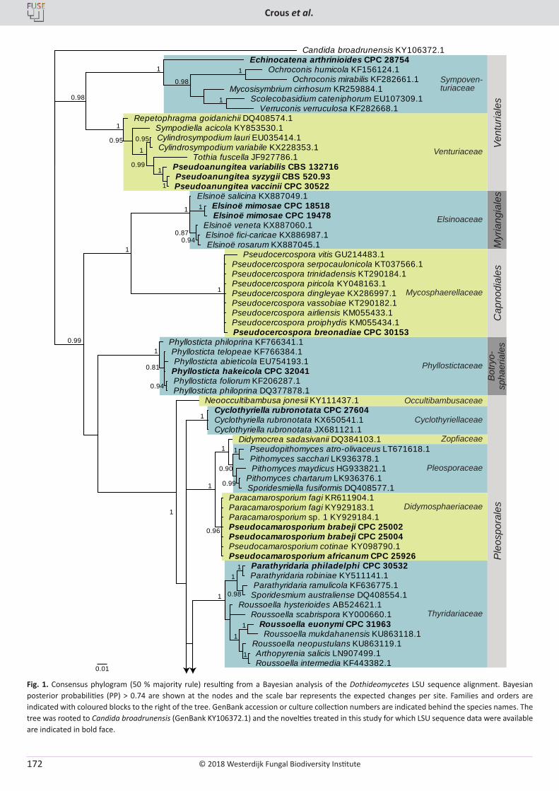
© 2018 Westerdijk Fungal Biodiversity Institute
Crous et al.
Editor-in-ChiefProf. dr P.W. Crous, Westerdijk Fungal Biodiversity Institute, P.O. Box 85167, 3508 AD Utrecht, The Netherlands.E-mail:[email protected]
172
0.01
1
1 1
1
0.98
1
1
1
0.99
1
0.90
1
0.96
1
1
0.94
0.81
1
0.94 0.87
1 1
1
1
0.99
1
1 0.99
0.95
1 0.95
1
1
1 0.98
1
0.98
Candida broadrunensis KY106372.1
Roussoella hysterioides AB524621.1 Roussoella scabrispora KY000660.1
Arthopyrenia salicis LN907499.1 Roussoella intermedia KF443382.1
Roussoella neopustulans KU863119.1
Roussoella euonymi CPC 31963 Roussoella mukdahanensis KU863118.1
Parathyridaria philadelphi CPC 30532 Parathyridaria robiniae KY511141.1 Parathyridaria ramulicola KF636775.1
Sporidesmium australiense DQ408554.1
Cyclothyriella rubronotata CPC 27604 Cyclothyriella rubronotata KX650541.1 Cyclothyriella rubronotata JX681121.1
Didymocrea sadasivanii DQ384103.1
Pithomyces chartarum LK936376.1 Sporidesmiella fusiformis DQ408577.1
Pithomyces maydicus HG933821.1
Pseudopithomyces atro-olivaceus LT671618.1 Pithomyces sacchari LK936378.1
Paracamarosporium fagi KR611904.1 Paracamarosporium fagi KY929183.1 Paracamarosporium sp. 1 KY929184.1 Pseudocamarosporium brabeji CPC 25002 Pseudocamarosporium brabeji CPC 25004 Pseudocamarosporium cotinae KY098790.1 Pseudocamarosporium africanum CPC 25926
Neooccultibambusa jonesii KY111437.1
Phyllosticta foliorum KF206287.1 Phyllosticta philoprina DQ377878.1
Phyllosticta telopeae KF766384.1 Phyllosticta abieticola EU754193.1
Phyllosticta hakeicola CPC 32041
Phyllosticta philoprina KF766341.1
Elsinoë fici-caricae KX886987.1 Elsinoë rosarum KX887045.1
Elsinoë veneta KX887060.1
Elsinoë salicina KX887049.1 Elsinoë mimosae CPC 18518 Elsinoë mimosae CPC 19478
Pseudocercospora vitis GU214483.1 Pseudocercospora serpocaulonicola KT037566.1 Pseudocercospora trinidadensis KT290184.1 Pseudocercospora piricola KY048163.1 Pseudocercospora dingleyae KX286997.1 Pseudocercospora vassobiae KT290182.1 Pseudocercospora airliensis KM055433.1 Pseudocercospora proiphydis KM055434.1 Pseudocercospora breonadiae CPC 30153
Pseudoanungitea variabilis CBS 132716 Pseudoanungitea syzygii CBS 520.93 Pseudoanungitea vaccinii CPC 30522
Tothia fuscella JF927786.1
Cylindrosympodium lauri EU035414.1 Cylindrosympodium variabile KX228353.1
Sympodiella acicola KY853530.1 Repetophragma goidanichii DQ408574.1
Echinocatena arthrinioides CPC 28754
Mycosisymbrium cirrhosum KR259884.1 Scolecobasidium cateniphorum EU107309.1
Verruconis verruculosa KF282668.1
Ochroconis humicola KF156124.1 Ochroconis mirabilis KF282661.1
Vent
uria
les
Myr
iang
iale
sC
apno
dial
esB
otry
o-sp
haer
iale
sP
leos
pora
les
Sympoven-turiaceae
Venturiaceae
Elsinoaceae
Mycosphaerellaceae
Phyllostictaceae
Occultibambusaceae
Cyclothyriellaceae
Zopfiaceae
Pleosporaceae
Didymosphaeriaceae
Thyridariaceae
Fig. 1. Consensus phylogram (50 % majority rule) resulting from a Bayesian analysis of the Dothideomycetes LSU sequence alignment. Bayesian posterior probabilities (PP) > 0.74 are shown at the nodes and the scale bar represents the expected changes per site. Families and orders are indicated with coloured blocks to the right of the tree. GenBank accession or culture collection numbers are indicated behind the species names. The tree was rooted to Candida broadrunensis (GenBank KY106372.1) and the novelties treated in this study for which LSU sequence data were available are indicated in bold face.
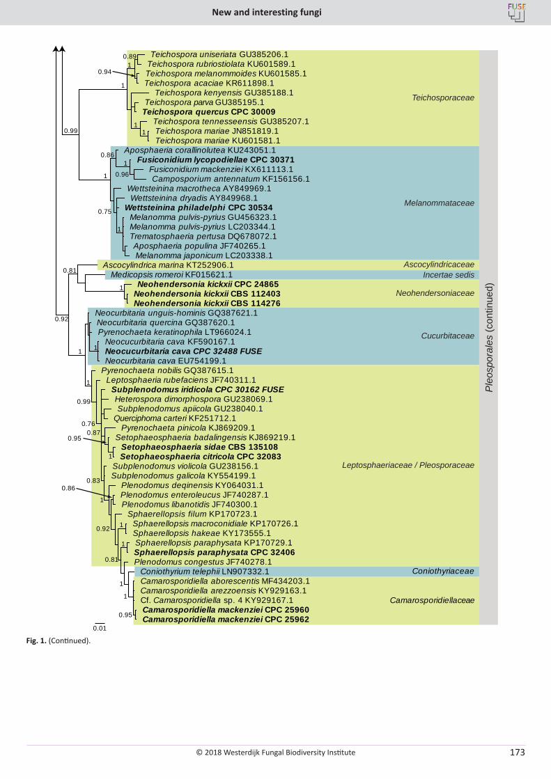
© 2018 Westerdijk Fungal Biodiversity Institute
New and interesting fungi
Editor-in-ChiefProf. dr P.W. Crous, Westerdijk Fungal Biodiversity Institute, P.O. Box 85167, 3508 AD Utrecht, The Netherlands.E-mail:[email protected]
173
0.01
0.96
1 0.86
1
0.75
1
1 1
0.89
0.94 1
1
0.99
0.95
1
1
1
0.81
1 0.92
0.86
1
0.83
1
0.95 0.87
0.76
0.99
1
1 1
1
0.81
0.92
Aposphaeria corallinolutea KU243051.1 Fusiconidium lycopodiellae CPC 30371
Fusiconidium mackenziei KX611113.1 Camposporium antennatum KF156156.1
Wettsteinina dryadis AY849968.1
Melanomma pulvis-pyrius GU456323.1
Aposphaeria populina JF740265.1 Melanomma japonicum LC203338.1
Melanomma pulvis-pyrius LC203344.1 Trematosphaeria pertusa DQ678072.1
Wettsteinina philadelphi CPC 30534
Wettsteinina macrotheca AY849969.1
Teichospora kenyensis GU385188.1 Teichospora parva GU385195.1
Teichospora quercus CPC 30009 Teichospora tennesseensis GU385207.1 Teichospora mariae JN851819.1 Teichospora mariae KU601581.1
Teichospora uniseriata GU385206.1 Teichospora rubriostiolata KU601589.1 Teichospora melanommoides KU601585.1 Teichospora acaciae KR611898.1
Camarosporidiella aborescentis MF434203.1 Camarosporidiella arezzoensis KY929163.1 Cf. Camarosporidiella sp. 4 KY929167.1 Camarosporidiella mackenziei CPC 25960 Camarosporidiella mackenziei CPC 25962
Coniothyrium telephii LN907332.1 Plenodomus congestus JF740278.1
Sphaerellopsis paraphysata KP170729.1 Sphaerellopsis paraphysata CPC 32406
Sphaerellopsis macroconidiale KP170726.1 Sphaerellopsis hakeae KY173555.1
Sphaerellopsis filum KP170723.1
Plenodomus enteroleucus JF740287.1 Plenodomus libanotidis JF740300.1
Plenodomus deqinensis KY064031.1
Subplenodomus violicola GU238156.1 Subplenodomus galicola KY554199.1
Setophaeosphaeria sidae CBS 135108 Setophaeosphaeria citricola CPC 32083
Setophaeosphaeria badalingensis KJ869219.1 Pyrenochaeta pinicola KJ869209.1
Subplenodomus apiicola GU238040.1 Querciphoma carteri KF251712.1
Subplenodomus iridicola CPC 30162 FUSE Heterospora dimorphospora GU238069.1
Leptosphaeria rubefaciens JF740311.1 Pyrenochaeta nobilis GQ387615.1
Neocurbitaria quercina GQ387620.1 Pyrenochaeta keratinophila LT966024.1
Neocucurbitaria cava KF590167.1 Neocucurbitaria cava CPC 32488 FUSE Neocurbitaria cava EU754199.1
Neocurbitaria unguis-hominis GQ387621.1
Ascocylindrica marina KT252906.1 Medicopsis romeroi KF015621.1
Neohendersonia kickxii CBS 112403 Neohendersonia kickxii CBS 114276
Neohendersonia kickxii CPC 24865
Ple
ospo
rale
s (c
ontin
ued)
Teichosporaceae
Melanommataceae
AscocylindricaceaeIncertae sedis
Neohendersoniaceae
Cucurbitaceae
Camarosporidiellaceae
Coniothyriaceae
Leptosphaeriaceae / Pleosporaceae
Fig. 1. (Continued).
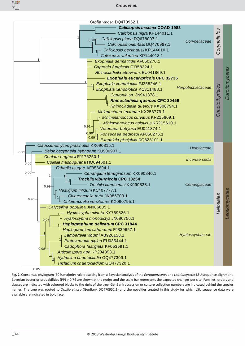
© 2018 Westerdijk Fungal Biodiversity Institute
Crous et al.
Editor-in-ChiefProf. dr P.W. Crous, Westerdijk Fungal Biodiversity Institute, P.O. Box 85167, 3508 AD Utrecht, The Netherlands.E-mail:[email protected]
174
Helotiaceae
Incertae sedis
Cenangiaceae
Hyaloscyphaceae
Hel
otia
les
Coryneliaceae
Cha
etot
hyria
les
Herpotrichiellaceae
Leot
iom
ycet
esE
urot
iom
ycet
es
Cor
ynel
iale
s 1
0.78
1
1
1
0.99 0.90
1
0.91
1
1
1
1
1
1 1
0.98
1
1
0.97
1
1 1
1
0.99
1 0.90
0.90
0.99
0.95
Orbilia vinosa DQ470952.1 Caliciopsis maxima COAD 1983 Caliciopsis nigra KP144011.1
Caliciopsis pinea DQ678097.1 Caliciopsis orientalis DQ470987.1
Caliciopsis beckhausii KP144010.1 Caliciopsis valentina KP144013.1
Exophiala dermatitidis AF050270.1 Capronia fungicola FJ358224.1
Fonsecaea pedrosoi AF050276.1 Exophiala pisciphila DQ823101.1
Veronaea botryosa EU041874.1
Minimelanolocus curvatus KR215609.1 Minimelanolocus asiaticus KR215610.1
Melanoctona tectonae KX258779.1
Rhinocladiella atrovirens EU041869.1
Exophiala xenobiotica FJ358246.1 Exophiala xenobiotica KC311483.1
Capronia sp. JN941378.1 Rhinocladiella quercus CPC 30459 Rhinocladiella quercus KX306794.1
Exophiala eucalypticola CPC 32736
Lambertella viburni AB926153.1 Protoventuria alpina EU035444.1 Cadophora fastigiata KF053591.1
Hydrocina chaetocladia GQ477309.1 Tricladium chaetocladium GQ477320.1
Articulospora atra KP234353.1
Hyaloscypha minuta KY769526.1 Hyaloscypha monodictys JN086756.1
Haplographium delicatum CPC 31844 Haplographium catenatum FJ839657.1
Calycellina populina JN086685.1
Trochila viburnicola CPC 30254 Trochila laurocerasi KX090835.1
Cenangium ferruginosum KX090840.1
Chlorencoelia torta JN086703.1 Chlorencoelia versiformis KX090795.1
Vestigium trifidum KC407777.1
Fabrella tsugae AF356694.1
Chalara hughesii FJ176250.1 Colipila masduguana HQ694501.1
Belonioscyphella hypnorum KU900907.1 Claussenomyces prasinulus KX090815.1
0.05
Fig. 2. Consensus phylogram (50 % majority rule) resulting from a Bayesian analysis of the Eurotiomycetes and Leotiomycetes LSU sequence alignment. Bayesian posterior probabilities (PP) > 0.74 are shown at the nodes and the scale bar represents the expected changes per site. Families, orders and classes are indicated with coloured blocks to the right of the tree. GenBank accession or culture collection numbers are indicated behind the species names. The tree was rooted to Orbilia vinosa (GenBank DQ470952.1) and the novelties treated in this study for which LSU sequence data were available are indicated in bold face.
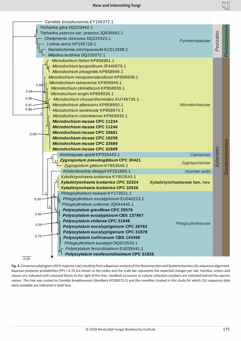
© 2018 Westerdijk Fungal Biodiversity Institute
New and interesting fungi
Editor-in-ChiefProf. dr P.W. Crous, Westerdijk Fungal Biodiversity Institute, P.O. Box 85167, 3508 AD Utrecht, The Netherlands.E-mail:[email protected]
175
Sor
dario
myc
etes
Pez
izom
ycet
es
Pez
izal
es
Pyronemataceae
Microdochiaceae
Xyl
aria
les
Zygosporiaceae
Xyladictyochaetaceae fam. nov.
Incertae sedis
Cainiaceae
Phlogicylindriaceae
0.05
1
0.75
0.90
0.99
1 0.95
1
1
1
1
1 1
1
1 1
0.86 0.97
0.98
1
0.98
0.98
0.98
1
1
1
Candida broadrunensis KY106372.1
Polyscytalum chilense CPC 31946 Polyscytalum eucalyptigenum CPC 28762
Polyscytalum nullicanum CBS 143406 Phlogicylindrium eucalypti DQ923534.1
Polyscytalum eucalyptigenum CPC 31878
Polyscytalum fecundissimum EU035441.1 Polyscytalum neofecundissimum CPC 31826
Polyscytalum grevilleae CPC 25576 Polyscytalum eucalyptorum CBS 137967
Phlogicylindrium eucalyptorum EU040223.1 Phlogicylindrium uniforme JQ044445.1
Phlogicylindrium mokarei KY173521.1
Xyladictyochaeta lusitanica KY853543.1 Xyladictyochaeta lusitanica CPC 32324 Xyladictyochaeta lusitanica CPC 32526
Kirstenboschia diospyri KF251660.1
Atrotorquata spartii KP325443.1 Zygosporium pseudogibbum CPC 30421
Zygosporium gibbum KY853546.1
Microdochium lycopodinum JF440979.1 Microdochium phragmitis KP858949.1
Microdochium fisheri KP858951.1
Microdochium tainanense KP858945.1
Microdochium albescens KP858950.1 Microdochium seminicola KP858974.1
Microdochium chrysanthemoides KU746735.1
Microdochium citrinidiscum KP858939.1 Microdochium sorghi KP858936.1
Microdochium colombiense KP858935.1 Microdochium musae CPC 11234 Microdochium musae CPC 11240 Microdochium musae CPC 32681 Microdochium musae CPC 16258 Microdochium musae CPC 32689 Microdochium musae CPC 32809
Microdochium neoqueenslandicum KP858938.1
Cheilymenia stercorea DQ220323.1 Lotinia verna KP195728.1 Ramsbottomia crechqueraultii KC012698.1 Miladina lecithina DQ220372.1
Tricharina gilva DQ220442.1 Tricharina praecox var. praecox JQ836562.1
Fig. 3. Consensus phylogram (50 % majority rule) resulting from a Bayesian analysis of the Pezizomycetes and Sordariomycetes LSU sequence alignment. Bayesian posterior probabilities (PP) > 0.74 are shown at the nodes and the scale bar represents the expected changes per site. Families, orders and classes are indicated with coloured blocks to the right of the tree. GenBank accession or culture collection numbers are indicated behind the species names. The tree was rooted to Candida broadrunensis (GenBank KY106372.1) and the novelties treated in this study for which LSU sequence data were available are indicated in bold face.
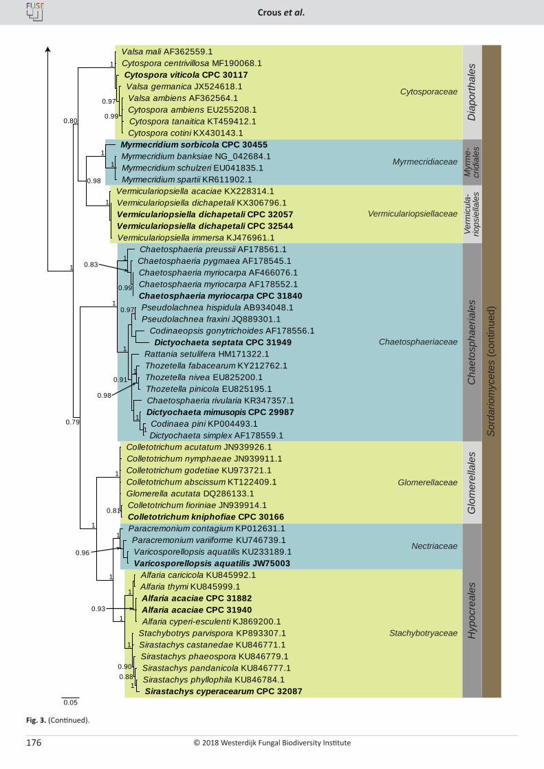
© 2018 Westerdijk Fungal Biodiversity Institute
Crous et al.
Editor-in-ChiefProf. dr P.W. Crous, Westerdijk Fungal Biodiversity Institute, P.O. Box 85167, 3508 AD Utrecht, The Netherlands.E-mail:[email protected]
176
Sor
dario
myc
etes
(con
tinue
d)
Dia
porth
ales
Cytosporaceae
Myr
me-
crid
iale
s
Myrmecridiaceae
Vermiculariopsiellaceae
Verm
icul
a-rio
psie
llale
s
Chaetosphaeriaceae
Cha
etos
phae
riale
sGlomerellaceae
Glo
mer
ella
les
Nectriaceae
Hyp
ocre
ales
Stachybotryaceae
0.05
1
1
1
0.98
0.99
0.97
1
0.80
0.97
1
1
0.98
1 0.91
1
0.99
0.83 1
1
0.93
1
1 0.88 0.90
1
1
0.96
1
1
0.81
1
1
0.79
1
Myrmecridium banksiae NG_042684.1 Myrmecridium schulzeri EU041835.1 Myrmecridium spartii KR611902.1
Myrmecridium sorbicola CPC 30455
Vermiculariopsiella acaciae KX228314.1 Vermiculariopsiella dichapetali KX306796.1 Vermiculariopsiella dichapetali CPC 32057 Vermiculariopsiella dichapetali CPC 32544 Vermiculariopsiella immersa KJ476961.1
Valsa ambiens AF362564.1 Cytospora ambiens EU255208.1 Cytospora tanaitica KT459412.1 Cytospora cotini KX430143.1
Valsa germanica JX524618.1 Cytospora viticola CPC 30117
Valsa mali AF362559.1 Cytospora centrivillosa MF190068.1
Pseudolachnea hispidula AB934048.1 Pseudolachnea fraxini JQ889301.1
Codinaeopsis gonytrichoides AF178556.1 Dictyochaeta septata CPC 31949
Codinaea pini KP004493.1 Dictyochaeta simplex AF178559.1
Dictyochaeta mimusopis CPC 29987 Chaetosphaeria rivularia KR347357.1
Thozetella fabacearum KY212762.1 Thozetella nivea EU825200.1 Thozetella pinicola EU825195.1
Rattania setulifera HM171322.1
Chaetosphaeria myriocarpa AF466076.1 Chaetosphaeria myriocarpa AF178552.1 Chaetosphaeria myriocarpa CPC 31840
Chaetosphaeria pygmaea AF178545.1 Chaetosphaeria preussii AF178561.1
Alfaria acaciae CPC 31882 Alfaria acaciae CPC 31940 Alfaria cyperi-esculenti KJ869200.1
Alfaria caricicola KU845992.1 Alfaria thymi KU845999.1
Stachybotrys parvispora KP893307.1 Sirastachys castanedae KU846771.1
Sirastachys pandanicola KU846777.1 Sirastachys phyllophila KU846784.1 Sirastachys cyperacearum CPC 32087
Sirastachys phaeospora KU846779.1
Paracremonium contagium KP012631.1 Paracremonium variiforme KU746739.1 Varicosporellopsis aquatilis KU233189.1 Varicosporellopsis aquatilis JW75003
Colletotrichum acutatum JN939926.1
Colletotrichum fioriniae JN939914.1 Colletotrichum kniphofiae CPC 30166
Colletotrichum nymphaeae JN939911.1 Colletotrichum godetiae KU973721.1 Colletotrichum abscissum KT122409.1 Glomerella acutata DQ286133.1
Fig. 3. (Continued).
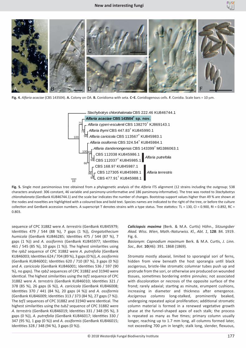
© 2018 Westerdijk Fungal Biodiversity Institute
New and interesting fungi
Editor-in-ChiefProf. dr P.W. Crous, Westerdijk Fungal Biodiversity Institute, P.O. Box 85167, 3508 AD Utrecht, The Netherlands.E-mail:[email protected]
177
sequence of CPC 31882 were A. terrestris (GenBank KU845979; Identities 479 / 544 (88 %), 7 gaps (1 %)), Gregatothecium humicola (GenBank KU846285; Identities 475 / 544 (87 %), 7 gaps (1 %)) and A. ossiformis (GenBank KU845977; Identities 461 / 545 (85 %), 10 gaps (1 %)). The highest similarities using the rpb2 sequence of CPC 31882 were A. putrefolia (GenBank KU846003; Identities 624 / 704 (89 %), 3 gaps (0 %)), A. ossiformis (GenBank KU846002; Identities 620 / 710 (87 %), 3 gaps (0 %)) and A. caricicola (GenBank KU846001; Identities 536 / 597 (90 %), no gaps). The rpb2 sequences of CPC 31882 and 31940 were identical. The highest similarities using the tef1 sequence of CPC 31882 were A. terrestris (GenBank KU846010; Identities 321 / 378 (85 %), 26 gaps (6 %)), A. caricicola (GenBank KU846008; Identities 370 / 441 (84 %), 20 gaps (4 %)) and A. ossiformis (GenBank KU846009; Identities 313 / 373 (84 %), 27 gaps (7 %)). The tef1 sequences of CPC 31882 and 31940 were identical. The highest similarities using the tub2 sequence of CPC 31882 were A. terrestris (GenBank KU846019; Identities 331 / 348 (95 %), 3 gaps (0 %)), A. putrefolia (GenBank KU846017; Identities 330 / 347 (95 %), 1 gap (0 %)) and A. ossiformis (GenBank KU846015; Identities 328 / 348 (94 %), 3 gaps (0 %)).
Caliciopsis maxima (Berk. & M.A. Curtis) Höhn., Sitzungsber Akad. Wiss. Wien, Math.-Naturwiss. Kl., Abt. 1, 128: 84. 1919. Fig. 6.Basionym: Capnodium maximum Berk. & M.A. Curtis, J. Linn. Soc., Bot. 10(46): 391. 1868 (1869).
Stromata mostly abaxial, limited to sporangial sori of ferns, hidden from view beneath the host sporangia until black ascigerous, bristle-like stromatic columnar tubes push up and protrude from the sori, or otherwise are produced on wounded tissues, sometimes bordering entire pinnules; not associated with discoloration or necrosis of the opposite surface of the frond; rarely adaxial; starting as minute, erumpent cushions, increasing in diameter and thickness after emergence. Ascigerous columns long-stalked, prominently beaked, undergoing repeated apical proliferation; additional stromatic column material is formed in a renewed vegetative growth phase at the funnel-shaped apex of each stalk; the process is repeated as many as five times; primary column usually longer, reaching up to 1.7 mm long, all columns formed later, not exceeding 700 µm in length; stalk long, slender, flexuous,
Fig. 4. Alfaria acaciae (CBS 143504). A. Colony on OA. B. Conidioma with seta. C–E. Conidiogenous cells. F. Conidia. Scale bars = 10 mm.
Alfaria dandenongensis CBS 143399T MG386063.1 CBS 112038 KU845986.1 CBS 112037T KU845985.1 CBS 168.97 KU845987.1
CBS 127305 KU845989.1 CBS 477.91T KU845988.1
Alfaria ossiformis CBS 324.54T KU845984.1
Alfaria cyperi-esculenti CBS 138270T KJ869143.1 Alfaria thymi CBS 447.83T KU845990.1 Alfaria caricicola CBS 113567T KU845983.1
Alfaria acaciae CBS 143504T sp. nov. Stachybotrys chlorohalonata CBS 222.46 KU846744.1
10
Alfaria putrefolia
Alfaria terrestris
100
100
71
7998
99
9694
66
83
Fig. 5. Single most parsimonious tree obtained from a phylogenetic analysis of the Alfaria ITS alignment (12 strains including the outgroup; 538 characters analysed: 306 constant, 46 variable and parsimony-uninformative and 186 parsimony-informative). The tree was rooted to Stachybotrys chlorohalonata (GenBank KU846744.1) and the scale bar indicates the number of changes. Bootstrap support values higher than 49 % are shown at the nodes and novelties are highlighted with a coloured box and bold text. Species names are indicated to the right of the tree, or before the culture collection and GenBank accession numbers. A superscript T denotes strains with a type status. Tree statistics: TL = 130, CI = 0.900, RI = 0.892, RC = 0.803.
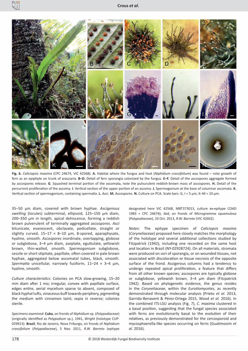
© 2018 Westerdijk Fungal Biodiversity Institute
Crous et al.
Editor-in-ChiefProf. dr P.W. Crous, Westerdijk Fungal Biodiversity Institute, P.O. Box 85167, 3508 AD Utrecht, The Netherlands.E-mail:[email protected]
178
35–50 µm diam, covered with brown hyphae. Ascigenous swelling (locules) subterminal, ellipsoid, 125–150 µm diam, 200–350 µm in length, apical dehiscence, forming a reddish brown pulverulent of terminally aggregated ascospores. Asci bitunicate, evanescent, obclavate, pedicellate, straight or slightly curved, 15–17 × 8–10 µm, 8-spored, aparaphysate, hyaline, smooth. Ascospores inordinate, overlapping, globose or subglobose, 3–4 µm diam, aseptate, eguttulate, yellowish brown, thin-walled, smooth. Spermogonium subglobose, sessile or short stipitate, papillate, often covered in pale brown hyphae, aggregated below ascomatal tubes, black, smooth. Spermatia unicellular, narrowly fusiform, 11–24 × 3–4 µm, hyaline, smooth.
Culture characteristics: Colonies on PCA slow-growing, 15–20 mm diam after 1 mo; irregular, convex with papillate surface, edges entire, aerial mycelium sparse to absent, composed of black hyphal tufts, vinaceous buff towards periphery, pigmenting the medium with cinnamon taint; sepia in reverse; colonies sterile.
Specimens examined: Cuba, on fronds of Niphidium sp. (Polypodiaceae) (originally identified as Polypodium sp.), 1941, Wright (holotype CUP-029913). Brazil, Rio de Janeiro, Nova Friburgo, on fronds of Niphidium crassifolium (Polypodiaceae), 5 Nov. 2011, R.W. Barreto (epitype
designated here VIC 42568, MBT373013, culture ex-epitype COAD 1983 = CPC 24674); ibid, on fronds of Microgramma squamulosa (Polypodiaceae), 10 Oct. 2013, R.W. Barreto (VIC 42602).
Notes: The epitype specimen of Caliciopsis maxima (Coryneliaceae) proposed here closely matches the morphology of the holotype and several additional collections studied by Fitzpatrick (1942), including one recorded on the same host and location in Brazil (NY-02928724). On all materials, stromata were produced on sori of sporangia, or on wounded tissues, not associated with discoloration or tissue necrosis of the opposite surface of the frond. Ascigerous columns had a tendency to undergo repeated apical proliferation, a feature that differs from all other known species; ascospores are typically globose or subglobose, yellowish brown, 3–4 µm diam (Fitzpatrick 1942). Based on phylogenetic evidence, the genus resides in the Coryneliaceae, within the Eurotiomycetes, as recently demonstrated through molecular analysis (Prieto et al. 2013, Garrido-Benavent & Pérez-Ortega 2015, Wood et al. 2016). In the combined ITS-LSU analysis (Fig. 7), C. maxima clustered in a basal position, suggesting that the fungal species associated with ferns are evolutionarily basal to the evolution of their relatives, as previously demonstrated for the cercosporoid and mycosphaerella-like species occurring on ferns (Guatimosim et al. 2016).
Fig. 6. Caliciopsis maxima (CPC 24674, VIC 42568). A. Habitat where the fungus and host (Niphidium crassifolium) was found – note growth of fern as an epiphyte on trunk of araucaria. B–D. Detail of fern sporangia colonized by the fungus. E–F. Detail of the ascospores aggregate formed by ascospores release. G. Squashed terminal portion of the ascomata, note the pulvurulent reddish-brown mass of ascospores. H. Detail of the percurrent proliferation of the ascoma. I. Vertical section of the upper portion of an ascoma. J. Spermogonium at the base of columnar ascomata. K. Vertical section of spermogonium, containing spermatia. L. Asci. M. Ascospores. N. Culture on PCA. Scale bars: G, I = 5 µm, K–M = 10 µm.
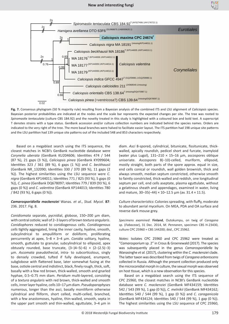
© 2018 Westerdijk Fungal Biodiversity Institute
New and interesting fungi
Editor-in-ChiefProf. dr P.W. Crous, Westerdijk Fungal Biodiversity Institute, P.O. Box 85167, 3508 AD Utrecht, The Netherlands.E-mail:[email protected]
179
Based on a megablast search using the ITS sequence, the closest matches in NCBI’s GenBank nucleotide database were Corynelia uberata (GenBank KU204606; Identities 474 / 544 (87 %), 21 gaps (3 %)), Caliciopsis pinea (GenBank KY099604; Identities 323 / 361 (89 %), 6 gaps (1 %)) and C. beckhausii (GenBank NR_132090; Identities 330 / 370 (89 %), 11 gaps (2 %)). The highest similarities using the LSU sequence were C. nigra (GenBank KP144011; Identities 771 / 825 (93 %), 5 gaps (0 %)), C. pinea (GenBank DQ678097; Identities 779 / 839 (93 %), 6 gaps (0 %)) and C. valentina (GenBank KP144013; Identities 780 / 842 (93 %), 6 gaps (0 %)).
Camarosporidiella mackenziei Wanas. et al., Stud. Mycol. 87: 236. 2017. Fig. 8.
Conidiomata separate, pycnidial, globose, 150–200 µm diam, with central ostiole; wall of 2–3 layers of brown textura angularis. Conidiophores reduced to conidiogenous cells. Conidiogenous cells tightly aggregated, lining the inner cavity, hyaline, smooth, subcylindrical to ampulliform or doliiform, proliferating percurrently at apex, 5–8 × 3–4 µm. Conidia solitary, hyaline, smooth, guttulate to granular, subcylindrical to ellipsoid, apex obtusely rounded, base truncate, (3–)4–5(–6) × (2–)2.5(–3) µm. Ascomata pseudothecial, intra- to subcorticolous, singly to densely crowded, tufted if fully developed, erumpent, subglobose with flattened base, later somewhat fusing at the base, ostiole central and indistinct, black, finely rough, thick, soft, basally with a few red brown, thick-walled, smooth and gnarled hyphae, 0.5–0.75 mm diam. Peridium multi-layered, consisting of a textura angularis with red brown, thick-walled and smooth cells, inner layer hyaline, cells 10–17 µm diam. Pseudoparaphyses numerous, longer than the asci, basally moniliform otherwise cylindrical and filiform, short celled, multi-celled, branched, with a few anastomoses, hyaline, thin-walled, smooth, septa in the upper part smooth and thin-walled, eguttulate, 3–4 µm in
diam. Asci 8-spored, cylindrical, bitunicate, fissitunicate, thick-walled, apically roundish, pedicel short and furcate, inamyloid (water plus Lugol), 131–210 × 15–16 µm, ascospores oblique uniseriate. Ascospores 8(–10)-celled, muriform, ellipsoid, mostly straight, both parts of the spore approx. equal in size, end cells conical or roundish, wall golden brownish, thick and always smooth, median septum constricted, otherwise smooth to faintly constricted, thick-walled and reddish, one longitudinal septum per cell, end cells aseptate, plasma eguttulate, without a gelatinous sheath and appendages, examined in water, living and mature, 30–35(–44) × 10–12.5 µm (av. 31.4 × 11.5).
Culture characteristics: Colonies spreading, with fluffy, moderate to abundant aerial mycelium. On MEA, PDA and OA surface and reverse dark mouse grey.
Specimens examined: Finland, Outokumpu, on twig of Caragana sp. (Fabaceae), 31 Dec. 2014, M. Pennanen, specimen CBS H-23430, culture CPC 25960 = CBS 144200; ibid., CPC 25962.
Notes: Isolates CPC 25960 and CPC 25962 were treated as “Camarosporium sp. 2” in Crous & Groenewald (2017). The species was subsequently placed in the genus Camarosporidiella by Wanasinghe et al. (2017), clustering within the C. mackenziei clade. The latter taxon was described from twigs of Caragana arborescens collected in Russia. Although the present collection produced only the microconidial morph in culture, the sexual morph was observed on host tissue, which is a new observation for this species.
Based on a megablast search using the ITS sequence of CPC 25960, the closest matches in NCBI’s GenBank nucleotide database were C. mackenziei (GenBank MF434159; Identities 542 / 543 (99 %), 1 gap (0 %)), C. melnikii (GenBank MF434162; Identities 540 / 544 (99 %), 1 gap (0 %)) and C. caraganicola (GenBank MF434124; Identities 540 / 544 (99 %), 1 gap (0 %)). The highest similarities using the LSU sequence of CPC 25960,
1
0.89
0.66
0.83
0.76 0.99
1
Spiromastix tentaculata CBS 184.92T (AY527406.1/AY176722.1)
Hamigera avellanea DTO 63F6 (GU968675.1/AB000620.1)
Caliciopsis maxima CPC 24674T
Caliciopsis nigra MA 18191 (missing/KP144011.1)
Caliciopsis beckhausii MA 18186T (KP144005.1/KP144010.1)
MA 18176T (KP144006.1/KP144012.1)
MA 18177 (KP144009.1/KP144014.1)
MA 18179 (KP144008.1/KP144013.1)
Caliciopsis indica GFCC 4947T (GQ259981.1/GQ259980.1)
Caliciopsis calicioides 211 (JX968549.1/missing)
Caliciopsis orientalis CBS 138.64T (missing/DQ470987.1)
Caliciopsis pinea (=ventricosa?) CBS 139.64 (missing/DQ678097.1)
2x
2x
Coryneliales
Eurotiales
2x
2x
0.02
Caliciopsis valentina
Fig. 7. Consensus phylogram (50 % majority rule) resulting from a Bayesian analysis of the combined ITS and LSU alignment of Caliciopsis species. Bayesian posterior probabilities are indicated at the nodes and the scale bar represents the expected changes per site. The tree was rooted to Spiromastix tentaculata (culture CBS 184.92) and the novelty treated in this study is highlighted with a coloured box and bold text. A superscript T denotes strains with a type status. GenBank accession and/or culture collection numbers are indicated behind the species names. Orders are indicated to the very right of the tree. The more basal branches were halved to facilitate easier layout. The ITS partition had 198 unique site patterns and the LSU partition had 139 unique site patterns out of the included 548 and 853 characters respectively.
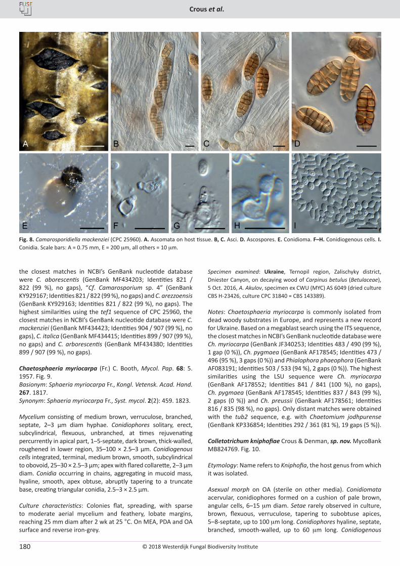
© 2018 Westerdijk Fungal Biodiversity Institute
Crous et al.
Editor-in-ChiefProf. dr P.W. Crous, Westerdijk Fungal Biodiversity Institute, P.O. Box 85167, 3508 AD Utrecht, The Netherlands.E-mail:[email protected]
180
the closest matches in NCBI’s GenBank nucleotide database were C. aborescentis (GenBank MF434203; Identities 821 / 822 (99 %), no gaps), “Cf. Camarosporium sp. 4” (GenBank KY929167; Identities 821 / 822 (99 %), no gaps) and C. arezzoensis (GenBank KY929163; Identities 821 / 822 (99 %), no gaps). The highest similarities using the tef1 sequence of CPC 25960, the closest matches in NCBI’s GenBank nucleotide database were C. mackenziei (GenBank MF434423; Identities 904 / 907 (99 %), no gaps), C. italica (GenBank MF434415; Identities 899 / 907 (99 %), no gaps) and C. arborescentis (GenBank MF434380; Identities 899 / 907 (99 %), no gaps).
Chaetosphaeria myriocarpa (Fr.) C. Booth, Mycol. Pap. 68: 5. 1957. Fig. 9.Basionym: Sphaeria myriocarpa Fr., Kongl. Vetensk. Acad. Hand. 267. 1817.Synonym: Sphaeria myriocarpa Fr., Syst. mycol. 2(2): 459. 1823.
Mycelium consisting of medium brown, verruculose, branched, septate, 2–3 µm diam hyphae. Conidiophores solitary, erect, subcylindrical, flexuous, unbranched, at times rejuvenating percurrently in apical part, 1–5-septate, dark brown, thick-walled, roughened in lower region, 35–100 × 2.5–3 µm. Conidiogenous cells integrated, terminal, medium brown, smooth, subcylindrical to obovoid, 25–30 × 2.5–3 µm; apex with flared collarette, 2–3 µm diam. Conidia occurring in chains, aggregating in mucoid mass, hyaline, smooth, apex obtuse, abruptly tapering to a truncate base, creating triangular conidia, 2.5–3 × 2.5 µm.
Culture characteristics: Colonies flat, spreading, with sparse to moderate aerial mycelium and feathery, lobate margins, reaching 25 mm diam after 2 wk at 25 °C. On MEA, PDA and OA surface and reverse iron-grey.
Specimen examined: Ukraine, Ternopil region, Zalischyky district, Dniester Canyon, on decaying wood of Carpinus betulus (Betulaceae), 5 Oct. 2016, A. Akulov, specimen ex CWU (MYC) AS 6049 (dried culture CBS H-23426, culture CPC 31840 = CBS 143389).
Notes: Chaetosphaeria myriocarpa is commonly isolated from dead woody substrates in Europe, and represents a new record for Ukraine. Based on a megablast search using the ITS sequence, the closest matches in NCBI’s GenBank nucleotide database were Ch. myriocarpa (GenBank JF340253; Identities 483 / 490 (99 %), 1 gap (0 %)), Ch. pygmaea (GenBank AF178545; Identities 473 / 496 (95 %), 3 gaps (0 %)) and Phialophora phaeophora (GenBank AF083191; Identities 503 / 533 (94 %), 2 gaps (0 %)). The highest similarities using the LSU sequence were Ch. myriocarpa (GenBank AF178552; Identities 841 / 841 (100 %), no gaps), Ch. pygmaea (GenBank AF178545; Identities 837 / 843 (99 %), 2 gaps (0 %)) and Ch. preussii (GenBank AF178561; Identities 816 / 835 (98 %), no gaps). Only distant matches were obtained with the tub2 sequence, e.g. with Chaetomium jodhpurense (GenBank KP336854; Identities 292 / 361 (81 %), 19 gaps (5 %)).
Colletotrichum kniphofiae Crous & Denman, sp. nov. MycoBank MB824769. Fig. 10.
Etymology: Name refers to Kniphofia, the host genus from which it was isolated.
Asexual morph on OA (sterile on other media). Conidiomata acervular, conidiophores formed on a cushion of pale brown, angular cells, 6–15 mm diam. Setae rarely observed in culture, brown, flexuous, verruculose, tapering to subobtuse apices, 5–8-septate, up to 100 mm long. Conidiophores hyaline, septate, branched, smooth-walled, up to 60 mm long. Conidiogenous
Fig. 8. Camarosporidiella mackenziei (CPC 25960). A. Ascomata on host tissue. B, C. Asci. D. Ascospores. E. Conidioma. F–H. Conidiogenous cells. I. Conidia. Scale bars: A = 0.75 mm, E = 200 mm, all others = 10 mm.
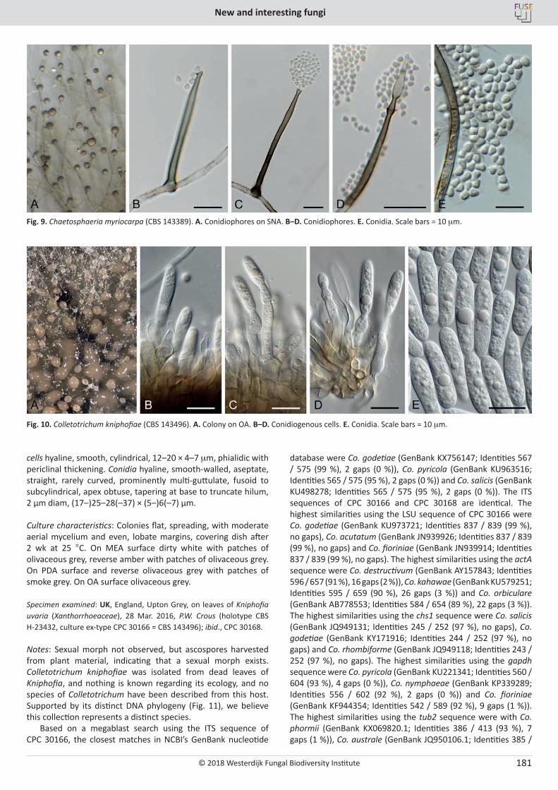
© 2018 Westerdijk Fungal Biodiversity Institute
New and interesting fungi
Editor-in-ChiefProf. dr P.W. Crous, Westerdijk Fungal Biodiversity Institute, P.O. Box 85167, 3508 AD Utrecht, The Netherlands.E-mail:[email protected]
181
cells hyaline, smooth, cylindrical, 12–20 × 4–7 mm, phialidic with periclinal thickening. Conidia hyaline, smooth-walled, aseptate, straight, rarely curved, prominently multi-guttulate, fusoid to subcylindrical, apex obtuse, tapering at base to truncate hilum, 2 µm diam, (17–)25–28(–37) × (5–)6(–7) µm.
Culture characteristics: Colonies flat, spreading, with moderate aerial mycelium and even, lobate margins, covering dish after 2 wk at 25 °C. On MEA surface dirty white with patches of olivaceous grey, reverse amber with patches of olivaceous grey. On PDA surface and reverse olivaceous grey with patches of smoke grey. On OA surface olivaceous grey.
Specimen examined: UK, England, Upton Grey, on leaves of Kniphofia uvaria (Xanthorrhoeaceae), 28 Mar. 2016, P.W. Crous (holotype CBS H-23432, culture ex-type CPC 30166 = CBS 143496); ibid., CPC 30168.
Notes: Sexual morph not observed, but ascospores harvested from plant material, indicating that a sexual morph exists. Colletotrichum kniphofiae was isolated from dead leaves of Kniphofia, and nothing is known regarding its ecology, and no species of Colletotrichum have been described from this host. Supported by its distinct DNA phylogeny (Fig. 11), we believe this collection represents a distinct species.
Based on a megablast search using the ITS sequence of CPC 30166, the closest matches in NCBI’s GenBank nucleotide
database were Co. godetiae (GenBank KX756147; Identities 567 / 575 (99 %), 2 gaps (0 %)), Co. pyricola (GenBank KU963516; Identities 565 / 575 (95 %), 2 gaps (0 %)) and Co. salicis (GenBank KU498278; Identities 565 / 575 (95 %), 2 gaps (0 %)). The ITS sequences of CPC 30166 and CPC 30168 are identical. The highest similarities using the LSU sequence of CPC 30166 were Co. godetiae (GenBank KU973721; Identities 837 / 839 (99 %), no gaps), Co. acutatum (GenBank JN939926; Identities 837 / 839 (99 %), no gaps) and Co. fioriniae (GenBank JN939914; Identities 837 / 839 (99 %), no gaps). The highest similarities using the actA sequence were Co. destructivum (GenBank AY157843; Identities 596 / 657 (91 %), 16 gaps (2 %)), Co. kahawae (GenBank KU579251; Identities 595 / 659 (90 %), 26 gaps (3 %)) and Co. orbiculare (GenBank AB778553; Identities 584 / 654 (89 %), 22 gaps (3 %)). The highest similarities using the chs1 sequence were Co. salicis (GenBank JQ949131; Identities 245 / 252 (97 %), no gaps), Co. godetiae (GenBank KY171916; Identities 244 / 252 (97 %), no gaps) and Co. rhombiforme (GenBank JQ949118; Identities 243 / 252 (97 %), no gaps). The highest similarities using the gapdh sequence were Co. pyricola (GenBank KU221341; Identities 560 / 604 (93 %), 4 gaps (0 %)), Co. nymphaeae (GenBank KP339289; Identities 556 / 602 (92 %), 2 gaps (0 %)) and Co. fioriniae (GenBank KF944354; Identities 542 / 589 (92 %), 9 gaps (1 %)). The highest similarities using the tub2 sequence were with Co. phormii (GenBank KX069820.1; Identities 386 / 413 (93 %), 7 gaps (1 %)), Co. australe (GenBank JQ950106.1; Identities 385 /
Fig. 9. Chaetosphaeria myriocarpa (CBS 143389). A. Conidiophores on SNA. B–D. Conidiophores. E. Conidia. Scale bars = 10 mm.
Fig. 10. Colletotrichum kniphofiae (CBS 143496). A. Colony on OA. B–D. Conidiogenous cells. E. Conidia. Scale bars = 10 mm.
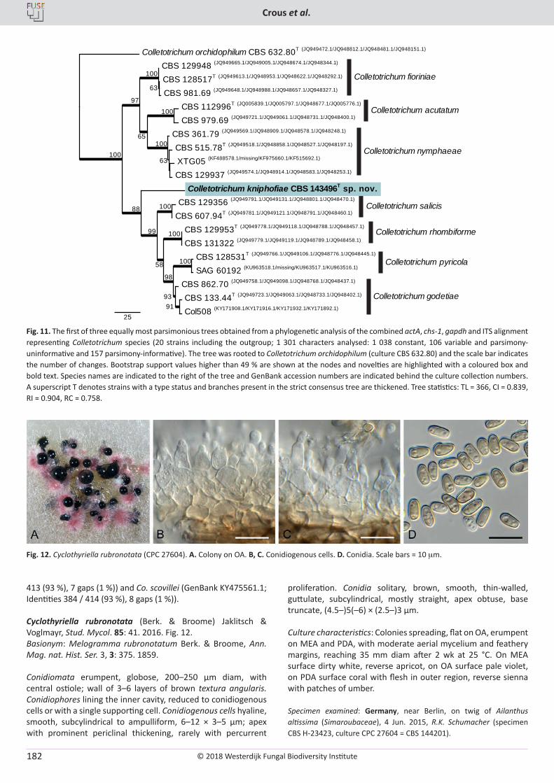
© 2018 Westerdijk Fungal Biodiversity Institute
Crous et al.
Editor-in-ChiefProf. dr P.W. Crous, Westerdijk Fungal Biodiversity Institute, P.O. Box 85167, 3508 AD Utrecht, The Netherlands.E-mail:[email protected]
182
413 (93 %), 7 gaps (1 %)) and Co. scovillei (GenBank KY475561.1; Identities 384 / 414 (93 %), 8 gaps (1 %)).
Cyclothyriella rubronotata (Berk. & Broome) Jaklitsch & Voglmayr, Stud. Mycol. 85: 41. 2016. Fig. 12.Basionym: Melogramma rubronotatum Berk. & Broome, Ann. Mag. nat. Hist. Ser. 3, 3: 375. 1859.
Conidiomata erumpent, globose, 200–250 µm diam, with central ostiole; wall of 3–6 layers of brown textura angularis. Conidiophores lining the inner cavity, reduced to conidiogenous cells or with a single supporting cell. Conidiogenous cells hyaline, smooth, subcylindrical to ampulliform, 6–12 × 3–5 µm; apex with prominent periclinal thickening, rarely with percurrent
proliferation. Conidia solitary, brown, smooth, thin-walled, guttulate, subcylindrical, mostly straight, apex obtuse, base truncate, (4.5–)5(–6) × (2.5–)3 µm.
Culture characteristics: Colonies spreading, flat on OA, erumpent on MEA and PDA, with moderate aerial mycelium and feathery margins, reaching 35 mm diam after 2 wk at 25 °C. On MEA surface dirty white, reverse apricot, on OA surface pale violet, on PDA surface coral with flesh in outer region, reverse sienna with patches of umber.
Specimen examined: Germany, near Berlin, on twig of Ailanthus altissima (Simaroubaceae), 4 Jun. 2015, R.K. Schumacher (specimen CBS H-23423, culture CPC 27604 = CBS 144201).
CBS 112996T (JQ005839.1/JQ005797.1/JQ948677.1/JQ005776.1)
CBS 979.69 (JQ949721.1/JQ949061.1/JQ948731.1/JQ948400.1)
XTG05 (KF488578.1/missing/KF975660.1/KF515692.1)
CBS 129937 (JQ949574.1/JQ948914.1/JQ948583.1/JQ948253.1)
CBS 515.78T (JQ949518.1/JQ948858.1/JQ948527.1/JQ948197.1)
CBS 361.79 (JQ949569.1/JQ948909.1/JQ948578.1/JQ948248.1)
CBS 128517T (JQ949613.1/JQ948953.1/JQ948622.1/JQ948292.1)
CBS 981.69 (JQ949648.1/JQ948988.1/JQ948657.1/JQ948327.1)
CBS 129948 (JQ949665.1/JQ949005.1/JQ948674.1/JQ948344.1)
CBS 133.44T (JQ949723.1/JQ949063.1/JQ948733.1/JQ948402.1)
Col508 (KY171908.1/KY171916.1/KY171932.1/KY171892.1)
CBS 862.70 (JQ949758.1/JQ949098.1/JQ948768.1/JQ948437.1)
CBS 128531T (JQ949766.1/JQ949106.1/JQ948776.1/JQ948445.1)
SAG 60192 (KU963518.1/missing/KU963517.1/KU963516.1)
CBS 129953T (JQ949778.1/JQ949118.1/JQ948788.1/JQ948457.1)
CBS 131322 (JQ949779.1/JQ949119.1/JQ948789.1/JQ948458.1)
CBS 129356 (JQ949791.1/JQ949131.1/JQ948801.1/JQ948470.1)
CBS 607.94T (JQ949781.1/JQ949121.1/JQ948791.1/JQ948460.1)
Colletotrichum kniphofiae CBS 143496T sp. nov.
Colletotrichum orchidophilum CBS 632.80T (JQ949472.1/JQ948812.1/JQ948481.1/JQ948151.1)
25
100
97
100
63
100
65100
63
88
99
100
100
58
98
100
9391
Colletotrichum fioriniae
Colletotrichum acutatum
Colletotrichum nymphaeae
Colletotrichum salicis
Colletotrichum rhombiforme
Colletotrichum pyricola
Colletotrichum godetiae
Fig. 11. The first of three equally most parsimonious trees obtained from a phylogenetic analysis of the combined actA, chs-1, gapdh and ITS alignment representing Colletotrichum species (20 strains including the outgroup; 1 301 characters analysed: 1 038 constant, 106 variable and parsimony-uninformative and 157 parsimony-informative). The tree was rooted to Colletotrichum orchidophilum (culture CBS 632.80) and the scale bar indicates the number of changes. Bootstrap support values higher than 49 % are shown at the nodes and novelties are highlighted with a coloured box and bold text. Species names are indicated to the right of the tree and GenBank accession numbers are indicated behind the culture collection numbers. A superscript T denotes strains with a type status and branches present in the strict consensus tree are thickened. Tree statistics: TL = 366, CI = 0.839, RI = 0.904, RC = 0.758.
Fig. 12. Cyclothyriella rubronotata (CPC 27604). A. Colony on OA. B, C. Conidiogenous cells. D. Conidia. Scale bars = 10 mm.
© 2018 Westerdijk Fungal Biodiversity Institute
New and interesting fungi
Editor-in-ChiefProf. dr P.W. Crous, Westerdijk Fungal Biodiversity Institute, P.O. Box 85167, 3508 AD Utrecht, The Netherlands.E-mail:[email protected]
183
Notes: The genus Cyclothyriella was recently introduced by Jaklitsch & Voglmayr (2016), who also treated the taxonomic history of this genus in detail. The asexual morph isolated in this study closely resembles that described and illustrated by Jaklitsch & Voglmayr (2016), who reported conidia as (2–)4.5–6(–6.5) × (2–) 2.7–3.5(–4) μm, first hyaline, becoming medium brown with age. This fungus is common in Europe, and here we present a culture from Germany to supplement the Austrian material studied by Jaklitsch & Voglmayr (2016).
Based on a megablast search using the ITS sequence, the closest matches in NCBI’s GenBank nucleotide database were Cyclothyriella rubronotata (GenBank NR_147651; Identities 564 / 564 (100 %), no gaps), Melanomma pulvis-pyrius (GenBank KY189979; Identities 400 / 476 (84 %), 20 gaps (4 %)) and Ascochyta medicaginicola (GenBank KX381183; Identities 359 / 419 (86 %), 10 gaps (2 %)). The highest similarities using the LSU sequence were Cy. rubronotata (GenBank KX650541; Identities 833 / 833 (100 %), no gaps), Thyridaria rubronotata (GenBank JX681121; Identities 833 / 833 (100 %), no gaps) and Neooccultibambusa jonesii (GenBank KY111437; Identities 781 / 812 (96 %), 2 gaps (0 %)).
Cylindriaceae Crous & L. Lombard, fam. nov. MycoBank MB824770.
Mycelium consisting of hyaline, smooth, septate, branched, hyphae. Conidiophores aggregated in sporodochia, or solitary, erect; hyphae and basal part of conidiophores becoming pale brown, smooth, subcylindrical, erect, septate, branched. Conidiogenous cells terminal and intercalary, subcylindrical, hyaline, smooth, with several sympodial flat-tipped loci, unthickened, not darkened. Ramoconidia hyaline, smooth, guttulate, subcylindrical. Conidia aseptate, hyaline, smooth, arranged in long, branched chains, scars unthickened, slightly refractive.
Type genus: Cylindrium Bonord.
Note: The genus Cylindrium, based on C. elongatum, was regarded by Lombard et al. (2015) as incertae sedis, and thus Cylindriaceae is herewith introduced to accommodate this genus.
Cylindrium algarvense (Cheew. & Crous) Crous, comb. nov. MycoBank MB824771.Basionym: Polyscytalum algarvense Cheew. & Crous, Persoonia 23: 73. 2009.
Description and illustration: Cheewangkoon et al. (2009).
Specimen examined: Portugal, Faro, Algarve, on Eucalyptus sp. (Myrtaceae), 24 Jan. 2007, P.W. Crous (holotype CBS H-20289, culture ex-type CPC 14936 = CBS 124770); ibid. (CPC 14937, CPC 14938).
Note: See discussion under Polyscytalum and Fig. 13.
Cylindrium purgamentum (Crous) Crous, comb. nov. MycoBank MB824772.Basionym: Polyscytalum purgamentum Crous, Persoonia 37: 363. 2016.
Description and illustration: Crous et al. (2016a).
Specimen examined: USA, Texas, Austin, on leaf litter, Aug. 2013, P.W. Crous (holotype CBS H-22899, culture ex-type CPC 29580 = CBS 142114).
Note: See discussion under Polyscytalum and Fig. 13.
Cylindrium syzygii (Crous & R.G. Shivas) Crous, comb. nov. MycoBank MB824773.Basionym: Pseudoidriella syzygii Crous & R.G. Shivas, Persoonia 27: 135. 2011.
Description and illustration: Crous et al. (2011).
Specimen examined: Australia, Queensland, Mackay, Eungella National Park, on leaves of Syzygium sp. (Myrtaceae), 14 Jul. 2009, P.W. Crous & K.L. Crous, (holotype CBS H-20758, cultures ex-type CPC 17233 = CBS 131307).
Note: See discussion under Polyscytalum and Fig. 13. By placing Pseudoidriella syzygii in Cylindrium, the genus Pseudoidriella is also reduced to synonymy with Cylindrium. This suggests that the conidiomata of Cylindrium could be reduced to solitary conidiophores, as well as sporodochia, as observed in this species. Interestingly enough, an LSU sequence attributed to Tristratiperidium microsporum also clustered in this clade (Fig. 3), which completely disagrees with the morphology of this fungus. This suggests that this sequence (MFLUCC) should be reconsidered. The ITS sequence from the same culture placed Tristratiperidium microsporum with Kirstenboschia diospyri (Fig. 13).
Cytospora viticola D.P. Lawr. et al., Pl. Pathol. 66: 718. 2017. Fig. 14.
Conidiomata (on PDA) with stromata up to 500 mm diam, rosette cytosporoid, subdivided by invaginations, up to four radially arranged. Conidiophores hyaline, smooth, branched, 1–3-septate, 15–20 × 2–3 mm, immersed in a mucilaginous layer. Conidiogenous cells phialidic with periclinal thickening and apical taper, 10–15 × 1.5–2 mm. Conidia hyaline, smooth, guttulate, allantoid, aseptate, (5–)6–7(–7.5) × 1(–1.5) mm.
Culture characteristics: Colonies spreading, with sparse aerial mycelium and smooth, lobate margins, covering dish after 1 mo at 25 °C. On MEA surface isabelline, reverse brown vinaceous. On PDA surface and reverse. On OA surface sepia.
Specimen examined: Hungary, Pécs wine region, on stems of Vitis vinifera (Vitaceae), 5 Nov. 2014, K.Z. Váczy (specimen CBS H-23278, culture T15 / 464 = CPC 30117 = CBS 143162).
Notes: Species of Cytospora are commonly known from woody plants and generally have wide host ranges. Two Cytospora species, C. vinacea and C. viticola, causing dieback and cankers of grapevines in the USA were recently described (Lawrence et al. 2017). Other species known from Vitis include C. ceratosperma (CBS 397.36), as well as the European taxa C. vitis and Valsa vitis, which are insufficiently known, and for which we could not trace type material. The isolate described here was associated with cankers on grapevines in Hungary, and is similar to C. viticola.
Based on a megablast search using the ITS sequence, the closest matches in NCBI’s GenBank nucleotide database were
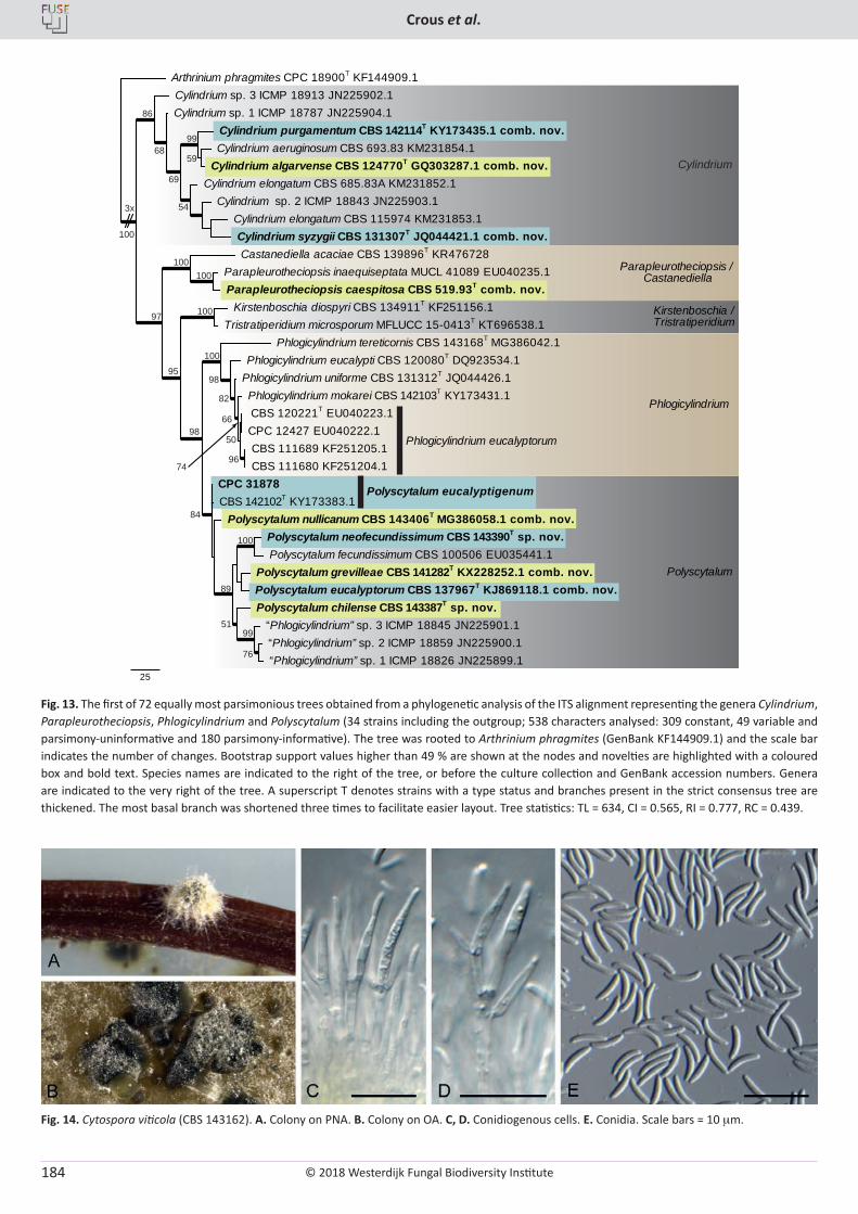
© 2018 Westerdijk Fungal Biodiversity Institute
Crous et al.
Editor-in-ChiefProf. dr P.W. Crous, Westerdijk Fungal Biodiversity Institute, P.O. Box 85167, 3508 AD Utrecht, The Netherlands.E-mail:[email protected]
184
Phlogicylindrium eucalyptorum
Polyscytalum eucalyptigenum
3x
100
86
68
69
59
54
95
97
100100
98
99
100
89
100
98
66
76
96
82
84
100
50
5199
74
Cylindrium
Parapleurotheciopsis / Castanediella
Kirstenboschia /Tristratiperidium
Phlogicylindrium
Polyscytalum
Parapleurotheciopsis inaequiseptata MUCL 41089 EU040235.1 Parapleurotheciopsis caespitosa CBS 519.93T comb. nov.
Castanediella acaciae CBS 139896T KR476728
Polyscytalum neofecundissimum CBS 143390T sp. nov. Polyscytalum fecundissimum CBS 100506 EU035441.1
Polyscytalum grevilleae CBS 141282T KX228252.1 comb. nov. Polyscytalum eucalyptorum CBS 137967T KJ869118.1 comb. nov. Polyscytalum chilense CBS 143387T sp. nov.
“Phlogicylindrium” sp. 3 ICMP 18845 JN225901.1 “Phlogicylindrium” sp. 2 ICMP 18859 JN225900.1 “Phlogicylindrium” sp. 1 ICMP 18826 JN225899.1
Polyscytalum nullicanum CBS 143406T MG386058.1 comb. nov.
CPC 31878 CBS 142102T KY173383.1
CBS 120221T EU040223.1 CPC 12427 EU040222.1 CBS 111689 KF251205.1 CBS 111680 KF251204.1
Phlogicylindrium mokarei CBS 142103T KY173431.1 Phlogicylindrium uniforme CBS 131312T JQ044426.1 Phlogicylindrium eucalypti CBS 120080T DQ923534.1
Phlogicylindrium tereticornis CBS 143168T MG386042.1
Kirstenboschia diospyri CBS 134911T KF251156.1 Tristratiperidium microsporum MFLUCC 15-0413T KT696538.1
Cylindrium aeruginosum CBS 693.83 KM231854.1 Cylindrium algarvense CBS 124770T GQ303287.1 comb. nov.
Cylindrium purgamentum CBS 142114T KY173435.1 comb. nov.
Cylindrium elongatum CBS 115974 KM231853.1 Cylindrium syzygii CBS 131307T JQ044421.1 comb. nov.
Cylindrium sp. 2 ICMP 18843 JN225903.1 Cylindrium elongatum CBS 685.83A KM231852.1
Cylindrium sp. 1 ICMP 18787 JN225904.1 Cylindrium sp. 3 ICMP 18913 JN225902.1
Arthrinium phragmites CPC 18900T KF144909.1
25
Fig. 13. The first of 72 equally most parsimonious trees obtained from a phylogenetic analysis of the ITS alignment representing the genera Cylindrium, Parapleurotheciopsis, Phlogicylindrium and Polyscytalum (34 strains including the outgroup; 538 characters analysed: 309 constant, 49 variable and parsimony-uninformative and 180 parsimony-informative). The tree was rooted to Arthrinium phragmites (GenBank KF144909.1) and the scale bar indicates the number of changes. Bootstrap support values higher than 49 % are shown at the nodes and novelties are highlighted with a coloured box and bold text. Species names are indicated to the right of the tree, or before the culture collection and GenBank accession numbers. Genera are indicated to the very right of the tree. A superscript T denotes strains with a type status and branches present in the strict consensus tree are thickened. The most basal branch was shortened three times to facilitate easier layout. Tree statistics: TL = 634, CI = 0.565, RI = 0.777, RC = 0.439.
Fig. 14. Cytospora viticola (CBS 143162). A. Colony on PNA. B. Colony on OA. C, D. Conidiogenous cells. E. Conidia. Scale bars = 10 mm.

© 2018 Westerdijk Fungal Biodiversity Institute
New and interesting fungi
Editor-in-ChiefProf. dr P.W. Crous, Westerdijk Fungal Biodiversity Institute, P.O. Box 85167, 3508 AD Utrecht, The Netherlands.E-mail:[email protected]
185
Cy. sibiraeae (GenBank KR045651; Identities 566 / 591 (96 %), 8 gaps (1 %)), Cy. chrysosperma (GenBank KT692596; Identities 568 / 594 (96 %), 7 gaps (1 %)) and Cy. germanica (GenBank KX168596; Identities 563 / 590 (95 %), 7 gaps (1 %)). Our ITS sequence is identical to Cy. viticola (GenBank KX256239; Identities 423 / 423 (100 %), no gaps), but was not a result in the megablast search as roughly half of the first internal spacer region sequence is missing for the deposited sequences of that species. The highest similarities using the LSU sequence were Valsa mali (GenBank AF362559; Identities 837 / 842 (99 %), 1 gap (0 %)), Cy. centrivillosa (GenBank MF190068; Identities 830 / 837 (99 %), 1 gap (0 %)) and Cy. ambiens (GenBank EU255208; Identities 772 / 779 (99 %), no gaps). The highest similarities using the actA sequence were Cy. salicicola (GenBank KU982637; Identities 163 / 180 (91 %), 6 gaps (3 %)), Cy. parasitica (GenBank KT459410; Identities 190 / 212 (90 %), 10 gaps (4 %)) and Cy. cincta (GenBank KU710994; Identities 223 / 250 (89 %), 13 gaps (5 %)). The highest similarities using the rpb2 sequence were Cy. berberidis (GenBank KU710948; Identities 658 / 727 (91 %), no gaps), Cy. schulzeri (GenBank KU710980; Identities 656 / 727 (90 %), no gaps) and Cy. rostrata (GenBank KU710974; Identities 656 / 727 (90 %), no gaps). The highest similarities using the tef1 sequence were Cy. viticola (GenBank KX256274; Identities 253 / 253 (100 %), no gaps), Cy. mali (GenBank KU710928; Identities 359 / 422 (85 %), 24 gaps (5 %)) and Cy. sophorae (GenBank KU710941; Identities 422 / 517 (82 %), 26 gaps (5 %)). The highest similarities using the tub2 sequence were V. malicola (GenBank KT934374; Identities 363 / 413 (88 %), 18 gaps (4 %)), V. sordida (GenBank KT428034; Identities 346 / 396 (87 %), 14 gaps (3 %)) and Cy. carbonacea (GenBank KP310825; Identities 343 / 395 (87 %), 16 gaps (4 %)).
Dictyochaeta mimusopis Crous & M.J. Wingf., sp. nov. MycoBank MB824774. Fig. 15.
Etymology: Name refers to Mimusops, the host genus from which this fungus was collected.
Mycelium consisting of branched, septate, hyaline, 3–4 µm diam hyphae. Conidiophores solitary, erect, pale brown, smooth, subcylindrical, unbranched, straight to flexuous, 1–6-septate, 40–150 × 3–4 µm. Conidiogenous cells monophialidic, integrated, terminal, pale brown, smooth, subcylindrical, 30–55 × 3(–3.5) µm; with flared apical collarette, 3–4 µm diam. Conidia solitary,
aseptate, hyaline, smooth, guttulate to granular, inequilateral, fusoid, outer plane convex, apex subacute, base truncate, 1–1.5 µm diam, (11–)16–18(–20) × 2.5–3(–3.5) µm, with a single unbranched, flexuous appendage at each end, (6–)7(–8) µm.
Culture characteristics: Colonies flat, spreading, with moderate aerial mycelium and smooth, lobate margins, reaching 50 mm diam after 2 wk at 25 °C. On MEA surface hazel with cinnamon pigment, reverse sepia. On PDA surface cinnamon, reverse amber. On OA surface sienna with patches of olivaceous grey.
Specimen examined: South Africa, Eastern Cape Province, Haga Haga, on leaves of Mimusops caffra (Sapotaceae), Dec. 2010, M.J. Wingfield (holotype CBS H-23412, culture ex-type CPC 29987 = CBS 143435).
Notes: Dictyochaeta mimusopis is closely allied to isolates in the Di. simplex complex (conidia 14–19 × 2.1–2.7 mm; Hughes & Kendrick 1968).
Based on a megablast search using the ITS sequence, the closest matches in NCBI’s GenBank nucleotide database were Di. simplex (GenBank EF029193; Identities 482 / 503 (96 %), 1 gap (0 %)), Di. fertilis (GenBank AF178540; Identities 469 / 491 (96 %), 5 gaps (1 %)) and Codinaea pini (GenBank NR_137943; Identities 485 / 521 (93 %), 22 gaps (4 %)). The highest similarities using the LSU sequence were Di. simplex (GenBank AF178559; Identities 830 / 836 (99 %), no gaps), Codinaea pini (GenBank KP004493; Identities 829 / 838 (99 %), 1 gap (0 %)) and Rattania setulifera (GenBank HM171322; Identities 815 / 835 (98 %), no gaps).
Dictyochaeta septata (B. Sutton & Hodges) Whitton et al., Fungal Diversity 4: 148. 2000. Fig. 16.Basionym: Codinaea septata B. Sutton & Hodges, Nova Hedwigia 26(2–3): 520. 1975.Synonym: Dictyochaeta septata (B. Sutton & Hodges) Aramb. & Cabello, Mycotaxon 34(2): 682 (1989) (nom. inval. Art 41.5, Melbourne).
Mycelium consisting of hyaline, septate, branched, smooth, 2–3 µm diam hyphae. Conidiophores solitary, erect, brown, smooth, subcylindrical, straight to flexuous, 1–3-septate, 30–120 × 4–6 µm. Conidiogenous cells terminal, integrated, pale brown, smooth, mono-, rarely polyphialidic, 10–30 × 4–5 µm; collarette flared, 3–4 µm diam. Conidia solitary, hyaline, smooth, guttulate, granular, subcylindrical, falcate, ends subobtuse, (14–)15–19(–
Fig. 15. Dictyochaeta mimusopsis (CBS 143435). A. Colony on OA. B–E. Conidiophores. F. Conidia. Scale bars = 10 mm.
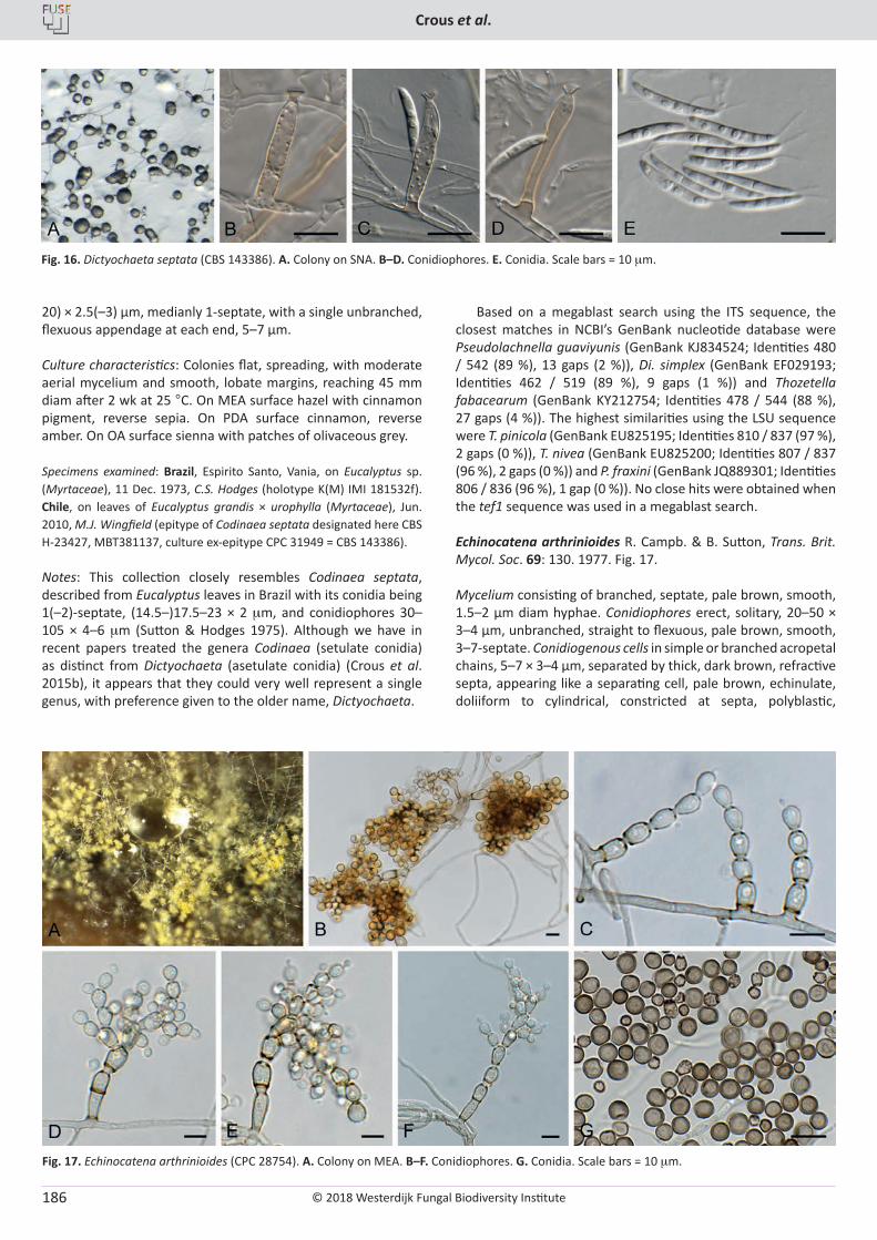
© 2018 Westerdijk Fungal Biodiversity Institute
Crous et al.
Editor-in-ChiefProf. dr P.W. Crous, Westerdijk Fungal Biodiversity Institute, P.O. Box 85167, 3508 AD Utrecht, The Netherlands.E-mail:[email protected]
186
20) × 2.5(–3) µm, medianly 1-septate, with a single unbranched, flexuous appendage at each end, 5–7 µm.
Culture characteristics: Colonies flat, spreading, with moderate aerial mycelium and smooth, lobate margins, reaching 45 mm diam after 2 wk at 25 °C. On MEA surface hazel with cinnamon pigment, reverse sepia. On PDA surface cinnamon, reverse amber. On OA surface sienna with patches of olivaceous grey.
Specimens examined: Brazil, Espirito Santo, Vania, on Eucalyptus sp. (Myrtaceae), 11 Dec. 1973, C.S. Hodges (holotype K(M) IMI 181532f). Chile, on leaves of Eucalyptus grandis × urophylla (Myrtaceae), Jun. 2010, M.J. Wingfield (epitype of Codinaea septata designated here CBS H-23427, MBT381137, culture ex-epitype CPC 31949 = CBS 143386).
Notes: This collection closely resembles Codinaea septata, described from Eucalyptus leaves in Brazil with its conidia being 1(–2)-septate, (14.5–)17.5–23 × 2 mm, and conidiophores 30–105 × 4–6 mm (Sutton & Hodges 1975). Although we have in recent papers treated the genera Codinaea (setulate conidia) as distinct from Dictyochaeta (asetulate conidia) (Crous et al. 2015b), it appears that they could very well represent a single genus, with preference given to the older name, Dictyochaeta.
Based on a megablast search using the ITS sequence, the closest matches in NCBI’s GenBank nucleotide database were Pseudolachnella guaviyunis (GenBank KJ834524; Identities 480 / 542 (89 %), 13 gaps (2 %)), Di. simplex (GenBank EF029193; Identities 462 / 519 (89 %), 9 gaps (1 %)) and Thozetella fabacearum (GenBank KY212754; Identities 478 / 544 (88 %), 27 gaps (4 %)). The highest similarities using the LSU sequence were T. pinicola (GenBank EU825195; Identities 810 / 837 (97 %), 2 gaps (0 %)), T. nivea (GenBank EU825200; Identities 807 / 837 (96 %), 2 gaps (0 %)) and P. fraxini (GenBank JQ889301; Identities 806 / 836 (96 %), 1 gap (0 %)). No close hits were obtained when the tef1 sequence was used in a megablast search.
Echinocatena arthrinioides R. Campb. & B. Sutton, Trans. Brit. Mycol. Soc. 69: 130. 1977. Fig. 17.
Mycelium consisting of branched, septate, pale brown, smooth, 1.5–2 µm diam hyphae. Conidiophores erect, solitary, 20–50 × 3–4 µm, unbranched, straight to flexuous, pale brown, smooth, 3–7-septate. Conidiogenous cells in simple or branched acropetal chains, 5–7 × 3–4 µm, separated by thick, dark brown, refractive septa, appearing like a separating cell, pale brown, echinulate, doliiform to cylindrical, constricted at septa, polyblastic,
Fig. 16. Dictyochaeta septata (CBS 143386). A. Colony on SNA. B–D. Conidiophores. E. Conidia. Scale bars = 10 mm.
Fig. 17. Echinocatena arthrinioides (CPC 28754). A. Colony on MEA. B–F. Conidiophores. G. Conidia. Scale bars = 10 mm.
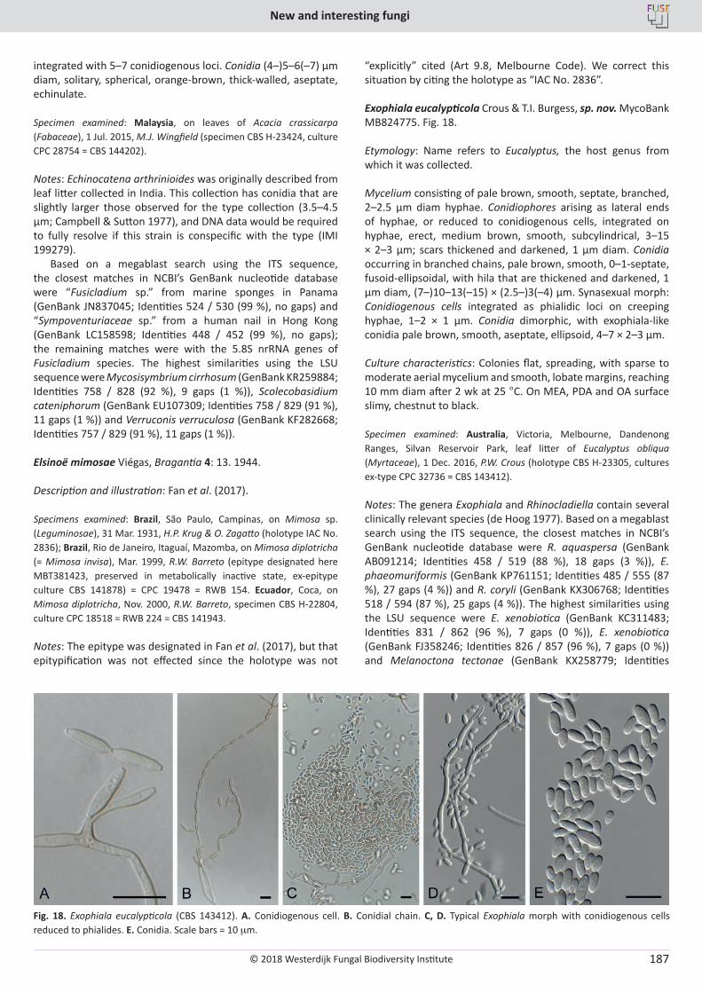
© 2018 Westerdijk Fungal Biodiversity Institute
New and interesting fungi
Editor-in-ChiefProf. dr P.W. Crous, Westerdijk Fungal Biodiversity Institute, P.O. Box 85167, 3508 AD Utrecht, The Netherlands.E-mail:[email protected]
187
integrated with 5–7 conidiogenous loci. Conidia (4–)5–6(–7) µm diam, solitary, spherical, orange-brown, thick-walled, aseptate, echinulate.
Specimen examined: Malaysia, on leaves of Acacia crassicarpa (Fabaceae), 1 Jul. 2015, M.J. Wingfield (specimen CBS H-23424, culture CPC 28754 = CBS 144202).
Notes: Echinocatena arthrinioides was originally described from leaf litter collected in India. This collection has conidia that are slightly larger those observed for the type collection (3.5–4.5 µm; Campbell & Sutton 1977), and DNA data would be required to fully resolve if this strain is conspecific with the type (IMI 199279).
Based on a megablast search using the ITS sequence, the closest matches in NCBI’s GenBank nucleotide database were “Fusicladium sp.” from marine sponges in Panama (GenBank JN837045; Identities 524 / 530 (99 %), no gaps) and “Sympoventuriaceae sp.” from a human nail in Hong Kong (GenBank LC158598; Identities 448 / 452 (99 %), no gaps); the remaining matches were with the 5.8S nrRNA genes of Fusicladium species. The highest similarities using the LSU sequence were Mycosisymbrium cirrhosum (GenBank KR259884; Identities 758 / 828 (92 %), 9 gaps (1 %)), Scolecobasidium cateniphorum (GenBank EU107309; Identities 758 / 829 (91 %), 11 gaps (1 %)) and Verruconis verruculosa (GenBank KF282668; Identities 757 / 829 (91 %), 11 gaps (1 %)).
Elsinoë mimosae Viégas, Bragantia 4: 13. 1944.
Description and illustration: Fan et al. (2017).
Specimens examined: Brazil, São Paulo, Campinas, on Mimosa sp. (Leguminosae), 31 Mar. 1931, H.P. Krug & O. Zagatto (holotype IAC No. 2836); Brazil, Rio de Janeiro, Itaguaí, Mazomba, on Mimosa diplotricha (= Mimosa invisa), Mar. 1999, R.W. Barreto (epitype designated here MBT381423, preserved in metabolically inactive state, ex-epitype culture CBS 141878) = CPC 19478 = RWB 154. Ecuador, Coca, on Mimosa diplotricha, Nov. 2000, R.W. Barreto, specimen CBS H-22804, culture CPC 18518 = RWB 224 = CBS 141943.
Notes: The epitype was designated in Fan et al. (2017), but that epitypification was not effected since the holotype was not
“explicitly” cited (Art 9.8, Melbourne Code). We correct this situation by citing the holotype as “IAC No. 2836”.
Exophiala eucalypticola Crous & T.I. Burgess, sp. nov. MycoBank MB824775. Fig. 18.
Etymology: Name refers to Eucalyptus, the host genus from which it was collected.
Mycelium consisting of pale brown, smooth, septate, branched, 2–2.5 µm diam hyphae. Conidiophores arising as lateral ends of hyphae, or reduced to conidiogenous cells, integrated on hyphae, erect, medium brown, smooth, subcylindrical, 3–15 × 2–3 µm; scars thickened and darkened, 1 µm diam. Conidia occurring in branched chains, pale brown, smooth, 0–1-septate, fusoid-ellipsoidal, with hila that are thickened and darkened, 1 µm diam, (7–)10–13(–15) × (2.5–)3(–4) µm. Synasexual morph: Conidiogenous cells integrated as phialidic loci on creeping hyphae, 1–2 × 1 µm. Conidia dimorphic, with exophiala-like conidia pale brown, smooth, aseptate, ellipsoid, 4–7 × 2–3 µm.
Culture characteristics: Colonies flat, spreading, with sparse to moderate aerial mycelium and smooth, lobate margins, reaching 10 mm diam after 2 wk at 25 °C. On MEA, PDA and OA surface slimy, chestnut to black.
Specimen examined: Australia, Victoria, Melbourne, Dandenong Ranges, Silvan Reservoir Park, leaf litter of Eucalyptus obliqua (Myrtaceae), 1 Dec. 2016, P.W. Crous (holotype CBS H-23305, cultures ex-type CPC 32736 = CBS 143412).
Notes: The genera Exophiala and Rhinocladiella contain several clinically relevant species (de Hoog 1977). Based on a megablast search using the ITS sequence, the closest matches in NCBI’s GenBank nucleotide database were R. aquaspersa (GenBank AB091214; Identities 458 / 519 (88 %), 18 gaps (3 %)), E. phaeomuriformis (GenBank KP761151; Identities 485 / 555 (87 %), 27 gaps (4 %)) and R. coryli (GenBank KX306768; Identities 518 / 594 (87 %), 25 gaps (4 %)). The highest similarities using the LSU sequence were E. xenobiotica (GenBank KC311483; Identities 831 / 862 (96 %), 7 gaps (0 %)), E. xenobiotica (GenBank FJ358246; Identities 826 / 857 (96 %), 7 gaps (0 %)) and Melanoctona tectonae (GenBank KX258779; Identities
Fig. 18. Exophiala eucalypticola (CBS 143412). A. Conidiogenous cell. B. Conidial chain. C, D. Typical Exophiala morph with conidiogenous cells reduced to phialides. E. Conidia. Scale bars = 10 mm.
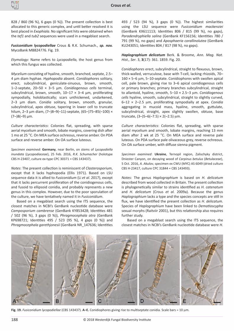
© 2018 Westerdijk Fungal Biodiversity Institute
Crous et al.
Editor-in-ChiefProf. dr P.W. Crous, Westerdijk Fungal Biodiversity Institute, P.O. Box 85167, 3508 AD Utrecht, The Netherlands.E-mail:[email protected]
188
828 / 860 (96 %), 6 gaps (0 %)). The present collection is best allocated to this generic complex, and until better resolved it is best placed in Exophiala. No significant hits were obtained when the tef1 and tub2 sequences were used in a megablast search.
Fusiconidium lycopodiellae Crous & R.K. Schumach., sp. nov. MycoBank MB824776. Fig. 19.
Etymology: Name refers to Lycopodiella, the host genus from which this fungus was collected.
Mycelium consisting of hyaline, smooth, branched, septate, 2.5–4 µm diam hyphae. Hyphopodia absent. Conidiophores solitary, erect, subcylindrical, geniculate-sinuous, brown, smooth, 1–2-septate, 20–50 × 3–5 µm. Conidiogenous cells terminal, subcylindrical, brown, smooth, 10–17 × 3–4 µm, proliferating sympodially, holoblastically; scars unthickened, undarkened, 2–3 µm diam. Conidia solitary, brown, smooth, granular, subcylindrical, apex obtuse, tapering in lower cell to truncate hilum, 2–3 µm diam, (7–)8–9(–11)-septate, (65–)75–85(–100) × (7–)8(–9) µm.
Culture characteristics: Colonies flat, spreading, with sparse aerial mycelium and smooth, lobate margins, covering dish after 1 mo at 25 °C. On MEA surface ochreous, reverse amber. On PDA surface and reverse amber. On OA surface luteous.
Specimen examined: Germany, near Berlin, on stems of Lycopodiella inundata (Lycopodiaceae), 25 Feb. 2016, R.K. Schumacher (holotype CBS H-23407, culture ex-type CPC 30371 = CBS 143437).
Notes: The present collection is reminiscent of Clasterosporium, except that it lacks hyphopodia (Ellis 1971). Based on LSU sequence data it is allied to Fusiconidium (Li et al. 2017), except that it lacks percurrent proliferation of the conidiogenous cells, and fusoid to ellipsoid conidia, and probably represents a new genus in this complex. However, due to the poor sporulation of the culture, we have tentatively named it in Fusiconidium.
Based on a megablast search using the ITS sequence, the closest matches in NCBI’s GenBank nucleotide database were Camposporium cambrense (GenBank KY853428; Identities 481 / 502 (96 %), 3 gaps (0 %)), Phragmocephala atra (GenBank KP698721; Identities 495 / 523 (95 %), 4 gaps (0 %)) and Phragmocephala garethjonesii (GenBank NR_147636; Identities
493 / 523 (94 %), 3 gaps (0 %)). The highest similarities using the LSU sequence were Fusiconidium mackenziei (GenBank KX611113; Identities 806 / 815 (99 %), no gaps), Paradendryphiella salina (GenBank KF156156; Identities 780 / 791 (99 %), no gaps) and Aposphaeria corallinolutea (GenBank KU243051; Identities 804 / 817 (98 %), no gaps).
Haplographium delicatum Berk. & Broome, Ann. Mag. Nat. Hist., Ser. 3, 3(17): 361. 1859. Fig. 20.
Conidiophores erect, subcylindrical, straight to flexuous, brown, thick-walled, verruculose, base with T-cell, lacking rhizoids, 70–160 × 5–6 mm, 5–10-septate. Conidiophores with swollen apical cell, pale brown, giving rise to 3–6 apical conidiogenous cells or primary branches; primary branches subcylindrical, straight to allantoid, hyaline, smooth, 5–10 × 2.5–3 mm. Conidiogenous cells hyaline, smooth, subcylindrical, straight to slightly curved, 6–12 × 2–2.5 mm, proliferating sympodially at apex. Conidia aggregating in mucoid mass, hyaline, smooth, guttulate, subcylindrical, straight, apex slightly swollen, obtuse, base truncate, (3–)5–6(–7.5) × 2(–2.5) mm.
Culture characteristics: Colonies flat, spreading, with sparse aerial mycelium and smooth, lobate margins, reaching 13 mm diam after 2 wk at 25 °C. On MEA surface and reverse pale luteous. On PDA surface pale olivaceous grey, reverse ochreous. On OA surface umber, with diffuse sienna pigment.
Specimen examined: Ukraine, Ternopil region, Zalischyky district, Dniester Canyon, on decaying wood of Carpinus betulus (Betulaceae), 5 Oct. 2016, A. Akulov, specimen ex CWU (MYC) AS 6049 (dried culture CBS H-23417, culture CPC 31844 = CBS 143493).
Notes: The genus Haplographium is based on H. delicatum described from wood collected in Britain. The present collection is phylogenetically similar to strains identified as H. catenatum and H. delicatum (Crous et al. 2009a). Because the genus Haplographium lacks a type and the species concepts are still in flux, we have identified the present collection as H. delicatum. Species of Haplographium have been linked to Dematioscypha sexual morphs (Raitviir 2001), but this relationship also requires further study.
Based on a megablast search using the ITS sequence, the closest matches in NCBI’s GenBank nucleotide database were H.
Fig. 19. Fusiconidium lycopodiellae (CBS 143437). A–E. Conidiophores giving rise to multiseptate conidia. Scale bars = 10 mm.
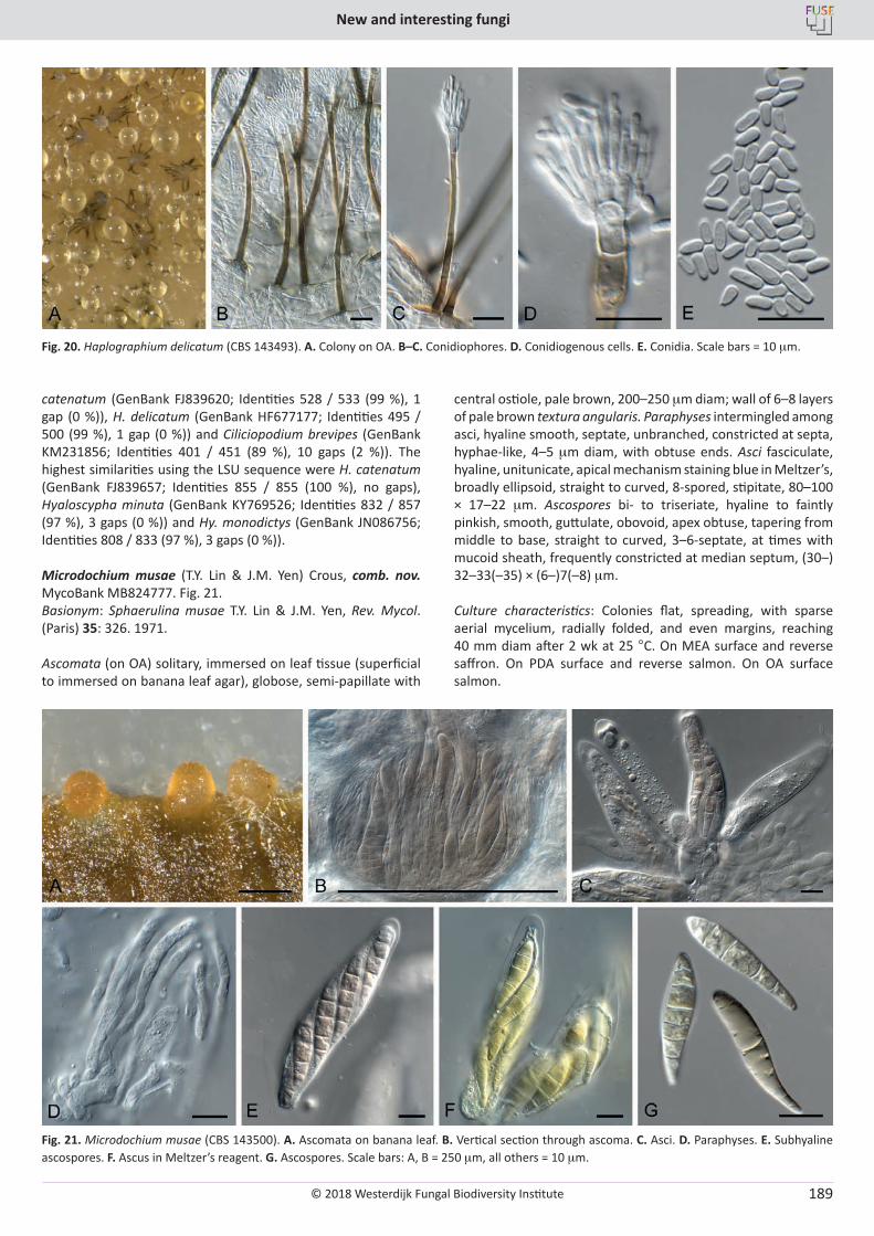
© 2018 Westerdijk Fungal Biodiversity Institute
New and interesting fungi
Editor-in-ChiefProf. dr P.W. Crous, Westerdijk Fungal Biodiversity Institute, P.O. Box 85167, 3508 AD Utrecht, The Netherlands.E-mail:[email protected]
189
catenatum (GenBank FJ839620; Identities 528 / 533 (99 %), 1 gap (0 %)), H. delicatum (GenBank HF677177; Identities 495 / 500 (99 %), 1 gap (0 %)) and Ciliciopodium brevipes (GenBank KM231856; Identities 401 / 451 (89 %), 10 gaps (2 %)). The highest similarities using the LSU sequence were H. catenatum (GenBank FJ839657; Identities 855 / 855 (100 %), no gaps), Hyaloscypha minuta (GenBank KY769526; Identities 832 / 857 (97 %), 3 gaps (0 %)) and Hy. monodictys (GenBank JN086756; Identities 808 / 833 (97 %), 3 gaps (0 %)).
Microdochium musae (T.Y. Lin & J.M. Yen) Crous, comb. nov. MycoBank MB824777. Fig. 21.Basionym: Sphaerulina musae T.Y. Lin & J.M. Yen, Rev. Mycol. (Paris) 35: 326. 1971.
Ascomata (on OA) solitary, immersed on leaf tissue (superficial to immersed on banana leaf agar), globose, semi-papillate with
central ostiole, pale brown, 200–250 mm diam; wall of 6–8 layers of pale brown textura angularis. Paraphyses intermingled among asci, hyaline smooth, septate, unbranched, constricted at septa, hyphae-like, 4–5 mm diam, with obtuse ends. Asci fasciculate, hyaline, unitunicate, apical mechanism staining blue in Meltzer’s, broadly ellipsoid, straight to curved, 8-spored, stipitate, 80–100 × 17–22 mm. Ascospores bi- to triseriate, hyaline to faintly pinkish, smooth, guttulate, obovoid, apex obtuse, tapering from middle to base, straight to curved, 3–6-septate, at times with mucoid sheath, frequently constricted at median septum, (30–) 32–33(–35) × (6–)7(–8) mm.
Culture characteristics: Colonies flat, spreading, with sparse aerial mycelium, radially folded, and even margins, reaching 40 mm diam after 2 wk at 25 °C. On MEA surface and reverse saffron. On PDA surface and reverse salmon. On OA surface salmon.
Fig. 20. Haplographium delicatum (CBS 143493). A. Colony on OA. B–C. Conidiophores. D. Conidiogenous cells. E. Conidia. Scale bars = 10 mm.
Fig. 21. Microdochium musae (CBS 143500). A. Ascomata on banana leaf. B. Vertical section through ascoma. C. Asci. D. Paraphyses. E. Subhyaline ascospores. F. Ascus in Meltzer’s reagent. G. Ascospores. Scale bars: A, B = 250 mm, all others = 10 mm.
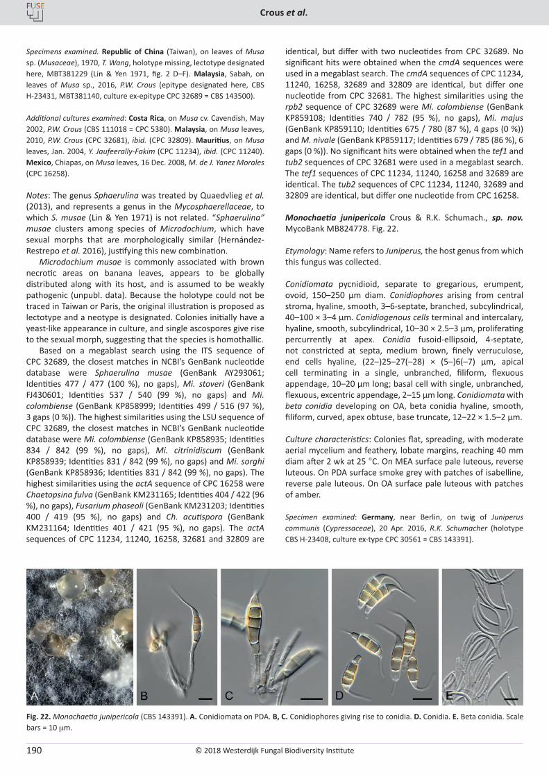
© 2018 Westerdijk Fungal Biodiversity Institute
Crous et al.
Editor-in-ChiefProf. dr P.W. Crous, Westerdijk Fungal Biodiversity Institute, P.O. Box 85167, 3508 AD Utrecht, The Netherlands.E-mail:[email protected]
190
Specimens examined. Republic of China (Taiwan), on leaves of Musa sp. (Musaceae), 1970, T. Wang, holotype missing, lectotype designated here, MBT381229 (Lin & Yen 1971, fig. 2 D–F). Malaysia, Sabah, on leaves of Musa sp., 2016, P.W. Crous (epitype designated here, CBS H-23431, MBT381140, culture ex-epitype CPC 32689 = CBS 143500).
Additional cultures examined: Costa Rica, on Musa cv. Cavendish, May 2002, P.W. Crous (CBS 111018 = CPC 5380). Malaysia, on Musa leaves, 2010, P.W. Crous (CPC 32681), ibid. (CPC 32809). Mauritius, on Musa leaves, Jan. 2004, Y. Jaufeerally-Fakim (CPC 11234), ibid. (CPC 11240). Mexico, Chiapas, on Musa leaves, 16 Dec. 2008, M. de J. Yanez Morales (CPC 16258).
Notes: The genus Sphaerulina was treated by Quaedvlieg et al. (2013), and represents a genus in the Mycosphaerellaceae, to which S. musae (Lin & Yen 1971) is not related. “Sphaerulina” musae clusters among species of Microdochium, which have sexual morphs that are morphologically similar (Hernández-Restrepo et al. 2016), justifying this new combination.
Microdochium musae is commonly associated with brown necrotic areas on banana leaves, appears to be globally distributed along with its host, and is assumed to be weakly pathogenic (unpubl. data). Because the holotype could not be traced in Taiwan or Paris, the original illustration is proposed as lectotype and a neotype is designated. Colonies initially have a yeast-like appearance in culture, and single ascospores give rise to the sexual morph, suggesting that the species is homothallic.
Based on a megablast search using the ITS sequence of CPC 32689, the closest matches in NCBI’s GenBank nucleotide database were Sphaerulina musae (GenBank AY293061; Identities 477 / 477 (100 %), no gaps), Mi. stoveri (GenBank FJ430601; Identities 537 / 540 (99 %), no gaps) and Mi. colombiense (GenBank KP858999; Identities 499 / 516 (97 %), 3 gaps (0 %)). The highest similarities using the LSU sequence of CPC 32689, the closest matches in NCBI’s GenBank nucleotide database were Mi. colombiense (GenBank KP858935; Identities 834 / 842 (99 %), no gaps), Mi. citrinidiscum (GenBank KP858939; Identities 831 / 842 (99 %), no gaps) and Mi. sorghi (GenBank KP858936; Identities 831 / 842 (99 %), no gaps). The highest similarities using the actA sequence of CPC 16258 were Chaetopsina fulva (GenBank KM231165; Identities 404 / 422 (96 %), no gaps), Fusarium phaseoli (GenBank KM231203; Identities 400 / 419 (95 %), no gaps) and Ch. acutispora (GenBank KM231164; Identities 401 / 421 (95 %), no gaps). The actA sequences of CPC 11234, 11240, 16258, 32681 and 32809 are
identical, but differ with two nucleotides from CPC 32689. No significant hits were obtained when the cmdA sequences were used in a megablast search. The cmdA sequences of CPC 11234, 11240, 16258, 32689 and 32809 are identical, but differ one nucleotide from CPC 32681. The highest similarities using the rpb2 sequence of CPC 32689 were Mi. colombiense (GenBank KP859108; Identities 740 / 782 (95 %), no gaps), Mi. majus (GenBank KP859110; Identities 675 / 780 (87 %), 4 gaps (0 %)) and M. nivale (GenBank KP859117; Identities 679 / 785 (86 %), 6 gaps (0 %)). No significant hits were obtained when the tef1 and tub2 sequences of CPC 32681 were used in a megablast search. The tef1 sequences of CPC 11234, 11240, 16258 and 32689 are identical. The tub2 sequences of CPC 11234, 11240, 32689 and 32809 are identical, but differ one nucleotide from CPC 16258.
Monochaetia junipericola Crous & R.K. Schumach., sp. nov. MycoBank MB824778. Fig. 22.
Etymology: Name refers to Juniperus, the host genus from which this fungus was collected.
Conidiomata pycnidioid, separate to gregarious, erumpent, ovoid, 150–250 µm diam. Conidiophores arising from central stroma, hyaline, smooth, 3–6-septate, branched, subcylindrical, 40–100 × 3–4 µm. Conidiogenous cells terminal and intercalary, hyaline, smooth, subcylindrical, 10–30 × 2.5–3 µm, proliferating percurrently at apex. Conidia fusoid-ellipsoid, 4-septate, not constricted at septa, medium brown, finely verruculose, end cells hyaline, (22–)25–27(–28) × (5–)6(–7) µm, apical cell terminating in a single, unbranched, filiform, flexuous appendage, 10–20 µm long; basal cell with single, unbranched, flexuous, excentric appendage, 2–15 µm long. Conidiomata with beta conidia developing on OA, beta conidia hyaline, smooth, filiform, curved, apex obtuse, base truncate, 12–22 × 1.5–2 µm.
Culture characteristics: Colonies flat, spreading, with moderate aerial mycelium and feathery, lobate margins, reaching 40 mm diam after 2 wk at 25 °C. On MEA surface pale luteous, reverse luteous. On PDA surface smoke grey with patches of isabelline, reverse pale luteous. On OA surface pale luteous with patches of amber.
Specimen examined: Germany, near Berlin, on twig of Juniperus communis (Cypressaceae), 20 Apr. 2016, R.K. Schumacher (holotype CBS H-23408, culture ex-type CPC 30561 = CBS 143391).
Fig. 22. Monochaetia junipericola (CBS 143391). A. Conidiomata on PDA. B, C. Conidiophores giving rise to conidia. D. Conidia. E. Beta conidia. Scale bars = 10 mm.
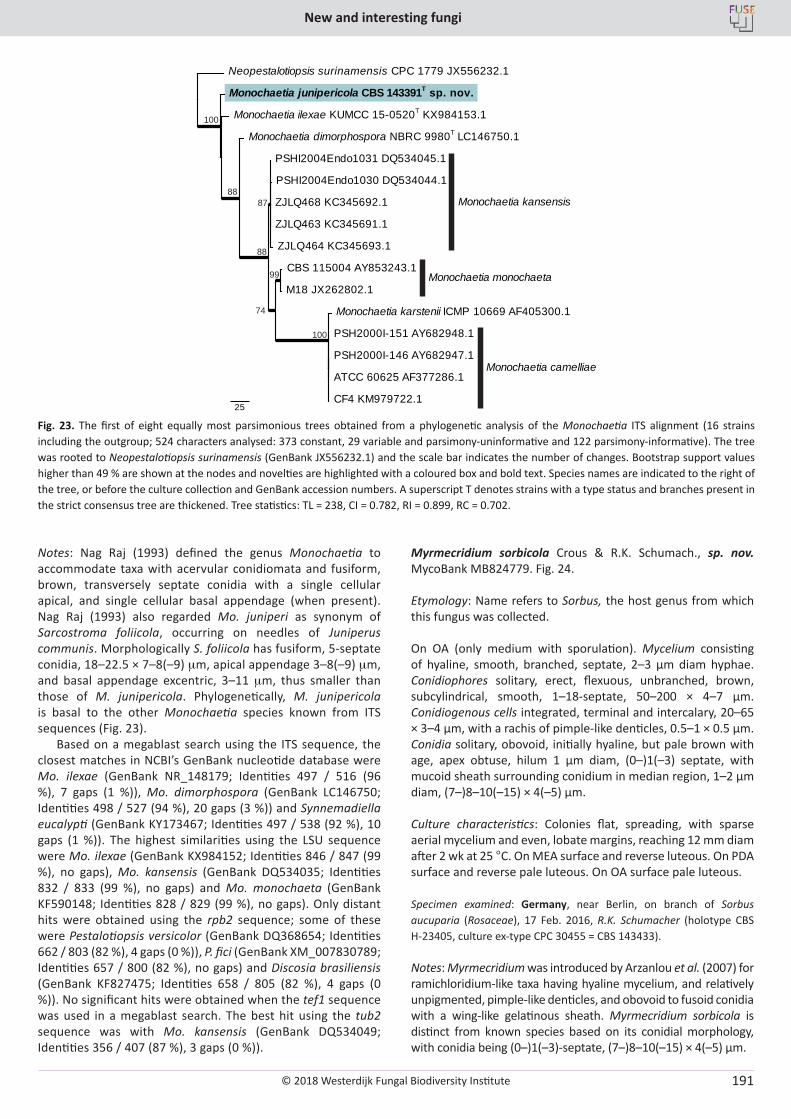
© 2018 Westerdijk Fungal Biodiversity Institute
New and interesting fungi
Editor-in-ChiefProf. dr P.W. Crous, Westerdijk Fungal Biodiversity Institute, P.O. Box 85167, 3508 AD Utrecht, The Netherlands.E-mail:[email protected]
191
Notes: Nag Raj (1993) defined the genus Monochaetia to accommodate taxa with acervular conidiomata and fusiform, brown, transversely septate conidia with a single cellular apical, and single cellular basal appendage (when present). Nag Raj (1993) also regarded Mo. juniperi as synonym of Sarcostroma foliicola, occurring on needles of Juniperus communis. Morphologically S. foliicola has fusiform, 5-septate conidia, 18–22.5 × 7–8(–9) mm, apical appendage 3–8(–9) mm, and basal appendage excentric, 3–11 mm, thus smaller than those of M. junipericola. Phylogenetically, M. junipericola is basal to the other Monochaetia species known from ITS sequences (Fig. 23).
Based on a megablast search using the ITS sequence, the closest matches in NCBI’s GenBank nucleotide database were Mo. ilexae (GenBank NR_148179; Identities 497 / 516 (96 %), 7 gaps (1 %)), Mo. dimorphospora (GenBank LC146750; Identities 498 / 527 (94 %), 20 gaps (3 %)) and Synnemadiella eucalypti (GenBank KY173467; Identities 497 / 538 (92 %), 10 gaps (1 %)). The highest similarities using the LSU sequence were Mo. ilexae (GenBank KX984152; Identities 846 / 847 (99 %), no gaps), Mo. kansensis (GenBank DQ534035; Identities 832 / 833 (99 %), no gaps) and Mo. monochaeta (GenBank KF590148; Identities 828 / 829 (99 %), no gaps). Only distant hits were obtained using the rpb2 sequence; some of these were Pestalotiopsis versicolor (GenBank DQ368654; Identities 662 / 803 (82 %), 4 gaps (0 %)), P. fici (GenBank XM_007830789; Identities 657 / 800 (82 %), no gaps) and Discosia brasiliensis (GenBank KF827475; Identities 658 / 805 (82 %), 4 gaps (0 %)). No significant hits were obtained when the tef1 sequence was used in a megablast search. The best hit using the tub2 sequence was with Mo. kansensis (GenBank DQ534049; Identities 356 / 407 (87 %), 3 gaps (0 %)).
Myrmecridium sorbicola Crous & R.K. Schumach., sp. nov. MycoBank MB824779. Fig. 24.
Etymology: Name refers to Sorbus, the host genus from which this fungus was collected.
On OA (only medium with sporulation). Mycelium consisting of hyaline, smooth, branched, septate, 2–3 µm diam hyphae. Conidiophores solitary, erect, flexuous, unbranched, brown, subcylindrical, smooth, 1–18-septate, 50–200 × 4–7 µm. Conidiogenous cells integrated, terminal and intercalary, 20–65 × 3–4 µm, with a rachis of pimple-like denticles, 0.5–1 × 0.5 µm. Conidia solitary, obovoid, initially hyaline, but pale brown with age, apex obtuse, hilum 1 µm diam, (0–)1(–3) septate, with mucoid sheath surrounding conidium in median region, 1–2 µm diam, (7–)8–10(–15) × 4(–5) µm.
Culture characteristics: Colonies flat, spreading, with sparse aerial mycelium and even, lobate margins, reaching 12 mm diam after 2 wk at 25 °C. On MEA surface and reverse luteous. On PDA surface and reverse pale luteous. On OA surface pale luteous.
Specimen examined: Germany, near Berlin, on branch of Sorbus aucuparia (Rosaceae), 17 Feb. 2016, R.K. Schumacher (holotype CBS H-23405, culture ex-type CPC 30455 = CBS 143433).
Notes: Myrmecridium was introduced by Arzanlou et al. (2007) for ramichloridium-like taxa having hyaline mycelium, and relatively unpigmented, pimple-like denticles, and obovoid to fusoid conidia with a wing-like gelatinous sheath. Myrmecridium sorbicola is distinct from known species based on its conidial morphology, with conidia being (0–)1(–3)-septate, (7–)8–10(–15) × 4(–5) µm.
100
100
88
88
87
74
99
PSH2000I-151 AY682948.1
PSH2000I-146 AY682947.1
ATCC 60625 AF377286.1
CF4 KM979722.1
Monochaetia karstenii ICMP 10669 AF405300.1
CBS 115004 AY853243.1
M18 JX262802.1
PSHI2004Endo1031 DQ534045.1
PSHI2004Endo1030 DQ534044.1
ZJLQ468 KC345692.1
ZJLQ463 KC345691.1
ZJLQ464 KC345693.1
Monochaetia dimorphospora NBRC 9980T LC146750.1
Monochaetia ilexae KUMCC 15-0520T KX984153.1
Monochaetia junipericola CBS 143391T sp. nov.
Neopestalotiopsis surinamensis CPC 1779 JX556232.1
25
Monochaetia kansensis
Monochaetia monochaeta
Monochaetia camelliae
Fig. 23. The first of eight equally most parsimonious trees obtained from a phylogenetic analysis of the Monochaetia ITS alignment (16 strains including the outgroup; 524 characters analysed: 373 constant, 29 variable and parsimony-uninformative and 122 parsimony-informative). The tree was rooted to Neopestalotiopsis surinamensis (GenBank JX556232.1) and the scale bar indicates the number of changes. Bootstrap support values higher than 49 % are shown at the nodes and novelties are highlighted with a coloured box and bold text. Species names are indicated to the right of the tree, or before the culture collection and GenBank accession numbers. A superscript T denotes strains with a type status and branches present in the strict consensus tree are thickened. Tree statistics: TL = 238, CI = 0.782, RI = 0.899, RC = 0.702.
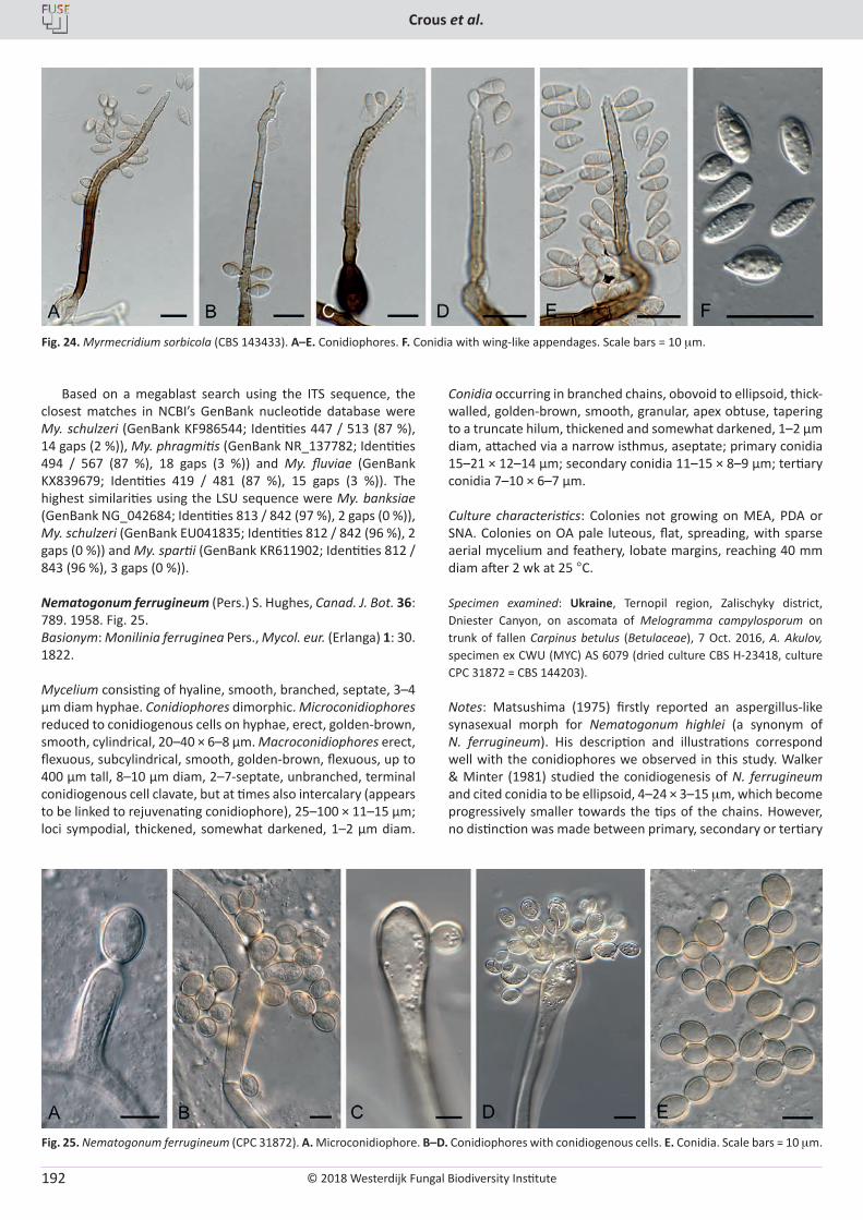
© 2018 Westerdijk Fungal Biodiversity Institute
Crous et al.
Editor-in-ChiefProf. dr P.W. Crous, Westerdijk Fungal Biodiversity Institute, P.O. Box 85167, 3508 AD Utrecht, The Netherlands.E-mail:[email protected]
192
Based on a megablast search using the ITS sequence, the closest matches in NCBI’s GenBank nucleotide database were My. schulzeri (GenBank KF986544; Identities 447 / 513 (87 %), 14 gaps (2 %)), My. phragmitis (GenBank NR_137782; Identities 494 / 567 (87 %), 18 gaps (3 %)) and My. fluviae (GenBank KX839679; Identities 419 / 481 (87 %), 15 gaps (3 %)). The highest similarities using the LSU sequence were My. banksiae (GenBank NG_042684; Identities 813 / 842 (97 %), 2 gaps (0 %)), My. schulzeri (GenBank EU041835; Identities 812 / 842 (96 %), 2 gaps (0 %)) and My. spartii (GenBank KR611902; Identities 812 / 843 (96 %), 3 gaps (0 %)).
Nematogonum ferrugineum (Pers.) S. Hughes, Canad. J. Bot. 36: 789. 1958. Fig. 25.Basionym: Monilinia ferruginea Pers., Mycol. eur. (Erlanga) 1: 30. 1822.
Mycelium consisting of hyaline, smooth, branched, septate, 3–4 µm diam hyphae. Conidiophores dimorphic. Microconidiophores reduced to conidiogenous cells on hyphae, erect, golden-brown, smooth, cylindrical, 20–40 × 6–8 µm. Macroconidiophores erect, flexuous, subcylindrical, smooth, golden-brown, flexuous, up to 400 µm tall, 8–10 µm diam, 2–7-septate, unbranched, terminal conidiogenous cell clavate, but at times also intercalary (appears to be linked to rejuvenating conidiophore), 25–100 × 11–15 µm; loci sympodial, thickened, somewhat darkened, 1–2 µm diam.
Conidia occurring in branched chains, obovoid to ellipsoid, thick-walled, golden-brown, smooth, granular, apex obtuse, tapering to a truncate hilum, thickened and somewhat darkened, 1–2 µm diam, attached via a narrow isthmus, aseptate; primary conidia 15–21 × 12–14 µm; secondary conidia 11–15 × 8–9 µm; tertiary conidia 7–10 × 6–7 µm.
Culture characteristics: Colonies not growing on MEA, PDA or SNA. Colonies on OA pale luteous, flat, spreading, with sparse aerial mycelium and feathery, lobate margins, reaching 40 mm diam after 2 wk at 25 °C.
Specimen examined: Ukraine, Ternopil region, Zalischyky district, Dniester Canyon, on ascomata of Melogramma campylosporum on trunk of fallen Carpinus betulus (Betulaceae), 7 Oct. 2016, A. Akulov, specimen ex CWU (MYC) AS 6079 (dried culture CBS H-23418, culture CPC 31872 = CBS 144203).
Notes: Matsushima (1975) firstly reported an aspergillus-like synasexual morph for Nematogonum highlei (a synonym of N. ferrugineum). His description and illustrations correspond well with the conidiophores we observed in this study. Walker & Minter (1981) studied the conidiogenesis of N. ferrugineum and cited conidia to be ellipsoid, 4–24 × 3–15 mm, which become progressively smaller towards the tips of the chains. However, no distinction was made between primary, secondary or tertiary
Fig. 24. Myrmecridium sorbicola (CBS 143433). A–E. Conidiophores. F. Conidia with wing-like appendages. Scale bars = 10 mm.
Fig. 25. Nematogonum ferrugineum (CPC 31872). A. Microconidiophore. B–D. Conidiophores with conidiogenous cells. E. Conidia. Scale bars = 10 mm.
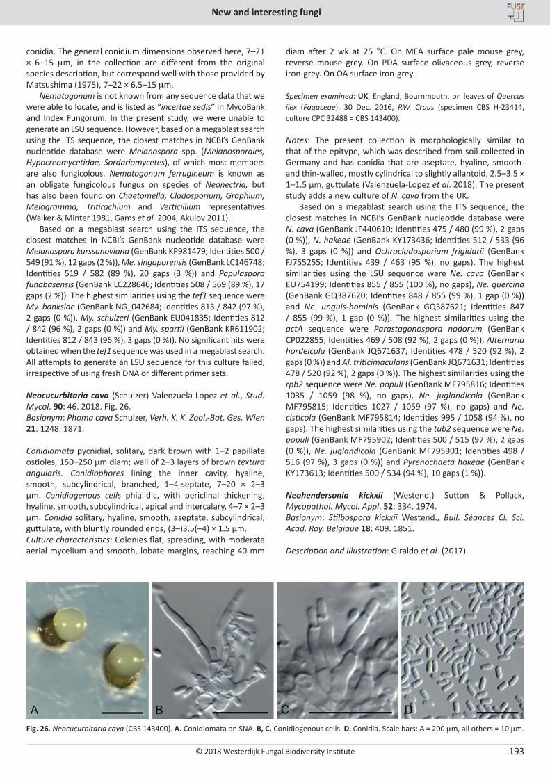
© 2018 Westerdijk Fungal Biodiversity Institute
New and interesting fungi
Editor-in-ChiefProf. dr P.W. Crous, Westerdijk Fungal Biodiversity Institute, P.O. Box 85167, 3508 AD Utrecht, The Netherlands.E-mail:[email protected]
193
conidia. The general conidium dimensions observed here, 7–21 × 6–15 mm, in the collection are different from the original species description, but correspond well with those provided by Matsushima (1975), 7–22 × 6.5–15 mm.
Nematogonum is not known from any sequence data that we were able to locate, and is listed as “incertae sedis” in MycoBank and Index Fungorum. In the present study, we were unable to generate an LSU sequence. However, based on a megablast search using the ITS sequence, the closest matches in NCBI’s GenBank nucleotide database were Melanospora spp. (Melanosporales, Hypocreomycetidae, Sordariomycetes), of which most members are also fungicolous. Nematogonum ferrugineum is known as an obligate fungicolous fungus on species of Neonectria, but has also been found on Chaetomella, Cladosporium, Graphium, Melogramma, Tritirachium and Verticillium representatives (Walker & Minter 1981, Gams et al. 2004, Akulov 2011).
Based on a megablast search using the ITS sequence, the closest matches in NCBI’s GenBank nucleotide database were Melanospora kurssanoviana (GenBank KP981479; Identities 500 / 549 (91 %), 12 gaps (2 %)), Me. singaporensis (GenBank LC146748; Identities 519 / 582 (89 %), 20 gaps (3 %)) and Papulaspora funabasensis (GenBank LC228646; Identities 508 / 569 (89 %), 17 gaps (2 %)). The highest similarities using the tef1 sequence were My. banksiae (GenBank NG_042684; Identities 813 / 842 (97 %), 2 gaps (0 %)), My. schulzeri (GenBank EU041835; Identities 812 / 842 (96 %), 2 gaps (0 %)) and My. spartii (GenBank KR611902; Identities 812 / 843 (96 %), 3 gaps (0 %)). No significant hits were obtained when the tef1 sequence was used in a megablast search. All attempts to generate an LSU sequence for this culture failed, irrespective of using fresh DNA or different primer sets.
Neocucurbitaria cava (Schulzer) Valenzuela-Lopez et al., Stud. Mycol. 90: 46. 2018. Fig. 26.Basionym: Phoma cava Schulzer, Verh. K. K. Zool.-Bot. Ges. Wien 21: 1248. 1871.
Conidiomata pycnidial, solitary, dark brown with 1–2 papillate ostioles, 150–250 µm diam; wall of 2–3 layers of brown textura angularis. Conidiophores lining the inner cavity, hyaline, smooth, subcylindrical, branched, 1–4-septate, 7–20 × 2–3 µm. Conidiogenous cells phialidic, with periclinal thickening, hyaline, smooth, subcylindrical, apical and intercalary, 4–7 × 2–3 µm. Conidia solitary, hyaline, smooth, aseptate, subcylindrical, guttulate, with bluntly rounded ends, (3–)3.5(–4) × 1.5 µm.Culture characteristics: Colonies flat, spreading, with moderate aerial mycelium and smooth, lobate margins, reaching 40 mm
diam after 2 wk at 25 °C. On MEA surface pale mouse grey, reverse mouse grey. On PDA surface olivaceous grey, reverse iron-grey. On OA surface iron-grey.
Specimen examined: UK, England, Bournmouth, on leaves of Quercus ilex (Fagaceae), 30 Dec. 2016, P.W. Crous (specimen CBS H-23414, culture CPC 32488 = CBS 143400).
Notes: The present collection is morphologically similar to that of the epitype, which was described from soil collected in Germany and has conidia that are aseptate, hyaline, smooth- and thin-walled, mostly cylindrical to slightly allantoid, 2.5–3.5 × 1–1.5 µm, guttulate (Valenzuela-Lopez et al. 2018). The present study adds a new culture of N. cava from the UK.
Based on a megablast search using the ITS sequence, the closest matches in NCBI’s GenBank nucleotide database were N. cava (GenBank JF440610; Identities 475 / 480 (99 %), 2 gaps (0 %)), N. hakeae (GenBank KY173436; Identities 512 / 533 (96 %), 3 gaps (0 %)) and Ochrocladosporium frigidarii (GenBank FJ755255; Identities 439 / 463 (95 %), no gaps). The highest similarities using the LSU sequence were Ne. cava (GenBank EU754199; Identities 855 / 855 (100 %), no gaps), Ne. quercina (GenBank GQ387620; Identities 848 / 855 (99 %), 1 gap (0 %)) and Ne. unguis-hominis (GenBank GQ387621; Identities 847 / 855 (99 %), 1 gap (0 %)). The highest similarities using the actA sequence were Parastagonospora nodorum (GenBank CP022855; Identities 469 / 508 (92 %), 2 gaps (0 %)), Alternaria hordeicola (GenBank JQ671637; Identities 478 / 520 (92 %), 2 gaps (0 %)) and Al. triticimaculans (GenBank JQ671631; Identities 478 / 520 (92 %), 2 gaps (0 %)). The highest similarities using the rpb2 sequence were Ne. populi (GenBank MF795816; Identities 1035 / 1059 (98 %), no gaps), Ne. juglandicola (GenBank MF795815; Identities 1027 / 1059 (97 %), no gaps) and Ne. cisticola (GenBank MF795814; Identities 995 / 1058 (94 %), no gaps). The highest similarities using the tub2 sequence were Ne. populi (GenBank MF795902; Identities 500 / 515 (97 %), 2 gaps (0 %)), Ne. juglandicola (GenBank MF795901; Identities 498 / 516 (97 %), 3 gaps (0 %)) and Pyrenochaeta hakeae (GenBank KY173613; Identities 500 / 534 (94 %), 10 gaps (1 %)).
Neohendersonia kickxii (Westend.) Sutton & Pollack, Mycopathol. Mycol. Appl. 52: 334. 1974. Basionym: Stilbospora kickxii Westend., Bull. Séances Cl. Sci. Acad. Roy. Belgique 18: 409. 1851.
Description and illustration: Giraldo et al. (2017).
Fig. 26. Neocucurbitaria cava (CBS 143400). A. Conidiomata on SNA. B, C. Conidiogenous cells. D. Conidia. Scale bars: A = 200 mm, all others = 10 mm.
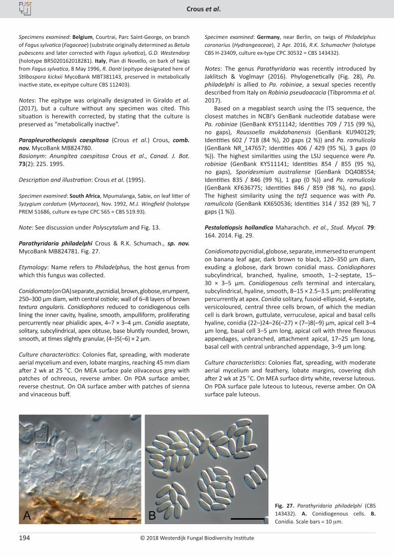
© 2018 Westerdijk Fungal Biodiversity Institute
Crous et al.
Editor-in-ChiefProf. dr P.W. Crous, Westerdijk Fungal Biodiversity Institute, P.O. Box 85167, 3508 AD Utrecht, The Netherlands.E-mail:[email protected]
194
Specimens examined: Belgium, Courtrai, Parc Saint-George, on branch of Fagus sylvatica (Fagaceae) (substrate originally determined as Betula pubescens and later corrected with Fagus sylvatica), G.D. Westendorp (holotype BR5020162018281). Italy, Pian di Novello, on bark of twigs from Fagus sylvatica, 8 May 1996, R. Danti (epitype designated here of Stilbospora kickxii MycoBank MBT381143, preserved in metabolically inactive state, ex-epitype culture CBS 112403).
Notes: The epitype was originally designated in Giraldo et al. (2017), but a culture without any specimen was cited. This situation is herewith corrected, by stating that the culture is preserved as “metabolically inactive”.
Parapleurotheciopsis caespitosa (Crous et al.) Crous, comb. nov. MycoBank MB824780. Basionym: Anungitea caespitosa Crous et al., Canad. J. Bot. 73(2): 225. 1995.
Description and illustration: Crous et al. (1995).
Specimen examined: South Africa, Mpumalanga, Sabie, on leaf litter of Syzygium cordatum (Myrtaceae), Nov. 1992, M.J. Wingfield (holotype PREM 51686, culture ex-type CPC 565 = CBS 519.93).
Note: See discussion under Polyscytalum and Fig. 13.
Parathyridaria philadelphi Crous & R.K. Schumach., sp. nov. MycoBank MB824781. Fig. 27.
Etymology: Name refers to Philadelphus, the host genus from which this fungus was collected.
Conidiomata (on OA) separate, pycnidial, brown, globose, erumpent, 250–300 µm diam, with central ostiole; wall of 6–8 layers of brown textura angularis. Conidiophores reduced to conidiogenous cells lining the inner cavity, hyaline, smooth, ampulliform, proliferating percurrently near phialidic apex, 4–7 × 3–4 µm. Conidia aseptate, solitary, subcylindrical, apex obtuse, base bluntly rounded, brown, smooth, at times slightly granular, (4–)5(–6) × 2 µm.
Culture characteristics: Colonies flat, spreading, with moderate aerial mycelium and even, lobate margins, reaching 45 mm diam after 2 wk at 25 °C. On MEA surface pale olivaceous grey with patches of ochreous, reverse amber. On PDA surface amber, reverse chestnut. On OA surface amber with patches of sienna and vinaceous buff.
Specimen examined: Germany, near Berlin, on twigs of Philadelphus coronarius (Hydrangeaceae), 2 Apr. 2016, R.K. Schumacher (holotype CBS H-23409, culture ex-type CPC 30532 = CBS 143432).
Notes: The genus Parathyridaria was recently introduced by Jaklitsch & Voglmayr (2016). Phylogenetically (Fig. 28), Pa. philadelphi is allied to Pa. robiniae, a sexual species recently described from Italy on Robinia pseudoacacia (Tibpromma et al. 2017).
Based on a megablast search using the ITS sequence, the closest matches in NCBI’s GenBank nucleotide database were Pa. robiniae (GenBank KY511142; Identities 709 / 715 (99 %), no gaps), Roussoella mukdahanensis (GenBank KU940129; Identities 602 / 718 (84 %), 20 gaps (2 %)) and Pa. ramulicola (GenBank NR_147657; Identities 406 / 429 (95 %), 3 gaps (0 %)). The highest similarities using the LSU sequence were Pa. robiniae (GenBank KY511141; Identities 854 / 855 (95 %), no gaps), Sporidesmium australiense (GenBank DQ408554; Identities 835 / 846 (99 %), 1 gap (0 %)) and Pa. ramulicola (GenBank KF636775; Identities 846 / 859 (98 %), no gaps). The highest similarity using the tef1 sequence was with Pa. ramulicola (GenBank KX650536; Identities 314 / 352 (89 %), 7 gaps (1 %)).
Pestalotiopsis hollandica Maharachch. et al., Stud. Mycol. 79: 164. 2014. Fig. 29.
Conidiomata pycnidial, globose, separate, immersed to erumpent on banana leaf agar, dark brown to black, 120–350 µm diam, exuding a globose, dark brown conidial mass. Conidiophores subcylindrical, branched, hyaline, smooth, 1–2-septate, 15–30 × 3–5 µm. Conidiogenous cells terminal and intercalary, subcylindrical, hyaline, smooth, 8–15 × 2.5–3.5 µm; proliferating percurrently at apex. Conidia solitary, fusoid-ellipsoid, 4-septate, versicoloured, central three cells brown, of which the median cell is dark brown, guttulate, verruculose, apical and basal cells hyaline, conidia (22–)24–26(–27) × (7–)8(–9) µm, apical cell 3–4 µm long, basal cell 3–5 µm long, apical cell with three flexuous appendages, unbranched, attachment apical, 17–25 µm long, basal cell with central unbranched appendage, 3–9 µm long.
Culture characteristics: Colonies flat, spreading, with moderate aerial mycelium and feathery, lobate margins, covering dish after 2 wk at 25 °C. On MEA surface dirty white, reverse luteous. On PDA surface pale luteous to luteous, reverse amber. On OA surface pale luteous.
Fig. 27. Parathyridaria philadelphi (CBS 143432). A. Conidiogenous cells. B. Conidia. Scale bars = 10 mm.
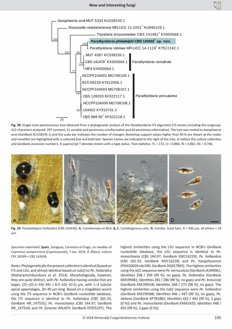
© 2018 Westerdijk Fungal Biodiversity Institute
New and interesting fungi
Editor-in-ChiefProf. dr P.W. Crous, Westerdijk Fungal Biodiversity Institute, P.O. Box 85167, 3508 AD Utrecht, The Netherlands.E-mail:[email protected]
195
Specimen examined: Spain, Zaragoza, Carretera el Frago, on needles of Cupressus sempervirens (Cupressaceae), 7 Jan. 2016, R. Blasco, culture CPC 30399 = CBS 143436.
Notes: Phylogenetically the present collection is identical (based on ITS and LSU, and almost identical based on tub2) to Pe. hollandica (Maharachchikumbura et al. 2014). Morphologically, however, they are quite distinct, with Pe. hollandica having conidia that are larger, (25–)25.5–33(–34) × 8.5–10(–10.5) μm, with 1–4 tubular apical appendages, 20–40 μm long. Based on a megablast search using the ITS sequence in NCBI’s GenBank nucleotide database, the ITS sequence is identical to Pe. hollandica (CBS 265.33; GenBank NR_147555), Pe. monochaeta (CBS 144.97; GenBank NR_147554) and Pe. funerea (ML4DY; GenBank EF055197). The
highest similarities using the LSU sequence in NCBI’s GenBank nucleotide database, the LSU sequence is identical to Pe. monochaeta (CBS 144.97; GenBank KM116229), Pe. hollandica (CBS 265.33; GenBank KM116228) and Pe. hangzhouensis (PSHI2002Endo390; GenBank DQ657865). The highest similarities using the tef1 sequence were Pe. verruculosa (GenBank JX399061; Identities 298 / 299 (99 %), no gaps), Pe. hollandica (GenBank KM199481; Identities 281 / 286 (98 %), no gaps) and Pe. brassicae (GenBank KM199558; Identities 268 / 273 (98 %), no gaps). The highest similarities using the tub2 sequence were Pe. hollandica (GenBank KM199388; Identities 446 / 447 (99 %), no gaps), Pe. italiana (GenBank KP781882; Identities 442 / 445 (99 %), 2 gaps (0 %)) and Pe. monochaeta (GenBank KX642435; Identities 448 / 452 (99 %), 2 gaps (0 %)).
100100
100
100
66
100
77
92
Parathyridaria philadelphi CBS 143432T sp. nov.
Parathyridaria robiniae MFLUCC 14-1119T KY511142.1
MUT 4397 KC339235.1
CBS 141479T KX650565.1
MF4 KX650564.1
NCCPF104001 MG708109.1
B15-06229 KY012056.1
NCCPF104003 MG708107.1
NCCPF104006 MG708108.1
104002 KY310731.1
CBS 868.95T KF322118.1
CBS 128203 KF322117.1
Thyridaria broussonetiae CBS 141481T KX650568.1
Roussoella mukdahanensis MFLUCC 11-0201T KU940129.1
Aaosphaeria arxii MUT 5333 KU158145.1
25
Parathyridaria ramulicola
Parathyridaria percutanea
Fig. 28. Single most parsimonious tree obtained from a phylogenetic analysis of the Parathyridaria ITS alignment (15 strains including the outgroup; 412 characters analysed: 297 constant, 51 variable and parsimony-uninformative and 64 parsimony-informative). The tree was rooted to Aaosphaeria arxii (GenBank KU158145.1) and the scale bar indicates the number of changes. Bootstrap support values higher than 49 % are shown at the nodes and novelties are highlighted with a coloured box and bold text. Species names are indicated to the right of the tree, or before the culture collection and GenBank accession numbers. A superscript T denotes strains with a type status. Tree statistics: TL = 172, CI = 0.866, RI = 0.861, RC = 0.746.
Fig. 29. Pestalotiopsis hollandica (CBS 143436). A. Conidiomata on BLA. B, C. Conidiogenous cells. D. Conidia. Scale bars: A = 300 mm, all others = 10 mm.
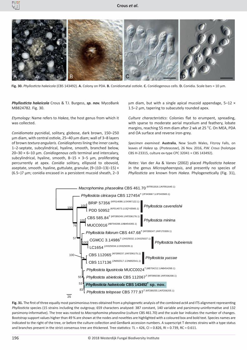
© 2018 Westerdijk Fungal Biodiversity Institute
Crous et al.
Editor-in-ChiefProf. dr P.W. Crous, Westerdijk Fungal Biodiversity Institute, P.O. Box 85167, 3508 AD Utrecht, The Netherlands.E-mail:[email protected]
196
Phyllosticta hakeicola Crous & T.I. Burgess, sp. nov. MycoBank MB824782. Fig. 30.
Etymology: Name refers to Hakea, the host genus from which it was collected.
Conidiomata pycnidial, solitary, globose, dark brown, 150–250 µm diam, with central ostiole, 25–40 mm diam; wall of 3–8 layers of brown textura angularis. Conidiophores lining the inner cavity, 1–2-septate, subcylindrical, hyaline, smooth, branched below, 20–30 × 6–10 µm. Conidiogenous cells terminal and intercalary, subcylindrical, hyaline, smooth, 8–15 × 3–5 µm, proliferating percurrently at apex. Conidia solitary, ellipsoid to obovoid, aseptate, smooth, hyaline, guttulate, granular, (9–)10–13(–15) × (6.5–)7 µm; conidia encased in a persistent mucoid sheath, 2–3
mm diam, but with a single apical mucoid appendage, 5–12 × 1.5–2 µm, tapering to subacutely rounded apex.
Culture characteristics: Colonies flat to erumpent, spreading, with sparse to moderate aerial mycelium and feathery, lobate margins, reaching 55 mm diam after 2 wk at 25 °C. On MEA, PDA and OA surface and reverse iron-grey.
Specimen examined: Australia, New South Wales, Fitzroy Falls, on leaves of Hakea sp. (Proteaceae), 26 Nov. 2016, P.W. Crous (holotype CBS H-23315, culture ex-type CPC 32041 = CBS 143492).
Notes: Van der Aa & Vanev (2002) placed Phyllosticta hakeae in the genus Microsphaeropsis, and presently no species of Phyllosticta are known from Hakea. Phylogenetically (Fig. 31),
Fig. 30. Phyllosticta hakeicola (CBS 143492). A. Colony on PDA. B. Conidiomatal ostiole. C. Conidiogenous cells. D. Conidia. Scale bars = 10 mm.
100
100
100
100
100
76
57
55
53
83
Phyllosticta abieticola CBS 112067T (KF289238.1/KF206208.1)
Phyllosticta hakeicola CBS 143492T sp. nov.
Phyllosticta telopeae CBS 777.97T (KF289255.1/KF206205.1)
Phyllosticta ligustricola MUCC0024T (AB704212.1/AB454269.1)
CBS 112065 (KF289237.1/KF206175.1)
CBS 117136 (JN692517.1/JN692541.1)
Phyllosticta foliorum CBS 447.68T (KF289247.1/KF170309.1)
CGMCC 3.14986T (JX025032.1/JX025037.1)
LC1654 (JX025034.1/JX025039.1)
CBS 585.84T (KF289249.1/KF206176.1)
MUCC0016 (AB704208.1/AB454265.1)
BRIP 57356 (KF014086.1/JX997122.1)
PDD 50952 (KF014073.1/JQ743565.1)
Phyllosticta citricarpa CBS 127454T (JF343667.1/JF343583.1)
Macrophomina phaseolina CBS 461.70 (KF951816.1/KF951640.1)
25
Phyllosticta cavendishii
Phyllosticta minima
Phyllosticta hubeiensis
Phyllosticta yuccae
Fig. 31. The first of three equally most parsimonious trees obtained from a phylogenetic analysis of the combined actA and ITS alignment representing Phyllosticta species (15 strains including the outgroup; 659 characters analysed: 387 constant, 140 variable and parsimony-uninformative and 132 parsimony-informative). The tree was rooted to Macrophomina phaseolina (culture CBS 461.70) and the scale bar indicates the number of changes. Bootstrap support values higher than 49 % are shown at the nodes and novelties are highlighted with a coloured box and bold text. Species names are indicated to the right of the tree, or before the culture collection and GenBank accession numbers. A superscript T denotes strains with a type status and branches present in the strict consensus tree are thickened. Tree statistics: TL = 426, CI = 0.826, RI = 0.739, RC = 0.611.
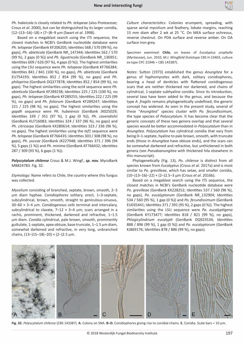
© 2018 Westerdijk Fungal Biodiversity Institute
New and interesting fungi
Editor-in-ChiefProf. dr P.W. Crous, Westerdijk Fungal Biodiversity Institute, P.O. Box 85167, 3508 AD Utrecht, The Netherlands.E-mail:[email protected]
197
Ph. hakeicola is closely related to Ph. telopeae (also Proteaceae; Crous et al. 2000), but can be distinguished by its larger conidia, (12–)13–16(–18) × (7–)8–9 mm (Swart et al. 1998).
Based on a megablast search using the ITS sequence, the closest matches in NCBI’s GenBank nucleotide database were Ph. telopeae (GenBank KF206205; Identities 568 / 570 (99 %), no gaps), Ph. abieticola (GenBank NR_147344; Identities 562 / 570 (99 %), 2 gaps (0 %)) and Ph. ligustricola (GenBank NR_136951; Identities 609 / 626 (97 %), 4 gaps (0 %)). The highest similarities using the LSU sequence were Ph. telopeae (GenBank KF766384; Identities 841 / 841 (100 %), no gaps), Ph. abieticola (GenBank EU754193; Identities 852 / 854 (99 %), no gaps) and Ph. philoprina (GenBank DQ377878; Identities 852 / 854 (99 %), no gaps). The highest similarities using the actA sequence were Ph. abieticola (GenBank KF289238; Identities 225 / 225 (100 %), no gaps), Ph. telopeae (GenBank KF289255; Identities 222 / 225 (99 %), no gaps) and Ph. foliorum (GenBank KF289247; Identities 221 / 225 (98 %), no gaps). The highest similarities using the gapdh sequence were Ph. hubeiensis (GenBank JX025029; Identities 339 / 351 (97 %), 1 gap (0 %)), Ph. cavendishii (GenBank KU716083; Identities 324 / 337 (96 %), no gaps) and Ph. citricarpa (GenBank KX280614; Identities 323 / 336 (96 %), no gaps). The highest similarities using the tef1 sequence were Ph. telopeae (GenBank KF766435; Identities 303 / 308 (98 %), no gaps), Ph. yuccae (GenBank JX227948; Identities 371 / 396 (94 %), 5 gaps (1 %)) and Ph. minima (GenBank KF766432; Identities 287 / 309 (93 %), 6 gaps (1 %)).
Polyscytalum chilense Crous & M.J. Wingf., sp. nov. MycoBank MB824783. Fig. 32.
Etymology: Name refers to Chile, the country where this fungus was collected.
Mycelium consisting of branched, septate, brown, smooth, 2–3 mm diam hyphae. Conidiophores solitary, erect, 1–3-septate, subcylindrical, brown, smooth, straight to geniculous-sinuous, 30–60 × 3–4 mm. Conidiogenous cells terminal and intercalary, subcylindrical to clavate, 7–12 × 3–4 mm; scars arranged in a rachis, prominent, thickened, darkened and refractive, 1–1.5 mm diam. Conidia cylindrical, pale brown, smooth, prominently guttulate, 1-septate, apex obtuse, base truncate, 1–1.5 mm diam, somewhat darkened and refractive, in very long, unbranched chains, (13–)15–18(–20) × (2–)2.5 mm.
Culture characteristics: Colonies erumpent, spreading, with sparse aerial mycelium and feathery, lobate margins, reaching 15 mm diam after 2 wk at 25 °C. On MEA surface ochreous, reverse chestnut. On PDA surface and reverse amber. On OA surface iron-grey.
Specimen examined: Chile, on leaves of Eucalyptus urophylla (Myrtaceae), Jun. 2010, M.J. Wingfield (holotype CBS H-23403, culture ex-type CPC 31946 = CBS 143387).
Notes: Sutton (1973) established the genus Anungitea for a genus of hyphomycetes with dark, solitary conidiophores, bearing a head of denticles with flattened conidiogenous scars that are neither thickened nor darkened, and chains of cylindrical, 1-septate subhyaline conidia. Since its introduction, several taxa have been added to the genus, and because the type A. fragilis remains phylogenetically undefined, the generic concept has widened. As seen in the present study, several of these “Anungitea” species cluster with Po. fecundissimum, the type species of Polyscytalum. It has become clear that the generic concepts of these two genera overlap and that several species would be better accommodated in Polyscytalum than in Anungitea. Polyscytalum has cylindrical conidia that vary from being 0–1-septate, hyaline to pale brown, smooth, with truncate ends (those in Anungitea have obtuse ends), and the scars can be somewhat darkened and refractive, but unthickened in both genera (see Pseudoanungitea with thickened hila elsewhere in this manuscript).
Phylogenetically (Fig. 13), Po. chilense is distinct from all species known from Eucalyptus (Crous et al. 2017a) and is most similar to Po. grevilleae, which has setae, and smaller conidia, (10–)13–16(–22) × (2–)2.5–3 µm (Crous et al. 2016b).
Based on a megablast search using the ITS sequence, the closest matches in NCBI’s GenBank nucleotide database were Po. grevilleae (GenBank KX228252; Identities 537 / 560 (96 %), no gaps), Po. eucalyptorum (GenBank NR_132904; Identities 534 / 560 (95 %), 1 gap (0 %)) and Po. fecundissimum (GenBank EU035441; Identities 371 / 391 (95 %), 2 gaps (0 %)). The highest similarities using the LSU sequence were Po. eucalyptigena (GenBank KY173477; Identities 818 / 821 (99 %), no gaps), Phlogicylindrium eucalypti (GenBank DQ923534; Identities 888 / 896 (99 %), 1 gap (0 %)) and Po. eucalyptorum (GenBank KJ869176; Identities 878 / 886 (99 %), no gaps).
Fig. 32. Polyscytalum chilense (CBS 143387). A. Colony on SNA. B–D. Conidiophores giving rise to conidial chains. E. Conidia. Scale bars = 10 mm.

© 2018 Westerdijk Fungal Biodiversity Institute
Crous et al.
Editor-in-ChiefProf. dr P.W. Crous, Westerdijk Fungal Biodiversity Institute, P.O. Box 85167, 3508 AD Utrecht, The Netherlands.E-mail:[email protected]
198
Polyscytalum eucalyptigenum (Crous & M.J. Wingf.) Crous & M.J. Wingf., comb. nov. MycoBank MB824784. Fig. 33.Basionym: Anungitea eucalyptigena Crous & M.J. Wingf., Persoonia 37: 339. 2016.
Description and illustration: Crous et al. (2016a).
Mycelium consisting of brown, smooth, septate, 2–3 µm diam hyphae. Conidiophores erect, solitary, subcylindrical, unbranched, brown, smooth, flexuous, 1–3-septate, 20–100 × 3–4 µm. Conidiogenous cells integrated, terminal, 7–15 × 3–4 µm, apex swollen with several sympodial loci, denticulate, flat-tipped, 1–2 × 2–2.5 µm, not thickened nor darkened. Ramoconidia subcylindrical, pale brown, smooth, 0–1-septate, 12–20 × 2.5–3 µm. Conidia occurring in long, unbranched chains, cylindrical with truncate ends, hyaline, smooth, guttulate, medianly 1-septate, (11–)13–17(–20) × (2–)2.5 µm.
Culture characteristics: Colonies flat, spreading, with sparse aerial mycelium and feathery, lobate margins, reaching 30 mm diam after 2 wk at 25 °C. On MEA surface ochreous, reverse iron-grey. On PDA surface amber, reverse brown vinaceous. On OA surface olivaceous grey.
Specimens examined: Chile, on leaves of Eucalyptus grandis × uromycoides (Myrtaceae), Jun. 2010, M.J. Wingfield (specimen CBS H-23421, culture CPC 31878 = CBS 143388). Malaysia, Kota Kinabalu, on leaf spots of Eucalyptus grandis × pellita (Myrtaceae), 30 May 2015, M.J. Wingfield (holotype CBS H-22888, culture ex-type CPC 28762 = CBS 142102).
Notes: The present collection from Chile is morphologically and phylogenetically (Fig. 13) similar to the ex-type strain of Po. eucalyptigenum from Malaysia (ramoconidia 16–20 × 2.5–3 µm, conidia (11–)14–16(–18) × (2–)2.5(–3) µm; Crous et al. 2016a). Based on a megablast search using the ITS sequence, the closest matches in NCBI’s GenBank nucleotide database were Po. eucalyptigena (GenBank KY173383; Identities 570 / 571 (99 %), no gaps), Po. grevilleae (GenBank KX228252; Identities 548 / 571 (96 %), 11 gaps (1 %)) and Po. eucalyptorum (GenBank NR_132904; Identities 545 / 571 (95 %), 12 gaps (2 %)). The highest similarities using the LSU sequence were Po. eucalyptorum (GenBank KY173477; Identities 819 / 821 (99 %), no gaps), Po. grevilleae (GenBank KX228304; Identities
824 / 831 (99 %), no gaps) and Phlogicylindrium eucalypti (GenBank DQ923534; Identities 835 / 844 (99 %), 1 gaps (0 %)).
Polyscytalum eucalyptorum (Crous & R.G. Shivas) Crous, comb. nov. MycoBank MB824785. Basionym: Anungitea eucalyptorum Crous & R.G. Shivas, Persoonia 32: 199. 2014.
Description and illustration: Crous et al. (2014).
Specimen examined: Australia, Queensland, Dave’s Creek, S28°12’13.7” E153°12’9.5”, on Eucalyptus (Myrtaceae) leaf litter, 11 Jul. 2009, P.W. Crous & R.G. Shivas, (holotype CBS H-21678, culture ex-type CPC 17207 = CBS 137967).
Polyscytalum grevilleae (Crous & Jacq. Edwards) Crous, comb. nov. MycoBank MB824786. Basionym: Anungitea grevilleae Crous & Jacq. Edwards, Persoonia 36: 327. 2016.
Description and illustration: Crous et al. (2016b).
Specimen examined: Australia, Victoria, Royal Botanic Gardens Cranbourne, S38°7’ 49.6” E145°16’9”, on leaves of Grevillea sp. (Proteaceae), 7 Nov. 2014, P.W. Crous & J. Edwards (holotype CBS H-22591, culture ex-type CPC 25576 = CBS 141282).
Polyscytalum neofecundissimum Crous & Akulov, sp. nov. MycoBank MB824787. Fig. 34.
Etymology: Name refers to its morphological similarity to Polyscytalum fecundissimum.
Conidiophores reduced to conidiogenous cells or erect, flexuous, subcylindrical, branched, up to 100 µm tall, pale brown, smooth. Conidiogenous cells terminal and intercalary, subcylindrical, pale brown, smooth, 20–25 × 3–4 µm, proliferating sympodially at apex, scars unthickened, 2–2.5 µm diam. Conidia occurring in chains, cylindrical with obtuse ends, hyaline, smooth, medianly 1-septate, guttulate, (12–)14–17(–20) × 2(–3) µm.
Culture characteristics: Colonies erumpent, spreading, surface folded, with moderate aerial mycelium and smooth, lobate
Fig. 33. Polyscytalum eucalyptigenum (CBS 143388). A–C. Conidiophores giving rise to conidial chains. D. Conidia. Scale bars = 10 mm.
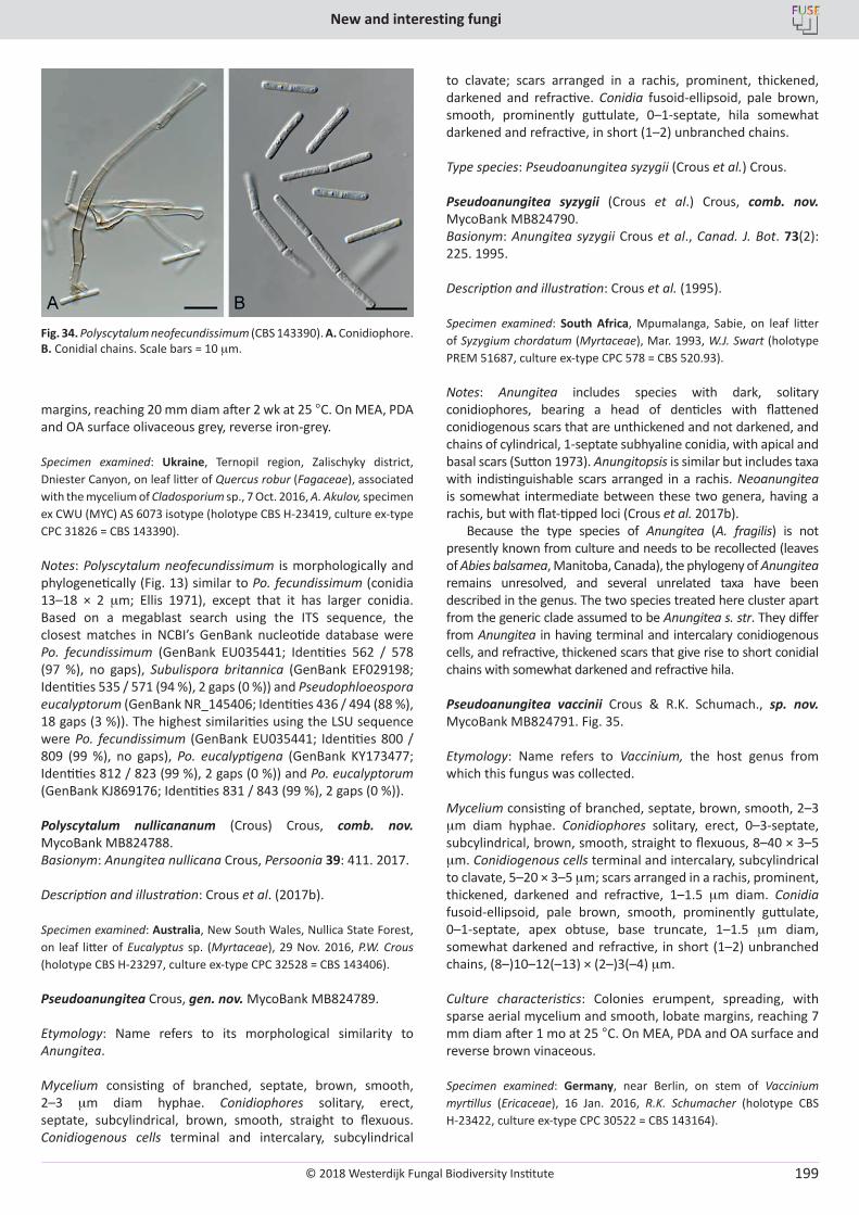
© 2018 Westerdijk Fungal Biodiversity Institute
New and interesting fungi
Editor-in-ChiefProf. dr P.W. Crous, Westerdijk Fungal Biodiversity Institute, P.O. Box 85167, 3508 AD Utrecht, The Netherlands.E-mail:[email protected]
199
margins, reaching 20 mm diam after 2 wk at 25 °C. On MEA, PDA and OA surface olivaceous grey, reverse iron-grey.
Specimen examined: Ukraine, Ternopil region, Zalischyky district, Dniester Canyon, on leaf litter of Quercus robur (Fagaceae), associated with the mycelium of Cladosporium sp., 7 Oct. 2016, A. Akulov, specimen ex CWU (MYC) AS 6073 isotype (holotype CBS H-23419, culture ex-type CPC 31826 = CBS 143390).
Notes: Polyscytalum neofecundissimum is morphologically and phylogenetically (Fig. 13) similar to Po. fecundissimum (conidia 13–18 × 2 mm; Ellis 1971), except that it has larger conidia. Based on a megablast search using the ITS sequence, the closest matches in NCBI’s GenBank nucleotide database were Po. fecundissimum (GenBank EU035441; Identities 562 / 578 (97 %), no gaps), Subulispora britannica (GenBank EF029198; Identities 535 / 571 (94 %), 2 gaps (0 %)) and Pseudophloeospora eucalyptorum (GenBank NR_145406; Identities 436 / 494 (88 %), 18 gaps (3 %)). The highest similarities using the LSU sequence were Po. fecundissimum (GenBank EU035441; Identities 800 / 809 (99 %), no gaps), Po. eucalyptigena (GenBank KY173477; Identities 812 / 823 (99 %), 2 gaps (0 %)) and Po. eucalyptorum (GenBank KJ869176; Identities 831 / 843 (99 %), 2 gaps (0 %)).
Polyscytalum nullicananum (Crous) Crous, comb. nov. MycoBank MB824788. Basionym: Anungitea nullicana Crous, Persoonia 39: 411. 2017.
Description and illustration: Crous et al. (2017b).
Specimen examined: Australia, New South Wales, Nullica State Forest, on leaf litter of Eucalyptus sp. (Myrtaceae), 29 Nov. 2016, P.W. Crous (holotype CBS H-23297, culture ex-type CPC 32528 = CBS 143406).
Pseudoanungitea Crous, gen. nov. MycoBank MB824789.
Etymology: Name refers to its morphological similarity to Anungitea.
Mycelium consisting of branched, septate, brown, smooth, 2–3 mm diam hyphae. Conidiophores solitary, erect, septate, subcylindrical, brown, smooth, straight to flexuous. Conidiogenous cells terminal and intercalary, subcylindrical
to clavate; scars arranged in a rachis, prominent, thickened, darkened and refractive. Conidia fusoid-ellipsoid, pale brown, smooth, prominently guttulate, 0–1-septate, hila somewhat darkened and refractive, in short (1–2) unbranched chains.
Type species: Pseudoanungitea syzygii (Crous et al.) Crous.
Pseudoanungitea syzygii (Crous et al.) Crous, comb. nov. MycoBank MB824790. Basionym: Anungitea syzygii Crous et al., Canad. J. Bot. 73(2): 225. 1995.
Description and illustration: Crous et al. (1995).
Specimen examined: South Africa, Mpumalanga, Sabie, on leaf litter of Syzygium chordatum (Myrtaceae), Mar. 1993, W.J. Swart (holotype PREM 51687, culture ex-type CPC 578 = CBS 520.93).
Notes: Anungitea includes species with dark, solitary conidiophores, bearing a head of denticles with flattened conidiogenous scars that are unthickened and not darkened, and chains of cylindrical, 1-septate subhyaline conidia, with apical and basal scars (Sutton 1973). Anungitopsis is similar but includes taxa with indistinguishable scars arranged in a rachis. Neoanungitea is somewhat intermediate between these two genera, having a rachis, but with flat-tipped loci (Crous et al. 2017b).
Because the type species of Anungitea (A. fragilis) is not presently known from culture and needs to be recollected (leaves of Abies balsamea, Manitoba, Canada), the phylogeny of Anungitea remains unresolved, and several unrelated taxa have been described in the genus. The two species treated here cluster apart from the generic clade assumed to be Anungitea s. str. They differ from Anungitea in having terminal and intercalary conidiogenous cells, and refractive, thickened scars that give rise to short conidial chains with somewhat darkened and refractive hila.
Pseudoanungitea vaccinii Crous & R.K. Schumach., sp. nov. MycoBank MB824791. Fig. 35.
Etymology: Name refers to Vaccinium, the host genus from which this fungus was collected.
Mycelium consisting of branched, septate, brown, smooth, 2–3 mm diam hyphae. Conidiophores solitary, erect, 0–3-septate, subcylindrical, brown, smooth, straight to flexuous, 8–40 × 3–5 mm. Conidiogenous cells terminal and intercalary, subcylindrical to clavate, 5–20 × 3–5 mm; scars arranged in a rachis, prominent, thickened, darkened and refractive, 1–1.5 mm diam. Conidia fusoid-ellipsoid, pale brown, smooth, prominently guttulate, 0–1-septate, apex obtuse, base truncate, 1–1.5 mm diam, somewhat darkened and refractive, in short (1–2) unbranched chains, (8–)10–12(–13) × (2–)3(–4) mm.
Culture characteristics: Colonies erumpent, spreading, with sparse aerial mycelium and smooth, lobate margins, reaching 7 mm diam after 1 mo at 25 °C. On MEA, PDA and OA surface and reverse brown vinaceous.
Specimen examined: Germany, near Berlin, on stem of Vaccinium myrtillus (Ericaceae), 16 Jan. 2016, R.K. Schumacher (holotype CBS H-23422, culture ex-type CPC 30522 = CBS 143164).
Fig. 34. Polyscytalum neofecundissimum (CBS 143390). A. Conidiophore. B. Conidial chains. Scale bars = 10 mm.
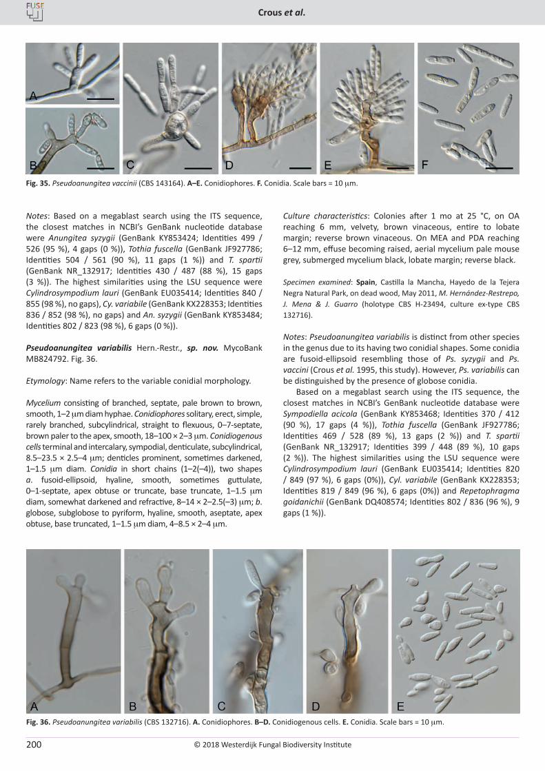
© 2018 Westerdijk Fungal Biodiversity Institute
Crous et al.
Editor-in-ChiefProf. dr P.W. Crous, Westerdijk Fungal Biodiversity Institute, P.O. Box 85167, 3508 AD Utrecht, The Netherlands.E-mail:[email protected]
200
Notes: Based on a megablast search using the ITS sequence, the closest matches in NCBI’s GenBank nucleotide database were Anungitea syzygii (GenBank KY853424; Identities 499 / 526 (95 %), 4 gaps (0 %)), Tothia fuscella (GenBank JF927786; Identities 504 / 561 (90 %), 11 gaps (1 %)) and T. spartii (GenBank NR_132917; Identities 430 / 487 (88 %), 15 gaps (3 %)). The highest similarities using the LSU sequence were Cylindrosympodium lauri (GenBank EU035414; Identities 840 / 855 (98 %), no gaps), Cy. variabile (GenBank KX228353; Identities 836 / 852 (98 %), no gaps) and An. syzygii (GenBank KY853484; Identities 802 / 823 (98 %), 6 gaps (0 %)).
Pseudoanungitea variabilis Hern.-Restr., sp. nov. MycoBank MB824792. Fig. 36.
Etymology: Name refers to the variable conidial morphology.
Mycelium consisting of branched, septate, pale brown to brown, smooth, 1–2 mm diam hyphae. Conidiophores solitary, erect, simple, rarely branched, subcylindrical, straight to flexuous, 0–7-septate, brown paler to the apex, smooth, 18–100 × 2–3 mm. Conidiogenous cells terminal and intercalary, sympodial, denticulate, subcylindrical, 8.5–23.5 × 2.5–4 mm; denticles prominent, sometimes darkened, 1–1.5 mm diam. Conidia in short chains (1–2(–4)), two shapes a. fusoid-ellipsoid, hyaline, smooth, sometimes guttulate, 0–1-septate, apex obtuse or truncate, base truncate, 1–1.5 mm diam, somewhat darkened and refractive, 8–14 × 2–2.5(–3) mm; b. globose, subglobose to pyriform, hyaline, smooth, aseptate, apex obtuse, base truncated, 1–1.5 mm diam, 4–8.5 × 2–4 mm.
Culture characteristics: Colonies after 1 mo at 25 °C, on OA reaching 6 mm, velvety, brown vinaceous, entire to lobate margin; reverse brown vinaceous. On MEA and PDA reaching 6–12 mm, effuse becoming raised, aerial mycelium pale mouse grey, submerged mycelium black, lobate margin; reverse black.
Specimen examined: Spain, Castilla la Mancha, Hayedo de la Tejera Negra Natural Park, on dead wood, May 2011, M. Hernández-Restrepo, J. Mena & J. Guarro (holotype CBS H-23494, culture ex-type CBS 132716).
Notes: Pseudoanungitea variabilis is distinct from other species in the genus due to its having two conidial shapes. Some conidia are fusoid-ellipsoid resembling those of Ps. syzygii and Ps. vaccini (Crous et al. 1995, this study). However, Ps. variabilis can be distinguished by the presence of globose conidia.
Based on a megablast search using the ITS sequence, the closest matches in NCBI’s GenBank nucleotide database were Sympodiella acicola (GenBank KY853468; Identities 370 / 412 (90 %), 17 gaps (4 %)), Tothia fuscella (GenBank JF927786; Identities 469 / 528 (89 %), 13 gaps (2 %)) and T. spartii (GenBank NR_132917; Identities 399 / 448 (89 %), 10 gaps (2 %)). The highest similarities using the LSU sequence were Cylindrosympodium lauri (GenBank EU035414; Identities 820 / 849 (97 %), 6 gaps (0%)), Cyl. variabile (GenBank KX228353; Identities 819 / 849 (96 %), 6 gaps (0%)) and Repetophragma goidanichii (GenBank DQ408574; Identities 802 / 836 (96 %), 9 gaps (1 %)).
Fig. 35. Pseudoanungitea vaccinii (CBS 143164). A–E. Conidiophores. F. Conidia. Scale bars = 10 mm.
Fig. 36. Pseudoanungitea variabilis (CBS 132716). A. Conidiophores. B–D. Conidiogenous cells. E. Conidia. Scale bars = 10 mm.
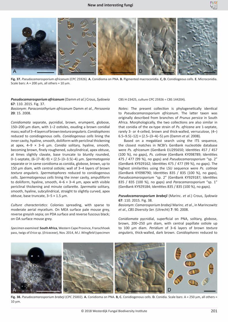
© 2018 Westerdijk Fungal Biodiversity Institute
New and interesting fungi
Editor-in-ChiefProf. dr P.W. Crous, Westerdijk Fungal Biodiversity Institute, P.O. Box 85167, 3508 AD Utrecht, The Netherlands.E-mail:[email protected]
201
Pseudocamarosporium africanum (Damm et al.) Crous, Sydowia 67: 110. 2015. Fig. 37.Basionym: Paraconiothyrium africanum Damm et al., Persoonia 20: 15. 2008.
Conidiomata separate, pycnidial, brown, erumpent, globose, 150–200 µm diam, with 1–2 ostioles, exuding a brown conidial mass; wall of 3–4 layers of brown textura angularis. Conidiophores reduced to conidiogenous cells. Conidiogenous cells lining the inner cavity, hyaline, smooth, doliiform with periclinal thickening at apex, 4–9 × 3–6 µm. Conidia solitary, hyaline, smooth, becoming brown, finely roughened, subcylindrical, apex obtuse, at times slightly clavate, base truncate to bluntly rounded, 0–1-septate, (6–)7–8(–9) × (2.5–)3–3.5(–4) µm. Spermatogonia separate or in same conidioma as conidia, globose, brown, up to 150 µm diam, with central ostiole; wall of 3–4 layers of brown textura angularis. Spermatophores reduced to conidiogenous cells. Spermatogenous cells lining the inner cavity, ampulliform to doliiform, hyaline, smooth, 4–6 × 3–4 µm, apex with visible periclinal thickening and minute collarette. Spermatia solitary, smooth, hyaline, subcylindrical, straight to slightly curved, apex obtuse, base truncate, 3–5 × 1.5 µm.
Culture characteristics: Colonies spreading, with sparse to moderate aerial mycelium. On MEA surface pale mouse grey, reverse greyish sepia; on PDA surface and reverse fuscous black; on OA surface mouse grey.
Specimen examined: South Africa, Western Cape Province, Franschhoek pass, twigs of Erica sp. (Ericaceae), Nov. 2014, M.J. Wingfield (specimen
CBS H-23425, culture CPC 25926 = CBS 144204).
Notes: The present collection is phylogenetically identical to Pseudocamarosporium africanum. The latter taxon was originally described from branches of Prunus persica in South Africa. Morphologically, the two collections are also similar in that conidia of the ex-type strain of Ps. africana are 1-septate, rarely 3- or 4-celled, brown and thick-walled, verruculose, (4–) 6.5–9.5(–12) × (2.5–)3–4(–5) mm (Damm et al. 2008).
Based on a megablast search using the ITS sequence, the closest matches in NCBI’s GenBank nucleotide database were Ps. africanum (GenBank EU295650; Identities 457 / 457 (100 %), no gaps), Ps. cotinae (GenBank KY098789; Identities 475 / 477 (99 %), no gaps) and Pseudocamarosporium “sp. 2” (GenBank KY929162; Identities 475 / 477 (99 %), no gaps). The highest similarities using the LSU sequence were Ps. cotinae (GenBank KY098790; Identities 835 / 835 (100 %), no gaps), Pseudocamarosporium “sp. 2” (GenBank KY929187; Identities 835 / 835 (100 %), no gaps) and Paracamarosporium “sp. 1” (GenBank KY929184; Identities 835 / 835 (100 %), no gaps).
Pseudocamarosporium brabeji (Marinc. et al.) Crous, Sydowia 67: 110. 2015. Fig. 38.Basionym: Camarosporium brabeji Marinc. et al., in Marincowitz et al., CBS Diversity Ser. (Utrecht) 7: 90. 2008.
Conidiomata pycnidial, superficial on PNA, solitary, globose, brown, 200–250 µm diam, with central papillate ostiole up to 100 µm diam. Peridium of 3–6 layers of brown textura angularis, thick-walled, dark brown. Conidiophores reduced to
Fig. 37. Pseudocamarosporium africanum (CPC 25926). A. Conidioma on PNA. B. Pigmented macroconidia. C, D. Conidiogeous cells. E. Microconidia. Scale bars: A = 200 mm, all others = 10 mm.
Fig. 38. Pseudocamarosporium brabeji (CPC 25002). A. Conidioma on PNA. B, C. Conidiogenous cells. D. Conidia. Scale bars: A = 250 mm, all others = 10 mm.
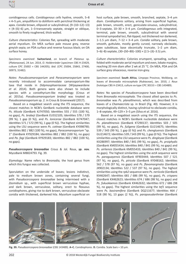
© 2018 Westerdijk Fungal Biodiversity Institute
Crous et al.
Editor-in-ChiefProf. dr P.W. Crous, Westerdijk Fungal Biodiversity Institute, P.O. Box 85167, 3508 AD Utrecht, The Netherlands.E-mail:[email protected]
202
conidiogenous cells. Conidiogenous cells hyaline, smooth, 5–8 × 4–5 µm, ampulliform to doliiform with periclinal thickening at apex. Conidia brown, ellipsoid or subcylindrical, (9–)10–12(–13) × (4–)5(–6) µm, 1–3-transversely septate, straight or oblique, smooth to finely roughened, thick-walled.
Culture characteristics: Colonies flat, spreading with moderate aerial mycelium. On MEA surface pale mouse grey, reverse greyish sepia; on PDA surface and reverse fuscous black; on OA surface honey.
Specimens examined: Switzerland, on branch of Platanus sp. (Platanaceae), 24 Jun. 2014, O. Holdenrieder (specimen CBS H-23429, culture CPC 25002 = CBS 144205); ibid. (CPC 25004, 25843, 27400, 30973, 31482).
Notes: Pseudocamarosporium and Paracamarosporium were recently introduced to accommodate camarosporium-like taxa that reside in Didymosphaeriaceae (Wijayawardene et al. 2014). Both genera were also shown to include species with a coniothyrium-like morphology (Crous et al. 2015a). Pseudocamarosporium brabeji was treated as Pseudocamarosporium sp. 2. in Crous & Groenewald (2017).
Based on a megablast search using the ITS sequence, the closest matches in NCBI’s GenBank nucleotide database were Ps. tilicola (GenBank KJ747050; Identities 555 / 555 (100 %), no gaps), Ps. brabeji (GenBank EU552105; Identities 578 / 579 (99 %), 1 gap (0 %)), and Ps. lonicerae (GenBank KJ747047; Identities 571 / 572 (99 %), 1 gap (0 %)). The highest similarities using the LSU sequence were Ps. cotinae (GenBank KY098790; Identities 882 / 882 (100 %), no gaps), Paracamarosporium “sp. 1” (GenBank KY929184; Identities 882 / 882 (100 %), no gaps) and Pa. fagi (GenBank KY929183; Identities 882 / 882 (100 %), no gaps).
Pseudocercospora breonadiae Crous & Jol. Roux, sp. nov. MycoBank MB824793. Fig. 39.
Etymology: Name refers to Breonadia, the host genus from which this fungus was collected.
Sporulation on the underside of leaves; lesions indistinct, pale to medium brown zones, containing several fungi, with Pseudocercospora breonadiae being intermixed with a Zasmidium sp., with superficial brown verruculose hyphae, and dark brown, verruculose, solitary, erect to flexuous conidiophores, giving rise to dark brown, verruculose obclavate conidia with thickened, darkened hila. Mycelium superficial on
host surface, pale brown, smooth, branched, septate, 3–4 mm diam. Conidiophores solitary, arising from superficial hyphae, pale brown, smooth, erect, geniculate-sinuous, subcylindrical, 0–3-septate, 10–30 × 3–4 mm. Conidiogenous cells integrated, terminal, pale brown, smooth, subcylindrical with several terminal sympodial loci, flat-tipped, not thickened nor darkened, 1–1.5 mm diam, 7–12 × 3–4 mm. Conidia solitary, pale brown, smooth, guttulate, mostly gently curved, narrowly obclavate, apex subobtuse, base obconically truncate, 1–2 mm diam, 5–6(–8)-septate, (30–)50–80(–100) × (2.5–)3(–3.5) mm.
Culture characteristics: Colonies erumpent, spreading, surface folded with moderate aerial mycelium and even, lobate margins, reaching 20 mm diam after 2 wk at 25 °C. On MEA, PDA and OA surface grey olivaceous, reverse iron-grey.
Specimen examined: South Africa, Limpopo Province, Wolkberg, on leaves of Breonadia microcephala (Rubiaceae), Jan. 2010, J. Roux (holotype CBS H-23413, culture ex-type CPC 30153 = CBS 143489).
Notes: No species of Pseudocercospora have been described from Breonadia microcephala. The closest allied species to Ps. breonadiae was Ps. planaltinensis, which was described from leaves of a Chamaecrista sp. in Brazil (Fig. 40). However, it is morphologically distinct, having cylindrical to obclavate conidia, 1–8-septate, 49–129 × 3–5 µm (Silva et al. 2016).
Based on a megablast search using the ITS sequence, the closest matches in NCBI’s GenBank nucleotide database were Ps. planaltinensis (GenBank KT290137; Identities 503 / 505 (99 %), no gaps), Ps. fuligena (GenBank GU214675; Identities 539 / 543 (99 %), 1 gap (0 %)) and Ps. chengtuensis (GenBank GU214672; Identities 539 / 543 (99 %), 1 gap (0 %)). The highest similarities using the LSU sequence were Ps. dingleyae (GenBank KX286997; Identities 840 / 841 (99 %), no gaps), Ps. proiphydis (GenBank KM055434; Identities 840 / 841 (99 %), no gaps) and Ps. airliensis (GenBank KM055433; Identities 840 / 841 (99 %), no gaps). The highest similarities using the actA sequence were Ps. paraguayensis (GenBank KF903444; Identities 507 / 521 (97 %), no gaps), Ps. piricola (GenBank KY048162; Identities 562 / 578 (97 %), no gaps) and Ps. flavomarginata (GenBank JX902134; Identities 522 / 537 (97 %), no gaps). The highest similarities using the rpb2 sequence were Ps. neriicola (GenBank KX462647; Identities 681 / 686 (99 %), no gaps), Ps. crispans (GenBank KX462623; Identities 674 / 686 (98 %), no gaps) and Ps. fukuokaensis (GenBank KX462632; Identities 672 / 686 (98 %), no gaps). The highest similarities using the tef1 sequence were Ps. basiramifera (GenBank DQ211677; Identities 464 / 518 (90 %), 13 gaps (2 %)), Ps. parapseudarthriae (GenBank
Fig. 39. Pseudocercospora breonadiae (CBS 143489). A–C. Conidiophores. D. Conidia. Scale bars = 10 mm.
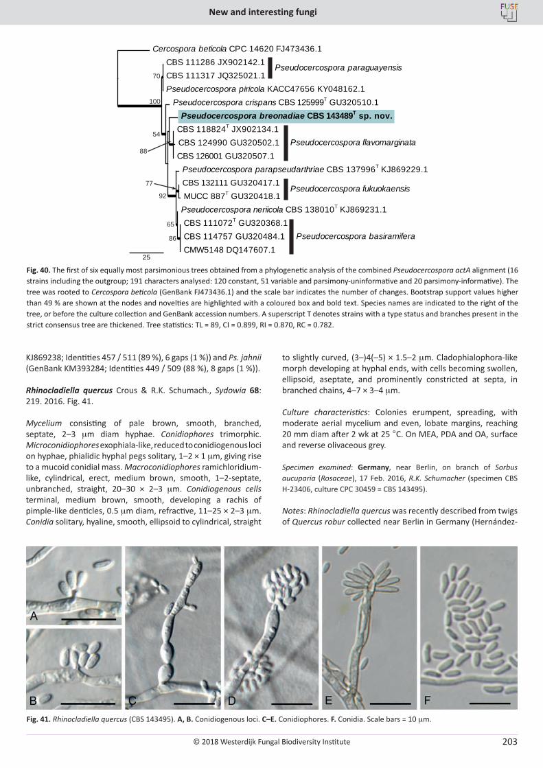
© 2018 Westerdijk Fungal Biodiversity Institute
New and interesting fungi
Editor-in-ChiefProf. dr P.W. Crous, Westerdijk Fungal Biodiversity Institute, P.O. Box 85167, 3508 AD Utrecht, The Netherlands.E-mail:[email protected]
203
KJ869238; Identities 457 / 511 (89 %), 6 gaps (1 %)) and Ps. jahnii (GenBank KM393284; Identities 449 / 509 (88 %), 8 gaps (1 %)).
Rhinocladiella quercus Crous & R.K. Schumach., Sydowia 68: 219. 2016. Fig. 41.
Mycelium consisting of pale brown, smooth, branched, septate, 2–3 mm diam hyphae. Conidiophores trimorphic. Microconidiophores exophiala-like, reduced to conidiogenous loci on hyphae, phialidic hyphal pegs solitary, 1–2 × 1 mm, giving rise to a mucoid conidial mass. Macroconidiophores ramichloridium-like, cylindrical, erect, medium brown, smooth, 1–2-septate, unbranched, straight, 20–30 × 2–3 mm. Conidiogenous cells terminal, medium brown, smooth, developing a rachis of pimple-like denticles, 0.5 mm diam, refractive, 11–25 × 2–3 mm. Conidia solitary, hyaline, smooth, ellipsoid to cylindrical, straight
to slightly curved, (3–)4(–5) × 1.5–2 mm. Cladophialophora-like morph developing at hyphal ends, with cells becoming swollen, ellipsoid, aseptate, and prominently constricted at septa, in branched chains, 4–7 × 3–4 mm.
Culture characteristics: Colonies erumpent, spreading, with moderate aerial mycelium and even, lobate margins, reaching 20 mm diam after 2 wk at 25 °C. On MEA, PDA and OA, surface and reverse olivaceous grey.
Specimen examined: Germany, near Berlin, on branch of Sorbus aucuparia (Rosaceae), 17 Feb. 2016, R.K. Schumacher (specimen CBS H-23406, culture CPC 30459 = CBS 143495).
Notes: Rhinocladiella quercus was recently described from twigs of Quercus robur collected near Berlin in Germany (Hernández-
100
70
54
88
92
65
86
77
CBS 111072T GU320368.1 CBS 114757 GU320484.1 CMW5148 DQ147607.1
Pseudocercospora neriicola CBS 138010T KJ869231.1
CBS 132111 GU320417.1 MUCC 887T GU320418.1
Pseudocercospora parapseudarthriae CBS 137996T KJ869229.1
Pseudocercospora breonadiae CBS 143489T sp. nov. CBS 118824T JX902134.1 CBS 124990 GU320502.1 CBS 126001 GU320507.1
Pseudocercospora crispans CBS 125999T GU320510.1
CBS 111286 JX902142.1 CBS 111317 JQ325021.1 Pseudocercospora piricola KACC47656 KY048162.1
Cercospora beticola CPC 14620 FJ473436.1
25
Pseudocercospora paraguayensis
Pseudocercospora flavomarginata
Pseudocercospora fukuokaensis
Pseudocercospora basiramifera
Fig. 40. The first of six equally most parsimonious trees obtained from a phylogenetic analysis of the combined Pseudocercospora actA alignment (16 strains including the outgroup; 191 characters analysed: 120 constant, 51 variable and parsimony-uninformative and 20 parsimony-informative). The tree was rooted to Cercospora beticola (GenBank FJ473436.1) and the scale bar indicates the number of changes. Bootstrap support values higher than 49 % are shown at the nodes and novelties are highlighted with a coloured box and bold text. Species names are indicated to the right of the tree, or before the culture collection and GenBank accession numbers. A superscript T denotes strains with a type status and branches present in the strict consensus tree are thickened. Tree statistics: TL = 89, CI = 0.899, RI = 0.870, RC = 0.782.
Fig. 41. Rhinocladiella quercus (CBS 143495). A, B. Conidiogenous loci. C–E. Conidiophores. F. Conidia. Scale bars = 10 mm.
© 2018 Westerdijk Fungal Biodiversity Institute
Crous et al.
Editor-in-ChiefProf. dr P.W. Crous, Westerdijk Fungal Biodiversity Institute, P.O. Box 85167, 3508 AD Utrecht, The Netherlands.E-mail:[email protected]
204
Restrepo et al. 2016). The morphology of the present collection on Sorbus aucuparia closely matches that of the type.
Based on a megablast search using the ITS sequence, the closest matches in NCBI’s GenBank nucleotide database were R. quercus (GenBank KX306769; Identities 633 / 642 (99 %), 1 gap (0 %)), Capronia sp. (GenBank AF050240; Identities 613 / 617 (99 %), 1 gap (0 %)) and Cladophialophora sp. (GenBank JX494354; Identities 621 / 634 (98 %), 3 gaps (0 %)). The highest similarities using the LSU sequence were R. quercus (GenBank KX306794; Identities 792 / 792 (100 %), no gaps), Capronia sp. (GenBank JN941378; Identities 799 / 801 (99 %), no gaps) and Ca. fungicola (GenBank FJ358224; Identities 768 / 801 (96 %), 3 gaps (0 %)). No significant hits were obtained when the tef1 and tub2 sequences were used in a megablast search.
Roussoella euonymi Crous & Akulov, sp. nov. MycoBank MB824794. Fig. 42.
Etymology: Name refers to Euonymus, the host genus from which this fungus was collected.
Conidiomata erumpent, globose, brown, pycnidial, 150–300 µm diam, with central ostiole, exuding a black conidial mass. Conidiophores reduced to conidiogenous cells, lining the inner cavity, hyaline, smooth, ampulliform to doliiform, proliferating percurrently at apex, 5–12 × 5–7 µm. Conidia solitary, ellipsoid, guttulate, aseptate, apex obtuse, base 2 µm diam, bluntly rounded, thick-walled, becoming warty, golden-brown to red-brown, (6–)7(–8) × (4–)5–6 µm.
Culture characteristics: Colonies flat, spreading, with moderate aerial mycelium and even, lobate margins, reaching 60 mm diam after 2 wk at 25 °C. On MEA surface olivaceous grey with patches of pale olivaceous grey, reverse pale olivaceous grey. On PDA surface olivaceous grey, reverse iron-grey. On OA surface iron-grey.
Specimen examined: Ukraine, Ternopil region, Zalischyky district, Dniester Canyon, on fallen branches of Euonymus europaeus (Celastraceae), 14 Oct. 2016, A. Akulov, specimen ex CWU (MYC) AS 6061 isotype (holotype CBS H-23420, culture ex-type CPC 31963 = CBS 143426).
Notes: Based on the LSU sequence, Roussoella euonymi is accommodated in the Roussoellaceae, being similar to other asexual species such as Ro. solani and Ro. mexicana (Crous et al. 2015b, c). Based on a megablast search using the ITS sequence, the closest matches in NCBI’s GenBank nucleotide database were numerous unidentified “Pleosporales sp.” sequences (e.g. GenBank HM116753; Identities 542 / 561 (97 %), 1 gap (0 %)), with the closest known species being Ro. neopustulans (GenBank KJ474833; Identities 441 / 474 (93 %), 5 gaps (1 %)) and Ro. pustulans (GenBank KJ474830; Identities 442 / 478 (92 %), 4 gaps (0 %)). The highest similarities using the LSU sequence were Ro. mukdahanensis (GenBank KU863118; Identities 837 / 847 (99 %), no gaps), Arthopyrenia salicis (GenBank LN907499; Identities 843 / 854 (99 %), no gaps) and Ro. neopustulans (GenBank KU863119; Identities 839 / 850 (99 %), no gaps). Only distant hits were obtained using the actA sequence; some of these were Stagonosporopsis cucurbitacearum (GenBank KX246908; Identities 459 / 509 (90 %), 10 gaps (1 %)), S. citrulli (GenBank KX246907; Identities 459 / 509 (90 %), 10 gaps (1 %))
and S. caricae (GenBank KX246909; Identities 458 / 509 (90 %), 10 gaps (1 %)). Only distant hits were obtained using the rpb2 sequence; for example with Torula herbarum (GenBank KF443393; Identities 582 / 735 (79 %), 6 gaps (0 %)). No significant hits were obtained with the tub2 sequence.
Setophaeosphaeria citricola Crous & M.J. Wingf., sp. nov. MycoBank MB824795. Fig. 43.
Etymology: Name refers to Citrus, the host genus from which this fungus was collected.
Ascomata on twigs immersed, black, 150–250 mm diam, globose, opening via a central ostiole that could with age become an irregular rupture in ascomatal wall; wall of 2–3 layers of brown textura angularis. Asci bitunicate, sessile, subcylindrical to narrowly ellipsoid, apical chamber 1–2 mm diam, stipitate, 50–70 × 11–15 mm. Ascospores multiseriate, hyaline, thin-walled, smooth, aseptate, fusoid-ellipsoidal, widest in upper third, apex subobtusely rounded, base obtuse, (16–)19–20(–22) × (4.5–)5(–6) mm. Conidiomata pycnidial, 150–250 mm diam, aggregated, globose, pale brown with dark brown central ostiole, 20–30 mm diam, ostiole surrounded by brown, thick-walled, verruculose, septate hyphae, up to 100 mm long, 4–5 mm diam at base, apex obtuse. Conidiophores reduced to conidiogenous cells lining the inner cavity, hyaline, smooth, ampulliform to doliiform, 5–7 × 5–6 mm, phialidic with prominent periclinal thickening. Conidia solitary, hyaline, smooth, aseptate, multiguttulate and granular, fusoid-ellipsoid, straight to irregularly twisted, apex obtuse, base truncate, 2 mm diam, (10–)12–14(–17) × 3–3.5(–4) mm.
Culture characteristics: Colonies flat, spreading, with sparse aerial mycelium and even, lobate margins, reaching 45 mm diam after 2 wk at 25 °C. On MEA surface pale mouse grey to mouse grey, reverse mouse grey. On PDA surface olivaceous grey, reverse mouse grey. On OA surface olivaceous grey.
Specimen examined: Australia, New South Wales, Mount Annan Botanical Garden, on leaves of Citrus australasica (Rutaceae), 25 Nov. 2016, P.W. Crous (holotype CBS H-23271, culture ex-type CPC 32083 = CBS 143179).
Notes: Coniothyrium sidae was recently described from a Sida sp. collected in Brazil (Quaedvlieg et al. 2013). Although the ITS is identical (Fig. 44), the morphology is very different, with the sexual morph having hyaline, aseptate ascospores, those of Con. sidae being brown, (3–)5-septate, (18–)20–24(–26) × (4–)5(–5.5) µm, and conidia being smaller, fusoid-ellipsoidal, straight to slightly curved, (9–)10–12(–13) × (2.5–)3 µm. Conidia of S. citri, described from Citrus in Italy, are smaller than those of S. citricola, 3.5–5 × 2–3 µm (Crous et al. 2017b).
Based on a megablast search using the ITS sequence, the closest matches in NCBI’s GenBank nucleotide database were Con. sidae (GenBank KF251149; Identities 518 / 520 (99 %), no gaps), Phaeosphaeria setosa (GenBank AF439500; Identities 472 / 476 (99 %), no gaps) and S. hemerocallidis (GenBank KJ869161; Identities 503 / 521 (97 %), 10 gaps (1 %)). The highest similarities using the LSU sequence were Con. sidae (GenBank KF251653; Identities 835 / 836 (99 %), no gaps), S. badalingensis (GenBank KJ869219; Identities 828 / 832 (99 %), no gaps) and Leptosphaeria rubefaciens (GenBank JF740311; Identities 843 / 854 (99 %), no gaps). The highest similarities
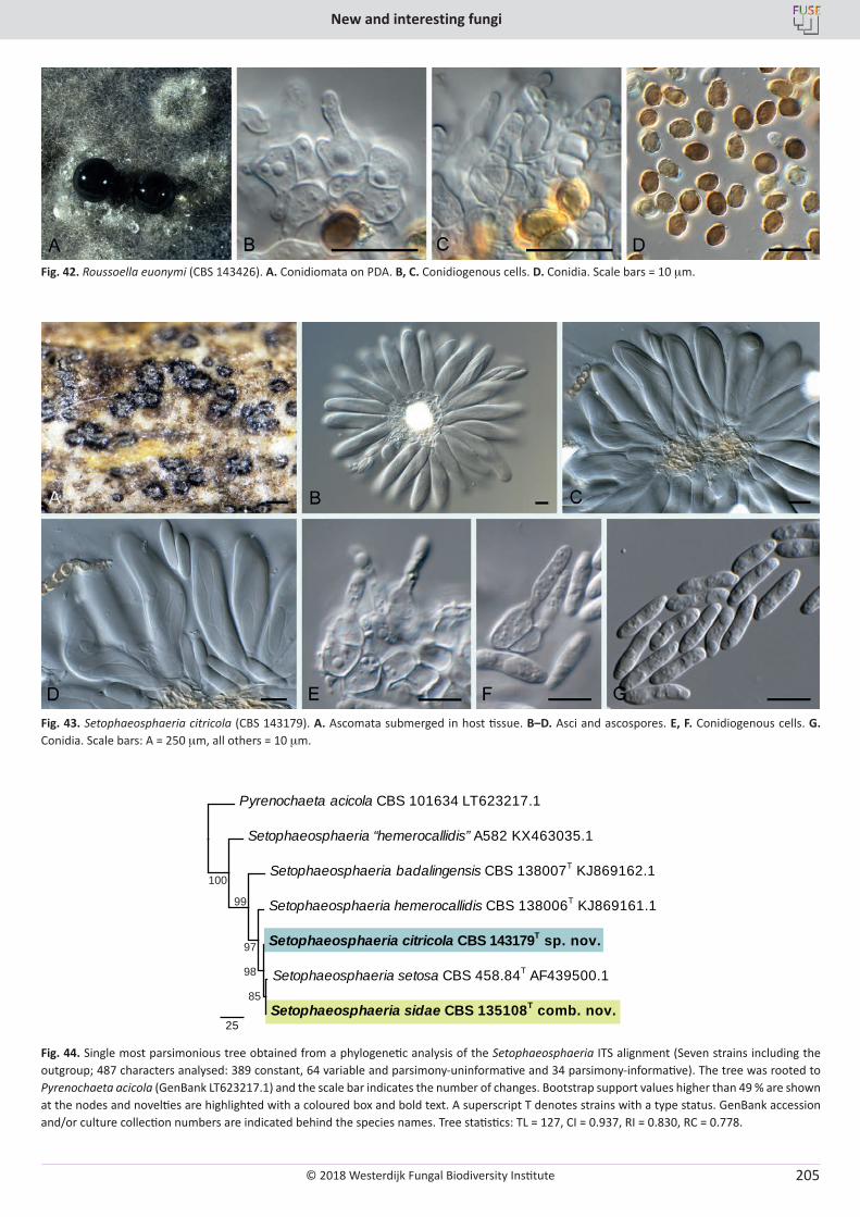
© 2018 Westerdijk Fungal Biodiversity Institute
New and interesting fungi
Editor-in-ChiefProf. dr P.W. Crous, Westerdijk Fungal Biodiversity Institute, P.O. Box 85167, 3508 AD Utrecht, The Netherlands.E-mail:[email protected]
205
Fig. 42. Roussoella euonymi (CBS 143426). A. Conidiomata on PDA. B, C. Conidiogenous cells. D. Conidia. Scale bars = 10 mm.
Fig. 43. Setophaeosphaeria citricola (CBS 143179). A. Ascomata submerged in host tissue. B–D. Asci and ascospores. E, F. Conidiogenous cells. G. Conidia. Scale bars: A = 250 mm, all others = 10 mm.
100
99
97
98
85
Setophaeosphaeria badalingensis CBS 138007T KJ869162.1
Setophaeosphaeria hemerocallidis CBS 138006T KJ869161.1
Setophaeosphaeria setosa CBS 458.84T AF439500.1
Setophaeosphaeria sidae CBS 135108T comb. nov.
Setophaeosphaeria citricola CBS 143179T sp. nov.
Setophaeosphaeria “hemerocallidis” A582 KX463035.1
Pyrenochaeta acicola CBS 101634 LT623217.1
25
Fig. 44. Single most parsimonious tree obtained from a phylogenetic analysis of the Setophaeosphaeria ITS alignment (Seven strains including the outgroup; 487 characters analysed: 389 constant, 64 variable and parsimony-uninformative and 34 parsimony-informative). The tree was rooted to Pyrenochaeta acicola (GenBank LT623217.1) and the scale bar indicates the number of changes. Bootstrap support values higher than 49 % are shown at the nodes and novelties are highlighted with a coloured box and bold text. A superscript T denotes strains with a type status. GenBank accession and/or culture collection numbers are indicated behind the species names. Tree statistics: TL = 127, CI = 0.937, RI = 0.830, RC = 0.778.
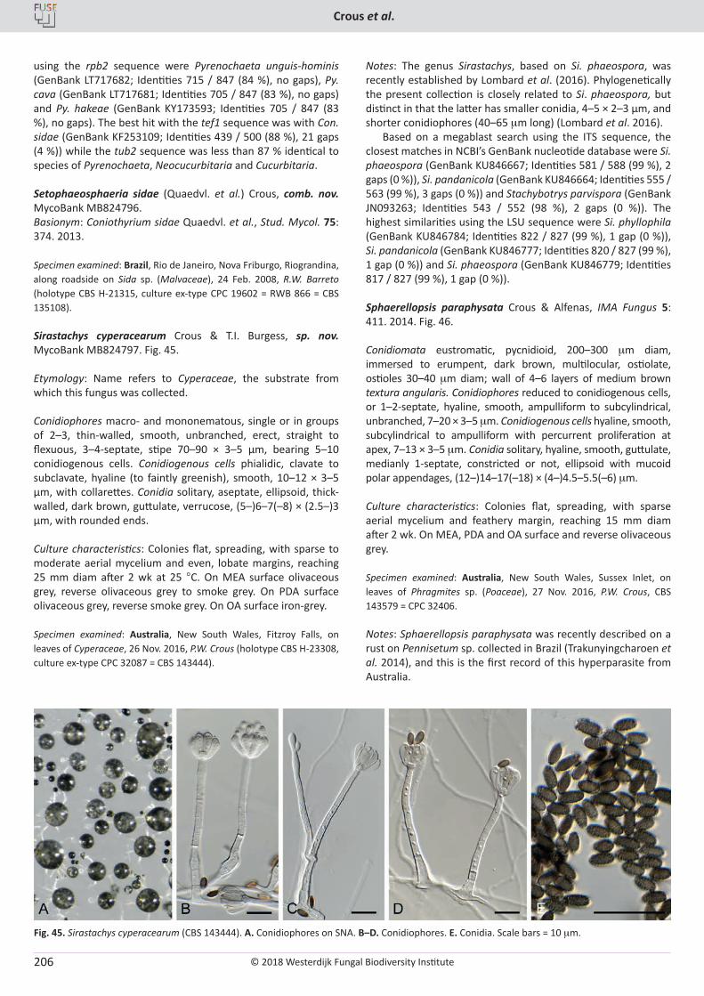
© 2018 Westerdijk Fungal Biodiversity Institute
Crous et al.
Editor-in-ChiefProf. dr P.W. Crous, Westerdijk Fungal Biodiversity Institute, P.O. Box 85167, 3508 AD Utrecht, The Netherlands.E-mail:[email protected]
206
using the rpb2 sequence were Pyrenochaeta unguis-hominis (GenBank LT717682; Identities 715 / 847 (84 %), no gaps), Py. cava (GenBank LT717681; Identities 705 / 847 (83 %), no gaps) and Py. hakeae (GenBank KY173593; Identities 705 / 847 (83 %), no gaps). The best hit with the tef1 sequence was with Con. sidae (GenBank KF253109; Identities 439 / 500 (88 %), 21 gaps (4 %)) while the tub2 sequence was less than 87 % identical to species of Pyrenochaeta, Neocucurbitaria and Cucurbitaria.
Setophaeosphaeria sidae (Quaedvl. et al.) Crous, comb. nov. MycoBank MB824796. Basionym: Coniothyrium sidae Quaedvl. et al., Stud. Mycol. 75: 374. 2013.
Specimen examined: Brazil, Rio de Janeiro, Nova Friburgo, Riograndina, along roadside on Sida sp. (Malvaceae), 24 Feb. 2008, R.W. Barreto (holotype CBS H-21315, culture ex-type CPC 19602 = RWB 866 = CBS 135108).
Sirastachys cyperacearum Crous & T.I. Burgess, sp. nov. MycoBank MB824797. Fig. 45.
Etymology: Name refers to Cyperaceae, the substrate from which this fungus was collected.
Conidiophores macro- and mononematous, single or in groups of 2–3, thin-walled, smooth, unbranched, erect, straight to flexuous, 3–4-septate, stipe 70–90 × 3–5 µm, bearing 5–10 conidiogenous cells. Conidiogenous cells phialidic, clavate to subclavate, hyaline (to faintly greenish), smooth, 10–12 × 3–5 µm, with collarettes. Conidia solitary, aseptate, ellipsoid, thick-walled, dark brown, guttulate, verrucose, (5–)6–7(–8) × (2.5–)3 µm, with rounded ends.
Culture characteristics: Colonies flat, spreading, with sparse to moderate aerial mycelium and even, lobate margins, reaching 25 mm diam after 2 wk at 25 °C. On MEA surface olivaceous grey, reverse olivaceous grey to smoke grey. On PDA surface olivaceous grey, reverse smoke grey. On OA surface iron-grey.
Specimen examined: Australia, New South Wales, Fitzroy Falls, on leaves of Cyperaceae, 26 Nov. 2016, P.W. Crous (holotype CBS H-23308, culture ex-type CPC 32087 = CBS 143444).
Notes: The genus Sirastachys, based on Si. phaeospora, was recently established by Lombard et al. (2016). Phylogenetically the present collection is closely related to Si. phaeospora, but distinct in that the latter has smaller conidia, 4–5 × 2–3 μm, and shorter conidiophores (40–65 mm long) (Lombard et al. 2016).
Based on a megablast search using the ITS sequence, the closest matches in NCBI’s GenBank nucleotide database were Si. phaeospora (GenBank KU846667; Identities 581 / 588 (99 %), 2 gaps (0 %)), Si. pandanicola (GenBank KU846664; Identities 555 / 563 (99 %), 3 gaps (0 %)) and Stachybotrys parvispora (GenBank JN093263; Identities 543 / 552 (98 %), 2 gaps (0 %)). The highest similarities using the LSU sequence were Si. phyllophila (GenBank KU846784; Identities 822 / 827 (99 %), 1 gap (0 %)), Si. pandanicola (GenBank KU846777; Identities 820 / 827 (99 %), 1 gap (0 %)) and Si. phaeospora (GenBank KU846779; Identities 817 / 827 (99 %), 1 gap (0 %)).
Sphaerellopsis paraphysata Crous & Alfenas, IMA Fungus 5: 411. 2014. Fig. 46.
Conidiomata eustromatic, pycnidioid, 200–300 mm diam, immersed to erumpent, dark brown, multilocular, ostiolate, ostioles 30–40 mm diam; wall of 4–6 layers of medium brown textura angularis. Conidiophores reduced to conidiogenous cells, or 1–2-septate, hyaline, smooth, ampulliform to subcylindrical, unbranched, 7–20 × 3–5 mm. Conidiogenous cells hyaline, smooth, subcylindrical to ampulliform with percurrent proliferation at apex, 7–13 × 3–5 mm. Conidia solitary, hyaline, smooth, guttulate, medianly 1-septate, constricted or not, ellipsoid with mucoid polar appendages, (12–)14–17(–18) × (4–)4.5–5.5(–6) mm.
Culture characteristics: Colonies flat, spreading, with sparse aerial mycelium and feathery margin, reaching 15 mm diam after 2 wk. On MEA, PDA and OA surface and reverse olivaceous grey.
Specimen examined: Australia, New South Wales, Sussex Inlet, on leaves of Phragmites sp. (Poaceae), 27 Nov. 2016, P.W. Crous, CBS 143579 = CPC 32406.
Notes: Sphaerellopsis paraphysata was recently described on a rust on Pennisetum sp. collected in Brazil (Trakunyingcharoen et al. 2014), and this is the first record of this hyperparasite from Australia.
Fig. 45. Sirastachys cyperacearum (CBS 143444). A. Conidiophores on SNA. B–D. Conidiophores. E. Conidia. Scale bars = 10 mm.
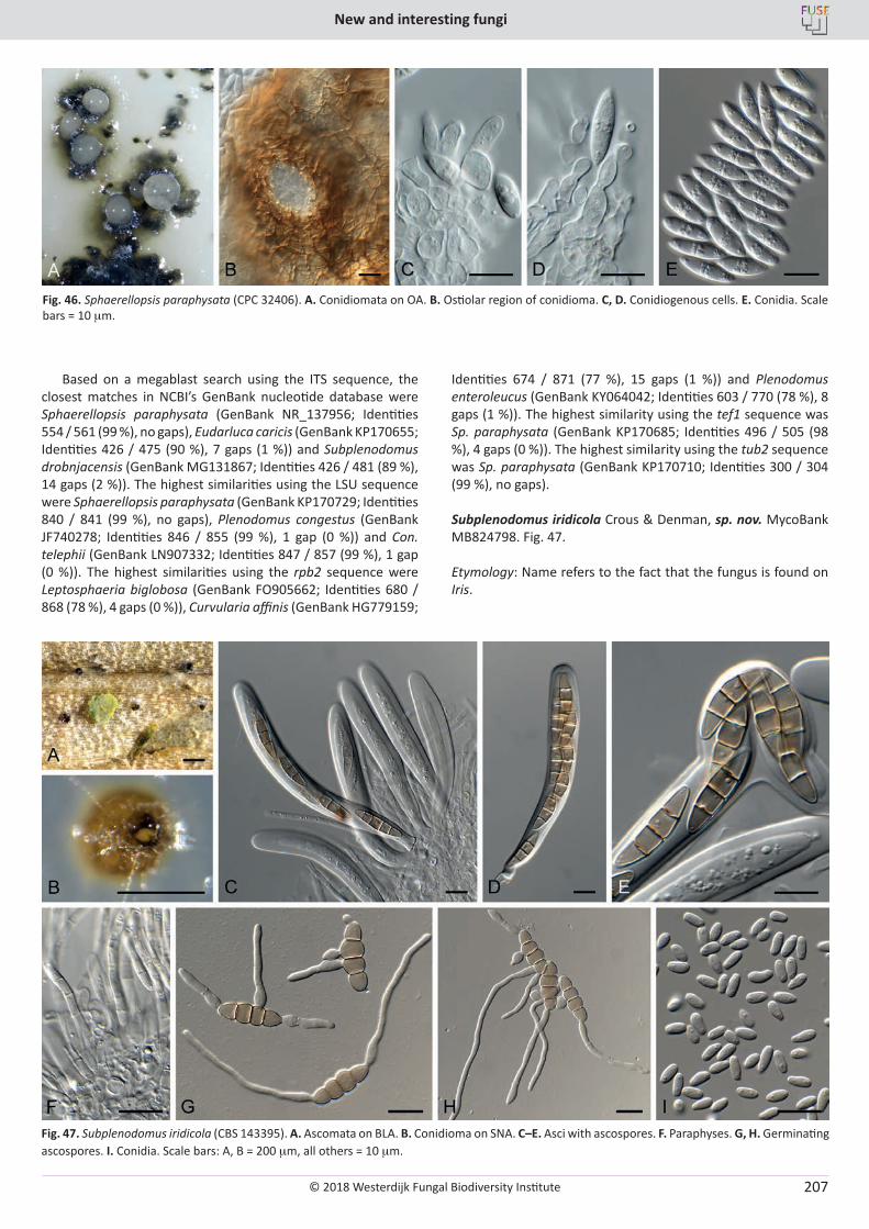
© 2018 Westerdijk Fungal Biodiversity Institute
New and interesting fungi
Editor-in-ChiefProf. dr P.W. Crous, Westerdijk Fungal Biodiversity Institute, P.O. Box 85167, 3508 AD Utrecht, The Netherlands.E-mail:[email protected]
207
Based on a megablast search using the ITS sequence, the closest matches in NCBI’s GenBank nucleotide database were Sphaerellopsis paraphysata (GenBank NR_137956; Identities 554 / 561 (99 %), no gaps), Eudarluca caricis (GenBank KP170655; Identities 426 / 475 (90 %), 7 gaps (1 %)) and Subplenodomus drobnjacensis (GenBank MG131867; Identities 426 / 481 (89 %), 14 gaps (2 %)). The highest similarities using the LSU sequence were Sphaerellopsis paraphysata (GenBank KP170729; Identities 840 / 841 (99 %), no gaps), Plenodomus congestus (GenBank JF740278; Identities 846 / 855 (99 %), 1 gap (0 %)) and Con. telephii (GenBank LN907332; Identities 847 / 857 (99 %), 1 gap (0 %)). The highest similarities using the rpb2 sequence were Leptosphaeria biglobosa (GenBank FO905662; Identities 680 / 868 (78 %), 4 gaps (0 %)), Curvularia affinis (GenBank HG779159;
Identities 674 / 871 (77 %), 15 gaps (1 %)) and Plenodomus enteroleucus (GenBank KY064042; Identities 603 / 770 (78 %), 8 gaps (1 %)). The highest similarity using the tef1 sequence was Sp. paraphysata (GenBank KP170685; Identities 496 / 505 (98 %), 4 gaps (0 %)). The highest similarity using the tub2 sequence was Sp. paraphysata (GenBank KP170710; Identities 300 / 304 (99 %), no gaps).
Subplenodomus iridicola Crous & Denman, sp. nov. MycoBank MB824798. Fig. 47.
Etymology: Name refers to the fact that the fungus is found on Iris.
Fig. 46. Sphaerellopsis paraphysata (CPC 32406). A. Conidiomata on OA. B. Ostiolar region of conidioma. C, D. Conidiogenous cells. E. Conidia. Scale bars = 10 mm.
Fig. 47. Subplenodomus iridicola (CBS 143395). A. Ascomata on BLA. B. Conidioma on SNA. C–E. Asci with ascospores. F. Paraphyses. G, H. Germinating ascospores. I. Conidia. Scale bars: A, B = 200 mm, all others = 10 mm.
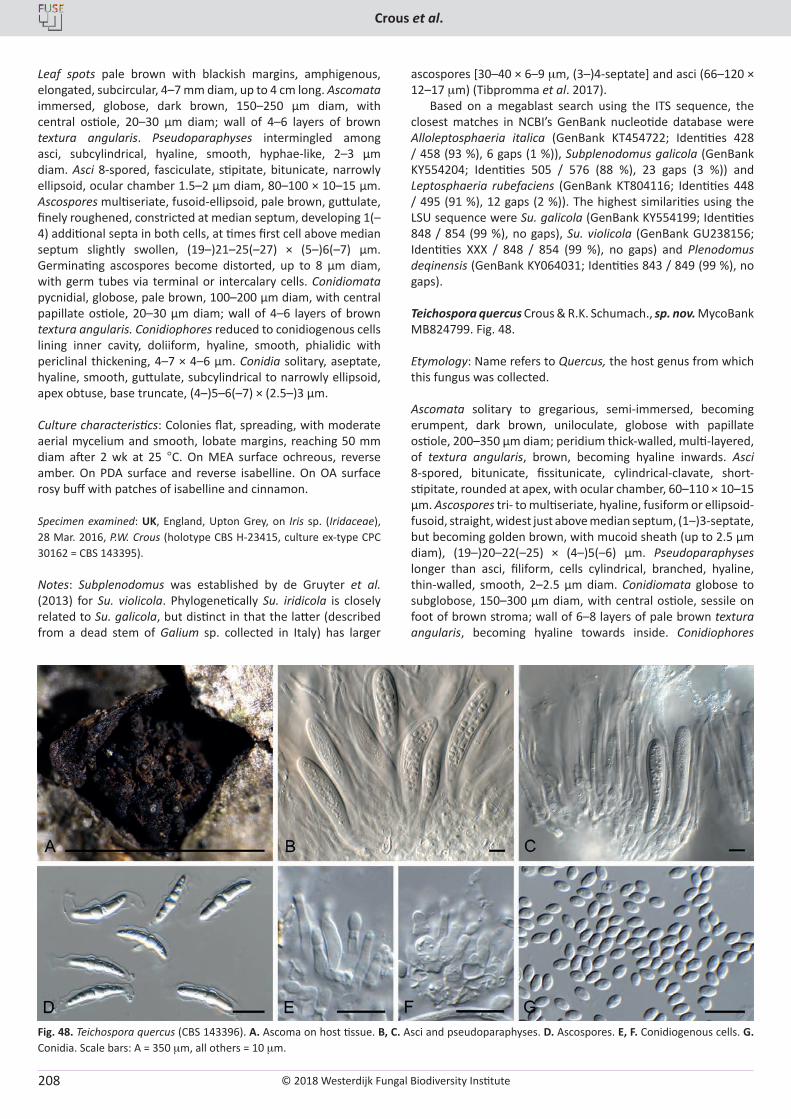
© 2018 Westerdijk Fungal Biodiversity Institute
Crous et al.
Editor-in-ChiefProf. dr P.W. Crous, Westerdijk Fungal Biodiversity Institute, P.O. Box 85167, 3508 AD Utrecht, The Netherlands.E-mail:[email protected]
208
Leaf spots pale brown with blackish margins, amphigenous, elongated, subcircular, 4–7 mm diam, up to 4 cm long. Ascomata immersed, globose, dark brown, 150–250 µm diam, with central ostiole, 20–30 µm diam; wall of 4–6 layers of brown textura angularis. Pseudoparaphyses intermingled among asci, subcylindrical, hyaline, smooth, hyphae-like, 2–3 µm diam. Asci 8-spored, fasciculate, stipitate, bitunicate, narrowly ellipsoid, ocular chamber 1.5–2 µm diam, 80–100 × 10–15 µm. Ascospores multiseriate, fusoid-ellipsoid, pale brown, guttulate, finely roughened, constricted at median septum, developing 1(–4) additional septa in both cells, at times first cell above median septum slightly swollen, (19–)21–25(–27) × (5–)6(–7) µm. Germinating ascospores become distorted, up to 8 µm diam, with germ tubes via terminal or intercalary cells. Conidiomata pycnidial, globose, pale brown, 100–200 µm diam, with central papillate ostiole, 20–30 µm diam; wall of 4–6 layers of brown textura angularis. Conidiophores reduced to conidiogenous cells lining inner cavity, doliiform, hyaline, smooth, phialidic with periclinal thickening, 4–7 × 4–6 µm. Conidia solitary, aseptate, hyaline, smooth, guttulate, subcylindrical to narrowly ellipsoid, apex obtuse, base truncate, (4–)5–6(–7) × (2.5–)3 µm.
Culture characteristics: Colonies flat, spreading, with moderate aerial mycelium and smooth, lobate margins, reaching 50 mm diam after 2 wk at 25 °C. On MEA surface ochreous, reverse amber. On PDA surface and reverse isabelline. On OA surface rosy buff with patches of isabelline and cinnamon.
Specimen examined: UK, England, Upton Grey, on Iris sp. (Iridaceae), 28 Mar. 2016, P.W. Crous (holotype CBS H-23415, culture ex-type CPC 30162 = CBS 143395).
Notes: Subplenodomus was established by de Gruyter et al. (2013) for Su. violicola. Phylogenetically Su. iridicola is closely related to Su. galicola, but distinct in that the latter (described from a dead stem of Galium sp. collected in Italy) has larger
ascospores [30–40 × 6–9 mm, (3–)4-septate] and asci (66–120 × 12–17 mm) (Tibpromma et al. 2017).
Based on a megablast search using the ITS sequence, the closest matches in NCBI’s GenBank nucleotide database were Alloleptosphaeria italica (GenBank KT454722; Identities 428 / 458 (93 %), 6 gaps (1 %)), Subplenodomus galicola (GenBank KY554204; Identities 505 / 576 (88 %), 23 gaps (3 %)) and Leptosphaeria rubefaciens (GenBank KT804116; Identities 448 / 495 (91 %), 12 gaps (2 %)). The highest similarities using the LSU sequence were Su. galicola (GenBank KY554199; Identities 848 / 854 (99 %), no gaps), Su. violicola (GenBank GU238156; Identities XXX / 848 / 854 (99 %), no gaps) and Plenodomus deqinensis (GenBank KY064031; Identities 843 / 849 (99 %), no gaps).
Teichospora quercus Crous & R.K. Schumach., sp. nov. MycoBank MB824799. Fig. 48.
Etymology: Name refers to Quercus, the host genus from which this fungus was collected.
Ascomata solitary to gregarious, semi-immersed, becoming erumpent, dark brown, uniloculate, globose with papillate ostiole, 200–350 µm diam; peridium thick-walled, multi-layered, of textura angularis, brown, becoming hyaline inwards. Asci 8-spored, bitunicate, fissitunicate, cylindrical-clavate, short-stipitate, rounded at apex, with ocular chamber, 60–110 × 10–15 µm. Ascospores tri- to multiseriate, hyaline, fusiform or ellipsoid-fusoid, straight, widest just above median septum, (1–)3-septate, but becoming golden brown, with mucoid sheath (up to 2.5 µm diam), (19–)20–22(–25) × (4–)5(–6) µm. Pseudoparaphyses longer than asci, filiform, cells cylindrical, branched, hyaline, thin-walled, smooth, 2–2.5 µm diam. Conidiomata globose to subglobose, 150–300 µm diam, with central ostiole, sessile on foot of brown stroma; wall of 6–8 layers of pale brown textura angularis, becoming hyaline towards inside. Conidiophores
Fig. 48. Teichospora quercus (CBS 143396). A. Ascoma on host tissue. B, C. Asci and pseudoparaphyses. D. Ascospores. E, F. Conidiogenous cells. G. Conidia. Scale bars: A = 350 mm, all others = 10 mm.
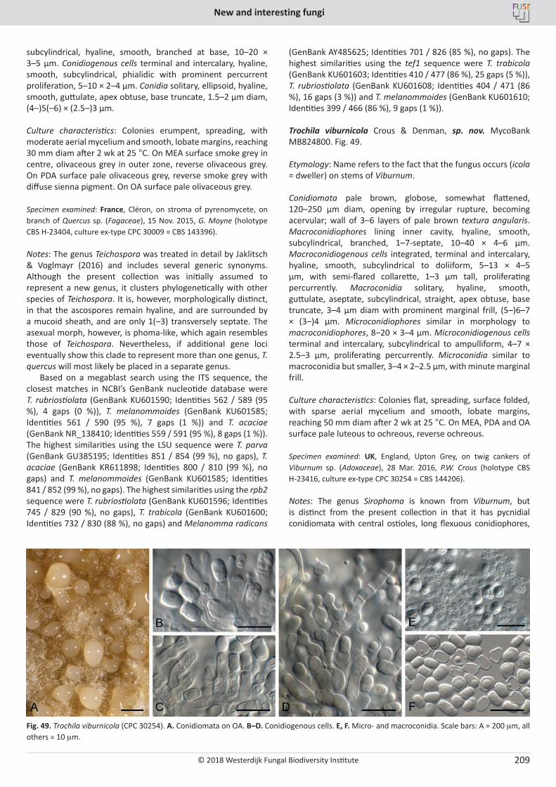
© 2018 Westerdijk Fungal Biodiversity Institute
New and interesting fungi
Editor-in-ChiefProf. dr P.W. Crous, Westerdijk Fungal Biodiversity Institute, P.O. Box 85167, 3508 AD Utrecht, The Netherlands.E-mail:[email protected]
209
subcylindrical, hyaline, smooth, branched at base, 10–20 × 3–5 µm. Conidiogenous cells terminal and intercalary, hyaline, smooth, subcylindrical, phialidic with prominent percurrent proliferation, 5–10 × 2–4 µm. Conidia solitary, ellipsoid, hyaline, smooth, guttulate, apex obtuse, base truncate, 1.5–2 µm diam, (4–)5(–6) × (2.5–)3 µm.
Culture characteristics: Colonies erumpent, spreading, with moderate aerial mycelium and smooth, lobate margins, reaching 30 mm diam after 2 wk at 25 °C. On MEA surface smoke grey in centre, olivaceous grey in outer zone, reverse olivaceous grey. On PDA surface pale olivaceous grey, reverse smoke grey with diffuse sienna pigment. On OA surface pale olivaceous grey.
Specimen examined: France, Cléron, on stroma of pyrenomycete, on branch of Quercus sp. (Fagaceae), 15 Nov. 2015, G. Moyne (holotype CBS H-23404, culture ex-type CPC 30009 = CBS 143396).
Notes: The genus Teichospora was treated in detail by Jaklitsch & Voglmayr (2016) and includes several generic synonyms. Although the present collection was initially assumed to represent a new genus, it clusters phylogenetically with other species of Teichospora. It is, however, morphologically distinct, in that the ascospores remain hyaline, and are surrounded by a mucoid sheath, and are only 1(–3) transversely septate. The asexual morph, however, is phoma-like, which again resembles those of Teichospora. Nevertheless, if additional gene loci eventually show this clade to represent more than one genus, T. quercus will most likely be placed in a separate genus.
Based on a megablast search using the ITS sequence, the closest matches in NCBI’s GenBank nucleotide database were T. rubriostiolata (GenBank KU601590; Identities 562 / 589 (95 %), 4 gaps (0 %)), T. melanommoides (GenBank KU601585; Identities 561 / 590 (95 %), 7 gaps (1 %)) and T. acaciae (GenBank NR_138410; Identities 559 / 591 (95 %), 8 gaps (1 %)). The highest similarities using the LSU sequence were T. parva (GenBank GU385195; Identities 851 / 854 (99 %), no gaps), T. acaciae (GenBank KR611898; Identities 800 / 810 (99 %), no gaps) and T. melanommoides (GenBank KU601585; Identities 841 / 852 (99 %), no gaps). The highest similarities using the rpb2 sequence were T. rubriostiolata (GenBank KU601596; Identities 745 / 829 (90 %), no gaps), T. trabicola (GenBank KU601600; Identities 732 / 830 (88 %), no gaps) and Melanomma radicans
(GenBank AY485625; Identities 701 / 826 (85 %), no gaps). The highest similarities using the tef1 sequence were T. trabicola (GenBank KU601603; Identities 410 / 477 (86 %), 25 gaps (5 %)), T. rubriostiolata (GenBank KU601608; Identities 404 / 471 (86 %), 16 gaps (3 %)) and T. melanommoides (GenBank KU601610; Identities 399 / 466 (86 %), 9 gaps (1 %)).
Trochila viburnicola Crous & Denman, sp. nov. MycoBank MB824800. Fig. 49.
Etymology: Name refers to the fact that the fungus occurs (icola = dweller) on stems of Viburnum.
Conidiomata pale brown, globose, somewhat flattened, 120–250 µm diam, opening by irregular rupture, becoming acervular; wall of 3–6 layers of pale brown textura angularis. Macroconidiophores lining inner cavity, hyaline, smooth, subcylindrical, branched, 1–7-septate, 10–40 × 4–6 µm. Macroconidiogenous cells integrated, terminal and intercalary, hyaline, smooth, subcylindrical to doliiform, 5–13 × 4–5 µm, with semi-flared collarette, 1–3 µm tall, proliferating percurrently. Macroconidia solitary, hyaline, smooth, guttulate, aseptate, subcylindrical, straight, apex obtuse, base truncate, 3–4 µm diam with prominent marginal frill, (5–)6–7 × (3–)4 µm. Microconidiophores similar in morphology to macroconidiophores, 8–20 × 3–4 µm. Microconidiogenous cells terminal and intercalary, subcylindrical to ampulliform, 4–7 × 2.5–3 µm, proliferating percurrently. Microconidia similar to macroconidia but smaller, 3–4 × 2–2.5 µm, with minute marginal frill.
Culture characteristics: Colonies flat, spreading, surface folded, with sparse aerial mycelium and smooth, lobate margins, reaching 50 mm diam after 2 wk at 25 °C. On MEA, PDA and OA surface pale luteous to ochreous, reverse ochreous.
Specimen examined: UK, England, Upton Grey, on twig cankers of Viburnum sp. (Adoxaceae), 28 Mar. 2016, P.W. Crous (holotype CBS H-23416, culture ex-type CPC 30254 = CBS 144206).
Notes: The genus Sirophoma is known from Viburnum, but is distinct from the present collection in that it has pycnidial conidiomata with central ostioles, long flexuous conidiophores,
Fig. 49. Trochila viburnicola (CPC 30254). A. Conidiomata on OA. B–D. Conidiogenous cells. E, F. Micro- and macroconidia. Scale bars: A = 200 mm, all others = 10 mm.
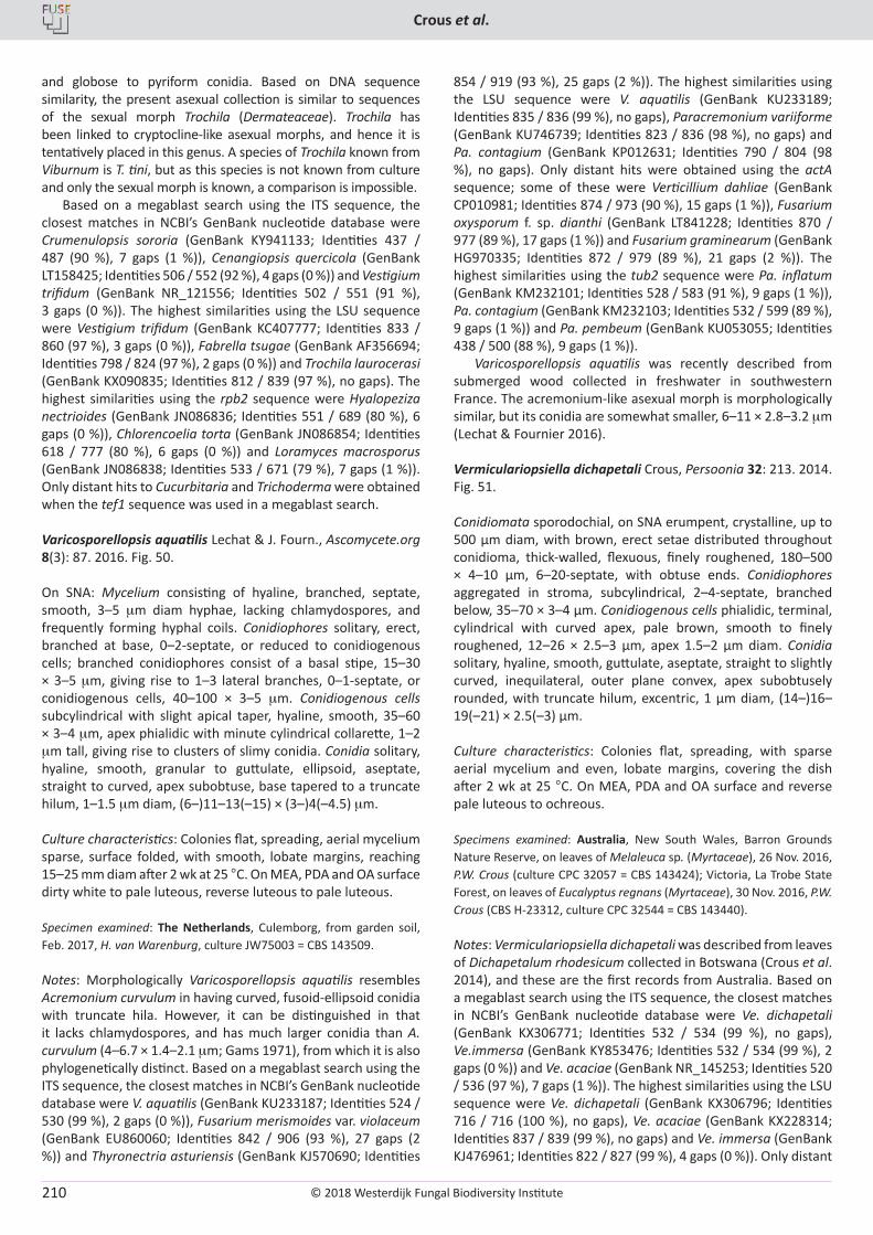
© 2018 Westerdijk Fungal Biodiversity Institute
Crous et al.
Editor-in-ChiefProf. dr P.W. Crous, Westerdijk Fungal Biodiversity Institute, P.O. Box 85167, 3508 AD Utrecht, The Netherlands.E-mail:[email protected]
210
and globose to pyriform conidia. Based on DNA sequence similarity, the present asexual collection is similar to sequences of the sexual morph Trochila (Dermateaceae). Trochila has been linked to cryptocline-like asexual morphs, and hence it is tentatively placed in this genus. A species of Trochila known from Viburnum is T. tini, but as this species is not known from culture and only the sexual morph is known, a comparison is impossible.
Based on a megablast search using the ITS sequence, the closest matches in NCBI’s GenBank nucleotide database were Crumenulopsis sororia (GenBank KY941133; Identities 437 / 487 (90 %), 7 gaps (1 %)), Cenangiopsis quercicola (GenBank LT158425; Identities 506 / 552 (92 %), 4 gaps (0 %)) and Vestigium trifidum (GenBank NR_121556; Identities 502 / 551 (91 %), 3 gaps (0 %)). The highest similarities using the LSU sequence were Vestigium trifidum (GenBank KC407777; Identities 833 / 860 (97 %), 3 gaps (0 %)), Fabrella tsugae (GenBank AF356694; Identities 798 / 824 (97 %), 2 gaps (0 %)) and Trochila laurocerasi (GenBank KX090835; Identities 812 / 839 (97 %), no gaps). The highest similarities using the rpb2 sequence were Hyalopeziza nectrioides (GenBank JN086836; Identities 551 / 689 (80 %), 6 gaps (0 %)), Chlorencoelia torta (GenBank JN086854; Identities 618 / 777 (80 %), 6 gaps (0 %)) and Loramyces macrosporus (GenBank JN086838; Identities 533 / 671 (79 %), 7 gaps (1 %)). Only distant hits to Cucurbitaria and Trichoderma were obtained when the tef1 sequence was used in a megablast search.
Varicosporellopsis aquatilis Lechat & J. Fourn., Ascomycete.org 8(3): 87. 2016. Fig. 50.
On SNA: Mycelium consisting of hyaline, branched, septate, smooth, 3–5 mm diam hyphae, lacking chlamydospores, and frequently forming hyphal coils. Conidiophores solitary, erect, branched at base, 0–2-septate, or reduced to conidiogenous cells; branched conidiophores consist of a basal stipe, 15–30 × 3–5 mm, giving rise to 1–3 lateral branches, 0–1-septate, or conidiogenous cells, 40–100 × 3–5 mm. Conidiogenous cells subcylindrical with slight apical taper, hyaline, smooth, 35–60 × 3–4 mm, apex phialidic with minute cylindrical collarette, 1–2 mm tall, giving rise to clusters of slimy conidia. Conidia solitary, hyaline, smooth, granular to guttulate, ellipsoid, aseptate, straight to curved, apex subobtuse, base tapered to a truncate hilum, 1–1.5 mm diam, (6–)11–13(–15) × (3–)4(–4.5) mm.
Culture characteristics: Colonies flat, spreading, aerial mycelium sparse, surface folded, with smooth, lobate margins, reaching 15–25 mm diam after 2 wk at 25 °C. On MEA, PDA and OA surface dirty white to pale luteous, reverse luteous to pale luteous.
Specimen examined: The Netherlands, Culemborg, from garden soil, Feb. 2017, H. van Warenburg, culture JW75003 = CBS 143509.
Notes: Morphologically Varicosporellopsis aquatilis resembles Acremonium curvulum in having curved, fusoid-ellipsoid conidia with truncate hila. However, it can be distinguished in that it lacks chlamydospores, and has much larger conidia than A. curvulum (4–6.7 × 1.4–2.1 mm; Gams 1971), from which it is also phylogenetically distinct. Based on a megablast search using the ITS sequence, the closest matches in NCBI’s GenBank nucleotide database were V. aquatilis (GenBank KU233187; Identities 524 / 530 (99 %), 2 gaps (0 %)), Fusarium merismoides var. violaceum (GenBank EU860060; Identities 842 / 906 (93 %), 27 gaps (2 %)) and Thyronectria asturiensis (GenBank KJ570690; Identities
854 / 919 (93 %), 25 gaps (2 %)). The highest similarities using the LSU sequence were V. aquatilis (GenBank KU233189; Identities 835 / 836 (99 %), no gaps), Paracremonium variiforme (GenBank KU746739; Identities 823 / 836 (98 %), no gaps) and Pa. contagium (GenBank KP012631; Identities 790 / 804 (98 %), no gaps). Only distant hits were obtained using the actA sequence; some of these were Verticillium dahliae (GenBank CP010981; Identities 874 / 973 (90 %), 15 gaps (1 %)), Fusarium oxysporum f. sp. dianthi (GenBank LT841228; Identities 870 / 977 (89 %), 17 gaps (1 %)) and Fusarium graminearum (GenBank HG970335; Identities 872 / 979 (89 %), 21 gaps (2 %)). The highest similarities using the tub2 sequence were Pa. inflatum (GenBank KM232101; Identities 528 / 583 (91 %), 9 gaps (1 %)), Pa. contagium (GenBank KM232103; Identities 532 / 599 (89 %), 9 gaps (1 %)) and Pa. pembeum (GenBank KU053055; Identities 438 / 500 (88 %), 9 gaps (1 %)).
Varicosporellopsis aquatilis was recently described from submerged wood collected in freshwater in southwestern France. The acremonium-like asexual morph is morphologically similar, but its conidia are somewhat smaller, 6–11 × 2.8–3.2 mm (Lechat & Fournier 2016).
Vermiculariopsiella dichapetali Crous, Persoonia 32: 213. 2014. Fig. 51.
Conidiomata sporodochial, on SNA erumpent, crystalline, up to 500 µm diam, with brown, erect setae distributed throughout conidioma, thick-walled, flexuous, finely roughened, 180–500 × 4–10 µm, 6–20-septate, with obtuse ends. Conidiophores aggregated in stroma, subcylindrical, 2–4-septate, branched below, 35–70 × 3–4 µm. Conidiogenous cells phialidic, terminal, cylindrical with curved apex, pale brown, smooth to finely roughened, 12–26 × 2.5–3 µm, apex 1.5–2 µm diam. Conidia solitary, hyaline, smooth, guttulate, aseptate, straight to slightly curved, inequilateral, outer plane convex, apex subobtusely rounded, with truncate hilum, excentric, 1 µm diam, (14–)16–19(–21) × 2.5(–3) µm.
Culture characteristics: Colonies flat, spreading, with sparse aerial mycelium and even, lobate margins, covering the dish after 2 wk at 25 °C. On MEA, PDA and OA surface and reverse pale luteous to ochreous.
Specimens examined: Australia, New South Wales, Barron Grounds Nature Reserve, on leaves of Melaleuca sp. (Myrtaceae), 26 Nov. 2016, P.W. Crous (culture CPC 32057 = CBS 143424); Victoria, La Trobe State Forest, on leaves of Eucalyptus regnans (Myrtaceae), 30 Nov. 2016, P.W. Crous (CBS H-23312, culture CPC 32544 = CBS 143440).
Notes: Vermiculariopsiella dichapetali was described from leaves of Dichapetalum rhodesicum collected in Botswana (Crous et al. 2014), and these are the first records from Australia. Based on a megablast search using the ITS sequence, the closest matches in NCBI’s GenBank nucleotide database were Ve. dichapetali (GenBank KX306771; Identities 532 / 534 (99 %), no gaps), Ve.immersa (GenBank KY853476; Identities 532 / 534 (99 %), 2 gaps (0 %)) and Ve. acaciae (GenBank NR_145253; Identities 520 / 536 (97 %), 7 gaps (1 %)). The highest similarities using the LSU sequence were Ve. dichapetali (GenBank KX306796; Identities 716 / 716 (100 %), no gaps), Ve. acaciae (GenBank KX228314; Identities 837 / 839 (99 %), no gaps) and Ve. immersa (GenBank KJ476961; Identities 822 / 827 (99 %), 4 gaps (0 %)). Only distant
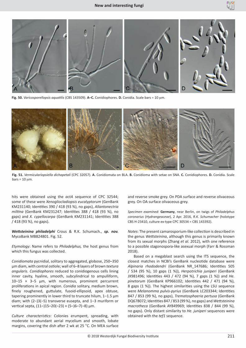
© 2018 Westerdijk Fungal Biodiversity Institute
New and interesting fungi
Editor-in-ChiefProf. dr P.W. Crous, Westerdijk Fungal Biodiversity Institute, P.O. Box 85167, 3508 AD Utrecht, The Netherlands.E-mail:[email protected]
211
hits were obtained using the actA sequence of CPC 32544; some of these were Xenogliocladiopsis eucalyptorum (GenBank KM231140; Identities 390 / 418 (93 %), no gaps), Allantonectria miltina (GenBank KM231247; Identities 388 / 418 (93 %), no gaps) and X. cypellocarpa (GenBank KM231141; Identities 388 / 418 (93 %), no gaps).
Wettsteinina philadelphi Crous & R.K. Schumach., sp. nov. MycoBank MB824801. Fig. 52.
Etymology: Name refers to Philadelphus, the host genus from which this fungus was collected.
Conidiomata pycnidial, solitary to aggregated, globose, 250–350 µm diam, with central ostiole; wall of 6–8 layers of brown textura angularis. Conidiophores reduced to conidiogenous cells lining inner cavity, hyaline, smooth, subcylindrical to ampulliform, 10–15 × 3–5 µm, with numerous, prominent percurrent proliferations in apical region. Conidia solitary, medium brown, finely roughened, guttulate, fusoid-ellipsoid, apex obtuse, tapering prominently in lower third to truncate hilum, 1–1.5 µm diam; with (2–)3(–5) transverse eusepta, and 1–3 muriform or vertical septa, (11–)15–20(–23) × (5–)6–7(–8) µm.
Culture characteristics: Colonies erumpent, spreading, with moderate to abundant aerial mycelium and smooth, lobate margins, covering the dish after 2 wk at 25 °C. On MEA surface
and reverse smoke grey. On PDA surface and reverse olivaceous grey. On OA surface olivaceous grey.
Specimen examined: Germany, near Berlin, on twigs of Philadelphus coronarius (Hydrangeaceae), 2 Apr. 2016, R.K. Schumacher (holotype CBS H-23410, culture ex-type CPC 30534 = CBS 143392).
Notes: The present camarosporium-like collection is described in the genus Wettsteinina, although this genus is primarily known from its sexual morphs (Zhang et al. 2012), with one reference to a possible stagonospora-like asexual morph (Farr & Rossman 2018).
Based on a megablast search using the ITS sequence, the closest matches in NCBI’s GenBank nucleotide database were Alpinaria rhododendri (GenBank NR_147686; Identities 505 / 534 (95 %), 10 gaps (1 %)), Herpotrichia juniperi (GenBank JX981496; Identities 443 / 472 (94 %), 7 gaps (1 %)) and He. pinetorum (GenBank KP966102; Identities 442 / 471 (94 %), 8 gaps (1 %)). The highest similarities using the LSU sequence were Melanomma pulvis-pyrius (GenBank LC203344; Identities 847 / 853 (99 %), no gaps), Trematosphaeria pertusa (GenBank DQ678072; Identities 847 / 853 (99 %), no gaps) and Wettsteinina macrotheca (GenBank AY849969; Identities 838 / 844 (99 %), no gaps). Only distant similarity to He. juniperi sequences were obtained with the tef1 sequence.
Fig. 50. Varicosporellopsis aquatilis (CBS 143509). A–C. Conidiophores. D. Conidia. Scale bars = 10 mm.
Fig. 51. Vermiculariopsiella dichapetali (CPC 32057). A. Conidiomata on BLA. B. Conidioma with setae on SNA. C. Conidiophores. D. Conidia. Scale bars = 10 mm.
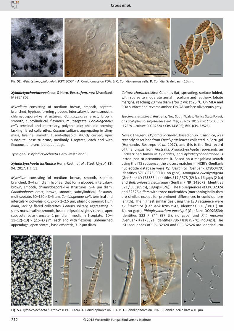
© 2018 Westerdijk Fungal Biodiversity Institute
Crous et al.
Editor-in-ChiefProf. dr P.W. Crous, Westerdijk Fungal Biodiversity Institute, P.O. Box 85167, 3508 AD Utrecht, The Netherlands.E-mail:[email protected]
212
Xyladictyochaetaceae Crous & Hern.-Restr., fam. nov. MycoBank MB824802.
Mycelium consisting of medium brown, smooth, septate, branched, hyphae, forming globose, intercalary, brown, smooth, chlamydospore-like structures. Conidiophores erect, brown, smooth, subcylindrical, flexuous, multiseptate. Conidiogenous cells terminal and intercalary, polyphialidic; phialidic opening lacking flared collarettes. Conidia solitary, aggregating in slimy mass, hyaline, smooth, fusoid-ellipsoid, slightly curved, apex subacute, base truncate, medianly 1-septate; each end with flexuous, unbranched appendage.
Type genus: Xyladictyochaeta Hern.-Restr. et al.
Xyladictyochaeta lusitanica Hern.-Restr. et al., Stud. Mycol. 86: 94. 2017. Fig. 53.
Mycelium consisting of medium brown, smooth, septate, branched, 3–4 µm diam hyphae, that form globose, intercalary, brown, smooth, chlamydospore-like structures, 5–6 µm diam. Conidiophores erect, brown, smooth, subcylindrical, flexuous, multiseptate, 60–150 × 3–5 µm. Conidiogenous cells terminal and intercalary, polyphialidic, 2–6 × 2–2.5 µm; phialidic opening 1 µm diam, lacking flared collarettes. Conidia solitary, aggregating in slimy mass, hyaline, smooth, fusoid-ellipsoid, slightly curved, apex subacute, base truncate, 1 µm diam, medianly 1-septate, (10–) 11–12(–13) × (2.5–)3 µm; each end with flexuous, unbranched appendage, apex central, base excentric, 3–7 µm diam.
Culture characteristics: Colonies flat, spreading, surface folded, with sparse to moderate aerial mycelium and feathery, lobate margins, reaching 20 mm diam after 2 wk at 25 °C. On MEA and PDA surface and reverse amber. On OA surface olivaceous grey.
Specimens examined: Australia, New South Wales, Nullica State Forest, on Eucalyptus sp. (Myrtaceae) leaf litter, 29 Nov. 2016, P.W. Crous, (CBS H-23291, culture CPC 32324 = CBS 143502); ibid. (CPC 32526).
Notes: The genus Xyladictyochaeta, based on Xy. lusitanica, was recently described from Eucalyptus leaves collected in Portugal (Hernández-Restrepo et al. 2017), and this is the first record of this fungus from Australia. Xyladictyochaeta represents an undescribed family in Xylariales, and Xyladictyochaetaceae is introduced to accommodate it. Based on a megablast search using the ITS sequence, the closest matches in NCBI’s GenBank nucleotide database were Xy. lusitanica (GenBank KY853479; Identities 571 / 573 (99 %), no gaps), Anungitea eucalyptigena (GenBank KY173383; Identities 517 / 578 (89 %), 16 gaps (2 %)) and Beltraniopsis neolitseae (GenBank NR_148072; Identities 521 / 583 (89 %), 19 gaps (3 %)). The ITS sequences of CPC 32324 and 32526 differs with three nucleotides (morphologically they are similar, except for prominent differences in conidiophore length). The highest similarities using the LSU sequence were Xy. lusitanica (GenBank KY853543; Identities 801 / 801 (100 %), no gaps), Phlogicylindrium eucalypti (GenBank DQ923534; Identities 822 / 844 (97 %), no gaps) and Phl. mokarei (GenBank KY173521; Identities 796 / 818 (97 %), no gaps). The LSU sequences of CPC 32324 and CPC 32526 are identical. No
Fig. 52. Wettsteinina philadelphi (CPC 30534). A. Conidiomata on PDA. B, C. Conidiogenous cells. D. Conidia. Scale bars = 10 mm.
Fig. 53. Xyladictyochaeta lusitanica (CPC 32324). A. Conidiophores on PDA. B–E. Conidiophores on SNA. F. Conidia. Scale bars = 10 mm.
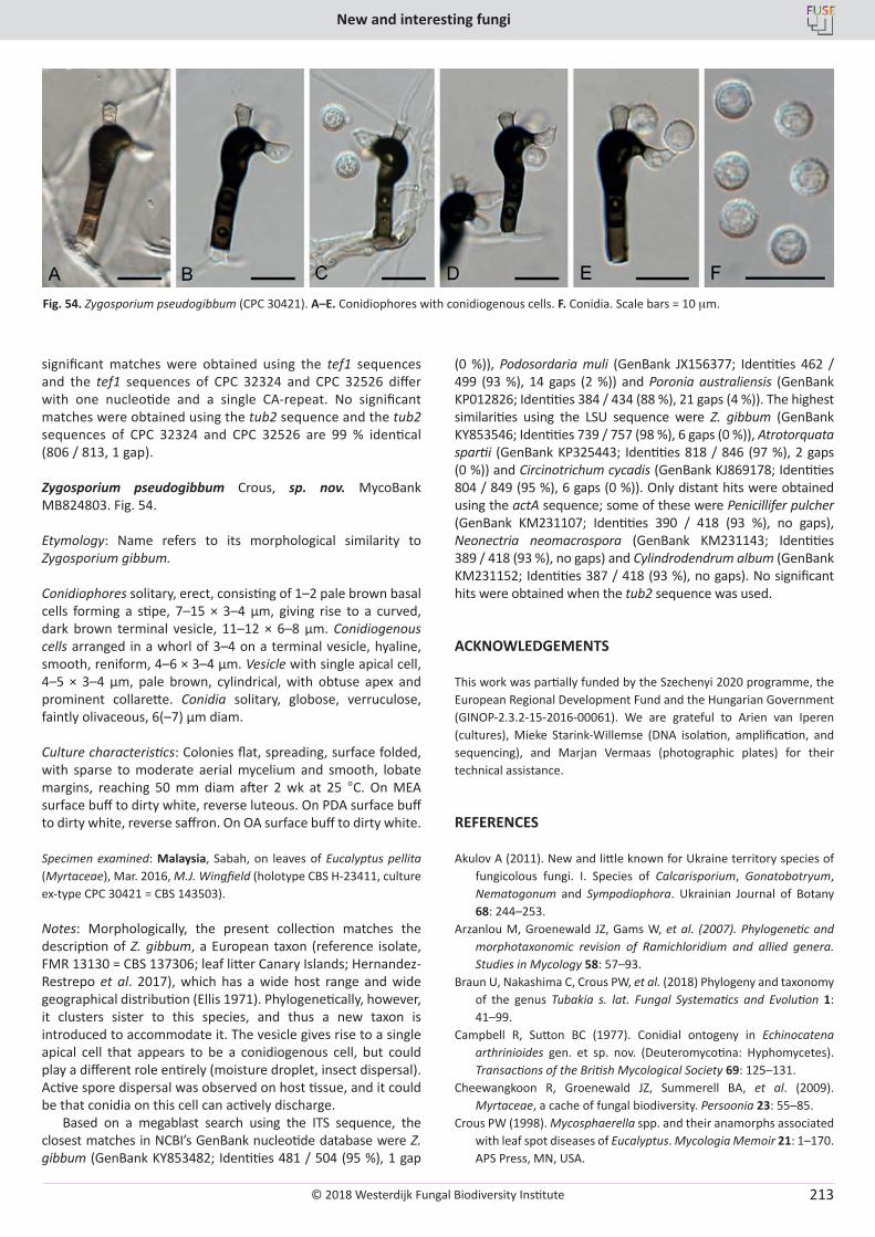
© 2018 Westerdijk Fungal Biodiversity Institute
New and interesting fungi
Editor-in-ChiefProf. dr P.W. Crous, Westerdijk Fungal Biodiversity Institute, P.O. Box 85167, 3508 AD Utrecht, The Netherlands.E-mail:[email protected]
213
significant matches were obtained using the tef1 sequences and the tef1 sequences of CPC 32324 and CPC 32526 differ with one nucleotide and a single CA-repeat. No significant matches were obtained using the tub2 sequence and the tub2 sequences of CPC 32324 and CPC 32526 are 99 % identical (806 / 813, 1 gap).
Zygosporium pseudogibbum Crous, sp. nov. MycoBank MB824803. Fig. 54.
Etymology: Name refers to its morphological similarity to Zygosporium gibbum.
Conidiophores solitary, erect, consisting of 1–2 pale brown basal cells forming a stipe, 7–15 × 3–4 µm, giving rise to a curved, dark brown terminal vesicle, 11–12 × 6–8 µm. Conidiogenous cells arranged in a whorl of 3–4 on a terminal vesicle, hyaline, smooth, reniform, 4–6 × 3–4 µm. Vesicle with single apical cell, 4–5 × 3–4 µm, pale brown, cylindrical, with obtuse apex and prominent collarette. Conidia solitary, globose, verruculose, faintly olivaceous, 6(–7) µm diam.
Culture characteristics: Colonies flat, spreading, surface folded, with sparse to moderate aerial mycelium and smooth, lobate margins, reaching 50 mm diam after 2 wk at 25 °C. On MEA surface buff to dirty white, reverse luteous. On PDA surface buff to dirty white, reverse saffron. On OA surface buff to dirty white.
Specimen examined: Malaysia, Sabah, on leaves of Eucalyptus pellita (Myrtaceae), Mar. 2016, M.J. Wingfield (holotype CBS H-23411, culture ex-type CPC 30421 = CBS 143503).
Notes: Morphologically, the present collection matches the description of Z. gibbum, a European taxon (reference isolate, FMR 13130 = CBS 137306; leaf litter Canary Islands; Hernandez-Restrepo et al. 2017), which has a wide host range and wide geographical distribution (Ellis 1971). Phylogenetically, however, it clusters sister to this species, and thus a new taxon is introduced to accommodate it. The vesicle gives rise to a single apical cell that appears to be a conidiogenous cell, but could play a different role entirely (moisture droplet, insect dispersal). Active spore dispersal was observed on host tissue, and it could be that conidia on this cell can actively discharge.
Based on a megablast search using the ITS sequence, the closest matches in NCBI’s GenBank nucleotide database were Z. gibbum (GenBank KY853482; Identities 481 / 504 (95 %), 1 gap
(0 %)), Podosordaria muli (GenBank JX156377; Identities 462 / 499 (93 %), 14 gaps (2 %)) and Poronia australiensis (GenBank KP012826; Identities 384 / 434 (88 %), 21 gaps (4 %)). The highest similarities using the LSU sequence were Z. gibbum (GenBank KY853546; Identities 739 / 757 (98 %), 6 gaps (0 %)), Atrotorquata spartii (GenBank KP325443; Identities 818 / 846 (97 %), 2 gaps (0 %)) and Circinotrichum cycadis (GenBank KJ869178; Identities 804 / 849 (95 %), 6 gaps (0 %)). Only distant hits were obtained using the actA sequence; some of these were Penicillifer pulcher (GenBank KM231107; Identities 390 / 418 (93 %), no gaps), Neonectria neomacrospora (GenBank KM231143; Identities 389 / 418 (93 %), no gaps) and Cylindrodendrum album (GenBank KM231152; Identities 387 / 418 (93 %), no gaps). No significant hits were obtained when the tub2 sequence was used.
ACKNOWLEDGEMENTS
This work was partially funded by the Szechenyi 2020 programme, the European Regional Development Fund and the Hungarian Government (GINOP-2.3.2-15-2016-00061). We are grateful to Arien van Iperen (cultures), Mieke Starink-Willemse (DNA isolation, amplification, and sequencing), and Marjan Vermaas (photographic plates) for their technical assistance.
REFERENCES
Akulov A (2011). New and little known for Ukraine territory species of fungicolous fungi. I. Species of Calcarisporium, Gonatobotryum, Nematogonum and Sympodiophora. Ukrainian Journal of Botany 68: 244–253.
Arzanlou M, Groenewald JZ, Gams W, et al. (2007). Phylogenetic and morphotaxonomic revision of Ramichloridium and allied genera. Studies in Mycology 58: 57–93.
Braun U, Nakashima C, Crous PW, et al. (2018) Phylogeny and taxonomy of the genus Tubakia s. lat. Fungal Systematics and Evolution 1: 41–99.
Campbell R, Sutton BC (1977). Conidial ontogeny in Echinocatena arthrinioides gen. et sp. nov. (Deuteromycotina: Hyphomycetes). Transactions of the British Mycological Society 69: 125–131.
Cheewangkoon R, Groenewald JZ, Summerell BA, et al. (2009). Myrtaceae, a cache of fungal biodiversity. Persoonia 23: 55–85.
Crous PW (1998). Mycosphaerella spp. and their anamorphs associated with leaf spot diseases of Eucalyptus. Mycologia Memoir 21: 1–170. APS Press, MN, USA.
Fig. 54. Zygosporium pseudogibbum (CPC 30421). A–E. Conidiophores with conidiogenous cells. F. Conidia. Scale bars = 10 mm.
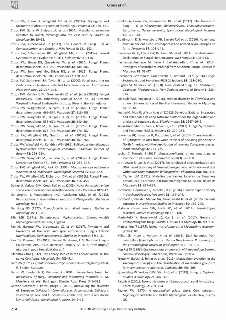
© 2018 Westerdijk Fungal Biodiversity Institute
Crous et al.
Editor-in-ChiefProf. dr P.W. Crous, Westerdijk Fungal Biodiversity Institute, P.O. Box 85167, 3508 AD Utrecht, The Netherlands.E-mail:[email protected]
214
Crous PW, Braun U, Wingfield MJ, et al. (2009a). Phylogeny and taxonomy of obscure genera of microfungi. Persoonia 22: 139–161.
Crous PW, Gams W, Stalpers JA, et al. (2004). MycoBank: an online initiative to launch mycology into the 21st century. Studies in Mycology 50: 19–22.
Crous PW, Groenewald JZ (2017). The Genera of Fungi – G 4: Camarosporium and Dothiora. IMA Fungus 8: 131–152.
Crous PW, Schumacher RK, Wingfield MJ, et al. (2015a). Fungal Systematics and Evolution: FUSE 1. Sydowia 67: 81–118.
Crous PW, Shivas RG, Quaedvlieg W, et al. (2014). Fungal Planet description sheets: 214–280. Persoonia 32: 184–306.
Crous PW, Summerell BA, Shivas RG, et al. (2011). Fungal Planet description sheets: 92–106. Persoonia 27: 130–162.
Crous PW, Summerell BA, Taylor JE, et al. (2000). Fungi occurring on Proteaceae in Australia: selected foliicolous species. Australasian Plant Pathology 29: 267–278.
Crous PW, Verkley GJM, Groenewald JZ, et al. (eds) (2009b) Fungal Biodiversity. [CBS Laboratory Manual Series no. 1.] Utrecht: Westerdijk Fungal Biodiversity Institute, Utrecht, the Netherlands.
Crous PW, Wingfield MJ, Burgess TI, et al. (2016a). Fungal Planet description sheets: 469–557. Persoonia 37: 218–403.
Crous PW, Wingfield MJ, Burgess TI, et al. (2017a). Fungal Planet description sheets: 558–624. Persoonia 38: 240–384.
Crous PW, Wingfield MJ, Burgess TI, et al. (2017b). Fungal Planet description sheets: 625–715. Persoonia 39: 270–467.
Crous PW, Wingfield MJ, Guarro J, et al. (2015b). Fungal Planet description sheets: 320–370. Persoonia 34: 167–266.
Crous PW, Wingfield MJ, Kendrick WB (1995). Foliicolous dematiaceous hyphomycetes from Syzygium cordatum. Canadian Journal of Botany 73: 224–234.
Crous PW, Wingfield MJ, Le Roux JJ, et al. (2015c). Fungal Planet Description Sheets: 371–399. Persoonia 35: 264–327.
Crous PW, Wingfield MJ, Park RF (1991). Mycosphaerella nubilosa a synonym of M. molleriana. Mycological Research 95: 628–632.
Crous PW, Wingfield MJ, Richardson DM, et al. (2016b). Fungal Planet description sheets: 400–468. Persoonia 36: 316–458.
Damm U, Verkley GJM, Crous PW, et al. (2008). Novel Paraconiothyrium species on stone fruit trees and other woody hosts. Persoonia 20: 9–17.
De Gruyter J, Woudenberg JHC, Aveskamp MM, et al. (2013). Redisposition of Phoma-like anamorphs in Pleosporales. Studies in Mycology 75: 1–36.
De Hoog GS (1977). Rhinocladiella and allied genera. Studies in Mycology 15: 1–140.
Ellis MB (1971). Dematiaceous Hyphomycetes. Commonwealth Mycological Institute: Kew, England.
Fan XL, Barreto RW, Groenewald JZ, et al. (2017). Phylogeny and taxonomy of the scab and spot anthracnose fungus Elsinoë (Myriangiales, Dothideomycetes). Studies in Mycology 87: 1–41.
Farr DF, Rossman AY (2018). Fungal Databases, U.S. National Fungus Collections, ARS, USDA. Retrieved January 12, 2018, from https:// nt.ars-grin.gov / fungaldatabases /
Fitzpatrick HM (1942). Revisionary studies in the Coryneliaceae. II. The genus Caliciopsis. Mycologia 34: 489–514.
Gams W (1971). Cephalosporium-artige Schimmelpilze (Hyphomycetes). G. Fischer, Stuttgart.
Gams W, Diederich P, Põldmaa K (2004). Fungicolous fungi. In: Biodiversity of fungi, inventory and monitoring methods (G. M. Mueller et al. eds). Burlington: Elsevier Acad. Press: 343–392.
Garrido-Benavent I, Pérez-Ortega S (2015). Unravelling the diversity of European Caliciopsis (Coryneliaceae, Ascomycota): Caliciopsis valentina sp. nov. and C. beckhausii comb. nov., with a worldwide key to Caliciopsis. Mycological Progress 14: 1–11.
Giraldo A, Crous PW, Schumacher RK, et al. (2017). The Genera of Fungi – G 3: Aleurocystis, Blastacervulus, Clypeophysalospora, Licrostroma, Neohendersonia, Spumatoria. Mycological Progress 16: 325–348.
Guatimosim E, Schwartsburd PB, Barreto RW, et al. (2016). Novel fungi from an ancient niche: cercosporoid and related sexual morphs on ferns. Persoonia 37: 106–141.
Hawksworth DL, Crous PW, Redhead SA, et al. (2011). The Amsterdam Declaration on Fungal Nomenclature. IMA Fungus 2: 105–112.
Hernández-Restrepo M, Gené J, Castañeda-Ruiz RF, et al. (2017). Phylogeny of saprobic microfungi from Southern Europe. Studies in Mycology 86: 53–97.
Hernández-Restrepo M, Groenewald JZ, Lombard L, et al. (2016). Fungal Systematics and Evolution: FUSE 2. Sydowia 68: 193–230.
Hughes SJ, Kendrick WB (1968). New Zealand fungi 12. Menispora, Codinaea, Menisporopsis. New Zealand Journal of Botany 6: 323–375.
Jaklitsch WM, Voglmayr H (2016). Hidden diversity in Thyridaria and a new circumscription of the Thyridariaceae. Studies in Mycology 85: 35–64.
Kearse M, Moir R, Wilson A, et al. (2012). Geneious Basic: an integrated and extendable desktop software platform for the organization and analysis of sequence data. Bioinformatics 28: 1647–1649.
Krisai-Greilhuber I, Chen Y, Jabeen S, et al. (2017). Fungal Systematics and Evolution: FUSE 3. Sydowia 69: 229–264.
Lawrence DP, Travadon R, Pouzoulet J, et al. (2017). Characterization of Cytospora isolates from wood cankers of declining grapevine in North America, with the descriptions of two new Cytospora species. Plant Pathology 66: 713–725.
Lechat C, Fournier J (2016). Varicosporellopsis, a new aquatic genus from South of France. Ascomycete.org 8(3): 96–100.
Li J, Jeewin R, Luo Z, et al. (2017). Morphological characterization and DNA based taxonomy of Fusiconidium gen. nov. with two novel taxa within Melanommataceae (Pleosporales). Phytotaxa 308: 206–218.
Lin TY, Yen JM (1971). Maladies des taches foliaires de Bananiers provoquees, à Formose, par trois champignons nouveaux. Revue de Mycologie 35: 317–327.
Lombard L, Houbraken J, Decock C, et al. (2016). Generic hyper-diversity in Stachybotriaceae. Persoonia 36: 156–246.
Lombard L, van der Merwe NA, Groenewald JZ, et al. (2015). Generic concepts in Nectriaceae. Studies in Mycology 80: 189–245.
Maharachchikumbura SSN, Hyde KD, et al. (2014). Pestalotiopsis revisited. Studies in Mycology 79: 121–186.
Marin-Felix Y, Groenewald JZ, Cai L, et al. (2017). Genera of phytopathogenic fungi: GOPHY 1. Studies in Mycology 86: 99–216.
Matsushima T (1975). Icones microfungorum a Matsushima lectorum. (Kobe): 101.
Miller SE, Hrcek J, Vojtech N, et al. (2013). DNA barcodes from caterpillars (Lepidoptera) from Papua New Guinea. Proceedings of the Entomological Society of Washington 115: 107–109.
Nag Raj TR (1993). Coelomycetous anamorphs with appendage-bearing conidia. Mycologue Publications, Waterloo, Ontario.
Prieto M, Baloch E, Tehler A, et al. (2013). Mazaedium evolution in the Ascomycota (Fungi) and the classification of mazaediate groups of formerly unclear relationship. Cladistics 29: 296–308.
Quaedvlieg W, Verkley GJM, Shin H-D, et al. (2013). Sizing up Septoria. Studies in Mycology 75: 307–390.
Raitviir A (2001). Taxonomic notes on Dematioscypha and Amicodisca. Czech Mycology 52: 289–294.
Rayner RW (1970). A mycological colour chart. Commonwealth Mycological Institute and British Mycological Society. Kew, Surrey, UK.
© 2018 Westerdijk Fungal Biodiversity Institute
New and interesting fungi
Editor-in-ChiefProf. dr P.W. Crous, Westerdijk Fungal Biodiversity Institute, P.O. Box 85167, 3508 AD Utrecht, The Netherlands.E-mail:[email protected]
215
Réblová M, Miller AN, Rossman AY, et al. (2016). Recommendations for competing sexual-asexually typified generic names in Sordariomycetes (except Diaporthales, Hypocreales, and Magnaporthales) IMA Fungus 7: 131–153.
Ronquist F, Teslenko M, Van der Mark P, et al. (2012). MrBayes 3.2: Efficient Bayesian phylogenetic inference and model choice across a large model space. Systematic Biology 61: 539–542.
Rossman AY, Crous PW, Hyde KD, et al. (2015). Recommended names for pleomorphic genera in Dothideomycetes. IMA Fungus 6: 507–523.
Silva M, Barreto RW, Pereira OL, et al. (2016). Exploring fungal mega-diversity: Pseudocercospora from Brazil. Persoonia 37: 142–172.
Smith H, Wingfield MJ, Crous PW, et al. (1996). Sphaeropsis sapinea and Botryosphaeria dothidea endophytic in Pinus spp. and Eucalyptus spp. in South Africa. South African Journal of Botany 62: 86–88.
Sutton BC (1973). Hyphomycetes from Manitoba and Saskatchewan, Canada. Mycological Papers 132: 1–143.
Sutton BC, Hodges CS Jr. (1975). Eucalyptus microfungi: Codinaea and Zanclospora species from Brazil. Nova Hedwigia 26: 517–525.
Swart L, Crous PW, Denman S, et al. (1998). Fungi occurring on Proteaceae. I. South African Journal of Botany 64: 137–145.
Swofford DL (2003). PAUP*: phylogenetic analysis using parsimony. (*and other methods). Version 4.0b10. Sinauer Associates, Sunderland.
Tibpromma S, Hyde KD, Jeewon R, et al. (2017). Fungal diversity notes 491–603: taxonomic and phylogenetic contributions to fungal taxa. Fungal Diversity 83: 1–261.
Trakunyingcharoen T, Lombard L, Groenewald JZ, et al. (2014). Mycoparasitic species of Sphaerellopsis, and allied lichenicolous and other genera. IMA Fungus 5: 391–414.
Valenzuela-Lopez N, Cano-Lira JF, Guarro J, et al. (2018). Coelomycetous Dothideomycetes with emphasis on the families Cucurbitariaceae and Didymellaceae. Studies in Mycology 90: 1–69.
Van der Aa HA, Vanev S (2002). A revision of the species described in Phyllosticta. Westerdijk Fungal Biodiversity Institute, Utrecht, The Netherlands.
Vu D, Groenewald M, Szöke S, et al. (2016). DNA barcoding analysis of more than 9000 yeast isolates contributes to quantitative thresholds for yeast species and genera delimitation. Studies in Mycology 85: 91–105.
Walker J, Minter DW (1981). Taxonomy of Nematogonum, Gonatobotrys, Gonatobotryum and Gonatorrhodiella. Transactions of the British Mycological Society 77: 299–319.
Wanasinghe DN, Hyde KD, Jeewon R, et al. (2017). Phylogenetic revision of Camarosporium (Pleosporineae, Dothideomycetes) and allied genera. Studies in Mycology 87: 207–256.
Wijayawardene NN, Hyde KD, Bhat DJ, et al. (2014). Camarosporium-like species are polyphyletic in Pleosporales; introducing Paracamarosporium and Pseudocamarosporium gen. nov. in Montagnulaceae. Cryptogamie, Mycologie 35: 177–198.
Wingfield MJ, De Beer ZW, Slippers B, et al. (2012). One fungus, one name promotes progressive plant pathology. Molecular Plant Pathology 13: 604–613.
Wood AR, Damm U, van der Linde EJ, et al. (2016). Finding the missing link: Resolving the Coryneliomycetidae within Eurotiomycetes. Persoonia 37: 37–56.
Zhang Y, Crous PW, Schoch CL, et al. (2012). Pleosporales. Fungal Diversity 53: 1–221.
Zhang Z, Schwartz S, Wagner L, et al. (2000). A greedy algorithm for aligning DNA sequences. Journal of Computational Biology 7: 203–214.
© 2018 Westerdijk Fungal Biodiversity Institute
Castellano et al.
Editor-in-ChiefProf. dr P.W. Crous, Westerdijk Fungal Biodiversity Institute, P.O. Box 85167, 3508 AD Utrecht, The Netherlands.E-mail:[email protected]
216
Fungal Systematics and Evolution is licensed under a Creative Commons Attribution-NonCommercial-ShareAlike 4.0 International License
© 2018 Westerdijk Fungal Biodiversity Institute 217
Editor-in-ChiefProf. dr P.W. Crous, Westerdijk Fungal Biodiversity Institute, P.O. Box 85167, 3508 AD Utrecht, The Netherlands.E-mail:[email protected]
Fungal Systematics and EvolutionVOLUME 1JUNE 2018
PAGES 217–228
doi.org/10.3114/fuse.2018.01.09
Position specificity in the genus Coreomyces (Laboulbeniomycetes, Ascomycota)
H. Sundberg1*, Å. Kruys2, J. Bergsten3, S. Ekman2
1Systematic Biology, Department of Organismal Biology, Evolutionary Biology Centre, Uppsala University, Uppsala, Sweden2Museum of Evolution, Uppsala University, Uppsala, Sweden3Department of Zoology, Swedish Museum of Natural History, Stockholm, Sweden
*Corresponding author: [email protected]
Abstract: To study position specificity in the insect-parasitic fungal genus Coreomyces (Laboulbeniaceae, Laboulbeniales), we sampled corixid hosts (Corixidae, Heteroptera) in southern Scandinavia. We detected Coreomyces thalli in five different positions on the hosts. Thalli from the various positions grouped in four distinct clusters in the resulting gene trees, distinctly so in the ITS and LSU of the nuclear ribosomal DNA, less so in the SSU of the nuclear ribosomal DNA and the mitochondrial ribosomal DNA. Thalli from the left side of abdomen grouped in a single cluster, and so did thalli from the ventral right side. Thalli in the mid-ventral position turned out to be a mix of three clades, while thalli growing dorsally grouped with thalli from the left and right abdominal clades. The mid-ventral and dorsal positions were found in male hosts only. The position on the left hemelytron was shared by members from two sister clades. Statistical analyses demonstrate a significant positive correlation between clade and position on the host, but also a weak correlation between host sex and clade membership. These results indicate that sex-of-host specificity may be a non-existent extreme in a continuum, where instead weak preferences for one host sex may turn out to be frequent.
Key words: CorixidaeDNAFungihost-specificityinsectphylogeny
Published online: 26 April 2018.
INTRODUCTION
Members of the Laboulbeniales are minute ascomycete fungi that average 0.2 mm in length and seldom exceed 1 mm in length, although some species have been reported to grow beyond 2 mm (Giard 1892, Thaxter 1896, Santamaria 1998). They are obligatory ectoparasites on arthropods tied to their host throughout the entire life cycle, and many species appear to be specific to a single or a few closely related host species (Scheloske 1969, Huldén 1983, Majewski 1994). The order comprises four families with around 140 genera and in excess of 2000 described species (Kirk et al. 2008), but some estimates suggest that the true number of species worldwide is in the interval 15 000–75 000 (Weir & Hammond 1997).
Ascospores are assumed to be transmitted between hosts mainly through direct contact during various behavioural interactions, e.g., mating (Richards & Smith 1954, 1955, Whisler 1968, De Kesel 1993, 1995, 1996) or, to a more limited extent, through spore-contaminated host habitat (Arwidsson 1946, Lindroth 1948, Scheloske 1969, De Kesel 1995). Infection experiments in Herpomyces have shown that species infecting cockroaches are highly specific to one or a few species in the same genus (Richards & Smith 1954). Only occasionally did weak infections occur in host species distantly related to the main host. De Kesel (1996), on the other hand, demonstrated that Laboulbenia slackensis, seemingly strictly host specific in nature, was able to infect a broad range of carabid hosts under artificial conditions. This indicates that the physiological properties of the host do not explain host specificity in L. slackensis. The closely related L. littoralis is found in the same habitat (coastal
marshland) but on a different host belonging to another coleopteran suborder, indicating that the species is restricted by habitat rather than host and that host shifts within the same habitat may result in speciation (De Kesel & Haelewaters 2014). Another example of habitat specificity is Rickia wasmannii, which infects several unrelated arthropod hosts sharing the same environment in Myrmica ant nests (Pfliegler et al. 2016). On the other hand, relatively few nominal species are known to exhibit a broad host range in nature, and some of these may turn out to consist of complexes of distinct species upon closer examination, e.g. with molecular tools (Weir & Blackwell 2005, Haelewaters & De Kesel 2017).
The Laboulbeniales are unique among the fungi in displaying “position specificity” (Peyritsch 1875, Thaxter 1896, Benjamin & Shanor 1952, Whisler 1968), a term coined to describe the phenomenon that a nominal parasite species only inhabits a specific, restricted part of its host species’ anatomy. Position specificity is also known in a variety of parasites in the animal kingdom, for instance in the flatworm class Monogenea (Littlewood 1997) and among the water mites (Martin 2004). In many cases among the Laboulbeniales, however, there is no simple one-to-one relationship between the parasite and its position on the host. It has been observed, for example, that positions may become less unique as time after infection passes and that this could be explained by secondary infections mediated by the behaviour of the host (Whisler 1968). In some cases, the same nominal species has been observed to inhabit different positions in males and females that come into contact during mating (Peyritsch 1875). Thaxter (1896) countered that positions were not as strictly upheld as suggested by Peyritsch
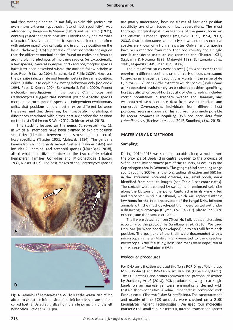
© 2018 Westerdijk Fungal Biodiversity Institute
Sundberg et al.
Editor-in-ChiefProf. dr P.W. Crous, Westerdijk Fungal Biodiversity Institute, P.O. Box 85167, 3508 AD Utrecht, The Netherlands.E-mail:[email protected]
218
and that mating alone could not fully explain this pattern. An even more extreme hypothesis, “sex-of-host specificity”, was advanced by Benjamin & Shanor (1952) and Benjamin (1971), who suggested that each host sex is inhabited by one member of a pair of closely related parasite species, each member often with unique morphological traits and in a unique position on the host. Scheloske (1976) rejected sex-of-host specificity and argued that the different nominal species found on males and females are merely morphotypes of the same species (or exceptionally, a few species). Several examples of di- and polymorphic species have later been described where the authors follow Scheloske (e.g. Rossi & Kotrba 2004, Santamaria & Faille 2009). However, the parasite infects male and female hosts in the same position, which is difficult to explain by mating behaviour only (Majewski 1994, Rossi & Kotrba 2004, Santamaria & Faille 2009). Recent molecular investigations in the genera Chitinomyces and Hesperomyces suggest that nominal position-specific species more or less correspond to species as independent evolutionary units, that positions on the host may be different between the sexes, and that there may be intraspecific morphological differences correlated with either host sex and/or the position on the host (Goldmann & Weir 2012, Goldman et al. 2013).
This study is focused on the genus Coreomyces (Fig. 1), in which all members have been claimed to exhibit position specificity (identical between host sexes) but not sex-of-host specificity (Thaxter 1931, Majewski 1994). The genus is known from all continents except Australia (Tavares 1985) and includes 21 nominal and accepted species (MycoBank 2018), all of which parasitize members of the two closely related hemipteran families Corixidae and Micronectidae (Thaxter 1931, Nieser 2002). The host ranges of the Coreomyces species
are poorly understood, because claims of host and position specificity are often based on few observations. The most thorough morphological investigations of the genus, focus on the eastern European species (Majewski 1973, 1994, 2003, 2008). Distribution ranges are poorly known and many nominal species are known only from a few sites. Only a handful species have been reported from more than one country and a single one is considered more or less cosmopolitan (Thaxter 1931, Sugiyama & Hayama 1981, Majewski 1988, Santamaria et al. 1991, Majewski 1994, Shen et al. 2006).
The aims of this study were to test (1) to what extent thalli growing in different positions on their corixid hosts correspond to species as independent evolutionary units in the sense of de Queiroz (2007), and (2) the extent to which species (understood as independent evolutionary units) display position specificity, host specificity, or sex-of-host specificity. Our sampling included corixid populations in southern Sweden and Denmark, and we obtained DNA sequence data from several markers and numerous Coremomyces individuals from different host positions, sexes and species. This approach was made possible by recent advances in acquiring DNA sequence data from Laboulbeniales (Haelewaters et al. 2015, Sundberg et al. 2018).
MATERIALS AND METHODS
Sampling
During 2014–2015 we sampled corixids along a route from the province of Uppland in central Sweden to the province of Skåne in the southernmost part of the country, as well as in the Copenhagen area in Denmark. The geographical sampling range spans roughly 300 km in the longitudinal direction and 550 km in the latitudinal. Potential localities, i.e., small ponds, were identified from satellite images (see Table 1 for coordinates). The corixids were captured by sweeping a reinforced colander along the bottom of the pond. Captured animals were killed and preserved in 99.7 % ethanol, which was replaced after a few hours for the best preservation of the fungal DNA. Infected animals with the most developed thalli were sorted out under a dissecting microscope (Olympus SZ1145 TR), placed in 99.7 % ethanol, and then stored at -20 °C.
Thalli were detached from 76 corixid individuals and crushed according to the protocol by Sundberg et al. (2018). We used from one (or when poorly developed) up to six thalli from each position. The positions of the thalli were documented with a microscope camera (Moticam 5) connected to the dissecting microscope. After the study, host specimens were deposited at the Museum of Evolution (UPSZ).
Molecular procedures
For DNA amplification we used the Terra PCR Direct Polymerase Mix (Clontech) and KAPA3G Plant PCR Kit (Kapa Biosystems). The PCR settings and primers followed the protocol described by Sundberg et al. (2018). PCR products showing clear single bands on an agarose gel were enzymatically cleaned with FastAP Thermosensitive Alkaline Phosphatase combined with Exonuclease I (Thermo Fisher Scientific Inc.). The concentrations and quality of the PCR products were checked on a 2100 Bioanalyzer (Agilent Technologies). We used four molecular markers: the small subunit (nrSSU), internal transcribed spacer
Fig. 1. Examples of Coreomyces sp. A. Thalli at the ventral side of the abdomen and at the inferior side of the left hemelytral margin of the corixid host. B. Detached thallus from the inferior margin of the left hemelytron. Scale bar = 100 µm.
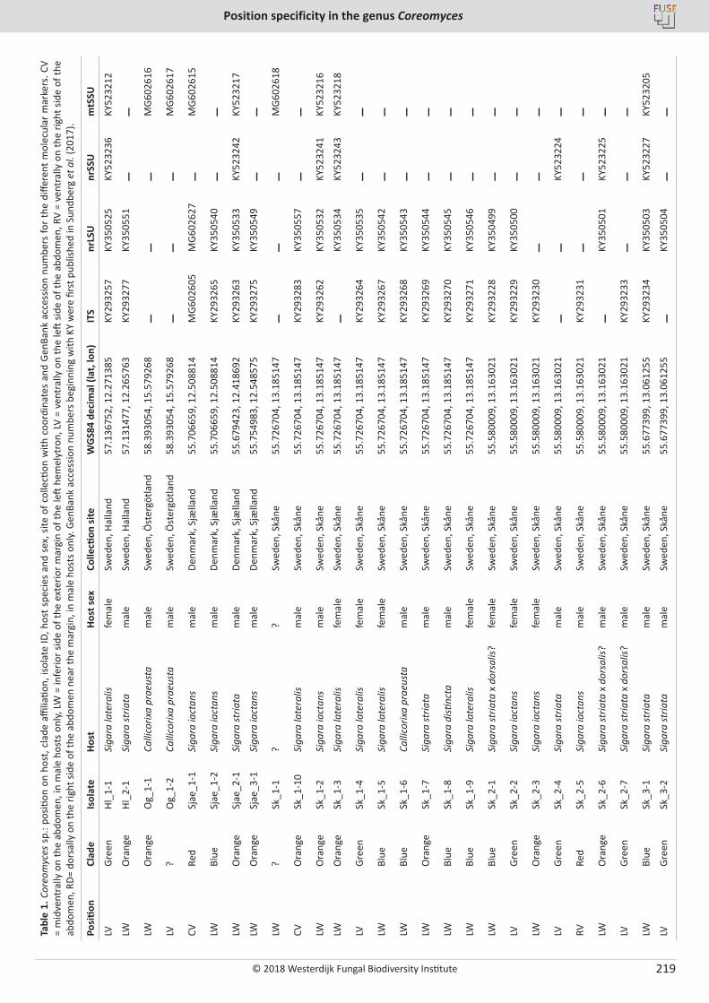
© 2018 Westerdijk Fungal Biodiversity Institute
Position specificity in the genus Coreomyces
Editor-in-ChiefProf. dr P.W. Crous, Westerdijk Fungal Biodiversity Institute, P.O. Box 85167, 3508 AD Utrecht, The Netherlands.E-mail:[email protected]
219
Tabl
e 1.
Cor
eom
yces
sp.
: pos
ition
on
host
, cla
de a
ffilia
tion,
isol
ate
ID, h
ost s
peci
es a
nd s
ex, s
ite o
f col
lecti
on w
ith c
oord
inat
es a
nd G
enBa
nk a
cces
sion
num
bers
for t
he d
iffer
ent m
olec
ular
mar
kers
. CV
= m
idve
ntra
lly o
n th
e ab
dom
en, i
n m
ale
host
s on
ly, LW
= in
ferio
r sid
e of
the
exte
rior m
argi
n of
the
left
hem
elyt
ron,
LV =
ven
tral
ly o
n th
e le
ft sid
e of
the
abdo
men
, RV
= ve
ntra
lly o
n th
e rig
ht s
ide
of th
e ab
dom
en, R
D= d
orsa
lly o
n th
e rig
ht si
de o
f the
abd
omen
nea
r the
mar
gin,
in m
ale
host
s onl
y. G
enBa
nk a
cces
sion
num
bers
beg
inni
ng w
ith K
Y w
ere
first
pub
lishe
d in
Sun
dber
g et
al.
(201
7).
Posi
tion
Clad
eIs
olat
eHo
stHo
st se
xCo
llecti
on si
teW
GS8
4 de
cim
al (l
at, l
on)
ITS
nrLS
Unr
SSU
mtS
SU
LVGr
een
Hl_1
-1Si
gara
late
ralis
fem
ale
Swed
en, H
alla
nd57
.136
752,
12.
2713
85KY
2932
57KY
3505
25KY
5232
36KY
5232
12
LWO
rang
eHl
_2-1
Siga
ra st
riata
mal
eSw
eden
, Hal
land
57.1
3147
7, 1
2.26
5763
KY29
3277
KY35
0551
––
LWO
rang
eO
g_1-
1Ca
llico
rixa
prae
usta
mal
eSw
eden
, Öst
ergö
tland
58.3
9305
4, 1
5.57
9268
––
–M
G602
616
LV?
Og_
1-2
Calli
corix
a pr
aeus
tam
ale
Swed
en, Ö
ster
götla
nd58
.393
054,
15.
5792
68–
––
MG6
0261
7
CVRe
dSj
ae_1
-1Si
gara
iact
ans
mal
eDe
nmar
k, S
jælla
nd55
.706
659,
12.
5088
14M
G602
605
MG6
0262
7–
MG6
0261
5
LWBl
ueSj
ae_1
-2Si
gara
iact
ans
mal
eDe
nmar
k, S
jælla
nd55
.706
659,
12.
5088
14KY
2932
65KY
3505
40–
–LW
Ora
nge
Sjae
_2-1
Siga
ra st
riata
mal
eDe
nmar
k, S
jælla
nd55
.679
423,
12.
4186
92KY
2932
63KY
3505
33KY
5232
42KY
5232
17
LWO
rang
eSj
ae_3
-1Si
gara
iact
ans
mal
eDe
nmar
k, S
jælla
nd55
.754
983,
12.
5485
75KY
2932
75KY
3505
49–
–LW
?Sk
_1-1
??
Swed
en, S
kåne
55.7
2670
4, 1
3.18
5147
––
–M
G602
618
CVO
rang
eSk
_1-1
0Si
gara
late
ralis
mal
eSw
eden
, Skå
ne55
.726
704,
13.
1851
47KY
2932
83KY
3505
57–
–LW
Ora
nge
Sk_1
-2Si
gara
iact
ans
mal
eSw
eden
, Skå
ne55
.726
704,
13.
1851
47KY
2932
62KY
3505
32KY
5232
41KY
5232
16
LWO
rang
eSk
_1-3
Siga
ra la
tera
lisfe
mal
eSw
eden
, Skå
ne55
.726
704,
13.
1851
47–
KY35
0534
KY52
3243
KY52
3218
LVGr
een
Sk_1
-4Si
gara
late
ralis
fem
ale
Swed
en, S
kåne
55.7
2670
4, 1
3.18
5147
KY29
3264
KY35
0535
––
LWBl
ueSk
_1-5
Siga
ra la
tera
lisfe
mal
eSw
eden
, Skå
ne55
.726
704,
13.
1851
47KY
2932
67KY
3505
42–
–LW
Blue
Sk_1
-6Ca
llico
rixa
prae
usta
mal
eSw
eden
, Skå
ne55
.726
704,
13.
1851
47KY
2932
68KY
3505
43–
–LW
Ora
nge
Sk_1
-7Si
gara
stria
tam
ale
Swed
en, S
kåne
55.7
2670
4, 1
3.18
5147
KY29
3269
KY35
0544
––
LWBl
ueSk
_1-8
Siga
ra d
istinc
tam
ale
Swed
en, S
kåne
55.7
2670
4, 1
3.18
5147
KY29
3270
KY35
0545
––
LWBl
ueSk
_1-9
Siga
ra la
tera
lisfe
mal
eSw
eden
, Skå
ne55
.726
704,
13.
1851
47KY
2932
71KY
3505
46–
–LW
Blue
Sk_2
-1Si
gara
stria
ta x
dor
salis
?fe
mal
eSw
eden
, Skå
ne55
.580
009,
13.
1630
21KY
2932
28KY
3504
99–
–LV
Gree
nSk
_2-2
Siga
ra ia
ctan
sfe
mal
eSw
eden
, Skå
ne55
.580
009,
13.
1630
21KY
2932
29KY
3505
00–
–LW
Ora
nge
Sk_2
-3Si
gara
iact
ans
fem
ale
Swed
en, S
kåne
55.5
8000
9, 1
3.16
3021
KY29
3230
––
–LV
Gree
nSk
_2-4
Siga
ra st
riata
mal
eSw
eden
, Skå
ne55
.580
009,
13.
1630
21–
–KY
5232
24–
RVRe
dSk
_2-5
Siga
ra ia
ctan
sm
ale
Swed
en, S
kåne
55.5
8000
9, 1
3.16
3021
KY29
3231
––
–LW
Ora
nge
Sk_2
-6Si
gara
stria
ta x
dor
salis
?m
ale
Swed
en, S
kåne
55.5
8000
9, 1
3.16
3021
–KY
3505
01KY
5232
25–
LVGr
een
Sk_2
-7Si
gara
stria
ta x
dor
salis
?m
ale
Swed
en, S
kåne
55.5
8000
9, 1
3.16
3021
KY29
3233
––
–LW
Blue
Sk_3
-1Si
gara
stria
tam
ale
Swed
en, S
kåne
55.6
7739
9, 1
3.06
1255
KY29
3234
KY35
0503
KY52
3227
KY52
3205
LVGr
een
Sk_3
-2Si
gara
stria
tam
ale
Swed
en, S
kåne
55.6
7739
9, 1
3.06
1255
–KY
3505
04–
–
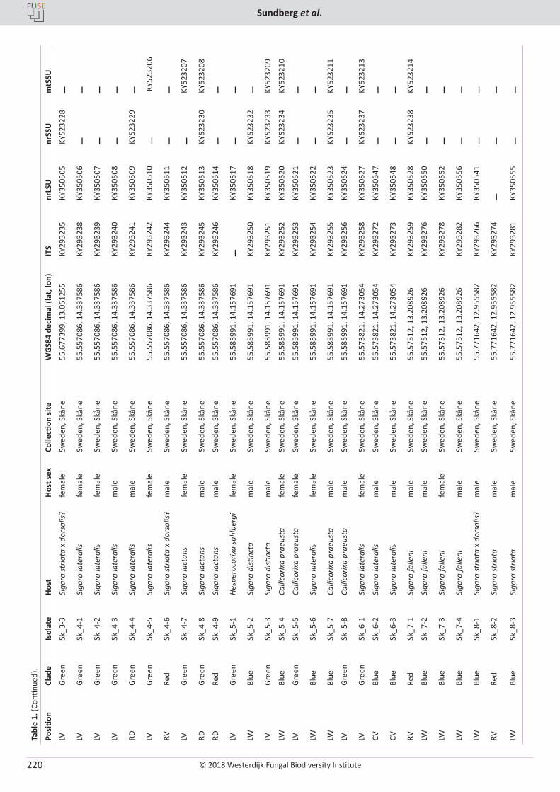
© 2018 Westerdijk Fungal Biodiversity Institute
Sundberg et al.
Editor-in-ChiefProf. dr P.W. Crous, Westerdijk Fungal Biodiversity Institute, P.O. Box 85167, 3508 AD Utrecht, The Netherlands.E-mail:[email protected]
220
Tabl
e 1.
(Con
tinue
d).
Posi
tion
Clad
eIs
olat
eHo
stHo
st se
xCo
llecti
on si
teW
GS8
4 de
cim
al (l
at, l
on)
ITS
nrLS
Unr
SSU
mtS
SU
LVGr
een
Sk_3
-3Si
gara
stria
ta x
dor
salis
?fe
mal
eSw
eden
, Skå
ne55
.677
399,
13.
0612
55KY
2932
35KY
3505
05KY
5232
28–
LVGr
een
Sk_4
-1Si
gara
late
ralis
fem
ale
Swed
en, S
kåne
55.5
5708
6, 1
4.33
7586
KY29
3238
KY35
0506
––
LVGr
een
Sk_4
-2Si
gara
late
ralis
fem
ale
Swed
en, S
kåne
55.5
5708
6, 1
4.33
7586
KY29
3239
KY35
0507
––
LVGr
een
Sk_4
-3Si
gara
late
ralis
mal
eSw
eden
, Skå
ne55
.557
086,
14.
3375
86KY
2932
40KY
3505
08–
–RD
Gree
nSk
_4-4
Siga
ra la
tera
lism
ale
Swed
en, S
kåne
55.5
5708
6, 1
4.33
7586
KY29
3241
KY35
0509
KY52
3229
–LV
Gree
nSk
_4-5
Siga
ra la
tera
lisfe
mal
eSw
eden
, Skå
ne55
.557
086,
14.
3375
86KY
2932
42KY
3505
10–
KY5
2320
6
RVRe
dSk
_4-6
Siga
ra st
riata
x d
orsa
lis?
mal
eSw
eden
, Skå
ne55
.557
086,
14.
3375
86KY
2932
44KY
3505
11–
–LV
Gree
nSk
_4-7
Siga
ra ia
ctan
sfe
mal
eSw
eden
, Skå
ne55
.557
086,
14.
3375
86KY
2932
43KY
3505
12–
KY52
3207
RDGr
een
Sk_4
-8Si
gara
iact
ans
mal
eSw
eden
, Skå
ne55
.557
086,
14.
3375
86KY
2932
45KY
3505
13KY
5232
30KY
5232
08
RDRe
dSk
_4-9
Siga
ra ia
ctan
sm
ale
Swed
en, S
kåne
55.5
5708
6, 1
4.33
7586
KY29
3246
KY35
0514
––
LVGr
een
Sk_5
-1He
sper
ocor
ixa
sahl
berg
ife
mal
eSw
eden
, Skå
ne55
.585
991,
14.
1576
91–
KY35
0517
––
LWBl
ueSk
_5-2
Siga
ra d
istinc
tam
ale
Swed
en, S
kåne
55.5
8599
1, 1
4.15
7691
KY29
3250
KY35
0518
KY52
3232
–LV
Gree
nSk
_5-3
Siga
ra d
istinc
tam
ale
Swed
en, S
kåne
55.5
8599
1, 1
4.15
7691
KY29
3251
KY35
0519
KY52
3233
KY52
3209
LWBl
ueSk
_5-4
Calli
corix
a pr
aeus
tafe
mal
eSw
eden
, Skå
ne55
.585
991,
14.
1576
91KY
2932
52KY
3505
20KY
5232
34KY
5232
10
LVGr
een
Sk_5
-5Ca
llico
rixa
prae
usta
fem
ale
Swed
en, S
kåne
55.5
8599
1, 1
4.15
7691
KY29
3253
KY35
0521
––
LWBl
ueSk
_5-6
Siga
ra la
tera
lisfe
mal
eSw
eden
, Skå
ne55
.585
991,
14.
1576
91KY
2932
54KY
3505
22–
–LW
Blue
Sk_5
-7Ca
llico
rixa
prae
usta
mal
eSw
eden
, Skå
ne55
.585
991,
14.
1576
91KY
2932
55KY
3505
23KY
5232
35KY
5232
11
LVGr
een
Sk_5
-8Ca
llico
rixa
prae
usta
mal
eSw
eden
, Skå
ne55
.585
991,
14.
1576
91KY
2932
56KY
3505
24–
–LV
Gree
nSk
_6-1
Siga
ra la
tera
lisfe
mal
eSw
eden
, Skå
ne55
.573
821,
14.
2730
54KY
2932
58KY
3505
27KY
5232
37KY
5232
13
CVBl
ueSk
_6-2
Siga
ra la
tera
lism
ale
Swed
en, S
kåne
55.5
7382
1, 1
4.27
3054
KY29
3272
KY35
0547
––
CVBl
ueSk
_6-3
Siga
ra la
tera
lism
ale
Swed
en, S
kåne
55.5
7382
1, 1
4.27
3054
KY29
3273
KY35
0548
––
RVRe
dSk
_7-1
Siga
ra fa
lleni
mal
eSw
eden
, Skå
ne55
.575
12, 1
3.20
8926
KY29
3259
KY35
0528
KY52
3238
KY52
3214
LWBl
ueSk
_7-2
Siga
ra fa
lleni
mal
eSw
eden
, Skå
ne55
.575
12, 1
3.20
8926
KY29
3276
KY35
0550
––
LWBl
ueSk
_7-3
Siga
ra fa
lleni
fem
ale
Swed
en, S
kåne
55.5
7512
, 13.
2089
26KY
2932
78KY
3505
52–
–LW
Blue
Sk_7
-4Si
gara
falle
nim
ale
Swed
en, S
kåne
55.5
7512
, 13.
2089
26KY
2932
82KY
3505
56–
–LW
Blue
Sk_8
-1Si
gara
stria
ta x
dor
salis
?m
ale
Swed
en, S
kåne
55.7
7164
2, 1
2.95
5582
KY29
3266
KY35
0541
––
RVRe
dSk
_8-2
Siga
ra st
riata
mal
eSw
eden
, Skå
ne55
.771
642,
12.
9555
82KY
2932
74–
––
LWBl
ueSk
_8-3
Siga
ra st
riata
mal
eSw
eden
, Skå
ne55
.771
642,
12.
9555
82KY
2932
81KY
3505
55–
–
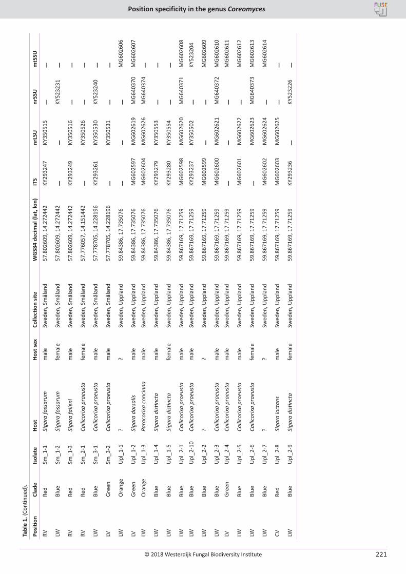
© 2018 Westerdijk Fungal Biodiversity Institute
Position specificity in the genus Coreomyces
Editor-in-ChiefProf. dr P.W. Crous, Westerdijk Fungal Biodiversity Institute, P.O. Box 85167, 3508 AD Utrecht, The Netherlands.E-mail:[email protected]
221
Tabl
e 1.
(Con
tinue
d).
Posi
tion
Clad
eIs
olat
eHo
stHo
st se
xCo
llecti
on si
teW
GS8
4 de
cim
al (l
at, l
on)
ITS
nrLS
Unr
SSU
mtS
SU
RVRe
dSm
_1-1
Siga
ra fo
ssar
umm
ale
Swed
en, S
mål
and
57.8
0260
9, 1
4.27
2442
KY29
3247
KY35
0515
––
LWBl
ueSm
_1-2
Siga
ra fo
ssar
umfe
mal
eSw
eden
, Sm
ålan
d57
.802
609,
14.
2724
42–
–KY
5232
31–
RVRe
dSm
_1-3
Siga
ra fa
lleni
mal
eSw
eden
, Sm
ålan
d57
.802
609,
14.
2724
42KY
2932
49KY
3505
16–
–RV
Red
Sm_2
-1Ca
llico
rixa
prae
usta
fem
ale
Swed
en, S
mål
and
57.7
7605
7, 1
4.15
1442
–KY
3505
26–
–LW
Blue
Sm_3
-1Ca
llico
rixa
prae
usta
mal
eSw
eden
, Sm
ålan
d57
.778
705,
14.
2281
96KY
2932
61KY
3505
30KY
5232
40–
LVGr
een
Sm_3
-2Ca
llico
rixa
prae
usta
mal
eSw
eden
, Sm
ålan
d57
.778
705,
14.
2281
96–
KY35
0531
––
LWO
rang
eU
pl_1
-1?
?Sw
eden
, Upp
land
59.8
4386
, 17.
7350
76–
––
MG6
0260
6
LVGr
een
Upl
_1-2
Siga
ra d
orsa
lism
ale
Swed
en, U
ppla
nd59
.843
86, 1
7.73
5076
MG6
0259
7M
G602
619
MG6
4037
0M
G602
607
LWO
rang
eU
pl_1
-3Pa
raco
rixa
conc
inna
mal
eSw
eden
, Upp
land
59.8
4386
, 17.
7350
76M
G602
604
MG6
0262
6M
G640
374
–LW
Blue
Upl
_1-4
Siga
ra d
istinc
tam
ale
Swed
en, U
ppla
nd59
.843
86, 1
7.73
5076
KY29
3279
KY35
0553
––
LWBl
ueU
pl_1
-5Si
gara
disti
ncta
fem
ale
Swed
en, U
ppla
nd59
.843
86, 1
7.73
5076
KY29
3280
KY35
0554
––
LWBl
ueU
pl_2
-1Ca
llico
rixa
prae
usta
mal
eSw
eden
, Upp
land
59.8
6716
9, 1
7.71
259
MG6
0259
8M
G602
620
MG6
4037
1M
G602
608
LWBl
ueU
pl_2
-10
Calli
corix
a pr
aeus
tam
ale
Swed
en, U
ppla
nd59
.867
169,
17.
7125
9KY
2932
37KY
3505
02–
KY52
3204
LWBl
ueU
pl_2
-2?
?Sw
eden
, Upp
land
59.8
6716
9, 1
7.71
259
MG6
0259
9–
–M
G602
609
LWBl
ueU
pl_2
-3Ca
llico
rixa
prae
usta
mal
eSw
eden
, Upp
land
59.8
6716
9, 1
7.71
259
MG6
0260
0M
G602
621
MG6
4037
2M
G602
610
LVGr
een
Upl
_2-4
Calli
corix
a pr
aeus
tam
ale
Swed
en, U
ppla
nd59
.867
169,
17.
7125
9–
––
MG6
0261
1
LWBl
ueU
pl_2
-5Ca
llico
rixa
prae
usta
mal
eSw
eden
, Upp
land
59.8
6716
9, 1
7.71
259
MG6
0260
1M
G602
622
–M
G602
612
LWBl
ueU
pl_2
-6Ca
llico
rixa
prae
usta
fem
ale
Swed
en, U
ppla
nd59
.867
169,
17.
7125
9–
MG6
0262
3M
G640
373
MG6
0261
3
LWBl
ueU
pl_2
-7?
?Sw
eden
, Upp
land
59.8
6716
9, 1
7.71
259
MG6
0260
2M
G602
624
–M
G602
614
CVRe
dU
pl_2
-8Si
gara
iact
ans
mal
eSw
eden
, Upp
land
59.8
6716
9, 1
7.71
259
MG6
0260
3M
G602
625
––
LWBl
ueU
pl_2
-9Si
gara
disti
ncta
fem
ale
Swed
en, U
ppla
nd59
.867
169,
17.
7125
9KY
2932
36–
KY52
3226
–
© 2018 Westerdijk Fungal Biodiversity Institute
Sundberg et al.
Editor-in-ChiefProf. dr P.W. Crous, Westerdijk Fungal Biodiversity Institute, P.O. Box 85167, 3508 AD Utrecht, The Netherlands.E-mail:[email protected]
222
(ITS) region (including ITS1, 5.8S, and ITS2), the large subunit (nrLSU) of the nuclear ribosomal RNA gene, and the small subunit of the mitochondrial ribosomal RNA gene (mrSSU). Sequencing was carried out by Wyzer Biosciences Inc., Cambridge (MA), and Macrogen Inc., Amsterdam, with the primers ITS4, ITS5, 5.8Shs2, 5.8Shs4, ctb6, LRhs1, LRhs3, LR7 NS4, NShs1, NShs4, NShs2, NShs3, mrSSU1, and mrSSU3R (Vilgalys & Hester 1990, White et al. 1990, Zoller et al. 1999, Sundberg et al. 2018).
Data analysis
Newly produced sequences were edited using Sequencher v. 5.4 and Geneious v. 7.1.9 and aligned together with previously published sequences (Sundberg et al. 2018) using MAFFT v. 7.312 (Katoh & Standley 2013). We used the E-INS-i algorithm with the PAM1 matrix for the nrSSU and mrSSU alignments, and the PAM20 matrix for the ITS and nrLSU alignments. The entire alignments were used in downstream analyses, i.e. no data were excluded.
Likelihood model selection as well as maximum likelihood analyses were performed using IQ-TREE v. 1.6 beta 4 (Nguyen et al. 2015, Kalyaanamoorthy et al. 2017). The best-fitting GTR family model was selected using the Bayesian Information Criterion (BIC) among candidates with one, two, or six substitution rates, with and without gamma-distributed rate heterogeneity among sites and a proportion of invariable sites. For the ITS data, we additionally selected the best-fitting time-irreversible RY Lie Markov model (Sumner et al. 2012, Fernández-Sánchez et al. 2015, Woodhams et al. 2015). Full likelihood optimization was carried out under each candidate model. For each marker, we performed searches for the best trees as well as non-parametric bootstrap analyses with 1 000 replicates under the selected model. The purpose of estimating a tree under a time-irreversible Lie Markov model was to infer the placement of the root. This is not a trivial task in Laboulbeniomycetes, because available DNA sequences are few and sequence divergence within the class is extreme compared to the amount of variation present in our data. The only marker displaying substantial variation in our data, the ITS, is essentially unalignable with any other available sequences. Therefore, we decided to instead use the information contained inside our data for rooting.
Bayesian inference was carried out using MrBayes v. 3.2.6 (Ronquist & Huelsenbeck 2003, Ronquist et al. 2012). We used the same best-fitting models as in the maximum likelihood analyses. Priors included a uniform distribution on topology, a uniform (0, 1) distribution on the proportion of invariable sites, a (1, 1) beta distribution on the transition/transversion rate ratio (when applicable), and a (1, 1, 1, 1) Dirichlet on state frequencies (when applicable). We assumed a compound Dirichlet prior on branch lengths (Rannala et al. 2012, Zhang et al. 2012). The gamma distribution component of this prior was set to α = 1 and β = α/(ML tree length), whereas the Dirichlet component was set to the default (1, 1). Three parallel Markov chain Monte Carlo (MCMC) runs were performed, each with four parallel chains and the temperature increment parameter set to 0.15 (Altekar et al. 2004). The appropriate degree of heating was determined by observing swap rates between the cold and hot chains in preliminary runs. Every 1000th tree was sampled. Runs were diagnosed every 106 generations, removing the first 50 % of the tree sample as burnin, and were set to halt automatically when converged before a maximum of 100 × 106 generations. Convergence was defined as an average standard deviation of splits (with frequency ≥ 0.1) across runs ≤ 0.01.
We used mPTP v. 0.2.3 in maximum likelihood mode to delineate species by locating the transition points between inter- and intraspecific processes in the accumulation of substitutions on the individual gene trees (Kapli et al. 2017). The original Poisson tree process (PTP) implementation assumes a single rate distribution across the tree for the coalescent process (Zhang et al. 2013), whereas the multi-rate Poisson tree process (mPTP) model allows multiple rate distributions, one for each inferred species. We applied both single-rate and multi-rate models to the already rooted Lie Markov model ITS tree and to a rooted version of the nrLSU tree. The nrSSU tree was not included in the analysis, because one potential species was represented by a singleton, whereas mrSSU data were excluded owing to lack of resolution. Likelihood ratio tests were performed to assess whether the multiple-rates model had better fit to the data than a single-rate model.
We performed multinomial logistic regression using the multinom() function of R package nnet v. 7.3.12 (Venables & Ripley 2002). Clade membership (four categories based on ITS, nrLSU, and nrSSU phylogenies) was treated as the response variable and position on host (five categories), host sex (male, female) and host genus (four categories) as predictor variables. We set green clade membership, host genus Sigara, host sex female, and left ventral position on the host as baseline categories. The Anova() function of R package car v. 2.1.6 (Fox & Weisberg 2011) was subsequently used to test the null hypotheses of no effect of each predictor term on the response using type-II chi-square likelihood ratio tests. We also carried out log-linear analysis on the same data using the loglm() function of R package MASS v. 7.3.47 (Venables & Ripley 2002). Starting from the saturated model, we carried out stepwise backward regression to find the best model according to the Akaike Information Criterion (AIC). All analyses, including 71 observations without missing data, were carried out with R v. 3.4.3 (R Core Team 2017).
RESULTS
Sampling outcome
Members of the genus Coreomyces were found on 10 species of corixids belonging to four genera: Callicorixa, Hesperocorixa, Paracorixa, and Sigara (Table 1). Three new host species for Coreomyces were noted, Sigara dorsalis. S. fossarum, and S. iactans. Six individuals of Sigara showed intermediary traits and were determined as potential hybrids. Among the hosts, 48 were males and 24 females (four individuals were lost during handling and not included in downstream statistical analyses). Coreomyces thalli were encountered in five different positions on the corixids (Fig. 2): inferior side of the exterior margin of the hemelytron (LW), ventrally on the left side of the abdomen (LV), ventrally on the right side of the abdomen (RV), dorsally on the right side of the abdomen near the margin (RD), and mid-ventrally on abdomen (CV).
Molecular data
All sequences produced are listed in Table 1 with GenBank accession numbers. Those beginning with KY were published by Sundberg et al. (2018). Alignments and resulting gene trees are available from TreeBASE (https://treebase.org) under study number S21919.
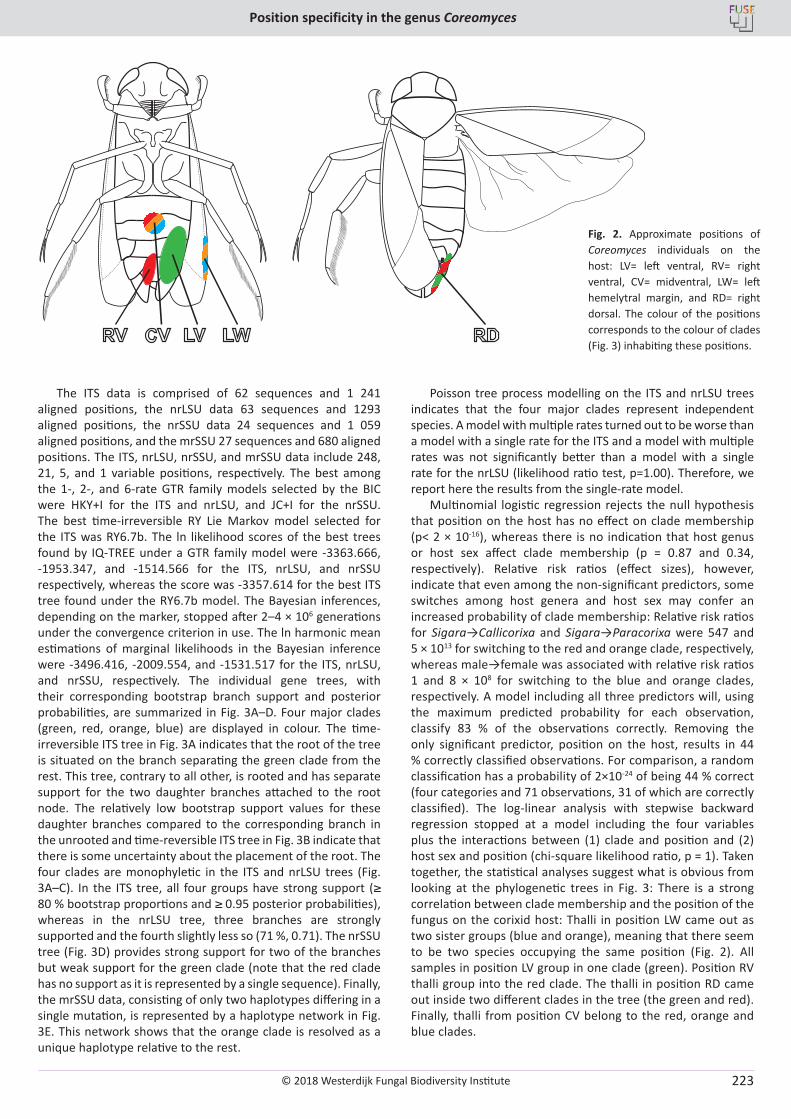
© 2018 Westerdijk Fungal Biodiversity Institute
Position specificity in the genus Coreomyces
Editor-in-ChiefProf. dr P.W. Crous, Westerdijk Fungal Biodiversity Institute, P.O. Box 85167, 3508 AD Utrecht, The Netherlands.E-mail:[email protected]
223
The ITS data is comprised of 62 sequences and 1 241 aligned positions, the nrLSU data 63 sequences and 1293 aligned positions, the nrSSU data 24 sequences and 1 059 aligned positions, and the mrSSU 27 sequences and 680 aligned positions. The ITS, nrLSU, nrSSU, and mrSSU data include 248, 21, 5, and 1 variable positions, respectively. The best among the 1-, 2-, and 6-rate GTR family models selected by the BIC were HKY+I for the ITS and nrLSU, and JC+I for the nrSSU. The best time-irreversible RY Lie Markov model selected for the ITS was RY6.7b. The ln likelihood scores of the best trees found by IQ-TREE under a GTR family model were -3363.666, -1953.347, and -1514.566 for the ITS, nrLSU, and nrSSU respectively, whereas the score was -3357.614 for the best ITS tree found under the RY6.7b model. The Bayesian inferences, depending on the marker, stopped after 2–4 × 10⁶ generations under the convergence criterion in use. The ln harmonic mean estimations of marginal likelihoods in the Bayesian inference were -3496.416, -2009.554, and -1531.517 for the ITS, nrLSU, and nrSSU, respectively. The individual gene trees, with their corresponding bootstrap branch support and posterior probabilities, are summarized in Fig. 3A–D. Four major clades (green, red, orange, blue) are displayed in colour. The time-irreversible ITS tree in Fig. 3A indicates that the root of the tree is situated on the branch separating the green clade from the rest. This tree, contrary to all other, is rooted and has separate support for the two daughter branches attached to the root node. The relatively low bootstrap support values for these daughter branches compared to the corresponding branch in the unrooted and time-reversible ITS tree in Fig. 3B indicate that there is some uncertainty about the placement of the root. The four clades are monophyletic in the ITS and nrLSU trees (Fig. 3A–C). In the ITS tree, all four groups have strong support (≥ 80 % bootstrap proportions and ≥ 0.95 posterior probabilities), whereas in the nrLSU tree, three branches are strongly supported and the fourth slightly less so (71 %, 0.71). The nrSSU tree (Fig. 3D) provides strong support for two of the branches but weak support for the green clade (note that the red clade has no support as it is represented by a single sequence). Finally, the mrSSU data, consisting of only two haplotypes differing in a single mutation, is represented by a haplotype network in Fig. 3E. This network shows that the orange clade is resolved as a unique haplotype relative to the rest.
Poisson tree process modelling on the ITS and nrLSU trees indicates that the four major clades represent independent species. A model with multiple rates turned out to be worse than a model with a single rate for the ITS and a model with multiple rates was not significantly better than a model with a single rate for the nrLSU (likelihood ratio test, p=1.00). Therefore, we report here the results from the single-rate model.
Multinomial logistic regression rejects the null hypothesis that position on the host has no effect on clade membership (p< 2 × 10-16), whereas there is no indication that host genus or host sex affect clade membership (p = 0.87 and 0.34, respectively). Relative risk ratios (effect sizes), however, indicate that even among the non-significant predictors, some switches among host genera and host sex may confer an increased probability of clade membership: Relative risk ratios for Sigara→Callicorixa and Sigara→Paracorixa were 547 and 5 × 10¹³ for switching to the red and orange clade, respectively, whereas male→female was associated with relative risk ratios 1 and 8 × 10⁸ for switching to the blue and orange clades, respectively. A model including all three predictors will, using the maximum predicted probability for each observation, classify 83 % of the observations correctly. Removing the only significant predictor, position on the host, results in 44 % correctly classified observations. For comparison, a random classification has a probability of 2×10-24 of being 44 % correct (four categories and 71 observations, 31 of which are correctly classified). The log-linear analysis with stepwise backward regression stopped at a model including the four variables plus the interactions between (1) clade and position and (2) host sex and position (chi-square likelihood ratio, p = 1). Taken together, the statistical analyses suggest what is obvious from looking at the phylogenetic trees in Fig. 3: There is a strong correlation between clade membership and the position of the fungus on the corixid host: Thalli in position LW came out as two sister groups (blue and orange), meaning that there seem to be two species occupying the same position (Fig. 2). All samples in position LV group in one clade (green). Position RV thalli group into the red clade. The thalli in position RD came out inside two different clades in the tree (the green and red). Finally, thalli from position CV belong to the red, orange and blue clades.
CVRV LV LW RD
Fig. 2. Approximate positions of Coreomyces individuals on the host: LV= left ventral, RV= right ventral, CV= midventral, LW= left hemelytral margin, and RD= right dorsal. The colour of the positions corresponds to the colour of clades (Fig. 3) inhabiting these positions.
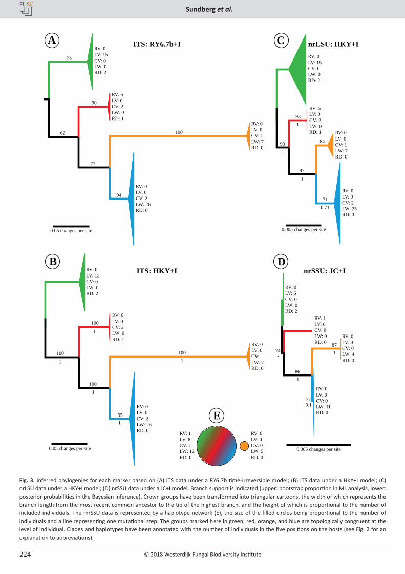
© 2018 Westerdijk Fungal Biodiversity Institute
Sundberg et al.
Editor-in-ChiefProf. dr P.W. Crous, Westerdijk Fungal Biodiversity Institute, P.O. Box 85167, 3508 AD Utrecht, The Netherlands.E-mail:[email protected]
224
75
90
10062
77
94
0.05 changes per site
RV: 0LV: 15CV: 0LW: 0RD: 2
RV: 6LV: 0CV: 2LW: 0RD: 1
RV: 0LV: 0CV: 1LW: 7RD: 0
RV: 0LV: 0CV: 2LW: 26RD: 0
93
93
84
97
71
0.005 changes per site
RV: 0LV: 18CV: 0LW: 0RD: 2
RV: 5LV: 0CV: 2LW: 0RD: 1 RV: 0
LV: 0CV: 1LW: 7RD: 0
RV: 0LV: 0CV: 2LW: 25RD: 0
ITS: RY6.7b+I nrLSU: HKY+I
ITS: HKY+I
100
100
100
95
0.05 changes per site
100
RV: 0LV: 15CV: 0LW: 0RD: 2
RV: 6LV: 0CV: 2LW: 0RD: 1
RV: 0LV: 0CV: 1LW: 7RD: 0
RV: 0LV: 0CV: 2LW: 26RD: 0
0.005 changes per site
RV: 0LV: 6CV: 0LW: 0RD: 2
RV: 1LV: 0CV: 0LW: 0RD: 0
RV: 0LV: 0CV: 0LW: 4RD: 0
RV: 0LV: 0CV: 0LW: 11RD: 0
nrSSU: JC+I
A C
74
86
77
B D
87
E
1
1
1
0.71
1
1 1
1
1
1
1
0.1
-
RV: 0LV: 0CV: 0LW: 5RD: 0
RV: 1LV: 8CV: 1LW: 12RD: 0
Fig. 3. Inferred phylogenies for each marker based on (A) ITS data under a RY6.7b time-irreversible model; (B) ITS data under a HKY+I model; (C) nrLSU data under a HKY+I model; (D) nrSSU data under a JC+I model. Branch support is indicated (upper: bootstrap proportion in ML analysis, lower: posterior probabilities in the Bayesian inference). Crown groups have been transformed into triangular cartoons, the width of which represents the branch length from the most recent common ancestor to the tip of the highest branch, and the height of which is proportional to the number of included individuals. The mrSSU data is represented by a haplotype network (E), the size of the filled circles being proportional to the number of individuals and a line representing one mutational step. The groups marked here in green, red, orange, and blue are topologically congruent at the level of individual. Clades and haplotypes have been annotated with the number of individuals in the five positions on the hosts (see Fig. 2 for an explanation to abbreviations).
© 2018 Westerdijk Fungal Biodiversity Institute
Position specificity in the genus Coreomyces
Editor-in-ChiefProf. dr P.W. Crous, Westerdijk Fungal Biodiversity Institute, P.O. Box 85167, 3508 AD Utrecht, The Netherlands.E-mail:[email protected]
225
DISCUSSION
Four species of Coreomyces
The four major clades (green, red, orange, blue) in Fig. 3 are identified and interpreted here as species, because (1) Poisson tree process modelling on the ITS and nrLSU trees indicates that the clades represent independent species, (2) they are topologically completely congruent across markers (although with different degrees of resolution), and (3) they correlate with ecological parameters, primarily the position on the host.
All species were present in the entire study area, from Uppland in the north to Skåne and Denmark in the south (Table 1). Species were also often mixed in the same host populations, and sometimes co-occurred on the same host individuals. The nomenclatural issues were not the focus of this study and assigning correct names, if available, will have to be the focus of a future research. However, thalli growing in three of the positions (CV, LV, and LW) are previously reported from Europe (Santamaria et al. 1991, Majewski 1994, De Kesel & Werbrouck 2008, Majewski 2008). The remaining RD and RV positions have not previously been explicitly reported in the literature. It remains unclear, however, whether they have been observed before, as early descriptions of Coreomyces tend to be vague about the exact positions on the host (Thaxter 1902, 1905, Spegazzini 1915, 1917, 1918, Thaxter 1931).
Specificity to host, position and sex
The Coreomyces taxa found in this study did not show any strict specificity to host species, as they occurred on five or seven corixid species belonging to two or three different but closely related genera within the subfamily Corixinae. This observation is in accord with available information on host preferences in the genus (e.g., Thaxter 1931, Majewski 1994). For instance, Coreomyces corisae, the species with the widest known host range, is known to parasitize a range of species belonging to at least four genera (Spegazzini 1918, Thaxter 1931, Santamaria et al. 1991, Majewski 1994, Santamaria 2003). Host ranges may, however, turn out to be narrower once species circumscriptions have been investigated by molecular means. Although we did not observe any strict host specificity, the effect sizes from the multinomial logistic regression suggest that some switches between host genera are associated with substantially increased odds for some of the clade memberships. The total effect of host genus is non-significant, however, and larger samples are needed to possibly detect subtle effects of this parameter.
Contrary to the strict position specificity reported for Coreomyces by Majewski (1973, 1994), our results indicate that none of the four species is restricted to only a single position on its host. Instead, each species inhabits two or three different positions, although one of them tends to be much preferred over the other(s). The latter observations explain why both the multinomial logistic regression and the log-linear analysis suggest a strong interaction between position on the host and clade membership. These findings are in agreement with Goldman & Weir (2012) and Goldmann et al. (2013). The former study demonstrated the presence of position specificity in Chitinomyces, with positions and morphology being different in male and female hosts. The latter investigation pointed to substantial morphological differences between conspecific thalli of Hesperomyces in different positions on the host, although it
was not clear whether these phenotypes were also correlated with host sex.
All Coreomyces species in our study were found on both male and female corixid hosts (Table 1). The log-linear analysis indicates that there is an interaction between host sex and the position on the host, suggesting positions are not entirely identical in males and females. This result is probably explained by individuals in positions CV (orange, blue, and red clades) and RD (red and green clades) being known only from male hosts in our data. Other positions appear to be less unequally distributed between male and female hosts. The multinomial logistic regression did not reject the null hypothesis of no effect of host sex on clade membership, nor did the stepwise log-linear model selection indicate that the interaction between host sex and clade membership is needed to explain the observed data. However, the multinomial logistic regression does suggest that host sex confers increased odds for membership in two of the clades. Sex-of-host specificity may be a non-existent extreme in a continuum, where instead weak preferences for one host sex may turn out to be frequent.
Dispersal and positioning of thalli
In this study we encountered positions present in both host sexes (LW, LV, RV) and those that are seemingly male-specific in our data (CV and RD), which is in agreement with earlier studies of position specificity (Goldmann & Weir 2012, Goldmann et al. 2013). These studies demonstrated that occurrences of the same species in the same dorsal position of both females and males is explained by mixed hetero- and homosexual mounting by males. Additional occurrences on the ventral side or on the legs in males are more easily explained, as they represent contact surfaces during heterosexual mounting. In corixid mating, males position themselves on the back of the females, or males in case of homosexual mounting (Popham 1961, Aiken 1982). Peters (1962) provided a detailed description of corixid mating behaviour. After mounting, the male swings his abdomen to the left and forces it under the female abdomen and finally becomes curled around the body of the female. This manoeuvre could account for the LV and LW positions on the left side of the body and possibly also the RV position on the right. The remaining, possibly male-specific positions (CV and RD), are more difficult to explain. An intriguing clue to how this could come about may be found in a peculiarity of the male corixid morphology. They are not bilaterally symmetric, both dextral and sinistral forms existing in many species (Schilthuizen 2013), the former normally being much more common. In sinistral males, the abdomen and the copulatory apparatus are reversed compared to dextral males, and consequently they instead wrap their abdomen around the right side of the female (Peters 1962). It could be speculated that rare occurrence of sinistral males in combination with homosexual mounting may explain the CV and RD positions.
Other kinds of behaviour associated with mating may also affect the exact positioning of the thalli. Jansson (1979), for example, indicated that the copulating animals to some extent change positions when they need to ascend to the water surface to breathe and that this requires some struggling. Agonistic behaviour should also be considered. Candolin (2004) documented males of Sigara falleni trying to hinder each other’s mounting attempts, while Jansson (1973, 1979) recorded a nudging behaviour among Arctocorisa males trying to
© 2018 Westerdijk Fungal Biodiversity Institute
Sundberg et al.
Editor-in-ChiefProf. dr P.W. Crous, Westerdijk Fungal Biodiversity Institute, P.O. Box 85167, 3508 AD Utrecht, The Netherlands.E-mail:[email protected]
226
outcompete each other. Non-social behaviour like cleaning was suggested by Huldén (1983) to result in the fungus occupying precise positions similar in both sexes. We regard this theory as unlikely, however, as we would then expect the fungus to inhabit the host limbs. This does not rule out that they may be accounted for by some male behaviour that actively transmits thalli from positions that are not sex-specific, as suggested by Benjamin (1971)
Although behaviour may explain occurrence patterns, there is also the possibility of passive infection through the host substrate. This has been suggested as an important mode of dispersal under terrestrial conditions (Arwidsson 1946, Lindroth 1948). In the case of Laboulbenia slackensis, however, active transmission was found to be far more important, whereas passive transmissions were independent of soil characteristics and often unsuccessful owing to the short life-span of the spores (De Kesel 1995). In the case of Coreomyces, the aquatic environment possibly favours spore longevity and consequently increases the chance of infection. However, the distinctly non-random positions of the thalli on the host indicate that indirect transmission of spores may not be important. Indirect transmission through a contaminated host substrate is also likely to be unfavourable in small host populations and when spore production is modest, as is the case in many Laboulbeniales (Huldén 1983).
Concluding remarks
This study has demonstrated that there are four species of Coreomyces in our sample, that they prefer but are not strictly specific to certain positions on the host, and that there may or may not be weak preferences on the part of the fungus regarding host sex and genus. These findings lead to further questions of importance to understand the evolution of Laboulbeniales: How do species boundaries arise to start with, when host preferences are not discontinuous? How does position specificity arise and how is it related to substrate requirements? How does mating take place in the fungus, if at all? Genomic data as well as controlled observations of host behaviour under laboratory conditions may help solve these and other questions.
ACKNOWLEDGEMENTS
This research was supported by grants from Helge Ax:son Johnsons stiftelse and Kapten Carl Stenholms donationsfond.
AUTHORS’ CONTRIBUTIONS
H.S. did the sampling and laboratory work. S.E. did the statistical and phylogenetic analysis. Figures were made by S.E. and H.S. Species determinations of the host material were made by J.B. The text was written jointly by H.S., Å.K. and S.E. with valuable input from J.B. Å.K. and S.E. supervised the project.
REFERENCES
Aiken RB (1982). Sound production and mating in a waterboatman, Palmacorixa nana (Heteroptera: Corixidae). Animal Behaviour 30: 54–61.
Altekar G, Dwarkadas S, Huelsenbeck JP, et al. (2004). Parallel Metropolis coupled Markov chain Monte Carlo for Bayesian phylogenetic inference. Bioinformatics 20: 407–415.
Arwidsson T (1946). Om svenska laboulbeniacéfynd. Svensk Botanisk Tidskrift 40: 307–309.
Benjamin RK (1971). Introduction and supplement to Roland Thaxter’s contribution towards a monograph of the Laboulbeniaceae. Bibliotheca Mycologia 30: 1–155.
Benjamin RK, Shanor L (1952). Sex of host specificity and position specificity of certain species of Laboulbenia on Bembidion picipes. American Journal of Botany 39: 125–131.
Candolin U (2004). Opposing selection on a sexually dimorphic trait through female choice and male competition in a water boatman. Evolution 58: 1861–1864.
De Kesel A (1993). Relations between host population density and spore transmission of Laboulbenia slackensis (Ascomycetes, Laboulbeniales) from Pogonus chalceus (Coleoptera, Carabidae). Belgian Journal of Botany 126: 155–163.
De Kesel A (1995). Relative importance of direct and indirect infection in the transmission of Laboulbenia slackensis (Ascomycetes, Laboulbeniales). Belgian Journal of Botany 128: 124–130.
De Kesel A (1996). Host specificity and habitat preference of Laboulbenia slackensis. Mycologia 88: 565–573.
De Kesel A, Haelewaters D (2014). Laboulbenia slackensis and L. littoralis sp. nov. (Ascomycota, Laboulbeniales), two sibling species as a result of ecological speciation. Mycologia 106: 407–414.
De Kesel A, Werbrouck T (2008). Belgian records of Laboulbeniales from aquatic insects. Sterbeeckia 28: 48–54.
de Queiroz K (2007). Species concepts and species delimitation. Systematic Biology 56: 879–886.
Fernández-Sánchez J, Sumner JG, Jarvis PD, et al. (2015). Lie Markov models with purine/pyrimidine symmetry. Journal of Mathematical Biology 70: 855–891.
Fox J, Weisberg S (2011). An R companion to applied regression. 2nd ed. Sage, Thousand Oaks, the United States of America.
Giard A (1892). Sur une Laboulbéniacée (Thaxteria künckeli nov. gen. et sp.), parasite de Mormolyce phyllodes Hagenbach. Comptes Rendus Hebdomadaires des Séances et Mémoires de la Société de Biologie 4: 156–158.
Goldmann L, Weir A (2012). Position specificity in Chitonomyces (Ascomycota, Laboulbeniomycetes) on Laccophilus (Coleoptera, Dytiscidae): a molecular approach resolves a century-old debate. Mycologia 104: 1143–1158.
Goldmann L, Weir A, Rossi, W (2013). Molecular analysis reveals two new dimorphic species of Hesperomyces (Ascomycota, Laboulbeniomycetes) parasitic on the ladybird Coleomegilla maculata (Coleoptera, Coccinellidae). Fungal Biology 117: 807–813.
Haelewaters D, De Kesel A (2017). De schimmel Hesperomyces virescens, een natuurlijke vijand van lieveheersbeestjes [in Dutch with English abstract]. Entomologische Berichten 77: 106–118.
Haelewaters D, Gorczak M, Pfliegler W, et al. (2015). Bringing Laboulbeniales into the 21st century: enhanced techniques for extraction and PCR amplification of DNA from minute ectoparasitic fungi. IMA Fungus 6: 363–372.
Huldén L (1983). Laboulbeniales (Ascomycetes) of Finland and adjacent parts of the U.S.S.R. Karstenia 23: 31–136.
Jansson A (1973). Stridulation and its significance in the genus Cenocorixa (Hemiptera, Corixidae). Behaviour 46: 1–36.
Jansson A (1979). Reproductive isolation and experimental hybridization between Arctocorisa carinata and A. germari (Heteroptera, Corixidae). Annales Zoologici Fennici 16: 89–104.
© 2018 Westerdijk Fungal Biodiversity Institute
Position specificity in the genus Coreomyces
Editor-in-ChiefProf. dr P.W. Crous, Westerdijk Fungal Biodiversity Institute, P.O. Box 85167, 3508 AD Utrecht, The Netherlands.E-mail:[email protected]
227
Kalyaanamoorthy S, Minh BQ, Wong TKF, et al. (2017). ModelFinder: Fast model selection for accurate phylogenetic estimates. Nature Methods 14: 587–589.
Kapli T, Lutteropp S, Zhang J, et al. (2017). Multi-rate Poisson tree processes for single-locus species delimitation under maximum likelihood and Markov chain Monte Carlo. Bioinformatics 33: 1630–1638.
Katoh K, Standley DM (2013). MAFFT multiple sequence alignment software version 7: improvements in performance and usability. Molecular Biology and Evolution 30: 772–780.
Kirk PM, Cannon PF, Minter DW, et al. (2008). Dictionary of the Fungi, 10th ed. CABI, Wallingford, the United Kingdom.
Lindroth CH (1948). Notes on the ecology of Laboulbeniaceae infesting carabid beetles. Svensk Botanisk Tidskrift 42: 34–41.
Littlewood DTJ, Rohde K, Clough KA (1997). Parasite speciation within or between host species? Phylogenetic evidence from site-specific polystome monogeneans. International Journal for Parasitology 27: 1289–1297.
Majewski T (1973). The genus Coreomyces Thaxter (Laboulbeniales) in Poland. Acta Mycologica 9: 217–228.
Majewski T (1988). Some Laboulbeniales (Ascomycotina) collected in Japan III. Species on coleopterous insects from Iriomote Island. Transactions of the Mycological Society of Japan 29: 249–264.
Majewski T (1994). The Laboulbeniales of Poland. Polish Botanical Studies 7: 1–466.
Majewski T (2003). Distribution and ecology of Laboulbeniales (Fungi, Ascomycetes) in the Białowieża Forest and its western foreland. Phytocoenosis 15 (S.N.), Supplementum Cartographiae Geobotanicae 16: 1–144.
Majewski T (2008). Atlas of the Geographical Distribution of Fungi in Poland. Fascicle 4. Laboulbeniales. W. Szafer Institute of Botany, Polish Academy of Sciences, Krakow, Poland.
Martin P (2004). Specifity of attachment sites of larval water mites (Hydrachnidia, Acari) on their insect hosts (Chironomidae, Diptera) - evidence from some stream-living species. Experimental and Applied Acarology 34: 95–112.
MycoBank (2018). URL: https://www.mycobank.org (accessed 10 Apr. 2018).
Nguyen LT, Schmidt HA, von Haeseler A, et al. (2015). IQ-TREE: A fast and effective stochastic algorithm for estimating maximum likelihood phylogenies. Molecular Biology and Evolution 32: 268–274.
Nieser N (2002). Guide to aquatic Heteroptera of Singapore and peninsular Malaysia. IV. Corixoidea. The Raffles Bulletin of Zoology 50: 263–274
Pfliegler WP, Báthori F, Haelewaters D, et al. (2016). Studies of Laboulbeniales on Myrmica ants (III): myrmecophilous arthropods as alternative hosts of Rickia wasmannii. Parasite 23: 1–7.
Peters W (1962). The morphology of Situs inversus in abdominal segmentation of Krizousacorixa femorata, Guérin (Heteroptera, Corixidae). Journal of Morphology 110: 141–155
Peyritsch J (1875). Über Vorkommen und Biologie von Laboulbeniaceen. Sitzungsberichte der Kaiserlichen Akademie der Wissenschaften. Mathematisch-Naturwissenschaftliche Classe 72: 377–385.
Popham EJ (1961). The function of paleal pegs of Corixidae (Hemiptera Heteroptera). Nature 190: 742–743.
R Core Team (2017). R: A language and environment for statistical computing. R Foundation for Statistical Computing, Vienna (Austria). URL: https://www.R-project.org/.
Rannala B, Zhu T, Yang Z (2012). Tail paradox, partial identifiability, and influential priors in Bayesian branch length inference. Molecular Biology and Evolution 29: 325–335.
Richards AG, Smith MN (1954). Infection of cockroaches with Herpomyces (Laboulbeniales). III. Experimental studies on host specificity. Botanical Gazette 116: 195–198.
Richards AG, Smith MN (1955). Infection of cockroaches with Herpomyces (Laboulbeniales). I. Life history studies. The Biological Bulletin 108: 206–218.
Ronquist F, Huelsenbeck JP (2003). MrBayes 3: Bayesian phylogenetic inference under mixed models. Bioinformatics 19: 1572–1574.
Ronquist F, Teslenko M, van der Mark P, et al. (2012). MrBayes 3.2: efficient Bayesian phylogenetic inference and model choice across a large model space. Systematic Biology 61: 539–542.
Rossi W, Kotrba M (2004). A new polymorphic species of Laboulbenia parasitic on a South American fly. Mycological Research 108: 1315–1319.
Santamaria S (1998). Laboulbeniales, I. Laboulbenia. Flora Mycologica Iberica 4: 1–186.
Santamaria S (2003). Laboulbeniales, II. Ascompsomyces–Ilyomyces. Flora Mycologica Iberica 5: 1–344.
Santamaria S, Balazuc J, Tavares II (1991). Distribution of the European Laboulbeniales (Fungi, Ascomycotina), an annotated list of species. Treballs de l’Institut Botanic de Barcelona 14: 5–123.
Santamaria S, Faille A (2009). New species of Laboulbenia and Rhachomyces (Laboulbeniales, Ascomycota), some of them polymorphic, parasitic on termiticolous ground beetles from tropical Africa. Nova Hedwigia 89: 97–120.
Scheloske H-W (1969). Beiträge zur biologie, ökologie und systematik der Laboulbeniales (Ascomycetes) unter besonder berücksichtigung des parasit-Wirt-Vergältnisses. Parasitogische Schriftenreihe 19: 1–176.
Scheloske H-W (1976). Eusynaptomyces benjaminii, spec. nova, (Ascomycetes, Laboulbeniales) und seine Anpassungen an das Fortpflanzungsverhalten seines Wirtes Enochrus testaceus (Coleoptera, Hydrophilidae). Plant Systematics and Evolution 126: 267–285.
Schilthuizen M (2013). Something gone awry: unsolved mysteries in the evolution of asymmetric animal genitalia. Animal Biology 63: 1–20.
Shen Y-H, Ye D-H, Li T-H, et al. (2006). Laboulbeniales. Flora Fungorum Sinicorum 28: 1–294.
Spegazzini C (1915). Segunda contribución al conocimiento de las Laboulbeniales italianas. Anales del Museo Nacional de Historia Natural de Buenos Aires 27: 37–74.
Spegazzini C (1917). Revision de las Laboulbeniales Argentinas. Anales del Museo Nacional de Historia Natural de Buenos Aires 29: 445–688.
Spegazzini C (1918). Observaciones microbiológicas. Anales de la Sociedad Científica Argentina 85: 311–323.
Sugiyama K, Hayama M (1981). Notes on Laboulbeniomycetes of Formosa II. Transactions of the Mycological Society of Japan 22: 187–196.
Sumner JG, Fernández-Sánchez J, Jarvis PD (2012). Lie Markov models. Journal of Theoretical Biology 298: 16–31.
Sundberg H, Ekman S, Kruys Å (2018). A crush on small fungi: An efficient and quick method for obtaining DNA from minute ascomycetes. Methods in Ecology and Evolution 9: 148–158.
Tavares II (1985). Laboulbeniales (Fungi, Ascomycetes). Mycologia Memoir 9: 1–627.
Thaxter R (1896). Contribution towards a monograph of the Laboulbeniaceæ. I. Memoirs of the American Academy of Arts and Sciences 12: 189–429.
Thaxter R (1902). Preliminary diagnoses of new species of Laboulbeniaceae. V. Proceedings of the American Academy of Arts and Sciences of Boston 38: 7–57.
Thaxter R (1905). Diagnoses of New Species of Laboulbeniaceae. VI. Proceedings of the American Academy of Arts and Sciences 41: 308–318.
© 2018 Westerdijk Fungal Biodiversity Institute
Sundberg et al.
Editor-in-ChiefProf. dr P.W. Crous, Westerdijk Fungal Biodiversity Institute, P.O. Box 85167, 3508 AD Utrecht, The Netherlands.E-mail:[email protected]
228
Thaxter R (1931). Contribution towards a monograph of the Laboulbeniaceæ. V. Memoirs of the American Academy of Arts and Sciences 16: 1–435.
Venables WN, Ripley BD (2002). Modern applied statistics with S. 4th ed. Springer, New York, USA.
Vilgalys R, Hester M (1990). Rapid genetic identification and mapping of enzymatically amplified ribosomal DNA from several Cryptococcus species. Journal of Bacteriology 172: 4238–4246.
Weir A, Blackwell M (2005). Fungal biotrophic parasites of insects and other arthropods. In: Vega FE, Blackwell M (eds), Insect-fungal associations: Ecology and evolution. Oxford University Press, New York, USA: 119–145.
Weir A, Hammond PM (1997). Laboulbeniales on beetles: host utilization patterns and species richness of the parasites. Biodiversity and Conservation 6: 701–719.
Whisler HC (1968). Experimental studies with a new species of Stigmatomyces (Laboulbeniales). Mycologia 60: 65–75.
White TJ, Bruns T, Lee S. et al. (1990). Amplification and direct sequencing of fungal ribosomal RNA genes for phylogenetics. In: Innis MA, Gelfand DH, Sninsky JJ, et al. (eds.), PCR protocols: A guide to methods and applications. Academic Press, San Diego, the United States of America: 315–322.
Woodhams MD, Fernández-Sánchez J, Sumner, JG (2015). A new hierarchy of phylogenetic models consistent with heterogeneous substitution rates. Systematic Biology 64: 638–650.
Zhang C, Rannala B, Yang Z (2012). Robustness of compound Dirichlet priors for Bayesian inference of branch lengths. Systematic Biology 61: 779–784.
Zhang J, Kapli P, Pavlidis P, et al. (2013). A general species delimitation method with applications to phylogenetic placements. Bioinformatics 29: 2869–2876.
Zoller S, Scheidegger C, Sperisen C (1999). PCR primers for the amplification of mitochondrial small subunit ribosomal DNA of lichenforming ascomycetes. The Lichenologist 31: 511–516.
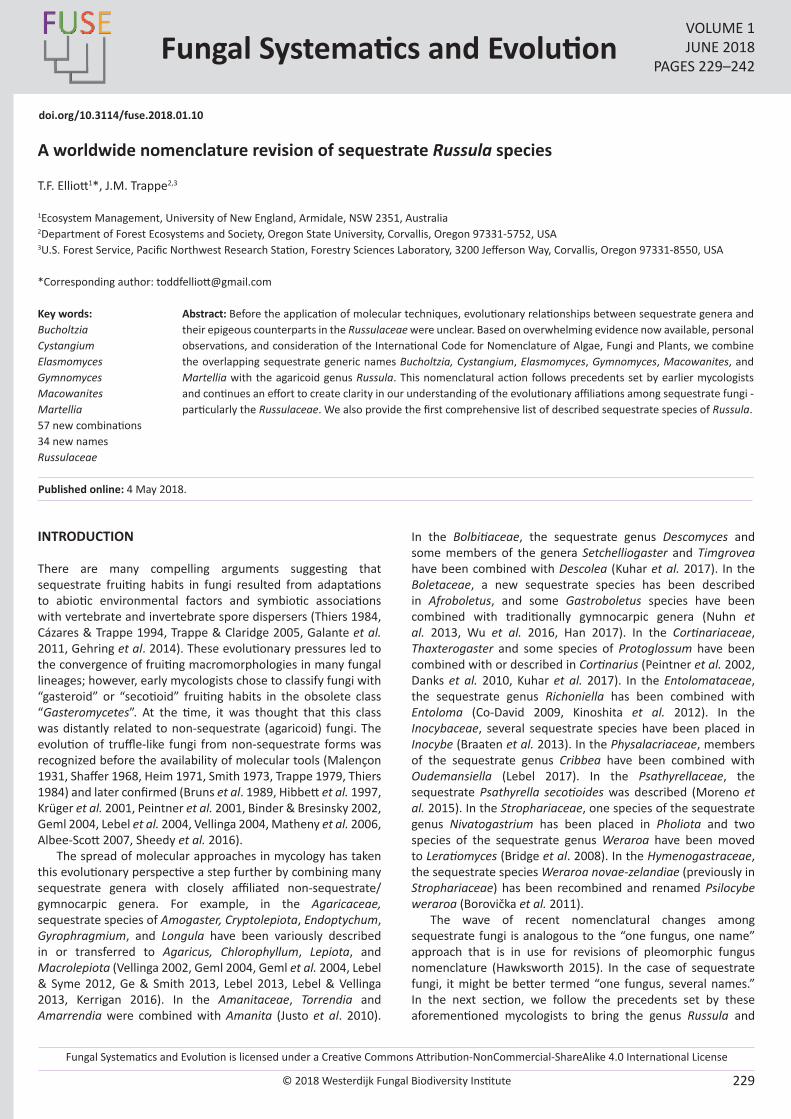
Fungal Systematics and Evolution is licensed under a Creative Commons Attribution-NonCommercial-ShareAlike 4.0 International License
© 2018 Westerdijk Fungal Biodiversity Institute 229
Editor-in-ChiefProf. dr P.W. Crous, Westerdijk Fungal Biodiversity Institute, P.O. Box 85167, 3508 AD Utrecht, The Netherlands.E-mail:[email protected]
Fungal Systematics and EvolutionVOLUME 1JUNE 2018
PAGES 229–242
doi.org/10.3114/fuse.2018.01.10
A worldwide nomenclature revision of sequestrate Russula species
T.F. Elliott1*, J.M. Trappe2,3
1Ecosystem Management, University of New England, Armidale, NSW 2351, Australia2Department of Forest Ecosystems and Society, Oregon State University, Corvallis, Oregon 97331-5752, USA 3U.S. Forest Service, Pacific Northwest Research Station, Forestry Sciences Laboratory, 3200 Jefferson Way, Corvallis, Oregon 97331-8550, USA
*Corresponding author: [email protected]
Abstract: Before the application of molecular techniques, evolutionary relationships between sequestrate genera and their epigeous counterparts in the Russulaceae were unclear. Based on overwhelming evidence now available, personal observations, and consideration of the International Code for Nomenclature of Algae, Fungi and Plants, we combine the overlapping sequestrate generic names Bucholtzia, Cystangium, Elasmomyces, Gymnomyces, Macowanites, and Martellia with the agaricoid genus Russula. This nomenclatural action follows precedents set by earlier mycologists and continues an effort to create clarity in our understanding of the evolutionary affiliations among sequestrate fungi - particularly the Russulaceae. We also provide the first comprehensive list of described sequestrate species of Russula.
Key words: BucholtziaCystangiumElasmomycesGymnomycesMacowanitesMartellia57 new combinations34 new namesRussulaceae
Published online: 4 May 2018.
INTRODUCTION
There are many compelling arguments suggesting that sequestrate fruiting habits in fungi resulted from adaptations to abiotic environmental factors and symbiotic associations with vertebrate and invertebrate spore dispersers (Thiers 1984, Cázares & Trappe 1994, Trappe & Claridge 2005, Galante et al. 2011, Gehring et al. 2014). These evolutionary pressures led to the convergence of fruiting macromorphologies in many fungal lineages; however, early mycologists chose to classify fungi with “gasteroid” or “secotioid” fruiting habits in the obsolete class “Gasteromycetes”. At the time, it was thought that this class was distantly related to non-sequestrate (agaricoid) fungi. The evolution of truffle-like fungi from non-sequestrate forms was recognized before the availability of molecular tools (Malençon 1931, Shaffer 1968, Heim 1971, Smith 1973, Trappe 1979, Thiers 1984) and later confirmed (Bruns et al. 1989, Hibbett et al. 1997, Krüger et al. 2001, Peintner et al. 2001, Binder & Bresinsky 2002, Geml 2004, Lebel et al. 2004, Vellinga 2004, Matheny et al. 2006, Albee-Scott 2007, Sheedy et al. 2016).
The spread of molecular approaches in mycology has taken this evolutionary perspective a step further by combining many sequestrate genera with closely affiliated non-sequestrate/gymnocarpic genera. For example, in the Agaricaceae, sequestrate species of Amogaster, Cryptolepiota, Endoptychum, Gyrophragmium, and Longula have been variously described in or transferred to Agaricus, Chlorophyllum, Lepiota, and Macrolepiota (Vellinga 2002, Geml 2004, Geml et al. 2004, Lebel & Syme 2012, Ge & Smith 2013, Lebel 2013, Lebel & Vellinga 2013, Kerrigan 2016). In the Amanitaceae, Torrendia and Amarrendia were combined with Amanita (Justo et al. 2010).
In the Bolbitiaceae, the sequestrate genus Descomyces and some members of the genera Setchelliogaster and Timgrovea have been combined with Descolea (Kuhar et al. 2017). In the Boletaceae, a new sequestrate species has been described in Afroboletus, and some Gastroboletus species have been combined with traditionally gymnocarpic genera (Nuhn et al. 2013, Wu et al. 2016, Han 2017). In the Cortinariaceae, Thaxterogaster and some species of Protoglossum have been combined with or described in Cortinarius (Peintner et al. 2002, Danks et al. 2010, Kuhar et al. 2017). In the Entolomataceae, the sequestrate genus Richoniella has been combined with Entoloma (Co-David 2009, Kinoshita et al. 2012). In the Inocybaceae, several sequestrate species have been placed in Inocybe (Braaten et al. 2013). In the Physalacriaceae, members of the sequestrate genus Cribbea have been combined with Oudemansiella (Lebel 2017). In the Psathyrellaceae, the sequestrate Psathyrella secotioides was described (Moreno et al. 2015). In the Strophariaceae, one species of the sequestrate genus Nivatogastrium has been placed in Pholiota and two species of the sequestrate genus Weraroa have been moved to Leratiomyces (Bridge et al. 2008). In the Hymenogastraceae, the sequestrate species Weraroa novae-zelandiae (previously in Strophariaceae) has been recombined and renamed Psilocybe weraroa (Borovička et al. 2011).
The wave of recent nomenclatural changes among sequestrate fungi is analogous to the “one fungus, one name” approach that is in use for revisions of pleomorphic fungus nomenclature (Hawksworth 2015). In the case of sequestrate fungi, it might be better termed “one fungus, several names.” In the next section, we follow the precedents set by these aforementioned mycologists to bring the genus Russula and
© 2018 Westerdijk Fungal Biodiversity Institute
Elliott & Trappe
Editor-in-ChiefProf. dr P.W. Crous, Westerdijk Fungal Biodiversity Institute, P.O. Box 85167, 3508 AD Utrecht, The Netherlands.E-mail:[email protected]
230
its associated sequestrate genera up to date with current understandings of sequestrate fungus nomenclature.
SEQUESTRATE SPECIES OF RUSSULA
Since the early 19th century, new sequestrate Russulae have been placed in one or more of several genera created expressly for them. In early observations of amyloid spore reactions to iodine solutions, Bucholtz (1901, 1903) linked the sequestrate genus Elasmomyces to the Russula lineage and Arcangeliella to Lactarius, even though the phylogenetic affinities were not well understood at the time. The number of sequestrate genera regarded as russuloid in pre-molecular times reached its zenith at five in the seminal paper by Singer & Smith (1960): Cystangium, Elasmomyces, Gymnomyces, Macowanites, and Martellia. A sixth, Bucholtzia, had been validly described by Lohwag (1924) but never accepted by the taxonomist community. Species in two other genera of sequestrate Russulaceae, Arcangeliella and Zelleromyces, are mostly resolved within the epigeous genus Lactarius, although some individual species may eventually be resolved within Russula or other genera in the Russulaceae (Buyck et al. 2008). Each of these 10 genera was circumscribed by characters thought to separate them from the others, but all show various degrees of intergradation (Calonge & Martin 2000, Trappe et al. 2002). Moreover, some sequestrate russuloid species had initially been erroneously assigned to families unrelated to the Russulaceae, including the Agaricaceae, Boletaceae, and Hydnangiaceae. The champion in nomenclatural complexity is Russula krjukowensis, which has been treated in seven genera (see the following species list for the formal new combination).
By the late 20th century, the phylogenetic relationship of sequestrate russuloid taxa to Russula was firmly demonstrated (Lebel & Trappe 2000, Lebel 2007). Morphological distinctions between some of those genera soon became blurred as additional new species proved to be morphological intermediates. Calonge & Martin (2000), Lebel & Trappe (2000, 2002), and Lebel (2007) attempted to sharpen the generic morphological distinctions, but phylogenetic analyses by Miller et al. (2001, 2006) clearly showed that the Russulaceae are monophyletic, and members of the six named sequestrate russuloid genera are nested among non-sequestrate Russula species with no consistent genetic relationship to each other or to any clade within Russula. The generic names erected to describe sequestrate morphologies of species we now combine with Russula have shown to be nomenclatural artifacts that in hindsight have little bearing on the phylogenetic positions of these taxa. No available phylograms provide evidence that any sequestrate Russula spp. belong to distinct lineages within the genus. The para- or polyphyly within phylogenies that include both sequestrate and non-sequestrate Russula spp. are nomenclatural relics, not genetics.
Over the last twenty years, multiple papers have been published to either describe new sequestrate species in Russula or Lactarius, recombine species from sequestrate genera with Russula or Lactarius, or suggest close or monophyletic relationships between species with the different morphologies (Martín et al. 1999, Miller et al. 2001, Desjardin 2003, Nuytinck et al. 2003, Eberhardt & Verbeken 2004, Lebel & Tonkin 2007, Li et al. 2013, Verbeken et al. 2014, Beenken et al. 2016, Lebel 2017, Looney et al. 2018). It has been widely suggested through published phylogenies that all remaining members
of Bucholtzia, Cystangium, Elasmomyces, Gymnomyces, Macowanites, and Martellia should be transferred to Russula. These combinations will reduce the confusion in the current “one fungus, several names” situation and will simplify how we refer to these fungi. Lebel (2007, 2017) did a meticulous job of combining all Australian sequestrate Russulae into Russula. Here we update the remaining described sequestrate Russula species. In an era of genetics, environmental sequencing, and analysis of ecological roles of fungi through sequencing of root tips, having multiple generic names representing fungi that share close genetic affinities leads to further confusion. As stated in the preamble to the 2012 International Code of Nomenclature for Algae, Fungi, and Plants: “The purpose of giving a name to a taxonomic group is not to indicate its characters or history, but to supply a means of referring to it and to indicate its taxonomic rank” (McNeill et al. 2012). Accordingly, we judge that the following nomenclatural revisions of the genus Russula provide the clearest and most accurate information to indicate their taxonomic rank. Some information is lost without the sequestrate generic names; however, this is easily remedied in phylograms by using an arbitrary symbol, colour, or font in association with sequestrate names in a list, caption, text, phylogram, etc., as done by Miller et al. (2001) and Hosaka et al. (2006).
METHODS
We considered genetics whenever possible, but it was impossible to source sequences of type specimens (or new enough specimens) collected from close to type localities to generate a full phylogeny of the species we cover. To resolve the nomenclatural issues in this study, we relied heavily on morphology and reviewed specimens and/or original descriptions of the species we combined to ensure that they belonged in Russula. With increased interest in the sequestrate Russulaceae, we hope that many of these taxa will be re-collected and sequenced to generate a world phylogeny of this group. Genetics would make it possible to resolve the placement of the sequestrate genera Arcangeliella and Zelleromyces within the closely affiliated gymnocarpic genera Lactarius, Lactifluus, and Multifurca.
All specific epithet headings are capitalized and arranged alphabetically in the list below. These species were originally published in the genera Bucholtzia, Cystangium, Elasmomyces, Gymnomyces, Macowanites, or Martellia, as listed in Index Fungorum (plus a few incorrectly placed in non-russuloid genera). In this way, readers can determine how and why an original sequestrate name has been converted to its corresponding Russula name. The protocols for these decisions follow Rossman (2014) and use an amended version of the format provided by Rossman et al. (2016). Sequestrate species with Russula names already or newly published herein are in boldface. The standard nomenclature abbreviation comb. nov. is used when the genus is being combined with Russula but the original specific epithet can still be retained; nom. nov. is used when a new name must be proposed to avoid creating a homonym with a previously described species of Russula. When a new name is needed to avoid producing Russula homonyms, its etymology and other relevant information are noted. MycoBank numbers are assigned and registered to each new combination or new name.
© 2018 Westerdijk Fungal Biodiversity Institute
Nomenclature of sequestrate Russula spp.
Editor-in-ChiefProf. dr P.W. Crous, Westerdijk Fungal Biodiversity Institute, P.O. Box 85167, 3508 AD Utrecht, The Netherlands.E-mail:[email protected]
231
REVISED NOMENCLATURE OF SEQUESTRATE RUSSULA SPP.
Generic type species: Russula emetica (Schaeff.) Pers., Observ. Mycol. (Lipsiae) 1: 100. 1796.
ABIETISRussula subabietis Trappe & T.F. Elliott, nom. nov. MycoBank MB821258.Replaced synonym: Gymnomyces abietis Trappe & Castellano, Mycotaxon 75: 161. 2000, non Russula abietum (J. Blum) Bon, Doc. Mycol. 13 (no. 50): 27. 1983.
Note: subabietis (“almost of the fir”) to avoid confusion between abietis (“of the fir”) with the related epithet abietum (“of firs”).
ACRISRussula acerba Trappe & T.F. Elliott, nom. nov. MycoBank MB821259.Replaced synonym: Macowanites acris Singer & A.H. Sm., Mem. Torrey Bot. Club 21(3): 86. 1960, non Russula acris Steinhaus, Hedwigia 27: 51. 1888.
Etymology and note: acerba (“bitter”) to avoid producing a homonym with the earlier acris (“sharp”) of Russula acris Steinhaus (1888).
AGARICINUSRussula agaricina (Kalchbr. ex Berk.) Trappe & T.F. Elliott, comb. nov. MycoBank MB 825252.Basionym: Macowania agaricina Kalchbr. ex Berk., Gard. Chron. 5: 785. 1876.Synonyms: Macowanites agaricinus (Kalchbr. ex Berk.) Kalchbr., Grevillea 10(55): 107. 1882. Russula agaricina (Kalchbr. ex Berk.) T. Lebel, [as ‘(Kalchbr.) T. Lebel.’], Austral. Syst. Bot. 20: 360. 2007, nom. inval. (Art. 41.5).
Note: Macowinia agaricina is a legitimate name although published under Kalchbrenner’s illegitimate genus name Macowania (Art. 55.1) and has to be considered in terms of priority issues (Art. 11.4). Lebel (in Lebel & Tonkin 2007) intended to reallocate this name to Russula but did not introduce a valid combination (basionym not cited). She only referred to Macowanites agaricinus, which is only an indirect reference to the true basionym and not sufficient to validate the combination (Art. 41.5).
ALBARussula stevemilleri Trappe & T.F. Elliott, nom. nov. MycoBank MB821262.Replaced synonym: Hydnangium album Harkn., Proc. Calif. Acad. Sci., Ser. 3, Bot. 1: 251. 1899, non Russula alba Velen., České Houby 1: 149. 1920.Synonyms: Martellia alba (Harkn.) Singer & A.H. Sm., Mem. Torrey Bot. Club 21: 41. 1961.Gymnomyces albus (Harkn.) Trappe, T. Lebel & Castellano, Mycotaxon 81: 198. 2002.
Etymology and note: In honor of Dr. Steve Miller for his continuing research on taxonomy and phylogeny of sequestrate and other Russula species. This new name is erected to avoid producing a homonym with Russula alba Velen. (1920).
ALBIDIGLEBARussula albidigleba (Singer & A.H. Sm.) Trappe & T.F. Elliott, comb. nov. MycoBank MB821263.Basionym: Macowanites albidiglebus Singer & A.H. Sm., Mem. Torrey Bot. Club 21(3): 85. 1960.
ALBIDOFLAVARussula albidoflava T. Lebel, Austral. Syst. Bot. 20: 361. 2007. MycoBank MB532175.
ALBOBRUNNEARussula albobrunnea T. Lebel, Austral. Syst. Bot. 20: 362. 2007. MycoBank MB532176.
ALPINUS Russula alpicola Trappe & T.F. Elliott, nom. nov. MycoBank MB821264.Replaced synonym: Macowanites alpinus Zeller, Mycologia 39: 291. 1947, non Russula alpina (A. Blytt & Rostr.) F.H. Møller & Jul. Schäff., Ann. Mycol. 38(2/4): 333. 1940.Synonym: Elasmomyces alpinus (Zeller) Zeller, Mycologia 40: 643. 1948.
Etymology and note: alpicola (“dweller of alps”), to avoid producing a homonym with the earlier Russula alpina (A. Blytt & Rostr.) F.H. Møller & Jul. Schäff. (1940).
AMMOPHILUSRussula ammophila (J.M. Vidal & Calonge) Trappe & T.F. Elliott, comb. nov. MycoBank MB821265.Basionym: Gymnomyces ammophilus J.M. Vidal & Calonge, Bol. Soc. Micol. Madrid 24: 66. 1999.Synonym: Macowanites ammophilus (J.M. Vidal & Calonge) J.M. Vidal & Calonge, Revista Catal. Micol. 24: 70. 2002.
ARENICOLARussula arenicola (S.L. Mill. & D. Mitch.) Trappe & T.F. Elliott, comb. nov. MycoBank MB821266.Basionym: Macowanites arenicola S.L. Mill. & D. Mitch., Mycotaxon 89: 284. 2004.
BALPINEUMRussula balpinea (Grgur.) T. Lebel, Muelleria 36: 12. 2017. MycoBank MB820015.Basionym: Cystangium balpineum Grgur., Larger Fungi of South Australia (Adelaide): 53 (1997).
BISPORUMRussula bispora (T. Lebel) T. Lebel, Muelleria 36: 12. 2017. MycoBank MB820016.Basionym: Cystangium bisporum T. Lebel, Austral. Syst. Bot. 16: 373. 2003.
BORANUPENSISRussula boranupensis (T. Lebel) T. Lebel, Muelleria 36: 12. 2017. MycoBank MB820018.Basionym: Gymnomyces boranupensis T. Lebel, Austral. Syst. Bot. 16: 403. 2003.
BRUNNEONIGRARussula brunneonigra T. Lebel, Austral. Syst. Bot. 20: 363. 2007. MycoBank MB532177.
© 2018 Westerdijk Fungal Biodiversity Institute
Elliott & Trappe
Editor-in-ChiefProf. dr P.W. Crous, Westerdijk Fungal Biodiversity Institute, P.O. Box 85167, 3508 AD Utrecht, The Netherlands.E-mail:[email protected]
232
BRUNNESCENSRussula shafferi Trappe & T.F. Elliott, nom. nov. MycoBank MB821312.Replaced synonym: Martellia brunnescens Singer & A.H. Sm., Mem. Torrey Bot. Club 21(3): 37. 1961, non Russula brunnescens Murrill, Lloydia 8: 264. 1946 (1945).Synonym: Gymnomyces brunnescens (Singer & A.H. Sm.) Trappe, T. Lebel & Castellano, Mycotaxon 81: 199. 2002.
Etymology and note: To honor Dr. Robert Shaffer for his important contributions to knowledge of North American Russulae. This new name is erected to avoid producing a homonym with the earlier Russula brunnescens Murrill (1945).
CALIFORNICARussula californica (Singer & A.H. Sm.) Trappe & T.F. Elliott, comb. nov. MycoBank MB821336.Basionym: Martellia californica Singer & A.H. Sm., Mem. Torrey Bot. Club 21(3): 33. 1961.Synonym: Gymnomyces californicus (Singer & A.H. Sm.) Trappe, T. Lebel & Castellano, Mycotaxon 81: 199. 2002, non Russula californiensis Burl., Mycologia 28: 262. 1936.
CANDIDUS
Note: Originally described as Hydnangium candidum Tul. & C. Tul. in 1843, this species was recombined by Zeller & Dodge (1935) as Sclerogaster candidus (Tul. & C. Tul.) Zeller & C.W. Dodge, and then by Vidal (2005) as Macowanites candidus (Tul. & C. Tul.) J.M. Vidal. According to Kirk (2010), S. candidus in the Sclerogastraceae, not the Russulaceae, is the correct name.
CAPITIS-ORAE Russula capitis-orae (Dring) T. Lebel, Muelleria 36: 12. 2017. MycoBank MB820020.Basionym: Elasmomyces capitis-orae Dring, in Dring & Pegler, Kew Bull. 32(3): 564. 1978.Synonym: Cystangium capitis-orae (Dring) T. Lebel, Mycotaxon 81: 197. 2002.
CHLORINOSMUSRussula chlorineolens Trappe & T.F. Elliott, nom. nov. MycoBank MB821355.Replaced synonym: Macowanites chlorinosmus A.H. Sm. & Trappe, Mycologia 55: 423. 1963, non Russula chlorinosma Burl., Mycologia 16: 22. 1924.
Etymology and note: “chlorineolens” (smelling like chlorine), to avoid producing a homonym with the earlier, similarly smelling Russula chlorinosmus Burl. (1924).
CINNAMOMEARussula unicalifornica Trappe & T.F. Elliott, nom. nov. MycoBank MB821338.Replaced synonym: Gymnomyces cinnamomeus Singer & A.H. Sm., Mem. Torrey Bot. Club 21(3): 51. 1960, non Russula cinnamomea Banning, Bot. Gaz. 6(1): 166. 1881.
Etymology and note: “unicalifornica” contraction of “University of California” in reference to the site of the type collection to avoid producing a homonym with the earlier Russula cinnamomea Banning (1881).
CITRINARussula harknessii Trappe & T.F. Elliott, nom. nov. MycoBank MB821339.Replaced synonym: Octaviania citrina Harkn., Proc. Calif. Acad. Sci., Ser. 3, Bot. 1: 252. 1899, non Russula citrina Gillet, Revue mycol., Toulouse 3(no. 11): 5. 1881, non Macowanites citrinus Singer & A.H. Sm., Mem. Torrey Bot. Club 21(3): 69. 1960.Synonyms: Hydnangium citrinum (Harkn.) Zeller & C.W. Dodge, Ann. Missouri. Bot. Gard. 22: 371. 1935.Gymnomyces citrinus (Harkn.) Trappe, T. Lebel & Castellano, Mycotaxon 81: 199. 2002.
Etymology and note: In honor of H. W. Harkness, pioneer in taxonomy of North American sequestrate fungi. This new name is erected to avoid producing a homonym with the earlier Russula citrina Gillet (1881).
CITRINUSRussula alleinstanleyae T.F. Elliott & Trappe, nom. nov. MycoBank MB823193.Replaced synonym: Macowanites citrinus Singer & A.H. Sm., Mem. Torrey Bot. Club 21(3): 69. 1960, non Russula citrina Gillet, Revue Mycol., (Toulouse) 3(no. 11): 5 (1881), non Gymnomyces citrinus (Harkn.) Trappe, T. Lebel & Castellano, Mycotaxon 81: 199. 2002.
Etymology and note: In honor of mycologist and educator Allein Stanley for her more than 40 years of dedicated service to the North American Mycological Association. This new name is erected to avoid producing a homonym with the earlier Russula citrina Gillet (1881).
CLAVATUM Russula clavata (T. Lebel) T. Lebel, Muelleria 36: 12. 2017. MycoBank MB820021.Basionym: Cystangium clavatum T. Lebel, Austral. Syst. Bot. 16: 375. 2003.
CLELANDIIRussula aurantirosea T. Lebel, Muelleria 36: 12. 2017. MycoBank MB820058.Replaced synonym: Gymnomyces clelandii T. Lebel, Austral. Syst. Bot. 16: 404. 2003, non Russula clelandii O.K. Mill. & R.N. Hilton, Sydowia 39: 128. 1987.
COMPACTARussula mattsmithii Trappe & T.F. Elliott, nom. nov. MycoBank MB821354.Replaced synonym: Gymnomyces compactus Singer & A.H. Sm., Mem. Torrey Bot. Club 21(3): 53. 1960, non Russula compacta Frost, Rep. (Annual) New York State Mus. Nat. Hist. 32: 32. 1879.
Etymology and note: In honor of Dr. Matt Smith for his extensive research on taxonomy and ecology of sequestrate fungi. This new name is erected to avoid producing a homonym with the earlier Russula compacta Frost (1879).
COSTATISPORUSRussula costatispora (T. Lebel) T. Lebel, Muelleria 36: 12. 2017. MycoBank MB820022.Basionym: Gymnomyces costatisporus T. Lebel, Austral. Syst. Bot. 16: 405. 2003.
© 2018 Westerdijk Fungal Biodiversity Institute
Nomenclature of sequestrate Russula spp.
Editor-in-ChiefProf. dr P.W. Crous, Westerdijk Fungal Biodiversity Institute, P.O. Box 85167, 3508 AD Utrecht, The Netherlands.E-mail:[email protected]
233
CREMEARussula dodgei Trappe & T.F. Elliott, nom. nov. MycoBank MB824950.Replaced synonym: Arcangeliella cremea Zeller & C.W. Dodge, Ann. Missouri Bot. Gard. 22: 367. 1935, non Russula cremea (Murrill) Singer, Lilloa 22: 711. 1951. Synonyms: Martellia cremea (Zeller & C.W. Dodge) Singer & A.H. Sm., Mem. Torrey Bot. Club 21(3): 45. 1961 (1960). Gymnomyces cremeus (Zeller & C.W. Dodge) Trappe et al., Mycotaxon 81: 199. 2002.
Etymology and note: In honor of C.W. Dodge, who collaborated with S. M. Zeller in describing numerous new species of seques-trate fungi, including this taxon and to avoid confusion with Rus-sula cremea (Murrill) Singer.
CRISTATARussula korystospora T. Lebel, Muelleria 36: 12. 2017. MycoBank MB821530.Replaced synonym: Gymnomyces cristatus T. Lebel, New Zealand J. Bot. 40: 491. 2002, non Russula cristata Romagn., Bull. Mens. Soc. Linn. Soc. Bot. Lyon 31(1): 177. 1962.
DEPAUPERATUMRussula depauperata (Singer & A.H. Sm.) Trappe & T.F. Elliott, comb. nov. MycoBank MB821344.Basionym: Cystangium depauperatum Singer & A.H. Sm., Mem. Torrey Bot. Club 21: 69. 1960.
DOMINGUEZIAERussula laurae Trappe & T.F. Elliott, nom. nov. MycoBank MB821357.Replaced synonym: Cystangium domingueziae Nouhra & Trierv.-Per., Mycologia 107: 94. 2014, non Gymnomyces dominguezii Mor.-Arr., J. Gómez & Calonge, Mycol. Res. 103: 215. 1999.
Etymology and note: In honor of Argentine mycologist, Laura S. Dominguez after whom the species was originally named. This new name is erected to avoid producing a homonym with the earlier and similar name Gymnomyces dominguezii Mor.-Arr., J. Gómez & Calonge (see following entry).
DOMINGUEZIIRussula dominguezii (Mor.-Arrl, J. Gómez & Calonge) Trappe & T.F. Elliott, comb. nov. MycoBank MB821346.Basionym: Gymnomyces dominguezii Mor.-Arr., J. Gómez & Calonge, Mycol. Res. 103: 215. 1999.
DURANGENSISRussula durangensis (Guzmán) Trappe & T.F. Elliott, comb. nov. MycoBank MB821358.Basionym: Macowanites durangensis Guzmán, Revista Mex. Micol. 4: 118. 1988.
EBURNEARussula eburnea (T. Lebel) T. Lebel, Muelleria 36: 12. 2017. MycoBank MB820023.Basionym: Gymnomyces eburneus T. Lebel, Austral. Syst. Bot. 16: 408. 2003.
ECHINOSPORUSRussula tombrunsii Trappe & T.F. Elliott, nom. nov. MycoBank MB823196.Replaced synonym: Macowanites echinosporus Zeller & C.W. Dodge, Ann. Missouri Bot. Gard. 6: 57. 1919, non Russula echinospora Singer, Bull. Soc. Mycol. France 55: 270. 1940, non Russula echinospora R. Heim, Rev. Mycol. (Toulouse) 32: 207. 1967, non Cystangium echinosporum (Zeller & C.W. Dodge) Trappe, T. Lebel & Castellano, Mycotaxon 81: 197. 2002, non Elasmomyces echinosporus Zeller & C.W. Dodge, Ann. Missouri Bot. Gard. 22: 370. 1935.
Etymology and note: In honor of Dr. Tom Bruns for his career-long research on taxonomy, phylogeny and ecology of ectomycorrhizal fungi. This new name is erected to avoid producing a homonym of R. echinospora Singer (1940).
EILDONENSISRussula eildonensis (G.W. Beaton et al.) T. Lebel, Muelleria 36: 12. 2017. MycoBank MB820024Basionym: Gymnomyces eildonensis G.W. Beaton et al., Kew Bull. 39: 680. 1984.
ELLIPSOSPORUMRussula ellipsospora (Zeller) Trappe & T.F. Elliott, comb. nov. MycoBank MB821361.Basionym: Hydnangium ellipsosporum Zeller, Mycologia 31: 13. 1939. Synonyms: Martellia ellipsospora (Zeller) Singer & A.H. Sm., Mem. Torrey Bot. Club 21: 30. 1960.Gymnomyces ellipsosporus (Zeller) Trappe, T. Lebel & Castellano, Mycotaxon 81: 199. 2002.
FALLAXRussula similaris Trappe & T.F. Elliott, nom. nov. MycoBank MB821362.Replaced synonym: Martellia fallax Singer & A.H. Sm., Mem. Torrey Bot. Club 21: 34. 1960, non Russula fallax (Schaeff.) Fr., Hymenomyc. Eur. (Upsaliae): 449. 1874.Synonym: Gymnomyces fallax (Singer & A.H. Sm.) Trappe, T. Lebel & Castellano, Mycotaxon 81: 199. 2002.
Etymology and note: “similaris” in reference to the similarity of the two epithets cited above, “fallax” (deceptive) deceiving their respective authors, and to avoid producing a homonym with the earlier R. fallax (Schaef.) Fr. (1874).
FERRUGINASCENSRussula ferruginascens (Singer & A.H. Sm.) Trappe & T.F. Elliott, comb. nov. MycoBank MB821363.Basionym: Gymnomyces ferruginascens Singer & A.H. Sm., Mem. Torrey Bot. Club 21: 54. 1960.
FLAVOVIRENSRussula olivaceoflava T. Lebel, Muelleria 36: 12. 2017. MycoBank MB821532.Replaced synonym: Cystangium flavovirens T. Lebel, Austral. Syst. Bot. 16: 377. 2003, non Russula flavovirens J. Bommer & M. Rousseau, Bull. Soc. Roy. Bot. Belgique 23(1): 310. 1884.
© 2018 Westerdijk Fungal Biodiversity Institute
Elliott & Trappe
Editor-in-ChiefProf. dr P.W. Crous, Westerdijk Fungal Biodiversity Institute, P.O. Box 85167, 3508 AD Utrecht, The Netherlands.E-mail:[email protected]
234
FOETENSRussula taetra Trappe & T.F. Elliott, nom. nov. MycoBank MB821365.Replaced synonym: Martellia foetens Singer & A.H. Sm., Mem. Torrey Bot. Club 21: 36. 1960, non Russula foetens Pers., Observ. Mycol. (Lipsiae) 1: 102. 1796.Synonym: Gymnomyces foetens (Singer & A.H. Sm.) Trappe et al., Mycotaxon 81: 199. 2002.
Etymology and note: taetra (“foul, offensive”), a related term to express the original epithet foetens (“stinking”) to avoid producing a homonym of the earlier Russula foetens Pers. (1796).
FRAGRANSRussula aromatica Trappe & T.F. Elliott, nom. nov. MycoBank MB821366.Replaced synonym: Martellia fragrans A.H. Sm., Mycologia 55: 437. 1963.Synonym: Gymnomyces fragrans (A.H. Sm.) Trappe, T. Lebel & Castellano, Mycotaxon 81: 199. 2002, non Russula fragrans Romagn., Bull. Mens. Soc. Linn. Soc. Bot. Lyon 23: 112. 1954.
Etymology and note: “aromatica”, a related term to express the original epithet fragrans “becoming fragrant” to avoid producing a homonym of the earlier Russula fragrans Romagn. (1954).
FULVESCENSRussula vilgalysii Trappe & T.F. Elliott, nom. nov. MycoBank MB821367.Replaced synonym: Macowanites fulvescens Singer & A.H. Sm., Mem. Torrey Bot. Club 21: 84. 1960, non Russula fulvescens Burl., N. Amer. Fl. (New York) 9: 229. 1915.
Etymology and note: In honor of Dr. Rytas Vilgalys for his research and teaching of all aspects of mycology. This new name is erected to avoid producing a homonym of the earlier Russula fulvescens Burl. (1915).
FULVISPORARussula fulvispora (A.H. Sm.) Trappe & T.F. Elliott, comb. nov. MycoBank MB821368.Basionym: Martellia fulvispora A.H. Sm., Mycologia 55: 438. 1963.Synonym: Gymnomyces fulvisporus (A.H. Sm.) Trappe, T. Lebel & Castellano, Mycotaxon 81: 199. 2002.
FURCATISPINARussula furcatispina (T. Lebel) T. Lebel, Muelleria 36: 12. 2017. MycoBank MB820025.Basionym: Gymnomyces furcatispinus T. Lebel, Austral. Syst. Bot. 16: 411. 2003.
FUSCOVIOLACEUSRussula fuscoviolaceus (Singer & A.H. Sm.) Trappe & T.F. Elliott, comb. nov. MycoBank MB821371.Basionym: Macowanites fuscoviolaceus Singer & A.H. Sm., Mem. Torrey Bot. Club 21: 87. 1960.
FUSCUS Russula spinispora T. Lebel, Muelleria 36: 12. 2017. MycoBank MB821534.Replaced synonym: Gymnomyces fuscus T. Lebel, New Zealand J.
Bot. 40: 495. 2002, non Russula fusca Quél., Compt. Rend. Assoc. Franç. Avancem. Sci. 15: 486. 1887.
GALBANARussula galbana T. Lebel, Austral. Syst. Bot. 20: 365. 2007. MycoBank MB532179.
GALILEENSISRussula galileensis (M.M. Moser, Binyam. & Aviz.-Hersh.) Trappe & T.F. Elliott, comb. nov. MycoBank MB821373.Basionym: Macowanites galileensis M.M. Moser, Binyam. & Aviz.-Hersh., Trans. Brit. Mycol. Soc. 68: 371. 1977.
GAMUNDIAERussula gamundiae (Nouhra & Trierv.-Per.) Trappe & T.F. Elliott, comb. nov. MycoBank MB821374.Basionym: Cystangium gamundiae Nouhra & Trierv.-Per., Mycologia 107: 95. 2014.
GILKEYAERussula gilkeyae (Zeller & C.W. Dodge) Trappe & T.F. Elliott, comb. nov. MycoBank MB821375.Basionym: Hydnangium gilkeyae Zeller & C.W. Dodge, Ann. Missouri Bot. Gard. 22: 371. 1935.Synonyms: Octaviania gilkeyae (Zeller & C.W. Dodge) Svrček, Fl. ČSR, B-1, Gasteromycetes: 192. 1958.Martellia gilkeyae (Zeller & C.W. Dodge) Singer & A.H. Sm., Mem. Torrey Bot. Club 21: 43. 1960. Gymnomyces gilkeyae (Zeller & C.W. Dodge) Trappe et al., Mycotaxon 81: 199. 2002.
GLAREARussula glarea (T. Lebel) T. Lebel, Muelleria 36: 12. 2017. MycoBank MB820026.Basionym: Gymnomyces glarea T. Lebel, Austral. Syst. Bot. 16: 412. 2003.
GRANDIHYPHATUMRussula grandihyphata (Nouhra & Trierv.-Per.) Trappe & T.F. Elliott, comb. nov. MycoBank MB821377.Basionym: Cystangium grandihyphatum Nouhra & Trierv.-Per., Mycologia 107: 96. 2014.
IDAHOENSISRussula idahoensis (Singer & A.H. Sm.) Trappe & T.F. Elliott, comb. nov. MycoBank MB821378.Basionym: Martellia idahoensis Singer & A.H. Sm., Mem. Torrey Bot. Club 21: 43. 1960. Synonym: Cystangium idahoense (Singer & A.H. Sm.) Trappe et al. [as ‘idahoensis’], Mycotaxon 81: 197. 2002.
ILICISRussula vidalii Trappe & T.F. Elliott, nom. nov. MycoBank MB821379.Replaced synonym: Gymnomyces ilicis J.M. Vidal & Llistos., Rivista Micol. 38: 160. 1995, non Russula ilicis Romagn. et al., Bull. Trimestriel Soc. Mycol. France 88: 33. 1972.
Etymology and note: In honor of J.D. Vidal, Spanish Catalonian mycologist who has specialized in taxonomy of sequestrate fungi. This new name is erected to avoid producing a homonym with the earlier Russula ilicis Romagn. (1972).
© 2018 Westerdijk Fungal Biodiversity Institute
Nomenclature of sequestrate Russula spp.
Editor-in-ChiefProf. dr P.W. Crous, Westerdijk Fungal Biodiversity Institute, P.O. Box 85167, 3508 AD Utrecht, The Netherlands.E-mail:[email protected]
235
IODIOLENSRussula iodiolens (A.H. Sm. & V.L. Wells) Trappe & T.F. Elliott, comb. nov. MycoBank MB821380.Basionym: Macowanites iodiolens, A.H. Sm. & V.L. Wells, Mycologia 55: 425. 1963.
JAVANICARussula boedijnii Trappe & T.F. Elliott, nom. nov. MycoBank MB823199.Replaced synonym: Hydnangium javanicum Henn., Hedwigia 40(Beibl.): 27. 1901, non Russula javanica Sacc. & P. Syd., Syll. Fung. (Abellini) 16: 46. 1902.Synonym: Gymnomyces javanicus (Henn.) M.E. Sm. & Schmull, Mycol. Progr. 10: 253. 2011.
Etymology and note: In honor of Dutch mycologist K.B. Boedijn, who specialized in taxonomy of Indonesian fungi including hypogeous species. This new name is erected to avoid producing a homonym with the earlier Russula javanica Sacc. & P. Syd. (1902).
KERMESINARussula kermesina T. Lebel, Austral. Syst. Bot. 20: 377. 2007. MycoBank MB531311.Replaced synonym: Macowanites carmineus McNabb, New Zealand J. Bot. 9: 359. 1971, non Russula carminea (Jul. Schäff.) Kühner & Romagn., in Romagnesi, Russules d’Europe Afr. Nord, Essai sur la Valeur Taxinomique et Specifique des Characteres des Spores et des Revetements: 447. 1967.
KRJUKOWENSISRussula krjukowensis (Bucholtz) Trappe & T.F. Elliott, comb. nov. MycoBank MB823514. Basionym: Secotium krjukowense Bucholtz, Hedwigia 40: 314. 1901.Synonyms: Secotium michailowskianum Bucholtz [as ‘michai-lowskjanum’], Hedwigia 40: 315. 1901.Elasmomyces michailowskianus (Bucholtz) Sacc. & D. Sacc. [as ‘michailowskjanus’], Syll. Fung. (Abellini) 17: 218. 1905.Elasmomyces krjukowensis f. pleurotopsis Bucholtz, Bull. Soc. Imp. Naturalistes Moscou, ser. 2, 24: 463. 1907.Bucholtzia krjukowensis (Bucholtz) Lohwag, Oesterr. Bot. Z. 73(7-9): 173. 1924.Arcangeliella krjukowensis var. michailowskiana (Bucholtz) Zeller & C.W. Dodge, Ann. Missouri. Bot. Gard. 22: 368. 1935.Arcangeliella krjukowensis (Bucholtz) Zeller & C.W. Dodge, Ann. Mo. Bot. Gdn 22: 368. 1935. var. krjukowensis.Hydnangium krjukowense (Bucholtz) Svrček, Fl. ČSR, B-1, Gasteromycetes: 206. 1958. var. krjukowense.Macowanites krjukowensis (Bucholtz) Singer & A.H. Sm., Mem. Torrey Bot. Club 21: 77. 1960.
LEUCOCARPUSRussula leucocarpa (T. Lebel) T. Lebel, Muelleria 36: 12. 2017. MycoBank MB820035.Basionym: Gymnomyces leucocarpus T. Lebel, New Zealand J. Bot. 40: 497. 2002.
LONGISPORUSRussula longispora (T. Lebel) T. Lebel, Muelleria 36: 12. 2017. MycoBank MB820036.Basionym: Gymnomyces longisporus T. Lebel, Austral. Syst. Bot. 16: 415. 2003.
LONGISTERIGMATUMRussula longisterigmata (Nouhra & Trierv.-Per.) Trappe & T.F. Elliott, comb. nov. MycoBank MB821384.Basionym: Cystangium longisterigmatum Nouhra & Trierv.-Per., Mycologia 107: 97. 2014.
LUTEIROSEA Russula luteirosea (Bougher) T. Lebel, Muelleria 36: 13 (2017). MycoBank MB822048.Basionym: Macowanites luteiroseus Bougher, Mycotaxon 63: 38. 1997.Synonym: Russula luteirosea (Bougher) T. Lebel, Austral. Syst. Bot. 20(4): 378. 2007, nom. inval.
LUTEOBRUNNEUMRussula luteobrunnea (T. Lebel) T. Lebel, Muelleria 36: 12. 2017. MycoBank MB820037.Basionym: Cystangium luteobrunneum T. Lebel, Austral. Syst. Bot. 16: 380. 2003.
LUTEOLUSRussula luteola (Harkn.) Trappe & T.F. Elliott, comb. nov. MycoBank MB821386.Basionym: Hydnangium luteolum Harkn., Proc. Calif. Acad. Sci., Ser. 3, Bot. 1: 251. 1899.Synonym: Gymnomyces luteolus (Harkn.) Trappe et al., Mycotaxon 81: 200. 2002, non Macowanites luteolus A.H. Sm. & Trappe, Mycologia 55: 427. 1963.
LUTEOLARussula stricklandorum T.F. Elliott & Trappe, nom. nov. MycoBank MB821387.Replaced synonym: Macowanites luteolus A.H. Sm. & Trappe, Mycologia 55: 427. 1963, non Russula luteola (Harkn.) Trappe & T.F. Elliott (this paper, see preceding entry).
Etymology and note: stricklandorum, in honor of Bob and Babs Strickland for their support of the natural sciences and contributions to conservation. This new name is erected to avoid producing a homonym with the preceding Russula luteola (Harkn.) Trappe & T.F. Elliott (2017).
LYMANENSERussula lymanensis (Cázares & Trappe) Trappe & T.F. Elliott, comb. nov. MycoBank MB821388.Basionym: Macowanites lymanensis Cázares & Trappe, Mycotaxon 42: 335. 1991.Synonym: Cystangium lymanense (Cázares & Trappe) Trappe et al. [as ‘lymanensis’], Mycotaxon 81: 197. 2002.
MACROCYSTIDIUM Russula macrocystidia (T. Lebel) T. Lebel, Muelleria 36: 12. 2017. MycoBank MB820038.Basionym: Cystangium macrocystidium T. Lebel, Austral. Syst. Bot. 16: 380. 2003.
MACULATARussula extramaculata Trappe & T.F. Elliott, nom. nov. MycoBank MB821390.Replaced synonym: Martellia maculata Singer & A.H. Sm., Mem. Torrey Bot. Club 21: 28. [1960] 1961, non Russula maculata Quél., Bull. Soc. Bot. France 24: 323. [1877] 1878.
© 2018 Westerdijk Fungal Biodiversity Institute
Elliott & Trappe
Editor-in-ChiefProf. dr P.W. Crous, Westerdijk Fungal Biodiversity Institute, P.O. Box 85167, 3508 AD Utrecht, The Netherlands.E-mail:[email protected]
236
Synonym: Cystangium maculatum (Singer & A.H. Sm.) Trappe et al., Mycotaxon 81: 197. 2002.
Etymology and note: extramaculatum (“outside maculata”) to avoid producing a homonym with the earlier Russula maculata Quél. (1878).
MATTIROLOANUSRussula mattiroloana (Cavara) T. Lebel, Muelleria 36: 12. 2017. MycoBank MB820039.Basionym: Elasmomyces mattiroloanus Cavara [as ‘mattirolianus’], Malpighia 11(9–10): 426. 1897.Synonyms: Secotium mattiroloanum (Cavara) E. Fisch., in Engler & Prantl, Nat. Pflanzenfam., Teil. I (Leipzig) 1: 301. 1900.Macowanites mattiroloanus (Cavara) T. Lebel & Trappe (as ‘mattirolianus’), Mycologia 92: 1194. 2000.
MEDITERRANEARussula mediterranea (G. Moreno et al.) Trappe & T.F. Elliott, comb. nov. MycoBank MB821392.Basionym: Martellia mediterranea G. Moreno et al., Mycotaxon 42: 227. 1991.
MEDLOCKIIRussula medlockii (Trappe & Castellano) Trappe & T.F. Elliott, comb. nov. MycoBank MB821393.Basionym: Martellia medlockii Trappe & Castellano, Mycologia 78: 918. 1986.Synonym: Cystangium medlockii (Trappe & Castellano) Trappe et al., Mycotaxon 81: 197. 2002.
MEGASPORUSRussula megaspora (Rodway) T. Lebel, Muelleria 36: 12. 2017. MycoBank MB820040.Basionym: Gymnomyces megasporus Rodway, Pap. & Proc. Roy. Soc. Tasmania ‘1925’: 168. 1926 (1925).Synonyms: Octaviania megaspora (Rodway) G. Cunn., New Zealand J. Sci. Technol. 22: 300B. 1941.Cystangium megasporum (Rodway) T. Lebel & Castellano, Mycologia 94: 335. 2002.
MESSAPICOIDESRussula messapicoides (Llistos. & J.M. Vidal) Trappe & T.F. Elliott, comb. nov. MycoBank MB821395.Basionym: Macowanites messapicoides Llistos. & J.M. Vidal, Rivista Micol. 38: 155. 1995.
MEXICANUSRussula guzmanii Trappe & T.F. Elliott, nom. nov. MycoBank MB821396.Replaced synonym: Macowanites mexicanus Guzmán, Revista Mex. Micol. 4: 116. 1988, non Russula mexicana Burl., Mycologia 3: 26. 1911.
Etymology and note: In honor of Prof. Gastón Guzmán, much accomplished Mexican mycologist and taxonomist. This new name is erected to avoid producing a homonym of the earlier Russula mexicana Burl. (1911).
MISTIFORMISRussula mistiformis (Mattir.) Trappe & T.F. Elliott, comb. nov. MycoBank MB821397.
Basionym: Martellia mistiformis Mattir., Malpighia 14: 78. 1900.Synonyms: Hydnangium mistiforme (Mattir.) Zeller & C.W. Dodge, Ann. Missouri Bot. Gard. 22: 372. 1935. Octaviania mistiformis (Mattir.) Svrček, Fl. ČSR, B-1, Gasteromycetes: 192. 1958.Gymnomyces mistiformis (Mattir.) T. Lebel & Trappe, Mycologia 92: 1999. 2000.
MOLLISRussula castellanoi Trappe & T.F. Elliott, nom. nov. MycoBank MB821398.Replaced synonym: Macowanites mollis Singer & A.H. Sm., Mem. Torrey Bot. Club 21: 79. 1960, non Russula mollis Quél., Compt. Rend. Assoc. Franç. Avancem. Sci. 11: 397. 1883 (1882).Synonym: Elasmomyces mollis (Singer & A.H. Sm.) Pegler & T.W.K. Young, Trans. Brit. Mycol. Soc. 72: 368. 1979.
Etymology and note: In honor of Dr. Michael (Caz) Castellano for his career-long fascination with and research on sequestrate fungi. This new name is erected to avoid producing a homonym with the earlier Russula mollis Quél. (1882).
MONOSPORARussula monospora (Boud. & Pat.) Trappe & T.F. Elliott, comb. nov. MycoBank MB821399.Basionym: Hydnangium monosporum Boud. & Pat., J. Bot., Paris 2: 445. 1888.Synonyms: Octaviania monospora (Boud. & Pat.) Lloyd, Mycol. Writ. 7(Letter 67): 1141. 1922.Martellia monospora (Boud. & Pat.) Astier & Pacioni, Doc. Mycol. 28(nos 109–110): 9. 1998.
MONOSPORUSRussula stewartii Trappe & T.F. Elliott, nom. nov. MycoBank MB821400.Replaced synonym: Gymnomyces monosporus E.L. Stewart & Trappe, Mycotaxon 2: 209. 1975.
Etymology and note: In honor of Dr. Elwin L. Stewart, mycologist and plant pathologist, for his contributions to taxonomy of sequestrate fungi. This new name is erected to avoid producing a homonym of the earlier Hydnangium monosporum Boud. & Pat. (1888). See previous entry.
MONTICOLARussula monticola (Harkn.) Trappe & T.F. Elliott, comb. nov. MycoBank MB821401.Basionym: Octaviania monticola Harkn., Proc. Calif. Acad. Sci., Ser. 3, Bot. 1: 254. 1899.Synonyms: Hydnangium monticola (Harkn.) Zeller & C.W. Dodge, Ann. Missouri Bot. Gard. 22: 372. 1935.Martellia monticola (Harkn.) Singer & A.H. Sm., Mem. Torrey Bot. Club 21: 40. 1961 (1960).Gymnomyces monticola (Harkn.) Trappe et al., Mycotaxon 81: 200. 2002.
NANJINGENSISRussula nanjingensis (B. Liu & K. Tao) Trappe & T.F. Elliott, comb. nov. MycoBank MB823202.Basionym: Martellia nanjingensis B. Liu & K. Tao, Acta Mycol. Sin. 12: 103. 1993.Synonyms: Gymnomyces nanjingensis (B. Liu & K. Tao) Trappe et
© 2018 Westerdijk Fungal Biodiversity Institute
Nomenclature of sequestrate Russula spp.
Editor-in-ChiefProf. dr P.W. Crous, Westerdijk Fungal Biodiversity Institute, P.O. Box 85167, 3508 AD Utrecht, The Netherlands.E-mail:[email protected]
237
al., Mycotaxon 81: 200. 2002.Arcangeliella nanjingensis (B. Liu & K. Tao) J.M. Vidal, Revista Catal. Micol. 26: 73. 2005 (2004).
NAUSEOSUSRussula hunsuckeri T.F. Elliott & Trappe, nom. nov. MycoBank MB823515.Replaced synonym: Macowanites nauseosus A.H. Sm., Mycologia 55: 428. 1963, non Russula nauseosa (Pers.) Fr., Epicr. Syst. Mycol. (Upsaliae): 363. [1836–1838] 1838.
Etymology and note: In honor of naturalist, mycologist, botanist, and philosopher Dr. Robert Hunsucker. This new name is erected to avoid producing a homonym with the earlier Russula nauseosa (Pers.) Fr. (1838).
NONDISTINCTARussula nondistincta (Trappe & Castellano) Trappe & T.F. Elliott, comb. nov. MycoBank MB823207.Basionym: Gymnomyces nondistincta Trappe & Castellano, Mycotaxon 75: 166. 2000.
NOTHOFAGIRussula nothofagi (E. Horak) Trappe & T.F. Elliott, comb. nov. MycoBank MB823517. Basionym: Elasmomyces nothofagi E. Horak, Sydowia 17(1–6): 211. [1963] 1962.Synonym: Cystangium nothofagi (E. Horak) Trappe et al., Mycotaxon 81: 198. 2002.
OCCIDENTALISRussula thiersii Trappe & T.F. Elliott, nom. nov. MycoBank MB823208. Replaced synonym: Martellia occidentalis Singer & A.H. Sm., Mem. Torrey Bot. Club 21: 42. 1960, non Russula occidentalis (Singer) Singer, Lilloa 22: 705. [1949] 1951, non Russula occidentalis Bon, Doc. Mycol. 12(no. 46): 32. 1982, nom. illeg.
Etymology and note: In honor of Dr. Harry Thiers for his long interest in and taxonomic studies of fungi and the Russulaceae. This new name is erected to avoid producing a homonym with the earlier Russula occidentalis Singer (1951).
ODORATUSRussula odorifera Trappe & T.F. Elliott, nom. nov. MycoBank MB823209.Replaced synonym: Elasmomyces odoratus Singer & A.H. Sm., Mem. Torrey Bot. Club 21: 57. 1960, non Russula odorata Romagn., Bull. Mens. Soc. Linn. Soc. Bot. Lyon 19: 76. 1950.Synonym: Macowanites odoratus (Singer & A.H. Sm.) Trappe et al., Mycotaxon 81: 202. 2002.
Etymology and note: odorifera (bearing an odor), to avoid producing a homonym with the earlier Russula odorata Romagn. (1950).
OLIDUSRussula olida (A.H. Sm.) Trappe & T.F. Elliott, comb. nov. MycoBank MB823210.Basionym: Macowanites olidus A.H. Sm., Mycologia 55: 428. 1963.
OREGONENSISRussula oregonensis (Zeller) Trappe & T.F. Elliott, comb. nov. MycoBank MB823211.Basionym: Hydnangium oregonense Zeller, Mycologia 33: 200. 1941Synonyms: Martellia oregonensis (Zeller) Singer & A.H. Sm., Mem. Torrey Bot. Club 21: 27. 1961 (1960).Cystangium oregonense (Zeller) Trappe et al., Mycotaxon 81: 198. 2002.
PALLIDUSRussula paneeroides T. Lebel, Muelleria 36: 12. 2017. MycoBank MB821533.Replaced synonym: Gymnomyces pallidus Massee & Rodway, in Massee, Bull. Misc. Inform. Kew: 125. 1898, non Russula pallida P. Karst., Hedwigia 35: 43. 1896.Synonym: Octaviania pallida (Massee & Rodway) G. Cunn., Proc. Linn. Soc. New South Wales 60: 119. 1935.
PARKSIIRussula parksii (Singer & A.H. Sm.) Trappe & T.F. Elliott, comb. nov. MycoBank MB823213.Basionym: Gymnomyces parksii Singer & A.H. Sm., Mem. Torrey Bot. Club 21: 55. 1960.
PARVISAXOIDESRussula parvisaxoides (T. Lebel) T. Lebel, Muelleria 36: 12. 2017. MycoBank MB822047.Basionym: Gymnomyces parvisaxoides T. Lebel, New Zealand J. Bot. 40: 498. 2002.
PHYMATODISPORUMRussula phymatodispora (G.W. Beaton et al.) T. Lebel Muelleria 36: 12. 2017. MycoBank MB820041.Basionym: Cystangium phymatodisporum G.W. Beaton et al., Kew Bull. 39: 672. 1984.
PILARussula pila (Pat.) Trappe & T.F. Elliott, comb. nov. MycoBank MB823216.Basionym: Hydnangium pila Pat., Bull. Soc. Mycol. France 26: 202. 1910.Synonyms: Octaviania pila (Pat.) Svrček, Fl. ČSR, B-1, Gasteromycetes: 199. 1958.Martellia pila (Pat.) J.M. Vidal, Bull. Soc. Catalana Micol. 14–15: 172. 1991.Gymnomyces pila (Pat.) Trappe, T. Lebel & Castellano, Mycotaxon 81: 200. 2002.
PILOSUSRussula pilosa (Zeller & C.W. Dodge) Trappe & T.F. Elliott, comb. nov. MycoBank MB823217.Basionym: Arcangeliella pilosa Zeller & C.W. Dodge, Ann. Missouri Bot. Gard. 22: 368. 1935.Synonyms: Elasmomyces pilosus (Zeller & C.W. Dodge) Singer & A.H. Sm., Mem. Torrey Bot. Club 21: 66. 1960.Macowanites pilosus (Zeller & C.W. Dodge) Trappe et al., Mycotaxon 81: 202. 2002.
PILOSELLARussula pilosella T. Lebel, Austral. Syst. Bot. 20: 379. 2007. MycoBank MB532181.
© 2018 Westerdijk Fungal Biodiversity Institute
Elliott & Trappe
Editor-in-ChiefProf. dr P.W. Crous, Westerdijk Fungal Biodiversity Institute, P.O. Box 85167, 3508 AD Utrecht, The Netherlands.E-mail:[email protected]
238
Replaced synonym: Hydnangium tomentosum J.W. Cribb, Pap. Dept. Bot. Univ. Queensland 3: 251. 1958, non Russula tomentosa Buyck, Bull. Jard. Bot. Natl. Belg. 58: 476. 1988.Synonyms: Martellia tomentosa (J.W. Cribb) A.H. Sm., Mycologia 54: 631. 1963.
PINETIRussula pineti (Singer) Trappe & T.F. Elliott, comb. nov. MycoBank MB823218.Basionym: Cystangium pineti Singer, Mycol. Helv. 1: 417. 1985.
PINICOLARussula piniamans Trappe & T.F. Elliott, nom. nov. MycoBank MB823225.Replaced synonym: Macowanites pinicola A.H. Sm., Mycologia 55: 430. 1963, non Russula pinicola Murrill, Lloydia 6: 213. 1943.
Etymology and note: piniamans (“friend of pine”), to avoid producing a homonym of the earlier Russula pinicola Murrill (1943)
PISIGLAREA Russula pisiglarea (T. Lebel) T. Lebel, Muelleria 36: 12. 2017. MycoBank MB820042.Basionym: Cystangium pisiglarea T. Lebel, Austral. Syst. Bot. 16: 383. 2003.
POLYCHROMUMRussula collubrina T. Lebel, Muelleria 36: 12. 2017. MycoBank MB820059.Replaced synonym: Cystangium polychromum Trappe & Claridge, Australas. Mycol. 22: 33. 2003, non Russula polychroma Singer ex Hora, Trans. Brit. Mycol. Soc. 43: 457. 1960.
PSEUDOEMETICUSRussula nancyweberae Trappe & T.F. Elliott, nom. nov. MycoBank MB823545.Replaced synonym: Macowanites pseudoemeticus A.H. Sm., Mycologia 55: 431. 1963, non Russula pseudoemetica Secr. ex Singer, Hedwigia 66: 190. 1926.
Etymology and note: In honor of Dr. Nancy Weber, for her extensive research on taxonomy of epigeous Ascomycetes, especially the genus Morchella. This new name is erected to avoid producing a homonym with the earlier Russula pseudoemetica Secr. ex Singer (1926).
PTEROSPERMUSRussula pterosperma (T. Lebel) T. Lebel, Muelleria 36: 12. 2017. MycoBank MB820043.Basionym: Gymnomyces pterospermus T. Lebel, Austral. Syst. Bot. 16: 416. 2003.
PUMICOIDEARussula pumicoidea T. Lebel, Austral. Syst. Bot. 20: 367. 2007. MycoBank MB532182.
REDDELLIIRussula reddellii T. Lebel, Austral. Syst. Bot. 20: 369. 2007. MycoBank MB532183.
REDOLENS Russula osphranticarpa T. Lebel, Muelleria 36: 12. 2017. MycoBank MB820061.Replaced synonym: Octaviania redolens G. Cunn., New Zealand J. Sci. Technol. 23B: 172. 1942, non Russula redolens Burl., Mycologia 13: 133. 1921. Synonym: Gymnomyces redolens (G. Cunn.) Pfister, Occas. Papers Farlow Herb. Cryptog. Bot. 9: 43. 1976.
RODWAYIRussula leonardii T. Lebel, Muelleria 36: 12. 2017. MycoBank MB820060.Replaced synonym: Gymnomyces rodwayi T. Lebel, Austral. Syst. Bot. 16: 418. 2003, non Russula rodwayi (Massee) T. Lebel, Muelleria 36: 11. 2017.
Russula rodwayi (Massee) T. Lebel, Muelleria 36: 12. 2017. MycoBank MB820044.Basionym: Secotium rodwayi Massee, Bull. Misc. Inform. Kew: 158. 1901.Synonyms: Elasmomyces rodwayi (Massee) Zeller, Mycologia 40: 642. 1948.Cystangium rodwayi (Massee) A.H. Sm., Mycologia 54: 633. 1963.
ROGERSIIRussula rogersii (Singer & A.H. Sm.) Trappe & T.F. Elliott, comb. nov. MycoBank MB823546.Basionym: Octavianina rogersii Singer & A.H. Sm., Mem. Torrey Bot. Club 21: 12. 1960.Synonym: Gymnomyces rogersii (Singer & A.H. Sm.) Trappe et al. Mycotaxon 81: 201. 2002.
ROLFALEXIIRussula rolfalexii (Trappe et al.) Trappe & T.F. Elliott, comb. nov. MycoBank MB823552.Basionym: Gymnomyces rolfalexii Trappe et al., Mycotaxon 81: 201. 2002.Synonym: Martellia parksii Singer & A.H. Sm., Mem. Torrey Bot. Club 21: 32. 1960, non Gymnomyces parksii Singer & A.H. Sm., Mem. Torrey Bot. Club 21: 55. 1960. See Russula parksii (Singer & A.H. Sm.) Trappe & T.F. Elliott this publication.
ROSEIPESRussula spataforae Trappe & T.F. Elliott, nom. nov. MycoBank MB823547.Replaced synonym: Elasmomyces roseipes Singer & A.H. Sm., Mem. Torrey Bot. Club 21: 64. 1960, non Russula roseipes Secr. ex Bres., Fung. Trident. 1: 37. 1881.Synonym: Macowanites roseipes (Singer & A.H. Sm.) Pegler & T.W.K. Young, Trans. Brit. Mycol. Soc. 72: 363. 1979.
Etymology and note: In honor of Dr. Joey Spatafora for his teaching and extensive research on phylogeny and taxonomy of macrofungi. This new name is established to avoid producing a homonym with Russula roseipes Secr. ex Bres. (1881).
ROSEOMACULATUSRussula roseomaculata (Singer & A.H. Sm.) Trappe & T.F. Elliott, comb. nov. MycoBank MB823548.Basionym: Gymnomyces roseomaculatus Singer & A.H. Sm., Mem. Torrey Bot. Club 21: 50. 1960.
© 2018 Westerdijk Fungal Biodiversity Institute
Nomenclature of sequestrate Russula spp.
Editor-in-ChiefProf. dr P.W. Crous, Westerdijk Fungal Biodiversity Institute, P.O. Box 85167, 3508 AD Utrecht, The Netherlands.E-mail:[email protected]
239
ROSTRATICYSTIDIARussula rostraticystidia T. Lebel, Austral. Syst. Bot. 20: 371. 2007. MycoBank MB532184.
RUBROLUTEARussula rubrolutea (T. Lebel) T. Lebel, Muelleria 36: 14. 2017. MycoBank MB822049.Basionym: Macowanites rubroluteus T. Lebel, New Zealand J. Bot. 40: 505. 2002.Synonym: Russula rubrolutea (T. Lebel) T. Lebel, Austral. Syst. Bot. 20: 378. 2007, nom. inval.
RUSSULOIDESRussula russuloides (Setch.) Trappe & T.F. Elliott, comb. nov. MycoBank MB823549.Basionym: Elasmomyces russuloides Setch., J. Mycol. 13: 240. 1907.Synonym: Macowanites russuloides (Setch.) Trappe et al., Mycotaxon 81: 202. 2002.
SICHUANENSISRussula sichuanensis G.J. Li & H.A. Wen, Mycotaxon 124: 179. 2013 MycoBank MB804645.
SEMINUDUS Russula seminuda (Massee & Rodway) T. Lebel, Muelleria 36: 12. 2017. MycoBank MB820045.Basionym: Gymnomyces seminudus Massee & Rodway, Bull. Misc. Inform. Kew: 125. 1898.Synonyms: Arcangeliella seminuda (Massee & Rodway) Zeller & C.W. Dodge, Ann. Missouri Bot. Gard. 23: 617. 1936.Octaviania seminuda (Massee & Rodway) G. Cunn., Trans. & Proc. Roy. Soc. New Zealand 67: 408. 1938.Cystangium seminudum (Massee & Rodway) T. Lebel & Castellano, Mycologia 94: 337. 2002.
SESSILERussula sessilis (Massee & Rodway) T. Lebel, Muelleria 36: 12. 2017. MycoBank MB820046.Basionym: Secotium sessile Massee & Rodway, Pap. & Proc. Roy. Soc. Tasmania ‘1911’: 31. [1911] 1912 [var. sessile].Synonyms: Elasmomyces sessilis (Massee & Rodway) Rodway, Pap. & Proc. Roy. Soc. Tasmania ‘1924’: 8. [1924] 1925.Cystangium sessile (Massee & Rodway) Singer & A.H. Sm., Mem. Torrey Bot. Club 21: 27. 1960.
SETCHELLIANUSRussula setchelliana (Singer & A.H. Sm.) Trappe & T.F. Elliott, comb. nov. MycoBank MB823550.Basionym: Macowanites setchellianus Singer & A.H. Sm., Mem. Torrey Bot. Club 21: 92. 1960.
SETIGERRussula setigera (Zeller) Trappe & T.F. Elliott, comb. nov. MycoBank MB823551.Basionym: Hydnangium setigerum Zeller, Mycologia 31: 14. 1939. Synonyms: Martellia setigera (Zeller) Singer & A.H. Sm., Mem. Torrey Bot. Club 21: 39. 1960.Gymnomyces setiger (Zeller) Trappe et al. [as ‚setigerus‘], Mycotaxon 81: 201. 2002.
SHULTZIAERussula shultziae (T. Lebel) T. Lebel, Muelleria 36: 12. 2017. MycoBank MB820047.Basionym: Cystangium shultziae T. Lebel, Austral. Syst. Bot. 16: 385. 2003.
SICHUANENSISRussula sichuanensis G.J. Li & H.A. Wen, Mycotaxon 124: 179. 2013. MycoBank MB804645.
SINUATARussula sinuata T. Lebel, Austral. Syst. Bot. 20: 374. 2007. MycoBank MB532185.
SOLIDUSRussula solida (Rodway) Trappe & T.F. Elliott, comb. nov. MycoBank MB823522. Basionym: Gymnomyces solidus Rodway, Pap. & Proc. Roy. Soc. Tasmania ‘1921’: 157. 1921.
SPARSUMRussula sparsa (T. Lebel) T. Lebel, Muelleria 36: 12. 2017. MycoBank MB820048.Basionym: Cystangium sparsum T. Lebel, Austral. Syst. Bot. 16: 388. 2003.
STIPITATUSRussula stipitata (H.A. Peters) Trappe & T.F. Elliott, comb. nov. MycoBank MB823523.Basionym: Elasmomyces stipitatus H.A. Peters, Mycologia 54: 112. 1962.Synonym: Macowanites stipitatus (H.A. Peters) Trappe et al., Mycotaxon 81: 202. 2002.
SUBALPINUSRussula orsonmilleri Trappe & T.F. Elliott, nom. nov. MycoBank MB823524.Replaced synonym: Martellia subalpina A.H. Sm., Mycologia 55: 439. 1963, non Russula subalpina O.K. Mill., Arctic and Alpine Mycology, First International Symposium on Arcto-Alpine Mycology, 1980 (Seattle): 136. 1982.Synonym: Gymnomyces subalpinus (A.H. Sm.) Trappe et al., Mycotaxon 81: 201. 2002.
Etymology and note: In honor of Dr. Orson K. Miller Jr. for his wide ranging contributions to taxonomy of North American fungi, including sequestrate and subalpine species. This new name is erected to avoid producing a homonym with the earlier Russula subalpina O.K. Miller (1963).
SUBFULVUSRussula subfulva (Singer & A.H. Sm.) Trappe & T.F. Elliott, comb. nov. MycoBank MB823525.Basionym: Martellia subfulva Singer & A.H. Sm., Mem. Torrey Bot. Club 21: 35. 1960.Synonym: Gymnomyces subfulvus (Singer & A.H. Sm.) Trappe et al., Mycotaxon 81: 201. 2002.
SUBLEVISPORUSRussula andaluciana T.F. Elliott & Trappe, nom. nov. MycoBank MB823526.
© 2018 Westerdijk Fungal Biodiversity Institute
Elliott & Trappe
Editor-in-ChiefProf. dr P.W. Crous, Westerdijk Fungal Biodiversity Institute, P.O. Box 85167, 3508 AD Utrecht, The Netherlands.E-mail:[email protected]
240
Replaced synonym: Gymnomyces sublevisporus Mor.-Arr. et al., Revista Catal. Micol. 24: 179. 2002, non Russula sublevispora (Romagn.) Kühner & Romagn., in Romagnesi, Russules d’Europe Afr. Nord, Essai sur la Valeur Taxinomique et Specifique des Characteres des Spores et des Revetements: 299. 1967.
Etymology and note: In reference to Andalucia, the region where it was originally collected, and to avoid producing a homonym with Russula sublevispora (Romagn.) Kühner & Romagn (1967).
SUBOCHRACEUSRussula subochracea (A.H. Sm.) Trappe & T.F. Elliott, comb. nov. MycoBank MB823527. Basionym: Martellia subochracea A.H. Sm., Mycologia 55: 440. 1963.Synonym: Gymnomyces subochraceus (A.H. Sm.) Trappe et al., Mycotaxon 81: 201. 2002.
SUBOLIVACEUSRussula subolivacea (A.H. Sm.) Trappe & T.F. Elliott, comb. nov. MycoBank MB823528. Basionym: Macowanites subolivaceus A.H. Sm., Mycologia 55: 432. 1963.
SUBROSACEUSRussula subrosacea (A.H. Sm.) Trappe & T.F. Elliott, comb. nov. MycoBank MB823529. Basionym: Macowanites subrosaceus A.H. Sm., Mycologia 55: 433. 1963.
TAPAWERARussula tapawera (T. Lebel) T. Lebel, Austral. Syst. Bot. 20: 379. 2007. MycoBank MB531354.Basionym: Macowanites tapawera T. Lebel, New Zealand J. Bot. 40: 507. 2002.
THAXTERIRussula thaxteri (Singer) Trappe & T.F. Elliott, comb. nov. MycoBank MB823530.Basionym: Martellia thaxteri Singer, Beih. Nova Hedwigia 29: 357. 1969.Synonym: Cystangium thaxteri (Singer) Trappe et al., Mycotaxon 81: 198. 2002.
THEODOROUIRussula theodoroui (T. Lebel) T. Lebel, Muelleria 36: 12. 2017. MycoBank MB820049.Basionym: Cystangium theodoroui T. Lebel, Austral. Syst. Bot. 16: 390. 2003.
TRAPPEIRussula trappei (T. Lebel) T. Lebel, Muelleria 36: 12. 2017. MycoBank MB820050.Basionym: Cystangium trappei T. Lebel, Austral. Syst. Bot. 16: 392. 2003.
VARIABILISPORARussula variabilispora (Singer & A.H. Sm.) Trappe & T.F. Elliott, comb. nov. MycoBank MB823531.Basionym: Martellia variabilispora Singer & A.H. Sm., Mem. Torrey Bot. Club 21: 27. 1960.Synonym: Cystangium variabilisporum (Singer & A.H. Sm.) Trappe et al., Mycotaxon 81: 198. 2002.
VARIISPORARussula variispora T. Lebel, Austral. Syst. Bot. 20: 375. 2007. MycoBank MB532186.
VESICULOSARussula marshallorum T.F. Elliott & Trappe, nom. nov. MycoBank MB823553.Replaced synonym: Martellia vesiculosa Singer & A.H. Sm., Mem. Torrey Bot. Club 21: 29. 1960, non Russula vesciculosa (Coker & Couch) Trappe & T.F. Elliott (this paper, see below).Synonym: Cystangium vesiculosum (Singer & A.H. Sm.) Trappe et al., Mycotaxon 81: 198. 2002.
Etymology and note: In honor of Australian truffle-growing Marshall family for their strong support of Australasian truffle research and conservation. This new name is erected to avoid producing a homonym with Russula vesciculosa (see following entry).
VESICULOSUSRussula vesiculosa (Coker & Couch) Trappe & T.F. Elliott, comb. nov. MycoBank MB823554.Basionym: Gymnomyces vesiculosus Coker & Couch, Gasteromycetes of the Eastern U.S. and Canada (Chapel Hill): 23. 1928.Synonym: Hydnangium vesiculosum (Coker & Couch) Zeller, Mycologia 40: 641. 1948.
VINACEODORUSRussula vinaceodora (Calonge & J.M. Vidal) Trappe & T.F. Elliott, comb. nov. MycoBank MB823532.Basionym: Macowanites vinaceodorus Calonge & J.M. Vidal, Mycotaxon 79: 2. 2001.
VINICOLORRussula vinicolor (A.H. Sm.) Trappe & T.F. Elliott, comb. nov. MycoBank MB823533.Basionym: Macowanites vinicolor A.H. Sm., Mycologia 55: 434. 1963.
WESTRESIIRussula westresii (T. Lebel) T. Lebel, Muelleria 36: 12. 2017. MycoBank MB820051.Basionym: Gymnomyces westresii T. Lebel, Austral. Syst. Bot. 16: 420. 2003.
WIRRABARENSISRussula wirrabarensis (Grgur.) T. Lebel, Muelleria 36: 12. 2017. MycoBank MB820052.Basionym: Gymnomyces wirrabarensis Grgur., Larger Fungi of South Australia (Adelaide): 86. 1997.
XANTHOCARPUM Russula xanthocarpa (T. Lebel) T. Lebel, Muelleria 36: 12. 2017. MycoBank MB820053.Basionym: Cystangium xanthocarpum T. Lebel, Austral. Syst. Bot. 16: 394. 2003.
XANTHOSPORUSRussula xanthospora (Hawker) Trappe & T.F. Elliott. comb. nov. MycoBank MB823534.Basionym: Hydnangium carneum var. xanthosporum Hawker, Trans. Brit. Mycol. Soc. 35: 281. 1952.
© 2018 Westerdijk Fungal Biodiversity Institute
Nomenclature of sequestrate Russula spp.
Editor-in-ChiefProf. dr P.W. Crous, Westerdijk Fungal Biodiversity Institute, P.O. Box 85167, 3508 AD Utrecht, The Netherlands.E-mail:[email protected]
241
Synonym: Gymnomyces xanthosporus (Hawker) A.H. Sm., Mycologia 54: 635. 1963.
XEROPHILUSRussula xerophila (M.E. Sm. & Trappe) Trappe & T.F. Elliott, comb. nov. MycoBank MB823535. Basionym: Gymnomyces xerophilus M.E. Sm. & Trappe, Mycol. Res. 110: 577. 2006.
YUNNANENSISRussula zangii Trappe & T.F. Elliott, nom. nov. MycoBank MB823536. Replaced synonym: Macowanites yunnanensis M. Zang, Acta Bot. Yunnan. 21: 37. 1999, non Russula yunnanensis (Singer) Singer, Mycologia 34: 72. 1942.
Etymology and note: In honor of M. Zang, original describer of the species. This new name is erected to avoid producing a homonym with the earlier Russula yunnanensis (Singer) Singer (1942).
ZELLERIANUSRussula zelleriana (Singer & A.H. Sm.) Trappe & T.F. Elliott, comb. nov. MycoBank MB823537.Basionym: Elasmomyces zellerianus Singer & A.H. Sm., Mem. Torrey Bot. Club 21(3): 61. 1960.Synonym: Macowanites zellerianus (Singer & A.H. Sm.) Trappe et al., Mycotaxon 81: 203. 2002.
ACKNOWLEDGEMENTS
We are grateful for support from the Marshall family of Terra Preta Truffles and the Stricklands of Walnut Creek Preserve. Konstanze Bensch of MycoBank, Shaun Pennycook of the New Zealand Fungal Herbarium and Mycotaxon, and Uwe Braun of Martin-Luther-Universität, Institut für Biologie, Halle, provided expertise on the more complicated cases of epithet priority. We commend Teresa Lebel for tackling the conversion of Australian sequestrate names to Russula names; we frequently cite her publications. We also appreciate Matt Smith and Rytas Vilgalys for providing phylogenetic insights during the early stages of this manuscript. We express our gratitude to the editor for his dedication to this manuscript. His astute editing and attention to detail made this publication possible.
REFERENCES
Albee-Scott SR (2007). Does secotioid inertia drive the evolution of false-truffles? Mycological Research 111: 1030–1039.
Braaten CC, Matheny PB, Viess DL, et al. (2013). Two new species of Inocybe from Australia and North America that include novel secotioid forms. Botany 92: 9–22.
Beenken L, Sainge MN, Kocyan A (2016). Lactarius megalopterus, a new angiocarpous species from a tropical rainforest in Central Africa, shows adaptations to endozoochorous spore dispersal. Mycological Progress 15: 1–10.
Binder M, Bresinsky A (2002). Derivation of a polymorphic lineage of Gasteromycetes from boletoid ancestors. Mycologia 94: 8–98.
Borovička J, Noordeloos ME, Gryndler M, et al. (2011). Molecular phylogeny of Psilocybe cyanescens complex in Europe, with reference to the position of the secotioid Weraroa novae-zelandiae. Mycological Progress 10: 149–155.
Bridge PD, Spooner BM, Beever RE, et al. (2008). Taxonomy of the fungus commonly known as Stropharia aurantiaca, with new combinations in Leratiomyces. Mycotaxon 103: 109–121.
Bruns TD, Fogel R, White TJ, et al. (1989). Accelerated evolution of a false-truffle from a mushroom ancestor. Nature 339: 140–142.
Bucholtz F (1903). Zur Morphologie und Systematik der Fungi hypogaei. Annales Mycologici 1: 152–174.
Buyck B, Hofstetter V, Eberhardt U, et al. (2008). Walking the thin line between Russula and Lactarius: the dilemma of Russula subsect. Ochricompactae. Fungal Diversity 28: 15–40.
Calonge FD, Martin M (2000). Morphological and molecular data on the taxonomy of Gymnomyces, Martellia, and Zelleromyces (Russulaceae). Mycotaxon 76: 9–15.
Cázares E, Trappe JM (1994). Spore dispersal of ectomycorrhizal fungi on a glacier forefront by mammal mycophagy. Mycologia 86: 507–510.
Co-David D, Langeveld D, Noordeloos ME (2009). Molecular phylogeny and spore evolution of Entolomataceae. Persoonia 23: 147–176.
Danks M, Lebel T, Vernes K (2010). ‘Cort short on a mountaintop’ – Eight new species of sequestrate Cortinarius from sub-alpine Australia and affinities to sections within the genus. Persoonia 24: 106–126.
Desjardin DE (2003). A unique ballistosporic hypogeous sequestrate Lactarius from California. Mycologia 95: 148–155.
Eberhardt U, Verbeken A (2004). Sequestrate Lactarius species from tropical Africa: L. angiocarpus sp. nov. and L. dolichocaulis comb. nov. Mycological Research 108: 1042–1052.
Galante TE, Horton TR, Swaney DP (2011). 95% of basidiospores fall within 1 m of the cap: a field-and modeling-based study. Mycologia 103: 1175–1183.
Ge ZW, Smith ME (2013). Phylogenetic analysis of rDNA sequences indicates that the sequestrate Amogaster viridiglebus is derived from within the agaricoid genus Lepiota (Agaricaceae). Mycological Progress 12: 151–155.
Gehring KA, Mueller RC, Haskins KE, et al. (2014). Convergence in mycorrhizal fungal communities due to drought, plant competition, parasitism and susceptibility to herbivory: consequences for fungi and host plants. Frontiers in Microbiology 5: doi:10.3389/fmicb.2014.00306.
Geml J (2004). Evolution in action: molecular evidence for recent emergence of secotioid genera Endoptychum, Gyrophragmium and Longula from Agaricus ancestors. Acta Microbiologica et Immunologica Hungarica 51: 97–108.
Geml J, Geiser DM, Royse DJ (2004). Molecular evolution of Agaricus species based on ITS and LSU rDNA sequences. Mycological Progress 3: 157–176.
Han LH, Buyck B, Yorou NS, et al. (2017). Afroboletus sequestratus (Boletales), the first species with sequestrate basidioma in the genus. Phytotaxa 305: 11–20.
Hawksworth DL (2015). Proposals to clarify and enhance the naming of fungi under the International Code of Nomenclature for Algae, Fungi, and Plants. IMA Fungus 6: 199–205.
Heim R (1971). The interrelationships between the Agaricales and Gasteromycetales. In: Evolution in the higher Basidiomycetes. (RH Petersen, ed.): 505–534. University of Tennessee Press, USA.
Hibbett DS, Pine EM, Langer E, et al. (1997). Evolution of gilled mushrooms and puffballs inferred from ribosomal DNA sequences. Proceedings of the National Academy of Sciences USA 94: 12002–12006.
Hibbett DS, Taylor JW (2013). Fungal systematics: is a new age of enlightenment at hand? Nature Reviews Microbiology 11: 129–133.
Hosaka K, Bates ST, Beever RE, et al. (2006). Molecular phylogenetics of the gomphoid-phalloid fungi with an establishment of the new
© 2018 Westerdijk Fungal Biodiversity Institute
Elliott & Trappe
Editor-in-ChiefProf. dr P.W. Crous, Westerdijk Fungal Biodiversity Institute, P.O. Box 85167, 3508 AD Utrecht, The Netherlands.E-mail:[email protected]
242
subclass Phallomycetidae and two new orders. Mycologia 98: 949–959.
Justo A, Morgenstern I, Hallen-Adams HE, et al. (2010). Convergent evolution of sequestrate forms in Amanita under Mediterranean climate conditions. Mycologia 102: 675–688.
Kerrigan RW (2016). Agaricus of North America. Memoirs of The New York Botanical Garden 114: 1–568.
Kinoshita A, Sasaki H, Nara K (2012). Multiple origins of sequestrate basidiomes within Entoloma inferred from molecular phylogenetic analyses. Fungal Biology 116: 1250–1262.
Kirk P (2010). Sclerogaster candidus. Species Fungorum. 2010-11-23.Krüger D, Binder M, Fischer M, et al. (2001). The Lycoperdales.
A molecular approach to the systematics of some gasteroid mushrooms. Mycologia 93: 947–957.
Kuhar F, Smith ME, Mujic A, et al. (2017). A systematic overview of Descolea (Agaricales) in the Nothofagaceae forests of Patagonia. Fungal Biology 121: 876–889.
Lebel T (2017). Nomenclatural changes and corrections for some previously described Australasian truffle-like fungi (Basidiomycetes). Muelleria 36: 8–14.
Lebel T (2013). Two new species of sequestrate Agaricus (section Minores) from Australia. Mycological Progress 12: 699–707.
Lebel T, Syme A (2012). Sequestrate species of Agaricus and Macrolepiota from Australia: new species and combinations and their position in a calibrated phylogeny. Mycologia 104: 496–520.
Lebel T, Tonkin JE (2007). Australasian species of Macowanites are sequestrate species of Russula (Russulaceae, Basidiomycota). Australian Systematic Botany 20: 355–381.
Lebel T, Thompson DK, Udovicic F (2004). Description and affinities of a new sequestrate fungus, Barcheria willisiana gen. et sp. nov. (Agaricales) from Australia. Mycological Research 108: 206–213.
Lebel T, Vellinga EC (2013). Description and affinities of a sequestrate Lepiota (Agaricaceae) from Australia. Mycological Progress 12: 525–532.
Li GJ, Zhao Q, Zhao D, et al. (2013). Russula atroaeruginea and R. sich-uanensis spp. nov. from southwest China. Mycotaxon 124: 173–188.
Lohwag HV (1924). Entwicklungsgeschichte und systematische Stellung von Secotium agaricoides. Österreichische Zeitschrift für Botanik 74: 161–174.
Looney BP, Meidl P, Piatek MJ, et al. (2018). Russulaceae: a new genomic dataset to study ecosystem function and evolutionary diversification of ectomycorrhizal fungi with their tree associates. New Phytologist 218: 54–65.
Malençon G (1931). La serie des Asterospores. Travaux Cryptogamiques Dedies a L. Mangin 1: 337–396.
Martín MP, Högberg N, Llistosella J (1999). Macowanites messapicoides, a hypogeous relative of Russula messapica. Mycological Research 103: 203–208.
Matheny PB, Curtis JM, Hofstetter V, et al. (2006). Major clades of Agaricales: a multilocus phylogenetic overview. Mycologia 98: 982–995.
McNeill J, Barrie FR, Buck WR, et al. (eds) (2012). International Code of Nomenclature for algae, fungi, and plants (Melbourne Code) adopted by the Eighteenth International Botanical Congress Melbourne, Australia, July 2011. [Regnum Vegetabile No. 154.] Königstein: Koeltz Scientific Books.
Miller SL, Larsson E, Larsson KH, et al. (2006). Perspectives in the new Russulales. Mycologia 98: 960–970.
Miller SL, McClean TM, Walker JF, et al. (2001). A molecular phylogeny of the Russulales including agaricoid, gasteroid and pleurotoid taxa. Mycologia 93: 344–354.
Moreno G, Heykoop M, Esqueda M, et al. (2015). Another lineage of secotioid fungi is discovered: Psathyrella secotioides sp. nov. from Mexico. Mycological Progress 14: 1–8.
Nuhn ME, Binder M, Taylor AFS, et al. (2013). Phylogenetic overview of the Boletineae. Fungal Biology 117: 479–511.
Nuytinck J, Verbeken A, Delarue S, et al. (2003). Systematics of European sequestrate lactarioid Russulaceae with spiny spore ornamentation. Belgian Journal of Botany 136: 145–153.
Peintner U, Bougher NL, Castellano MA, et al. (2001). Multiple origins of sequestrate fungi related to Cortinarius (Cortinariaceae). American Journal of Botany 88: 2168–2179.
Peintner U, Moser M, Vilgalys R (2002). Thaxterogaster is a taxonomic synonym of Cortinarius: new combinations and new names. Mycotaxon 81: 177–184.
Rossman A (2014). Lessons learned from moving to one scientific name for fungi. IMA Fungus 5: 81–89.
Rossman AY, Allen WC, Castlebury LA (2016). New combinations of plant-associated fungi resulting from the change to one name for fungi. IMA Fungus 7: 1–7.
Shaffer RL (1968). Keys to genera of higher fungi. 2nd edn. University of Michigan Biological Station, USA.
Sheedy EM, Ryberg M, Lebel T, et al. (2016). Dating the emergence of truffle-like fungi in Australia, by using an augmented meta-analysis. Australian Systematic Botany 29: 284–302.
Singer R, Smith AH (1960). Studies on secotiaceous fungi IX. The astrogstraceous series. Memoirs of the Torrey Botanical Club 21: 1–112.
Smith AH (1973). Agaricales and related secotioid Gasteromycetes. In: The Fungi IVB. (GC Ainsworth, FK Sparrow, & AS Sussman, eds): 421–450. Academic Press, USA.
Thiers HD (1984). The secotioid syndrome. Mycologia 76: 1–8. Trappe JM (1979). The orders, families, and genera of hypogeous
Ascomycotina (truffles and their relatives). Mycotaxon 9: 297–340.Trappe JM, Claridge AW (2005). Hypogeous fungi: evolution of
reproductive and dispersal strategies through interactions with animals and mycorrhizal plants. In: The fungal community—its organization and role in the ecosystem (J Dighton, JF White, P Oudemans, eds): 613–623. Taylor and Francis, USA.
Trappe JM, Lebel T, Castellano MA (2002). Nomenclatural revisions in the sequestrate russuloid genera. Mycotaxon 81: 195–214.
Vellinga EC (2002). New combinations in Chlorophyllum. Mycotaxon 83: 415–417.
Vellinga EC (2004). Genera in the family Agaricaceae: evidence from nrITS and nrLSU sequences. Mycological Research 108: 354–377.
Verbeken A, Stubbe D, van de Putte K, et al. (2014). Tales of the unexpected: angiocarpous representatives of the Russulaceae in tropical South East Asia. Persoonia 32: 13–24.
Vidal JM (2004). Macowanites candidus, a new combination for Hydnangium candidum Tul. et C. Tul. Revista Catalana de Micologia 26: 83–96.
Wu G, Zhao K, Li YC, et al. (2016). Four new genera of the fungal family Boletaceae. Fungal Diversity 81: 1–24.
Zeller SM, Dodge CW (1935). New species of Hydnangiaceae. Annals of the Missouri Botanical Garden 22: 365–373.
Fungal Systematics and Evolution (ISSN: 2589-3823, E-ISSN: 2589-3831)
About Fungal Systematics and Evolution
Fungal Systematics and Evolution has an OPEN ACCESS publishing policy. All manuscripts will undergo peer review before acceptance, and will be published as quickly as possible following acceptance. There are no page charges or length restrictions, or fees for colour plates. The official journal language is English. All content submitted to Fungal Systematics and Evolution is checked for plagiarism.
Fungal Systematics and Evolution is licensed under a Creative Commons Attribution-NonCommercial-ShareAlike 4.0 International License
For more information and ordering of other books and publications, see www.westerdijkinstitute.nl and www.fuse-org.com.
About the Westerdijk Fungal Biodiversity Institute
The Westerdijk Fungal Biodiversity Institute is the largest expertise centre for microfungi in the world. Our renowned researchers dedicate themselves to enriching and expanding our Fungal Biobank. We explore the fungal kingdom to gain a better understanding of their fungal characteristics and their potential applications, especially in the fields of industry, agriculture and healthcare.
The Westerdijk Fungal Biodiversity Institute is part of the Royal Netherlands Academy of Arts and Sciences (KNAW).
Explore - study - preserve
Editor-in-ChiefProf. dr P.W. Crous, Westerdijk Fungal Biodiversity Institute, P.O. Box 85167, 3508 AD Utrecht, The Netherlands.E-mail:[email protected]
Editor-in-ChiefProf. dr P.W. Crous, Westerdijk Fungal Biodiversity Institute, P.O. Box 85167, 3508 AD Utrecht, The Netherlands.E-mail:[email protected]
Fungal Systematics and Evolution
Fungal Systematics and Evolution
1
Volume 1, June 2018


![[VHDN] FULL](https://img.pdfslide.net/doc/110x75/633391eab94d6238420243b4/vhdn-full.jpg)