Embed Size (px)
Citation preview
Mapping the initial DNA breaks in apoptotic Jurkat cellsusing ligation-mediated PCR
QY Liu*,1, M Ribecco-Lutkiewicz1, C Carson1, L Testolin1,
D Bergeron, T Kohwi-Shigematsu2, PR Walker1 and M Sikorska1
1 Apoptosis Research Group, Institute for Biological Sciences, NationalResearch Council of Canada, Ottawa, Ontario, Canada K1A 0R6
2 Department of Cell and Molecular Biology, Lawrence Berkeley NationalLaboratory, Berkeley, CA 94720, USA
* Correspondence: M Sikorska and QY Liu, Apoptosis Research Group, Institutefor Biological Sciences, National Research Council of Canada, 1200 MontrealRoad, Bldg M-54, Ottawa, Ontario, Canada K1A 0R6. Tel: +1 613 993 5916 or+1 613 990 0850; Fax: +1 613 990 7963;E-mail: [email protected]
Received 8.7.02; revised 4.9.02; accepted 16.9.02Edited by S Nagata
AbstractApoptotic DNA degradation could be initiated by theaccumulation of single-strand (ss) breaks in vulnerablechromatin regions, such as base unpairing regions (BURs),which might be preferentially targeted for degradation by bothproteases and nucleases. We tested this hypothesis in anti-Fas-treated apoptotic Jurkat cells. Several nuclear proteinsknown for their association with both MARs and the nuclearmatrix, that is, PARP, NuMA, lamin B and SATB1, weredegraded, but the morphological rearrangement of the BUR-binding SATB1 protein was one of the earliest detectedchanges. Subsequently, we have identified several genescontaining sequences homologous to the 25 bp BUR elementof the IgH gene, a known SATB1-binding site, and examinedthe integrity of genomic DNA in their vicinity. Multiple ssbreaks were found in close proximity to these sites relative toadjacent regions of DNA. Consistent with our prediction, theresults indicated that the initiation of DNA cleavage in anti-Fas-treated Jurkat cells occurred within the BUR sites, whichlikely became accessible to endonucleases due to thedegradation of BUR-binding proteins.Cell Death and Differentiation (2003) 10, 278–289. doi:10.1038/sj.cdd.4401146
Keywords: MARs; BUR-binding proteins; proteolysis;
endonucleolysis
Abbreviations: ss, single strand; ds, double strand; BURs, base
unpairing regions; MARs, matrix attachment regions; HMG, high-
mobility group phosphoprotein gene; MYC, c-myc proto-
oncogene; PRM, protamine gene cluster; PDH, pyruvate dehy-
drogenase alpha subunit gene; RCL, red cell type low molecular
weight acid phosphatase gene; CAD, caspase-activated DNase;
ICAD, inhibitor of caspase-activated DNase; DFF, DNA frag-
mentation factor; EMSA, electrophoretic mobility shift assay; SW,
Southwestern blotting; LM-PCR, Ligation-mediated PCR; PFGE,
pulsed field gel electrophoresis; CAGE, conventional agarose gel
electrophoresis
Introduction
DNA cleavage and the associated collapse of chromatinstructure have long been considered to be the fundamentalbiochemical and morphological hallmarks of apoptosis.Despite the well-established history of these critical events,the molecular mechanisms that initiate the nuclear breakdownprocesses have not been fully elucidated. Apoptotic DNAcleavage produces a characteristic pattern of both high andlow molecular weight fragments. Initially, large 50–300 kbpDNA fragments are generated and, as degradationprogresses, oligonucleosomal ladders are produced, at leastin some cell types.1,2 The large DNA fragments are thought toarise from the cleavage of DNA domains at matrix attachmentregions (MARs) and may result from the accumulation ofsingle-strand (ss) breaks introduced in the vulnerable DNAregions in contact with the nuclear matrix.3–7 Many chromatin-associated proteins, such as DNA topoisomerase II, PARP,NuMA, lamin B, SAF-A and SATB1, have been shown toundergo site-specific proteolysis during apoptosis.8–13 Thisprocess can be temporally linked to the initiation of DNAcleavage, suggesting that the latter may be a consequence ofthe loss of a protective protein coating.
The organization of chromatin within the eukaryotic nucleusinvolves the attachment of DNA loops to the nuclear matrix atAT-rich sequences located at the base of the loop. Thesesequences are known as scaffold or matrix attachmentregions, S/MAR.14–16 Although MARs can be found through-out the genome, they are often located at the boundaries oftranscription units near enhancer regions or within origins ofDNA replication. Typically, MARs of several kilobases arefound at the borders of chromatin domains, whereas shorterelements occur within certain enhancers or in introns.15
Based on information assembled in a database on scaffold/matrix attached regions,17 the mean length of a MARsequence is approximately 2 kb. MARs contain shortelements, approximately 150–200 bp, called base unpairingregions or BURs, which under negative superhelical strain,potentiate the unwinding of DNA strands.18,19 Although noprimary consensus sequence for BURs has been identified, ithas been documented that they contain a core unwindingelement of approximately 25 bp. The unwinding elementconsists of ATC tracts, which confer both the base unpairingproperties and high-affinity binding to the nuclear matrix.20–22
It is well documented that these elements represent protein-binding sites and several BUR-binding proteins have beenidentified, including SATB1, SAF-A, PARP, Ku subunit ofDNA-PK, Bright and HMG-I(Y).13,21,23,24 Proteolysis of theseproteins during apoptosis has been well documented,25,26 butthe relationship between their cleavage and the initiation of
Cell Death and Differentiation (2003) 10, 278–289
& 2003 Nature Publishing Group All rights reserved 1350-9047/03 $25.00
www.nature.com/cdd
DNA degradation has not been explored. We predicted thatdegradation of BUR-binding proteins renders DNA at thesespecific sites vulnerable to initial cleavage events duringapoptosis. We have used a well-characterized model ofapoptosis, the anti-Fas treatment of Jurkat cells, to explorethis link.
From a Blast search of the Genbank database, we selecteda number of genes, namely high-mobility group phospho-protein (HMG), c-myc proto-oncogene (MYC), protaminegene cluster (protamine 1, protamine 2 and transition protein,PRM), pyruvate dehydrogenase alpha subunit gene (PDH)and low molecular weight acid phosphatase (red cell type,RCL), which contain sequences highly similar to the 25 bpBUR element of the IgH gene and known SATB1-bindingsite.21,27 Subsequently, we have shown by electrophoreticmobility shift assay (EMSA) and Southwestern (SW) blotting(SW) that these sequences interacted with a subset of nuclearproteins and that this binding was compromised in cellsundergoing apoptosis. Furthermore, using ligation-mediatedPCR (LM-PCR) we obtained evidence for the accumulation ofss DNA breaks in close proximity to these sites, relative toadjacent stretches of DNA. To our knowledge, this is the firstevidence that identifies BUR elements as the sites where theinitiation of DNA cleavage in apoptosis might occur.
Results
Temporal correlation between nuclear proteolysisand endonucleolysis
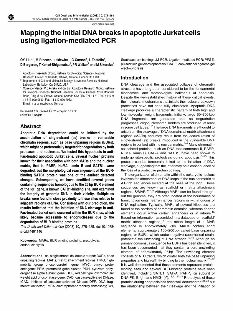
Jurkat cells undergo apoptosis readily in response to anti-Fastreatment. However, cell death is an asynchronous processand, at present, there is no clear understanding of whether thecell-cycle-related changes in chromatin influence the mole-cular mechanisms of DNA degradation. In order to avoid thiscomplication, we have blocked DNA synthesis in Jurkat cellsby an overnight aphidicolin treatment prior to the induction ofapoptosis. This treatment blocked approximately 80% of thecells in G1 without any negative effect on their viability, asmeasured by MTT assay (data not shown). These synch-ronized cells were subsequently treated with anti-Fas anti-body. A loss of cell viability became noticeable after 3 h, andmore pronounced after 4 h of treatment. At this time point,approximately 35% of the cells were dead (Table 1).However, apoptotic DNA fragments of 50 kb and smallerwere already clearly seen in samples isolated after 2 h of anti-Fas treatment (Figure 1a). These DNA fragments weresubjected to further analysis by two-dimensional DNAelectrophoresis with a denaturation step in alkaline bufferpreceding conventional electrophoresis in the second dimen-sion. This procedure reveals the presence of ss breaks incleaved DNA fragments.7,28 Indeed, the denaturation treat-ment resulted in the liberation of a smear of ss fragments of allsizes, not only from the apoptotic DNA fragments but alsofrom the high molecular weight DNA, which remained in the
well after the PFGE separation (Figure 1b, indicated byarrows). There was no significant number of ss DNA breaks inthe control sample.
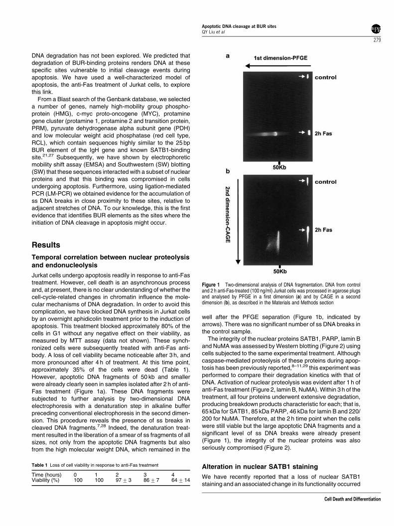
The integrity of the nuclear proteins SATB1, PARP, lamin Band NuMA was assessed by Western blotting (Figure 2) usingcells subjected to the same experimental treatment. Althoughcaspase-mediated proteolysis of these proteins during apop-tosis has been previously reported,8–11,29 this experiment wasperformed to compare their degradation kinetics with that ofDNA. Activation of nuclear proteolysis was evident after 1 h ofanti-Fas treatment (Figure 2, lamin B, NuMA). Within 3 h of thetreatment, all four proteins underwent extensive degradation,producing breakdown products characteristic for each; that is,65 kDa for SATB1, 85 kDa PARP, 46 kDa for lamin B and 220/200 for NuMA. Therefore, at the 2 h time point when the cellswere still viable but the large apoptotic DNA fragments and asignificant level of ss DNA breaks were already present(Figure 1), the integrity of the nuclear proteins was alsoseriously compromised (Figure 2).
Alteration in nuclear SATB1 staining
We have recently reported that a loss of nuclear SATB1staining and an associated change in its functionality occurred
Table 1 Loss of cell viability in response to anti-Fas treatment
Time (hours) 0 1 2 3 4Viability (%) 100 100 977 3 8677 64714
Figure 1 Two-dimensional analysis of DNA fragmentation. DNA from controland 2 h anti-Fas-treated (100 ng/ml) Jurkat cells was processed in agarose plugsand analysed by PFGE in a first dimension (a) and by CAGE in a seconddimension (b), as described in the Materials and Methods section
Apoptotic DNA cleavage at BUR sitesQY Liu et al
279
Cell Death and Differentiation
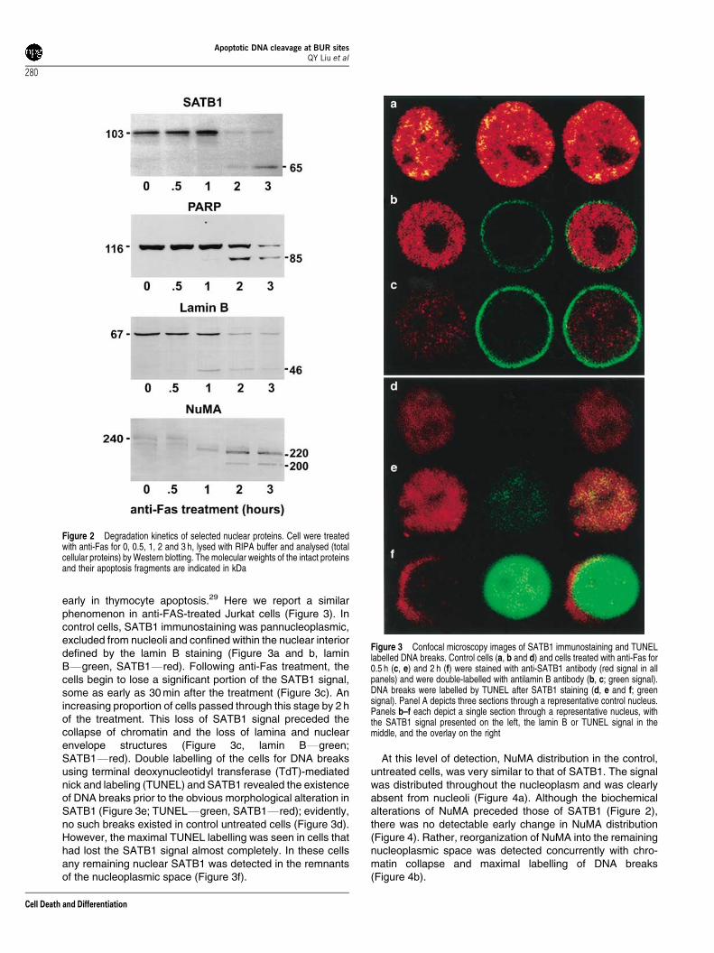
early in thymocyte apoptosis.29 Here we report a similarphenomenon in anti-FAS-treated Jurkat cells (Figure 3). Incontrol cells, SATB1 immunostaining was pannucleoplasmic,excluded from nucleoli and confined within the nuclear interiordefined by the lamin B staining (Figure 3a and b, laminBFgreen, SATB1Fred). Following anti-Fas treatment, thecells begin to lose a significant portion of the SATB1 signal,some as early as 30 min after the treatment (Figure 3c). Anincreasing proportion of cells passed through this stage by 2 hof the treatment. This loss of SATB1 signal preceded thecollapse of chromatin and the loss of lamina and nuclearenvelope structures (Figure 3c, lamin BFgreen;SATB1Fred). Double labelling of the cells for DNA breaksusing terminal deoxynucleotidyl transferase (TdT)-mediatednick and labeling (TUNEL) and SATB1 revealed the existenceof DNA breaks prior to the obvious morphological alteration inSATB1 (Figure 3e; TUNELFgreen, SATB1Fred); evidently,no such breaks existed in control untreated cells (Figure 3d).However, the maximal TUNEL labelling was seen in cells thathad lost the SATB1 signal almost completely. In these cellsany remaining nuclear SATB1 was detected in the remnantsof the nucleoplasmic space (Figure 3f).
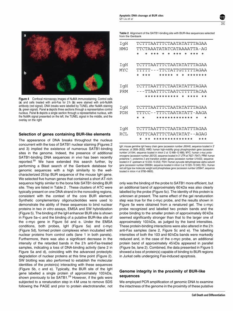
At this level of detection, NuMA distribution in the control,untreated cells, was very similar to that of SATB1. The signalwas distributed throughout the nucleoplasm and was clearlyabsent from nucleoli (Figure 4a). Although the biochemicalalterations of NuMA preceded those of SATB1 (Figure 2),there was no detectable early change in NuMA distribution(Figure 4). Rather, reorganization of NuMA into the remainingnucleoplasmic space was detected concurrently with chro-matin collapse and maximal labelling of DNA breaks(Figure 4b).
Figure 2 Degradation kinetics of selected nuclear proteins. Cell were treatedwith anti-Fas for 0, 0.5, 1, 2 and 3 h, lysed with RIPA buffer and analysed (totalcellular proteins) by Western blotting. The molecular weights of the intact proteinsand their apoptosis fragments are indicated in kDa
Figure 3 Confocal microscopy images of SATB1 immunostaining and TUNELlabelled DNA breaks. Control cells (a, b and d) and cells treated with anti-Fas for0.5 h (c, e) and 2 h (f) were stained with anti-SATB1 antibody (red signal in allpanels) and were double-labelled with antilamin B antibody (b, c; green signal).DNA breaks were labelled by TUNEL after SATB1 staining (d, e and f; greensignal). Panel A depicts three sections through a representative control nucleus.Panels b–f each depict a single section through a representative nucleus, withthe SATB1 signal presented on the left, the lamin B or TUNEL signal in themiddle, and the overlay on the right
Apoptotic DNA cleavage at BUR sitesQY Liu et al
280
Cell Death and Differentiation
Selection of genes containing BUR-like elements
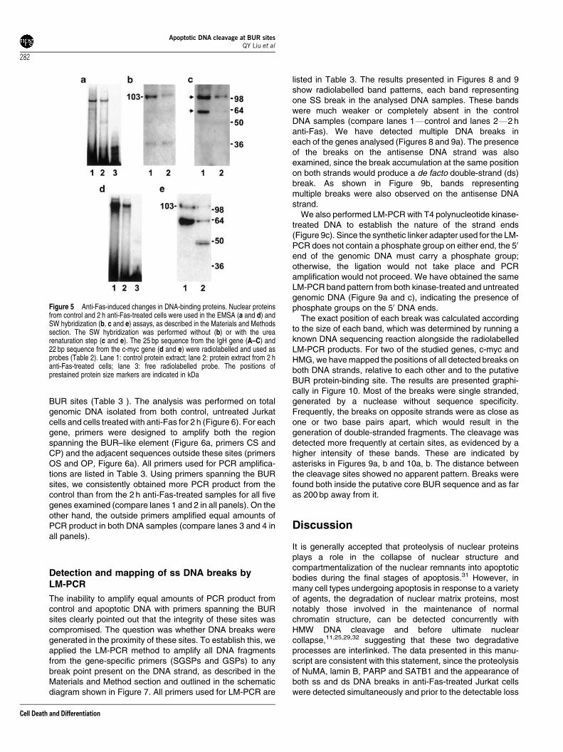
The appearance of DNA breaks throughout the nucleusconcurrent with the loss of SATB1 nuclear staining (Figures 2and 3) implied the existence of numerous SATB1-bindingsites in the genome. Indeed, the presence of additionalSATB1-binding DNA sequences in vivo has been recentlyreported.30 We have extended this search further, byperforming a Blast search of the Genbank database forgenomic sequences with a high similarity to the well-characterized 25 bp BUR sequence of the mouse IgH gene.We selected five human genes that contained a short AT-richsequence highly similar to the bona fide SATB1-binding BURsite. They are listed in Table 2 . These clusters of ATC weretypically present on one DNA strand in the noncoding regions,consistent with the context of the core BUR element.Synthetic complementary oligonucleotides were used todemonstrate the ability of these sequences to bind nuclearproteins in two in vitro assays, EMSA and SW hybridization(Figure 5). The binding of the IgH enhancer BUR site is shownin Figure 5a–c and the binding of a putative BUR-like site ofthe c-myc gene in Figure 5d and e. Under the EMSAconditions, both probes, IgH (Figure 5a) and c-myc(Figure 5d), formed protein complexes when incubated withnuclear proteins from control cells (lane 1 in both panels).Furthermore, there was also a significant decrease in theintensity of the retarded bands in the 2 h anti-Fas-treatedsamples, indicating a loss of DNA-binding activity (lane 2 inFigure 5a and d), coinciding with the advanced proteolyticdegradation of nuclear proteins at this time point (Figure 2).SW blotting was also performed to establish the molecularidentities of the protein(s) interacting with these sequences(Figure 5b, c and e). Typically, the BUR site of the IgHgene labelled a single protein of approximately 103 kDa,shown previously to be SATB1.29 However, if the gels weresubjected to a renaturation step in 4 M urea to remove SDSfollowing the PAGE and prior to protein electrotransfer, not
only was the binding of the probe to SATB1 more efficient, butan additional band of approximately 60 kDa was also clearlylabelled by the probe (Figure 5c). The identity of this protein isunknown at present. The same effect of the gel renaturationstep was true for the c-myc probe, and the results shown inFigure 5e were obtained from a renatured gel. The c-mycprobe recognized and labelled two protein bands and theprobe binding to the smaller protein of approximately 60 kDaseemed significantly stronger than that to the larger one ofapproximately 103 kDa, as judged by the band intensities.These protein-binding interactions were also altered in the 2 hanti-Fas samples (lane 2, Figure 5c and e). The labellingintensities of both the 103 and 60 kDa bands were markedlyreduced and, in the case of the c-myc probe, an additionalprotein band of approximately 45 kDa appeared in parallel(Figure 5e, lane 2). Combined, the data presented in Figure 5showed a loss of protein(s) capable of binding to BUR regionsin Jurkat cells undergoing Fas-induced apoptosis.
Genome integrity in the proximity of BUR-likesequences
We employed PCR amplification of genomic DNA to examinethe intactness of the genome in the proximity of these putative
Figure 4 Confocal microscopy images of NuMA immunostaining. Control cells(a) and cells treated with anti-Fas for 2 h (b) were stained with anti-NuMAantibody (red signal). DNA breaks were labelled by TUNEL after NuMA staining(b, green signal). Panel a depicts three sections through a representative controlnucleus. Panel b depicts a single section through a representative nucleus, withthe NuMA signal presented on the left, the TUNEL signal in the middle, and theoverlay on the right
Table 2 Alignment of the SATB1-binding site with BUR-like sequences selectedfrom the Genbank
IgH TCTTTAATTTCTAATATATTTAGAAHMG TTCTAAATATATCATAAAATTA-AG * * *** * * *** * *** *
IgH TCTTTAATTTCTAATATATTTAGAAMYC TTTTT---TTCTATTGTTTTTAGAA * *** ***** * * *******
IgH TCTTTAATTTCTAATATATTTAGAAPRM ---TTAATTTCTAATCTTTTTACAA ************ * **** **
IgH TCTTTAATTTCTAATATATTTAGAAPDH TTTCC--TTTCTAATATATT-AAGA * * ************* * *
IgH TCTTTAATTTCTAATATATTTAGAARCL TGTTCAATTTCTAATATAT--AGAG * ** ************** ***
IgH: mouse germline IgH heavy chain gene (accession number J00440, sequence located in 30
enhancer, at 2608–2632); HMG: human high-mobility group phosphoprotein gene (accessionnumber L41044, sequence located in intron 2 at 12 468–12 488); MYC: human c-myc proto-oncogene (accession number J00120, sequence located in 30 UTR at 7627–7647); PRM: humanprotamine 1, protamine 2 and transition protein genes (accession number U15422, sequencelocated in 50 upstream at 10 233–10 253); PDH: Human pyruvate dehydrogenase alpha subunitgene (accession number D90084, sequence located in intron 5 at 10 320–10 340); RCL: Humanred cell type low molecular weight acid phosphatase gene (accession number U25847, sequencelocated in intron 4 at 9780–9800).
Apoptotic DNA cleavage at BUR sitesQY Liu et al
281
Cell Death and Differentiation
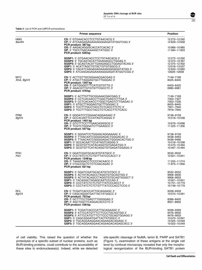
BUR sites (Table 3 ). The analysis was performed on totalgenomic DNA isolated from both control, untreated Jurkatcells and cells treated with anti-Fas for 2 h (Figure 6). For eachgene, primers were designed to amplify both the regionspanning the BUR–like element (Figure 6a, primers CS andCP) and the adjacent sequences outside these sites (primersOS and OP, Figure 6a). All primers used for PCR amplifica-tions are listed in Table 3. Using primers spanning the BURsites, we consistently obtained more PCR product from thecontrol than from the 2 h anti-Fas-treated samples for all fivegenes examined (compare lanes 1 and 2 in all panels). On theother hand, the outside primers amplified equal amounts ofPCR product in both DNA samples (compare lanes 3 and 4 inall panels).
Detection and mapping of ss DNA breaks byLM-PCR
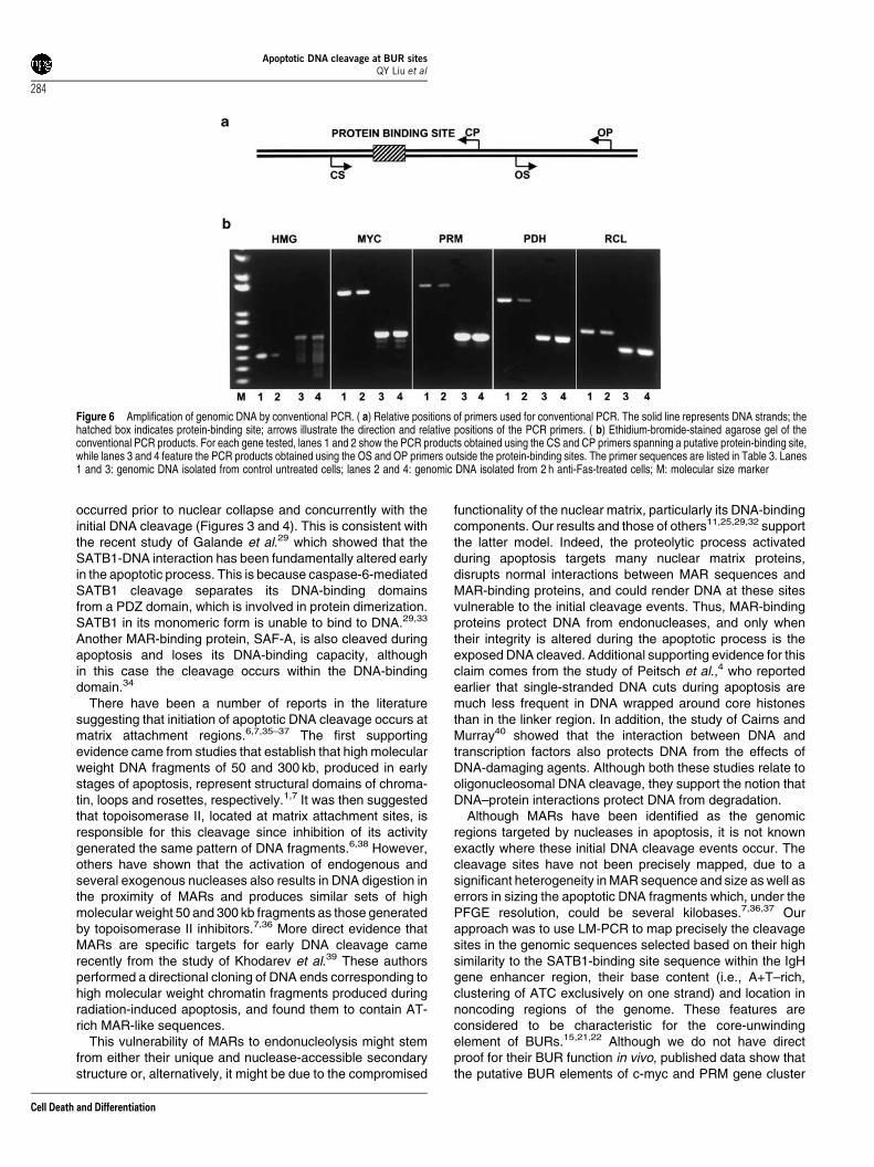
The inability to amplify equal amounts of PCR product fromcontrol and apoptotic DNA with primers spanning the BURsites clearly pointed out that the integrity of these sites wascompromised. The question was whether DNA breaks weregenerated in the proximity of these sites. To establish this, weapplied the LM-PCR method to amplify all DNA fragmentsfrom the gene-specific primers (SGSPs and GSPs) to anybreak point present on the DNA strand, as described in theMaterials and Method section and outlined in the schematicdiagram shown in Figure 7. All primers used for LM-PCR are
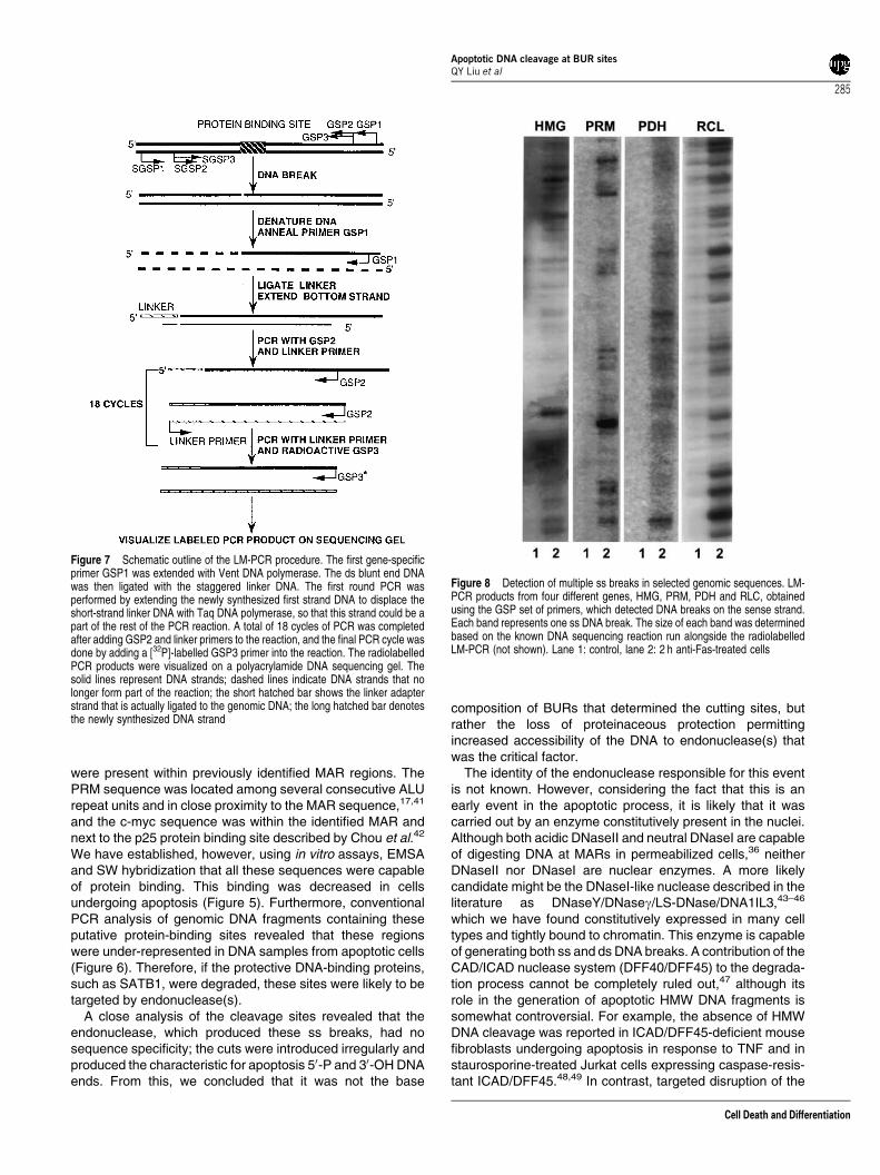
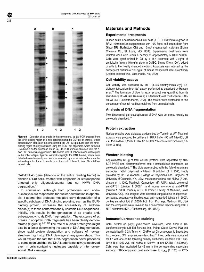
listed in Table 3. The results presented in Figures 8 and 9show radiolabelled band patterns, each band representingone SS break in the analysed DNA samples. These bandswere much weaker or completely absent in the controlDNA samples (compare lanes 1Fcontrol and lanes 2F2 hanti-Fas). We have detected multiple DNA breaks ineach of the genes analysed (Figures 8 and 9a). The presenceof the breaks on the antisense DNA strand was alsoexamined, since the break accumulation at the same positionon both strands would produce a de facto double-strand (ds)break. As shown in Figure 9b, bands representingmultiple breaks were also observed on the antisense DNAstrand.
We also performed LM-PCR with T4 polynucleotide kinase-treated DNA to establish the nature of the strand ends(Figure 9c). Since the synthetic linker adapter used for the LM-PCR does not contain a phosphate group on either end, the 50
end of the genomic DNA must carry a phosphate group;otherwise, the ligation would not take place and PCRamplification would not proceed. We have obtained the sameLM-PCR band pattern from both kinase-treated and untreatedgenomic DNA (Figure 9a and c), indicating the presence ofphosphate groups on the 50 DNA ends.
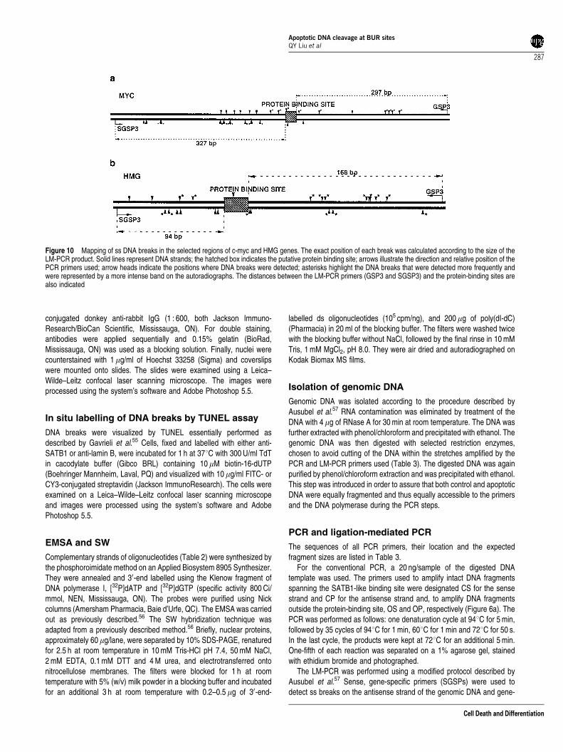
The exact position of each break was calculated accordingto the size of each band, which was determined by running aknown DNA sequencing reaction alongside the radiolabelledLM-PCR products. For two of the studied genes, c-myc andHMG, we have mapped the positions of all detected breaks onboth DNA strands, relative to each other and to the putativeBUR protein-binding site. The results are presented graphi-cally in Figure 10. Most of the breaks were single stranded,generated by a nuclease without sequence specificity.Frequently, the breaks on opposite strands were as close asone or two base pairs apart, which would result in thegeneration of double-stranded fragments. The cleavage wasdetected more frequently at certain sites, as evidenced by ahigher intensity of these bands. These are indicated byasterisks in Figures 9a, b and 10a, b. The distance betweenthe cleavage sites showed no apparent pattern. Breaks werefound both inside the putative core BUR sequence and as faras 200 bp away from it.
Discussion
It is generally accepted that proteolysis of nuclear proteinsplays a role in the collapse of nuclear structure andcompartmentalization of the nuclear remnants into apoptoticbodies during the final stages of apoptosis.31 However, inmany cell types undergoing apoptosis in response to a varietyof agents, the degradation of nuclear matrix proteins, mostnotably those involved in the maintenance of normalchromatin structure, can be detected concurrently withHMW DNA cleavage and before ultimate nuclearcollapse,11,25,29,32 suggesting that these two degradativeprocesses are interlinked. The data presented in this manu-script are consistent with this statement, since the proteolysisof NuMA, lamin B, PARP and SATB1 and the appearance ofboth ss and ds DNA breaks in anti-Fas-treated Jurkat cellswere detected simultaneously and prior to the detectable loss
Figure 5 Anti-Fas-induced changes in DNA-binding proteins. Nuclear proteinsfrom control and 2 h anti-Fas-treated cells were used in the EMSA (a and d) andSW hybridization (b, c and e) assays, as described in the Materials and Methodssection. The SW hybridization was performed without (b) or with the urearenaturation step (c and e). The 25 bp sequence from the IgH gene (A–C) and22 bp sequence from the c-myc gene (d and e) were radiolabelled and used asprobes (Table 2). Lane 1: control protein extract; lane 2: protein extract from 2 hanti-Fas-treated cells; lane 3: free radiolabelled probe. The positions ofprestained protein size markers are indicated in kDa
Apoptotic DNA cleavage at BUR sitesQY Liu et al
282
Cell Death and Differentiation
of cell viability. This raised the question of whether theproteolysis of a specific subset of nuclear proteins, such asBUR-binding proteins, could contribute to the accessibility ofthese sites to endonuclease(s). Indeed, while we detected
site-specific cleavage of NuMA, lamin B, PARP and SATB1(Figure 1), examination of these antigens at the single celllevel by confocal microscopy revealed that only the morpho-logical reorganization of the BUR-binding SATB1 protein
Table 3 List of PCR and LMPCR primersGene
Primer sequence Position
HMG CS: 50 GTGAACACCTCCTGTAACACG 30 12 272–12 292BamHI CP: 50 ATCAAGAGAGAAAAGGGAGATATGGTCGG 30 12 629–12 658
PCR product: 387 bpOS: 50 AAGACAGGGCACCATCACAC 30 10 966–10 985OP: 50 TGCCTGACAAAAGCTATGGC 30 11 584–11 603PCR product: 638 bp
SGSP1: 50 GTGAACACCTCCTGTAACACG 30 12 272–12 292SGSP2: 50 TGCAGTACATTGAAGAGCCTGGAG 30 12 374–12 397SGSP3: 50 GCAGTACATTGAAGAGCCTGGAGTACAG 30 12 375–12 402GSP1: 50 ACATTAGCTGTGCTGTGTGGCG 30 13 016–13 037GSP2: 50 CACATCAAGAGAGAAAAGGGAGATATGG 30 12 633–12 660GSP3: 50 ATCAAGAGAGAAAAGGGAGATATGGTCGG 30 12629–12658
MYC CS: 50 ACTTGTTGCGGAAACGACGAG 30 7149–7169XhoI, Bgl I/II CP: 50 ATGCTTAGGAGTGCTTGGGAC 30 8425–8445
PCR product: 1297 bpOS: 50 GATGGGGTTTCATCGTGTTG 30 6403–6422OP: 50 AGACGTTGTGTGTTCGCCTC 30 6980–6961PCR product: 578 bp
SGSP1: 50 ACTTGTTGCGGAAACGACGAG 30 7149–7169SGSP2: 50 CCTCACAACCTTGGCTGAGTCTTGA 30 7303–7327SGSP3: 50 CCTCACAACCTTGGCTGAGTCTTGAGAC 30 7303–7330GSP1: 50 ATGCTTAGGAGTGCTTGGGAC 30 8425–8445GSP2: 50 TCCTTTGCCTACCTCTCACCTTCTC 30 7921–7945GSP3: 50 TCCTTTGCCTACCTCTCACCTTCTCACC 7918–7945
PRM CS: 50 GGGATCCTGGAACAGGAAAAC 30 9138–9159Bgl I/II CP: 50 GCCACAATTCCATAGTCAGGG 30 10 518–10 538
PCR product: 1401 bpOS: 50 GTCTTTCCTTGAACAGGGCG 30 10 679–10 698OP: 50 TCGTCTGGGATCTTGAAGCC 30 11 225–11 206PCR product: 547 bp
SGSP1: 50 GGGATCCTGGAACAGGAAAAC 30 9138–9159SGSP2: 50 TTAACAATCGGGGAGACTGGGACAC 30 9438–9463SGSP3: 50 TTAACAATCGGGGAGACTGGGACACTGG 30 9438–9466GSP1: 50 GCCACAATTCCATAGTCAGGG 30 10 518–10 538GSP2: 50 GCGTGTTCATACAGGTGTGAGATGG 30 10 470–10 494GSP3: 50 GCGTGTTCATACAGGTGTGAGATGGAGG 30 10 467–10 494
PDH CS: 50 GGATCGATGCACATGTATGCC 30 9532–9552Pst I CP: 50 CCCTATCTCTCTGTTTATCCCACCT 30 10 921–10 941
PCR product: 1248 bpOS: 50 TAAAGGACCTCCCCACAACC 30 11 255–11 274OP: 50 CTGCCGCTCTCTCAACAGAC 30 11 875–11 856PCR product: 621 bp
SGSP1: 50 GGATCGATGCACATGTATGCC 30 9532–9552SGSP2: 50 ACTATACAGCCTAGGTGTGCAGTGG 30 9906–9930SGSP3: 50 ACTATACAGCCTAGGTGTGCAGTGGGCT 30 9906–9933GSP1: 50 TACAGGCTAGAGCAATCCCAG 30 10 921–10 941GSP2: 50 CCCTATCTCTCTGTTTATCCCACCT 30 10 751–10 779GSP3: 50 CCCTATCTCTCTGTTTATCCCACCTCCG 30 10 748–10 779
RCL CS: 50 TCGATCACCCATTGCAGAAGC 30 9339–9359Bgl I/II CP: 50 CAGCAGGATGATTACTATGGCC 30 10 070–10 091
PCR product: 753 bpOS: 50 GCTTTCCTGAGTTTGGGGAG 30 8386–8405OP: 50 AGCTGGTTCAGGACACCCTC 30 8921–8902PCR product: 536 bp
SGSP1: 50 TCGATCACCCATTGCAGAAGC 30 9339–9359SGSP2: 50 ATTCCATGTTTCTTCCCTGCAGTGG 30 9476–9500SGSP3: 50 ATTCCATGTTTCTTCCCTGCAGTGGAGG 30 9476–9503GSP1: 50 CAGCAGGATGATTACTATGGCC 30 10 070–10 091GSP2: 50 TGCAGGAAGGAACAGAAGACAGAGC 30 10 025–10 049GSP3: 50 TGCAGGAAGGAACAGAAGACAGAGCACC 30 10 022–10 049
Apoptotic DNA cleavage at BUR sitesQY Liu et al
283
Cell Death and Differentiation
occurred prior to nuclear collapse and concurrently with theinitial DNA cleavage (Figures 3 and 4). This is consistent withthe recent study of Galande et al.29 which showed that theSATB1-DNA interaction has been fundamentally altered earlyin the apoptotic process. This is because caspase-6-mediatedSATB1 cleavage separates its DNA-binding domainsfrom a PDZ domain, which is involved in protein dimerization.SATB1 in its monomeric form is unable to bind to DNA.29,33
Another MAR-binding protein, SAF-A, is also cleaved duringapoptosis and loses its DNA-binding capacity, althoughin this case the cleavage occurs within the DNA-bindingdomain.34
There have been a number of reports in the literaturesuggesting that initiation of apoptotic DNA cleavage occurs atmatrix attachment regions.6,7,35–37 The first supportingevidence came from studies that establish that high molecularweight DNA fragments of 50 and 300 kb, produced in earlystages of apoptosis, represent structural domains of chroma-tin, loops and rosettes, respectively.1,7 It was then suggestedthat topoisomerase II, located at matrix attachment sites, isresponsible for this cleavage since inhibition of its activitygenerated the same pattern of DNA fragments.6,38 However,others have shown that the activation of endogenous andseveral exogenous nucleases also results in DNA digestion inthe proximity of MARs and produces similar sets of highmolecular weight 50 and 300 kb fragments as those generatedby topoisomerase II inhibitors.7,36 More direct evidence thatMARs are specific targets for early DNA cleavage camerecently from the study of Khodarev et al.39 These authorsperformed a directional cloning of DNA ends corresponding tohigh molecular weight chromatin fragments produced duringradiation-induced apoptosis, and found them to contain AT-rich MAR-like sequences.
This vulnerability of MARs to endonucleolysis might stemfrom either their unique and nuclease-accessible secondarystructure or, alternatively, it might be due to the compromised
functionality of the nuclear matrix, particularly its DNA-bindingcomponents. Our results and those of others11,25,29,32 supportthe latter model. Indeed, the proteolytic process activatedduring apoptosis targets many nuclear matrix proteins,disrupts normal interactions between MAR sequences andMAR-binding proteins, and could render DNA at these sitesvulnerable to the initial cleavage events. Thus, MAR-bindingproteins protect DNA from endonucleases, and only whentheir integrity is altered during the apoptotic process is theexposed DNA cleaved. Additional supporting evidence for thisclaim comes from the study of Peitsch et al.,4 who reportedearlier that single-stranded DNA cuts during apoptosis aremuch less frequent in DNA wrapped around core histonesthan in the linker region. In addition, the study of Cairns andMurray40 showed that the interaction between DNA andtranscription factors also protects DNA from the effects ofDNA-damaging agents. Although both these studies relate tooligonucleosomal DNA cleavage, they support the notion thatDNA–protein interactions protect DNA from degradation.
Although MARs have been identified as the genomicregions targeted by nucleases in apoptosis, it is not knownexactly where these initial DNA cleavage events occur. Thecleavage sites have not been precisely mapped, due to asignificant heterogeneity in MAR sequence and size as well aserrors in sizing the apoptotic DNA fragments which, under thePFGE resolution, could be several kilobases.7,36,37 Ourapproach was to use LM-PCR to map precisely the cleavagesites in the genomic sequences selected based on their highsimilarity to the SATB1-binding site sequence within the IgHgene enhancer region, their base content (i.e., A+T–rich,clustering of ATC exclusively on one strand) and location innoncoding regions of the genome. These features areconsidered to be characteristic for the core-unwindingelement of BURs.15,21,22 Although we do not have directproof for their BUR function in vivo, published data show thatthe putative BUR elements of c-myc and PRM gene cluster
Figure 6 Amplification of genomic DNA by conventional PCR. ( a) Relative positions of primers used for conventional PCR. The solid line represents DNA strands; thehatched box indicates protein-binding site; arrows illustrate the direction and relative positions of the PCR primers. ( b) Ethidium-bromide-stained agarose gel of theconventional PCR products. For each gene tested, lanes 1 and 2 show the PCR products obtained using the CS and CP primers spanning a putative protein-binding site,while lanes 3 and 4 feature the PCR products obtained using the OS and OP primers outside the protein-binding sites. The primer sequences are listed in Table 3. Lanes1 and 3: genomic DNA isolated from control untreated cells; lanes 2 and 4: genomic DNA isolated from 2 h anti-Fas-treated cells; M: molecular size marker
Apoptotic DNA cleavage at BUR sitesQY Liu et al
284
Cell Death and Differentiation
were present within previously identified MAR regions. ThePRM sequence was located among several consecutive ALUrepeat units and in close proximity to the MAR sequence,17,41
and the c-myc sequence was within the identified MAR andnext to the p25 protein binding site described by Chou et al.42
We have established, however, using in vitro assays, EMSAand SW hybridization that all these sequences were capableof protein binding. This binding was decreased in cellsundergoing apoptosis (Figure 5). Furthermore, conventionalPCR analysis of genomic DNA fragments containing theseputative protein-binding sites revealed that these regionswere under-represented in DNA samples from apoptotic cells(Figure 6). Therefore, if the protective DNA-binding proteins,such as SATB1, were degraded, these sites were likely to betargeted by endonuclease(s).
A close analysis of the cleavage sites revealed that theendonuclease, which produced these ss breaks, had nosequence specificity; the cuts were introduced irregularly andproduced the characteristic for apoptosis 50-P and 30-OH DNAends. From this, we concluded that it was not the base
composition of BURs that determined the cutting sites, butrather the loss of proteinaceous protection permittingincreased accessibility of the DNA to endonuclease(s) thatwas the critical factor.
The identity of the endonuclease responsible for this eventis not known. However, considering the fact that this is anearly event in the apoptotic process, it is likely that it wascarried out by an enzyme constitutively present in the nuclei.Although both acidic DNaseII and neutral DNaseI are capableof digesting DNA at MARs in permeabilized cells,36 neitherDNaseII nor DNaseI are nuclear enzymes. A more likelycandidate might be the DNaseI-like nuclease described in theliterature as DNaseY/DNaseg/LS-DNase/DNA1IL3,43–46
which we have found constitutively expressed in many celltypes and tightly bound to chromatin. This enzyme is capableof generating both ss and ds DNA breaks. A contribution of theCAD/ICAD nuclease system (DFF40/DFF45) to the degrada-tion process cannot be completely ruled out,47 although itsrole in the generation of apoptotic HMW DNA fragments issomewhat controversial. For example, the absence of HMWDNA cleavage was reported in ICAD/DFF45-deficient mousefibroblasts undergoing apoptosis in response to TNF and instaurosporine-treated Jurkat cells expressing caspase-resis-tant ICAD/DFF45.48,49 In contrast, targeted disruption of the
Figure 7 Schematic outline of the LM-PCR procedure. The first gene-specificprimer GSP1 was extended with Vent DNA polymerase. The ds blunt end DNAwas then ligated with the staggered linker DNA. The first round PCR wasperformed by extending the newly synthesized first strand DNA to displace theshort-strand linker DNA with Taq DNA polymerase, so that this strand could be apart of the rest of the PCR reaction. A total of 18 cycles of PCR was completedafter adding GSP2 and linker primers to the reaction, and the final PCR cycle wasdone by adding a [32P]-labelled GSP3 primer into the reaction. The radiolabelledPCR products were visualized on a polyacrylamide DNA sequencing gel. Thesolid lines represent DNA strands; dashed lines indicate DNA strands that nolonger form part of the reaction; the short hatched bar shows the linker adapterstrand that is actually ligated to the genomic DNA; the long hatched bar denotesthe newly synthesized DNA strand
Figure 8 Detection of multiple ss breaks in selected genomic sequences. LM-PCR products from four different genes, HMG, PRM, PDH and RLC, obtainedusing the GSP set of primers, which detected DNA breaks on the sense strand.Each band represents one ss DNA break. The size of each band was determinedbased on the known DNA sequencing reaction run alongside the radiolabelledLM-PCR (not shown). Lane 1: control, lane 2: 2 h anti-Fas-treated cells
Apoptotic DNA cleavage at BUR sitesQY Liu et al
285
Cell Death and Differentiation
CAD/DFF40 gene (deletion of the entire reading frame) inchicken DT40 cells, treated with etoposide or staurosporineaffected only oligonucleosomal but not HMW DNAdegradation.50
In conclusion, although both proteolysis and endo-nucleolysis are responsible for nuclear destruction in apopto-sis, it seems that protease-mediated early degradation of aspecific subclass of DNA-binding proteins, such as the BUR-binding protein, increases the accessibility of endonu-clease(s) to these conformationally unstable DNA sequences.Initially, this results in the generation of ss breaks and,subsequently, to ds DNA fragmentation. The existence of ssbreaks in apoptotic DNA fragments has been clearly demon-strated (Figure 1).7,51,52 The rate of nuclear proteolysis mightalso be a factor determining the extent of DNA fragmentation,since rapid protein degradation and collapse of nuclearstructure might stop DNA cleavage at any given stage. Thiswould explain the fact that DNA degradation never proceedsto completion and that the DNA ladder is not always observedeven in cells containing nucleases capable of internucleo-somal DNA cleavage.
Materials and Methods
Experimental treatments
Human acute T-cell leukaemia Jurkat cells (ATCC T1B152) were grown inRPMI 1640 medium supplemented with 10% foetal calf serum (both fromGibco BRL, Burlington, ON) and 10 mg/ml gentamycin sulphate (SigmaChemical Co., St. Louis, MO, USA). Experimental treatments wereinitiated when cells reach a density of approximately 500 000 cells/ml.Cells were synchronized in G1 by a 16 h treatment with 2mg/ml ofaphidicolin (from a 10 mg/ml stock in DMSO; Sigma Chem. Co.), addeddirectly to the freshly changed medium. Apoptosis was induced by thesubsequent addition of 100 ng/ml of mouse monoclonal anti-Fas antibody(Upstate Biotech. Inc., Lake Placid, NY, USA).
Cell viability assays
Cell viability was assessed by MTT (3,[4,5-dimethylthiazol-2-yl] 2,5-diphenyl-tetrazolium bromide) assay, performed as described by Hansenet al.53 The formation of blue formazan product was quantified from itsabsorbance at 570 vs 630 nm using a Titertech 96-well multiscanner EAR-400AT (SLT-Labinstruments, USA). The results were expressed as thepercentage of control readings obtained from untreated cells.
Analysis of DNA fragmentation
Two-dimensional gel electrophoresis of DNA was performed exactly aspreviously described.28
Protein extraction
Nuclear proteins were extracted as described by Testolin et al.54 Total cellextracts were prepared by cell lysis in RIPA buffer (50 mM Tris-HCl, pH7.4, 150 mM NaCl, 2 mM EDTA, 0.1% SDS, 1% sodium deoxycholate, 1%Triton X-100).
Western blotting
Approximately 60 mg of total cellular proteins were separated by 10%SDS-PAGE and electrotransferred onto a nitrocellulose membrane, aspreviously described.44 The blots were probed with the following primaryantibodies: rabbit polyclonal anti-lamin B (dilution of 1 : 2000, kindlyprovided by Dr. HJ Worman, College of Physicians and Surgeons ofUniversity of Columbia, NY, USA), mouse monoclonal anti-NuMA (A-204,dilution of 1 : 1000, Matritech, Cambridge, MA, USA), rabbit polyclonalanti-SATB1 (dilution 1 : 5000)21 and mouse monoclonal anti-PARP(dilution 1 : 5000, courtesy of Dr. G Poirier, Faculty of Medicine, LavalUniversity, QC). The antigens were detected using alkaline phosphatase-conjugated secondary antibodies: goat anti-mouse-IgG (dilution 1 : 2000),donkey antirabbit IgG (1 : 5000), both from Promega, Madison, WI, USAand the complexes were revealed by a colorimetric reaction using BCIP/NBT substrates (KPL, Gaithersburg, MA, USA).
Immunofluorescence staining
Cells, settled on poly-L-lysine-coated coverslips, were fixed in 3%paraformaldehyde (JB EM Services Inc., Pointe Claire, Dorval, PQ) andpermeabilized in 0.2% Triton X-100 (Pierce/ Chromatographic SpecialtiesInc., Nepean, ON), as previously described.11 Fixed cells were incubatedfor 1 h with one of the following primary antibodies, diluted in PBS: anti-lamin B (1 : 250 v/v), anti-NuMA (1 : 25 v/v) or anti-SATB1 (1 : 500 v/v).Cells were then incubated for 45 min in the corresponding secondaryantibody: FITC-conjugated goat anti-mouse Ig G(Fc) (1 : 125) or CY3-
Figure 9 Detection of ss breaks in the c-myc gene. (a) LM-PCR products fromthe MAR-binding region of c-myc obtained using the GSP set of primers, whichdetected DNA breaks on the sense strand. (b) LM-PCR products from the MAR-binding region of c-myc obtained using the SGSP set of primers, which detectedDNA breaks on the antisense strand. (c) LM-PCR products obtained from the c-myc GSP primers using genomic DNA treated with T4 polynucleotide kinase priorto the linker adaptor ligation. Asterisks highlight the DNA breaks, which weredetected more frequently and were represented by a more intense band on theautoradiographs. Lane 1: results from the control; lane 2: from 2 h anti-Fas-treated cells
Apoptotic DNA cleavage at BUR sitesQY Liu et al
286
Cell Death and Differentiation
conjugated donkey anti-rabbit IgG (1 : 600, both Jackson Immuno-Research/BioCan Scientific, Mississauga, ON). For double staining,antibodies were applied sequentially and 0.15% gelatin (BioRad,Mississauga, ON) was used as a blocking solution. Finally, nuclei werecounterstained with 1mg/ml of Hoechst 33258 (Sigma) and coverslipswere mounted onto slides. The slides were examined using a Leica–Wilde–Leitz confocal laser scanning microscope. The images wereprocessed using the system’s software and Adobe Photoshop 5.5.
In situ labelling of DNA breaks by TUNEL assay
DNA breaks were visualized by TUNEL essentially performed asdescribed by Gavrieli et al.55 Cells, fixed and labelled with either anti-SATB1 or anti-lamin B, were incubated for 1 h at 371C with 300 U/ml TdTin cacodylate buffer (Gibco BRL) containing 10 mM biotin-16-dUTP(Boehringer Mannheim, Laval, PQ) and visualized with 10 mg/ml FITC- orCY3-conjugated streptavidin (Jackson ImmunoResearch). The cells wereexamined on a Leica–Wilde–Leitz confocal laser scanning microscopeand images were processed using the system’s software and AdobePhotoshop 5.5.
EMSA and SW
Complementary strands of oligonucleotides (Table 2) were synthesized bythe phosphoroimidate method on an Applied Biosystem 8905 Synthesizer.They were annealed and 30-end labelled using the Klenow fragment ofDNA polymerase I, [32P]dATP and [32P]dGTP (specific activity 800 Ci/mmol, NEN, Mississauga, ON). The probes were purified using Nickcolumns (Amersham Pharmacia, Baie d’Urfe, QC). The EMSA was carriedout as previously described.56 The SW hybridization technique wasadapted from a previously described method.56 Briefly, nuclear proteins,approximately 60mg/lane, were separated by 10% SDS-PAGE, renaturedfor 2.5 h at room temperature in 10 mM Tris-HCl pH 7.4, 50 mM NaCl,2 mM EDTA, 0.1 mM DTT and 4 M urea, and electrotransferred ontonitrocellulose membranes. The filters were blocked for 1 h at roomtemperature with 5% (w/v) milk powder in a blocking buffer and incubatedfor an additional 3 h at room temperature with 0.2–0.5 mg of 30-end-
labelled ds oligonucleotides (105 cpm/ng), and 200mg of poly(dI-dC)(Pharmacia) in 20 ml of the blocking buffer. The filters were washed twicewith the blocking buffer without NaCl, followed by the final rinse in 10 mMTris, 1 mM MgCl2, pH 8.0. They were air dried and autoradiographed onKodak Biomax MS films.
Isolation of genomic DNA
Genomic DNA was isolated according to the procedure described byAusubel et al.57 RNA contamination was eliminated by treatment of theDNA with 4 mg of RNase A for 30 min at room temperature. The DNA wasfurther extracted with phenol/chloroform and precipitated with ethanol. Thegenomic DNA was then digested with selected restriction enzymes,chosen to avoid cutting of the DNA within the stretches amplified by thePCR and LM-PCR primers used (Table 3). The digested DNA was againpurified by phenol/chloroform extraction and was precipitated with ethanol.This step was introduced in order to assure that both control and apoptoticDNA were equally fragmented and thus equally accessible to the primersand the DNA polymerase during the PCR steps.
PCR and ligation-mediated PCR
The sequences of all PCR primers, their location and the expectedfragment sizes are listed in Table 3.
For the conventional PCR, a 20 ng/sample of the digested DNAtemplate was used. The primers used to amplify intact DNA fragmentsspanning the SATB1-like binding site were designated CS for the sensestrand and CP for the antisense strand and, to amplify DNA fragmentsoutside the protein-binding site, OS and OP, respectively (Figure 6a). ThePCR was performed as follows: one denaturation cycle at 941C for 5 min,followed by 35 cycles of 941C for 1 min, 601C for 1 min and 721C for 50 s.In the last cycle, the products were kept at 721C for an additional 5 min.One-fifth of each reaction was separated on a 1% agarose gel, stainedwith ethidium bromide and photographed.
The LM-PCR was performed using a modified protocol described byAusubel et al.57 Sense, gene-specific primers (SGSPs) were used todetect ss breaks on the antisense strand of the genomic DNA and gene-
Figure 10 Mapping of ss DNA breaks in the selected regions of c-myc and HMG genes. The exact position of each break was calculated according to the size of theLM-PCR product. Solid lines represent DNA strands; the hatched box indicates the putative protein binding site; arrows illustrate the direction and relative position of thePCR primers used; arrow heads indicate the positions where DNA breaks were detected; asterisks highlight the DNA breaks that were detected more frequently andwere represented by a more intense band on the autoradiographs. The distances between the LM-PCR primers (GSP3 and SGSP3) and the protein-binding sites arealso indicated
Apoptotic DNA cleavage at BUR sitesQY Liu et al
287
Cell Death and Differentiation
specific primers (GSPs) to identify ss breaks on the sense strand of thegenomic DNA. The assay was performed using 2 mg per sample ofrestriction digested genomic DNA. The DNA was denatured and firstannealed with the GSP1 primers. The first-strand synthesis wasaccomplished with Vent DNA polymerase (NE Biolabs, Beverly, MA,USA). The ds blunt end DNA was then ligated with the staggered linkerDNA. The first round PCR reaction was performed by extending the newlysynthesized first DNA strand at 721C for 3 min in the presence of 0.5ml ofthe advantage Taq DNA polymerase (Clontech, Palo Alto, CA, USA), todisplace the short-strand linker DNA so that this DNA strand becomes thetemplate for the remaining PCR cycles. Following addition of the GSP2and the linker primers, a further 18 PCR cycles were completed and thefinal cycle was done in the presence of [32P]-labelled GSP3 primers. The[32P]- labelled PCR products were visualized on a polyacrylamide DNAsequencing gel with a known DNA sequencing reaction running along theside to determine the size of each PCR product. In a parallel experiment,the restriction enzyme-digested DNA was phosphorylated with 10 units ofT4 polynucleotide kinase (NE Biolabs, Beverly, MA, USA) prior to the LM-PCR reaction. The steps required for the completion of LM-PCR areoutlined in Figure 7.
Acknowledgments
We thank Harvey Miller for the Genbank searches, Julie Leblanc for the2D-PFGE, and Tom Devecseri for the figure preparation.
References
1. Walker PR and Sikorska M (1997) New aspects of the mechanism of DNAfragmentation in apoptosis. Biochem. Cell. Biol. 75: 287–299
2. Sikorska M and Walker PR (1998) Endonuclease activities and apoptosis. In:Lockshin RA, Zakeri Z, Tilley JL, eds. When Cells Die. New York: Wiley-Liss,Inc, pp. 211–242
3. Probst H and Herzog R (1985) DNA regions associated with the nuclear matrixof Ehrlich ascites cells expose single-stranded sites after deproteinization. Eur.J. Biochem. 146: 167–171
4. Peitsch MC, Muller C and Tschopp J (1993) DNA fragmentation duringapoptosis is caused by frequent single-strand cuts. Nucleic. Acids. Res. 21:4206–4209
5. Luderus ME, den Blaauwen JL, de Smit OJ, Compton DA and vanDriel R (1994) Binding of matrix attachment regions to lamin polymersinvolves single- stranded regions and the minor groove. Mol. Cell Biol 14:6297–6305
6. Lagarkova MA, Iarovaia OV and Razin SV (1995) Large-scale fragmentation ofmammalian DNA in the course of apoptosis proceeds via excision ofchromosomal DNA loops and their oligomers. J. Biol. Chem. 270: 20239–20241
7. Walker PR, LeBlanc J and Sikorska M (1997) Evidence that DNA fragmentationin apoptosis is initiated and propagated by single-strand breaks. Cell DeathDiffer. 4: 506–515
8. Kaufmann SH, Desnoyers S, Ottaviano Y, Davidson NE and Poirier GG (1993)Specific proteolytic cleavage of poly(ADP-ribose) polymerase: an early markerof chemotherapy-induced apoptosis. Cancer Res 53: 3976–3985
9. Neamati N, Fernandez A, Wright S, Kiefer J and McConkey DJ (1995)Degradation of lamin B1 precedes oligonucleosomal DNA fragmentation inapoptotic thymocytes and isolated thymocyte nuclei. J. Immunol 154: 3788–3795
10. Tewari M, Quan LT, O’Rourke K, Desnoyers S, Zeng Z, Beidler DR, Poirier GG,Salvesen GS and Dixit VM (1995) Yama/CPP32 beta, a mammalian homologof CED-3, is a CrmA-inhibitable protease that cleaves the death substratepoly(ADP-ribose) polymerase. Cell 81: 801–809
11. Weaver VM, Carson CE, Walker PR, Chaly N, Lach B, Raymond Y, Brown DLand Sikorska M (1996) Degradation of nuclear matrix and DNA cleavage inapoptotic thymocytes. J. Cell Sci. 109: 45–56
12. Buendia B, Santa-Maria A and Courvalin JC (1999) Caspase-dependentproteolysis of integral and peripheral proteins of nuclear membranesand nuclear pore complex proteins during apoptosis. J. Cell Sci. 112:1743–1753
13. Galande S and Kohwi-Shigematsu T (1999) Poly(ADP-ribose) polymerase andKu autoantigen form a complex and synergistically bind to matrix attachmentsequences. J. Biol. Chem. 274: 20521–20528
14. Craig JM, Boyle S, Perry P and Bickmore WA (1997) Scaffold attachmentswithin the human genome. J. Cell Sci. 110: 2673–2682
15. Bode J, Benham C, Knopp A and Mielke C (2000) Transcriptionalaugmentation: modulation of gene expression by scaffold/matrix-attached regions (S/MAR elements). Crit. Rev. Eukaryot. Gene Expr. 10:73–90
16. Chernov IP, Akopov SB, Nikolaev LG and Sverdlov ED (2000) Identificationand mapping of nuclear matrix-attachment regions in a one megabase locus ofhuman chromosome 19q13.12: long-range correlation of S/MARs and genepositions. J. Cell Biochem. 84: 590–600
17. Liebich I, Bode J, Frisch M and Wingender E (2002) S/MARt DB: a database onscaffold/matrix attached regions. Nucleic Acids Res. 30: 372–374
18. Kohwi-Shigematsu T and Kohwi Y (1990) Torsional stress stabilizes extendedbase unpairing in suppressor sites flanking immunoglobulin heavy chainenhancer. Biochemistry 29: 9551–9560
19. Benham C, Kohwi-Shigematsu T and Bode J (1997) Stress-induced duplexDNA destabilization in scaffold/matrix attachment regions. J. Mol. Biol. 274:181–196
20. Bode J, Kohwi Y, Dickinson L, Joh T, Klehr D, Mielke C and Kohwi-ShigematsuT (1992) Biological significance of unwinding capability of nuclear matrix-associating DNAs. Science 255: 195–197
21. Dickinson LA, Joh T, Kohwi Y and Kohwi-Shigematsu T (1992) A tissue-specificMAR/SAR DNA-binding protein with unusual binding site recognition. Cell 70:631–645
22. Kohwi-Shigematsu T, deBelle I, Dickinson LA, Galande S and Kohwi Y (1998)Identification of base-unpairing region-binding proteins and characterization oftheir in vivo binding sequences. Methods Cell Biol. 53: 323–354
23. Herrscher RF, Kaplan MH, Lelsz DL, Das C, Scheuermann R and Tucker PW(1995) The immunoglobulin heavy-chain matrix-associating regions are boundby Bright: a B cell-specific trans-activator that describes a new DNA-bindingprotein family. Genes Dev. 9: 3067–3082
24. Liu WM, Guerra-Vladusic FK, Kurakata S, Lupu R and Kohwi-Shigematsu T(1999) HMG-I(Y) recognizes base-unpairing regions of matrix attachmentsequences and its increased expression is directly linked to metastatic breastcancer phenotype. Cancer Res. 59: 5695–5703
25. Gotzmann J, Meissner M and Gerner C (2000) The fate of the nuclear matrix-associated-region-binding protein SATB1 during apoptosis. Cell Death Differ.7: 425–438
26. Kipp M, Schwab BL, Przybylski M, Nicotera P and Fackelmayer FO(2000) Apoptotic cleavage of scaffold attachment factor A (SAF-A) bycaspase-3 occurs at a noncanonical cleavage site. J. Biol. Chem. 275: 5031–5036
27. deBelle I, Cai S and Kohwi-Shigematsu T (1998) The genomic sequencesbound to special AT-rich sequence-binding protein 1 (SATB1) in vivo in Jurkat Tcells are tightly associated with the nuclear matrix at the bases of the chromatinloops. J. Cell Biol 141: 335–348
28. Walker PR, LeBlanc J, Smith B, Pandey S and Sikorska M (1999) Detectionof DNA fragmentation and endonucleases in apoptosis. Methods 17:329–338
29. Galande S, Dickinson LA, Mian IS, Sikorska M and Kohwi-Shigematsu T (2001)SATB1 cleavage by caspase 6 disrupts PDZ domain-mediated dimerization,causing detachment from chromatin early in T-cell apoptosis. Mol. Cell Biol. 21:5591–5604
30. Ramakrishnan M, Liu WM, DiCroce PA, Posner A, Zheng J, Kohwi-ShigematsuT and Krontiris TG (2000) Modulated binding of SATB1, a matrix attachmentregion protein, to the AT-rich sequence flanking the major breakpoint region ofBCL2. Mol. Cell Biol. 20: 868–877
31. Kidd VJ, Lahti JM and Teitz T (2000) Proteolytic regulation of apoptosis. Semin.Cell Dev. Biol. 11: 191–201
32. Casiano CA, Martin SJ, Green DR and Tan EM (1996) Selective cleavage ofnuclear autoantigens during CD95 (Fas/APO-1)- mediated T cell apoptosis. J.Exp. Med. 184: 765–770
Apoptotic DNA cleavage at BUR sitesQY Liu et al
288
Cell Death and Differentiation

33. Nakagomi K, Kohwi Y, Dickinson LA and Kohwi-Shigematsu T (1994) A novelDNA-binding motif in the nuclear matrix attachment DNA-binding proteinSATB1. Mol. Cell Biol. 14: 1852–1860
34. Gohring F, Schwab BL, Nicotera P, Leist M and Fackelmayer FO(1997) The novel SAR-binding domain of scaffold attachment factorA (SAF-A) is a target in apoptotic nuclear breakdown. EMBO J. 16:7361–7371
35. Oberhammer FA, Hochegger K, Froschl G, Tiefenbacher R and Pavelka M(1994) Chromatin condensation during apoptosis is accompanied bydegradation of lamin A+B, without enhanced activation of cdc2 kinase. J.Cell Biol. 126: 827–837
36. Gromova II, Nielsen OF and Razin SV (1995) Long-range fragmentation of theeukaryotic genome by exogenous and endogenous nucleases proceeds in aspecific fashion via preferential DNA cleavage at matrix attachment sites. J.Biol. Chem. 270: 18685–18690
37. Schoenlein PV, Barrett JT and Welter D (1999) The degradation profile ofextrachromosomal circular DNA during cisplatin-induced apoptosis isconsistent with preferential cleavage at matrix attachment regions.Chromosoma 108: 121–131
38. Filipski J, LeBlanc J, Youdale T, Sikorska M and Walker PR (1990)Periodicity of DNA folding in higher order chromatin structures. EMBO J. 9:1319–1327
39. Khodarev NN, Bennett T, Shearing N, Sokolova I, Koudelik J, Walter S,Villalobos M and Vaughan AT (2000) LINE L1 retrotransposable element istargeted during the initial stages of apoptotic DNA fragmentation. J. CellBiochem. 79: 486–495
40. Cairns MJ and Murray V (1998) Detection of protein–DNA interactions at beta-globin gene cluster in intact human cells utilizing hedamycin as DNA-damagingagent. DNA Cell Biol. 17: 325–333
41. Schmid C, Heng HH, Rubin C, Ye CJ and Krawetz SA (2001) Sperm nuclearmatrix association of the PRM1 - PRM2 - TNP2 domain is independent ofAlu methylation. Mol. Hum. Reprod. 7: 903–911
42. Chou RH, Churchill JR, Flubacher MM, Mapstone DE and Jones J (1990)Identification of a nuclear matrix-associated region of the c-myc protooncogeneand its recognition by a nuclear protein in the human leukemia HL-60 cell line.Cancer Res. 50: 3199–3206
43. Baron WF, Pan CQ, Spencer SA, Ryan AM, Lazarus RA and Baker KP (1998)Cloning and characterization of an actin-resistant DNase I-like endonucleasesecreted by macrophages. Gene 215: 291–301
44. Liu QY, Pandey S, Singh RK, Lin W, Ribecco M, Borowy-Borowski H, Smith B,LeBlanc J, Walker PR and Sikorska M (1998) DNaseY: a rat DNaseI-like genecoding for a constitutively expressed chromatin-bound endonuclease.Biochemistry 37: 10134–10143
45. Shiokawa D and Tanuma S (1998) Molecular cloning and expression of a cDNAencoding an apoptotic endonuclease DNase gamma. Biochem J. 332: 713–720
46. Yakovlev AG, Wang G, Stoica BA, Simbulan-Rosenthal CM, Yoshihara K andSmulson ME (1999) Role of DNAS1L3 in Ca 2+- and Mg 2+-dependent cleavageof DNA into oligonucleosomal and high molecular mass fragments. NucleicAcids Res. 27: 1999–2005
47. Enari M, Sakahira H, Yokoyama H, Okawa K, Iwamatsu A and Nagata S (1998)A caspase-activated DNase that degrades DNA during apoptosis, and itsinhibitor ICAD. Nature 39: 43–50
48. Boulares AH, Zoltoski AJ, Yakovlev A, Xu M and Smulson ME (2001) Roles ofDNA fragmentation factor and poly(ADP-ribose) polymerase in an amplificationphase of tumor necrosis factor-induced apoptosis. J. Biol. Chem. 276: 38185–38192
49. Sakahira H, Enari M, Ohsawa Y and Uchiyama Y (1990) Apoptoticnuclear morphological; change without DNA fragmentation. Curr. Biol. 9:543–546
50. Samejima K, Tone S and Earnshaw W (2001) CAD/DFF40 nuclease isdispensible for high molecular weight DNA cleavage and stage I chromatincondensation in apoptosis. J. Biol. Chem. 276: 45427–45432
51. Tomei LD, Shapiro JP and Cope FO (1993) Apoptosis in C3H/10T1/2 mouseembryonic cells: evidence for internucleosomal DNA modification in theabsence of double-strand cleavage. Proc. Natl. Acad. Sci. USA 90: 853–857
52. Chen J, Jin K, Chen M, Pei W, Kawaguchi K, Greenberg DA and Simon RP(1997) Early detection of DNA strand breaks in the brain after transient focalischemia: implications for the role of DNA damage in apoptosis and neuronalcell death. J. Neurochem. 69: 232–245
53. Hansen MB, Nielsen SE and Berg K (1989) Re-examination and furtherdevelopment of a precise and rapid dye method for measuring cell growth/cellkill. J. Immunol. Methods 119: 203–210
54. Testolin L, Carson C, Wang Y, Walker PR, Armato U and Sikorska M(1997) Jun and JNK kinase are activated in thymocytes in responseto VM26 and radiation but not glucocorticoids. Exp. Cell Res. 230:220–232
55. Gavrieli Y, Sherman Y and Ben Sasson SA (1992) Identification ofprogrammed cell death in situ via specific labeling of nuclear DNAfragmentation. J. Cell Biol. 119: 493–501
56. Kwast-Welfeld J, deBelle I, Walker PR, Whitfield JF and Sikorska M (1993)Identification of a new cAMP response element-binding factor by Southwesternblotting. J. Biol. Chem. 268: 19581–19585
57. Ausubel FM, Brent R, Kingston RE, Moore DD, Smith JA, Seidman JG andStruhl K (1987). Current Protocols in Molecular Biology. New York: John Wileyand Sons Inc
Apoptotic DNA cleavage at BUR sitesQY Liu et al
289
Cell Death and Differentiation


