Embed Size (px)
Citation preview

Silica-Directed Mast Cell Activation Is Enhanced byScavenger ReceptorsJared M. Brown, Emily J. Swindle, Nataliya M. Kushnir-Sukhov, Andrij Holian, and Dean D. Metcalfe
Laboratory of Allergic Diseases, National Institute of Allergy and Infectious Diseases, National Institutes of Health, Bethesda, Maryland;and Center for Environmental Health Sciences, Department of Biomedical and Pharmaceutical Sciences, University of Montana,Missoula, Montana
Inhalation of crystalline silica results in pulmonary fibrosis and silico-sis. It has been suggested that mast cells play a role in these condi-tions. How mast cells would influence pathology is unknown. Wethus explored mast cell interactions with silica in vitro and in B6.Cg-KitW-sh mast cell–deficient mice. B6.Cg-KitW-sh mice did not developinflammation or significant collagen deposition after instillation ofsilica, while C57Bl/6 wild-type mice did have these findings. Giventhis supporting evidence of a role for mast cells in the developmentof silicosis, we examined the ability of silica to activate mousebone marrow–derived mast cells (BMMC), including degranulation(�-hexosaminidase release); production of reactive oxygen species(ROS) and inflammatory mediators; and the effects of silica on Fc�RI-dependent activation. Silica did not induce mast cell degranulation.However, TNF-�, IL-13, monocyte chemotactic protein-1, proteaseactivity, and production of ROS were dose-dependently increasedafter silica exposure, and production was enhanced after Fc�RI stim-ulation. This mast cell activation was inhibited by anti-inflammatorycompounds. As silica mediates some effects in macrophagesthrough scavenger receptors (SRs), we first determined that mastcells express scavenger receptors; then explored the involvementof SR-A and macrophage receptor with colleagenous structure(MARCO). Silica-induced ROS formation, apoptosis, and TNF-� pro-duction were reduced in BMMC obtained from SR-A, MARCO, andSR-A/MARCO knockout mice. These findings demonstrate that silicadirects mast cell production of inflammatory mediators, in partthrough SRs, providing insight into critical events in the pathogene-sis and potential therapeutic targets in silicosis.
Keywords: B6.Cg-kitW-sh sash mouse; CD204; macrophage receptor withcollagenous structure; mast cell; silicosis; SR-A
Among environmental exposures that lead to pathologic changesin tissues is crystalline silica, which can result in occupationalsilicosis from inhalation of silica dusts in manufacturing, con-struction, farming, and mining operations. Silicosis, for whichthere is no effective treatment, leads to decreased pulmonaryfunction and increased susceptibility to diseases of the respira-tory tract (1, 2).
Involvement of mast cells in silica-induced pulmonary in-flammation has been suggested by two clinical observations.First, the number of mast cells within the lungs of individualsexposed to silica dust is increased (3). Second, an increase inmast cells staining for basic fibroblast growth factor locatedwithin silicotic nodules has been reported in lung sectionsobtained from patients with silicosis (4). Despite the suggestive
(Received in original form June 2, 2006 and in final form July 31, 2006 )
This work was supported by NIH intramural funds, NIH R01/ES04804 and COBREP20RR01760.
Correspondence and requests for reprints should be addressed to Jared M. Brown,Ph.D., Laboratory of Allergic Diseases, National Institute of Allergy and InfectiousDiseases, National Institutes of Health, Building 10, Room 11C209, 10 CenterDrive, MSC 1881, Bethesda, MD 20892–1881. E-mail: [email protected]
Am J Respir Cell Mol Biol Vol 36. pp 43–52, 2007Originally Published in Press as DOI: 10.1165/rcmb.2006-0197OC on August 10, 2006Internet address: www.atsjournals.org
CLINICAL RELEVANCE
This study demonstrates a role for mast cells in silicosis andis the first study to examine direct effects of silica on mastcell biology. It provides evidence that treatment of silicosisshould explore mast cells as a potential therapeutic target.
evidence for a role of mast cells in the development of silicosis,there are no systematic studies of the effects of silica on mastcells.
Before entering into experiments testing the hypothesis thatsilica can direct mast cell activation, a critical role for mast cellsin promoting silicosis was first verified in mast cell–deficient sashmice (B6.Cg-kitW-sh) after exposure to silica. Because the mastcell–deficient mice did not develop silicosis, while mice withnormal mast cells did develop pathology, we entered into ex-periments to determine the ability of silica to directly activatemast cells. Cultured bone marrow–derived murine mast cells(BMMC) were exposed to silica, followed by analysis of cellviability, for effects on degranulation, and for the productionof reactive oxygen species (ROS), cytokines, and proteolyticactivity. The possible role of scavenger receptor class A (SR-A)and macrophage receptor with collagenous structure (MARCO)was explored in the genesis of positive responses to silica expo-sure. As will be shown, mast cells exposed to silica respondprimarily by production of cytokines, the synthesis of which canbe inhibited with anti-inflammatory agents and is enhanced byscavenger receptors.
MATERIALS AND METHODS
Cell Culture and Silica Treatment
Mouse BMMC were cultured from femoral marrow cells of C57Bl/6wild-type, SR-A KO, MARCO KO, and SR-A/MARCO double knock-out (KO) mice. Cells were cultured in RPMI 1640 medium supple-mented with 10% FBS, 100 U/ml penicillin, 100 �g/ml streptomycin,25 mM HEPES, 1.0 mM sodium pyruvate, nonessential amino acids(BioSource International, Camarillo, CA), 0.0035% 2-ME, and 300 ng/mlrecombinant mouse IL-3 (PeproTech, Rocky Hill, NJ). BMMC wereused after 4–6 wk of culture. Silica (Min-U-Sil-5, with an average particlesize of 1.5–2 �m) was obtained from Pennsylvania Glass Sand Corpora-tion (Pittsburgh, PA) and was acid washed, dried, and determined tobe free of endotoxin. Cells were treated with silica in 96-well plates at0, 6.25, 12.5, 25, or 50 �g/cm2. Water-soluble dexamethasone (Sigma-Aldrich, St. Louis, MO) was used at 10�6 M and was added to BMMCcultures 1 h before the addition of silica.
Mice
Mast cell–deficient sash mice (B6.Cg-kitW-sh) and wild-type (C57Bl/6)mice were obtained from Jackson Laboratories at 4–6 wk of age (BarHarbor, ME). Mice were anesthetized with tribromoethanol (0.1 mg/kg)before intranasal instillation of either 30 �l sterile saline or a 30 �lsterile saline suspension of 1 mg crystalline silica. All mice receivedtwo instillations 2 wk apart. Six mice were instilled per group, and the

44 AMERICAN JOURNAL OF RESPIRATORY CELL AND MOLECULAR BIOLOGY VOL 36 2007
four groups consisted of C57Bl/6 wild-type mice or B6.Cg-kitW-sh mastcell–deficient mice instilled with either saline or silica. Mice were killed3 mo after the first instillation of silica.
SR-AI/II KO mice and MARCO KO mice were generously pro-vided by Dr. L. Kobzick of the Harvard School of Public Health(Boston, MA) (5, 6). Both knockouts were backcrossed for at least eightgenerations to the C57Bl/6 background. SR-A I/II/MARCO double KOmice were generated in the laboratory of Dr. L. Kobzik by intercrossof the single SR-A I/II and MARCO knockouts. Founding C57Bl/6wild-type mice were purchased from Jackson Laboratories. Mice usedin these studies were propagated by homozygous mating colonies andmaintained in microisolator units within the University of Montanaspecific pathogen–free animal facility. Cages, bedding, and food weresterilized by autoclaving, and mice were handled with aseptic forcepsor aseptic gloves. Mice were allowed food and water ad libitum andwere used experimentally between 6 and 8 wk of age. All animal useprocedures were in accordance with National Institutes of Health andapproved by the University of Montana institutional animal care anduse committee.
Identification of SR-A I/II and MARCO KO Mice
DNA samples were extracted from mouse tail and amplified usingforward (5�-CAAGTGATACATCTCAAGGTC-3�), reverse (5�-CTGTAGATTCACGGACTCTG-3�), and Neo insert (5�-GAGGAGTAGAAGGTGGCGCGAA-3�) primers (Operon Technologies, Alameda,CA) encompassing the site of the SR-A I/II and Neo insert (7). ForMARCO idenfication, DNA samples were amplified using the followingprimers: MARCO WT, forward (5�-CAGCTGGGTCCATACCAGC-3�) and reverse (5�-CTGGAGAGCCTCGTTGACC-3�); and MARCOKO, forward (5�-CCACGCTCATCGATAATTTCAC-3�) and reverse(5�-CCTGCAGTGGCCGTCGTTTTA-3�). Amplification was per-formed in a PTC-200 Gradient Cycler (MJ Research, Las Vegas, NV)with the following parameters: 1 min 94�C, 1 min 60�C, 1 min 72�C and sub-jected to 35 cycles of PCR. Genotyping was determined by separationof the amplified PCR products by 1.5% agarose gel electrophoresis at75 V for 2.5 h. C57Bl/6 WT mice show a 440-bp band, whereas SR-A I/IIKO mice show a 325-bp band. MARCO WT primers produce a 500-bpband and MARCO KO primers produce an 850-bp band.
Lung Histology
Three months after either saline or silica instillation, C57Bl/6 wild-typeand B6.Cg-kitW-sh mast cell–deficient mice were given a lethal dose ofsodium pentobarbital. Lungs were then perfused with 1–2 ml Histo-choice fixative (Amresco, Solon, OH), embedded in paraffin blocksand sectioned at 5 �m, and mounted on glass slides (American Histo-Labs, Inc, Gaithersburg, MD) (8). Sections were stained with Gomori’strichrome, hematoxylin and eosin (H&E), and toluidine blue (AmericanHistoLabs).
Cell Viability and Apoptosis
Cell viability was determined by trypan blue staining and lactate dehy-drogenase (LDH) release (9). The percentage of LDH release wasmeasured in supernatants of BMMC exposed to silica for 24 h (Bio-Vision, Mountain View, CA). Untreated cells were used as a negativecontrol and 1%-triton X-100 (Sigma-Aldrich)–treated BMMC wereused as a positive control, representing 100% LDH release. Apoptosiswas determined by DNA fragmentation using a TiterTacs TUNELAssay (R&D Systems, Minneapolis, MN) (10). Equal numbers of cells(1 � 105/well) were plated in a 96-well plate, and silica was added for24 h at 6.25, 12.5, 25, or 50 �g/cm2. Nontreated cells were used as anegative control and a DNA nuclease–generated positive control wasused. Experiments were repeated three times. The reported values aremean optical density (OD) values from each treatment.
Degranulation and Cytokine Release
BMMC were seeded at 5 � 104 cells/well in 96-well flat-bottom platesand sensitized with 100 ng/ml mouse IgE anti-DNP (Sigma-Aldrich)for 24 h for degranulation experiments (11). For treated samples, silicawas added at 0, 6.25, 12.5, 25, or 50 �g/cm2 for 24 h before addition ofincreasing concentrations of DNP-HSA (0–1000 ng/ml) (Sigma-Aldrich). After 30 min incubation of antigen at 37�C, p-nitrophenyl-N-acteyl-�-D-glucopyranoside was added to cell supernatants and lysates
for 90 min as a chromogenic substrate for N-acetyl-�-D-hexosaminidase(Sigma-Aldrich) (11). The reaction was stopped with 0.2 M glycine.Optical density was measured at 405 nm using a GENios ELISA platereader (ReTirSoft, Inc., Toronto, ON, Canada). �-Hexosaminidase re-lease was expressed as the percentage of total cell content after sub-tracting background release from unstimulated cells. Cytokines weremeasured in cell culture supernatant of BMMC seeded at 2 � 105 cells/well for 24 h after addition of 6.25, 12.5, 25, or 50 �g/cm2 silica. Forexperiments with silica exposure and Fc�RI stimulation, BMMC wereexposed to silica for a total of 24 h at 0–50 �g/cm2 and sensitizedovernight with 100 ng/ml IgE anti-DNP (Sigma-Aldrich). DNP-HSA(Sigma-Aldrich) was added at 100 ng/ml for a total of 8 h beforesupernatant collection for cytokine analysis. Mouse TNF-, IL-13, andmonocyte chemotactic protein (MCP)-1 (CCL2) were measured usingQuantikine ELISA kits (R&D Systems, Minneapolis, MN).
Protease Assay
Silica-exposed BMMC were examined for protease activity using a Quan-ticleave Protease Assay Kit (Pierce, Rockford, IL). Briefly, supernatantsfrom BMMC exposed to 0, 6.25, 12.5, 25, or 50 �g/cm2 silica for 24 hwere collected and analyzed for the ability to cleave succinylated casein.A control blank without casein from each sample was run in parallel andsubtracted as background. Experiments were repeated three times. Thereported values are mean OD values measured at 450 nm.
Reactive Oxygen Species Detection
ROS were measured by a 96-well plate assay employing the fluorescentprobe dichlorofluoroscein (DCF) (12). BMMC (1 � 106/ml) were incu-bated with DCF diacetate (20 �M) in cell culture medium for 15 minat 4�C with rotation. Cells were then washed in HEPES buffer (10 ml)and seeded at 200,000 per well in a black opaque 96-well microplate.Zileuton (Cayman Chemical, Ann Arbor, MI) and Trolox (EMD Bio-sciences, San Diego, CA) (10 �l) were added and DCF fluorescencemonitored for 5 min before the addition of silica. After addition ofsilica, DCF fluorescence was then monitored for a further 10 min usinga GENios fluorescent plate reader (ReTirSoft Inc.) set at an excitationwavelength of 492 nm and emission wavelength of 535 nm. Fluorescencewas expressed as relative fluorescent units (RFU). The kinetic data wascollected using an XFlour4 macro within Microsoft Excel.
RT-PCR
BMMC (1 � 106 cells/condition) from C57Bl/6 mice were exposed tosilica for 30 min, 1 h, or 2 h at 0, 6.25, 12.5, 25, or 50 �g/cm2. Cellswere collected and total mRNA was isolated using QIAshredders andRNeasy Mini Kits (Qiagen, Valencia, CA). RNA was transcribed tocDNA using a Reaction Ready First Strand cDNA synthesis kit(Superarray, Frederick, MD). Gene-specific primers for SR-A I (MSR1),SR-A II (MSR2), MARCO, and glyceraldehyde phosphate dehydroge-nase (GAPDH) were purchased from Superarray and transcripts wereamplified using a Reaction Ready Hotstart “Sweet” PCR kit accordingto manufacturer’s instructions (Superarray).
Statistics
Statistical analysis employed the software package PRISM, version 4(GraphPad, San Diego, CA). Differences between untreated and silica-treated samples were assessed using one-way ANOVA with Bonferronipost-test. Area under the curve (AUC) was calculated for ROS mea-surements using PRISM. All values are reported as means SEM.
RESULTS
Mast Cell–Deficient Mice Fail to Develop Inflammation andExtensive Collagen Deposition after Silica Exposure
To assess the role of mast cells in the development of silicosis,mast cell–deficient mice (B6.Cg-KitW-sh) were administered two1-mg instillations of silica, with effects on lung histology exam-ined 3 mo after the first instillation of silica. As determined byGomori’s trichrome staining, silica-exposed wild-type mice demon-strated increased collagen deposition within the lung as com-pared with saline-instilled wild-type mice (compare Figure 1A

Brown, Swindle, Kushnir-Sukhov, et al.: Silica-Directed Mast Cell Activation 45
Figure 1. Silica inducescollagen deposition inC57Bl/6 wild-type, butnot B6.Cg-kitW-sh mastcell–deficient mouse lungs3 mo after two intranasalinstillations of 1 mg silica.C57Bl/6 mice were in-stilled with either saline(A) or silica (C ). B6.Cg-kitW-sh received instilla-tions of either saline (B )or silica (D). Lungs werestained with Gomori’s tri-chrome stain to examinecollagen deposition. Im-ages were obtained at�5 magnification and arerepresentative of six miceper group.
with 1C). By H&E staining, wild-type mice were found to havedeveloped granulomatous lesions typical of silicotic lesions, alve-olar thickening, and pronounced inflammatory infiltrates withinthe interstitial and alveolar spaces (compare Figure 2A with 2B).In contrast, mast cell–deficient mice exposed to silica did not
Figure 2. Silica inducesinflammation in C57Bl/6wild-type, but notB6.Cg-kitW-sh mast cell–deficient mouse lungs 3mo after two intranasalinstillations of 1 mg sil-ica. C57Bl/6 mice wereinstilled with either sa-line (A ) or silica (C ).B6.Cg-kitW-sh received in-stillations of either saline(B ) or silica (D ). Lungswere stained with H&Eto examine inflammation.Images were obtained at�5 magnification and arerepresentative of six miceper group.
develop granulomatous lesions, alveolar thickening, or any sig-nificant inflammation or collagen deposition (compare Figures1D and 2D with 1C and 2C). There were no differences in thenumber of lung mast cells in silica-exposed wild-type mice ascompared with saline controls (data not shown). As expected,

46 AMERICAN JOURNAL OF RESPIRATORY CELL AND MOLECULAR BIOLOGY VOL 36 2007
no mast cells were detectable in B6.Cg-KitW-sh mast cell–deficientmouse lungs, and no mast cells were observed after silica expo-sure (data not shown). These observations confirm a role formast cells in silicosis.
Silica Induces Apoptosis in BMMC but Has Minimal Effecton Degranulation
A series of experiments was then initiated to determine theeffects of silica on cultured mast cells. At the start of theseexperiments, BMMC were visually examined after exposure tosilica for 24 h. An apparent internalization of silica particles wasobserved (see Figures 3A and 3B). This was not unexpected, asmast cells are known to be phagocytic and macrophages similarlyinternalize silica (13, 14).
Silica exposure has been reported to induce cytotoxicity (apo-ptosis and necrosis) in alveolar macrophages and thus contributeto tissue inflammation (15–18). As shown in Figure 4A, silica didinduce between 5 and 15% LDH release from BMMC exposed to6.25 �g/cm2 silica, and at 50 �g/cm2 silica, between 20 and 40%LDH release was observed. To further examine the ability ofsilica to affect viability of BMMC, the TUNEL assay was alsoemployed. Figure 4B shows a dose-dependent increase in cellsstaining positive for DNA fragmentation as determined by an
Figure 3. Silica interacts with and is internalized by BMMC. Images of(A ) untreated or (B ) BMMC exposed to 25 �g/cm2 silica for 24 h andstained with Wright-Giemsa (magnification: �100). Arrows indicate silicaparticles.
in vitro TUNEL assay. Significant differences between untreatedBMMC and 12.5, 25, and 50 �g/cm2 silica were seen 24 h afterexposure (Figure 4B, P � 0.05). Consistent with LDH release(Figure 4A), silica induces apoptosis in BMMC at higher doses.However, by either assay, a majority of the cells remained viable24 h after silica exposure.
Silica exposure was next examined for the ability to alterFc�RI-mediated degranulation of BMMC as one mechanism bywhich silica might enhance inflammation mediated by mast cells.Pretreatment of BMMC (sensitized with mouse IgE anti-DNP)with silica at doses of 6.25, 25, and 50 �g/cm2 minimally decreased�-hexosaminidase release after addition of increasing amountsof DNP-HSA compared with BMMC that were not exposed tosilica (Figure 4C). Silica exposure by itself similarly did notinduce �-hexosaminidase release from mouse BMMC (data notshown). These results reveal that silica has little indirect effect onFc�RI-mediated mast cell degranulation, nor does silica directlyinduce degranulation.
Silica Induces Production of Reactive Oxygen Speciesin BMMC
Silica exposure induces the production of ROS in macrophagesand has been implicated in mediating biological effects such asfibrosis after silica exposure (19). Therefore, the ability of mastcells to produce ROS after silica exposure was assessed. Figure4D (insert) shows that induction of ROS occurs rapidly and issustained after addition of silica. Total ROS production mea-sured as area under the curve in silica-exposed BMMC wassignificantly increased after exposure to 12.5, 25, and 50 �g/cm2
silica compared with untreated BMMC (Figure 4D).
Silica Induces Cytokine and Chemokine Production andEnhances IgE-Mediated Cytokine and ChemokineProduction in BMMC
Silicosis has been associated with increased production of profi-brotic cytokines and chemokines in bronchoalveolar lavage fluid,including TNF-, IL-13, and MCP-1 (CCL2) (20–22). Therefore,the ability of silica to directly induce production of such media-tors from BMMC, as well as the ability of silica to enhanceFc�RI-mediated cytokine and chemokine production, was ex-plored. After 24 h of silica exposure, the production of TNF-,IL-13, and MCP-1 by BMMC are dose-dependently increased(Figures 5A–5C, respectively). Silica-exposed BMMC that weresensitized with IgE anti-DNP and challenged with DNP-HSA for8 h showed enhanced production of TNF-, IL-13, and MCP-1(Figures 5D–5F).
In addition to cytokine production, protease activity was mea-sured in the supernatant of silica-exposed BMMC. The overallprotease activity within the supernatant from BMMC exposed tosilica at doses of 12.5, 25, and 50 �g/cm2 for 24 h was significantlyincreased as compared with untreated BMMC (Figure 6).
Mast Cells Express Scavenger Receptors: Apoptosis and ROSProduction Is Reduced in SR-A–, MARCO–, andSR-A/MARCO–Deficient BMMC
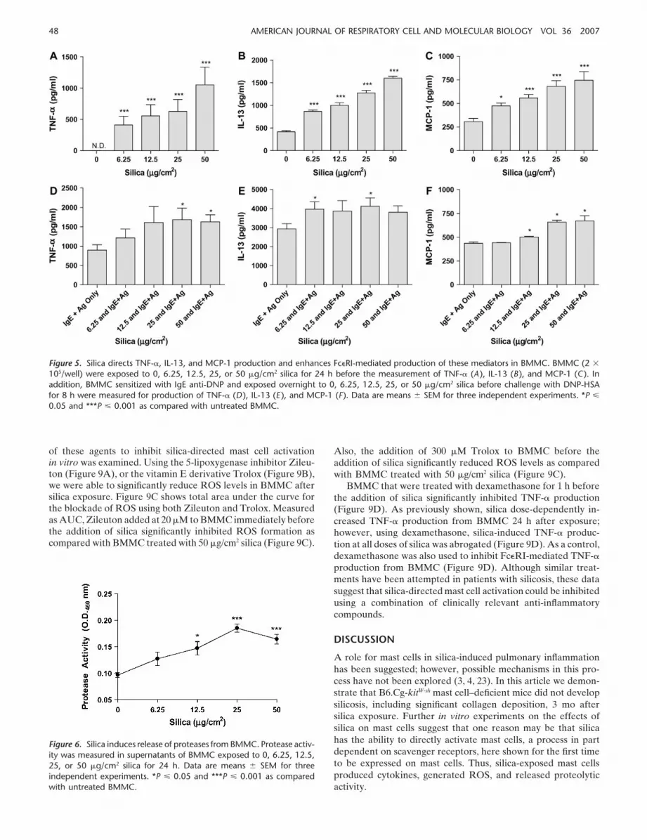
Silica has been reported in part to mediate effects through SR-Aon macrophages (15, 16). To determine if mast cells also expressscavenger receptors, we examined these cells for the presence ofmRNA for these receptors. Figure 7 demonstrates mRNA expres-sion for two closely related scavenger receptors, SR-A I/II andMARCO, both before and after silica exposure in BMMC. WhilemRNA for SR-A II (MSR2) was increased after silica exposureover a time course, all scavenger receptors appeared to increaseby 2 h after silica exposure ranging from 6.25–50 �g/cm2 (Figures 7A

Brown, Swindle, Kushnir-Sukhov, et al.: Silica-Directed Mast Cell Activation 47
Figure 4. Silica induces LDH release, apoptosis, and ROS production, but has little effect on �-hexosaminidase release in BMMC. (A ) LDH releasewas measured in supernatant of silica-exposed BMMC. (B ) Apoptosis was determined by DNA fragmentation in BMMC (1 � 105/well) exposedto 0, 6.25, 12.5, 25, or 50 �g/cm2 silica for 24 h. (C ) Fc�RI-mediated �-hexosaminidase release, as a measure of degranulation, was determined24 h after exposure of BMMC (50,000/well) to 0, 6.25, 12.5, 25, or 50 �g/cm2 silica. (D ) ROS production in silica-exposed BMMC where silicawas added to BMMC 5 min after addition of the fluorescent probe DCF, at doses of 0, 6.25, 12.5, 25, or 50 �g/cm2. Intracellular ROS productionwas measured up to 30 min (insert in D ). Area under the curve of silica-induced ROS production with ionomycin as a positive control (D ). Dataare means SEM for three experiments. *P � 0.05, **P � 0.01, and ***P � 0.001 as compared with untreated BMMC.
and 7B). Because of this evidence that mast cells express scaven-ger receptors, experiments were entered into examining the ef-fects of silica on mast cells cultured from scavenger receptorKO mice.
As shown earlier (Figure 4B), silica exposure at 50 �g/cm2
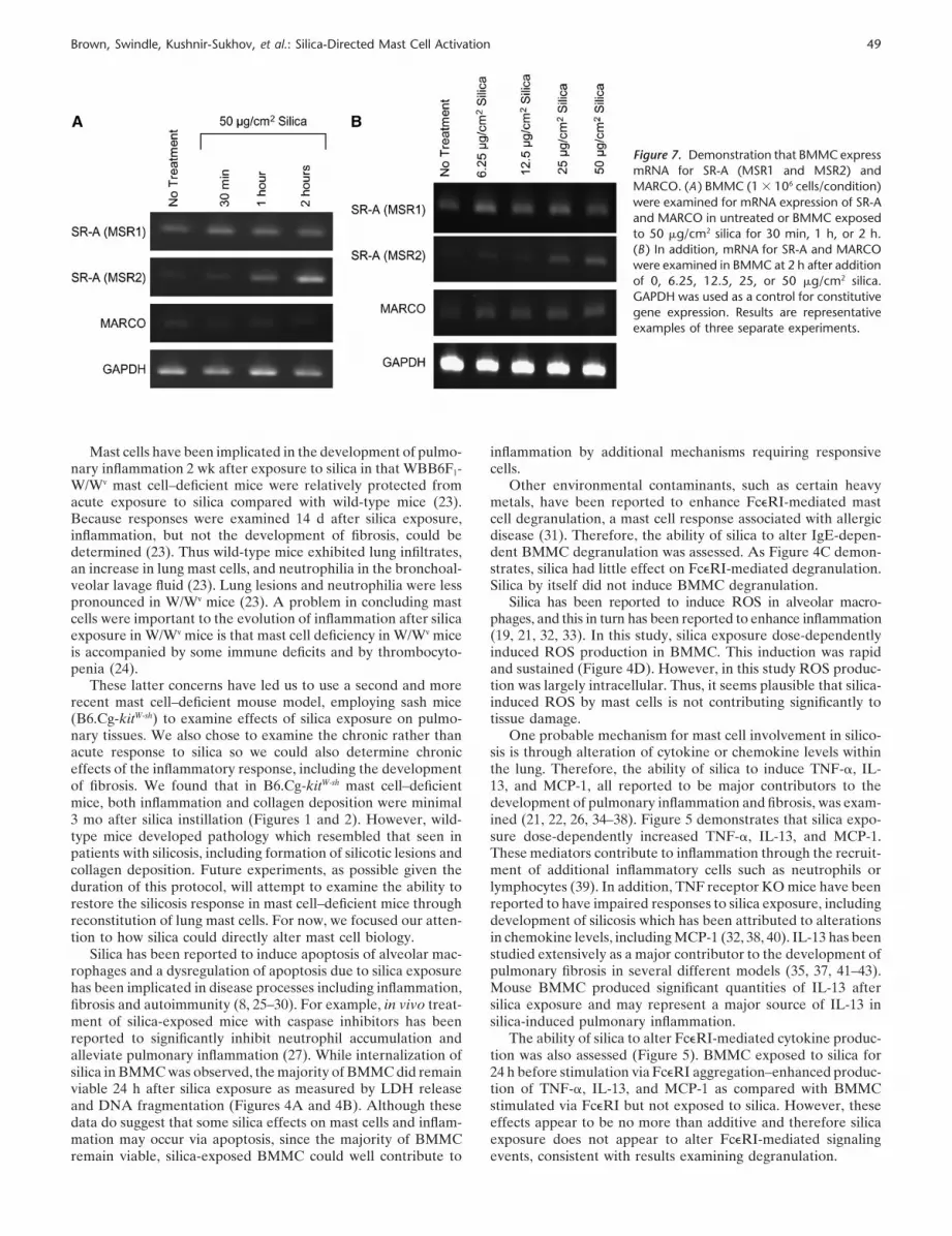
induces DNA fragmentation as compared with untreated cells.Therefore, the induction of apoptosis by silica in BMMC derivedfrom SR-A I/II KO, MARCO KO, and SR-A/MARCO doubleKO mice was examined. As shown in Figure 8A, there was asignificant decrease in the level of DNA fragmentation in silica-exposed BMMC from MARCO KO and SR-A/MARCO doubleKO mice when compared with silica-exposed BMMC fromC57Bl/6 mice. ROS production in BMMC from SR-A KO,MARCO KO, and SR-A/MARCO double KO mice was simi-larly less compared with ROS production by BMMC from C57Bl/6 controls after exposure to 50 �g/cm2 silica (Figures 8B and8C). These results indicate that scavenger receptors are able tomediate in part the apoptotic state of BMMC, as well as ROSproduction, in response to silica.
TNF-� Production Is Altered in Silica-Exposed BMMC fromSR-A– and MARCO–Deficient Mice
Due to the changes in apoptosis and redox states observed inSR-A– and MARCO–deficient BMMC after silica exposure, therole of these scavenger receptors in mediating TNF- productionafter silica exposure was examined. Figure 8D shows a role forSR-A in enhancing cytokine production after silica exposure.TNF- is significantly decreased after 50 �g/cm2 silica exposurein the SR-A KO and SR-A/MARCO double KO BMMC ascompared with WT BMMC exposed to the same dose of silica(Figure 8D, P � 0.01).
Treatment of BMMC with Zileuton, Trolox, andDexamethasone Reduced Silica-Directed MastCell Activation
There is currently no effective treatment for silicosis; however,steroids and anti-inflammatory agents have been used in anattempt to control symptoms and the progression of disease.Using anti-inflammatory and antioxidant compounds, the ability
48 AMERICAN JOURNAL OF RESPIRATORY CELL AND MOLECULAR BIOLOGY VOL 36 2007
Figure 5. Silica directs TNF-, IL-13, and MCP-1 production and enhances Fc�RI-mediated production of these mediators in BMMC. BMMC (2 �
105/well) were exposed to 0, 6.25, 12.5, 25, or 50 �g/cm2 silica for 24 h before the measurement of TNF- (A ), IL-13 (B ), and MCP-1 (C ). Inaddition, BMMC sensitized with IgE anti-DNP and exposed overnight to 0, 6.25, 12.5, 25, or 50 �g/cm2 silica before challenge with DNP-HSAfor 8 h were measured for production of TNF- (D ), IL-13 (E ), and MCP-1 (F ). Data are means SEM for three independent experiments. *P �
0.05 and ***P � 0.001 as compared with untreated BMMC.
of these agents to inhibit silica-directed mast cell activationin vitro was examined. Using the 5-lipoxygenase inhibitor Zileu-ton (Figure 9A), or the vitamin E derivative Trolox (Figure 9B),we were able to significantly reduce ROS levels in BMMC aftersilica exposure. Figure 9C shows total area under the curve forthe blockade of ROS using both Zileuton and Trolox. Measuredas AUC, Zileuton added at 20 �M to BMMC immediately beforethe addition of silica significantly inhibited ROS formation ascompared with BMMC treated with 50 �g/cm2 silica (Figure 9C).
Figure 6. Silica induces release of proteases from BMMC. Protease activ-ity was measured in supernatants of BMMC exposed to 0, 6.25, 12.5,25, or 50 �g/cm2 silica for 24 h. Data are means SEM for threeindependent experiments. *P � 0.05 and ***P � 0.001 as comparedwith untreated BMMC.
Also, the addition of 300 �M Trolox to BMMC before theaddition of silica significantly reduced ROS levels as comparedwith BMMC treated with 50 �g/cm2 silica (Figure 9C).
BMMC that were treated with dexamethasone for 1 h beforethe addition of silica significantly inhibited TNF- production(Figure 9D). As previously shown, silica dose-dependently in-creased TNF- production from BMMC 24 h after exposure;however, using dexamethasone, silica-induced TNF- produc-tion at all doses of silica was abrogated (Figure 9D). As a control,dexamethasone was also used to inhibit Fc�RI-mediated TNF-production from BMMC (Figure 9D). Although similar treat-ments have been attempted in patients with silicosis, these datasuggest that silica-directed mast cell activation could be inhibitedusing a combination of clinically relevant anti-inflammatorycompounds.
DISCUSSION
A role for mast cells in silica-induced pulmonary inflammationhas been suggested; however, possible mechanisms in this pro-cess have not been explored (3, 4, 23). In this article we demon-strate that B6.Cg-kitW-sh mast cell–deficient mice did not developsilicosis, including significant collagen deposition, 3 mo aftersilica exposure. Further in vitro experiments on the effects ofsilica on mast cells suggest that one reason may be that silicahas the ability to directly activate mast cells, a process in partdependent on scavenger receptors, here shown for the first timeto be expressed on mast cells. Thus, silica-exposed mast cellsproduced cytokines, generated ROS, and released proteolyticactivity.
Brown, Swindle, Kushnir-Sukhov, et al.: Silica-Directed Mast Cell Activation 49
Figure 7. Demonstration that BMMC expressmRNA for SR-A (MSR1 and MSR2) andMARCO. (A ) BMMC (1 � 106 cells/condition)were examined for mRNA expression of SR-Aand MARCO in untreated or BMMC exposedto 50 �g/cm2 silica for 30 min, 1 h, or 2 h.(B ) In addition, mRNA for SR-A and MARCOwere examined in BMMC at 2 h after additionof 0, 6.25, 12.5, 25, or 50 �g/cm2 silica.GAPDH was used as a control for constitutivegene expression. Results are representativeexamples of three separate experiments.
Mast cells have been implicated in the development of pulmo-nary inflammation 2 wk after exposure to silica in that WBB6F1-W/Wv mast cell–deficient mice were relatively protected fromacute exposure to silica compared with wild-type mice (23).Because responses were examined 14 d after silica exposure,inflammation, but not the development of fibrosis, could bedetermined (23). Thus wild-type mice exhibited lung infiltrates,an increase in lung mast cells, and neutrophilia in the bronchoal-veolar lavage fluid (23). Lung lesions and neutrophilia were lesspronounced in W/Wv mice (23). A problem in concluding mastcells were important to the evolution of inflammation after silicaexposure in W/Wv mice is that mast cell deficiency in W/Wv miceis accompanied by some immune deficits and by thrombocyto-penia (24).
These latter concerns have led us to use a second and morerecent mast cell–deficient mouse model, employing sash mice(B6.Cg-kitW-sh) to examine effects of silica exposure on pulmo-nary tissues. We also chose to examine the chronic rather thanacute response to silica so we could also determine chroniceffects of the inflammatory response, including the developmentof fibrosis. We found that in B6.Cg-kitW-sh mast cell–deficientmice, both inflammation and collagen deposition were minimal3 mo after silica instillation (Figures 1 and 2). However, wild-type mice developed pathology which resembled that seen inpatients with silicosis, including formation of silicotic lesions andcollagen deposition. Future experiments, as possible given theduration of this protocol, will attempt to examine the ability torestore the silicosis response in mast cell–deficient mice throughreconstitution of lung mast cells. For now, we focused our atten-tion to how silica could directly alter mast cell biology.
Silica has been reported to induce apoptosis of alveolar mac-rophages and a dysregulation of apoptosis due to silica exposurehas been implicated in disease processes including inflammation,fibrosis and autoimmunity (8, 25–30). For example, in vivo treat-ment of silica-exposed mice with caspase inhibitors has beenreported to significantly inhibit neutrophil accumulation andalleviate pulmonary inflammation (27). While internalization ofsilica in BMMC was observed, the majority of BMMC did remainviable 24 h after silica exposure as measured by LDH releaseand DNA fragmentation (Figures 4A and 4B). Although thesedata do suggest that some silica effects on mast cells and inflam-mation may occur via apoptosis, since the majority of BMMCremain viable, silica-exposed BMMC could well contribute to
inflammation by additional mechanisms requiring responsivecells.
Other environmental contaminants, such as certain heavymetals, have been reported to enhance Fc�RI-mediated mastcell degranulation, a mast cell response associated with allergicdisease (31). Therefore, the ability of silica to alter IgE-depen-dent BMMC degranulation was assessed. As Figure 4C demon-strates, silica had little effect on Fc�RI-mediated degranulation.Silica by itself did not induce BMMC degranulation.
Silica has been reported to induce ROS in alveolar macro-phages, and this in turn has been reported to enhance inflammation(19, 21, 32, 33). In this study, silica exposure dose-dependentlyinduced ROS production in BMMC. This induction was rapidand sustained (Figure 4D). However, in this study ROS produc-tion was largely intracellular. Thus, it seems plausible that silica-induced ROS by mast cells is not contributing significantly totissue damage.
One probable mechanism for mast cell involvement in silico-sis is through alteration of cytokine or chemokine levels withinthe lung. Therefore, the ability of silica to induce TNF-, IL-13, and MCP-1, all reported to be major contributors to thedevelopment of pulmonary inflammation and fibrosis, was exam-ined (21, 22, 26, 34–38). Figure 5 demonstrates that silica expo-sure dose-dependently increased TNF-, IL-13, and MCP-1.These mediators contribute to inflammation through the recruit-ment of additional inflammatory cells such as neutrophils orlymphocytes (39). In addition, TNF receptor KO mice have beenreported to have impaired responses to silica exposure, includingdevelopment of silicosis which has been attributed to alterationsin chemokine levels, including MCP-1 (32, 38, 40). IL-13 has beenstudied extensively as a major contributor to the development ofpulmonary fibrosis in several different models (35, 37, 41–43).Mouse BMMC produced significant quantities of IL-13 aftersilica exposure and may represent a major source of IL-13 insilica-induced pulmonary inflammation.
The ability of silica to alter Fc�RI-mediated cytokine produc-tion was also assessed (Figure 5). BMMC exposed to silica for24 h before stimulation via Fc�RI aggregation–enhanced produc-tion of TNF-, IL-13, and MCP-1 as compared with BMMCstimulated via Fc�RI but not exposed to silica. However, theseeffects appear to be no more than additive and therefore silicaexposure does not appear to alter Fc�RI-mediated signalingevents, consistent with results examining degranulation.
50 AMERICAN JOURNAL OF RESPIRATORY CELL AND MOLECULAR BIOLOGY VOL 36 2007
Figure 8. Apoptosis, ROS, and TNF- production are reduced in silica exposed BMMC cultured from SR-AI/II KO, MARCO KO, or SR-A/MARCOdouble KO mice. (A ) DNA fragmentation was measured in BMMC obtained from scavenger receptor KO mice and exposed to 50 �g/cm2 silicafor 24 h. Data are presented as the percentage decrease in OD values as compared with C57Bl/6 WT BMMC exposed to 50 �g/cm2 silica. (B )ROS production using the fluorescent probe DCF, following exposure to 50 �g/cm2 silica in SR-A KO, MARCO KO and SR-A/MARCO double KOBMMC is shown over 25 min. (C ) ROS production was also analyzed as AUC in BMMC from scavenger receptor KO and WT mice exposed to 50�g/cm2 silica. (D ) TNF- production measured by ELISA is shown 24 h after addition of 50 �g/cm2 silica in SR-A KO, MARCO KO, and SR-A/MARCO double KO BMMC (silica doses presented in a linear form for representation only). Data are presented as means SEM (n � 3). *P �
0.05 and **P � 0.01 as compared with wild-type BMMC exposed to the same dose of silica.
In addition to cytokine production, an increase in the proteaseactivity was measured within the supernatant of silica-exposedBMMC (Figure 6). The production of mast cell proteases may beanother important mechanism in the development of silicosis sincemast cell chymase has been reported to be involved in a bleomycin-induced model of pulmonary fibrosis by mediating the cleavageof TGF-�1 to its active form (44). In addition to chymase, mastcell tryptase has been reported to be involved in activation offibroblasts and stimulation of collagen production (45–47).
The recognition of silica by macrophages and their subse-quent activation has been reported to occur via scavenger recep-tors (15, 16, 48, 49). The expression of, and a functional rolefor, these scavenger receptors has not been reported when con-sidering mast cells. Therefore, mRNA expression of SR-A onBMMC, as well as a closely related receptor, MARCO, was firstexamined. Figure 6 shows mRNA for scavenger receptors inBMMC and that silica exposure increased mRNA levels for
SR-A II (MSR2) within 2 h. Thus BMMC had mRNA for bothSR-A I (MSR1) and MARCO; however, only SR-A II messageappeared to increase after silica exposure.
We were thus able to use BMMC from mice deficient inSR-A I/II, MARCO, or both, to demonstrate that these receptorsmodulate some of the effects of silica on mast cells (Figure7). Apoptosis, ROS, and TNF- production were decreased inMARCO KO and SR-A/MARCO double KO BMMC aftersilica exposure. In contrast to other reports, it appears thatMARCO is playing a more prominent role in enhancing cellularresponses to silica in mast cells compared with SR-A I/II (15,16, 48). This is consistent with reports that MARCO is moreimportant in binding of TiO2 and unopsonized bacteria in alveo-lar macrophages than is SR-A I/II (6, 50). These data demon-strate that scavenger receptors enhance the response to silica inBMMC. Whether additional scavenger receptors not yet identi-fied are also involved remains a possibility.

Brown, Swindle, Kushnir-Sukhov, et al.: Silica-Directed Mast Cell Activation 51
Figure 9. Treatment of silica exposed BMMC with Zileuton and Trolox reduces ROS formation and treatment with dexamethasone inhibits silica-directed TNF- production. (A ) Zileuton, a 5-lipoxygenase inhibitor, was added to BMMC at concentrations of 2, 10, and 20 �M 5 min beforeaddition of 50 �g/cm2 silica to inhibit ROS formation. (B ) In addition, the vitamin E derivative Trolox was added at concentrations of 30, 100,and 300 �M 5 min before addition of 50 �g/cm2 silica to inhibit intracellular ROS formation. (C ) Area under the curve was calculated for thereduction of silica-induced ROS formation using Zileuton and Trolox. (D ) TNF- was measured in BMMC (2 � 105/well) that were treated withdexamethasone (10�6 M) 1 h before the addition of 50 �g/cm2 silica for 24 h. BMMC that were sensitized overnight with IgE anti-DNP andchallenged for 8 h with 100 ng/ml DNP-HSA were used as a control for the inhibition of TNF- production by dexamethasone. Data are means
SEM for three independent experiments. *P � 0.05, **P � 0.01, and ***P � 0.001 as compared with silica-exposed BMMC not treated with eitherZileuton, Trolox, or dexamethasone. ##P � 0.01 and ###P � 0.001, silica-exposed BMMC treated with dexamethasone as compared with silica-exposed BMMC.
Finally, the ability to inhibit silica-directed mast cell activationwas examined using several anti-inflammatory and antioxidantcompounds including Zileuton (5-lipoxygenase inhibitor), Trolox(vitamin E derivative), and dexamethasone. Both Zileuton andTrolox were able to reduce ROS formation in BMMC after silicaexposure, and dexamethasone was able to significantly inhibitsilica-directed TNF- production from BMMC (Figure 9). Al-though similar treatments have been explored for silicosis, theyhave largely not been effective because the disease is not detecteduntil late stages. However, these data suggest that several clinicallyrelevant compounds that in part target mast cells may be beneficialin the treatment of silicosis if the disease was detected early.
In total, the data presented demonstrate that silica can directmast cell activation leading to production of inflammatory medi-ators, and that these responses are enhanced by mast cell scaven-ger receptors. Through this process, mast cells appear to havea significant role in the recruitment of inflammatory cells leading
to the development of silicosis. Taken together, these data pro-vide evidence that treatment of silicosis and possibly other parti-cle-induced inflammatory conditions should explore the mastcell as an important therapeutic target.
Conflict of Interest Statement : None of the authors has a financial relationshipwith a commercial entity that has an interest in the subject of this manuscript.
References
1. American thoracic society committee of the scientific assembly on envi-ronmental and occupational health. Adverse effects of crystalline silicaexposure. Am J Respir Crit Care Med 1997;155:761–768.
2. Bang KM, Mazurek JM, Attfield MD. Silicosis mortality, prevention,and control–United States, 1968–2002. MMWR Morb Mortal WklyRep 2005;54:401–405.
3. Kopinski P, Czunko P, Soja J, Lackowska B, Gil K, Jedynak U, SzczeklikJ, Sladek K, Chlap Z. Pneumonol Alergol Pol 2000;68:109–119.(cytoimmunologic changes in material obtained from bronchoalveolar la-vage (bal) in asymptomatic individuals chronically exposed to silica dust).
52 AMERICAN JOURNAL OF RESPIRATORY CELL AND MOLECULAR BIOLOGY VOL 36 2007
4. Hamada H, Vallyathan V, Cool CD, Barker E, Inoue Y, Newman LS.Mast cell basic fibroblast growth factor in silicosis. Am J Respir CritCare Med 2000;161:2026–2034.
5. Suzuki H, Kurihara Y, Takeya M, Kamada N, Kataoka M, Jishage K,Ueda O, Sakaguchi H, Higashi T, Suzuki T, et al. A role for macrophagescavenger receptors in atherosclerosis and susceptibility to infection.Nature 1997;386:292–296.
6. Arredouani M, Yang Z, Ning Y, Qin G, Soininen R, Tryggvason K, KobzikL. The scavenger receptor marco is required for lung defense againstpneumococcal pneumonia and inhaled particles. J Exp Med2004;200:267–272.
7. Babaev VR, Gleaves LA, Carter KJ, Suzuki H, Kodama T, Fazio S,Linton MF. Reduced atherosclerotic lesions in mice deficient for totalor macrophage-specific expression of scavenger receptor-a. ArteriosclerThromb Vasc Biol 2000;20:2593–2599.
8. Brown JM, Archer AJ, Pfau JC, Holian A. Silica accelerated systemicautoimmune disease in lupus-prone new zealand mixed mice. Clin ExpImmunol 2003;131:415–421.
9. Decker T, Lohmann-Matthes ML. A quick and simple method for thequantitation of lactate dehydrogenase release in measurements of cellu-lar cytotoxicity and tumor necrosis factor (tnf) activity. J ImmunolMethods 1988;115:61–69.
10. Brown JM, Schwanke CM, Pershouse MA, Pfau JC, Holian A. Effects ofrottlerin on silica-exacerbated systemic autoimmune disease in newzealand mixed mice. Am J Physiol Lung Cell Mol Physiol 2005;289:L990–L998.
11. Iwaki S, Tkaczyk C, Satterthwaite AB, Halcomb K, Beaven MA, MetcalfeDD, Gilfillan AM. Btk plays a crucial role in the amplification of fcepsilonri-mediated mast cell activation by kit. J Biol Chem 2005;280:40261–40270.
12. Keston AS, Brandt R. The fluorometric analysis of ultramicro quantitiesof hydrogen peroxide. Anal Biochem 1965;11:1–5.
13. Stringer B, Imrich A, Kobzik L. Flow cytometric assay of lung macrophageuptake of environmental particulates. Cytometry 1995;20:23–32.
14. Malaviya R, Abraham SN. Mast cell modulation of immune responses tobacteria. Immunol Rev 2001;179:16–24.
15. Chao SK, Hamilton RF, Pfau JC, Holian A. Cell surface regulation ofsilica-induced apoptosis by the sr-a scavenger receptor in a murine lungmacrophage cell line (mh-s). Toxicol Appl Pharmacol 2001;174:10–16.
16. Hamilton RF, de Villiers WJ, Holian A. Class a type ii scavenger receptormediates silica-induced apoptosis in chinese hamster ovary cell line.Toxicol Appl Pharmacol 2000;162:100–106.
17. Thibodeau M, Giardina C, Hubbard AK. Silica-induced caspase activationin mouse alveolar macrophages is dependent upon mitochondrial integ-rity and aspartic proteolysis. Toxicol Sci 2003;76:91–101.
18. Thibodeau MS, Giardina C, Knecht DA, Helble J, Hubbard AK. Silica-induced apoptosis in mouse alveolar macrophages is initiated by lyso-somal enzyme activity. Toxicol Sci 2004;80:34–48.
19. Fubini B, Hubbard A. Reactive oxygen species (ros) and reactive nitrogenspecies (rns) generation by silica in inflammation and fibrosis. FreeRadic Biol Med 2003;34:1507–1516.
20. Barbarin V, Xing Z, Delos M, Lison D, Huaux F. Pulmonary overexpres-sion of il-10 augments lung fibrosis and th2 responses induced by silicaparticles. Am J Physiol Lung Cell Mol Physiol 2005;288:L841–L848.
21. Rimal B, Greenberg AK, Rom WN. Basic pathogenetic mechanisms insilicosis: current understanding. Curr Opin Pulm Med 2005;11:169–173.
22. Rose CE Jr, Sung SS, Fu SM. Significant involvement of ccl2 (mcp-1) ininflammatory disorders of the lung. Microcirculation 2003;10:273–288.
23. Suzuki N, Horiuchi T, Ohta K, Yamaguchi M, Ueda T, Takizawa H, HiraiK, Shiga J, Ito K, Miyamoto T. Mast cells are essential for the fulldevelopment of silica-induced pulmonary inflammation: a study with mastcell-deficient mice. Am J Respir Cell Mol Biol 1993;9:475–483.
24. Grimbaldeston MA, Chen CC, Piliponsky AM, Tsai M, Tam SY, GalliSJ. Mast cell-deficient w-sash c-kit mutant kit w-sh/w-sh mice as a modelfor investigating mast cell biology in vivo. Am J Pathol 2005;167:835–848.
25. Pfau JC, Brown JM, Holian A. Silica-exposed mice generate autoantibod-ies to apoptotic cells. Toxicology 2004;195:167–176.
26. Borges VM, Falcao H, Leite-Junior JH, Alvim L, Teixeira GP, Russo M,Nobrega AF, Lopes MF, Rocco PM, Davidson WF, et al. Fas ligandtriggers pulmonary silicosis. J Exp Med 2001;194:155–164.
27. Borges VM, Lopes MF, Falcao H, Leite-Junior JH, Rocco PR, DavidsonWF, Linden R, Zin WA, DosReis GA. Apoptosis underlies immunopa-
thogenic mechanisms in acute silicosis. Am J Respir Cell Mol Biol2002;27:78–84.
28. McCabe MJ Jr. Mechanisms and consequences of silica-induced apoptosis.Toxicol Sci 2003;76:1–2.
29. Takata-Tomokuni A, Ueki A, Shiwa M, Isozaki Y, Hatayama T, Katsuy-ama H, Hyodoh F, Fujimoto W, Ueki H, Kusaka M, et al. Detection,epitope-mapping and function of anti-fas autoantibody in patients withsilicosis. Immunology 2005;116:21–22.
30. Wang L, Bowman L, Lu Y, Rojanasakul Y, Mercer RR, Castranova V,Ding M. Essential role of p53 in silica-induced apoptosis. Am J PhysiolLung Cell Mol Physiol 2005;288:L488–L496.
31. Walczak-Drzewiecka A, Wyczolkowska J, Dastych J. Environmentallyrelevant metal and transition metal ions enhance fc epsilon ri-mediatedmast cell activation. Environ Health Perspect 2003;111:708–713.
32. Barrett EG, Johnston C, Oberdorster G, Finkelstein JN. Antioxidant treat-ment attenuates cytokine and chemokine levels in murine macrophagesfollowing silica exposure. Toxicol Appl Pharmacol 1999;158:211–220.
33. Singal M, Finkelstein JN. Amorphous silica particles promote inflamma-tory gene expression through the redox sensitive transcription factor,ap-1, in alveolar epithelial cells. Exp Lung Res 2005;31:581–597.
34. Lee CG, Homer RJ, Zhu Z, Lanone S, Wang X, Koteliansky V, ShipleyJM, Gotwals P, Noble P, Chen Q, et al. Interleukin-13 induces tissuefibrosis by selectively stimulating and activating transforming growthfactor beta(1). J Exp Med 2001;194:809–821.
35. Wynn TA. Il-13 effector functions. Annu Rev Immunol 2003;21:425–456.36. Piguet PF, Collart MA, Grau GE, Sappino AP, Vassalli P. Requirement
of tumour necrosis factor for development of silica-induced pulmonaryfibrosis. Nature 1990;344:245–247.
37. Matsushita M, Yamamoto T, Nishioka K. Upregulation of interleukin-13and its receptor in a murine model of bleomycin-induced scleroderma.Int Arch Allergy Immunol 2004;135:348–356.
38. Okuma T, Terasaki Y, Kaikita K, Kobayashi H, Kuziel WA, KawasujiM, Takeya M. C-c chemokine receptor 2 (ccr2) deficiency improvesbleomycin-induced pulmonary fibrosis by attenuation of both macro-phage infiltration and production of macrophage-derived matrix metal-loproteinases. J Pathol 2004;204:594–604.
39. Zhu Z, Ma B, Zheng T, Homer RJ, Lee CG, Charo IF, Noble P, EliasJA. Il-13-induced chemokine responses in the lung: role of ccr2 in thepathogenesis of il-13-induced inflammation and remodeling. J Immunol2002;168:2953–2962.
40. Pryhuber GS, Huyck HL, Baggs R, Oberdorster G, Finkelstein JN. Induc-tion of chemokines by low-dose intratracheal silica is reduced in tnfr i(p55) null mice. Toxicol Sci 2003;72:150–157.
41. Jakubzick C, Kunkel SL, Puri RK, Hogaboam CM. Therapeutic targetingof il-4- and il-13-responsive cells in pulmonary fibrosis. Immunol Res2004;30:339–349.
42. Kolodsick JE, Toews GB, Jakubzick C, Hogaboam C, Moore TA, McKen-zie A, Wilke CA, Chrisman CJ, Moore BB. Protection from fluoresceinisothiocyanate-induced fibrosis in il-13-deficient, but not il-4-deficient,mice results from impaired collagen synthesis by fibroblasts. J Immunol2004;172:4068–4076.
43. Misson P, van den Brule S, Barbarin V, Lison D, Huaux F. Markersof macrophage differentiation in experimental silicosis. J Leukoc Biol2004;76:926–932.
44. Tomimori Y, Muto T, Saito K, Tanaka T, Maruoka H, Sumida M, FukamiH, Fukuda Y. Involvement of mast cell chymase in bleomycin-inducedpulmonary fibrosis in mice. Eur J Pharmacol 2003;478:179–185.
45. Cairns JA, Walls AF. Mast cell tryptase stimulates the synthesis of typei collagen in human lung fibroblasts. J Clin Invest 1997;99:1313–1321.
46. Levi-Schaffer F, Piliponsky AM. Tryptase, a novel link between allergicinflammation and fibrosis. Trends Immunol 2003;24:158–161.
47. Abe M, Kurosawa M, Ishikawa O, Miyachi Y, Kido H. Mast cell tryptasestimulates both human dermal fibroblast proliferation and type i colla-gen production. Clin Exp Allergy 1998;28:1509–1517.
48. Iyer R, Hamilton RF, Li L, Holian A. Silica-induced apoptosis mediatedvia scavenger receptor in human alveolar macrophages. Toxicol ApplPharmacol 1996;141:84–92.
49. Kobzik L. Lung macrophage uptake of unopsonized environmental particu-lates. Role of scavenger-type receptors. J Immunol 1995;155:367–376.
50. Palecanda A, Paulauskis J, Al-Mutairi E, Imrich A, Qin G, Suzuki H,Kodama T, Tryggvason K, Koziel H, Kobzik L. Role of the scavengerreceptor marco in alveolar macrophage binding of unopsonized environ-mental particles. J Exp Med 1999;189:1497–1506.