Embed Size (px)
Citation preview

MATERIALSORIGINAL RESEARCH ARTICLE
published: 13 February 2015doi: 10.3389/fmats.2015.00009
Mechanical behavior of osteoporotic bone at sub-lamellarlength scalesInes Jimenez-Palomar 1, Anna Shipov 2, Ron Shahar 2 and Asa H. Barber 1,3*1 School of Engineering and Materials Science, Queen Mary University of London, London, UK2 Koret School of Veterinary Medicine, The Hebrew University of Jerusalem, Jerusalem, Israel3 School of Engineering, University of Portsmouth, Portsmouth, UK
Edited by:Federico Bosia, University of Torino,Italy
Reviewed by:Guy M. Genin, Washington Universityin St. Louis, USAFei Hang, South China University ofTechnology, China
*Correspondence:Asa H. Barber , School of Engineering,University of Portsmouth, PortsmouthPO1 2UP, UKe-mail: [email protected]
Osteoporosis is a disease known to promote bone fragility but the effect on the mechan-ical properties of bone material, which is independent of geometric effects, is particularlyunclear. To address this problem, micro-beams of osteoporotic bone were prepared usingfocused ion beam microscopy and mechanically tested in compression using an atomicforce microscope while observing them using in situ electron microscopy.This experimen-tal approach was shown to be effective for measuring the subtle changes in the mechanicalproperties of bone material required to evaluate the effects of osteoporosis. Osteoporoticbone material was found to have lower elastic modulus and increased strain to failurewhen compared to healthy bone material, while the strength of osteoporotic and healthybone was similar. Surprisingly, the increased strain to failure for osteoporotic bone materialprovided enhanced toughness relative to the control samples, suggesting that lowering ofbone fragility due to osteoporosis is not defined by material performance. A mechanism issuggested based on these results and previous literature that indicates degradation of theorganic material in osteoporosis bone is responsible for resultant mechanical properties.
Keywords: bone, osteoporosis, micromechanics, AFM, FIB
INTRODUCTIONOsteoporosis is one of the most significant types of bone diseasethat causes degradation of bone’s mechanical function. Osteo-porosis is characterized by significant changes in bone structurecausing increases in bone fragility and therefore an increase infracture risk (Kilbanski et al., 2001). The clinical importance ofosteoporosis has been vigorously investigated in recent years dueto the amount of people affected. In the United States alone, thecosts of fractures resulting from osteoporosis have been estimatedto be from 10 to 18 billion dollars per year and are expected toincrease to 60 billion by the year 2020 (Iacono, 2007). The effects ofosteoporosis on bone are characterized by two distinct forms; TypeI, which refers to the loss of trabecular bone mass after menopausedue to lack of estrogen and Type II, which refers to loss of corticaland trabecular bone in both men and women as a result of aging(Marcus and Bouxsein, 2010). The disturbances in osteoporoticbone structure are known to be due to changes in metabolic con-ditions such as hormonal changes (decrease in estrogen levels,growth hormone deficiency, increase in parathyroid hormone),steroids (glucocorticoid deficiency), diet, and lifestyle (reductionin calcium intake, lack of vitamin D, sedentary lifestyles) (Haugeet al., 2003; Iacono, 2007). Both Type I and Type II osteoporosisshare the common effect of increased susceptibility to catastrophicfracture in bone.
Bone fragility due to osteoporosis has been examined in termsof changes in bone structure and resultant influence on mechan-ical properties. The ability of bone to resist catastrophic fracturedepends on structure including bone mass, spatial distributionsuch as shape and micro-architecture, as well as the intrinsic
properties of the bone material (Bouxsein, 2001). Bone fragility istherefore determined by the complex interaction between theseparameters and, ultimately, the failure of mechanical functiondue to the diseased osteoporotic bone state (Turner, 2002). How-ever, a range of bone mechanical properties are known to controlcatastrophic failure, particularly the strength (ultimate stress),stiffness (elastic modulus), and energy absorption quality (work-to-fracture) of bone structures (Turner, 2002). Structural changesin osteoporotic bone at the macrostructural, architectural, andmicrostructural levels are typically diagnosed as a reduction ofbone density. This density loss is consistent across the variety oforganizations found in bone such that cortical bone displays areduction in bone mass and trabecular bone exhibits thinning andloss of the number of trabecular struts across the body (Carterand Hayes, 1976; Wu et al., 2008; Kennedy et al., 2009; Zebazeet al., 2010) from the onset of osteoporosis. The stiffness andstrength of trabecular bone are typically related to bone density ina non-linear fashion with either a squared (Rice et al., 1988), cubic(Carter and Hayes, 1976) or more complex (Marcus and Bouxsein,2010) relationship to the change in density. However, this non-linear relationship between bone density and resultant mechanicalproperties for trabecular bone has been explained by consideringvariations in bone volume fraction, trabecular orientation, trabec-ular interconnectivity, and structural anisotropy, which result in alinear relationship between bone density and mechanical proper-ties for loading along the main trabecular orientation (Silva andGibson, 1997; Keaveny et al., 2001). The geometry and organi-zational structure of osteoporotic bone therefore clearly definemechanical properties. Changes in material composition have
www.frontiersin.org February 2015 | Volume 2 | Article 9 | 1

Jimenez-Palomar et al. Osteoporosis at the sub-lamellar level
been additionally identified as lowering strength in osteoporoticbone, particularly due to decreases in the degree of mineralizationas porosity increases (Currey, 1988; Schaffler and Burr, 1988) orincreases in mineralization due to the continuous aggregations ofmineral without resorption (Grynpas, 1993). Additional compo-sitional changes in cortical bone induced by osteoporosis includecollagen content and orientation of collagen fibrils, the extent andnature of collagen cross-linking (Burr, 2002), as well as the num-ber and composition of cement lines (Burr et al., 1988) that causefatigue-induced micro-damage (Burr et al., 1997; Burr, 2003).Many of these latter factors affecting osteoporotic bone mechan-ical properties are more closely related to compositional changesat the smaller length scales existing at lower hierarchical levels.The structural changes in osteoporotic bone at higher hierarchicallevels provide mechanical performance that is thus dependent onboth the constituent material properties and the changes in bonegeometry due to reduction in bone mass. However, the effect ofosteoporosis on bone mechanics remains uncertain such that den-sity alone cannot, for example, account for the decrease in stiffnessand strength of trabecular bone, with strain to failure almost inde-pendent of density (Keaveny et al., 2001). The material propertiesof bone dominate at the lower hierarchical levels of bone wheregeometric and structural factors can be ignored. Thus, evaluatingthe quality of the bone material requires suitable mechanical testsat relatively small length scales.
The effect of the quality of osteoporotic bone on mechani-cal properties is important as current diagnosis methods purelybased on bone density scales from x-ray scans are not optimal. Forexample, the National Osteoporosis Guideline Group (NOGG) hasplaced guidelines for the diagnosis of osteoporosis, which take intoaccount the patient’s medical history along with the x-ray mea-sured bone mineral density (BMD) index. BMD alone has been apoor indicator for potential increases in bone fragility and is onlyable to predict 60% of the variations in bone strength (Ammannand Rizzoli, 2003). The quality of the bone material brought onby a patient’s lifestyles and other factors affecting the quality ofbone material has been suggested as being an important consid-eration in determining bone fragility due to osteoporosis (WHO,2012). Further techniques have been developed in order to quan-tify the mineral content of bone and assess the quality of bonematerial. These techniques include microradiography (Boivin andBaud, 1984; Boivin and Meunier, 2002), quantitative backscatteredelectron imaging (qBEI) (Roschger et al., 2003) and synchrotronradiation micro computed tomography (SRµCT) (Borah et al.,2005). All of these methods perform measurements in what isreferred to as bone mineralization density distribution (BMDD).BMDD is a measure of the mineral content in small areas definedas image pixels or voxels and can distinguish local variations inmineral content. BMD is a potentially poorer description of osteo-porosis as an estimate of the total amount of mineral in a scannedarea of whole bone, but is the current method used clinically(Roschger et al., 2008). Imaging techniques used to quantify bonemineral distribution have been previously combined with addi-tion structural or mechanical testing, notably nanoindentation(Guo and Goldstein, 2000), scanning acoustic microscopy (SAM)(Katz and Meunier, 1993), Raman spectroscopy (McCreadie et al.,2006), and Fourier transform infrared imaging (FTIR) (Paschalis
et al., 2004) in order to correlate mineral content to structure andfunction relationships (Roschger et al., 2008).
Compositional changes in bone material due to osteoporosishave been shown to decrease the degree of mineralization andcollagen cross-linking, resulting in bone fragility (Paschalis et al.,2004; Marcus and Bouxsein, 2010). Reductions in the degree ofmineralization have been further emphasized as detrimental tothe material properties of bone (Ciarelli et al., 2003). The stiffnessversus toughness of bone is determined in part by the mineralcontent (Currey, 1988; McCreadie et al., 2006) and exhibits signif-icant degradation in mechanical properties with relatively smallmineral content changes, which increase bone fragility (Roschgeret al., 2008). In the case of osteoporosis, a decrease or an increasein mineralization may therefore be detrimental to the mechani-cal properties of bone (Ciarelli et al., 2003; Roschger et al., 2008).Low mineralization levels, or hypomineralization, cause reduc-tions in stiffness and strength while high mineralization levels,or hypermineralization, reduce fracture toughness (Ciarelli et al.,2003). Hypomineralization occurs either due to lack of time forsecondary mineralization to occur after bone remodeling or dueto pathological conditions affecting mineralization. Conversely,hypermineralization only occurs when changes in crystal sizeor shape provide increased packing for a higher mineral den-sity (Roschger et al., 2008). The significance of changes in theproperties of bone material has led to works that attempt to mea-sure mechanical properties of bone at small length scales, thusignoring geometric effects at higher hierarchical levels. Notableexperiments at the microstructural level were performed apply-ing nanoindentation on trabecule from the lumbar region of17-month-old control and ovariectomized (OVH) Sprague Daw-ley rats. These results showed no change in elastic modulus orhardness at the microscopic level between control and diseasedspecimens (Guo and Goldstein, 2000), suggesting that osteoporo-sis does not change the material properties of bone but insteadonly induces changes in bone density. A similar study by Maï-moun et al. (2012) showed a reduction in bone density due toa depletion of oestrogen in Sprague Dawley OVH rats, but areduction in the elastic modulus in trabecular bone, which con-tradicted previous results. Additional studies attempting to assessthe effect of osteoporosis on the mechanical properties of bonehave also shown significant mechanical variations. Nanoindenta-tion applied to cross-sections of osteoporotic and healthy boneof female human femurs was shown to give no change in elasticmodulus even though the results of the qBEI analysis showed alower mineralization level for the osteoporotic samples (Fratzl-Zelman et al., 2009). This lack of a decrease in elastic moduluswith lower mineralization in osteoporotic bone was attributed tochanges in the organic matrix determining mechanical perfor-mance. Specifically, increasing the stiffness of the collagen fibrilsof the organic matrix can occur with an increase in the cross-linking between protein chains, which may compensate for thelow mineral content or a change in the mineral–organic interfaceduring osteoporosis. Such an observation is important as con-sideration of changes in the softer organic phase in addition tovariations in the volume fraction of the harder mineral must beconsidered in osteoporotic bone. The lack of clarity in osteoporoticbone mechanics is emphasized when considering aged bone, with
Frontiers in Materials | Mechanics of Materials February 2015 | Volume 2 | Article 9 | 2
Jimenez-Palomar et al. Osteoporosis at the sub-lamellar level
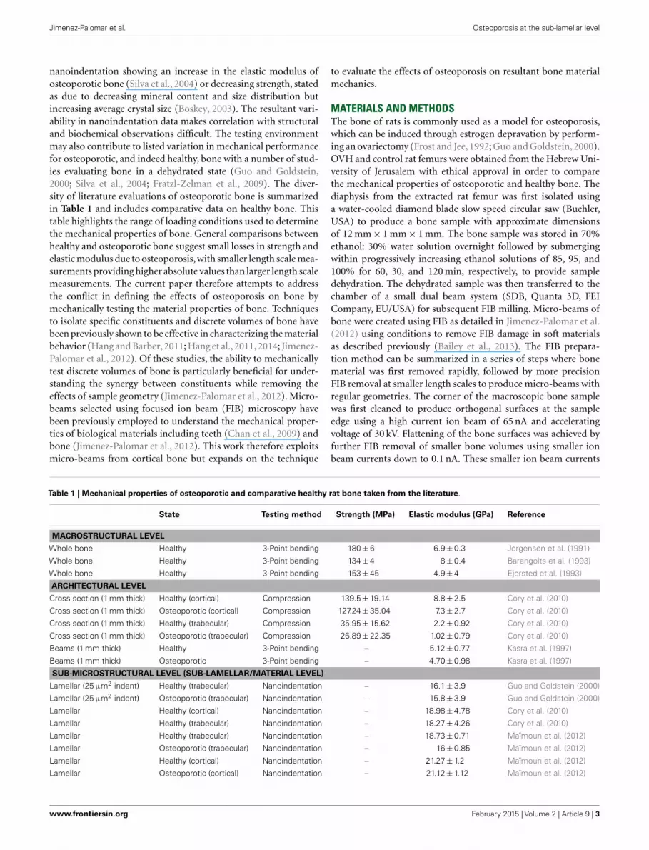
nanoindentation showing an increase in the elastic modulus ofosteoporotic bone (Silva et al., 2004) or decreasing strength, statedas due to decreasing mineral content and size distribution butincreasing average crystal size (Boskey, 2003). The resultant vari-ability in nanoindentation data makes correlation with structuraland biochemical observations difficult. The testing environmentmay also contribute to listed variation in mechanical performancefor osteoporotic, and indeed healthy, bone with a number of stud-ies evaluating bone in a dehydrated state (Guo and Goldstein,2000; Silva et al., 2004; Fratzl-Zelman et al., 2009). The diver-sity of literature evaluations of osteoporotic bone is summarizedin Table 1 and includes comparative data on healthy bone. Thistable highlights the range of loading conditions used to determinethe mechanical properties of bone. General comparisons betweenhealthy and osteoporotic bone suggest small losses in strength andelastic modulus due to osteoporosis, with smaller length scale mea-surements providing higher absolute values than larger length scalemeasurements. The current paper therefore attempts to addressthe conflict in defining the effects of osteoporosis on bone bymechanically testing the material properties of bone. Techniquesto isolate specific constituents and discrete volumes of bone havebeen previously shown to be effective in characterizing the materialbehavior (Hang and Barber, 2011; Hang et al., 2011, 2014; Jimenez-Palomar et al., 2012). Of these studies, the ability to mechanicallytest discrete volumes of bone is particularly beneficial for under-standing the synergy between constituents while removing theeffects of sample geometry (Jimenez-Palomar et al., 2012). Micro-beams selected using focused ion beam (FIB) microscopy havebeen previously employed to understand the mechanical proper-ties of biological materials including teeth (Chan et al., 2009) andbone (Jimenez-Palomar et al., 2012). This work therefore exploitsmicro-beams from cortical bone but expands on the technique
to evaluate the effects of osteoporosis on resultant bone materialmechanics.
MATERIALS AND METHODSThe bone of rats is commonly used as a model for osteoporosis,which can be induced through estrogen depravation by perform-ing an ovariectomy (Frost and Jee, 1992; Guo and Goldstein, 2000).OVH and control rat femurs were obtained from the Hebrew Uni-versity of Jerusalem with ethical approval in order to comparethe mechanical properties of osteoporotic and healthy bone. Thediaphysis from the extracted rat femur was first isolated usinga water-cooled diamond blade slow speed circular saw (Buehler,USA) to produce a bone sample with approximate dimensionsof 12 mm× 1 mm× 1 mm. The bone sample was stored in 70%ethanol: 30% water solution overnight followed by submergingwithin progressively increasing ethanol solutions of 85, 95, and100% for 60, 30, and 120 min, respectively, to provide sampledehydration. The dehydrated sample was then transferred to thechamber of a small dual beam system (SDB, Quanta 3D, FEICompany, EU/USA) for subsequent FIB milling. Micro-beams ofbone were created using FIB as detailed in Jimenez-Palomar et al.(2012) using conditions to remove FIB damage in soft materialsas described previously (Bailey et al., 2013). The FIB prepara-tion method can be summarized in a series of steps where bonematerial was first removed rapidly, followed by more precisionFIB removal at smaller length scales to produce micro-beams withregular geometries. The corner of the macroscopic bone samplewas first cleaned to produce orthogonal surfaces at the sampleedge using a high current ion beam of 65 nA and acceleratingvoltage of 30 kV. Flattening of the bone surfaces was achieved byfurther FIB removal of smaller bone volumes using smaller ionbeam currents down to 0.1 nA. These smaller ion beam currents
Table 1 | Mechanical properties of osteoporotic and comparative healthy rat bone taken from the literature.
State Testing method Strength (MPa) Elastic modulus (GPa) Reference
MACROSTRUCTURAL LEVEL
Whole bone Healthy 3-Point bending 180±6 6.9±0.3 Jorgensen et al. (1991)
Whole bone Healthy 3-Point bending 134±4 8±0.4 Barengolts et al. (1993)
Whole bone Healthy 3-Point bending 153±45 4.9±4 Ejersted et al. (1993)
ARCHITECTURAL LEVEL
Cross section (1 mm thick) Healthy (cortical) Compression 139.5±19.14 8.8±2.5 Cory et al. (2010)
Cross section (1 mm thick) Osteoporotic (cortical) Compression 127.24±35.04 7.3±2.7 Cory et al. (2010)
Cross section (1 mm thick) Healthy (trabecular) Compression 35.95±15.62 2.2±0.92 Cory et al. (2010)
Cross section (1 mm thick) Osteoporotic (trabecular) Compression 26.89±22.35 1.02±0.79 Cory et al. (2010)
Beams (1 mm thick) Healthy 3-Point bending – 5.12±0.77 Kasra et al. (1997)
Beams (1 mm thick) Osteoporotic 3-Point bending – 4.70±0.98 Kasra et al. (1997)
SUB-MICROSTRUCTURAL LEVEL (SUB-LAMELLAR/MATERIAL LEVEL)
Lamellar (25 µm2 indent) Healthy (trabecular) Nanoindentation – 16.1±3.9 Guo and Goldstein (2000)
Lamellar (25 µm2 indent) Osteoporotic (trabecular) Nanoindentation – 15.8±3.9 Guo and Goldstein (2000)
Lamellar Healthy (cortical) Nanoindentation – 18.98±4.78 Cory et al. (2010)
Lamellar Healthy (trabecular) Nanoindentation – 18.27±4.26 Cory et al. (2010)
Lamellar Healthy (trabecular) Nanoindentation – 18.73±0.71 Maïmoun et al. (2012)
Lamellar Osteoporotic (trabecular) Nanoindentation – 16±0.85 Maïmoun et al. (2012)
Lamellar Healthy (cortical) Nanoindentation – 21.27±1.2 Maïmoun et al. (2012)
Lamellar Osteoporotic (cortical) Nanoindentation – 21.12±1.12 Maïmoun et al. (2012)
www.frontiersin.org February 2015 | Volume 2 | Article 9 | 3

Jimenez-Palomar et al. Osteoporosis at the sub-lamellar level
avoid observable ion beam damage. FIB milling was additionallyperformed parallel to the produced sample faces in all preparationsteps to reduce embedding the impinging gallium ions from theFIB within the discrete beam volumes produced. A short columnbetween each of the micro-beams was retained in order to pre-vent the re-deposition of milled material and gallium ions on toneighboring beams. Thus, bone material sputtered from the FIB ismore likely to redeposit on the short columns instead of the sam-ple micro-beams. The average micro-beam dimensions producedat the end of the bone material sample were 8 µm× 2 µm× 2 µm,with the long axis of the micro-beam aligned in the direction of thelong axis of the femur. These dimensions at micron length scalesare comparable to bone lamellae and therefore remove structuralfeatures present at larger length scales.
Resultant bone micro-beams were removed from the SDB setupand placed in a closed vessel containing Hank’s buffer solution for2 h to allow bone rehydration. Samples were then returned to theSDB system for subsequent mechanical testing, with the prior sam-ple rehydration shown to preserve the mechanical properties of thewet bone for up to 2 h in such an environment (Jimenez-Palomaret al., 2012). Mechanical testing of the bone micro-beams wascarried out using an atomic force microscope (AFM) integratedwithin the SDB (Hang et al., 2011). A physiologically relevant com-pressive loading configuration was used, with the load appliedin the direction of the long axis of the micro-beams. The AFMallowed the application of load to the micro-beams while scan-ning electron microscopy (SEM) within the SDB system was usedto observe the deformation and resultant failure of the samples.Mechanical testing was achieved by first translating the AFM tiptoward individual bone micro-beams until compressive load wasapplied parallel to the micro-beam long axis as shown in Figure 1.In situ SEM was used to observe the movement of the AFM tiptoward the end of the micro-beam and ensure that the AFM tipfully contacted the top of the beam as shown in Figure 2. Com-pression of four osteoporotic and six healthy bone micro-beamswas carried out under quasi-static loading rates.
RESULTSCompression of bone micro-beams using the AFM produced cor-responding data for the force applied to the sample and resultantdeformation. Figure 2 shows SEM imaging highlighting the load-ing of the micro-beam sample in compression until failure of thesample occurred. The stress and strain induced in the bone micro-beams were calculated using the force-deflection curves generatedfrom the AFM system. Stress is calculated by dividing the forceapplied to the sample from the AFM tip by the cross-sectionalarea of the micro-beam sample, measured from SEM, whereasstrain is calculated by dividing the change in length by the totalmicro-beam sample length, as shown in Eqs 1 and 2 below.
σ =f
A(1)
ε =∆L
L0(2)
Where σ is the stress in the compressed micro-beam sample, f isthe force applied by the AFM, A is the micro-beam cross-sectional
FIGURE 1 | Scanning electron micrograph of the AFM tip attached tothe force detection system of the AFM cantilever approaching a bonemicro-beam for in situ mechanical testing. The arrow indicates thedirection of the long axis of bone and micro-beam principal axis.
area, ε is the strain in the bone micro-beam, ∆L is change inlength of the beam, and L0 is the original length of the bone micro-beam prior to mechanical deformation. The resultant stress–strainbehavior for the osteoporotic and control bone micro-beams isshown in Figure 3. The stress generally increases in a relativelylinear manner with increasing strain until failure of the micro-beam. We note that local non-linearity is due to the interferometermeasurement system of the AFM as described previously (Hanget al., 2011). The elastic modulus, strength, and the strain to failurevalues calculated from the bone micro-beam compression testsin Figure 3 are shown in Table 2 for the healthy bone controland the osteoporotic OVH model samples. The OVH bone hasan average elastic modulus of 1.59± 1.26 GPa, almost half thevalue of the elastic modulus of 2.9± 1.45 GPa for the control sam-ple. The OVH and control samples exhibit similar strength valuesof 169.23± 21.35 and 169.51± 66.19 MPa, respectively. A largeraverage strain to failure of 10% is recorded for the OVH sampleswhen compared to ~6% for control samples. The toughness of thebone defined by the area under the stress–strain curves is ~8 and5 J·m−3 for OVH and control samples, respectively. The increasedtoughness displayed for the OVH samples is surprising as osteo-porotic bone is commonly associated with brittle failure. However,we reiterate that the work presented here examines the materialproperties of bone and the enhanced toughness is due to increasedstrain to failure of the material. The fragility of osteoporotic boneassociated with larger or whole bone samples is therefore absentwhen evaluating the small-scale material performance.
The mechanical property values recorded from the micro-beam compression of this work generally lie within the archi-tectural range of previous literature as listed in Table 1. The
Frontiers in Materials | Mechanics of Materials February 2015 | Volume 2 | Article 9 | 4

Jimenez-Palomar et al. Osteoporosis at the sub-lamellar level
FIGURE 2 | Scanning electron micrograph showing compression of ratbone micro-beams (A) in the unloaded state with the AFM tip awayfrom the bone micro-beam and (B) during contact of the AFM tip withthe bone micro-beam causing compressive loading.
FIGURE 3 | Stress–strain curves for compression of control andovariectomized (OVH) rat bone micro-beams.
average elastic modulus values in our micro-beam compressionshow larger variations between osteoporotic and healthy bonesamples than the results in Table 1, highlighting the sensitiv-ity of the technique in elucidating mechanical changes in bone
Table 2 | Elastic modulus, strength, and strain to failure values of both
control and ovariectomized (OVH) rat femur bone micro-beams tested
in compression.
Beam no. Elastic modulus (GPa) Strength (MPa) Strain (%)
Control
Average 2.9±1.45 169.51±66.19 6.3±1.89
1 3.06 204.22 6.8
4 3.62 180.53 5.88
5 2.37 73.91 3.32
6 4.65 248.35 7.16
8 0.78 140.52 8.35
OVH
Average 1.59±1.26 169.23±21.35 10±4.04
1 3.46 201.09 5.24
2 1.08 156.17 8.46
3 1.07 161.38 11.74
4 0.74 158.26 14.57
material due to osteoporosis. Indeed, error associated with theaverage elastic modulus values is expected to be due to variabil-ity in the orientation of collagen fibrils within the micro-beamthat is measurable using the experimental setup. We suggest thatthis enhanced mechanical sensitivity of the technique is due tothe removal of sample geometry effects, such as shape of thebone or porosity that is found at larger length scales, using FIB.Many bone structures also have strain to failure values consid-erably lower than our values and this may be attributed to thelack of geometric effects, such as porosity, which provide strainconcentrations locally whereas the bulk of the material remainsat lower strain. Indeed, glassy polymers such as polystyrene areanalogous to this potential situation where mechanical testing ofsmaller volumes of material removes the effect of defects, causingincreases in strain to failure (van der Sanden et al., 1993). Othermechanical testing techniques such as nanoindentation typicallyprobe significantly smaller volumes than the micro-beams of thiswork and are potentially sensitive to more variability from thelocation of the indenting probe at the sample surface, with con-siderable issues related to the uncertainty in the composition ofthe material and resultant contact area with the indenting probepreviously reviewed (Lewis and Nyman, 2008). The enhanced sen-sitivity of the micro-beam compression in our work indicates aclear decrease of bone elastic modulus properties with osteoporo-sis as shown in Figure 3, which potentially contradicts some worksin Table 1 that indicate little variability in the elastic modulus ofosteoporotic bone compared to healthy bone. This lowering of theelastic modulus of osteoporotic bone has been suggested as beingdue to mechanical degradation of the collagen in osteoporoticbone from reductions in the level of immature collagen cross-links and decreases in collagen fibril diameters (Currey, 2003).Compositional changes in collagen, such as the ratio of α1 toα2 chains in different phenotypes of COLIA1 found in Type 1collagen, appear to influence the fracture risk of bone that is inde-pendent to the changes in bone mass (McGuigan et al., 2001). Acorresponding decrease in bone strength is not observed in our
www.frontiersin.org February 2015 | Volume 2 | Article 9 | 5

Jimenez-Palomar et al. Osteoporosis at the sub-lamellar level
work and suggests that the failure of the material does not changewith osteoporosis although the interactions between constituentsare affected. Indeed, molecular modeling has indicated consid-erable variation in mechanical behavior of collagen fibrils as afunction of cross-linking density, including significant changes inelastic modulus as well as regime of cross-linking densities thatprovide minimal changes in strength (Buehler, 2008). We wouldtherefore expect a decrease in elastic modulus of OVH samples asthe stress transfer between protein molecules becomes inefficientfrom the previously reported changes in cross-linking density.Such a mechanism can additionally describe the lack of a loss instrength as the same collagen protein molecules are failing in boththe healthy and OVH samples. The proposed mechanism heretherefore describes deformation and failure of osteoporotic bonein terms of protein from the collagen in bone. Non-collagenousproteins (NCPs) present between collagen fibrils have also beenshown to control fracture behavior of bone (Hang et al., 2014)and are known to chemically change in osteoporotic bone (Srogaand Vashishth, 2012). While the performance of osteoporotic bonematerial has been defined in this paper, the origin of the mechan-ical changes is still contentious, with collagen, mineral, NCPs,and their interactions all potentially contributing to mechanics.Such a complex synergy has been highlighted previously whenconsidering compensation mechanisms where increased stiffnessfor the organic phase is balanced by a decrease in mineral con-tent that results in similar nanoindentation hardness for diseasedand healthy bone (Fratzl-Zelman et al., 2009). Future develop-ment of mechanistic explanations for osteoporotic bone there-fore requires a comprehensive understanding of all constituentmaterials together.
CONCLUSIONThe compressive elastic modulus, strength, and strain to failureof bone micro-beams were measured in order to assess the effectof osteoporosis on the mechanical properties of bone as a mater-ial at the sub-lamellar level. Although compression testing hereincannot be directly compared to previous studies in the literature,results showed a decrease in the elastic modulus of osteoporoticbone compared to a control. This decrease in the elastic moduluswith osteoporosis was additionally associated with relatively con-stant micro-beam strength and a small increase in failure strain,with associated changes in material toughness. The origin of osteo-porotic induced decreases in bone elastic modulus was suggestedas being due to mechanical degradation of the collagen within thebone material.
REFERENCESAmmann, P., and Rizzoli, R. (2003). Bone strength and its determinants. Osteoporos.
Int. 14, 13–18. doi:10.1007/s00198-002-1345-4Bailey, R. J., Geurts, R., Stokes, D. J., Jong, F. D., and Barber, A. H. (2013). Eval-
uating focused ion beam induced damage in soft materials. Micron 50, 51–56.doi:10.1016/j.micron.2013.04.005
Barengolts, E. I., Curry, D. J., Bapna, M. S., and Kukreja, S. C. (1993). Effectsof endurance exercise on bone mass and mechanical properties in intactand ovariectomized rats. J. Bone Miner. Res. 8, 937–942. doi:10.1002/jbmr.5650080806
Boivin, G., and Baud, C. A. (1984). “Microradiographic methods for calcifiedtissues,” in Methods for Calcified Tissue Preparation, ed. G. R. Dickson (Ams-terdam: Elsevier), 391–411.
Boivin, G., and Meunier, P. J. (2002). The degree of mineralization of bone tissuemeasured by computerized quantitative contact microradiography. Calcif. TissueInt. 70, 503–511. doi:10.1007/s00223-001-2048-0
Borah, B., Ritman, E. L., Dufresne, T. E., Jorgensen, S. M., Liu, S., Sacha, J., et al.(2005). The effect of risedronate on bone mineralization as measured by micro-computed tomography with synchrotron radiation: correlation to histomorpho-metric indices of turnover. Bone 37, 1–9. doi:10.1016/j.bone.2005.03.017
Boskey, A. (2003). Bone mineral crystal size. Osteoporos. Int. 14, 16–21. doi:10.1007/s00198-003-1468-2
Bouxsein, M. (2001). “Biomechanics of age-related fractures,” in Osteoporosis, 2ndEdn, eds R. Marcus, D. Feldman, and J. Kelsey (San Diego, CA: Academic Press),509–534.
Buehler, M. J. (2008). Nanomechanics of collagen fibrils under varying cross-linkdensities: atomistic and continuum studies. J. Mech. Behav. Biomed. Mater. 1,59–67. doi:10.1016/j.jmbbm.2007.04.001.
Burr, D. (2003). Microdamage and bone strength. Osteoporos. Int. 14, S67–S72.doi:10.1007/s00198-003-1476-2
Burr, D. B. (2002). The contribution of the organic matrix to bone’s material prop-erties. Bone 31, 8–11. doi:10.1016/S8756-3282(02)00815-3
Burr, D. B., Forwood, M. R., Fyhrie, D. P., Martin, R. B., Schaffler, M. B., and Turner,C. H. (1997). Bone microdamage and skeletal fragility in osteoporotic and stressfractures. J. Bone Miner. Res. 12, 6–15. doi:10.1359/jbmr.1997.12.1.6
Burr, D. B., Schaffler, M. B., and Frederickson, R. G. (1988). Composition of thecement line and its possible mechanical role as a local interface in human com-pact bone. J. Biomech. 21, 939–945. doi:10.1016/0021-9290(88)90132-7
Carter, D. R., and Hayes, W. C. (1976). Bone compressive strength: the influence ofdensity and strain rate. Science 149, 1174–1176. doi:10.1126/science.996549
Chan, Y. L., Ngan, A. H. W., and King, N. M. (2009). Use of focused ion beammilling for investigating the mechanical properties of biological tissues: astudy of human primary molars. J. Mech. Behav. Biomed. Mater. 2, 375–383.doi:10.1016/j.jmbbm.2009.01.006
Ciarelli, T. E., Fyhrie, D. P., and Parfitt,A. M. (2003). Effects of vertebral bone fragilityand bone formation rate on the mineralization levels of cancellous bone fromwhite females. Bone 32, 311–315. doi:10.1016/S8756-3282(02)00975-4
Cory, E., Nazarian,A., Entezari,V.,Vartanians,V.,Müller, R., and Snyder, B. D. (2010).Compressive axial mechanical properties of rat bone as functions of bone vol-ume fraction, apparent density and micro-ct based mineral density. J. Biomech.43, 953–960. doi:10.1016/j.jbiomech.2009.10.047
Currey, J. D. (1988). The effect of porosity and mineral content on the young’s mod-ulus of elasticity of compact bone. J. Biomech. 21, 131–139. doi:10.1016/0021-9290(88)90006-1
Currey, J. D. (2003). Role of collagen and other organics in the mechanical propertiesof bone. Osteoporos. Int. 14, S29–S36. doi:10.1007/s00198-003-1470-8
Ejersted, C., Andreassen, T. T., Oxlund, H., Jorgensen, P. H., Bak, B., Haggblad,J., et al. (1993). Human parathyroid hormone (1-34) and (1-84) increase themechanical strength and thickness of cortical bone in rats. J. Bone Miner. Res. 8,1097–1101. doi:10.1002/jbmr.5650080910
Fratzl-Zelman, N., Roschger, P., Gourrier, A., Weber, M., Misof, B. M., Loveridge,N., et al. (2009). Combination of nanoindentation and quantitative backscat-tered electron imaging revealed altered bone material properties associated withfemoral neck fragility. Calcif. Tissue Int. 85, 335–343. doi:10.1007/s00223-009-9289-8
Frost, H. M., and Jee, W. S. S. (1992). On the rat model of human osteopenias andosteoporoses. Bone Miner. 18, 227–236. doi:10.1016/0169-6009(92)90809-R
Grynpas, M. (1993). Age and disease-related changes in the mineral of bone. Calcif.Tissue Int. 53, S57–S64. doi:10.1007/BF01673403
Guo, X. E., and Goldstein, S. A. (2000). Vertebral trabecular bone microscopic tissueelastic modulus and hardness do not change in ovariectomized rats. J. Orthop.Res. 18, 333–336. doi:10.1002/jor.1100180224
Hang, F., and Barber, A. H. (2011). Nano-mechanical properties of individualmineralized collagen fibrils from bone tissue. J. R. Soc. Interface 8, 500–505.doi:10.1098/rsif.2010.0413
Hang, F., Gupta, H. S., and Barber, A. H. (2014). Nanointerfacial strength betweennon-collagenous protein and collagen fibrils in antler bone. J. R. Soc. Interface11, 20130993. doi:10.1098/rsif.2013.0993
Hang, F., Lu, D., Bailey, R. J., Jimenez-Palomar, I., Stachewicz, U., Cortes-Ballesteros,B., et al. (2011). In situ tensile testing of nanofibers by combining atomicforce microscopy and scanning electron microscopy. Nanotechnology 22, 365708.doi:10.1088/0957-4484/22/36/365708
Frontiers in Materials | Mechanics of Materials February 2015 | Volume 2 | Article 9 | 6

Jimenez-Palomar et al. Osteoporosis at the sub-lamellar level
Hauge, E. M., Steiniche, T., and Andreassen, T. T. (2003). “Histomorphometry ofmetabolic bone conditions,” in Handbook of Histology Methods for Bone andCartilage, eds Y. H. An and K. L. Martin (Totowa, NJ: Human Press Inc), 391–410.
Iacono, M. V. (2007). Osteoporosis: a national public health priority. J. Perianesth.Nurs. 22, 175–183. doi:10.1016/j.jopan.2007.03.009
Jimenez-Palomar, I., Shipov, A., Shahar, R., and Barber, A. H. (2012). Influenceof SEM vacuum on bone micromechanics using in situ AFM. J. Mech. Behav.Biomed. Mater. 5, 149–155. doi:10.1016/j.jmbbm.2011.08.018
Jorgensen, P. H., Bak, B., and Andreassen, T. T. (1991). Mechanical proper-ties and biochemical composition of rat cortical femur and tibia after long-term treatment with biosynthetic human growth hormone. Bone 12, 353–359.doi:10.1016/8756-3282(91)90022-B
Kasra, M., Vanin, C. M., Maclusky, N. J., Casper, R. F., and Grynpas, M. D. (1997).Effects of different estrogen and progestin regimens on the mechanical propertiesof rat femur. J. Orthop. Res. 15, 118–123. doi:10.1002/jor.1100150117
Katz, J. L., and Meunier, A. (1993). Scanning acoustic microscope studies of theelastic properties of osteons and osteon lamellae. J. Biomech. Eng. 115, 543–548.doi:10.1115/1.2895537
Keaveny, T. M., Morgan, E. F., Niebur, G. L., and Yeh, O. C. (2001). Biomechanicsof trabecular bone. Annu. Rev. Biomed. Eng. 3, 307–333. doi:10.1146/annurev.bioeng.3.1.307
Kennedy, O. D., Brennan, O., Rackard, S. M., Staines, A., O’Brien, F. J., Taylor, D.,et al. (2009). Effects of ovariectomy on bone turnover, porosity, and biomechan-ical properties in ovine compact bone 12 months postsurgery. J. Orthop. Res. 27,303–309. doi:10.1002/jor.20750
Kilbanski, A., Adams-Campbell, L., Bassford, T., Blair, S. N., Boden, S. D., Dickersin,K., et al. (2001). Osteoporosis prevention, diagnosis, and therapy. JAMA 285,785–795. doi:10.1001/jama.285.6.785
Lewis, G., and Nyman, J. S. (2008). The use of nanoindentation for characterizingthe properties of mineralized hard tissues: state-of-the art review. J. Biomed.Mater. Res. B Appl. Biomater. 87, 286–301. doi:10.1002/jbm.b.31092
Maïmoun, L., Brennan-Speranza, T. C., Rizzoli, R., and Ammann, P. (2012).Effects of ovariectomy on the changes in microarchitecture and material levelproperties in response to hind leg disuse in female rats. Bone 51, 586–591.doi:10.1016/j.bone.2012.05.001
Marcus, R., and Bouxsein, M. L. (2010). “The nature of osteoporosis,” in Fundamen-tals of Osteoporosis, eds R. Marcus, D. Feldman, D. A. Nelson, and C. J. Rosen(San Diego, CA: Academic Press), 25–34.
McCreadie, B. R., Morris, M. D., Chen, T., Sudhaker, R. D., Finney, W. F., Widjaja, E.,et al. (2006). Bone tissue compositional differences in women with and withoutosteoporotic fracture. Bone 39, 1190–1195. doi:10.1016/j.bone.2006.06.008
McGuigan, F. E. A., Armbrecht, G., Smith, R., Felsenberg, D., Reid, D. M., and Ral-ston, S. H. (2001). Prediction of osteoporotic fractures by bone densitometry andCOLIA1 genotyping: a prospective, population-based study in men and women.Osteoporos. Int. 12, 91–96. doi:10.1007/s001980170139
Paschalis, E. P., Shane, E., Lyritis, G., Skarantavos, G., Mendelsohn, R., and Boskey,A. L. (2004). Bone fragility and collagen cross-links. J. Bone Miner. Res. 19,2000–2004. doi:10.1359/jbmr.040820
Rice, J. C., Cowin, S. C., and Bowman, J. A. (1988). On the dependence of the elastic-ity and strength of cancellous bone on apparent density. J. Biomech. 21, 155–168.doi:10.1016/0021-9290(88)90008-5
Roschger, P., Gupta, H. S., Berzlanovich, A., Ittner, G., Dempster, D. W., Fratzl, P.,et al. (2003). Constant mineralization density distribution in cancellous humanbone. Bone 32, 316–323. doi:10.1016/S8756-3282(02)00973-0
Roschger, P., Paschalis, E. P., Fratzl, P., and Klaushofer, K. (2008). Bone mineraliza-tion density distribution in health and disease. Bone 42, 456–466. doi:10.1016/j.bone.2007.10.021
Schaffler, M. B., and Burr, D. B. (1988). Stiffness of compact bone: effects of porosityand density. J. Biomech. 21, 13–16. doi:10.1016/0021-9290(88)90186-8
Silva, M. J., Brodt, M. D., Fan, Z., and Rho, J. Y. (2004). Nanoindentation and whole-bone bending estimates of material properties in bones from the senescenceaccelerated mouse SAMP6. J. Biomech. 37, 1639–1646. doi:10.1016/j.jbiomech.2004.02.018
Silva, M. J., and Gibson, L. J. (1997). Modeling the mechanical behavior of verte-bral trabecular bone: effects of age-related changes in microstructure. Bone 21,191–199. doi:10.1016/S8756-3282(97)00100-2
Sroga, G. E., and Vashishth, D. (2012). Effects of bone matrix proteins on fractureand fragility in osteoporosis. Curr. Osteoporos. Rep. 10, 141–150. doi:10.1007/s11914-012-0103-6
Turner, C. H. (2002). Biomechanics of bone: determinants of skeletal fragility andbone quality. Osteoporos. Int. 13, 97–104. doi:10.1007/s001980200000
van der Sanden, M. C. M., Meijer, H. E. H., and Lemstra, P. J. (1993). Deformationand toughness of polymeric systems: 1. The concept of critical thickness. Polymer34, 2148–2154. doi:10.1016/0032-3861(93)90249-A
WHO. (2012). National Osteoporosis Guideline Group (NOGG) [Online]. Sheffield:University of Sheffield. Available at: http://www.shef.ac.uk/NOGG/
Wu, Z.-X., Lei, W., Hu, Y.-Y., Wang, H.-Q., Wan, S.-Y., Ma, Z.-S., et al. (2008).Effect of ovariectomy on BMD, micro-architecture and biomechanics of cor-tical and cancellous bones in a sheep model. Med. Eng. Phys. 30, 1112–1118.doi:10.1016/j.medengphy.2008.01.007
Zebaze, R. M. D., Ghasem-Zadeh, A., Bohte, A., Iuliano-Burns, S., Mirams, M., Price,R. I., et al. (2010). Intracortical remodelling and porosity in the distal radius andpost-mortem femurs of women: a cross-sectional study. Lancet 375, 1729–1736.doi:10.1016/S0140-6736(10)60320-0
Conflict of Interest Statement: The authors declare that the research was conductedin the absence of any commercial or financial relationships that could be construedas a potential conflict of interest.
Received: 12 December 2014; paper pending published: 29 December 2014; accepted:23 January 2015; published online: 13 February 2015.Citation: Jimenez-Palomar I, Shipov A, Shahar R and Barber AH (2015) Mechani-cal behavior of osteoporotic bone at sub-lamellar length scales. Front. Mater. 2:9. doi:10.3389/fmats.2015.00009This article was submitted to Mechanics of Materials, a section of the journal Frontiersin Materials.Copyright © 2015 Jimenez-Palomar, Shipov, Shahar and Barber. This is an open-access article distributed under the terms of the Creative Commons Attribution License(CC BY). The use, distribution or reproduction in other forums is permitted, providedthe original author(s) or licensor are credited and that the original publication in thisjournal is cited, in accordance with accepted academic practice. No use, distribution orreproduction is permitted which does not comply with these terms.
www.frontiersin.org February 2015 | Volume 2 | Article 9 | 7


![[Secondary prevention in osteoporotic fractures. The GIOS project]](https://img.pdfslide.net/doc/110x75/63487f43de40dd034d093bb9/secondary-prevention-in-osteoporotic-fractures-the-gios-project.jpg)
