Embed Size (px)
Citation preview

Please cite this article in press as: Guet et al., Mechanical Role of Actin Dynamics in the Rheology of the Golgi Complex and in Golgi-Associated Trafficking Events, Current Biology (2014), http://dx.doi.org/10.1016/j.cub.2014.06.048
Mechanical Role of Actin Dy
Current Biology 24, 1–12, August 4, 2014 ª2014 Elsevier Ltd All rights reserved http://dx.doi.org/10.1016/j.cub.2014.06.048
Articlenamics
in the Rheology of the Golgi Complexand in Golgi-Associated Trafficking Events
David Guet,1,2,3 Kalpana Mandal,1,2 Mathieu Pinot,1
Jessica Hoffmann,1,4 Yara Abidine,1,5 Walter Sigaut,1,6
Sabine Bardin,1 Kristine Schauer,1 Bruno Goud,1,*
and Jean-Baptiste Manneville1,*1CNRS-Institut Curie, UMR144, 26 rue d’Ulm, 75248 ParisCedex 05, France
Summary
Background: In vitro studies have shown that physical param-eters, such asmembrane curvature, tension, and composition,influence the budding and fission of transport intermediates.Endocytosis in living cells also appears to be regulated bythe mechanical load experienced by the plasma membrane.In contrast, how these parameters affect intracellular mem-brane trafficking in living cells is not known. To address thisquestion, we investigate here the impact of a mechanicalstress on the organization of the Golgi complex and on the for-mation of transport intermediates from the Golgi complex.Results: Using confocal microscopy, we visualize the de-formation of Rab6-positive Golgi membranes applied by aninternalized microsphere trapped in optical tweezers andsimultaneously measure the corresponding forces. Our resultsshow that the force necessary to deform Golgi membranesdrops when actin dynamics is altered and correlates withmyosin II activity. We also show that the applied stress has along-range effect on Golgi membranes, perturbs the dynamicsof Golgi-associated actin, and induces a sharp decrease in theformation of Rab6-positive vesicles from the Golgi complex aswell as tubulation of Golgi membranes.Conclusions: We suggest that acto-myosin contractilitystrongly contributes to the local rigidity of the Golgi complexand regulates the mechanics of the Golgi complex to controlintracellular membrane trafficking.
Introduction
The Golgi complex plays a central role in intracellular mem-brane trafficking. Despite constant inbound and outboundmembrane fluxes, the Golgi complex maintains a stable struc-ture and a typical cis, median, and trans cisternae organiza-tion. The architecture of the Golgi complex is thought to resultfrom interactions between Golgi membranes, the cytoskel-eton, and a protein scaffold, the so-called Golgi matrix.
2Co-first author3Present address: Unite Pathologie et Virologie Moleculaire INSERM U944
CNRS UMR9212, Institut Universitaire d’Hematologie, Hopital Saint-Louis,
1 avenue Vellefaux, 75010 Paris, France4Present address: Ecole Normale Superieure, 45 rue D’Ulm, 75230 Paris
Cedex 05, France5Present address: CNRS-Universite Joseph Fournier Grenoble, Laboratoire
Interdisciplinaire de Physique (LiPhy), 140 Avenue de la Physique BP87,
38402 Saint Martin d’Heres, France6Present address: LMGP-Grenoble INP-Minatec, 3 parvis Louis Neel CS
50257, 38016 Grenoble Cedex 1, France
*Correspondence: [email protected] (B.G.), jean-baptiste.manneville@
curie.fr (J.-B.M.)
Although the microtubule cytoskeleton has been known tobe involved in Golgi organization for a long time, new criticalplayers in Golgi architecture have been identified in the pastten years (for a review, see [1]). The actin cytoskeleton inparticular actively participates in the maintenance of the Golgiarchitecture [2]. Although technically difficult to visualize, actinand actin nucleators are associated with Golgi membranes[3–5], and perturbation of actin dynamics leads to disorganiza-tion of the Golgi cisternae [6, 7]. Myosins also localize to theGolgi complex [8] and have been involved in the formation oftransport intermediates from the trans side of the Golgi com-plex [9–11], and myosin II-driven contractility is required forthe fission of Rab6-positive vesicles [9]. Moreover, the forminmDia1, an activator of actin polymerization, has been recentlyimplicated in the maintenance of the Golgi structure and theformation of Rab6-positive transport intermediates [12].Experiments based on biophysical approaches have shown
thatmechanical parameters, such asmembrane rigidity or ten-sion, are critical for intracellular transport [13, 14]. Studies inliving cells have focused on the plasma membrane [15].Recently, mechanical stretching or osmotic swelling in cellsand in vitro reconstitution on model membranes confirmedthat a high mechanical load on the plasma membrane limitsmembrane invagination in both clathrin-dependent [16] andclathrin-independent [17] endocytosis. Similar data on intra-cellular compartments are lacking, mostly because accessingintracellular membranes for mechanical experiments in livingcells is technically challenging.In order to probe the architecture of the Golgi complex and
test for the potential effects of a mechanical stress exerted onthe Golgi complex on membrane trafficking, we designed anew intracellular micromanipulation technique based on theoptical trapping of internalized micrometer-sized latex beadscoupled with fast confocal imaging. The technique allows,for the first time to our knowledge, the application and mea-surement of a force up to a few hundreds of piconewtons ona fluorescently labeled intracellular organelle and simulta-neously visualization of the induced deformation.
Results
Mechanical Deformation of Golgi Membranes
To apply and measure a force on Golgi membranes, we havedeveloped an intracellularmicromanipulation technique basedon optical trapping of micrometer-sized beads. Latex beads(diameter 2 mm) were endocytosed overnight in RPE1 cellsand accumulated in the perinuclear region (Figure 1A, top).The endocytic membranes surrounding the beads were tightlyapposed on the bead surface and were negative for the earlyendosome marker Rab5 but positive for the late endosomal/lysosomal markers LAMP1 (Figure 1B) and CD63 (data notshown). A mechanical force was applied on the Golgi complexby pushing it against a bead held in a fixed optical trap (Fig-ure 1A, bottom). The pushing force was generated by displac-ing themicroscope stage. The forcewasmeasured by trackingthe position of the bead (see Experimental Procedures) andthe Golgi complex was simultaneously imaged by fastconfocal microscopy. Using this technique, we were able to

Figure 1. Probing the Mechanics of Golgi Membranes
(A) Experimental set up. 2 mm red fluorescent beads were endocytosed overnight in RPE-1 cells expressing GFP-Rab6 to visualize the Golgi complex (1). A
bead located close to the Golgi complex was trapped using optical tweezers (2). Golgi membranes were then pushed against the bead held at a fixed
(legend continued on next page)
Current Biology Vol 24 No 152
Please cite this article in press as: Guet et al., Mechanical Role of Actin Dynamics in the Rheology of the Golgi Complex and in Golgi-Associated Trafficking Events, Current Biology (2014), http://dx.doi.org/10.1016/j.cub.2014.06.048

Mechanics of the Golgi Complex3
Please cite this article in press as: Guet et al., Mechanical Role of Actin Dynamics in the Rheology of the Golgi Complex and in Golgi-Associated Trafficking Events, Current Biology (2014), http://dx.doi.org/10.1016/j.cub.2014.06.048
efficiently deform the Golgi complex by applying forces in the100–200 pN range.
Golgi Membranes Are Flexible and Mechanically CoupledThe Golgi complex was visualized using GFP-Rab6 that local-izes to trans-Golgi and TGN (trans-Golgi network) membranesand to small vesicles that shuttle between the Golgi complexand the cell periphery [9]. Rab6-positive Golgi ribbons werebent upon application of a mechanical force (Figure 1C, MovieS1 available online), showing that the Golgi complex is a flex-ible structure that can be deformed by w100–200 pN forces.Similar results were obtained using another Golgi marker,GFP-tagged COPI subunit εCOP (data not shown). To investi-gate the effect of the applied force on the Golgi structure, weacquired projections of 3D confocal stacks before and afterapplying a mechanical constraint. In a control experiment, asexpected, the two images could be superimposed (Figure 1D,No constraint). This was not the case after application of theforce (Figure 1D, With constraint), indicating that structuralchanges are induced on Rab6-positive Golgi membranes bythe mechanical constraint, both near and far (up to 10 mm)from the trapped bead (Figures 1D and 1E). These findingssuggest that Rab6-positive Golgi membranes are mechani-cally coupled and that a rigid component propagates theconstraint along Golgi membranes.
The Microenvironment Rigidity Increases Close to the
Golgi ComplexTo characterize the mechanical properties of the microenvi-ronment of the beads when they approach the Golgi complex,we compared the local stiffness a few micrometers away fromRab6-positive Golgi stacks (Movie S2, left) with the stiffnessmeasured in close proximity to Rab6-positive membranes(Movie S2, right). The cell was moved automatically via atwo-axis nanopositioning piezo-stage in a stepwise fashionto bring Rab6-positive membranes initially about 1 mm awayfrom the bead either into contact with the bead or alternativelyfarther away from the bead. Five 0.5 mm steps were performedduring 1 min (Figure 2A) and the bead displacement wasmeasured. The bead displacement depends on the visco-elastic properties of the surrounding microenvironment. Fig-ure 2A depicts four expected behaviors.
We first tested the technique in cell extracts prepared eitherfrom Xenopus oocytes or from HeLa cells (Figure 2B, graycurves). In both cases the total bead displacement did notexceed 0.25 mm. In cells, after each 0.5 mm step displacement
position in the optical trap by moving the microscope stage either manually vi
(B) Themembrane surrounding the bead is positive for late endosomal and lyso
LAMP1 (blue) 5 hr after addition of the beads (red). A zoom of the boxed regio
(C) Example of mechanical deformation of Golgi membranes visualized with
(Movie S1). The position of the optical trap is indicated by a cross in the first i
indicated in the top right corner of each image. The boxed areas in the last tw
(D) Long-range effect of amechanical constraint on Golgi membranes.Maximum
pushed by manual displacement of the microscope stage against a bead held
constraint was applied (i.e., the bead was trapped for 1 min without stage displ
and beads in red, respectively, before and after optical trapping. The bottom
(green) and after (red) optical trapping. Two higher-magnification images of reg
complex. The position of the trap is indicated by a cross and the trapped bead
fication colocalization images point to zones where the shape of Golgi membr
profiles along two dashed lines (1 and 2) are shown below the images (green,
(E) Graph showing colocalization of the GFP-Rab6 signals before and after op
constraint was applied (n = 6 cells) and when a constraint was applied (n =
5 mm-wide region close to the bead (near bead) and in the remaining Golgi ar
compared to control indicating both short- and long-range effects of the cons
Scale bars represent 5 mm. See also Movie S1.
of the stage, the bead experienced a step increase followed byan exponential relaxation of its position toward the trap center.When the load on the bead became too high, the bead wasejected out of the trap (see Movie S2, right). The softness in-dex, the bead step amplitude, and the relaxation time (param-eters defined in Experimental Procedures) were calculatedand compared between two conditions: Rab6-positive Golgimembranes were either moved away from the bead or movedtoward the bead (Figures 2B and 2C). In the latter condition,the softness index decreased while the bead step amplitudeand the relaxation time increased. In addition, the frequencyof bead ejection from the optical trap was more than 5 timeshigher for beads brought close to the Golgi complex than forbeads pushed away from the Golgi membranes toward thecytoplasm (Figure 2D).By plotting the softness index measured when the beads
were pushed toward the Golgi complex as a function of thedistance separating the bead from the nearest Rab6-positivemembrane, we could detect an increase in rigidity when thebeads entered a region within 1.5 mm from the Rab6-positiveGolgi membranes (Figure S1B). We confirmed this stiffeningby measuring the complex shear modulus of the Golgimicroenvironment using an oscillatory displacement of thepiezo-stage (see Supplemental Experimental Proceduresand Figure S1C). Both the storage modulus G’ and the lossmodulus G’’ increased when regions closer than 1.5 mm tothe Rab6-positive Golgi membranes were probed.Taken together, these measurements indicate that the local
microenvironment stiffens as the bead approaches the Golgicomplex.
Actin Dynamics and Acto-Myosin Contractility Control theRigidity of the Golgi Complex
We next investigated the role of actin and acto-myosincontractility in themechanical properties of theGolgi complex.In a first set of experiments performed on cells treated or not
with cytochalasin D (cytoD), which induces actin depolymer-ization by binding to F-actin and impairing its polymerization,Rab6-positive membranes were pushed manually against thebead. In control cells, the force rapidly reached a plateau ofabout 100 pN, preventing further displacement (Figure 3A,see also Movie S3, left). In contrast, in cytoD-treated cells,the force dropped after each displacement of the cell, allowingthe bead to penetrate between Rab6-positive Golgi mem-branes on distances up to 10 mm (Figure 3B, see also MovieS3, right). The maximum displacement was about 2.5 times
a a joystick or automatically via a nanopositioning piezo-stage (3).
somalmarkers. Left: RPE-1 cells were fixed and stained for Rab5 (green) and
n is shown in lower images.
GFP-Rab6 (green). Images shown are stills from a 1 min time-lapse movie
mage. The direction of the force is shown by an arrow and its magnitude is
o images are shown on the lower images.
projections of confocal stacks of a cell in which the Golgi membranes were
in the optical trap for 1 min (right column) and of a control cell in which no
acement, left column). The top and middle images show GFP-Rab6 in green
images shows the colocalization (yellow) of the GFP-Rab6 signals before
ions near the bead and far from the bead are shown for the constrained Golgi
is shown by circles in the colocalization images. Arrows in the high-magni-
anes is most affected by the mechanical constraint. Fluorescence intensity
before application of the constraint; red, after application of the constraint).
tical trapping from the whole Rab6-positive Golgi area measured when no
22 cells). In the case of a constrained Golgi complex, colocalization in a
ea (far from bead) was measured. Both values are significantly decreased
traint. Error bars represent mean 6 SEM.

Figure 2. The Local Stiffness Increases in Proximity of the Golgi Complex
(A) Probing the visco-elastic properties of the bead microenvironment. Left: Schematics illustrating the position of the optical trap, the stage displacement
(double blue arrow), the bead displacement (black arrow), and the displacement and/or deformation of the Golgi membranes (green arrow) in four model
cases. From left to right: the Golgi complex is not deformable, the Golgi complex behaves as a rigid or soft visco-elastic material, and the Golgi complex
exerts no friction on the bead. Right: Graphs showing the displacement of the bead after five 0.5 mm step displacements of the piezo-stage in the same four
cases: (1) the bead is embedded in or contacts a very rigid nondeformable microenvironment (black line) and its position follows that of the stage (dbead =
dstage); (2) and (3) the bead is embedded in a visco-elasticmicroenvironment and its position relaxes exponentially after each 0.5 mmstep (green line for rigid;
red line for soft); and (4) the bead is embedded in amicroenvironment with vanishing viscosity that exerts a vanishing friction (gray line) and its position is not
affected by the stage displacement (dbead = 0).
(B) Rab6-positive membranes were pushed away from (red lines) or toward (green lines) a bead held in the optical trap by moving the piezo-stage using five
0.5 mm steps in 1 min (Movie S2). Grey curves show the same experiments performed in Xenopus oocyte extracts (dark gray) and HeLa cell extracts (light
gray). The graph shows the averaged (n = 8 for ‘‘away,’’ n = 25 for ‘‘toward,’’ n = 16 for Xenopus, n = 11 for HeLa) displacements of the bead as a function of
time. Error bars shown in gray represent mean 6 SEM.
(C) Measurements of the softness index, the bead step amplitude, and the relaxation time in the cytoplasm (‘‘away,’’ n = 7 cells) or in close proximity of Rab6-
positive membranes (‘‘toward,’’ n = 22 cells). Error bars represent mean 6 SEM.
(D) Frequency of bead ejection from the optical trap in the cytoplasm (‘‘away,’’ n = 8 cells) or in the Golgi area (‘‘toward,’’ n = 14 cells) after five 0.5 mm steps
(left) and frequency of bead ejection at each individual step (right).
See also Movie S2.
Current Biology Vol 24 No 154
Please cite this article in press as: Guet et al., Mechanical Role of Actin Dynamics in the Rheology of the Golgi Complex and in Golgi-Associated Trafficking Events, Current Biology (2014), http://dx.doi.org/10.1016/j.cub.2014.06.048
higher in cells treated with cytoD than in nontreated cells (Fig-ure 3C). The softness index increased with the concentrationof cytoD. These results show that depolymerizing actin softensRab6-positive Golgi membranes.
To quantify the role of actin in the local mechanical proper-ties of the Golgi area, the pushing force was then applied bymoving the cell automatically in a stepwise fashion asdescribed above. The softness index, the bead step ampli-tude, and the relaxation time were measured in the presenceor absence of cytoD and jasplakinolide, which blocks actin dy-namics in cells by disrupting actin filaments and inducing
polymerization of monomeric actin into amorphous masses.The two drugs had very similar effects (Figure 4A) and inducedan increase in the softness index, a decrease in the bead stepamplitude, and no significant effect on the relaxation time (Fig-ure 4C). The frequency of bead ejection out of the trap dropped(Figure 4D). In contrast, no effect of actin depolymerizationwas detected when the trapped beads were pushed awayfrom the Rab6-positive Golgi membranes (Figure S2).To directly visualize the dynamics of polymerized actin
before and after the application of the force, we used cells sta-bly expressing LifeAct-mCherry and labeled with the live Golgi
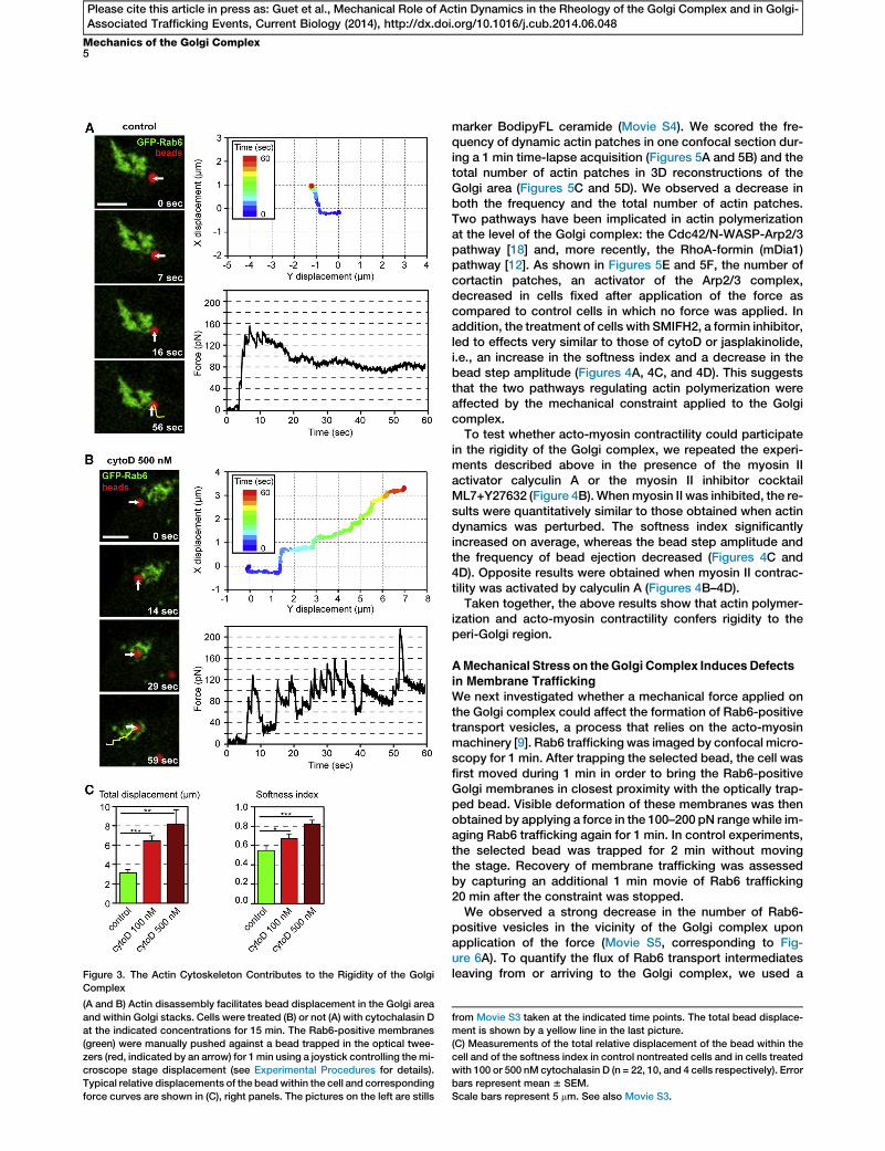
Figure 3. The Actin Cytoskeleton Contributes to the Rigidity of the Golgi
Complex
(A and B) Actin disassembly facilitates bead displacement in the Golgi area
and within Golgi stacks. Cells were treated (B) or not (A) with cytochalasin D
at the indicated concentrations for 15 min. The Rab6-positive membranes
(green) were manually pushed against a bead trapped in the optical twee-
zers (red, indicated by an arrow) for 1min using a joystick controlling themi-
croscope stage displacement (see Experimental Procedures for details).
Typical relative displacements of the beadwithin the cell and corresponding
force curves are shown in (C), right panels. The pictures on the left are stills
Mechanics of the Golgi Complex5
Please cite this article in press as: Guet et al., Mechanical Role of Actin Dynamics in the Rheology of the Golgi Complex and in Golgi-Associated Trafficking Events, Current Biology (2014), http://dx.doi.org/10.1016/j.cub.2014.06.048
marker BodipyFL ceramide (Movie S4). We scored the fre-quency of dynamic actin patches in one confocal section dur-ing a 1 min time-lapse acquisition (Figures 5A and 5B) and thetotal number of actin patches in 3D reconstructions of theGolgi area (Figures 5C and 5D). We observed a decrease inboth the frequency and the total number of actin patches.Two pathways have been implicated in actin polymerizationat the level of the Golgi complex: the Cdc42/N-WASP-Arp2/3pathway [18] and, more recently, the RhoA-formin (mDia1)pathway [12]. As shown in Figures 5E and 5F, the number ofcortactin patches, an activator of the Arp2/3 complex,decreased in cells fixed after application of the force ascompared to control cells in which no force was applied. Inaddition, the treatment of cells with SMIFH2, a formin inhibitor,led to effects very similar to those of cytoD or jasplakinolide,i.e., an increase in the softness index and a decrease in thebead step amplitude (Figures 4A, 4C, and 4D). This suggeststhat the two pathways regulating actin polymerization wereaffected by the mechanical constraint applied to the Golgicomplex.To test whether acto-myosin contractility could participate
in the rigidity of the Golgi complex, we repeated the experi-ments described above in the presence of the myosin IIactivator calyculin A or the myosin II inhibitor cocktailML7+Y27632 (Figure 4B).Whenmyosin II was inhibited, the re-sults were quantitatively similar to those obtained when actindynamics was perturbed. The softness index significantlyincreased on average, whereas the bead step amplitude andthe frequency of bead ejection decreased (Figures 4C and4D). Opposite results were obtained when myosin II contrac-tility was activated by calyculin A (Figures 4B–4D).Taken together, the above results show that actin polymer-
ization and acto-myosin contractility confers rigidity to theperi-Golgi region.
AMechanical Stress on theGolgi Complex InducesDefects
in Membrane TraffickingWe next investigated whether a mechanical force applied onthe Golgi complex could affect the formation of Rab6-positivetransport vesicles, a process that relies on the acto-myosinmachinery [9]. Rab6 trafficking was imaged by confocal micro-scopy for 1 min. After trapping the selected bead, the cell wasfirst moved during 1 min in order to bring the Rab6-positiveGolgi membranes in closest proximity with the optically trap-ped bead. Visible deformation of these membranes was thenobtained by applying a force in the 100–200 pN range while im-aging Rab6 trafficking again for 1 min. In control experiments,the selected bead was trapped for 2 min without movingthe stage. Recovery of membrane trafficking was assessedby capturing an additional 1 min movie of Rab6 trafficking20 min after the constraint was stopped.We observed a strong decrease in the number of Rab6-
positive vesicles in the vicinity of the Golgi complex uponapplication of the force (Movie S5, corresponding to Fig-ure 6A). To quantify the flux of Rab6 transport intermediatesleaving from or arriving to the Golgi complex, we used a
from Movie S3 taken at the indicated time points. The total bead displace-
ment is shown by a yellow line in the last picture.
(C) Measurements of the total relative displacement of the bead within the
cell and of the softness index in control nontreated cells and in cells treated
with 100 or 500 nM cytochalasin D (n = 22, 10, and 4 cells respectively). Error
bars represent mean 6 SEM.
Scale bars represent 5 mm. See also Movie S3.

Figure 4. Influence of Actin Dynamics and Acto-Myosin Contractility on the Mechanics of the Golgi Complex
(A) Perturbing actin dynamics softens Golgi membranes. Cells were treated with 250 nM cytochalasin D (cytoD) for 15 min, 200 nM jasplakinolide (jaspla) for
30–60 min, or 25 mM SMIFH2 for 30–60 min. Rab6-positive membranes were then pushed toward a bead held in the optical trap by moving the piezo-stage
using five 0.5 mm steps in 1 min as in Figure 2. The graph shows the averaged bead displacement in control cells (n = 25 cells) and in cells treated with cyto-
chalasin D (n = 11 cells), jasplakinolide (n = 15 cells), or SMIFH2 (n = 12 cells) as a function of time. Error bars shown in gray represent mean6 SEM. Note that
the curves corresponding to drug-treated cells are almost superimposed.
(B) Activating (resp. inhibiting) myosin II contractility stiffens (resp. softens) Golgi membranes. Cells were treated with the activator of myosin II contractility
calyculin A (calA, 0.5 nM) for a maximum duration of 12 min or with the myosin II inhibitors ML7 (30 mM) and Y27632 (10 mM) for 20 min. The graph shows the
averaged bead displacement in control cells (n = 25 cells) and in cells treated with calyculin A (n = 12 cells) or with ML7+Y27632 (n = 21 cells). Error bars
shown in gray represent mean 6 SEM.
(C) Softness index, bead step amplitude, and relaxation time in the same conditions as in (A) and (B) (n = 22, 11, 15, 12, 20, and 12 in control, cytochalasin D,
jasplakinolide, SMIFH2, ML7+Y27632, and calyculin A conditions, respectively). Error bars represent mean 6 SEM.
(D) Frequency of bead ejection from the optical trap after five 0.5 mmsteps (left) and at each individual step (right) in the same conditions as in (A) and (B) (n =
22, 11, 15, 12, 20, and 12 in control, cytochalasin D, jasplakinolide, SMIFH2, ML7+Y27632, and calyculin A conditions, respectively).
See also Figure S2.
Current Biology Vol 24 No 156
Please cite this article in press as: Guet et al., Mechanical Role of Actin Dynamics in the Rheology of the Golgi Complex and in Golgi-Associated Trafficking Events, Current Biology (2014), http://dx.doi.org/10.1016/j.cub.2014.06.048
kymograph-based approach (Figure S3). A 2-fold decrease inthe flux of Rab6 intermediates was measured after applicationof themechanical force. This decrease was similar for the pop-ulation of vesicles moving out of the Golgi region or the popu-lation of vesicles moving back to the Golgi complex. A partial
recovery of Rab6 trafficking (80% of control) was observed af-ter 20 min in the absence of force (Figure 6B). In addition, nosignificant alteration of the microtubule network in the vicinityof the Golgi was observed at the immunofluorescence levelfollowing bead displacement (Figures S4A and S4B).

Mechanics of the Golgi Complex7
Please cite this article in press as: Guet et al., Mechanical Role of Actin Dynamics in the Rheology of the Golgi Complex and in Golgi-Associated Trafficking Events, Current Biology (2014), http://dx.doi.org/10.1016/j.cub.2014.06.048
In the majority of the cells (60%), the decrease in the numberof Rab6-positive vesicles was accompanied by the formationof Rab6-positive tubules still connected to the Golgi (MovieS5 and Figure 6A). The average number of tubules increasedmore than 3-fold and their length doubled upon applicationof the force (Figure 6C). This phenotype is indicative of a fissiondefect as the KIF5B kinesin is pulling on Rab6-positive mem-branes that cannot detach from the Golgi [9]. A similar defectin fission was previously observed following inhibition ofmyosin II activity [9] or mDia1 depletion [12]. Altogether, theseresults suggest that applying a mechanical force on Golgimembranes impacts on actin dynamics and myosin activity,which are necessary for the production of Rab6-positive trans-port vesicles.
Contribution of Other Proteins to Golgi RigidityAlthough actin disassembly leads to a strong decrease in Golgirigidity, the values of the mechanical parameters do not reachthe levels measured away from the Golgi complex (Figure 2),suggesting that other elements participate in Golgimechanics.Proteins of the Golgi matrix such as GRASPs or golgins [1]could also contribute to the Golgi rigidity. To test the role ofthe Golgi matrix, we depleted the Golgi matrix protein giantinusing siRNA (Figures S5A and S5B) and repeated the visco-elastic relaxation experiments. We observed a 30% decreasein the rigidity of the peri-Golgi area when the expression levelof giantin was reduced (Figures S5C–S5E), similar to thecontribution of the actin cytoskeleton.
Discussion
A Novel Tool to Probe the Mechanics of IntracellularMembranes
Optical tweezers were used previously to trap and move avariety of intracellular organelles and to measure the forcesexerted by molecular motors in vivo [19–22]. More recently,internalization of micrometer-sized beads followed by opticalor magnetic trapping was used to measure motor forcesinvolved in endosome or phagosome transport [23–27]. Weshow here that optical trapping of an internalized bead canbe used as a wedge to deform an intracellular organelle andmeasure the corresponding applied force. Using simultaneousfast confocal imaging, we are able to correlate the observeddeformation with the measured force.
Two characteristics of our assay should be discussed. First,since they penetrate by phagocytosis, beads are surroundedbyamembraneof late endosomal/lysosomal origin (Figure 1B).This membrane appears to be tightly apposed on the beadsurface so that it should not perturb significantly the forcemeasurements performed on the bead. The membrane could,however, mediate interactions with the cytoskeleton viamolecular motors. Forces exerted by molecular motors andbinding forces between motors and their cytoskeletal tracksare of the order of a few pN (see [28] and [29] for reviews and[24, 26, 27]). This suggests that the forces involved in our exper-iments (100–200 pN) induce detachment of the beads from thecytoskeleton. Accordingly, the diffusion of the bead wasenhanced after displacing the bead by 0.5 mm, correspondingto a 100 pN force, from their initial position (Figure S1A).
A second issue concerns the size of the beads. Thedisplacement of a 2 mm diameter bead within the cell couldsignificantly alter intracellular organization, in particular thecytoskeleton. Forces required to break actin stress fibers,which contain not only actin but also myosins and actin
crosslinkers, are of the order of 400 nN [30], three orders ofmagnitude larger than the forces applied by the bead. In ourexperiments, microtubule dynamics did not seem to be signif-icantly altered by the displacement of the bead (Figures S4Aand S4B). The timescale of our experiment (1 min) is similarto that of actin or microtubule dynamics [31, 32], suggestingthat despite the large size of the bead, the displacement ofthe bead is slow enough to allow reorganization of the cyto-skeleton. Aweak impact of bead displacement on themicrotu-bule cytoskeleton is further supported by the observation ofRab6-positive tubules that are pulled from Golgi membranesalong microtubules [9] after application of the mechanicalconstraint (Figure 6). Finally, beads found in the perinuclear re-gion did not contact the plasma membrane (Figure S4C), indi-cating that a potential effect of actin on force measurementsshould be solely due to actin bound to the Golgi complexand not to cortical actin. Depolymerizing actin had no effecton the force exerted on beads located a few micrometersaway from the Golgi complex (Figure S2), further suggestingthat our measurements are not sensitive to cortical actin.
Forces in the 100 pN Range Are Required to Deform theGolgi Complex
Visible deformation of GFP-Rab6 Golgi membranes wasobtained upon application of 100–200 pN forces (Figure 1and Movie S1), significantly higher than the forces requiredto deform lipid membranes, which are of the order of 10–50pN. The viscous drag force due to the bead displacementwithin the cytosol, given by Fvisc = 6phcytoRv (where hcyto isthe cytoplasm viscosity, R is the bead radius, and v isthe velocity of the stage displacement), could contributesignificantly to the total force exerted on the bead.Taking R = 1 mm, v z 0.5 mm/s, and hcyto z 1022104hwater z0.1210 Pa.s [33, 34], we find Fvisc z 1–100 pN depending onthe value of the cytoplasm viscosity. However, because forcesincrease as the bead approaches the Golgi complex (Figures2, S1B, and S1C), the viscous drag due the cytoplasm is clearlynot the only player.
Microrheology of the Peri-Golgi AreaThe microrheology assay we developed is particularly welladapted to probe the microenvironment of the Golgi complex.The measured parameters (softness index, bead step ampli-tude, relaxation time, and ejection frequency) are used hereas global indicators of the Golgi mechanics independently ofany visco-elastic model. To relate our measurements withstandard rheological parameters, we developed a simple theo-retical description based on a single Kelvin-Voigt element (aspring of elasticity E and a dashpot of viscosity h in parallel)to model the microenvironment of the bead (SupplementalExperimental Procedures and Figure S1D). As expected,within this description, the softness index increases whilethe bead step amplitude decreases when both E and hdecrease (Figure S1E). This simple model also accounts wellfor the short-term visco-elastic relaxation following a stepdisplacement and the experimental data were fitted to obtainthe elasticity E and viscosity h of the bead microenvironment(Figure S1G). These data show that both elasticity and viscos-ity more than double close to the Golgi complex.Our measurements of the complex shear modulus of the
Golgi microenvironment using an oscillatory displacement ofthe stage further confirms our analysis in terms of softness in-dex.When the distance to the Rab6-positive Golgimembranesdecreases, a similar increase is observed for both the storage

Figure 5. A Mechanical Constraint Induces Defects in Golgi-Associated Actin Dynamics
(A–D) A force was applied on the Golgi complex of LifeAct-mCherry-expressing cells by pressing Golgi membranes labeled with the live Golgi marker Bod-
ipyFL ceramide for 1 min against a bead held in an optical trap (see Movie S4).
(legend continued on next page)
Current Biology Vol 24 No 158
Please cite this article in press as: Guet et al., Mechanical Role of Actin Dynamics in the Rheology of the Golgi Complex and in Golgi-Associated Trafficking Events, Current Biology (2014), http://dx.doi.org/10.1016/j.cub.2014.06.048

Figure 6. A Mechanical Constraint Induces Defects in Membrane Trafficking
(A) A force was applied on the Golgi complex by pressing Rab6-positive Golgi membranes for 1 min against a bead held in an optical trap (stills are from
Movie S5). The position of the bead is indicated by a blue circle and the direction of the force by a white arrow. Vesicles are shown by red arrowheads before
application of the constraint and tubes are shown by red arrows after application of a 1 min-long constraint. Scale bars represent 5 mm.
(B) Flux of vesicles entering (inbound) or exiting (outbound) the Golgi area before and after application of a 1 min-long constraint and after a 20 min-long
recoverywithout force (n = 11 cells and 5 cells for recovery experiments). The number of vesicles is normalized by the value before optical trapping. In control
experiments (right), the bead was trapped without applying a constraint on the Golgi complex as in Figure 1D (n = 12 cells). Error bars represent
mean 6 SEM.
(C) Frequency of Rab6-positive tubule formation and tube length before and after applying a force on the Golgi complex. Error bars represent mean6 SEM.
See also Figures S3 and S4 and Movie S5.
Mechanics of the Golgi Complex9
Please cite this article in press as: Guet et al., Mechanical Role of Actin Dynamics in the Rheology of the Golgi Complex and in Golgi-Associated Trafficking Events, Current Biology (2014), http://dx.doi.org/10.1016/j.cub.2014.06.048
modulus G’ and the loss modulus G’’ (Figure S1C) as for thesoftness index (Figure S1B) or for the values of the elasticityand viscosity derived from the theoretical description.
Role of Actin and Acto-Myosin Contractility in the
Mechanical Properties of the Golgi ComplexPrevious electronmicroscopy studies showed that actin formsa cortex around Golgi membranes that could participate intethering Golgi stacks [2]. We show here that the actin cyto-skeleton plays a major role in the rigidity of the Golgi complex.The Golgi complex is stiffer than the surrounding cytoplasm
(A and B) Time projections of a 1 min movie before (top) and after (bottom) t
colocalizingwith theGolgimarkerwas scoredas thenumberofdynamicLifeAct-
(C and D) Maximum projections of 3D confocal acquisitions in the Golgi area be
number of LifeAct-mCherry patches colocalizing with the Golgi marker was sco
(E andF) A forcewas appliedon theGolgi complexofGFP-Rab6-expressing cells
then fixed and labeled for cortactin. The number of cortactin patches colocalizi
In (A), (C), and (E), the signal-to-noise ratio in the raw images (left) was improved u
in white, actin or cortactin in red, and the Golgi marker in green. The blue arrow
patches. Scale bars represent 5 mm. Error bars represent mean6 SEM. See als
(Figure 2) and this increase in stiffness is largely due to poly-merized actin (Figures 3 and 4). In contrast, no effect of actindisassembly was measured in the cytoplasm (Figure S2) inagreement with a previous report [35]. The viscoelastic relaxa-tion time t was slightly but significantly larger in close prox-imity to the Golgi complex than in the cytoplasm (Figure 2C)with a value of w1 s, in the range of relaxation times of actingels in vitro [36, 37] and of the cell actin cortex [38], further sug-gesting that the bead enters an F-actin layer when it is pushedon Golgi membranes. Interestingly, the mechanical constraintapplied to the Golgi complex affected the two pathways that
he application of a mechanical constraint. The frequency of actin patches
mCherry patches visibleduring the1minmovieper unitGolgi area (n=8cells).
fore (top) and after (bottom) the application of a mechanical constraint. The
red per unit Golgi area (n = 12 cells).
for 1min (bottom). In control cells, no constraintwasapplied (top). Cellswere
ng with GFP-Rab6 was scored per unit Golgi area (n = 10 cells).
sing the ndSafir denoising algorithm [46] (middle and right). Beads are shown
indicates the direction of the force. Arrowheads point to actin or cortactin
o Movie S4.

Current Biology Vol 24 No 1510
Please cite this article in press as: Guet et al., Mechanical Role of Actin Dynamics in the Rheology of the Golgi Complex and in Golgi-Associated Trafficking Events, Current Biology (2014), http://dx.doi.org/10.1016/j.cub.2014.06.048
lead to actin polymerization (Cdc42/N-WASP-Arp2/3 and Rho-mDia1), suggesting that both contribute to actin dynamics atthe Golgi.
Besides the structural role of actin, myosin II-dependentcontraction appears to be crucial to maintain the rigidity ofthe Golgi complex and its high resistance tomechanical defor-mation (Figure 4). Another striking feature of the mechanicalresponse of the Golgi complex is the acto-myosin-dependentejection of the beads out of the optical trap (Figure 4D). Actinpolymerization and acto-myosin contractility have beenshown to be force dependent [39] on similar time scales asin our experiments, which could explain the response of theGolgi complex to load. Similar to the plasmamembrane wherethe acto-myosin cortex is considered as an active gel that canexhibit shape instabilities [40], the Golgi structure could beactively regulated downstream of the mechanical constraintby an acto-myosin-dependent mechanism.
Mechanical Control of Membrane Transport
A mechanical constraint applied on the Golgi complex de-creases the number of Rab6-positive vesicles and leads tothe formation of Rab6-positive tubules connected to the Golgicomplex. This phenotype is strikingly similar to that observedupon myosin II inhibition or mDia1 depletion on the dynamicsof Rab6-positive intermediates [9, 12]. A likely interpretation ofthese results is that the mechanical constraint impacts actinorganization, in particular via the Rho-mDia1 pathway, result-ing in the inhibition of the fission of Rab6-positive vesicles andthe appearance of tubular intermediates that cannot detachfrom Golgi membranes. Alternatively, the mechanical load onthe actin cytoskeleton could modify the motor activity ofmyosin II [41]. Another possibility is that the mechanicalconstraint impacts membrane tension, since an increase intension was shown in vitro to inhibit membrane deformation,which precedes the formation of transport vesicles [13, 17,42]. In support of this hypothesis, we observed in a significantnumber of cells a decrease in the number of Rab6-positivevesicles without apparent tubulation of Golgi membranes.
ConclusionWehave developed a new technique to study themechanics ofintracellular compartments and obtained quantitative informa-tion on the architecture of the Golgi complex and on the dy-namics of intracellular transport to and from this organelle.Our mechanical measurements showing a stiffening of themicroenvironment in close proximity to Golgi membranesmay have important implications for membrane targeting andfusion of vesicles with Golgi membranes and for intra-Golgitransport. Although actin appears to be crucial for Golgi me-chanics, Golgi matrix proteins also probably play a role, asillustrated by the decrease in Golgi rigidity after giantin deple-tion. Future studies should aim at deciphering the relativecontribution of the actin cytoskeleton, Golgi matrix proteins,and spectrins in Golgi mechanics.
Our results also suggest that amechanical stress has a long-range impact onmembrane trafficking by inhibiting membranebudding and/or membrane fission. It will be particularly inter-esting to follow the fate of a secreted cargo to determinewhether secretion itself depends on the mechanical state oftheGolgi complex. For instance, during development or duringtumor formation, cells experience high compressive stressesthat could be transmitted to intracellular organelles, in partic-ular the Golgi complex, and thereby could impact membranetrafficking.
Experimental Procedures
Cell Culture, Cell Extracts, and Reagents
RPE-1 (human retinal pigment epithelial) cells were cultured in DMEM-F12
supplemented with 10% FCS. The stable cell line expressing GFP-Rab6
was described previously [43]. The stable cell line expressing εCOP-GFP
was a gift from Cathy Jackson and Samuel Bouvet (Institut Jacques Monod,
Paris, France). The stable cell lines expressing EB1-GFP and LifeAct-
mCherry were gifts from Mathieu Piel (Institut Curie, Paris, France). For
immunofluorescence experiments, cells were seeded on glass coverslips
and fixed with 4% paraformaldehyde or methanol. Primary antibodies
used were: anti-Rab5 (BD Biosciences #610724), anti-LAMP1 (BD Biosci-
ences #555798), anti-btubulin (Sigma #T4026), and anti-cortactin (Merck
Millipore). Secondary antibodies were from Jackson ImmunoResearch Lab-
oratories. The plasma membrane was labeled with CellMask Orange (exc
550 nm/em 570 nm) or CellMask Deep Red (exc 650 nm/em 670 nm) (Invitro-
gen). For live cell experiments, cells were seeded on glass-bottom dishes
(MatTek Corporation, ref P50G-1.5-14-F) or glass-bottom dishes with a
50 mm mesh-size grid (Ibidi m-dish) to relocate cells in which a mechanical
constraint was applied after fixation. The live Golgi marker BodipyFL cer-
amide (Invitrogen) were used to label the Golgi complex in wild-type
RPE-1 cells. In experiments where actin dyamics was perturbed, cells
were treated with 100–500 nM cytochalasin D (Sigma) or 200 nM jasplakino-
lide (Merck Millipore) or 25 mM SMIFH2 (Merck Millipore). Myosin II activity
was inhibited with a combination of the myosin light chain kinase inhibitor
ML7 (30 mM, Sigma) and the Rho kinase inhibitor Y27632 (10 mM, Calbio-
chem) for 20–40min [9]. Myosin II activity was increased with 0.5 nM calycu-
lin A (Merck Millipore). Metaphase II-arrested Xenopus meiotic extracts
were kind gifts from Zoher Gueroui (Ecole Normale Superieure, Paris,
France) and were prepared as described previously [44].
Intracellular Optical Micromanipulation
Our set-up is based on a previously described system combining optical
trapping and confocal imaging (see Supplemental Experimental Proce-
dures). Latex beads with diameter 2 mm (red fluorescent exc 580/em 605
ref F88265 from Invitrogen or Flash Red fluorescent exc 660/em 690 ref
FS05F/9833 from Bangs Laboratories) were incubated overnight with the
cells. Before the experiment, cells were washed once in PBS then imaged
in culture medium supplemented with 20 mM HEPES. Typically 3–5 beads
were internalized by cell and were found in the perinuclear area. Since the
trapping force is proportional to the infrared laser intensity gradient and
to the third power of the bead radius and because of the high viscosity of
thecytoplasm(hcytow102–104hwaterw0.1210Pa.s [33, 34]), efficient trapping
waspossibleonlywith largeenoughbeads (2mmindiameter) andat relatively
high laser power (1Wat the fiber output corresponding tow145mWafter the
objective). The total duration of optical trapping was limited to 2 min for a
given cell. In these conditions, cell viability was not affected.
The force F acting on the optically trapped bead was measured by
tracking the position of the bead x relative to the trap center x0 according
to F = 2k(x 2 x0), where k is the trap stiffness. Calibration of our optical
trap by the viscous drag method yielded a trap stiffness of k = 200 6
10 pN mm21 W21. Single-particle tracking of the bead imaged by confocal
fluorescence microscopy at 15–30 frames/s was performed using a Matlab
program (kindly provided by Gil Toombes and Patricia Bassereau).
To apply a force on the Golgi complex, a bead located close to Rab6-pos-
itive membranes was first trapped and the microscope stage was then
moved to bring the Golgi complex in contact with the bead. Stage displace-
ment was performed either manually using the microscope joystick in the
‘‘constant speed’’ mode set at the lowest possible value (0.5 mm/s) (see Sup-
plemental Experimental Procedures) or automatically using the nanoposi-
tioning piezo-stage controlled by the NanoRoute3D software (Mad City
Labs) for microrheological measurements as described below.
Microrheological Measurements and Analysis
Five 0.5 mm steps in x or y direction were performed by piezo-controlled
stage displacement. Each stepwas followed by a 10 s pause (see Figure 2A).
This protocol allowed pushing a bead on distances up to 2.5 mm into an
intracellular compartment. After a 0.5 mm piezo-stage displacement step,
the bead could either relax toward the center of the trap or, if the viscous
drag was larger than the trapping force, fall off the optical trap. In the latter
case, which we refer to as ‘‘bead ejection,’’ the bead followed the displace-
ment of the stage or was actively pushed farther away from the trap center.
To characterize the rigidity of the microenvironment of the bead, we defined
and measured three parameters: the bead step amplitude, the softness

Mechanics of the Golgi Complex11
Please cite this article in press as: Guet et al., Mechanical Role of Actin Dynamics in the Rheology of the Golgi Complex and in Golgi-Associated Trafficking Events, Current Biology (2014), http://dx.doi.org/10.1016/j.cub.2014.06.048
index, and the frequency of bead ejection. The bead step amplitude corre-
sponds to the displacement of the bead after a 0.5 mm step displacement of
the piezo-stage. The softness index is defined by s = (dpiezo 2 dbead) / dpiezo,
where dbead and dpiezo are the displacements of the bead within the trap and
of the piezo-stage, respectively. If the viscous drag is larger than the trap-
ping force and the bead is no longer held in the trap, dpiezo = dbead and
s = 0. If the bead is pushed out of the trap, dbead can be greater than dpiezo
and s can take negative values. The frequency of bead ejection is the num-
ber of cases where the bead falls off the optical trap after a 0.5 mm piezo-
stage displacement step during an experiment lasting 1 min.
Colocalization of GFP-Rab6 Signals before and after Application of a
Mechanical Constraint
Two dual-color confocal stackswere acquired to image Rab6-positive Golgi
membranes and the optically trapped bead before and after application of a
100–200 pN force bymanual displacement of the stage for 1min. Control ex-
periments were performed similarly except that the stage position remained
fixed during 1min between the first and second confocal stack acquisitions.
Colocalization was quantified with ImageJ and the JACoP plug-in [45].
Quantification of the Transport of Rab6-Positive Intermediates
Rab6-positive transport intermediates were imaged at 1 frame/s using the
galvanometric mode averaging to increase the signal to noise ratio of the
confocal images. Time-lapse movies were taken during 1 min. Movements
of Rab6-positive intermediates from or toward the Golgi complex were
discriminated by superimposing two kymographs along two lines drawn
parallel to the Golgi complex but separated by a few pixels (Figure S3 and
Supplemental Experimental Procedures).
Statistical Analysis
Results are representative of at least three independent experiments. Data
are expressed as means 6 standard error mean. Statistical relevance was
evaluated with Student’s t tests and the p value is indicated (n.s., nonsignif-
icant; *p < 0.1, **p < 0.05, ***p < 0.001).
Supplemental Information
Supplemental Information includes five figures, Supplemental Experimental
Procedures, and five movies and can be found with this article online at
http://dx.doi.org/10.1016/j.cub.2014.06.048.
Acknowledgments
We thank Patricia Bassereau, Benoıt Sorre, Aurelien Roux, Gil Toombes,
and Xavier Mezanges for assistance in the design of the experimental set-
up. We thank Rob Phillips, Pierre Sens, Bruno Antonny, Cathy Jackson,
Samuel Bouvet, Jean Salamero, Catherine Dargemont, Stephanie
Miserey-Lenkei, and Giovanni Capello for stimulating discussions. This
work was in part performedwithin the Nikon Imaging Center @ Institut Curie.
Assistance from the BioImaging Cell and Tissue Core Facility of the Institut
Curie (PICT-IBiSA) is acknowledged. D.G. is funded by a grant from the
French Ministry of Research. Other funding is from Institut Curie, CNRS
(Program ‘‘Prise de risque a l’interface Physique-Biologie’’), ANR (grant
number ANR09-JCJC-0020-01), and INSERM Plan Cancer 2009-2013
INSERM - CEA Tecsan (grant number PC201125).
Received: April 2, 2013
Revised: May 6, 2014
Accepted: June 18, 2014
Published: July 17, 2014
References
1. Lowe, M. (2011). Structural organization of the Golgi apparatus. Curr.
Opin. Cell Biol. 23, 85–93.
2. Egea, G., Lazaro-Dieguez, F., and Vilella, M. (2006). Actin dynamics at
theGolgi complex inmammalian cells. Curr. Opin. Cell Biol. 18, 168–178.
3. Chen, J.-L., Lacomis, L., Erdjument-Bromage, H., Tempst, P., and
Stamnes, M. (2004). Cytosol-derived proteins are sufficient for Arp2/3
recruitment and ARF/coatomer-dependent actin polymerization on
Golgi membranes. FEBS Lett. 566, 281–286.
4. Dubois, T., Paleotti, O., Mironov, A.A., Fraisier, V., Stradal, T.E.B., De
Matteis, M.A., Franco, M., and Chavrier, P. (2005). Golgi-localized
GAP for Cdc42 functions downstream of ARF1 to control Arp2/3 com-
plex and F-actin dynamics. Nat. Cell Biol. 7, 353–364.
5. Carreno, S., Engqvist-Goldstein, A.E., Zhang, C.X., McDonald, K.L., and
Drubin, D.G. (2004). Actin dynamics coupled to clathrin-coated vesicle
formation at the trans-Golgi network. J. Cell Biol. 165, 781–788.
6. di Campli, A., Valderrama, F., Babia, T., De Matteis, M.A., Luini, A., and
Egea, G. (1999). Morphological changes in the Golgi complex correlate
with actin cytoskeleton rearrangements. Cell Motil. Cytoskeleton 43,
334–348.
7. Kondylis, V., van Nispen tot Pannerden, H.E., Herpers, B., Friggi-Grelin,
F., and Rabouille, C. (2007). The golgi comprises a paired stack that is
separated at G2 by modulation of the actin cytoskeleton through Abi
and Scar/WAVE. Dev. Cell 12, 901–915.
8. Brownhill, K., Wood, L., and Allan, V. (2009). Molecular motors and the
Golgi complex: staying put and moving through. Semin. Cell Dev. Biol.
20, 784–792.
9. Miserey-Lenkei, S., Chalancon, G., Bardin, S., Formstecher, E., Goud,
B., and Echard, A. (2010). Rab and actomyosin-dependent fission of
transport vesicles at the Golgi complex. Nat. Cell Biol. 12, 645–654.
10. Dippold, H.C., Ng, M.M., Farber-Katz, S.E., Lee, S.K., Kerr, M.L.,
Peterman, M.C., Sim, R., Wiharto, P.A., Galbraith, K.A., Madhavarapu,
S., et al. (2009). GOLPH3 bridges phosphatidylinositol-4- phosphate
and actomyosin to stretch and shape the Golgi to promote budding.
Cell 139, 337–351.
11. Anitei, M., and Hoflack, B. (2012). Bridging membrane and cytoskeleton
dynamics in the secretory and endocytic pathways. Nat. Cell Biol. 14,
11–19.
12. Zilberman, Y., Alieva, N.O., Miserey-Lenkei, S., Lichtenstein, A., Kam, Z.,
Sabanay, H., and Bershadsky, A. (2011). Involvement of the Rho-mDia1
pathway in the regulation of Golgi complex architecture and dynamics.
Mol. Biol. Cell 22, 2900–2911.
13. Manneville, J.-B., Casella, J.-F., Ambroggio, E., Gounon, P., Bertherat,
J., Bassereau, P., Cartaud, J., Antonny, B., and Goud, B. (2008). COPI
coat assembly occurs on liquid-disordered domains and the associated
membrane deformations are limited by membrane tension. Proc. Natl.
Acad. Sci. USA 105, 16946–16951.
14. Pinot, M., Goud, B., and Manneville, J.-B. (2010). Physical aspects of
COPI vesicle formation. Mol. Membr. Biol. 27, 428–442.
15. Gauthier, N.C., Masters, T.A., and Sheetz, M.P. (2012). Mechanical feed-
back between membrane tension and dynamics. Trends Cell Biol. 22,
527–535.
16. Boulant, S., Kural, C., Zeeh, J.-C., Ubelmann, F., and Kirchhausen, T.
(2011). Actin dynamics counteract membrane tension during clathrin-
mediated endocytosis. Nat. Cell Biol. 13, 1124–1131.
17. Romer, W., Pontani, L.L., Sorre, B., Rentero, C., Berland, L., Chambon,
V., Lamaze, C., Bassereau, P., Sykes, C., Gaus, K., and Johannes, L.
(2010). Actin dynamics drive membrane reorganization and scission in
clathrin-independent endocytosis. Cell 140, 540–553.
18. Matas, O.B., Martınez-Menarguez, J.A., and Egea, G. (2004).
Association of Cdc42/N-WASP/Arp2/3 signaling pathway with Golgi
membranes. Traffic 5, 838–846.
19. Ashkin, A., Schutze, K., Dziedzic, J.M., Euteneuer, U., and Schliwa, M.
(1990). Force generation of organelle transport measured in vivo by an
infrared laser trap. Nature 348, 346–348.
20. Hosokawa, C., Kudoh, S.N., Kiyohara, A., and Taguchi, T. (2011). Optical
trapping of synaptic vesicles in neurons. Appl. Phys. Lett. 98, 163705.
21. van der Honing, H.S., de Ruijter, N.C., Emons, A.M., and Ketelaar, T.
(2010). Actin and myosin regulate cytoplasm stiffness in plant cells: a
study using optical tweezers. New Phytol. 185, 90–102.
22. Welte, M.A., Gross, S.P., Postner, M., Block, S.M., and Wieschaus, E.F.
(1998). Developmental regulation of vesicle transport in Drosophila em-
bryos: forces and kinetics. Cell 92, 547–557.
23. Robert, D., Nguyen, T.-H., Gallet, F., and Wilhelm, C. (2010). In vivo
determination of fluctuating forces during endosome trafficking using
a combination of active and passive microrheology. PLoS ONE 5,
e10046.
24. Hendricks, A.G., Holzbaur, E.L.F., andGoldman, Y.E. (2012). Forcemea-
surements on cargoes in living cells reveal collective dynamics ofmicro-
tubule motors. Proc. Natl. Acad. Sci. USA 109, 18447–18452.
25. Shekhar, S., Cambi, A., Figdor, C.G., Subramaniam, V., and Kanger, J.S.
(2012). A method for spatially resolved local intracellular mechano-
chemical sensing and organelle manipulation. Biophys. J. 103, 395–404.

Current Biology Vol 24 No 1512
Please cite this article in press as: Guet et al., Mechanical Role of Actin Dynamics in the Rheology of the Golgi Complex and in Golgi-Associated Trafficking Events, Current Biology (2014), http://dx.doi.org/10.1016/j.cub.2014.06.048
26. Rai, A.K., Rai, A., Ramaiya, A.J., Jha, R., and Mallik, R. (2013). Molecular
adaptations allow dynein to generate large collective forces inside cells.
Cell 152, 172–182.
27. Blehm, B.H., Schroer, T.A., Trybus, K.M., Chemla, Y.R., and Selvin, P.R.
(2013). In vivo optical trapping indicates kinesin’s stall force is reduced
by dynein during intracellular transport. Proc. Natl. Acad. Sci. USA 110,
3381–3386.
28. Veigel, C., and Schmidt, C.F. (2011). Moving into the cell: single-mole-
cule studies of molecular motors in complex environments. Nat. Rev.
Mol. Cell Biol. 12, 163–176.
29. Mikhailenko, S.V., Oguchi, Y., and Ishiwata, S. (2010). Insights into
the mechanisms of myosin and kinesin molecular motors from the sin-
gle-molecule unbinding force measurements. J. R. Soc. Interface 7
(Suppl 3 ), S295–S306.
30. Deguchi, S., Ohashi, T., and Sato, M. (2006). Tensile properties of single
stress fibers isolated from cultured vascular smooth muscle cells.
J. Biomech. 39, 2603–2610.
31. Hotulainen, P., and Lappalainen, P. (2006). Stress fibers are generated
by two distinct actin assembly mechanisms in motile cells. J. Cell
Biol. 173, 383–394.
32. Desai, A., and Mitchison, T.J. (1997). Microtubule polymerization
dynamics. Annu. Rev. Cell Dev. Biol. 13, 83–117.
33. Kalwarczyk, T., Ziebacz, N., Bielejewska, A., Zaboklicka, E., Koynov, K.,
Szyma�nski, J., Wilk, A., Patkowski, A., Gapi�nski, J., Butt, H.-J., and
Hołyst, R. (2011). Comparative analysis of viscosity of complex liquids
and cytoplasm of mammalian cells at the nanoscale. Nano Lett. 11,
2157–2163.
34. Parker, W.C., Chakraborty, N., Vrikkis, R., Elliott, G., Smith, S., and
Moyer, P.J. (2010). High-resolution intracellular viscosity measurement
using time-dependent fluorescence anisotropy. Opt. Express 18,
16607–16617.
35. Van Citters, K.M., Hoffman, B.D., Massiera, G., and Crocker, J.C. (2006).
The role of F-actin and myosin in epithelial cell rheology. Biophys. J. 91,
3946–3956.
36. Hvidt, S., and Janmey, P.A. (1990). Elasticity and flow properties of actin
gels. Makromolekulare Chemie. Macromol. Symp. 39, 209–213.
37. Zaner, K.S., and Valberg, P.A. (1989). Viscoelasticity of F-actin
measured with magnetic microparticles. J. Cell Biol. 109, 2233–2243.
38. Bausch, A.R., Ziemann, F., Boulbitch, A.A., Jacobson, K., and
Sackmann, E. (1998). Local measurements of viscoelastic parameters
of adherent cell surfaces by magnetic bead microrheometry. Biophys.
J. 75, 2038–2049.
39. Mackintosh, F.C., and Schmidt, C.F. (2010). Active cellular materials.
Curr. Opin. Cell Biol. 22, 29–35.
40. Joanny, J.-F., and Prost, J. (2009). Active gels as a description of the
actin-myosin cytoskeleton. HFSP J 3, 94–104.
41. Kovacs, M., Thirumurugan, K., Knight, P.J., and Sellers, J.R. (2007).
Load-dependent mechanism of nonmuscle myosin 2. Proc. Natl.
Acad. Sci. USA 104, 9994–9999.
42. Romer, W., Berland, L., Chambon, V., Gaus, K., Windschiegl, B., Tenza,
D., Aly, M.R., Fraisier, V., Florent, J.C., Perrais, D., et al. (2007). Shiga
toxin induces tubular membrane invaginations for its uptake into cells.
Nature 450, 670–675.
43. Schauer, K., Duong, T., Bleakley, K., Bardin, S., Bornens, M., and Goud,
B. (2010). Probabilistic density maps to study global endomembrane
organization. Nat. Methods 7, 560–566.
44. Pinot, M., Chesnel, F., Kubiak, J.Z., Arnal, I., Nedelec, F.J., and Gueroui,
Z. (2009). Effects of confinement on the self-organization of microtu-
bules and motors. Curr. Biol. 19, 954–960.
45. Bolte, S., and Cordelieres, F.P. (2006). A guided tour into subcellular co-
localization analysis in light microscopy. J. Microsc. 224, 213–232.
46. Boulanger, J., Kervrann, C., Bouthemy, P., Elbau, P., Sibarita, J.-B., and
Salamero, J. (2010). Patch-based nonlocal functional for denoising fluo-
rescence microscopy image sequences. IEEE Trans. Med. Imaging 29,
442–454.


