Embed Size (px)
Citation preview

www.elsevier.com/locate/jpedsurg
Journal of Pediatric Surgery (2013) 48, 2261–2270
Median arcuate ligament syndrome in the pediatricpopulation☆,☆☆
Grace Z. Makb,⁎, Christopher Speaker b, Kristen Anderson c, Colleen Stiles-Shields c,Jonathan Lorenz d, Tina Drossos c, Donald C. Liu b, Christopher L. Skelly a ,⁎⁎
aSection of Vascular and Endovascular Surgery, Department of Surgery, University of ChicagoMedicine, Chicago IL, 60637, USAbSection of Pediatric Surgery, Department of Surgery, University of Chicago Medicine, Chicago IL, 60637, USAcDepartment of Psychiatry and Behavioral Neuroscience, University of Chicago Medicine, Chicago IL, 60637, USAdDepartment of Radiology, University of Chicago Medicine, Chicago IL, 60637, USA
Received 5 January 2013; revised 20 February 2013; accepted 2 March 2013
fa
fa
0h
Key words:Celiac artery;Vascular compression;Functional abdominal pain
AbstractObjectives: Median arcuate ligament syndrome (MALS) is a vascular compression syndrome withsymptoms that overlap chronic functional abdominal pain (CFAP). We report our experience treatingMALS in a pediatric cohort previously diagnosed with CFAP.Patients and Methods: We prospectively evaluated 46 pediatric (b21 years of age) patients diagnosedwith MALS at a tertiary care referral center from 2008 to 2012. All patients had previously beendiagnosed with CFAP. Patients were evaluated for celiac artery compression by duplex ultrasound anddiagnosis was confirmed by computed tomography. Quality of life (QOL) was determined by pre- andpostsurgical administration of PedsQL™ questionnaire. The patients underwent laparoscopic release ofthe median arcuate ligament overlying the celiac artery which included surgical neurolysis. Weexamined the hemodynamic changes in parameters of the celiac artery and perioperative QOL outcomesto determine correlation.Results: All patients had studies suggestive of MALS on duplex and computed tomography; 91%(n = 42) positive for MALS were females. All patients underwent a technically satisfactory laparoscopicsurgical release resulting in a significant improvement in blood flow through the celiac artery. There wereno deaths and a total of 9 complications, 8 requiring a secondary procedure; 33 patients were administeredQOL surveys. 18 patients completed the survey with 15 (83%) patients reporting overall improvement inthe QOL. Overall, 31/46 patients (67%) reported improvement of symptoms since the time of surgery.Conclusions:MALSwas found to be more common in pediatric females than males. Laparoscopic releaseof the celiac artery can be performed safely in the pediatric population. Surgical release of the artery andresultant neurolysis resulted in significant improvement in the blood flow, symptoms, and overall QOL inthis cohort. The overall improvement in QOL outcome measures after surgery leads us to conclude that
☆ Conflict of interest declarations: The authors have no conflicts of interest relevant to this article to disclose.☆☆ Financial disclosures: The authors have no financial conflicts of interest relevant to this article to disclose.⁎ Correspondence to: G.Z. Mak, University of Chicago Medical Center, 5839 S. Maryland Ave, MC 4062, Chicago, IL 60637. Tel.: +1 773 702 6175;
x: +1 773 702 1192.⁎⁎ Correspondence to: C. Skelly, University of Chicago Medical Center, 5841 S. Maryland Ave. MC 5028, Chicago, IL 60637. Tel.: +1 773 702 6128;
x: +1 773 702 0863.E-mail addresses: [email protected] (G.Z. Mak), [email protected] (C.L. Skelly).
022-3468/$ – see front matter © 2013 Elsevier Inc. All rights reserved.ttp://dx.doi.org/10.1016/j.jpedsurg.2013.03.003

2262 G.Z. Mak et al.
MALS might be earlier diagnosed and possibly treated in patients with CFAP. We recommend amultidisciplinary team approach to care for these complex patients.© 2013 Elsevier Inc. All rights reserved.
The majority of chronic abdominal pain in children is
Table 1 Guidelines for standard GI workup in patients withpossible MALS.
1. Complete blood count with differential, platelet count2. ESR, C-reactive protein3. Amylase, lipase4. Comprehensive metabolic panel (including liver functiontests)
5. Prealbumin6. Thyroid fuction tests (T4, TSH)7. Serum IgA, tissue transglutaminase IgA, IgG, deamidatedantigliadin IgG, and IgA
8. UGI alone or UGI with small bowel follow through9. Upper endoscopy with biopsy10. Abdominal ultrasound11. Urinalysis, ± toxicology screen if indicated, pregnancy test inadolescent females if indicated
12. Growth charts (over previous 2–3 years), BMI
ESR: erythrocyte sedimentation rate. UGI: upper gastrointestinal. BMI:body mass index.
thought to be functional (CFAP), that is, without demon-strable evidence of an underlying anatomic, metabolic,infectious, inflammatory, or neoplastic disorder [1,2]. Manyof these difficult patients carry a host of symptom-baseddiagnoses, including functional dyspepsia, abdominal mi-graine, and especially, irritable bowel syndrome (IBS), allwithin the domain of what is now better known as functionalgastrointestinal disorders (FGID). This classification wasmost recently updated as the Rome III criteria in 2006 [1–6].The pathophysiology for FGID is poorly understood, but isthought to involve abnormalities in the enteric nervoussystem leading to dysregulation of brain-gut communicationto explain altered bowel motility, visceral hypersensitivity,and stress-mediated effects in the pathogenesis of functionalabdominal pain [1–4]. IBS is perhaps the best examplewhere annual direct and indirect management costs areestimated to be $8 and $25 billion respectively with evidencepointing to its origin in childhood of those with CFAP[1,2,7–11]. As opposed to chronic abdominal pain wherethere is demonstrable pathology, i.e. celiac disease, inflam-matory bowel disease, etc., for which there are establishedtreatment strategies with mostly predictable outcomes,treatment for FGID remains unproven and published resultsmostly difficult to interpret [12–17].
Median arcuate ligament syndrome (MALS), also knownas celiac artery compression syndrome, was first described byHarjola in 1963 [18]. The hallmark symptoms of postprandialabdominal epigastric pain, nausea, occasional diarrhea andweight loss, overlap with those of CFAP. AlthoughMALS hasbeen advocated as an unusual cause of abdominal pain, theevidence has been based principally on anecdotal or smallsingle-center retrospective analysis rather than level 1 or level2 evidence [18–20]. In anatomical terms, MALS is felt to becaused by a compressive anatomic relationship of thediaphragmatic crura to the celiac vessels leading to decreasedflow, a steal phenomenon and resultant postprandial abdom-inal pain [18,21,22,19]. Similarly it has been suggested thatneurogenic compression may lead to the clinical symptoms[23]. Only recently have advances in noninvasive, highdefinition duplex ultrasound scan and CT or MR angiographyallowed vascular occlusive diseases such as MALS to be morereadily diagnosed based on objective measurement of vesselflow velocity and alterations in vascular architecture. Leverag-ing these advances in diagnostic imaging in the context of anapparent noncoincidental overlap of GI symptoms betweenMALS and FGID, we prospectively evaluated MALS in 46pediatric patients diagnosed with CFAP within the domain ofFGID, and we report on the surgical outcomes and quality oflife of these patients.
1. Patients and methods
1.1. Patients
We evaluated 46 patients (42 females, 4 males; ages 8.6–20.5 years; median 16.6; mean 16.2 ± 0.5 years) withdiagnosis of CFAP with duplex US at a single tertiary careinstitution between August 2008 and January 2012. This wasperformed under an IRB approved protocol. A complete GIwork up was performed by the patient's gastroenterologist.Typical workup included a complete battery of studiesshown in table 1. Study requirements included 1) diagnosisof FGID with chronic abdominal pain by a gastroenterologistand 2) thorough GI evaluation for abdominal pain with aminimum of upper endoscopy and abdominal/pelvic CTscan. Patients with chronic abdominal pain with demonstra-ble pathology were excluded. Informed consent wasobtained from the patient or guardian (b18 y) as requiredby the University of Chicago Institutional Review Board(#16997A).
1.2. Duplex ultrasound protocol
Prior to any intervention, all patients had a duplexultrasound in our accredited Intersocietal AccreditationCommission (IAC) laboratory for vascular testing to fullyevaluate the visceral vessels. All patients fasted overnight tominimize the amount of abdominal gas present at the time of

2263Median arcuate ligament syndrome
study. All mesenteric duplex scans were performed withATL HDI 3000 (Advanced Technology Laboratories,Bothel, Washington), ATL HDI 5000, or Acuson Sequoia512 9 (Acuson Corp, Moutainview, CA) ultrasound scannerwith linear array 4–7 MHz or 5–10 MHz transducers.Similarly, all ultrasounds were performed by registeredvascular technologists and were subsequently reviewed byboard-certified vascular surgeons. The mesenteric arterieswere examined in the supine position. The standard protocolfor mesenteric duplex examination included the highest peaksystolic and end diastolic velocities from the proximal, midand distal superior mesenteric, proximal and distal celiac,proximal inferior mesenteric, proximal hepatic and proximalsplenic arterial segments. Also, any pre or poststenoticspectral waveforms were retained for confirmation of flow-reducing stenosis. Further, the patient was asked to take adeep inhalation and hold it while peak systolic velocities(PSV) were measured followed by similar measurementswith deep exhalation. For the celiac, hepatic and splenicarteries, a PSV greater than 200 cm/sec and an end diastolicvelocity greater than 55 cm/sec suggested a flow-reducingstenosis (N70%), the presence of a post-stenotic Dopplersignal was then confirmed. Spectral broadening of thewaveform may also be present throughout the cardiac cycle.Color flow depicting a luminal reduction and a color bruit inthe same segment of the artery served as a complement to thevelocity data and supported the presence of a stenosis.Further, demonstration of a decrease in PSV with deepinspiration was suggestive of MALS.
1.3. Computed tomography angiogram (CTA)protocol
CTA of the abdomen was performed using Philips 64- and256-slice scanners (Philips Healthcare, Andover, MA).Imaging was obtained at 64 × 0.625 mm collimationreconstructed at a slice thickness of 0.90 mm and a sliceincrement of 0.45 mm. Volume-rendered and maximum-intensity-projection images were constructed. The arterialphase was obtained in expiration to maximize evaluation forsuspected median arcuate ligament syndrome. Delayedimaging was performed in deep inspiration. On average110 cc of Omnipaque 350 (GE Healthcare, Waukesha, WI)contrast was administered intravenously. All CT imageswere interpreted by a single board certified radiologist (JL).
1.4. Operative procedure and follow-up
Forty-six patients diagnosed with MALS underwent alaparoscopic median arcuate ligament release with intrao-perative duplex. Both pediatric surgeon and vascular surgeonwere present and participated in each case. Generalanesthetic was administered with the option of an epiduralfor postoperative pain control. Initial laparoscopy wasperformed by the Pediatric Surgeon (DL, GM). The
operative procedure was based on the initial report byRoayaie et al [24] and modified for the pediatric population.Five 5 mm ports were placed in standard fashion: periumbi-lical port for camera placement; port in the right subcostalregion in the anterior axillary line for liver retraction; port inthe right subcostal region in the midclavicular line as anoperating port; port in the left subcostal region in the anterioraxillary line for stomach retraction; and a second operatingport in the left subcostal region in the midclavicular line.Upon entry into the peritoneal cavity, a diagnosticlaparoscopy was performed to rule out other causes ofabdominal pain that may not have been evident on prior workup. The lesser sac was opened by dividing the gastrohepaticligament. The crural fibers of the diaphragm were exposedby mobilizing the esophagus laterally to visualize the “V”shaped configuration. The vascular surgeon (CS) thenproceeded to take down the median arcuate ligamentutilizing hook electrocautery. Progress was made in a cranialto caudal direction starting in the supraceliac position andprogressing along the anterior surface of the aorta exposingthe proximal celiac and distally to the bifurcation. Acomplete dissection was performed by transecting all tissueoverlying the celiac artery including the large nervecomplexes as well as lymphatics to ensure a completerelease of the vessel. We then proceeded to perform ascomplete a circumferential dissection of the celiac artery aspossible. Upon complete exposure of the artery, weperformed an intraoperative transabdominal duplex utilizingthe same procedure and RVTs as previously described.Completion parameters included primarily loss of respiratoryvariation of the PSV with Valsalva maneuver (performed byAnesthesia providing a peak inspiratory pressure of 10cmH20) and secondarily: significant reduction in the PSVand reduction in celiac/aortic PSV ratio. The patients wereadmitted postoperatively to the general floor. A clear liquiddiet was initiated on postoperative day one and graduallyadvanced as tolerated to regular diet. Patients weredischarged once tolerating regular diet, had adequate paincontrol with oral pain medication, and were adequatelyambulating. Follow-up in clinic in 1–2 weeks after dischargewas performed to monitor incisions. Repeat vascular duplexstudies were performed at 6 months and one year postop-eratively. Additionally, patients follow-up with psychiatry at6 and 12 months postoperatively.
1.5. No improvement protocol
If after operative recovery the patient reports noimprovement, follow up investigation is initiated. Repeatduplex ultrasound is performed, and if significantlyincreased velocities and continued respiratory variation arefound, then angiography with possible angioplasty is offeredto the patient. If repeat duplex reveals normalized hemody-namic parameters, repeat CTA is performed to evaluate forintraabdominal pathology as a result of the surgery. If the

2264 G.Z. Mak et al.
CTA is normal, the patient is then offered a celiac plexusnerve block performed by anesthesia.
1.6. Quality of life (QOL)
Assessment of surgical treatment outcome was deter-mined by pre- and postsurgical administration of PedsQL™questionnaires [25]. This validated questionnaire wasadministered during both the pre and postoperative periods.This validated survey addresses problems with health andactivities (including pain), feelings, interpersonal interac-tions, and work/studies. We began using the survey in Juneof 2009. For all patients the PedsQL™ questionnaires wereprovided at the initial preoperative visit. Instructions wereprovided by a team member and collected on the day ofsurgery or in the preoperative period. For patients less than18 years of age, an additional, corresponding parentalquestionnaire was used to validate the child's score. Thereported score represents an average of the parental andchild score. The same questionnaires were then adminis-tered at or around the 6 month to 1 year follow up clinicvisits. Scores were based on physical, emotional, social andschool functioning and converted to a 0–100 scale withhigher scores indicating better QOL. Furthermore, allpatients' charts were reviewed for patient reported docu-mentation of improvement of symptoms in the postopera-tive period. Patients who did not return the survey, receivedfollow-up e-mails that included the QOL survey. Allpatients received follow up e-mails and a general questionas to whether they experienced improvement in symptomssince the time of surgery.
1.7. Statistics
Microsoft Excel software (Seattle, WA) was used foranalysis. A two tailed t-test for independent samples wasused to compare means. For the quality of life analysis, apaired t-test was performed. A p-value b 0.05 was consid-ered significant.
2. Results
Forty-six patients labeled with CFAP by a gastroenterol-ogist were diagnosed with MALS by duplex ultrasound.Representative images are shown in Fig. 1A and B. Forty-two (91%) of the patients were female and the median agewas 16.5 years (8.5–20.5). These patients were found tohave duplex criteria consistent with celiac artery compres-sion. Hemodynamic parameters of the 46 patients revealed:mean PSV 381 cm/s (228–1050 cm/s), end diastolic veloc-ity (EDV) 141 cm/s (26–620 cm/s). With deep inspirationthe mean PSV dropped to 197 cm/s (80–427 cm/s)(p b 0.001) reflecting an average decrease in velocity of180 cm/s (Table 2). Patients with a positive duplex
(PSV N200 cm/s, EDV N55 cm/s, ΔPSV with inspirationN60 cm/s) then underwent CTA to confirm findings prior tosurgery (Fig. 1C and D) or provided CTA or magneticresonant angiography (MRA) from the referring institution.These imaging studies confirmed celiac compression prior tosurgery and provided more anatomic detail for operativeplanning. Duplex US correlated with CTA or MRA in 93%(43/46) of cases. After multidisciplinary consensus, patientsunderwent laparoscopic surgery that resulted in a significantimprovement in the hemodynamic parameters (Table 2).With respect to anatomy, there were no intraoperativevascular anomalies missed by CTA. In the three cases inwhich CTA did not correlate with Duplex US, all three metinclusion criteria based on the Duplex US. Two of the threepatients had nonsignificant (b20%) stenosis of the origin ofthe celiac artery. The third patient had no evidence ofcompression. All results were discussed with patients andfamilies as well as the multidisciplinary team and surgerywas offered to these patients. Two of the three patients havereported improvement in symptoms with one of the patientswith a documented 7% increase in the BMI but withoccasional crampiness. The second patient reported signif-icant improvement of her symptoms at six months. The thirdpatient who had a positive duplex and negative CTA reporteda decrease in QOL from 45 to 39. The patient wanted topursue alternative therapies elsewhere.
QOL surveys were distributed to thirty-three patients inthe preoperative and postoperative periods. A total of 18patients completed both the pre and postoperative QOLforms. Due to a combination of factors including the lateinitiation of survey administration in the study group after thefirst few patients had already undergone surgery as well aspoor patient compliance led to the low number of totalpatients that had completed both pre- and postoperative QOLsurveys. The average total preoperative score was 57.9 andthe average total postoperative score was 76.7 with a medianfollow up of 11.8 months (6.5–24.3 months. This improve-ment in QOL was significant with a p-value of 7 × 10⁻⁵
(Fig. 2). Overall, 15/18 (83%) patients who completed thequestionnaire had improvement of the QOL. Because of the55% return rate on the QOL (18 of the 33 patients given thesurvey), we reviewed the charts of the remaining patients andvia e-mail contact asked specifically if they had animprovement of their quality of life since surgery. Overall,31/46 patients (67%) responded that they had improvementsince the time of surgery. Of those who had no improvement,7 have been classified by gastroenterologists and areundergoing treatment for FGID and 8 failed to respond tofollow up requests. When the groups were divided into thosepatients that reported improvement from the surgery andthose who did not, the post operative duplex revealed PSV(229 ± 14 vs. 248 ± 29; p = 0.38), PSV with inspiration(179 ± 10 vs. 182 ± 15; p = 0.9) and ratio of celiac/aorticPSV (1.3 ± 0.1 vs. 1.6 ± 0.1; p = 0.09), ΔPSV (39 ± 10 vs.59 ± 22; p = 0.3). We found no significant hemodynamicvariable that was predictive of improvement or no

Fig. 1 Ultrasound with B-mode imaging showing the aorta in logitudinal section and arrow pointing at the origin of the celiac artery duringexpiration (A) with corresponding increase in peak systolic velocity to 426 cm/s. B: The aorta in logitudinal section with the origin of the celiacartery during inspiration with a corresponding decrease in peak systolic velocity to 202 cm/s. C: CT-angiogram in sagittal view showing theorigin of the celiac artery arising from the Aorta with a “J-Hook” configuration (arrow) during expiration and (D) during deep inspiration. Notethe increase of the lumen size with deep inspiration (arrow) in D. The origin of the superior mesenteric artery is normal in appearance.
2265Median arcuate ligament syndrome
improvement when we compared the preoperative hemody-namics (Table 3) and the postoperative hemodynamics(Table 4). The QOL questionnaire corresponded well withdocumentation of the patient stated quality of life. Themedian follow up time for the entire cohort was 8.9 months(range, 1.5–34.7 months).
Overall, morbidity and mortality was low. There were nodeaths associated with the procedure and a total of nine
Table 2 Hemodynamic characteristics of patients screened with dupl
Mean PSV (cm/s) Mean PSV withinsp. (cm/s)
Preop 381 ± 23 ⁎ 197 ± 11 ⁎
Intraop 212 ± 8 222 ± 12Postop 235 ± 11 180 ± 7
PSV: peak systolic velocity; EDV: end diastolic velocity; Insp: inspiration.⁎ p b 0.001 when preoperative parameters were compared with postoperati
complications. In this pediatric cohort, we had no earlyconversions to an open procedure and no blood trans-fusions. Two patients had a feeling of fullness in their chestthat required esophageal dilation. This was early in theexperience and we surmised was owing to our initialpractice of completely dividing the crura from theesophageal hiatus to the aortic hiatus. We then reapproxi-mated the crural fibers with interrupted stitches. In all
ex ultrasound.
Mean change in PSVwith respiration (cm/s)
Mean PSV:aortic ratio
180 ± 16 ⁎ 2.2 ± 0.14 ⁎
4 ± 5 1.2 ± 0.0546 ± 9 1.4 ± 0.06
ve parameters.
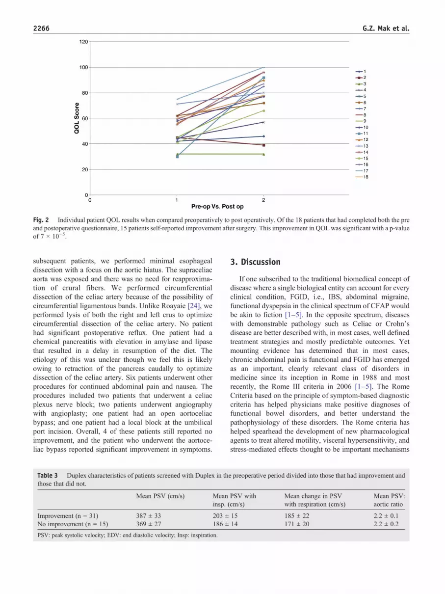
Fig. 2 Individual patient QOL results when compared preoperatively to post operatively. Of the 18 patients that had completed both the preand postoperative questionnaire, 15 patients self-reported improvement after surgery. This improvement in QOL was significant with a p-valueof 7 × 10⁻⁵.
2266 G.Z. Mak et al.
subsequent patients, we performed minimal esophagealdissection with a focus on the aortic hiatus. The supraceliacaorta was exposed and there was no need for reapproxima-tion of crural fibers. We performed circumferentialdissection of the celiac artery because of the possibility ofcircumferential ligamentous bands. Unlike Roayaie [24], weperformed lysis of both the right and left crus to optimizecircumferential dissection of the celiac artery. No patienthad significant postoperative reflux. One patient had achemical pancreatitis with elevation in amylase and lipasethat resulted in a delay in resumption of the diet. Theetiology of this was unclear though we feel this is likelyowing to retraction of the pancreas caudally to optimizedissection of the celiac artery. Six patients underwent otherprocedures for continued abdominal pain and nausea. Theprocedures included two patients that underwent a celiacplexus nerve block; two patients underwent angiographywith angioplasty; one patient had an open aortoceliacbypass; and one patient had a local block at the umbilicalport incision. Overall, 4 of these patients still reported noimprovement, and the patient who underwent the aortoce-liac bypass reported significant improvement in symptoms.
Table 3 Duplex characteristics of patients screened with Duplex in ththose that did not.
Mean PSV (cm/s) Meaninsp. (
Improvement (n = 31) 387 ± 33 203 ±No improvement (n = 15) 369 ± 27 186 ±
PSV: peak systolic velocity; EDV: end diastolic velocity; Insp: inspiration.
3. Discussion
If one subscribed to the traditional biomedical concept ofdisease where a single biological entity can account for everyclinical condition, FGID, i.e., IBS, abdominal migraine,functional dyspepsia in the clinical spectrum of CFAP wouldbe akin to fiction [1–5]. In the opposite spectrum, diseaseswith demonstrable pathology such as Celiac or Crohn'sdisease are better described with, in most cases, well definedtreatment strategies and mostly predictable outcomes. Yetmounting evidence has determined that in most cases,chronic abdominal pain is functional and FGID has emergedas an important, clearly relevant class of disorders inmedicine since its inception in Rome in 1988 and mostrecently, the Rome III criteria in 2006 [1–5]. The RomeCriteria based on the principle of symptom-based diagnosticcriteria has helped physicians make positive diagnoses offunctional bowel disorders, and better understand thepathophysiology of these disorders. The Rome criteria hashelped spearhead the development of new pharmacologicalagents to treat altered motility, visceral hypersensitivity, andstress-mediated effects thought to be important mechanisms
e preoperative period divided into those that had improvement and
PSV withcm/s)
Mean change in PSVwith respiration (cm/s)
Mean PSV:aortic ratio
15 185 ± 22 2.2 ± 0.114 171 ± 20 2.2 ± 0.2

Table 4 Duplex characteristics of patients screened with Duplex in the postoperative period divided into those that had improvement andthose that did not.
Mean PSV (cm/s) Mean PSV withinsp. (cm/s)
Mean change in PSV withrespiration (cm/s)
Mean PSV:aortic ratio
Improvement (n = 31) 229 ± 14 179 ± 10 39 ± 10 1.3 ± 0.1No improvement (n = 15) 248 ± 26 182 ± 15 59 ± 22 1.6 ± 0.1
PSV: peak systolic velocity; EDV: end diastolic velocity; Insp: inspiration.
2267Median arcuate ligament syndrome
in the pathogenesis of FGID. Although many of thesetreatment trials are promising, definitive statements concern-ing therapeutic efficacy are still limited in large part owing toyet to be established guidelines for clinical trial research. Yetbillions of dollars per year continue to be spent on frequentphysician visits, costly medications, tests and procedures, notto mention the accumulation of lost days at school and workfor those with CFAP and their caregivers [1,2]. This does noteven take into account the effects on these patients' lives forwhich no price can be placed.
Celiac compression or MALS, similar to FGID occurstypically in young females with CFAP and share similarsymptoms of postprandial, midepigastric pain, and otherconstitutional symptoms such as nausea, vomiting, andoccasional diarrhea. Some 40 years after it was firstdescribed, MALS remains a controversial and vexingcondition. The link between symptoms and narrowing ofthe celiac axis, celiac nerve plexus, or both remainsunknown. Most suggest an ischemic origin where occlusionof the axis would lead to intestinal angina. Alternativehypotheses include a steal phenomena resulting in a water-shed effect on the celiac plexus, thus producing pain; and/ordisruption of neuroenteric pain pathways affecting visceralhypersensitivity mediated through the celiac ganglion, thelatter perhaps similar to the pathogenesis of FGID. Critics ofMALS have suggested that occlusion of both the celiac andsuperior mesenteric artery would be necessary to produceclassic intestinal angina and have pointed out that the fewreports of long-term follow-up after celiac decompressionhave often been disappointing [26].
The bulk of MALS literature has been case reportsincluding one from our own institution in 1994 of MALS in aset of 27-year old monozygotic twin females with diagnosisof chronic abdominal pain [27]. What is striking is that twokey features of MALS, young female predominance and itsassociated GI symptoms, overlap with those of CFAP [1,2].Given this overlap, we hypothesized that perhaps a subset ofpatients, most likely young females with chronic abdominalpain previously diagnosed with FGID may actually haveMALS instead. The two conditions may not be mutuallyexclusive as evidenced by the known coexistence of FGIDwith GI diseases of demonstrable pathology, like Crohn's.Our initial duplex scans of these patients with CFAPrevealed a cohort with a positive screen for MALS.Substantiating the diagnosis was a significant decrease inPSV with deep inhalation where downward excursion of the
diaphragm to decompress the celiac axis correlated withimprovement in velocities. Importantly, 67% of the patientshad overall improvement of symptoms after they hadundergone a release of the celiac artery, raising a questionabout the initial diagnosis of CFAP.
For control purposes to highlight celiac pathology,normal PSV were demonstrated in the aorta, and neighbor-ing truncal vessels. While surgery in this area hasconsiderable associated risks including injury to the aorta,bowel, pancreas, esophagus, stomach, portal structures andliver, our approach has shown minimal morbidity andmortality. Surgery was only considered complete uponobjective normalization of respiratory variation and/ornormalization of the flow velocities often requiring tediousdissection. In our cohort of patients, we were able tosignificantly improve all hemodynamic variables with ourlaparoscopic approach. This correlated with an objectivelyimproved quality of life based on total PedsQL™ scores.Because our response rate to PedsQL™ was only 55% wewanted to objectively document success or failure somedical records were reviewed and attempts at contactingthe patients were made. If the last medical recorddocumented continued pain and the patient was unreachable,we documented this as no improvement. With this protocol,we found patient reported improvement since the time ofsurgery in 31 of the 46 patients or 67% of patients. Althougha success rate of 67% seems low, when considered within thecontext of these patients previously being labeled withFGID, we feel that there may be a surgical benefit with therelease of the celiac artery in an appropriately selectedpatient population of FGID.
As to the causes for failure to improve, there may beseveral. First, it is possible that these patients are truly CFAPwith concomitant MALS. Second, there may be a componentof nerve compression that was inadequately released at thetime of surgery. For these patients, celiac plexus block wasused as a last resort. Unfortunately, no improvement wasseen in the two patients who had continued pain andnormalization of velocities. Whether this was owing to afailed celiac plexus block or CFAP is unclear. Thirdly, wefeel that there is a strong correlation between chronicphysical pain and psychological pain. For this reason, wehave made psychiatric and chronic pain screening and followup a requirement for all patients. If there are any concerns inthe preoperative setting that may indicate psychiatric therapyas a benefit, we try to initiate this as soon as possible prior to

2268 G.Z. Mak et al.
surgery. We hope to report on this pathway in the near future.Finally, there is a possibility of fixed residual stenoses afterexternal decompression that results in continued ischemia.These patients were found to have increased velocities onrepeat duplex postoperatively. The idea that the laparoscopicapproach in adolescents is limited by the inability toreconstruct the artery was highlighted by Said et al [28]and we have adapted the approach of surgical decompressionfollowed by either endovascular or open reconstruction hashighlighted by Rosborough [29]. No patient had anintraoperative conversion for bleeding. A total of threepatients in our study underwent adjunct vascular procedures:one patient underwent balloon angioplasty for a residual webwith excellent results and resolution of pain; the secondpatient had a web with a kink and was recommended to havea vascular reconstruction in conjunction with ongoingpsychiatric counseling and this case continues; the thirdpatient underwent a celiac reconstruction and reportsresolution of symptoms. We are encouraged by our resultsthat the majority of our adolescent patients have improvedpain and QOL with just the laparoscopic release alonewithout adjunct vascular reconstruction. The excellentresults reported by Roseborough [29] reported that ninevascular interventions were performed in six of the 15patients. Given the self reported 85% improvement and the67% survey reported improvement of QOL in our study, wefeel that three vascular interventions out of 45 patients mayreflect that earlier intervention during adolescence may resultin less fixed stenoses and a decreased need for adjunctvascular reconstruction. We are cautious in stating thisbecause this study was not designed to look at fixed stenosesand we did note a less than perfect compliance rate.Furthermore, in two of these patients, there was arterialkinking that resulted post release and would not be addressedby endovascular approaches.
As our experience grew, we noted trends and developedconcerns about the compliance rate to the PedsQL™survey as well as the recurring fact that these patients werelabeled as “difficult patients”. We felt that there may beseveral reasons why the compliance was low: first, thebroad demographic location of many patients meantrelying on patients to return surveys; second, for thosepatients that had no improvement, there was no benefit infilling out the survey; third, for those patients that hadimprovement, they could now move on and were notthinking about the survey; and finally, the age group itselfmakes compliance challenging.
Given the midrange compliance, high complexity of thepatients, the young age, and the multifactorial etiologies ofpain, we have since assembled a multidisciplinary team toaddress all the needs of the patient. We have assembledexperts in child Psychiatry and eating disorders; painmanagement; GI; Radiology (both CT and interventional);Pediatric and Vascular surgery. The multidisciplinary teamapproach has provided a major advantage in our ability toselect patients who are appropriate candidates for surgery.
Reilly et al. [30] published their series of 51 patients anddemonstrated sustained relief in patients with postprandialpain, age between 40–60, and weight loss of 20 pounds ormore. These parameters unfortunately are not all transferra-ble to the adolescent population. We did observe that patientswith postprandial pain tended to have improvement in thepain compared to patients with nausea. To overcome thisdifficulty in determining who will benefit from an operation,we have come to rely on the input opinion from each memberof the team. If any member feels that surgery is not indicated,or should be delayed we will not proceed down a surgicalpathway until we have addressed any confounding issue. Wecarry this approach into the operating room where both thepediatric surgeon and vascular surgeon are present. Initiallaparoscopy and dissection of the diaphragmatic crura areperformed by the pediatric surgeon and the aortic and celiacdissection is performed by the vascular surgeon. Given thecomplexity of this disease process, we have becomehypervigilant about safety and feel that we need to beready to convert to open immediately; therefore, werecommend this combined service approach. We havefound that this multidisciplinary approach throughout thecourse of the clinical pathway is patient centered andcomprehensive with respect to evaluation and execution ofthe treatment plan.
This report suffers from several limitations inherent to thedescriptive study design. There is an inherent selection biasas we are a tertiary care hospital and found that many of thepatients sought us out because of the persistence of pain andlack of a specific diagnosis. Because of this self-referralpattern, we were somewhat limited in follow up. To the bestof our ability we attempted QOL follow up and patientreported improvement of abdominal pain, but as such arelimited in the long term weight gain, pain medication use anduse of GI medications. Furthermore, we realize that despiteour best attempts, 28 of the patients did not complete the fullquestionnaires which results in selection bias.
We realize that radiation exposure in this youngpopulation is of great concern. The gold standard fordiagnosis of MALS is invasive angiography with measure-ments of pressure gradients. However, we felt that thecombination of duplex ultrasound and a CTA withinspiratory and expiratory phases would give us the requireddata to support the duplex ultrasound. We initially choseCTA because magnetic resonant angiography has a tendencyto have inadequate spatial resolution and movement artifacts[31,32]. Additionally, there is very little literature on the useof MRA in the pediatric population particularly in visuali-zation of the celiac artery. The literature that is available doesnote that despite the fact that there is no exposure to ionizingradiation, there are risks from contrast agents as well as thefrequent requirement of sedation or general anesthesia [33].Although we still use CTA, we have been moving towardmore frequent utilization of MRA. We also realize thatlonger follow-up is necessary to fully understand theramifications of the surgery.

2269Median arcuate ligament syndrome
Overall, we believe that our results demonstrate anoverlooked diagnosis of MALS in pediatric females withCFAP. The finding of young female predominance appearsnoncoincidental and especially relevant considering that thisis also a main feature of FGID. We are encouraged by ourearly surgical results after ligament release. Whether theclinical improvement seen is owing to restoration ofadequate celiac blood flow, positive alteration of yetundefined neuroenteric circuitry affecting visceral hypersen-sitivity through neurolysis, or both, remains an interestingquestion. Perhaps long term follow-up of a large pool ofMALS patients with FGID analyzing changes in symptomclustering patterns before and after surgery would helpanswer this question, and, more importantly, help gain betterunderstanding of the pathophysiology of FGID leading tomore targeted treatment trials.
In conclusion, MALS was more commonly diagnosed inpediatric females than males. Laparoscopic release of themedian arcuate ligament overlying the celiac artery can beperformed safely in the pediatric population with the releaseof the artery and resultant neurolysis, resulting in significantimprovement in the hemodynamics, symptoms, and overallQOL in this cohort. The overall improvement in QOLoutcome measures after surgery leads us to conclude thatMALS might be earlier diagnosed and possibly treated inpatients with CFAP. In the previously thought group ofchildren with the diagnosis of CFAP, MALS may be atreatable etiology with minimally invasive treatment. Wethus advocate increased awareness of the diagnosis ofMALS, appropriate screening for patients with possibleMALS, a multidisciplinary team approach for the care andselection of surgical candidates with MALS, and continuedstudy of these patients following surgery to better determinelong-term effects.
Acknowledgments
Diane Nilsson, University of Chicago Medicine; CynthiaChou MD, Lake Forest Pediatrics Associates; University ofChicago Medical Center Vascular Lab. The Quality of lifestudy described in this paper was carried out using thePedsQL™, developed by Dr. James W. Varni.
Dr. Skelly is funded by NIH K-08-HL091053 as wellby the American Vascular Association/American Collegeof Surgeons and NHLBI Jointly Sponsored MentoredClinical Scientist Development Award. The contents ofthis study are solely the responsibility of the authors anddo not necessarily represent the official views of theNHLBI or the NIH.
We particularly would like to express our gratitude tothe late Dr. Donald Liu who not only was one of theinnovators of this technique and the MALS program at theUniversity of Chicago, but also a valued mentor,colleague, and friend.
References
[1] Lehmann HP, Boyle JT, Gerson WT, et al. Clinical report: chronicabdominal pain in children. Pediatrics 2005;115:812-5.
[2] Di Lorenzo C, Colletti RB, Lehmann HP, et al. Chronic abdominal painin children: a technical report of the American Academy of Pediatricsand the North American Society for Pediatric Gastroenterology,Hepatology and Nutrition. J Ped Gastroenterol Nutr 2005;40:249-61.
[3] Drossman DA, Corazziari E, Talley NJ, et al. The functionalgastrointestinal disorders. Diagnosis, pathophysiology and treatment:a multinational consensus. 2nd ed. Degnon Associates: McLeanVirginia; 2000.
[4] Drossman DA. The functional gastrointestinal disorders and the RomeIII process. Gastroenterology 2006;130:1377-90.
[5] Thompson WG. The road to Rome. Gastroenterology 2006;130:1552-6.
[6] Drossman DA, Li Z, Andruzzi E, et al. U.S. householder survey offunctional gastrointestinal disorders: prevalence, sociodemography,and health impact. Dig Dis Sci 1993;38:1569-80.
[7] Koloski NA, Talley NJ, Boyce PM. Predictors of health care seekingfor irritable bowel syndrome and nonulcer dyspepsia: a critical reviewof the literature on symptom and psychological factors. Am JGastroenterol 2001;96:1340-9.
[8] Saito YA, Schoenfeld P, Locke GRI. The epidemiology of irritablebowel syndrome in North America: a systematic review. Am JGastroenterol 2002;97:1910-5.
[9] Longstreth GF, Wilson A, Knight K, et al. Irritable bowel syndrome,health care use and costs: a US managed care perspective. Am JGastroenterol 2003;98:600-7.
[10] Talley NJ, Gabriel SE, Harmsen WS, et al. Medical costs incommunity subjects with irritable bowel syndrome. Gastroenterology1995;109:1736-41.
[11] Martin R, Barron JJ, Zacker C C. Irritable syndrome: toward a cost-effective management approach. Am J Manag Care 2001;201(7):S268-75.
[12] Lembo A, Weber HC, Farraye FA. Alosetron in irritable bowelsyndrome: strategies for the use in a common gastrointestinal disorder.Drugs 2003;63:1895-905.
[13] Galligan JJ, Vanner S. Basic and clinical pharmacology of newmotility promoting agents. Neurogastroenterol Motil 2005;17:643-53.
[14] Sagami Y, Shimada Y, Tayama J, et al. Effect of corticotrophin-releasing hormone receptor antagonist on colonic sensory and motorfunction in patients with irritable bowel syndrome. Gut 2004;53:958-64.
[15] Kline RM, Kline JJ, Di Palma J, et al. Enteric-coated, pH-dependentpeppermint oil capsules for the treatment of irritable bowel syndromein children. J Pediatr 2001;138:125-8.
[16] See MC, Birnbaum AH, Schechter CB, et al. Double-blind, placebo-controlled trial of famotidine in children with abdominal pain anddyspepsia: global and quantitative assessment. Dig Dis Sci 2001;46:985-92.
[17] Symon DN, Russell G. Double-blind, placebo-controlled trial ofpizotifen syrup in the treatment of abdominal migraine. Arch Dis Child1995;72:48-50.
[18] Harjola PT. A rare obstruction of the coeliac artery. Ann Chir Gynaec1963;52:547.
[19] Dunbar JD, Molnar W, Beman FF, et al. Compression of the celiactrunk and abdominal angina. Am J Roentgenol Radium Ther Nucl Med1965;95(3):731-44.
[20] Harjola PT, Lahtiharju A. Celiac axis syndrome: abdominal anginacaused by external compression of the celiac artery. Am J Surg1968;115:864-9.
[21] Stanley JC, Fry WJ. Median arcuate ligament syndrome. Arch Surg1971;103(2):252-8.
[22] Loukas M, Pinyard J, Vaid S, et al. Clinical anatomy of celiac arterycompression syndrome: a review. Clin Anat 2007;20:612-7.

2270 G.Z. Mak et al.
[23] Watson WC. Coeliac-axis compression. Lancet 1977;2(8037):561-2.
[24] Roayaie S, Jossart G, Gitlitz D, et al. Laparoscopic release of celiacartery compression syndrome facilitated by laparoscopic ultrasoundscanning to confirm restoration of flow. J Vasc Surg 2000;32(4):814-7.
[25] Varni JW, Seid M, Rode CA. The PedsQL™: measurement model forthe pediatric quality of life inventory. Medical Care 1999;37(2):126-39.
[26] Evans W. Long-term evaluation of the celiac band syndrome. Surgery1974;76:867-71.
[27] Bech F, Loesberg A, Rosenblum J, et al. Median arcuate ligamentcompression in monozygotic twins. J Vasc Surg 1994;19(5):934-8.
[28] Said SM, Zarroug AE, Gloviczki P, et al. Pediatric median arcuateligament syndrome: first report of familial pattern and transperitoneallaparoscopic release. J Pediatr Surg 2010;45(12):E17-20.
[29] Roseborough GS. Laparoscopic management of celiac artery com-pression syndrome. J Vasc Surg 2009;50(1):124-33.
[30] Reilly LM, Ammar AD, Stoney RJ, et al. Late results followingoperative repair for celiac artery compression syndrome. J Vasc Surg1985;2(1):79-91.
[31] Dellegrottaglie S, Sanz J, Rajagopalan S. Technology insight: clinicalrole of magnetic resonance angiography in the diagnosis andmanagement of renal artery stenosis. Nat Clin Pract Cardiovasc Med2006;3(6):329-38.
[32] Leiner T, Schoenberg SO. Current status of renal artery magneticresonance imaging: theoretical and practical considerations and thepotential role of blood-pool contrast agents. Eur Radiol 2007;17(Suppl. 2):B13-7.
[33] Grist TM, Thornton FJ. Magnetic resonance angiography in children:technique, indications, and imaging findings. Pediatr Radiol 2005;35(1):26-39.


