Embed Size (px)
Citation preview

doi:10.1182/blood-2005-04-1739Prepublished online August 4, 2005;2005 106: 3396-3404
Christensen, Karsten Wessel Eriksen, Klaus Qvortrup, Niels Odum and Tord LabudaBarbara Bonnesen, Cathrine Orskov, Susanne Rasmussen, Peter Johannes Holst, Jan Pravsgaard mouse fetal liverMEK kinase 1 activity is required for definitive erythropoiesis in the
http://bloodjournal.hematologylibrary.org/content/106/10/3396.full.htmlUpdated information and services can be found at:
(1930 articles)Signal Transduction � (3131 articles)Hematopoiesis and Stem Cells �
Articles on similar topics can be found in the following Blood collections
http://bloodjournal.hematologylibrary.org/site/misc/rights.xhtml#repub_requestsInformation about reproducing this article in parts or in its entirety may be found online at:
http://bloodjournal.hematologylibrary.org/site/misc/rights.xhtml#reprintsInformation about ordering reprints may be found online at:
http://bloodjournal.hematologylibrary.org/site/subscriptions/index.xhtmlInformation about subscriptions and ASH membership may be found online at:
Copyright 2011 by The American Society of Hematology; all rights reserved.Washington DC 20036.by the American Society of Hematology, 2021 L St, NW, Suite 900, Blood (print ISSN 0006-4971, online ISSN 1528-0020), is published weekly
For personal use only. by guest on June 2, 2013. bloodjournal.hematologylibrary.orgFrom

HEMATOPOIESIS
MEK kinase 1 activity is required for definitive erythropoiesis in themouse fetal liverBarbara Bonnesen, Cathrine Orskov, Susanne Rasmussen, Peter Johannes Holst, Jan Pravsgaard Christensen,Karsten Wessel Eriksen, Klaus Qvortrup, Niels Odum, and Tord Labuda
Mitogen-activated protein kinase/extracel-lular signal to regulated kinase (MEK)kinase 1 (MEKK1) is a c-Jun N-terminalkinase (JNK) activating kinase known tobe implicated in proinflammatory re-sponses and cell motility. Using micedeficient for MEKK1 kinase activity(Mekk1�KD ) we show a role for MEKK1 indefinitive mouse erythropoiesis. AlthoughMekk1�KD mice are alive and fertile on a129 � C57/BL6 background, the fre-
quency of Mekk1�KD embryos that de-velop past embryonic day (E) 14.5 isdramatically reduced when backcrossedinto the C57/BL6 background. At E13.5,Mekk1�KD embryos have normal morphol-ogy but are anemic due to failure ofdefinitive erythropoiesis. When Mekk1�KD
fetal liver cells were transferred to lethallyirradiated wild-type hosts, mature redblood cells were generated from the mu-tant cells, suggesting that MEKK1 func-
tions in a non–cell-autonomous manner.Based on immunohistochemical and he-moglobin chain transcription analysis,we propose that the failure of definitiveerythropoiesis is due to a deficiency inenucleation activity caused by insuffi-cient macrophage-mediated nuclear DNAdestruction. (Blood. 2005;106:3396-3404)
© 2005 by The American Society of Hematology
Introduction
Production of red blood cells (erythropoiesis) is an essential step invertebrate embryogenesis making it possible to advance fromdiffusion-restricted to circulation-assisted growth. In the mouseembryo, primitive erythropoiesis is initiated in the yolk sac aroundembryonic day (E) 7.5. Yolk sac–derived erythrocytes are large,nucleated cells that do not complete the final stages of normal adulterythropoiesis.1 Around E9.5 erythropoiesis is initiated in the fetalliver2,3; concurrently, the yolk sac gradually becomes less impor-tant for erythropoiesis and around E14.5 90% of the erythrocytespresent in circulation are of fetal liver origin.4,5 The generation ofmature erythrocytes involves the commitment of pluripotent hema-topoietic stem cells progressing through erythroid blast-formingunit (BFU-E) and erythroid colony-forming unit (CFU-E) progeni-tor-cell stages, proerythroblast and several erythroblast stages,ultimately terminating into mature enucleated erythrocytes. More-over, the development of fetal liver erythropoiesis is considered toproceed through 2 distinct phases: the first phase (E11-13), inwhich erythroid maturation proceeds in the absence of macro-phages, and the second phase, involving the establishment oferythroblastic islands consisting of a central macrophage sur-rounded by erythroblasts.6 The central macrophages of the erythro-blastic islands have been suggested to induce several key biologicprocesses in the erythroid precursors including proliferation anddifferentiation, prevention of apoptosis, and promotion of erythro-
blast enucleation.7 Furthermore, recent studies suggest that failureof definitive erythropoiesis in deoxyribonuclease (DNase) II–deficient mice is due to impaired erythroblast enucleation, causedby inability of the central macrophages to digest nuclear DNAexpelled from erythroblasts.8,9
The conserved mitogen activated protein kinase (MAPK)family members c-Jun N-terminal kinase (JNK) and p38 have beenimplicated in stress and proinflammatory signal transduction andrecently in erythropoiesis.10-12 A number of cytokines are involvedin the erythropoiesis, including erythropoietin and stem-cell factor(SCF),13,14 and it has been shown that erythropoietin inducessustained JNK activity and supports proliferation of erythropoietin-dependent cell lines12; additionally, p38 seems to be required forposttranscriptional regulation of erythropoietin expression.10 Ho-mozygous deletion of either JNK1 or JNK2 in mice has no apparenteffect on hematopoiesis,15,16 which might be explained by redun-dancy between the JNK isoforms. In contrast, the Jnk1�/�Jnk2�/�
embryos die early in embryonic development (E11.5-12.5) due todysregulation of apoptosis in brain development, making thecontribution of the JNK kinases in definitive erythropoiesis in vivodifficult to study further. We have investigated the role of the JNK-activating kinase mitogen-activated protein kinase/extracellularsignal to regulated kinase (MEK) kinase 1 (MEKK1) in develop-ment. Mice deficient in MEKK1 kinase activity (Mekk1�KD mice)
From the Institute of Molecular Biology and Physiology, Department ofImmunology, the Department of Medical Anatomy, the Department of MedicalPharmacology, and the Department of Medical Microbiology and Immunology,University of Copenhagen, Denmark.
Submitted April 28, 2005; accepted July 3, 2005. Prepublished online as BloodFirst Edition Paper, August 4, 2005; DOI 10.1182/blood-2005-04-1739.
Supported in part by The Danish Medical Research Councils (SSVF), TheAlfred Benzon Foundation, The Johann Weiman F. Seedorff and WifeFoundation (Købmand i Odense Johann og Hanne Weimann f. Seedorff’sLegat), The Leo Foundation, The Willumsen Foundation (Fabrikant EinarWillumsens Mindelegat), The Danielsen Foundation (Aase og EjnarDanielsens Fond), The Magnus Bergvall Foundation (Magnus Bergvalls
Stiftelse), the Else and Mogens Wedell-Wedellborgs Foundation, The FraenkelFoundation (Eva og Henry Fraenkels Mindefond), and the University ofCopenhagen.
The online version of the article contains a data supplement.
Reprints: Tord Labuda, Department of Medical Microbiology and Immunology,IMMI 22.5, The Panum Institute, Blegdamsvej 3c, DK-2200 N Copenhagen,Denmark; e-mail: [email protected].
The publication costs of this article were defrayed in part by page chargepayment. Therefore, and solely to indicate this fact, this article is herebymarked ‘‘advertisement’’ in accordance with 18 U.S.C. section 1734.
© 2005 by The American Society of Hematology
3396 BLOOD, 15 NOVEMBER 2005 � VOLUME 106, NUMBER 10
For personal use only. by guest on June 2, 2013. bloodjournal.hematologylibrary.orgFrom
were generated and are alive and fertile on a C57/BL6 � 129background.17,18 Unexpectedly, when backcrossing the Mekk1�KD/�
heterozygotes into the C57/BL6 background, we observed adramatic decrease in the frequency of live Mekk1�KD embryos thatdevelop past E14.5. At E14.5 all mutant embryos studied, althoughmorphologically normal, were anemic and showed defectivedefinitive erythropoiesis with accumulation of nucleated lateerythroblasts.
Materials and methods
Mice
Mekk1�KD, Jnk1�/�, and Jnk2�/� mice have been described previ-ously.15,18,19 The Mekk1�KD, Jnk1�/�, and Mekk1�KD Jnk2�/� mice wereobtained by crossing Mekk1�KD/� (C57BL/6 � 129/F1) with Jnk1�/�
(C57BL/6) or Jnk2�/� (C57BL/6) mice. Mekk1�KD/� (C57BL/6 � 129/F1)were backcrossed to C57BL/6 for 8 generations. Mouse tail DNA wasprepared from approximately 1-mm tail snips of embryos and 4-week-oldprogeny. Genotyping was performed by polymerase chain reaction (PCR)analysis of tail- and embryo-derived DNA.
Tissue staining
For endothelial stainings, E13.5 sections of acetone-fixed, paraffin-embedded mouse embryos were stained with MECA-32 Rat IgG2a (B&DBiosciences, San Jose, CA) against mouse panendothelial-cell antigenfollowed by biotin antirat antibody, and enhanced using the TSA system asdescribed by the manufacturer (NEN, Boston, MA). For macrophagestainings, raw frozen sections of wild-type (wt) and Mekk1�KD embryosfrom E13.5 were stained with phycoerythrin (PE)–conjugated F4/80(Caltag, Burlingame, CA) and afterward fixed with 4% paraformaldehyde.
Frozen sections of wt and Mekk1�KD mice from E13.5 were used fordetection of apoptosis using the terminal deoxynucleotidyl transferase(TdT)–mediated deoxyuridine triphosphate (dUTP) digoxigenin nick end-labeling (TUNEL) staining method. Briefly, sections were boiled in 0.1 Mcitrate buffer (pH 6.0) in a microwave oven for 2 minutes to permeabilizeapoptotic cells. After cooling down, slides were incubated in 0.1 MTris-HCl, 20% bovine serum albumin (BSA), 3% fetal porcine serum, for30 minutes at room temperature and immersed in the TUNEL reactionmixture (Roche, Mannheim, Germany) for 1 hour at 37°C in a humidifiedchamber. Positive cells were visualized in a fluorescence microscope.Slides were viewed using an Axioskop 2 microscope equipped with PlanNEOFLUAR 10�/0.30 or 40�/0.75 objective lenses (Zeiss, Oberkochen,Germany). Images were captured with a ColorSnap digital camera (Photo-metrics, Tucson, AZ), and were processed using Image Pro Plus software(Media Cybernetics, Silver Spring, MD) and Adobe Photoshop 7.0 software(Adobe, San Jose, CA).
Reconstitution of hematopoiesis in lethally irradiated mice
Fetal liver (FL) cells from wt, Mekk1�KD, Jnk2�/�, or MEKK Jnk2�/� E14.5embryos were used to reconstitute lethally irradiated B6.SJL (CD45.1)female mice, exposed to 950 rad of � irradiation. A total number of 1 � 106
FL cells were injected into the tail vein of the host. Reconstitution ofhematopoiesis (red blood cell [RBC] quantity, hematocrit value, hemoglo-bin concentration, mean corpuscular volume of erythrocytes, white bloodcell [WBC] quantity, neutrophil, lymphocyte, monocyte, macrophage,eosinophil, and basophil percentages of total WBCs as well as plateletquantity) was examined with an automatic cell analyzer (Sysmex SF-3000;Kobe, Japan) every 2 weeks after reconstitution, and flow cytometricanalysis was performed 12 weeks after reconstitution.
Flow cytometry
Single-cell suspensions of wt, Mekk1�KD, Jnk2�/�, or Mekk1 Jnk2�/� fetallivers were obtained at E13. For analysis of reconstituted mice, peripheralblood was collected from retro-orbital veins, and lysis of erythrocytes was
performed. Staining of cells for flow cytometry was performed in 0.5%BSA in phosphate-buffered saline (PBS) using the following monoclonalantibodies: fluorescein isothiocyanate (FITC)–conjugated anti-CD44 (IM7),anti-CD34 (RAM34) PE, anti–c-kit (CD117) FITC, anti-Ter119 PE,anti-CD45.2 FITC, anti-CD45.1 PE, anti–Gr-1 PE, anti-CD11b (Mac1)PerCP-Cy5.5, anti-CD24 (HSA) biotin, anti-CD4 PE, and anti-CD43 PE(all from B&D Biosciences). Streptavidin TriColor (TC), anti-CD8 TC, andanti-B220 TC were from Caltag. Cell-surface expression of the differentmarkers was analyzed in a Becton Dickinson FACScan Calibur usingCellQuest Pro (B&D Biosciences) software; a minimum of 20 000 cellswere counted.
RNA analysis
Total RNA was isolated from livers of wt and Mekk1�KD, Jnk2�/�, orMekk1�KD Jnk2�/� E13.5 embryos using the TRI REAGENT (MolecularResearch Center, Cincinnati, OH) purification method as recommended bythe manufacturer. For cDNA synthesis, 1 �g total RNA was converted tocDNA using RevertAid First Strand cDNA Synthesis Kit (Fermentas) asrecommended by the manufacturer. RNase protection assay was performedas previously described20 using an mCK-4 Multi-Probe Template Set fromBD Pharmingen (San Jose, CA). Band volume analysis was performedusing a Typhoon 9410 variable mode imager and ImageMaster Totallab 2.0software (Amersham Pharmacia Biotech, Piscataway, NJ). The concentra-tions of stem-cell factor (SCF), interleukin-3 (IL-3), IL-11, IL-7, GM-CSF,M-CSF, G-CSF, LIF, or IL-6 were normalized relative to those ofglyceraldehyde-3-phosphate dehydrogenase (GADPH) and L32.
Quantitative PCR
Quantitative PCR–based measurement of RNA abundance was carried outusing gene-specific double fluorescent–labeled probes and the BrilliantQPCR Master Mix (Stratagene, La Jolla, CA), which uses ROX (Rhoda-mine-X) passive reference dye to normalize for non-PCR–related fluores-cence signal variation. 6-FAM (5-carboxyfluorescein) or in the case ofGADPH, HEX (4,7,2�,4�,5�,7�-hexachloro-6-carboxyfluorescein) was the5� fluorescent reporter, and Black Hole Quencher (BHQ-1) was added to the3� end as a quencher. The following primer and probe sequences were used:Mgadph-571 forward primer (F), 5�-CAATGTGTCCGTCGTGGA-3�;mGADPH-654 reverse primer (R), 5�-GATGCCTGCTTCACCACC-3�;mGADPH-599 probe, 5�-CGCCTGGAGAAACCTGCCAAGTAT-3�,mHg�-105F, 5�-TAGCTTCCCCACCACCAA-3�; mHg�-183R, 5�-CTTGC-CGTGACCCTTGAC-3�; mHg�-133 probe, 5�-CTTCACATTTGATGTA-AGCCACGGC-3�; mHg-327F, 5�-TGCGATCGTGATTGTGCT-3�;mHg-435R, 5�-CTTGTGAGCCAGGGCAGT-3�; mHg-390 probe, 5�-CTTCCAGAAGGTGGTGGCTGGAGT-3�; mHg-157F, 5�-TTCTTAAC-CCCCCAAGCCC-3�; mHg-233R, 5�-TTCTTAACCCCCAAGCCC-3�;mHg-182 probe, 5�-TTAGAGCCCATGGCAAGAAAGTGC-3�; mEPO-61F, 5�-GGCCTCCCAGTCCTCTGT-3�; mEPO-168R, 5�-TGCACAAC-CCATCGTGAC-3�; mEPO-95 probe; 5�-TCTGCGACAGTCGAGTTCTG-GAGA-3�; mSCF-616F, 5�-AAGGCCCCTGAAGACTCG-3�; mSCF-701R, 5�-GCTCCAAAAGCAAAGCCA-3�; mSCF-636 probe, 5�-CCT-ACAATGGACAGCCATGGCATT-3�.
PCR was performed at 95°C for 10 minutes and then cycles at 95°C for30 seconds, 55°C for 1 minute, and 72°C for 30 seconds, on an Mx3000Preal-time PCR system machine (Stratagene).
Results were analyzed as: relative gene transcription � 2(efficiencygene of interest / ct gene of interest � efficiency GADPH / ct GADPH),where ct indicates cycle threshold.
Macrophage phagocytosis assay
FL cells were isolated from E13.5 Jnk2�/�, Mekk1�K/� Jnk2�/�, andMekk1�KD Jnk2�/� embryos, and were resuspended in complete medium(RPMI plus 10% fetal bovine serum [FBS]) and allowed to attach to plastic(12-well dishes) for 90 minutes at 37°C in 5% CO2. Adherent macrophages(obtained by washing wells in RPMI to remove nonadherent cells) wereoffered an excess of latex beads (Sigma, St Louis, MO) and furtherincubated in complete medium for 3 hours. Macrophages were washed in
FETAL LIVER ERYTHROPOIESIS REQUIRES MEKK1 3397BLOOD, 15 NOVEMBER 2005 � VOLUME 106, NUMBER 10
For personal use only. by guest on June 2, 2013. bloodjournal.hematologylibrary.orgFrom

RPMI and stained with hematoxylin. The number of phagocytosed beadsper cell were determined by phase-contrast microscopy.
Reconstitution of FLM-erythroblast clusters
Clusters composed of fetal liver macrophages (FLMs) and erythroblastswere isolated as described.21 Briefly, FLs from wt or Mekk1�KD E13.5embryos were placed in prewarmed 0.05% collagenase (Sigma) and0.002% DNase (Sigma) in RPMI and digested for 1 hour at 37°C withgentle rotation. After tissue dissociation, cells were washed 3 times,suspended in 300 �L complete medium (RPMI plus 10% FBS) and allowedto attach to glass coverslips for 20 minutes at 37°C in 5% CO2. Cells werethen flooded with medium and incubated for an additional 4 hours.Adherent macrophages with attached erythroid clusters were obtained bywashing coverslips in RPMI to remove nonadherent cells (“native”clusters) before staining with F4/80-PE (Caltag) and biotin-labeled Ter119antibody (Caltag) followed by visualization using the tyramide amplifica-tion system as recommended by the manufacturer, and streptavidinecoupled to Alexa 488 (Molecular Probes, Eugene, OR).
Results
Mekk1�KD and Mekk1�KD Jnk2 �/� embryos die in midgestationand are anemic
To identify at which developmental stage embryos deficient inMEKK1 kinase activity die, the phenotypes and genotypes ofoffspring from Mekk1�KD/� intercrosses (backcrossed into C57/BL6 for 8 generations) were examined between E8.5 and E18.5.Live Mekk1�KD embryos at Mendelian frequencies were detectedup to midgestation (Table 1). Thereafter, the frequency wasconsiderably lower than expected. To confirm that the effect ofMEKK1 in development is mediated through JNK, we crossed theMekk1�KD to JNK1-deficient (Jnk1�/�) and JNK2-deficient(Jnk2�/�) mice.
Although a small number of Mekk1�KD mice survived toadulthood, the double-deficient mice (Mekk1�KD Jnk2�/� andMekk1�KD Jnk1�/�) all died during embryonic development(Table 1). The phenotype of Mekk1�KD Jnk1�/� and Mekk1�KD
Jnk2�/� was identical to that of the Mekk1�KD embryos (Figure 1,data not shown). However, as the breeding efficiency of theMekk1�KD/� Jnk1�/� intercrosses was very low, only limitedstudies were performed on these mice. The majority of thework in this study was performed on Mekk1�KD and Mekk1�KD
Jnk2�/� mice.
At E14.5, live Mekk1�KD and Mekk1�KD Jnk2�/� embryos,macroscopically, were paler than wt counterparts and their majorblood vessels lacked color (Figure 1A). The FL was smaller,hypocellular, and liver necrosis as well as erythroplaki of the head
Table 1. Genotypes of live offspring from Mekk1�KD/� intercrosses, Mekk1�KD/� Jnk2�/� intercrosses, and Mekk1�KD/� Jnk1�/� intercrosses
Stage
MEKK1 genotype JNK2 genotype JNK1 genotype
Total(no. oflitters) Wt (%)
Mekk1�KD/�
(%)Mekk1�KD
(%)
Total(no. oflitters)
Jnk2�/�
(%)
Jnk2�/�
Mekk1�KD/�
(%)
Jnk2�/�
Mekk1�KD
(%)
Total(no. oflitters)
Jnk1�/�
(%)
Jnk1�/�
Mekk1�KD/�
(%)
Jnk1�/�
Mekk1�KD
(%)
E8.5 9 (1) 1 (11) 7 (78) 1 (11) ND ND ND ND ND ND ND ND
E10.5 9 (1) 3 (33) 4 (44) 2 (22) 6 (1) 1 (17) 3 (50) 2 (33) ND ND ND ND
E12.5 ND ND ND ND 53 (6) 13 (25) 28 (53) 12 (23) ND ND ND ND
E13.5 68 (12) 18 (26) 29 (43) 21 (31) 78 (12) 23 (29) 40 (51) 15 (19) 11 (2) 1 (9) 7 (63) 3 (27)
E14.5 125 (19) 38 (30) 60 (48) 27 (22) 75 (12) 31 (41) 29 (39) 15 (20) 6 (2) 0 (0) 5 (83) 1 (17)
E15.5 70 (9) 17 (24) 47 (67) 6 (9) 41 (7) 7 (17) 28 (68) 6 (15) 8 (2) 3 (37) 5 (62) 0 (0)
E16.5 22 (3) 8 (36) 13 (59) 1 (5) 16 (3) 4 (25) 11 (69) 1 (6) ND ND ND ND
E17.5 18 (3) 5 (28) 12 (67) 1 (5) 36 (5) 10 (28) 26 (72) 0 (0) ND ND ND ND
E18.5 13 (2) 4 (31) 9 (69) 0 (0) ND ND ND ND 3 (1) 1 (33) 2 (66) 0 (0)
Postnatal 217 (42) 78 (36) 135 (62) 4 (2) 291 (57) 115 (40) 176 (60) 0 (0) 30 (11) 11 (37) 19 (63) 0 (0)
Surviving fetuses were defined as those with beating heart at the time of dissection. Unless noted otherwise, data are expressed as the number of embryos, withpercentages within parenthesis.
ND indicates not determined.
Figure 1. Mekk1�KD and Mekk1�KD Jnk2�/� embryos are anemic and exhibit astriking increase in the number of nucleated erythrocytes. (A) E14.5 wt,Mekk1�KD, Jnk2�/�, and Mekk1�KD Jnk2�/� mouse embryos (Theiler stages 1, 2).Note the absence of color in major blood vessels, the general paleness, and thesmaller liver of the Mekk1�KD and Mekk1�KD Jnk2�/� embryos. Also note theerythroplaki of the Mekk1�KD Jnk2�/� embryo. (B) Sections through major bloodvessels of wt and Mekk1�KD (E15.5), and Jnk2�/� and Mekk1�KD Jnk2�/� (E14.5)embryos stained with H&E. Note the almost complete absence of enucleatederythrocytes in the Mekk1�KD and Mekk1�KD Jnk2�/� embryos. Bar � 75 �m. (C)Subcutaneous sections of wt, Mekk1�KD (E15.5), Jnk2�/�, and Mekk1�KD Jnk2�/�
(E14.5) embryos stained with H&E. Note marked decrease in the number ofcirculating erythrocytes in the Mekk1�KD and Mekk1�KD Jnk2�/� samples. Bar � 100�m. (D) Subcutaneous panendothelium stained sections of E13.5 wt, Mekk1�KD,Jnk2�/�, and Mekk1�KD Jnk2�/� embryos. Bar � 100 �m.
3398 BONNESEN et al BLOOD, 15 NOVEMBER 2005 � VOLUME 106, NUMBER 10
For personal use only. by guest on June 2, 2013. bloodjournal.hematologylibrary.orgFrom

and neck region was occasionally observed. In order to establishthe cause of death, we performed various histologic and immuno-histochemical analysis of E14.5 to E16.5 Mekk1�KD and Mekk1�KD
Jnk2�/� embryos. Histologic examination of wt, Mekk1�KD, andMekk1�KD Jnk2�/� fetuses revealed a marked increase in thenumber of nucleated erythrocytes in circulation. While bloodvessels of wt and Jnk2�/� embryos contained mostly enucleatederythrocytes; the erythrocytes found in blood vessels of Mekk1�KD
and Mekk1�KD Jnk2�/� embryos were mostly nucleated and veryfew mature red blood cells were detected (Figure 1B; Table 2). Thealtered blood picture correlated with a decrease in the total amountof circulating erythrocytes in Mekk1�KD and Mekk1�KD Jnk2�/�
embryos (Figure 1C). These data show that Mekk1�KD and Mekk1�KD
Jnk2�/� fetuses are severely anemic just prior to their death.The early lethality of the Jnk1�/�Jnk2�/� double-knockout
embryos makes the total contribution of JNK in hematopoiesisdifficult to study in vivo. Additionally, redundancy between theJNK isoforms may compensate for the loss of one gene or the other.Nevertheless, in order to investigate whether the relative contribu-tion of JNK1 or JNK2 became more apparent during conditions ofanemia, we examined reconstitution of hematopoiesis in Jnk1�/�
and Jnk2�/� mice 3 days after induction of severe anemia.However, Sysmex Counter Analysis revealed no differences com-pared with wt controls in any of the parameters investigated,including RBC quantity, hematocrit value, hemoglobin concentra-tion, and mean corpuscular volume of erythrocytes (SupplementalFigure S1A-D, available at the Blood website; click on theSupplemental Figures link at the top of the online article).Similarly, the WBC quantity, percentages of neutrophils, basophils,eosinophils, monocytes, and lymphocytes were indistinguishablebetween Jnk1�/�, Jnk2�/�, and wt mice (data not shown). Thesedata suggest that redundancy between the JNK isoforms maycompensate for the loss of either JNK1 or JNK2 also duringhematopoiesis.
p38-deficient mice are anemic and succumb in 2 separate wavesof embryonic lethality.10,22-24 A fraction of the embryos developpast E16.5 where they die due to failed erythropoiesis.10 However,the first critical time point occurs around E12.5. Two independentstudies22,23 have suggested that the early lethality is due to placentalinsufficiencies. Both studies noted an abnormal morphology withreduction in the labyrinthine layer and lack of interminglingembryonic and maternal blood vessels consistent with a lack ofnormal vascularization. We found no abnormalities in the morphol-ogy of placentas from Mekk1�KD or Mekk1�KD Jnk2�/� fetuses onE13.5. Gross morphology as well as closer examination revealednormal layer organization with fetal blood vessels penetrating into
the labyrinthine and spongiotrophoblast layers (SupplementalFigure S2A-B). Neither could we observe any differences inendothelial development between wt, Mekk1�KD, and Mekk1�KD Jnk2�/�
fetuses as judged by immunohistochemical staining (Figure 1D).
Defective definitive erythropoiesis in Mekk1�KD embryos
We examined FL hematopoiesis in E13.5 wt, Mekk1�KD, andMekk1�KD Jnk2�/� embryos by flow cytometry using markers thatare characteristic of different hematopoietic cell types and develop-mental stages, including CD34 (detecting hematopoietic progeni-tors), CD44 (detecting all hematopoietic cells), c-kit (hematopoi-etic stem cells), and Ter119 (erythroid cells).25-28
Analysis of FL single-cell suspensions revealed an increase inthe relative fraction of CD34lowc-kitpos cells (representing a popula-tion of hematopoietic stem cells capable of long-term reconstitu-tion)29 in Mekk1�KD as well as Mekk1�KD Jnk2�/� fetal livers ascompared with wt or Jnk2�/� FLs (Figure 2A). Fetal livers ofMekk1�KD and Mekk1�KD Jnk2�/� embryos also exhibited a mod-est increase in the relative abundance of CD34posc-kitneg cells(Figure 2A).
The relative overproduction of hematopoietic stem cells andprogenitors in Mekk1�KD and Mekk1�KD Jnk2�/� fetal livers couldbe a compensatory response to anemia. Indeed, Mekk1�KD andMekk1�KD Jnk2�/� fetal livers showed a substantial increase inTer119pos CD44neg cells (Figure 2A). Ter119pos cells are committederythropoietic precursors beyond the CFU-E stage25,27 and CD44expression is down-regulated during the final steps of erythroid-cell differentiation.26 These data support our histologic observa-tions (Figure 1B) and suggest that the larger population of Ter119pos
CD44neg cells observed in Mekk1�KD and Mekk1�KD Jnk2�/� ascompared with control FLs represent an increase of cells very latein erythropoietic development. These results were confirmed usinga recently developed flow cytometric assay30 that distinguishesvarious stages of erythroid-cell differentiation based on double-staining FL cells with anti-CD71 and anti-Ter119. Double stainingof E13.5 FL cells from Mekk1�KD embryos revealed a substantialincrease in the percentage of chromatophilic/orthocromatophilicerythroblasts as compared with wt littermate embryos (Supplemen-tal Figure S3).
Further support for a defect in erythropoiesis at a stage beyondhemoglobin synthesis in Mekk1�KD and Mekk1�KD Jnk2�/� em-bryos was provided by quantitative (Q) PCR analysis of globinmRNA expression. The levels of �-globin mRNA, which isexpressed exclusively during primitive erythropoiesis,31 were equalin wt, Mekk1�KD, and Mekk1�KD Jnk2�/� fetal livers (Figure 2B),
Table 2. Increased amount of orthochromatic normoblasts in Mekk1�KD and Jnk2�/� MEKK1�KD fetuses
Stage
Genotype
Wt* Mekk1�KD Jnk2�/�Mekk1�KD
Orthochromatic normoblastsin circulation, % N
Orthochromatic normoblastsin circulation, % N
Orthochromatic normoblastsin circulation, % N
E11.5 100 � 0 5 ND ND ND ND
E12.5 83 � 4 6 ND ND ND ND
E13.5 44 � 14 8 64 1 ND ND
E14.5 23 � 8 7 72 � 15† 6 83 � 10† 7
E15.5 2 � 3 6 80 1 50 � 15† 6
E16.5 0 � 0 7 74 1 ND ND
Plus/minus values represent standard deviation. Values that are given without standard deviation are for cases in which only one mouse was analyzed.ND indicates not determined.*Jnk2�/� and Mekk1�KD/� were identical to wt.†P .05 compared with wt.
FETAL LIVER ERYTHROPOIESIS REQUIRES MEKK1 3399BLOOD, 15 NOVEMBER 2005 � VOLUME 106, NUMBER 10
For personal use only. by guest on June 2, 2013. bloodjournal.hematologylibrary.orgFrom

showing that the nucleated erythroblasts found in the circulation ofMekk1�KD and Mekk1�KD Jnk2�/� embryos are not yolk sacderived. The levels of �-globin, expressed both during primitiveand definitive erythropoiesis and the expression of �major-globintranscripts, which first appear in the FL during definitive erythropoi-esis, were also similar in wt, Mekk1�KD, and Mekk1�KD Jnk2�/� FLs(Figure 2B). As the �major-globin expression maximum is notreached until the polychromatic erythroblast stage,32-34 the defect inthe erythropoietic development must occur beyond this stage. Wealso analyzed hemoglobin chain expression at the protein level bytricine gel analysis8 and no abnormalities were observed (datanot shown).
These results were further confirmed by measuring the ability ofcells, derived from E14.5 wt and Mekk1�KD FLs, to form immatureBFU-Es as well as the more mature CFU-Es. These colony typesreflect the presence of committed erythroid progenitors.35 Asexpected, we observed no significant difference in the frequency ofimmature BFU-Es, mature BFU-Es, and CFU-Es formed from wtand Mekk1�KD fetal liver cells (Supplemental Figure S4A-C). Wealso examined the ability of Mekk1�KD FLs in leukocyte develop-ment and found no defects in granulocyte macrophage–CFU(CFU-GM) or granulocyte-erythrocyte-macrophage-megakaryo-cyte–CFU (CFU-GEMM) development (Supplemental FigureS4D-E). Collectively, these results show that primitive blood-celldevelopment proceeds normally in Mekk1�KD and Mekk1�KD Jnk2�/�
embryos but definitive erythropoiesis is severely defective at theorthochromatic erythroblast stage.
Hematopoietic growth factors and cytokines are producedin normal amounts in Mekk1�KD embryos
Numerous studies have implicated the MAP kinases in the produc-tion of various cytokines as well as in the cellular response to thesecytokines. We therefore explored the possibility that MEKK1 maybe required for optimal expression of erythropoietin, SCF, or otherhematopoietic growth factors. RNA was extracted from FLs ofmorphologically normal and viable E13.5 wt, Mekk1�KD, Jnk2�/�,and Mekk1�KD Jnk2�/� embryos. The RNA levels of erythropoietin
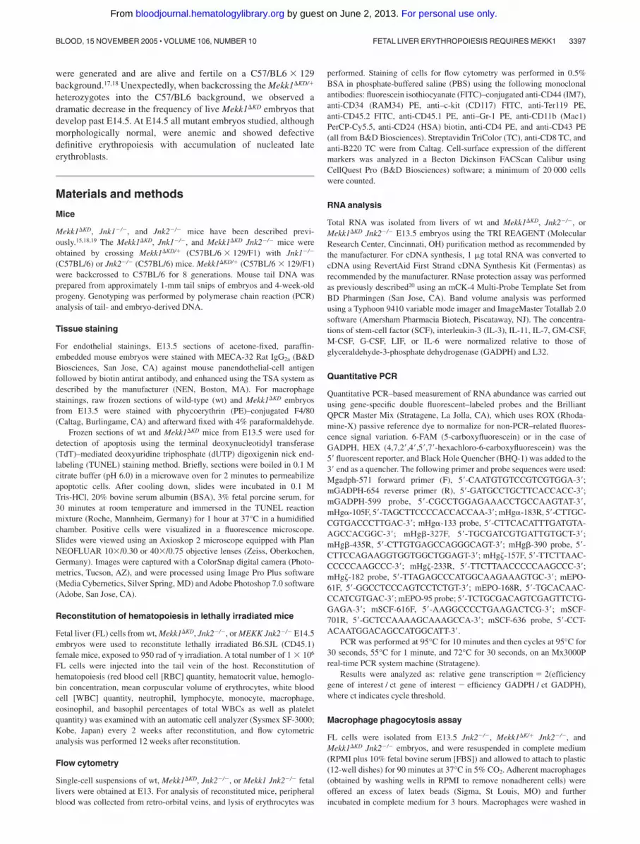
and SCF were first examined by Q-PCR analysis and the relativeamount of IL-3, IL-11, IL-7, GM-CFS, M-CSF, G-CSF, LIF, IL-6,and SCF RNA transcripts was further examined by RPA analysis.However, no difference in the relative amounts of RNA wasdetected between wt, Mekk1�KD, and Mekk1�KD Jnk2�/� samples(Figure 3A-B). To confirm that the RNA expression levels werereflected at the protein level, we performed Western blot analysis ofFL protein extractions for SCF and erythropoietin, as well asimmunohistochemistry for SCF. No differences were found (datanot shown).
Mekk1�KD hematopoietic stem cells reconstitute lethallyirradiated hosts
Our data suggest that the major aberration in Mekk1�KD andMekk1�KD Jnk2�/� embryos, which lead to a defect in definitiveerythropoiesis, is insufficient enucleation. To examine whether theenucleation deficiency was intrinsic to the Mekk1�KD and Mekk1�KD
Jnk2�/� maturing blood cells or extrinsic, we tested the ability ofMekk1�KD and Mekk1�KD Jnk2�/� fetal liver hematopoietic stemcells (HSCs) to reconstitute hematopoiesis in lethally irradiatedhosts. Adult B6.SJL mice, which express the CD45.1 antigen, werelethally irradiated and injected with single-cell suspensions ofE14.5 Jnk2�/� or Mekk1�KD Jnk2�/� FL cells, which express theCD45.2 antigen. While mock-injected lethally irradiated mice didnot survive beyond 2 weeks, mice reconstituted with either Jnk2�/�
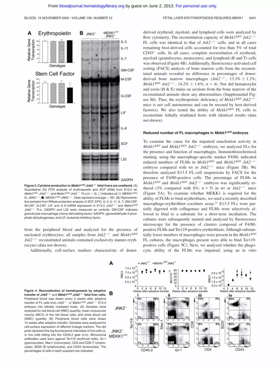
or Mekk1�KD Jnk2�/� FL cells survived until the end of the study(4 months). Every 2 weeks after reconstitution, peripheral bloodwas analyzed for RBC quantity, hematocrit value, hemoglobinconcentration, mean corpuscular volume of erythrocytes, WBCquantity, and percentages of neutrophils, lymphocytes, monocytes,macrophages, eosinophils, and basophils as well as platelet quan-tity by Sysmex Counter Analysis (Figure 4A and SupplementalFigure S5). The mean corpuscular volume of erythrocytes corre-lates with their maturation state; orthochromatic erythroblasts aremuch larger than mature erythrocytes and can therefore beenvisioned by this analysis. Furthermore, blood smears were made
Figure 2. Definitive erythropoiesis is defective inMekk1�KD and Mekk1�KD Jnk2�/� embryos. Flow cytometryof FL hematopoietic cells from E13.5 wt, Mekk1�KD, Jnk2�/�,and Mekk1�KD Jnk2�/� embryos and relative expression ofglobin genes in E13.5 FL of wt, Mekk1�KD, Jnk2�/�, andMekk1�KD Jnk2�/� embryos. (A) Expression of different cell-surface markers is depicted. Cell suspensions from E13.5FLs were labeled with anti–c-kit, which marks hematopoieticstem cells; anti-CD44, which marks all hematopoietic cells;anti-Ter119, to identify differentiated erythroid cells; or anti-CD34 to mark hematopoietic progenitors. The dot plotsrepresent the log fluorescence intensities of live cells. (B)Relative expression of globin chains was measured byreal-time PCR. The globin chain is found only in yolksac–derived erythroblasts. The � globin chain is found in bothyolk sac–derived and FL-derived erythroblasts. The major
globin chain is found only in FL-derived erythroblasts and iscontinuously expressed until just prior to enucleation. GADPHmRNA levels were used for normalization in all cases.Percentage of cells in each quadrant is indicated. Datarepresent the average � standard deviation (SD) from 3 to 5animals per group.
3400 BONNESEN et al BLOOD, 15 NOVEMBER 2005 � VOLUME 106, NUMBER 10
For personal use only. by guest on June 2, 2013. bloodjournal.hematologylibrary.orgFrom
from the peripheral blood and analyzed for the presence ofnucleated erythrocytes; all samples from Jnk2�/� and Mekk1�KD
Jnk2�/� reconstituted animals contained exclusively mature eryth-rocytes (data not shown).
Additionally, cell-surface markers characteristic of donor-
derived erythroid, myeloid, and lymphoid cells were analyzed byflow cytometry. The reconstitution capacity of Mekk1�KD Jnk2�/�
FL cells was identical to that of Jnk2�/� cells, and in all casesremaining host-derived cells accounted for less than 5% of totalCD45� cells. In all cases, complete reconstitution of erythroid,myeloid (granulocytes, monocytes), and lymphoid (B and T) cellswas observed (Figure 4B). Additionally, fluorescence-activated cellsorting (FACS) analysis of bone marrow cells from the reconsti-tuted animals revealed no difference in percentages of donor-derived bone marrow macrophages (Jnk2�/�, 13.3% � 1.2%;Mekk1�KD Jnk2�/�, 14.2% � 1.6%; n � 4). Nor did hematoxylinand eosin (H & E) stains on sections from the bone marrow of thereconstituted animals show any abnormalities (Supplemental Fig-ure S6). Thus, the erythropoietic deficiency of Mekk1�KD Jnk2�/�
mice is not cell autonomous and can be rescued by host-derivedfactor(s). We also tested the ability of Mekk1�KD FL cells toreconstitute lethally irradiated hosts with identical results (datanot shown).
Reduced number of FL macrophages in Mekk1�KD embryos
To examine the cause for the impaired enucleation activity inMekk1�KD and Mekk1�KD Jnk2�/� embryos, we analyzed FLs forthe presence and function of macrophages. Immunohistochemicalstaining, using the macrophage-specific marker F4/80, indicatedreduced numbers of FLMs in Mekk1�KD and Mekk1�KD Jnk2�/�
embryos compared with wt or Jnk2�/� mice (Figure 5B). Wetherefore analyzed E13.5 FL-cell suspensions by FACS for thepresence of F4/80-positive cells. The percentage of FLMs inMekk1�KD and Mekk1�KD Jnk2�/� embryos was significantly re-duced (3% compared with 8%; n � 5) in wt or Jnk2�/� mice(Figure 5A). To examine whether MEKK1 is required for theability of FLMs to bind erythroblasts, we used a recently describedmacrophage-erythroblast coculture assay.21 E13.5 FLs were par-tially digested with collagenase and FLMs were selectively al-lowed to bind to a substrate for a short-term incubation. Thecultures were subsequently stained and analyzed by fluorescencemicroscopy for the presence of clusters composed of F4/80-positive FLMs and Ter119-positive erythroblasts. Although substan-tially lower numbers of macrophages were present in the Mekk1�KD
FL cultures, the macrophages present were able to bind Ter119-positive cells (Figure 5C). Next, we analyzed whether the phago-cytic ability of the FLMs was impaired, using an in vitro
Figure 4. Reconstitution of hematopoiesis by adoptivetransfer of Jnk2�/� and Mekk1�KD Jnk2�/� fetal liver cells.Peripheral blood was drawn every 2 weeks after adoptivetransfer of FL cells from Jnk2�/� or Mekk1�KD Jnk2�/� E14.5embryos into lethally irradiated hosts. (A) Samples wereanalyzed for red blood cell (RBC) quantity, mean corpuscularvolume (MCV) of the red blood cells, and white blood cell(WBC) quantity. (B) Peripheral blood cells were drawn12 weeks after adoptive transfer. Samples were analyzed forcell-surface expression of different lineage markers. The dotplots represent the log fluorescence intensities of live cells (i),or live cells falling into the CD45.2 gate (ii-iv). Monoclonalantibodies used were against Ter119 (erythroid cells), Gr-1(granulocytes), Mac1 (monocytes), CD4 and CD8 (T lympho-cytes), B220 (B lymphocytes), and CD43 (leukocytes). Thepercentages of cells in each quadrant are indicated.
Figure 3. Cytokine production in Mekk1�KD Jnk2�/� fetal livers are unaltered. (A)Quantitative (Q) PCR analysis of erythropoetin and SCF cDNA from E13.5 wt,Mekk1�KD, Jnk2�/�, and Mekk1�KD Jnk2�/� FLs (n � 5). � indicates wt; u, MEKK1�KD;z, JNK2�/�; f, MEKK1�KD JNK2�/�. Data represent average � SD. (B) Representa-tive samples from RNase protection analysis of SCF, EPO, IL-3, IL-11, IL-7, GM-CSF,M-CSF, G-CSF, LIF, and IL-6 mRNA expression in E13.5 Jnk2�/� and Mekk1�KD
Jnk2�/� FLs. GADPH and L32 were measured as controls. GM-CSF indicatesgranulocyte-macrophage colony-stimulating factor; GADPH, glyceraldehyde-3 phos-phate dehydrogenase; and LIF, leukemia inhibitory factor.
FETAL LIVER ERYTHROPOIESIS REQUIRES MEKK1 3401BLOOD, 15 NOVEMBER 2005 � VOLUME 106, NUMBER 10
For personal use only. by guest on June 2, 2013. bloodjournal.hematologylibrary.orgFrom

phagocytosis assay. The ability of Mekk1�KD Jnk2�/� FLMs toengulf latex beads was not inferior to FLMs from wt embryos(Figure 5D). Moreover, transmission electron microscopy of E13.5FL sections revealed the presence of erythoroblastic islands inMekk1�KD FLs (Supplemental Figure S7). We next examinedwhether the clearance of apoptotic cells was impaired in Mekk1�KD
and Mekk1�KD Jnk2�/� FLs. Indeed, a substantial increase ofTUNEL-positive cells was detected in Mekk1�KD FLs (Figure 6B,and in higher magnification D). These apoptotic foci did notcolocalize with Ter119-positive cells (Supplemental Figure S8),suggesting that expelled nuclei from erythroblasts accumulate inMekk1�KD and Mekk1�KD Jnk2�/� FLs. The accumulation of partlydegraded DNA appeared specific to the liver, since we observed noincrease of apoptotic foci in any other tissue (including brain, lung,heart, kidney, intestine, and extremities; data not shown).
A previous study has reported accumulation of phagolysosomescontaining undigested nucleic material in FLMs from embryoslacking DNAse II.8 However, transmission electron microscopy ofFLMs from Mekk1�KD embryos revealed no abnormal structures(Supplemental Figure S7).
Discussion
We have discovered a new physiologic function for the MAPKpathway in embryonic development. In the present study weprovide evidence strongly suggesting that MEKK1-JNK signalingis required for degradation of nuclear DNA extruded from erythroidprecursors during the late stages of definitive erythropoiesis in thefetal liver.
Several targeted gene approaches have been used to evaluate thebiologic function of the JNK signaling pathway in mice. The firststudies in which the JNK genes were individually disruptedproduced mice that appeared morphologically normal, suggestingthat the JNK genes were able to complement each other in mosttissues. Animals deficient in both JNK1 and JNK3 or JNK2 andJNK3 survive normally, whereas Jnk1�/�Jnk2�/� mice die duringembryonic development due to severe dysregulation of apoptosis inthe brain.36 The early lethality of the Jnk1�/� Jnk2�/� double-knockout embryos makes the total contribution of JNK in hemato-poiesis difficult to estimate. However, the relative contribution ofJNK1 or JNK2 may become more apparent during conditions ofanemia in the Jnk1�/� or Jnk2�/� mice. We therefore examinedreconstitution of hematopoiesis in Jnk1�/�and Jnk2�/� mice 3 daysafter challenging them with severe anemia. However, we observedno differences compared to wt controls in any of the parametersinvestigated, suggesting that redundancy between the JNK iso-forms may compensate for the loss of either JNK1 or JNK2 alsoduring hematopoiesis.
Mice deficient for the upstream JNK regulators MKK4 and MKK7are also embryonic lethal. MKK4-deficient embryos, similarly toc-Jun–deficient animals,37,38 suffer from severe anemia and die at E10.5to E12.5 from abnormal liver development,39,40 suggesting that MKK4is required for c-Jun activation during embryonic development. Thecause of death in MKK7�/� embryos remains inconclusive.41 Disruptionof activators further upstream of MKK4 and MKK7 within the MEKKfamily includes (in addition to MEKK1) MEKK2 and MEKK3.MEKK2-deficient mice develop normally and are fertile.42 In contrast,mice lacking MEKK3 die at E11 due to failure of cardiovasculardevelopment.43 Previous studies on MEKK1-deficient (Mekk1�/�) andMEKK1 kinase–deficient (Mekk1�KD) mice have suggested that thesemice develop normally with the exception of a defect in eyelid closure, a
Figure 6. Defective degradation and accumulation of TUNEL-positive nucleicmaterial in Mekk1�KD fetal livers. Histochemical analysis of FLs. Nucleic material inwt and Mekk1�KD FLs, as shown by TUNEL stain (green). Note the large amount ofabnormal apoptotic foci in the Mekk1�KD fetal liver B, and in higher magnification D.A, B: bar � 75 �m; C, D: bar � 100 �m.
Figure 5. Decreased number of fetal liver macrophages in Mekk1�KD embryos.(A) Fetal liver cell suspensions from wt and Mekk1�KD E13.5 embryos were stainedwith macrophage-specific anti-F4/80 antibodies and analyzed by flow cytometry. Thedot plots represent the log fluorescence intensities of live cells. Numbers indicate thepercentage of F4/80-positive cells. (B) Immunohistochemical staining of E13.5 fetalliver sections from wt and Mekk1�KD embryos with PE-conjugated anti-F4/80.(C) Immunohistochemistry of “native” erythroblast-FLM clusters from wt and Mekk1�KD
E13.5 embryos. Cell clusters were stained for F4/80 (red) and Ter119 (green).(D) E13.5 FLMs were isolated from Jnk2�/�, Mekk1�KD/� Jnk2�/�, and Mekk1�KD
Jnk2�/� embryos and offered an excess of latex beads in an in vitro phagocytosisassay. Macrophages were counterstained with hematoxylin and the number ofphagocytosed beads per cell was counted by phase-contrast microscopy. A minimumof 3 FLs was examined per genotype.
3402 BONNESEN et al BLOOD, 15 NOVEMBER 2005 � VOLUME 106, NUMBER 10
For personal use only. by guest on June 2, 2013. bloodjournal.hematologylibrary.orgFrom

phenotype that is associated with control of cell migration.18,44 However,the Mekk1�/� or Mekk1�KD mice used in previous studies have, to thebest of our knowledge, all been C57/BL6 � 129 hybrids. We observed askewing of the Mendelian frequencies of live pups born after intercross-ing the Mekk1�KD/� C57/BL/6 � 129 hybrids (Mekk1�/� � 29.2%,Mekk1�KD/� � 52.8%, Mekk1�KD � 19.9%; n � 232). This skewingwas influenced by the genetic background, since fewer than 2%Mekk1�KD pups were born after backcrossing into the C57/BL6background. Nevertheless, crossing the Mekk1�KD/� C57/BL/6 � 129 hybrids with Jnk1�/� or Jnk2�/� mice resulted in embry-onic lethality of all Mekk1�KD Jnk1�/� and Mekk1�KD Jnk2�/�
embryos (regardless of background), suggesting that an intactMEKK1-JNK signaling pathway is required for normal embryonicdevelopment. Mekk1�KD, Mekk1�KD Jnk1�/�, and Mekk1�KD Jnk2�/�
embryos have identical phenotypes, survive up to midgestation,and display normal morphology but are highly anemic. Interest-ingly, a similar phenotype has been observed in p38�-deficientmice.10 However, while the anemia in p38��/� mice is attributed todefective erythropoietin gene expression, normal levels of erythro-poietin at both mRNA and protein level were observed in FLs fromMekk1�KD and Mekk1�KD Jnk2�/� embryos. In addition to erythro-poietin, we also investigated the expression levels of other growthfactors and cytokines suggested to be involved in hematopoieticdevelopment, including SCF, IL-3, IL-6, IL-7, IL-11, GM-CFS,M-CFS, G-CFS, and LIF. However, we observed no differenceswhen compared with FLs from wt controls. The FLs of Mekk1�KD
and Mekk1�KD Jnk2�/� also contain BFU-Es and CFU-Es formed atnormal frequencies, and can reconstitute erythropoiesis in lethallyirradiated hosts. It is therefore clear that MEKK1 is not required forthe production of, or the response to, cytokines and/or growthfactors required for the expansion and differentiation of erythroidprogenitors up to the stage where the orthochromatic erythroblastslose their nuclei and become reticulocytes.
Interestingly, the phenotype of mice lacking DNaseII (a lysoso-mal DNase in macrophages) is almost identical to Mekk1�KD and
Mekk1�KD Jnk2�/� embryos. DNaseII�/� mice die before birth,suffer from severe anemia, and exhibit accumulation of erythro-blasts that fail to undergo enucleation.8,9 This anemic phenotype isa consequence of macrophages failing to aid in digestion of nuclearDNA expelled from erythroblasts during definitive erythropoiesis.8
In our study, FLs from Mekk1�KD and Mekk1�KD Jnk2�/� embryoscontain reduced levels of macrophages and TUNEL stains showedextensive accumulation of apoptotic bodies in the FLs of Mekk1�KD
and Mekk1�KD Jnk2�/� mice. These apoptotic bodies did notcolocalize with Ter119-positive erythroblasts but rather appearedfree in the extra cellular space, suggesting that they representnuclear DNA extruded from erythroid precursor cells. Since thephagocytic capacity of FLMs from Mekk1�KD and Mekk1�KD
Jnk2�/� was found to be similar to wt fetal liver macrophages, ourdata indicate that the observed accumulation of nucleated erythro-blasts is dependent quantitatively rather than qualitatively onFLMs. In conclusion, we have shown that a functional MEKK1-JNK signaling pathway is critically required for macrophage-assisted, erythroblast nuclear enucleation during FL erythropoiesis.The role for macrophages in phagocytosis and degradation oferythroblast nuclei during erythropoiesis has been established.8,21
Morover, FLMs are part of a delicate microenvironment wherecommunication through soluble mediators (ie, cytokines), directcell-to-cell contacts between stromal cells (including fibroblasts,endothelial cells, epithelial cells, and FLMs), and cell-matrixinteractions are in crucial balance, ultimately supporting erythropoi-esis. Whether or how the MEKK1-JNK signaling pathway isrequired for factors extrinsic to FLMs during development, differ-entiation, proliferation, and/or maturation remains to be investigated.
Acknowledgment
We thank Michael Karin for providing the Mekk1�KD, Jnk1�/�andJnk2�/� animals and for helpful review of the manuscript.
References
1. Moore MA, Metcalf D. Ontogeny of the haemo-poietic system: yolk sac origin of in vivo and invitro colony forming cells in the developingmouse embryo. Br J Haematol. 1970;18:279-296.
2. Houssaint E. Differentiation of the mouse hepaticprimordium, II: extrinsic origin of the haemopoi-etic cell line. Cell Differ. 1981;10:243-252.
3. Palis J, Robertson S, Kennedy M, Wall C, KellerG. Development of erythroid and myeloid pro-genitors in the yolk sac and embryo proper of themouse. Development. 1999;126:5073-5084.
4. Mucenski ML, McLain K, Kier AB, et al. A func-tional c-myb gene is required for normal murinefetal hepatic hematopoiesis. Cell. 1991;65:677-689.
5. Trimborn T, Gribnau J, Grosveld F, Fraser P.Mechanisms of developmental control of tran-scription in the murine alpha- and beta-globin loci.Genes Dev. 1999;13:112-124.
6. Sasaki K, Iwatsuki H. Origin and fate of the cen-tral macrophages of erythroblastic islands in thefetal and neonatal mouse liver. Microsc Res Tech.1997;39:398-405.
7. Sadahira Y, Mori M. Role of the macrophage inerythropoiesis. Pathol Int. 1999;49:841-848.
8. Kawane K, Fukuyama H, Kondoh G, et al. Re-quirement of DNase II for definitive erythropoiesisin the mouse fetal liver. Science. 2001;292:1546-1549.
9. Krieser RJ, MacLea KS, Longnecker DS, et al.Deoxyribonuclease IIalpha is required during thephagocytic phase of apoptosis and its loss
causes perinatal lethality. Cell Death Differ. 2002;9:956-962.
10. Tamura K, Sudo T, Senftleben U, et al. Require-ment for p38alpha in erythropoietin expression: arole for stress kinases in erythropoiesis. Cell.2000;102:221-231.
11. Jacobs-Helber SM, Ryan JJ, Sawyer ST. JNKand p38 are activated by erythropoietin (EPO) butare not induced in apoptosis following EPO with-drawal in EPO-dependent HCD57 cells. Blood.2000;96:933-940.
12. Jacobs-Helber SM, Sawyer ST. Jun N-terminalkinase promotes proliferation of immature ery-throid cells and erythropoietin-dependent celllines. Blood. 2004;104:696-703.
13. Wu H, Liu X, Jaenisch R, Lodish HF. Generationof committed erythroid BFU-E and CFU-E pro-genitors does not require erythropoietin or theerythropoietin receptor. Cell. 1995;83:59-67.
14. Wu H, Klingmuller U, Besmer P, Lodish HF. Inter-action of the erythropoietin and stem-cell-factorreceptors. Nature. 1995;377:242-246.
15. Dong C, Yang DD, Wysk M, et al. Defective T celldifferentiation in the absence of Jnk1. Science.1998;282:2092-2095.
16. Sabapathy K, Hu Y, Kallunki T, et al. JNK2 is re-quired for efficient T-cell activation and apoptosisbut not for normal lymphocyte development. CurrBiol. 1999;9:116-125.
17. Xia Y, Makris C, Su B, et al. MEK kinase 1 is criti-cally required for c-Jun N-terminal kinase activa-tion by proinflammatory stimuli and growth factor-
induced cell migration. Proc Natl Acad Sci U S A.2000;97:5243-5248.
18. Zhang L, Wang W, Hayashi Y, et al. A role forMEK kinase 1 in TGF-beta/activin-induced epi-thelium movement and embryonic eyelid closure.EMBO J. 2003;22:4443-4454.
19. Yang DD, Conze D, Whitmarsh AJ, et al. Differen-tiation of CD4� T cells to Th1 cells requires MAPkinase JNK2. Immunity. 1998;9:575-585.
20. Eriksen KW, Sommer VH, Woetmann A, et al.Bi-phasic effect of interferon (IFN)-alpha: IFN-alpha up- and down-regulates interleukin-4 sig-naling in human T cells. J Biol Chem. 2004;279:169-176.
21. Iavarone A, King ER, Dai XM, et al. Retinoblas-toma promotes definitive erythropoiesis by re-pressing Id2 in fetal liver macrophages. Nature.2004;432:1040-1045.
22. Mudgett JS, Ding J, Guh-Siesel L, et al. Essentialrole for p38alpha mitogen-activated protein ki-nase in placental angiogenesis. Proc Natl AcadSci U S A. 2000;97:10454-10459.
23. Adams RH, Porras A, Alonso G, et al. Essentialrole of p38alpha MAP kinase in placental but notembryonic cardiovascular development. Mol Cell.2000;6:109-116.
24. Ihle JN. The challenges of translating knockoutphenotypes into gene function. Cell. 2000;102:131-134.
25. Ikuta K, Kina T, MacNeil I, et al. A developmentalswitch in thymic lymphocyte maturation potential
FETAL LIVER ERYTHROPOIESIS REQUIRES MEKK1 3403BLOOD, 15 NOVEMBER 2005 � VOLUME 106, NUMBER 10
For personal use only. by guest on June 2, 2013. bloodjournal.hematologylibrary.orgFrom

occurs at the level of hematopoietic stem cells.Cell. 1990;62:863-874.
26. Kansas GS, Muirhead MJ, Dailey MO. Expres-sion of the CD11/CD18, leukocyte adhesion mol-ecule 1, and CD44 adhesion molecules duringnormal myeloid and erythroid differentiation inhumans. Blood. 1990;76:2483-2492.
27. Neubauer H, Cumano A, Muller M, et al. Jak2 de-ficiency defines an essential developmentalcheckpoint in definitive hematopoiesis. Cell.1998;93:397-409.
28. Ghaffari S, Smadja-Joffe F, Oostendorp R, et al.CD44 isoforms in normal and leukemic hemato-poiesis. Exp Hematol. 1999;27:978-993.
29. Ikuta K, Weissman IL. Evidence that hematopoi-etic stem cells express mouse c-kit but do notdepend on steel factor for their generation. ProcNatl Acad Sci U S A. 1992;89:1502-1506.
30. Zhang J, Socolovsky M, Gross AW, Lodish HF.Role of Ras signaling in erythroid differentiation ofmouse fetal liver cells: functional analysis by aflow cytometry-based novel culture system.Blood. 2003;102:3938-3946.
31. Whitelaw E, Tsai SF, Hogben P, Orkin SH. Regu-lated expression of globin chains and the ery-throid transcription factor GATA-1 during erythro-
poiesis in the developing mouse. Mol Cell Biol.1990;10:6596-6606.
32. Nienhuis AW, Benz EJ Jr. Regulation of hemoglo-bin synthesis during the development of the redcell (first of three parts). N Engl J Med. 1977;297:1318-1328.
33. Nienhuis AW, Benz EJ Jr. Regulation of hemoglo-bin synthesis during the development of the redcell (second of three parts). N Engl J Med. 1977;297:1371-1381.
34. Nienhuis AW, Benz EJ Jr. Regulation of hemoglo-bin synthesis during the development of the redcell (third of three parts). N Engl J Med. 1977;297:1430-1436.
35. Gregory CJ, Eaves AC. Three stages of erythro-poietic progenitor cell differentiation distinguishedby a number of physical and biologic properties.Blood. 1978;51:527-537.
36. Kuan CY, Yang DD, Samanta Roy DR, et al. TheJnk1 and Jnk2 protein kinases are required forregional specific apoptosis during early brain de-velopment. Neuron. 1999;22:667-676.
37. Eferl R, Sibilia M, Hilberg F, et al. Functions ofc-Jun in liver and heart development. J Cell Biol.1999;145:1049-1061.
38. Hilberg F, Aguzzi A, Howells N, Wagner EF. c-jun
is essential for normal mouse development andhepatogenesis. Nature. 1993;365:179-181.
39. Ganiatsas S, Kwee L, Fujiwara Y, et al. SEK1 de-ficiency reveals mitogen-activated protein kinasecascade crossregulation and leads to abnormalhepatogenesis. Proc Natl Acad Sci U S A. 1998;95:6881-6886.
40. Nishina H, Vaz C, Billia P, et al. Defective liverformation and liver cell apoptosis in mice lackingthe stress signaling kinase SEK1/MKK4. Devel-opment. 1999;126:505-516.
41. Dong C, Yang DD, Tournier C, et al. JNK is re-quired for effector T-cell function but not for T-cellactivation. Nature. 2000;405:91-94.
42. Guo Z, Clydesdale G, Cheng J, et al. Disruptionof Mekk2 in mice reveals an unexpected rolefor MEKK2 in modulating T-cell receptor signaltransduction. Mol Cell Biol. 2002;22:5761-5768.
43. Yang J, Boerm M, McCarty M, et al. Mekk3 is es-sential for early embryonic cardiovascular devel-opment. Nat Genet. 2000;24:309-313.
44. Yujiri T, Ware M, Widmann C, et al. MEK kinase 1gene disruption alters cell migration and c-Jun NH2-terminal kinase regulation but does not cause a mea-surable defect in NF- kappa B activation. Proc NatlAcad Sci U S A. 2000;97:7272-7277.
3404 BONNESEN et al BLOOD, 15 NOVEMBER 2005 � VOLUME 106, NUMBER 10
For personal use only. by guest on June 2, 2013. bloodjournal.hematologylibrary.orgFrom


