Embed Size (px)
Citation preview

This is a pre-copy-editing, author-produced PDF of an article accepted for publication in Human Reproduction following peer review. The definitive publisher-authenticated version 2014 Jun 25. pii: deu159. [Epub ahead of print] is available online at http://humrep.oxfordjournals.org/content/early/recent.
Manuscript HUMREP-14-0269
Mesenchymal stem/stromal cells in Postmenopausal Endometrium
Running title :MSC in postmenopausal endometrium
AUTHORS
Ulrich, D1, 3 Tan, KS1; Deane, J1; Schwab, K1; Cheong, A 1, Rosamilia, A 1,2,4; Gargett,
CE1,2,4*
1 The Ritchie Centre, MIMR-PHI Institute of Medical Research, 27-31 Wright Street,
Clayton, 3168, Clayton, VIC, Australia
2 Monash University Department of Obstetrics and Gynaecology, Monash Medical Centre,
246 Clayton Road, Clayton, 3168, VIC, Australia
3 Current address: Department of Obstetrics and Gynaecology, Medical University Graz,
8045 Graz, Austria
4 equal senior authors
* Corresponding Author
A/Prof Caroline Gargett, PhD
The Ritchie Centre,
Monash Institute of Medical Research
27-31 Wright Street
Clayton, 3168
Melbourne, VIC, Australia
Email: [email protected]
Keywords
Mesenchymal stem cells, mesenchymal stromal cells, endometrial MSC, W5C5, SUSD2,
estrogen, postmenopausal, endometrium
1
2
3
4
5
6
7
8
9
10
11
12
13
14
15
16
17
18
19
20
21
22
23
24
25
26

ABSTRACT
Study question: Are there mesenchymal stem/ stromal cells in postmenopausal
endometrium with adult stem cell properties that can be prospectively isolated from a
biopsy?
Summary answer: Perivascular W5C5+ cells isolated from postmenopausal endometrial
biopsies displayed characteristic mesenchymal stem/stromal cell (MSC) properties of
clonogenicity, multipotency and surface phenotype irrespective of whether women are or
are not pre-treated with estrogen to regenerate the endometrium.
What is known already: Recently MSCs have been identified in human premenopausal
endometrium, and can be prospectively isolated using a single marker, W5C5/SUSD2.
Study design, size, duration: Endometrial tissue both from functional and basal layers
was obtained from 17 premenopausal (pre-MP), 19 postmenopausal (post-MP) without
hormonal treatment, and 15 postmenopausal women on estrogen replacement therapy
(post-MP+ E2) collected through a prospective phase IV clinical trial over 2 years.
Endometrial tissue was obtained from women by biopsy (curettage) just prior to
undergoing hysterectomy and assessed for histological and in vitro analysis of MSC
properties.
Participants/materials, setting, methods: Postmenopausal women less than 65 years of
age were treated with or without E2 for 6-8 weeks prior to tissue collection. Serum E2
levels were determined by estradiol immunoenzymatic assay. The effect of E2 on
endometrial thickness and glandular and luminal epithelial height was determined using
image analysis. Endometrial tissue was dissociated into single cell suspensions and MSC
properties were examined in freshly isolated and short-term cultured, magnetic bead-
purified W5C5+ cells. MSC properties were assessed using clonogenicity, serial cloning,
mesodermal differentiation in adipogenic, chondrogenic, osteogenic and myogenic
27
28
29
30
31
32
33
34
35
36
37
38
39
40
41
42
43
44
45
46
47
48
49
50
51

induction culture media and surface phenotype by flow cytometric assays. Estrogen
receptor α expression in W5C5+ cells was examined using dual colour
immunofluorescence. Vascularity was analysed using CD34 and alpha smooth muscle
actin immunostaining and subsequent image analysis.
Main results and the role of chance:
A small population of stromal cells with MSC properties was purified with the W5C5
antibody from postmenopausal endometrium, whether atrophic from low circulating
estrogen or regenerated from systemic estrogen treatment, similar to premenopausal
endometrium. The MSC derived from postmenopausal endometrium treated with or
without E2 fulfil the minimum MSC criteria: clonogenicity, surface phenotype (CD29+,
CD44+, CD73+, CD105+, CD140b+, CD146+) and multipotency. Postmenopausal
endometrial MSCs (eMSC) also have comparable properties to premenopausal eMSC with
respect to self renewal in vitro and W5C5 expression. The W5C5+ cells were located
perivascularly as expected and did not express estrogen receptor α.
Limitations, reasons for caution: The properties of MSC derived from postmenopausal
endometrium were evaluated in vitro and their in vivo tissue reconstitution capacity has not
been established as it has for premenopausal eMSC.
Wider implications of the findings: The endometrium is an accessible source of MSC
obtainable with minimum morbidity that could be used for future clinical applications as a
cell-based therapy. This study shows that menopausal women can access their eMSC by a
simple biopsy for use in autologous therapies, particularly if their endometrium has been
regenerated by short-term E2 treatment, provided they have an intact uterus and are not
contraindicated for short-term E2 treatment. Endometrial MSC in postmenopausal women
possess key MSC properties and are a promising source of MSC independent of a woman’s
age.
52
53
54
55
56
57
58
59
60
61
62
63
64
65
66
67
68
69
70
71
72
73
74
75
76

Study funding/competing interest(s): This study was supported by the National Health
and Medical Research Council (NHMRC) of Australia grant (1021126) (CEG, AR) and
Senior Research Fellowship (1042298) (CEG), Australian Gynaecological Endoscopic
Society grant (AR) and Victorian Government's Operational Infrastructure Support
Program.
Trial registration number: CTNRN12610000563066
77
78
79
80
81
82
83
84
INTRODUCTION
Human mesenchymal stem cells or multipotent stromal cells (MSC) have been identified in
almost every adult tissue; bone marrow, adipose tissue, synovial membrane and the
endometrium (da Silva Meirelles et al. 2006, Beltrami et al. 2007, Schwab and Gargett
2007, Crisan et al. 2008). Originally MSC were identified by their adherence to plastic and
differentiation into mesodermal lineages; adipocytic, chondrocytic and osteoblastic
(Prockop 1997, Pittenger et al. 1999). More recently it has been shown that bone marrow
MSC (bmMSC) also differentiate into endodermal and neuroectodermal lineages (Torrente
and Polli 2008, Morikawa et al. 2009). Cultured MSC are highly proliferative with
capacity to produce millions of cells from a single clonogenic cell (Gargett et al. 2009).
MSC have characteristic surface markers including CD29, CD44, CD73, CD105, but not
haematopoietic cell markers CD34, CD45, CD14, CD11b, CD79α, CD19 and HLA-DR
(Dominici et al. 2006, Caplan 2007). The identification of more specific markers, Stro-1,
CD146, CD271, has enabled the prospective isolation of MSC from bone marrow
(Simmons and Torok-Storb 1991, Gronthos et al. 2003, Buhring et al. 2007). BmMSC
have anti-inflammatory and immunomodulatory properties which make them an attractive
source for tissue engineering and regenerative medicine applications (Salem and
Thiemermann 2010, Le Blanc and Mougiakakos 2012).
Endometrial MSC (eMSC) were recently discovered and characterised in premenopausal
endometrium where they are thought to regenerate the stromal vascular component of the
functional layer each month (Gargett et al. 2009). EMSC also possess high capacity for
proliferation, differentiate into mesodermal lineages and express characteristic MSC
surface markers, fulfilling the minimal criteria for defining MSC (Dominici 2006).
Originally eMSC were prospectively isolated from hysterectomy tissue using co-
expression of 2 markers (CD140b/ PDGFRβ and CD146) by FACS sorting (Schwab and
85
86
87
88
89
90
91
92
93
94
95
96
97
98
99
100
101
102
103
104
105
106
107
108
109

Gargett 2007). EMSC can now be prospectively isolated from endometrial biopsy tissue
using the single marker W5C5 using magnetic bead sorting (Masuda et al. 2012). W5C5
recognises an epitope of the Sushi Domain containing 2 (SUSD2) molecule
(Sivasubramaniyan et al. 2013). Almost all clonogenic endometrial stromal cells were
found in the W5C5+ fraction. Similar to CD140b+CD146+ cells, W5C5+ cells meet the
defining criteria for MSC (Masuda et al 2012). Clonogenic stromal cells have also been
identified in postmenopausal endometrium, although the sample size examined was small
(n=4) (Schwab et al. 2005). EMSC are an attractive source of MSC as they can be easily
obtained through an office biopsy procedure without anaesthesia or scarring (Gargett et al.
2012) or a simple curettage for many women. Protocols are being developed for culture
expansion of eMSC under clinical grade Good Manufacturing Practices (cGMP) conditions
making them an ideal source for future clinical applications, particularly in women’s
health, where they could be used autologously (Rajaraman et al. 2013).
Clinical conditions for which eMSC could be utilised as a cell-based therapy may affect
postmenopausal women, for example pelvic organ prolapse (Hunskaar et al. 2005,
Boennelycke et al. 2013, Ulrich et al. 2013). To date, eMSC have only been characterised
in premenopausal women. Due to hormonal depletion, postmenopausal endometrium is
thin and atrophic, difficult to biopsy without anaesthesia, and we hypothesized a low yield
of eMSC. However, postmenopausal endometrium has significant regenerative potential,
particularly when systemic estrogen is administered. A thick functional endometrium can
be generated and indeed postmenopausal women in their 60s have born children via IVF
(Paulson et al. 2002).
Therefore, the aim of this study was to determine whether postmenopausal endometrium
contains a population of eMSC and to characterize these postmenopausal eMSC for
clonogenicity, mesodermal differentiation, self renewal and surface phenotype. We
110
111
112
113
114
115
116
117
118
119
120
121
122
123
124
125
126
127
128
129
130
131
132
133
134

hypothesized that eMSC can be prospectively isolated from endometrial biopsies of
postmenopausal endometrium, that they would be present at similar frequency as in
premenopausal endometrium and that they possess similar properties to premenopausal
eMSC.
MATERIALS AND METHODS
Human tissue and ethical approval
Human endometrial tissue including underlying myometrium was collected from 17
premenopausal (pre-MP) and 19 postmenopausal (post-MP) (longer than 12 months since
last period) undergoing hysterectomy who were not taking hormones (Table 1). We also
collected endometrial biopsies from 15 postmenopausal women on oral estrogen
replacement therapy (post- MP+E2). Due to the low endometrial stromal cell yield from
atrophic postmenopausal endometrium, a single arm phase IV clinical trial was registered
with the Therapeutic Goods Association (CTNRN12610000563066) to treat
postmenopausal women with short term estrogen to regenerate the endometrium and obtain
higher cell yields for the collection and analysis of eMSC. The enrolled women (n= 15)
took oral estrogen replacement therapy (Progynova 2mg daily for 6-8 weeks) which was
ceased 2 days prior to scheduled hysterectomy. The biopsy was obtained by curettage just
prior to surgical removal of the uterus. Inclusion criteria were at least 12 months since the
last period and age less than 65 years. Exclusion criterion was any condition where
systemic oestrogen use was contra-indicated, including current/past history of breast
cancer, other oestrogen responsive tumours, liver adenoma, thrombo-embolism,
undiagnosed vaginal bleeding, uncontrolled hypertension or ongoing oral hormone
replacement therapy (HRT). The women were assessed for these exclusion criteria by
urogynaecologist AR. Informed written consent was obtained from each patient and ethics
135
136
137
138
139
140
141
142
143
144
145
146
147
148
149
150
151
152
153
154
155
156
157
158
159

approval was obtained from the Monash Health Human Research and Ethics Committee B
and Cabrini HREC.
The collected tissues (n=51) were used for histological and/ or cell culture analysis. Serum
samples were also collected to determine the hormonal status of the postmenopausal
women (n=10) and determine circulating estrogen levels in the estrogen-treated women
(n=10). Endometrial samples (n=26) collected in collection medium (HEPES-buffered
DMEM/F12 medium containing 5% fetal calf serum and 5% antibiotics-antimycotics)
were immediately transported to the laboratory and processed within 24 hours. Samples
(n=32) were also frozen in OCT for cryostat sections and fixed for paraffin sections. Blood
from Post-MP and Post-MP+E2 was drawn and centrifugated at 2000 g for 20 minutes at 4o
C to collect serum. Estrogen levels were determined by a competitive binding estradiol
immunoenzymatic assay performed by Monash Health Pathology Laboratory.
Histology
Tissues collected for histological analysis were fixed in 10% formalin for 24 hours, then
embedded in paraffin and oriented so that the full thickness endometrium from
myometrium to the lumen could be examined (Figure 1A-C). These were sectioned into
5µm sections and stained with haematoxylin and eosin (H+E) to determine endometrial
thickness.
Immunohistochemistry
To determine the estrogen effect on the luminal and glandular epithelial height, sections
from postmenopausal endometrium were stained with the epithelial marker Cytokeratin 18.
To determine the total blood vessel area as a measure of vascularity, sections were
immunostained with anti-human CD34, and vessels invested with pericytes and smooth
160
161
162
163
164
165
166
167
168
169
170
171
172
173
174
175
176
177
178
179
180
181
182
183
184

muscle cells were immune-stained with anti-human αSMA (Abberton et al. 1999). Sections
underwent dewaxing, rehydrating in graded alcohols and antigen retrieval using citric acid
buffer (0.1M, pH 6.0) by microwaving for 5 minutes on high power. After cooling to RT
and three washes in PBS, endogenous peroxidase was quenched by 3% H2O2, followed by
a protein block step (Protein Block serum free, ready to use, Dako®, Glostrup, Denmark)
for 30 minutes at RT. The primary antibodies (1:100 for CK18 and CD34, 1:400 for
αSMA) and isotype controls at the same concentration (IgG1, IgG2a) (all from Dako) were
incubated overnight at 4° C, sections were then washed and the Envision+ System HRP
secondary antibody (Mouse Envision Kit, Dako) applied for 30 minutes at RT, respectively
as done previously (Ulrich et al. 2012) . Colour was developed with 3,3'-Diaminobenzidine
(DAB).
Immunofluorescence
To determine the location of the W5C5+ cells and their estrogen receptor-α status,
endometrial tissue was sequentially immunostained with anti-estrogen receptor-α (clone
6F11, Leica Microsystems, Australia) followed by phycoerythrin (PE)-conjugated anti-
W5C5 (BioLegend, USA) (Masuda et al. 2012). Frozen sections were cut at 8µm from
OCT embedded tissues, thawed at RT, fixed in 4% paraformaldehyde for 10 minutes,
washed in PBS and treated with 0.2% Triton X-100 in PBS for 15 minutes. Protein block
(Dako ®) was applied for 30 minutes and sections were incubated with anti-estrogen
receptor-α (1:100) in PBS for 2 hours at RT followed by secondary anti-mouse Alexa
Fluor 488 (Life Technologies, Australia). Sections were washed three times in PBS,
blocked with mouse IgG for 30 minutes, incubated with PE-anti- W5C5 (1:100) for 2
hours at RT, washed three times with PBS, counterstained with Hoechst 33258 and imaged
on a Nikon C1 confocal microscope.
185
186
187
188
189
190
191
192
193
194
195
196
197
198
199
200
201
202
203
204
205
206
207
208
209
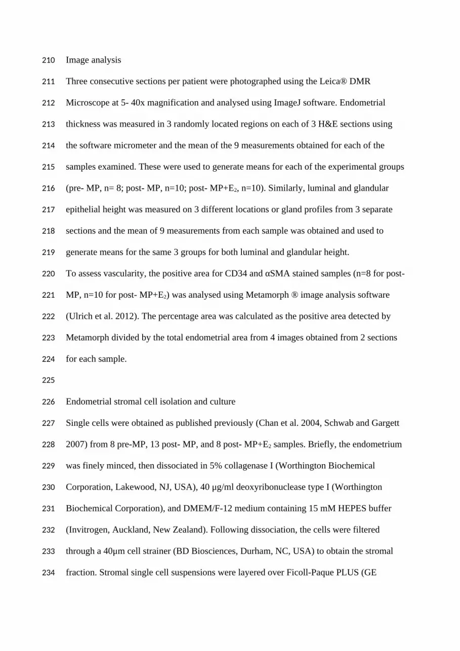
Image analysis
Three consecutive sections per patient were photographed using the Leica® DMR
Microscope at 5- 40x magnification and analysed using ImageJ software. Endometrial
thickness was measured in 3 randomly located regions on each of 3 H&E sections using
the software micrometer and the mean of the 9 measurements obtained for each of the
samples examined. These were used to generate means for each of the experimental groups
(pre- MP, n= 8; post- MP, n=10; post- MP+E2, n=10). Similarly, luminal and glandular
epithelial height was measured on 3 different locations or gland profiles from 3 separate
sections and the mean of 9 measurements from each sample was obtained and used to
generate means for the same 3 groups for both luminal and glandular height.
To assess vascularity, the positive area for CD34 and αSMA stained samples (n=8 for post-
MP, n=10 for post- MP+E2) was analysed using Metamorph ® image analysis software
(Ulrich et al. 2012). The percentage area was calculated as the positive area detected by
Metamorph divided by the total endometrial area from 4 images obtained from 2 sections
for each sample.
Endometrial stromal cell isolation and culture
Single cells were obtained as published previously (Chan et al. 2004, Schwab and Gargett
2007) from 8 pre-MP, 13 post- MP, and 8 post- MP+E2 samples. Briefly, the endometrium
was finely minced, then dissociated in 5% collagenase I (Worthington Biochemical
Corporation, Lakewood, NJ, USA), 40 μg/ml deoxyribonuclease type I (Worthington
Biochemical Corporation), and DMEM/F-12 medium containing 15 mM HEPES buffer
(Invitrogen, Auckland, New Zealand). Following dissociation, the cells were filtered
through a 40μm cell strainer (BD Biosciences, Durham, NC, USA) to obtain the stromal
fraction. Stromal single cell suspensions were layered over Ficoll-Paque PLUS (GE
210
211
212
213
214
215
216
217
218
219
220
221
222
223
224
225
226
227
228
229
230
231
232
233
234

healthcare Bio-Sciences AB, Uppsala, Sweden) and centrifuged to remove red blood cells.
The endometrial stromal cells were used fresh or cultured for 1 passage in DMEM medium
containing 10% fetal calf serum (Invitrogen), 5% antibiotics-antimycotics and 2 mM
glutamine (Invitrogen) to obtain sufficient cell numbers for experiments. Cells were
harvested by TrypLE Express (Life Technologies, Auckland, New Zealand) and eMSC
were extracted using magnetic beads conjugated to the W5C5 antibody as described
(Masuda et al. 2012). Briefly, cell suspensions (up to 1x10⁷ cells / 100 µl) were labelled
with the PE-conjugated W5C5 antibody (Biolegend, San Diego, CA, USA) in 0.5 % Fetal
Calf Serum in PBS (Bead Medium) for 30 minutes at 4C followed by 3 washing steps in
PBS, then incubated for 30 minutes in the dark with the anti-PE antibody-conjugated
MACS MicroBeads (Miltenyi Biotec, Bergisch Gladbach, Germany). Up to 1x10⁸ cells /
500 μl were applied to MS columns (Miltenyi Biotec) in a magnetic field, followed by
washing the column with 500μl Bead Medium three times. The W5C5- cells passed
through the column, magnetically labelled W5C5+ cells were retained. The columns were
removed from the magnetic field and W5C5+ cells were flushed out with 1 ml of Bead
Medium. The W5C5+ cells were assayed for cloning efficiency, and the remaining cells
were cultured for one more passage for differentiation assays and phenotyping.
MSC functional properties
Clonogenicity was determined by seeding fresh and cultured W5C5+ cells at clonal density
(10 and 50 cells/cm2) on fibronectin-coated 100 mm tissue culture plates (BD Biosciences,
San Jose, California, USA). Cells were incubated at 37°C in 5% CO2 incubator for at least
2 weeks; DMEM+ media supplemented with 10ng/ml human fibroblast growth factor 2
(FGF2; Millipore, Billerica, MA) (F-DMEM+) was changed weekly. Colonies were
monitored microscopically to ensure they were derived from single cells. Large clones
235
236
237
238
239
240
241
242
243
244
245
246
247
248
249
250
251
252
253
254
255
256
257
258
259

were harvested in cloning rings using TrypLE Express and subcloned twice, at seeding
densities of 5-10 cells/cm2 (Gargett et al. 2009).
For differentiation, P2 W5C5+ cells were cultured in 4 well plates on coverslips at 1 X 10⁴
cells/ cm2 using specific induction media to obtain adipocytes, osteoblasts and
chondrocytes and smooth muscle cells as previously described (Gargett et al. 2009).
Controls were cultured in DMEM+ medium. After 4 weeks, adipogenic differentiation was
evaluated by detection of lipid accumulation using oil red O staining; osteogenic
differentiation was evaluated by histochemical detection of Alkaline Phosphatase stained
osteoblasts; myogenic differentiation was evaluated by detection of myofibroblasts and
smooth muscle cells using alpha-smooth muscle staining as described previously
(Rajaraman et al. 2013). For chondrogenic differentiation, 3-5x10⁵ cells were cultured as a
micromass pellet in a centrifuge tube in chondrogenic differentiation medium for 4 weeks.
The pellet was fixed in 10% formalin, embedded in 4% Agar, processed through graded
alcohols and xylene, then embedded in paraffin and cut into 5um sections. Chondrocyte
matrix production was visualized using Alcian blue staining and photographed using a
Leica microscope at 10x magnification (Rajaraman et al. 2013) . Differentiation capacity
was scored as 0 (no differentiation), 1(< than 50% of the cells differentiated), and 2 (>
50% of the cells differentiated).
To determine the phenotype of the postmenopausal W5C5+ cells, single-colour flow
cytometry on P1 cells was used for known MSC surface phenotype markers (W5C5,
CD140b (PDGFRß) (R&D Systems, Minneapolis, MN, USA), CD146 (CC9 culture
supernatant, kind gift from Prof David Haylock, CSIRO, Clayton, Victoria, Australia),
CD29 (BD Biosciences, San Jose, California, USA), CD44 (BD Biosciences), CD73 (BD
Biosciences), CD105 (BD Biosciences) as previously described (Masuda et al. 2012).
Contaminating cells were analysed using haematopoietic (CD34; BD Biosciences) and
260
261
262
263
264
265
266
267
268
269
270
271
272
273
274
275
276
277
278
279
280
281
282
283
284

myeloid cell makers (CD45; BD Biosciences). Controls were isotype matched IgG used at
the same concentration as primary antibodies. A minimum of 5 x 10⁴ cells for controls and
for surface markers of interest were incubated with individual antibodies in separate tubes
for 30 min at 4C, followed by incubation with a PE-labelled anti-mouse IgG1 secondary
antibody (BD Biosciences). Cells were centrifuged and washed at 4C with Bench Medium
after each incubation and examined in a MoFlo® XDP cell sorter (Beckman Coulter). The
initial selection of cells for analysis was based on the forward versus side scatter profile.
The percentage of positive cells was based on IgG control setting of gates to < 2% positive
cells (Masuda et al. 2012) and analysed by Summit Software v5.2.
Statistics
GraphPad Prism v5 was used for statistical analysis. Results are reported as median (range)
or mean ± SEM for each group. Since the data were normally distributed (Kolgomorov-
Smirnof normality test), one way ANOVA and Holm- Sidak post hoc test for pairwise
comparisons were undertaken for assessment of differences between groups. P values <
0.05 were considered as statistically significant.
RESULTS
The mean age of the patients and other demographic parameters are shown in Table 1. The
premenopausal women were significantly younger compared to the two postmenopausal
groups (p< 0.01). Median time since the last menstrual period was 13 (10-20) years for the
E2 treated women and 9 (1-15) for the non E2 treated women (p< 0.05).
Mean serum E2 levels for women treated with E2 were significantly higher than for
postmenopausal women without E2 treatment (Table 1).
285
286
287
288
289
290
291
292
293
294
295
296
297
298
299
300
301
302
303
304
305
306
307
308
309

Evidence of estrogen effects on estrogen-treated postmenopausal endometrium
To demonstrate that postmenopausal endometrium was responsive to E2 (Progynova)
treatment we examined endometrial thickness and endometrial epithelial cell height. To
assess the thickness of the postmenopausal endometrium we used H+E stained sections
(Figure 1 A-C). Pre- MP and post- MP+E2 endometrium were significantly thicker than
post-MP (p< 0.05) (Figure 1D). Systemic estrogen levels also influence the height of
endometrial epithelium (Gomes et al. 1997). In CK18 immunostained tissue, there was a
trend towards taller luminal epithelium (LE) in the premenopausal women compared to the
postmenopausal groups, although this was not significant (p> 0.05) (Figure 1E). Glandular
epithelial (GE) height was measured in the basalis layer of pre-MP endometrium since
there was no clearly distinguishable functionalis layer in the post- MP groups and previous
studies have shown that postmenopausal epithelium has a similar gene expression profile
as the basalis of postmenopausal epithelium (Nguyen et al. 2012). GE height was similar in
the menopausal tissues and there was no difference between pre- MP and postmenopausal
women treated with or without E2 (Figure 1F). There was also no significant difference
between the GE height of basal glands and those adjacent to the LE (results not shown).
Quantification W5C5+ cells in postmenopausal endometrium
We next measured the proportion of endometrial stromal cells that expressed the eMSC
marker, W5C5 in freshly dissociated samples. The mean stromal cell yield from
endometrial tissue of estrogen treated women was 1.6 x 10⁶ ± 4.8x 10⁵ (n=4) and 0.7 x 10⁵
± 0.3 x 10⁵ (n=4) for the untreated women per 1 gram of tissue. Insufficient cell numbers
were obtained from 3 postmenopausal samples without estrogen treatment after the
isolation procedure and could not be used for the further experiments. To determine the
percentage of W5C5+ cells present in stromal cells cultured from PostMP endometrium,
310
311
312
313
314
315
316
317
318
319
320
321
322
323
324
325
326
327
328
329
330
331
332
333
334

cells at passage one (P1) were harvested by trypsin, labelled with W5C5 antibodies and
passed through a magnetic bead column to select the W5C5+ cells, which were then
counted. In post- MP+ E2 P1 cultures, 3.7± 1.8% (n=7) of the cells were W5C5+ cells
which compares with 7.7 ± 6.3% (n=8) in post- MP cultures (p=0.69).
Surface phenotype of human postmenopausal endometrial W5C5+ cells
Cultured W5C5+ cells were analysed for expression of typical MSC phenotypic markers
(Dominici et al. 2006) using flow cytometry (Figure 2A). The postmenopausal W5C5+
cells expressed MSC markers as shown in Table 2 without any significant differences
between the two postmenopausal groups.
Multi-lineage differentiation of postmenopausal endometrial W5C5+ cells
We next examined whether the postmenopausal W5C5+ cells could undergo multilineage
differentiation, a key MSC property (Dominici et al. 2006). W5C5+ cells derived from
postmenopausal endometrium from women treated with (n= 6) or without estrogen (n= 6)
differentiated into adipocytes to a similar extent when cultured in adipogenic induction
medium (Figure 2B, Table 3). Similarly, W5C5+ cells derived from women treated with
and without E2 differentiated into chondrocytes producing a cartilaginous-like Alcian Blue
stained matrix (Figure 2C). W5C5+ cells cultured in osteogenic induction medium
differentiated into osteocytes, shown by alkaline phosphatase reactivity (Figure 2D).
Similarly W5C5+ cells cultured in myogenic induction medium differentiated into -SMA-
expressing smooth muscle cells (Figure 2E). There was no difference in the capacity of
W5C5+ cells obtained from postMP or postMP+E2 endometrium to undergo multilineage
differentiation (Table 3).
335
336
337
338
339
340
341
342
343
344
345
346
347
348
349
350
351
352
353
354
355
356
357
358
359

Clonogenicity and serial cloning of postmenopausal endometrial W5C5+ cells
MSC are clonogenic and we therefore examined the clonogenicity of P1 W5C5+ cells
derived from pre- and postmenopausal endometrium (Figure 3A, B). The mean cloning
efficiency for W5C5+ cells from Post- MP+ E2 was 3.4 ± 0.9 %, n=6), and comparable to
Post- MP (3.7 ± 0.4 %, n=8) but statistically lower than P1 Pre-MP (8.83 ± 0.4, n=6,
P<0.01) (Figure 3C). The clonogenicity of fresh (P0) pre- MP and post-MP+ E2 was
similar with cloning efficiencies of 3.8 ± 0.9 % vs 1.8 ± 0.1 %, respectively. Both Post-MP
and Post-MP+E2 W5C5+ cells underwent substantial self renewal by undergoing serial
cloning at least three times (Figure 3D). The cloning efficiency of secondary (S1) and
tertiary (S2) clones was significantly lower in postmenopausal compared to premenopausal
samples as shown in Figure 3D. The cloning efficiency of P1 pre-MP and post-MP+ E2
W5C5+ was double that of freshly isolated Pre-MP cells (results not shown).
Location of W5C5 in postmenopausal endometrium-
Since premenopausal eMSC reside in a perivascular location in both functionalis and
basalis (Schwab and Gargett 2007, Masuda et al. 2012), we investigated the localization of
the W5C5+ cells in postmenopausal sections by dual- colour immunofluorescence.
Postmenopausal W5C5+ cells were similarly identified in a perivascular location in both
small and large vessels throughout the endometrium (Figure 4A, B). Since estrogen drives
endometrial growth in postmenopausal women we examined whether they expressed
estrogen receptor- α (ERα). None of the W5C5+ cells expressed ERα, even though ERα
stained some glandular epithelial and stromal cells in both Post- MP and Post- MP+ E2
tissues. We then looked at the effect of estrogen treatment on vessel density in
postmenopausal endometrium. We stained the endometrium with CD34 to mark the
endothelial cells (Figure 5A, B) and detect both capillaries and larger vessels, and αSMA
360
361
362
363
364
365
366
367
368
369
370
371
372
373
374
375
376
377
378
379
380
381
382
383
384
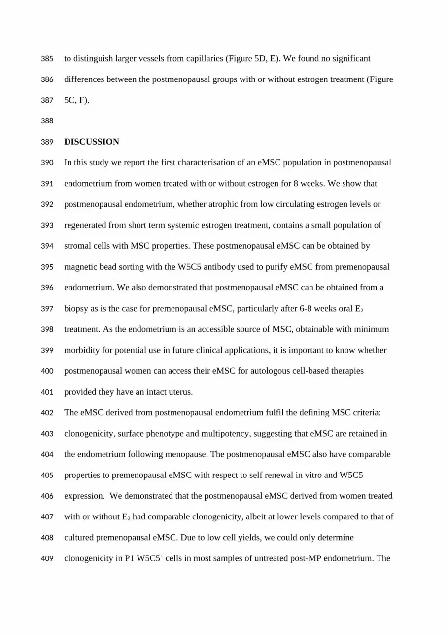
to distinguish larger vessels from capillaries (Figure 5D, E). We found no significant
differences between the postmenopausal groups with or without estrogen treatment (Figure
5C, F).
DISCUSSION
In this study we report the first characterisation of an eMSC population in postmenopausal
endometrium from women treated with or without estrogen for 8 weeks. We show that
postmenopausal endometrium, whether atrophic from low circulating estrogen levels or
regenerated from short term systemic estrogen treatment, contains a small population of
stromal cells with MSC properties. These postmenopausal eMSC can be obtained by
magnetic bead sorting with the W5C5 antibody used to purify eMSC from premenopausal
endometrium. We also demonstrated that postmenopausal eMSC can be obtained from a
biopsy as is the case for premenopausal eMSC, particularly after 6-8 weeks oral E2
treatment. As the endometrium is an accessible source of MSC, obtainable with minimum
morbidity for potential use in future clinical applications, it is important to know whether
postmenopausal women can access their eMSC for autologous cell-based therapies
provided they have an intact uterus.
The eMSC derived from postmenopausal endometrium fulfil the defining MSC criteria:
clonogenicity, surface phenotype and multipotency, suggesting that eMSC are retained in
the endometrium following menopause. The postmenopausal eMSC also have comparable
properties to premenopausal eMSC with respect to self renewal in vitro and W5C5
expression. We demonstrated that the postmenopausal eMSC derived from women treated
with or without E2 had comparable clonogenicity, albeit at lower levels compared to that of
cultured premenopausal eMSC. Due to low cell yields, we could only determine
clonogenicity in P1 W5C5+ cells in most samples of untreated post-MP endometrium. The
385
386
387
388
389
390
391
392
393
394
395
396
397
398
399
400
401
402
403
404
405
406
407
408
409
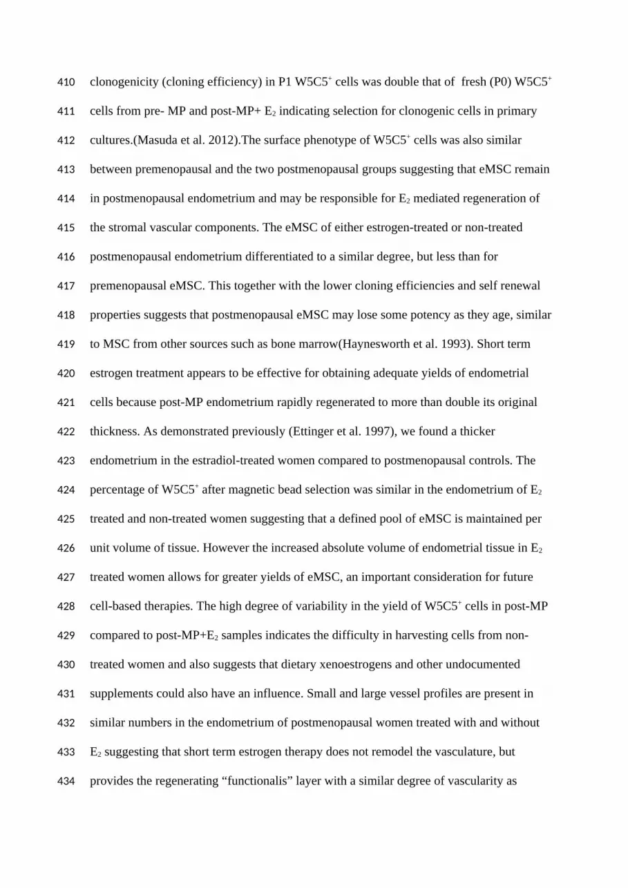
clonogenicity (cloning efficiency) in P1 W5C5+ cells was double that of fresh (P0) W5C5+
cells from pre- MP and post-MP+ E2 indicating selection for clonogenic cells in primary
cultures.(Masuda et al. 2012).The surface phenotype of W5C5+ cells was also similar
between premenopausal and the two postmenopausal groups suggesting that eMSC remain
in postmenopausal endometrium and may be responsible for E2 mediated regeneration of
the stromal vascular components. The eMSC of either estrogen-treated or non-treated
postmenopausal endometrium differentiated to a similar degree, but less than for
premenopausal eMSC. This together with the lower cloning efficiencies and self renewal
properties suggests that postmenopausal eMSC may lose some potency as they age, similar
to MSC from other sources such as bone marrow(Haynesworth et al. 1993). Short term
estrogen treatment appears to be effective for obtaining adequate yields of endometrial
cells because post-MP endometrium rapidly regenerated to more than double its original
thickness. As demonstrated previously (Ettinger et al. 1997), we found a thicker
endometrium in the estradiol-treated women compared to postmenopausal controls. The
percentage of W5C5+ after magnetic bead selection was similar in the endometrium of E2
treated and non-treated women suggesting that a defined pool of eMSC is maintained per
unit volume of tissue. However the increased absolute volume of endometrial tissue in E2
treated women allows for greater yields of eMSC, an important consideration for future
cell-based therapies. The high degree of variability in the yield of W5C5+ cells in post-MP
compared to post-MP+E2 samples indicates the difficulty in harvesting cells from non-
treated women and also suggests that dietary xenoestrogens and other undocumented
supplements could also have an influence. Small and large vessel profiles are present in
similar numbers in the endometrium of postmenopausal women treated with and without
E2 suggesting that short term estrogen therapy does not remodel the vasculature, but
provides the regenerating “functionalis” layer with a similar degree of vascularity as
410
411
412
413
414
415
416
417
418
419
420
421
422
423
424
425
426
427
428
429
430
431
432
433
434

atrophic endometrium. Our study, also confirms a previously reported lack of difference in
vascularity observed between hormone-treated and non-treated premenopausal
endometrium as determined by semiquantitative CD34 immunohistochemistry (Hickey et
al. 1996). This study also reported a similar vascularity between atrophied and cycling
endometrium.
The luminal epithelial height did not differ between the two postmenopausal groups in
contrast to previous findings (Gomes et al. 1997). This could be due to the shorter E2
treatment time in our study, 6-8 weeks versus three months in the Gomes et al study,
possibly not long enough to remodel the surface epithelium. The glandular epithelial height
also did not show significant differences compared with the basalis layer of premenopausal
women. These findings suggest that short term E2 treatment has little impact on the
endometrial epithelium, suggesting that this approach is safe for those women who are not
contraindicated for taking E2.
Short term oral E2 rapidly increases endometrial growth suggesting that an approximately
8 week treatment is sufficient to obtain a reasonably thick endometrium (Ettinger et al.
1997) without inducing endometrial hyperplasia (Furness et al. 2012) It is not quite clear
how E2 regenerates the endometrium since W5C5+ perivascular cells did not express ERα.
Similar observations were made in studies of label retaining cells in mice (Chan et al.
2012) and side population and clonogenic cells in human endometrium (Cervello et al.
2011, Schuring et al. 2011), suggesting that E2 may act via estrogen receptors on their
neighbouring niche cells (Gargett 2007). Our study also showed that eMSC survived in the
absence of estrogen and do not require E2 for trophic support as they were present in
similar proportion in untreated atrophic and E2 treated post- MP endometrium.
For this study we only supplemented women without contraindicated medical conditions
with 2 mg Progynova daily for 6- 8 weeks and found no associated adverse effects. The
435
436
437
438
439
440
441
442
443
444
445
446
447
448
449
450
451
452
453
454
455
456
457
458
459

participants were closely monitored. An initial dose of one mg Progynova was insufficient
to increase endometrial thickness to yield significant numbers of cells (unpublished
observation).
The endometrium is one of the few tissues where MSC can be obtained without
anaesthesia, invasive and painful interventions, particularly in parous women. MSC from
the endometrium provide an accessible alternate source of MSC for use in cell-based
therapies (Ulrich et al. 2013). It appears that eMSC reside in the endometrium after a
woman’s fertile years have ceased. We show for the first time that these eMSC can be
readily harvested from postmenopausal women using an office based biopsy, particularly if
they are pre-treated with estrogen to regenerate the endometrium to yield sufficient
numbers. Similarly, eMSC can be harvested from premenopausal women (Schuring et al
F&s) The human endometrium is therefore a possible source of MSC independent of a
woman’s age. Declaration of Author’s roles
DU: participation in study design, execution, analysis, manuscript writing and critical
discussion
KST: execution, analysis and critical discussion
JD: execution, analysis and critical discussion
KS: execution, analysis and critical discussion
AC: execution, analysis and critical discussion
AR: participation in study design, manuscript editing and critical discussion
CEG: conception and participation in study design, manuscript editing and critical
discussion
ACKNOWLEDGMENTS
460
461
462
463
464
465
466
467
468
469
470
471
472
473
474
475
476
477
478
479
480
481
482
483

The authors acknowledge Liz Fitzgerald for managing the clinical trial, Yao Han and
Pamela Mamers for collection of the tissue.
FUNDING
This study was supported by the National Health and Medical Research Council
(NHMRC) of Australia grant (1021126) (CEG, AR) and Senior Research Fellowship
(1042298) (CEG), Australian Gynaecological Endoscopic Society grant (AR) and
Victorian Government's Operational Infrastructure Support Program.
CONFLICT OF INTEREST
The authors have nothing to declare.
484
485
486
487
488
489
490
491
492

FIGURE LEGENDS
Figure 1 Effect of oral estrogen on postmenopausal endometrium compared with
premenopausal endometrium.
H+E stained endometrium from hysterectomy tissues in (A) pre- MP, (B) post- MP, (C)
post- MP+E2. Dotted line indicates border between endometrium and myometrium. Scale
bar 200 µm. (D) endometrial thickness, (E) luminal epithelial (LE) height, and (F)
glandular epithelial (GE) height measured in the basalis layer of Pre-MP endometrium .
Bars are mean SEM of n =6 samples/group. *P < 0.05.
Figure 2
Properties of postmenopausal eMSC: (A) surface phenotype showing representative flow
cytometric analysis of P1 cultured postmenopausal W5C5+ cells from a single
representative sample from a woman treated with (+E2) or without (-E2) oral estrogen for
6-8 weeks. Aggregate data is shown in Table 2. Multilineage differentiation of P1 W5C5+
cells from post- MP+E2 endometrium in various induction media for 4 weeks (B)
adipocytes stained with Oil Red O, (C) chondrocytes (red nuclei) showing production of
Alcian Blue stained cartilage-like matrix, (D) alkaline phosphatase positive osteoblasts and
(E) smooth muscle cells immunostained with -smooth muscle actin. Insets are stained
control cultures. Scale bars; B, D and E 100 µm; C 25µm.
Figure 3
Cloning efficiency and serial cloning analysis for measuring clonogenicity and self-
renewal of human endometrial pre- and postmenopausal eMSC. Representative cloning
plates of (A) post-MP and (B) post-MP+E2 W5C5+ cells. Arrows show individual clones
removed for serial cloning. Clonogenicity of (C) P1 W5C5+ cells from pre-MP, post-MP
and post- MP+ E2 samples and (D) at each round of serial cloning. Results are means ±
SEM (n=6/group). **Significant difference between pre-MP and post-MP (P < 0.01) and
493
494
495
496
497
498
499
500
501
502
503
504
505
506
507
508
509
510
511
512
513
514
515
516
517

post- MP±E2 (** P < 0.01). *P < 0.05, ***P<0.001. S0, first cloning; S1, first serial
cloning (secondary clones); S2, second serial cloning (tertiary clones).
Figure 4
W5C5+ perivascular cells do not express ERα. Dual colour immunofluorescence of (A)
post- MP and (B) post- MP+E2 endometrial tissue. W5C5+ cells around blood vessels
fluoresce red, ERα positive nuclei fluoresces green-blue. Unstained nuclei blue (Hoechst
stained). Scale bar 20 µm. Arrow indicates representative perivascular W5C5+ cells. G,
gland.
Figure 5
Vessel profiles in postmenopausal endometrium. CD34 immunostained (A) post- MP and
(B) post- MP+E2 endometrium with (C) immunoquantification showing % of CD34
positive area. Dotted lines show endometrial myometrial border. Alpha-SMA
immunostained (D) post- MP, and (E) post- MP+E2 endometrium counterstained with
haematoxylin, with (F) immunoquantification showing % αSMA positive area. Scale bar in
A+B 100 µm, D+E 200 µm. Bars are means SEM of n= 8 post-MP and n= 10 post-
MP+E2.
518
519
520
521
522
523
524
525
526
527
528
529
530
531
532
533
534
535
536
537
538

References
Abberton, K. M., D. L. Healy and P. A. W. Rogers. Smooth muscle alpha actin and myosin heavy chain expression in the vascular smooth muscle cells surrounding human endometrial arterioles. Hum. Reprod. 1999; 14(12): 3095-3100.
Beltrami, A. R., D. Cesselli, N. Bergamin, P. Marcon, S. Rigo, E. Puppato, F. D'Aurizio, R. Verardo, S. Piazza, A. Pignatelli, A. Poz, U. Baccarani, D. Damiani, R. Fanin, L. Mariuzzi, N. Finato, P. Masolini, S. Burelli, O. Belfuzzi, C. Schneider and C. A. Beltrami. Multipotent cells can be generated in vitro from several adult human organs (heart, liver, and bone marrow). Blood 2007; 110(9): 3438-3446.
Boennelycke, M., S. Gras and G. Lose. Tissue engineering as a potential alternative or adjunct to surgical reconstruction in treating pelvic organ prolapse. Int Urogynecol J 2013; 24(5): 741-747.
Buhring, H. J., V. L. Battula, S. Treml, B. Schewe, L. Kanz and W. Vogel. Novel markers for the prospective isolation of human MSC. Ann. N. Y. Acad. Sci. 2007; 1106: 262-271.
Caplan, A. I. Adult mesenchymal stem cells for tissue engineering versus regenerative medicine. J. Cell. Physiol. 2007; 213(2): 341-347.
Cervello, I., A. Mas, C. Gil-Sanchis, L. Peris, A. Faus, P. T. K. Saunders, H. O. D. Critchley and C. Simon. Reconstruction of Endometrium from Human Endometrial Side Population Cell Lines. PLoS One 2011; 6(6).
Chan, R. W., K. E. Schwab and C. E. Gargett. Clonogenicity of human endometrial epithelial and stromal cells. Biol. Reprod. 2004; 70(6): 1738-1750.
Chan, R. W. S., T. Kaitu'u-Lino and C. E. Gargett. Role of Label-Retaining Cells in Estrogen-Induced Endometrial Regeneration. Reprod. Sci. 2012; 19(1): 102-114.
Crisan, M., S. Yap, L. Casteilla, C. W. Chen, M. Corselli, T. S. Park, G. Andriolo, B. Sun, B. Zheng, L. Zhang, C. Norotte, P. N. Teng, J. Traas, R. Schugar, B. M. Deasy, S. Badylak, H. J. Buhring, J. P. Giacobino, L. Lazzari, J. Huard and B. Peault. A perivascular origin for mesenchymal stem cells in multiple human organs. Cell Stem Cell 2008; 3(3): 301-313.
da Silva Meirelles, L., P. C. Chagastelles and N. B. Nardi. Mesenchymal stem cells reside in virtually all post-natal organs and tissues. J. Cell Sci. 2006; 119(Pt 11): 2204-2213.
Dominici, M., K. Le Blanc, I. Mueller, I. Slaper-Cortenbach, F. Marini, D. Krause, R. Deans, A. Keating, D. Prockop and E. Horwitz. Minimal criteria for defining multipotent mesenchymal stromal cells. The International Society for Cellular Therapy position statement. Cytotherapy 2006; 8(4): 315-317.
Ettinger, B., L. Bainton, D. H. Upmalis, J. T. Citron and A. VanGessel. Comparison of endometrial growth produced by unopposed conjugated estrogens or by micronized estradiol in postmenopausal women. Am. J. Obstet. Gynecol. 1997; 176(1): 112-117.
Furness, S., H. Roberts, J. Marjoribanks and A. Lethaby. Hormone therapy in postmenopausal women and risk of endometrial hyperplasia. Cochrane Database of Systematic Reviews 2012;(8).
Gargett, C. E. Uterine stem cells: What is the evidence? Hum. Reprod. Update 2007; 13(1): 87-101.
539
540541542
543544545546547
548549550
551552
553554
555556557
558559
560561
562563564565566
567568
569570571572
573574575
576577578
579580

Gargett, C. E., H. P. T. Nguyen and L. Ye. Endometrial regeneration and endometrial stem/progenitor cells. Rev. Endocr. Metab. Disord. 2012; 13(4): 235-251.
Gargett, C. E., K. E. Schwab, R. M. Zillwood, H. P. Nguyen and D. Wu. Isolation and culture of epithelial progenitors and mesenchymal stem cells from human endometrium. Biol. Reprod. 2009; 80(6): 1136-1145.
Gomes, A. M. N., E. C. Baracat, M. J. Simoes, M. A. Haidar, G. R. A. Focchi, J. EvencioNeto and G. R. deLima. Morphologic and morphometric aspects of the endometrium of postmenopausal women before and after cyclic oestrogen replacement treatment. European Journal of Obstetrics Gynecology and Reproductive Biology 1997; 74(1): 79-82.
Gronthos, S., A. C. Zannettino, S. J. Hay, S. Shi, S. E. Graves, A. Kortesidis and P. J. Simmons. Molecular and cellular characterisation of highly purified stromal stem cells derived from human bone marrow. J. Cell Sci. 2003; 116(Pt 9): 1827-1835.
Haynesworth, S. F., V. M. Goldberg and A. I. Caplan. Diminution of the number of mesenchymal stem-cells as a cause for skeletal aging. 1993. Musculoskeletal Soft-Tissue Aging: Impact on Mobility. J. A. Buckwalter, V. M. Goldberg and S. L. Y. Woo: 79-87.
Hickey, M., T. M. Lau, P. Russell, I. S. Fraser and P. A. W. Rogers. Microvascular density in conditions of endometrial atrophy. Hum. Reprod. 1996; 11(9): 2009-2013.
Hunskaar, S., K. Burgio, A. Clark, M. C. Lapitan, R. Nelson, U. Sillen and D. Thom. Epidemiology of urinary (UI) and faecal (FI) incontinence and pelvic organ prolapse (POP). 2005. http://www.icsoffice.org/Publications/ICI_3/v1.pdf/chap5.pdf.
Le Blanc, K. and D. Mougiakakos. Multipotent mesenchymal stromal cells and the innate immune system. Nat. Rev. Immunol. 2012; 12(5): 383-396.
Masuda, H., S. S. Anwar, H.-J. Buehring, J. R. Rao and C. E. Gargett. A Novel Marker of Human Endometrial Mesenchymal Stem-Like Cells. Cell Transplant. 2012; 21(10): 2201-2214.
Morikawa, S., Y. Mabuchi, Y. Kubota, Y. Nagai, K. Niibe, E. Hiratsu, S. Suzuki, C. Miyauchi-Hara, N. Nagoshi, T. Sunabori, S. Shimmura, A. Miyawaki, T. Nakagawa, T. Suda, H. Okano and Y. Matsuzaki. Prospective identification, isolation, and systemic transplantation of multipotent mesenchymal stem cells in murine bone marrow. J. Exp. Med. 2009; 206(11): 2483-2496.
Nguyen, H. P., C. N. Sprung and C. E. Gargett. Differential expression of Wnt signaling molecules between pre- and postmenopausal endometrial epithelial cells suggests a population of putative epithelial stem/progenitor cells reside in the basalis layer. Endocrinology 2012; 153(6): 2870-2883.
Paulson, R. J., R. Boostanfar, P. Saadat, E. Mor, D. E. Tourgeman, C. C. Slater, M. M. Francis and J. K. Jain. Pregnancy in the sixth decade of life - Obstetric outcomes in women of advanced reproductive age. Jama-Journal of the American Medical Association 2002; 288(18): 2320-2323.
Pines, A., D. W. Sturdee and A. H. MacLennan. Quality of life and the role of menopausal hormone therapy. Climacteric 2012; 15(3): 213-216.
Pittenger, M. F., A. M. Mackay, S. C. Beck, R. K. Jaiswal, R. Douglas, J. D. Mosca, M. A. Moorman, D. W. Simonetti, S. Craig and D. R. Marshak. Multilineage potential of adult human mesenchymal stem cells. Science 1999; 284(5411): 143-147.
581582
583584585
586587588589590
591592593
594595596
597598
599600601
602603
604605606
607608609610611
612613614615
616617618619
620621
622623624

Prockop, D. J. Marrow stromal cells as steam cells for nonhematopoietic tissues. Science 1997; 276(5309): 71-74.
Rajaraman, G., J. White, K. S. Tan, D. Ulrich, A. Rosamilia, J. A. Werkmeister and C. E. Gargett. Optimisation and scale up culture of human endometrial multipotent mesenchymal stromal cells: potential for clinical application. Tissue Eng Part C Methods 2013; 19(1): 80-92.
Salem, H. K. and C. Thiemermann. Mesenchymal stromal cells: current understanding and clinical status. Stem Cells 2010; 28(3): 585-596.
Schuring, A. N., J. Braun, S. Wullner, L. Kiesel and M. Gotte. mRNA-expression of ERalpha, ERbeta, and PR in clonal stem cell cultures obtained from human endometrial biopsies. TheScientificWorldJournal 2011; 11: 1762-1769.
Schwab, K. E., R. W. S. Chan and C. E. Gargett. Putative stem cell activity of human endometrial epithelial and stromal cells during the menstrual cycle. Fertil. Steril. 2005; 84: 1124-1130.
Schwab, K. E. and C. E. Gargett. Co-expression of two perivascular cell markers isolates mesenchymal stem-like cells from human endometrium. Hum. Reprod. 2007; 22(11): 2903-2911.
Simmons, P. J. and B. Torok-Storb. Identification of stromal cell precursors in human bone marrow by a novel monoclonal antibody, STRO-1. Blood 1991; 78(1): 55-62.
Sivasubramaniyan, K., A. Harichandan, S. Schumann, M. Sobiesiak, C. Lengerke, A. Maurer, H. Kalbacher and H. J. Buhring. Prospective Isolation of Mesenchymal Stem Cells from Human Bone Marrow Using Novel Antibodies Directed Against Sushi Domain Containing 2. Stem cells and development 2013; 22(13): 1944-1954.
Torrente, Y. and E. Polli. Mesenchymal Stem Cell Transplantation for Neurodegenerative Diseases. Cell Transplant. 2008; 17(10-11): 1103-1113.
Ulrich, D., S. L. Edwards, K. Su, K. S. Tan, J. F. White, J. A. Ramshaw, C. Lo, A. Rosamilia, J. A. Werkmeister and C. E. Gargett. Human Endometrial Mesenchymal Stem Cells Modulate the Tissue Response and Mechanical Behaviour of Polyamide Mesh Implants for Pelvic Organ Prolapse Repair. Tissue Eng. 2013; 20(3-4): 785-798.
Ulrich, D., S. L. Edwards, J. F. White, T. Supit, J. A. M. Ramshaw, C. Lo, A. Rosamilia, J. A. Werkmeister and C. E. Gargett. A Preclinical Evaluation of Alternative Synthetic Biomaterials for Fascial Defect Repair Using a Rat Abdominal Hernia Model. PLoS One 2012; 7(11).
Ulrich, D., R. Muralitharan and C. Gargett. Toward the use of endometrial and menstrual blood mesenchymal stem cells for cell-based therapies. Expert Opin. Biol. Ther. 2013; 13(10).
625626
627628629630
631632
633634635
636637638
639640641
642643
644645646647
648649
650651652653
654655656657
658659660
661
662


