Embed Size (px)
Citation preview

Mesolimbic Component of the Ascending Cholinergic Pathways:Electrophysiological-Pharmacological Study
STEFAN M. BRUDZYNSKI,1 LUDMILA KADISHEVITZ,2 AND XIAO-WEN FU2
1Department of Psychology, Brock University, St. Catharines, Ontario L2S 3A1; and 2Department of ClinicalNeurological Sciences, London Health Sciences Centre, London, Ontario N6A 5A5, Canada
Brudzynski, Stefan M., Ludmila Kadishevitz, and Xiao-Wen nuclei in the thalamus, hypothalamus, basal forebrain, sep-Fu. Mesolimbic component of the ascending cholinergic pathways: tum, basal ganglia and medial frontal, and olfactory corticeselectrophysiological-pharmacological study. J. Neurophysiol. 79: (Cornwall et al. 1990; Fibiger 1982; Hallanger et al. 1987;1675–1686, 1998. The cholinergic input from the pontomes- Herrero et al. 1991; Jones and Beaudet 1987; Paxinos andencephalic cholinergic neurons to the diencephalic and basal fore- Butcher 1985; Satoh and Fibiger 1986; Semba et al. 1988;brain structures has been implicated in a number of limbically
Vincent et al. 1986; Woolf and Butcher 1986; Woolf etcontrolled overt behaviors. The cellular mechanism by which theal. 1990). The functional role of this ascending cholinergiccholinergic terminals initiate behavioral manifestations is not clear.system remains the key for understanding sleep-waking cy-The objective of this study was to investigate the effects of thecle, emotional arousal, and the initiation of a number ofascending cholinergic projection from the laterodorsal tegmental
nucleus (LDT) on neuronal firing in the anterior hypothalamic- defensive and alarm behavioral patterns.medial preoptic region (AHMP), known to be involved in agonistic The present study is focused on the component of thebehavior. Experiments were performed on urethan-anesthetized ascending projections from the pontomesencephalic cholin-rats. Iontophoretic application of carbachol (CCh) into the vicinity ergic cells, which are targeted at the medial limbic structuresof single cells in the AHMP caused a dose-dependent decrease in
(Consolo et al. 1990; Cornwall et al. 1990; Lewis and Shutethe mean firing rate of 83% of units and an increase in 10% of1967; Mesulam et al. 1989). The ascending projections formunits. The inhibitory effect of CCh, but not the excitatory effect,a mesolimbic component of the ascending reticular activat-was reversed by iontophoretic pretreatment with scopolamine. Theing system (Moruzzi and Magoun 1949; Shute and Lewisinhibition of the firing rate was repeatable for the same dose of
CCh and dose dependent. Electrical stimulation of neurons in the 1963, 1967), which plays an important role in the regulationLDT caused a comparable, current-dependent decrease in the mean of limbically driven behavior. The ascending cholinergicfiring rate of AHMP neurons that also was reversed by pretreatment projections are associated with the activation that accompanyof neurons in the AHMP with scopolamine. The antagonizing ef- the states of arousal, wakefulness, and paradoxical sleep,fects of scopolamine were reversible with time. The same units in behavioral activation, emotional arousal manifested by spe-the AHMP that inhibited their firing to stimulation of the LDT also
cies-typical threatening or alarm vocalizations, and auto-responded with a similar inhibition to local iontophoretic CCh.nomic symptoms (Brudzynski 1981, 1994; Imeri et al. 1995;Finally, the fluorescent carbocyanine dye, 4-(4-(dihexadecylami-Jones 1993; Talwar and Kumar 1994). It has been demon-no)styryl)-N-methylpyridinium iodide, (DiA), has been used as
a retrograde axonal tracer and was injected into the recording sites strated in the past that systemic or intraventricular cholino-immediately after the electrophysiological recordings. After 1 wk, mimetic agents with strong muscarinic properties have po-DiA dye was found in numerous neurons in the LDT as shown by tent effects on the general behavior, particularly in inducingthe fluorescence confocal microscopy. Results of the study suggest autonomic manifestations, defensive vocalization, and ago-that LDT cholinergic neurons project and terminate in the AHMP nistic behavioral responses to external stimuli (Baker et al.and that their activation causes a decrease in the mean firing rate
1960; Funke et al. 1962; Leslie 1965; Koff and Langfittof the AHMP neurons. It is postulated that this inhibitory effect is1966). However, all these symptoms could not be obtainedimplicated in the initiation of some of the behavioral patterns likewhen lesions of various limbic, particularly medial dience-defensive or alarm vocalization and behavioral inhibition.phalic structures, had been made (Gellen et al. 1972; Koffand Langfitt 1966).
I N T R O D U C T I O N It has been demonstrated by using direct intracerebral in-jection of muscarinic cholinergic agents in cats, that theseOn the basis of results of immunohistochemical studiesbehavioral symptoms were induced from an elongated, butand in situ hybridization for choline acetyltransferaselimited, strip of medial structures, from the periaqueductalmRNA, two distinctive subgroups of cholinergic perikaryagray, through the medial hypothalamic and preoptic regions,(CH5 and Ch6) were distinguished in the pontomesencepha-and intralaminar thalamic nuclei to the septal nuclei (Baxterlic region within the pedunculopontine and in the laterodor-1967, 1968; Brudzynski and Eckersdorf 1988; Brudzynskisal tegmental nuclei (LDT) (Armstrong et al. 1983; Butcheret al. 1995; Decsi 1974; Decsi and Nagy 1977; Decsi et al.1995; Kimura et al. 1981; Lauterborn et al. 1993; Mesulam1969; Myers 1964; Varszegi and Decsi 1967).et al. 1983, 1984, 1989; Vilaro et al. 1994). Although a
Similar responses to direct cholinergic stimulation, mea-substantial portion of the pedunculopontine cholinergic neu-sured by changes in locomotor activity and by the productionrons has descending projections (Jones 1990; Woolf andof 22-kHz ultrasonic alarm calls, also have been reportedButcher 1989), the LDT has diverse and extensive ascending
projections. The ascending projections innervate numerous for the rat (Brudzynski and Bihari 1990; Brudzynski et al.
16750022-3077/98 $5.00 Copyright q 1998 The American Physiological Society
J376-7/ 9k27$$ap17 03-04-98 13:05:34 neupal LP-Neurophys

S. M. BRUDZYNSKI, L. KADISHEVITZ, AND X.-W. FU1676
1.5 mm from midline, and from 5.0 to 6.5 mm below the cortex1990). Functional mapping of the decrease in locomotorsurface (Paxinos and Watson 1986).activity and vocalizational responses in the rat brain deline-
ated a similar brain system to that in the cat brain (Brudzyn-ski 1994; Brudzynski et al. 1989). Moreover, results of the Electrical stimulation and extracellular single-unit
recordingsmapping studies in these two species outlined a system strik-ingly similar to the pattern of projection of the ascending
Electrical stimulation was delivered by stainless steel concentricpontomesencephalic cholinergic neurons (Satoh and Fibiger bipolar electrodes (NE-100, Rhodes Medical Instruments, Wood-1986; Woolf at al. 1990). land Hills, CA) with tip separation of 0.5 mm. The monophasic
It has been postulated, therefore, that cholinergically in- square-wave pulses (1-ms width, 100–800 mA, 1 Hz) were gener-duced behavioral responses are triggered by cholinergic ter- ated by a Grass S44 stimulator coupled to a Grass stimulationminals originating from the pontomesencephalic cholinergic isolation unit (SIU-5). Six-barrel pipettes for recording and ionto-
phoresis were pulled from F. Haer capillary glass tubing (1.0 mmcells (Brudzynski and Barnabi 1996). It has been shown inOD) with a Narishige vertical puller (Model PE-2). The centralrecent experiments on rats, that stimulation of neurons ofbarrel was filled with 2 M NaCl solution and served as a recordingthe LDT with L-glutamate induced comparable 22-kHzelectrode. The electrode impedance ranged from 3 to 7 MV, asalarm calls to those induced by cholinergic stimulation ofmeasured by microelectrode tester (model BL-100, Winston Elec-mediobasal diencephalic structures. Moreover, L-glutamatetronics, San Francisco, CA) using 135 Hz AC test current of 5 1stimulation of the LDT was ineffective, or less effective, 10010 A. One of the side barrels was filled with 2% pontamine sky
when the terminal fields in the anterior hypothalamic-medial blue (Chicago sky blue, Sigma Chemical, St. Louis, MO) in 0.5preoptic region (AHMP) were pretreated with scopolamine, M sodium acetate for marking of the recording site. Recordeda muscarinic antagonist (Brudzynski and Barnabi 1996). signals were connected to the probe headstage of the Axoprobe-1
The cellular mechanism by which the ascending choliner- DC amplifier (Axon Instruments, Forest City, CA) and then cou-pled to a signal processing unit for high-pass filtering and furthergic inputs initiate behavioral manifestations is not clear. Itamplification (Intronix, Rexdale, ON). The amplified signal washas been found in the acute rat preparation that a predomi-fed into a window discriminator (Frederick Haer, Brunswick, MA)nantly muscarinic agent—carbachol (CCh) —caused a de-to isolate action potentials. Standard pulses corresponding to thecrease in the firing rate of spontaneously active neurons inindividual action potentials were multiplexed with stimulus triggersthe AHMP (Brudzynski et al. 1991). The question arisesand event markers and fed to an AST 286 personal computerwhether or not these neuronal responses can be attributed to equipped with a Data-Translation DT2801A A/D conversion card.
the effects of the ascending cholinergic projection from the The multiplexed signal was sampled and decoded at 1 kHz by apontomesencephalic cell groups. PCIPEE program (CY Electronics, London, ON). The data were
The goal of the present study is to test the hypothesis displayed continuously in real time as running time histogram orthat the ascending cholinergic fibers originating from the poststimulus time histogram during sampling and saved for analy-
sis. Peristimulus time histograms were compiled from the storedpontomesencephalic cells have a predominantly inhibitorydata files off-line. Significant changes in activity of neurons afterinfluence on the firing rate of neurons in the rat mediobasalstimulation were quantified by comparing the height of each 1-msdiencephalic regions from which behavioral responses topoststimulus bin of the peristimulus time histogram with the aver-cholinergic stimulation have been obtained. The AHMP re-age height of the bin for 100 ms before the stimulus. These datagion has been chosen for the study because the prominentalso were recalculated for the mean firing rate/s and compared forbehavioral manifestations with the decrease in locomotor three arbitrary chosen, 100-ms bouts of time: 100 ms before the
activity (Brudzynski et al. 1989) and high magnitude of stimulus, 100 ms immediately after the stimulus, and 101–200 msalarm vocalizations (Brudzynski 1994) have been obtained after the stimulus. Significant changes in the activity of neuronsfrom that region. after iontophoretic ejection were quantified by comparing the mean
firing rate for 20-s ejection bout with the mean firing rate/20 simmediately before and after the ejection.
M E T H O D S
Animals and surgery Microiontophoresis
Side barrels of the multibarrel pipette were filled with 0.5 MSeventy-four male, Wistar rats, weighing 230–350 g were usedin the study. Animals were anesthetized with an initial urethan centrifuged solutions of carbachol (CCh, carbamylcholine chlo-
ride) , sodium glutamate, or scopolamine hydrochloride, saline anddose of 1.15 g/kg ip and a supplementary dose of 0.08 g/kg wasadded after a couple of hours if necessary. The total dose of urethan 2% pontamine sky blue (all compounds from Sigma Chemical) .
All pharmacological agents were dissolved in distilled water andwas within the range of doses that do not suppress spontaneousactivity of neurons in explored areas (Cross and Dyer 1971; Dyball retained with 1 nA current. The current was generated by a Dagan
6400 Micro-iontophoresis current generator (Dagan, Minneapolis,and McPhail 1974). Rats were placed in a Kopf stereotaxic appara-tus in the Faraday enclosure and their body temperature maintained MN). For the technical description of the generator, see Stone
(1985). The drugs were ejected using °120 nA current (usuallyat 37.5 { 0.37C as measured by the rectal probe. The skull wasopened, the dura reflected, and the exposed brain surface was cov- 20–60 nA), positive for CCh, scopolamine, or saline and negative
for glutamate, applied for 20–30 s for a single agent and °70 sered with warmed 4% agar in Ringer solution after the electrodepenetration. The recording of single-unit activity was performed for double ejection. Periods of maintaining the retention current
between ejections were controlled in time to avoid build-up offrom the AHMP between stereotaxic frontal planes 7.0 and 8.5mm anterior from the interaural zero plane, between sagittal planes solute at the tip of the pipette and to allow a quantitative assessment
of the successive ejection-induced changes in the firing rate (Brad-0.1 and 1.3 mm lateral from midline, and from 7 to 9.5 mm belowthe surface of the cortex according to the stereotaxic atlas by Pax- shaw et al. 1973). The current effect was compensated automati-
cally by an additional barrel filled with 2 M NaCl. For furtherinos and Watson (1986). Electrodes for electrical stimulation wereplaced stereotaxically in the LDT and adjoining structures, between details of the recording and iontophoresis, see Brudzynski et al.
(1991). To antagonize the effects of CCh, scopolamine was ejectedfrontal planes 0.9 and 00.4 mm, between sagittal planes 0.1 and
J376-7/ 9k27$$ap17 03-04-98 13:05:34 neupal LP-Neurophys

MESOLIMBIC CHOLINERGIC PATHWAYS 1677
dialkylaminostyryl probe, DiA [4-(4-(dihexadecylamino)styryl)-N-methylpyridinium iodide, D-3883, Molecular Probes, EugeneOR], has been used as a retrograde axonal tracer. DiA is brightfluorescent green (Snider et al. 1992), diffuses more rapidly inmembranes, and stains membranes more uniformly than other car-bocyanine dyes. Injection solution of DiA was prepared by dissolv-ing 2.5 mg of DiA in 0.9 ml of 100% ethanol with 0.1 ml ofdimethylsulfoxide and subsequently filtering through 5-mm porefilter (Millipore, Bedford, MA). After the electrophysiological re-cordings were finished, 0.1 ml of DiA was injected into the re-cording site in the AHMP via a glass pipette (50- to 60-mm tipdiam) connected with a Hamilton 1-ml syringe. Seven animals witha body weight not exceeding 300 g were injected. After 6 h survivaltime, the rats were anesthetized deeply with urethan and perfusedtranscardially with 0.9% NaCl solution followed by 10% formalde-hyde in 0.1 M phosphate buffer saline (PBS). The brains wereremoved, stored in fixative for 48 h at 277C and subsequentlyimmersed in 10, 20, and 30% sucrose in PBS for 24 h in eachconcentration. After 1 wk of storage in a black box at room temper-ature, the brains were sectioned for 50-mm horizontal preparationson a cryostat microtome (Leitz, Canada), transferred into PBSsolution, mounted immediately on glass slides, coverslipped usingDPX mountant (BDH Chemicals, Toronto, ON) and stored at0157C for confocal microscopy analysis.
Confocal microscopyThe DiA-injected preparations were analyzed under Bio-Rad
MRC-600 confocal laser scanning microscope equipped with kryp-
FIG. 1. Responses of anterior hypothalamic-medial preoptic neurons(AHMP) to inotophoretically applied carbachol at 20–60 nA of the ejectingcurrent. A : percentage of inhibitory, excitatory responses, and unrespondingneurons to carbachol. B : averaged neuronal activity of the same neuronsexpressed in the mean firing rate per second during 20-s ejection of carba-chol (CCh) in comparison with 20-s control period before and after applica-tion of carbachol (Con). Data are shown for inhibitory results ( left) , excit-atory results (middle) , and no responding units (right 3 bars) . Verticalbars represent {SE. **t[82] Å 5.5, P õ 0.0001 in comaprison to the initialbaseline firing rate (Con). There was no significant difference betweenbaseline firing rates of cells that showed inhibition vs. excitation ( t-test,Pú 0.5) .
from an adjoining barrel (1–5 nA) just before the ejection of CChin such a way that both ejection periods partially overlapped. Insome cells, no ejecting current was applied and scopolamine wasallowed to passively diffuse from the tip of the pipette during theconcurrent ejection of CCh. Effects obtained by ejection of 1–5nA of scopolamine or by its passive diffusion were basically thesame. Passive diffusion appeared to sufficiently antagonize theeffects of CCh and provided evidence that effects of scopolaminewere not caused by a potential local anesthetic action of this drug(Curtis and Phillis 1960).
Fluorescent tracer injections
To study neuronal connections of the recording site, the groupof a long-alkyl chain, carbocyanine and aminostyryl dyes havebeen selected (Balice-Gordon et al. 1993; Honing and Hume 1986,1989). This group of dyes was chosen because they can be injectedpostmortem in fixed tissues (Godement et al. 1987; Mufson et al.
FIG. 2. Examples of inhibitory responses of 2 anterior hypothalamic-1990) in the same animals that were used for electrophysiologicalmedial preoptic neurons (1 in A and the other in B) to iontophoretic applica-recordings in nonsurvival experiments. The dyes do not transfertion of CCh for 20 s with the increasing ejecting current. A : running timebetween intact membranes and have been used as retrograde tracershistogram showing inhibitory responses to the increasing doses of carbachol(Godement et al. 1987; Honig and Hume 1989). These lipophilic at the ejecting current of 20–60 nA. Ejection time for carbachol at 60 nA
dyes can bind both in vivo and in vitro to lipid components of the was prolonged to 30 s. B : inhibitory responses to carbachol were repeatableplasma membrane, diffuse in the plane of the membrane, resulting for the same ejecting current (20 nA) but potentiated by an increase in thein uniform staining of the entire cell plasma membrane (Godement ejecting current (40 nA). Same unit responded with an increase in firing
rate to ejection of glutamate (GLU) at 20 nA.et al. 1987). After electrophysiological recording, the lipophilic
J376-7/ 9k27$$ap17 03-04-98 13:05:34 neupal LP-Neurophys

S. M. BRUDZYNSKI, L. KADISHEVITZ, AND X.-W. FU1678
FIG. 4. Location of the recording site with ejecting pipettes for CChand SCO in the anterior hypothalamic medial preoptic region (AHMP),and the stimulating electrode in the laterodorsal tegmental nucleus (LDT)shown on the parasagittal section of the rat brain. Scale is in mm from thefrontal, interaural zero plane, according to the Paxinos and Watson (1986)stereotaxic atlas. BN, bed nucleus of stria terminalis; cc, corpus callosum;CE, cerebellum; HI, hippocampal formation; HY, medial and posteriorhypothalamus; NA, nucleus accumbens; OB, olfactory bulb; PAG, perique-ductal grey matter; RF, reticular formation; SE, septal nuclei; TH, thalamicFIG. 3. Effects of iontophoretically applied scopolamine (SCO) on thenuclei.neuronal responses in the anterior hypothalamic-medial preoptic region
(AHMP) induced by ejection of CCh. A : running time histogram showingmean firing rate of an AHMP neuron. Unit showed decrease in the meanfiring rate to ejection of CCh (20 nA) for 25 s. Inhibitory response wasreversed by pretreatment with scopolamine (SCO) ejected at 1 nA for 25s. This antagonism was repeatable for the same cell. B : averaged neuronalactivity of 8 neurons expressed in mean firing rate ({SE) during ejectionof CCh (20–40 nA) compared with the control activity 20 s before andafter application of carbachol (Con, left 3 bars) . Inhibitory response tocarbachol was reversed by pretreatment with scopolamine at 1–2 nA(SCO / CCh, right 3 bars) . **t[7] Å 6.5, P õ 0.0001 in comparison withthe activity before carbachol (Con) and t[7] Å 8.2, Põ 0.0001 in compari-son to the activity during carbachol with scopolamine pretreatment (SCO /CCh, right) .
ton/argon mixed gas laser (Baker and Reese 1993). The filterblock, which allows detection of a green-emitting probe was used.The confocal imaging was made with a 110, 140, and 160 Nikonoil immersion Fluotar lens. The gain setting varied from 3 to 8depending on the contrast of the image. Sequential optical sectionswere collected as a function of the depth of the tissue by automati-cally indexing the position of the specimen stage along the z axisand recording the image at each step. Each optical section was thesynthesis of 15–17 scans, which were processed digitally by Kal-man filtering to optimize the signal-to-noise ratio. The resultingoptical images appeared as white neurons on a black background.Occasionally, for better visualization of the dendritic arborization,pseudocolor was used and the images were merged using all-pixelmerge mode, to indicate the site of maximal luminescence. Toseparate the labeled and unlabeled regions of the preparation, linescanning through the labeled neuron and unlabeled surroundingbackground areas were performed and curves of maximum pixelintensity were obtained. Prints of selected images were obtained
FIG. 5. Peristimulus histograms showing 3 types of neuronal responsesfrom a color video printer (Sony, model UP-5000).of the AHMP neurons to single pulse (2 ms) stimulation (S) of the latero-dorsal tegmental nucleus. A : inhibitory response to 400-mA stimulus com-piled from 123 superimposed sweeps. B : excitatory response to 900-mAHistological verification and statisticsstimulus compiled from 151 superimposed sweeps. C : excitatory response
Recording sites were marked by expelling pontamine sky blue by followed by an inhibition to 800-mA stimulus compiled from 95 superim-posed sweeps.a cathodal current of 5–8 mA for 10–20 min (Lodge et al. 1974)
J376-7/ 9k27$$ap17 03-04-98 13:05:34 neupal LP-Neurophys

MESOLIMBIC CHOLINERGIC PATHWAYS 1679
by a local concurrent application of scopolamine in the AHMP.Some of the neurons were tested for both, iontophoretic CCh andstimulation of the LDT and for the effects of scopolamine on bothof these responses. These results would demonstrate electrophysio-logically that activation of the LDT may produce comparable, mus-carinic neuronal responses in the AHMP to those induced by CChin the same structure. Finally, the last step included the injectionof DiA into the recording site to demonstrate retrogradely labeledcells within the region of the LDT.
R E S U L T S
Effects of CCh on firing rate of AHMP neurons
A total of 100 spontaneously active neurons (100%) werestudied in the AHMP. Neurons with a stable firing rate forat least 40–60 s were selected for further study. The averagefiring rate of units was 2.01 { 0.22 Hz (mean { SE). Inresponse to iontophoretically applied CCh (20- to 60-nAejecting current) , the vast majority of these cells (83%)responded with a decrease in the mean firing rate and only10% of cells showed increase in the mean firing rate (Fig.1A) . On average, the mean firing rate of 2.48 { 0.15 Hzwas significantly reduced to 0.9 { 0.09 Hz during ejection
FIG. 6. Responses of AHMP neurons to single pulse (2 ms) stimulationof the laterodorsal tegmental nucleus. A : percentage of inhibitory (Inhib.) ,excitatory responses (Excit.) , and unresponding units (No Resp.) to stimula-tion. B : averaged neuronal activity of neurons that showed inhibitory re-sponses (n Å 52), expressed in the mean firing rate per second during 100-ms bout immediately after the stimulus (S) and compared with 100-mscontrol period before the stimulus (Before Stim.) and the following 100ms, i.e., from 101–200 ms poststimulus (After Stim.) . Vertical lines repre-sents SE. **t[51] Å 8.14, P õ 0.0001.
and later were observed as an intense blue dot on the histologicalpreparations. Stimulation sites were marked with an electrolytic irondeposition by passing 10 mA anodal current through the stimulatingelectrode for 1 min. The animal then was killed with an overdoseof urethan and transcardially perfused with 0.9% NaCl solutionfollowed by 10% formaldehyde in PBS containing 2% potassiumferricyanide. The ferricyanide reacted in the iron deposit in the tissueto form a Prussian blue spot that marked the tip of the stimulatingelectrode. The sites of recording and stimulation were marked oncomposite diagrams using a drawing attachment of the stereo-zoommicroscope (SZH, Olympus Optical, Japan). Results were analyzedusing Student’s t-test for paired comparisons.
Experimental design
The study was conducted in three steps. The first step was fo-FIG. 7. Responses of the AHMP neurons showing inhibition to increas-cused on the effects of iontophoretically ejected CCh on the firing
ing intensities of the stimulating current applied to the electrode in therate of spontaneously active AHMP neurons and the antagonizinglaterodorsal tegmental nucleus. A : three peristimulus time histograms show-effects of scopolamine on CCh-induced responses. The results ing increasing inhibition to stimulation (S) with 200-, 400-, and 600-mA
would demonstrate predominance of cholinergically induced inhib- current, respectively. Histograms are compiled from 44 to 64 superimposeditory neuronal responses in the AHMP mediated by muscarinic sweeps. B : relationship between the average inhibition time in ms causedreceptors. Second, responses of AHMP neurons to electrical stimu- by stimulation of the laterodorsal tegmental nucleus and the intensity of
the stimulating current in mA. Vertical lines represent SE.lation of the LDT and its vicinity were recorded and antagonized
J376-7/ 9k27$$ap17 03-04-98 13:05:34 neupal LP-Neurophys

S. M. BRUDZYNSKI, L. KADISHEVITZ, AND X.-W. FU1680
not responding cells) . Responses of the units were classifiedaccording to their primary response. Examples of an inhibi-tory and two excitatory responses are shown in Fig. 5, A–C. Although two types of excitatory responses could bedistinguished, excitation (Fig. 5B) and excitation followedby inhibition (Fig. 5C) , both types showed primary excita-tion and were counted jointly. Thus three types of responseswere distinguished: inhibition, excitation, and no change inthe mean firing rate. Sixty-nine units (27%) responded tostimulation of LDT (Fig. 6A) . The majority of the re-sponding neurons (75%) were inhibited by LDT stimulation,i.e., three times more cells responded with inhibition thanwith excitation. Moreover, the latency period (from the timeof the stimulus occurrence to the first significantly changed
FIG. 8. Antagonism by scopolamine of the inhibitory responses of the bin) was different for the excitatory and inhibitory effects.AHMP neurons to stimulation of the laterodorsal tegmental nucleus. A : Average latency for the total inhibition of firing was 3.2 {averaged neuronal activity of 6 neurons that showed inhibitory responses 0.5 ms, whereas the average latency for the significant excit-to stimulation of the laterodorsal tegmental nucleus. Activity is expressed
atory effects was 11.8 { 1.8 ms (see Fig. 5, A and B) . Mostas a mean firing rate/s during 100-ms bout immediately after the stimulus(S) and compared with 100-ms control period before the stimulus (BeforeStim.) , and the following 100 ms, i.e., from 101–200 ms poststimulus(After Stim.) . B : average neuronal activity of the same 6 neurons duringconcurrent application of scopolamine (S / SCO) into the vicinity of therecorded neuron in the AHMP region. Vertical lines represent SE. *t[5] Å2,86, P õ 0.03 in comparison with the activity before stimulation (BeforeStim.) and t[5] Å 2.72, P õ 0.04 in comparison with the activity duringstimulation with scopolamine pretreatment (S / CCh, right) .
of CCh at 20- to 40-nA level (63.7% reduction, t[82] Å5.5, P õ 0.0001, Fig. 1B) . Firing rate returned to the pre-ejection level almost instantly with termination of theejecting current for each neuron (Fig. 1B) . Ten neuronsincreased their firing rate from an average of 1.53 { 0.41Hz to 2.79 { 0.57 Hz during ejection of the same doses ofCCh. The increase, however, did not reach the significancelevel ( t[12] Å 2.0, n.s.) . The inhibitory responses to CChwere dose dependent (Fig. 2, A and B) and repeatable forthe same current (Fig. 2B, left) . The same cells respondedinvariably with an increase in the mean firing rate to ejectionof 10–20 nA of glutamate as tested for n Å 65 units (Fig.2B, right) .
Effects of scopolamine on CCh-induced effects
Ejection of scopolamine (1–5 nA) before or during theapplication of CCh attenuated or reversed the CCh-inducedinhibitory effects (Fig. 3, A and B) . The antagonizing effectsof scopolamine were repeatable on the same cells (Fig. 3A) .As tested for eight cells, an average decrease in the meanfiring rate by 72% ( t[7] Å 6.5, P õ 0.0001) was reversedby scopolamine to a nonsignificant value of 25% below thepreejection control level ( t[7] Å 3.48, n.s., Fig. 3B) . Theincrease in firing rate to CCh was not sensitive to precedingor concurrent ejection of scopolamine for the range of theejecting current of 1–5 nA.
FIG. 9. Example units in the AHMP region showing reversal of the theResponses of AHMP neurons to stimulation of LDT inhibitoty, but not excitatory, responses caused by stimulation (S) of the
laterodorsal tegmental nucleus by iontophoretic application of scopolamineThe experimental set-up is illustrated in Fig. 4. The mean (SCO). Stimulation of the laterodorsal tegmental nucleus caused inhibition
of firing rate of the AHMP unit (A) . Application of scopolamine (/SCO)firing rate of AHMP neurons was recorded before and imme-into the vicinity of the recorded neuron abolished the inhibition (B) . Inhibi-diately after a 1-ms electrical stimulus was delivered to thetory response returned 45 min later in the same neuron (C) . Increase inLDT. A total of 255 spontaneously active neurons (100%) firing rate of the AHMP unit to stimulation (D) was not reversed by pretreat-
were tested in the AHMP with the mean firing rate of 1.01 { ment with scopolamine (/SCO, E) and remained unchanged 45 min later(F) . A–C, 125 superimposed sweeps; D–F, 116 superimposed sweeps.0.09 (for nÅ 106, which includes all responding cells and 37
J376-7/ 9k27$$ap17 03-04-98 13:05:34 neupal LP-Neurophys

MESOLIMBIC CHOLINERGIC PATHWAYS 1681
FIG. 10. Responses of the same AHMP neuron to stimulation of the laterodorsal tegmental nucleus or to carbachol, andeffects of scopolamine on these responses. A : stimulation (S) of the laterodorsal tegmental nucleus caused inhibition of thefiring rate ( top) . Inhibition was reversed by iontophoretic pretreatment of the neuron with scopolamine (/SCOP, bottom) .Iontophoretic application of carbachol (CCh) into the vicinity of the same neuron caused a dose-dependent (40–120 nA)decrease in the mean firing rate as shown on the running time histogram (B , left) . Responses to iontophoretic carbachol(80–120 nA) were reversed or attenuated by concurrent application of scopolamine (SCO / CCh in B) . Same unit respondedwith an increase in firing rate after iontophoretic application of glutamate (GLU, 30 nA, right) . Ejection time for carbacholwas 30 s and for scopolamine / carbachol 70 s. ●, application of scopolamine; retaining current was stopped and thescopolamine was allowed to passively diffuse from the pipette without any ejecting current.
of the neurons inhibited by stimulation of the LDT had a inhibitory effects of the LDT reappeared in the same neuron45 min after application of scopolamine (Fig. 9C) . Ejectionlower firing rate than cells responding with excitation, how-
ever, there was no significant difference between baseline of scopolamine into the vicinity of neurons that respondedwith an excitation to the LDT stimulation had no effect onfiring of cells showing inhibitory versus excitatory responses
( t-test, P ú 0.08). On average, the value of firing rate per the response (Fig. 9, D–F) .Finally, responses of two neurons were studied both tosecond, calculated for the 100-ms bouts of time, significantly
decreased from 0.77 { 0.1 Hz before the stimulus to 0.36 { iontophoretic CCh and to stimulation of the LDT (Fig. 10).Stimulation of LDT, which caused inhibition of the firing0.05 Hz immediately after the stimulus ( t[51] Å 8.14, Põ
0.0001, Fig. 6B) and returned the a similar level in the rate in the AHMP neurons (Fig. 10A) , was ineffective whenscopolamine was ejected into the vicinity of the recordedfollowing 100-ms bout after the stimulus. The average time
of the total inhibitory period induced by stimulation of LDT neuron. The same neuron was tested for its responses toejection of CCh (Fig. 10B) . Ejection of CCh caused a cur-was 26.1 { 1.4 ms (maximum 57 ms). The inhibition time
was dependent on the stimulating current and increased pro- rent (dose)-dependent decrease in the mean firing rate ofthis neuron (40–120 nA CCh). Application of scopolamineportionally to the intensity of the stimulus in the LDT (Fig.
7, A and B) . ( in this case by passive diffusion from the pipette tip) par-tially antagonized the effects of CCh (Fig. 10B, SCO /CCh). The same neuron responded with the usual excitatoryEffects of scopolamine on effects induced by LDTresponse to iontophoretic glutamate (Fig. 10B, GLU).stimulation
Iontophoretic application of scopolamine into the AHMPduring the period of stimulation of the LDT (see Fig. 4) Localization of recording and stimulation sites and theirreversed the effects of stimulation. The mean firing rate anatomic connectionper second, calculated for 100-ms bouts of time, showed asignificant decrease immediately after the stimulus, as com- Localization of all neurons tested with CCh and studied
in response to the stimulation of the LDT is illustrated inpared with 100-ms bout before the stimulus (Fig. 8A) . Theaverage value of the firing rate significantly decreased from Fig. 11, on the right, and the left side of the brain, respec-
tively. The majority of cells that showed inhibitory responses0.48{ 0.2 Hz to 0.23{ 0.09 Hz as a result of the stimulationof the LDT ( t[5] Å 2.86, P õ 0.03) and returned to the occupied the region of the medial AHMP and, partially, the
lateral portion of the paraventricular nucleus. Neurons withprestimulus level in the following 100-ms bout (Fig. 8A) .However, similar LDT stimulation had no effect on the firing excitatory responses are more scattered but show a tendency
to be located in the bed nucleus of stria terminalis, periforni-of the same neurons when scopolamine was ejected intothe vicinity of the recorded neuron (Fig. 8B) . Effects of cal region, and in the vicinity of the retrochiasmatic region.
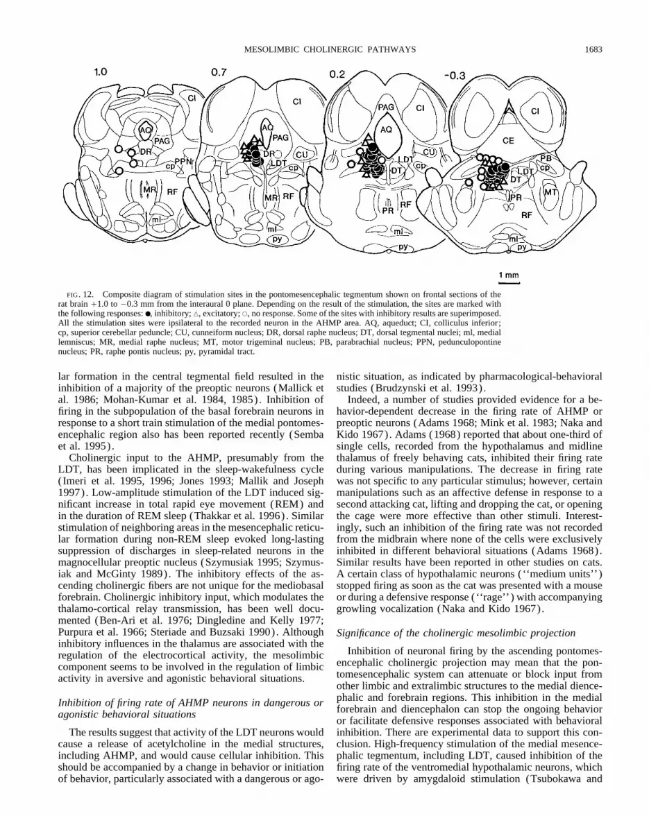
Localization of the stimulation sites in the tegmentum isscopolamine were reversible as tested in four neurons (Fig.9, A–C) . Stimulation of the LDT caused significant inhibi- shown in Fig. 12. All stimulation sites that caused inhibition
of the firing rate of AHMP neurons were located in or attion of the firing rate of a AHMP neuron before the drugapplication (Fig. 9A) . This inhibitory effect was reversed the border of the LDT. Locations of the stimulation electrode
that yielded an increase in the firing rate or no change arewhen scopolamine was ejected concurrently into the vicinityof the recorded neuron in the AHMP (Fig. 9B, /SCO). The scattered further around from the LDT. The excitatory ef-
J376-7/ 9k27$$ap17 03-04-98 13:05:34 neupal LP-Neurophys

S. M. BRUDZYNSKI, L. KADISHEVITZ, AND X.-W. FU1682
acetyltransferase (Dencev et al. 1996). These observationswill be further studied and published separately.
D I S C U S S I O N
Inhibitory responses of medial AHMP neurons tocholinergic stimulation
Our results have indicated that iontophoretic applicationof CCh into the vicinity of single cells in the AHMP causeda dose-dependent decrease in the mean firing rate of themajority of units. The inhibitory effect of CCh, but not theexcitatory effect, was reversed by iontophoretic pretreatmentwith scopolamine. Electrical stimulation of the LDT neuronscaused a comparable current-dependent decrease in the meanfiring rate of AHMP neurons. Effects of LDT stimulationwere reversed by iontophoretic pretreatment of the recordedneurons in the AHMP with scopolamine. The same AHMPneurons that were inhibited by LDT stimulation also re-sponded to CCh with the decrease in firing rate.
Numerous studies reported both the excitatory and inhibi-tory effects of cholinergic agents on the firing rate of AHMPneurons (Bloom et al. 1963; Hsieh and Pan 1990; Jell 1973;Kow and Pfaff 1985; Oomura et al. 1969). It seems, how-ever, that the most medial neurons, which are not necessarilyembedded within any particular nucleus and are not project-ing to the neurohypophysis, show predominantly inhibitoryresponses to iontophoretic ejection of CCh or acetylcholine(Brudzynski et al. 1991; Hsieh and Pan 1990; Moss et al.1972). More caudally, neurons of the ventromedial hypotha-lamic nucleus showed a comparable number of excited orinhibited neurons to acetylcholine (Kow and Pfaff 1985).
FIG. 11. Localization of all the recorded units in the AHMP regionOn the other hand, cells of the paraventricular nucleus thatshown on the frontal section of the rat brain 7.2 to 8.2 mm from the zero,project to the neurohypophysis or neurons in the supraopticinteraural plane. All cells localized on the right side of the brain were tested
with carbachol, and all the cells localized on the left side of the brain were nucleus or arcuate nucleus predominantly increased theirtested for effects of stimulation of the laterodorsal tegmental nucleus. ●, mean firing rate after application of acetylcholine or CChinhibitory responses; m, excitatory responses; s, no response. Neurons (Dreifuss and Kelly 1970; Gribkoff et al. 1988; Lin et al.showing no response were marked for carbachol only (n Å 6, right side of
1993; Moss et al. 1972). Excitatory responses to acetylcho-the brain) , whereas neurons with no responses to electrical stimulation ofline or CCh also were obtained from the forebrain choliner-the laterodorsal tegmental nucleus (n Å 186) were omitted on the left
side of the brain for clarity. AA, anterior amygdalar area; AM, amygdalar gic neurons projecting to the neocortex (Lamour et al. 1986;complex; CP, caudate putamen; DB, diagonal band (horizontal limb); EN, Levine et al. 1986).endopiriform nucleus; fx, fornix; GP, globus pallidus; ic, internal capsule;
All of these results suggest that there is a subpopulationLH, lateral hypothalamic area; LP, lateral preoptic area; mt, mammillotha-of medially located, cholinoceptive neurons in AHMP thatlamic tract; ox, optic chiasm; PA, paraventricular hypothalamic nucleus;
RC, retrochiasmatic area; RE, reuniens thalamic nucleus; RT, reticular thal- respond with a decrease in firing rate to acetylcholine. Me-amic nucleus; sm, stria medullaris; SP, subpallidal area; VP, ventral pal- dial localization of these neurons is consistent with resultslidum; ZI, zona incerta. of functional mapping of behavioral responses in the rat and
cat brain (Brudzynski 1994; Brudzynski et al. 1995). Furtherfects were obtained primarily from the periaqueductal gray functional and anatomic evidence suggest that these inhibi-and the vicinity of the dorsal tegmental nucleus. tory effects are caused by cholinergic input from the LDT
To demonstrate that LDT neurons project to the AHMP, nucleus.to the areas where the recorded neurons were localized, DiAwas injected into the medial portion of AHMP in seven Inhibitory responses of AHMP neurons to LDT stimulationanimals. A summary diagram of all injection sites for theDiA in the AHMP and an example of LDT staining are Inhibitory neuronal responses in the AHMP to stimulation
of the LDT were obtained only from electrodes localizedshown in Fig. 13, A and B. One week after the DiA injection,a dense group of labeled cells was found within the LDT in almost precisely within the LDT nucleus. Furthermore, after
injection of the fluorescent lipophilic carbocyanine dye, DiA,all of the animals (Fig. 13B) . DiA was found uniformlydistributed on the cell body and dendrites and in the cell in into the recording sites immediately after an electrophysio-
logical experiment, DiA was found in numerous neurons incytoplasmic granules but not within the cell nucleus. Thevast majority of neurons was stained reliably by DiA. Our the LDT, 1 wk later.
Inhibitory responses of the AHMP neurons to stimulationlatest preliminary results indicate that, after injection of flu-orogold into the AHMP, many, but not all of the stained LDT of the mesencephalic tegmentum have been reported. High-
frequency electrical stimulation of the mesencephalic reticu-cells are double labeled with the antibody against choline
J376-7/ 9k27$$ap17 03-04-98 13:05:34 neupal LP-Neurophys
MESOLIMBIC CHOLINERGIC PATHWAYS 1683
FIG. 12. Composite diagram of stimulation sites in the pontomesencephalic tegmentum shown on frontal sections of therat brain /1.0 to 00.3 mm from the interaural 0 plane. Depending on the result of the stimulation, the sites are marked withthe following responses: ●, inhibitory; n, excitatory; s, no response. Some of the sites with inhibitory results are superimposed.All the stimulation sites were ipsilateral to the recorded neuron in the AHMP area. AQ, aqueduct; CI, colliculus inferior;cp, superior cerebellar peduncle; CU, cunneiform nucleus; DR, dorsal raphe nucleus; DT, dorsal tegmental nuclei; ml, mediallemniscus; MR, medial raphe nucleus; MT, motor trigeminal nucleus; PB, parabrachial nucleus; PPN, pedunculopontinenucleus; PR, raphe pontis nucleus; py, pyramidal tract.
lar formation in the central tegmental field resulted in the nistic situation, as indicated by pharmacological-behavioralstudies (Brudzynski et al. 1993).inhibition of a majority of the preoptic neurons (Mallick et
al. 1986; Mohan-Kumar et al. 1984, 1985). Inhibition of Indeed, a number of studies provided evidence for a be-havior-dependent decrease in the firing rate of AHMP orfiring in the subpopulation of the basal forebrain neurons in
response to a short train stimulation of the medial pontomes- preoptic neurons (Adams 1968; Mink et al. 1983; Naka andKido 1967). Adams (1968) reported that about one-third ofencephalic region also has been reported recently (Semba
et al. 1995). single cells, recorded from the hypothalamus and midlinethalamus of freely behaving cats, inhibited their firing rateCholinergic input to the AHMP, presumably from the
LDT, has been implicated in the sleep-wakefulness cycle during various manipulations. The decrease in firing ratewas not specific to any particular stimulus; however, certain(Imeri et al. 1995, 1996; Jones 1993; Mallik and Joseph
1997). Low-amplitude stimulation of the LDT induced sig- manipulations such as an affective defense in response to asecond attacking cat, lifting and dropping the cat, or openingnificant increase in total rapid eye movement (REM) and
in the duration of REM sleep (Thakkar et al. 1996). Similar the cage were more effective than other stimuli. Interest-ingly, such an inhibition of the firing rate was not recordedstimulation of neighboring areas in the mesencephalic reticu-
lar formation during non-REM sleep evoked long-lasting from the midbrain where none of the cells were exclusivelyinhibited in different behavioral situations (Adams 1968).suppression of discharges in sleep-related neurons in the
magnocellular preoptic nucleus (Szymusiak 1995; Szymus- Similar results have been reported in other studies on cats.A certain class of hypothalamic neurons (‘‘medium units’’)iak and McGinty 1989). The inhibitory effects of the as-
cending cholinergic fibers are not unique for the mediobasal stopped firing as soon as the cat was presented with a mouseor during a defensive response (‘‘rage’’) with accompanyingforebrain. Cholinergic inhibitory input, which modulates the
thalamo-cortical relay transmission, has been well docu- growling vocalization (Naka and Kido 1967).mented (Ben-Ari et al. 1976; Dingledine and Kelly 1977;Purpura et al. 1966; Steriade and Buzsaki 1990). Although Significance of the cholinergic mesolimbic projectioninhibitory influences in the thalamus are associated with the
Inhibition of neuronal firing by the ascending pontomes-regulation of the electrocortical activity, the mesolimbicencephalic cholinergic projection may mean that the pon-component seems to be involved in the regulation of limbictomesencephalic system can attenuate or block input fromactivity in aversive and agonistic behavioral situations.other limbic and extralimbic structures to the medial dience-phalic and forebrain regions. This inhibition in the medialInhibition of firing rate of AHMP neurons in dangerous orforebrain and diencephalon can stop the ongoing behavioragonistic behavioral situationsor facilitate defensive responses associated with behavioralinhibition. There are experimental data to support this con-The results suggest that activity of the LDT neurons would
cause a release of acetylcholine in the medial structures, clusion. High-frequency stimulation of the medial mesence-phalic tegmentum, including LDT, caused inhibition of theincluding AHMP, and would cause cellular inhibition. This
should be accompanied by a change in behavior or initiation firing rate of the ventromedial hypothalamic neurons, whichwere driven by amygdaloid stimulation (Tsubokawa andof behavior, particularly associated with a dangerous or ago-
J376-7/ 9k27$$ap17 03-04-98 13:05:34 neupal LP-Neurophys

S. M. BRUDZYNSKI, L. KADISHEVITZ, AND X.-W. FU1684
of all the neurons in the zona incerta, which were excitedby stimulation of the ventromedial hypothalamic nucleus,were inhibited by acetylcholine (Eaton and Moss 1989).
In summary, the results of the present study suggest thatthe LDT cholinergic neurons project and terminate on neu-rons in the mediobasal regions of the forebrain with theiractivation causing a decrease in the mean firing rate of theseneurons. This projection can be attributed to the cholinergiccomponent of the ascending reticular activating system (Sa-toh and Fibiger 1986; Shute and Lewis 1963, 1967; Vincentet al. 1986; Woolf et al. 1990) . On the basis of the presentresults and previous behavioral studies, it is postulated thatthe inhibitory influence of the ascending cholinergic fibersin the mediobasal forebrain and diencephalon may play arole in the initiation of some of the behavioral patterns.Thus the inhibition of neuronal firing represent an electro-physiological correlate accompanying some agonistic be-havioral manifestations, defensive or alarm vocalization andbehavioral inhibition manifested by a decrease in locomotoractivity.
The study was supported by the grant from the Natural Sciences andEngineering Research Council of Canada to Dr. Stefan M. Brudzynski.
Address for reprint requests: S. M. Brudzynski, Dept. of Psychology,Brock University, 500 Glenridge Ave., St. Catharines, Ontario, L2S 3A1Canada.
Received 7 May 1997; accepted in final form 18 December 1997.
REFERENCES
ADAMS, D. B. The activity of single cells in the midbrain and hypothalamusof the cat during affective defense behavior. Arch. Ital. Biol. 106: 243–269, 1968.
ARMSTRONG, D. M., SAPER, C. B., LEVEY, A. I., WAINER, B. H., AND TERRY,R. D. Distribution of cholinergic neurons in rat brain: demonstrated by theimmunocytochemical localization of choline acetyltransferase. J. Comp.Neurol. 216: 53–68, 1983.
BAKER, W. W., HOSKO, M. J., RUTT, W. J., AND MCGRATH, J. R. Trem-orine-induced rage and its antagonism by atropine. Proc. Soc. Exp. Biol.Med. 104: 214–217, 1960.
BAKER, G. AND REESE, B. Using confocal laser scanning microscopy toinvestigate the organization and development of neuronal projectionslabelled with DiI. Methods Cell Biol. 38: 325–344, 1993.
BALICE-GORDON, R. J., CHUA, C. K., NELSON, C. C., AND LICHTMAN, J. W.Gradual loss of synaptic cartels procedes axon withdrawal at developingneuromuscular junctions. Neuron 11: 801–815, 1993.
BAXTER, B. L. Comparison of the same behavioral effects of electrical orFIG. 13. Labeling of the laterodorsal tegmental nucleus with the fluo- chemical stimulation applied at the same brain loci. Exp. Neurol. 19:
rescent dye, 4-(4-(dihexadecylamino)styryl)-N-methylpyridinium iodide, 412–432, 1967.(DiA), after its injection into the recording site in the AHMP region. A : BAXTER, B. L. Elicitation of emotional behavior by electrical or chemicalsummary diagram of the maximal extent of the DiA spread from 7 DiA stimulation applied at the same loci in the cat mesencephalon. Exp.injections in the AHMP region is shown on the left side of the rat brain Neurol. 21: 1–10, 1968.sections between the frontal stereotaxic planes 7.2–8.2 mm anterior to BEN-ARI, Y., DINGLEDINE, R., KANAZAWA, T., AND KELLY, J. S. Inhibitorythe interaural line according to the Paxinos and Watson (1986) atlas. An effects of acetylcholine on neurons in the feline nucleus reticularis thal-exemplary injection site of the DiA (0.1 ml ) into the anterior hypothalamic ami. J. Physiol. (Lond.) 261: 647–671, 1976.area (AH) is shown on the right side of the brain (r) , with the resulting BLOOM, F. E., OLIVER, A. P., AND SALMOIRAGHI, G. C. The responsivenessstaining in B. B : dense group of labeled cells in the laterodorsal tegmental of individual hypothalamic neurons to microelectrophoretically adminis-nucleus on the horizontal cross-section of the brain, 1 wk after the injection. tered endogenous amines. Int. J. Neuropharmacol. 2: 181–193, 1963.Scale bar Å 50 mm. AH, anterior hypothalamic area; BRADSHAW, C. M., ROBERTS, M.H.T., AND SZABADI, E. Kinetics of the
release of noradrenaline from micropipettes: interaction between ejectingand retaining currents. Br. J. Pharmacol. 49: 667–677, 1973.Sutin 1963). For comparison, the same stimulation could
BRUDZYNSKI, S. M. Carbachol-induced agonistic behavior in cats: aggres-facilitate or had no effect on the firing of neurons driven by sive or defensive response? Acta Neurobiol. Exp. 41: 15–32, 1981.stimulation of the septum. Although, acetylcholine is not the BRUDZYNSKI, S. M. Ultrasonic vocalization induced by intracerebral carba-
chol in rats: localization and dose-response study. Behav. Brain Res. 63:only transmitter released by tegmental stimulation, it seems133–143, 1994.likely that this inhibitory effect is due to pontomesencephalic
BRUDZYNSKI, S. M. AND BARNABI, F. Contribution of the ascending cholin-cholinergic input and release of acetylcholine. Further evi- ergic pathways in the production of ultrasonic vocalization in the rat.dence for cholinergic inhibition of the input from other struc- Behav. Brain Res. 80: 145–152, 1996.
BRUDZYNSKI, S. M. AND BIHARI, F. Ultrasonic vocalization in rats producedtures was reported for medial zona incerta. The firing rate
J376-7/ 9k27$$ap17 03-04-98 13:05:34 neupal LP-Neurophys

MESOLIMBIC CHOLINERGIC PATHWAYS 1685
by cholinergic stimulation of the brain. Neurosci. Lett. 109: 222–226, The origins of cholinergic and other subcortical afferents to the thalamusin the rat. J. Comp. Neurol. 262: 105–124, 1987.1990.
BRUDZYNSKI, S. M. AND ECKERSDORF, B. Vocalization accompanying emo- HERRERO, M.T., INSAUSTI, R., AND GONZALO, L. M. Cortical projectionsfrom the laterodorsal and dorsal tegmental nuclei. A fluorescent retro-tional-aversive response induced by carbachol in the cat: reproducibility
and a dose-response study. Neuropsychopharmacology 1: 311–320, grade tracing study in the rat. Neurosci. Lett. 123: 144–147, 1991.1988. HONIG, M. G. AND HUME, R. I. Fluorescent carbocyanine dyes allow living
neurons of identified origin to be stained in long-term cultures. J. Cell.BRUDZYNSKI, S. M., ECKERSDORF, B., AND GOLEBIEWSKI, H. Emotional-aversive nature of the behavioral response induced by carbachol in cats. Biol. 193: 171–187, 1986.J. Psychiatr. Neurosci. 18: 38–45, 1993. HONIG, M. G. AND HUME, R. I. DiI and DiO: versatile fluorescent dyes for
neuronal labelling and pathway tracing. Trends Neurosci. 12: 333–341,BRUDZYNSKI, S. M., ECKERSDORF, B., AND GOLEBIEWSKI, H. Regional speci-ficity of the emotional-aversive response induced by carbachol in the cat 1989.brain. A quantitative mapping study. J. Psychiatr. Neurosci. 20: 119– HSIEH, T. F. AND PAN, J. T. Extracellular single-unit studies of suprachias-132, 1995. matic neurons in brain slices. Effect of serotonin, dopamine, carbachol
and LHRH. Chin. J. Physiol. 33: 255–268, 1990.BRUDZYNSKI, S. M., MCLACHLAN, R. S., AND GIRVIN, J. P. Cholinergicallymediated reduction of locomotor activity from the basal forebrain of the IMERI, L., BIANCHI, S., ANGELI, P., AND MANCIA, M. Stimulation of cholin-rat. Exp. Neurol. 105: 197–205, 1989. ergic receptors in the medial preoptic area affects sleep and cortical
temperature. Am. J. Physiol. 269 (Regulatory Integrative Comp. Physiol.BRUDZYNSKI, S. M., MCLACHLAN, R. S., AND GIRVIN, J. P. Involvement ofM1 and M2 muscarinic receptors of the basal forebrain in cholinergically 38): R294–R299, 1995.mediated changes in the rat locomotion. Prog. Neuro-Psychopharmacol. IMERI, L., BIANCHI, S., ANGELI, P. AND MANCIA, M. Muscarinic receptorBiol. Psychiatr. 14: 807–812, 1990. subtypes in the medial preoptic area and sleep-wake cycles. Neuroreport
7: 417–420, 1996.BRUDZYNSKI, S. M., MCLACHLAN, R. S., BIHARI, F., AND GIRVIN, J. P. Re-sponse of neurons of the rat anterior hypothalamic-preoptic area to intra- JELL, R. M. Responses of hypothalamic neurons to local temperature andcerebrally applied carbachol. Brain Res. Bull. 26: 929–934, 1991. to acetylcholine, noradrenaline and 5-hydroxytryptamine. Brain Res. 55:
123–134, 1973.BUTCHER, L. L. Cholinergic neurons and networks. In: The Rat NervousSystem, (2nd ed.) , edited by G. Paxinos. San Diego, CA: Academic JONES, B. E. Immunohistochemical study of choline acetyltransferase-im-Press, 1995, p. 1003–1015. munoreactive processes and cells innervating the pontomedullary reticu-
lar formation in the rat. J. Comp. Neurol. 295: 485–514, 1990.CONSOLO, S., BERTORELLI, R., FORLONI, G. L., AND BUTCHER, L. L. Cholin-ergic neurons of the pontomesencephalic tegmentum release acetylcho- JONES, B. E. The organization of central cholinergic systems and their func-line in the basal nuclear complex of freely moving rats. Neuroscience tional importance in sleep-waking states. Prog. Brain Res. 98: 61–71.37: 717–723, 1990. JONES, B. E. AND BEAUDET, A. Retrograde labelling of neurons in the brain
CORNWALL, J., COOPER, J. D., AND PHILLIPSON, O. T. Afferent and efferent stem following injections of [3H]choline into the forebrain of the rat.connections of the laterodorsal tegmental nucleus in the rat. Brain Res. Exp. Brain Res. 65: 437–448, 1987.Bull. 25: 271–284, 1990. KAYAMA, Y., OTHA, M., AND JODO, E. Firing of ’possibly’ cholinergic
CROSS, B. A. AND DYER, R. G. Unit activity in rat diencephalic islands— neurons in the rat laterodorsal tegmental nucleus during sleep and wake-the effects of anaesthetics. J. Physiol. (Lond.) 212: 467–481, 1971. fulness. Brain Res. 569: 210–220, 1992.
CURTIS, D. R. AND PHILLIS, J. W. The action of procaine and atropine on KIMURA, H., MCGEER, P. L., PENG, J. H., AND MCGEER, E. G. The centralspinal neurons. J. Physiol. (Lond.) 153: 17–34, 1960. cholinergic system studies by choline acetyltransferase immunohisto-
chemistry in the cat. J. Comp. Neurol. 200: 151–201, 1981.DECSI, L. Behavioral effects of intracerebrally injected carbachol on unre-strained cats. Pharmacol. Biochem. Behav. 2: 141–143, 1974. KOFF, G. Y. AND LANGFITT, T. W. Tremorine-induced rage and the limbic
system. Arch. Int. Pharmacodyn. 164: 272–285, 1966.DECSI, L. AND NAGY, J. Adrenergic modulation of a cholinergic emotionalreaction in the cat’s thalamus. Psychopharmacology 54: 303–305, 1977. KOW, L. M. AND PFAFF, D. W. Estrogen effects on neuronal responsiveness
to electrical and neurotransmitter stimulation: an in vitro study on theDECSI, L., VARSZEGI, M. K., AND MEHES, J. Direct chemical stimulation ofvarious subcortical brain areas in unrestrained cats. Recent Dev. Neuro- ventromedial nucleus of the hypothalamus. Brain Res. 347: 1–10, 1985.biol. Hung. 2: 182–211, 1969. LAMOUR, Y., DUTAR, P., RASCOL, O., AND JOBERT, A. Basal forebrain
neurons projecting to the rat frontoparietal cortex: electrophysiologicalDENCEV, A., HRYCYSHYN, A. W., AND BRUDZYNSKI, S. M. Cholinergic pro-jection to the basal forebrain involved in the initiation of ultrasonic vocal- and pharmacological properties. Brain Res. 362: 122–131, 1986.ization in the rat. Abstr. Int. Behav. Neurosci. Soc. 5: 60, 1996. LAUTERBORN, J. C., ISACKSON, P. J., MONTALVO, R., AND GALL, C. M. In
situ hybridization localization of choline acetyltransferase mRNA in adultDINGLEDINE, R. AND KELLY, J. S. Brain stem stimulation and the acetylcho-line-evoked inhibition of neurons in the feline nucleus reticularis thalami. rat brain and spinal cord. Mol. Brain Res. 17: 59–69, 1993.J. Physiol. (Lond.) 271: 135–154, 1977. LESLIE, G. B. Central stimulant properties of compounds with peripheral
muscarinic properties. Nature 208: 1291–1293, 1965.DREIFUSS, J. J. AND KELLY, J. S. Excitation of identified supraoptic neuronsby the iontophoretic application of acetylcholine. J. Physiol. (Lond.) 212: LEVINE, M. S., CEPEDA, C., AND BUCHWALD, N. A. Electrophysiological68–69P, 1970. properties of basal forebrain neurons: responses to microphoretic applica-
tion of putative neurotransmitters. Soc. Neurosci. Abstr. 12: 1468, 1986.DYBALL, R.E.J. AND MCPHAIL, C. J. Unit activity in the supraoptic andparaventricular nuclei -the effects of anaesthetics. Brain Res. 67: 43–50, LEWIS, P. R., AND SHUTE, C. C. D. The cholinergic limbic system: projec-1974. tions to hippocampal formation, medial cortex, nuclei of the ascending
cholinergic reticular system, and the subfornical organ and supra-opticEATON, M. J. AND MOSS, R. L. Electrophysiology and pharmacology ofneurons of the medial zona incerta: an in vitro slice study. Brain Res. crest. Brain 90: 521–542, 1967.502: 117–126, 1989. LIN, J. Y., LI, C. S., AND PAN, J. T. Effects of various neuroactive substances
on single-unit activities of hypothalamic arcuate neurons in brain slices.FIBIGER, H. C. The organization and some projections of cholinergic neu-rons of the mammalian forebrain. Brain Res. Rev. 4: 327–388, 1982. Brain Res. Bull. 31: 587–594, 1993.
LODGE, D., CADDY, K. W., HEADLEY, P. M., AND BISCOE, T. J. The locationFUNKE, A.B.H., BIJLSMAN, U. G., AND NAUTA, W. T. Tremorine-inducedrage and its antagonism by some antiparkinson drugs. Arch. Int. Pharma- of neurons with pontamine sky blue. Neuropharmacology 13: 481–485,
1974.codyn. 135: 447–453, 1962.GELLEN, B., GYORGY, L., AND DODA, M. Influence of the surgical isolation MALLICK, B. N. AND JOSEPH, M. M. Role of cholinergic inputs to the medial
preoptic area in regulation of sleep-wakefulness and body temperatureof the hypothalamus on oxotremorine-induced rage reaction and sympa-thetic response in the cat. Acta Physiol. Acad. Sci. Hung. 42: 195–202, in freely moving rats. Brain Res. 750: 311–317, 1997.1972. MALLICK, B. N., MOHAN-KUMAR, V. M., CHHINA, G. S., AND SINGH, B.
Comparison of rostro-caudal brain stem influence on preoptic neuronsGODEMENT, P., VANSELOW, J., THANOS, S., AND BONHOEFFER, F. A studyin developing visual system with a new method of staining neurons and and cortical EEG. Brain Res. Bull. 16: 121–125, 1986.their processes in fixed tissue. Development 101: 697–713, 1987. MESULAM, M.-M., GEULA, C., BOTHWELL, M. A., AND HERSH, L. B. Human
reticular formation: Cholinergic neurons of the pedunculopontine andGRIBKOFF, V. K., CHRISTIAN, E. P., ROBINSON, J. H., DEADWYLER, S. A.,AND DUDEK, F. E. Cholinergic excitation of supraoptic neurons in hypo- laterodorsal tegmental nuclei and some cytochemical comparisons to
forebrain cholinergic neurons. J. Comp. Neurol. 281: 611–633, 1989.thalamic slices. Neuropharmacology 27: 721–727, 1988.HALLANGER, A. E., LEVEY, A. I., LEE, H. J., RYE, D. B., AND WAINER, B. H. MESULAM, M.-M., MUFSON, E. J., LEVEY, A. I., AND WAINER, B. H. Atlas
J376-7/ 9k27$$ap17 03-04-98 13:05:34 neupal LP-Neurophys

S. M. BRUDZYNSKI, L. KADISHEVITZ, AND X.-W. FU1686
of cholinergic neurons in the forebrain and upper brainstem of the ma- immunohistochemistry, and electrophysiology in the rat. J. Comp. Neu-rol. 267: 433–453, 1988.caque based on monoclonal choline acetyltransferase immunohistochem-
istry and acetylcholinesterase histochemistry. Neuroscience 12: 669–686, SHUTE, C.C.D. AND LEWIS, P. R. Cholinesterase-containing systems of thebrain of the rat. Nature 199: 1160–1164, 1963.1984.
MESULAM, M.-M., MUFSON, E. J., WAINER, B. H., AND LEVEY, A. I. Central SHUTE, C.C.D. AND LEWIS, P. R. The ascending cholinergic reticular system:neocortical, olfactory and subcortical projections. Brain 90: 497–522,cholinergic pathways in the rat: An overview based on an alternative
nomenclature (Ch1-Ch6). Neuroscience 10: 1185–1201, 1983. 1967.SNIDER, W. D., ZHANG, L., YUSOOF, S., GORUKANTI, N., AND TSERING, C.MINK, J. W., SINNAMON, H. M., AND ADAMS, D. B. Activity of basal fore-
brain neurons in the rat during motivated behaviors. Behav. Brain Res. Interactions between dorsal root axons and their target motor neurons indeveloping mammalian spinal cord. J. Neurosci. 12: 3494–3508, 1992.8: 85–108, 1983.
MOHAN-KUMAR, V., MALLICK, B. N., CHHINA, G. S., AND SINGH, B. Influ- STERIADE, M. AND BUZSAKI, G. Parallel activation of thalamic and corticalneurons by brainstem and basal forebrain cholinergic systems. In: Brainence of ascending reticular activating system on preoptic neuronal activ-
ity. Exp. Neurol. 86: 40–52, 1984. Cholinergic System, edited by M. Steriade and D. Biesold, eds.) . Oxford,UK: Oxford Univ. Press, 1990, p. 3–62.MOHAN-KUMAR, V., MALLICK, B. N., CHHINA, G. S., AND SINGH, B. Alter-
ations in preoptic unit activity on stimulation of caudal brain stem EEG- STONE, T. W. Microiontophoresis and pressure ejection. IBRO Handb. Ser.Methods Neurosci. 8: 1–124, 1985.synchronizing structures. Exp. Neurol. 89: 304–313, 1985.
MORUZZI, G. AND MAGOUN, H. W. Brain stem reticular formation and acti- SZYMUSIAK, R. Magnocellular nuclei of the basal forebrain: substrates ofsleep and arousal regulation. Sleep 18: 478–500, 1995.vation of the EEG. EEG Clin. Neurophysiol. 1: 455–473, 1949.
MOSS, R. L., URBAN, I., AND CROSS, B. A. Microelectrophoresis of choliner- SZYMUSIAK, R. AND MCGINTY, D. Sleep-waking discharge of basal fore-brain projection neurons in cats. Brain Res. Bull. 22: 423–430, 1989.gic and aminergic drugs on paraventricular neurons. Am. J. Physiol. 223:
310–318, 1972. TALWAR, A. AND KUMAR, V. M. Effect of carbachol injection in the medialpreoptic area on sleep-wakefulness and body temperature in free movingMUFSON, E., BRADY, D., AND KORDOWER, J. Tracing neuronal connections
in postmortem human hippocampal complex with the carbocyanine dye rats. India J. Physiol. Pharmacol. 38: 163–168, 1994.THAKKAR, M., PORTAS, C., AND MCCARLEY, R. W. Chronic low-amplitudeDiI. Neurobiol. Aging 11: 649–653, 1990.
MYERS, R. D. Emotional and autonomic responses following hypothalamic electrical stimulation of the laterodorsal tegmental nucleus of freely mov-ing cats increases REM sleep. Brain Res. 723: 223–227, 1996.chemical stimulation. Can. J. Psychol. 18: 6–14, 1964.
NAKA, K.-I. AND KIDO, R. Hypothalamic spike potentials recorded by TSUBOKAWA, T. AND SUTIN, J. Mesencephalic influence upon the hypotha-lamic ventromedial nucleus. EEG Clin. Neurophysiol. 15: 804–810,chronically implanted tungsten microelectrodes. Brain Res. 5: 422–424,
1967. 1963.VARSZEGI, M. K. AND DECSI, L. Some characteristics of the rage reactionOOMURA, Y., OOYAMA, H., YAMAMOTO, T., ONO, T., AND KOBAYASHI, N.
Behavior of hypothalamic unit activity during electrophoretic application evoked by chemical stimulation of the hypothalamus. Acta Physiol. Acad.Sci. Hung. 32: 61–68, 1967.of drugs. Ann. NY Acad. Sci. 157: 642–665, 1969.
PAXINOS, G. AND BUTCHER, L. Organizational principles of the brain as VICENT, S. R., SATOH, K., ARMSTRONG, D. M., PANULA, P., VALE, W.,AND FIBIGER, H. C. Neuropeptides and Nadph-diaphorase activity in therevealed by choline acetyltransferase and cholinesterase distributions and
projections. In: The Rat Nervous System. Forebrain and Midbrain, edited ascending cholinergic reticular system of the rat. Neuroscience 17: 167–182, 1986.by G. Paxinos. Orlando, FL: Academic Press, p. 487–521, vol. 1, 1985.
PAXINOS, G. AND WATSON, C. The Rat Brain in Stereotaxic Coordinates VILARO, M. T., PALACIOS, J. M., AND MENGOD, G. Multiplicity of musca-rinic autoreceptors subtypes? Comparison of the distribution of choliner-(2nd ed.) . Sydney: Academic Press, 1986.
PURPURA, D. P., MCMURTRY, J. C., AND MEAKAWA, K. Synaptic events in gic cells and cells containing mRNA for five subtypes of muscarinicreceptors in the rat brain. Mol. Brain Res. 21: 30–46, 1994.ventrolateral thalamic neurons during suppression of recruiting responses
by brain stem reticular stimulation. Brain Res. 1: 63–76, 1966. WOOLF, N. J. AND BUTCHER, L. L. Cholinergic system in the rat brain. III.Projections from the pontomesencephalic tegmentum to the thalamus,SATOH, K. AND FIBIGER, H. C. Cholinergic neurons of the laterodorsal teg-
mental nucleus: efferent and afferent connections. J. Comp. Neurol. 253: tectum, basal ganglia, and basal forebrain. Brain Res. Bull. 16: 603–637,1986.277–302, 1986.
SEMBA, K., DETARI, L., AND RASMUSSON, D. D. Cortical EEG-related activ- WOOLF, N. J. AND BUTCHER, L. L. Cholinergic systems in the rat brain. IV.Descending projections of the pontomesencephalic tegmentum. Brainity of basal forebrain neurons and their responses to brainstem and sensory
stimulation in urethane-anesthetized rat. Soc. Neuroci. Abstr. 21: 957, Res. Bull. 23: 519–540, 1989.WOOLF, N. J., HARRISON, J. B., AND BUCHWALD, J. S. Cholinergic neurons1995.
SEMBA, K., REINER, P. B., MCGEER, E. G., AND FIBIGER, H. C. Brainstem of the feline pontomesencephalon. II. Ascending anatomical projections.Brain Res. 520: 55–72, 1990.afferents to the magnocellular basal forebrain studied by axonal transport,
J376-7/ 9k27$$ap17 03-04-98 13:05:34 neupal LP-Neurophys


