Embed Size (px)
Citation preview

*For correspondence:
Competing interests: The
authors declare that no
competing interests exist.
Funding: See page 11
Received: 15 March 2018
Accepted: 16 August 2018
Published: 17 August 2018
Reviewing editor: Louis J
Ptacek, University of California,
San Francisco, United States
Copyright Pappas et al. This
article is distributed under the
terms of the Creative Commons
Attribution License, which
permits unrestricted use and
redistribution provided that the
original author and source are
credited.
A cell autonomous torsinA requirementfor cholinergic neuron survival and motorcontrolSamuel S Pappas1, Jay Li1,2, Tessa M LeWitt1, Jeong-Ki Kim3,4,5,Umrao R Monani3,4,5, William T Dauer1,2,6*
1Department of Neurology, University of Michigan, Ann Arbor, United States; 2Celland Molecular Biology Program, University of Michigan, Ann Arbor, United States;3Department of Cell Biology, Columbia University Medical Center, New York,United States; 4Center for Motor Neuron Biology and Disease, Columbia UniversityMedical Center, New York, United States; 5Department of Pathology, ColumbiaUniversity Medical Center, New York, United States; 6Department of Cell andDevelopmental Biology, University of Michigan, Ann Arbor, United States
Abstract Cholinergic dysfunction is strongly implicated in dystonia pathophysiology. Previously
(Pappas et al., 2015;4:e08352), we reported that Dlx5/6-Cre mediated forebrain deletion of the
DYT1 dystonia protein torsinA (Dlx-CKO) causes abnormal twisting and selective degeneration of
dorsal striatal cholinergic interneurons (ChI) (Pappas et al., 2015). A central question raised by that
work is whether the ChI loss is cell autonomous or requires torsinA loss from neurons synaptically
connected to ChIs. Here, we addressed this question by using ChAT-Cre mice to conditionally
delete torsinA from cholinergic neurons (‘ChAT-CKO’). ChAT-CKO mice phenocopy the Dlx-CKO
phenotype of selective dorsal striatal ChI loss and identify an essential requirement for torsinA in
brainstem and spinal cholinergic neurons. ChAT-CKO mice are tremulous, weak, and exhibit trunk
twisting and postural abnormalities. These findings are the first to demonstrate a cell autonomous
requirement for torsinA in specific populations of cholinergic neurons, strengthening the
connection between torsinA, cholinergic dysfunction and dystonia pathophysiology.
DOI: https://doi.org/10.7554/eLife.36691.001
IntroductionMultiple lines of evidence implicate striatal cholinergic dysfunction in dystonia pathophysiology
(Pappas et al., 2015; Albin et al., 2003; Eskow Jaunarajs et al., 2015; Pappas et al., 2014). The
symptoms of DYT1 dystonia, caused by a loss of function mutation in the gene encoding torsinA
(Ozelius et al., 1997), are reduced by antimuscarinic treatments (e.g., trihexyphenidyl)(Burke et al.,
1986). Antimuscarinic agents also reduce motor (Pappas et al., 2015) and electrophysiological
(Maltese et al., 2014) abnormalities in DYT1 mouse models. Striatal cholinergic dysfunction is a
common feature of multiple DYT1 animal models (Pappas et al., 2015; Martella et al., 2009;
Pisani et al., 2006; Sciamanna et al., 2012a; Sciamanna et al., 2012b), and experimental ablation
of striatal cholinergic interneurons (ChI) can lead to abnormal postures (Kaneko et al., 2000).
We demonstrated previously that deletion of torsinA from forebrain GABAergic and cholinergic
neurons (using Dlx5/6-cre; ‘Dlx-CKO’) causes highly selective degeneration of dorsal striatal ChI
roughly coincident with the juvenile onset of abnormal limb clasping and twisting movements
(Pappas et al., 2015). Selective ChI abnormalities are also present in postmortem tissue from DYT1
subjects (Pappas et al., 2015). Abnormal movements in Dlx-CKO mice are reduced by clinically rele-
vant antimuscarinic treatments, strengthening model therapeutic validity and suggesting shared
Pappas et al. eLife 2018;7:e36691. DOI: https://doi.org/10.7554/eLife.36691 1 of 15
RESEARCH ADVANCE

pathophysiology with human dystonia. This work highlights the importance of elucidating the mech-
anism of selective ChI loss. A critical first step toward this goal is to determine whether the ChI loss
observed in Dlx-CKO mice results from a cell autonomous role of torsinA in these cells or, alterna-
tively, whether loss of torsinA from synaptically connected cells is also required. The major aim of
these studies was to address this fundamental question.
To determine whether torsinA-related ChI loss is cell autonomous, we generated and character-
ized cholinergic neuron selective conditional torsinA knockout mice (ChAT-CKO). We find that
ChAT-CKO mice phenocopy the selective degeneration of dorsal striatal ChI observed in Dlx-CKO
mice (basal forebrain neuron numbers are normal in both models). Assessment of non-forebrain cho-
linergic populations demonstrates that pedunculopontine and laterodorsal tegmental brainstem cho-
linergic neurons, and spinal motor neurons also require torsinA for survival or normal function.
ChAT-CKO mice exhibit severe motor and postural abnormalities that are distinct from Dlx-CKO
mice. These findings are the first to establish a cell autonomous requirement for torsinA in ChI, as
well as identifying additional vulnerable cholinergic neuron populations. This in vivo study fundamen-
tally advances and expands understanding of the requirement of torsinA for normal cholinergic sys-
tem function, opening new directions for the study of mechanisms contributing to selective neuronal
dysfunction in dystonia.
Results and discussionTo determine if ChI neurodegeneration is a cell autonomous effect of torsinA loss, we conditionally
deleted torsinA from cholinergic neurons (Chat-IRES-Cre+, Tor1aFlx/-; ‘ChAT-CKO’ mice; Cre-recom-
binase expression occurs before birth and is completely selective for cholinergic neurons; Figure 1—
figure supplement 1 [Madisen et al., 2010]). Unilateral unbiased stereology of ChAT-immunoreac-
tive neurons in the dorsal striatum from 1 year old mice demonstrates a ~ 34% reduction in the num-
ber of dorsal striatal ChI in ChAT-CKO mice compared to control mice (Figure 1A,B). This finding
was confirmed in an independent cohort using bilateral unbiased stereology (48% reduction; Fig-
ure 1—figure supplement 2A). The number of striatal non-cholinergic neurons was not different
from controls (Figure 1—figure supplement 2B,C), demonstrating that there are no secondary cell
loss effects of ChI degeneration, and that torsinA loss of function-mediated neurodegeneration is
highly specific. These findings establish a cell autonomous torsinA requirement for ChI survival.
ChI cell loss is strikingly selective in Dlx-CKO mice, occurring primarily in the dorsal aspects of the
striatum, with approximately six times greater cell loss in the dorsolateral compared to ventromedial
striatum (57% vs 9% cell density reduction in Dlx-CKO mice; [Pappas et al., 2015]). To examine if
the cell autonomous ChI degeneration in ChAT-CKO mice follows a similar subregion-selective pat-
tern, we determined the density of ChAT-immunoreactive neurons in each quadrant of the dorsal
striatum (as previously [Pappas et al., 2015]). Significant reductions in ChI number were limited to
the dorsolateral and dorsomedial segments of the dorsal striatum (72% and 54% cell density reduc-
tions in dorsolateral and dorsomedial, vs 12% and �4% in ventrolateral and ventromedial segments;
Figure 1C). This topographic pattern of cell loss was present throughout the entire rostro-caudal
extent of the striatum (Figure 1C,D, Figure 1—figure supplement 3). The dorsolateral selectivity of
ChI neuron loss is highly relevant, as the dorsolateral striatum is a key motor circuit node functionally
integrated according to topographic inputs, whereas ventromedial striatal neurons are connected in
associative and limbic circuits (Alexander et al., 1986; Haber, 2016; Parent and Hazrati, 1995). In
contrast, the basal forebrain contains cholinergic projection neurons subserving cognitive and atten-
tional control (Hasselmo and Sarter, 2011; Ballinger et al., 2016), which do not degenerate in
either model (Figure 1E,F). Conditional deletion of torsinA from forebrain cholinergic neurons there-
fore mimics the region-selective vulnerability observed in Dlx-CKO mice, demonstrating a cell auton-
omous requirement for torsinA in select cholinergic populations. To determine if differing time
courses of torsinA loss (via differing torsinA half lives) contributes to selective vulnerability, we
assessed torsinA levels in dorsal vs ventral striatal ChI at P0. Surprisingly, despite uniform prenatal
Cre recombinase expression and preferential loss of dorsal ChI, torsinA levels were reduced to a
greater extent in ventral ChI (dorsal ChI contained 82% of control torsinA levels, while ventral ChI
had ~52% remaining; Figure 1—figure supplement 4). Non-vulnerable basal forebrain cholinergic
Pappas et al. eLife 2018;7:e36691. DOI: https://doi.org/10.7554/eLife.36691 2 of 15
Research advance Neuroscience

WT Cre Control Het Control Chat-CKO0
5
10
Ch
at+
Cell N
um
ber
(X1000)
Unilateral Striatal ChI Count
*
DL DM VL VM
-20
0
20
40
60
80
% L
oss o
f C
hat+
Cells
Striatal ChI Loss
DL DM VL VM0
10
20
30
40
50
Ch
at+
cells/m
m2
ChI Density in Dorsal Striatum
WT
Cre Control
Het Control
ChAT-CKO
** !"# !$#
%"# %$#
NBM MS/VDB GP0
1
2
3
4
5
P75+
cell n
um
ber
(X1000)
Basal Forebrain Cholinergic Neurons
Control
Chat-CKO
!"#$%&'()#*%)%&'+,- ."$$ +,- !/0$*)1'*0'!"#$%&'()#*%)23
4%$%&'5"#/6#%*0'+,"&*0/#7*8
90*&%)/#%&'()#*%)%&'+,-
:; +,<;=+>?
88 88
+"0)#"&
+,<;=+>?
@4A A( BC
*D8D
BC
(;
*D8D
BC
(;
!"#$%&'()#*%)23'E +,<; -3320",*$)"8,/3*$)#1
4%$%&'5"#/6#%*0E CFG'-3320",*$)"8,/3*$)#1
! "
# $
% &
Figure 1. Conditional cholinergic neuron deletion of torsinA causes cell autonomous loss of striatal cholinergic neurons. (A) Unilateral stereological
quantification of the number of ChAT-positive neurons in the striatum of ChAT-CKO and control mice (One-way ANOVA F(3,28) = 3.589, p=0.02,
Dunnett’s multiple comparisons test: adjusted p value = 0.049; ‘WT’=Tor1aFlx/+; ‘Cre Control’=ChAT-Cre+, Tor1a Flx/+; ‘Het Control’=Tor1 aFlx/-; ‘ChAT-
CKO’=ChAT-Cre+, Tor1aFlx/-). (B) ChAT immunohistochemistry of coronal sections containing dorsal striatum from WT and ChAT-CKO mice
(cc = corpus callosum). (C) Percent reduction in cell density by striatal quadrant (DL = dorsolateral; DM = dorsomedial, VL = ventrolateral,
VM = ventromedial). (D) Significant ChI loss is selective for dorsal striatal quadrants. Cell density quantification in control and ChAT-CKO striatal
quadrants (Two-way ANOVA main effect of genotype F(3,112) = 24.02, p<0.0001; main effect of quadrant F(3,112)=8.398, p<0.0001; interaction
F(9,112)=8.11, p<0.0001. Post-hoc Tukey’s multiple comparisons test). (E) Basal forebrain neurons are spared in ChAT-CKO mice. Stereological
quantification of P75-immunoreactive basal forebrain cholinergic neurons in the nucleus basalis of meynert (NBM), medial septum/nucleus of the
vertical limb of the diagonal band (MS/VDB), and globus pallidus (GP). No differences in the number of cholinergic neurons was observed (NBM,
Figure 1 continued on next page
Pappas et al. eLife 2018;7:e36691. DOI: https://doi.org/10.7554/eLife.36691 3 of 15
Research advance Neuroscience

neurons exhibited 49% of control torsinA immunoreactivity (Figure 1—figure supplement 5). These
findings demonstrate that a more rapid loss of torsinA during development does not contribute to
the unique vulnerability of dorsal ChI.
TorsinA deletion is restricted to forebrain structures in Dlx-CKO mice. In contrast, ChAT-CKO
mice lack torsinA in all cholinergic neurons throughout the neuraxis, enabling us to assess the impact
of torsinA loss in additional cholinergic populations. Unbiased stereology of ChAT-immunoreactive
neurons in the brainstem demonstrates significantly fewer cholinergic neurons in the pedunculopon-
tine (PPN) and laterodorsal tegmental (LDT) nuclei in 1 year old Chat-CKO mice (Figure 2A–D). The
PPN and LDT also contain GABAergic, and glutamatergic neurons (Mena-Segovia, 2016), which sig-
nificantly outnumber cholinergic neurons (Mena-Segovia et al., 2009; Wang and Morales, 2009).
Unbiased stereology of NeuN +neurons in PPN and LDT showed no significant change in the overall
number of neurons (Figure 2A,C). Because cholinergic neurons are a minority of cells in the PPN
and LDT, it is possible that a significant reduction of this small sub-population cannot be detected
when assessed by counting overall NeuN +neuron number. It is also possible that PPN and LDT cho-
linergic neurons exhibit reduced ChAT expression rather than actual cell loss. Regardless, either pos-
sibility demonstrates a cell autonomous role for torsinA for normal function of these cells. These
findings also indicate that the loss or dysfunction of brainstem cholinergic neurons does not have
deleterious effects on the viability of surrounding neurons. Consistent with this finding, there was no
evidence of reactive microgliosis or astrogliosis in the brainstem (Figure 2—figure supplement 1).
Quantification of the number of spinal motor neurons (C3-C5; [Kim et al., 2017]) demonstrated sig-
nificantly fewer motor neurons in ChAT-CKO mice (Figure 2E,F).
The identification of cholinergic dysfunction or loss in PPN and LDT is notable, as considerable
data implicate these cells in motor and postural control. PPN and LDT cholinergic neurons are dis-
tributed in a rostrocaudal continuum in the brainstem, forming a coordinated functional unit (Mena-
Segovia, 2016; Mena-Segovia and Bolam, 2017). PPN and LDT cholinergic neurons topographically
innervate the striatum and striatal-projecting thalamic and midbrain dopamine neurons
(Dautan et al., 2014), such that rostral PPN modulates motor-related circuits, LDT innervates limbic
circuits, and caudal PPN targets both regions (Mena-Segovia, 2016; Xiao et al., 2016) via both
direct and indirect inputs. Consistent with a central role in modulating locomotor activity, optoge-
netic stimulation of PPN cholinergic neurons alters locomotion speed, while stimulation of adjacent
glutamatergic neurons induces locomotion (Xiao et al., 2016; Roseberry et al., 2016; Capelli et al.,
2017). Cholinergic PPN lesion alone or in combination with dopaminergic denervation impairs gait
and causes postural abnormalities in primates (Grabli et al., 2013; Karachi et al., 2010). In rodents,
cholinergic-selective PPN lesion impairs performance on the accelerating rotarod and alters sensori-
motor gating (MacLaren et al., 2014a; MacLaren et al., 2014b), while non-specific PPN ablation
alters gait (Blanco-Lezcano et al., 2017) and impairs reversal learning (Syed et al., 2016). Human
neuroimaging and postmortem studies also provide support for a connection between PPN choliner-
gic integrity and motor function. PPN cholinergic loss is linked to gait abnormalities in Parkinson dis-
ease (Karachi et al., 2010; Bohnen et al., 2009), and brainstem lesions (including PPN loss) can
Figure 1 continued
t(13)=1.684, p=0.11; MS/VDB, t(13)=1.537, p=0.148; GP, t(13)=0.5, p=0.625). (F) P75 immunohistochemistry of sagittal sections containing basal forebrain
cholinergic neuron populations. i.c. = internal capsule, ST = striatum.
DOI: https://doi.org/10.7554/eLife.36691.002
The following figure supplements are available for figure 1:
Figure supplement 1. ChAT-Cre is expressed prenatally.
DOI: https://doi.org/10.7554/eLife.36691.003
Figure supplement 2. Independent cohort confirmation of selective striatal cholinergic neuron loss in ChAT-CKO mice.
DOI: https://doi.org/10.7554/eLife.36691.004
Figure supplement 3. ChAT-positive neurons are reduced in a topographic pattern throughout the rostrocaudal extent of the dorsal striatum.
DOI: https://doi.org/10.7554/eLife.36691.005
Figure supplement 4. Time course of torsinA protein loss in dorsal and ventral striatum.
DOI: https://doi.org/10.7554/eLife.36691.006
Figure supplement 5. Time course of torsinA protein loss in basal forebrain.
DOI: https://doi.org/10.7554/eLife.36691.007
Pappas et al. eLife 2018;7:e36691. DOI: https://doi.org/10.7554/eLife.36691 4 of 15
Research advance Neuroscience

Figure 2. ChAT-CKO mice have significantly fewer brainstem and spinal cord cholinergic neurons. (A,B) Stereological quantification of ChAT-positive or
NeuN-positive neurons in the pedunculopontine nucleus (PPN) of control and ChAT-CKO mice (ChAT; t(14)=4.531, p=0.0005. NeuN; t(14)=0.095, p=0.92).
(C,D) Stereological quantification of ChAT-positive or NeuN-positive neurons in the laterdorsal tegmental nucleus (LDT) of control and ChAT-CKO mice
(ChAT; t(14)=3.571, p=0.003. NeuN; t(14)=1.934, p=0.073). (E,F) Quantification of the number of ChAT-positive neurons in the cervical spinal cord of
control and ChAT-CKO mice (t(6)=3.654, p=0.0107). Scale bars = 100 mm.
DOI: https://doi.org/10.7554/eLife.36691.008
The following figure supplement is available for figure 2:
Figure supplement 1. Absence of gliosis in the brainstem of ChAT-CKO mice.
DOI: https://doi.org/10.7554/eLife.36691.009
Pappas et al. eLife 2018;7:e36691. DOI: https://doi.org/10.7554/eLife.36691 5 of 15
Research advance Neuroscience

Figure 3. Motor behavior is severely disrupted in ChAT-CKO mice. (A) Representative image of a control and ChAT-CKO mouse demonstrates severe
kyphosis and unkempt coat. (B) ChAT-CKO mice exhibit significantly increased kyphotic curvature during locomotion (Mann-Whitney U = 35, p<0.0001).
(C) ChAT-CKO mice exhibit a significantly reduced latency to fall during forelimb suspension (Mann-Whitney U = 71.5, p<0.0001). (D, E) ChAT-CKO
mice are hypoactive in the open field (horizontal movement, t(23)=2.345, p=0.028; vertical rears, welch-corrected t(15.1) = 2.345, p=0.033). (F) Performance
on the accelerated rotarod does not significantly differ from controls (two-way repeated measures ANOVA, genotype, F(1,43)=0.75, p=0.389; trial,
F(9,387)=55.63, p<0.0001; interaction, F(9,387)=1.194, p=0.297). (G - I) ChAT-CKO mouse gait is abnormal during locomotion (paw angle, two-way ANOVA
main effect of genotype, F(1,56)=30.54, p<0.0001, main effect of limb F(3,56)=51.02, p<0.0001, interaction F(3,56)=13.51, p<0.0001, post-hoc Sidak’s
multiple comparisons test. Stance width, t(14)=3.329, p=0.005. Stride length, two-way ANOVA genotype F(1,28)=3.164, p=0.086, limb F(1,28)=0.02,
p=0.887, interaction F(1,28)=0.0001, p=0.989).
DOI: https://doi.org/10.7554/eLife.36691.010
The following video and figure supplements are available for figure 3:
Figure supplement 1. Representative examples of control and ChAT-CKO spinal cords demonstrate significant kyphotic curvature.
DOI: https://doi.org/10.7554/eLife.36691.011
Figure supplement 2. ChAT-CKO mice are significantly hypoactive.
DOI: https://doi.org/10.7554/eLife.36691.012
Figure 3—video 1. Representative video demonstrating tremulousness, kyphosis, and hyperactivity in ChAT-CKO mice, as compared to controls.
DOI: https://doi.org/10.7554/eLife.36691.013
Figure 3 continued on next page
Pappas et al. eLife 2018;7:e36691. DOI: https://doi.org/10.7554/eLife.36691 6 of 15
Research advance Neuroscience

result in complex dystonia (Jankovic and Patel, 1983; LeDoux and Brady, 2003; Loher and Krauss,
2009; Zweig et al., 1988; Mente et al., 2018). Systematic cholinergic brainstem cell counts have
not been performed in DYT1 dystonia postmortem samples; most studies have failed to demon-
strate neuronal inclusions or overt cell loss in this region (Paudel et al., 2014; Pratt et al., 2016;
McNaught et al., 2004).
Motor behavior is severely disrupted in ChAT-CKO mice, but is distinct from the Dlx-CKO pheno-
type (Figure 3; Table 1). ChAT-CKO pups are initially indistinguishable from littermates, but at
approximately 4 weeks of age develop a hunched posture, have unkempt fur, and exhibit reduced
responsiveness to handling (Figure 3A, Figure 3—figure supplement 1). Whereas normal mice
exhibit a slight dorsal spinal curvature at rest, ChAT-CKO mice exhibit severe kyphosis, including
during locomotion (assessed by two observers blind to experimental conditions; Figure 3B; Fig-
ure 3—figure supplement 1; Figure 3—video 1) (Guyenet et al., 2010). ChAT-CKO mice also
exhibit signs of weakness, including a significantly reduced ability to hang by the forelimbs
(Figure 3C), tremulous movements, labored breathing (Figure 3—video 1), and significantly
reduced horizontal and vertical movement in the open field (Figure 3D,E, Figure 3—figure supple-
ment 2). Remarkably, performance on the accelerating rotarod during two days of training appears
normal (Figure 3F). The normal rotarod behavior differs from models of motor neuron and neuro-
muscular disease, suggesting that neuromuscular weakness is modest in ChAT-CKO, and less likely
to contribute to other behavioral phenotypes (e.g., postural abnormality). The gait of ChAT-CKO
mice is also significantly altered (Figure 3G–I). This constellation of behavioral phenotypes is distinct
from Dlx-CKO mice (Table 1), in which loss of dorsal striatal ChI is associated with a set of persistent
abnormal action-induced motor behaviors, including limb clasping and trunk twisting during tail sus-
pension and open field hyperactivity (Pappas et al., 2015). ChAT-CKO mice did not exhibit fore- or
hindlimb clasping during tail suspension, but did exhibit tremulousness and trunk twisting (15 CKO,
19 heterozygous, 22 Cre control, and 19 wild type mice observed; Figure 3—video 2). These results
suggest that dorsal striatal ChI neurodegeneration may not, by itself, be sufficient to cause limb
clasping during tail suspension. However, the co-occurrence of brainstem and spinal cord
Figure 3 continued
Figure 3—video 2. ChAT-CKO exhibit twisting and tremulousness, but not limb clasping during tail suspension.
DOI: https://doi.org/10.7554/eLife.36691.014
Table 1. Behavioral properties of Dlx-CKO and ChAT-CKO mice.
Motor function Dlx-CKO ChAT-CKO
Pappas et al., 2015 eLife 4:e08352 present manuscript
Tail suspension Trunk twisting Trunk twisting
Forelimb clasping -
Hindlimb clasping -
- Tremulousness
Open field Hyperactivity Hypoactivity
Rotarod Normal Normal
Response to handling Exaggerated Reduced
Weakness, latency to fall Grid hang reduction Wire hang reduction
Gait Normal by eye Abnormal by eye
Slightly reduced stance width Increased stance width
- Increased paw angle
Overt postural abnormalities - Severe kyphosis
Tremulous movement - Severe
Labored breathing - Severe
DOI: https://doi.org/10.7554/eLife.36691.015
Pappas et al. eLife 2018;7:e36691. DOI: https://doi.org/10.7554/eLife.36691 7 of 15
Research advance Neuroscience
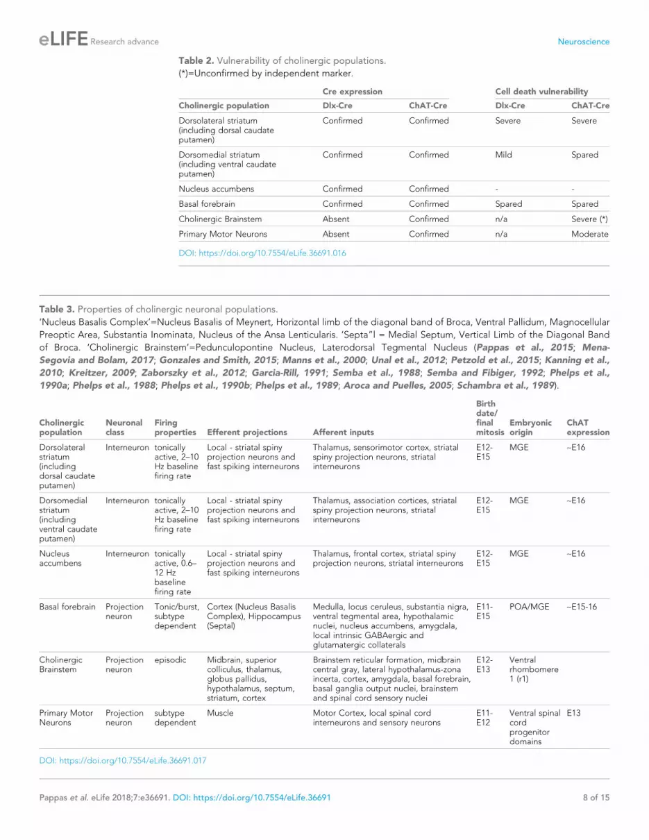
Table 2. Vulnerability of cholinergic populations.
(*)=Unconfirmed by independent marker.
Cre expression Cell death vulnerability
Cholinergic population Dlx-Cre ChAT-Cre Dlx-Cre ChAT-Cre
Dorsolateral striatum(including dorsal caudateputamen)
Confirmed Confirmed Severe Severe
Dorsomedial striatum(including ventral caudateputamen)
Confirmed Confirmed Mild Spared
Nucleus accumbens Confirmed Confirmed - -
Basal forebrain Confirmed Confirmed Spared Spared
Cholinergic Brainstem Absent Confirmed n/a Severe (*)
Primary Motor Neurons Absent Confirmed n/a Moderate
DOI: https://doi.org/10.7554/eLife.36691.016
Table 3. Properties of cholinergic neuronal populations.
‘Nucleus Basalis Complex’=Nucleus Basalis of Meynert, Horizontal limb of the diagonal band of Broca, Ventral Pallidum, Magnocellular
Preoptic Area, Substantia Inominata, Nucleus of the Ansa Lenticularis. ‘Septa”l = Medial Septum, Vertical Limb of the Diagonal Band
of Broca. ‘Cholinergic Brainstem’=Pedunculopontine Nucleus, Laterodorsal Tegmental Nucleus (Pappas et al., 2015; Mena-
Segovia and Bolam, 2017; Gonzales and Smith, 2015; Manns et al., 2000; Unal et al., 2012; Petzold et al., 2015; Kanning et al.,
2010; Kreitzer, 2009; Zaborszky et al., 2012; Garcia-Rill, 1991; Semba et al., 1988; Semba and Fibiger, 1992; Phelps et al.,
1990a; Phelps et al., 1988; Phelps et al., 1990b; Phelps et al., 1989; Aroca and Puelles, 2005; Schambra et al., 1989).
Cholinergicpopulation
Neuronalclass
Firingproperties Efferent projections Afferent inputs
Birthdate/finalmitosis
Embryonicorigin
ChATexpression
Dorsolateralstriatum(includingdorsal caudateputamen)
Interneuron tonicallyactive, 2–10Hz baselinefiring rate
Local - striatal spinyprojection neurons andfast spiking interneurons
Thalamus, sensorimotor cortex, striatalspiny projection neurons, striatalinterneurons
E12-E15
MGE ~E16
Dorsomedialstriatum(includingventral caudateputamen)
Interneuron tonicallyactive, 2–10Hz baselinefiring rate
Local - striatal spinyprojection neurons andfast spiking interneurons
Thalamus, association cortices, striatalspiny projection neurons, striatalinterneurons
E12-E15
MGE ~E16
Nucleusaccumbens
Interneuron tonicallyactive, 0.6–12 Hzbaselinefiring rate
Local - striatal spinyprojection neurons andfast spiking interneurons
Thalamus, frontal cortex, striatal spinyprojection neurons, striatal interneurons
E12-E15
MGE ~E16
Basal forebrain Projectionneuron
Tonic/burst,subtypedependent
Cortex (Nucleus BasalisComplex), Hippocampus(Septal)
Medulla, locus ceruleus, substantia nigra,ventral tegmental area, hypothalamicnuclei, nucleus accumbens, amygdala,local intrinsic GABAergic andglutamatergic collaterals
E11-E15
POA/MGE ~E15-16
CholinergicBrainstem
Projectionneuron
episodic Midbrain, superiorcolliculus, thalamus,globus pallidus,hypothalamus, septum,striatum, cortex
Brainstem reticular formation, midbraincentral gray, lateral hypothalamus-zonaincerta, cortex, amygdala, basal forebrain,basal ganglia output nuclei, brainstemand spinal cord sensory nuclei
E12-E13
Ventralrhombomere1 (r1)
Primary MotorNeurons
Projectionneuron
subtypedependent
Muscle Motor Cortex, local spinal cordinterneurons and sensory neurons
E11-E12
Ventral spinalcordprogenitordomains
E13
DOI: https://doi.org/10.7554/eLife.36691.017
Pappas et al. eLife 2018;7:e36691. DOI: https://doi.org/10.7554/eLife.36691 8 of 15
Research advance Neuroscience

neurodegeneration and tremulousness in ChAT-CKO mice could modify a clasping phenotype and
therefore limit this strength of this conclusion.
While no single system or experimental approach can fully model a disease, the extreme postural
abnormalities (kyphosis and twisting) in ChAT-CKO mice are reminiscent of Oppenheim’s original
description of dystonia (Klein and Fahn, 2013), suggesting that a constellation of cholinergic abnor-
malities may contribute to such a phenotype. The abnormal gait, tremulous movement, weakness,
labored breathing, and appearance of reduced muscle mass in ChAT-CKO mice are consistent with
brainstem and spinal cord pathology, yet the time course of ChAT-CKO abnormalities (beginning
during development) differ from motor neuron and neuromuscular disease models, in which behav-
ioral phenotypes typically emerge in adulthood (9–11 months of age; (Dickinson and Meikle, 1973;
Bridges et al., 1992; Deconinck et al., 1997; Grady et al., 1997; Laws and Hoey, 2004; Liu et al.,
2016; Sopher et al., 2004; Monks et al., 2007). Early motor behavioral manifestations also occur in
Dlx-CKO and other DYT1 models, emphasizing the importance of torsinA function during develop-
ment and maturation at behavioral (Pappas et al., 2015; Liang et al., 2014), cellular (Pappas et al.,
2018), and molecular levels (Tanabe et al., 2016).
These findings establish a cell autonomous requirement of torsinA for the normal function and
survival of distinct populations of cholinergic neurons. Comparison of basic cellular properties
between susceptible and invulnerable cholinergic neuron populations does not identify obvious pat-
terns driving selective vulnerability (Tables 2 and 3). Within the striatum, dorsal ChI are highly vulner-
able to cell death, while ventral ChI are spared. It is unclear whether molecular differences within
different ChI populations drive vulnerability, or if differences in connectivity or response to inputs
contributes to their loss; these possibilities are not mutually exclusive. While often considered a sin-
gle neuronal class, an existing and enlarging literature demonstrates that dorsal and ventral striatal
ChI exhibit significant differences in morphology, regulation, and receptor expression (reviewed in
[Gonzales and Smith, 2015]), as well as differing firing patterns during behavioral tasks (Yarom and
Cohen, 2011) and responses to serotonergic input (Virk et al., 2016). These differences implicate
the presence of multiple ChI subclasses, though it is important to note that the spared ‘ventral’ pop-
ulation here represents the ventral part of the dorsal striatum, not the nucleus accumbens. Thala-
mostriatal and corticostriatal input is highly topographic (Alexander et al., 1986; Smith et al.,
2004), raising the possibility that aberrant input from different thalamic nuclei or cortical regions (or
aberrant response to that input) could alter the susceptibility of dorsal vs ventral ChI. It is likely that
a combination of these and other factors plays a role in the differential susceptibility of cholinergic
neuronal populations, including their molecular profiles (e.g., protective factors in some neurons,
susceptibility factors in others), the response to afferent inputs, and their inherent physiological
properties.
These studies greatly strengthen the connection between torsinA and cholinergic dysfunction,
demonstrating that specific cholinergic populations exhibit a cell autonomous selective vulnerability
to torsinA deficiency, while others – basal forebrain and ventral striatum – are spared. These findings
open novel avenues of study aimed at defining the molecular mechanisms responsible for this cell
autonomous selective vulnerability, and circuit-level analyses to ameliorate the effects of cholinergic
neurotransmission abnormalities.
Materials and methods
Key resources table
Reagent type (species)or resource Designation Source or reference Identifiers Additional information
Gene(Mus musculus)
Tor1a NA NCBI Gene: 30931;MGI:1353568
Encodes TorsinA
Strain, strainbackground(M. musculus)
ChAT-Cre Jackson Laboratories Stock ID 006410 Chattm2(cre)Lowl; (Chat-IRES-Cre)
Strain, strainbackground(M. musculus)
Tor1aFlx/Flx Jackson Laboratories Stock ID 025832 Tor1atm3.1Wtd
Continued on next page
Pappas et al. eLife 2018;7:e36691. DOI: https://doi.org/10.7554/eLife.36691 9 of 15
Research advance Neuroscience

Continued
Reagent type (species)or resource Designation Source or reference Identifiers Additional information
Strain, strainbackground(M. musculus)
Tor1a-/- Jackson Laboratories Stock ID 006251 Tor1atm1Wtd
Antibody CholineAcetyltransferase
Millipore AB144P RRID: AB_2079751 1:100
Antibody P75 NeurotrophinReceptor
Santa Cruz sc6188 RRID: AB_2267254 1:100
Antibody NeuN Cell Signaling #12943 RRID: AB_2630395 1:500
Antibody GFAP Cell Signaling #3670P RRID: AB_561049 1:1000
Antibody Iba-1 Wako 019–19741 RRID: AB_839504 1:500
Antibody TorsinA Abcam ab34540 RRID: AB_2240792 1:100
Antibody anti-mouse ThermoFisherA-31571
RRID: AB_162542 1:800
Antibody anti-rabbit ThermoFisherA-21206
RRID: AB_2535792 1:800
Antibody anti-rabbit ThermoFisherA-31572
RRID: AB_162543 1:800
Antibody anti-goat ThermoFisherA-21432
RRID: AB_2535853 1:800
Antibody anti-goat JacksonImmunoresearch705-065-003
RRID: AB_2340396 1:800
Commercial assayor kit
ABC HRP Kit(Standard)
Vector Laboratories Pk-6100 Vectastain eliteABC kit
AnimalsChAT-CKO mice were generated by crossing Chattm2(cre)Lowl mice (Rossi et al., 2011) with Tor1aFlx/
Flx mice (Liang et al., 2014), using the breeding strategy described in (Pappas et al., 2015), and
maintained as previously described (Pappas et al., 2015).
Sample size estimationSample sizes for histological and behavioral studies were determined by performing a power analysis
of the open field or striatal cholinergic stereological data (mean and std. dev.) from (Pappas et al.,
2015), an alpha of 0.01, and beta of 0.1. (Kane SP. Sample Size Calculator. ClinCalc: http://clincalc.
com/stats/samplesize.aspx). Experimental cohorts were generated accordingly.
Table 4. Antibodies used for immunohistochemistry.
Level Antigen Host Conjugated Dilution Source
Primary Choline Acetyltransferase Goat - 1:100 Millipore AB144P
Primary P75 Neurotrophin Receptor Goat - 1:100 Santa Cruz sc6188
Primary NeuN Rabbit - 1:500 Cell Signaling #12943
Primary GFAP Mouse - 1:1000 Cell Signaling #3670P
Primary Iba-1 Rabbit - 1:500 Wako 019–19741
Primary TorsinA Rabbit - 1:100 Abcam ab34540
Secondary anti-mouse Donkey Alexafluor-647 1:800 ThermoFisher A-31571
Secondary anti-rabbit Donkey Alexafluor-488 1:800 ThermoFisher A-21206
Secondary anti-rabbit Donkey Alexafluor-555 1:800 ThermoFisher A-31572
Secondary anti-goat Donkey Alexafluor-555 1:800 ThermoFisher A-21432
Secondary anti-goat Donkey biotin 1:800 Jackson Immunoresearch 705-065-003
DOI: https://doi.org/10.7554/eLife.36691.018
Pappas et al. eLife 2018;7:e36691. DOI: https://doi.org/10.7554/eLife.36691 10 of 15
Research advance Neuroscience

Imaging and stereologyBrain sections were generated and stained with immunohistochemistry using the methods described
in (Pappas et al., 2015; Pappas et al., 2018). Antibodies and reagents are listed in Table 4. Sec-
tions were observed with epifluorescence or brightfield microscopy (Pappas et al., 2018), and unbi-
ased stereological cell counting was performed with StereoInvestigator software using the Optical
Fractionator probe (specific parameters in Table 5). Striatal cell density was quantified as done previ-
ously (Pappas et al., 2015). Spinal cord neurons were quantified as described in (Kim et al., 2017).
Behavioral analysisTail suspension, forelimb wire suspension, open field, accelerating rotarod, and gait analysis were
performed as described in (Pappas et al., 2015). Kyphosis severity was scored as described in
(Guyenet et al., 2010).
Statistical analysist-tests, one-way, or two-way ANOVA with posthoc corrections for multiple comparisons were per-
formed to compare experimental groups (details in each figure legend). If variances were signifi-
cantly different between groups, non-parametric tests were performed.
AcknowledgementsWe thank Stephanie Mrowczynski for expert technical assistance and the Dauer lab for helpful com-
ments and suggestions. This research was supported by generous support from Tyler’s Hope for a
Dystonia Cure and the following grants: RO1NS077730 (William T Dauer), RO1NS057482,
R21NS099921, and R56NS104218 (Umrao R Monani).
Additional information
Funding
Funder Grant reference number Author
National Institute of Neurolo-gical Disorders and Stroke
RO1NS077730 William T Dauer
Tyler’s Hope for a DystoniaCure
William T Dauer
National Institutes of Health RO1NS057482 Umrao R Monani
National Institutes of Health R21NS099921 Umrao R Monani
National Institutes of Health R56NS104218 Umrao R Monani
The funders had no role in study design, data collection and interpretation, or the
decision to submit the work for publication.
Table 5. Stereology parameters.
Region Marker Counting frame (mm) Grid size (mm) Guard zone (mm) Dissector (mm) Section cut thickness (mm)
Striatum ChAT 100 � 100 250 � 250 1 10 40
NBM P75 90 � 90 200 � 200 5 30 50
MS/VDB P75 90 � 90 200 � 200 5 30 50
GP P75 100 � 100 140 � 140 5 30 50
PPN and LDT ChAT 75 � 75 150 � 150 5 30 50
PPN and LDT NeuN 40 � 40 250 � 250 5 30 50
DOI: https://doi.org/10.7554/eLife.36691.019
Pappas et al. eLife 2018;7:e36691. DOI: https://doi.org/10.7554/eLife.36691 11 of 15
Research advance Neuroscience

Author contributions
Samuel S Pappas, Conceptualization, Data curation, Formal analysis, Investigation, Writing—original
draft, Writing—review and editing; Jay Li, Tessa M LeWitt, Formal analysis, Investigation, Writing—
review and editing; Jeong-Ki Kim, Investigation, Writing—review and editing; Umrao R Monani,
Resources, Supervision, Funding acquisition, Writing—review and editing; William T Dauer, Concep-
tualization, Resources, Supervision, Funding acquisition, Writing—review and editing
Author ORCIDs
Samuel S Pappas http://orcid.org/0000-0002-6980-2058
Jay Li http://orcid.org/0000-0002-8146-4450
Jeong-Ki Kim http://orcid.org/0000-0003-0218-1215
William T Dauer http://orcid.org/0000-0003-1775-7504
Ethics
Animal experimentation: All experiments were performed according to the recommendations in the
Guide for the Care and Use of Laboratory Animals of the National Institutes of Health. All proce-
dures involving animals were approved by the University of Michigan Institutional Animal Care and
Use Committee (animal use protocol PRO00006600). All effort was taken to minimize the number of
animals used and to prevent discomfort or distress.
Decision letter and Author response
Decision letter https://doi.org/10.7554/eLife.36691.023
Author response https://doi.org/10.7554/eLife.36691.024
Additional files
Supplementary files. Transparent reporting form
DOI: https://doi.org/10.7554/eLife.36691.020
Data availability
All data generated during this study are included in the manuscript and supporting files
ReferencesAlbin RL, Cross D, Cornblath WT, Wald JA, Wernette K, Frey KA, Minoshima S. 2003. Diminished striatal [123I]iodobenzovesamicol binding in idiopathic cervical dystonia. Annals of Neurology 53:528–532. DOI: https://doi.org/10.1002/ana.10527, PMID: 12666122
Alexander GE, DeLong MR, Strick PL. 1986. Parallel organization of functionally segregated circuits linking basalganglia and cortex. Annual Review of Neuroscience 9:357–381. DOI: https://doi.org/10.1146/annurev.ne.09.030186.002041, PMID: 3085570
Aroca P, Puelles L. 2005. Postulated boundaries and differential fate in the developing rostral hindbrain. BrainResearch Reviews 49:179–190. DOI: https://doi.org/10.1016/j.brainresrev.2004.12.031, PMID: 16111548
Ballinger EC, Ananth M, Talmage DA, Role LW. 2016. Basal Forebrain Cholinergic Circuits and Signaling inCognition and Cognitive Decline. Neuron 91:1199–1218. DOI: https://doi.org/10.1016/j.neuron.2016.09.006,PMID: 27657448
Blanco-Lezcano L, Jimenez-Martin J, Dıaz-Hung ML, Alberti-Amador E, Wong-Guerra M, Gonzalez-Fraguela ME,Estupinan-Dıaz B, Serrano-Sanchez T, Francis-Turner L, Delgado-Ocana S, Nunez-Figueredo Y, Vega-Hurtado Y,Fernandez-Jimenez I. 2017. Motor dysfunction and alterations in glutathione concentration, cholinesteraseactivity, and BDNF expression in substantia nigra pars compacta in rats with pedunculopontine lesion.Neuroscience 348:83–97. DOI: https://doi.org/10.1016/j.neuroscience.2017.02.008, PMID: 28212832
Bohnen NI, Muller ML, Koeppe RA, Studenski SA, Kilbourn MA, Frey KA, Albin RL. 2009. History of falls inParkinson disease is associated with reduced cholinergic activity. Neurology 73:1670–1676. DOI: https://doi.org/10.1212/WNL.0b013e3181c1ded6, PMID: 19917989
Bridges LR, Coulton GR, Howard G, Moss J, Mason RM. 1992. The neuromuscular basis of hereditarykyphoscoliosis in the mouse. Muscle & Nerve 15:172–179. DOI: https://doi.org/10.1002/mus.880150208,PMID: 1372391
Pappas et al. eLife 2018;7:e36691. DOI: https://doi.org/10.7554/eLife.36691 12 of 15
Research advance Neuroscience
Burke RE, Fahn S, Marsden CD. 1986. Torsion dystonia: a double-blind, prospective trial of high-dosagetrihexyphenidyl. Neurology 36:160–164. DOI: https://doi.org/10.1212/WNL.36.2.160, PMID: 3511401
Capelli P, Pivetta C, Soledad Esposito M, Arber S. 2017. Locomotor speed control circuits in the caudalbrainstem. Nature 551:373–377. DOI: https://doi.org/10.1038/nature24064, PMID: 29059682
Dautan D, Huerta-Ocampo I, Witten IB, Deisseroth K, Bolam JP, Gerdjikov T, Mena-Segovia J. 2014. A majorexternal source of cholinergic innervation of the striatum and nucleus accumbens originates in the brainstem.Journal of Neuroscience 34:4509–4518. DOI: https://doi.org/10.1523/JNEUROSCI.5071-13.2014, PMID: 24671996
Deconinck AE, Rafael JA, Skinner JA, Brown SC, Potter AC, Metzinger L, Watt DJ, Dickson JG, Tinsley JM,Davies KE. 1997. Utrophin-dystrophin-deficient mice as a model for Duchenne muscular dystrophy. Cell 90:717–727. DOI: https://doi.org/10.1016/S0092-8674(00)80532-2, PMID: 9288751
Dickinson AG, Meikle VM. 1973. Genetic kyphoscoliosis in mice. The Lancet 1:1186. DOI: https://doi.org/10.1016/S0140-6736(73)91186-0, PMID: 4123573
Eskow Jaunarajs KL, Bonsi P, Chesselet MF, Standaert DG, Pisani A. 2015. Striatal cholinergic dysfunction as aunifying theme in the pathophysiology of dystonia. Progress in Neurobiology 127-128:91–107. DOI: https://doi.org/10.1016/j.pneurobio.2015.02.002, PMID: 25697043
Garcia-Rill E. 1991. The pedunculopontine nucleus. Progress in Neurobiology 36:363–389. DOI: https://doi.org/10.1016/0301-0082(91)90016-T, PMID: 1887068
Gonzales KK, Smith Y. 2015. Cholinergic interneurons in the dorsal and ventral striatum: anatomical andfunctional considerations in normal and diseased conditions. Annals of the New York Academy of Sciences1349:1–45. DOI: https://doi.org/10.1111/nyas.12762, PMID: 25876458
Grabli D, Karachi C, Folgoas E, Monfort M, Tande D, Clark S, Civelli O, Hirsch EC, Francois C. 2013. Gaitdisorders in parkinsonian monkeys with pedunculopontine nucleus lesions: a tale of two systems. Journal ofNeuroscience 33:11986–11993. DOI: https://doi.org/10.1523/JNEUROSCI.1568-13.2013, PMID: 23864685
Grady RM, Teng H, Nichol MC, Cunningham JC, Wilkinson RS, Sanes JR. 1997. Skeletal and cardiac myopathiesin mice lacking utrophin and dystrophin: a model for Duchenne muscular dystrophy. Cell 90:729–738.DOI: https://doi.org/10.1016/S0092-8674(00)80533-4, PMID: 9288752
Guyenet SJ, Furrer SA, Damian VM, Baughan TD, La Spada AR, Garden GA. 2010. A simple compositephenotype scoring system for evaluating mouse models of cerebellar ataxia. Journal of Visualized Experiments.DOI: https://doi.org/10.3791/1787, PMID: 20495529
Haber SN. 2016. Corticostriatal circuitry. Dialogues in Clinical Neuroscience 18:7–21. PMID: 27069376Hasselmo ME, Sarter M. 2011. Modes and models of forebrain cholinergic neuromodulation of cognition.Neuropsychopharmacology 36:52–73. DOI: https://doi.org/10.1038/npp.2010.104, PMID: 20668433
Jankovic J, Patel SC. 1983. Blepharospasm associated with brainstem lesions. Neurology 33:1237–1240.DOI: https://doi.org/10.1212/WNL.33.9.1237, PMID: 6684264
Kaneko S, Hikida T, Watanabe D, Ichinose H, Nagatsu T, Kreitman RJ, Pastan I, Nakanishi S. 2000. Synapticintegration mediated by striatal cholinergic interneurons in basal ganglia function. Science 289:633–637.DOI: https://doi.org/10.1126/science.289.5479.633, PMID: 10915629
Kanning KC, Kaplan A, Henderson CE. 2010. Motor neuron diversity in development and disease. Annual Reviewof Neuroscience 33:409–440. DOI: https://doi.org/10.1146/annurev.neuro.051508.135722, PMID: 20367447
Karachi C, Grabli D, Bernard FA, Tande D, Wattiez N, Belaid H, Bardinet E, Prigent A, Nothacker HP, Hunot S,Hartmann A, Lehericy S, Hirsch EC, Francois C. 2010. Cholinergic mesencephalic neurons are involved in gaitand postural disorders in Parkinson disease. Journal of Clinical Investigation 120:2745–2754. DOI: https://doi.org/10.1172/JCI42642, PMID: 20628197
Kim JK, Caine C, Awano T, Herbst R, Monani UR. 2017. Motor neuronal repletion of the NMJ organizer, Agrin,modulates the severity of the spinal muscular atrophy disease phenotype in model mice. Human MolecularGenetics 26:2377–2385. DOI: https://doi.org/10.1093/hmg/ddx124, PMID: 28379354
Klein C, Fahn S. 2013. Translation of Oppenheim’s 1911 paper on dystonia. Movement Disorders 28:851–862.DOI: https://doi.org/10.1002/mds.25546, PMID: 23893442
Kreitzer AC. 2009. Physiology and pharmacology of striatal neurons. Annual Review of Neuroscience 32:127–147. DOI: https://doi.org/10.1146/annurev.neuro.051508.135422, PMID: 19400717
Laws N, Hoey A. 2004. Progression of kyphosis in mdx mice. Journal of Applied Physiology. 97:1970–1977.DOI: https://doi.org/10.1152/japplphysiol.01357.2003, PMID: 15234960
LeDoux MS, Brady KA. 2003. Secondary cervical dystonia associated with structural lesions of the central nervoussystem. Movement Disorders 18:60–69. DOI: https://doi.org/10.1002/mds.10301, PMID: 12518301
Liang CC, Tanabe LM, Jou S, Chi F, Dauer WT. 2014. TorsinA hypofunction causes abnormal twisting movementsand sensorimotor circuit neurodegeneration. Journal of Clinical Investigation 124:3080–3092. DOI: https://doi.org/10.1172/JCI72830, PMID: 24937429
Liu Y, Pattamatta A, Zu T, Reid T, Bardhi O, Borchelt DR, Yachnis AT, Ranum LP. 2016. C9orf72 BAC MouseModel with Motor Deficits and Neurodegenerative Features of ALS/FTD. Neuron 90:521–534. DOI: https://doi.org/10.1016/j.neuron.2016.04.005, PMID: 27112499
Loher TJ, Krauss JK. 2009. Dystonia associated with pontomesencephalic lesions. Movement Disorders 24:157–167. DOI: https://doi.org/10.1002/mds.22196, PMID: 18951533
MacLaren DA, Markovic T, Clark SD. 2014a. Assessment of sensorimotor gating following selective lesions ofcholinergic pedunculopontine neurons. European Journal of Neuroscience 40:3526–3537. DOI: https://doi.org/10.1111/ejn.12716, PMID: 25208852
Pappas et al. eLife 2018;7:e36691. DOI: https://doi.org/10.7554/eLife.36691 13 of 15
Research advance Neuroscience
MacLaren DA, Santini JA, Russell AL, Markovic T, Clark SD. 2014b. Deficits in motor performance afterpedunculopontine lesions in rats–impairment depends on demands of task. European Journal of Neuroscience40:3224–3236. DOI: https://doi.org/10.1111/ejn.12666, PMID: 24995993
Madisen L, Zwingman TA, Sunkin SM, Oh SW, Zariwala HA, Gu H, Ng LL, Palmiter RD, Hawrylycz MJ, Jones AR,Lein ES, Zeng H. 2010. A robust and high-throughput Cre reporting and characterization system for the wholemouse brain. Nature Neuroscience 13:133–140. DOI: https://doi.org/10.1038/nn.2467, PMID: 20023653
Maltese M, Martella G, Madeo G, Fagiolo I, Tassone A, Ponterio G, Sciamanna G, Burbaud P, Conn PJ, Bonsi P,Pisani A. 2014. Anticholinergic drugs rescue synaptic plasticity in DYT1 dystonia: role of M1 muscarinicreceptors. Movement Disorders 29:1655–1665. DOI: https://doi.org/10.1002/mds.26009, PMID: 25195914
Manns ID, Alonso A, Jones BE. 2000. Discharge properties of juxtacellularly labeled and immunohistochemicallyidentified cholinergic basal forebrain neurons recorded in association with the electroencephalogram inanesthetized rats. The Journal of Neuroscience 20:1505–1518. DOI: https://doi.org/10.1523/JNEUROSCI.20-04-01505.2000, PMID: 10662840
Martella G, Tassone A, Sciamanna G, Platania P, Cuomo D, Viscomi MT, Bonsi P, Cacci E, Biagioni S, Usiello A,Bernardi G, Sharma N, Standaert DG, Pisani A. 2009. Impairment of bidirectional synaptic plasticity in thestriatum of a mouse model of DYT1 dystonia: role of endogenous acetylcholine. Brain 132:2336–2349.DOI: https://doi.org/10.1093/brain/awp194, PMID: 19641103
McNaught KS, Kapustin A, Jackson T, Jengelley TA, Jnobaptiste R, Shashidharan P, Perl DP, Pasik P, OlanowCW. 2004. Brainstem pathology in DYT1 primary torsion dystonia. Annals of Neurology 56:540–547.DOI: https://doi.org/10.1002/ana.20225, PMID: 15455404
Mena-Segovia J, Bolam JP. 2017. Rethinking the Pedunculopontine Nucleus: From Cellular Organization toFunction. Neuron 94:7–18. DOI: https://doi.org/10.1016/j.neuron.2017.02.027, PMID: 28384477
Mena-Segovia J, Micklem BR, Nair-Roberts RG, Ungless MA, Bolam JP. 2009. GABAergic neuron distribution inthe pedunculopontine nucleus defines functional subterritories. The Journal of Comparative Neurology 515:397–408. DOI: https://doi.org/10.1002/cne.22065, PMID: 19459217
Mena-Segovia J. 2016. Structural and functional considerations of the cholinergic brainstem. Journal of NeuralTransmission 123:731–736. DOI: https://doi.org/10.1007/s00702-016-1530-9, PMID: 26945862
Mente K, Edwards NA, Urbano D, Ray-Chaudhury A, Iacono D, Alho A, Alho EJL, Amaro E, Horovitz SG, HallettM. 2018. Pedunculopontine Nucleus Cholinergic Deficiency in Cervical Dystonia. Movement Disorders 33:827–834. DOI: https://doi.org/10.1002/mds.27358, PMID: 29508906
Monks DA, Johansen JA, Mo K, Rao P, Eagleson B, Yu Z, Lieberman AP, Breedlove SM, Jordan CL. 2007.Overexpression of wild-type androgen receptor in muscle recapitulates polyglutamine disease. PNAS 104:18259–18264. DOI: https://doi.org/10.1073/pnas.0705501104, PMID: 17984063
Ozelius LJ, Hewett JW, Page CE, Bressman SB, Kramer PL, Shalish C, de Leon D, Brin MF, Raymond D, CoreyDP, Fahn S, Risch NJ, Buckler AJ, Gusella JF, Breakefield XO. 1997. The early-onset torsion dystonia gene(DYT1) encodes an ATP-binding protein. Nature Genetics 17:40–48. DOI: https://doi.org/10.1038/ng0997-40,PMID: 9288096
Pappas SS, Darr K, Holley SM, Cepeda C, Mabrouk OS, Wong JM, LeWitt TM, Paudel R, Houlden H, KennedyRT, Levine MS, Dauer WT. 2015. Forebrain deletion of the dystonia protein torsinA causes dystonic-likemovements and loss of striatal cholinergic neurons. eLife 4:e08352. DOI: https://doi.org/10.7554/eLife.08352,PMID: 26052670
Pappas SS, Leventhal DK, Albin RL, Dauer WT. 2014. Mouse models of neurodevelopmental disease of the basalganglia and associated circuits. Current Topics in Developmental Biology 109:97–169. DOI: https://doi.org/10.1016/B978-0-12-397920-9.00001-9, PMID: 24947237
Pappas SS, Liang CC, Kim S, Rivera CO, Dauer WT. 2018. TorsinA dysfunction causes persistent neuronal nuclearpore defects. Human Molecular Genetics 27:407–420. DOI: https://doi.org/10.1093/hmg/ddx405, PMID: 29186574
Parent A, Hazrati LN. 1995. Functional anatomy of the basal ganglia. I. The cortico-basal ganglia-thalamo-corticalloop. Brain Research Reviews 20:91–127. DOI: https://doi.org/10.1016/0165-0173(94)00007-C, PMID: 7711769
Paudel R, Kiely A, Li A, Lashley T, Bandopadhyay R, Hardy J, Jinnah HA, Bhatia K, Houlden H, Holton JL. 2014.Neuropathological features of genetically confirmed DYT1 dystonia: investigating disease-specific inclusions.Acta Neuropathologica Communications 2:159. DOI: https://doi.org/10.1186/s40478-014-0159-x, PMID: 25403864
Petzold A, Valencia M, Pal B, Mena-Segovia J. 2015. Decoding brain state transitions in the pedunculopontinenucleus: cooperative phasic and tonic mechanisms. Frontiers in Neural Circuits 9:68. DOI: https://doi.org/10.3389/fncir.2015.00068, PMID: 26582977
Phelps PE, Barber RP, Brennan LA, Maines VM, Salvaterra PM, Vaughn JE. 1990a. Embryonic development offour different subsets of cholinergic neurons in rat cervical spinal cord. The Journal of Comparative Neurology291:9–26. DOI: https://doi.org/10.1002/cne.902910103, PMID: 2298930
Phelps PE, Barber RP, Vaughn JE. 1988. Generation patterns of four groups of cholinergic neurons in rat cervicalspinal cord: a combined tritiated thymidine autoradiographic and choline acetyltransferaseimmunocytochemical study. The Journal of Comparative Neurology 273:459–472. DOI: https://doi.org/10.1002/cne.902730403, PMID: 3209733
Phelps PE, Brady DR, Vaughn JE. 1989. The generation and differentiation of cholinergic neurons in rat caudate-putamen. Developmental Brain Research 46:47–60. DOI: https://doi.org/10.1016/0165-3806(89)90142-9,PMID: 2706771
Pappas et al. eLife 2018;7:e36691. DOI: https://doi.org/10.7554/eLife.36691 14 of 15
Research advance Neuroscience

Phelps PE, Brennan LA, Vaughn JE. 1990b. Generation patterns of immunocytochemically identified cholinergicneurons in rat brainstem. Developmental Brain Research 56:63–74. DOI: https://doi.org/10.1016/0165-3806(90)90165-U, PMID: 2279332
Pisani A, Martella G, Tscherter A, Bonsi P, Sharma N, Bernardi G, Standaert DG. 2006. Altered responses todopaminergic D2 receptor activation and N-type calcium currents in striatal cholinergic interneurons in a mousemodel of DYT1 dystonia. Neurobiology of Disease 24:318–325. DOI: https://doi.org/10.1016/j.nbd.2006.07.006, PMID: 16934985
Pratt D, Mente K, Rahimpour S, Edwards NA, Tinaz S, Berman BD, Hallett M, Ray-Chaudhury A. 2016.Diminishing evidence for torsinA-positive neuronal inclusions in DYT1 dystonia. Acta NeuropathologicaCommunications 4:85. DOI: https://doi.org/10.1186/s40478-016-0362-z, PMID: 27531128
Roseberry TK, Lee AM, Lalive AL, Wilbrecht L, Bonci A, Kreitzer AC. 2016. Cell-Type-Specific Control ofBrainstem Locomotor Circuits by Basal Ganglia. Cell 164:526–537. DOI: https://doi.org/10.1016/j.cell.2015.12.037, PMID: 26824660
Rossi J, Balthasar N, Olson D, Scott M, Berglund E, Lee CE, Choi MJ, Lauzon D, Lowell BB, Elmquist JK. 2011.Melanocortin-4 receptors expressed by cholinergic neurons regulate energy balance and glucose homeostasis.Cell Metabolism 13:195–204. DOI: https://doi.org/10.1016/j.cmet.2011.01.010, PMID: 21284986
Schambra UB, Sulik KK, Petrusz P, Lauder JM. 1989. Ontogeny of cholinergic neurons in the mouse forebrain.The Journal of Comparative Neurology 288:101–122. DOI: https://doi.org/10.1002/cne.902880109, PMID: 2794134
Sciamanna G, Hollis R, Ball C, Martella G, Tassone A, Marshall A, Parsons D, Li X, Yokoi F, Zhang L, Li Y, Pisani A,Standaert DG. 2012b. Cholinergic dysregulation produced by selective inactivation of the dystonia-associatedprotein torsinA. Neurobiology of Disease 47:416–427. DOI: https://doi.org/10.1016/j.nbd.2012.04.015,PMID: 22579992
Sciamanna G, Tassone A, Mandolesi G, Puglisi F, Ponterio G, Martella G, Madeo G, Bernardi G, Standaert DG,Bonsi P, Pisani A. 2012a. Cholinergic dysfunction alters synaptic integration between thalamostriatal andcorticostriatal inputs in DYT1 dystonia. Journal of Neuroscience 32:11991–12004. DOI: https://doi.org/10.1523/JNEUROSCI.0041-12.2012, PMID: 22933784
Semba K, Fibiger HC. 1992. Afferent connections of the laterodorsal and the pedunculopontine tegmental nucleiin the rat: a retro- and antero-grade transport and immunohistochemical study. The Journal of ComparativeNeurology 323:387–410. DOI: https://doi.org/10.1002/cne.903230307, PMID: 1281170
Semba K, Vincent SR, Fibiger HC. 1988. Different times of origin of choline acetyltransferase- and somatostatin-immunoreactive neurons in the rat striatum. The Journal of Neuroscience 8:3937–3944. DOI: https://doi.org/10.1523/JNEUROSCI.08-10-03937.1988, PMID: 2903916
Smith Y, Raju DV, Pare JF, Sidibe M. 2004. The thalamostriatal system: a highly specific network of the basalganglia circuitry. Trends in Neurosciences 27:520–527. DOI: https://doi.org/10.1016/j.tins.2004.07.004,PMID: 15331233
Sopher BL, Thomas PS, LaFevre-Bernt MA, Holm IE, Wilke SA, Ware CB, Jin LW, Libby RT, Ellerby LM, La SpadaAR. 2004. Androgen receptor YAC transgenic mice recapitulate SBMA motor neuronopathy and implicateVEGF164 in the motor neuron degeneration. Neuron 41:687–699. DOI: https://doi.org/10.1016/S0896-6273(04)00082-0, PMID: 15003169
Syed A, Baker PM, Ragozzino ME. 2016. Pedunculopontine tegmental nucleus lesions impair probabilisticreversal learning by reducing sensitivity to positive reward feedback. Neurobiology of Learning and Memory131:1–8. DOI: https://doi.org/10.1016/j.nlm.2016.03.010, PMID: 26976089
Tanabe LM, Liang CC, Dauer WT. 2016. Neuronal Nuclear Membrane Budding Occurs during a DevelopmentalWindow Modulated by Torsin Paralogs. Cell Reports 16:3322–3333. DOI: https://doi.org/10.1016/j.celrep.2016.08.044, PMID: 27653693
Unal CT, Golowasch JP, Zaborszky L. 2012. Adult mouse basal forebrain harbors two distinct cholinergicpopulations defined by their electrophysiology. Frontiers in Behavioral Neuroscience 6:21. DOI: https://doi.org/10.3389/fnbeh.2012.00021, PMID: 22586380
Virk MS, Sagi Y, Medrihan L, Leung J, Kaplitt MG, Greengard P. 2016. Opposing roles for serotonin incholinergic neurons of the ventral and dorsal striatum. Proceedings of the National Academy of Sciences 113:734–739. DOI: https://doi.org/10.1073/pnas.1524183113, PMID: 26733685
Wang HL, Morales M. 2009. Pedunculopontine and laterodorsal tegmental nuclei contain distinct populations ofcholinergic, glutamatergic and GABAergic neurons in the rat. European Journal of Neuroscience 29:340–358.DOI: https://doi.org/10.1111/j.1460-9568.2008.06576.x, PMID: 19200238
Xiao C, Cho JR, Zhou C, Treweek JB, Chan K, McKinney SL, Yang B, Gradinaru V. 2016. Cholinergic MesopontineSignals Govern Locomotion and Reward through Dissociable Midbrain Pathways. Neuron 90:333–347.DOI: https://doi.org/10.1016/j.neuron.2016.03.028, PMID: 27100197
Yarom O, Cohen D. 2011. Putative cholinergic interneurons in the ventral and dorsal regions of the striatum havedistinct roles in a two choice alternative association task. Frontiers in Systems Neuroscience 5:36. DOI: https://doi.org/10.3389/fnsys.2011.00036, PMID: 21660109
Zaborszky L, van den Pol A, Gyengesi E. 2012. The basal forebrain cholinergic projection system in mice. In: TheMouse Nervous System. p. 684–714 . DOI: https://doi.org/10.1016/B978-0-12-369497-3.10028-7
Zweig RM, Hedreen JC, Jankel WR, Casanova MF, Whitehouse PJ, Price DL. 1988. Pathology in brainstemregions of individuals with primary dystonia. Neurology 38:702–706. DOI: https://doi.org/10.1212/WNL.38.5.702, PMID: 3362365
Pappas et al. eLife 2018;7:e36691. DOI: https://doi.org/10.7554/eLife.36691 15 of 15
Research advance Neuroscience


