Embed Size (px)
Citation preview
Eur. J. Biochem. 17 (1970) 296-318
Messenger RNA in HeLa Cells : An Investigation of Free and Polyribosome-Bound Cytoplasmic Messenger Ribonucleoprotein Particles by Kinetic Labelling and Electron Microscopy
Georges SPOHR, Nicole GRANBOULAN~, Carlos MOREL, end Klaus SCHERRER Departement de Biologie Molbculaire, Institut Suisse de Recherches Experimentales sur le Cancer, Lausanne ;
Centre de Microscopie Electronique, Universite de Lausanne and Laboratoire de Microscopie Electronique, Institut de Recherche Scientifique sur le Cancer, Centre National de la Recherche Scientifique, Villejuif
(Received June 8/August 8,1970)
Messenger RNA (mRNA) and messenger like RNA (mlRNA) were investigated in the cyto- plasm of HeLa cells while ribosomal RNA synthesis was arrested. Under these conditions, func- tional mRNA associated in polyribosomes and cytoplasmic free mlRNA are formed and can be labelled selectively to steady state.
All cytoplasmic non-ribosomal RNA sedimenting a t more than 6-7 S exists in the form of ribonucleoprotein complexes which pre-exist in the cell, and are stable upon cell lysis, sedimen- tation and (after fixation) CsCl density gradient analysis. The functional, true mRNA is contained in a complex of mRNA and protein which bands in association with ribosomes (e = 1.52 to 1.60 g/cm3) in CsCl density gradients or, released by EDTA, a t its own intrinsic density of 1.40-1.88 g/cm3. The cytoplasmic free mlRNA bands as a particle of mlRNA-containing ribo- nucleoprotein at an identical low density. The molecular weight spectrum of mRNA is identical t o that of mlRNA and the sedimentation pattern of the mRNA-protein complex released from polyribosomes is similar to that of the free mlRNA - protein complex.
The physico-chemical separation of mRNA - and mlRNA - protein complexes allowed us to follow their relative kinetics of synthesis and decay. Each type of ribonucleoprotein obeys a different, strictly time-dependent pattern. Label enters the pool of free mlRNA - protein com- plexes first and may, in a pulse-chase experiment, be partially chased into polyribosomes. At steady state (6 h) 40-60°/, of the labelled RNA remains in the form of free mlRNA-protein particles. These cannot be chased into polyribosomes, the kinetics of mRNA. and mlRNA- protein complexes decay following identical patterns.
These findings are in agreement with a model according to which mlRNA from the nucleus first joins the pool of free ribonucleoprotein. Then, the activated mRNA- protein complexes attach to ribosomal subunits and form polyribosomes whereas inactivated mlRNA * protein complexes remain free in the cytoplasm.
In order to further strengthen the evidence in favour of the real existence of mRNA- and mlRNA * protein complexes in the cells, the corresponding fractions from sedimentation or CsCl density gradients were observed in the electron microscope. By this method it was possible to see small cytoplasmic particles which have not before been identified. The rounded structures, with diameters ranging from 100 h to 200 8, seem to consist of the coiling of a 35 8 wide pearl- like chain which may also be identsed in polyribosomes. The frequency of these particles is highest in the mlRNA. protein band (e = 1.40-1.48 g/cmS). Thus they may correspond to the mRNA - protein complex. However since they share some morphological features with other known biological structures the evidence is not conclusive.
t We are desolated to announce to our friends and col- legues that Nicole Granbouland died in an accident shortly before the completion of this manuscript. We dedicate this paper to her memory.
Unusuul Abbreviations. mRNA, messenger RNA (this term is restricted to functional mRNA isolated from active
ribosomal RNA; Azso unit, the quantity of material con-
at 260 nm, when measured in a l-cm path length cell.
The existence in animal cells of messenger ribo- nucleoprotein particles, formed by the association of nascent and functional messenger RNA with specific protein, has long been considered possible on theoretical grounds. It was an enigma how mes- senger RNA in eukariotic cells is protected against non-specific degradation on its way from the chro- matin to the polyribosome~ especia’’y in such
PolPibosomes) ; mlRNA, messenger-like RNA; rRNA,
tained in 1 ml of a solution which has an absorbance of 1 as avian erythroblasts, which are rich in ribonucleases.
Vo1.17, No.2,1970 G. SPOHR, N. GRANBOULAN, C. MOREL, and K. SCHERRER 297
Furthermore, the existence of messenger RNA species with different half lives in the same cell led to the postulation of factors confering specific stability to particular types of messenger [l].
The demonstration that messenger-like RNA (mlRNA) (cf. [2-41) forms complexes with proteins in the nucleus [5] and in the cytoplasm [6] not asso- ciated with ribosomal particles was the first ex- perimental evidence for the existence of such struc- tures. However, conclusive evidence was lacking that true messenger RNA was present in these complexes.
The experiments of Perry and Kelley [7], Hen- shaw [8], Burny et al. [9] and Cartouzou et al. [lo] demonstrated similar ribonucleoprotein complexes in functional polyribosomes and thus gave the first hint to the existence of specific ribonucleoprotein particles involving true mRNA.
However the interpretation of these data became uncertain since the discovery by Girard and Balti- more [ 111 of non-specific RNA-protein associations in cytoplasmic extracts of animal cells.
Control experiments carried out in our laboratory showed that the formation of artificial complexes between RNA and proteins of cytoplasmic extracts of HeLa cells may indeed occur but depend on the kind of purified RNA added. Ribosomal RNA forms few such artifacts but messenger-like RNA fractions engage in them to a large extent.
However, theoretical arguments were still in favour of the existence of messenger-protein com- plexes. Their supposed function in messenger stability and possibly regulation, as well as the ex- perimental evidence in favour of the existence of mRNA - protein complexes encouraged us to find other lines of evidence for their real existence and functions.
A reproducible, time dependent pattern of syn- thesis and decay of particular ribonucleoprotein particles excluding random aggregations would be a strong argument in favour of their real existence. Thus, we chose the possibility t o distinguish artifacts from reality by kinetic labelling. Furthermore, we decided to attempt the direct demonstration of these particles by electron microscopy.
It was possible to demonstrate a reproducible pattern of labelling and decay of free and poly- ribosome-bound messenger ribonucleoprotein par- ticles in our experiments. They show the existence of two classes of mRNA-protein particles in the cytoplasm : those which engage with polyribosomes having passed through the pool of free particles and another class which remains always free in the cytoplasm, apparently unable to attach to ribosomes and thus to express its message. By elec- tron microscopy we have found a hitherto unidenti- fied cytoplasmic structure which, although sharing
some morphological features with other biological entities may possibly represent the mRNA * protein complex.
MATERIAL AND METHODS Substances
Sucrose : RNAse free preparations from Mann Research Laboratories (U.S.A.). Actinomycin D : two preparations graciously given by RhBne- Poulenc (Prance) and by Merck, Sharp and Dohme (U.S.A.) were used. Sodium deoxycholate: from Fluka (Switzerland). It was purified from an ethanol solution by precipitation with hexane or a hexanel acetone mixture. Sodium dodecylsulfate : from Serva (Germany). Triton X-100: from Mann Research Laboratories (U.S.A.). [3H]Uridine : from the Radio- chemical Center (Amersham, England) with a specific activity of more than 20 C/mmole. Media for tissue culture: from Gibco (U.S.A.).
Solutions Lysis buffer: 0.01 M KCI, 0.001 M MgCI,, 0.005M
2-mercaptoethanol, 0.01 M triethanolamine, pH 7.4. Suspension buffer: 0.01 M KC1, 0.001 M MgCl,, 0.01 M triethanolamine pH 7.4. Gradient buffer: 0.01 M triethanolamine, 0.01 M NaC1, 0.001 M MgCl,. Fixation buffer: 0.01 M KCI, 0.003 M MgClz, 0.03 Triethanolamine pH 7.8. Dialysis buffer: 0.01 M KC1, 0.003 M MgCl,, 0.01 Triethanolamine pH 7.8. Sucrose gradients where not specified 10-40°/,, sucrose in suspension buffer. The percentage of sucrose solutions are always given in weight per volume.
METHODS
Cell Culture and Labelling Techniques HeLa cells (clone S,) were grown in suspension
with a generation time of about 2 4 h in Eagle's spinner medium, supplemented with loo/, calf serum. Actinomycin D was added at a concentration of 0.05 pglml 30 min prior to labelling [12]. Where not specified, tritiated uridine was added diluted to a final concentration of 0.5 pM.
Cell Fractionation Labelling was stopped by the addition of about
one third the volume of frozen (- 20") Earle's saline (without magnesium and calcium). The cells were centrifuged a t 280 x g for 5 min, washed first with 20 times their volume of Earle's saline and then with 20 volumes of isotonic sucrose in lysis buffer. Then the washed cells were suspended in 8 times their volume of hypotonic lysis buffer.
After 3 min of swelling, Triton X-100 was added to a concentration of 0.25O/,; 2 min later isotonicity was restored by the addition of 2 M sucrose (in lysis
298 Messenger RNA in HeLa Cells Eur. J. Biochem.
buffer) to 0.25 M; 10 min after suspension, the cells were disrupted by 6-12 measured strokes of a tight Dounce glass homogenizer. I n order to keep nuclear breakage negligible, homogenization was stopped when about 70
Unbroken cells, nuclei, mitochondria and lyso- somes were sedimented for 20 min a t 10000 xg. The postmitochondrial supernatant (S-10000) was ad- justed to a concentration of 0.5OlO sodium deoxy- cholate and immediately layered on a sucrose gra- dient. Alternatively, all cytoplasmic ribosomal and messenger ribonucleoprotein particles were sedi- mented for 3 h a t 360000 xg through a cushion of 15°/0 sucrose in suspension buffer into the "cyto- plasmic particle pellet". Under these conditions more than 9001, of the labelled RNA heavier than 6 s sediments associated with proteins. This pellet was resuspended in 0.25 M sucrose in suspension buffer with the help of a small Dounce homogenizer. The suspension was clarified by a 15 min centrifugation a t 4500 xg. All operations were performed a t temper- atures between 0" and 4".
of the nuclei were free.
Caesium Chloride and Density-Gradient Centrifugation
Buoyant density centrifugation was carried out in the presence of formaldehyde according to Spirin et al. [13,14].
Usually 2 to 4 units of absorbance a t 260 nm of particles were adjusted to Sol0 formaldehyde in 1.0 ml fixation buffer and kept at 4" for at least 20 h. Subsequently, the sample was dialysed exhaustively against dialysis buffer containing formaldehyde a t
The dialysed sample, in a final volume of 1.6 ml, was adjusted with solid CsCl to a density of e = 1.4Og/ cm3 and the solution was layered over 1.9 ml of CsCl a t a density of 1.60 in fixation buffer. Centrifugation was for 24 to 48 h a t 32000 rev./min in the SW-56 Spinco Rotor.
Alternatively, the Spinco SW-65 Rotor was used. I n this case, heavy and light solutions (2ml each) had a density of 1.65 and 1.30 g/cm3, respectively. Centrifugation was for 18 h at 50000 rev./min.
6 drop-fractions were collected from the bottom of the tubes and absorbance was determined a t 260nm; the density of every 5th fraction was determined by refractive index and corrected for the error induced by formaldehyde.
1 "0.
General Methods
RNA extraction, sucrose gradient analysis and radioactivity assays were carried out according to Scherrer [MI.
Electron Microscopy Techniques Gradient samples (sucrose or caesium chloride)
were treated with 6OI0 formaldehyde in fixation buffer (if not already fixed) and prepared for electron microscopy as follows : one droplet was deposited on a grid covered with it thin collodion membrane stabilized by a carbon film. After about 15 sec the excess of liquid was removed by absorption with a filter paper. The grid was immediately rinsed with the buffer used for the preparation of the samples (without sucrose or CsC1). Without allowing i t to dry, the material was negatively stained with a freshly prepared aqueous solution of uranyl acetate (0.5 Ole), and then immediately examined in the elec- tron microscope. As known, when the concentration of the biological material on the grid is very low, the stain obtained is positive. I n some cases sodium silicotungstate (1 in distilled water containing Mg++ at the same molarity as the buffer) was also used.
For shadowing, the excess buffer used to rinse the grids was removed with it filter paper and the grids were dried in air. Then they were shadowed with platinum-carbon a t an angle of 7" by rotation under a vacuum of 5 x mm Hg. All specimens were examined with a Hitachi Electron Microscope at a nominal magnification of 40000 for stained prepara- tions and 20000 or 25000 for shadowed grids.
RESULTS The Influx of naRNA to Cytoplasm and
Polyribosomes under Conditions of Arrested rRNA Synthesis
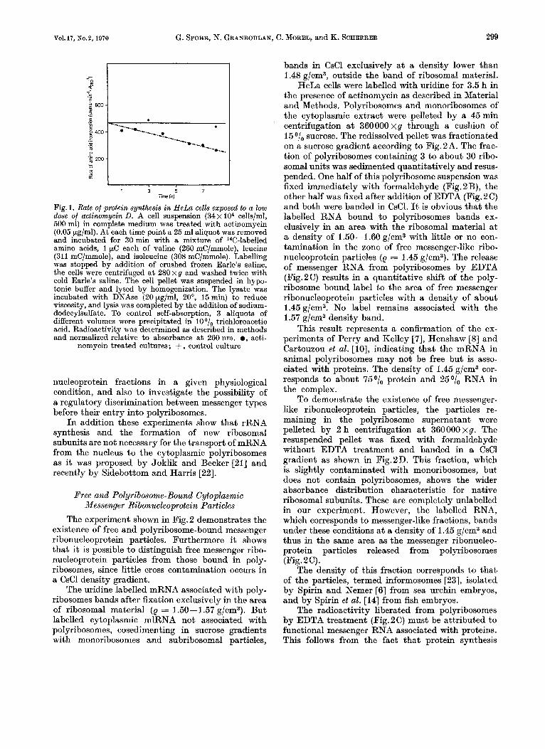
I n order to follow the influx of messenger-like RNA to the cytoplasm in the absence of ribosomal RNA synthesis we took advantage of the obser- vation of Perry [16-171 and Georgiev et al. [18] that low doses of actinomycin D specifically inhibit 45 S RNA formation in the nucleolus without seri- ously affecting mlRNA synthesis in the nucleoplasm. Exploratory experiments showed that the dose of 0.05 pg/ml actinomycin D, close to that chosen by Penman et al. [12], was optimal for our purpose.
Earlier reports in the literature [19] claim that the rate of protein synthesis is not affected under these conditions. We found that it drops slowly to about 6001, of the initial rate after 8 h of exposure (Fig.1). Since the half lives of polyribosomes and mRNA in HeLa cells after complete inhibition of RNA synthesis by a high actinomycin dose are about 3 h [20] it is evident that under our conditions new messenger RNA must reach the polyribosomes and be translated in order to maintain this level of protein synthesis. This labelling system is adequate to our purpose, since our main goal is to investigate the pattern of influx of mRNA into the various ribo-
Vol. 17, No.2, 1970 G. SPOHR, N. GRANBOULAN, C. MOREL, and K. SCHERRER 299
! I .c
I I 1 3 5 7
Time (h)
Fig. 1. Rate of protein synthesis in HeLa cells exposed to a low dose of actinomycin D. A cell suspension (34 x lo4 cells/ml, 500 ml) in complete medium was treated with actinomycin (O.O5.pg/ml). At each time point a 25 ml aliquot was removed and incubated for 30min with a mixture of l4C-labe1led amino acids, 1 pC each of valine (260 mC/mmole), leucine (311 mC/mmole), and isoleucine (308 mC/mmole). Labelling was stopped by addition of crushed frozen Earle’s saline, the cells were centrifuged at 280xg and washed twice with cold Earle’s saline. The cell pellet was suspended in hypo- tonic buffer and lysed by homogenization. The lysate was incubated with DNAse (20 pg/ml, 20°, 15 min) to reduce viscosity, and lysis was completed by the addition of sodium- dodecylsulfate. To control self-absorption, 3 aliquots of different volumes were precipitated in 10 O/,, trichloroacetic acid. Radioactivity was determined as described in methods and normalized relative to absorbance a t 260 nm. 0, acti-
nomycin treated cultures; + , control culture
nucleoprotein fractions in a given physiological condition, and also to investigate the possibility of a regulatory discrimination between messenger types before their entry into polyribosomes.
In addition these experiments show that rRNA synthesis and the formation of new ribosomal subunits are not necessary for the transport of mRNA from the nucleus to the cytoplasmic polyribosomes as it was proposed by Joklik and Becker [21] and recently by Sidebottom and Harris [22].
Pree and Polyribosome-Bound Cytoplasmic Messenger Ribonuckoprotein Particles
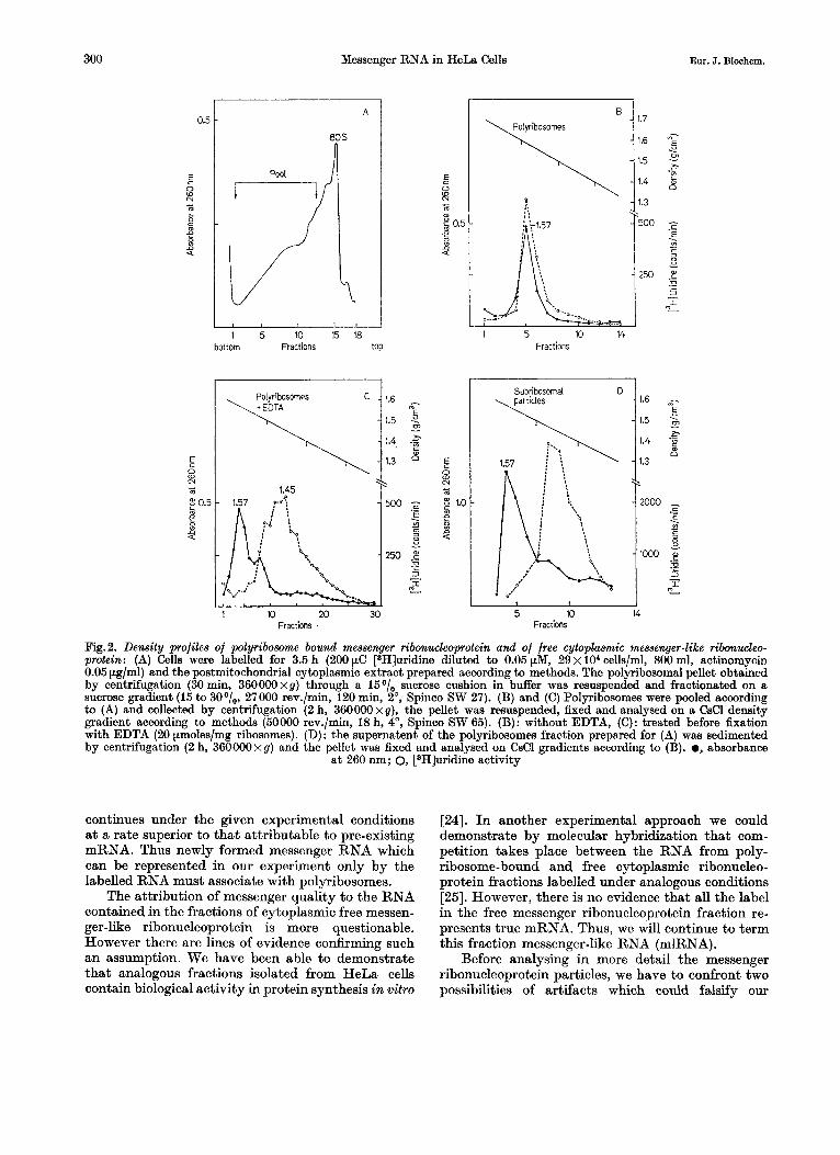
The experiment shown in Fig. 2 demonstrates the existence of free and polyribosome-bound messenger ribonucleoprotein particles. Furthermore it shows that it is possible to distinguish free messenger ribo- nucleoprotein particles from those bound in poly- ribosomes, since little cross contamination occurs in a CsCl density gradient.
The uridine labelled mRNA associated with poly- ribosomes bands after fixation exclusively in the area of ribosomal material (e = 1.50-1.57 g/cm3). But labelled cytoplasmic mlRNA not associated with polyribosomes, cosedimenting in sucrose gradients with monoribosomes and subribosomal particles,
bands in CsCl exclusively a t a density lower than 1.48 g/cm3, outside the band of ribosomal material.
HeLa cells were labelled with uridine for 3.5 h in the presence of actinomycin as described in Material and Methods. Polyribosomes and monoribosomes of the cytoplasmic extract were pelleted by a 45min centrifugation a t 360000 x g through a cushion of 15 sucrose. The redissolved pellet was fractionated on a sucrose gradient according to Fig.2A. The frac- tion of polyribosomes containing 3 to about 30 ribo- somal units was sedimented quantitatively and resus- pended. One half of this polyribosome suspension was fixed immediately with formaldehyde (Fig. 2B), the other half was fixed after addition of EDTA (Fig.2C) and both were banded in CsC1. It is obvious that the labelled RNA bound to polyribosomes bands ex- clusively in an area with the ribosomal material at a density of 1.50-1.60 g/cm3 with little or no con- tamination in the zone of free messenger-like ribo- nucleoprotein particles (e = 1.45 g/cm3). The release of messenger RNA from polyribosomes by EDTA (Fig.2C) results in a quantitative shift of the poly- ribosome bound label to the area of free messenger ribonucleoprotein particles with a density of about 1.45 g/cm3. No label remains associated with the 1.57 g/cm3 density band.
This result represents a confirmation of the ex- periments of Perry and Kelley [7], Henshaw [8] and Cartouzou et al. [lo], indicating that the mRNA in animal polyribosomes may not be free but is asso- ciated with proteins. The density of 1.45 g/cm3 cor- responds to about 75O/, protein and 25O/, RNA in the complex.
To demonstrate the existence of free messenger- like ribonucleoprotein particles, the particles re- maining in the polyribosome supernatant were pelleted by 2 h centrifugation a t 360000 xg. The resuspended pellet was fixed with formaldehyde without EDTA treatment and banded in a CsCl gradient as shown in Fig.2D. This fraction, which is slightly contaminated with monoribosomes, but does not contain polyribosomes, shows the wider absorbance distribution characteristic for native ribosomal subunits. These are completely unlabelled in our experiment. However, the labelled RNA, which corresponds to messenger-like fractions, bands under these conditions a t a density of 1.45 g/cm3 and thus in the same area as the messenger ribonucleo- protein particles released from polyribosomes (Fig. 2 C) .
The density of this fraction corresponds to that of the particles, termed informosomes [23], isolated by Spirin and Nemer [6] from sea urchin embryos, and by Spirin et al. [I41 from fish embryos.
The radioactivity liberated from polyribosomes by EDTA treatment (Fig.2C) must be attributed to functional messenger RNA associated with proteins. This follows from the fact that protein synthesis
300 Messenger RNA in HeLa Cells Eur. J. Biochem.
0.5
E c
N m - 2 e a 9
m
E
p 0
N
m
m f! 2 9
a
A
1 I t
1 5 10 15 18 Ittorn Fractions top
Polyribosornes + EDTA
1 10 20 30 Fractions .
0
1 5 10 14 Fractions
Fig.2. Density profiles of polyribosome bound messenger ribonwleoprotein and of free cytoplasmic messenger-like ribonwleo- protein: (A) Cells were labelled for 3.5 h (200 pC [3H]uridine diluted to 0.05 pM, 29 x lo4 cells/ml, 800 ml, actinomycin 0.05 pg/ml) and the postmitochondrial cytoplasmic extract prepared according to methods. The polyribosomal pellet obtained by centrifugation (30 min, 360000xg) through a 15O/, sucrose cushion in buffer was resuspended and fractionated on a sucrose gradient (15 to 30°/,, 27000 rev./min, 120 min, 2O, Spinco SW 27). (B) and (C) Polyribosomes were pooled according to (A) and collected by centrifugation (2 h, 360000xg), the pellet was resuspended, fixed and analysed on a CsCl density gradient according to methods (50000 rev./min, 18 h, 4 O , Spinco SW 65). (B): without EDTA, (C): treated before fixation with EDTA (20 pmoles/mg ribosomes). (D): the supernatent of the polyribosomes fraction prepared for (A) was sedimented by centrifugation (2 h, 36000Oxg) and the pellet was fixed and analysed on CsCl gradients according to (B). 0, absorbance
a t 260 nm; 0, rH]uridine activity
continues under the given experimental conditions at a rate superior to that attributable to pre-existing mRNA. Thus newly formed messenger RNA which can be represented in our experiment only by the labelled RNA must associate with polyribosomes.
The attribution of messenger quality to the RNA contained in the fractions of cytoplasmic free messen- ger-like ribonucleoprotein is more questionable. However there are lines of evidence confirming such an assumption. We have been able to demonstrate that analogous fractions isolated from HeLa cells contain biological activity in protein synthesis in vitro
[24]. In another experimental approach we could demonstrate by molecular hybridization that com- petition takes place between the RNA from poly- ribosome-bound and free cytoplasmic ribonucleo- protein fractions labelled under analogous conditions [25]. However, there is no evidence that all the label in the free messenger ribonucleoprotein fraction re- presents true mRNA. Thus, we will continue to term this fraction messenger-like RNA (mlRNA).
Before analysing in more detail the messenger ribonucleoprotein particles, we have to confront two possibilities of artifacts which could falsify our
Vol. 17, No. 2, 1970 G. SPOHR, N. GRANBOULAN, C. MOREL, and K. SCHERRER 301
0.50
E a L
025
1.6 1.5 1.4 Density (9/cm3)
1.6 1.5 1.4 Density (9/cm3) Density (9/cm3)
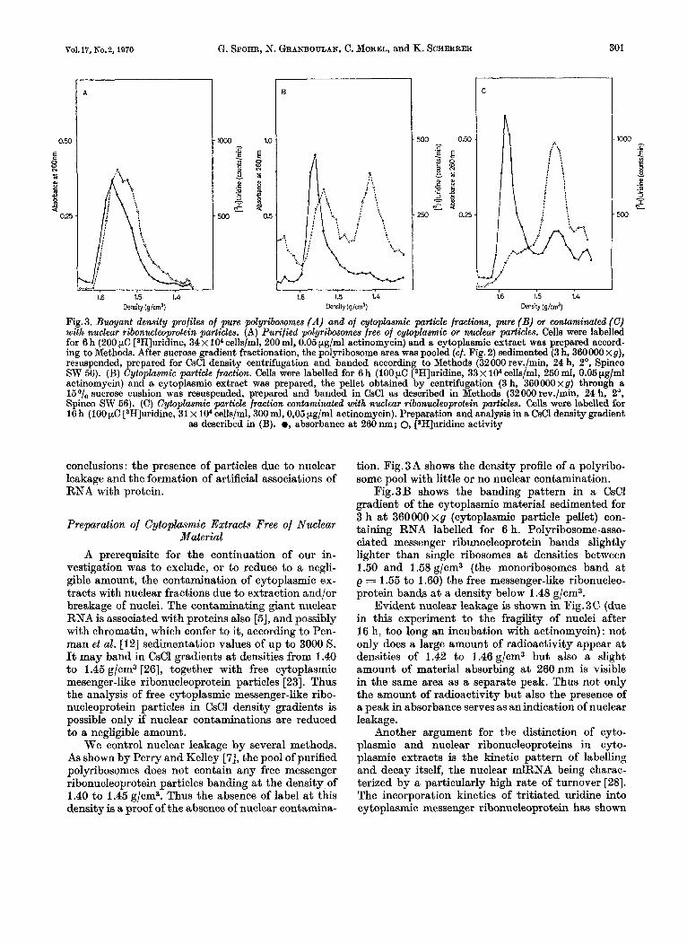
Fig.3. Buoyant density profiles of pure polyribosomes (A) and of cytoplasmic particle fractions, pure (B) or contaminated (C) with nuclear ribonucleoprotein particles. (A) Purified polyribosomes free of cytoplaamic or nuclear particles. Cells were labelled for 6 h (200 pC [3H]uridine, 34 x lo" cells/ml, 200 ml, 0.05 pg/ml actinomycin) and a cytoplasmic extract was prepared accord- ing to Methods. After sucrose gradient fractionation, the polyribosome area was pooled (cf. Fig. 2) sedimented (3 h, 360000 xg), resuspended, prepared for CsCl density centrifugation and banded according to Methods (32000 rev./min, 24 h, 2O, Spinco SW 56). (B) Cytoplasmic particle fraction. Cells were labelled for 6 h (100 pC [3H]uridine, 33 x lo4 cells/ml, 250 ml, 0.05 pg/ml actinomycin) and a cytoplasmic extract was prepared, the pellet obtained by centrifugation (3 h, 360000xg) through a 15O/,, sucrose cushion was resuspended, prepared and banded in CsCl as described in Methods (32000 rev./min, 24 h, 2O, Spinco SW 56). (C) Cytoplamnic particle fraction wntaminated with nuclear ribonucleoprotein particles. Cells were labelled for 16 h (100 pC [3H]uridine, 31 x lo4 cells/ml, 300 ml, 0,05 pg/ml actinomycin). Preparation and analysis in a CsCl density gradient
aa described in (B). 0, absorbance at 260 nm; 0, [*H]uridine activity
conclusions : the presence of particles due to nuclear leakage and the formation of artificial associations of RNA with protein.
Preparation of Cytoplasmic Extracts Free of Nuclear Material
A prerequisite for the continuation of our in- vestigation was to exclude, or to reduce to a negli- gible amount, the contamination of cytoplasmic ex- tracts with nuclear fractions due to extraction and/or breakage of nuclei. The contaminating giant nuclear RNA is associated with proteins also [5], and possibly with chromatin, which confer to it, according to Pen- man et al. [12] sedimentation values of up to 3000 s. It may band in CsCl gradients at densities from 1.40 to 1.45 g/cm3 [26], together with free cytoplasmic mesenger-like ribonucleoprotein particles [23]. Thus the analysis of free cytoplasmic messenger-like ribo- nucleoprotein particles in CsCl density gradients is possible only if nuclear contaminations are reduced to a negligible amount.
We control nuclear leakage by several methods. As shown by Perry and Kelley [7], the pool of purified polyribosomes does not contain any free messenger ribonucleoprotein particles banding at the density of 1.40 to 1.45 g/cm3. Thus the absence of label at this density is a, proof of the absence of nuclear contamina-
tion. Fig.3A shows the density profile of a polyribo- some pool with little or no nuclear contamination.
Fig.3B shows the banding pattern in a CsCl gradient of the cytoplasmic material sedimented for 3 h at 360000 xg (cytoplasmic particle pellet) con- taining RNA labelled for 6 h. Polyribosome-asso- ciated messenger ribunocleoprotein bands slightly lighter than single ribosomes a t densities between 1.60 and 1.58 &/em3 (the monoribosomes band a t e = 1.55 to 1.60) the free messenger-like ribonucleo- protein bands at a density below 1.48 g/cm3.
Evident nuclear leakage is shown in Fig.3C (due in this experiment to the fragility of nuclei after 16 h, too long an incubation with actinomycin) : not only does a large amount of radioactivity appear a t densities of 1.42 to 1.46 g/cm3 but also a slight amount of material absorbing at 260 nm is visible in the same area as a separate peak. Thus not only the amount of radioactivity but also the presence of a peak in absorbance serves as an indication of nuclear leakage.
Another argument for the distinction of cyto- plasmic and nuclear ribonucleoproteins in cyto- plasmic extracts is the kinetic pattern of labelling and decay itself, the nuclear mlRNA being charac- terized by a particularly high rate of turnover [28]. The incorporation kinetics of tritiated uridine into cytoplasmic messenger ribonucleoprotein has shown
302 Messenger RNA in HeLa Cells Eur. J. Biochem.
E c z 2 3.0
z 2 w z 2.0
m
m
1.0
A
1.3
10 20 30 Fractions
10 20 30 Fractions
C
Fig. 4. Artificial association of purified RNA with proteins present in the cytoplasmic particle fraction. Polyribosomes, ribosomes and subribosomal particles were sedimented into one pellet and resuspended in buffer according to Methods. Purified [W]- uridine-labelled RNA was added and incubated in the cold for 15 min, the mixture was fixed with formaldehyde and banded in CsCl density gradients (50000 rev./min, 18 h, 2 O , Spinco SW 65). (A) 28 S ribosomal RNA added, (B, C) nuclear mlRNA
added
that a steady state of synthesis and decay is reached a t approximately 6 h (cf. Fig.ll). On the contrary, the nucleoplasmic heterogeneous RNA approaches steady state already after 2 h [12].
This extensive discussion should point out the importance we give to the control of the cytoplasmic origin of the complexes subject to this type of analysis. The method finally adopted for preparing cytoplasmic extracts is described in Methods. The restoration of isotonicity before cellular lysis limited the extraction of nuclei. (Hypotonicity during cell homogenisation led inevitably to nuclear leakage.) The limitation of homogenization t o a level where about 700/, of nuclei only are liberated reduced nuclear breakage to a negligible level.
Control of Artificial Association of RNA with Protein in the Cytoplasmic Extracts
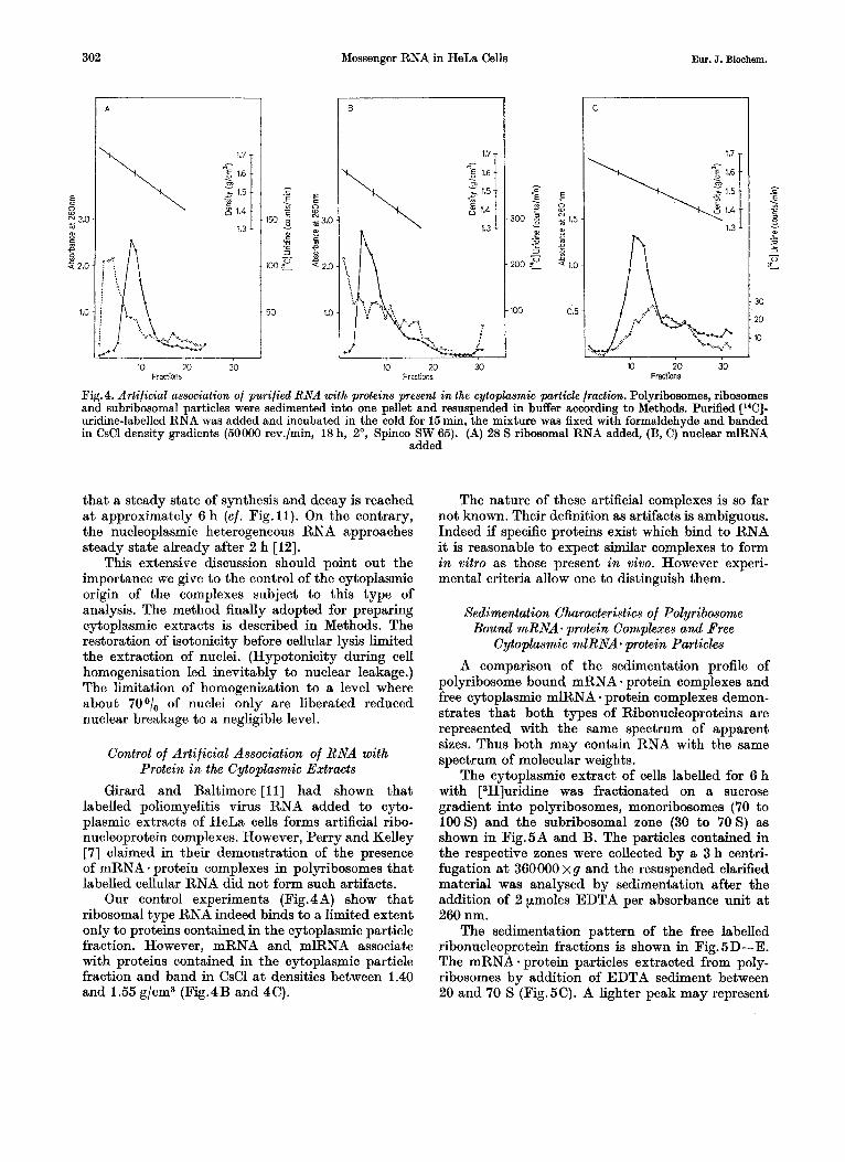
Girard and Baltimore [ l l ] had shown that labelled poliomyelitis virus RNA added to cyto- plasmic extracts of HeLa cells forms artificial ribo- nucleoprotein complexes. However, Perry and Kelley [7] claimed in their demonstration of the presence of mRNA - protein complexes in polyribosomes that labelled cellular RNA did not form such artifacts.
Our control experiments (Fig.4A) show that ribosomal type RNA indeed binds to a limited extent only to proteins contained in the cytoplasmic particle fraction. However, mRNA and mlRNA associate with proteins contained in the cytoplasmic particle fraction and band in CsCl at densities between 1.40 and 1.55 g/cm3 (Fig. 4 B and 4 C).
The nature of these artificial complexes is so far not known. Their definition as artifacts is ambiguous. Indeed if specific proteins exist which bind to RNA it is reasonable to expect similar complexes to form in vitro as those present in vivo. However experi- mental criteria allow one to distinguish them.
Sedimentation Characteristics of Polyribosome Bound mRNA. protein Complexes and Free
Cytoplasmic mlRNA* protein Particles A comparison of the sedimentation profile of
polyribosome bound mRNA protein complexes and free cytoplasmic mlRNA * protein complexes demon- strates that both types of Ribonucleoproteins are represented with the same spectrum of apparent sizes. Thus both may contain RNA with the same spectrum of molecular weights.
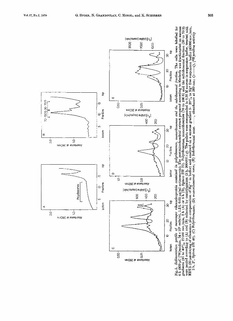
The cytoplasmic extract of cells labelled for 6 h with [3H]uridine was fractionated on a sucrose gradient into polyribosomes, monoribosomes (70 to 100s) and the subribosomal zone (30 to 70s) as shown in Fig.5A and B. The particles contained in the respective zones were collected by a 3 h centri- fugation a t 360000 x g and the resuspended clarified material was analysed by sedimentation after the addition of 2 pmoles EDTA per absorbance unit a t 260 nm.
The sedimentation pattern of the free labelled ribonucleoprotein fractions is shown in Fig.6D-E. The mRNA - protein particles extracted from poly- ribosomes by addition of EDTA sediment between 20 and 70 S (Fig.5C). A lighter peak may represent
2 .o
E N
.m
a,
c s m 1.0
.D
a 2
A
Polyr
ibos
omes
1
5 10
15
B
70-1
005
30-7
05 JJ
5 10
15
botto
m
Frac
tions
to
p bo
ttom
I r
C
I 1.0
Jo Fr
actio
ns
top
0.50
JE
lo 20
30
botto
m
Frac
tions
to
p 10
20
30
10 20
30
bo
ttom
Fr
actio
ns
top
botto
m
Frac
tions
to
p
Fig.
5. S
edim
enta
tion
prof
ile o
f m
esse
nger
rib
onuc
leop
rote
in c
onta
ined
in
poly
ribo
som
es,
mon
orib
osom
es a
nd t
he s
ubrib
osom
al f
ract
ion.
The
cel
ls w
ere
labe
lled
for
6 h
(800
pC
C3H
]urid
ine,
34 x
lo4
cells
lml,
1.2
1, 0.
05 p
g/m
l ac
tinom
ycin
). T
he p
ostm
itoch
ondr
ial
extr
act p
repa
red
acco
rdin
g to
Met
hods
was
fra
ctio
nate
d on
suc
rose
gr
adie
nts
(10 to 40°/,, 2
7000
rev.
/min
, 2
h (A
) or
6 h
(B),
Spi
nco
SW 2
7). P
olyr
ibos
omes
, m
onor
ibos
omes
(70
to 1
00 S
) and
the
sub
ribo
som
al f
ract
ion
(30
to 7
0 S)
w
ere
pool
ed a
ccor
ding
to
(A) a
nd (B
) col
lect
ed b
y ce
ntri
fuga
tion
(3 h,
360
000 X
g).
The
pel
lets
wer
e re
susp
ende
d in
0.2
5 M
suc
rose
in s
uspe
nsio
n bu
ffer
, tre
ated
wit
h E
DT
A (2
0 pm
oles
/mg
of r
ibos
omes
plu
s co
mpe
nsat
ion
of M
gff
in b
uffe
r) a
nd a
naly
sed
on a
suc
rose
gra
dien
t 5-2
0°/,
in
Mg+
free
sus
pens
ion
buff
er (
4000
0 rev
./min
, 3
h, 2
", S
pinc
o SW
40)
. (C
) Pol
yrib
osom
es;
(D) m
onor
ibos
omal
zon
e; (
E) S
ubri
boso
mal
zon
e. -,
abso
rban
ce a
t 260
nm
; O
-----O
, rH
]uri
dine
act
ivit
y
0 W 8
304 Messenger RNA in HeLa Cells Eur. J. Biochem.
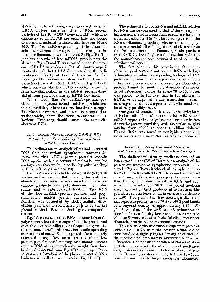
tRNA bound to activating enzymes as well as small mRNA - protein particles. The mlRNA protein particles of the 70 to 100 S zone (Fig.5D) which, as demonstrated in Fig. 7 B are essentially not bound to ribosomal material, sediment also between 10 and 70 S. The free mlRNA. protein particles from the subribosomal zone show a predominance of particles in the sedimentation zone below 50 S (Fig.5E). The gradient analysis of free mlRNA - protein particles shown in Fig. 5 D and E was carried out in the pres- ence of EDTA in analogy to Fig. 5C. Control experi- ments showed that EDTA does not alter the sedi- mentation velocity of labelled RNA in the free messenger-like ribonucleoprotein fraction. Thus the particles of the entire 30 to 100 S area (Fig.BD+ E) which contains the free mlRNA * protein show the same size distribution as the mRNA * protein disso- ciated from polyribosomes by EDTA treatment.
We conclude that free mlRNA .protein par- ticles and polysome-bound mRNA - protein-con- taining particles, or in other terms inactive messenger- like ribonucleoprotein and active messenger ribo- nucleoprotein, show the same sedimentation be- haviour. Thus they should contain the same size classes of RNA.
Sedimentation Characteristics of Labelled RNA Extracted from Free and Polyribosome-Bound
mRNA *protein Particles The sedimentation analysis of phenol extracted
RNA from the various cytoplasmic fractions de- monstrates that mRNA - protein particles contain RNA species with a spectrum of molecular weights analogous to that we found earlier by pulse labelling in HeLa cell polyribosomes [20].
HeLa cells were labelled to steady state (6 h) with midine as described in Methods and the postmito- chondrial cytoplasmic particles were fractionated on sucrose gradients into polyribosomes, monoribo- somes and a subribosomal fraction. The RNA of the free mlRNA-protein particles and poly- some-bound mRNA * protein contained in these fractions was extracted by dodecylsulfate disso- ciation (and directly sedimented [29]) or by the hot phenol method. Both methods gave comparable results.
Fig. 6 demonstrates that RNA extracted from the polyribosome-bound messenger ribonucleoprotein and from free messenger-like ribonucleoprotein gives rise to the same overall sedimentation profile spreading from 6 S to about 30 S. As expected, the separately extracted heavy free messenger-like ribonucleo- protein particles cosedimenting with monoribosomes contain RNA of higher molecular weight then those in the subribosomal zone (Fig.6B and C resp.). Poly- acrylamide gel analysis of the phenol extracted RNA leads to essentially the same results (Fig.6D-F).
The sedimentation of mRNA and mlRNA relative to rRNA can be compared to that of the correspond- ing messenger ribonucleoprotein particles relative to ribosomal subunits (Fig.5). The overall pattern of the RNA or ribonucleoprotein spectra are the same : poly- ribosomes contain the full spectrum of sizes whereas the free messenger-like ribonucleoprotein particles or their RNA have higher sedimentation values in the monoribosomes area compared to those in the subribosomal zone.
The fact that in this experiment the mono- ribosome pool contains not only mlRNA with high sedimentation values corresponding to large mlRNA particles but also smaller types may be attributed either to the presence of some messenger ribonucleo- protein bound to small polyribosomes (“mono-or di-polyribosomes”), since the entire 70 to 100 S area was pooled, or to the fact that in the absence of EDTA or of high salt some association between messenger-like ribonucleoprotein and ribosomal ma- terial may possibly occur.
Our general conclusion is that in the cytoplasm of HeLa cells (free of mitochondria) mRNA and mlRNA types exist, polyribosome-bound or in free ribonucleoprotein particles, with molecular weights ranging from 50000 to about 1 million daltons. Heavier RNA was found in negligible amounts in experiments where no nuclear leakage had occurred.
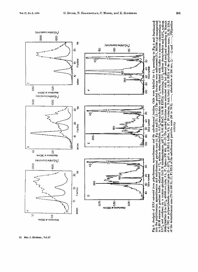
Density Profiles of Individual Messenger and Messenger-Like Ribonucleoprotein Fractions The shallow CsCl density gradients obtained a t
lower speed in the SW-56 Rotor allow analysis of the particulate fraction of cytoplasmic extracts in more detail (Fig. 7). Postmitochondrial cytoplasmic ex- tracts from cells labelled for 3 or 6 h were fractionated on sucrose gradients into pure polyribosomes (more than 150 S), monoribosomes (70 to 100 S) and sub- ribosomal particles (30-70 s). The pooled fractions were analysed on CsCl gradients after fixation. The polyribosomal material bands in an area a t a density of 1.50- 1.60 g/cm3, the free messenger-like ribo- nucleoprotein present in the 70 S to 100 S pool bands at a buyoant density of approximately 1.40- 1.50 g/cm3 and that of the 30 S to 70 S sedimentation zone bands at a density lower than 1.45 g/cm3. The 70- 100 S zone contains little labelled messenger ribonucleoprotein bound to monoribosomes.
The fact that the free ribonucleoprotein particles containing mlRNA from the heavier sedimentation zone band a t a slightly higher density than those of the subribosomal area may be attributed to intrinsic differences in composition of different classes of these particles or perhaps to the attachment of small mes- senger ribonucleoprotein particles to ribosomal sub- units. However, as shown in Fig.5D the 70-100 S zone contains mainly large, messenger ribonucleo-
N
0
A
2000
1.0
0
20
30
botto
m
Frac
tions
to
p
0.75 -
E R 2 0.50
-
0.25 -
Slice
num
ber
0
10
20
30
botto
m
Frac
tions
tw
Slic
e nu
mbe
r
10
20
30
botto
m
Frac
tions
to
p
?
Fig.
6. A
naly
sis
of R
NA
ext
ract
ed f
rom
pol
yrib
osom
es, m
onor
ibos
omes
and
the
sub
ribo
som
al f
ract
ion.
Cel
ls w
ere
labe
lled
for
6 h
acco
rdin
g to
Fig
.5 a
nd f
ract
iona
ted
into
the
pol
yrib
osom
e, m
onor
ibos
ome a
nd s
ubri
boso
mal
par
ticle
zon
e (c
f. Fi
g.5A
and
B).
(A-C
) T
he p
oole
d fr
actio
ns w
ere
sedi
men
ted
(3 h,
360
000x
g) r
esus
pend
ed
in 0
.25
M s
ucro
se in
gra
dien
t bu
ffer
, ad
just
ed t
o 1 O/,
sodi
um d
odec
ylsu
lfat
e, 0
.5O
/, de
oxyc
hola
te a
nd 0
.5O
/, ur
ea, d
ilute
d w
ith b
uffe
r to
a su
cros
e co
ncen
trat
ion
of
3.5O
/, an
d an
alys
ed o
n a
sucr
ose
grad
ient
(0.01 M
trie
than
olam
ine,
pH
7.4
, 0.
01 M
NaC
I, 0.
01 M
ED
TA
) co
ntai
ning
0.2O
/, so
dium
deo
xych
olat
e an
d 0.
20/,
sodi
um
dode
cyls
ulfa
te (5
to 2
0°/,,
400
00 re
v./m
in,
6 h,
2",
Spi
nco
SW 4
0).
(D-F
) T
he p
oole
d fr
actio
ns w
ere
extr
acte
d w
ith h
ot p
heno
l and
ana
lyse
d ac
cord
ing
to M
irau
lt et
al.
[30]
on
poly
acry
lam
ide e
xpon
entia
l gel
s (2
--15O
/, ge
l in
E-R
NA
buf
fer,
9 h,
2",
10
volts
/cm
, 3 m
A/t
ube)
. (A
, D) R
NA
of
the
poly
ribos
ome
zone
; (B
, E)
RN
A
of t
he m
onor
ibos
ome
zone
(70
to 1
00 S
); (C
, F) R
NA
of
the
subr
ibos
omal
par
ticle
s zo
ne (
30 to
70
S). -,
abso
rban
ce a
t 26
0 nm
; an
d . - -
. -. , [S
HIu
ridin
e ac
tivity
W
0
01
306 Messenger RNA in HeLa Cells Eur. J. Biochem.
A
Polyribosms I
1.6 1.5 1.4 Density (g/cml)
~
D
Polyribosomes + EDTA
,E 8 N * I 8 ? 0.3 z 0.2
0.1
E c
E -
B
Maor ibo s o m e s 70-03s zme
1.6 1.5 1.4 Density (g/cml)
E
Mono r i bo s o m e s + EDTA
0.4
E c
- E s 0.2
0.10
E c w * 8
4 0.05 3
C
Subribosml particles
30-70s m
1.6 1.5 1.4 Denw'ty (gIcm3
F
Sukibosomal particles
EDTA
1.6 1.5 1.4 Density ( g i c m l )
Fig. 7. Buoyant density profiles of messenger ribonucleoprotein particles contained in polyribosomes, monoribosomes and the sub- ribosomal fraction before and after EDTA treatment. Cells were labelied for 3 (C and F) or 6 (A, D, and B, E) h (800 $2, [9H]- uridine, 33 x lo4 cells/ml, 1.5 1, 0.05 pg/ml actinomycin), the postmitochondrial extract was prepared according to Methods and fractionated on a sucrose gradient into polyribosomes, monoribosomes and the subribosomal particle zone (cf. Fig. 5). The pooled fractions were sedimented (3 h, 360000 x g ) resuspended in 0.25 M sucrose in suspension buffer, treated or not with EDTA (20 pmoles/mg ribosome plus compensation for Mg in buffer). CsCl density analysis according to Methods (32000 rev./min, 24 h, 2", Spinco SW 56). (A, D) polyribosomes; (B, E) monoribosomes; (C-F) subribosomal particles.
(A-C) controls; (D-F) EDTA treated. 0-0, absorbance at 260nm; @--a, [3H]uridine activity
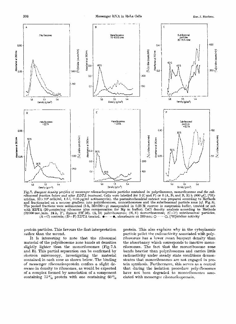
protein particles. This favours the first interpretation rather than the second.
It is interesting to note that the ribosomal material of the polyribosome zone bands a t densities slightly lighter than the monoribosomes (Fig. 7 A and B). This partial separation can be confirmed by electron microscopy, investigating the material contained in each zone as shown below. The binding of messenger ribonucleoprotein confers a slight de- crease in density to ribosomes, as would be expected of a complex formed by association of a component containing 75O/, protein with one containing BOO/,
protein. This also explains why in the cytoplasmic particle pellet the radioactivity associated with poly- ribosomes has a lower mean buoyant density than the absorbancy which corresponds to inactive mono- ribosomes. The fact that the monoribosome zone bands heavier than polyribosomes and carries little radioactivity under steady state conditions demon- strates that monoribosomes are not engaged in pro- tein synthesis. Furthermore, this serves as a control that during the isolation procedure polyribosomes have not been degraded to monoribosomes asso- ciated with messenger ribonucleoprotein.
Vol.17, x0.2, 1970 G. SPOHR, N. GRANBOULAN, C . MOREL. and I<. SCHERRER 307
I
2.0
N +
2 2 5: m
a < 1.0
1 10 20 30 Fractions
2 .o E c 8 N c
d i? 5: 2
m
1.0
2.0
E c
3 c
ar c .n a 2
1.0
Fractions
Fractions
Fractions
Fig. 8. Labelling pattern in density profiles of free messenger-like ribonucleoprotein and polyrihosome bound messenger ribonucleo- protein. Cells were labelled for the indicated time periods (150 pC [3H]uridine diluted to 0.1 pV, 66 x lo4 cells/ml, 600 ml, 0.05 pg/ml actinomgcin) as indicated in Methods. Aliquots of 125 ml were chilled, the cytoplasmic particle pellet was prepared and analysed in CsCl density gradients as indicated in Nethods (50000 rev./min, 18 h, 2", Spinco SW 65). 0 , absorbance
a t 260 nm; 0, [3H]uridine activity
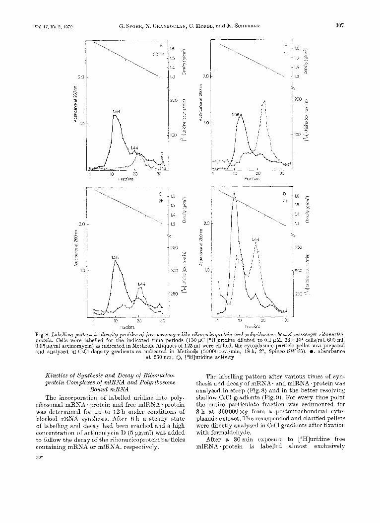
Kinetics of Synthesis and Decay of Ribonucleo- protein Complexes of mlRNA and Polyribosorne
Bound mRNA The incorporation of labelled uridine into poly-
ribosomal mRNA * protein and free mlRNA * protein was determined for up to I2 h under conditions of blocked rRKA synthesis. After 6 h a steady state of labelling and decay had been reached and a high concentration of actinornycin I> (5 &ml) was added to follow the decay of the ribonucleoprotein particles containing mRNA or mlRNA, respectively. LO+
The labelling pattern after various times of syn- thesis and decay of mRNA. and mlRNA. protein was analysed in steep (Fig.8) and in the better resolving shallow CsCl gradients (Fig.9). For every time point the entire particulate fraction was sedimentcd for 3 h at 360000 xg from a postmitochondrial cpto- plasmic extract. The resuspended and clarified pellets were directly analysed in CsCl gradients after fixation with formaldehyde.
After a 30min exposure t o [3H]uridine free mlRNA * protein is labelled almost exclusively
A
E 0.e
I E 0.4
1.6
1.5
1.4
hit
y (g
lVn3
)
E
1.6
1.5
1.4
Dens
ity (g
/un3
)
C
F
D
G
Fig.
9. P
atte
rn i
n de
nsity
pro
files
of
labe
lling
and
dec
ay of
m
esse
nger
rib
onue
leop
rote
in pa
rtic
les.
Cel
ls w
ere
labe
lled
for 6
h (5
00 pC
[3H
]urid
ine,
33
x lO
'cells
/ml,
1300
ml,
0.05
pg/
ml
actin
omyc
in),
acco
rdin
g to
Met
hods
; af
ter
6 h
the
cultu
re w
as a
djus
ted
to 5
pg/
ml a
ctin
omyc
in in
ord
er t
o in
duce
the
chas
e. Aft
er th
e in
dica
ted
times
15
0 m
l aliq
uots
wer
e ch
illed
and
the
cyt
opla
smic
par
ticle
pel
let
prep
ared
and
ana
lyse
d in
CsC
l den
sity
gra
dien
ts (
3200
0 rev
./min
24
h, 2
", S
pinc
o SW
56)
as
in&
ca
ted
in M
etho
ds.
(A)
1 h;
(B
) 2
h; (
C)
4 h;
(D
) 6 h
; (E
) 6 h
and
1 h
chas
e; (
F) 6
h a
nd 3
h c
hase
; (G
) 6 h
and
6 h
cha
se.
0-0
, ab
sorb
ance
at
260
nm;
M I-' f 0
@--a,
[3H
]urid
ine a
ctiv
ity
U B
Vo1.17, No.2, 1970 G. SPOHR, N. GRANBOULAN, C. MOREL, and K. SCHERRER
1 I
309
4 s 12 Time (h)
4 8 12 Time (h)
Pig.10. Kinetics of RNA labelliny in whole cells and in the cytoplasmic particle fraction. Cells were labelled as indicated in Fig. 9. To determine the radioactivity in whole cells 1 ml aliquots of the culture were sedimented at 280 x g, washed twice with cold Earle’s saline and lysed in isotonic saline with 0.5 sodium dodecylsulfate. 10 trichloroacetic precipitable radioactivity was determined as described in Methods. The specific activity of the cytoplasmic particle fraction prepared as indicated in Fig.9 was determined according to Methods. (A): whole cells; (B): cytoplasmic particle pellet. (-) con-
trol; (----) 5 pg/ml actinomycin added a t 6 h, as indicated by the arrow
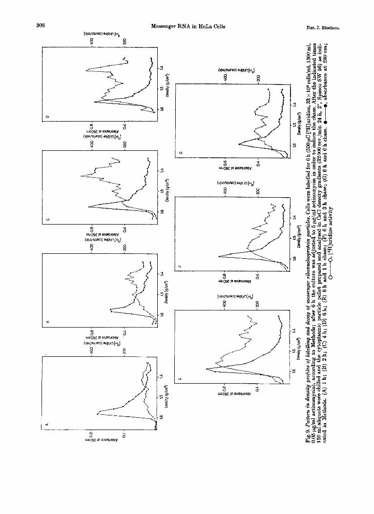
(Fig. 8A). Subsequently more enters the cytoplasm and some polyribosome-bound label appears (Fig. 8B and C). After 4 h of labelling (Fig. 8D) approximati- vely the same amount of radioactivity is present in mRNA * protein and free mlRNA * protein.
A similar pattern may be observed in more shallow gradients (Fig.9A to D). Incorporation of label into free mlRNA * protein precedes always incorporation into polyribosomes. In the presence of the low actinomycin concentration used a steady state of synthesis and decay is reached after approxi- mately 6 h. The synthesis of mRNA. and mlRNA- protein was followed for up to 12 h. This represents the practical time limit for these experiments due to the increased fragility of the nuclei after prolongued exposure to actinomycin.
If after 6 h of labelling a high dose of actinomycin (5 pg/ml) was added to inhibit completely further RNA synthesis, the decay of both polysome-bound mRNA * protein and free mlRNA * protein could be observed.
In these shallow gradients a different pattern of synthesis and decay was evident in the various density zones. I n several experiments it was ob- served that the light mlRNA * protein of the I .40 g/cm3 density zone disappear more rapidly than others (Fig.9E-G). If in the first hour of actinomycin chase the disappearance of free mlRNA * protein seems to be more rapid than in polyribosome-bound mlRNA . protein, a continued chase reveals approxi- mately the same rate of decay in both fractions.
I n order to express quantitatively the pattern of mRNA * and mlRNA * protein synthesis and decay observed, the radioactivities corresponding to the free and polyribosome bound ribonucleoprotein zones were integrated. Specific activities were calcu- lated by normalisation relative to the total ribosomal material present in a gradient.
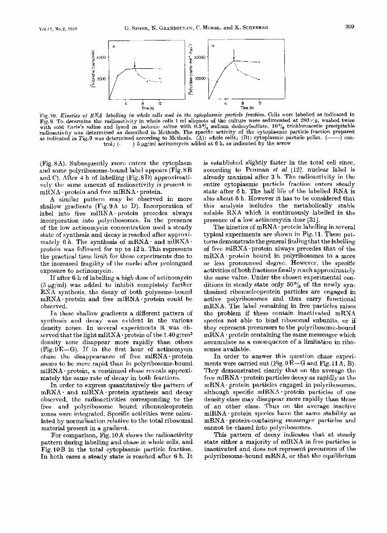
For comparison, Fig. 10A shows the radioactivity pattern during labelling and chase in whole cells, and Fig. 10B in the total cytoplasmic particle fraction. I n both cases a steady state is reached after 6 h. It
is established slightly faster in the total cell since, according to Penman et al. [IZ], nuclear label is already maximal after 3 h. The radioactivity in the entire cytoplasmic particle fraction enters steady state after 6 h. The half life of the labelled RNA is also about 6 h. However it has to be considered that this analysis includes the metabolically stable soluble RNA which is continuously labelled in the presence of a low actinomycin dose [31].
The kinetics of mRNA. protein labelling in several typical experiments are shown in Fig. 11. These pat- terns demonstrate the general finding that the labelling of free mlRNA-protein always precedes that of the mRNA protein bound in polyribosomes to a more or less pronounced degree. However, the specific activities of both fractions finally reach approximately the same value. Under the chosen experimental con- ditions in steady state only 50°/, of the newly syn- thesized ribonucleoprotein particles are engaged in active polyribosomes and thus carry functional mRNA. The label remaining in free particles raises the problem if these contain inactivated mRNA species not able to bind ribosomal subunits, or if they represent precursors to the polyribosome-bound mRNA - protein containing the same messenger which accumulate as a consequence of a limitation in ribo- somes available.
In order to answer this question chase experi- ments were carried out (Fig.9E-G and Fig. I IA, B). They demonstrated clearly that on the average the free mlRNA * protein particles decay as rapidly as the mRNA * protein particles engaged in polyribosomes, although specific mlRNA - protein particles of one density class may disappear more rapidly than those of an other class. Thus on the average inactive mlRNA - protein species have the same stability as mRNA * protein-containing messenger particles and cannot be chased into polyribosomes.
This pattern of decay indicates that a t steady state either a majority of mlRNA in free particles is inactivated and does not represent precursors of the polyribosome-bound mRNA, or that the equilibrium
I A
-! 'E I
4 8 12 Time (h)
4 8 Time [h)
[ ', ,~, & ~ , .\, ~... b
....... ~ ... ... .........
4 8 Time (h)
D
1 2 3 4 Time (h)
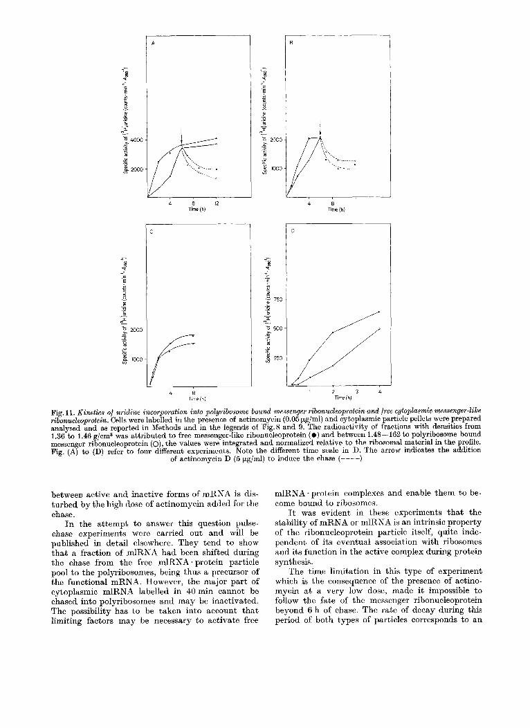
Fig. 11. Kinetics of uridine incorporation into polyribosome bound messenger ribonucleoprotein and free cytoplasmic messenger-like ribonucleoprotein. Cells were labelled in the presence of actinomycin (0.05 pg/ml) and cytoplasmic particle pellets were prepared analysed and as reported in Methods and in the legends of Fig.8 and 9. The radioactivity of fractions with densities from 1.36 to 1.46 g/cm3 was attributed to free messenger-like ribonucleoprotein ( 0 ) and between 1.48-162 to polyribosome bound messenger ribonucleoprotein (o), the values were integrated and normalized relative to the ribosomal material in the profile. Fig. (A) to (D) refer to four different experiments. Note the different time scale in D. The arrow indicates the addition
of actinomycin D (5 pg/ml) to induce the chase (----)
between active and inactive forms of mRNA is dis- turbed by the high dose of actinomycin added for the chase.
In the attempt to answer this question pulse- chase experiments were carried out and will be published in detail elsewhere. They tend to show that a fraction of mlRNA had been shifted during the chase from the free mlRNA * protein particle pool to the polyribosomes, being thus a precursor of the functional mRNA. However, the major part of cytoplasmic mlRNA labelled in 40min cannot be chased into polyribosomes and may be inactivated. The possibility has to be taken into account that limiting factors may be necessary to activate free
mlRNA. protein complexes and enable them to be- come bound to ribosomes.
It was evident in these experiments that the stability of mRNA or mlRNA is an intrinsic property of the ribonucleoprotein particle itself, quite inde- pendent of its eventual association with ribosomes and its function in the active complex during protein synthesis.
The time limitation in this type of experiment which is the consequence of the presence of actino- mycin a t a very low dose, made it impossible to follow the fate of the messenger ribonucleoprotein beyond 6 h of chase. The rate of decay during this period of both types of particles corresponds to an
I 100 A
I 100 i
I 100 A
I 1000 8
I 1000 A
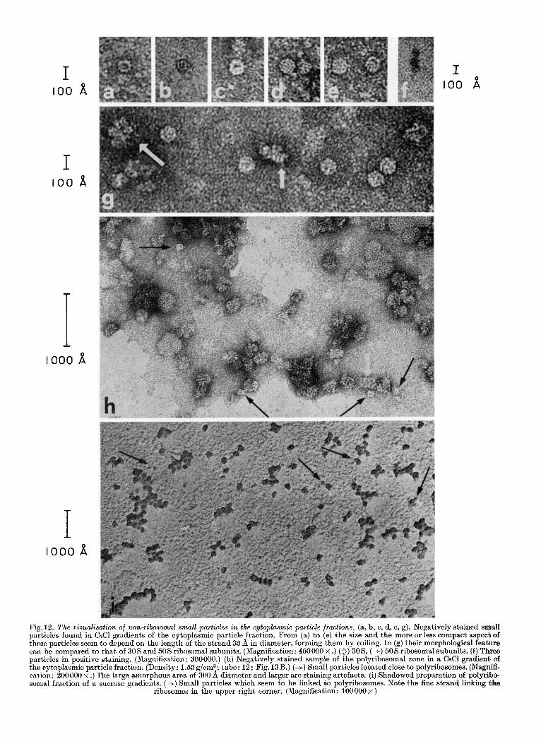
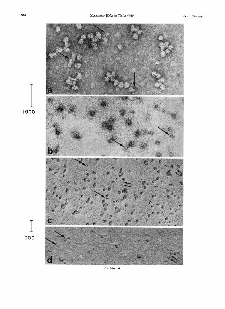
Fig. 12. The visualisation of non-ribosomal small particles in the cytoplasmic particle fractions. (a, b, c, d, e, g). Negatively stained small particles found in CsCl gradients of the cytoplasmic particle fraction. From (a) to (e) the size and the more or less compact aspect of these particles seem to depend on the length of the strand 35 fi in diameter, forming them by coiling. I n (g) their morphological feature can be compared to that of 305 and 50s ribosomal subunits. (Magnification: 4 5 0 0 0 0 ~ .) (0) 30S, (+) 505 ribosomal subunits. ( f ) Three particles in positive staining. (Magnification: 300000.) (h) Negatively stained sample of the polyribosomal zone in a CsCl gradient of the cytoplasmic particle fraction. (Density: 1.55g/cm3; tube: 12; Fig. 13B.) (+) Small particles located close to polyribosomes. (Magnifi- cation: 200000 x .) The large amorphous area of 300 fi diameter and larger are staining artefacts. (i) Shadowed preparation of polyribo- soma1 fraction of a sucrose gradients. (+) Small particles which seem to be linked to polyribosomes. Note the fine strand linking the
ribosomes in the upper right corner. (Magnification: 100000 x )
312 Messenger RNA in HeLa Cells Eur. J. Bioehem.
average half life of 3 h. In an earlier investigation we had shown that the half life of polyribosomes and thus of messenger RNA function is also of the order of 3 h in HeLa cells [20]. I n view of these experiments the disappearance of active polyribosomes and thus the arrest of messenger RNA function is the conse- quence of the decay of the messenger or of the mRNA * protein complex itself.
The Visualisation of Ribonucleoprotein Particles Containing mRNA and mlRNA
in the Electron Microscope Besides easily identified ribosomal material such
as polyribosomes, monoribosomes and ribosomal subunits, electron micrographs of the cytoplasmic particle fraction (15 to 400 S) from HeLa cells show a distinct type of particle. We observed small, generally round particles, with a diameter varying from 110 to 200 d. They seem to be formed by the coiling of a thin continuous strand presenting regu- larly located small knobs along its length (Fig. 12a, b, c, d, e, g). This strand measures about 35 d in thick- ness. The size of the particles and their more or less compact aspects seem to vary with the length of the coiled strand. Thus, some of them appear with a small central hole (Fig. 12a, c) and, most frequently, they are compact and electron dense (Fig. 12d, e, g).
The presence of these particles can be demon- strated as well by negative staining, uranyl acetate
giving best results, as by shadowing. They are also detectable by positive staining but not so easily as ribosomal material (Fig. 12f).
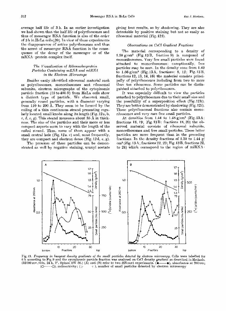
Observations on CsCl Gradient Fractions The material corresponding to a density of
1.58 g/cm3 (Fig. 13B, fraction 9) is composed of monoribosomes. Very few small particles were found attached to monoribosomes : exceptionally, free particles may be seen. In the density zone from 1.49 to 1.56 g/cm3 (Fig.I3A, fractions: 8, 12; Fig.l3B, fractions 12, 13, 14, 16) the material consists princi- pally of polyribosomes including from two to more than ten ribosomes. Some particles can be distin- guished attached to polyribosomes.
It was especially difficult to view the particles attached to polyribosomes due to their small size and the possibility of a superposition effect (Fig. 12h). They are better demonstrated by shadowing (Fig. 12i). These polyribosomal fractions also contain mono- ribosomes and very rare free small particles.
At densities from 1.44 to 1.48g/cm3 (Fig.13A: fractions 18, 19; Fig.13B: fractions 18, 20) the ob- served material consists of ribosomal subunits, monoribosomes and free small particles. These latter particles are more frequent than in the preceding fractions. I n the density fractions of 1.39 to 1.44 g/ om3 (Fig. 13A; fractions 22, 23; Fig. 13B, fractions 22, to 24) which correspond to the region of mlRNA.
1.0
E
8 (v c
3
' 0.5
g
A
1.7 T
I 20 30 10
bottom Fractions top
1.0
E 0 R ... 0
c m f 2 9 0.5
0 1.7 1
- 500 c ._ E . Lo c
3 00 ._ E 3
250 *-
- v .- - I
10 20 30 bottom Fractions top
Fig. 13. Frequency in buoyant density gradients of the small particles detected by electron microscopy. Cells were labelled for 6 h according to Fig. 9 and the cytoplasmic particle fraction was analysed on CsCl density gradient as described in Methods. (32000rev./min, 24 h, 2", Spinco SW 56.) (A) and (B) refer to two different experiments. (0-o), absorbance a t 260nm;
(o---a), radioactivity; (+ --+ ), number of small particles detected by electron microscopy
Vol. 17, No. 2, 1970 G. SPOHR, N. GRANBOULAN, C. MOREL, and K. SCHERRER 313
protein complexes as defined in the biochemical experiments, the free small particles are found more frequently than in fractions of higher density or those of lower density. These fractions contain also some more or less degraded ribosomal subunits. The frequency of the free small particles decreases in the following fractions corresponding to Q = 1.33 to 1.38g/cm3 (Fig. 13A: fraction27; Fig. 13B, fraction 26) where the zone of pure proteins free of RNA begins.
Since the small particles did not appear as the only material in the mlRNA * protein region of the gradients, an attempt was made to count them on the micrographs and to compare their mean number per electron microscopical area to the pattern of radioactivity. These data are given with some reservation due to the problem of regular distribution on electron microscope grids. Unfortunately, the material was not abundant enough to use the filtra- tion technique which is known to give a better distribution [32].
It can be seen (Fig.13) that the frequency dis- tribution of the small particles roughly follows the curve of radioactivity. The richest tubes are found in the zone of the labelled mlRNA. and mRNA protein complexes. It has to be taken into consideration that many small particles attached to polyribosomes may not be seen due t o the above mentioned superposition effect or their unwinding into the 35 d wide strand.
Observation of Pure Polyribosomes In a pure fraction of polyribosomes isolated on a
sucrose gradient some small particles attached to polyribosomes are visible. Free particles are excep- tional (Fig. 12i and 14a).
When this fraction is treated with EDTA (0.5 or 0.1 pmoles per A,,, unit, 30 min, O0) before form- aldehyde fixation and preparation for electron microscopy, some free small particles can be detected either by negative staining or by shadowing (Fig. 14b and c). The ribosomal material is principally in the form of 30 S and 50 S subunits. Some of them present a “tail” (F ig .14~) which may be interpreted as an elongated piece of the 35 d diameter strand which would, after complete condensation, form the small particles by coiling. The “knobs” are clearly visible in the developed 35 d strand.
After exposure of the polyribosomal fraction to 3 M urea (30 min, 20”), free small particles are de- tectable (Fig. 14d). The ribosomal structures are profoundly altered by this treatment presenting extended structures distinctly larger than the tails mentioned above. Thus the small particles seem to be more resistant than ribosomes although their structure has become looser, as can be detected in negatively stained preparations.
DISCUSSION This investigation of cytoplasmic messenger RNA
in animal cells was started in order to find new lines of evidence for the physicochemical reality in vivo of messenger-ribonucleoprotein complexes, which have been described by several laboratories [6- 10, 231. Their biochemical characterisation is crucial in view of the role such ribonucleoprotein associations may play in messenger RNA stability, transport, function, and possibly regulation.
In the following we will discuss some of the ex- perimental evidence and present our conclusions.
Control of Artificial RNA-Protein Association The artificial association of proteins with RNA
observed first by Girard and Baltimore [ I l l and recently by Baltimore and Huang [34] constitutes a major argument which could cast doubt on the reality and significance of the mRNA * protein complexes observed in vivo.
Perry and Kelly [7] demonstrated that purified mlRNA did not bind cytoplasmic proteins in L cells. However artificial associations were observed under our conditions in HeLa cells (cf. Fig. 4) in agreement with Baltimore and Huang [34]. We did not observe the formation of a specific band at Q = 1.45 g/cm3 which Spirin [23] demonstrated to form in particle free cytoplasmic extracts mixed with purified RNA, but found rather a non-specific absorption of RNA to preexisting structures across the zone of active polyribosomes and mlRNA * protein particles.
Two considerations should be taken into account : a) If mRNA-protein complexes play a phy-
siological role in the cell, then the formation of ribonucleoprotein complexes upon mixing of their constituents must be expected a priori. The extent of their formation on addition of purified RNA to a lysate or particle fraction will depend on the avail- ability of the corresponding proteins and their bind- ing equilibrium, i. e. the physicochemical stability of the complex.
b) If some cytoplasmic proteins may bind purified RNA it does not follow necessarily that ribonucleo- protein particles already present will bind RNA to the same extent and with equal stability. The two phenomena may be of very different nature. The resistance of native mRNA * protein particles to EDTA or to 3M urea (a treatment which destroys ribosomes) speaks in favour of a relatively tight binding of the protein in the complex, a situation that may not necessarily apply to the absorption of externally added RNA.
Thus the problem is to develop a biochemical distinction between the native particle and a non- specific absorption of RNA.
Such an experimental distinction may be derived from the specific time dependent pattern of syn-
Eur. J. Biochem. 314 Messenger RNA in HeLa Cells
10
T
3 A
~ ~ Ill_r
Fig. 14a-d
Val. 17, No.2, 1970 G. SPOHR, N. GRANBOULAN, C. MOREL, and K. SCHERRER 315
thesis of two distinct types of mRNAeprotein particles. Furthermore the demonstration of Orchin- nikov et al. [35] and Henshaw and Loebenstein [36] that unlabelled purified RNA added to the lysis buffer will not reduce the specific activity of the complex labelled in vivo shows that a differentiation is possible. All these arguments are in favour of the real existence of mRNA. protein complexes in the cell.
Messenger Characteristics of the RNA in Ribo- nucleoprotein Particles
One of the critical questions in the interpretation of our experimental results is the attribution of messenger quality to the labelled RNA contained in ribonucleoprotein complexes.
In the absence of a direct test in vitro producing a specific protein in a cell free ribosomal system after addition of the specific mRNA, the only RNA to which messenger quality can be attributed is by definition the functional mRNA in the polyribo- some. However, a labelled RNA in the polyribosome can only be identified with the messenger if it can be demonstrated that the continuation of protein synthesis depends on formation and attachment to ribosomes of mRNA newly synthesized during the labelling period. This condition is fulfilled in tissue culture-cells, even in a condition of arrested growth, as could be shown in this investigation.
Furthermore the size of mRNA is a crucial feature since, in a given type of cells and assuming a pre- valence of monocistronic translation, the mRNA molecules should correspond in size to the types of polypeptide chains being synthesized in this cell.
The observed sedimentation spectrum of mRNA in HeLa cells is in agreement with the expectation: the vast majority of polyribosomal labelled mRNA molecules corresponds to molecular weights of 0.1 to 1 . 0 ~ 1 0 ~ daltons and thus to polypeptide chains of 10000 to 100000 daltons, prevalent in animal cells.
It may be pointed out that essentially no mRNA or mlRNA sedimenting more rapidly than 35 S could be found in polyribosomes. The presence of RNA
Fig. 14. Polyribosomal fraction isolated from a sucrose gradient and treated with EDTA or urea. (a) Negatively stained sample of a polyribosomal sucrose gradient fraction (-+) small particles attached to polyribosomes (Magnification : 200000 x ). (b, c) Same preparation of polyribosomes treated with EDTA (20 pmoles/mg, 30 min, 0”) before formaldehyde fixation. The polyribosomal structure is destroyed ; only monoribosomes and ribosomal subunits are present. On shadow-casted preparations some of them present a “tail” (3); (+) free small particles. (Magnification: (b) 200000~ ; (c) 100000 x ). (d) Same preparation of polyribosomes as in (a) treated with 3 M urea (30 min 20’) before formaldehyde fixation : Destruction of the polyribosomal structure. Alter- ations in the morphological appearance of ribosomal material. (+) Free small particles; tails (3 ) (Magnification: 100000 x )
with higher molecular weights was correlated with the occurrence of nuclear contamination.
The attribution of messenger quality to the RNA contained in the free mlRNA 1 protein particles is much more ambigous than the identification of the mRNA in polyribosomes. Therefore we classify this RNA as messenger-like in order to distinguish it from true mRNA. However several arguments can be brought forward favouring the presence of true messenger in free mlRNA protein particles. (a) As will be shown elsewhere [24] bio- logical activity can be detected in subribosomal fractions, free of polyribosomes and ribosome bound mRNA * protein, by incubation in vivo under the con- ditions for cell-free protein synthesis. (b) By com- petitive hybridization we could show that common sequences must exist in labelled RNA extracted from polyribosomes and from free mlRNA * protein particles [25] . ( c ) The spectrum of mRNA sizes is essentially the same in polyribosomes and in free mlRNA. protein complexes (cf. Fig. 6). (d) In pulse chase experiments a fraction of the RNA in free mlRNA protein can be chased into polyribosomes.
We may thus conclude that true mRNA is present both in polyribosome bound mRNA * protein and to some extent in free mlRNA * protein particles.
The Ribonucleoprotein Complex and Messenger RNA Transport
The results presented in this paper also permit some conclusions relative to the mechanism of messenger RNA transport. It was suggested that mRNA is transported associated with 45 S native ribosomal subunits [21,37,38] or with incomplete ribosomal particles [39]. Sidebottom and Harris [22] proposed recently that nucleolar ribosome formation was essential for mRNA transport. Spirin and co- workers [40] showed on the contrary that newly synthesized vaccinia virus-specific mRNA has all the characteristics of a free mlRNA *protein complex and is not associated with polyribosomes.
Our experimental evidence supports Spirin’s model by contradicting the existence of a linkage between messenger transport and the transfer of newly synthesized ribosomal material into the cyto- plasm :
a) Protein synthesis and transfer of mRNA to the cytoplasm and into polyribosomes proceeds in the absence of rRNA synthesis; this was arrested in our experiment by a low dose of actinomycin D as visible in Fig. 2 C and D.
b) The labelled mlRNA unattached to polyribo- somes sediments in the form of ribonucleoprotein complexes which spread over the entire 10 S to 80 S zone of a sucrose gradient (cf. Fig. 5D, E). EDTA does not influence this pattern. The presence of labelled material in the 50 S or in the 30 S sedimentation
316 Messenger RNA in HeLa Cells Bur. J. Biochem.
zone is due to a fortuitous superposition effect. Furthermore, EDTA treatment, which breaks up the mRNA-ribosome association (cf. Fig. 7A and D) does not alter the density distribution of free mlRNA * protein particles (e l . Fig.7C and F). Thus no associa- tion of free mlRNA . protein with ribosomal material could be observed.
The presence of newly synthesized ribosomal 28 S and 18 S RNA in a CsCl gradient a t densities below 1.50 g/cm3 shown by Lissitzky et al. [39] to be asso- ciated mainly with the “clarification pellet” of re- dissolved cytoplasmic particles may be due to denatured and agglomerated ribosomal material. We could observe such material in the electron micro- scope along with the mlRNA * protein particles in the density zone of I .40- 1.45 g/cm3.
It may be concluded that cytoplasmic mlRNA is transported in the form of a ribonucleoprotein com- plex as proposed by Spirin et al. [14]. Ribosome syn- thesis is not necessary for mRNA transport.
However, there is a limitation to this conclusion. We cannot exclude experimentally whether pre- existing ribosomal subunits of a nuclear pool may play a role in a very short lived, transient phase [41], nor that ribosome formation may be essential for mRNA transfer during a phase of regulational adaptation.
The Visualisation of mRNA ‘Protein Particles The electron microscopical investigation presented
here in a preliminary form and which will be extended in a forthcoming paper [42], reveals the presence of small rounded particles in cytoplasmic fractions con- taining ribosomal material, like polyribosomes, mono- ribosomes or ribosomal subunits. The detailed mor- phological appearance and their size is variable. The electron microscopical pictures suggest a spectrum of morphological variations from the smallest type (110 d diameter) with a central hole or excentric cleft, to the largest (more that 200 d in diameter) which seems to be full and more electron dense; this form is the most frequent found in HeLa cells. These variations can be attributed to differences in the length of the 35i f wide strand which forms the rounded particles by coiling.
A possible interpretation of the structure seen is that the messenger ribonucleoprotein strand seen in polyribosomes (cf. Fig. IZi), upon complete liberation from (or in between) ribosomes, would coil up into a random coil and be fixed by formaldehyde as the spherical particles we observe.
However, we cannot exclude a t this stage of the investigation that some different biological materials may give similar images. I n this respect the ultra- structural resemblance of the smallest particle having a central hole with RNA polymerase [43,44] or a contaminant of RNA polymerase preparations
[45] should be noted. These enzymes, which however, should not occur in quantity in cytoplasmic extracts, are more homogenous in size and rod shaped rather than spherical. This morphological feature generally leads to the stacking of individual molecules, an effect which we could never observe in our preparations.
The possibility of a viral contamination can be excluded by considerations of morphology, size and cellular localisation.
Ferritin molecules resembles closely the smallest of the structures we observed and it cannot fully be excluded that some of the small particles shown (Fig.I2a, b) may be ferritin indeed. However fer- ritin molecules are more homogenous in size (about I00 d in diameter) whereas the full particles we de- scribe have diameters of up to more than 200 if. They are also less regular in shape (ferritin appears often rectangular) and we never observed the typical tetrad structure. A further consideration is that, although in HeLa cells ferritin exists, it could not be demonstrated in our preparations.
Similar small particles can be observed in electron micrographs illustrating earlier papers on ribosomal subunits [46] or polyribosomes from mammalian cells [47] but no description of these particles was given. Slayter et al. [48] mention the presence of particles 100 d in diameter in rabbit reticulocyte polyribo- somes without giving any identification.
Considering the detailed description of poly- ribosomes, monoribosomes and ribosomal subunits given by Shelton and Kuff [49] the morphological appearance of the small particles with diameters ranging from 11Od to 200d is easy to distinguish from ribosomal material : the 50 S ribosomal subunit has a diameter of 270 d and the 30 S subunit is ellip- soidal with axes 270 if and 150 d in length (Fig. 12g).
The difficulties encountered in seeing mRNA - pro- tein particles associated with polyribosomal structures may have two explanation. (a) Many may be masked because of their small size by a superposition effect with ribosomes in negative staining as well as after shadowing. (b) The partial or complete uncoiling of the 35 if wide strand when engaged in polyribosomes is another interesting and suggestive possibility for the difficulty of their identification in the active complex of translation. In this respect it should be recalled that they could be identified again as free particles if the polyribosomal fraction was treated with EDTA or 3 M urea.
I n summary we consider the electron microsco- pical evidence as preliminary only and suggestive but in no way conclusive. The morphological identity of the free mlRNA-protein complex remains to be proven.
The Role of mRNA - and mlRNA . Protein Complexes The essential result of this investigation is the
finding that messenger RNA in the cytoplasm of
Vol. 17, No. 2,1970 G. SPOHR, N. GRANBOULAN, C. MOREL, and K. SCHERRER 317
HeLa cells is present in the form of ribonucleoprotein complexes. This observation is in agreement with those of Perry and Kelly [7] in L cells, of Henshaw [8] in liver, of Burny et al. [9] in rabbit reticulo- cytes and of Cartouzou et al. [lo] in thyroid tissue, that mRNA in polyribosomes of animal cells is asso- ciated with protein.
Furthermore, our results are in agreement with those of Spirin and coworkers [6, 14,231 and of Kafatos [50] concerning the existence of free ribonucleo- protein particles containing mlRNA in the cytoplasm of developing embryos, of tissue culture cells and of insects. Our findings support the “Informosome” model [14,23] of cytoplasmic mRNA transport and fit into the general scheme of “cascade regulation” in animal cells [3,4,51].
Such a mechanism calls for modulating agents which could confer to mRNA specificity in respect to stability, transport and the capability to attach to ribosomal subnits in the process of initiation of protein synthesis. It is evident that protein molecules would be ideal for such a mechanism in view of their capability to interact with macromolecules like DNA, RNA and the constituents of cell membranes, as well as with small molecular weight effectors inducing allosteric transitions. Samarina et al. [5,26] and others [53,54], have shown that nascent mlRNA in the nucleus is already associated with proteins. Much of these ribonucleoprotein complexes, which exist also in HeLa cells [12,55], remain in the nucleus [52, 561. However, some of them are transferred to the cytoplasm constituting the pool of free mlRNA * protein particles described in this investigation.
This transfer from the nucleus to the cytoplasm is paralleled by an apparent reduction in size of the giant mlRNA molecules: although we can detect in the nucleus of HeLa cells, under the labelling condi- tions used during this investigation, a majority of mlRNA molecules in a broad sedimentation zone from 28 S up to an estimated 150 S (as will be shown elsewhere [52]) we never could detect in the cyto- plasm appreciable amounts of RNA with sedimenta- tion constants of more than 35 S. Since the appear- ance of heavier molecules in the cytoplasm could be correlated always with nuclear leakage, we have to consider that, a t least in HeLa cells, the “non-transla- table, pre-ribosomal informosomes” described by Spirin [23] as co-sedimenting with polyribosomes might be of nuclear origin.
However we found during this investigation that non-translatable, messenger-like ribonucleoprotein complexes indeed do exist in the cytoplasm of HeLa cells and contain mlRNA spread over sedimentation zones from 6 to 30 S, comparable thus in size to the mRNA in polyribosomes. 40 to 60°/, of the cyto- plasmic free mlRNA does not associate with ribo- somes in steady state: only a small fraction of this free pool can be chased into polyribosomes. The
majority of free mlRNA * protein complexes decays as free particles at the same rate as the polyribosomal mRNA-protein complexes. This average rate of decay is not exponential in time and shows a kinetic hetero- geneity for the mlRNA - protein particles as well as the polyribosomal mRNA protein particles. We can conclude that they disappear with half-lives of about 1.5 h for the rapidly decaying and of up to about 6 h for the more stable fractions.
We propose that the stability of the translatable messenger RNA and of the non-translated messenger- like RNA of the free pool is an intrinsic property of the ribonucleoprotein complex. The stability of an mRNA may thus be a function of the equilibrium constant of an RNA-protein association which is in dynamic equilibrium with its constituents. Accord- ing to such a model, every protecting protein molecule would dissociate from the complex with a given statistical frequency. The size of the non-protected area and the duration of the periods of non-protection against hydrolytic or enzymatic attacks would determine the life span of an RNA molecule of specific sequence. Local nucleotide sequences are the basis of regions with specific secondary structure and would determine per se or by a process of “induced fit” the binding of the protein molecules.
The same kind of consideration may be justified in regard to the activation or inactivation of an mRNA: in fact, the binding constant of the protein moiety in the mRNA. protein complex might be allosterically influenced by cytoplasmic agents. Translational regulation could be understood assuming that the binding energy of this protein would be in competition with the energy provided by the translational system for translocation since ribosomes moving along the mRNA would have to displace temporarily the protein from the complex.
I n fact it is a surprising finding that about 50°/, of mlRNA in the cytoplasm does not enter the trans- lation process. This amount may possibly be ex- plained by the low actinomycin dose used for this investigation. However, since the rate of protein syn- thesis is affected less, such an explanation cannot account for the amount of free mRNA. Thus a more physiological interpretation of the equilibrium be- tween ribonucleoprotein complexes of mRNA and mlRNA may be justified. Further work will have to be done in order to determine the actual equilibrium between these complexes under normal conditions.
In embryonic tissue large amounts of maternal messenger RNA are present which had been com- pletely “masked’) in the oocyte prior to fertilisation [57]. They may account for the large amount of non- translatable mRNA in early development. The large fraction of inactivated messenger ribonucleoprotein in tissue culture cells points to the possibility that a more general system of regulation not limited to developing systems may be involved. It would ex-
318 G. SPORR et al.: Messenger RNA in HeLa Cells Eur. J. Biochem.
plain many of the experimental facts brought forward by Tomkins et al. [58] and others in favour of post- transcriptional controls operating in animal protein synthesis, possibly within the frame of a multilevel regulational system [51].
We gratefully acknowledge the excellent assistance of our technical collaborators: Raymonde Cornuz, who provided us with cells, Pierre-AndrB Briand, RBmy Moret and Clau- dine Coet. We thank Mr. Alain Gautier, director of the “Centre de Microscopie Electronique de l’Universit6 de Lausanne”, for providing us the facilities of his laboratory and for helpful discussions. The assistance during the preparation of this manuscript of RBmy Moret, Otto Jenny, Annette Muhlbauer and Claude MBrel, and the administra- tive assistance of Mr. I. Marcovitch (Virology Department, ISREC) and of the Central Services of our Institute (Mr. M. Zagnoli) are gratefully acknowledged. Our special gratitude goes to our colleagues Ronald Hancock and Nicholas Acheson for critical reading of this manuscript. This work was supported by the Swiss National Foundation (grants 4769.3, 5323.3 and 3.164.69). One of us (C. M.) is indebted to the World Health Organization for a Research Training Grant. (M8/181/4/M. 77).
1.
2.
3.
4.
5.
6. 7. 8. 9.
10.
11.
12.
13.
14.
15.
16.
17. 18.
19.
20.
21. 22.
REFERENCES Scherrer, K., In Experimental Biology and Medicine
(edited by E. Hagen, W. Weschler, and P. Zilliken), Vol. I, Morphological and Biochemical Aspects of Cytodifferent;ation, S . Karger, Base1 and New York
Scherrer, K., and Marcaud, L., Bull. SOC. Chim. Biol. 47
Scherrer. K.. Marcaud. L.. Zaide1a.F.. London. I. M.. and
1967, pp. 244-263.
(1965) 1697.
Gros,F., Proc. Nat. A&?. j c i . U’.S.A. 56 (1966a) i571. Scherrer, K., Marcaud, L., Zajdela, F., Breckenridge, B.,
and Gros, F., Bull. SOC. Chim. Biol. 48 (1966b) 1037. Samarina, 0. P., Krichevskaya, A. A., and Georgiev,
G. P., Nature (London), 210 (1966) 1319. Spirin, A. S., and Nemer, M., Science, 150 (1965) 214. Perry, R.P., and Kelley, D. E., J . Mol. Biol. 35 (1968a) 37. Henshaw, E. C., J . Mol. Biol. 36 (1968) 401. Burny, A., Huez, G., Marbaix, G., and Chantrenne, H.,
Biochim. Biophys. Acta, 190 (1969) 228. Cartouzou, G., Poirhe, J. C., and Lissitzky, S., Eur. J .
Biochem. 8 (1969) 357. Girard, M., and Baltimore, D., Proc. Nut. A d . Sci. U. 8. A . 56 (1966) 999.
Penman. S.. Vesco. C.. and Penman. M.. J . Mol. Biol. , , ~~ - . . I ,
34 (i66sj49. SDirin. A. S.. Belitsina. N. V.. and Lerman. M. I.. J .
L M ~ l . Biol.‘ 14 (1965) 611. ’
Spirin, A. S., Belitsina. N. V., and Aitkhozhin. M. A.. -Zh.’Obshch. Biol. 2 5 ( 5 ) (1964) 321 and translation in: Fed. Proc. 24 (1965) 907.
Scherrer, K., In: Fundamental Techniques in Virology (edited by K. Habel and N. P. Salzman), Academic Press, New York 1969, p. 413.
Perry, R. P., Proc. Nat. Acad. Xci. U. S. A . 48 (1962) 2179.
Perry, R. P., Exp. Cell Res. 29 (1963) 400. Georgiev, P., Samarina, 0. P., Lerman, M. I.,
Smirnov. M. N.. and Severtzov. A. N.. Nature [Lon- ,- don), 206 (1963) 1291.
Reich, E.. Franklin. R. M., Shatkin. A. J.. and Tatum. E. L., Science, 134 (1961) 556.
Proc. Nut. Acud. Sci. U. S. A . 49 (1963) 654.
’
Penman, S., Scherrer, K., Becker, Y., and Darnell, J. E.,
Joklik,W.K., and Becker,Y., J . Mol. Biol. 13 (1965) 511. Sidebottom, E., and Harris, H., J . Cell. Sci. 5 (1969) 351.
23. Spirin, A. S., Eur. J . Biochem. 10 (1969) 20. 24. Chezzi, C., Grosclaude, J., and Scherrer, K., unpublished
25. Scherrer, IZ., unpublished results. 26. Samarina, 0. P., Lukanidin, E. M., Molnar, J., and
Georgiev, G. P., J . Mol. Biol. 33 (1968) 251. 27. Soeiro, R., Vaughan, M. H., Warner, J. R., and Darnell,
J. E., Jr., J . Cell. Biol. 39 (1968) 112. 28. Scherrer, K., Lathan, H., and Darnell, J. E., Proc. Nut.
A d . Sci. U . S. A . 49 (1963) 240. 29. Gilbert, W., J . Mol. Biol. 6 (1963) 389. 30. Mirault, M. E., and Scherrer, K., unpublished results. 31. Perry, R. P., and Kelley, D. E., J . Cell. Physiol. 72
(1968b) 235. 32. Kellenberger, E., personal communication. 33. Fuhr, J. E., London, I. M., and Grayzel, A. I., Proc. Nat.
A d . Sci. U . S. A . 63 (1969) 129. 34. Baltimore, D., and Huang, A.S., J . Mol. Biol. 47 (1970) 263. 35. Ovchinnikov, L. P., Avanesov, A. E., and Spirin, A. S.,
Mol. Biol., U. S. S. R. 3 (1969) 465. 36. Henshaw, E. C., and Loebenstein, J., Biochim. Biophys.
Acta, 199 (1970) 405. 37. Henshaw, E. C., Revel, M., and Hiatt, H. H., J . Mol.
Biol. 14 (1965) 241. 38. McConkey, E. H., and Hopkins, J. W., J . Mol. Biol. 14
results.
(1965) 257. 39. Lissitzkv. S.. PoirBe. J. C.. Cartouzou. G.. and GrB-
goire,‘J., Eur. J . Biochem. 12 (1970) 104.’ 40. Belitsina, N. V., Ovchinnikov, L. P., Spirin, A. S., Gen-
don, Y., and Chernos, V. I., Mol. Biol. U.S. S. R. 2 (1968) 727.
41. Ringborg, U., personal communication. 42. Granboulan, N., Kayibanda, B., Spohr, G., and Scher-
43. Fuchs, E., Zillig, W., Hofschneider, P. H., and Preuss,
44. Colvill, A. J. E., Van Bruggen, E. F. J., and Fernandez-
45. Lubin, M., J. Mol. Biol. 39 (1969) 219. 46. Florendo, N. T., and Foster, L. B., J . Ultrastr. Res. 30
47. Benedetti. E. L.. Zweers. A.. and Bloemendal. H.. Bio-
rer, K., unpublished results.
A., J . Mol. Biol. 10 (1964) 546.
Moran, H., J . Mol. Biol. 17 (1966) 302.
(1970) 1. I ,
chem. J: 108 (1968) 765.
J . Mol. Biol. 7 (1963) 652.
‘
48. Slayter, H. S., Warner, J. R., Rich, A., and Hall, C. E.,
49. Shelton. E.. and Kuff. E. L.. J . Mol. Biol. 22 (1966) 23. 50. Kafatos, F:C., Proc. Nut. A d . Sci. U. S. A . ‘69 (1968)
1251. 51. Scherrer, K., and Marcaud, L., J . Cell. Physiol. 72,
Sup. 1 (1968) 181. 52. Scherrer, K., Spohr, G., Granboulan, N., Morel, C., Gros-
Claude, J., and Chezzi, C., Cold Spring Harbor Symp. Quant. Biol. 35 (1970), in press.
53. Parsons, J. T., and McCarthy, K. S., J . Biol. Chem. 243 (1968) 5377.
54. Kohler, K., and Arends, S., Eur. J . Biochem. 5 (1968) 500. 55. Morel, C., Spohr, G., and Scherrer, K., unpublished ob-
56. Shearer, R. W., and McCarthy, B. J., Biochemistry, 6
57. Spirin, A. S., Curr. Top. Dev. Biol. 1 (1966) 1. 58. Tomkins, G. M., Gelehrter, T. D., Grannen, R. D., Martin,
D., Samuels, H. H., and Thompson, E. B., Science, 166 (1969) 1474.
servations.
(1967) 283.
G. Spohr and K. Scherrer Institut Suisse de Recherche8 Experimentales sur le Cancer Rue du Bugnon 21, CH-1005 Lausanne, Switzerland
C. Morel’s permanent address : Faculdade de Cihcias M6dicas Universidade de Brasilia, Brasilia, D. F., Brasil


