Embed Size (px)
Citation preview
Metabolic Engineering of Cofactor F420 Production inMycobacterium smegmatisGhader Bashiri1,2*, Aisyah M. Rehan1,2, David R. Greenwood2,3, James M. J. Dickson1,2, Edward N.
Baker1,2
1 Structural Biology Laboratory, School of Biological Sciences, The University of Auckland, Auckland, New Zealand, 2 Maurice Wilkins Centre for Molecular Biodiscovery,
School of Biological Sciences, The University of Auckland, Auckland, New Zealand, 3 Centre for Genomics and Proteomics, School of Biological Sciences, The University of
Auckland, Auckland, New Zealand
Abstract
Cofactor F420 is a unique electron carrier in a number of microorganisms including Archaea and Mycobacteria. It has beenshown that F420 has a direct and important role in archaeal energy metabolism whereas the role of F420 in mycobacterialmetabolism has only begun to be uncovered in the last few years. It has been suggested that cofactor F420 has a role in thepathogenesis of M. tuberculosis, the causative agent of tuberculosis. In the absence of a commercial source for F420, M.smegmatis has previously been used to provide this cofactor for studies of the F420-dependent proteins from mycobacterialspecies. Three proteins have been shown to be involved in the F420 biosynthesis in Mycobacteria and three other proteinshave been demonstrated to be involved in F420 metabolism. Here we report the over-expression of all of these proteins inM. smegmatis and testing of their importance for F420 production. The results indicate that co–expression of the F420
biosynthetic proteins can give rise to a much higher F420 production level. This was achieved by designing and preparing anew T7 promoter–based co-expression shuttle vector. A combination of co–expression of the F420 biosynthetic proteins andfine-tuning of the culture media has enabled us to achieve F420 production levels of up to 10 times higher compared withthe wild type M. smegmatis strain. The high levels of the F420 produced in this study provide a suitable source of thiscofactor for studies of F420-dependent proteins from other microorganisms and for possible biotechnological applications.
Citation: Bashiri G, Rehan AM, Greenwood DR, Dickson JMJ, Baker EN (2010) Metabolic Engineering of Cofactor F420 Production in Mycobacteriumsmegmatis. PLoS ONE 5(12): e15803. doi:10.1371/journal.pone.0015803
Editor: Annalisa Pastore, National Institute for Medical Research, Medical Research Council, United Kingdom
Received October 18, 2010; Accepted November 24, 2010; Published December 29, 2010
Copyright: � 2010 Bashiri et al. This is an open-access article distributed under the terms of the Creative Commons Attribution License, which permitsunrestricted use, distribution, and reproduction in any medium, provided the original author and source are credited.
Funding: The authors would like to thank New Zealand Lottery Grants Board (Health), the Maurice & Phyllis Paykel Trust and the Allan Wilson Centre forMolecular Ecology and Evolution for funding to purchase the EnVision Multilabel plate reader. This research was supported by the Health research Council of NewZealand, the Foundation for Research, Science and Technology of New Zealand. A.M.R. is a recipient of a PhD scholarship from the IPTA Academic TrainingScheme from The Ministry of Higher Education, Malaysia and International Islamic University Malaysia. The funders had no role in study design, data collection andanalysis, decision to publish, or preparation of the manuscript.
Competing Interests: The authors have declared that no competing interests exist.
* E-mail: [email protected]
Introduction
The cofactor F420 was first identified chemically in methano-
genic Archaea in 1972 [1], although a compound with similar
characteristics was previously described in Mycobacteria in the
early 1960s [2,3]. Since its discovery, F420 and its precursor FO (so
called 5-deazaflavins) have been found in a variety of (micro)or-
ganisms, including Archaea, bacteria and eukaryotic species
(Table 1). F420 is named on the basis of its intense absorbance/
fluorescence at 420 nm (emission 480 nm), which is redox
dependent and is lost upon reduction of the cofactor. It also has
unique chemical and biological characteristics; the isoalloxazine
chromophore of F420 is structurally very similar to that of the
flavins (FMN and FAD), although it is functionally similar to
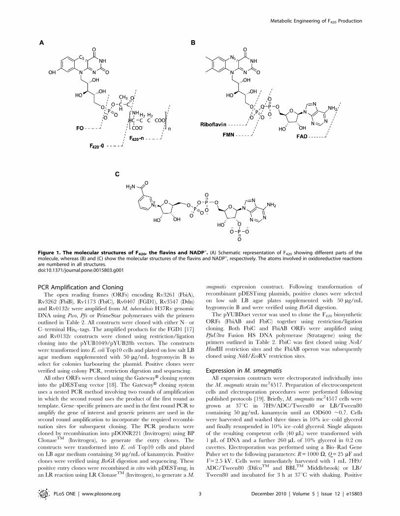
NAD(P)+ (Figure 1). Functionally, F420 is a two–electron carrier
involved in hydride transfer reactions. The redox potential of
F420H2/F420+2e2 (2360 mV) is lower than those of the classical
hydrogen carriers NAD(P)H/NAD(P)+2e2 (2320 mV) and
FADH2/FAD+2e2 (2219 mV) [4,5].
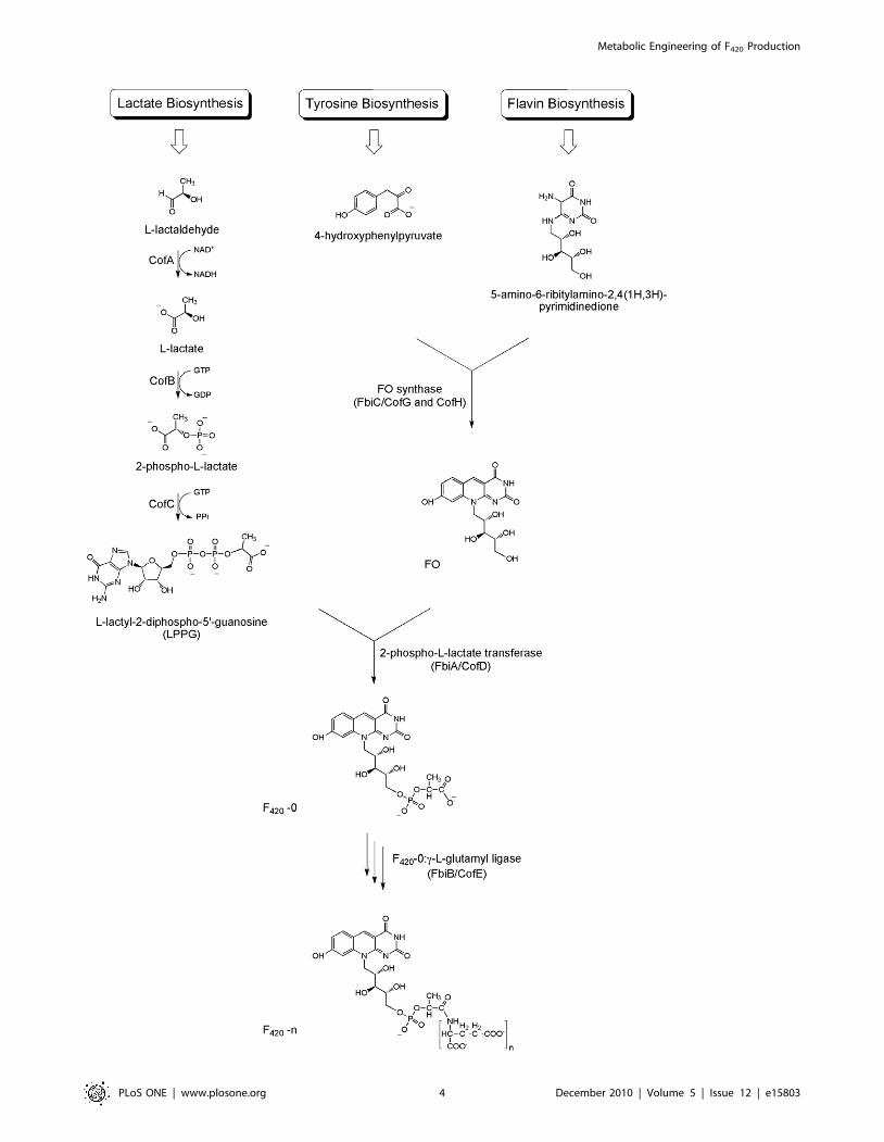
A key biosynthetic precursor of F420 is FO (7,8-didemethyl-8-
hydroxy-5-deazariboflavin), comprising an isoalloxazine ring and
ribitol moieties. Formation of F420 follows a series of biochemical
reactions and is completed by the addition of a phospholactate
group, and finally a poly–glutamate tail in which L–glutamate
residues are linked together via c–glutamyl bonds (Figure 2) [6,7].
The length of the poly–glutamate tail constitutes the main
difference between the F420 cofactors from different microorgan-
isms, the number of residues varying from 2–9. There are
suggestions, however, that the type of a– or c–glutamyl linkage in
the terminal glutamate residue could also be different in some
Archaeal species [6,8,9,10].
F420 is not commercially available and researchers working on
F420–dependent proteins have to prepare it as required. With the
discovery of new F420–dependent enzymes and increasing interest
in F420–dependent reactions, especially in the case of the
pathogen Mycobacterium tuberculosis (Mtb), a resource with high
yields of F420 production is required. F420 has been previously
purified from various microorganisms, including Archaea
(Methanobacterium, Methanococcus and Methanosarcina spe-
cies) and Actinomycetes (Actinomadura, Actinoplanes, Strepto-
myces, Rhodococcus, Nocardia and Mycobacteria species), with
differing yields [11]. F420 purification in all cases, however,
essentially follows the same principle; precipitation of cellular
proteins using heat or an organic solvent, followed by separation
PLoS ONE | www.plosone.org 1 December 2010 | Volume 5 | Issue 12 | e15803
of F420 from remaining cellular components based on its acidic
nature [4]. In order to purify F420, a number of different
chromatographic steps have been used, including ion exchange,
adsorption, HPLC and gel filtration chromatography [6,9,11].
Isabelle et al. have reported thorough analyses of F420–producing
microorganisms, and based on ‘‘ease of growth, fewer hazards,
and lower costs’’ concluded that M. smegmatis is the best source
for F420 production, providing there is no requirement for a
particular number of glutamate residues in the F420 poly–
glutamate tail [11].
Our initial F420 purification trials indicated that M. smegmatis
transformed to over–express the M. tuberculosis protein FGD1
(F420–dependent glucose-6-phosphate dehydrogenase 1) could
produce higher levels of F420 compared with the wild type strains.
This observation prompted us to thoroughly investigate the effects
on F420 production of over–expression of other proteins known to
be involved in F420 biosynthesis and metabolism in Mycobacteria.
These include three proteins in the F420 biosynthetic pathway, viz.
FbiA (Rv3261) [12], FbiB (Rv3262) [12] and FbiC (Rv1173) [13]
and three other proteins which are shown to be involved in F420
metabolism: FGD1 (Rv0407) [9,14], Ddn (Rv3547) [15] and
Rv0132c (author’s unpublished data).
Here we describe the development of vectors to co–express Mtb
proteins in M. smegmatis. We further show that by co–expressing
enzymes associated with F420 production and manipulating growth
conditions, greatly increased levels of F420 can be obtained. With
the growing recognition that F420 plays a crucial role in
Mycobacteria and other organisms, this readily available source
of the cofactor will be useful for testing its physiological and
biochemical roles, and for possible applications in biotechnology.
Materials and Methods
Preparation of New Mycobacterial VectorsThe pYUB1049 vector (5795 bp) is a product of ligation
between the vectors pMS134 and pET28b–cmaA2 [16], resulting
in a vector with a cloned gene between NdeI and BamHI
restriction sites. The pYUB1049 vector was subjected to restri-
ction digestion using NcoI (single site) and BlpI (two sites)
restriction sites, in order to obtain a linear vector without the
multiple cloning site. The plasmid was first digested to
completion with NcoI (Roche Applied Science) and dephosphor-
ylated using calf intestinal alkaline phosphatase (New England
Biolabs) followed by ethanol precipitation. The NcoI–cut linear
pYUB1049 vector was subjected to a partial digestion with BlpI
(BpuI102I isoschizomer, Fermentas) for 20 minutes and the
reactions stopped using 5 mL 0.5 M EDTA. The digested vector
was run on a 0.5% agarose gel and a DNA fragment corres-
ponding to 4705 bp was excised and gel–purified.
The pET28b and pETDuet–1 vectors (Novagen) were double–
digested using NcoI and BlpI enzymes. The resulting multiple
cloning site fragments, 216 and 382 bp respectively, were ligated
separately into the NcoI/BlpI fragment of the pYUB1049 vector
using T4 DNA ligase (Roche Applied Science). Ligation mixtures
were electroporated into E. coli TOP10 cells and the positive
colonies were selected on low salt LB agar plates (tryptone 10 g/L,
yeast extract 5 g/L, NaCl 5 g/L and agar 15 g/L, pH 8.0)
containing 50 mg/mL hygromycin B. Positive clones were verified
using restriction digestion and sequencing. The resulting vectors
were designated as pYUB28b (4921 bp) and pYUBDuet
(5087 bp), respectively.
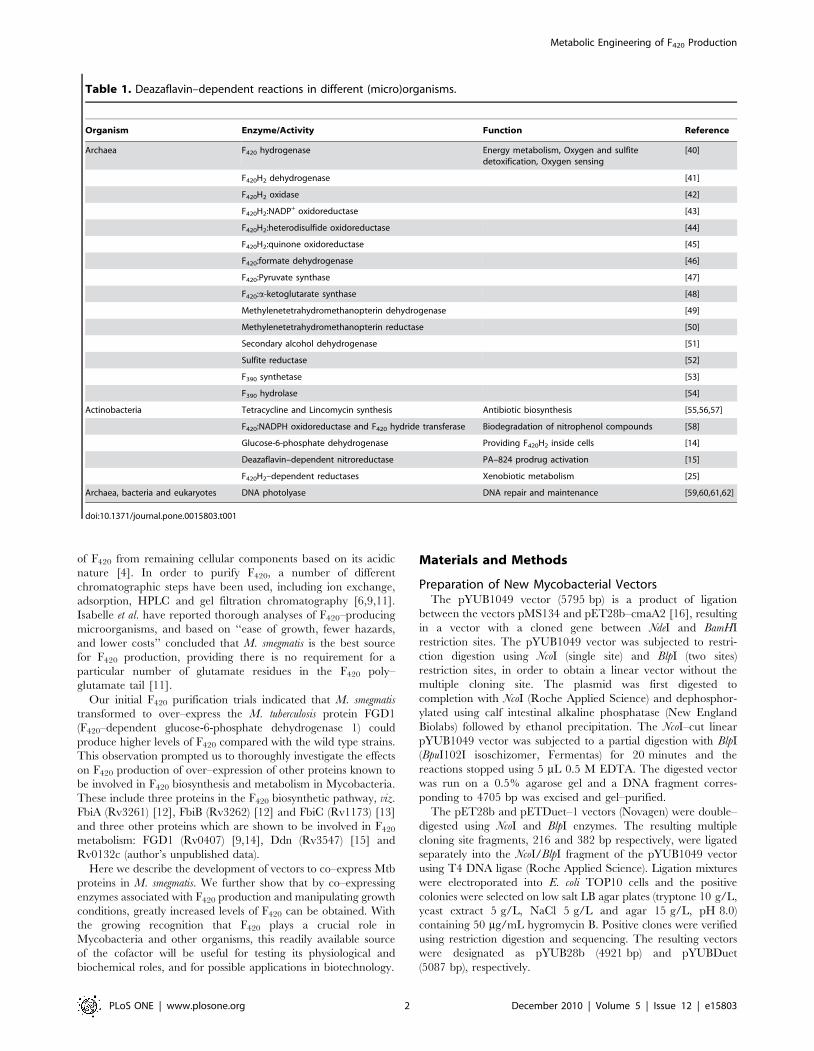
Table 1. Deazaflavin–dependent reactions in different (micro)organisms.
Organism Enzyme/Activity Function Reference
Archaea F420 hydrogenase Energy metabolism, Oxygen and sulfitedetoxification, Oxygen sensing
[40]
F420H2 dehydrogenase [41]
F420H2 oxidase [42]
F420H2:NADP+ oxidoreductase [43]
F420H2:heterodisulfide oxidoreductase [44]
F420H2:quinone oxidoreductase [45]
F420:formate dehydrogenase [46]
F420:Pyruvate synthase [47]
F420:a-ketoglutarate synthase [48]
Methylenetetrahydromethanopterin dehydrogenase [49]
Methylenetetrahydromethanopterin reductase [50]
Secondary alcohol dehydrogenase [51]
Sulfite reductase [52]
F390 synthetase [53]
F390 hydrolase [54]
Actinobacteria Tetracycline and Lincomycin synthesis Antibiotic biosynthesis [55,56,57]
F420:NADPH oxidoreductase and F420 hydride transferase Biodegradation of nitrophenol compounds [58]
Glucose-6-phosphate dehydrogenase Providing F420H2 inside cells [14]
Deazaflavin–dependent nitroreductase PA–824 prodrug activation [15]
F420H2–dependent reductases Xenobiotic metabolism [25]
Archaea, bacteria and eukaryotes DNA photolyase DNA repair and maintenance [59,60,61,62]
doi:10.1371/journal.pone.0015803.t001
Metabolic Engineering of F420 Production
PLoS ONE | www.plosone.org 2 December 2010 | Volume 5 | Issue 12 | e15803
PCR Amplification and CloningThe open reading frames (ORFs) encoding Rv3261 (FbiA),
Rv3262 (FbiB), Rv1173 (FbiC), Rv0407 (FGD1), Rv3547 (Ddn)
and Rv0132c were amplified from M. tuberculosis H37Rv genomic
DNA using Pwo, Pfx or PrimeStar polymerases with the primers
outlined in Table 2. All constructs were cloned with either N– or
C–terminal His6–tags. The amplified products for the FGD1 [17]
and Rv0132c constructs were cloned using restriction/ligation
cloning into the pYUB1049/pYUB28b vectors. The constructs
were transformed into E. coli Top10 cells and plated on low salt LB
agar medium supplemented with 50 mg/mL hygromycin B to
select for colonies harbouring the plasmid. Positive clones were
verified using colony PCR, restriction digestion and sequencing.
All other ORFs were cloned using the GatewayH cloning system
into the pDESTsmg vector [18]. The GatewayH cloning system
uses a nested PCR method involving two rounds of amplification
in which the second round uses the product of the first round as
template. Gene–specific primers are used in the first round PCR to
amplify the gene of interest and generic primers are used in the
second round amplification to incorporate the required recombi-
nation sites for subsequent cloning. The PCR products were
cloned by recombination into pDONR221 (Invitrogen) using BP
ClonaseTM (Invitrogen), to generate the entry clones. The
constructs were transformed into E. coli Top10 cells and plated
on LB agar medium containing 50 mg/mL of kanamycin. Positive
clones were verified using BsrGI digestion and sequencing. These
positive entry clones were recombined in vitro with pDESTsmg, in
an LR reaction using LR ClonaseTM (Invitrogen), to generate a M.
smegmatis expression construct. Following transformation of
recombinant pDESTsmg plasmids, positive clones were selected
on low salt LB agar plates supplemented with 50 mg/mL
hygromycin B and were verified using BsrGI digestion.
The pYUBDuet vector was used to clone the F420 biosynthetic
ORFs (FbiAB and FbiC) together using restriction/ligation
cloning. Both FbiC and FbiAB ORFs were amplified using
PfuUltra Fusion HS DNA polymerase (Stratagene) using the
primers outlined in Table 2. FbiC was first cloned using NcoI/
HindIII restriction sites and the FbiAB operon was subsequently
cloned using NdeI/EcoRV restriction sites.
Expression in M. smegmatisAll expression constructs were electroporated individually into
the M. smegmatis strain mc24517. Preparation of electrocompetent
cells and electroporation procedures were performed following
published protocols [19]. Briefly, M. smegmatis mc24517 cells were
grown at 37uC in 7H9/ADC/Tween80 or LB/Tween80
containing 50 mg/mL kanamycin until an OD600 ,0.7. Cells
were harvested and washed three times in 10% ice–cold glycerol
and finally resuspended in 10% ice–cold glycerol. Single aliquots
of the resulting competent cells (40 mL) were transformed with
1 mL of DNA and a further 260 mL of 10% glycerol in 0.2 cm
cuvettes. Electroporation was performed using a Bio–Rad Gene
Pulser set to the following parameters: R = 1000 V, Q = 25 mF and
V = 2.5 kV. Cells were immediately harvested with 1 mL 7H9/
ADC/Tween80 (DifcoTM and BBLTM Middlebrook) or LB/
Tween80 and incubated for 3 h at 37uC with shaking. Positive
Figure 1. The molecular structures of F420, the flavins and NADP+. (A) Schematic representation of F420 showing different parts of themolecule, whereas (B) and (C) show the molecular structures of the flavins and NADP+, respectively. The atoms involved in oxidoreductive reactionsare numbered in all structures.doi:10.1371/journal.pone.0015803.g001
Metabolic Engineering of F420 Production
PLoS ONE | www.plosone.org 3 December 2010 | Volume 5 | Issue 12 | e15803
Metabolic Engineering of F420 Production
PLoS ONE | www.plosone.org 4 December 2010 | Volume 5 | Issue 12 | e15803
transformants were selected by plating on 7H10/ADC (DifcoTM
and BBLTM Middlebrook) or LBT agar plates containing 50 mg/
mL each of kanamycin and hygromycin B.
Protein expression was performed either in autoinduction [20],
LB or 7H9/ADC media supplemented with 0.05% Tween80 and
50 mg/mL each of kanamycin and hygromycin B. A single
transformed colony was selected from a 7H10/ADC plate and
used to inoculate a starter culture in MDG media (25 mM
Na2HPO4, 25 mM KH2PO4, 50 mM NH4Cl, 5 mM Na2SO4,
2 mM MgSO4, 0.5% D-glucose, 0.25% L-aspartate, 0.26 metal
mix) [20]. The starter culture was grown for 48–72 h at 37uC and
was freshly used at a dilution of 1:100 to inoculate expression
cultures of ZYM–5052 autoinduction (1% tryptone, 0.5% yeast
extract, 25 mM Na2HPO4, 25 mM KH2PO4, 50 mM NH4Cl,
5 mM Na2SO4, 2 mM MgSO4, 0.5% glycerol, 0.05% glucose,
0.2% alpha–lactose, 16 metal mix), LB or 7H9/ADC. The
expression cultures were grown for 4 days at 37uC for maximal
expression [17]. LB, MDG and 7H9/ADC cultures were induced
using IPTG at a final concentration of 0.1 or 1 mM.
Western Blot AnalysesM. smegmatis cells expressing different constructs were lysed twice
using a cell disruptor (Constant Systems Ltd.) and centrifuged at
16,0006g to pellet non–lysed cells and other insoluble material.
Protein samples were separated on a 15% SDS–PAGE gel and
transferred to polyvinylidene difluoride (PVDF) membranes using
a wet transfer protocol (200mA, 3 hours) [21]. His–tagged
recombinant proteins were detected using a mouse monoclonal
anti–His antibody and horseradish peroxidase–conjugated anti–
mouse antibody (GE Healthcare). The Luminol (ECL plus kit, GE
Healthcare) chemiluminescence was detected using an LAS4000
imaging system (Fujifilm).
FO and F420 CharacterizationM. smegmatis cells expressing different M. tuberculosis proteins
were grown in identical conditions to late log phase or stationary
phase. In all expression cultures the ZYP–5052 autoinduction
media was used for F420 production experiments and the media to
flask volume ratio was kept constant at 20%. In order to optimize
the media for F420 production, the ZY component of ZYM–5052
media was replaced by commonly used media bases including 26ZY, YT (0.8% tryptone, 0.5% yeast extract and 42.77 mM NaCl),
TB (1.2% tryptone, 2.4% yeast extract and 0.4% glycerol), SOB
(2% tryptone, 0.5% yeast extract, 8.56 mM NaCl, 2.5 mM KCl
and 10 mM MgCl2) and SOC (SOB with 20 mM glucose). Iron
and sulphur supplements (ferric ammonium citrate, ferric citrate
and ferrous sulphate all at 0.1 mg/mL and L–cysteine at 1 mM)
were also added to the expression media as a possible requirement
for the FbiC enzyme. L–glutamate and manganese chloride
(1 mM final concentration) were also added to the expression
media to evaluate their necessity for FbiB–mediated F420
production [22].
To ascertain the optimum growth period for F420 production,
eight identical cultures of M. smegmatis cells expressing the
recombinant FbiABC construct were set up. Each culture had a
wild type M. smegmatis culture as a control. At 24 h intervals, one
culture each of control and recombinant FbiABC–expressing M.
smegmatis cells were harvested and processed to monitor the F420
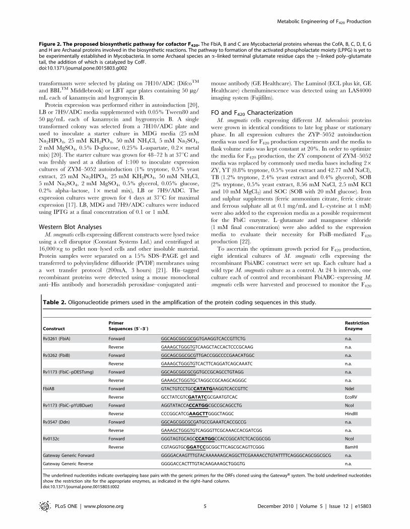
Figure 2. The proposed biosynthetic pathway for cofactor F420. The FbiA, B and C are Mycobacterial proteins whereas the CofA, B, C, D, E, Gand H are Archaeal proteins involved in the biosynthetic reactions. The pathway to formation of the activated phospholactate moiety (LPPG) is yet tobe experimentally established in Mycobacteria. In some Archaeal species an a–linked terminal glutamate residue caps the c–linked poly–glutamatetail, the addition of which is catalyzed by CofF.doi:10.1371/journal.pone.0015803.g002
Table 2. Oligonucleotide primers used in the amplification of the protein coding sequences in this study.
ConstructPrimerSequences (59–39)
RestrictionEnzyme
Rv3261 (FbiA) Forward GGCAGCGGCGCGGTGAAGGTCACCGTTCTG n.a.
Reverse GAAAGCTGGGTGTCAAGCTACCACTCCCGCAAG n.a.
Rv3262 (FbiB) Forward GGCAGCGGCGCGTTGACCGGCCCCGAACATGGC n.a.
Reverse GAAAGCTGGGTGTCACTTCAGGATCAGCAAATC n.a.
Rv1173 (FbiC–pDESTsmg) Forward GGCAGCGGCGCGGTGCCGCAGCCTGTAGG n.a.
Reverse GAAAGCTGGGTGCTAGGCCGCAAGCAGGGC n.a.
FbiAB Forward GTACTGTCCTGCCATATGAAGGTCACCGTTC NdeI
Reverse GCCTATCGTCGATATCGCGAATGTCAC EcoRV
Rv1173 (FbiC–pYUBDuet) Forward AGGTATACCACCATGGCGCCGCAGCCTG NcoI
Reverse CCCGGCATCGAAGCTTGGGCTAGGC HindIII
Rv3547 (Ddn) Forward GGCAGCGGCGCGATGCCGAAATCACCGCCG n.a.
Reverse GAAAGCTGGGTGTCAGGGTTCGCAAACCACGATCGG n.a.
Rv0132c Forward GGGTAGTGCAGCCCATGGCCACCGGCATCTCACGGCGG NcoI
Reverse CGTAGGTGCGGATCCGCGGCTTCAGCGCAGTTCGGG BamHI
Gateway Generic Forward GGGGACAAGTTTGTACAAAAAAGCAGGCTTCGAAAACCTGTATTTTCAGGGCAGCGGCGCG n.a.
Gateway Generic Reverse GGGGACCACTTTGTACAAGAAAGCTGGGTG n.a.
The underlined nucleotides indicate overlapping base pairs with the generic primers for the ORFs cloned using the GatewayH system. The bold underlined nucleotidesshow the restriction site for the appropriate enzymes, as indicated in the right–hand column.doi:10.1371/journal.pone.0015803.t002
Metabolic Engineering of F420 Production
PLoS ONE | www.plosone.org 5 December 2010 | Volume 5 | Issue 12 | e15803
production level. The procedure was carried out for eight days and
the F420 production ratio for each day was calculated by dividing
the F420 fluorescence from FbiABC–expressing cells by fluores-
cence of the wild type control.
M. smegmatis cells were centrifuged for 15 min at 160006g and
the resulting media were used for FO characterization. The cell
pellets were washed with 25 mM sodium phosphate buffer, pH 7.0
and were subsequently resuspended in 1 mL of the same buffer
per 100 mg of cells (wet weight). The cell suspensions were
autoclaved at 121uC for 15 min to break the cells open and were
then centrifuged for 15 min at 160006g. Fluorescence of the
media and the extract were monitored using excitation wavelength
of 420 nm (405610 nm filter) and emission wavelength of 480 nm
(485615 nm filter). All fluorescence experiments were performed
using an EnVision Multilabel plate reader (Perkin Elmer) in a 96–
well plate format and were carried out in triplicate.
The autoclaved cell extracts were further purified using a
HiTrap QFF ion exchange column (GE Healthcare) to separate
the intracellular FO from the F420. The extract was run on the
column pre–equilibrated with 25 mM sodium phosphate buffer,
pH 7.0 and was subsequently washed with five column volumes
of buffer. Two yellow fractions were eluted at 200 and 500 mM
NaCl, respectively. The purified fractions were used for mass
spectrometry analysis, together with the media from the previous
step. The media (1 mL) was treated with an equal volume of cold
acetone to precipitate the protein and the solution was then
evaporated down to ,0.5 mL to drive off the acetone. A mix of
water and 5% aqueous methanol with 0.1% formic acid was
added to bring the final concentration of methanol to less than
1% (total volume 4 mL). All samples were then applied to a pre–
equilibrated Alltech Maxi–Clean 300 mg large pore 100A C–18
SPE cartridge and washed with 4 mL 5% methanol containing
0.1% formic acid followed by 4 mL 10% methanol. Compounds
were eluted with 4 mL 80% methanol containing 5 mM
ammonium bicarbonate pH 8.5. Eluates were evaporated under
nitrogen and redissolved in 80% methanol and 20 mM
ammonium acetate ready for mass spectrometry. Samples were
infused at 3 mL/min under negative electrospray conditions into
an LTQ–FT mass spectrometer (Thermo Scientific). The ion
intensity data were obtained using a source voltage of 2.5 kV and
capillary temperature of 225uC. Ions were examined in both the
ion trap and ion cyclotron resonance cells, the latter to obtain
high resolution (100,000 at m/z 400) accurate mass data. This
was necessary to confirm the atomic composition of the ions and
help deconvolute the contribution of metal ion adducts (Na+/K+)
to the levels of individual poly–glutamate species. Up to four
sodium ions were adducted to produce some double charged
negative ions.
Results
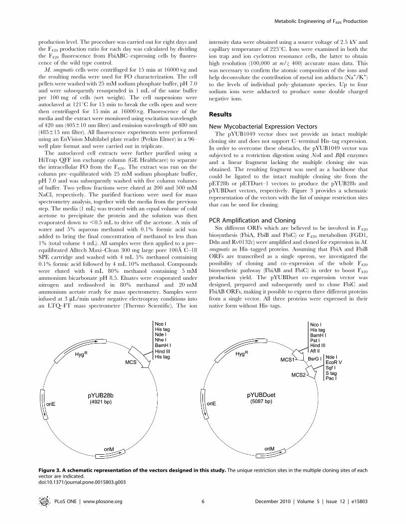
New Mycobacterial Expression VectorsThe pYUB1049 vector does not provide an intact multiple
cloning site and does not support C–terminal His–tag expression.
In order to overcome these obstacles, the pYUB1049 vector was
subjected to a restriction digestion using NcoI and BlpI enzymes
and a linear fragment lacking the multiple cloning site was
obtained. The resulting fragment was used as a backbone that
could be ligated to the intact multiple cloning site from the
pET28b or pETDuet–1 vectors to produce the pYUB28b and
pYUBDuet vectors, respectively. Figure 3 provides a schematic
representation of the vectors with the list of unique restriction sites
that can be used for cloning.
PCR Amplification and CloningSix different ORFs which are believed to be involved in F420
biosynthesis (FbiA, FbiB and FbiC) or F420 metabolism (FGD1,
Ddn and Rv0132c) were amplified and cloned for expression in M.
smegmatis as His–tagged proteins. Assuming that FbiA and FbiB
ORFs are transcribed as a single operon, we investigated the
possibility of cloning and co–expression of the whole F420
biosynthetic pathway (FbiAB and FbiC) in order to boost F420
production yield. The pYUBDuet co–expression vector was
designed, prepared and subsequently used to clone FbiC and
FbiAB ORFs, making it possible to express three different proteins
from a single vector. All three proteins were expressed in their
native form without His–tags.
Figure 3. A schematic representation of the vectors designed in this study. The unique restriction sites in the multiple cloning sites of eachvector are indicated.doi:10.1371/journal.pone.0015803.g003
Metabolic Engineering of F420 Production
PLoS ONE | www.plosone.org 6 December 2010 | Volume 5 | Issue 12 | e15803
Expression of Proteins in M. smegmatisThe six F420 biosynthetic or metabolic ORFs cloned into
pYUB1049/pYUB28b/pDESTsmg vectors were expressed in M.
smegmatis as individual proteins. Each of these proteins were cloned
with either N– or C–terminal His–tags, making it possible to detect
the protein expression using monoclonal anti–His antibodies. The
western blotting experiments indicated that all proteins were
expressed in M. smegmatis cells, as shown by appearance of correct–
sized bands for the appropriate proteins (data not shown).
The expression of proteins from the pYUBDuet vector could
not be detected using western blotting, as they did not contain any
tags; however, their successful expression could be inferred from
FO and F420 production as discussed later.
Cofactor F420 ProductionIndividual M. smegmatis cultures harbouring six different con-
structs (FbiA, FbiB, FbiC, FGD1, Ddn and Rv0132c) were grown in
order to find out the over–expression effect of these targets on F420
production. Three different media were initially used to express the
proteins; LBT with IPTG induction, MDG with no or low
induction using IPTG, and ZYM–5052 autoinduction media.
Based on growth rate and cell mass, ZYM–5052 media was selected
as the best media and was used to continue F420 production
experiments. The fluorescence signals of the expression media and
the cell extracts were monitored at 420 nm, enabling the detection
of both FO and F420. It has been reported that FO comprises 1–7%
of the total intracellular deazaflavin in Mycobacteria [8]; we used
fluorescence at 420 nm to evaluate the F420 contents of the cellular
extracts without taking into account the small portion of the
fluorescence signal coming from FO.
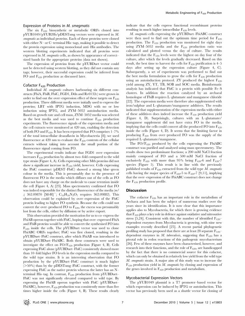
The experimental results indicate that FGD1 over–expression
increases F420 production by almost two–fold compared to the wild
type strain (Figure 4, A). Cells expressing other Mtb proteins did not
show a significant increase in F420 yield, however. Cells expressing
the FbiC construct (pDEST–FbiC) showed a strong blue–green
colour in the media. This is presumably due to the presence of
fluorescent FO in the media which diffuses out of the cells as FO
does not have any charge on the molecule to cause retention inside
the cell (Figure 4, A) [23]. Mass spectrometry confirmed that FO
was indeed responsible for the distinct fluorescence of the media (m/
z 362.09870 [M-H]2; C16H16N3O7 requires 362.09882). This
observation could be explained by over–expression of the FbiC
protein leading to higher FO synthesis. Because the cells could not
convert the over–produced FO to F420, the excess was presumably
lost from the cells, either by diffusion or by active export.
This observation provided the motivation for us to co–express the
FbiAB operon together with FbiC, hoping that over–expressed FbiA
and FbiB proteins would be able to convert the synthesised FO into
F420 inside the cells. The pYUBDuet vector was used to clone
FbiABC ORFs together; FbiC was first cloned, resulting in the
pYUBDuet–FbiC construct, after which FbiAB was introduced to
obtain pYUBDuet–FbiABC. Both these constructs were used to
investigate the effect on FO/F420 production (Figure 4, B). Cells
expressing FbiC alone (pYUBDuet–FbiC) consistently showed more
than 10–fold higher FO levels in the expression media compared to
the wild type strains. It is an interesting observation that FO
production by the pYUBDuet–FbiC construct is much higher
(.50%) than by the pDESTsmg–FbiC construct, with the former
expressing FbiC as the native protein whereas the latter has an N–
terminal His–tag. In contrast, F420 production from pYUBDuet–
FbiC was not significantly elevated compared to wild type. By
expressing the FbiAB operon together with FbiC (pYUBDuet–
FbiABC), however, F420 production was consistently more than five
times higher inside the cells (Figure 4, B). These results clearly
indicate that the cells express functional recombinant proteins
resulting in much higher intracellular F420 levels.
M. smegmatis cells expressing the pYUBDuet–FbiABC construct
were then used to find out the optimum time period for F420
production. The F420 production was monitored for eight days
using ZYM–5052 media and the F420 production ratio was
calculated and plotted versus the day of culture. The results
indicated that the F420 levels were the highest on day four of the
culture, after which the levels gradually decreased. Based on this
result, the best time to harvest the cells for F420 purification is 4–5
days after setting up the expression culture (Figure 4, C).
Subsequently, a set of experiments was performed to find out
the best media formulation to grow the cells for F420 production
using an autoinduction protocol. ZY produced the highest F420
yield among ZY, YT, TB, SOB and SOC media. Bioinformatic
analysis has indicated that FbiC is a protein with possible Fe–S
clusters. In addition the reaction catalyzed by an archaeal
homologue of FbiB requires L–glutamate and manganese chloride
[22]. The expression media were therefore also supplemented with
iron/sulphur and L–glutamate/manganese additives. The results
indicated that supplementation of the expression media with either
of these additives does indeed increase the F420 production yield
(Figure 4, D). Surprisingly, cultures with an L–glutamate/
manganese supplement did not have extra FO in the media,
implying that the cells could convert all the produced FO into F420
inside the cells (Figure 4, D). It seems that the limiting factor in
producing F420 from over–produced FO was the supply of the
required L–glutamate/manganese.
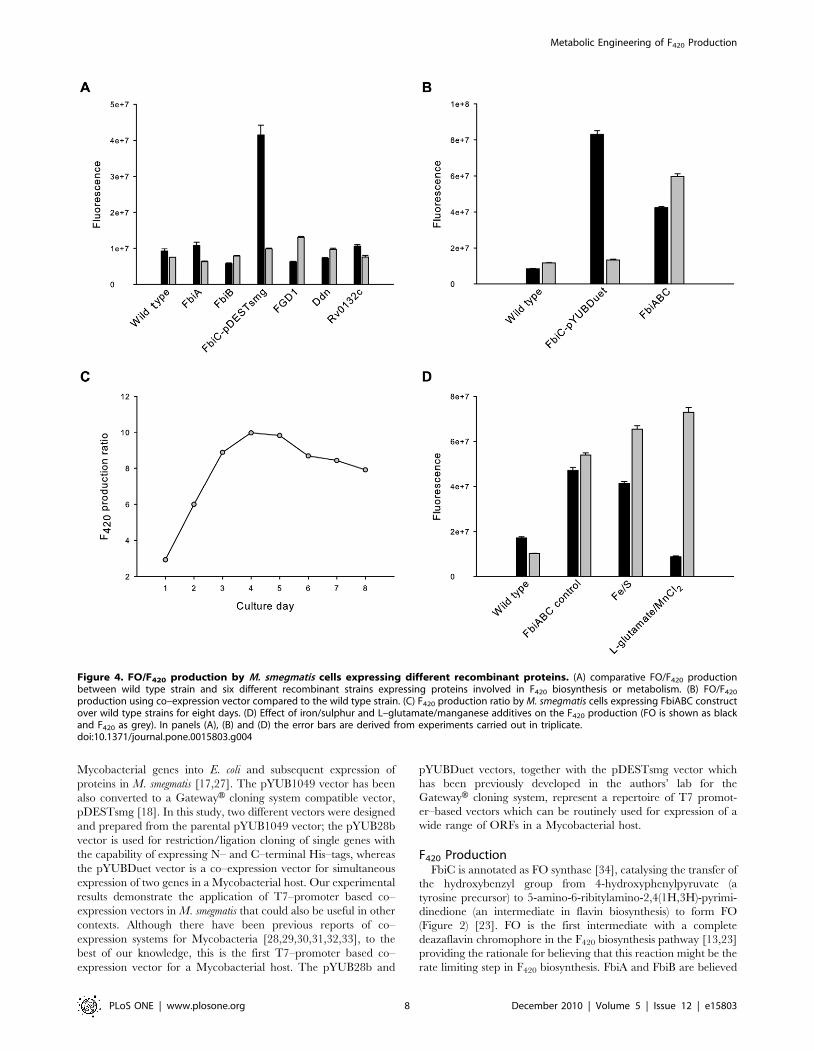
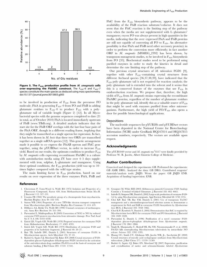
The FO/F420 produced by the cells expressing the FbiABC
construct was purified and analysed using mass spectrometry. The
results show two predominant fractions; a 200 mM NaCl fraction
mainly composed of FO and a 500 mM NaCl fraction of
exclusively F420 with more than 95% being F420-6 and F420-7
species (Figure 5). This result is in line with the previously
published results of F420 extracted from the wild type M. smegmatis
cells having the major species of F420-5 to F420-7 [9,11], implying
that the over–expression of the FbiABC construct does not change
the F420 production profile.
Discussion
The cofactor F420 has an important role in the metabolism of
Archaea and has been the subject of numerous studies over the
years since its identification. It is now clear that this importance
applies also to Mycobacteria, for which there is growing evidence
that F420 plays a key role in defence against oxidative and nitrosative
stress [5,24]. Consistent with this, the number of identified F420–
dependent enzymes from Mycobacteria is growing, with nine new
examples recently described [25]. A recent partial phylogenetic
profiling study has proposed that there are at least 28 separate F420–
dependent enzymes in M. tuberculosis, suggesting that F420 has a
pivotal role in redox reactions of this pathogenic mycobacterium
[26]. Few of these enzymes have been characterised, however, and
research into their functions, and the role of F420, are handicapped
by the fact that there is no commercial source for this cofactor,
which can only be obtained in relatively low yield from the wild type
M. smegmatis strain. A major aim of this study was to increase the
F420 production yield in M. smegmatis by cloning and expression of
the genes involved in F420 production and metabolism.
Mycobacterial Expression VectorsThe pYUB1049 plasmid is a T7 promoter–based vector for
which expression can be induced by IPTG or autoinduction. This
vector has previously been used as a shuttle vector for cloning of
Metabolic Engineering of F420 Production
PLoS ONE | www.plosone.org 7 December 2010 | Volume 5 | Issue 12 | e15803
Mycobacterial genes into E. coli and subsequent expression of
proteins in M. smegmatis [17,27]. The pYUB1049 vector has been
also converted to a GatewayH cloning system compatible vector,
pDESTsmg [18]. In this study, two different vectors were designed
and prepared from the parental pYUB1049 vector; the pYUB28b
vector is used for restriction/ligation cloning of single genes with
the capability of expressing N– and C–terminal His–tags, whereas
the pYUBDuet vector is a co–expression vector for simultaneous
expression of two genes in a Mycobacterial host. Our experimental
results demonstrate the application of T7–promoter based co–
expression vectors in M. smegmatis that could also be useful in other
contexts. Although there have been previous reports of co–
expression systems for Mycobacteria [28,29,30,31,32,33], to the
best of our knowledge, this is the first T7–promoter based co–
expression vector for a Mycobacterial host. The pYUB28b and
pYUBDuet vectors, together with the pDESTsmg vector which
has been previously developed in the authors’ lab for the
GatewayH cloning system, represent a repertoire of T7 promot-
er–based vectors which can be routinely used for expression of a
wide range of ORFs in a Mycobacterial host.
F420 ProductionFbiC is annotated as FO synthase [34], catalysing the transfer of
the hydroxybenzyl group from 4-hydroxyphenylpyruvate (a
tyrosine precursor) to 5-amino-6-ribitylamino-2,4(1H,3H)-pyrimi-
dinedione (an intermediate in flavin biosynthesis) to form FO
(Figure 2) [23]. FO is the first intermediate with a complete
deazaflavin chromophore in the F420 biosynthesis pathway [13,23]
providing the rationale for believing that this reaction might be the
rate limiting step in F420 biosynthesis. FbiA and FbiB are believed
Figure 4. FO/F420 production by M. smegmatis cells expressing different recombinant proteins. (A) comparative FO/F420 productionbetween wild type strain and six different recombinant strains expressing proteins involved in F420 biosynthesis or metabolism. (B) FO/F420
production using co–expression vector compared to the wild type strain. (C) F420 production ratio by M. smegmatis cells expressing FbiABC constructover wild type strains for eight days. (D) Effect of iron/sulphur and L–glutamate/manganese additives on the F420 production (FO is shown as blackand F420 as grey). In panels (A), (B) and (D) the error bars are derived from experiments carried out in triplicate.doi:10.1371/journal.pone.0015803.g004
Metabolic Engineering of F420 Production
PLoS ONE | www.plosone.org 8 December 2010 | Volume 5 | Issue 12 | e15803
to be involved in production of F420 from the precursor FO
molecule; FbiA in generating F420–0 from FO and FbiB in adding
glutamate residues to F420–0 to produce F420 with a poly–
glutamate tail of variable length (Figure 2) [12]. In all Myco-
bacterial species with the genome sequences completed to date (21
in total, as of October 2010) FbiA is located immediately upstream
of FbiB (www.TBdb.org). A detailed analysis indicates that the
start site for the FbiB ORF overlaps with the last four base pairs of
the FbiA ORF, though in a different reading frame, implying that
they might be transcribed as a single operon for expression. In fact,
it has been shown in M. bovis that these two ORFs are transcribed
together as a single mRNA species [12]. This genetic arrangement
made it possible to co–express the FbiAB operon and FbiC gene
together, using the pYUBDuet vector, in order to increase F420
yield. Based on our results, the optimum condition to produce F420
by M. smegmatis cells expressing recombinant FbiABC is a culture
with autoinduction media using ZY base over 4–5 days supple-
mented with iron, sulphur, L–glutamate and manganese. Using
these optimal conditions, the F420 production yield was up to 10–
times higher compared with the wild type strains.
The main limiting factor in F420 production, based on our
results on over–expression of the three enzymes FbiA, FbiB and
FbiC from the F420 biosynthetic pathway, appears to be the
availability of the FbiB reaction substrate/cofactor. It does not
seem that the FbiC reaction is the limiting step of the pathway
even when the media are not supplemented with L–glutamate/
manganese; excess FO was always present in high quantities in the
media, indicating that the over–expressed FbiA and FbiB proteins
are still not capable of converting all FO to F420. An alternative
possibility is that FbiA and FbiB need other accessory protein(s) in
order to perform the conversion more efficiently; in fact another
ORF in M. smegmatis (MSMEG_2392) has been shown, by
transposon mutagenesis studies, to be involved in F420 biosynthesis
from FO [35]. Biochemical studies need to be performed using
purified enzymes in order to study the kinetics in detail and
determine the rate limiting step of the pathway.
Our previous crystal structures of M. tuberculosis FGD1 [9],
together with other F420–containing crystal structures from
different Archaeal species [36,37,38,39], have indicated that the
F420 poly–glutamate tail is not required for reaction catalysis; the
poly–glutamate tail is extended into the solvent and it seems that
this is a conserved feature of the enzymes that use F420 in
oxidoreduction reactions. We propose that, therefore, the high
yields of F420 from M. smegmatis strains expressing the recombinant
FbiABC proteins, regardless of the number of glutamate residues
in the poly–glutamate tail, identify this as a valuable source of F420
that might be used with enzymes purified from other microor-
ganisms. Furthermore, the high yield of FO/F420 also opens a
door for possible biotechnological applications.
DepositionsThe nucleotide sequences for pYUB28b and pYUBDuet vectors
have been deposited in the National Centre for Biotechnology
Information (NCBI) under GenBank HQ247814 and HQ247815
accession numbers, respectively. The vectors are available upon
request.
Acknowledgments
The pYUB1049 vector and M. smegmatis mc24517 were kindly provided by
Professor W. R. Jacobs, Albert Einstein College of Medicine.
Author Contributions
Conceived and designed the experiments: GB. Performed the experiments:
GB AMR DRG. Analyzed the data: GB DRG. Contributed reagents/
materials/analysis tools: JMJD. Wrote the paper: GB JMJD ENB.
Acquisition of funding/supervision: ENB.
References
1. Cheeseman P, Toms-Wood A, Wolfe RS (1972) Isolation and Properties of a
Fluorescent Compound, Factor 420, from Methanobacterium Strain M.o.H.J Bacteriol 112: 527–531.
2. Cousins FB (1960) The prosthetic group of a chromoprotin from mycobacteria.Biochim Biophys Acta 40: 532–534.
3. Sutton WB (1964) Properties of a new TPN-like electron transport componentfrom Mycobacterium phlei. Biochem Biophys Res Commun 15: 414–419.
4. DiMarco AA, Bobik TA, Wolfe RS (1990) Unusual coenzymes of methanogen-esis. Annu Rev Biochem 59: 355–394.
5. Purwantini E, Mukhopadhyay B (2009) Conversion of NO2 to NO by reduced
coenzyme F420 protects mycobacteria from nitrosative damage. Proc Natl AcadSci U S A 106: 6333–6338.
6. Eirich LD, Vogels GD, Wolfe RS (1978) Proposed structure for coenzyme F420from Methanobacterium. Biochemistry 17: 4583–4593.
7. Eirich LD, Vogels GD, Wolfe RS (1979) Distribution of coenzyme F420 andproperties of its hydrolytic fragments. J Bacteriol 40: 20–27.
8. Bair TB, Isabelle DW, Daniels L (2001) Structures of coenzyme F(420) inMycobacterium species. Arch Microbiol 176: 37–43.
9. Bashiri G, Squire CJ, Moreland NJ, Baker EN (2008) Crystal structures of F420-dependent glucose-6-phosphate dehydrogenase FGD1 involved in the activation
of the anti-tuberculosis drug candidate PA-824 reveal the basis of coenzyme and
substrate binding. J Biol Chem 283: 17531–17541.
10. Graupner M, White RH (2003) Methanococcus jannaschii Coenzyme F420 Analogs
Contain a Terminal a-Linked Glutamate. J Bacteriol 185: 662–4665.
11. Isabelle D, Simpson DR, Daniels L (2002) Large-scale production of coenzyme F420-5,6 by using Mycobacterium smegmatis. Appl Environ Microbiol 68: 5750–5755.
12. Choi KP, Bair TB, Bae YM, Daniels L (2001) Use of transposon Tn5367
mutagenesis and a nitroimidazopyran-based selection system to demonstrate arequirement for fbiA and fbiB in coenzyme F(420) biosynthesis by Mycobacterium
bovis BCG. J Bacteriol 183: 7058–7066.
13. Choi K-P, Kendrick N, Daniels L (2002) Demonstration that fbiC is required byMycobacterium bovis BCG for coenzyme F420 and FO biosynthesis. J Bacteriol
184: 2420–2428.
14. Purwantini E, Daniels L (1996) Purification of a novel coenzyme F420-dependent glucose-6-phosphate dehydrogenase from Mycobacterium smegmatis.
J Bacteriol 178: 2861–2866.
15. Singh R, Manjunatha U, Boshoff HI, Ha YH, Niyomrattanakit P, et al. (2008)PA-824 kills nonreplicating Mycobacterium tuberculosis by intracellular NO
release. Science 322: 1392–1395.
16. Huang CC, Smith CV, Glickman MS, Jacobs WR, Jr., Sacchettini JC (2002)Crystal structures of mycolic acid cyclopropane synthases from Mycobacterium
tuberculosis. J Biol Chem 277: 11559–11569.
17. Bashiri G, Squire CJ, Baker EN, Moreland NJ (2007) Expression, purification
and crystallization of native and selenomethionine labeled Mycobacterium
Figure 5. The F420 production profile from M. smegmatis cellsover–expressing the FbiABC construct. The F420–6 and F420–7species constitute the main species as deduced using mass spectrometry.doi:10.1371/journal.pone.0015803.g005
Metabolic Engineering of F420 Production
PLoS ONE | www.plosone.org 9 December 2010 | Volume 5 | Issue 12 | e15803
tuberculosis FGD1 (Rv0407) using a Mycobacterium smegmatis expression system.
Protein Expr Purif 54: 38–44.18. Goldstone RM, Moreland NJ, Bashiri G, Baker EN, Shaun Lott J (2008) A new
Gateway vector and expression protocol for fast and efficient recombinant
protein expression in Mycobacterium smegmatis. Protein Expr Purif 57: 81–87.19. Cirillo JD, Weisbrod TR, William R, Jacobs J (1993) Efficient electrotransfor-
mation of Mycobacterium smegmatis. RichmondCalifornia: Bio-Rad Laboratories.20. Studier FW (2005) Protein production by auto-induction in high-density shaking
cultures. Protein Expr Purif 41: 207–234.
21. Towbin H, Staehelin T, Gordon J (1979) Electrophoretic transfer of proteinsfrom polyacrylamide gels to nitrocellulose sheets: procedure and some
applications. Proc Natl Acad Sci U S A 76: 4350–4354.22. Nocek B, Evdokimova E, Proudfoot M, Kudritska M, Grochowski LL, et al.
(2007) Structure of an amide bond forming F(420):gamma-glutamyl ligase fromArchaeoglobus fulgidus – a member of a new family of non-ribosomal peptide
synthases. J Mol Biol 372: 456–469.
23. Graham DE, Xu H, White RH (2003) Identification of the 7,8-didemethyl-8-hydroxy-5-deazariboflavin synthase required for coenzyme F(420) biosynthesis.
Arch Microbiol 180: 455–464.24. Darwin KH, Ehrt S, Gutierrez-Ramos J-C, Weich N, Nathan CF (2003) The
proteasome of Mycobacterium tuberculosis is required for resistance to nitric
oxide. Science 302: 1963–1966.25. Taylor MC, Jackson CJ, Tattersall DB, French N, Peat TS, et al. (2010)
Identification and characterization of two families of F420H2-dependentreductases from Mycobacteria that catalyze aflatoxin degradation. Mol
Microbiol 78: 561–575.26. Selengut JD, Haft DH (2010) Unexpected Abundance of Coenzyme F420-
dependent enzymes in the Genomes of Mycobacterium tuberculosis and other
Actinobacteria. J Bacteriol 192: 5788–5798.27. Robson J, McKenzie JL, Cursons R, Cook GM, Arcus VL (2009) The vapBC
operon from Mycobacterium smegmatis is an autoregulated toxin-antitoxinmodule that controls growth via inhibition of translation. J Mol Biol 390:
353–367.
28. Chang Y, Mead D, Dhodda V, Brumm P, Fox BG (2009) One-plasmid tunablecoexpression for mycobacterial protein-protein interaction studies. Protein Sci
18: 2316–2325.29. George KM, Yuan Y, Sherman DR, Barry CE, 3rd (1995) The biosynthesis of
cyclopropanated mycolic acids in Mycobacterium tuberculosis. Identificationand functional analysis of CMAS-2. J Biol Chem 270: 27292–27298.
30. Harth G, Maslesa-Galic S, Horwitz MA (2004) A two-plasmid system for stable,
selective-pressure-independent expression of multiple extracellular proteins inmycobacteria. Microbiology 150: 2143–2151.
31. Kaps I, Ehrt S, Seeber S, Schnappinger D, Martin C, et al. (2001) Energytransfer between fluorescent proteins using a co-expression system in
Mycobacterium smegmatis. Gene 278: 115–124.
32. Luo Y, Chen X, Szilvasi A, O’Donnell MA (2000) Co-expression of interleukin-2and green fluorescent protein reporter in mycobacteria: in vivo application for
monitoring antimycobacterial immunity. Mol Immunol 37: 527–536.33. Slayden RA, Lee RE, Barry CE, 3rd (2000) Isoniazid affects multiple
components of the type II fatty acid synthase system of Mycobacteriumtuberculosis. Mol Microbiol 38: 514–525.
34. Cole ST, Brosch R, Parkhill J, Garnier T, Churcher C, et al. (1998) Deciphering
the biology of Mycobacterium tuberculosis from the complete genome sequence.Nature 393: 537–544.
35. Guerra-Lopez D, Daniels L, Rawat M (2007) Mycobacterium smegmatis mc2155 fbiC and MSMEG_2392 are involved in triphenylmethane dye decoloriza-
tion and coenzyme F420 biosynthesis. Microbiology 153: 2724–2732.
36. Aufhammer SW, Warkentin E, Berk H, Shima S, Thauer RK, et al. (2004)Coenzyme binding in F420-dependent secondary alcohol dehydrogenase, a
member of the bacterial luciferase family. Structure 12: 361–370.37. Aufhammer SW, Warkentin E, Ermler U, Hagemeier CH, Thauer RK, et al.
(2005) Crystal structure of methylenetetrahydromethanopterin reductase (Mer)
in complex with coenzyme F420: Architecture of the F420/FMN binding site ofenzymes within the nonprolyl cis-peptide containing bacterial luciferase family.
Protein Sci 14: 1840–1849.38. Ceh K, Demmer U, Warkentin E, Moll J, Thauer RK, et al. (2009) Structural
basis of the hydride transfer mechanism in F(420)-dependent methylenetetrahy-dromethanopterin dehydrogenase. Biochemistry 48: 10098–10105.
39. Warkentin E, Mamat B, Sordel-Klippert M, Wicke M, Thauer RK, et al. (2001)
Structures of F420H2:NADP+ oxidoreductase with and without its substratesbound. EMBO J 20: 6561–6569.
40. Jacobson FS, Daniels L, Fox JA, Walsh CT, Orme-Johnson WH (1982)
Purification and properties of an 8-hydroxy-5-deazaflavin-reducing hydrogenase
from Methanobacterium thermoautotrophicum. J Biol Chem 257: 3385–3388.
41. Deppenmeier U, Blaut M, Mahlmann A, Gottschalk G (1990) Membrane-
bound F420H2-dependent heterodisulfide reductase in methanogenic bacterium
strain Gol and Methanolobus tindarius. FEBS Lett 261: 199–203.
42. Seedorf H, Dreisbach A, Hedderich R, Shima S, Thauer RK (2004) F420H2
oxidase (FprA) from Methanobrevibacter arboriphilus, a coenzyme F420-
dependent enzyme involved in O2 detoxification. Arch Microbiol 182: 126–137.
43. Tzeng SF, Wolfe RS, Bryant MP (1975) Factor 420-dependent pyridine
nucleotide-linked hydrogenase system of Methanobacterium ruminantium.
J Bacteriol 121: 184–191.
44. Deppenmeier U, Blaut M, Mahlmann A, Gottschalk G (1990) Reduced
coenzyme F420: heterodisulfide oxidoreductase, a proton- translocating redox
system in methanogenic bacteria. Proc Natl Acad Sci U S A 87: 9449–9453.
45. Kunow J, Linder D, Stetter KO, Thauer RK (1994) F420H2: quinone
oxidoreductase from Archaeoglobus fulgidus. Characterization of a membrane-
bound multisubunit complex containing FAD and iron-sulfur clusters.Eur J Biochem 223: 503–511.
46. Tzeng SF, Bryant MP, Wolfe RS (1975) Factor 420-dependent pyridine
nucleotide-linked formate metabolism of Methanobacterium ruminantium.J Bacteriol 121: 192–196.
47. Zeikus JG, Fuchs G, Kenealy W, Thauer RK (1977) Oxidoreductases involved
in cell carbon synthesis of Methanobacterium thermoautotrophicum. J Bacteriol132: 604–613.
48. Fuchs G, Stupperich E (1982) Autotrophic CO2 fixation pathway inMethanobacterium thermoautotrophicum. Zentralbl Bakteriol Hyg Abt 1
Orig C 3: 277–288.
49. Hartzell PL, Zvilius G, Escalante-Semerena JC, Donnelly MI (1985) CoenzymeF420 dependence of the methylenetetrahydromethanopterin dehydrogenase of
Methanobacterium thermoautotrophicum. Biochem Biophys Res Commun 133:
884–890.
50. Ma K, Thauer RK (1990) Purification and properties of N5, N10-methylene-
tetrahydromethanopterin reductase from Methanobacterium thermoautotrophicum
(strain Marburg). Eur J Biochem 191: 187–193.
51. Widdel F, Wolfe RS (1989) Expression of secondary alcohol dehydrogenase in
methanogenic bacteria and purification of the F420-specific enzyme from
Methanogenium thermophilum strain TCI. Arch Microbiol 152: 322–328.
52. Johnson EF, Mukhopadhyay B (2005) A new type of sulfite reductase, a novel
coenzyme F420-dependent enzyme, from the methanarchaeon Methanocaldo-
coccus jannaschii. J Biol Chem 280: 38776–38786.
53. Vermeij P, Detmers FJ, Broers FJ, Keltjens JT, Van der Drift C (1994)
Purification and characterization of coenzyme F390 synthetase from Methano-
bacterium thermoautotrophicum (strain delta H). Eur J Biochem 226: 185–191.
54. Vermeij P, Vinke E, Keltjens JT, Van der Drift C (1995) Purification and
properties of coenzyme F390 hydrolase from Methanobacterium thermoauto-
trophicum (strain Marburg). Eur J Biochem 234: 592–597.
55. Coats JH, Li GP, Kuo MS, Yurek DA (1989) Discovery, production, and
biological assay of an unusual flavenoid cofactor involved in lincomycin
biosynthesis. J Antibiot (Tokyo) 42: 472–474.
56. McCormick JRD, Morton GO (1982) Identity of cosynthetic factor 1 of
Streptomyces aureofaciens and fragment FO from coenzyme F420 of
Methanobacterium sp. J Am Chem Soc 104: 4014–4015.
57. Rhodes PM, Winskill N, Friend EJ, Warren M (1981) Biochemical and genetic
comparison of Streptomyces rimosus mutants impaired in oxytetracycline
biosynthesis. J Gen Microbiol 124: 329–338.
58. Ebert S, Rieger PG, Knackmuss HJ (1999) Function of coenzyme F420 in
aerobic catabolism of 2,4, 6-trinitrophenol and 2,4-dinitrophenol by Nocar-
dioides simplex FJ2-1A. J Bacteriol 181: 2669–2674.
59. Eker AP, Hessels JKC, Velde Jvd (1988) Photoreactivating enzyme from the
green alga Scenedesmus acutus. Evidence for the presence of two different flavin
chromophores. Biochemistry 27: 1758–1765.
60. Eker AP, Kooiman P, Hessels JK, Yasui A (1990) DNA photoreactivating
enzyme from the cyanobacterium Anacystis nidulans. J Biol Chem 265:
8009–8015.
61. Glas AF, Maul MJ, Cryle M, Barends TR, Schneider S, et al. (2009) The
archaeal cofactor F0 is a light-harvesting antenna chromophore in eukaryotes.
Proc Natl Acad Sci U S A 106: 11540–11545.
62. Kiener A, Gall R, Rechsteiner T, Leisinger T (1985) Photoreactivation in
Methanobacterium thermoautotrophicum. Arch Microbiol 143: 147–150.
Metabolic Engineering of F420 Production
PLoS ONE | www.plosone.org 10 December 2010 | Volume 5 | Issue 12 | e15803


