Embed Size (px)
Citation preview

Metabolism of Tryptophan in IsolatedPerfused Rat Liver]'2
C. Y. NG," Y. HACINO,4 P. B. SWAN ANDL. M. HENDERSON
Department of Biochemistry, College of Biological Sciences and Divisionof Nutrition, College of Agriculture, Forestry and Home Economic»,university of Minnesota, St. Paul, Minnesota 55101
ABSTRACT The fate of tryptophan in the isolated perfused rat liver was investigated. It was shown that the liver removed the L-isomer more rapidly from the per-fusate than the D-isomer. The 10 g liver was capable of a removal rate of 35 mg/hourwhen 42 mg of DL-tryptophan was given initially, followed by infusion at the rate of42mg/hour. Carbon-14 from DL-tryptophan-2-"C appeared in CO2 much more rapidlyand to a greater extent within 3 hours than did that from DL-tryptophan-7a-14C. Incontrast to whole-animal studies, it was shown that overloading doses of acetoacetatowere labeled by DL-tryptophan-5- or 7a-14C. The labeling pattern in acetoacetate wassimilar to that observed when 14C-fatty acids are the substrate. Acetoacetate overloading suppressed the production of radioactive carbon dioxide from DL-tryptophan-7a-14C.
The central role of the liver in the metabolism of amino acids and in the synthesis and degradation of protein has beendemonstrated in part through the use of theisolated perfused rat liver. Comparison ofhepatectomized rats and their perfusedlivers has shown that several amino acidsare degraded largely by the liver (1). Theliver appears to regulate the level of aminoacids circulating in the blood (2-4) andit is responsible not only for the synthesisand degradation of hepatic protein, butfor the synthesis and degradation of muchof the plasma protein as well (5-8). Theisolated perfused liver has been particularlyuseful in defining the liver's role, sincehormonal and other factors controllingamino acid and protein metabolism canbe more easily controlled experimentallythan in the whole animal and because theactivity of extrahepatic tissue is excluded.
Tryptophan is among the amino acidswhich seem to be degraded largely by theliver (1). This essential amino acid is ofparticular interest because it is often limiting in the diet of man and animals, andbecause it is present in mammalian tissuesin very small amounts (9). Recently, interest in the fate of tryptophan within animal tissues has been further stimulated bythe suggestion that it plays a unique rolein the control of protein biosynthesis (10,11) and in control of gluconeogenesis (12).Therefore, it would be useful to investigate
further the metabolism of tryptophan inthe liver since this organ is the primarysite of gluconeogenesis and is also veryactive in protein biosynthesis. The isolatedperfused rat liver is a particularly convenient system for this study.
METHODS
The liver and blood donors were malealbino rats of the Sprague-Dawley strain,fed a commercial laboratory ration 5 andallowed food and water ad libitum. In certain experiments, as indicated, rats fastedfor 48 to 72 hours were used as liverdonors. The animals were maintained indarkness from 6:00 PM to 6:00 AM in aroom with constant temperature and humidity.
Blood donors were retired male breedersweighing approximately 600 g. The animal was placed under anesthesia withdiethyl ether, the abdominal cavity wasopened, and blood was drawn from the
Received for publication July 17, 1969.1 Supported by Grant no. GM 11710 from the Na
tional Institutes of Health and by funds provided bythe Minnesota Agricultural Experiment Station. Scientific Journal Series No. 6991, Agricultural Experiment Station, University of Minnesota, St. Paul,Minnesota.
2 Presented in part at the Seventh InternationalCongress of Biochemistry, Tokyo, Japan, August, 1967.Metabolism of tryptophan by perfused rat liver, 1968.Medical Journal of Osaka University, 19: 25 (abstract).
3 Deceased.4 Present address : Nagoya University, School of
Medicine, Nagoya, Japan.5 Purina Laboratory Chow, Ralston Purina Company,
St. Louis, Mo.
J. NUTRITION,99: 465-473. 465
by guest on June 4, 2015jn.nutrition.org
Dow
nloaded from

466 NG, HACINO, SWAN AND HENDERSON
abdominal aorta into a syringe containingheparin. The peri usate (40 to 50 ml)consisted of either rat blood or rat plasmawhich had been diluted one-third to one-half with Ringer's bicarbonate buffer. The
perfusate was circulated in the perfusionapparatus for approximately 30 minutesprior to the insertion of the liver, andtryptophan was added 30 to 45 minutesafter perfusion began. In some experiments, as indicated, glucose was added tothe perfusate in an amount to give aninitial concentration of 5 mg/ml. Liverdonors ranged in weight from 225 to 325 g,the majority weighing between 275 and300 g. The livers were removed by a procedure similar to that described by Milleret al. (5). The perfusion apparatus wasa slight modification of that described byGreen and Miller (7). A mixture of 95%oxygen and 5% carbon dioxide was usedfor aeration. Carbon dioxide coming fromthe chamber was trapped in a 1:2 (v:v)mixture of aminoethanol and methoxy-ethanol. Livers were perfused for a periodof 2 to 4 hours and their condition wasevaluated on the basis of bile production,rate of perfusate flow and the appearanceof the surface.
Carbon-14 was determined in a scintillation counter " using the scintillation fluiddescribed previously (13). Plasma andwhole blood were dissolved in a 1 N solution of hyamine hydroxide in methanolbefore the scintillation fluid was added.In some cases, hydrogen peroxide wasadded with the hyamine hydroxide to decolorize the sample.The DL-tryptophan-7a-1''C and DL-trypto-phan-5-14C have been described previously(14, 15) (fig. 1). Other labeled compounds were from commercial sources.7
In overloading experiments, plasma wasdeproteinized with tungsuc acid and ace-toacetate was isolated by silicic acid chro-
CH2CHNH2COOH
matography using a modification of theprocedure described by Marvel and Rands(16). Two methods were used for the degradation of acetoacetate : 1) decarboxyla-tion in an acidic medium containing mercuric sulfate (17), giving acetone as themercury complex and carbon dioxidewhose specific activity was estimated usinga vibraung-reed electrometer; and 2) controlled oxidation with potassium permanganate (18) yielding carbon dioxide fromcarbon-1, formate from carbon-2 and acetate from carbons 3 and 4. Formate wasoxidized to carbon dioxide using mercuricsulfate. Acetate was subjected to Schmidtdegradation (19).
Tryptophan was assayed chemically bya modification of the procedures of Opien-ska-Blauth et al. (20) and Fischi (21).
RESULTS
When the disappearance of tryptophanfrom the perfusate was followed by chemical assay, the pattern shown in figure 2was obtained. Removal of L-tryptophanwas more rapid than that of D-tryptophan,resulting in a lower plasma level of tryptophan after 1 hour. When L-tryptophanwas used within approximately 10 minutes
30 60TIME (min)
Fig. 2 Rate of uptake of the D- and L-isomersof tryptophan by the isolated rat liver perfusedwith diluted rat plasma. The liver was perfusedfor 1 hour to remove endogenous tryptophanfrom the perfusate. At 1 hour (• •) and2.5 hours (A A), 0.9 mg of L-tryptophanwas added. At 4 hours ((D-tryptophan was added.
1.1 mg of
Fig. 1 Numbering system of the tryptophanmolecule.
6Nuclear-Chicago Model 725, Nuclear-Chicago Corporation, Des Plaines, 111.
7DL-Tryptophan-2-14Cwas purchased from Tracerlab,Inc., Richmond, Calif.; acetoacetate-3->4Cwas obtainedfrom Nuclear-Chicago Corporation, Des Plaines, 111.,and glutarate-l,5-14C was from Calbiochem, LosAngeles, Calif.
by guest on June 4, 2015jn.nutrition.org
Dow
nloaded from

METABOLISM OF TRYPTOPHAN IN RAT LIVER 467
the plasma tryptophan level fell to aboutone-half the 15 ug/ml found in the intactanimal. The curves in figure 2 were obtained using diluted plasma. As shown intable 1, however, when higher amounts oftryptophan were infused at a constant rate,the removal of DL-tryptophan was moreefficient when erythrocytes were presentin the perfusion medium. When dilutedblood was used, it was necessary to infusetryptophan at a rate four times higher inorder to support a plasma concentrationequivalent to that maintained when diluted plasma was used.
The liver removed large amounts oftryptophan from the perfusate; table 1illustrates the rate of removal when it wasinfused at high concentrations. This aminoacid was not concentrated within theerythrocytes under the conditions used inthese experiments. When low concentrations of L-tryptophan (15 ug/ml) were incubated at 37°with diluted whole blood,there was equilibration of the tryptophanbetween the plasma and red cells within 5minutes. When larger amounts were used(90 ug/ml), equilibration required a longertime.
The total amount of radioactive carbondioxide which was produced in 2 to 3 hoursby the isolated perfused rat liver dependedupon the position of labeling of the tryptophan molecule and to a limited extent uponthe amount of tryptophan which was used.As is shown in table 2, there was little orno difference in the amount of radioactivecarbon dioxide which was produced whenDL-tryptophan-7a-14C was infused at a rateof 21 mg/hour or a rate of 42 mg/hour,
TABLEiEffect of rate of infusion of m.-tryptophan on
plasma concentration and liver uptake
Initialdosemg2.02.02.010.321.042.0Rate
ofinfusionmg/Hr1.01.04.010.821.042.0Avgplasma
concn'«3/ntl30s437110410584Initialrate of
uptake-mg/hr2.555122035
since the percentage of 14Cin carbon dioxide decreased by a factor of two when thedosage was doubled. More radioactive carbon dioxide was obtained when the "Cwas in the carboxyl of the side chain orthe 2-positÃon of the indole ring of thetryptophan. Similar yields of "CO* fromC-5 and C-7a were observed (table 3)when tryptophan was given as a singledose at the start of the experiment. Onlyabout 1% of the radioactivity from DL-tryptophan-5-"C was in carbon dioxidecompared to 12 to 16% from DL-trypto-phan-7a-14C. At very high dosages (150mg) there was about twice as much radioactivity in carbon dioxide from DL-trypto-phan-2-14C as from DL-tryptophan-7a-14C.
The initial rate at which I4COawas released was also a function of the positionof the 14Cin the tryptophan given (fig. 3).When the concentration of DL-tryptophan-2-14Cin the blood was maintained at 30ug/ml, production of radioactive carbon
TABLE 2Percentage of 14Cappearing in CO¡during 3 hours
as a function of infusion rate and position ofthe "C in tryptophan1
SubstrateDL-Trp-2-"'CDL-Trp-2-14CDL-Trp-7a-'4CDL-Trp-7a-I4CL-Trp-»COOHDL-Trp-7a-»CDL-Trp-7a-14CDL-Trp-7a-HCDL-Trp-7a-14CInitialdosemg2.02.010.310.310.321.021.042.042.0Infusionratemg/hr1.04.210.810.810.821.021.042.042.0Percentageof «CinC02%33.644.76.611.821.76.54.53.43.4
i Perfusate was diluted blood.
TABLE 3Percentage of 14C from tryptophan appearing in
expired COj during 3 hours ofliver perfusion
1 Over the 2-hour period of infusion.* Based on first 30 minutes of perfusion.3 Perfusate was diluted plasma; diluted blood was
used in other experiments.
PositionoflabelDL-Trp-7a-»CDL-Trp-7a-"Ci.-Trp-7a-»CDL-Trp-5-"CDL-Trp-7a-"CDL-Trp-7a-"CDL-Trp-2-»CSingledosemg1.2«1.10.7'1.3«150150150Percentageof "C inCO2%12.615.016.31.24.37.916.4
1Perfusate was diluted plasma; diluted whole bloodwas used in other experiments.
by guest on June 4, 2015jn.nutrition.org
Dow
nloaded from
468 NG, HACINO, SWAN AND HENDERSON
I 2 3TIME (hr )
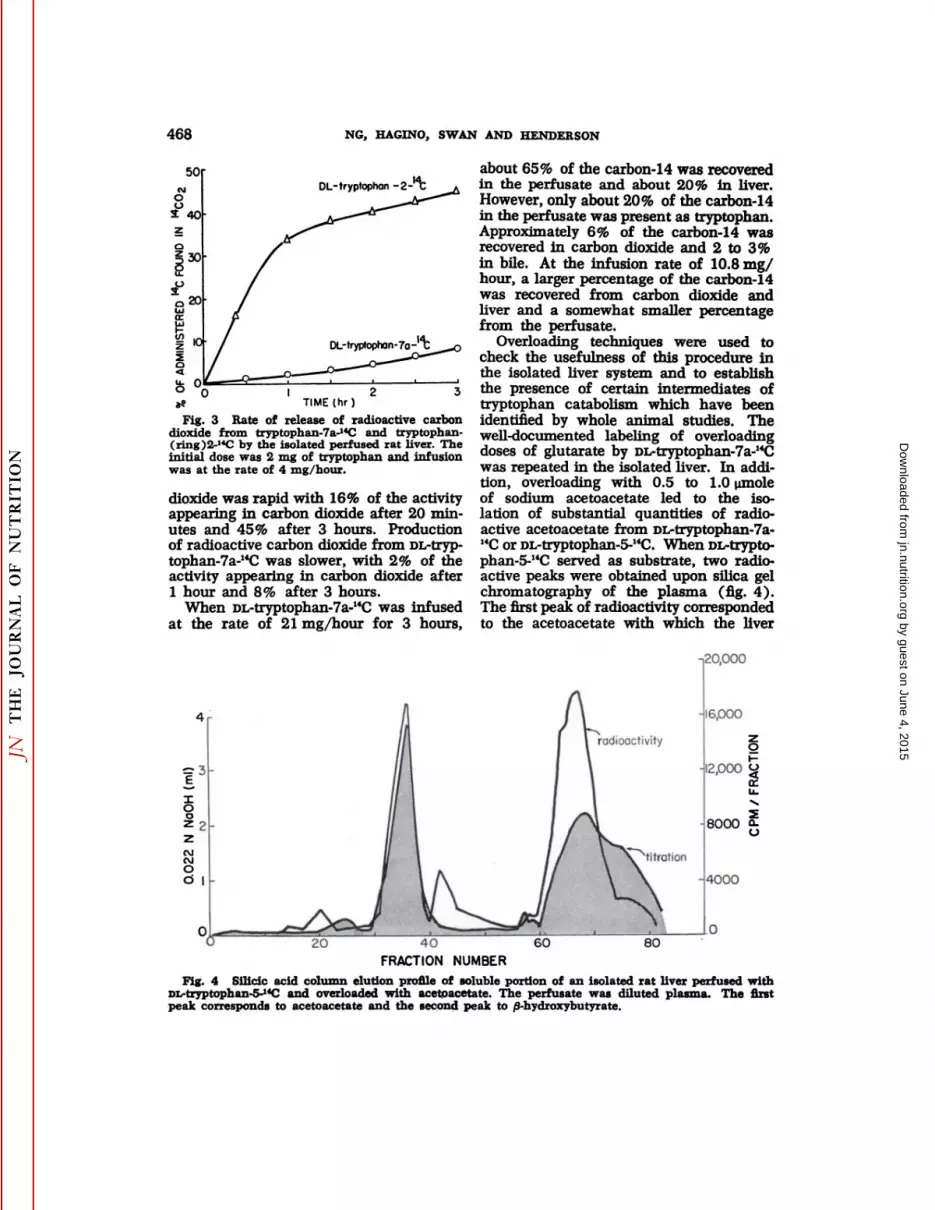
Fig. 3 Rate of release of radioactive carbondioxide from tryptophan-7a-HC and tryptophan-(ring)2-14C by the isolated perfused rat liver. Theinitial dose was 2 mg of tryptophan and infusionwas at the rate of 4 mg/hour.
dioxide was rapid with 16% of the activityappearing in carbon dioxide after 20 minutes and 45% after 3 hours. Productionof radioactive carbon dioxide from DL-tryptophan-7a-14C was slower, with 2% of theactivity appearing in carbon dioxide after1 hour and 8% after 3 hours.
When DL-tryptophan-7a-14C was infusedat the rate of 21 mg/hour for 3 hours,
about 65% of the carbon-14 was recoveredin the perfusate and about 20% in liver.However, only about 20% of the carbon-14in the perfusate was present as tryptophan.Approximately 6% of the carbon-14 wasrecovered in carbon dioxide and 2 to 3%in bile. At the infusion rate of 10.8 mg/hour, a larger percentage of the carbon-14was recovered from carbon dioxide andliver and a somewhat smaller percentagefrom the perfusate.
Overloading techniques were used tocheck the usefulness of this procedure inthe isolated liver system and to establishthe presence of certain intermediates oftryptophan catabolism which have beenidentified by whole animal studies. Thewell-documented labeling of overloadingdoses of glutarate by DL-tryptophan-7a-"Cwas repeated in the isolated liver. In addition, overloading with 0.5 to 1.0 umoleof sodium acetoacetate led to the isolation of substantial quantities of radioactive acetoacetate from DL-tryptophan-7a-HCor DL-tryptophan-5-14C. When DL-trypto-phan-5-14C served as substrate, two radioactive peaks were obtained upon silica gelchromatography of the plasma (fig. 4).The first peak of radioactivity correspondedto the acetoacetate with which the liver
-,20,000
20 40FRACTION NUMBER
60 80
Fig. 4 Silicic acid column elution profile of soluble portion of an isolated rat liver perfused withDL-tryptophan-5-14Cand overloaded with acetoacetate. The perfusate was diluted plasma. The firstpeak corresponds to acetoacetate and the second peak to /3-hydroxybutyrate.
by guest on June 4, 2015jn.nutrition.org
Dow
nloaded from

METABOLISM OF TRYPTOPHAN IN RAT LIVER 469
was overloaded. The slight displacement ofthe curve for radioactivity as comparedwith the titration curve may be an example of an isotope fractionation phenomenon such as has been discussed by Klein(22). The second peak of radioactivity appeared in a region characteristic of ß-hy-droxybutyrate, although the titration curveshowed the presence of other acids in thisregion. Treatment with ß-hydroxybutyratedehydrogenase indicated that this peak wascomposed of both the D- and L-isomers,the D-isomer being the major component.
Overs from both fed and fasted ratswere overloaded with acetoacetate and theradioactive acetoacetate obtained from DL-tryptophan-7a-I4C was degraded. Results ofthese experiments are shown in table 4.
TABLE 4Distribution of "C acetoacetate obtained from
DL-rrj/ptophan-7a-"C using liver perfusedwith diluted plasma for 2.0 hours 1
Treat -entTreatment Percentage i 14CRatioac°toate CO/COOH
FedFedFedFastedFasted2.45.62.95.63.00.40.50.43.61.60.680.740.820.730.93
»0.82 to 2.95 mg (0.9 to 3.8 /id) DL-tryptophan-7a-«Cwas added to perfusate together with 1.0 minolesodium acetoacetate.
A higher percentage of the carbon-14 wasisolated in acetoacetate when livers fromfasted rats were used; however, the ratioof radioactivity in the carbonyl carbon tothat in the carboxyl carbon appeared notto differ between fed and fasted livers andranged between 0.7 and 0.9.
When DL-tryptophan-5-14C was perfusedand the liver was overloaded with acetoace
tate, about 3% of the radioactivity wasisolated in acetoacetate (table 5). Theratio of carbon-14 in the methyl to theméthylènewas approximately 6. The useof glutarate-l,5-14C or acetate-l-HC in similar overloading experiments (table 6)yielded a carbonyl-to-carboxyl carbon-14ratio similar to that obtained with DL-tryptophan-7a-14C. Glutarate-3-14C yieldeda ratio of 10. Perfusion of the liver withvery small (1 mg) or very large (1 mmole)amounts of sodium acetoacetate-3-I4C resulted in substantial production of radioactive carbon dioxide, and with the highdosage a higher carbonyl-to-carboxyl carbon-14 ratio was obtained than with trypto-phan (table 7).
Acetoacetate overloading suppressed theamount of carbon-14 appearing in carbondioxide from DL-tryptophan-7a-14C (table 4vs. table 3). Since acetoacetate evidentlyinterfered with production of radioactivecarbon dioxide from DL-tryptophan-7a-14C,several brief attempts were made to locatethe site of interference. No effect of acetoacetate overloading on tryptophan pyr-rolase was noted when the enzyme wasmeasured by in vitro assay following theperfusion (23) or by release of 14CO2fromtryptophan-ring 2-I4Cduring perfusion. Thecombination of 3-hydroxyanthranilate oxy-genase and picolinic carboxylase activity,measured by the rate of release of 14CO2from carboxyl-labeled 3-hydroxyanthranilate, was likewise not reduced by the presence of acetoacetate. No effect of acetoacetate on lysine degradation could bedetected, suggesting that acetoacetate didnot influence the oxidative decarboxylationof a-keto-adipoyl-coenzyme A. The rate ofconversion of kynurenine to 3-hydroxyanthranilate was not measured, so it seems
TABLE 5Distribution of UC in acetoacetate obtained from m-tryptophan-5-i4C
using liver perfused with diluted plasma '
TreatmentFed
Fed'Duration
ofexperimenthr
2.04.0Percentage
of »CinCOZ%
<11.7Percentage
of "C inacetoacetate%
3.12.7"C
RatioCHs/CHi5.6
6.8l 1.2 to 1.3 mg ni.-trypt(iph:m-5-li'C was added to perfusate together with 1.0 mmole sodium
acetoacetate.>500 mg glucose/ml perfusate.
by guest on June 4, 2015jn.nutrition.org
Dow
nloaded from

470 NG, HACINO, SWAN AND HENDERSON
TABLE 6Distribution of 14C in acetoacetate obtained from glutarate-lfC and acetate-1^
using liver perfused with diluted plasma for 2 hours
TreatmentFedFastedFedFedFastedSubstrate>Glutarate-l,5-"CGlutarate-l,5-14CGlutarate-3-14CAcetate-l-"CAcetate-l-"CPercentageof 14Cinacetoacetate%
1.62.14.26.15.0"C
RatioCO/COOH0.670.4510.30.870.48
•2 Ci of substrate was added to the perfusate together with 1.0 mmole sodium acetoacetate.
TABLE 7Utilization of sodium acetoacetate-3-liC by liver
perfused for 2 hours with diluted blood
Dosage<
Img1 mmolePercentage
of "C inCOj%
22-243.8-5.0CO/COOH
«CRatio inacetoacetate37.0
possible that the effect of acetoacetate wason this portion of the pathway (24).
DISCUSSIONThe uptake of L-tryptophan by the iso
lated perfused rat liver appeared to be arapid process which was directly relatedto the concentration of L-tryptophan in theperfusate. It was somewhat stereospecificsince D-tryptophan was not taken up asrapidly as the L-isomer. However, no evidence was obtained in these experimentsfor concentrative uptake of tryptophan(compared with plasma levels) by rat ery-throcytes or liver. Evidence as to whetheror not the isolated rat liver takes up aminoacids other than tryptophan against a concentration gradient is limited. Schimassekand Gerok (4) have reported that, whenthe isolated h'ver is perfused with an aminoacid-free medium, amino acids obtainedfrom protein degradation in the h'ver are
transported to the perfusate. In their studies, after 3 hours of perfusion during whichtime no amino acids were added, plasmaconcentrations of threonine, methionine,lysine and histÃdine were lower than liverconcentrations. Valine, isoleucine and leu-cine were present in higher concentrationsin the perfused h'ver than in whole animals
and the concentration of each was approximately the same in the h'ver and the per
fusate. Miller et al. (2) have reportedstudies in which a mixture of amino acidswas added to the perfusate and their removal by the h'ver was followed. The es
sential amino acids methionine, phenyla-lanine, threonine, and lysine appeared tobe removed from the perfusate by the liver.However, the levels of valine, isoleucine,leucine and histidine in the perfusate wereincreased during the 6 hours of perfusion.These investigators did not report the freeamino acid levels of the livers, so no comparison of h'ver and plasma amino acid
concentrations can be made. The behaviorof valine, leucine and isoleucine in thesestudies with the isolated liver is probablyrelated to the finding by Miller et al. (1)that the branched-chain amino acids areoxidized to a greater extent by extrahepaticthan hepatic tissue. Fisher and Kerly (3)have reported that all amino acids measured were present at a higher concentration in liver than in plasma after 2.5 hoursof perfusion without adding amino acidsto the perfusate; tryptophan was not measured in their studies. It has been demonstrated that insulin and hydrocortisone(25) and insulin and growth hormone (1)stimulate the uptake of amino acids by theisolated liver. Thus perfusion of rat liverwithout added hormones may not permitthe same type of transport that occurs inthe whole animal. With infusion rates of10 to 42 mg of tryptophan per hour, however, and in the absence of added hormones, h'ver concentrations of free trypto
phan appeared to be nearly equal to plasmaconcentrations in the present studies. Under these conditions plasma concentrationsof tryptophan were much higher than thoseseen in the whole animal and these high
by guest on June 4, 2015jn.nutrition.org
Dow
nloaded from

METABOLISM OF TRYPTOPHAN IN RAT LIVER 471
levels may have masked concentratile uptake.
Kim and Miller (26) have recently reported rates of tryptophan uptake by theisolated rat liver comparable to the initialrates of uptake reported here. When theyinfused 150 mg of L-tryptophan over a 5-hour period (30 mg/hour) they observeda disappearance rate of 2 mg/g liver perhour in the first 30 minutes. As is shownin table 1, infusion of tryptophan at therate of 21 mg/hour, after an initial doseof 21 mg, resulted in a disappearance rateof 20 mg/hour per liver in the first 30minutes. The liver weights in these experiments averaged 10 g; therefore, the disappearance rate in the present studies was2 mg/g liver per hour, or the same as thatreported by Kim and Miller (26). Thisrapid initial rate of disappearance was notmaintained in the present studies, however, as evidenced by increased plasmaconcentrations of tryptophan, whereas Kimand Miller (26) maintained this rapid uptake throughout the 5 hours of infusion.
The apparent limited capacity of theisolated rat liver to metabolize increasingamounts of tryptophan to carbon dioxidewhen DL-tryptophan-7a-14C was the tracer,may be related to the lessened ability ofthe isolated liver to adapt to high levels oftryptophan compared with the whole animal where tryptophan stimulates hydrocortisone secretion. However, some increasein tryptophan pyrrolase activity in isolatedliver perfused with high levels of tryptophan has been reported (27), and probably reflects activation of the enzyme(28). In the present studies twice asmuch I4CO2was obtained from 150 mg ofDL-tryptophan-(indole)2-14C as was obtained from 150 mg of DL-tryptophan-7a-14C,which suggests a limited capacity forcomplete oxidation of the benzene ring oftryptophan. It may be that additionaltryptophan is converted to quinolinic acidwhich is excreted into the perfusate assuch, or the benzene ring may be metabolized in greater amounts to nicotinamidemononucleotide and its metabolites, ratherthan being degraded to carbon dioxide.Kim and Miller (26) have reported 14CO2production in isolated rat liver with infusion of increasing amounts of DL-trypto-
phan-/?-14C. When they infused either 30mg or 70 mg of tryptophan, about the sameamount was coverted to 14CO2in each case,suggesting also a limited capacity for oxidation of the side chain of tryptophan.
Miller et al. (1) obtained about 9% ofthe radioactivity from DL-tryptophan-a-I4Cin carbon dioxide during perfusion of ratliver. This is similar to data obtained inthis laboratory with 10 mg of DL-trypto-phan-ct-14Cinfused during 2 hours of perfusion,8 although lack of information inthe former study (1) as to dosage andlength of experiment prohibits direct comparison. A more extensive investigation ofthe degradation of tryptophan in the isolated perfused rat liver was undertakenby Altman and Gerber (29) who used L-tryptophan-3H and DL-tryptophan-2-"C.They reported only about 10% of the carbon-14 in carbon dioxide after 2 hours ofperfusion. This is in contrast to the higheramounts (40% in 2 hours) obtained inthe present studies. Although the totaldosage was 10 mg in both studies, it wasadministered differently in that Altmanand Gerber gave it at the start of the experiment whereas in the present studyonly 2 mg was given initially and the remainder was infused at the rate of 4 mgper hour. It does not seem likely, however, that this experimental differencecould account for the difference in 14CO2production. The results of Altman andGerber are similar to those obtained byMiller et al. (1) with DL-tryptophan-ct-"C.The suppliers of labeled tryptophan sometimes refer to a-labeled tryptophan as tryp-tophan-2-14C, so this discrepancy might beexplained by a misunderstanding of theposition of the label. It was evident thatI4CO2 production was highest from DL-tryptophan-2-I4C and lowest from DL-trypto-phan-5-14C, with DL-tryptophan-7a-14C giving intermediate amounts. The data ofKim and Miller (26) suggest that DL-trypto-phan-/3-I4C is intermediate between DL-tryptophan-7a-14C and DL-tryptophan-5-I4Cin the extent of oxidation to 14CO2in theisolated rat liver.
Tryptophan degradation in the rat involves the conversion of the carbons ofthe benzene ring to a-ketoadipic acid, fol-
«T. Tao and P. Swan. Unpublished results.
by guest on June 4, 2015jn.nutrition.org
Dow
nloaded from

472 NG, HACINO, SWAN AND HENDERSON
lowed by oxidative decarboxylation to glu-taryl coenzyme A, further oxidative lossof the free carboxyl carbon (30) to formcrotonyl coenzyme A, then L-hydroxybuty-ryl coenzyme A, acetoacetyl coenzyme Aand, finally, acetyl coenzyme A. Thus, thelast steps of the degradative pathway fortryptophan are analogous to those for theoxidation of fatty acids. In this sequenceof reactions carbon-7a of tryptophan becomes carbon-1 of glutaryl coenzyme Aand, hence, the carboxyl carbon of acetoacetyl coenzyme A and acetyl coenzyme A.However, a small portion of glutaryl coenzyme A equilibrates with glutaric acid,resulting in some randomization of thelabel between carbons 1 and 5 of glutarylcoenzyme A (31) and subsequent loss ofthe label in carbon-5 as 14CO2.Thus, itwould be predicted that acetyl coenzyme Aarising from tryptophan-7a-14C would belabeled only in the carboxyl carbon, andif the isolated acetoacetic acid were formedentirely from direct condensation of acetylcoenzyme A molecules, it should have acarbonyl-I4C/carboxyl-14C ratio of 1. Likewise, tryptophan-5-"C would label acetylcoenzyme A in the methyl carbon and condensation would result in acetoacetic acidwith a methyl-14C/methylene-14C ratio of 1.However, the carbonyl-14C/carboxyl-I4C ratios for acetoacetic acid formed from tryp-tophan-7a-14C, glutaric acid-l,5-I4C andacetate-l-14C were all lower than 1. Conversely, the methyl-I4C/methylene-14C ratios for acetoacetic acid formed from tryp-tophan-5-14C and the carbonyl-14C/carboxyl-I4Cratios for acetoacetic acid formed fromglutaric acid-3-14Cwere both higher than 1.These results are in agreement with thoseobtained in studies of labeling patterns inacetoacetic acid formed from oxidation offatty acids (32). It was suggested thatthere is preferential retention of the methylterminal 2-carbon unit of fatty acids boundto the thiolase. Hence, when the methylend is labeled, condensation of this acetylgroup with acetyl coenzyme A results inretention of label in the carbons 3 and 4of acetoacetyl coenzyme A. On the otherhand, label in all other carbons of fattyacids is diluted to a greater extent because of free equilibration of these carbonswith the cold acetyl coenzyme A pool. Freeacetoacetic acid may result from direct
cleavage of acetoacetyl coenzyme A (33)or through formation and subsequentcleavage of /J-hydroxy-/3-methylglutaryl coenzyme A (34). In the latter case, carbons3 and 4 of acetoacetyl coenzyme A are retained as carbons 3 and 4 of acetoaceticacid, but carbons 1 and 2 of acetoacetylcoenzyme A are exchanged with the acetylcoenzyme A of the cell. This subject hasbeen reviewed by Wakil and Bressler (35).
The production of radioactive carbon dioxide from acetoacetate by the perfusedliver in the absence of extrahepatic tissuewas of interest. Much of the 1 mmole ofacetoacetate used in overloading was notrecovered, but it was uncertain whetherthis represented decarboxylation to giveacetone, which was lost by evaporation, orreduction to yield /3-hydroxybutyrate. However, when acetoacetate-3-14C was used insmall amounts (less than 1 mg), 22 to24% of the radioactive carbon appeared incarbon dioxide and thus small amounts ofthe acetoacetate were apparently being oxidized. Perhaps, as has been suggested byMcCann (36), acetoacetate was first converted to D-/3-hydroxybutyrate (37) whichcan be converted to the coenzyme A derivative by liver (38).
ACKNOWLEDGMENTThe authors are grateful to R. K. Ghol-
son for supplying the DL-tryptophan-2-14C.
LITERATURE CITED1. Miller, L. L., W. T. Burke and D. E. Haft
1956 Amino acid metabolism studies withthe isolated perfused rat liver. In: Some Aspects of Amino Acid Supplementation, ed.,W. H. Cole. Rutgers University Press, NewBrunswick, N. J., p. 44.
2. Miller, L. L., W. T. Burke and D. E. Haft1955 Interrelations in amino acid and carbohydrate metabolism. Studies of the nitrogensparing action of carbohydrate with the isolated perfused rat liver. Federation Proc.,14: 707.
3. Fisher, M. M., and M. Kerly 1964 Aminoacid metabolism in perfused rat liver. J.Physiol., 374:273.
4. Schimassek, H., and W. Gerok 1965 Control of the levels of free amino acids inplasma by the liver. Biochem. Z., 343: 407.
5. Miller, L. L., C. G. Ely, M. L. Watson andW. F. Bale 1951 The dominant role of theliver in plasma protein synthesis. J. Exp.Med., 94: 431.
6. Miller, L. L., and W. F. Bale 1954 Synthesis of all plasma protein fractions except
by guest on June 4, 2015jn.nutrition.org
Dow
nloaded from

METABOLISM OF TRYPTOPHAN IN RAT LIVER 473
gamma globulins by the liver. J. Exp. Med.,99: 125.
7. Green, M., and L. L. Miller 1960 Proteincatabolism and protein synthesis in perfusedlivers of normal and alloxan-diabetic rats.J. Biol. Chem., 235: 3202.
8. Cohen, S., and A. H. Gordon 1958 Catabolism of plasma albumin by the perfused ratliver. Biochem. J., 70: 544.
9. Wellers, G., J. Chevan and N. Galent 1966Les "pools" d'acides aminéslibres dans lestissus du rat normal. C. R. Soc. Biol., 160:739.
10. Fleck, A., J. Shepherd and H. N. Munro1965 Protein synthesis in rat liver: Influence of amino acids in diet on microsomesand polysomes. Science, 150: 628.
11. Sidransky, H., D. S. R. Sarma, M. Bongiornoand E. Verney 1968 Effect of dietary tryp-tophan on hepatic polyribosomes and proteinsynthesis in fasted mice. J. Biol. Chem., 243:1123.
12. Veneziale, C. M., P. Walter, N. Kneer andN. A. Lardy 1967 Influence of L-trypto-phan and its metabolites on gluconeogenesisin the isolated, perfused liver. Biochemistry,6: 2129.
13. Polan, C. E., W. G. Smith, C. Y. Ng, R. H.Hammerstedt and L. M. Henderson 1967Metabolism of hydroxylysine by rats. J. Nutr.,91: 143.
14. Henderson, L. M., D. R. Rao and R. F. Ny-strom 1958 DL-Tryptophan-7a-14C.In: Biochemical Preparations, vol. 6, ed., C. S. Vest-ling. John Wiley & Sons, Inc., New York,p. 90.
15. Mathur, G. P., C. Y. Ng and L. M. Henderson 1964 The synthesis and metabolismof DL-tryptophan-5-I4C.J. Biol. Chem., 239:2184.
16. Marvel, C. S., and R. D. Rands 1950 Separation of organic acids. J. Amer. Chem.Soc., 72: 2642.
17. Schepartz, B., and S. Gurin 1949 The intermediary metabolism of phenylalanine labeled with radioactive carbon. J. Biol. Chem.,180: 663.
18. Weinhouse, S., and R. H. Millington 1949Ketone body formation from tyrosine. J. Biol.Chem., 181: 645.
19. Phares, E. F. 1951 Degradation of labeledpropionic and acetic acids. Arch. Biochem.Biophys., 33: 173.
20. Opienska-Blauth, J., M. Charezinski and J.H. Berbec 1963 A new, rapid method ofdetermining tryptophan. Anal. Biochem., 6:69.
21. Fischi, J. 1960 Quantitative colorimetriedetermination of tryptophan. J. Biol. Chem.,235: 999.
22. Klein, P. D. 1966 The occurrence and significance of isotope fractionation duringanalytical separations of large molecules. In:Advances in Chromatography, vol. 3, eds.,
J. G. Giddings and R. A. Keller. Marcel Dek-ker, Inc., New York, p. 3.
23. Knox, W. E., and V. H. Auerbach 1955The hormonal control of tryptophan peroxi-dase in the rat. J. Biol. Chem., 234: 307.
24. Shastri, N. V., S. G. Nayuda and M. C. Nath1967 Effect of acetoacetate and /3-hydroxy-butyrate on the biosynthesis of niacin fromtryptophan. J. Vitaminol., 13: 47.
25. Chambers, J. W., R. H. Georg and A. D.Bass 1965 Effect of hydrocortisone and insulin on uptake of a-amino-isobutyric acid byisolated perfused rat liver. Mol. Pharmacol.,1:66.
26. Kim, J. H., and L. L. Miller 1969 Thefunctional significance of changes in activityof the enzymes, tryptophan pyrrolase andtyrosine transaminase, after induction in intact rats and in the isolated, perfused ratliver. J. Biol. Chem., 244: 1410.
27. Price, J. B., and L. S. Dietrich 1957 Theinduction of tryptophan peroxidase in theisolated perfused liver. J. Biol. Chem., 227:633.
28. Knox, W. E. 1966 The regulation of tryptophan pyrrolase activity by tryptophan. In:Advances in Enzyme Regulation, vol. 4, ed.,G. Weber. Pergamon Press, New York, p. 287.
29. Altman, K. I., and G. B. Gerber 1967 Catabolism of tryptophan by the isolated, perfused liver and intestine. Nature, 213:911.
30. Besrat, A., C. E. Polan and L. M. Henderson1969 Mammalian metabolism of glutaricacid. J. Biol. Chem., 244: 1461.31. Meghal, S. K., H. S. Cheung, R. M. O'Nealand R. E. Koeppe 1966 Metabolism of DL-lysine-2- and -6-l4Cin rats and dogs. J. Biol.Chem., 241:2622.
32. Geyer, R. B., M. Cunningham and J. Pender-gast 1950 Acetoacetic acid formation invitro from odd and even numbered fattyacids. J. Biol. Chem., 185: 461.
33. Segal, H. L., and G. K. K. Menon 1961Acetoacetate formation from acetoacetyl co-enzyme A in liver mitochondria: Effect ofendocrine state and nature of the system.J. Biol. Chem., 236: 2872.
34. Lynen, F., U. Henning, C. Bublitz, B. Sorboand L. Kroplin-Rueff 1958 Der chemischeMechanismus der Acetessigsäurebildung inder Leber. Biochem. Z., 330: 269.
35. Wakil, S. J., and R. Bressler 1962 Fattyacid metabolism and ketone body formation.Metabolism, 11: 742.
36. McCann, W. P. 1957 The oxidation of ketone bodies by mitochondria from liver andperipheral tissues. J. Biol. Chem., 226: 15.
37. Azzone, G. F., L. Ernster and E. D. Weinbach1963 Succinate-linked acetoacetate reduction. I. Endergonic reduction of acetoacetateby succinate in liver mitochondria. J. Biol.Chem., 238: 1825.
38. Lehninger, A. L., and G. D. Greville 1953The enzymic oxidation of d- and i-/3-hydroxy-butyrate. Biochim. Biophys. Acta, 12: 188.
by guest on June 4, 2015jn.nutrition.org
Dow
nloaded from


