Embed Size (px)
Citation preview
International Journal of Pharmaceutics 298 (2005) 219–232
Pharmaceutical Nanotechnology
Micelle-like nanoparticles of PLA–PEG–PLA triblockcopolymer as chemotherapeutic carrier
Subbu S. Venkatramana,∗, Pan Jiea, Feng Minb,Boey Yin Chiang Freddya, Gan Leong-Huatc
a School of Materials Science and Engineering, N4.1-1-30 Nanyang Avenue, Nanyang Technological University,Singapore 639798, Singapore
b Department of Applied Biology and Chemical Technology, Hong Kong Polytechnic University,Hung Hom, Kowloon, Hong Kong
c Natural Sciences and Science Education, National Institute of Education,Nanyang Technological University, Singapore 637616, Singapore
Received 14 December 2004; received in revised form 8 March 2005; accepted 18 March 2005
Abstract
Triblock copolymer PLA–PEG–PLA were synthesized using ring opening polymerization with different LA/EG ratio. Micellaraggregates were prepared from these block copolymers and characterized. The degradation characteristics of selected copolymerswere assessed in both micellar and film forms. Surface segregation of PEG was also quantified as a function of copolymerc The drugr©
K
1
b7Ati
asedls ofaged,eting,ever,
bytes,tra-
theting
0
omposition. Anti-cancer drugs 5-FU and paclitaxel were loaded into the micellar nanospheres with good efficiency.elease profile showed good control over the release of paclitaxel from these polymers.
2005 Elsevier B.V. All rights reserved.
eywords:PLA; PEG; Drug carrier; 5-FU; Paclitaxel
. Introduction
Micelle like nanoparticles are smaller than typicallood cells, such as erythrocytes or lymphocytes (ca.–10�m), hence may be injected into the bloodstream.fter intravenous injection, such particles may be able
o achieve site- or tissue-specific delivery by exploit-ng physiological clearance mechanisms in the body.
∗ Corresponding author. Tel.: +65 6790 4259; fax: +65 6790 9081.
The drug incorporated in the particles is then releafter the uptake of the nanoparticles inside the celthe target tissue. Several advantages may be envissuch as dose reduction as a consequence of targenhanced efficacy and reduced side effects. Howmost particulate delivery systems are eliminatedthe reticuloendothelial system (RES) within minuirrespective of their chemical composition, after invenous injection. These particles accumulate inliver and spleen following phagocytosis. For targe
378-5173/$ – see front matter © 2005 Elsevier B.V. All rights reserved.doi:10.1016/j.ijpharm.2005.03.023
220 S.S. Venkatraman et al. / International Journal of Pharmaceutics 298 (2005) 219–232
to non-RES targets, such as tumor cells, avoidance ofrecognition by the RES is considered to be a majorobstacle (Hagan et al., 1996; Gref et al., 1995).
To achieve long circulation time in blood and con-sequently localization in non-RES tissues, variousattempts have been made, mainly by chemically attach-ing or adsorbing appropriate polymers or moleculeson the particle surface, which would reduce or mini-mize the interaction with opsonins (Gref et al., 1994).Such particles constitute a so-called stealth therapeuticsystem. With stealth technologies, we can get the distri-bution of carrier systems to non-RES sites, and improvethe immunospecific targeting strategies (Crommelinand Storm, 1998). Poly (ethylene glycol) PEG is widelyused in this system (Storm et al., 1995; Okano et al.,1994).
A triblock copolymer of poly(lactic acid)-poly(ethylene glycol)-poly(lactic acid), PLA–PEG–PLAis a good candidate for achieving stealth characteris-tics, in addition to being biodegradable by hydrolysis.With the hydrophobic PLA (A) block and hydrophilicPEG (B) block, micelle-like nanoparticles can eas-ily be obtained. The hydrophobic core can serve asreservoir for drugs and PEG shell play a dual rolein stabilizing particles and dictating biodistribution ofdrugs.
Compared to poly(lactic-co-glycolic acid) (PLGA),microspheres prepared from these biodegradable blockcopolymers show striking differences in their releasebehavior for macromolecular model compounds, sucha TC-d rss iscento alsoe er(
e)( t ap nemI rugs as al site(
ofCw ow-e sp-
nea, flushing, rash and urticaria (Onetto et al., 1995).This hypersensitivity reaction is ascribed to the pres-ence of the Cremophor® EL in the TaxolTM. So itis necessary to find an alternative to the solubilizingagent. In this paper, a series of triblock copolymerswith different molecular weight and LA/EG ratio havebeen synthesized and evaluated as drug delivery car-riers for 5-FU and paclitaxel. The structure, thermaland degradation properties of the copolymers werecharacterized; micelle formation and surface compo-sitions of the micelles were studied, and related toboth degradation and drug release. We concentrateon the short-term degradation kinetics in this paper,as this is most relevant to the drug release profilesreported.
2. Materials and methods
2.1. Materials
l-Lactide, purchased from PURAC Far East Pte Ltd,was recrystallized three times from dry ethyl acetateand dried in vacuo at 40◦C for 12 h. Stannous octoatewas purchased from Sigma and used without furtherpurification. The poly (ethylene glycol) (Mn = 2000,4000, 8000 Zur Synthese, Merck and Alderich), wasdried at 120◦C for 6 h in vacuo, prior to use. Toluenewas distilled over sodium and benzophenone was usedas indicator for the dryness of the toluene. It was usedu asd asr
2
2yl
a them EGw eckfl eckfl , inta ar-r ron-m tioni thyl
s fluorescein isothiocyanate-linked Dextrans (FIextrans) (Mw 400–500,000 g/mol). ABA polymehow a molecular mass dependent release, reminf cross-linked hydrogels. Recently, paclitaxel wasncapsulated in PLA–PEG–PLA triblock copolymRuan and Feng, 2003).
5-Fluorouracil (5-fluoro-2,4-pyrimidinedion5-FU) is a widely-used chemotherapeutic, buroblem with 5-FU therapy is its toxicity to the boarrow and the gastrointestinal tract (Chabner, 1982).
t has been recognized that, optimally, this dhould be dosed once or twice a week, preferablyong-acting injectable and targeted to the desiredFu et al., 2002).
Paclitaxel, formulated in a 50:50 mixtureremophor® EL:absolute ethanol as TaxolTM, is theorld’s most successful chemotherapeutic drug. Hver, TaxolTM also brings side effects such as dy
ntil the color turned into purple. Ethyl acetate wistilled over P4O10. Other chemicals were usedeceived.
.2. Experimental procedure
.2.1. Synthesis of the copolymerThe l-lactide was first recrystallized from eth
cetate, to remove the residual impurities inonomer. The recrystallized lactide and dried Pere transferred into a 250 ml pre-dried three-nask. Dry toluene was added into the three-nask followed by stannous octoate (0.05% (w/w)oluene solution). The mixture was heated to 110◦Cnd allowed to reflux for 12 h. All syntheses were cied out under an oxygen- and moisture-free envient. The product was purified by repeated dissolu
nto dichloromethane and precipitation from cold e
S.S. Venkatraman et al. / International Journal of Pharmaceutics 298 (2005) 219–232 221
ether and ethanol. The isolated polymer was dried at40◦C under vacuum.
2.2.2. Copolymer characterizationThe copolymers were characterized by NMR. NMR
studies were carried out on a Bruker 400 MHz instru-ment, operating at 400 MHz (1H) and 100 MHz (13C).CDCl3 was used as solvent and TMS as internal refer-ence.
Molecular weight (MW) and molecular weight dis-tribution (MWD) of the polymers were determinedusing an Agilent series 1100 gel permeation chromato-graph. A mixed bed column (PLGel Mixed Bed C,5�, 300× 7.5 mm, Polymer Laboratories), along witha guard column (50× 7.5 mm, Polymer Laboratories),was employed. The mobile phase was a mixture oftetrahydrofuran (THF) and dichloromethane (DCM) ina volume ratio of 80:20. Molecular weights of sampleswere obtained relative to polystyrene standards. Theflow rate was 1 ml/min and the temperature (both thecolumn compartment and the flow cell of the refractiveindex detector) was 35± 0.1◦C.
The thermal properties of the polymer were studiedusing a modulated DSC (DSC 2920, TA Instuments,USA). The heating rate was 3◦C min−1. The samplewas then cooled at the rate of 10◦C per min to−60◦Cand re-heated to 200◦C. Apparent melting tempera-tures were obtained from the endothermic peak and theenthalpy of fusion, from the area of the melting peak.The glass transition temperature (Tg) was determinedf
2n
ol-v orD ith2 hes ap-o hilef edt osem set ventw them Them dry-i
2.2.4. Characterization of micellar particles2.2.4.1. Examination of particle morphology.Thesurface morphology of the nanoparticles was observedusing a JEOL JSM-6340F field emission scanning elec-tron microscope (FESEM) at 5.0 kV. The samples weremounted on a metal holder using double-sided coppertape, and then coated with gold using a pulse plasmasystem before observation.
2.2.4.2. Determination of particle size.The measure-ment of the particle size distribution was carried out byBI-MAS (multi angle sizing option on the Zetaplus,Brookhaven Instruments Corporation). All measure-ments were performed at 25◦C at a measurement angleof 90◦C, in triplicate. The measurement yields a hydro-dynamic diameter for the particles.
2.2.4.3. Criticalmicelle concentration (CMC)andsta-bility of the micelle to added electrolyte.The criticalmicelle concentration (CMC) of the copolymers wasdetermined by fluorescence spectroscopy using pyreneas fluorescence probe. Upon formation of micelles,pyrene would move into the inside of the micelles fromthe aqueous phase which results in an alteration in theintensity ratio (I1/I3) of pyrene fluorescence bands I(371 nm) an III (383 nm).
The effect of the added electrolyte to the aggrega-tion behavior was studied using the method reportedby Riley et al. (1999), by adding 0.5 ml of eachcolloidal dispersion to 2.5 ml of Na2SO4 solutionso ur-i ter1 ingS int( n atw rstd ssedb tera witha
2Sci-
e itha d ah am-p s,s lides.
rom the second run.
.2.3. Preparation of the micellar likeanoparticles with triblock copolymer
The copolymer was dissolved in 1 ml of organic sent (acetone, THF, DMF (dimethyl formamide)MAC (dimethyl acetamide)) and then mixed w0 ml of distilled water, under magnetic stirring. Tolution was stirred overnight at 400 rpm/min to evrate the organic solvent for acetone and THF, w
or DMF and DMAC, the dialysis method was uso remove the organic solvent. In brief, as only tholecules smaller than the cut-off MW can diffu
hrough the dialysis membrane, the organic solas replaced by the dialysis medium (water) whileicelles remained inside the dialysis membrane.icelle-like nanoparticles were obtained by freeze
ng and kept in a desiccator.
f varying concentration (0–0.8 M), and measng the turbidity at a wavelength of 500 nm af5 min (Shimadzu UV-2501 PC, UV–vis Recordpectrophotometer). The critical flocculation po
CFPT) was taken as the electrolyte concentratiohich a dramatic increase in the turbidity was fietected. The reversibility of this process was assey diluting the flocculated dispersion with wand assessing if the flocs could be redispersedgitation.
.2.5. Determination of surface compositionXPS measurements were carried out on a VG
ntific ESCA LAB Mk II spectrometer equipped wn Mg K� X-ray source (1235.6 eV photons) anemispherical energy analyzer. The copolymer sles were analyzed in film and particle form. For filmolutions were spin-coated on pre-cleaned glass s
222 S.S. Venkatraman et al. / International Journal of Pharmaceutics 298 (2005) 219–232
After removing the solvent in vacuum, they are testedby XPS. For particles, the freeze-dried nanoparticles(ALPHA 1–4 LSC, Christ GmbH) were mounted onthe glass slide with the help of double-side adhesivetape. The tape was fully covered by the freeze driednanoparticles.
A series of survey spectra were recorded over a bind-ing energy range of 0–1000 eV using passive energy20 eV. In all cases the survey spectra recorded thepresence of only oxygen (O1s 533 eV) and carbon(C 1s 285 eV) at the surface. Detailed analysis of C1s regions of each sample was recorded over a bind-ing energy range of 280–300 eV. Charge referencingwas performed using the C–H peak (285.0 eV) forPLA–PEG–PLA samples. The curve fitting was per-formed by the computer program Kratos provided bythe equipment manufacturer. A Gaussian–Lorentzianfunction was applied to all data. No asymmetry com-ponent was introduced.
2.2.6. In vitro degradation of polymer filmsPolymer films cast from 5% (w/v) DCM solutions
on Teflon® plates with the help of automatic film appli-cator (Gardner, AG-2150), were allowed to dry for72 h. Residual solvents were then removed in vac-uum at room temperature for 2 days. Films were cutinto 2 cm× 2 cm slabs with a thickness of around50�m. The samples were totally immersed in 30 mlof isotonic phosphate buffered saline solution (PBS,pH 7.4, Sigma) in sealed bottle and equilibrated in aw esw antw ights( er-t rei liken n tofi
2n2 tiow onw teru tor O1 ed tor ial-
ysis and centrifuge, was carefully collected and thefree drug amount was obtained from the concentra-tion of the drug in the liquid. 1 ml PBS was addedinto the remaining residue and the colloidal solutionwas placed into dialysis membrane again. The mem-brane was placed in bottles with 10 ml PBS (pH 7.4)in an incubator at 37◦C. At a fixed time interval, therelease medium was replaced by 10 ml fresh PBS.The concentration of released drug was determinedby its absorbance at 266 nm (Shimadzu UV-2501PC).The cumulative released amount of 5-FU was calcu-lated by the concentration and volume of the solutionmedium.
2.2.7.2. Paclitaxel.The copolymer and paclitaxelwith certain ratio were both dissolved in DMF. Theclear solution was dropwise added into the 10 mldeionized water with mild magnetic stirring. Thecolloidal solution was centrifuged at 50,000×g for2 h (4◦C, Jounan® KR25i) to remove the supernatantcontaining free drug and organic solvent. The residuewas redispersed into 3 ml deionized water and dividedinto three equal portions. 1 ml solution was added into3 separate centrifuge tubes containing 9 ml PBS andput into 37◦C water bath. The release medium wasreplaced at certain time interval and HPLC was usedto quantify the paclitaxel.
Reverse-phase HPLC was used to quantify theamount of paclitaxel released from the copolymerm medo ith aV heo me,2 .A eda cli-t n-c
t
d
ater bath in 37◦C. At certain time intervals, samplere withdrawn and dried in vacuum until consteight was reached. Changes of the molecular we
GPC), film morphology (FESEM), thermal propies (MDSC) and chemical structure (NMR) wenvestigated. For selected copolymers, micelle-anoparticles were also degraded for comparisolms.
.2.7. In vitro drug release from micelle likeanoparticles.2.7.1. 5-FU.Copolymer and drug at a certain raere both dissolved in 3 ml DMF. This solutias added dropwise into 15 ml of deionized wander medium magnetic stirring. After dialysisemove DMF (Medicell International Ltd. MWC2,000–14,000 Da), the suspension was centrifugemove the water. The liquid removed from the d
icelles. The chromatographic analysis is perforn an Agilent 1100 Series instrument equipped wW detector and a ZORBAX 300SB-C18 column. Tperating conditions were as follows: sample volu0�l; mobile phase flow-rate, 1.0 ml/min;λ = 227 nmn amount of 50/50 (v/v) acetonitrile/water is uss mobile phase. Calibration curve was run with pa
axel/acetonitrile solution with different paclitaxel coentrations:
heoretical drug content (%)
= mass of drug used in formulation
mass of copolymer and drug in formulation× 100,
rug entrapment (%)
= mass of drug in particles
mass of drug used in formulation× 100,
S.S. Venkatraman et al. / International Journal of Pharmaceutics 298 (2005) 219–232 223
Table 1PLAx/PEGy/PLAx triblock copolymers obtained from polymerization of L-lactide
Copolymer 1H-NMR GPC
LA/EG infeed
LA/EG inproducta
DPPLA Mn (PLAx/PEGy/PLAx) Mnb (PLAx/PEGy/PLAx) Mw/Mn
c
PLA17–PEG91–PLA17 0.50 0.37 17 6480 7940 1.07PLA61–PEG91–PLA61 1.52 1.34 61 12710 13280 1.10PLA68–PEG91–PLA68 2.09 1.47 68 13710 14680 1.11PLA90–PEG91–PLA90 2.49 1.98 90 17940 17310 1.12PLA41–PEG45–PLA41 2.05 1.82 41 7950 10520 1.11PLA97–PEG45–PLA97 4.52 4.26 97 15940 22800 1.19PLA62–PEG182–PLA62 0.95 0.68 62 16900 15690 1.22
a LA/EG in product and DPPLA are determined from the integration ratio of resonance due to PEG blocks at 3.64 ppm (–O–CH2–CH2–) andto the PLA blocks at 5.17 ppm (Me–CH* <) in the1H-NMR. DPPEG= 4000/44 = 91, DPPLA = DPPEG
* (LA/EG)/2.b Based on polystyrene standards.c Polydispersities of copolymer are determined from GPC, it is the ratio of weight average molecular weight (Mw) and number average
molecular weight (Mn).
drug content (%)
= mass of drug in nanoparticles
mass of nanoparticles recovered× 100,
nanoparticle recovery (%)
= mass of nanoparticles recovered
mass of polymer, drug and any excipient used in formulation
×100.
3. Results and discussion
3.1. Synthesis and purification of the copolymer
A series of PLA–PEG–PLA block copolymer havebeen synthesized. FromTable 1, it could be found thatwith the increase of the LA/EG in feed ratio, the molec-ular weight of the copolymer increased as expected.The number average molecular weights of the ABA
rum (C
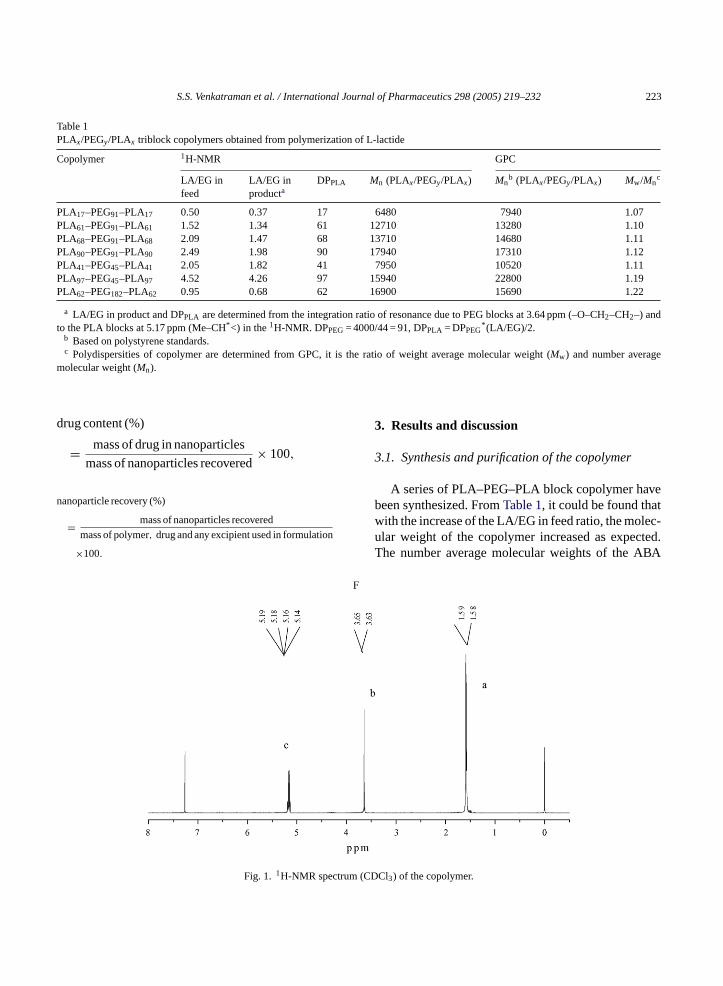
Fig. 1. 1H-NMR spect DCl3) of the copolymer.
224 S.S. Venkatraman et al. / International Journal of Pharmaceutics 298 (2005) 219–232
block copolymers ranged from 6400 to 18000 and themolecular weight distribution are narrow, as seen inTable 1.
3.2. Structure characterization
In Fig. 1, a typical1H-NMR spectrum is shown. Thepresence of methine (CH) and methyl (CH3) protons inPLA was observed at around 5.17 ppm (c) and 1.60 ppm(a) respectively. Tetralet and doublet split peaks wereobserved. The methene protons in CH2 group of PEGwere around 3.64 ppm.
The 13C-NMR spectra (not shown) of the synthe-sized copolymer is used to confirm the PLA blocks, bynoting the presence of the CO, CH and CH3 groupsin the PLA block at 170.0, 69.4 and 17.0 ppm, respec-tively. The CH2 group in PEG block could be found at71.0 (b) ppm.
3.3. Thermal analysis
The homopolymers PLLA (I.V. = 1.04) and PEG 2 k,4 k, 8 k were studied for comparison to the triblockcopolymers. Thermal data are summarized inTable 2.The copolymers show two distinct melting peaks forthe PLA and PEG, indicating good microphase sepa-r chedt tureo be
found that in the presence of relatively long PLAchain, short PEG segments (PLA41–PEG45–PLA41,PLA91–PEG45–PLA91) do not crystallize, whichagrees with the results of S.MLi et al. (1996)(Rashkovet al., 1996). Similarly, for copolymers with longerPEG segments (PLAx–PEG91–PLAx), crystallinity isfound for PEG only when the LA segment is short(PLA17 and PLA61 only). As for the PLA blocks, fora given PEG segment length, both theTm and the DCincrease with increasing PLA length. It should be notedthat PLA also has aTg just at about 50◦C, which is closeto theTm of PEG.
3.4. Hydrolytic degradation of triblock copolymer
For the application of those biodegradable poly-mers as drug delivery carriers, it is very importantto know the degradation profiles. The long-term (upto 90 weeks) degradation characteristics have beenstudied (Hu and Liu, 1994; Li et al., 1998), but notthe details of the shorter-term profile (1–3 weeks),which is more important for drug release predictionof common chemotherapeutics. In this paper, four dif-ferent triblock copolymers were chosen to study thedegradation properties within the first 3 weeks, in rela-tion to the drug release profile. For one copolymer(PLA90–PEG91–PLA90), the degradation was studiedfor both film and particle forms.
The MW data for the four polymers in film formsa eM tivep
TT
S LA)
PPP 4P 7P 8P 5P 3P 3PPP
D ce 100
ation. The presence of the PLA sequences attao PEG blocks decreased the melting temperaf both the PEG block and PLA block. It can
able 2hermal properties of triblock copolymers
ample LA/EGratio
Tm (PEG)(◦C)
Tm (P(◦C)
LLA – 177.5LA17–PEG91–PLA17 0.4 37.3 –LA61–PEG91–PLA61 1.3 42.5 141.LA67–PEG91–PLA67 1.5 – 148.LA90–PEG91–PLA90 2.0 – 153.LA41–PEG45–PLA41 1.8 – 146.LA97–PEG45–PLA97 4.3 – 162.LA62–PEG182–PLA62 0.7 48.5 150.EG (2000) 50.4EG (4000) 59.6EG (8000) 59.8
C = degree of crystallinity of PLA block, rating it to the referen
re compared inFig. 2, while Fig. 2 shows how thWD changes with degradation of two representaolymers.
�Hm
(J/g)Tc(PLA)(◦C)
�Hc
(J/g)DC(%)
Tg
(◦C)
54– – – – –34.9 – – 37.3 −2.745.7 95.7 11.1 37.0 −1.447.9 102.2 8.3 42.3 −0.743.4 93.6 1.7 46.3 −0.161.1 108.3 11.5 53.4 −3.123.6 88.6 5.8 19 −1.2
–−0.83.5
% crystalline L–PLA (93.6 J/g) (Leenslag et al., 1984).
S.S. Venkatraman et al. / International Journal of Pharmaceutics 298 (2005) 219–232 225
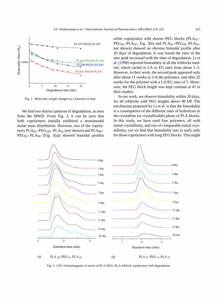
Fig. 2. Molecular weight changes as a function of time.
We find two distinct patterns of degradation, as seenfrom the MWD. FromFig. 3, it can be seen thatboth copolymers initially exhibited a monomodalmolar mass distribution. However, two of the copoly-mers PLA62–PEG182–PLA62 (not shown) and PLA90–PEG91–PLA90 (Fig. 3(a)) showed bimodal profiles
while copolymers with shorter PEG blocks (PLA97–PEG45–PLA97, Fig. 3(b) and PLA41–PEG45–PLA41,not shown) showed no obvious bimodal profile after20 days of degradation. It was found the ratio of thenew peak increased with the time of degradation.Li etal. (1998)reported bimodality in all the triblocks stud-ied, which varied in LA to EG ratio from about 1–5.However, in their work, the second peak appeared onlyafter about 11 weeks in 3 of the polymers, and after 25weeks for the polymer with a LA/EG ratio of 5. More-over, the PEG block length was kept constant at 41 intheir studies.
In our work, we observe bimodality within 20 days,for all triblocks with PEG lengths above 90 DP. Themechanism proposed by Li et al. is that the bimodalityis a consequence of the different rates of hydrolysis inthe crystalline (or crystallisable) phase of PLA blocks.In this study, we have used four polymers, all withinitial crystallinity, and two of comparable initial crys-tallinity; yet we find that bimodality sets in early onlyfor those copolymers with long PEG blocks. This might
PLA–P
Fig. 3. GPC chromatograms of series of EG–PLA triblock copolymers with degradation.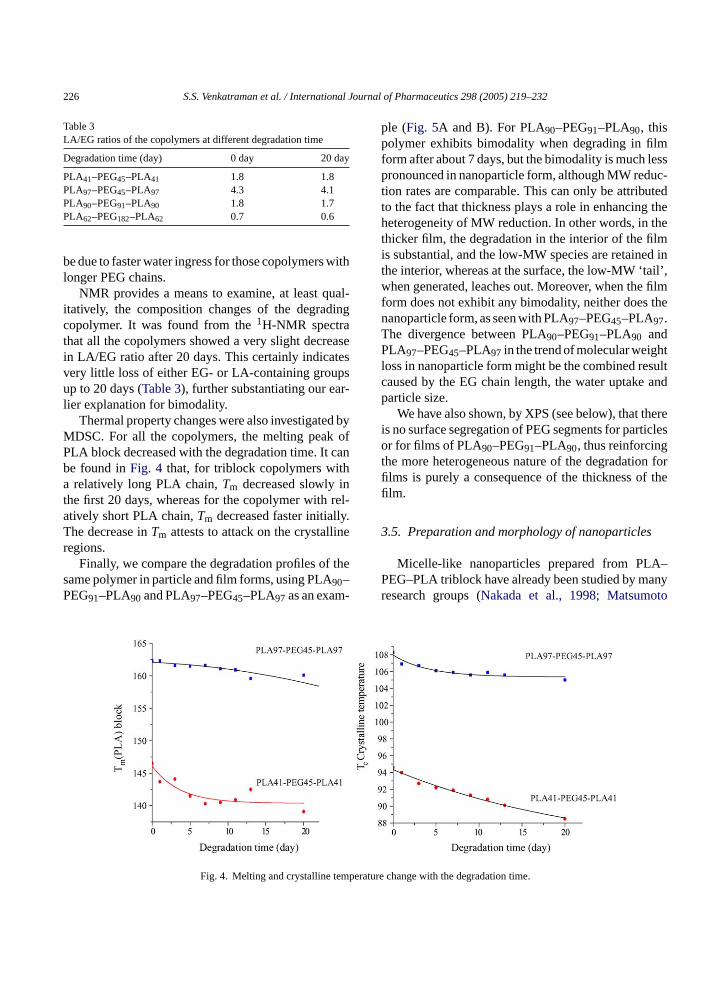
226 S.S. Venkatraman et al. / International Journal of Pharmaceutics 298 (2005) 219–232
Table 3LA/EG ratios of the copolymers at different degradation time
Degradation time (day) 0 day 20 day
PLA41–PEG45–PLA41 1.8 1.8PLA97–PEG45–PLA97 4.3 4.1PLA90–PEG91–PLA90 1.8 1.7PLA62–PEG182–PLA62 0.7 0.6
be due to faster water ingress for those copolymers withlonger PEG chains.
NMR provides a means to examine, at least qual-itatively, the composition changes of the degradingcopolymer. It was found from the1H-NMR spectrathat all the copolymers showed a very slight decreasein LA/EG ratio after 20 days. This certainly indicatesvery little loss of either EG- or LA-containing groupsup to 20 days (Table 3), further substantiating our ear-lier explanation for bimodality.
Thermal property changes were also investigated byMDSC. For all the copolymers, the melting peak ofPLA block decreased with the degradation time. It canbe found inFig. 4 that, for triblock copolymers witha relatively long PLA chain,Tm decreased slowly inthe first 20 days, whereas for the copolymer with rel-atively short PLA chain,Tm decreased faster initially.The decrease inTm attests to attack on the crystallineregions.
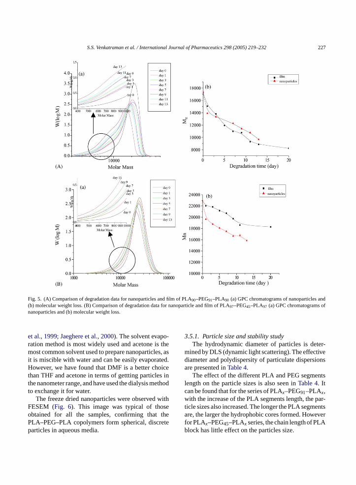
Finally, we compare the degradation profiles of thesame polymer in particle and film forms, using PLA90–PEG91–PLA90 and PLA97–PEG45–PLA97 as an exam-
ple (Fig. 5A and B). For PLA90–PEG91–PLA90, thispolymer exhibits bimodality when degrading in filmform after about 7 days, but the bimodality is much lesspronounced in nanoparticle form, although MW reduc-tion rates are comparable. This can only be attributedto the fact that thickness plays a role in enhancing theheterogeneity of MW reduction. In other words, in thethicker film, the degradation in the interior of the filmis substantial, and the low-MW species are retained inthe interior, whereas at the surface, the low-MW ‘tail’,when generated, leaches out. Moreover, when the filmform does not exhibit any bimodality, neither does thenanoparticle form, as seen with PLA97–PEG45–PLA97.The divergence between PLA90–PEG91–PLA90 andPLA97–PEG45–PLA97 in the trend of molecular weightloss in nanoparticle form might be the combined resultcaused by the EG chain length, the water uptake andparticle size.
We have also shown, by XPS (see below), that thereis no surface segregation of PEG segments for particlesor for films of PLA90–PEG91–PLA90, thus reinforcingthe more heterogeneous nature of the degradation forfilms is purely a consequence of the thickness of thefilm.
3.5. Preparation and morphology of nanoparticles
Micelle-like nanoparticles prepared from PLA–PEG–PLA triblock have already been studied by manyr to
mperat
Fig. 4. Melting and crystalline teesearch groups (Nakada et al., 1998; Matsumo
ure change with the degradation time.
S.S. Venkatraman et al. / International Journal of Pharmaceutics 298 (2005) 219–232 227
Fig. 5. (A) Comparison of degradation data for nanoparticles and film of PLA90–PEG91–PLA90 (a) GPC chromatograms of nanoparticles and(b) molecular weight loss. (B) Comparison of degradation data for nanoparticle and film of PLA97–PEG45–PLA97 (a) GPC chromatograms ofnanoparticles and (b) molecular weight loss.
et al., 1999; Jaeghere et al., 2000). The solvent evapo-ration method is most widely used and acetone is themost common solvent used to prepare nanoparticles, asit is miscible with water and can be easily evaporated.However, we have found that DMF is a better choicethan THF and acetone in terms of gettting particles inthe nanometer range, and have used the dialysis methodto exchange it for water.
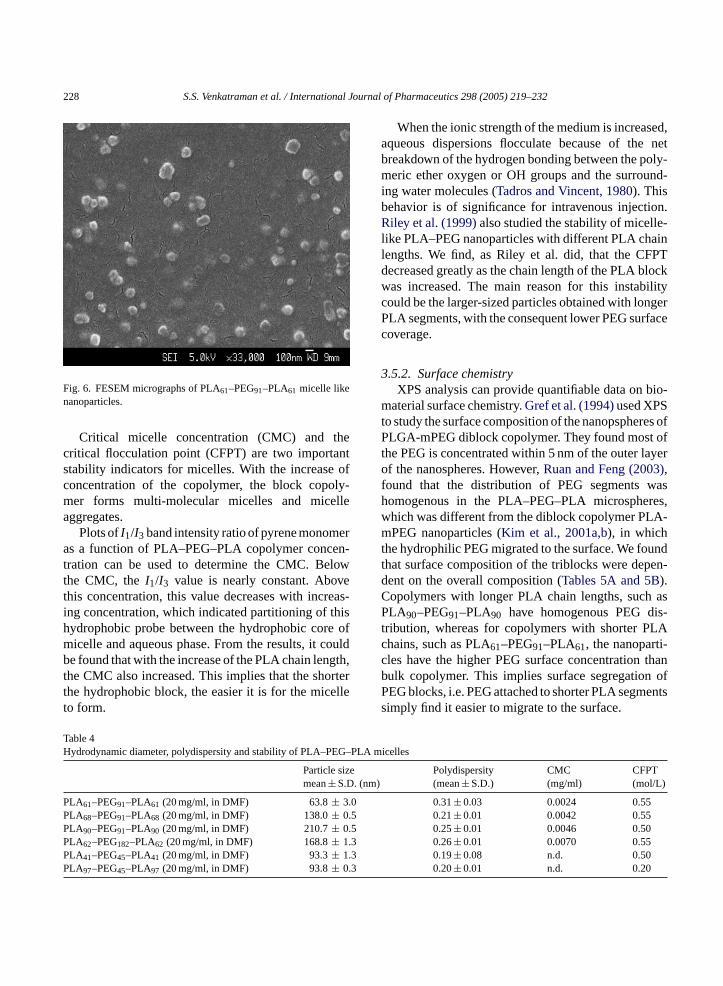
The freeze dried nanoparticles were observed withFESEM (Fig. 6). This image was typical of thoseobtained for all the samples, confirming that thePLA–PEG–PLA copolymers form spherical, discreteparticles in aqueous media.
3.5.1. Particle size and stability studyThe hydrodynamic diameter of particles is deter-
mined by DLS (dynamic light scattering). The effectivediameter and polydispersity of particulate dispersionsare presented inTable 4.
The effect of the different PLA and PEG segmentslength on the particle sizes is also seen inTable 4. Itcan be found that for the series of PLAx–PEG91–PLAx,with the increase of the PLA segments length, the par-ticle sizes also increased. The longer the PLA segmentsare, the larger the hydrophobic cores formed. Howeverfor PLAx–PEG45–PLAx series, the chain length of PLAblock has little effect on the particles size.
228 S.S. Venkatraman et al. / International Journal of Pharmaceutics 298 (2005) 219–232
Fig. 6. FESEM micrographs of PLA61–PEG91–PLA61 micelle likenanoparticles.
Critical micelle concentration (CMC) and thecritical flocculation point (CFPT) are two importantstability indicators for micelles. With the increase ofconcentration of the copolymer, the block copoly-mer forms multi-molecular micelles and micelleaggregates.
Plots ofI1/I3 band intensity ratio of pyrene monomeras a function of PLA–PEG–PLA copolymer concen-tration can be used to determine the CMC. Belowthe CMC, theI1/I3 value is nearly constant. Abovethis concentration, this value decreases with increas-ing concentration, which indicated partitioning of thishydrophobic probe between the hydrophobic core ofmicelle and aqueous phase. From the results, it couldbe found that with the increase of the PLA chain length,the CMC also increased. This implies that the shorterthe hydrophobic block, the easier it is for the micelleto form.
When the ionic strength of the medium is increased,aqueous dispersions flocculate because of the netbreakdown of the hydrogen bonding between the poly-meric ether oxygen or OH groups and the surround-ing water molecules (Tadros and Vincent, 1980). Thisbehavior is of significance for intravenous injection.Riley et al. (1999)also studied the stability of micelle-like PLA–PEG nanoparticles with different PLA chainlengths. We find, as Riley et al. did, that the CFPTdecreased greatly as the chain length of the PLA blockwas increased. The main reason for this instabilitycould be the larger-sized particles obtained with longerPLA segments, with the consequent lower PEG surfacecoverage.
3.5.2. Surface chemistryXPS analysis can provide quantifiable data on bio-
material surface chemistry.Gref et al. (1994)used XPSto study the surface composition of the nanopspheres ofPLGA-mPEG diblock copolymer. They found most ofthe PEG is concentrated within 5 nm of the outer layerof the nanospheres. However,Ruan and Feng (2003),found that the distribution of PEG segments washomogenous in the PLA–PEG–PLA microspheres,which was different from the diblock copolymer PLA-mPEG nanoparticles (Kim et al., 2001a,b), in whichthe hydrophilic PEG migrated to the surface. We foundthat surface composition of the triblocks were depen-dent on the overall composition (Tables 5A and 5B).C asP is-t LAc i-c thanb ofP entss
Table 4Hydrodynamic diameter, polydispersity and stability of PLA–PEG–PL
Particle sizemean± S.D. (nm)
PLA61–PEG91–PLA61 (20 mg/ml, in DMF) 63.8± 3.0PLA68–PEG91–PLA68 (20 mg/ml, in DMF) 138.0± 0.5PLA90–PEG91–PLA90 (20 mg/ml, in DMF) 210.7± 0.5PLA62–PEG182–PLA62 (20 mg/ml, in DMF) 168.8± 1.3PLA41–PEG45–PLA41 (20 mg/ml, in DMF) 93.3± 1.3PLA97–PEG45–PLA97 (20 mg/ml, in DMF) 93.8± 0.3
opolymers with longer PLA chain lengths, suchLA90–PEG91–PLA90 have homogenous PEG d
ribution, whereas for copolymers with shorter Phains, such as PLA61–PEG91–PLA61, the nanopartles have the higher PEG surface concentrationulk copolymer. This implies surface segregationEG blocks, i.e. PEG attached to shorter PLA segmimply find it easier to migrate to the surface.
A micelles
Polydispersity(mean± S.D.)
CMC(mg/ml)
CFPT(mol/L)
0.31± 0.03 0.0024 0.550.21± 0.01 0.0042 0.550.25± 0.01 0.0046 0.500.26± 0.01 0.0070 0.550.19± 0.08 n.d. 0.500.20± 0.01 n.d. 0.20
S.S. Venkatraman et al. / International Journal of Pharmaceutics 298 (2005) 219–232 229
Table 5AXPS data for particle and film of PLA90–PEG91–PLA90
Peak Pure copolymer Nanoparticles
Binding energy (eV) Theoretical (%) Experimental (%) Binding energy (eV) Experimental (%)
a 284.995 22.68 22.22 285.009 21.16c 287.327 22.68 22.22 287.119 23.01d 289.241 22.69 22.98 289.258 23.44b 286.150 31.95 32.57 286.050 32.39
Table 5BXPS data (1s regions) for particle and film of PLA61–PEG91–PLA61
Peak Pure copolymer Nanoparticles
Binding energy (eV) Theoretical (%) Experimental (%) Binding energy (eV) Experimental (%)
a 285.000 19.09 19.11 285.000 17.88c 287.370 19.09 19.11 287.470 17.72d 289.243 19.08 19.12 289.656 16.08b 286.254 42.74 42.66 286.344 48.32
Table 6Hydrodynamic diameter, polydispersity of PLA–PEG–PLA nanoparticles w/o drug
Particle size (nm)mean± S.D.(without drug)
Particle size (nm)mean± S.D.(with drug)
Polydispersitymean± S.D.(without drug)
Polydispersitymean± S.D.(with drug)
PLA61–PEG91–PLA61 63.8± 3.0 75.0± 6.1 0.31± 0.03 0.17± 0.04PLA68–PEG91–PLA68 138.0± 0.5 179.1± 20.0 0.22± 0.01 0.22± 0.01PLA90–PEG91–PLA90 210.7± 0.5 219.4± 1.7 0.25± 0.01 0.24± 0.01
These data clearly show that in order to generatestable particles with potential ‘stealth’ capability, weneed to use particles made from long PEG and shortPLA segments, such as PLA61–PEG91–PLA61. We are
currently evaluating the interactions of these particleswith macrophages in vitro.
3.6. In vitro drug release
After drug incorporation, particle sizes changesomewhat, as shown inTables 6and8A. The size distri-bution is narrow. It could also be seen fromTable 7, thatthe efficiency of drug entrapment was greatly affected
Table 7Effect of different PLA block chain length on 5-FU drug entrapment
Different copolymera Different polymer/drug ratiob
PLA61–PEG91–PLA61
PLA68–PEG91–PLA68
PLA90–PEG91–PLA90
Polymer/drug= 10:1
Polymer/drug= 10:2
Polymer/drug= 10:5
Theoretical drug loading (%) 9.1 9.1 9.1 9.1 16.7 33.4Drug entrapment (%) 14.2 15.5 12.6 13.1 19.7 38.8
a The polymer/drug ratio maintained at 10:1.b PLA68–PEG91–PLA68 was used in this research.
230 S.S. Venkatraman et al. / International Journal of Pharmaceutics 298 (2005) 219–232
Table 8AParticle size of drug loaded and blank micelle-like nanoparticle
Copolymer Blank (nm) particlesize± S.D.
100�g (nm) particlesize± S.D.
300�g (nm) particlesize± S.D.
500�g (nm) particlesize± S.D.
PLA90–PEG91–PLA90 133± 1.0a 145± 0.8 145± 0.9 147± 0.7
a The polymer concentration in DMF is 10 mg/ml.
Table 8BDrug entrapment efficiency of nanoparticles prepared with triblock copolymer with different polymer drug feed ratio
Taxol in feed(�g)
Nanoparticlerecovery (%)
Theoretical drugloading (%)
Drugcontent (%)
EntrapmentEfficiency (%)
PLA90–PEG91–PLA90 100.00 41.82 1.00 0.13 13.55300.00 69.71 3.00 0.76 25.87500.00 72.79 5.00 1.54 31.41
by the initial polymer/drug concentration ratio whilethe chain length of PLA block had little effect.
3.6.1. Drug release data3.6.1.1. 5-FU.The solubility of 5-FU in water is about12.5 mg/ml, so it is not a hydrophobic drug. 5-FUwas continuously releasedin vitro over 300 h. The rateof release was high, in accordance with other work(Avgoustakis et al., 2002; Kim et al., 2001a,b). FromFig. 7, it is clear that the release profiles for nanoparti-cles made from different block copolymer were nearlythe same. Basically, there is not much control over therelease rate for this drug.
Most of the drug is released within 2 to 3 days, bydiffusion and burst; there is perhaps a later degradation-
F oly-m
controlled release for a small fraction of the drug. Webelieve that this profile is due to the hydrophilic natureof the drug which does not permit encapsulation in themicelle core.
3.6.1.2. Paclitaxel.When a very hydrophobic drug,such as paclitaxel, is incorporated into these micelle-like particles with a hydrophobic core, much bettercontrol is obtained over the release. First, at lowertheoretical drug loading (1–5%), the drug entrapmentefficiency remains comparable to 5-FU with high the-oretical drug loading (9–33%), as seen inTable 8B.Second, the drug release rate is much lower, and com-plete release of drug is accomplished only after 12 days(Fig. 8). Interestingly, the release profile is indicative ofdiffusion-controlled release only over this time period.
F -c
ig. 7. Release profile of 5-FU from nanoparticles of triblock coper PLA–PEG–PLA with different PLA blocks.
ig. 8. Release of paclitaxel from PLA90–PEG91–PLA90 nanopartiles.
S.S. Venkatraman et al. / International Journal of Pharmaceutics 298 (2005) 219–232 231
In comparison to reported data on similar systems(Ruan and Feng, 2003; Liggins and Burt, 2002; Burtet al., 1999), we find that the release rate from our tri-block copolymer is faster compared to the nanoparticlerelease reported by Ruan. Reasons for the discrepancymay be the lower particle size in our work (210 nm)compared to Ruan’s (20 microns); the size differencemay also explain the relatively higher encapsulationefficiency reported in Ruan’s work. Somewhat in agree-ment with Ruan, we find an inverse dependence ofrelease rate on drug loading, i.e., lower loadings appearto have faster rate of release.
4. Conclusions
PLA–PEG–PLA triblock copolymers were synthe-sized by a ring-opening reaction of L-lactide with PEGusing stannous octoate as catalyst. Thermal propertiesof these polymers indicate that the PLA and PEG phaseseparate into separate crystalline domains; however,when compared to pure PLA polymer, these copoly-mers all degrade faster, presumably due to higherhydrophobicity.
The details of the MWD during degradation revealsome interesting trends. The copolymers PLA62–PEG182–PLA62 and PLA90–PEG91–PLA90 showbimodal profiles within 20 days of degradation, whilethe other copolymers do not. We believe that thebimodality is due to heterogeneous degradation occur-r sterr
theA iona hatD ofo HF.F si itht lsoi llef ablet ssP thats LAsl gths tics.
Experiments to delineate this more definitively are inprogress.
Drug incorporation and release studies with onehydrophilic and one hydrophobic drug clearly showedthat micelles formed from these copolymers clearlyincorporate more hydrophobic drug, and control itsrelease much more effectively. In particular, these sys-tems may be used to encapsulate paclitaxel and todeliver it via parenteral administrations.
References
Avgoustakis, K., Beletsi, A., Panagi, Z., Klepetsanis, P., Ithankissios,D.S., 2002. PLGA–mPEG nanoparticles of cisplatin: in vitronanoparticle degradation, in vitro drug release and in vivo drugresidence in blood properties. J. Control. Rel. 79, 123–125.
Burt, H.M., Zhang, X.C., Toleikis, P., Embree, L., Hunter, W.L.,1999. Development of copolymers of poly(d,l-lactide) andmethoxypolyethylene glycol as micellar carriers of paclitaxel.Colloids Surf. B: Biointerf. 16, 161–171.
Chabner, B.A., 1982. Pyrimidine Antagonists in Pharmacologic Prin-ciples of Cancer Treatment. Saunders, Philadelphia, pp. 132–142.
Crommelin, D.J.A., Storm, G., 1998. Targeting of drugs 6 strategiesfor stealth therapeutic systems. In: Bregoriadis, G., McCormack,B. (Eds.), NATO ASI Ser., Ser. A: Life Sci., 300, pp. 121–130.
Fu, Y.J., Shyu, S.S., Su, F., Yu, P.C., 2002. Development of biodegrad-able co-poly(d,l-lactic/glycolic acid) microspheres for the con-trolled release of 5-FU by the spray drying method. Colloids Surf.B: Biointerf. 25, 269–279.
Gref, R., Minamitake, Y., Peracchia, M.T., Trubetskoy, V., Torchilin,V., Langer, R., 1994. Biodegradable long-circulating polymeric.Nanospheres Sci. 263, 1600–1603.
G lledlized
H , L.,ers
rsible
H tionJ.
J r, E.,lockO).
K W.,llar13,
K .,ly-m.
ing within 20 days for these copolymers, due to a faate of water ingress.
Micelle-like nanoparticles were prepared withBA triblock copolymers using solvent-evaporatnd solvent-dialysis method. It was found tMAC and DMF were better solvents in termsbtaining the nanoparticles than acetone and Tor the PLAx–PEG91–PLAx series, particle size
ncreased with increasing PLA segment length. Whe increase of the PLA chain length, the CMC ancreased, indicating greater difficulty of miceormation. Moreover, such micelles are also less sto flocculation, which we attribute to overall leEG surface coverage. XPS measurements showurface segregation of PEG is found only when Pegment length is relatively small (DP of PLA∼ 60 or
ess), clearly indicating that this maximum PLA lenhould not be exceeded for stealth characteris
ref, R., Domb, A., Quellec, P., Blunk, T., 1995. The Controintravoues delivery of drug using PEG-coated sterically stabnanospheres. Adv. Drug. Deliv. Rev. 15, 215–233.
agan, S.A., Coombes, A.G.A., Dunn, S.E., Davies, M.C., IllumDavis, S.S., 1996. Polylactide-poly(ethylene glycol) copolymas drug delivery systems.1. Characterization of water dispemicelle-forming systems. Langmuir 12, 2153–2161.
u, D.S.G., Liu, H.J., 1994. Structural-Analysis and degradabehaviour in polyethylene-glycol poly (l-lactide) copolymers.Appl. Polym. Sci. 41, 473–482.
aeghere, F.De., Alllemann, E., Feijen, J., Kissel, T., DoelkeGurny, R., 2000. Freeze-drying and lyopreservation of diband triblock poly(lactid acid)-poly(ethylene oxide) (PLA-PEcopolymer annoparticles. Pharm. Dev. Technol. 5, 473–483
im, S.C., Kim, D.W., Shim, Y.H., Bang, J.S., Oh, H.S., Kim, S.Seo, M.H., 2001a. In vitro evaluation of polymeric micepaclitaxel formulation toxicity and efficacy. J. Control. Rel.191–202.
im, S.Y., Kim, J.H., Kim, D., An, J.H., LEE, D.S., Kim, S.C2001b. Drug-releasing kinetics of MPEG/PLLA block copomer micelles with different PLLA block lengths J. Appl. PolySci. 82, 2599–2605.
232 S.S. Venkatraman et al. / International Journal of Pharmaceutics 298 (2005) 219–232
Leenslag, J.W., Gogolewski, S., Pennings, A.J., 1984. Resorbablematerials of poly(l-lactide). V. Influence of the structure on themechanical properties and hydolisability of PLLA fibres pro-cedures by a dry spinning method. J. Appl. Polym. Sci. 29,2829–2842.
Li, S.M., Rashkov, I., Espartero, J.L., Manolova, N., Vert, M.,1996. Synthesis, characterization, and hydrolytic degradationof PLA/PEO/PLA triblock copolymers with long poly(l-lacticacid) blocks. Macromolecules 29, 57–62.
Li, S.M., Anjard, S., Rashkov, I., Vert, M., 1998. Hydrolytic degra-dation of PLA/PEO/PLA triblock copolymers prepared in thepresence of Zn metal or CaH2. Polymer 39, 5421–5430.
Liggins, R.T., Burt, H.M., 2002. Polyether–polyester diblock copoly-mers for the preparation of paclitaxel loaded polymeric micelleformulations. Adv. Drug Deliv. Rev. 54, 191–202.
Matsumoto, J., Nakada, Y., Sakurai, K., Nakamura, T., Takahashi, Y.,1999. Preparation of nanoparticles consisted of poly(l-lactide)-poly(ethylene glycol)-poly(l-lactide) and their evaluation invitro. Int. J. Pharm. 185, 93–101.
Nakada, Y., Tudomi, R., Sakurai, K., Takahashi, Y., 1998. Evalua-tion of long-circulationg nanoparticles using biodegradable ABAtriblock copolymers containing of poly(l-lactic acid) A-blocksattached to central poly(oxyethylene) B-blocks in vivo. Int. J.Pharm 175, 109–117.
Okano, T., Yui, N., Yokoyama, M., Yoshida, R., 1994. In Advancein Polymeric Systems for Drug Delivery. Gordon and BreachScience, Yverdon, Switzerland, 24–66.
Onetto, N., Dougan, M., Hellmann, S., Gustafson, N., Bur-roughs, J., Florezyk, A., Canetta, R., Rozenweig, M., 1995.Safety profile. In: McGuire, W.P., Rowinsky, E.K. (Eds.),Paclitaxel in Cancer Treatment. Marcel Dekker, pp. 175–186.
Rashkov, I., Manolova, N., Li, S.M., Espartero, J.I., Vert,M., 1996. Synthesis, characteriazation and hydrolyticdegradation of PLA/PEO/PLA Triblock copolymers withshort poly(l-lactic acid) chains. Macromolecules 29, 50–56.
Riley, T., Govende, T., Stolinik, S., Xiong, C.D., Garnett, M.C., Illum,L., Davis, S.S., 1999. Colloidal stability and drug incorporationaspects of micellar-like PLA–PEG nanoparticles. Colloids Surf.B: Biointer. 16, 147–159.
Ruan, G., Feng, S.S., 2003. Preparation and characterizationof poly(lactic acid)–poly(ethylene glycol)–poly(lactic acid)(PLA–PEG–PLA) microspheres for controlled release of pacli-taxel. Biomaterials 24, 5037–5044.
Storm, G., Belliot, S.H., Daemen, T., Lasic, D.D., 1995. Surfacemodification of nanoparticles of nanoparticles to oppose uptakeby the mononuclear phagocyte system. Adv. Drug Del. Rev. 17,3–48.
Tadros, Th.F., Vincent, B., 1980. Influence of temperatureand electrolytes on the adsorption of poly(ethylene oxide)-poly(propylene oxide) block copolymer on polystyrene latex andon the stability of the polymer-coated particles. J. Phys. Chem.84, 1575–1580.


