Embed Size (px)
Citation preview
BioMed CentralBMC Cancer
ss
Open AcceResearch articleMicroarray comparative genomic hybridization detection of chromosomal imbalances in uterine cervix carcinomaAlfredo Hidalgo1,8, Michael Baudis2, Iver Petersen3, Hugo Arreola1, Patricia Piña1, Guelaguetza Vázquez-Ortiz1, Dulce Hernández4, José González5, Minerva Lazos6, Ricardo López1, Carlos Pérez1, José García7, Karla Vázquez1, Brenda Alatorre1 and Mauricio Salcedo*1Address: 1Laboratorio de Oncología Genómica, Unidad de Investigación Médica en Enfermedades Oncológicas, Centro Médico Nacional Siglo XXI-IMSS, México, 2Division of Pediatric Haematology/Oncology, University of Florida, Gainesville, USA, 3Institute of Pathology, University Hospital Charité, Berlin, Germany, 4Servicio de Epidemiología, Hospital de Oncologia, Centro Médico Nacional Siglo XXI-IMSS, México, 5Clínica de Displasias, Hospital de Gineco-Obstetrica No. 4, Luis Castelazo Ayala-IMSS, México, 6Departamento de Patología, Facultad de Medicina UNAM-Hospital General de México, SS, México, 7Laboratorio de Biología Teórica, Departamento de Investigación, Universidad La Salle, México and 8Instituto Nacional de Medicina Genomica, Secretaria de Salud, Mexico
Email: Alfredo Hidalgo - [email protected]; Michael Baudis - [email protected]; Iver Petersen - [email protected]; Hugo Arreola - [email protected]; Patricia Piña - [email protected]; Guelaguetza Vázquez-Ortiz - [email protected]; Dulce Hernández - [email protected]; José González - [email protected]; Minerva Lazos - [email protected]; Ricardo López - [email protected]; Carlos Pérez - [email protected]; José García - [email protected]; Karla Vázquez - [email protected]; Brenda Alatorre - [email protected]; Mauricio Salcedo* - [email protected]
* Corresponding author
AbstractBackground: Chromosomal Comparative Genomic Hybridization (CGH) has been applied to allstages of cervical carcinoma progression, defining a specific pattern of chromosomal imbalances inthis tumor. However, given its limited spatial resolution, chromosomal CGH has offered onlygeneral information regarding the possible genetic targets of DNA copy number changes.
Methods: In order to further define specific DNA copy number changes in cervical cancer, weanalyzed 20 cervical samples (3 pre-malignant lesions, 10 invasive tumors, and 7 cell lines), using theGenoSensor microarray CGH system to define particular genetic targets that suffer copy numberchanges.
Results: The most common DNA gains detected by array CGH in the invasive samples werelocated at the RBP1-RBP2 (3q21-q22) genes, the sub-telomeric clone C84C11/T3 (5ptel), D5S23(5p15.2) and the DAB2 gene (5p13) in 58.8% of the samples. The most common losses were foundat the FHIT gene (3p14.2) in 47% of the samples, followed by deletions at D8S504 (8p23.3), CTDP1-SHGC- 145820 (18qtel), KIT (4q11-q12), D1S427-FAF1 (1p32.3), D9S325 (9qtel), EIF4E (eukaryotictranslation initiation factor 4E, 4q24), RB1 (13q14), and DXS7132 (Xq12) present in 5/17 (29.4%)of the samples.
Conclusion: Our results confirm the presence of a specific pattern of chromosomal imbalancesin cervical carcinoma and define specific targets that are suffering DNA copy number changes inthis neoplasm.
Published: 09 July 2005
BMC Cancer 2005, 5:77 doi:10.1186/1471-2407-5-77
Received: 22 February 2005Accepted: 09 July 2005
This article is available from: http://www.biomedcentral.com/1471-2407/5/77
© 2005 Hidalgo et al; licensee BioMed Central Ltd. This is an Open Access article distributed under the terms of the Creative Commons Attribution License (http://creativecommons.org/licenses/by/2.0), which permits unrestricted use, distribution, and reproduction in any medium, provided the original work is properly cited.
Page 1 of 7(page number not for citation purposes)
BMC Cancer 2005, 5:77 http://www.biomedcentral.com/1471-2407/5/77
BackgroundUterine cervix carcinoma (UCC) represents the secondcause of death among the female population worldwide.The fact that more than 99% of all the cervical invasivetumors are positive for infection with high risk humanpapillomavirus (HPV) suggests that this is one of the mostimportant factors for the development of this neoplasm[1,2]. These viruses can induce cellular transformation byseveral mechanisms; the viral oncoproteins E6 and E7 caninteract with cellular proteins involved in important cellu-lar functions, such as tumor suppression, apoptosis, cellcycle control, genomic instability, transcriptional regula-tion and immune evasion [3].
The induction of genomic instability by HPV seems to beparticularly important for the establishment and develop-ment of an invasive tumor [4,5] since this increasedgenomic plasticity would generate cellular clones withenhanced transforming and invasive potential [6].
Metaphase comparative genomic hybridization (mCGH)has been applied to study different stages of this tumor[4,7-19], detecting specific patterns of chromosomalimbalances that arises very early during the developmentof cervical carcinoma, suggesting that the gain of chromo-some 3q is one of the most important genetic alterationthat defines the transition from a pre-malignant lesion toan invasive carcinoma [4]. Some of these imbalances havebeen related to specific clinical behaviors, such as the pres-ence of lymph node metastases [9]. However, given thespatial resolution of mCGH [20], little is known about theidentity of specific genes that might be the targets ofregional chromosomal imbalances. Matrix-based CGH orarray CGH overcomes this problem increasing the sensi-tivity for the detection of DNA copy number changes atspecific loci, through the use of well defined genomicDNA fragments whose mapping location is known,arrayed onto a solid surface [21-23], thereby achieving aresolution of copy number imbalances up to the singlegene level.
In order to refine the patterns of chromosomal imbal-ances present in cervical carcinoma, and trying to identifyspecific genes that might be targets of copy numberchanges in this tumor, we applied microarray CGH on 20uterine cervix-derived samples (three pre-malignantlesions, 10 invasive tumors and seven UCC derived celllines) to detect DNA copy number changes at the singlegene level.
MethodsCervical tissuesAll described procedures have been evaluated andapproved by the local committee of ethics of the MexicanInstitute of Social Security (IMSS), and all samples were
taken after informed consent from the patients. The pre-malignant lesions and the invasive tumors were collectedby colposcopy-directed biopsies at the GynecologyDepartment of the Hospital General de México, MexicoCity. The biopsies were divided in three sections. The cen-tral part was used for genomic DNA extraction using theWizard Genomic kit (Promega, Madison, WI, USA), andthe extremes were fixed with 70% ethanol overnight andparaffin embedded. Hematoxilin-eosin stained sectionsfrom these biopsies were analyzed in order to confirm thepresence of at least 70% tumoral cells in the samples.
Cell linesThe cell lines included in this study were: CasKi, SiHa,both positive for HPV16, and HeLa (HPV18) The CaLoand ViBo cell lines were established from stage IIB inva-sive tumors, while INBL and RoVa from a stage IVA tumor.These cells are HPV18 positive and were established fromtumor explants at the laboratory of Cell differentiationand Cancer of the National University of Mexico [24]. Thechromosomal CGH profiles of CaLo, ViBo, INBL andRoVa have been published recently [19].
HPV detection and typingHPV detection was carried out by PCR using the consen-sus primers MY09 and MY11 for the L1 region of the viralgenome. After a 5 min. denaturation at 94°C, 100 ng ofDNA were subjected to 35 amplification cycles with thefollowing parameters: 94°C for 1 min., 55°C for 2 min.and 73°C for 3 min., with a final extension step of 7 min.at 72°C. The amplicon was labeled using the Big Dyesequencing kit and sequenced on an ABI371 sequencer(Applied Biosystems, Foster City, CA, USA). BLAST http://www.ncbi.nlm.nih.gov/BLAST/ sequence comparisonwas used in order to define the viral type.
Microarray CGHMicroarray CGH was performed using the GenoSensorArray 300 system, following the manufacturer's instruc-tions (ABBOT-Vysis, Downers Grove, IL, USA). Each arraycontains 861 spots, representing 287 chromosomalregions that are commonly altered in human cancer, suchas telomeres, regions involved in microdeletions, onco-genes, and tumor suppressor genes. Briefly, 100 ng ofgenomic DNA were labeled by a random primer reactionduring two hours. Tumor DNA was labeled with Cy3 andthe normal female reference DNA with Cy5. After thelabeling reaction, the probes were digested with DNAse at15°C for one hr., followed by two ethanol-purifications;finally the probe size was checked by gel electrophoresis.The hybridization mixture consisted of 2.5 µl of each ofthe differentially labeled DNAs plus 25 µl of hybridiza-tion buffer provided in the kit. This mixture was dena-tured at 80°C for 10 min. at 80°C, followed byincubation at 37°C for one hr. Five µl of this probe were
Page 2 of 7(page number not for citation purposes)
BMC Cancer 2005, 5:77 http://www.biomedcentral.com/1471-2407/5/77
applied onto the spotted area of the array under a cover-slip and hybridized in a humid chamber containing 50%formamide (FA)/2XSSC at 37°C for 72 hrs. After hybridi-zation, the arrays were washed 3X in 50%FA/2XSSC at40°C for 10 min/wash, followed by four 5 min. washes in1XSSC at room temperature. Finally, the arrays werebriefly rinsed in distilled water, mounted and counter-stained in the dark for 45 min. with DAPI (4,6-diamino-2-phenylindole).
Image capture and analysisArray analysis was performed immediately after counter-staining using the GenoSensor scanner and software. Thissystem generates a "genomic analysis report", indicatingwhich chromosome regions in the array are involved incopy number changes, as well as a spreadsheet containingthe data generated by a single experiment. In order tocompare all the experiments, a database was created usingthe normalized, bias corrected, tumor/normal ratio valueof each experiment [see additional file 1]. Since each spotin the array is present in triplicates, the median of thethree spots of each probe in the array was calculated andits log2 transformed value was used for further analysis. Afluorescence ratio >1.25 (log2 = 0.32) was considered as aDNA gain, while DNA losses were scored when the ratiowas <0.75 (log2 = -0.41). A ratio >2 (log2 = 1) was consid-ered as a high copy number amplification.
ResultsHPV detection and typingOne of the premalignant lesions was positive for HPV16infection; one for HPV31 and the other for HPV58. In theinvasive tumors, seven were positive for HPV16 and inthree cases, we were not able to detect HPV sequenceswith the oligonucleotides we used for PCR amplification.As expected, CasKi and SiHa were positive for HPV16,while HeLa, INBL, CaLo, ViVo and RoVa were positive forHPV18.
Microarray comparative genomic hybridizationAll our samples, except one pre-malignant lesion, pre-sented alterations, ranking from 1/287 (alterations/totaltargets in the array) in a pre-malignant lesion to 175/287alterations in the cell line RoVa. We found almosttwice the number of DNA gains than DNA losses (571 vs.298) and the average number of copy number alterations(ANCA=total number of alterations in the sample collec-tive/total number of cases) was 43.45 per case.
One of the pre-malignant lesions did not show any alter-ation, while amplifications at MSH2-KCNK12 (2p22.3-2p22.1), TCL1A (14q32.1) and TOP1 (20q12) werefound in a second pre-malignant lesion and DMBT1(10q25.3), ERBB2 (17q12), and 4qTEL11 (4qtel) amplifi-cation was found in the third sample from this group.
In the invasive tumors and the cell lines, the most com-mon amplifications (58.8% of the samples) were found atthe clones RBP1-RBP2 (retinol binding protein 1 and 2,3q21-q22), present as a high copy number amplification(HCNA) in two samples; DAB2 (disabled homolog 2,mitogen-responsive phosphoprotein (Drosophila), 5p13;C84C11/T3 (5ptel) and D5S23 (5p15.2), followed bygains of Tp63 (3q27-q29, 2 HCNA); EGFR (Epidermalgrowth factor receptor, 7p12.3-p12.1, 4 HCNA) andD5S2064 (5p15.2), in 52.9% of the invasive samples andamplification of INS (Insulin, 11ptel) in 47% of thesamples.
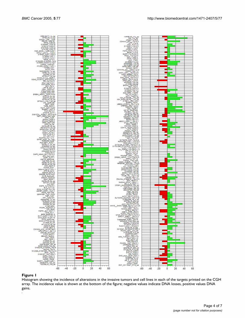
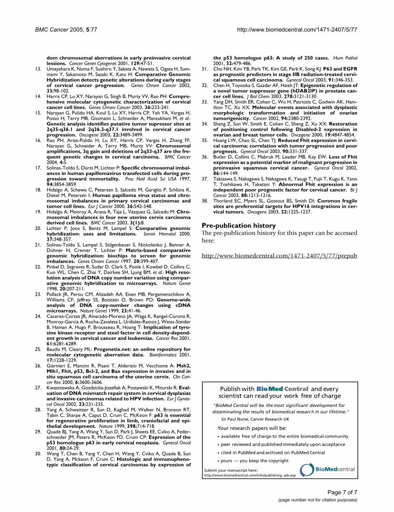
The most common deletion was found at the clone corre-sponding to the FHIT (Fragile histidine triad) gene(3p14.2), present in 47% of the invasive samples, fol-lowed by deletions at D8S504 (8p23.3), CTDP1-SHGC-145820 (18qtel), KIT (4q11-q12), D1S427-FAF1(1p32.3), D9S325 (9qtel), EIF4E (eukaryotic translationinitiation factor 4E, 4q24), RB1 (13q14), and DXS7132(Xq12) present in 29.4% of the samples. A histogram ofthe DNA copy number alterations detected in the tumorsamples analyzed by array CGH is presented in figure 1.The results of these experiments can be accessed throughthe Progenetix CGH database http://www.progenetix.com[25].
DiscussionPrevious studies using chromosomal CGH have delimiteda specific pattern of chromosomal imbalances in cervicalcarcinoma. However, there is little knowledge regardingthe identity of particular genes that might be the targetsfor these copy number changes, making microarray CGHan attractive method in order to define these particulargene targets.
There was concordance between the alterations detectedby microarray CGH and the pattern of chromosomalalterations already described by chromosomal CGH.Although the array that we used did not cover the entiregenome, we were able to detect alterations at particulargenes and genetic markers that might be related to thetransformation process in the cervical epithelium.
It is important to notice that the limited number of pre-malignant lesions analyzed did not allowed us to detectany particular region that might be related with this stageof the disease. However, an interesting candidate geneamplified in one pre-malignant sample and in 5 invasivetumors was MSH2-KCNK12 (2p22.3-2p22.1). This gene isthe human homolog of the E. coli mismatch repair genemutS, and has been found mutated in hereditary nonpoly-posis colon cancer. Higher MSH2 expression has beendescribed in cervical intraepithelial neoplasias and inva-sive cervical carcinomas than in non-neoplastic cervical
Page 3 of 7(page number not for citation purposes)
BMC Cancer 2005, 5:77 http://www.biomedcentral.com/1471-2407/5/77
Histogram showing the incidence of alterations in the invasive tumors and cell lines in each of the targets printed on the CGH arrayFigure 1Histogram showing the incidence of alterations in the invasive tumors and cell lines in each of the targets printed on the CGH array. The incidence value is shown at the bottom of the figure; negative values indicate DNA losses, positive values DNA gains.
Page 4 of 7(page number not for citation purposes)
BMC Cancer 2005, 5:77 http://www.biomedcentral.com/1471-2407/5/77
lesions. An altered expression of this gene has also beenproposed as an important event during cervical carcino-genesis [26,27]. Interestingly, the invasive samples show-ing MSH2 amplification presented with a high number ofalterations (>40), suggesting a possible connectionbetween increased copy number of this gene and chromo-somal instability in invasive cervical carcinomas.
One of the most important genetic events during cervicalcarcinoma progression is the gain of 3q. This alterationhas been detected in early stages of cervical transforma-tion and in cooperation with other imbalances, seems toplay an important role in tumor development. Microarrayanalysis identified among the most prevalent alterationsin cervical tumors and cell lines the amplification of theRBP1- RBP2 (Retinol binding protein 1 and 2, 58.8%) andTp63 (52.9% of the samples) genes, located at 3q21-q22and 3q27-q29, respectively.
Tp63 is a homolog of the p53 tumor suppressor gene. Itsprotein is inactivated by the E6 HPV oncoprotein andplays a primordial role in the development of squamo-stratified epithelia. Tp63 is highly expressed in the basalstratum of these epithelia with diminished expression inthe differentiated strata, suggesting that the presence ofthis protein preserves the self-renewal capacity of the epi-thelial stem cells after an asymmetric division, in whichone of the daughter cell must conserve its epithelial stem-cell properties and the other daughter cell is committed tothe differentiation process [28]. This protein has beendetected in human cervical tissues in the basal and para-basal layers of the ectocervical squamous epithelium, andit is not present in the differentiated layers. In premalig-nant lesions and invasive squamous tumors, a strong p63expression has been described [29,30]. The presence ofthis protein has also been associated with poor survivaland locoregional failure after radiation and chemotherapy[31]. Expression of the epidermal growth factor receptor(EGFR, 7p12.3-p12.1), which was found amplified in52.9% of the invasive tumors that we analyzed, was foundto be a prognostic predictor of extrapelvic failure aftertreatment, and the expression of both molecules wasfound to be a very good risk factor measurement inpatients with stage IIB squamous cell carcinoma of theuterine cervix, who had received radiotherapy and concur-rent chemotherapy [31].
DAB2 on 5p13 was amplified in 58.8% of the invasivecases. The DAB2 gene has been identified as a potenttumor suppressor gene in prostate and ovarian carcinoma[32], and loss of expression of this gene has been associ-ated with the transition of ovarian epithelial cells to pre-malignant states [33]. DAB2 has been implicated in cellpositioning control and seems to mediate the require-ment for basement membrane attachment of epithelial
cells [34]. To our knowledge, there are no availablereports analyzing the expression of this gene in the uterinecervix or in cervical carcinoma. The amplification of thisgene seems contrary to its putative role as a tumor sup-pressor gene. A possible explanation for this observationmight be the loss of one allele followed by the amplifica-tion of the remaining chromosome. However, since arrayCGH does not offer any type of information regarding theparental origin of the amplified chromosome, this situa-tion can not be confirmed.
Detection of the TERC gene amplification has beenrecently proposed as a potential marker for the evaluationof cervical carcinoma progression [35]; however, wedetected amplification of the clone representing this genein less than 10% of the samples analyzed by CGH arrays.FISH analysis, as described by Heselmayer et al., detecteda higher prevalence of nuclei with a diploid pattern thanthose with a tetraploid pattern, even in the high gradelesions. Furthermore, the percentage of nuclei with morethan 2 copies of 3q, including the tetraploid cells rangedbetween 3.3 to 50% of the CIN3 (cervical intraepithelialneoplasia grade 3). Dellas et al., [9] used in situ hybridi-zation to analyze the prevalence of 3q amplifications incervical cancer tissue arrays, detecting low level amplifica-tions in most of the tumors studied. These results suggestthat these low copy number gains might not be ade-quately detected by chromosome or even array CGH, dueto the contamination with normal cells and/or the pres-ence of a high number of diploid or tetraploid cells in thesample.
Regarding DNA losses, the FHIT (fragile histidine triad,3p14.2) gene suffered losses in 47% of the cases. Aberrantexpression of this gene has been well documented in cer-vical carcinoma and has been related to lymph nodemetastasis, parametrial invasion, and vaginal involve-ment in invasive tumors [36]. An association betweenFHIT gene abnormalities and infection with particularHPV types has been suggested, since 87% of the cases withabsent FHIT expression were positive for HPV16 infection[37]. Furthermore, abnormal expression of this gene hasbeen found in significantly younger patients than thosewith normal expression, suggesting that abnormalities inthe regulation of this gene might be accelerating carcino-genesis in cooperation with HPV [37]. These observationsmight be related to the preferential integration of HPVinto fragile sites, particularly FRA3B, where FHIT islocated [38].
ConclusionIn conclusion, microarray CGH allowed the detection ofparticular genes located in regions with common DNAcopy number changes in cervical carcinoma. Further stud-ies using CGH arrays with a higher resolution and the
Page 5 of 7(page number not for citation purposes)
BMC Cancer 2005, 5:77 http://www.biomedcentral.com/1471-2407/5/77
possibility to combine LOH with copy number changes,might be useful for the detection of gene specific targetsthat are relevant for the genesis and progression of cervicalcarcinoma.
AbbreviationsCGH: Comparative Genomic Hybridization, UCC: Uter-ine cervix carcinoma, HPV: Human papilloma virus,DAPI: 4,6-diamino-2-phenylindole.
Competing interestsThe author(s) declare that they do not have any compet-ing interests.
Authors' contributionsAH: Performed the microarray CGH experiments, dataanalysis and paper writing; MB: Help with data submis-sion to the Progenetix database, data analysis; IP: pro-vided training for the experiments, CGH data analysis; PP:tissue processing; GV: HPV typing; DH: sample collection;JG: provided access to the samples; ML: access to samples,histopathological analysis; RL: sample collection; CP:DNA extraction; JG: Help with data analysis; KV: DNAextraction; BA: HPV typing; MS: project coordinator.
Additional material
AcknowledgementsThis work was partially funded through the 7114 and 34686 grants from the Mexican Council of Science and technology (CONACyT) and the Mexican Institute for Social Security (IMSS-FOFOI FP- 2001–2003). AH, GVO, CP, RL were recipients of scholarships from the CONACyT, IMSS and DGEP-UNAM. We would like to thank Abbott-Vysis for providing the CGH array system for this analysis. This work was submitted in partial fulfillment of the requirements for the Ph.D. degree of HA at the Ph.D. in Biomedical Sci-ences, National University of Mexico.
References1. Walboomers J, Jacobs M, Manos M, Bosch X, Kummer A, Shah K,
Snijders P, Peto J, Meijer C, Muñoz N: Human Papilloma Virus isa necessary cause of invasive cervical cancer worldwide. JPathol 1999, 189:12-19.
2. Bosch X, Muñoz N: The viral etiology of cervical cancer. VirusRes 2002, 89:183-190.
3. Münger K, Howley P: Human papillomavirus immortalizationand transforming functions. Virus Res 2002, 89:213-228.
4. Ried T, Hesselmeyer K, Blegen H, Schröck E, Auer G: Genomicchanges defining the genesis, progression and malignancy insolid human tumors: a phenotype/genotype correlation.Genes Chrom Cancer 1999, 25:195-204.
5. Pihan G, Wallace J, Zhou Y, Doxsey S: Centrosome abnormalitiesand chromosome instability occur together in pre-invasivecarcinomas. Cancer Res 2003, 63:1398-1404.
6. Cahill DP, Kinzler KW, Vogelstein B, Lengauer C: Genetic instabil-ity and darwinian selection in tumours. Trends Cell Biol 1999,9:M57-M60.
7. Heselmeyer K, Schröck E, Du Manoir S, Blegen H, Shah K, SteinbeckR, Auer G, Ried T: Gain of chromosome 3q defines the transi-tion from severe dysplasia to invasive carcinoma of the uter-ine cervix. Proc Natl Acad Sci USA 1996, 93:479-484.
8. Heselmeyer K, Macville M, Schröck E, Blegen H, Hellström A, Shah K,Auer G, Ried T: Advanced stage cervical carcinomas aredefined by a recurrent pattern of chromosomal aberrationsrevealing high genetic instability and a consistent gain ofchromosome arm 3q. Genes Chrom Cancer 1997, 19:233-240.
9. Dellas A, Torhorst J, Jiang F, Proffit J, Schultheiss E, Holzgreve W, Sau-ter G, Mihatsch M, Moch H: Prognostic value of genomic alter-ations in invasive cervical squamous cell carcinoma stage IBdetected by comparative genomic hybridization. Cancer Res1999, 59:3475-3479.
10. Kirchhoff M, Rose H, Petersen B, Maahr J, Gerdes T, Lundsteen C,Bryndorf T, Kryger-Baggesen N, Christensen L, Engelholm S, Philip J:Comparative genomic hybridization reveals a recurrent pat-tern of chromosomal aberrations in severe dysplasia/carci-noma in situ of the cervix and in advanced-stage cervicalcarcinoma. Genes Chrom Cancer 1999, 24:144-150.
11. Allen D, White D, Hutchins A, Scurry J, Tabrizi S, Garland S, ArmesJ: Progressive genetic aberrations detected by comparativegenomic hybridization in squamous cell carvical cancer. Br JCancer 2000, 83:1659-1663.
12. Kirchhoff M, Rose H, Petersen B, Maahr J, Gerdes T, Philip J, Lunds-teen C: Comparative genomic hybridization reveals non-ran-
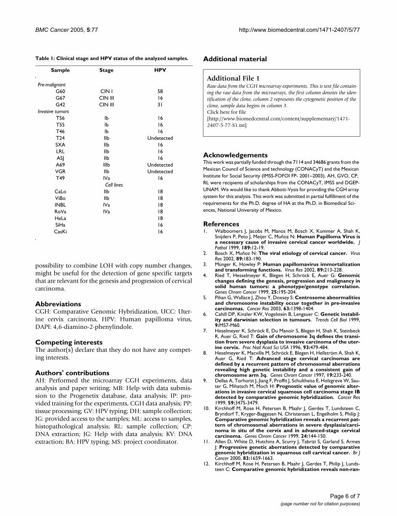
Table 1: Clinical stage and HPV status of the analyzed samples.
Sample Stage HPV
Pre-malignantG60 CIN I 58G67 CIN III 16G42 CIN III 31
Invasive tumorsT56 Ib 16T55 Ib 16T46 Ib 16T24 IIb UndetectedSXA IIb 16LRL IIb 16ASJ IIb 16A69 IIIb UndetectedVGR IIb UndetectedT49 IVa 16
Cell linesCaLo IIb 18ViBo IIb 18INBL IVa 18RoVa IVa 18HeLa 18SiHa 16CasKi 16
Additional File 1Raw data from the CGH microarray experiments. This is text file contain-ing the raw data from the microarrays, the first column denotes the iden-tification of the clone, column 2 represents the cytogenetic position of the clone, sample data begins in column 3.Click here for file[http://www.biomedcentral.com/content/supplementary/1471-2407-5-77-S1.txt]
Page 6 of 7(page number not for citation purposes)
BMC Cancer 2005, 5:77 http://www.biomedcentral.com/1471-2407/5/77
Publish with BioMed Central and every scientist can read your work free of charge
"BioMed Central will be the most significant development for disseminating the results of biomedical research in our lifetime."
Sir Paul Nurse, Cancer Research UK
Your research papers will be:
available free of charge to the entire biomedical community
peer reviewed and published immediately upon acceptance
cited in PubMed and archived on PubMed Central
yours — you keep the copyright
Submit your manuscript here:http://www.biomedcentral.com/info/publishing_adv.asp
BioMedcentral
dom chromosomal aberrations in early preinvasive cervicallessions. Cancer Genet Cytogenet 2001, 129:47-51.
13. Umayahara K, Numa F, Suehiro Y, Sakata A, Nawata S, Ogata H, Sum-inami Y, Sakamoto M, Sasaki K, Kato H: Comparative GenomicHybridization detects genetic alterations during early stagesof cervical cancer progression. Genes Chrom Cancer 2002,33:98-102.
14. Harris CP, Lu XY, Narayan G, Singh B, Murty VV, Rao PH: Compre-hensive molecular cytogenetic characterization of cervicalcancer cell lines. Genes Chrom Cancer 2003, 36:233-241.
15. Narayan G, Pulido HA, Koul S, Lu XY, Harris CP, Yeh YA, Vargas H,Posso H, Terry MB, Gissmann L, Schneider A, Mansukhani M, et al.:Genetic analysis identifies putative tumor supressor sites at2q35-q36.1 and 2q36.3-q37.1 involved in cervical cancerprogression. Oncogene 2003, 22:3489-3499.
16. Rao PH, Arias-Pulido H, Lu XY, Harris CP, Vargas H, Zhang FF,Narayan G, Schneider A, Terry MB, Murty VV: Chromosomalamplifications, 3q gain and deletions of 2q33-q37 are the fre-quent genetic changes in cervical carcinoma. BMC Cancer2004, 4:5.
17. Solinas-Toldo S, Dürst M, Lichter P: Specific chromosomal imbal-ances in human papillomavirus transfected cells during pro-gression toward immortality. Proc Natl Acad Sci USA 1997,94:3854-3859.
18. Hidalgo A, Schewe C, Petersen S, Salcedo M, Gariglio P, Schlüns K,Dietel M, Petersen I: Human papilloma virus status and chro-mosomal imbalances in primary cervical carcinomas andtumor cell lines. Eur J Cancer 2000, 36:542-548.
19. Hidalgo A, Monroy A, Arana R, Taja L, Vázquez G, Salcedo M: Chro-mosomal imbalances in four new uterine cervix carcinomaderived cell lines. BMC Cancer 2003, 3(1):8.
20. Lichter P, Joos S, Bentz M, Lampel S: Comparative genomichybridization: uses and limitations. Semin Hematol 2000,37:348-357.
21. Solinas-Toldo S, Lampel S, Stilgenbauer S, Nickolenko J, Benner A,Dohner H, Cremer T, Lichter P: Matrix-based comparativegenomic hybridization: biochips to screen for genomicimbalances. Genes Chrom Cancer 1997, 20:399-407.
22. Pinkel D, Segraves R, Sudar D, Clark S, Poole I, Kowbel D, Collins C,Kuo WL, Chen C, Zhai Y, Dairkee SH, Ljung BM, et al.: High reso-lution analysis of DNA copy number variation using compar-ative genomic hybridization to microarrays. Nature Genet1998, 20:207-211.
23. Pollack JR, Perou CM, Alizadeh AA, Eisen MB, Pergamenschikov A,Williams CF, Jeffrey SS, Botstein D, Brown PO: Genome-wideanalysis of DNA copy-number changes using cDNAmicroarrays. Nature Genet 1999, 23:41-46.
24. Caceres-Cortes JR, Alvarado-Moreno JA, Waga K, Rangel-Corona R,Monroy-Garcia A, Rocha-Zavaleta L, Urdiales-Ramos J, Weiss-SteiderB, Haman A, Hugo P, Brousseau R, Hoang T: Implication of tyro-sine kinase receptor and steel factor in cell density-depend-ent growth in cervical cancer and leukemias. Cancer Res 2001,61:6281-6289.
25. Baudis M, Cleary ML: Progenetix.net: an online repository formolecular cytogenetic aberration data. Bioinformatics 2001,17:1228-1229.
26. Giarnieri E, Mancini R, Pisani T, Alderisio M, Vecchione A: Msh2,Mlh1, Fhit, p53, Bcl-2, and Bax expression in invasive and insitu squamous cell carcinoma of the uterine cervix. Clin Can-cer Res 2000, 6:3600-3606.
27. Kwasniewska A, Gozdzicka-Jozefiak A, Postawski K, Miturski R: Eval-uation of DNA mismatch repair system in cervical dysplasiasand invasive carcinomas related to HPV infection. Eur J Gynae-col Oncol 2002, 23:231-235.
28. Yang A, Schweitzer R, Sun D, Kaghad M, Walker N, Bronson RT,Tabin C, Sharpe A, Caput D, Crum C, McKeon F: p63 is essentialfor regenerative proliferation in limb, craniofacial and epi-thelial development. Nature 1999, 398:714-718.
29. Quade BJ, Yang A, Wang Y, Sun D, Park J, Sheets EE, Cviko A, Feder-schneider JM, Peters R, McKeon FD, Crum CP: Expression of thep53 homologue p63 in early cervical neoplasia. Gynecol Oncol2001, 80:24-29.
30. Wang T, Chen B, Yang Y, Chen H, Wang Y, Cviko A, Quade B, SunD, Yang A, Mckeon F, Crum C: Histologic and immunopheno-typic classification of cervical carcinomas by expression of
the p53 homologue p63: A study of 250 cases. Hum Pathol2001, 32:479-486.
31. Cho NH, Kim YB, Park TK, Kim GE, Park K, Song KJ: P63 and EGFRas prognostic predictors in stage IIB radiation-treated cervi-cal squamous cell carcinoma. Gynecol Oncol 2003, 91:346-353.
32. Chen H, Toyooka S, Gazdar AF, Hsieh JT: Epigenetic regulation ofa novel tumor suppressor gene (hDAB2IP) in prostate can-cer cell lines. J Biol Chem 2003, 278:3121-3130.
33. Yang DH, Smith ER, Cohen C, Wu H, Patriotis C, Godwin AK, Ham-ilton TC, Xu XX: Molecular events associated with dysplasticmorphologic transformation and initiation of ovariantumorigenicity. Cancer 2002, 94:2380-2392.
34. Sheng Z, Sun W, Smith E, Cohen C, Sheng Z, Xu XX: Restorationof positioning control following Disabled-2 expression inovarian and breast tumor cells. Oncogene 2000, 19:4847-4854.
35. Huang LW, Chao SL, Chen TJ: Reduced Fhit expression in cervi-cal carcinoma: correlation with tumor progression and poorprognosis. Gynecol Oncol 2003, 90:331-337.
36. Butler D, Collins C, Mabruk M, Leader MB, Kay EW: Loss of Fhitexpression as a potential marker of malignant progression inpreinvasive squamous cervical cancer. Gynecol Oncol 2002,86:144-149.
37. Takizawa S, Nakagawa S, Nakagawa K, Yasugi T, Fujii T, Kugu K, YanoT, Yoshikawa H, Taketani Y: Abnormal Fhit expression is anindependent poor prognostic factor for cervical cancer. Br JCancer 2003, 88:1213-1216.
38. Thorland EC, Myers SL, Gostout BS, Smith DI: Common fragilesites are preferential targets for HPV16 integrations in cer-vical tumors. Oncogene 2003, 22:1225-1237.
Pre-publication historyThe pre-publication history for this paper can be accessedhere:
http://www.biomedcentral.com/1471-2407/5/77/prepub
Page 7 of 7(page number not for citation purposes)


