Embed Size (px)
Citation preview

Non-surgical management of uterine fibroids
Giovanna Tropeano1, Sonia Amoroso and Giovanni Scambia
Department of Obstetrics and Gynecology, Universita Cattolica del Sacro Cuore, Largo Francesco Vito 1, 00168 Rome, Italy
1Correspondence address. E-mail: [email protected]
BACKGROUND: Efforts to develop alternatives to surgery for management of symptomatic uterine fibroids haveprovided new techniques and new medications. This review summarizes the existing literature on uterine artery embo-lization (UAE) and investigational studies on four newer approaches. METHODS: PubMed, Cochrane and Embasewere searched up to December 2007. Studies reporting side-effects and complications and presenting numericaldata on at least one outcome measure were included. RESULTS: Case studies report 50–60% reduction in fibroidsize and 85–95% relief of symptoms following UAE. The largest of these studies reported an in-hospital complicationrate of 2.7% (90 of 3041 patients) and a post-discharge complication rate of 26% (710 of 2729 patients). Eight studiescompared UAE with conventional surgery. Best evidence suggested that UAE offered shorter hospital stays (1–2 daysUAE versus 5–5.8 days surgery, 3 randomized controlled trials (RCTs)) and recovery times (9.5–28 days UAE versus36.2–63 days surgery, 3 RCTs) and similar major complication rates (2–15% UAE versus 2.7–20% surgery, 3 RCTs).Four studies analysing cost-effectiveness found UAE more cost-effective than surgery. There is insufficient evidenceregarding fertility and pregnancy outcome after UAE. Five feasibility studies after transvaginal temporary uterineartery occlusion in 75 women showed a 40–50% reduction in fibroid volume and two early studies using magneticresonance guided–focused ultrasound showed symptom relief at 6 months in 71% of 109 women. Two small RCTsassessing mifepristone and asoprisnil showed promising results. CONCLUSIONS: Good quality evidence supportsthe safety and effectiveness of UAE for women with symptomatic fibroids. The current available data are insufficientto routinely offer UAE to women who wish to preserve or enhance their fertility. Newer treatments are stillinvestigational.
Keywords: embolization; fibroid; focused ultrasound; medical therapy
Introduction
Uterine fibroids are the most common female pelvic tumour,
typically reported to occur in 20–40% of reproductive aged
women, and up to 70% of white and 80% of black women by
the age of 50 years (Baird et al., 2003). Most fibroids are asympto-
matic, but nearly half of women with fibroids have significant and
often disabling symptoms including heavy menstrual bleeding,
pain and pressure symptoms (Baird et al., 2003). Traditionally,
symptomatic fibroids have been treated with myomectomy or hys-
terectomy performed by laparotomy (Matchar et al., 2001). In an
effort to reduce the cost, morbidity and lifestyle impact of major
surgery, various less invasive surgical procedures, including mini-
laparotomy and operative endoscopy, have been introduced over
the years, but these are only applicable in a minority of cases
(ACOG Committee on Practice Bulletins—Gynecology, 2001).
Over the last few years a variety of new treatment approaches
have become available to women with symptomatic fibroids.
Undoubtedly the most significant therapeutic innovation has
been the advent of uterine artery embolization (UAE) as a form
of non-surgical management. New technology has provided
additional minimally invasive options such as percutaneous laser
ablation, cryoablation, transvaginal uterine artery occlusion and
magnetic resonance imaging (MRI)-guided focused ultrasound
that are currently under intense investigation. Furthermore, new
medications have been introduced, that show promise for practi-
cal, long-term medical therapy for symptomatic fibroids.
The objective of this review is to summarize the published lit-
erature on three of the latest fibroid therapies—UAE, transvaginal
uterine artery occlusion and MRI-guided focused ultrasound—
with special focus on the evidence concerning the benefits and
risks of these minimally invasive options. We will also provide
a summary of the available clinical data on two novel medical
therapeutics for fibroid treatment—mifepristone and asoprisnil.
Literature search criteria
A systematic literature search was conducted using three standard
electronic databases—PubMed, Cochrane Collaboration resources
and Embase. All computerized searches were performed using the
# The Author 2008. Published by Oxford University Press on behalf of the European Society of Human Reproduction and Embryology. All rights reserved.
For Permissions, please email: [email protected] 259
Human Reproduction Update, Vol.14, No.3 pp. 259–274, 2008 doi:10.1093/humupd/dmn006
Advance Access publication March 14, 2008
by guest on Decem
ber 29, 2015http://hum
upd.oxfordjournals.org/D
ownloaded from

Medical Subject Heading terms ‘UAE’, ‘transvaginal uterine
artery occlusion’, ‘MRI-guided focused ultrasound’, ‘focused
ultrasound surgery’, ‘medical therapy’, ‘mifepristone’, ‘asoprisnil’
and ‘fibroid* or leiomyoma* or leiomyomata*’ and publication
type ‘clinical trial’.
The search was performed in December 2007 for all available
papers, and the articles selected had to be written in English. All
bibliographies were cross-referenced to identify additional perti-
nent studies. Letters and editorials were excluded.
Studies were included if they were clinical in nature, measured
fibroid-specific symptoms and uterine and/or fibroid sizes before
and after treatment, reported side-effects and complications
associated with treatment and presented numerical data on at
least one outcome measure.
We did not perform meta-analytic techniques to summarize
treatment outcomes because of individual study variation in
outcome measures.
Uterine artery embolization
UAE is a percutaneous, image-guided procedure which is per-
formed by a properly trained and experienced interventional radi-
ologist (Spies and Sacks, 2004). In essence, it involves the
placement of an angiographic catheter into the uterine arteries
via a common femoral artery approach and injection of embolic
agents (in most cases, polyvinyl alcohol particles or tris–acryl
gelatin microspheres) into both uterine arteries until the flow
becomes sluggish (Fig. 1) (Ravina et al., 1995; Hutchins et al.,
1999; Spies et al., 1999; Pelage et al., 2000; Walker and Pelage
2002; Pron et al., 2003a; Spies et al., 2004a).
The proposed mechanism of UAE’s action is that occluding or
markedly reducing uterine blood flow at the arteriolar level will
produce an irreversible ischaemic injury to the fibroids, causing
them to undergo necrosis and shrink, while the normal myome-
trium is able to recover (Aitken et al., 2006; Banu et al., 2007).
The procedure, which is usually performed under intravenous
conscious sedation, generally requires �1 h to complete. With
operator experience and proper technique, patient radiation
exposure during UAE is comparable to that received during
routine diagnostic imaging procedures (Andrews and Brown,
2000; Nikolic et al., 2000; Zupi et al., 2003; White et al., 2007).
Immediately after the end of the procedure, most patients
experience moderate-to-severe ischaemic pain for 8–12 h—
usually requiring parenteral analgesia (narcotics and non-steroidal
anti-inflammatory drugs)—but pain severity gradually decreases
during the next 12 h (Worthington-Kirsch and Koller, 2002a,b),
and a day-case or overnight stay is required at most.
Recovery is typically brief and relatively mild, with 4–5 days of
recurrent uterine cramping and constitutional embolization symp-
toms (generalized malaise, fatigue, nausea and low-grade fever).
Figure 1: The UAE procedure.
Drawings (A) and (B) illustrate the path of the catheter to deliver embolization particles to occlude the uterine arteries. Angiograms show the fibroid blood supply
before (C) and after (D) UAE.
Tropeano et al.
260
by guest on Decem
ber 29, 2015http://hum
upd.oxfordjournals.org/D
ownloaded from

Patients can usually return to normal activities within 8–14 days
(Spies et al., 2001a; Walker and Pelage, 2002; Pron et al.,
2003b; Bruno et al., 2004; Worthington-Kirsch et al., 2005).
Clinical outcomes
Since it was first described in 1995 (Ravina et al., 1995), UAE for
fibroids has been shown in several large case series to be effective
in substantially reducing the fibroid size (on average by 50–60%)
and relieving bleeding and other fibroid-related symptoms, with
reported overall success rates ranging from 85 to 95% at short-
and mid-term follow-up (Worthington-Kirsch et al., 1998;
Goodwin et al., 1999; Hutchins et al., 1999; Pelage et al., 2000;
McLucas et al., 2001a; Spies et al., 2001a; Katsumori et al.,
2002; Walker and Pelage, 2002; Watson and Walker, 2002; Pron
et al., 2003a; Spies et al., 2005a; Lohle et al., 2006). All these
studies also reported high rates (87–97%) of patient satisfaction
with outcome, measured at various points in time and along
varied scales.
The studies in which UAE outcomes were evaluated with the
use of a validated instrument, the uterine fibroid symptom and
quality of life (QOL) questionnaire (UFS–QOL) (Spies et al.,
2002a), reported significant improvements in fibroid-specific
symptoms and health-related QOL measures up to 3 years after
the procedure (Smith et al., 2004; Spies et al., 2005a; Scheurig
et al., 2006; Goodwin et al., 2008).
Longer-term outcome data have been recently published with
three prospective trials with follow-up periods exceeding 5 years
(Spies et al., 2005b; Katsumori et al., 2006; Walker and Barton-
Smith, 2006). These studies reported rates of symptom control
after UAE ranging from 84 to 97% at 1 year and from 73 to
89.5% at 5 years (Spies et al., 2005b; Katsumori et al., 2006;
Walker and Barton-Smith, 2006). These data suggest that,
although there is some decline in the improvement percentage
over time, levels of symptom control after UAE remain high in
the long-term.
Complications
Reported complication rates of UAE are low. In a single-centre
prospective study of 400 consecutive patients, the overall morbid-
ity rate, using the American College of Obstetricians and Gynae-
cologists (ACOG) quality indicators for perioperative morbidity,
was 5% (Spies et al., 2002b). Most complications were minor
and occurred during the first 3 months after the procedure. There
were five (1.25%) major complications, one of which (0.25%)
required hysterectomy. The FIBROID registry, the largest pro-
spective study published to date, reported an in-hospital compli-
cation rate of 2.7% (90 of 3041 patients), with a 0.6% rate of
major adverse events (e.g. re-admission, major therapy, unplanned
increase in care, or permanent adverse sequelae) and a post-
discharge complication rate of 26% (710 of 2729 patients), with
a 4.1% rate of major adverse events (Worthington-Kirsch et al.,
2005).
A relatively common complication of UAE is vaginal expulsion
of an infarcted fibroid, with a reported rate of up to 10% (Spies
et al., 2002b; Walker and Pelage, 2002). This complication is
more frequently seen in patients with submucosal fibroids or intra-
mural fibroids with a submucosal component. Expulsion most
often occurs within 6 months after the procedure, but there are
reports of this event after a period of time as long as 4 years
(Marret et al., 2004). In most cases, the infarcted fibroid is
expelled spontaneously (Fig. 2), and no additional treatment
is necessary. Hysteroscopic resection or dilation and curettage is
reserved for cases in which the fibroid is only partially infarcted
and remains firmly attached to the uterine wall due to the increased
risk of secondary infection (Spies et al., 2002b; Marret et al., 2004;
Rajan et al., 2004).
When uncomplicated, fibroid expulsion can restore the uterine
anatomy to nearer normal more rapidly than otherwise (Felemban
et al., 2001; Gulati et al., 2004; Kroencke et al., 2003; Hehenkamp
et al., 2004; Park et al., 2005). In a minority of cases, however,
retention of necrotic fibroid tissue may result in chronic vaginal
discharge due to shedding of fibroid material into the endometrial
cavity (Walker et al., 2004; Ogliari et al., 2005). This condition
can be treated effectively by hysteroscopic resection of the necro-
tic fibroid material (Walker et al., 2004).
The most serious, although rare, complication of UAE is the
occurrence of intrauterine infection, which has been reported in
less than 1% of procedures (Hutchins et al., 1999; Spies et al.,
2001a,b; Walker and Pelage, 2002; Pron et al., 2003b;
Worthington-Kirsch et al., 2005). If left untreated or refractory
to antibiotics, uterine infection can lead to sepsis and the need
for emergency hysterectomy (Aungst et al., 2004; Nikolic et al.,
2004). Two deaths from uterine infection and overwhelming
sepsis have also been reported after UAE (Vashist et al., 1999;
de Block et al., 2003).
Specific risk factors for this potentially life-threatening compli-
cation have yet to be determined. Clinical experience and evidence
from a case report (Vashist et al., 1999) suggest that infection may
originate from the vagina and/or the urinary tract, which under-
lines the importance of pre-procedure screening for genitourinary
infection. There is also evidence that certain pre-existing con-
ditions, such as a coexistent adnexal pathology (Nikolic et al.,
2004), and some minor post-procedure complications, in particular
fibroid expulsion (Spies et al., 2002b; Marret et al., 2004), are
associated with a higher risk of infection. Results of a retrospec-
tive analysis of 414 UAE procedures showed that, although the
occurrence of infection after UAE tended to be associated with
the presence of submucosal fibroids, the association was not stat-
istically significant in multivariate analysis (Rajan et al., 2004).
In addition to two deaths from septic shock (Vashist et al., 1999;
de Block et al., 2003), other three deaths following UAE have thus
far been reported, one from pulmonary embolism (Lanocita et al.,
1999) and two from uncertain causes (Worthington-Kirsch, 2002),
in more than 100 000 procedures performed worldwide. If we
assume that all these deaths were related to the procedure, the mor-
tality risk would be 0.05:1000, which compares favourably with
the estimated mortality rate of �0.38:1000 following hyster-
ectomy for non-obstetric benign disease (Maresh et al., 2002).
Treatment failure
Individual study variations in the definition of UAE failure
(symptom persistence, or recurrence, or need for additional
therapy) and in the period of reporting for failures make results
difficult to compare between different study cohorts. Large case
series with less than 2 years of follow-up reported rates of treat-
ment failure, defined as the need for subsequent interventions,
ranging from 5.5 to 9.5% (Walker and Pelage, 2002; Spies
Non-surgical options for fibroid management
261
by guest on Decem
ber 29, 2015http://hum
upd.oxfordjournals.org/D
ownloaded from

et al., 2005a; Huang et al., 2006). Recently published data on 1278
patients completing 36-month follow-up in the FIBROID registry
showed that during the course of the study, hysterectomy, myo-
mectomy or repeat UAE were performed in 9.8, 2.8 and 1.8% of
the patients, respectively (Goodwin et al., 2008). Longer-term pro-
spective studies reported re-interventions rates at 5 years ranging
from 12.7 to 21%, with rates of hysterectomy varying from 3 to
17.8% (Spies et al., 2005b; Walker and Barton-Smith, 2006; Kat-
sumori et al., 2006). In comparison, reported 5-year crude rates of
re-interventions after abdominal myomectomy ranged from 8.6 to
55.1%, with crude rates of hysterectomy varying from 4.3 to
26.7% (Fauconnier et al., 2000).
There are several possible reasons for UAE failure. First, since
the procedure causes fibroid shrinkage but preserves normal
uterine tissue, it is possible that new fibroids will develop and
symptoms recur. The risk of fibroid recurrence after embolization
has not yet been defined. A prospective study using transvaginal
ultrasound reported appearance of new fibroids in 8.2% (7 of
85) of patients at a median of 30 months after the procedure
(Marret et al., 2003).
On the other hand, results from MRI follow-up examinations up
to 3 years after UAE indicated that many clinical recurrences were
not caused by development of new fibroids but related to
re-growth of incompletely infarcted fibroids (Pelage et al.,
2004). Incomplete fibroid infarction is most often related to tech-
nical aspects of the procedure such as the presence of collateral
blood supply to the fibroids (usually from the ovarian arteries)
or difficulties in cannulating both uterine arteries as a result of ana-
tomical variation, arterial spasm, or current use of a gonadotropin
releasing-hormone agonist (GnRH-agonist) (Spies, 2003).
Successful embolization of only one uterine artery is generally
regarded as a technical UAE failure as, in most cases, both uterine
arteries contribute to the fibroid blood supply (Spies, 2003). Data
from clinical trials support a relationship between unilateral UAE
and the risk of treatment failure (Smith et al., 2004; Spies et al.,
2005a; Gabriel-Cox et al., 2007). One study of 81 UAE patients,
of whom seven received unilateral embolization, reported a
trend toward needing additional treatment if bilateral UAE was
not performed (Smith et al., 2004). In multivariate analysis of
1701 UAE procedures, the FIBROID registry reported a negative
effect on symptom scores at 6 and 12 months for patients who had
unilateral embolization (Spies et al., 2005a). More recently, a
study of 562 UAE patients, of whom 33 (5.9%) had unilateral
embolization, reported that women receiving unilateral UAE
were 2.2 times more likely to undergo hysterectomy by 5 years
than those having bilateral embolization (Gabriel-Cox et al.,
2007). Unilateral UAE remained a significant predictor of sub-
sequent hysterectomy even after controlling for age in multivariate
analysis (Gabriel-Cox et al., 2007).
Studies assessing the effect of baseline uterine characteristics on
the likelihood of treatment failure reported conflicting results:
three studies found no relationships between uterine or fibroid
size and the risk of failure (McLucas et al., 2001a; Katsumori
et al., 2003; Huang et al., 2006), whereas other studies reported
Figure 2: Spontaneous fibroid expulsion in a 50-year-old woman.
(A) Magnetic resonance (MR) image obtained 23 months after UAE shows an infarcted fibroid (*) distorting the uterine cavity. The patient provided a history of pain
and vaginal discharge shortly before this MR study. (B) MR image obtained 25 months after UAE, the fibroid is no longer seen. Uncomplicated fibroid passage had
occurred 34 days after the previous MR study. (C) The expelled fibroid.
Tropeano et al.
262
by guest on Decem
ber 29, 2015http://hum
upd.oxfordjournals.org/D
ownloaded from

that larger fibroid size and higher fibroid number were significant
predictors of failure (Marret et al., 2005; Spies et al., 2005a;
Isonishi et al., 2007; Goodwin et al., 2008).
Studies also found that a history of prior fibroid surgery signifi-
cantly increased the risk of failure (McLucas et al., 2001a; Huang
et al., 2006).
Coexisting pelvic diseases may also increase the chance of
treatment failure. Although no studies have specifically assessed
the relationship between undiagnosed concomitant adenomyosis
and the incidence of clinical failure of UAE, adenomyosis was
detected on histopathologic examination in up to 36% of uteri
removed after embolization because of symptom persistence or
recurrence (McLucas et al., 2002; Marret et al., 2005; Huang
et al., 2006; Gabriel-Cox et al., 2007).
Other comorbidities that can predispose patients to a poor clini-
cal outcome include endometriosis and ovarian cysts, that may be
a cause of persistent pain after UAE, and chronic salpingitis and
endometritis that may increase the chance of life-threatening
infection (Hovsepian et al., 2004).
There is no doubt, therefore, that appropriate patient selection is
crucial for obtaining a successful outcome from this therapy. On
the basis of reports of expert committees, supplemented by evi-
dence from clinical trials, UAE should only be considered for
women with significant symptoms (bleeding, pain and/or pressure
symptoms) specifically attributable to uterine fibroids who might
otherwise be advised to have surgical treatment (Hovsepian
et al., 2004; ACOG Committee Opinion, 2004; SOGC Clinical
Practice Guidelines, 2005; National Institute for Health and Clini-
cal Excellence, 2007). The ideal candidates are women who no
longer desire fertility but wish to avoid surgery and/or retain
their uterus or are poor surgical risks. Embolization is also an
excellent option for patients who will not accept blood transfu-
sions and for those who are severely anaemic and require immedi-
ate intervention. Current advice is that UAE should not be offered
to asymptomatic women or to women whose only fibroid-related
complaint is infertility (Hovsepian et al., 2004; ACOG Committee
Opinion, 2004; SOGC Clinical Practice Guidelines, 2005;
National Institute for Health and Clinical Excellence, 2007).
Careful pre-procedure evaluation is essential to exclude preg-
nancy and genital tract malignancy. Other absolute contraindica-
tions include comorbidities that may increase the risk for
infectious complications (e.g. pelvic inflammatory disease, salpin-
gitis or endometritis, or active genitourinary infection), the pre-
sence of an adnexal mass and conditions that contraindicate any
endovascular procedure (e.g. reduced immune status, severe coa-
gulopathy, severe contrast medium allergy, or impaired renal func-
tion). The desire to avoid a hysterectomy under any circumstances
is also an absolute contraindication to UAE as there is a small
(,1%) risk of hysterectomy because of procedure-related compli-
cations (Hovsepian et al., 2004; SOGC Clinical Practice Guide-
lines, 2005).
Menopausal status is no longer considered an absolute contrain-
dication to UAE as there have been reports of successful outcomes
from this therapy in carefully selected post-menopausal women
with fibroid-related pressure symptoms (Chrisman et al., 2007).
Relative contraindications to UAE include recent use of
GnRH-agonist as this drug may impact on the technical success
of the procedure, and coexisting adenomyosis or endometriosis
as these conditions may increase the risk of treatment
failure (Hovsepian et al., 2004; SOGC Clinical Practice Guide-
lines, 2005; National Institute for Health and Clinical Excellence,
2007).
Patient evaluation for UAE should include hysteroscopy and/or
endometrial sampling, if indicated, and imaging studies using
transvaginal and transabdominal ultrasound and/or MRI (Bazot
et al., 2001; Spielmann et al., 2006; Vitiello and McCarthy,
2006) to assess the uterine size and number, size and location of
the fibroids, screen for adenomyosis and rule out any other signifi-
cant pelvic pathology (Walker and Pelage, 2002; Pelage et al.,
2005).
There are no restrictions to the size and number of fibroids that
can be treated with UAE. Anatomic exclusion criteria only include
submucosal fibroids that may be effectively treated with hystero-
scopic resection, and pedunculated subserosal fibroids with a
narrow stalk (attachment point ,50% of the diameter) because
of the potential risk and complications from infarction of the
stalk and subsequent fibroid detachment from the uterus
(Andrews et al., 2004; Katsumori et al., 2005).
A very large fibroid uterus is not a contraindication to UAE
since favourable clinical outcomes have been observed even in
women with greater than 24-week pregnancy size uteri (Prollius
et al., 2004). However, uterine volume reduction following UAE
generally does not exceed 50%, and women with a very large
uterus who consider this option should be counselled that their
bulk-related symptoms may not be as well treated as their men-
strual complaints.
Comparative studies
There are eight studies published that have compared the out-
comes of UAE versus conventional surgical procedures for symp-
tomatic fibroids (Table I).
So far, three studies have been performed in which clinical out-
comes of UAE were compared with those of abdominal myomect-
omy, two retrospective cohort studies (Broder et al., 2002; Razavi
et al., 2003) and one prospective, but non-randomized, trial
(Goodwin et al., 2006). Overall, these studies consistently reported
that the two procedures were equally safe and effective in relieving
fibroid-related symptoms. However, likely related to heterogen-
eity in the baseline patient characteristics and in the methods
and timing of follow-up, there was some discrepancy in specific
outcome results (Broder et al., 2002; Razavi et al., 2003;
Goodwin et al., 2006).
Broder and colleagues compared outcomes of 51 UAE patients
and 30 myomectomy patients at a follow-up period ranging from 3
to 5 years (Broder et al., 2002). The investigators found no signifi-
cant differences between the groups in the rates of overall
symptom improvement and patient satisfaction with outcome,
but the rate of subsequent interventions was higher for the UAE
than for the myomectomy group (P ¼ 0.004). However, UAE
patients were older (P , 0.001) and more likely to have under-
gone prior surgery for fibroids (78 versus 3%, P , 0.001),
suggesting they may have had more extensive disease than myo-
mectomy patients.
A similar retrospective study by Razavi and colleagues reported
on 67 UAE patients and 44 myomectomy patients with mean
follow-up periods of 14.6 and 14.3 months, respectively (Razavi
et al., 2003). Women in the UAE group were older (P , 0.05)
Non-surgical options for fibroid management
263
by guest on Decem
ber 29, 2015http://hum
upd.oxfordjournals.org/D
ownloaded from

and more likely to present with menorrhagia (P , 0.05) than
women in the myomectomy group. In this study, the rate of
improvement in menorrhagia was higher for the UAE than for the
myomectomy group (92 versus 64%, P , 0.05), whereas the rate
of improvement in pressure symptoms was higher for
the myomectomy than for the UAE group (76 versus 91%,
P , 0.05). The investigators also reported shorter hospital stays
(0 versus 2.9 days, P , 0.05), faster recovery times (8 versus 36
days, P , 0.05) and a lower overall morbidity rate (11 versus
25%) for UAE than for myomectomy. Finally, they found no differ-
ences between the groups in the rate of subsequent interventions.
The multi-centre prospective study by Goodwin et al. (2006)
compared the outcomes of 149 UAE patients and 60 myomectomy
patients. Women in the UAE group were older (P , 0.001), had
more numerous fibroids (P , 0.0001), and were more likely to
present with menorrhagia (P ¼ 0.02) than women in the myomect-
omy group. At 6 months, the rate of overall clinical success,
defined as significant improvement in both QOL and menstrual
bleeding scores, was similar for the UAE (81.2%) and the myo-
mectomy group (75%). Differences between the groups were
identified and included the mean hospital stay (,1 day for UAE
versus 2.5 days for myomectomy, P , 0.001), the mean recovery
time (14.6 days for UAE versus 44.4 days for myomectomy, P ,
0.05) and the overall complication rate (22.1% for UAE versus
40% for myomectomy, P ¼ 0.01). Re-intervention rates did not
differ between the groups.
To date, there have been four studies one multi-center prospec-
tive (Spies et al., 2004a,b), one multi-center retrospective cohort
(Dutton et al., 2007) and two RCTs (Pinto et al., 2003, and the
EMMY trial published in four papers Hehenkamp et al., 2005,
2006; Volkers et al., 2006, 2007) published that compared UAE
with hysterectomy. In a third RCT, outcomes of UAE were com-
pared with outcomes of a mixed group of hysterectomies and myo-
mectomies (Edwards et al., 2007) (Table I).
The prospective study by Spies and colleagues compared UAE
versus a mixed group of hysterectomies (abdominal, laparoscopic,
vaginal and laparoscopically assisted vaginal) (Spies et al.,
2004b). Women undergoing UAE were more likely to have had
previous fibroid surgery (P , 0.04) and had more numerous
fibroids (P , 0.021) and larger mean uterine volume (P ,
0.001) than hysterectomy patients. The investigators reported sig-
nificant differences between the groups in the mean hospital stay
(0.8 days for UAE versus 2.3 days for hysterectomy, P , 0.001)
and recovery time (10.7 days for UAE and 32.5 days for hyster-
ectomy, P , 0.001). At 12-month follow-up, UAE patients
reported a marked improvement compared with baseline in
blood loss scores (P , 0.001) and menorrhagia questionnaire
scores (P , 0.001). The rate of improvement in pelvic pain at
12 months was higher for the hysterectomy than for the UAE
group (98 versus 84%, P ¼ 0.021), but there were no differences
between the groups in the degree of improvement in pressure
symptoms, overall health assessment, and QOL scores or the
Table I. Published comparative studies of the outcomes from UAE versus surgical procedures for symptomatic fibroids.
Author (year) Study design Procedures
no. of pts at
procedure
Length of
follow-up
(No. of pts)
Clinical success rate
(%)
Satisfaction
rate (%)
Major AE rate
(%)
Subsequent
intervention
rate (%)
Broder et al.
(2002)
retrosp UAE 59 46 mo (51) 92 94 NR 29
AM 38 49 mo (30) 90 79 NR 3
P ¼ 0.004
Razavi et al.
(2003)
retrosp UAE 67 14 mo (62) 92 NR NR 8
AM 44 15 mo (40) 64 NR NR 10
P , 0.05
Goodwin et al.
(2006)
prosp UAE 149 6 mo (121) 81.2 NR 4 2
AM 60 6 mo (45) 75 NR 1.7 1.7
Spies et al.
(2004b)
prosp UAE 102 12 mo (76) 61% decrease in
menorrhagia scores1
90 3.9 2
H 50 12 mo (30) NR 97 12 8
Dutton et al.
(2007)
retrosp UAE 972 4.6 y (649) 85.2 91 3.9 18.3
H 762 8.6 y (459) 99.2 85 11.3 NR
P , 0.0001 P ¼ 0.007 P , 0.0001
Pinto et al. (2003) RCT UAE 38 6 mo (36) 86 78 2 5.5
AH 19 6 mo (17) NR 88 20 0
Hehenkamp et al.
(2005)
RCT (EMMY) UAE 88 1.5 mo (81) NR NR 4.9 1.1
H 89 1.5 mo (75) NR NR 2.7 1.1
Volkers et al.
(2007)
UAE 88 24 mo (81) 76.5 3 NR NR 23.5†
H 89 24 mo (73) NR NR NR 8
Edwards et al.
(2007)
RCT (REST) UAE 106 12 mo (95) 18-point increase in
SF-36 scores2
88 15 9††
Surg 51 12 mo (45) 24-point increase in
SF-36 scores2
93 20 0
NR, not reported; retrosp, retrospective; prosp, prospective; RCT, randomized controlled trial. AE, adverse events; Pts, patients; AM, abdominal myomectomy;AH, abdominal hysterectomy; H, abdominal, vaginal, laparoscopic or laparoscopically assisted vaginal hysterectomy; Surg ¼ abdominal myomectomy orhysterectomy. 1Ruta Menorrhagia Severity Scale (Ruta et al., 1995). 2SF-36, Medical Outcomes Study 36-Item Short-Form Health Survey (Jenkinson et al.,1996). 3Clinical success defined as no need to perform a hysterectomy in the first 2 years after UAE. †Included four (4.9%) interventions due to bilaterallyfailed UAE procedures. ††Included two (1.7%) interventions due to bilaterally failed UAE procedures.
Tropeano et al.
264
by guest on Decem
ber 29, 2015http://hum
upd.oxfordjournals.org/D
ownloaded from

rate of patient satisfaction with outcomes. The overall morbidity
was higher in the hysterectomy than in the UAE group (34
versus 14.7%, P ¼ 0.01), but the incidence of major complications
was similar for the two groups. Finally, there were no significant
differences between the groups in the rate of subsequent
interventions.
The recently published Hysterectomy or Percutaneous Emboli-
sation For Uterine Leiomyomata (HOPEFUL) study was a retro-
spective cohort comparing long-term safety and efficacy of UAE
and hysterectomy (abdominal, laparoscopic, vaginal or laparosco-
pically assisted vaginal) (Dutton et al., 2007). The study, which
involved 18 hospitals in the UK, included 1108 women treated
with UAE (n ¼ 649) or hysterectomy (n ¼ 459) from the
mid-1990s. The average length of follow-up was 4.6 years for
the UAE and 8.6 years for the hysterectomy cohort. At baseline
UAE women were younger (P , 0.0001) and more likely to
have had prior pelvic surgery (P , 0.0001) than hysterectomy
women. The investigators reported lower rates of overall morbid-
ity and major morbidity for the UAE than the hysterectomy cohort
(P , 0.001 and 0.0001, respectively) and adjusted odds ratios for
UAE versus hysterectomy of 0.48 (95% CI 0.26–0.89) for all
complications and 0.25 (95% CI 0.13–0.48) for major compli-
cations. More hysterectomy women reported symptomatic relief
(P , 0.0001) and feeling better (P , 0.0001), but patient satisfac-
tion was higher with UAE than with hysterectomy (P ¼ 0.007).
One hundred and nineteen women (18.3%) in the UAE cohort
had a subsequent intervention (e.g. hysterectomy, myomectomy
or repeat UAE) during follow-up. After adjusting for differential
time of follow-up, there was a 23% (95% CI 19–27%) chance
of requiring further treatment within the first 7 years.
The earliest of the RCTs comparing UAE and hysterectomy was
small (enrolling only 57 women) and used a controversial
randomized-consent methodology, in which women who were
randomly assigned to hysterectomy were not informed about the
study or about the possible alternative treatment (Pinto et al.,
2003). The primary outcome measure was the mean length of hos-
pital stay, which was shorter for the UAE than for the hyster-
ectomy group (1.7 versus 5.8 days, P , 0.001). Patients in the
UAE group also had shorter recovery times than women in the
hysterectomy group (9.5 versus 36.2 days, P , 0.001). The inves-
tigators found no differences between the groups in the overall
complication rate within the first 30 days of treatment, but hyster-
ectomy women were more likely to experience major compli-
cations than UAE women (20 versus 2%). At 6 months of
follow-up, the clinical success rate for the UAE group, which
was based on the cessation of bleeding, was 86% (31 of 37
patients). By 6 months, the rate of subsequent interventions for
the UAE group was of 5.5% (2 of 36 patients).
The second RCT of UAE versus hysterectomy (abdominal,
vaginal, laparoscopic or laparoscopically assisted vaginal) was
the EMbolization versus hysterectoMY (EMMY) trial, which
involved 28 Dutch hospitals and enrolled 177 patients (Hehenkamp
et al., 2005). The primary endpoint of the study was the elimin-
ation of menorrhagia after a follow-up period of 2 years, with
UAE considered equivalent to hysterectomy if menorrhagia
resolved in at least 75% of patients with no significant differences
in major complications between the two procedures.
The first three publications from the EMMY trial reported on
short-term outcomes from UAE versus hysterectomy (Hehenkamp
et al., 2005, 2006, Volkers et al., 2006). The investigators reported
a shorter mean hospital stay (2 versus 5.1 days, P , 0.001) but
higher rates of minor complications (58 versus 40%, P ¼ 0.024)
and readmissions (11 versus 0%, P ¼ 0.003) during the first 6
weeks after discharge for the UAE group than for the hyster-
ectomy group. However, there were no significant differences
between the groups in the rates of major in-hospital and post-
discharge complications (Hehenkamp et al., 2005). Women in
the UAE group also reported significantly less pain during the
first 24 h postoperatively (P ¼ 0.012) and returned to work
sooner (28 versus 63 days, P , 0.001) than hysterectomy patients
(Hehenkamp et al., 2006). Of note, the rate of procedural UAE
failure, defined as the failure to perform bilateral UAE, reported
from this trial was of 17.3% (Volkers et al., 2006), that was
much higher than the failure rates previously reported in large
UAE series (0.5–7.3%) (Spies et al., 2001a; Pron et al., 2003c;
Worthington-Kirsch et al., 2005). This could be, at least partly,
explained by the fact that the majority (19 of 26) of the interven-
tional radiologists in the EMMY trial were relatively inexperi-
enced with UAE, having performed less than 10 embolization
procedures before the study started.
The fourth EMMY publication reported 2 years’ outcomes from
UAE versus hysterectomy (Volkers et al., 2007). At 2 years after
treatment, 23.5% (19 of 81) of UAE patients had undergone a hys-
terectomy because of persistent or recurrent symptoms. There
were no significant differences between the two groups in the
degree of improvement compared to baseline in pain (84.9% for
UAE versus 78% for hysterectomy) and pressure symptoms
(66.2% for UAE versus 69.2% for hysterectomy). On the basis
of these findings, the investigators concluded that, although the
failure rate of UAE, defined as the necessity to perform a hyster-
ectomy in the first 2 years after the procedure, stayed within the
preset failure rate maximum of 25%, the 76.5% clinical success
rate was much lower than results from earlier uncontrolled UAE
series (Volkers et al., 2007). However, when interpreting the
results from this trial, it must be taken into consideration that
the 23.5% failure rate of UAE reported at 2 years was affected
by 4 (4.9%) cases of bilaterally failed procedure and 10 (12.3%)
cases of unilateral embolization. Since the impact of technically
failed procedures on clinical outcomes was not evaluated, the
degree to which the high incidence of technical UAE failure in
this trial could have influenced the outcome results remains
unknown.
The Randomized Trial of Embolization versus Surgical Treat-
ment for Fibroids (REST), which involved 27 hospitals in the
UK, compared outcomes for 106 women randomly assigned to
UAE versus 51 assigned to abdominal surgery (43 hysterectomies
and 8 myomectomies) (Edwards et al., 2007). The primary
outcome measure was health-related QOL at 1 year, as assessed
on the Medical Outcomes Study 36-item Short-Form General
Health Survey (SF-36) (Jenkinson et al., 1996). The investigators
found no differences between groups in QOL measures at 1 year,
although women in both groups had substantial improvements in
each component of the SF-36 score compared to baseline. The
UAE group had shorter hospital stays (1 day compared to 5
days, P , 0.001) and recovery times (20 days compared with 62
days, P , 0.001) than the hysterectomy group. There were no
differences between the groups in the rate of major complications
(15% for the UAE and 20% for the surgical group). Of note, 3
Non-surgical options for fibroid management
265
by guest on Decem
ber 29, 2015http://hum
upd.oxfordjournals.org/D
ownloaded from

(2.8%) of the major complications in the UAE group were cancers,
which were highly unlikely to be related to treatment. At 1 year of
follow-up, 10 (9%) women in the UAE group had required
additional interventions (hysterectomy or repeated UAE) to treat
persistent or recurrent symptoms. Of these re-interventions, two
(1.9%) were due to bilaterally failed UAE procedures.
Overall, results from published RCTs yielded consistent evi-
dence that UAE offers an advantage over conventional surgery
for fibroids in terms of shorter hospital stay and quicker return
to daily activities. Clinical outcomes after embolization also
appear to be similar to that of surgery, with most women reporting
symptom control and satisfaction with outcome up to 2 years after
treatment. Overall morbidity and major complication rates associ-
ated with UAE also appear to be similar to those with surgery.
To date, there have been eight published studies analysing the
cost and cost-effectiveness of UAE in comparison with standard
surgical procedures (Table II) (Al-Fozan et al., 2002;
Baker et al., 2002; Pourrat et al., 2003; Beinfeld et al., 2004;
Edwards et al., 2007; Dembek et al., 2007; Goldberg et al.,
2007; Wu et al., 2007). In these studies, hospital costs associated
with UAE were lower, largely because of shorter lengths of stay,
which compensated well for the higher physicians’ fees and
imaging costs than those of surgery (Al-Fozan et al., 2002;
Baker et al., 2002; Dembek et al., 2007; Goldberg et al., 2007;
Wu et al., 2007). Comparative cost-effectiveness analyses also
showed that UAE was more cost-effective than surgery (Pourrat
et al., 2003; Beinfeld et al., 2004; Edwards et al., 2007; Wu
et al., 2007), even when the costs of repeat procedures and associ-
ated complications were factored in (Wu et al., 2007).
Reproductive outcomes
The effect UAE may have on subsequent fertility and pregnancy
remains understudied. This is the reason why this therapy is still
regarded by most as relatively contraindicated in women who
wish to preserve their fertility (Hovsepian et al., 2004; ACOG
Committee Opinion, 2004; SOGC Clinical Practice Guidelines,
2005).
Transient or permanent amenorrhea with other symptoms of
ovarian failure has been reported in up to 5% of women after
UAE (Spies et al., 2001b; Walker and Pelage, 2002; Pron et al.,
2003a). Most of these cases have been in women over the age
45, but there have been anecdotal reports of ovarian failure in
younger women (Chrisman et al., 2000; Walker and Pelage, 2002).
The most likely cause is believed to be non-target ovarian
embolization via the utero-ovarian collaterals, potentially result-
ing in ovarian ischaemia and loss of ovarian follicles (Payne
et al., 2002; Tulandi et al., 2002). Because of these observations,
concern has been raised about the possible risk of premature
ovarian failure in premenopausal women undergoing the pro-
cedure. To address this issue, several studies evaluated the
effects of UAE on ovarian reserve by measuring basal (cycle
day 3) serum FSH levels before and after treatment in women of
different ages (Spies et al., 2001b; Ahmad et al., 2002; Healey
et al., 2004; Hovsepian et al., 2006). All these studies reported
no significant changes in basal FSH in most women after UAE
(Spies et al. 2001b; Ahmad et al., 2002; Healey et al., 2004;
Hovsepian et al., 2006).
A prospective study of 20 regularly cycling women undergoing
embolization specifically addressed the issue of the impact of UAE
on ovarian reserve in younger women (up to age 40) (Tropeano
et al., 2004). Results of this study indicated no change in
ovarian reserve, as assessed by basal FSH and estradiol levels
and ultrasound-based ovarian volume and antral follicle count,
up to 12 months after the procedure (Tropeano et al., 2004).
Although these findings are reassuring, larger series with longer-
term follow-up periods are clearly needed to make conclusive
statements on the potential subclinical impact of UAE on
ovarian function.
As a result of the abundant collateral arterial circulation, normal
uterine tissue usually recovers from the reduction in uterine blood
flow induced by bilateral UAE. Ultrasound and MRI follow-up
examinations have documented rapid revascularization of the
normal myometrium and an essentially normal appearance of
the endometrium at 3–6 months after embolization (deSouza
and Williams, 2002; Pelage et al., 2004). However, there have
been case reports of uterine necrosis (Godfrey and Zbella, 2001;
Shashoua et al., 2002; Gabriel et al., 2004; Torigian et al.,
2005), uterine wall defect (De Iaco et al., 2002) and endometrial
atrophy following UAE (Tropeano et al., 2003). As a result,
there is still a concern for potential effects of the procedure on
the integrity of the myometrium and endometrium.
Nevertheless, published series of uncomplicated pregnancies
and normal deliveries after UAE continue to increase (Table III)
(Ravina et al., 2000; McLucas et al., 2001b; Carpenter and
Walker, 2005; Pron et al., 2005; Walker and McDowell, 2006;
Holub et al., 2007). These reports demonstrate that women can
conceive and carry a pregnancy successfully to term after UAE.
They also demonstrate higher rates of spontaneous abortion
(Ravina et al., 2000; McLucas et al., 2001b; Carpenter and
Walker, 2005; Pron et al., 2005; Walker and Mc Dowell, 2006;
Holub et al., 2007), preterm delivery (Ravina et al., 2000; Carpenter
and Walker, 2005; Pron et al., 2005; Walker and McDowell, 2006;
Holub et al., 2007), Caesarean section (Ravina et al., 2000;
McLucas et al., 2001b; Carpenter and Walker, 2005; Pron et al.,
2005; Walker and McDowell, 2006; Holub et al., 2007), abnormal
placentation (Pron et al., 2005; Carpenter and Walker, 2005;
Walker and Mc Dowell, 2006) and post-partum haemorrhage
(Carpenter and Walker, 2005; Pron et al., 2005; Walker and
McDowell, 2006; Holub et al., 2007) following UAE compared
to the general population. However, interpretation of the data
from these UAE cohorts is difficult because of confounding
factors such as the advanced maternal age, prior reproductive
issues and the severe fibroid disease present in most patients. It
must be also remembered that while UAE is effective at shrinking
fibroids and improving symptoms, it does not remove fibroids, and
there are known associations between fibroids, pregnancy loss, and
certain pregnancy complications (Bajekal and Li, 2000; Coronado
et al., 2000).
To what extent pre-embolization fibroid characteristics may
influence subsequent reproductive outcome remains unknown.
Results of a retrospective MRI analysis of fibroid location in a
cohort of 51 women achieving pregnancy after UAE showed
that all subtypes of fibroids were present in women achieving a
live birth (n ¼ 35) versus those who did not (n ¼ 16) (Walker
and Bratby, 2007). On the basis of these findings, the authors
suggested that the location of fibroids treated with UAE does
Tropeano et al.
266
by guest on Decem
ber 29, 2015http://hum
upd.oxfordjournals.org/D
ownloaded from

not have a negative impact on subsequent fertility (Walker and
Bratby, 2007). However, as pointed out by the authors, this was
a small retrospective study which was further limited by the pre-
sence of multiple fibroids in most women, which made it difficult
to establish effects of individual fibroids in specific locations.
Whether pregnancy outcomes after UAE are different from
those after myomectomy remains to be determined. One study
comparing reported pregnancy outcomes after UAE versus those
from a published series of pregnancies after laparoscopic myo-
mectomy found that pregnancies after embolization were
Table II. Published comparative studies of costs and cost-effectiveness of uterine artery embolization (UAE) versus surgical procedures for symptomaticfibroids.
Author (year) Procedures
no. of pts
Design, methods, and measures Results
Baker et al.
(2002)
UAE 23 Charges retrospectively collected and
analyzed from one hospital billing system. Total professional cost
UAE
2,220*
AM
1,611
AM 17 Cost-to-charge ratios used to convert
charges to costs.
Total hospital cost 3,193* 5,598
Costs given in 2001 US dollars.
Al-Fozan et al.
(2002)
UAE 85 AM 91 Costs retrospectively obtained from one
hospital administrative database.
UAE AM
Total hospital cost 1,007† 1,782
AH 340 VH 29 Costs given in 2001 Canadian dollars. AH VH
1,933 1,515
Pourrat et al.
(2003)
UAE 39 Hospital, drug, and additional examination
costs and effectiveness (success rate) at 6
months retrospectively measured from the
perspective of one hospital.
UAE VH
VH 33
Total cost at 6 months
Effectiveness
2,134
92%
2,789
100%
Cost-effectiveness ratios calculated as total
costs divided by the success rates.
Cost-effectiveness ratio 2,320 2,789
Costs given in 2000 Euros.
Beinfeld et al.
(2004)
UAE Decision model comparing costs and
effectiveness measured in QALYs. Total cost until menopause
UAE
6,916
H
7,847
H Cohort of patients aged 40 not desiring
future pregnancy followed until menopause.
No. of QALYs 8.29 8.18
Data on efficacy, safety, utility, and costs
obtained from the literature.
Incremental cost/QALY 2,007 H dominated
by UAE8Costs given in 1999 US dollars.
Goldberg et al.
(2007)
UAE 136 Cost and reimbursement data retrospectively
obtained from one hospital decision support
database.
Total hospital cost
UAE
2,707§
AM
5,676
AH
5,707
AM 105
Hospital net incomes calculated as hospital
net reimbursements minus total hospital
costs.
Hospital reimbursement 2,764§ 4,961 5,135
AH 299
Costs given in 2002 US dollars.
Hospital net income 57§ 2715 2572
Edwards et al.
(2007)
UAE 106
Surg 51
Resource use (hospital, patient and
follow-up costs) up to 12 months after
treatment collected prospectively and
analyzed from a National Health Service
perspective.
Total cost per patient/year
UAE
1,727*
Surg
2,673
Costs given in 2004 UK sterling.
Dembek et al.
(2007)
UAE 125
AM 704
Procedure and follow-up costs up to
12-months after treatment retrospectively
collected and analyzed from a nationally
representative payer reimbursement
perspective.
Procedure cost
Total cost per patient/year
UAE
5,968§
10,519
AM
7,299
9,652
H
7,707
10,044
H 2836
Costs given in 2002 US dollars.
Wu et al. (2007) UAE 649 Probabilistic decision model comparing
costs and effectiveness measured in QALYs
over the time horizon, from the initial
procedure to menopause.
UAE H
Total cost until menopause 2,536 3,282
H 459 Data on efficacy, safety, utility, and costs
obtained from a comparative cohort and the
literature.
No. of QALYs 8.203 8.241
Costs given in 2004 UK sterling.
Incremental cost/QALY 2,263 H dominated
by UAE8
AM ¼ abdominal myomectomy; AH ¼ abdominal hysterectomy; VH ¼ vaginal hysterectomy; H ¼ hysterectomy; Surg ¼ abdominal myomectomy orhysterectomy; QALYs ¼ quality-adjusted life years.* P , .05 compared with the other group; † P , .05 compared with the other three groups; § P , .05 compared with the other two groups.8 dominated means the strategy had higher costs and lower effectiveness.
Non-surgical options for fibroid management
267
by guest on Decem
ber 29, 2015http://hum
upd.oxfordjournals.org/D
ownloaded from
associated with a significantly higher risk of preterm delivery and
malpresentation (Goldberg et al., 2004). However, the reliability
of these conclusions can be questioned as this study was subjected
to a wide range of substantial biases, including heterogeneity in
the demographics of the populations that were compared and the
inability to control for other confounding variables such as the
fibroid size, number and location (Goldberg et al., 2004).
Clearly, well-designed prospective case–control studies are
necessary to draw any conclusions about the relative impact of
UAE and more conventional uterus-preserving procedures on
pregnancy outcomes.
To date, only one randomized trial has been published compar-
ing outcomes of UAE and myomectomy (abdominal or laparo-
scopic) in young women (up to age 40) seeking fertility
preservation (Mara et al., 2006, 2007). Six-month ultrasound
follow-up showed no significant uterine pathology in 43% (13 of
30) of women assigned to UAE compared with 82% (27 of 33)
of women assigned to myomectomy (Mara et al., 2006). The
investigators also reported a 37% re-intervention rate in the
UAE group compared with 6.1% in the myomectomy group by
6 months after treatment (Mara et al., 2006). In a follow-up to
the initial results from this trial, the investigators reported on
reproductive outcomes in a cohort of 66 women assigned to
UAE (n ¼ 26) or myomectomy (n ¼ 40) who tried to conceive
after treatment (Mara et al., 2007). At a mean follow-up of 25
months, there were more pregnancies (n ¼ 33) and deliveries
(n ¼ 19) and fewer abortions (n ¼ 6) in the myomectomy than
in the UAE group (17 pregnancies, 5 deliveries and 9 abortions)
(P , 0.05), but obstetrical and perinatal results were similar in
both groups. On the basis of these findings, the authors concluded
that compared with UAE, myomectomy appears to have superior
reproductive outcomes within 2 years of treatment (Mara et al.,
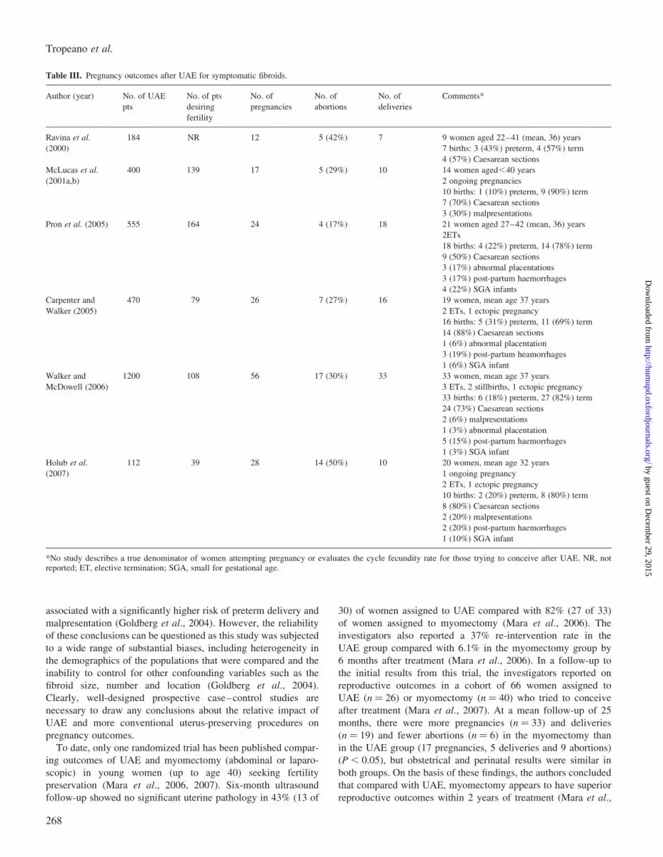
Table III. Pregnancy outcomes after UAE for symptomatic fibroids.
Author (year) No. of UAE
pts
No. of pts
desiring
fertility
No. of
pregnancies
No. of
abortions
No. of
deliveries
Comments*
Ravina et al.
(2000)
184 NR 12 5 (42%) 7 9 women aged 22–41 (mean, 36) years
7 births: 3 (43%) preterm, 4 (57%) term
4 (57%) Caesarean sections
McLucas et al.
(2001a,b)
400 139 17 5 (29%) 10 14 women aged,40 years
2 ongoing pregnancies
10 births: 1 (10%) preterm, 9 (90%) term
7 (70%) Caesarean sections
3 (30%) malpresentations
Pron et al. (2005) 555 164 24 4 (17%) 18 21 women aged 27–42 (mean, 36) years
2ETs
18 births: 4 (22%) preterm, 14 (78%) term
9 (50%) Caesarean sections
3 (17%) abnormal placentations
3 (17%) post-partum haemorrhages
4 (22%) SGA infants
Carpenter and
Walker (2005)
470 79 26 7 (27%) 16 19 women, mean age 37 years
2 ETs, 1 ectopic pregnancy
16 births: 5 (31%) preterm, 11 (69%) term
14 (88%) Caesarean sections
1 (6%) abnormal placentation
3 (19%) post-partum heamorrhages
1 (6%) SGA infant
Walker and
McDowell (2006)
1200 108 56 17 (30%) 33 33 women, mean age 37 years
3 ETs, 2 stillbirths, 1 ectopic pregnancy
33 births: 6 (18%) preterm, 27 (82%) term
24 (73%) Caesarean sections
2 (6%) malpresentations
1 (3%) abnormal placentation
5 (15%) post-partum haemorrhages
1 (3%) SGA infant
Holub et al.
(2007)
112 39 28 14 (50%) 10 20 women, mean age 32 years
1 ongoing pregnancy
2 ETs, 1 ectopic pregnancy
10 births: 2 (20%) preterm, 8 (80%) term
8 (80%) Caesarean sections
2 (20%) malpresentations
2 (20%) post-partum haemorrhages
1 (10%) SGA infant
*No study describes a true denominator of women attempting pregnancy or evaluates the cycle fecundity rate for those trying to conceive after UAE. NR, notreported; ET, elective termination; SGA, small for gestational age.
Tropeano et al.
268
by guest on Decem
ber 29, 2015http://hum
upd.oxfordjournals.org/D
ownloaded from

2007). However, due to the small sample size, this study lacked
sufficient power to appropriately assess the relative impact of
the procedures on fertility rates and pregnancy outcomes.
Overall, the currently available data on fertility and pregnancy
after UAE are insufficient to routinely offer UAE as an alterna-
tive to myomectomy to women who wish to preserve, or
enhance, their fertility. However, for women with prior myo-
mectomy, very large or difficult-to-remove fibroids, or in whom
surgery is contraindicated UAE may be a safer approach than
myomectomy.
In summary, UAE appears to be an excellent treatment option
for most women with symptomatic fibroids, especially for those
who no longer desire fertility but wish to avoid surgery or are
poor surgical risks. Appropriate pre-procedure selection and
careful follow-up of patients are necessary to optimize clinical
outcomes from this therapy. For this reason, an interdisciplinary
approach involving both gynaecologists and interventional radiol-
ogists, with gynaecologists taking a pivotal role in the selection,
co-management and follow-up of patients undergoing UAE
should be implemented into clinical practice.
Transvaginal temporary uterine artery occlusion
Transvaginal uterine artery occlusion is an alternative method of
reducing blood flow in the uterine arteries for the treatment of
uterine fibroids. It is based on the theory that fibroids are killed
by temporary uterine artery occlusion through a mechanism of
transient uterine ischaemia (Burbank and Hutchins, 2000; Lichtin-
ger et al., 2003). The procedure is performed by placing a Doppler
ultrasound-enabled transvaginal clamp (Flostat, Vascular Control
Systems, San Juan Capistrano, CA, USA) in the vaginal fornices
and, guided by Doppler ultrasound auditory signals, positioning
it to occlude the uterine arteries by mechanical compression
against the cervix. The clamp is left in place for 6 h and then
removed.
Preliminary clinical feasibility studies in a total of 75 women
(Dickner et al., 2004; Istre et al., 2004; Garza-Leal et al., 2005;
Lichtinger et al., 2005; Vilos et al., 2006) reported few
procedure-related complications, including two cases of hydrone-
phrosis requiring temporary stenting, and successful short-term
outcomes, with a reduction of 40–50% in uterine and fibroid
volume and symptomatic improvement in 80–90% of patients at
6-month follow-up.
Potential advantages of this technique over UAE are no radi-
ation exposure, no risk of non-target embolization, and the
absence of significant post-procedure pain in most patients.
However, although reported short-term results of transvaginal
occlusion are similar to those of UAE, long-term outcomes
might not be as favourable since the degree of tissue ischaemia
affected by this procedure is dramatically less than with UAE. It
appears, therefore, that long-term studies are needed to discern
whether transvaginal uterine artery occlusion will lead to
durable results, and to compare it with UAE.
MRI-guided focused ultrasound
MRI-guided focused ultrasound (MRgFUS) is a new, minimally
invasive method of thermal ablation for treating fibroids that
received the US Food and Drug Administration (FDA) approval
in 2004. With this technique, high-intensity ultrasound waves
pass through the anterior abdominal wall and converge into a
precise target point within the fibroid to cause a temperature rise
(55–908C) sufficient to induce coagulative necrosis within a few
seconds. Concurrent MRI allows accurate tissue targeting and
real-time temperature feedback, thereby achieving controlled
localized thermal ablation (Stewart et al., 2003; Tempany et al.,
2003; Hindley et al., 2004).
The procedure is performed with a device (ExAblate 2000;
InSightec, Haifa, Israel) that integrates a standard MRI unit with
a focused piezoelectric phased-array transducer in the MRI
table. During treatment, which is performed under intravenous
conscious sedation, the patient is positioned prone on the MR
table with the abdomen directly above the transducer.
The treatment itself consists of consecutive exposures to
focused ultrasound energy (sonications), each one lasting �20 s
and resulting in a small (�0.5 cm3) bean-shaped ablated
volume. Between sonications there is a pause of �90 s to elapse
for the tissue to return to its baseline temperature. Multiple sonica-
tions are required to cover the entire target volume, which is typi-
cally limited to a maximum of 150 cm3 of tissue, and total
procedure time is usually over 3 h. Common symptoms during
the procedure are short-term lower abdominal pain, leg pain and
buttock pain. Patients are usually discharged home �1 h after
the end of the procedure and return to usual activities, on
average, within 48 h (Stewart et al., 2003).
To date, publications on MRgFUS have been case series
showing technical feasibility (Stewart et al., 2003; Tempany
et al., 2003) and short-term outcomes (Hindley et al., 2004;
Hesley et al., 2006; Smart et al., 2006; Stewart et al., 2006).
The pivotal trial of MRgFUS for fibroids was a multi-center
study including 109 premenopausal women with fibroids smaller
than 10 cm in diameter (Stewart et al., 2006). During the pro-
cedures, less than 10% (on average, 25 cm3) of the targeted
fibroid volume was treated, and most patients had only one
fibroid treated. The procedure was relatively well tolerated, with
only 16% of women reporting severe pain during the treatment
and 8% reporting severe-to-moderate pain after the procedure.
There were relatively few serious complications including one
case of sciatic nerve palsy that resolved by 12 months, superficial
skin burns in 5% of patients and one case of skin burn that caused
ulceration. At 6 months, despite only modest reductions in targeted
fibroid volumes (on average, 13.5%), 77 (71%) of the 109
treated patients showed a significant symptom improvement, as
measured by the symptom severity score of the UFS-QOL ques-
tionnaire (Spies et al., 2002a). At 12 months, with only 82
women of the original cohort available for evaluation, 51% (42
of 82) reported continued symptom control, whereas 28% (23 of
82) had sought alternative surgical therapy.
Better outcomes in terms of rates of symptom relief (83% at 6
months and 89% at 12 months of follow-up) and reductions in
the targeted fibroid volume (on average, 21% at 6 months and
37% at 12 months) were reported in 49 women with fibroids
greater than 10 cm in diameter, who were pretreated with
GnRH-agonist for 3 months (Smart et al., 2006). On the basis of
these findings, the authors concluded that reducing fibroid vascu-
larity by pre-procedural GnRH-agonist therapy may enhance the
thermoablative effect of MRgFUS, resulting in a greater clinical
response to treatment (Smart et al., 2006).
Non-surgical options for fibroid management
269
by guest on Decem
ber 29, 2015http://hum
upd.oxfordjournals.org/D
ownloaded from

Changes in treatment protocol, with decreased restrictions on
treatment parameters, have been reported to increase effectiveness
of MRgFUS (Fennessy et al., 2007). Recently published data on
359 women completing 24-month follow-up in all clinical trials
of MRgFUS showed greater symptom reduction (P , 0.001) and
lower risk of additional treatments (P ¼ 0.001) over the first 24
months for women receiving more aggressive treatment (more
than 20% of the fibroid volume ablated) compared to those
having less extensive treatment (20% or less of the fibroid
volume ablated) (Stewart et al., 2007).
As the total number of cases treated by MRgFUS is quite small
when compared with UAE, it is difficult to draw inferences about
the relative pros and cons of these two minimally invasive options.
Advantages of MRgFUS are no radiation exposure, no risk of non-
target embolization, and the absence of significant post-procedure
pain, which eliminates the need for an overnight stay and likely
speeds the return to daily activities. However, compared with
UAE, MRgFUS has more numerous restrictions to the size and
number of fibroids that can be treated as the time required to
perform the procedure is volume dependant, and it is also less
effective in reducing uterine and fibroid size. In addition, several
patient characteristics including fibroids close to neurovascular
bundles or sensitive organs, such as bladder and bowel, and
those outside the image area, and the presence of bowel loops or
abdominal wall scars in the projected ultrasound beam pathway
may preclude patients from undergoing treatment (Tempany
et al., 2003).
Questions still remain to be answered regarding the safety of this
procedure. From a preliminary feasibility study (Tempany et al.,
2003), it is apparent that, likely related to collateral thermal
injury, the area of tissue necrosis measured by histopathologic
examination of post-procedure hysterectomy specimens is on
average 3-fold higher than the treatment volume predicted by
MRI. This not only seems to argue against the notion of ‘focused’
delivery of ultrasound energy but also raises concerns about the
risk of expansion of the thermal injury beyond the boundary of
the fibroid into normal myometrium or extrauterine structures.
Another unresolved issue with MRgFUS concerns the durability
of clinical improvement over time. Since only a small amount of
fibroid tissue is usually ablated, patients undergoing the procedure
may be at risk for re-growth of the residual viable fibroid tissue
and recurrence of symptoms.
Overall, the data to date suggest that MRgFUS holds promise for
incisionless surgery for appropriately selected patients with symp-
tomatic fibroids, but still are too few to draw conclusions about the
safety and long-term effectiveness of this treatment modality.
New medications
All medications currently available for fibroid treatment including
GnRH-agonist and sex steroids are unsuitable for long-term use
because of their significant side-effects (Young et al., 2004). In
recent years, new medications have been introduced that offer
the promise of practical, long-term, medical therapy for sympto-
matic fibroids. To date, the most encouraging results in terms of
fibroid volume reduction, symptom relief, and compliance with
long-term administration have been obtained with mifepristone,
a progesterone receptor antagonist, and asoprisnil, a selective pro-
gesterone receptor modulator.
Mifepristone has been the first available active antiprogestin
(Philibert et al., 1981; Philibert, 1984). It has been shown to
decrease fibroid size and blood flow to the fibroids (Murphy
et al., 1993, 1995; Reinsch et al., 1994). Early studies of
women with symptomatic fibroids reported that daily adminis-
tration of mifepristone at doses ranging from 5 to 50 mg for
3 to 6 months resulted in a 26–74% reduction in fibroid
volume and a decrease in the prevalence and severity of
fibroid-related symptoms, including menorrhagia, dysmenorrhea
and pelvic pressure (Steinauer et al., 2004). However, a high
incidence (28%) of endometrial hyperplasia was observed in
patients screened with endometrial biopsies (Steinauer et al.,
2004).
A prospective, randomized, controlled trial comparing the out-
comes of 5 mg per day to 10 mg per day of mifepristone reported
significant reductions compared with baseline in uterine volume
and menstrual blood loss in both groups at 6 months, with no sig-
nificant differences between the groups (Eisinger et al., 2003). In a
follow-up of the original trial, the investigators assessed the occur-
rence of endometrial hyperplasia after 18 months of mifepristone
therapy in 21 of the 40 original women in the study (Eisinger et al.,
2005). They found no case of hyperplasia at 6 and 12 months
among women treated with 5 mg, and a 14% rate at 6 months
and 5% rate at 12 months for the group treated with 10 mg
(Eisinger et al., 2005).
More recently, in a randomized, double-blinded, placebo-
controlled trial including 42 women with symptomatic fibroids,
treatment with mifepristone 5 mg per day for 26 weeks led to a
47% reduction in fibroid size and a significant improvement in
fibroid-related symptoms and QOL scores (Fiscella et al., 2006).
By 12 months, 9 (41%) of 22 women randomized to mifepristone
had become amenorrheic (Fiscella et al., 2006). The drug was well
tolerated, and no endometrial hyperplasia was noted in any partici-
pant (Fiscella et al., 2006). Although these data are very encoura-
ging, further studies in larger samples with longer periods of
treatment are needed to reliably assess the long-term safety and
efficacy of this drug.
Another promising medication for fibroid treatment is asoprisnil,
a selective progesterone receptor modulator with mixed agonist/antagonist activity. This drug has been shown to inhibit prolifer-
ation and induce apoptosis in uterine fibroid cells (Chen et al.,
2006; Wang et al., 2006). In addition, it has an inhibitory effect
on the endometrium as a result of suppressed endometrial angiogen-
esis and/or function of the spiral arteries (Chwalisz et al., 2004,
2005). Small observational studies of women with symptomatic
fibroids showed that daily treatment with asoprisnil at doses
ranging from 5 to 25 mg for 3 months reduced fibroid volume in
a dose-dependent manner and suppressed both abnormal and
normal uterine bleeding, with no effects on circulating estrogen
levels and no breakthrough bleeding (Chwalisz et al., 2005).
In a recent randomized, double-blind, placebo-controlled trial
involving 129 women, a 3-month treatment with asoprisnil doses
of 5, 10 and 25 mg daily suppressed uterine bleeding in 28, 64
and 83% of women, respectively, and reduced fibroid volume
and fibroid-related pressure symptoms (Chwalisz et al., 2007).
Asoprisnil treatment was associated with follicular-phase estrogen
concentration and minimal hypoestrogenic symptoms (Chwalisz
et al., 2007). These promising findings warrant replication
through larger controlled trials over extended treatment intervals.
Tropeano et al.
270
by guest on Decem
ber 29, 2015http://hum
upd.oxfordjournals.org/D
ownloaded from

Conclusions
Expanding non-surgical treatment options for fibroids are advan-
cing care for women, who are now increasingly willing to be
treated while keeping the constraints and sequelae of treatment
to a minimum. At the same time, the new possibilities afforded
by these minimally invasive options do raise challenging questions
about changing indications for surgery in the management of
uterine fibroids.
So far, however, the availability of alternative treatments has
failed to substantially change everyday clinical practice, and the
majority of women with symptomatic fibroids are still managed
surgically. This is, at least partly, because some of the newer
approaches including MRI-guided focused ultrasound and tempor-
ary uterine artery occlusion are still investigational, which poses
the problem of their long-term safety and efficacy compared
with standard surgical treatment.
It is more difficult to be sure of the reasons why UAE remains
underused in spite of the accumulating good-quality evidence to
support its safety and effectiveness. The major issues, however,
seem to be about increasing the gynaecologists’ awareness and
acceptance of UAE as a viable treatment option for fibroids and
improving the collaboration between gynaecologists and interven-
tional radiologists to facilitate optimal care for patients.
References
ACOG Committee on Practice Bulletins – Gynecology. ACOG practicebulletin: Surgical alternatives to hysterectomy in the management ofleiomyomas [number 16, May 2000]. Int J Gynecol Obstet2001;73:285–293.
ACOG Committee Opinion. Uterine artery embolization [number 293, Feb2004]. Obstet Gynecol 2004;103:403–404.
Ahmad A, Qadan L, Hassan N, Najarian K. Uterine artery embolizationtreatment of uterine fibroids: effect on ovarian function in youngerwomen. J Vasc Interv Radiol 2002;13:1017–1020.
Aitken E, Khaund A, Hamid SA, Millan D, Campbell S. The normal humanmyometrium has a vascular spatial gradient absent in small fibroids.Hum Reprod 2006;21:2669–2678.
Al-Fozan H, Dufort J, Kaplow M, Valenti D, Tulandi T. Cost analysis ofmyomectomy, hysterectomy, and uterine artery embolization. Am JObstet Gynecol 2002;187:1401–1404.
Andrews RT, Brown PH. Uterine artery embolization: factors influencingpatient radiation exposure. Radiology 2000;217:713–722.
Andrews RT, Spies JB, Sacks D, Worthington-Kirsc RL, Niedzwiecki GA,Marx MV, Hovsepian DM, Miller DL, Siskin GP, Raabe RD et al.Patient care and uterine artery embolization for leiomyomata. J VascInterv Radiol 2004;15:115–120.
Aungst M, Wilson M, Vournas K, McCarthy S. Necrotic leiomyoma andgram-negative sepsis eight weeks after uterine artery embolization.Obstet Gynecol 2004;104:1161–1164.
Baird DD, Dunson DB, Hill MC, Cousins D, Schectman JM. High cumulativeincidence of uterine leiomyoma in black and white women: ultrasoundevidence. Am J Obstet Gynecol 2003;188:100–107.
Baker CM, Winkel CA, Subramanian S, Spies JB. Estimated costs for uterineartery embolization and abdominal myomectomy for uterineleiomyomata: a comparative study at a single institution. J Vasc InterRadiol 2002;13:1207–1210.
Bajekal N, Li TC. Fibroids, infertility and pregnancy wastage. Hum ReprodUpdate 2000;6:614–620.
Banu NS, Gaze DC, Bruce H, Collinson PO, Belli AM, Manyonda IT. Markersof muscle ischemia, necrosis, and inflammation following uterine arteryembolization in the treatment of symptomatic uterine fibroids. Am JObstet Gynecol 2007;196:213.e1–213.e5.
Bazot M, Cortez A, Darai E, Rouger J, Chopier J, Antoine JM, Uzan S.Ultrasonography compared with magnetic resonance imaging for thediagnosis of adenomyosis: correlation with histopathology. Hum Reprod2001;16:2427–2433.
Beinfeld MT, Bosch JL, Isaacson KB, Gazelle GS. Cost-effectiveness ofuterine artery embolization and hysterectomy for uterine fibroids.Radiology 2004;230:207–213.
Broder MS, Goodwin S, Chen G, Tang LJ, Costantino MM, Nguyen MH,Yegul TN, Erberich H. Comparison of long-term outcomes ofmyomectomy and uterine artery embolization. Obstet Gynecol2002;100:864–868.
Bruno J, Sterbis K, Flick P, McCullough M, Cramp M, Murphy-Skrynarz K,Spies JB. Recovery after uterine artery embolization for leiomyomas: adetailed analysis of its duration and severity. J Vasc Interv Radiol2004;15:801–807.
Burbank F, Hutchins FL, Jr. Uterine artery occlusion by embolization orsurgery for treatment of fibroids: a unifying hypothesis - transientuterine ischemia. J Am Assoc Gynecol Laparosc 2000;7:S1–S49.
Carpenter TT, Walker WJ. Pregnancy following uterine artery embolisation forsymptomatic fibroids: a series of 26 completed pregnancies. BJOG2005;112:321–325.
Chen W, Ohara N, Wang J, Xu Q, Liu J, Morikawa A, Sasaki H, Yoshida S,DeManno DA, Chwalisz K et al. A novel selective progesteronereceptor modulator asoprisnil (J867) inhibits proliferation and inducesapoptosis in cultured human uterine leiomyoma cells in the absence ofcomparable effects on myometrial cells. J Clin Endocrinol Metab2006;91:1296–1304.
Chrisman HB, Saker MB, Ryu RK, Nemcek AA, Jr, Gerbie MV, Milad MP,Smith SJ, Sewall LE, Omary RA, Vogelzang RL. The impact of uterinefibroid embolization on resumption of menses and ovarian function.J Vasc Interv Radiol 2000;11:699–703.
Chrisman HB, Minocha J, Ryu RK, Vogelzang RL, Nikolaidis P, Omary RA.Uterine artery embolization: a treatment option for symptomatic fibroid inpostmenopausal women. J Vasc Interv Radiol 2007;18:451–454.
Chwalisz K, DeManno D, Garg R, Larsen L, Mattia-Goldberg C, Stickler T.Therapeutic potential for the selective progesterone receptor modulatorasoprisnil in the treatment of leiomyomata. Semin Reprod Med2004;22:113–119.
Chwalisz K, Perez MC, DeManno D, Winkel C, Schubert G, Elger W. Selectiveprogesterone receptor modulator development and use in the treatment ofleiomyomata and endometriosis. Endocr Rev 2005;26:423–438.
Chwalisz K, Larsen L, Mattia-Goldberg C, Edmonds A, Elger W, Winkel CA.A randomized, controlled trial of asoprisnil, a novel selective progesteronereceptor modulator, in women with uterine leiomyomata. Fertil Steril2007;87:1399–1412.
Coronado GD, Marshall LM, Schwartz SM. Complications in pregnancy, labor,and delivery with uterine leiomyomas: a population-based study. ObstetGynecol 2000;95:764–769.
de Block S, de Vries C, Prinssen HM, Blaauwgeers HL, Jorna-Meijer LB. Fatalsepsis after uterine artery embolization with microspheres. J Vasc IntervRadiol 2003;14:779–783.
De Iaco PA, Muzzupapa G, Golfieri R, Ceccarini M, Roset B, Baroncini S. Auterine wall defect after uterine artery embolization for symptomaticmyomas. Fertil Steril 2002;77:176–178.
deSouza NM, Williams AD. Uterine arterial embolization for leiomyomas:perfusion and volume changes at MR imaging and relation to clinicaloutcome. Radiology 2002;222:367–374.
Dembek CJ, Pelletier EM, Isaacson KB, Spies JB. Payer costs in patientsundergoing uterine artery embolization, hysterectomy, or myomectomyfor treatment of uterine fibroids. J Vasc Interv Radiol 2007;18:1207–1213.
Dickner SK, Cooper JM, Diaz D. A nonincisional, Doppler-guided transvaginalapproach to uterine artery identification and control of uterine perfusion.J Am Assoc Gynecol Laparosc 2004;11:55–58.
Dutton S, Hirst A, McPherson K, Nicholson T, Maresh M. A UK multicentreretrospective cohort study comparing hysterectomy and uterine arteryembolisation for the treatment of symptomatic uterine fibroids(HOPEFUL study): main results on medium-term safety and efficacy.BJOG 2007;114:1340–1351.
Edwards RD, Moss JG, Lumsden MA, Wu O, Murray LS, Twaddle S, MurrayGD. Uterine artery embolization versus surgery for symptomatic uterinefibroids. N Engl J Med 2007;356:360–370.
Eisinger SH, Meldrum S, Fiscella K, le Roux HD, Guzick DS. Low-dosemifepristone for uterine leiomyomata. Obstet Gynecol 2003;101:243–250.
Eisinger SH, Bonfiglio T, Fiscella K, Meldrum S, Guzick DS. Twelve-monthsafety and efficacy of low-dose mifepristone for uterine myomas.J Minim Invasive Gynecol 2005;12:227–233.
Non-surgical options for fibroid management
271
by guest on Decem
ber 29, 2015http://hum
upd.oxfordjournals.org/D
ownloaded from

Fauconnier A, Chapron C, Babaki-Fard K, Dubuisson JB. Recurrence ofleiomyomata after myomectomy. Hum Reprod Update 2000;6:595–602.
Felemban A, Stein L, Tulandi T. Uterine restoration after repeated expulsion ofmyomas after uterine artery embolization. J Am Assoc Gynecol Laparosc2001;8:442–444.
Fennessy FM, Tempany CM, McDannold NJ, So MJ, Hesley G, Gostout B,Kim HS, Holland GA, Sarti DA, Hynynenk K et al. Uterineleiomyomas: MR imaging-guided focused ultrasound surgery—resultsof different treatment protocols. Radiology 2007;243:885–893.
Fiscella K, Eisinger SH, Meldrum S, Feng C, Fisher SG, Guzick DS. Effect ofmifepristone for symptomatic leiomyomata on quality of life and uterinesize. Obstet Gynecol 2006;108:1381–1387.
Gabriel H, Pinto CM, Kumar M, Nikolaidis P, Miller FH, WeinrachDM, Vogelzang RL. MRI detection of uterine necrosis after uterineartery embolization for fibroids. AJR Am J Roentgenol 2004;183:733–736.
Gabriel-Cox K, Jacobson GF, Armstrong MA, Hung YY, Learman LA.Predictors of hysterectomy after uterine artery embolization forleiomyoma. Am J Obstet Gynecol 2007;196:588.e1–588.e6.
Garza-Leal J, Eligonad G, Castillo L. Fibroid treatment by temporary uterineartery occlusion using Doppler-guided clamp. J Min Invas Gynecol2005;12:S82–S83.
Godfrey CD, Zbella EA. Uterine necrosis after uterine artery embolization forleiomyoma. Obstet Gynecol 2001;98:950–952.
Goodwin S, Mc Lucas B, Lee M, Chen G, Perrella R, Vedantham S, Muir S, LaiA, Sayre JW, DeLeon M. Uterine artery embolization for the treatment ofuterine leiomyomata: mid-term results. J Vasc Inter Radiol1999;10:1159–1165.
Goodwin SC, Bradley LD, Lipman JC, Stewart EA, Nosher JL, Sterling KM,Barth MH, Siskin GP, Shlansky-Goldberg RD. Uterine arteryembolization versus myomectomy: a multicenter comparative study.Fertil Steril 2006;85:14–21.
Goodwin SC, Spies JB, Worthington-Kirsch R, Peterson E, Pron G, Li S,Myers E. Uterine artery embolization for treatment of leiomyomata.Long-term outcomes from the FIBROID Registry. Obstet Gynecol2008;111:22–33.
Goldberg J, Pereira L, Berghella V, Diamond J, Darai E, Seinera P, SeracchioliR. Pregnancy outcomes after treatment for fibromyomata: uterine arteryembolization versus laparoscopic myomectomy. Am J Obstet Gynecol2004;191:18–21.
Goldberg J, Bussard A, McNeil J, Diamond J. Cost and reimbursement for threefibroid treatments: abdominal hysterectomy, abdominal myomectomy,and uterine fibroid embolization. Cardiovasc Interv Radiol 2007;30:54–58.
Gulati MS, Srinivasan A, Paul SB, Bhatla N. Uterine restoration followingfibroid expulsion after uterine artery embolisation using gelfoam.J Postgrad Med 2004;50:80–80.
Healey S, Buzaglo K, Seti K, Seti L, Valenti D, Tulandi T. Ovarian functionafter uterine artery embolization and hysterectomy. J Am Assoc GynecolLaparosc 2004;11:348–352.
Hehenkamp WJ, Volkers NA, Van Swijndregt AD, De Blok S, ReekersJA, Ankum WM. Myoma espulsion after uterine artery embolization:complication or cure? Am J Obstet Gynecol 2004;191:1713–1715.
Hehenkamp WJ, Volkers NA, Donderwinkel PF, de Blok S, Birnie E, AnkumWM, Reekers JA. Uterine artery embolization versus hysterectomy in thetreatment of symptomatic uterine fibroids (EMMY trial): peri- andpostprocedural results from a randomized controlled trial. Am J ObstetGynecol 2005;193:1618–1629.
Hehenkamp WJ, Volkers NA, Birnie E, Reekers JA, Ankum WM. Pain andreturn to daily activities after uterine artery embolization andhysterectomy in the treatment of symptomatic uterine fibroids: resultsfrom the randomized EMMY trial. Cardiovasc Interv Radiol2006;29:179–187.
Hesley GK, Felmlee JP, Gebhart JB, Dunagan KT, Gorny KR, Kesler JB,Brandt KR, Glantz JN, Gostout BS. Non invasive treatment of uterinefibroids: early Mayo Clinic experience with magnetic resonanceimaging-guided focused ultrasound. Mayo Clin Proc 2006;81:936–942.
Hindley J, Gedroyc WM, Regan L, Stewart E, Tempany C, Hynyen K,Mcdannold N, Inbar Y, Itzchak Y, Rabinovici J et al. MRI guidance offocused ultrasound therapy of uterine fibroids: early results. AJR AmJ Roentgenol 2004;183:1713–1719.
Holub Z, Mara M, Kuzal D, Jabor A, Maskova J, Eim J. Pregnancy outcomesafter uterine artery occlusion: prospective multicentric study. Fertil Steril2007 (Epub ahead of print).
Hovsepian DM, Siskin GP, Bonn J, Cardella JF, Clark TW, Lampmann LE,Miller DL, Omary RA, Pelage JP, Rajan D et al. Quality improvementguidelines for uterine artery embolization for symptomaticleiomyomata. J Vasc Interv Radiol 2004;15:535–541.
Hovsepian DM, Ratts VS, Rodriguez M, Huang JS, Aubuchon MG, PilgramTK. A prospective comparison of the impact of uterine arteryembolization, myomectomy, and hysterectomy on ovarian function.J Vasc Interv Radiol 2006;17:1111–1115.
Huang JY, Kafy S, Dugas A, Valenti D, Tulandi T. Failure of uterine fibroidembolization. Fertil Steril 2006;85:30–35.
Hutchins FL, Jr, Worthington-Kirsch R, Berkowitz RP. Selective uterine arteryembolization as primary treatment for symptomatic leiomyomata uteri.J Am Assoc Gynecol Laparosc 1999;6:279–284.
Isonishi S, Coleman RL, Himara M, Iida Y, Kitai S, Nagase M, Ochiai K.Analysis of prognostic factors for patients with leiomyoma treated withuterine arterial embolization. Am J Obstet Gynecol 2007; Nov 9; Epubahead of print.
Istre O, Hald K, Qvigstad E. Multiple myomas treated with a temporary,non-invasive, Doppler-directed, transvaginal uterine artery clamp. J AmAssoc Gynecol Laparosc 2004;11:273–276.
Jenkinson C, Peto V, Coulter A. Making sense of ambiguity: evaluation ofinternal reliability and face validity of the SF 36 questionnaire inwomen presenting with menorrhagia. Qual Health Care 1996;5:9–12.
Katsumori T, Nakajima K, Mihara T, Tokuhiro M. Uterine artery embolizationusing gelatin sponge particles alone for symptomatic uterine fibroids:mid-term results. AJR Am J Roentgenol 2002;178:135–139.
Katsumori T, Nakajima K, Mihara T. Is a large fibroid a high-risk factor foruterine artery embolization? Am J Roentgenol 2003;181:1309–1314.
Katsumori T, Akazawa K, Mihara T. Uterine artery embolization forpeduncolated subserosal fibroids. AJR Am J Roentgenol 2005;18:399–402.
Katsumori T, Kasahara T, Akazawa K. Long-term outcomes of uterine arteryembolization using gelatine sponge particles alone for symptomaticfibroids. AJR Am J Roentgenol 2006;186:848–854.
Kroencke TJ, Gauruder-Burmester A, Enzweiler CN, Taupitz M, Hamm B.Disintegration and stepwise expulsion of a large uterine leiomyomawith restoration of the uterine architecture after successful uterinefibroid embolization: case report. Hum Reprod 2003;18:863–865.
Lanocita R, Frigerio LF, Patelli G, Di Tolla G, Spreatico C. A fatalcomplication of percutaneous transcatheter embolization for treatmentof uterine fibroids. In: SMIT/CIMIT 11th Annual Scientific Meeting.Boston, MA, USA, 1999.
Lohle PN, Boekkooi FP, Smeets AJ, Pieters JJ, Vervest HA, LampmannLE, Sluzewski M. Limited uterine artery embolization for leiomyomaswith tris-acryl gelatine microspheres: 1-year follow-up. J Vasc IntervRadiol 2006;17:283–287.
Lichtinger M, Burbank F, Hallson L, Herbert S, Uyeno J, Iones M. The timecourse of myometrial ischemia and reperfusion after laparoscopicuterine artery occlusion-theoretical implication. J Am Assoc GynecolLaparosc 2003;10:554–563.
Lichtinger M, Herbert S, Memmolo A. Temporary, transvaginal occlusion ofthe uterine arteries: a feasibility and safety study. J Min Invas Gynecol2005;12:40–42.
Mara M, Fucikova Z, Maskova J, Kuzel D, Haakova L. Uterine fibroidembolization versus myomectomy in women wishing to preservefertility: preliminary results from a randomized controlled trial. Eur JObstet Gynecol Reprod Biol 2006;126:226–233.
Mara M, Maskova J, Fucinova Z, Kuzel D, Belsan T, Sosna O. Midterm clinicaland first reproductive results of a randomized controlled trial comparinguterine fibroid embolization and myomectomy. Cardiovasc IntervRadiol 2007 (Epub ahead of print).
Maresh MJ, Metcalfe MA, McPherson K, Overton C, Hale V, Hargreaves J,Bridgman S, Dobbins J, Cosbard A. The VALUE national hysterectomystudy: description of the patients and their surgery. BJOG2002;109:302–312.
Marret H, Alonso AM, Cottier JP, Tranquart F, Herbreteau D, Body G.Leiomyoma recurrence after uterine artery embolization. J Vasc IntervRadiol 2003;14:1395–1400.
Marret H, Keris Yle B, Acker O, Cottier JP, Herbreteau D. Late leiomyomaexpulsion after uterine artery embolization. J Vasc Interv Radiol2004;15:1483–1485.
Marret H, Cottier JP, Alonso AM, Giraudeau B, Body G, Herbreteau D.Predictive factors for fibroid recurrence after uterine arteryembolisation. BJOG 2005;112:461–465.
Tropeano et al.
272
by guest on Decem
ber 29, 2015http://hum
upd.oxfordjournals.org/D
ownloaded from

Matchar DB, Myers ER, Barber MW, Couchman GM, Datta S, Gray RN,Gustilo-Ashby T, Kolimaga JT, McCrory DC. Management of uterinefibroids. Evid Rep Technol Assess (Summ) 2001;34:1–6.
McLucas B, Adler L, Perrella R. Uterine fibroid embolization: nonsurgicaltreatment for symptomatic fibroids. J Am Coll Surg 2001a;192:95–105.
McLucas B, Goodwin S, Adler L, Rappaport A, Reed R, Perrella R. Pregnancyfollowing uterine fibroid embolization. Int J Gynaecol Obstet2001b;74:1–7.
McLucas B, Perrella R, Adler L. Embolization for the treatment ofadenomyosis. AJR Am J Roentgenol 2002;178:1028–1029.
Murphy AA, Kettel LM, Morales AJ, Roberts VJ, Yen SS. Regression ofuterine leiomyomata in response to the antiprogesterone RU 486. J ClinEndocrinol Metab 1993;76:513–517.
Murphy AA, Morales AJ, Kettel LM, Yen SS. Regression of uterineleiomyomata to the antiprogesterone RU486: dose-response effect.Fertil Steril 1995;64:187–190.
National Institute for Health and Clinical Excellence. Heavy MenstrualBleeding. London: NICELondon, 2007. NICE guideline CG44.2007.http://guidance.nice.org.uk/CG44/guidance/pdf/English.
Nikolic B, Abbara S, Levy E, Imaoka I, Lundsten ML, Jha RC, Spies JB.Influence of radiographic technique and equipment on adsorbed ovariandose associated with uterine artery embolization. J Vasc Interv Radiol2000;11:1173–1178.
Nikolic B, Nguyen K, Martin LG, Redd DC, Best I, Silverstein MI. Pyosalpinxdeveloping from a preexisting hydrosalpinx after uterine arteryembolization. J Vasc Interv Radiol 2004;15:297–301.
Ogliari KS, Mohallem SV, Barrozo P, Viscomi F. A uterine cavity-myomacommunication after uterine artery embolization: two case reports.Fertil Steril 2005;83:220–222.
Payne JF, Robboy SJ, Haney AF. Embolic microspheres within ovarian arterialvasculature after uterine artery embolization. Obstet Gynecol2002;100:883–886.
Park HR, Kim MD, Kim NK, Kim HJ, Yoon SW, Park WK, Lee MH. Uterinerestoration after repeated sloughing of fibroids. Eur Radiol2005;15:1850–1854.
Pelage JP, Le Dref O, Soyer P, Kardache M, Dahan H, Abitbol M, Merland JJ,Ravina JH, Rymer R. Fibroid-related menorrhagia: treatment withsuperselective embolization of the uterine arteries and midtermfollow-up. Radiology 2000;215:428–431.
Pelage JP, Guaou NG, Jha RC, Ascher SM, Spies JB. Uterine fibroid tumors:long-term MR imaging outcome after embolization. Radiology2004;230:803–809.
Pelage JP, Jacob D, Fazel A, Namur J, Laurent A, Rymer R, Le Dref O.Midterm results of uterine artery embolization for symptomaticadenomyosis: initial experience. Radiology 2005;234:948–953.
Philibert D, Deraedt R, Teutsch G. RU38486: A Potent Antiglucocorticoid invivo. Tokyo, Japan: The VII International Congress of PharmacologyTokyo, Japan, 1981.
Philibert D. RU38486: an original multifaceted antihormone in vivo. In:Agaruval M (ed). Adrenal Steroid Antagonism. Berlin: Walter deGruyter and Co.Berlin, 1984. 77–101.
Pinto I, Chimeno P, Romo A, Paul L, Haya J, de la Cal MA, Bajo J. Uterinefibroids: Uterine artery embolization versus abdominal hysterectomy fortreatment—a prospective, randomized, and controlled clinical trial.Radiology 2003;226:425–431.
Pourrat XJL, Fourquet F, Gueriff F, Viratelle N, Herbreteau D, Marret H.Medico-economic approach to the management of uterine myomas: a6-month cost-effectiveness study of pelvic embolization versus vaginalhysterectomy. Eur J Obstet Gynecol Reprod Biol 2003;111:59–64.
Prollius A, de Vries C, Loggenberg E, du Plessis A, Nel M, Wessels PH.Uterine artery embolisation for symptomatic fibroids: the effect of thelarge uterus on outcome. BJOG 2004;111:239–242.
Pron G, Bennett J, Common A, Wall J, Asch M, Sniderman K. The OntarioUterine Fibroid Embolization Trial. Part 2. Uterine fibroid reduction andsymptom relief after uterine artery embolization for fibroids. FertilSteril 2003a;79:120–127.
Pron G, Bennett J, Common A, Sniderman K, Asch M, Bell S, Kozak R,Vanderburgh L, Garvin G, Simons M et al. Technical results and effectsof operator experience on uterine artery embolization for fibroids: theOntario Uterine Fibroid Embolization Trial. J Vasc Interv Radiol2003c;14:545–554.
Pron G, Mocarski E, Bennet J, Vilos G, Common A, Zaid M, Sniderman K,Asch M, Kozak R, Simons M et al. Tolerance, hospital stay, and recoveryafter uterine artery embolization for fibroids: the Ontario Uterine FibroidEmbolization Trial. J Vasc Interv Radiol 2003b;14:1243–1250.
Pron G, Mocarski E, Bennett J, Vilos G, Common A, Vanderburgh L OntarioUFE Collaborative Group. Pregnancy after uterine artery embolization forleiomyomata: the Ontario multicenter trial. Obstet Gynecol 2005;105:67–76.
Rajan DK, Beecroft JR, Clark TW, Asch MR, Simons ME, Kachura JR,Sved M, Sniderman KW. Risk of intrauterine infectious complicationsafter uterine artery embolization. J Vasc Interv Radiol 2004;12:1414–1421.
Ravina JH, Herbreteau D, Ciraru-Vigneron N, Bouret JM, Houdart E,Aymard A, Merland JJ. Arterial embolisation to treat uterine myomata.Lancet 1995;346:671–672.
Ravina JH, Vigneron N, Aymard A, Le Dref O, Merland JJ. Pregnancy afterembolization of uterine myoma: report of 12 cases. Fertil Steril2000;73:1241–1243.
Razavi MK, Hwang G, Jahed A, Modanloo S, Chen B. Abdominalmyomectomy versus uterine fibroid embolization in the treatment ofsymptomatic uterine leiomyomas. AJR Am J Roentgenol2003;180:1571–1575.
Reinsch RC, Murphy AA, Morales AJ, Yen SS. The effects of RU 486 andleuprolide acetate on uterine artery blood flow in the fibroid uterus: aprospective, randomized study. Am J Obstet Gynecol 1994;170:1623–1627.
Ruta DA, Garrat AM, Chadha YC, Flett GM, Hall MH, Russell IT. Assessmentof patients with menorrhagia: how valid is a structured clinical history as ameasure of health status? Qual Life Res 1995;4:33–40.
Scheurig C, Gauruder-Burmester A, Kluner C, Kurzeja R, Lembcke A,Zimmermann E, Hamm B, Kroencke T. Uterine artery embolization forsymptomatic fibroids: short-term versus mid- term changes indisease-specific symptoms, quality of life and magnetic resonanceimaging results. Hum Reprod 2006;21:3270–3277.
Shashoua AR, Stringer NH, Pearlman JB, Behmaram B, Stringer EA. Ischemicuterine rupture and hysterectomy 3 months after uterine arteryembolization. J Am Assoc Gynecol Laparosc 2002;9:217–220.
Smart DO, Hindley JT, Regan L, Gedroyc WG. Gonadotrophin-releasinghormone and magnetic resonance guided ultrasound surgery for uterineleiomyomata. Obstet Gynecol 2006;108:49–54.
Smith WJ, Upton E, Shuster EJ, Klein AJ, Schwartz ML. Patient satisfactionand disease specific quality of life after uterine artery embolization. AmJ Obstet Gynecol 2004;190:1697–1703.
SOGC Clinical Practice Guidelines. Uterine fibroid embolization (UFE). Int JGynaecol Obstet 2005;89:305–318.
Spielmann AL, Keogh C, Forster BB, Martin ML, Machan LS. Comparison ofMRI and sonography in the preliminary evaluation for fibroidembolization AJR. Am J Roentgenol 2006;187:1499–1504.
Spies JB, Scialli AR, Jha RC, Imaoka I, Ascher SM, Fraga VM, Barth Kh.Initial results from uterine fibroid embolization for symptomaticleiomyomata. J Vasc Interv Radiol 1999;10:1149–1157.
Spies JB, Ascher SA, Roth AR, Kim J, Levy EB, Gomez-Jorge J. Uterine arteryembolization for leiomyomata. Obstet Gynecol 2001a;98:29–34.
Spies JB, Roth AR, Gonsalves SM, Murphy-Skrzyniarz KM. Ovarian functionafter uterine artery embolization for leiomyomata: assessment with use ofserum follicle stimulating hormone assay. J Vasc Interv Radiol2001b;12:437–442.
Spies JB, Coyne K, Guaou Guaou N, Boyle D, Skyrnarz-Murphy K,Gonzalves SM. The UFS-QOL, a new disease-specific symptom andhealth-related quality of life questionnaire for leiomyomata. ObstetGynecol 2002a;99:290–300.
Spies JB, Spector A, Roth AR, Baker CM, Mauro L, Murphy-Skrynarz K.Complications after uterine artery embolization for leiomyomas. ObstetGynecol 2002b;100:873–880.
Spies JB. Uterine artery embolization for fibroids: understanding the technicalcauses of failure. J Vasc Interv Radiol 2003;14:11–14.
Spies JB, Sacks D. Credentials for uterine artery embolization. J Vasc IntervRadiol 2004;15:111–113.
Spies JB, Allison S, Flick P, McCullough M, Sterbis K, Cramp M, Bruno J, JhaR. Polyvinyl alcohol particles and tris-acryl gelatin microspheres foruterine artery embolization for leiomyomas: results of a randomizedcomparative study. J Vasc Interv Radiol 2004a;15:793–800.
Spies JB, Cooper JM, Worthington-Kirsch R, Lipman JC, Mills BB, BenenatiJF. Outcome from uterine embolization and hysterectomy forleiomyomas: results of a multi-center study. Am J Obstet Gynecol2004b;191:22–31.
Spies JB, Myers ER, Worthington- Kirsch R, Mulgund J, Goodwin S, MauroM. The FIBROID Registry: symptom and quality-of-life status 1 yearafter therapy. Obstet Gynecol 2005a;106:1309–1318.
Non-surgical options for fibroid management
273
by guest on Decem
ber 29, 2015http://hum
upd.oxfordjournals.org/D
ownloaded from

Spies JB, Bruno J, Czeyda-Pommersheim F, Magee ST, Ascher SA, Jha RC.Long-term outcome of uterine artery embolization of leiomyomas.Obstet Gynecol 2005b;106:933–939.
Steinauer J, Pritts EA, Jackson R, Jacoby AF. Systematic review ofmifepristone for the treatment of uterine leiomyomata. Obstet Gynecol2004;103:1331–1336.
Stewart EA, Gedroyc WM, Tempany CM, Quade BJ, Inbar Y, Ehrenstein T,Shushan A, Hindley T, Goldin RD, David M et al. Focused ultrasoundtreatment of uterine fibroid tumors: safety and feasibility of anon-invasive thermoablative technique. Am J Obstet Gynecol2003;189:48–54.
Stewart EA, Rabinovici J, Tempany CM, Inbar Y, Regan L, Gostout B, HesleyG, Kim HS, Hengst S, Gedroyc WM. Clinical outcomes of focusedultrasound surgery for the treatment of uterine fibroids. Fertil Steril2006;85:22–29.
Stewart EA, Gostout B, Rabinovici J, Kim HS, Regan L, Tempany CM.Sustained relief of leiomyoma symptoms by using focused ultrasoundsurgery. Obstet Gynecol 2007;110:279–284.
Tempany CM, Stewart EA, McDannold N, Quade BJ, Jolesz FA, Hynynen K.MR imaging-guided focused ultrasound surgery of uterine leiomyomas: afeasibility study. Radiology 2003;226:897–905.
Torigian DA, Siegelman ES, Terhune KP, Butts SF, BlascoL, Shlansky-Goldberg RD. MRI of uterine necrosis after uterine arteryembolization for treatment of uterine leiomyomata. Am J Roentgenol2005;184:555–559.
Tropeano G, Litwicka K, Di Stasi C, Romano D, Mancuso S. Permanentamemorrhea associated with endometrial atrophy after uterine arteryembolization for symptomatic uterine fibroids. Fertil Steril2003;79:132–135.
Tropeano G, Di Stasi C, Litwicka K, Romano D, Draisci G, Mancuso S. Uterineartery embolization does not have adverse effects on ovarian reserve inregularly cycling women younger than 40 years. Fertil Steril2004;81:1055–1061.
Tulandi T, Sammour A, Valenti D, Child TJ, Seti L, Tan SL. Ovarian reserveafter uterine artery embolization for leiomyomata. Fertil Steril2002;78:197–198.
Vashist A, Studd J, Carey A, Burn P. Fatal septicaemia after fibroidembolization. Lancet 1999;354:307–308.
Vilos GA, Vilos EC, Romano W, Abu-Rafea B. Temporary uterine arteryocclusion for treatment of menorrhagia and uterine fibroids using anincisionless Doppler-guided transvaginal clamp: case report. HumReprod 2006;21:269–271.
Vitiello D, McCarthy S. Diagnostic imaging of myomas. Obstet Gynecol ClinNorth Am 2006;33:85–95.
Volkers NA, Hehenkamp WJ, Birnie E, de Vries C, Holt C, AnkumWM, Reekers JA. Uterine artery embolization in the treatment ofsymptomatic uterine fibroids tumors (EMMY Trial): periproceduralresults and complications. J Vasc Inter Radiol 2006;17:471–480.
Volkers NA, Hehenkamp WJ, Birnie E, Ankum WM, Reekers JA.Uterine artery embolization versus hysterectomy in the treatmentof symptomatic uterine fibroids: 2 years’ outcome from therandomized EMMY trial. Am J Obstet Gynecol 2007;196:519.e1–519.e11.
Walker WJ, Pelage JP. Uterine artery embolisation for symptomatic fibroids:clinical results in 400 women with imaging follow-up. Br J ObstetGynecol 2002;109:1263–1272.
Walker WJ, Carpenter TT, Kent AS. Persistent vaginal discharge after uterineartery embolization for fibroid tumors: cause of the condition, magneticresonance imaging appearance, and surgical treatment. Am J ObstetGynecol 2004;190:1230–1233.
Walker WJ, Barton-Smith P. Long-term follow-up of uterine arteryembolization—an effective alternative in the treatment of fibroids.BJOG 2006a;113:464–468.
Walker WJ, McDowell SJ. Pregnancy after uterine artery embolization forleiomyomata: a series of 56 completed pregnancies. Am J ObstetGynecol 2006b;195:1266–1271.
Walker WJ, Bratby MJ. Magnetic Resonance Imaging (MRI) analysis of fibroidlocation in women achieving pregnancy after uterine artery embolization.Cardiovasc Interv Radiol 2007;30:876–881.
Wang J, Ohara N, Wang Z, Chen W, Morikawa A, Sasaki H, DeManno DA,Chwalisz K, Maruo T. A novel selective progesterone receptormodulator down-regulates the expression of EGF, IGF-1. TGFb3, andtheir receptors in cultured uterine leiomyoma cells. Hum Reprod2006;21:1869–1877.
Watson GM, Walker WJ. Uterine artery embolisation for the treatment ofsymptomatic fibroids in 114 women: reduction in size of the fibroidsand women’s view of the success of the treatment. Br J Obstet Gynecol2002;10:129–135.
White AM, Banovac F, Spies JB. Patient radiation exposure during uterinefibroid embolization and the dose attributable to aortography. J VascInterv Radiol 2007;18:573–576.
Worthington-Kirsch R, Popky GL, Hutchins FL, Jr. Uterine arterialembolization for the management of leiomyomas: quality-of-lifeassessment and clinical response. Radiology 1998;208:625–629.
Worthington-Kirsch R. Personal communication. 31st Annual Meeting of theAmerican Association of the Gynecologic Laparoscopists. Miami Beach,Florida, November 20–24 2002.
Worthington-Kirsch R, Koller NE. Time course of pain after uterineartery embolization for fibroid disease. Medscape Womens Health2002b;7:4.
Worthington-Kirsch R, Spies JB, Myers ER, Mulgund J, Mauro M, Pron G,Peterson ED, Goodwin S. The Fibroid Registry for Outcomes Data(FIBROID) for uterine embolization: short term outcomes. ObstetGynecol 2005;106:52–59.
Wu O, Briggs A, Dutton S, Hirst A, Maresh M, Nicholson A, McPherson K.Uterine artery embolization or hysterectomy for the treatment ofsymptomatic uterine fibroids: a cost-utility analysis of the Hopefulstudy. BJOG 2007;114:1352–1362.
Young SL, Al-Hendy A, Copland JA. Potential non hormonal therapeutics formedical treatment of leiomyomas. Semin Reprod Med 2004;22:121–130.
Zupi E, Pocek M, Dauri M, Marconi M, Sbracia M, Piccione E, Simonetti G.Selective uterine artry embolization in the management of uterinemyomas. Fertil Steril 2003;79:107–111.
Submitted on September 14, 2007; resubmitted on January 17, 2008; acceptedon January 31, 2008
Tropeano et al.
274
by guest on Decem
ber 29, 2015http://hum
upd.oxfordjournals.org/D
ownloaded from


