Embed Size (px)
Citation preview
Microbial Growth ControlI Physical Antimicrobial Control 756
26.1 Heat Sterilization 75626.2 Radiation Sterilization 75926.3 Filter Sterilization 760
II Chemical AntimicrobialControl 76226.4 Chemical Growth Control 76226.5 Chemical Antimicrobial Agents for
External Use 763
III Antimicrobial Agents Used In Vivo 76726.6 Synthetic Antimicrobial Drugs 76726.7 Naturally Occurring
Antimicrobial Drugs: Antibiotics 770
26.8 β-Lactam Antibiotics: Penicillins andCephalosporins 771
26.9 Antibiotics from Prokaryotes 772
IV Control of Viruses and Eukaryotic Pathogens 77426.10 Antiviral Drugs 77426.11 Antifungal Drugs 776
V Antimicrobial Drug Resistance andDrug Discovery 77826.12 Antimicrobial Drug Resistance 77826.13 The Search for New Antimicrobial
Drugs 782
Filtration of an aqueous liquidthrough the tiny pores of amembrane filter traps anymicrobial cells that werepresent in the liquid andrenders it sterile.
26
UNIT 8 • Antimicrobial Agents and Pathogenicity756
With this chapter we begin to study the relationships between
microorganisms and humans. We start with the agents and
methods used for control of microbial growth. The goal is to either
reduce or eliminate the microbial load and limit microbial effects.
A few agents eliminate microbial growth entirely by
sterilization—the killing or removal of all viable organisms from
a growth medium or surface. In certain circumstances, however,
sterility is not attainable or practical, as in fresh foods. Microor-
ganisms can be effectively controlled by limiting or inhibiting
their growth. For example, we wash fresh produce to remove
most existing bacteria, limiting their growth. Likewise, we inhibit
microbial growth on body surfaces by washing. Neither of these
processes, however, kills or removes all microbes.
Methods for inhibiting rapid microbial growth include decon-
tamination and disinfection. Decontamination is the treatment
of an object or surface to make it safe to handle. For example,
simply wiping a table after a meal removes contaminating micro-
organisms and their potential nutrients. Disinfection, in con-
trast, directly targets pathogens, although it may not eliminate all
microorganisms. Specialized chemical or physical agents called
disinfectants can kill microorganisms or inhibit microbial
growth. Bleach (sodium hypochlorite) solution, for example, is a
disinfectant used to clean and disinfect food preparation areas.
Under certain circumstances, it may be necessary to destroy all
microorganisms. Such measures are necessary, for instance, when
making microbiological media or preparing surgical instruments.
Sterilization completely eliminates all microorganisms, including
endospores, and also eliminates all viruses. Microbial control in
vivo is much more difficult: Clinically useful bacteriocidal (bacte-
ria killing) agents or bacteriostatic (bacteria inhibiting) agents
must selectively prevent or reduce bacterial growth, while causing
no harm to the host.
In this chapter we first examine methods of microbial control
that are used in vitro. We then discuss antimicrobial drugs used
in humans and animals.
I Physical Antimicrobial Control
Physical methods are used in industry, medicine, and in the
home to achieve microbial decontamination, disinfection,
and sterilization. Heat, radiation, and filtration are commonly
used to destroy or remove microorganisms. These methods
prevent microbial growth or decontaminate areas or materials
harboring microorganisms. Here we discuss physical control
mechanisms and present some practical examples.
26.1 Heat SterilizationPerhaps the most widespread method used for controlling micro-
bial growth is the use of heat as a sterilization method. Factors
that affect a microorganism’s susceptibility to heat include the
temperature and duration of the heat treatment and whether the
heat is moist or dry.
Measuring Heat SterilizationAll microorganisms have a maximum growth temperature
beyond which viability decreases ( Section 5.12). Microorgan-
isms lose viability at very high temperatures because most
macromolecules lose structure and function, a process called
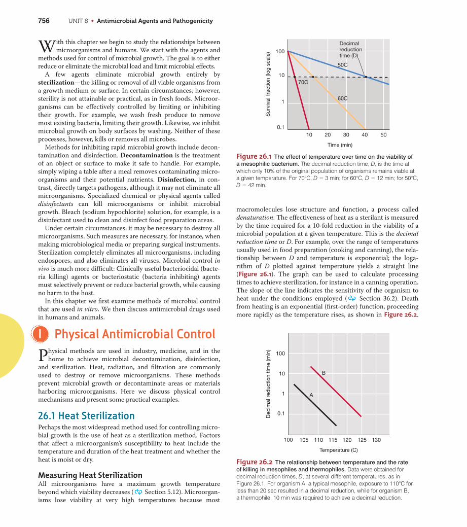
denaturation. The effectiveness of heat as a sterilant is measured
by the time required for a 10-fold reduction in the viability of a
microbial population at a given temperature. This is the decimal
reduction time or D. For example, over the range of temperatures
usually used in food preparation (cooking and canning), the rela-
tionship between D and temperature is exponential; the loga-
rithm of D plotted against temperature yields a straight line
(Figure 26.1). The graph can be used to calculate processing
times to achieve sterilization, for instance in a canning operation.
The slope of the line indicates the sensitivity of the organism to
heat under the conditions employed ( Section 36.2). Death
from heating is an exponential (first-order) function, proceeding
more rapidly as the temperature rises, as shown in Figure 26.2.
Figure 26.1 The effect of temperature over time on the viability of
a mesophilic bacterium. The decimal reduction time, D, is the time atwhich only 10% of the original population of organisms remains viable ata given temperature. For 70°C, D = 3 min; for 60°C, D = 12 min; for 50°C,D = 42 min.
0.1
1
10
10 20 30 40 50
100
Sur
viva
l fra
ctio
n (lo
g sc
ale)
Time (min)
Decimalreductiontime (D)
70C
50C
60C
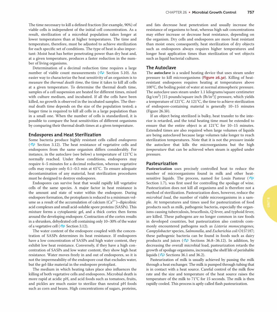
Figure 26.2 The relationship between temperature and the rate
of killing in mesophiles and thermophiles. Data were obtained fordecimal reduction times, D, at several different temperatures, as in Figure 26.1. For organism A, a typical mesophile, exposure to 110°C forless than 20 sec resulted in a decimal reduction, while for organism B, a thermophile, 10 min was required to achieve a decimal reduction.
100
105 110 115 120 125 130
Temperature (C)
Dec
imal
red
uctio
n tim
e (m
in)
10 B
A1
0.1
100
CHAPTER 26 • Microbial Growth Control 757
The time necessary to kill a defined fraction (for example, 90%) of
viable cells is independent of the initial cell concentration. As a
result, sterilization of a microbial population takes longer at
lower temperatures than at higher temperatures. The time and
temperature, therefore, must be adjusted to achieve sterilization
for each specific set of conditions. The type of heat is also impor-
tant: Moist heat has better penetrating power than dry heat and,
at a given temperature, produces a faster reduction in the num-
ber of living organisms.
Determination of a decimal reduction time requires a large
number of viable count measurements ( Section 5.10). An
easier way to characterize the heat sensitivity of an organism is to
measure the thermal death time, the time it takes to kill all cells
at a given temperature. To determine the thermal death time,
samples of a cell suspension are heated for different times, mixed
with culture medium, and incubated. If all the cells have been
killed, no growth is observed in the incubated samples. The ther-
mal death time depends on the size of the population tested; a
longer time is required to kill all cells in a large population than
in a small one. When the number of cells is standardized, it is
possible to compare the heat sensitivities of different organisms
by comparing their thermal death times at a given temperature.
Endospores and Heat SterilizationSome bacteria produce highly resistant cells called endospores
( Section 3.12). The heat resistance of vegetative cells and
endospores from the same organism differs considerably. For
instance, in the autoclave (see below) a temperature of 1218C is
normally reached. Under these conditions, endospores may
require 4–5 minutes for a decimal reduction, whereas vegetative
cells may require only 0.1–0.5 min at 658C. To ensure adequate
decontamination of any material, heat sterilization procedures
must be designed to destroy endospores.
Endospores can survive heat that would rapidly kill vegetative
cells of the same species. A major factor in heat resistance is
the amount and state of water within the endospore. During
endospore formation, the protoplasm is reduced to a minimum vol-
ume as a result of the accumulation of calcium (Ca21)–dipicolinic
acid complexes and small acid-soluble spore proteins (SASPs). This
mixture forms a cytoplasmic gel, and a thick cortex then forms
around the developing endospore. Contraction of the cortex results
in a shrunken, dehydrated cell containing only 10–30% of the water
of a vegetative cell ( Section 3.12).
The water content of the endospore coupled with the concen-
tration of SASPs determines its heat resistance. If endospores
have a low concentration of SASPs and high water content, they
exhibit low heat resistance. Conversely, if they have a high con-
centration of SASPs and low water content, they show high heat
resistance. Water moves freely in and out of endospores, so it is
not the impermeability of the endospore coat that excludes water,
but the gel-like material in the endospore protoplast.
The medium in which heating takes place also influences the
killing of both vegetative cells and endospores. Microbial death is
more rapid at acidic pH, and acid foods such as tomatoes, fruits,
and pickles are much easier to sterilize than neutral pH foods
such as corn and beans. High concentrations of sugars, proteins,
and fats decrease heat penetration and usually increase the
resistance of organisms to heat, whereas high salt concentrations
may either increase or decrease heat resistance, depending on
the organism. Dry cells and endospores are more heat resistant
than moist ones; consequently, heat sterilization of dry objects
such as endospores always requires higher temperatures and
longer heat application times than sterilization of wet objects
such as liquid bacterial cultures.
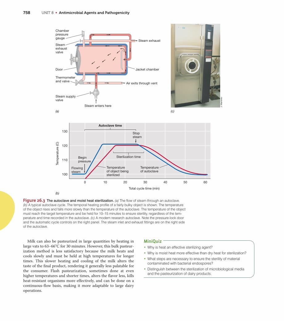
The AutoclaveThe autoclave is a sealed heating device that uses steam under
pressure to kill microorganisms (Figure 26.3a). Killing of heat-
resistant endospores requires heating at temperatures above
1008C, the boiling point of water at normal atmospheric pressure.
The autoclave uses steam under 1.1 kilograms/square centimeter
(kg/cm2) [15 pounds/square inch (lb/in2)] pressure, which yields
a temperature of 1218C. At 1218C, the time to achieve sterilization
of endospore-containing material is generally 10–15 minutes
(Figure 26.3b).
If an object being sterilized is bulky, heat transfer to the inte-
rior is retarded, and the total heating time must be extended to
ensure that the entire object is at 1218C for 10–15 minutes.
Extended times are also required when large volumes of liquids
are being autoclaved because large volumes take longer to reach
sterilization temperatures. Note that it is not the pressure inside
the autoclave that kills the microorganisms but the high
temperature that can be achieved when steam is applied under
pressure.
PasteurizationPasteurization uses precisely controlled heat to reduce the
number of microorganisms found in milk and other heat-
sensitive liquids. The process, named for Louis Pasteur (
Section 1.7), was first used for controlling the spoilage of wine.
Pasteurization does not kill all organisms and is therefore not a
method of sterilization. Pasteurization does, however, reduce the
microbial load, the number of viable microorganisms in a sam-
ple. At temperatures and times used for pasteurization of food
products such as milk, pathogenic bacteria, especially the organ-
isms causing tuberculosis, brucellosis, Q fever, and typhoid fever,
are killed. These pathogens are no longer common in raw foods
in developed countries, but pasteurization also controls com-
monly encountered pathogens such as Listeria monocytogenes,
Campylobacter species, Salmonella, and Escherichia coli O157:H7;
these pathogenic bacteria can be found in foods such as dairy
products and juices ( Sections 36.8–36.12). In addition, by
decreasing the overall microbial load, pasteurization retards the
growth of spoilage organisms, increasing the shelf life of perishable
liquids ( Sections 36.1 and 36.2).
Pasteurization of milk is usually achieved by passing the milk
through a heat exchanger. The milk is pumped through tubing that
is in contact with a heat source. Careful control of the milk flow
rate and the size and temperature of the heat source raises the
temperature of the milk to 718C for 15 seconds. The milk is then
rapidly cooled. This process is aptly called flash pasteurization.
UN
IT 8
UNIT 8 • Antimicrobial Agents and Pathogenicity758
Milk can also be pasteurized in large quantities by heating in
large vats to 63–668C for 30 minutes. However, this bulk pasteur-
ization method is less satisfactory because the milk heats and
cools slowly and must be held at high temperatures for longer
times. This slower heating and cooling of the milk alters the
taste of the final product, rendering it generally less palatable for
the consumer. Flash pasteurization, sometimes done at even
higher temperatures and shorter times, alters the flavor less, kills
heat-resistant organisms more effectively, and can be done on a
continuous-flow basis, making it more adaptable to large dairy
operations.
MiniQuiz• Why is heat an effective sterilizing agent?
• Why is moist heat more effective than dry heat for sterilization?
• What steps are necessary to ensure the sterility of materialcontaminated with bacterial endospores?
• Distinguish between the sterilization of microbiological mediaand the pasteurization of dairy products.
Figure 26.3 The autoclave and moist heat sterilization. (a) The flow of steam through an autoclave. (b) A typical autoclave cycle. The temporal heating profile of a fairly bulky object is shown. The temperatureof the object rises and falls more slowly than the temperature of the autoclave. The temperature of the objectmust reach the target temperature and be held for 10–15 minutes to ensure sterility, regardless of the tem-perature and time recorded in the autoclave. (c) A modern research autoclave. Note the pressure-lock doorand the automatic cycle controls on the right panel. The steam inlet and exhaust fittings are on the right sideof the autoclave.
Chamberpressuregauge
Steamexhaustvalve
Door
(a) (c)
(b)
Thermometerand valve
Steam supplyvalve
Tem
per
atur
e (C
)
130
120
110
100
0 10 20 30 40 50 60
Total cycle time (min)
Air exits through vent
J. M
artin
ko
Jacket chamber
Steam exhaust
Autoclave time
Sterilization timeBeginpressure
Flowingsteam
Temperatureof object beingsterilized
Temperatureof autoclave
Stopsteam
Steam enters here
CHAPTER 26 • Microbial Growth Control 759
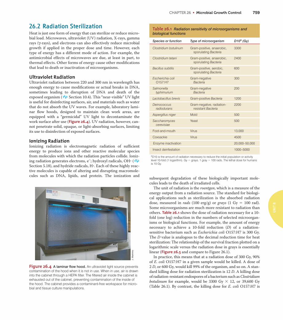
Figure 26.4 A laminar flow hood. An ultraviolet light source preventscontamination of the hood when it is not in use. When in use, air is drawninto the cabinet through a HEPA filter. The filtered air inside the cabinet isexhausted out of the cabinet, preventing contamination of the inside ofthe hood. The cabinet provides a contaminant-free workspace for micro-bial and tissue culture manipulations.
J. M
artin
ko
Table 26.1 Radiation sensitivity of microorganisms andbiological functions
Species or function Type of microorganism D10a (Gy)
26.2 Radiation SterilizationHeat is just one form of energy that can sterilize or reduce micro-
bial load. Microwaves, ultraviolet (UV) radiation, X-rays, gamma
rays (γ-rays), and electrons can also effectively reduce microbial
growth if applied in the proper dose and time. However, each
type of energy has a different mode of action. For example, the
antimicrobial effects of microwaves are due, at least in part, to
thermal effects. Other forms of energy cause other modifications
that lead to death or inactivation of microorganisms.
Ultraviolet RadiationUltraviolet radiation between 220 and 300 nm in wavelength has
enough energy to cause modifications or actual breaks in DNA,
sometimes leading to disruption of DNA and death of the
exposed organism ( Section 10.4). This “near-visible” UV light
is useful for disinfecting surfaces, air, and materials such as water
that do not absorb the UV waves. For example, laboratory lami-
nar flow hoods, designed to maintain clean work areas, are
equipped with a “germicidal” UV light to decontaminate the
work surface after use (Figure 26.4). UV radiation, however, can-
not penetrate solid, opaque, or light-absorbing surfaces, limiting
its use to disinfection of exposed surfaces.
Ionizing RadiationIonizing radiation is electromagnetic radiation of sufficient
energy to produce ions and other reactive molecular species
from molecules with which the radiation particles collide. Ioniz-
ing radiation generates electrons, e–; hydroxyl radicals, OH? (
Section 5.18), and hydride radicals, H?. Each of these highly reac-
tive molecules is capable of altering and disrupting macromole-
cules such as DNA, lipids, and protein. The ionization andsubsequent degradation of these biologically important mole-
cules leads to the death of irradiated cells.
The unit of radiation is the roentgen, which is a measure of the
energy output from a radiation source. The standard for biologi-
cal applications such as sterilization is the absorbed radiation
dose, measured in rads (100 erg/g) or grays (1 Gy 5 100 rad).
Some microorganisms are much more resistant to radiation than
others. Table 26.1 shows the dose of radiation necessary for a 10-
fold (one log) reduction in the numbers of selected microorgan-
isms or biological functions. For example, the amount of energy
necessary to achieve a 10-fold reduction (D) of a radiation-
sensitive bacterium such as Escherichia coli O157:H7 is 300 Gy.
The D value is analogous to the decimal reduction time for heat
sterilization: The relationship of the survival fraction plotted on a
logarithmic scale versus the radiation dose in grays is essentially
linear (Figure 26.5 and compare to Figure 26.1).
In practice, this means that at a radiation dose of 300 Gy, 90%
of E. coli O157:H7 in a given sample would be killed. A dose of
2 D, or 600 Gy, would kill 99% of the organism, and so on. A stan-
dard killing dose for radiation sterilization is 12 D. A killing dose
of radiation-resistant endospores of a bacterium such as Clostridium
botulinum for example, would be 3300 Gy 3 12, or 39,600 Gy
(Table 26.1). By contrast, the killing dose for E. coli O157:H7 is
UN
IT 8
Clostridium botulinum Gram-positive, anaerobic,sporulating Bacteria
3300
Clostridium tetani Gram-positive, anaerobic,sporulating Bacteria
2400
Bacillus subtilis Gram-positive, aerobic,sporulating Bacteria
600
Escherichia coliO157:H7
Gram-negative Bacteria
300
Salmonella typhimurium
Gram-negative Bacteria
200
Lactobacillus brevis Gram-positive Bacteria 1200
Deinococcus radiodurans
Gram-negative, radiation-resistant Bacteria
2200
Aspergillus niger Mold 500
Saccharomyces cerevisiae
Yeast 500
Foot-and-mouth Virus 13,000
Coxsackie Virus 4500
Enzyme inactivation 20,000–50,000
Insect deinfestation 1000–5000
aD10 is the amount of radiation necessary to reduce the initial population or activity level 10-fold (1 logarithm). Gy = grays. 1 gray = 100 rads. The lethal dose for humansis 10 Gy.
UNIT 8 • Antimicrobial Agents and Pathogenicity760
Table 26.3 Recommended radiation dose for decontaminationof selected foods
Food type kiloGrays
Fruit 1
Poultry 3
Spices, seasonings 30
only 3600 Gy. In general, microorganisms are much more resis-
tant to ionizing radiation than are multicellular organisms. For
example, the lethal radiation dose for humans can be as low as
10 Gy if delivered over a short time (several minutes)!
Radiation PracticesSeveral radiation sources are useful for sterilization. Common
sources of ionizing radiation include cathode ray tubes that gen-
erate electron beams, X-ray machines, and radioactive nuclides60Co and 137Cs, which are relatively inexpensive by-products of
nuclear fission. These sources produce electrons (e2), X-rays, or
γ-rays, respectively, all of which have sufficient energy to effi-
ciently kill microorganisms. In addition, X-rays and γ-rays pene-
trate solids and liquids, making them ideal for treatment of bulk
items such as ground beef or cereal grains.
Radiation is currently used for sterilization and decontamina-
tion in the medical supplies and food industries. In the United
States, the Food and Drug Administration has approved the use
of radiation for sterilization of such diverse items as surgical sup-
plies, disposable labware, drugs, and even tissue grafts (Table26.2). However, because of the required specialized equipment,
costs, and hazards associated with radiation techniques, this type
of sterilization is limited to large industrial applications or spe-
cialized facilities.
Certain foods and food products are also routinely irradiated
to ensure sterilization, pasteurization, or insect deinfestation.
Radiation is approved by the World Health Organization and
can be used in the United States for decontamination of foods
particularly susceptible to microbial contamination such as
fresh produce, meat products, chicken, and spices (Table 26.3and Section 36.2). The use of radiation for these purposes is
an established and accepted technology in many countries.
However, the practice has not been readily accepted in some
countries such as the United States because of fears of possible
radioactive contamination, alteration in nutritional value, pro-
duction of toxic or carcinogenic products, and perceived “off”
tastes in irradiated food.
MiniQuiz• Define the decimal reduction dose and the killing dose for
radiation treatment of microorganisms.
• Why is ionizing radiation more effective than UV radiation forsterilization of food products?
26.3 Filter SterilizationHeat is an effective way to decontaminate most liquids and can
even be used to treat gases. Heat-sensitive liquids and gases,
however, must be treated by other methods. Filtration accom-
plishes decontamination and even sterilization without exposure
to denaturing heat. The liquid or gas is passed through a filter, a
device with pores too small for the passage of microorganisms,
but large enough to allow the passage of the liquid or gas. The selec-
tion of filters for sterilization must account for the size range of
the contaminants to be excluded. Some microbial cells are
greater than 10 �m in diameter, while the smallest bacteria are
less than 0.3 �m in diameter. Historically, selective filtration
methods were used to define and isolate viruses, most of which
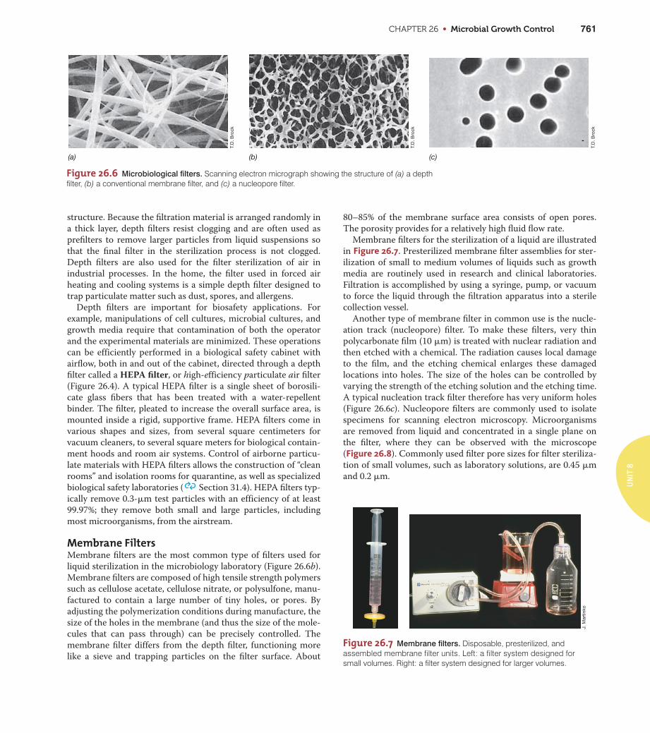
range from 25 nm to 200 nm (0.2 �m) in diameter. Figure 26.6illustrates major types of filters.
Depth FiltersA depth filter is a fibrous sheet or mat made from a random array
of overlapping paper or borosilicate (glass) fibers (Figure 26.6a).
The depth filter traps particles in the network of fibers in the
Radiation (Grays)
Sur
viva
l fra
ctio
n (lo
g sc
ale)
10% survival
0.01
0.1 D10
1
Figure 26.5 Relationship between the survival fraction and the
radiation dose of a microorganism. The D10, or decimal reductiondose, can be interpolated from the data as shown.
Table 26.2 Medical and laboratory products sterilized by radiation
Tissue grafts DrugsMedical and laboratory supplies
Cartilage Chloramphenicol Disposable labwareTendon Ampicillin Culture mediaSkin Tetracycline SyringesHeart valve Atropine Surgical equipment
Vaccines SuturesOintments
CHAPTER 26 • Microbial Growth Control 761
(a) (b) (c)
T.D
. Bro
ck
T.D
. Bro
ck
T.D
. Bro
ck
Figure 26.6 Microbiological filters. Scanning electron micrograph showing the structure of (a) a depthfilter, (b) a conventional membrane filter, and (c) a nucleopore filter.
J. M
artin
ko
Figure 26.7 Membrane filters. Disposable, presterilized, andassembled membrane filter units. Left: a filter system designed for small volumes. Right: a filter system designed for larger volumes.
structure. Because the filtration material is arranged randomly in
a thick layer, depth filters resist clogging and are often used as
prefilters to remove larger particles from liquid suspensions so
that the final filter in the sterilization process is not clogged.
Depth filters are also used for the filter sterilization of air in
industrial processes. In the home, the filter used in forced air
heating and cooling systems is a simple depth filter designed to
trap particulate matter such as dust, spores, and allergens.
Depth filters are important for biosafety applications. For
example, manipulations of cell cultures, microbial cultures, and
growth media require that contamination of both the operator
and the experimental materials are minimized. These operations
can be efficiently performed in a biological safety cabinet with
airflow, both in and out of the cabinet, directed through a depth
filter called a HEPA filter, or high-efficiency particulate air filter
(Figure 26.4). A typical HEPA filter is a single sheet of borosili-
cate glass fibers that has been treated with a water-repellent
binder. The filter, pleated to increase the overall surface area, is
mounted inside a rigid, supportive frame. HEPA filters come in
various shapes and sizes, from several square centimeters for
vacuum cleaners, to several square meters for biological contain-
ment hoods and room air systems. Control of airborne particu-
late materials with HEPA filters allows the construction of “clean
rooms” and isolation rooms for quarantine, as well as specialized
biological safety laboratories ( Section 31.4). HEPA filters typ-
ically remove 0.3-�m test particles with an efficiency of at least
99.97%; they remove both small and large particles, including
most microorganisms, from the airstream.
Membrane FiltersMembrane filters are the most common type of filters used for
liquid sterilization in the microbiology laboratory (Figure 26.6b).
Membrane filters are composed of high tensile strength polymers
such as cellulose acetate, cellulose nitrate, or polysulfone, manu-
factured to contain a large number of tiny holes, or pores. By
adjusting the polymerization conditions during manufacture, the
size of the holes in the membrane (and thus the size of the mole-
cules that can pass through) can be precisely controlled. The
membrane filter differs from the depth filter, functioning more
like a sieve and trapping particles on the filter surface. About
80–85% of the membrane surface area consists of open pores.
The porosity provides for a relatively high fluid flow rate.
Membrane filters for the sterilization of a liquid are illustrated
in Figure 26.7. Presterilized membrane filter assemblies for ster-
ilization of small to medium volumes of liquids such as growth
media are routinely used in research and clinical laboratories.
Filtration is accomplished by using a syringe, pump, or vacuum
to force the liquid through the filtration apparatus into a sterile
collection vessel.
Another type of membrane filter in common use is the nucle-
ation track (nucleopore) filter. To make these filters, very thin
polycarbonate film (10 �m) is treated with nuclear radiation and
then etched with a chemical. The radiation causes local damage
to the film, and the etching chemical enlarges these damaged
locations into holes. The size of the holes can be controlled by
varying the strength of the etching solution and the etching time.
A typical nucleation track filter therefore has very uniform holes
(Figure 26.6c). Nucleopore filters are commonly used to isolate
specimens for scanning electron microscopy. Microorganisms
are removed from liquid and concentrated in a single plane on
the filter, where they can be observed with the microscope
(Figure 26.8). Commonly used filter pore sizes for filter steriliza-
tion of small volumes, such as laboratory solutions, are 0.45 �m
and 0.2 �m.
UN
IT 8
UNIT 8 • Antimicrobial Agents and Pathogenicity762
MiniQuiz• Why are filters used for sterilization of heat-sensitive liquids?
• Describe the use of depth filters for maintaining clean air inhospitals, laboratories, and the home.
II Chemical AntimicrobialControl
In the home, workplace, and laboratory, chemicals are routinely
used to control microbial growth. An antimicrobial agent is a
natural or synthetic chemical that kills or inhibits the growth of
microorganisms. Agents that kill organisms are called -cidal
agents, with a prefix indicating the type of microorganism killed.
Thus, they are called bacteriocidal, fungicidal, and viricidal
agents because they kill bacteria, fungi, and viruses, respectively.
Agents that do not kill but only inhibit growth are called -static
agents. These include bacteriostatic, fungistatic, and viristatic
compounds.
26.4 Chemical Growth ControlAntimicrobial agents can differ in their selective toxicity. Nonse-
lective agents have similar effects on all cells. Selective agents are
more toxic for microorganisms than for animal tissues. Antimi-
crobial agents with selective toxicity are especially useful for
treating infectious diseases because they kill selected microor-
ganisms in vivo without harming the host. They are described
later in this chapter. Here we discuss chemical agents that have
relatively broad toxicity and are widely used for limiting micro-
bial growth in vitro.
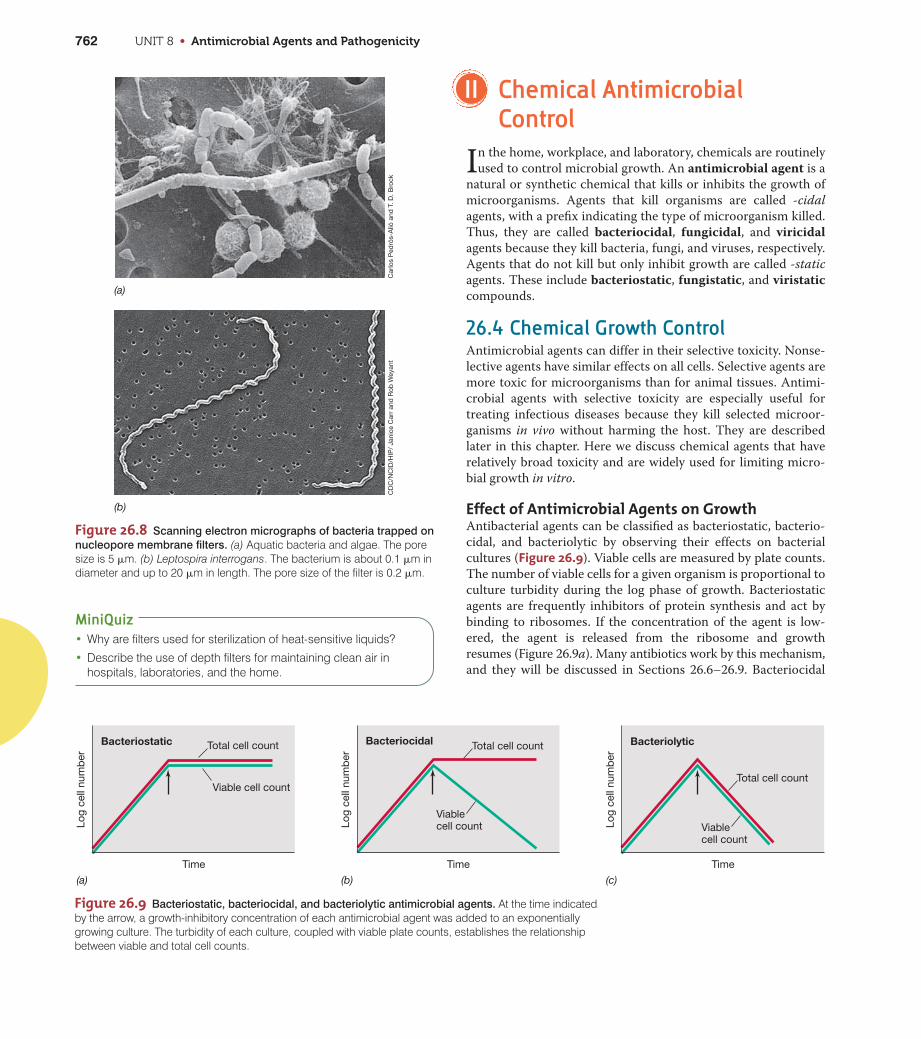
Effect of Antimicrobial Agents on GrowthAntibacterial agents can be classified as bacteriostatic, bacterio-
cidal, and bacteriolytic by observing their effects on bacterial
cultures (Figure 26.9). Viable cells are measured by plate counts.
The number of viable cells for a given organism is proportional to
culture turbidity during the log phase of growth. Bacteriostatic
agents are frequently inhibitors of protein synthesis and act by
binding to ribosomes. If the concentration of the agent is low-
ered, the agent is released from the ribosome and growth
resumes (Figure 26.9a). Many antibiotics work by this mechanism,
and they will be discussed in Sections 26.6–26.9. Bacteriocidal
Car
los
Ped
rós-
Alió
and
T. D
. Bro
ck
CD
C/N
CID
/HIP
/ Ja
nice
Car
r an
d R
ob W
eyan
t
(a)
(b)
Figure 26.8 Scanning electron micrographs of bacteria trapped on
nucleopore membrane filters. (a) Aquatic bacteria and algae. The poresize is 5 �m. (b) Leptospira interrogans. The bacterium is about 0.1 �m indiameter and up to 20 �m in length. The pore size of the filter is 0.2 �m.
Figure 26.9 Bacteriostatic, bacteriocidal, and bacteriolytic antimicrobial agents. At the time indicatedby the arrow, a growth-inhibitory concentration of each antimicrobial agent was added to an exponentiallygrowing culture. The turbidity of each culture, coupled with viable plate counts, establishes the relationshipbetween viable and total cell counts.
Total cell count Total cell count
Total cell countViable cell count
Viablecell count Viable
cell count
Bacteriostatic Bacteriocidal Bacteriolytic
Log
cell
num
ber
Log
cell
num
ber
Log
cell
num
ber
Time Time Time
(c)(b)(a)
CHAPTER 26 • Microbial Growth Control 763
Figure 26.10 Antimicrobial agent susceptibility assay using
dilution methods. The assay defines the minimum inhibitory concentra-tion (MIC). A series of increasing concentrations of antimicrobial agent isprepared in the culture medium. Each tube is inoculated with a specificconcentration of a test organism, followed by a defined incubation period.Growth, measured as turbidity, occurs in those tubes with antimicrobialagent concentrations below the MIC.
T. D
. Bro
ck
Minimuminhibitoryconcentration
Discs containing antimicrobial agents are placedon surface
Inoculate plate witha liquid culture ofa test organism
Nutrientagar plate
Incubate for 24–48 h
Test organism shows susceptibility to some agents, indicated by inhibition of bacterial growth around discs (zones of inhibition)
Figure 26.11 Antimicrobial agent susceptibility assay using diffu-
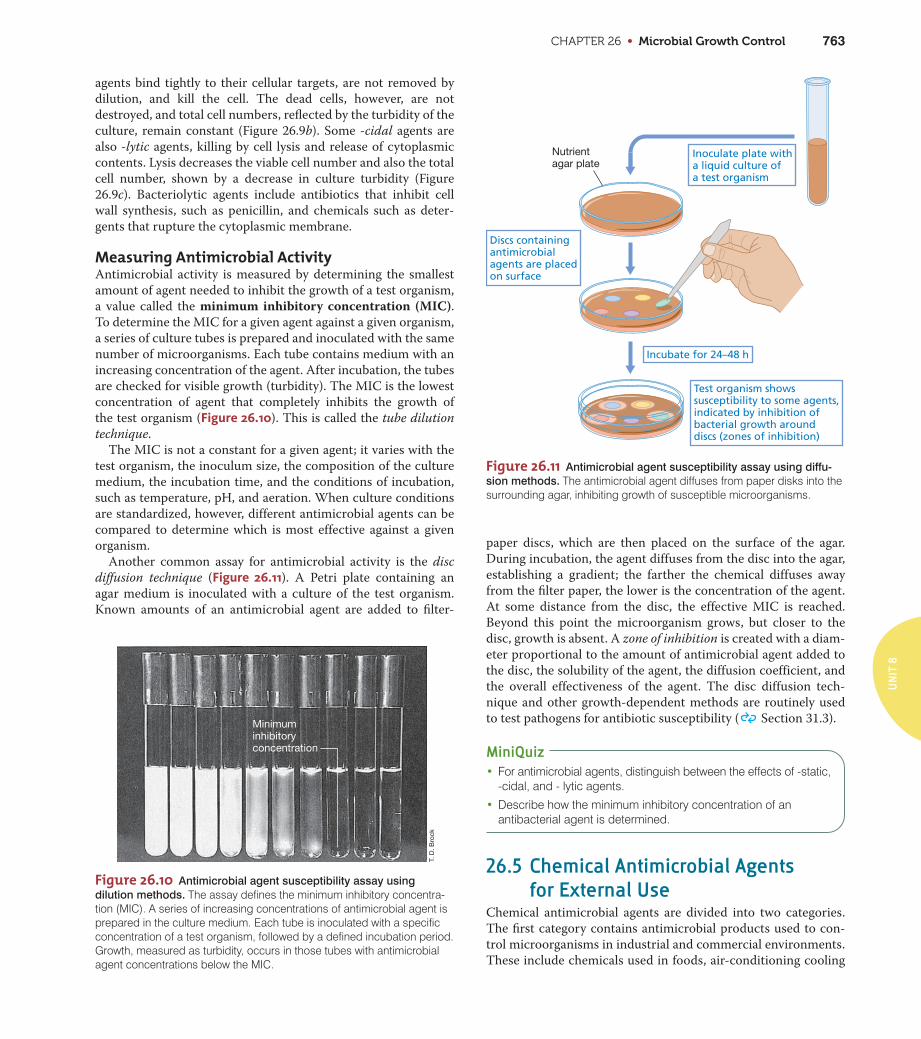
sion methods. The antimicrobial agent diffuses from paper disks into thesurrounding agar, inhibiting growth of susceptible microorganisms.
agents bind tightly to their cellular targets, are not removed by
dilution, and kill the cell. The dead cells, however, are not
destroyed, and total cell numbers, reflected by the turbidity of the
culture, remain constant (Figure 26.9b). Some -cidal agents are
also -lytic agents, killing by cell lysis and release of cytoplasmic
contents. Lysis decreases the viable cell number and also the total
cell number, shown by a decrease in culture turbidity (Figure
26.9c). Bacteriolytic agents include antibiotics that inhibit cell
wall synthesis, such as penicillin, and chemicals such as deter-
gents that rupture the cytoplasmic membrane.
Measuring Antimicrobial ActivityAntimicrobial activity is measured by determining the smallest
amount of agent needed to inhibit the growth of a test organism,
a value called the minimum inhibitory concentration (MIC).
To determine the MIC for a given agent against a given organism,
a series of culture tubes is prepared and inoculated with the same
number of microorganisms. Each tube contains medium with an
increasing concentration of the agent. After incubation, the tubes
are checked for visible growth (turbidity). The MIC is the lowest
concentration of agent that completely inhibits the growth of
the test organism (Figure 26.10). This is called the tube dilution
technique.
The MIC is not a constant for a given agent; it varies with the
test organism, the inoculum size, the composition of the culture
medium, the incubation time, and the conditions of incubation,
such as temperature, pH, and aeration. When culture conditions
are standardized, however, different antimicrobial agents can be
compared to determine which is most effective against a given
organism.
Another common assay for antimicrobial activity is the disc
diffusion technique (Figure 26.11). A Petri plate containing an
agar medium is inoculated with a culture of the test organism.
Known amounts of an antimicrobial agent are added to filter-
paper discs, which are then placed on the surface of the agar.
During incubation, the agent diffuses from the disc into the agar,
establishing a gradient; the farther the chemical diffuses away
from the filter paper, the lower is the concentration of the agent.
At some distance from the disc, the effective MIC is reached.
Beyond this point the microorganism grows, but closer to the
disc, growth is absent. A zone of inhibition is created with a diam-
eter proportional to the amount of antimicrobial agent added to
the disc, the solubility of the agent, the diffusion coefficient, and
the overall effectiveness of the agent. The disc diffusion tech-
nique and other growth-dependent methods are routinely used
to test pathogens for antibiotic susceptibility ( Section 31.3).
MiniQuiz• For antimicrobial agents, distinguish between the effects of -static,
-cidal, and - lytic agents.
• Describe how the minimum inhibitory concentration of anantibacterial agent is determined.
26.5 Chemical Antimicrobial Agentsfor External Use
Chemical antimicrobial agents are divided into two categories.
The first category contains antimicrobial products used to con-
trol microorganisms in industrial and commercial environments.
These include chemicals used in foods, air-conditioning cooling
UN
IT 8
UNIT 8 • Antimicrobial Agents and Pathogenicity764
towers, textile and paper products, fuel tanks, and so on; some of
these chemicals are so toxic that exposure can affect human
health. Table 26.4 provides examples of industrial applications
for chemicals used to control microbial growth.
The second category of chemical antimicrobial agents contains
products designed to prevent growth of human pathogens in
inanimate environments and on external body surfaces. This cat-
egory is subdivided into sterilants, disinfectants, sanitizers, and
antiseptics.
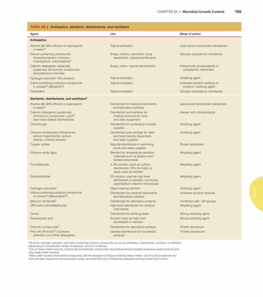
SterilantsChemical sterilants, also called sterilizers or sporicides,
destroy all forms of microbial life, including endospores. Chemi-
cal sterilants are used in situations where it is impractical to use
heat (Section 26.1) or radiation (Section 26.2) for decontamina-
tion or sterilization. Hospitals and laboratories, for example,
must be able to decontaminate and sterilize heat-sensitive mate-
rials, such as thermometers, lensed instruments, polyethylene
tubing, catheters, and reusable medical equipment such as
respirometers. Some form of cold sterilization is usually used for
these purposes. Cold sterilization is performed in enclosed
devices that resemble autoclaves, but which employ a gaseous
chemical agent such as ethylene oxide, formaldehyde, peroxy-
acetic acid, or hydrogen peroxide. Liquid sterilants such as a
sodium hypochlorite (bleach) solution or amylphenol are used
for instruments that cannot withstand high temperatures or gas
(Table 26.5).
Disinfectants and Sanitizers Disinfectants are chemicals that kill microorganisms, but not
necessarily endospores, and are used on inanimate objects. For
example, disinfectants such as ethanol and cationic detergents
are used to disinfect floors, tables, bench tops, walls, and so on.
These agents are important for infection control in, for example,
hospitals and other medical settings. General disinfectants are
used in households, swimming pools, and water purification sys-
tems (Table 26.5).
Sanitizers are agents that reduce, but may not eliminate,
microbial numbers to levels considered to be safe. Food contact
sanitizers are widely used in the food industry to treat surfaces
such as mixing and cooking equipment, dishes, and utensils.
Non–food contact sanitizers are used to treat surfaces such as
counters, floors, walls, carpets, and laundry (Table 26.5).
Antiseptics and GermicidesAntiseptics and germicides are chemical agents that kill or
inhibit growth of microorganisms and that are nontoxic enough
to be applied to living tissues. Most of the compounds in this cat-
egory are used for handwashing (Microbial Sidebar, “Preventing
Antimicrobial Drug Resistance”) or for treating surface wounds
(Table 26.5). Under some conditions, certain antiseptics are also
effective disinfectants; they are effective antimicrobial agents
when applied to inanimate surfaces. Ethanol, for example, is cate-
gorized as an antiseptic, but can also be a disinfectant. This
depends on the concentration of ethanol used and the exposure
time, with disinfection generally requiring higher ethanol con-
centrations and exposure times of several minutes. The Food and
Drug Administration in the United States regulates the formula-
tion, manufacture, and use of antiseptics and germicides because
these agents involve direct human exposure and contact.
Antimicrobial EfficacySeveral factors affect the efficacy of chemical antimicrobial
agents. For example, many disinfectants are neutralized by
organic material. These materials reduce effective disinfectant
concentrations and microbial killing capacity. Furthermore,
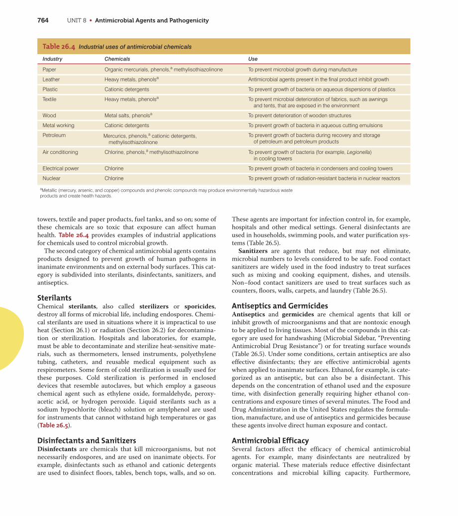
Table 26.4 Industrial uses of antimicrobial chemicals
Industry Chemicals Use
Paper Organic mercurials, phenols,a methylisothiazolinone To prevent microbial growth during manufacture
Leather Heavy metals, phenolsa Antimicrobial agents present in the final product inhibit growth
Plastic Cationic detergents To prevent growth of bacteria on aqueous dispersions of plastics
Textile Heavy metals, phenolsa To prevent microbial deterioration of fabrics, such as awnings and tents, that are exposed in the environment
Wood Metal salts, phenolsa To prevent deterioration of wooden structures
Metal working Cationic detergents To prevent growth of bacteria in aqueous cutting emulsions
Petroleum Mercurics, phenols,a cationic detergents, methylisothiazolinone
To prevent growth of bacteria during recovery and storage of petroleum and petroleum products
Air conditioning Chlorine, phenols,a methylisothiazolinone To prevent growth of bacteria (for example, Legionella)in cooling towers
Electrical power Chlorine To prevent growth of bacteria in condensers and cooling towers
Nuclear Chlorine To prevent growth of radiation-resistant bacteria in nuclear reactors
aMetallic (mercury, arsenic, and copper) compounds and phenolic compounds may produce environmentally hazardous wasteproducts and create health hazards.
CHAPTER 26 • Microbial Growth Control 765
UN
IT 8
Table 26.5 Antiseptics, sterilants, disinfectants, and sanitizers
Agent Use Mode of action
Antiseptics
Alcohol (60–85% ethanol or isopropanol in water)a
Topical antiseptic Lipid solvent and protein denaturant
Phenol-containing compounds (hexachlorophene, triclosan, chloroxylenol, chlorhexidine)b
Soaps, lotions, cosmetics, bodydeodorants, topical disinfectants
Disrupts cytoplasmic membrane
Cationic detergents, especially quaternary ammonium compounds (benzalkonium chloride)
Soaps, lotion, topical disinfectants Interact with phospholipids of cytoplasmic membrane
Hydrogen peroxidea (3% solution) Topical antiseptic Oxidizing agent
Iodine-containing iodophor compoundsin solutiona (Betadine®)
Topical antiseptic Iodinates tyrosine residues of proteins; oxidizing agent
Octenidine Topical antiseptic Disrupts cytoplasmic membrane
Sterilants, disinfectants, and sanitizersc
Alcohol (60–85% ethanol or isopropanolin water)a
Disinfectant for medical instrumentsand laboratory surfaces
Lipid solvent and protein denaturant
Cationic detergents (quaternary ammonium compounds, Lysol®
and many related disinfectants)
Disinfectant and sanitizer for medical instruments, food and dairy equipment
Interact with phospholipids
Chlorine gas Disinfectant for purification of watersupplies
Oxidizing agent
Chlorine compounds (chloramines, sodium hypochlorite, sodium chlorite, chlorine dioxide)
Disinfectant and sanitizer for dairy and food industry equipment, and water supplies
Oxidizing agent
Copper sulfate Algicide disinfectant in swimming pools and water supplies
Protein precipitant
Ethylene oxide (gas) Sterilant for temperature-sensitive materials such as plastics and lensed instruments
Alkylating agent
Formaldehyde 3–8% solution used as surface disinfectant, 37% (formalin) or vapor used as sterilant
Alkylating agent
Glutaraldehyde 2% solution used as high-level disinfectant or sterilant, commonly used fixative in electron microscopy
Alkylating agent
Hydrogen peroxidea Vapor used as sterilant Oxidizing agentIodine-containing iodophor compounds
in solutiona (Wescodyne®)Disinfectant for medical instruments
and laboratory surfacesIodinates tyrosine residues
Mercuric dichlorideb Disinfectant for laboratory surfaces Combines with –SH groupsOPA (ortho-phthalaldehyde) High-level disinfectant for medical
instrumentsAlkylating agent
Ozone Disinfectant for drinking water Strong oxidizing agent
Peroxyacetic acid Solution used as high-level disinfectant or sterilant
Strong oxidizing agent
Phenolic compoundsb Disinfectant for laboratory surfaces Protein denaturant
aAlcohols, hydrogen peroxide, and iodine-containing iodophor compounds can act as antiseptics, disinfectants, sanitizers, or sterilantsdepending on concentration, length of exposure, and form of delivery.bUse of heavy metal (mercury) compounds and phenolic compounds may produce environmentally hazardous waste products andmay create health hazards.cMany water-soluble antimicrobial compounds, with the exception of those containing heavy metals, can be used as sanitizers forfood and dairy equipment and preparation areas, provided their use is followed by adequate draining before food contact.
Pine oils (Pine-Sol®) (contains phenolics and other detergents)
General disinfectant for household surfaces
Protein denaturant
Preventing Antimicrobial Drug Resistance
MICROBIAL SIDEBAR
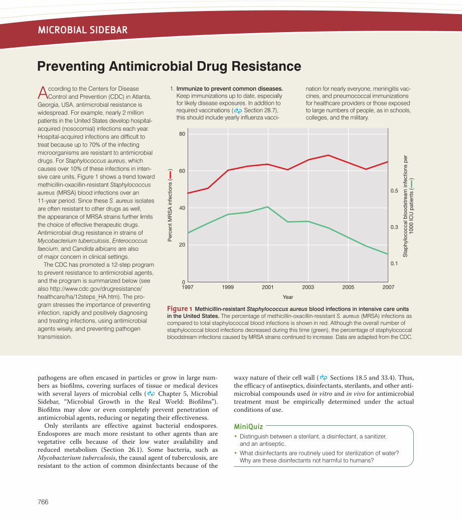
According to the Centers for DiseaseControl and Prevention (CDC) in Atlanta,
Georgia, USA, antimicrobial resistance iswidespread. For example, nearly 2 millionpatients in the United States develop hospital-acquired (nosocomial) infections each year.Hospital-acquired infections are difficult totreat because up to 70% of the infectingmicroorganisms are resistant to antimicrobialdrugs. For Staphylococcus aureus, whichcauses over 10% of these infections in inten-sive care units, Figure 1 shows a trend towardmethicillin-oxacillin-resistant Staphylococcusaureus (MRSA) blood infections over an 11-year period. Since these S. aureus isolatesare often resistant to other drugs as well, the appearance of MRSA strains further limitsthe choice of effective therapeutic drugs.Antimicrobial drug resistance in strains ofMycobacterium tuberculosis, Enterococcusfaecium, and Candida albicans are also of major concern in clinical settings.
The CDC has promoted a 12-step programto prevent resistance to antimicrobial agents,and the program is summarized below (seealso http://www.cdc.gov/drugresistance/healthcare/ha/12steps_HA.htm). The pro-gram stresses the importance of preventinginfection, rapidly and positively diagnosingand treating infections, using antimicrobialagents wisely, and preventing pathogentransmission.
1. Immunize to prevent common diseases.Keep immunizations up to date, especiallyfor likely disease exposures. In addition torequired vaccinations ( Section 28.7),this should include yearly influenza vacci-
nation for nearly everyone, meningitis vac-cines, and pneumococcal immunizationsfor healthcare providers or those exposedto large numbers of people, as in schools,colleges, and the military.
Year
1997 1999 2001 2003 2005 2007
20
0
40
60
80
Per
cent
MR
SA
infe
ctio
ns (
)
0.1
0.3
0.5
Sta
phy
loco
ccal
blo
odst
ream
infe
ctio
ns p
er10
00 IC
U p
atie
nts
(
)
Figure 1 Methicillin-resistant Staphylococcus aureus blood infections in intensive care units
in the United States. The percentage of methicillin-oxacillin-resistant S. aureus (MRSA) infections ascompared to total staphylococcal blood infections is shown in red. Although the overall number ofstaphylococcal blood infections decreased during this time (green), the percentage of staphylococcalbloodstream infections caused by MRSA strains continued to increase. Data are adapted from the CDC.
pathogens are often encased in particles or grow in large num-
bers as biofilms, covering surfaces of tissue or medical devices
with several layers of microbial cells ( Chapter 5, Microbial
Sidebar, “Microbial Growth in the Real World: Biofilms”).
Biofilms may slow or even completely prevent penetration of
antimicrobial agents, reducing or negating their effectiveness.
Only sterilants are effective against bacterial endospores.
Endospores are much more resistant to other agents than are
vegetative cells because of their low water availability and
reduced metabolism (Section 26.1). Some bacteria, such as
Mycobacterium tuberculosis, the causal agent of tuberculosis, are
resistant to the action of common disinfectants because of the
waxy nature of their cell wall ( Sections 18.5 and 33.4). Thus,
the efficacy of antiseptics, disinfectants, sterilants, and other anti-
microbial compounds used in vitro and in vivo for antimicrobial
treatment must be empirically determined under the actual
conditions of use.
MiniQuiz• Distinguish between a sterilant, a disinfectant, a sanitizer,
and an antiseptic.
• What disinfectants are routinely used for sterilization of water?Why are these disinfectants not harmful to humans?
766
2. Avoid unnecessary introduction ofparenteral devices such as catheters.All present a risk of introducing infectiousagents into the body. If such devices arenecessary, remove them as soon aspossible.
3. Target the pathogen. Attempt to culturethe infectious agent while targeting antimi-crobial drug treatment for the most likelypathogens. After positive culture results,adjust the therapy to target the knownpathogen and its antibiotic susceptibility.
4. Access the experts. For serious infections,follow up with an infectious disease expert.Get a second opinion if conditions do notrapidly improve after treatment has begun.
5. Practice antimicrobial control. Be awareand current in knowledge of appropriateantimicrobial drugs and their use. Be surethe treatment offered is current and rec-ommended for the pathogen.
6. Use local data. Obtain and understandthe antibiotic susceptibility profile for theinfectious agent from local healthcaresources.
7. Treat infection, not contamination. Anti-septic techniques must be followed toobtain appropriate samples from infectedtissues. Contaminating organisms maybe present on skin, catheters, or IV lines.Obtain cultures only from the site ofinfection.
8. Treat infection, not colonization. Treat the pathogen and not other colonizing
microorganisms that are not causingdisease. For example, cultures fromnormal skin and throat are often colo-nized with potential pathogens such asStaphylococcus species. These mayhave nothing to do with the currentinfection.
9. Treat with the least exotic antimicrobialagent that will eliminate the pathogen.Treatment with the latest broad-spectrumantibiotic, while efficacious, may not bewarranted if other drugs are still effective.The more a drug is used, the greater thechance that resistant organisms willdevelop. For example, some Enterococcusisolates are already resistant to van-comycin, a relatively new broad-spectrumantibiotic, largely because the drug wasover-prescribed to treat MRSA infectionswhen it was first introduced.
10. Monitor antimicrobial use. Antimicrobialuse should be discontinued as soon asthe prescribed course of treatment iscompleted. If an infection cannot bediagnosed, treatment should be dis-continued. For example, in the case of pharyngitis (sore throat), antibiotictreatment for Streptococcus pyogenes(strep throat) is often started before throatculture results are confirmed. If throatcultures are negative for S. pyogenes,treatment with antibiotics should bestopped. Antibiotics are ineffective fortreatment of the viruses that are the most probable causes of pharyngitis.
11. Isolate the pathogen. Keep areas around infected persons free fromcontamination. Clean up and containbody fluids appropriately. Decontaminatelinens, clothes, and other potentialsources of contamination. In a hospitalsetting, infection control experts shouldbe consulted.
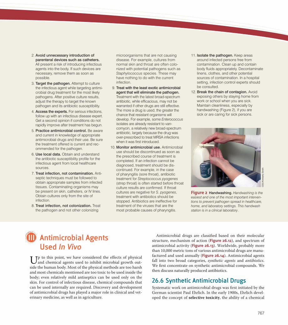
12. Break the chain of contagion. Avoidexposing others by staying home fromwork or school when you are sick.Maintain cleanliness, especially by handwashing (Figure 2), if you are sick or are caring for sick persons.
CD
C/K
imb
erly
Sm
ith
Figure 2 Handwashing. Handwashing is theeasiest and one of the most important interven-tions to prevent pathogen spread in healthcare,home, and laboratory settings. This handwashstation is in a clinical laboratory.
III Antimicrobial Agents Used In Vivo
Up to this point, we have considered the effects of physical
and chemical agents used to inhibit microbial growth out-
side the human body. Most of the physical methods are too harsh
and most chemicals mentioned are too toxic to be used inside the
body; even relatively mild antiseptics can be used only on the
skin. For control of infectious disease, chemical compounds that
can be used internally are required. Discovery and development
of antimicrobial drugs has played a major role in clinical and vet-
erinary medicine, as well as in agriculture.
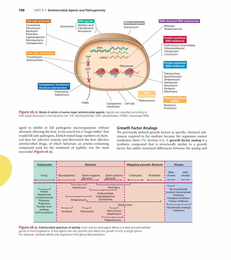
Antimicrobial drugs are classified based on their molecular
structure, mechanism of action (Figure 26.12), and spectrum of
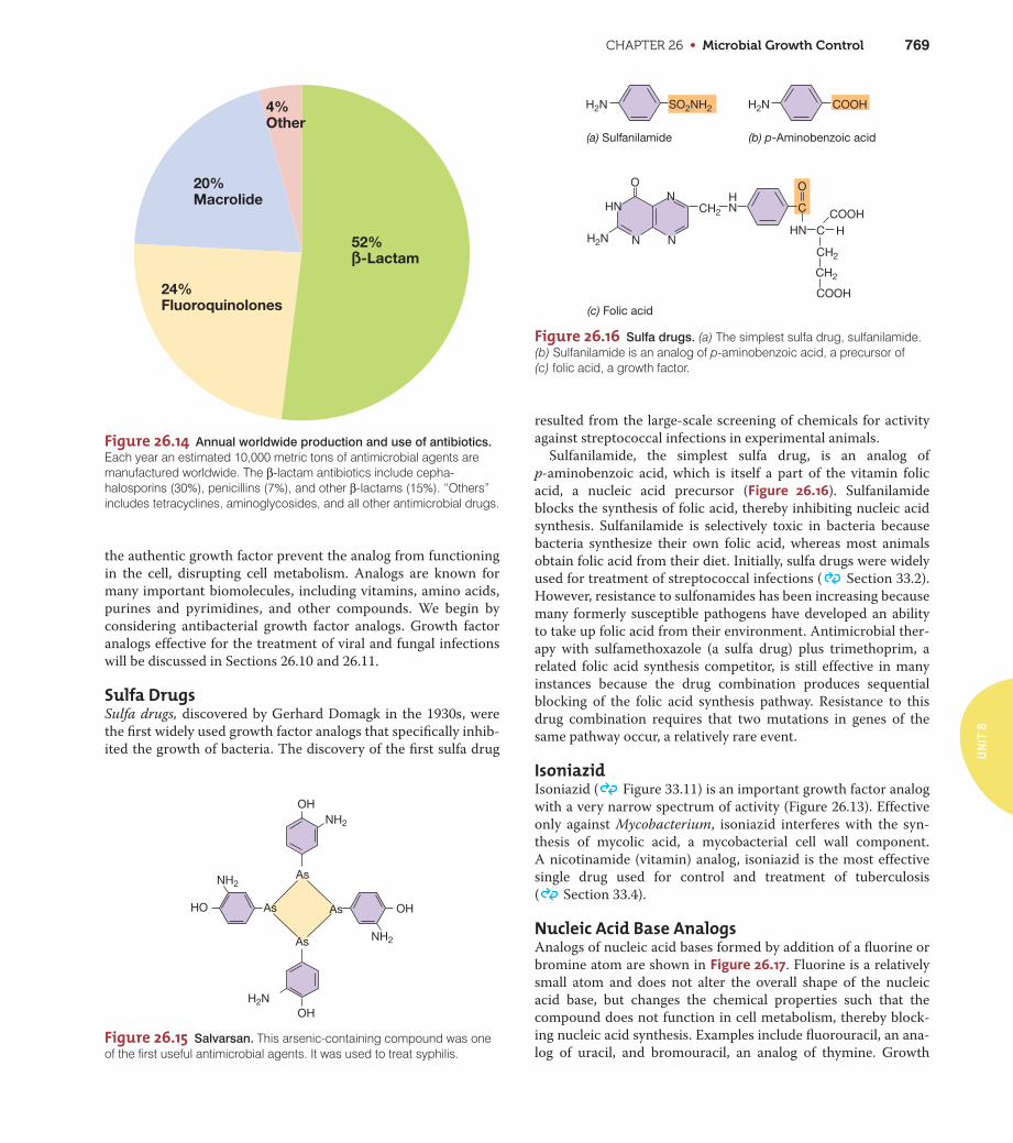
antimicrobial activity (Figure 26.13). Worldwide, probably more
than 10,000 metric tons of various antimicrobial drugs are manu-
factured and used annually (Figure 26.14). Antimicrobial agents
fall into two broad categories, synthetic agents and antibiotics.
We first concentrate on synthetic antimicrobial compounds. We
then discuss naturally produced antibiotics.
26.6 Synthetic Antimicrobial DrugsSystematic work on antimicrobial drugs was first initiated by the
German scientist Paul Ehrlich. In the early 1900s, Ehrlich devel-
oped the concept of selective toxicity, the ability of a chemical
767
UNIT 8 • Antimicrobial Agents and Pathogenicity768
DNA
mRNA
DHF
THF
30 30 30
5050 50
Ribosomes
CycloserineVancomycinBacitracinPenicillinsCephalosporinsMonobactamsCarbapenems
Cell wall synthesis
Nalidixic acidCiprofloxacinNovobiocin
DNA gyrase
Actinomycin
RNA elongationQuinolones Rifampin
Streptovaricins
DNA-directed RNA polymerase
Erythromycin (macrolides)ChloramphenicolClindamycinLincomycin
TetracyclinesSpectinomycinStreptomycinGentamicinKanamycinAmikacinNitrofurans
Protein synthesis(tRNA)
Lipidbiosynthesis
MupirocinPuromycin
PABA
PolymyxinsDaptomycin
Cytoplasmic membrane structure and function
Cell wallPlatensimycin
Cytoplasmicmembrane
Folic acid metabolism
Protein synthesis(50S inhibitors)
Protein synthesis(30S inhibitors)
TrimethoprimSulfonamides
Figure 26.12 Mode of action of some major antimicrobial agents. Agents are classified according totheir target structures in the bacterial cell. THF, tetrahydrofolate; DHF, dihydrofolate; mRNA, messenger RNA.
Nonnucleosidereverse transcriptase
inhibitorsProtease inhibitorsFusion inhibitors
RNAvirusesFungi Mycobacteria Gram-negative
BacteriaGram-positiveBacteria
Chlamydia Rickettsia
Tobramycin Penicillins
SulfonamidesCephalosporins
QuinolonesStreptomycin
PolymyxinsIsoniazid
Tetracycline
Eukaryotes
AzolesAllylamines
CycloheximidePolyenesPolyoxins
Nucleic acidanalogs
Echinocandins
Nucleoside analogsInterferon
Bacteria Obligately parasitic Bacteria Viruses
DNAviruses
VancomycinDaptomycin
Platensimycin
Figure 26.13 Antimicrobial spectrum of activity. Each antimicrobial agent affects a limited and well-definedgroup of microorganisms. A few agents are very specific and affect the growth of only a single genus. For example, isoniazid affects only organisms in the genus Mycobacterium.
agent to inhibit or kill pathogenic microorganisms without
adversely affecting the host. In his search for a “magic bullet” that
would kill only pathogens, Ehrlich tested large numbers of chem-
ical dyes for selective toxicity and discovered the first effective
antimicrobial drugs, of which Salvarsan, an arsenic-containing
compound used for the treatment of syphilis, was the most
successful (Figure 26.15).
Growth Factor AnalogsWe previously defined growth factors as specific chemical sub-
stances required in the medium because the organisms cannot
synthesize them ( Section 4.1). A growth factor analog is a
synthetic compound that is structurally similar to a growth
factor, but subtle structural differences between the analog and
CHAPTER 26 • Microbial Growth Control 769
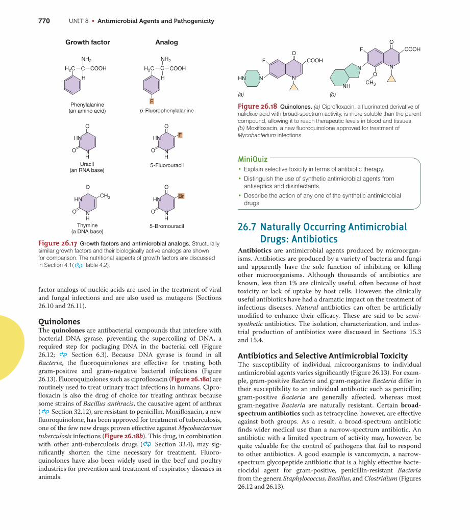
Figure 26.14 Annual worldwide production and use of antibiotics.
Each year an estimated 10,000 metric tons of antimicrobial agents aremanufactured worldwide. The β-lactam antibiotics include cepha-halosporins (30%), penicillins (7%), and other β-lactams (15%). “Others”includes tetracyclines, aminoglycosides, and all other antimicrobial drugs.
52%ß-Lactam
4%Other
20%Macrolide
24%Fluoroquinolones
OH
OH
OH
HO
NH2
NH2
NH2
H2N
As
As
AsAs
Figure 26.15 Salvarsan. This arsenic-containing compound was oneof the first useful antimicrobial agents. It was used to treat syphilis.
H2N H2N
H2N
SO2NH2 COOH
O
HNN
N N
CH2
CH2
CH2
C
O
HN C HCOOH
COOH
(c) Folic acid
NH
(a) Sulfanilamide (b) p-Aminobenzoic acid
Figure 26.16 Sulfa drugs. (a) The simplest sulfa drug, sulfanilamide.(b) Sulfanilamide is an analog of p-aminobenzoic acid, a precursor of (c) folic acid, a growth factor.
resulted from the large-scale screening of chemicals for activity
against streptococcal infections in experimental animals.
Sulfanilamide, the simplest sulfa drug, is an analog of
p-aminobenzoic acid, which is itself a part of the vitamin folic
acid, a nucleic acid precursor (Figure 26.16). Sulfanilamide
blocks the synthesis of folic acid, thereby inhibiting nucleic acid
synthesis. Sulfanilamide is selectively toxic in bacteria because
bacteria synthesize their own folic acid, whereas most animals
obtain folic acid from their diet. Initially, sulfa drugs were widely
used for treatment of streptococcal infections ( Section 33.2).
However, resistance to sulfonamides has been increasing because
many formerly susceptible pathogens have developed an ability
to take up folic acid from their environment. Antimicrobial ther-
apy with sulfamethoxazole (a sulfa drug) plus trimethoprim, a
related folic acid synthesis competitor, is still effective in many
instances because the drug combination produces sequential
blocking of the folic acid synthesis pathway. Resistance to this
drug combination requires that two mutations in genes of the
same pathway occur, a relatively rare event.
IsoniazidIsoniazid ( Figure 33.11) is an important growth factor analog
with a very narrow spectrum of activity (Figure 26.13). Effective
only against Mycobacterium, isoniazid interferes with the syn-
thesis of mycolic acid, a mycobacterial cell wall component.
A nicotinamide (vitamin) analog, isoniazid is the most effective
single drug used for control and treatment of tuberculosis
( Section 33.4).
Nucleic Acid Base AnalogsAnalogs of nucleic acid bases formed by addition of a fluorine or
bromine atom are shown in Figure 26.17. Fluorine is a relatively
small atom and does not alter the overall shape of the nucleic
acid base, but changes the chemical properties such that the
compound does not function in cell metabolism, thereby block-
ing nucleic acid synthesis. Examples include fluorouracil, an ana-
log of uracil, and bromouracil, an analog of thymine. Growth
UN
IT 8
the authentic growth factor prevent the analog from functioning
in the cell, disrupting cell metabolism. Analogs are known for
many important biomolecules, including vitamins, amino acids,
purines and pyrimidines, and other compounds. We begin by
considering antibacterial growth factor analogs. Growth factor
analogs effective for the treatment of viral and fungal infections
will be discussed in Sections 26.10 and 26.11.
Sulfa DrugsSulfa drugs, discovered by Gerhard Domagk in the 1930s, were
the first widely used growth factor analogs that specifically inhib-
ited the growth of bacteria. The discovery of the first sulfa drug
UNIT 8 • Antimicrobial Agents and Pathogenicity770
factor analogs of nucleic acids are used in the treatment of viral
and fungal infections and are also used as mutagens (Sections
26.10 and 26.11).
QuinolonesThe quinolones are antibacterial compounds that interfere with
bacterial DNA gyrase, preventing the supercoiling of DNA, a
required step for packaging DNA in the bacterial cell (Figure
26.12; Section 6.3). Because DNA gyrase is found in all
Bacteria, the fluoroquinolones are effective for treating both
gram-positive and gram-negative bacterial infections (Figure
26.13). Fluoroquinolones such as ciprofloxacin (Figure 26.18a) are
routinely used to treat urinary tract infections in humans. Cipro-
floxacin is also the drug of choice for treating anthrax because
some strains of Bacillus anthracis, the causative agent of anthrax
( Section 32.12), are resistant to penicillin. Moxifloxacin, a new
fluoroquinolone, has been approved for treatment of tuberculosis,
one of the few new drugs proven effective against Mycobacterium
tuberculosis infections (Figure 26.18b). This drug, in combination
with other anti-tuberculosis drugs ( Section 33.4), may sig-
nificantly shorten the time necessary for treatment. Fluoro-
quinolones have also been widely used in the beef and poultry
industries for prevention and treatment of respiratory diseases in
animals.
MiniQuiz• Explain selective toxicity in terms of antibiotic therapy.
• Distinguish the use of synthetic antimicrobial agents fromantiseptics and disinfectants.
• Describe the action of any one of the synthetic antimicrobialdrugs.
26.7 Naturally Occurring AntimicrobialDrugs: Antibiotics
Antibiotics are antimicrobial agents produced by microorgan-
isms. Antibiotics are produced by a variety of bacteria and fungi
and apparently have the sole function of inhibiting or killing
other microorganisms. Although thousands of antibiotics are
known, less than 1% are clinically useful, often because of host
toxicity or lack of uptake by host cells. However, the clinically
useful antibiotics have had a dramatic impact on the treatment of
infectious diseases. Natural antibiotics can often be artificially
modified to enhance their efficacy. These are said to be semi-
synthetic antibiotics. The isolation, characterization, and indus-
trial production of antibiotics were discussed in Sections 15.3
and 15.4.
Antibiotics and Selective Antimicrobial ToxicityThe susceptibility of individual microorganisms to individual
antimicrobial agents varies significantly (Figure 26.13). For exam-
ple, gram-positive Bacteria and gram-negative Bacteria differ in
their susceptibility to an individual antibiotic such as penicillin;
gram-positive Bacteria are generally affected, whereas most
gram-negative Bacteria are naturally resistant. Certain broad-
spectrum antibiotics such as tetracycline, however, are effective
against both groups. As a result, a broad-spectrum antibiotic
finds wider medical use than a narrow-spectrum antibiotic. An
antibiotic with a limited spectrum of activity may, however, be
quite valuable for the control of pathogens that fail to respond
to other antibiotics. A good example is vancomycin, a narrow-
spectrum glycopeptide antibiotic that is a highly effective bacte-
riocidal agent for gram-positive, penicillin-resistant Bacteria
from the genera Staphylococcus, Bacillus, and Clostridium (Figures
26.12 and 26.13).
H2C H2CC
H
NH2
COOH
O
O
HN
NH
O
O
HN
NH
CH3
O
O
HN
NH
O
O
HN
NH
F
F
Br
Phenylalanine(an amino acid) p-Fluorophenylalanine
5-Fluorouracil
5-BromouracilThymine(a DNA base)
Uracil(an RNA base)
Growth factor Analog
C
H
NH2
COOH
Figure 26.17 Growth factors and antimicrobial analogs. Structurallysimilar growth factors and their biologically active analogs are shown for comparison. The nutritional aspects of growth factors are discussed in Section 4.1( Table 4.2).
Figure 26.18 Quinolones. (a) Ciprofloxacin, a fluorinated derivative ofnalidixic acid with broad-spectrum activity, is more soluble than the parentcompound, allowing it to reach therapeutic levels in blood and tissues.(b) Moxifloxacin, a new fluoroquinolone approved for treatment ofMycobacterium infections.
COOHO
F
N NHN
(a)
COOH
CH3
O
O
F
N N
NH
(b)
CHAPTER 26 • Microbial Growth Control 771
R C N
O
O
H
HN
H
H
S CH3
CH3COO–(Na+, K+)
β-Lactamring
Thiazolidinering
6-Aminopenicillanic acid
Benzylpenicillin(penicillin G)Gram-positive activity
β-lactamase-sensitive
SEMISYNTHETIC PENICILLINS
Methicillinacid-stable,
β-lactamase-resistant
Oxacillinacid-stable,
β-lactamase-resistant
Ampicillinbroadened spectrum of activity
(especially against gram-negative
Bacteria), acid-stable,
β-lactamase-sensitive
Carbenicillinbroadened spectrum of activity
(especially against Pseudomonas
aeruginosa), acid-stable but
ineffective orally,
β-lactamase-sensitive
34
562
CH2 CO
OCH3
OCH3
COOH
CH CO
CH CO
CH3
CO
N
NH2
O
CO
N-Acyl group
N-Acyl group
Designation
NATURAL PENICILLIN
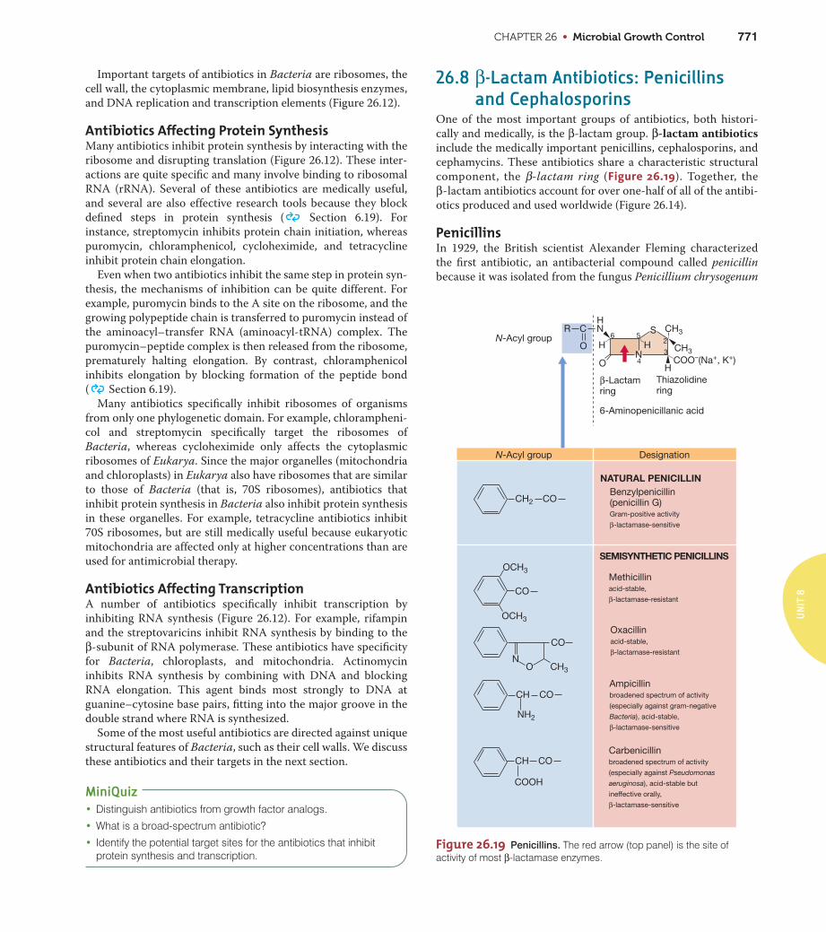
Figure 26.19 Penicillins. The red arrow (top panel) is the site ofactivity of most β-lactamase enzymes.
Important targets of antibiotics in Bacteria are ribosomes, the
cell wall, the cytoplasmic membrane, lipid biosynthesis enzymes,
and DNA replication and transcription elements (Figure 26.12).
Antibiotics Affecting Protein SynthesisMany antibiotics inhibit protein synthesis by interacting with the
ribosome and disrupting translation (Figure 26.12). These inter-
actions are quite specific and many involve binding to ribosomal
RNA (rRNA). Several of these antibiotics are medically useful,
and several are also effective research tools because they block
defined steps in protein synthesis ( Section 6.19). For
instance, streptomycin inhibits protein chain initiation, whereas
puromycin, chloramphenicol, cycloheximide, and tetracycline
inhibit protein chain elongation.
Even when two antibiotics inhibit the same step in protein syn-
thesis, the mechanisms of inhibition can be quite different. For
example, puromycin binds to the A site on the ribosome, and the
growing polypeptide chain is transferred to puromycin instead of
the aminoacyl–transfer RNA (aminoacyl-tRNA) complex. The
puromycin–peptide complex is then released from the ribosome,
prematurely halting elongation. By contrast, chloramphenicol
inhibits elongation by blocking formation of the peptide bond
( Section 6.19).
Many antibiotics specifically inhibit ribosomes of organisms
from only one phylogenetic domain. For example, chlorampheni-
col and streptomycin specifically target the ribosomes of
Bacteria, whereas cycloheximide only affects the cytoplasmic
ribosomes of Eukarya. Since the major organelles (mitochondria
and chloroplasts) in Eukarya also have ribosomes that are similar
to those of Bacteria (that is, 70S ribosomes), antibiotics that
inhibit protein synthesis in Bacteria also inhibit protein synthesis
in these organelles. For example, tetracycline antibiotics inhibit
70S ribosomes, but are still medically useful because eukaryotic
mitochondria are affected only at higher concentrations than are
used for antimicrobial therapy.
Antibiotics Affecting TranscriptionA number of antibiotics specifically inhibit transcription by
inhibiting RNA synthesis (Figure 26.12). For example, rifampin
and the streptovaricins inhibit RNA synthesis by binding to the
β-subunit of RNA polymerase. These antibiotics have specificity
for Bacteria, chloroplasts, and mitochondria. Actinomycin
inhibits RNA synthesis by combining with DNA and blocking
RNA elongation. This agent binds most strongly to DNA at
guanine–cytosine base pairs, fitting into the major groove in the
double strand where RNA is synthesized.
Some of the most useful antibiotics are directed against unique
structural features of Bacteria, such as their cell walls. We discuss
these antibiotics and their targets in the next section.
MiniQuiz• Distinguish antibiotics from growth factor analogs.
• What is a broad-spectrum antibiotic?
• Identify the potential target sites for the antibiotics that inhibitprotein synthesis and transcription.
26.8 β-Lactam Antibiotics: Penicillinsand Cephalosporins
One of the most important groups of antibiotics, both histori-
cally and medically, is the β-lactam group. β-lactam antibiotics
include the medically important penicillins, cephalosporins, and
cephamycins. These antibiotics share a characteristic structural
component, the β-lactam ring (Figure 26.19). Together, the
β-lactam antibiotics account for over one-half of all of the antibi-
otics produced and used worldwide (Figure 26.14).
PenicillinsIn 1929, the British scientist Alexander Fleming characterized
the first antibiotic, an antibacterial compound called penicillin
because it was isolated from the fungus Penicillium chrysogenum
UN
IT 8
UNIT 8 • Antimicrobial Agents and Pathogenicity772
(Figure 26.19). The antibiotic, however, was not immediately rec-
ognized as a potentially important clinical drug. Even though
sulfa drugs were widely available in the 1930s, their efficacy was
mostly limited to the treatment of infections by gram-positive
organisms such as Streptococcus; most other bacterial diseases
were uncontrollable. However, in 1939, Howard Florey and his
colleagues, motivated by the impending world war, developed a
process for the large-scale production of penicillin. Penicillin G
was the first clinically useful antibiotic. This new β-lactam antibi-
otic was dramatically effective in controlling staphylococcal and
pneumococcal infections among military personnel and was
more effective for treating streptococcal infections than sulfa
drugs. By the end of World War II in 1945, penicillin became
available for general use and pharmaceutical companies began to
look for and develop other antibiotics, leading to drugs that revo-
lutionized the treatment of infectious diseases.
Penicillin G is active primarily against gram-positive Bacteria
because gram-negative Bacteria are impermeable to the antibi-
otic. Chemical modification of the penicillin G structure, how-
ever, significantly changes the properties of the resulting
antibiotic. Many chemically modified semisynthetic penicillins
are quite effective against gram-negative Bacteria. Figure 26.19
shows the structures of some of the penicillins. For example,
ampicillin and carbenicillin, semisynthetic penicillins, are effec-
tive against some gram-negative Bacteria. The structural differ-
ences in the N-acyl groups of these semisynthetic penicillins
allow them to be transported inside the gram-negative outer
membrane ( Section 3.7), where they inhibit cell wall synthe-
sis. Penicillin G is also sensitive to β-lactamase, an enzyme pro-
duced by a number of penicillin-resistant Bacteria (Section
26.12). Oxacillin and methicillin are widely used β-lactamase-
resistant semisynthetic penicillins.
Mechanism of ActionThe β-lactam antibiotics are inhibitors of cell wall synthesis.
An important feature of bacterial cell wall synthesis is trans-
peptidation, the reaction that results in the cross-linking of two
glycan-linked peptide chains ( Section 5.4 and Figure 5.7).
The transpeptidase enzymes bind to penicillin or other β-lactam
antibiotics. Thus, these transpeptidases are called penicillin-
binding proteins (PBPs). When PBPs bind penicillin, they cannot
catalyze the transpeptidase reaction, but cell wall synthesis con-
tinues. As a result, the newly synthesized bacterial cell wall is no
longer cross-linked and cannot maintain its strength. In addition,
the antibiotic–PBP complex stimulates the release of autolysins,
enzymes that digest the existing cell wall. The result is a weak-
ened, self-degrading cell wall. Eventually the osmotic pressure
differences between the inside and outside of the cell cause lysis.
By contrast, vancomycin, also a cell wall synthesis inhibitor, does
not bind to PBPs, but binds directly to the terminal D-alanyl-
D-alanine peptide on the peptidoglycan precursors ( Figure
5.7); this effectively blocks transpeptidation.
Because the cell wall and its synthesis mechanisms are unique
to Bacteria, the β-lactam antibiotics are highly selective and are
not toxic to host cells. However, some individuals develop aller-
gies to β-lactam antibiotics after repeated courses of antibiotic
therapy.
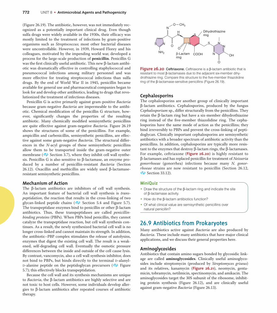
CephalosporinsThe cephalosporins are another group of clinically important
β-lactam antibiotics. Cephalosporins, produced by the fungus
Cephalosporium sp., differ structurally from the penicillins. They
retain the β-lactam ring but have a six-member dihydrothiazine
ring instead of the five-member thiazolidine ring. The cepha-
losporins have the same mode of action as the penicillins; they
bind irreversibly to PBPs and prevent the cross-linking of pepti-
doglycan. Clinically important cephalosporins are semisynthetic
antibiotics with a broader spectrum of antibiotic activity than the
penicillins. In addition, cephalosporins are typically more resis-
tant to the enzymes that destroy β-lactam rings, the β-lactamases.
For example, ceftriaxone (Figure 26.20) is highly resistant to
β-lactamases and has replaced penicillin for treatment of Neisseria
gonorrhoeae (gonorrhea) infections because many N. gonor-
rhoeae strains are now resistant to penicillin (Section 26.12,
Section 33.12).
MiniQuiz• Draw the structure of the β-lactam ring and indicate the site
of β-lactamase activity.
• How do the β-lactam antibiotics function?
• Of what clinical value are semisynthetic penicillins over natural penicillin?
26.9 Antibiotics from ProkaryotesMany antibiotics active against Bacteria are also produced by
Bacteria. These include many antibiotics that have major clinical
applications, and we discuss their general properties here.
AminoglycosidesAntibiotics that contain amino sugars bonded by glycosidic link-
age are called aminoglycosides. Clinically useful aminoglyco-
sides include streptomycin (produced by Streptomyces griseus)
and its relatives, kanamycin (Figure 26.21), neomycin, genta-
micin, tobramycin, netilmicin, spectinomycin, and amikacin. The
aminoglycosides target the 30S subunit of the ribosome, inhibit-
ing protein synthesis (Figure 26.12), and are clinically useful
against gram-negative Bacteria (Figure 26.13).
O
OO
CH3
NH2
S N
HN
N
N
S
S NN
OH
O
NCOOH
Dihydrothiazinering
β-Lactam ring
Figure 26.20 Ceftriaxone. Ceftriaxone is a β-lactam antibiotic that isresistant to most β-lactamases due to the adjacent six-member dihy-drothiazine ring. Compare this structure to the five-member thiazolidinering of the β-lactamase-sensitive penicillins (Figure 26.19).
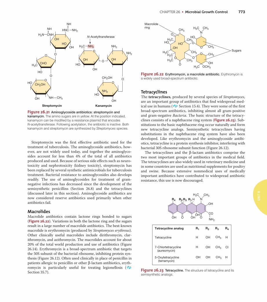
CHAPTER 26 • Microbial Growth Control 773
N
HO
O
O
O
O
O
O
HO
OH
O
HO
OH
Sugars
Macrolidering
CH3
CH3
CH3CH3
OCH3
CH3
CH3
CH3H3C
H3C
H3C
H3C
H3C
H2C
Figure 26.22 Erythromycin, a macrolide antibiotic. Erythromycin isa widely used broad-spectrum antibiotic.
OH OHO OOH
N
OH
CO
Tetracycline analog
Tetracycline H OH H
7-Chlortetracycline (aureomycin)
H OH Cl
5-Oxytetracycline (terramycin)
OH OH H
R1 R2 R3 R4
R4 R1 H
H3C
R2 R3
CH3
CH3
CH3
CH3
NH2
Figure 26.23 Tetracycline. The structure of tetracycline and itssemisynthetic analogs.
Streptomycin was the first effective antibiotic used for the
treatment of tuberculosis. The aminoglycoside antibiotics, how-
ever, are not widely used today, and together the aminoglyco-
sides account for less than 4% of the total of all antibiotics
produced and used. Because of serious side effects such as neuro-
toxicity and nephrotoxicity (kidney toxicity), streptomycin has
been replaced by several synthetic antimicrobials for tuberculosis
treatment. Bacterial resistance to aminoglycosides also develops
readily. The use of aminoglycosides for treatment of gram-
negative infections has decreased since the development of the
semisynthetic penicillins (Section 26.8) and the tetracyclines
(discussed later in this section). Aminoglycoside antibiotics are
now considered reserve antibiotics used primarily when other
antibiotics fail.
MacrolidesMacrolide antibiotics contain lactone rings bonded to sugars
(Figure 26.22). Variations in both the lactone ring and the sugars
result in a large number of macrolide antibiotics. The best-known
macrolide is erythromycin (produced by Streptomyces erythreus).
Other clinically useful macrolides include dirithromycin, clar-
ithromycin, and azithromycin. The macrolides account for about
20% of the total world production and use of antibiotics (Figure
26.14). Erythromycin is a broad-spectrum antibiotic that targets
the 50S subunit of the bacterial ribosome, inhibiting protein syn-
thesis (Figure 26.12). Often used clinically in place of penicillin in
patients allergic to penicillin or other β-lactam antibiotics, eryth-
romycin is particularly useful for treating legionellosis (
Section 35.7).
TetracyclinesThe tetracyclines, produced by several species of Streptomyces,
are an important group of antibiotics that find widespread med-
ical use in humans ( Section 15.4). They were some of the first
broad-spectrum antibiotics, inhibiting almost all gram-positive
and gram-negative Bacteria. The basic structure of the tetracy-
clines consists of a naphthacene ring system (Figure 26.23). Sub-
stitutions to the basic naphthacene ring occur naturally and form
new tetracycline analogs. Semisynthetic tetracyclines having
substitutions in the naphthacene ring system have also been
developed. Like erythromycin and the aminoglycoside antibi-
otics, tetracycline is a protein synthesis inhibitor, interfering with
bacterial 30S ribosome subunit function (Figure 26.12).
The tetracyclines and the β-lactam antibiotics comprise the
two most important groups of antibiotics in the medical field.
The tetracyclines are also widely used in veterinary medicine and
in some countries are used as nutritional supplements for poultry
and swine. Because extensive nonmedical uses of medically
important antibiotics have contributed to widespread antibiotic
resistance, this use is now discouraged.
UN
IT 8
H2C
CH2OH
NH2
NH2
NH2
NH2
HO
HO
OH
OH
O
O
OO
OH
OH
N-Acetyltransferase
Kanamycin
NH
H2N CNH
HO
HNC NH2
NH
OH
OH
CHOH3C
OO
HOO
OHO
OH NH CH3
CH2OH
Streptomycin
Figure 26.21 Aminoglycoside antibiotics: streptomycin and
kanamycin. The amino sugars are in yellow. At the position indicated,kanamycin can be modified by a resistance plasmid that encodes N-acetyltransferase. Following acetylation, the antibiotic is inactive. Bothkanamycin and streptomycin are synthesized by Streptomyces species.
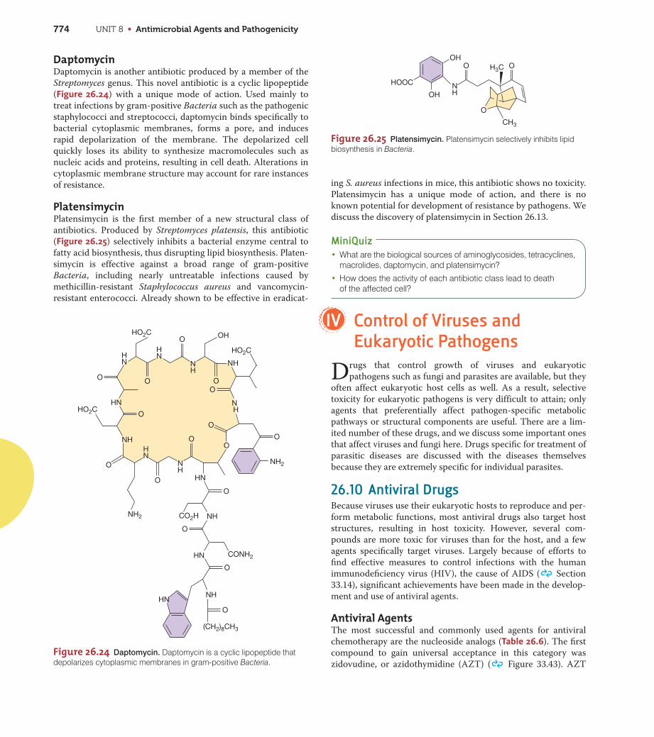
UNIT 8 • Antimicrobial Agents and Pathogenicity774
DaptomycinDaptomycin is another antibiotic produced by a member of the
Streptomyces genus. This novel antibiotic is a cyclic lipopeptide
(Figure 26.24) with a unique mode of action. Used mainly to
treat infections by gram-positive Bacteria such as the pathogenic
staphylococci and streptococci, daptomycin binds specifically to
bacterial cytoplasmic membranes, forms a pore, and induces
rapid depolarization of the membrane. The depolarized cell
quickly loses its ability to synthesize macromolecules such as
nucleic acids and proteins, resulting in cell death. Alterations in
cytoplasmic membrane structure may account for rare instances
of resistance.
PlatensimycinPlatensimycin is the first member of a new structural class of
antibiotics. Produced by Streptomyces platensis, this antibiotic
(Figure 26.25) selectively inhibits a bacterial enzyme central to
fatty acid biosynthesis, thus disrupting lipid biosynthesis. Platen-
simycin is effective against a broad range of gram-positive
Bacteria, including nearly untreatable infections caused by
methicillin-resistant Staphylococcus aureus and vancomycin-
resistant enterococci. Already shown to be effective in eradicat-
ing S. aureus infections in mice, this antibiotic shows no toxicity.
Platensimycin has a unique mode of action, and there is no
known potential for development of resistance by pathogens. We
discuss the discovery of platensimycin in Section 26.13.
MiniQuiz• What are the biological sources of aminoglycosides, tetracyclines,
macrolides, daptomycin, and platensimycin?
• How does the activity of each antibiotic class lead to death of the affected cell?
IV Control of Viruses andEukaryotic Pathogens
Drugs that control growth of viruses and eukaryotic
pathogens such as fungi and parasites are available, but they
often affect eukaryotic host cells as well. As a result, selective
toxicity for eukaryotic pathogens is very difficult to attain; only
agents that preferentially affect pathogen-specific metabolic
pathways or structural components are useful. There are a lim-
ited number of these drugs, and we discuss some important ones
that affect viruses and fungi here. Drugs specific for treatment of
parasitic diseases are discussed with the diseases themselves
because they are extremely specific for individual parasites.
26.10 Antiviral DrugsBecause viruses use their eukaryotic hosts to reproduce and per-
form metabolic functions, most antiviral drugs also target host
structures, resulting in host toxicity. However, several com-
pounds are more toxic for viruses than for the host, and a few
agents specifically target viruses. Largely because of efforts to
find effective measures to control infections with the human
immunodeficiency virus (HIV), the cause of AIDS ( Section
33.14), significant achievements have been made in the develop-
ment and use of antiviral agents.
Antiviral AgentsThe most successful and commonly used agents for antiviral
chemotherapy are the nucleoside analogs (Table 26.6). The first
compound to gain universal acceptance in this category was
zidovudine, or azidothymidine (AZT) ( Figure 33.43). AZT
NH
HN
NH
N H
NH2
HNH
N NH
HN
NH
O
O
O
O
O
O
O
O
O
O
O
HN
NH
HN
NHHN
O
OHHO2C
HO2C
HO2C
CO2H
CONH2
(CH2)8CH3
NH2
O
O
O
O
Figure 26.24 Daptomycin. Daptomycin is a cyclic lipopeptide thatdepolarizes cytoplasmic membranes in gram-positive Bacteria.
O
O
O
OHNH
OH
HOOC
CH3
H3C
Figure 26.25 Platensimycin. Platensimycin selectively inhibits lipidbiosynthesis in Bacteria.
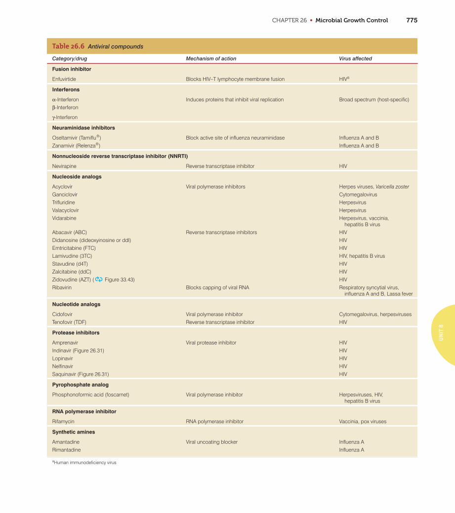
CHAPTER 26 • Microbial Growth Control 775
UN
IT 8
Table 26.6 Antiviral compounds
Category/drug Mechanism of action Virus affected
Fusion inhibitor
Enfuvirtide Blocks HIV–T lymphocyte membrane fusion HIVa
Interferons
α-Interferon Induces proteins that inhibit viral replication Broad spectrum (host-specific)β-Interferon
γ-Interferon
Neuraminidase inhibitors
Oseltamivir (Tamiflu®) Block active site of influenza neuraminidase Influenza A and BZanamivir (Relenza®) Influenza A and B
Nonnucleoside reverse transcriptase inhibitor (NNRTI)
Nevirapine Reverse transcriptase inhibitor HIV
Nucleoside analogs
Acyclovir Viral polymerase inhibitors Herpes viruses, Varicella zosterGanciclovir CytomegalovirusTrifluridine HerpesvirusValacyclovir HerpesvirusVidarabine Herpesvirus, vaccinia,
hepatitis B virusAbacavir (ABC) Reverse transcriptase inhibitors HIVDidanosine (dideoxyinosine or ddI) HIVEmtricitabine (FTC) HIVLamivudine (3TC) HIV, hepatitis B virusStavudine (d4T) HIVZalcitabine (ddC) HIVZidovudine (AZT) ( Figure 33.43) HIVRibavirin Blocks capping of viral RNA Respiratory syncytial virus,
influenza A and B, Lassa fever
Nucleotide analogs
Cidofovir Viral polymerase inhibitor Cytomegalovirus, herpesvirusesTenofovir (TDF) Reverse transcriptase inhibitor HIV
Protease inhibitors
Amprenavir Viral protease inhibitor HIVIndinavir (Figure 26.31) HIVLopinavir HIVNelfinavir HIVSaquinavir (Figure 26.31) HIV
Pyrophosphate analog
Phosphonoformic acid (foscarnet) Viral polymerase inhibitor Herpesviruses, HIV, hepatitis B virus
RNA polymerase inhibitor
Rifamycin RNA polymerase inhibitor Vaccinia, pox viruses
Synthetic amines
Amantadine Viral uncoating blocker Influenza ARimantadine Influenza A
aHuman immunodeficiency virus
UNIT 8 • Antimicrobial Agents and Pathogenicity776
inhibits retroviruses such as HIV ( Sections 33.14 and 9.12).
Azidothymidine is chemically related to thymidine but is a
dideoxy derivative, lacking the 3¿-hydroxyl group. AZT inhibits
multiplication of retroviruses by blocking reverse transcription
and production of the virally encoded DNA intermediate. This
inhibits multiplication of HIV. A number of other nucleoside
analogs having analogous mechanisms have been developed for
the treatment of HIV and other viruses.
Nearly all nucleoside analogs, or nucleoside reverse transcrip-
tase inhibitors (NRTI), work by the same mechanism, inhibiting
elongation of the viral nucleic acid chain by a nucleic acid poly-
merase. The nucleotide analog cidofovir works in the same way
(Table 26.6). Because the normal cell function of nucleic acid repli-
cation is targeted, these drugs usually induce some host toxicity.
Many NRTIs also lose their antiviral potency with time due to the
emergence of drug-resistant viruses ( Section 33.14).
Several other antiviral agents target the key enzyme of retro-
viruses, reverse transcriptase. Nevirapine, a nonnucleoside reverse
transcriptase inhibitor (NNRTI), binds directly to reverse tran-
scriptase and inhibits reverse transcription. Phosphonoformic
acid, an analog of inorganic pyrophosphate, inhibits normal inter-
nucleotide linkages, preventing synthesis of viral nucleic acids. As
with the NRTIs, the NNRTIs generally induce some level of host
toxicity because their action also affects normal host cell nucleic
acid synthesis.
Protease inhibitors are another class of antiviral drugs that are
effective for treatment of HIV (Table 26.6 and see Figure 26.31).
These drugs prevent viral replication by binding the active site of
HIV protease, inhibiting this enzyme from processing large viral
proteins into individual viral components, thus preventing virus
maturation ( Sections 21.11, 26.13, and 33.14).
A final category of anti-HIV drugs is represented by a single
drug, enfuvirtide, a fusion inhibitor composed of a 36-amino
acid synthetic peptide that binds to the gp41 membrane protein
of HIV (Table 26.6 and Section 33.14). Binding of the gp41
protein by enfuvirtide stops the conformational changes neces-
sary for the fusion of HIV and T lymphocyte membranes, thus
preventing infection of cells by HIV.
Influenza Antiviral AgentsTwo categories of drugs effectively limit influenza infection. The
adamantanes amantadine and rimantadine are synthetic amines
that interfere with an influenza A ion transport protein, inhibit-
ing virus uncoating and subsequent replication. The neu-
raminidase inhibitors oseltamivir (brand name Tamiflu) and
zanamivir (Relenza) block the active site of neuraminidase in
influenza A and B viruses, inhibiting virus release from infected
cells. Zanamivir is used only for treatment of influenza, whereas
oseltamivir is used for both treatment and prophylaxis. The
adamantanes are less useful than the neuraminidase inhibitors
because resistance to adamantanes develops rapidly in strains of
influenza virus ( Section 33.8).
InterferonsVirus interference is a phenomenon in which infection with one
virus interferes with subsequent infection by another virus. Sev-
eral small proteins are the cause of interference; the proteins are
called interferons. Interferons are small proteins in the cytokine
family ( Section 30.10) that prevent viral replication by stimu-
lating the production of antiviral proteins in uninfected cells.
Interferons are formed in response to live virus, inactivated virus,
and viral nucleic acids. Interferon is produced in large amounts
by cells infected with viruses of low virulence, but little is pro-
duced against highly virulent viruses. Highly virulent viruses
inhibit cell protein synthesis before interferon can be produced.
Interferons are also induced by natural and synthetic double-
stranded RNA (dsRNA) molecules. In nature, dsRNA exists only
in virus-infected cells as the replicative form of RNA viruses such
as rhinoviruses (cold viruses) ( Section 33.7); the dsRNA from
the infecting virus signals the animal cell to produce interferon.
Interferons from virus-infected cells interact with receptors on
uninfected cells, promoting the synthesis of antiviral proteins
that function to prevent further virus infection. Interferons are
produced in three molecular forms: IFN-α is produced by leuko-
cytes, IFN-β is produced by fibroblasts, and IFN-γ is produced
by immune lymphocytes.
Interferon activity is host-specific rather than virus-specific.
That is, interferon produced by a member of one species can only
activate receptors on cells from the same species. As a result,
interferon produced by cells of an animal in response to, for
example, a rhinovirus, could also inhibit multiplication of,
for example, influenza viruses in cells within the same species,
but has no effect on the multiplication of any virus in cells from
other animal species.
Interferons produced in vitro have potential as possible antivi-
ral and anticancer agents. Several approved recombinant inter-
ferons are available. However, the use of interferons as antiviral
agents is not widespread because interferon must be delivered
locally in high concentrations to stimulate the production of
antiviral proteins in uninfected host cells. Thus, the clinical util-
ity of these antiviral agents depends on our ability to deliver
interferon to local areas in the host through injections or
aerosols. Alternatively, appropriate interferon-stimulating signals
such as viral nucleotides, nonvirulent viruses, or even synthetic
nucleotides, if given to host cells prior to viral infection, might
stimulate natural production of interferon.
MiniQuiz• Why are there relatively few effective antiviral agents? Such
agents are not used to treat common viral illnesses such as colds;why not?
• What steps in the viral maturation process are inhibited bynucleoside analogs? By protease inhibitors? By interferons?
26.11 Antifungal DrugsFungi, like viruses, pose special problems for the development of
chemotherapy. Because fungi are Eukarya, much of their cellular
machinery is the same as that of animals and humans; antifungal
agents that act on metabolic pathways in fungi often affect corre-
sponding pathways in host cells, making the drugs toxic. As a
result, many antifungal drugs can be used only for topical (sur-
face) applications. However, a few drugs are selectively toxic for
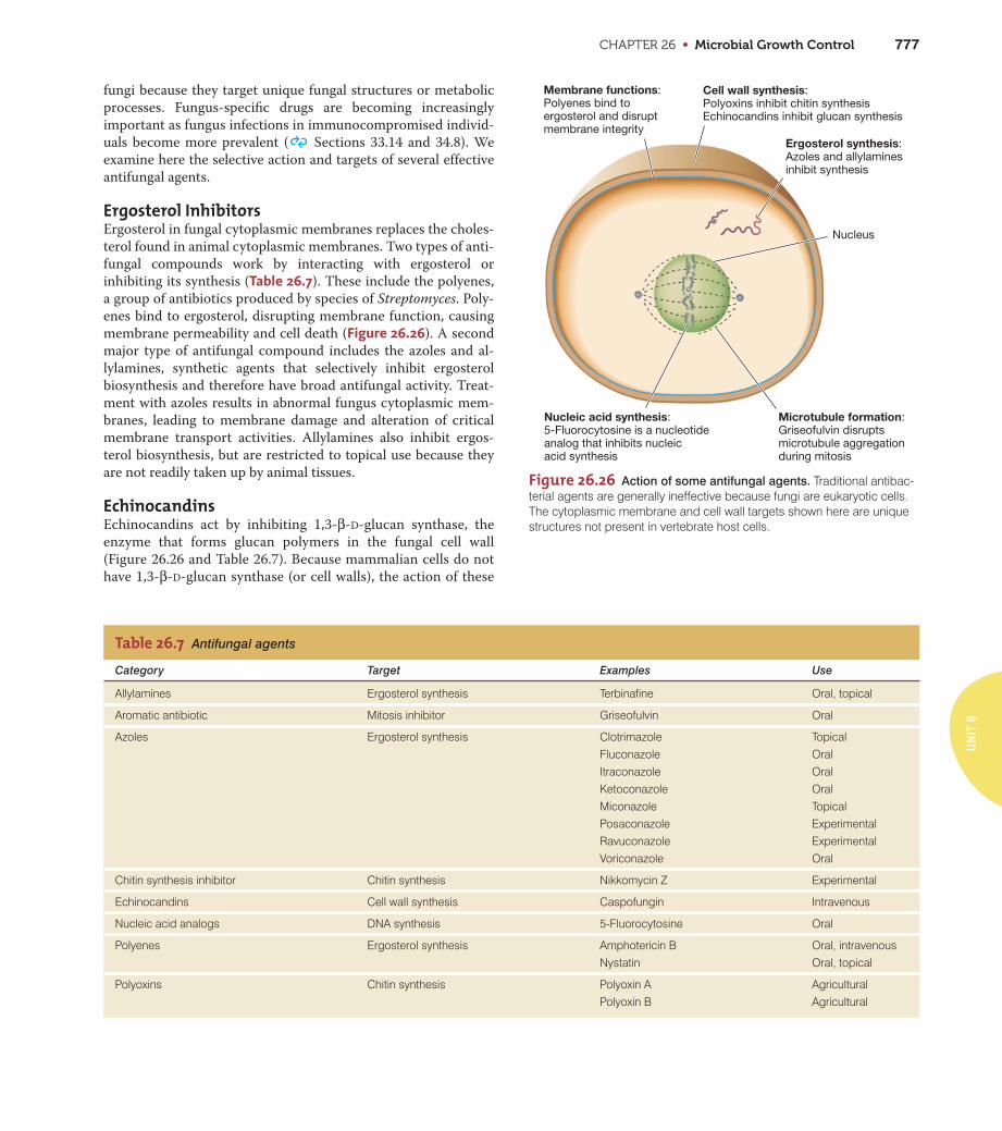
CHAPTER 26 • Microbial Growth Control 777
UN
IT 8
fungi because they target unique fungal structures or metabolic
processes. Fungus-specific drugs are becoming increasingly
important as fungus infections in immunocompromised individ-
uals become more prevalent ( Sections 33.14 and 34.8). We
examine here the selective action and targets of several effective
antifungal agents.
Ergosterol InhibitorsErgosterol in fungal cytoplasmic membranes replaces the choles-
terol found in animal cytoplasmic membranes. Two types of anti-
fungal compounds work by interacting with ergosterol or
inhibiting its synthesis (Table 26.7). These include the polyenes,
a group of antibiotics produced by species of Streptomyces. Poly-
enes bind to ergosterol, disrupting membrane function, causing
membrane permeability and cell death (Figure 26.26). A second
major type of antifungal compound includes the azoles and al-
lylamines, synthetic agents that selectively inhibit ergosterol
biosynthesis and therefore have broad antifungal activity. Treat-
ment with azoles results in abnormal fungus cytoplasmic mem-
branes, leading to membrane damage and alteration of critical
membrane transport activities. Allylamines also inhibit ergos-
terol biosynthesis, but are restricted to topical use because they
are not readily taken up by animal tissues.
EchinocandinsEchinocandins act by inhibiting 1,3-β-D-glucan synthase, the
enzyme that forms glucan polymers in the fungal cell wall
(Figure 26.26 and Table 26.7). Because mammalian cells do not
have 1,3-β-D-glucan synthase (or cell walls), the action of these
Table 26.7 Antifungal agents
Category Target Examples Use
Allylamines Ergosterol synthesis Terbinafine Oral, topical
Aromatic antibiotic Mitosis inhibitor Griseofulvin Oral
Azoles Ergosterol synthesis Clotrimazole TopicalFluconazole OralItraconazole OralKetoconazole OralMiconazole TopicalPosaconazole ExperimentalRavuconazole ExperimentalVoriconazole Oral
Chitin synthesis inhibitor Chitin synthesis Nikkomycin Z Experimental
Echinocandins Cell wall synthesis Caspofungin Intravenous
Nucleic acid analogs DNA synthesis 5-Fluorocytosine Oral
Polyenes Ergosterol synthesis Amphotericin B Oral, intravenousNystatin Oral, topical
Polyoxins Chitin synthesis Polyoxin A AgriculturalPolyoxin B Agricultural
Cell wall synthesis:Polyoxins inhibit chitin synthesisEchinocandins inhibit glucan synthesis
Ergosterol synthesis:Azoles and allylaminesinhibit synthesis
Nucleus
Microtubule formation:Griseofulvin disruptsmicrotubule aggregationduring mitosis
Membrane functions:Polyenes bind toergosterol and disruptmembrane integrity
Nucleic acid synthesis:5-Fluorocytosine is a nucleotideanalog that inhibits nucleicacid synthesis
Figure 26.26 Action of some antifungal agents. Traditional antibac-terial agents are generally ineffective because fungi are eukaryotic cells.The cytoplasmic membrane and cell wall targets shown here are uniquestructures not present in vertebrate host cells.
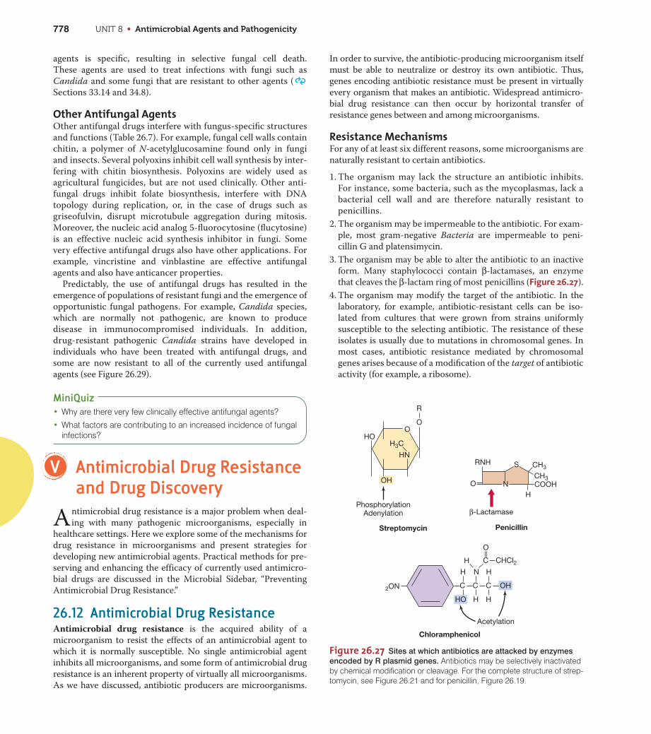
UNIT 8 • Antimicrobial Agents and Pathogenicity778
agents is specific, resulting in selective fungal cell death.
These agents are used to treat infections with fungi such as
Candida and some fungi that are resistant to other agents (
Sections 33.14 and 34.8).
Other Antifungal AgentsOther antifungal drugs interfere with fungus-specific structures
and functions (Table 26.7). For example, fungal cell walls contain
chitin, a polymer of N-acetylglucosamine found only in fungi
and insects. Several polyoxins inhibit cell wall synthesis by inter-
fering with chitin biosynthesis. Polyoxins are widely used as
agricultural fungicides, but are not used clinically. Other anti-
fungal drugs inhibit folate biosynthesis, interfere with DNA
topology during replication, or, in the case of drugs such as
griseofulvin, disrupt microtubule aggregation during mitosis.
Moreover, the nucleic acid analog 5-fluorocytosine (flucytosine)
is an effective nucleic acid synthesis inhibitor in fungi. Some
very effective antifungal drugs also have other applications. For
example, vincristine and vinblastine are effective antifungal
agents and also have anticancer properties.
Predictably, the use of antifungal drugs has resulted in the
emergence of populations of resistant fungi and the emergence of
opportunistic fungal pathogens. For example, Candida species,
which are normally not pathogenic, are known to produce
disease in immunocompromised individuals. In addition,
drug-resistant pathogenic Candida strains have developed in
individuals who have been treated with antifungal drugs, and
some are now resistant to all of the currently used antifungal
agents (see Figure 26.29).
MiniQuiz• Why are there very few clinically effective antifungal agents?
• What factors are contributing to an increased incidence of fungalinfections?
V Antimicrobial Drug Resistanceand Drug Discovery
Antimicrobial drug resistance is a major problem when deal-
ing with many pathogenic microorganisms, especially in
healthcare settings. Here we explore some of the mechanisms for
drug resistance in microorganisms and present strategies for
developing new antimicrobial agents. Practical methods for pre-
serving and enhancing the efficacy of currently used antimicro-
bial drugs are discussed in the Microbial Sidebar, “Preventing
Antimicrobial Drug Resistance.”
26.12 Antimicrobial Drug ResistanceAntimicrobial drug resistance is the acquired ability of a
microorganism to resist the effects of an antimicrobial agent to
which it is normally susceptible. No single antimicrobial agent
inhibits all microorganisms, and some form of antimicrobial drug
resistance is an inherent property of virtually all microorganisms.
As we have discussed, antibiotic producers are microorganisms.
In order to survive, the antibiotic-producing microorganism itself
must be able to neutralize or destroy its own antibiotic. Thus,
genes encoding antibiotic resistance must be present in virtually
every organism that makes an antibiotic. Widespread antimicro-
bial drug resistance can then occur by horizontal transfer of
resistance genes between and among microorganisms.
Resistance MechanismsFor any of at least six different reasons, some microorganisms are
naturally resistant to certain antibiotics.
1. The organism may lack the structure an antibiotic inhibits.
For instance, some bacteria, such as the mycoplasmas, lack a
bacterial cell wall and are therefore naturally resistant to
penicillins.
2. The organism may be impermeable to the antibiotic. For exam-
ple, most gram-negative Bacteria are impermeable to peni-
cillin G and platensimycin.
3. The organism may be able to alter the antibiotic to an inactive
form. Many staphylococci contain β-lactamases, an enzyme
that cleaves the β-lactam ring of most penicillins (Figure 26.27).
4. The organism may modify the target of the antibiotic. In the
laboratory, for example, antibiotic-resistant cells can be iso-
lated from cultures that were grown from strains uniformly
susceptible to the selecting antibiotic. The resistance of these
isolates is usually due to mutations in chromosomal genes. In
most cases, antibiotic resistance mediated by chromosomal
genes arises because of a modification of the target of antibiotic
activity (for example, a ribosome).
Streptomycin
O
RNH S CH3
CH3COOHN
H
β-Lactamase
Penicillin
PhosphorylationAdenylation
2ON C
H
HO
C
N
H
C
H
H
OH
H C
O
CHCl2
Acetylation
Chloramphenicol
R
OO
HO
OH
HN
H3C
Figure 26.27 Sites at which antibiotics are attacked by enzymes
encoded by R plasmid genes. Antibiotics may be selectively inactivatedby chemical modification or cleavage. For the complete structure of strep-tomycin, see Figure 26.21 and for penicillin, Figure 26.19.
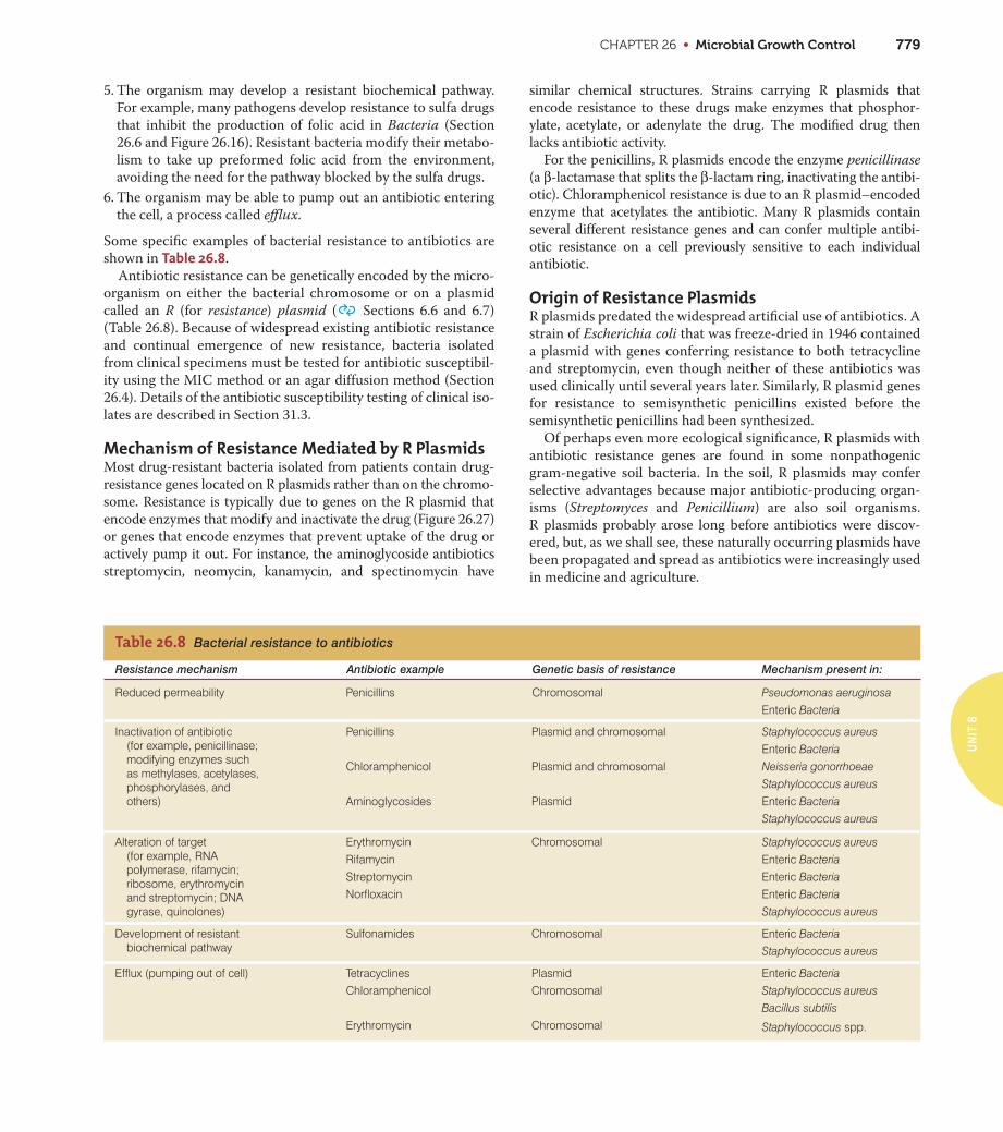
CHAPTER 26 • Microbial Growth Control 779
UN
IT 8
5. The organism may develop a resistant biochemical pathway.
For example, many pathogens develop resistance to sulfa drugs
that inhibit the production of folic acid in Bacteria (Section
26.6 and Figure 26.16). Resistant bacteria modify their metabo-
lism to take up preformed folic acid from the environment,
avoiding the need for the pathway blocked by the sulfa drugs.
6. The organism may be able to pump out an antibiotic entering
the cell, a process called efflux.
Some specific examples of bacterial resistance to antibiotics are
shown in Table 26.8.
Antibiotic resistance can be genetically encoded by the micro-
organism on either the bacterial chromosome or on a plasmid
called an R (for resistance) plasmid ( Sections 6.6 and 6.7)
(Table 26.8). Because of widespread existing antibiotic resistance
and continual emergence of new resistance, bacteria isolated
from clinical specimens must be tested for antibiotic susceptibil-
ity using the MIC method or an agar diffusion method (Section
26.4). Details of the antibiotic susceptibility testing of clinical iso-
lates are described in Section 31.3.
Mechanism of Resistance Mediated by R PlasmidsMost drug-resistant bacteria isolated from patients contain drug-
resistance genes located on R plasmids rather than on the chromo-
some. Resistance is typically due to genes on the R plasmid that
encode enzymes that modify and inactivate the drug (Figure 26.27)
or genes that encode enzymes that prevent uptake of the drug or
actively pump it out. For instance, the aminoglycoside antibiotics
streptomycin, neomycin, kanamycin, and spectinomycin have
similar chemical structures. Strains carrying R plasmids that
encode resistance to these drugs make enzymes that phosphor-
ylate, acetylate, or adenylate the drug. The modified drug then
lacks antibiotic activity.
For the penicillins, R plasmids encode the enzyme penicillinase
(a β-lactamase that splits the β-lactam ring, inactivating the antibi-
otic). Chloramphenicol resistance is due to an R plasmid–encoded
enzyme that acetylates the antibiotic. Many R plasmids contain
several different resistance genes and can confer multiple antibi-
otic resistance on a cell previously sensitive to each individual
antibiotic.
Origin of Resistance PlasmidsR plasmids predated the widespread artificial use of antibiotics. A
strain of Escherichia coli that was freeze-dried in 1946 contained
a plasmid with genes conferring resistance to both tetracycline
and streptomycin, even though neither of these antibiotics was
used clinically until several years later. Similarly, R plasmid genes
for resistance to semisynthetic penicillins existed before the
semisynthetic penicillins had been synthesized.
Of perhaps even more ecological significance, R plasmids with
antibiotic resistance genes are found in some nonpathogenic
gram-negative soil bacteria. In the soil, R plasmids may confer
selective advantages because major antibiotic-producing organ-
isms (Streptomyces and Penicillium) are also soil organisms.
R plasmids probably arose long before antibiotics were discov-
ered, but, as we shall see, these naturally occurring plasmids have
been propagated and spread as antibiotics were increasingly used
in medicine and agriculture.
Alteration of target (for example, RNA polymerase, rifamycin; ribosome, erythromycin and streptomycin; DNA gyrase, quinolones)
ErythromycinRifamycinStreptomycinNorfloxacin
Chromosomal Staphylococcus aureusEnteric BacteriaEnteric BacteriaEnteric BacteriaStaphylococcus aureus
Development of resistant biochemical pathway
Sulfonamides Chromosomal Enteric BacteriaStaphylococcus aureus
Efflux (pumping out of cell) TetracyclinesChloramphenicol
Erythromycin
PlasmidChromosomal
Chromosomal
Enteric BacteriaStaphylococcus aureusBacillus subtilis
Staphylococcus spp.
Penicillins
Chloramphenicol
Aminoglycosides
Plasmid and chromosomal
Plasmid and chromosomal
Plasmid
Staphylococcus aureusEnteric Bacteria
Neisseria gonorrhoeaeStaphylococcus aureusEnteric BacteriaStaphylococcus aureus
Table 26.8 Bacterial resistance to antibiotics
Resistance mechanism Antibiotic example Genetic basis of resistance Mechanism present in:
Reduced permeability Penicillins Chromosomal Pseudomonas aeruginosaEnteric Bacteria
Inactivation of antibiotic (for example, penicillinase; modifying enzymes such as methylases, acetylases, phosphorylases, and others)
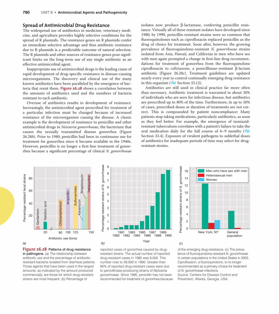
Figure 26.28 Patterns of drug resistance
in pathogens. (a) The relationship betweenantibiotic use and the percentage of antibiotic-resistant bacteria isolated from diarrheal patients.Those agents that have been used in the largestamounts, as indicated by the amount producedcommercially, are those for which drug-resistantstrains are most frequent. (b) Percentage of
UNIT 8 • Antimicrobial Agents and Pathogenicity780
Spread of Antimicrobial Drug ResistanceThe widespread use of antibiotics in medicine, veterinary medi-
cine, and agriculture provides highly selective conditions for the
spread of R plasmids. The resistance genes on R plasmids confer
an immediate selective advantage and thus antibiotic resistance
due to R plasmids is a predictable outcome of natural selection.
The R plasmids and other sources of resistance genes pose signif-
icant limits on the long-term use of any single antibiotic as an
effective antimicrobial agent.
Inappropriate use of antimicrobial drugs is the leading cause of
rapid development of drug-specific resistance in disease-causing
microorganisms. The discovery and clinical use of the many
known antibiotics have been paralleled by the emergence of bac-
teria that resist them. Figure 26.28 shows a correlation between
the amounts of antibiotics used and the numbers of bacteria
resistant to each antibiotic.
Overuse of antibiotics results in development of resistance.
Increasingly, the antimicrobial agent prescribed for treatment of
a particular infection must be changed because of increased
resistance of the microorganism causing the disease. A classic
example is the development of resistance to penicillin and other
antimicrobial drugs in Neisseria gonorrhoeae, the bacterium that
causes the sexually transmitted disease gonorrhea (Figure
26.28b). Prior to 1980, penicillin had been in continuous use for
treatment for gonorrhea since it became available in the 1940s.
However, penicillin is no longer a first-line treatment of gonor-
rhea because a significant percentage of clinical N. gonorrhoeae
isolates now produce β-lactamase, conferring penicillin resis-
tance. Virtually all of these resistant isolates have developed since
1980; by 1990, penicillin-resistant strains were so common that
fluoroquinolones such as ciprofloxacin replaced penicillin as the
drug of choice for treatment. Soon after, however, the growing
prevalence of fluoroquinolone-resistant N. gonorrhoeae strains
isolated from Asia, Hawaii, and California in men who have sex
with men again prompted a change in first-line drug recommen-
dations for treatment of gonorrhea from the fluoroquinolone
ciprofloxacin to ceftriaxone, a penicillinase-resistant β-lactam
antibiotic (Figure 26.28c). Treatment guidelines are updated
nearly every year to control continually emerging drug resistance
in this organism ( Section 33.12).
Antibiotics are still used in clinical practice far more often
than necessary. Antibiotic treatment is warranted in about 20%
of individuals who are seen for infectious disease, but antibiotics
are prescribed up to 80% of the time. Furthermore, in up to 50%
of cases, prescribed doses or duration of treatments are not cor-
rect. This is compounded by patient noncompliance: Many
patients stop taking medications, particularly antibiotics, as soon
as they feel better. For example, the emergence of isoniazid-
resistant tuberculosis correlates with a patient’s failure to take the
oral medication daily for the full course of 6–9 months (
Section 33.4). Exposure of virulent pathogens to sublethal doses
of antibiotics for inadequate periods of time may select for drug-
resistant strains.
9
8
7
6
5
4
3
2
1
0Per
cent
age
resi
stan
t st
rain
s of
N. g
onor
rhoe
ae
(a) (b)
19801981
19821983
19841985
19861987
19881989
1990
Year
8
10
12
14
16
6
4
2
0
Per
cent
age
resi
stan
t st
rain
s of
N. g
onor
rhoe
ae
(c)
Men who have sex with menHeterosexual men
New York, NY Generalpopulation
Antibiotic use (tons)
20 80 100 125 150
20
40
60
80
Per
cent
age
resi
stan
t fe
cal s
trai
ns
Gen
tam
icin
Nal
idix
ic a
cid
Chl
oram
phe
nico
l
Am
pic
illin
Sul
fona
mid
esK
anam
ycin
Str
epto
myc
in
Tetr
acyc
line
Women
reported cases of gonorrhea caused by drug-resistant strains. The actual number of reporteddrug-resistant cases in 1985 was 9,000. Thisnumber rose to 59,000 in 1990. Greater than 95% of reported drug-resistant cases were dueto penicillinase-producing strains of Neisseriagonorrhoeae. Since 1990, penicillin has not beenrecommended for treatment of gonorrhea because
of the emerging drug resistance. (c) The preva-lence of fluoroquinolone-resistant N. gonorrhoeaein certain populations in the United States in 2003.Ciprofloxacin, a fluoroquinolone, is no longerrecommended as a primary choice for treatmentof N. gonorrhoeae infections. Source: Centers for Disease Control andPrevention, Atlanta, Georgia, USA.
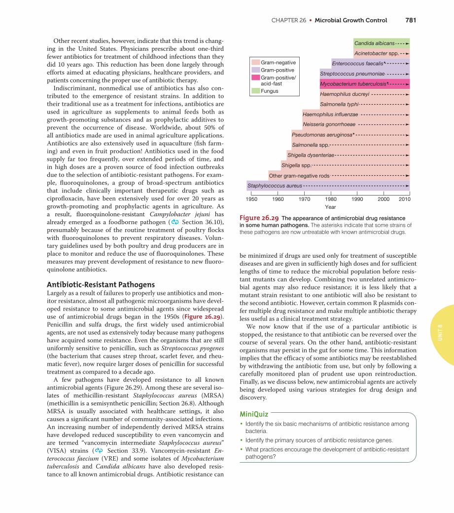
CHAPTER 26 • Microbial Growth Control 781
UN
IT 8
Other recent studies, however, indicate that this trend is chang-
ing in the United States. Physicians prescribe about one-third
fewer antibiotics for treatment of childhood infections than they
did 10 years ago. This reduction has been done largely through
efforts aimed at educating physicians, healthcare providers, and
patients concerning the proper use of antibiotic therapy.
Indiscriminant, nonmedical use of antibiotics has also con-
tributed to the emergence of resistant strains. In addition to
their traditional use as a treatment for infections, antibiotics are
used in agriculture as supplements to animal feeds both as
growth-promoting substances and as prophylactic additives to
prevent the occurrence of disease. Worldwide, about 50% of
all antibiotics made are used in animal agriculture applications.
Antibiotics are also extensively used in aquaculture (fish farm-
ing) and even in fruit production! Antibiotics used in the food
supply far too frequently, over extended periods of time, and
in high doses are a proven source of food infection outbreaks
due to the selection of antibiotic-resistant pathogens. For exam-
ple, fluoroquinolones, a group of broad-spectrum antibiotics
that include clinically important therapeutic drugs such as
ciprofloxacin, have been extensively used for over 20 years as
growth-promoting and prophylactic agents in agriculture. As
a result, fluoroquinolone-resistant Campylobacter jejuni has
already emerged as a foodborne pathogen ( Section 36.10),
presumably because of the routine treatment of poultry flocks
with fluoroquinolones to prevent respiratory diseases. Volun-
tary guidelines used by both poultry and drug producers are in
place to monitor and reduce the use of fluoroquinolones. These
measures may prevent development of resistance to new fluoro-
quinolone antibiotics.
Antibiotic-Resistant PathogensLargely as a result of failures to properly use antibiotics and mon-
itor resistance, almost all pathogenic microorganisms have devel-
oped resistance to some antimicrobial agents since widespread
use of antimicrobial drugs began in the 1950s (Figure 26.29).
Penicillin and sulfa drugs, the first widely used antimicrobial
agents, are not used as extensively today because many pathogens
have acquired some resistance. Even the organisms that are still
uniformly sensitive to penicillin, such as Streptococcus pyogenes
(the bacterium that causes strep throat, scarlet fever, and rheu-
matic fever), now require larger doses of penicillin for successful
treatment as compared to a decade ago.
A few pathogens have developed resistance to all known
antimicrobial agents (Figure 26.29). Among these are several iso-
lates of methicillin-resistant Staphylococcus aureus (MRSA)
(methicillin is a semisynthetic penicillin; Section 26.8). Although
MRSA is usually associated with healthcare settings, it also
causes a significant number of community-associated infections.
An increasing number of independently derived MRSA strains
have developed reduced susceptibility to even vancomycin and
are termed “vancomycin intermediate Staphylococcus aureus”
(VISA) strains ( Section 33.9). Vancomycin-resistant En-
terococcus faecium (VRE) and some isolates of Mycobacterium
tuberculosis and Candida albicans have also developed resis-
tance to all known antimicrobial drugs. Antibiotic resistance can
be minimized if drugs are used only for treatment of susceptible
diseases and are given in sufficiently high doses and for sufficient
lengths of time to reduce the microbial population before resis-
tant mutants can develop. Combining two unrelated antimicro-
bial agents may also reduce resistance; it is less likely that a
mutant strain resistant to one antibiotic will also be resistant to
the second antibiotic. However, certain common R plasmids con-
fer multiple drug resistance and make multiple antibiotic therapy
less useful as a clinical treatment strategy.
We now know that if the use of a particular antibiotic is
stopped, the resistance to that antibiotic can be reversed over the
course of several years. On the other hand, antibiotic-resistant
organisms may persist in the gut for some time. This information
implies that the efficacy of some antibiotics may be reestablished
by withdrawing the antibiotic from use, but only by following a
carefully monitored plan of prudent use upon reintroduction.
Finally, as we discuss below, new antimicrobial agents are actively
being developed using various strategies for drug design and
discovery.
MiniQuiz• Identify the six basic mechanisms of antibiotic resistance among
bacteria.
• Identify the primary sources of antibiotic resistance genes.
• What practices encourage the development of antibiotic-resistantpathogens?
Other gram-negative rods
Shigella dysenteriae
Salmonella spp.
Neisseria gonorrhoeae
Pseudomonas aeruginosa*
Haemophilus influenzae
Acinetobacter spp.
Candida albicans
Mycobacterium tuberculosis*
Salmonella typhi
Haemophilus ducreyi
Streptococcus pneumoniae
1950 1960 1970 1980
Year
1990 2000 2010
Staphylococcus aureus
Shigella spp.
Enterococcus faecalis*Gram-negativeGram-positive
Fungus
Gram-positive/acid-fast
Figure 26.29 The appearance of antimicrobial drug resistance
in some human pathogens. The asterisks indicate that some strains ofthese pathogens are now untreatable with known antimicrobial drugs.
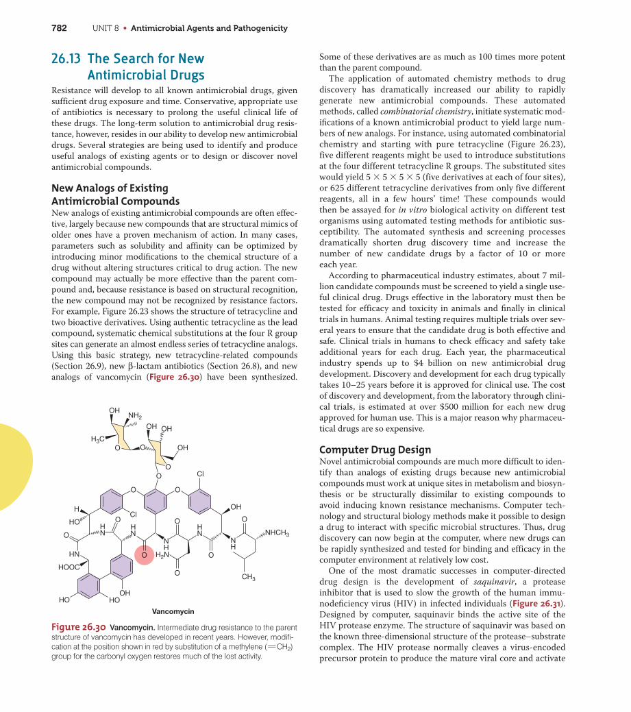
UNIT 8 • Antimicrobial Agents and Pathogenicity782
26.13 The Search for New Antimicrobial Drugs
Resistance will develop to all known antimicrobial drugs, given
sufficient drug exposure and time. Conservative, appropriate use
of antibiotics is necessary to prolong the useful clinical life of
these drugs. The long-term solution to antimicrobial drug resis-
tance, however, resides in our ability to develop new antimicrobial
drugs. Several strategies are being used to identify and produce
useful analogs of existing agents or to design or discover novel
antimicrobial compounds.
New Analogs of Existing Antimicrobial CompoundsNew analogs of existing antimicrobial compounds are often effec-
tive, largely because new compounds that are structural mimics of
older ones have a proven mechanism of action. In many cases,
parameters such as solubility and affinity can be optimized by
introducing minor modifications to the chemical structure of a
drug without altering structures critical to drug action. The new
compound may actually be more effective than the parent com-
pound and, because resistance is based on structural recognition,
the new compound may not be recognized by resistance factors.
For example, Figure 26.23 shows the structure of tetracycline and
two bioactive derivatives. Using authentic tetracycline as the lead
compound, systematic chemical substitutions at the four R group
sites can generate an almost endless series of tetracycline analogs.
Using this basic strategy, new tetracycline-related compounds
(Section 26.9), new β-lactam antibiotics (Section 26.8), and new
analogs of vancomycin (Figure 26.30) have been synthesized.
Some of these derivatives are as much as 100 times more potent
than the parent compound.
The application of automated chemistry methods to drug
discovery has dramatically increased our ability to rapidly
generate new antimicrobial compounds. These automated
methods, called combinatorial chemistry, initiate systematic mod-
ifications of a known antimicrobial product to yield large num-
bers of new analogs. For instance, using automated combinatorial
chemistry and starting with pure tetracycline (Figure 26.23),
five different reagents might be used to introduce substitutions
at the four different tetracycline R groups. The substituted sites
would yield 5 3 5 3 5 3 5 (five derivatives at each of four sites),
or 625 different tetracycline derivatives from only five different
reagents, all in a few hours’ time! These compounds would
then be assayed for in vitro biological activity on different test
organisms using automated testing methods for antibiotic sus-
ceptibility. The automated synthesis and screening processes
dramatically shorten drug discovery time and increase the
number of new candidate drugs by a factor of 10 or more
each year.
According to pharmaceutical industry estimates, about 7 mil-
lion candidate compounds must be screened to yield a single use-
ful clinical drug. Drugs effective in the laboratory must then be
tested for efficacy and toxicity in animals and finally in clinical
trials in humans. Animal testing requires multiple trials over sev-
eral years to ensure that the candidate drug is both effective and
safe. Clinical trials in humans to check efficacy and safety take
additional years for each drug. Each year, the pharmaceutical
industry spends up to $4 billion on new antimicrobial drug
development. Discovery and development for each drug typically
takes 10–25 years before it is approved for clinical use. The cost
of discovery and development, from the laboratory through clini-
cal trials, is estimated at over $500 million for each new drug
approved for human use. This is a major reason why pharmaceu-
tical drugs are so expensive.
Computer Drug DesignNovel antimicrobial compounds are much more difficult to iden-
tify than analogs of existing drugs because new antimicrobial
compounds must work at unique sites in metabolism and biosyn-
thesis or be structurally dissimilar to existing compounds to
avoid inducing known resistance mechanisms. Computer tech-
nology and structural biology methods make it possible to design
a drug to interact with specific microbial structures. Thus, drug
discovery can now begin at the computer, where new drugs can
be rapidly synthesized and tested for binding and efficacy in the
computer environment at relatively low cost.
One of the most dramatic successes in computer-directed
drug design is the development of saquinavir, a protease
inhibitor that is used to slow the growth of the human immu-
nodeficiency virus (HIV) in infected individuals (Figure 26.31).
Designed by computer, saquinavir binds the active site of the
HIV protease enzyme. The structure of saquinavir was based on
the known three-dimensional structure of the protease–substrate
complex. The HIV protease normally cleaves a virus-encoded
precursor protein to produce the mature viral core and activate
H3C
NH2
H2N
NHCH3
CH3
H
HN
HOOC
HO HOOH
HN
HN
HN
NH
NH
HO O
OO
OH
OH OH
OH
O O
O O
O
O
Cl
OH
Vancomycin
O
O
O
O
Cl
Figure 26.30 Vancomycin. Intermediate drug resistance to the parentstructure of vancomycin has developed in recent years. However, modifi-cation at the position shown in red by substitution of a methylene ( CH2)group for the carbonyl oxygen restores much of the lost activity.
w
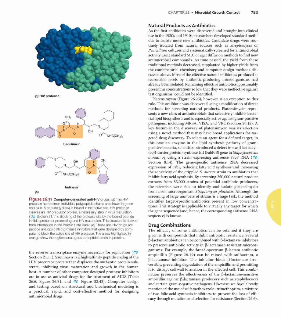
CHAPTER 26 • Microbial Growth Control 783
UN
IT 8
the reverse transcriptase enzyme necessary for replication (
Section 21.11). Saquinavir is a high-affinity peptide analog of the
HIV precursor protein that displaces the authentic protein sub-
strate, inhibiting virus maturation and growth in the human
host. A number of other computer-designed protease inhibitors
are in use as antiviral drugs for the treatment of AIDS (Table
26.6, Figure 26.31, and Figure 33.43). Computer design
and testing based on structural and biochemical modeling is
a practical, rapid, and cost-effective method for designing
antimicrobial drugs.
Natural Products as AntibioticsAs the first antibiotics were discovered and brought into clinical
use in the 1930s and 1940s, researchers developed standard meth-
ods to isolate more new antibiotics. Candidate drugs were rou-
tinely isolated from natural sources such as Streptomyces or
Penicillium cultures and systematically screened for antimicrobial
activity using standard MIC or agar diffusion methods to find new
antimicrobial compounds. As time passed, the yield from these
traditional methods decreased, supplanted by higher yields from
the combinatorial chemistry and computer design methods dis-
cussed above. Most of the effective natural antibiotics produced at
reasonable levels by antibiotic-producing microorganisms had
already been isolated. Remaining effective antibiotics, presumably
present in concentrations so low that they were ineffective against
test organisms, could not be identified.
Platensimycin (Figure 26.25), however, is an exception to this
rule. This antibiotic was discovered using a modification of direct
methods for screening natural products. Platensimycin repre-
sents a new class of antimicrobials that selectively inhibits bacte-
rial lipid biosynthesis and is especially active against gram-positive
pathogens, including MRSA, VISA, and VRE (Section 26.12). A
key feature in the discovery of platensimycin was its selection
using a novel method that may have broad applications for tar-
geted drug discovery. To select an agent for a defined target, in
this case an enzyme in the lipid synthesis pathway of gram-
positive bacteria, scientists introduced a defect in the β-ketoacyl-
(acyl-carrier protein) synthase I/II (FabF/B) gene in Staphylococcus
aureus by using a strain expressing antisense FabF RNA (
Section 8.14). The gene-specific antisense RNA decreased
expression of FabF, reducing fatty acid synthesis and increasing
the sensitivity of the crippled S. aureus strain to antibiotics that
inhibit fatty acid synthesis. By screening 250,000 natural product
extracts from 83,000 strains of potential antibiotic producers,
the scientists were able to identify and isolate platensimycin
from a soil microorganism, Streptomyces platensis. Although the
screening of large numbers of strains is a huge task, the method
identifies target-specific antibiotics present in low concentra-
tions. This strategy is applicable to virtually any target for which
the gene sequence (and, hence, the corresponding antisense RNA
sequence) is known.
Drug CombinationsThe efficacy of some antibiotics can be retained if they are
given with compounds that inhibit antibiotic resistance. Several
β-lactam antibiotics can be combined with β-lactamase inhibitors
to preserve antibiotic activity in β-lactamase-resistant microor-
ganisms. For example, the broad-spectrum β-lactam antibiotic
ampicillin (Figure 26.19) can be mixed with sulbactam, a
β-lactamase inhibitor. The inhibitor binds β-lactamase irre-
versibly, preventing degradation of the ampicillin and permitting
it to disrupt cell wall formation in the affected cell. This combi-
nation preserves the effectiveness of the β-lactamase-sensitive
ampicillin against β-lactamase producers such as staphylococci
and certain gram-negative pathogens. Likewise, we have already
mentioned the use of sulfamethoxazole–trimethoprim, a mixture
of two folic acid synthesis inhibitors, to prevent the loss of effi-
cacy through mutation and selection for resistance (Section 26.6).
Figure 26.31 Computer-generated anti-HIV drugs. (a) The HIVprotease homodimer. Individual polypeptide chains are shown in greenand blue. A peptide (yellow) is bound in the active site. HIV proteasecleaves an HIV precursor protein, a necessary step in virus maturation( Section 21.11). Blocking of the protease site by the bound peptideinhibits precursor processing and HIV maturation. This structure is derivedfrom information in the Protein Data Bank. (b) These anti-HIV drugs arepeptide analogs called protease inhibitors that were designed by com-puter to block the active site of HIV protease. The areas highlighted inorange show the regions analogous to peptide bonds in proteins.
(a) HIV protease
(b)
NH
NHC(CH3)2
NHC(CH3)2
NH2
N N
N
N NH
O
O
O
OOH
OH
NH
O
O
OH
Saquinavir
Indinavir
UNIT 8 • Antimicrobial Agents and Pathogenicity784
Drug combination therapy approaches have revolutionized
treatment of HIV infections. Currently, a combination therapy
consisting of nucleoside analogs and a protease inhibitor is rec-
ommended. This drug treatment protocol is termed HAART, for
highly active anti-retroviral therapy. As with antimicrobial com-
bination regimens, HAART is designed to target two indepen-
dent viral functions; the nucleoside analogs target virus replication
and the protease inhibitors target virus maturation. Because the
probability of a single virus developing resistance to multiple
drugs is less than the probability of developing resistance to a sin-
gle drug, HAART-resistant strains are relatively uncommon (
Section 33.14).
BacteriophagesBacteriophages are viruses that infect bacteria ( Sections
9.8–9.10). Bacteriophage therapy has been used on a limited basis
for over 80 years to treat infections in animals and, in a few
instances, in humans. Phages interact with individual bacterial
cell surface components and show specificity for particular bac-
terial species. The attached phage enters the cell, replicates, and
kills the bacterial host in the process. The efficacy and efficiency
of these agents for human treatment is largely untested and
somewhat controversial, although clinical trials of several prod-
ucts are ongoing. However, because bacteria can acquire resis-
tance to a phage infection through mutations that alter receptors
or reduce the susceptibility of the cell wall to phage enzymes,
bacteriophage therapy will likely be susceptible to resistance, just
as for most chemical antimicrobial agents.
MiniQuiz• Explain the advantages and disadvantages of developing new
drugs based on existing drug analogs.
• How can computer drug design aid in the search for new drugs?
• Explain the use of antisense RNA for drug discovery.
Big Ideas26.1 Sterilization is the killing of all organisms and viruses, and heat is
the most widely used method of sterilization. The temperature
employed must eliminate the most heat-resistant organisms,
usually bacterial endospores. An autoclave permits applications
of steam heat under pressure, achieving temperatures above the
boiling point of water, which kills endospores. Pasteurization
does not sterilize liquids, but reduces microbial load, kills most
pathogens, and inhibits the growth of spoilage microorganisms.
26.2 Controlled doses of electromagnetic radiation effectively inhibit
microbial growth. Ultraviolet radiation is used for decontaminat-
ing surfaces and materials that do not absorb light, such as air
and water. Ionizing radiation that can penetrate solid or light-
absorbing materials is used for sterilization and decontamination
in the medical and food industries.
26.3 Filters remove microorganisms from air or liquids. Depth filters,
including HEPA filters, are used to remove microorganisms and
other contaminants from liquids or air. Membrane filters are used
for sterilization of heat-sensitive liquids, and nucleation track filters
are used to isolate specimens for electron microscopy.
26.4 Chemicals are often used to control microbial growth. Chemicals
that kill organisms are called -cidal agents; those that inhibit
growth are called -static agents; those that lyse organisms are
called -lytic agents. Antimicrobial agents are tested for efficacy
by determining their ability to inhibit growth in vitro.
26.5 Sterilants, disinfectants, and sanitizers are used to decontami-
nate nonliving material. Antiseptics and germicides are used to
reduce microbial growth on living tissues. Antimicrobial com-
pounds have commercial, healthcare, and industrial applications.
26.6 Synthetic antimicrobial agents are selectively toxic for Bacteria,
viruses, and fungi. Synthetic growth factor analogs such as sulfa
drugs, isoniazid, and nucleic acid analogs are metabolic inhibitors.
Quinolones inhibit the action of DNA gyrase in Bacteria.
26.7 Antibiotics are a chemically diverse group of antimicrobial
compounds that are produced by microorganisms. Although
many antibiotics are known, only a few are clinically effective.
Each antibiotic works by inhibiting a specific cellular process or
function in the target microorganisms.
26.8 The β-lactam compounds, including the penicillins and the
cephalosporins, are the most important single class of clinical
antibiotics. These antibiotics and their semisynthetic derivatives
target cell wall synthesis in Bacteria. They have low host toxicity
and collectively have a broad spectrum of activity.
26.9 The aminoglycosides, macrolides, and tetracycline antibiotics
are structurally complex molecules produced by Bacteria and
are active against other Bacteria. These antibiotics selectively
interfere with protein synthesis in Bacteria. Daptomycin and
UN
IT 8
platensimycin are structurally novel antibiotics that target cyto-
plasmic membrane functions and lipid biosynthesis, respectively.
26.10 Effective antiviral agents selectively target virus-specific enzymes
and processes. Clinically useful antiviral agents include nucleo-
side analogs and other drugs that inhibit nucleic acid polymerases
and viral genome replication. Agents such as protease inhibitors
interfere with viral maturation steps. Host cells also produce the
antiviral interferon proteins that stop viral replication.
26.11 Antifungal agents fall into many chemical categories. Because
fungi are Eukarya, selective toxicity is hard to achieve, but some
effective antifungal agents are available. Treatment of fungal
infections is an emerging human health issue.
26.12 The use of antimicrobial drugs inevitably leads to resistance in
the targeted microorganisms. The development of resistance can
be accelerated by the indiscriminate use of antimicrobial drugs.
A few pathogens have developed resistance to all known antimi-
crobial drugs.
26.13 New antimicrobial compounds are constantly being discovered
and developed to deal with drug-resistant pathogens and to
enhance our ability to treat infectious diseases. Analogs of existing
drugs are often synthesized and used as next-generation antimi-
crobial compounds. Computer drug design is an important tool
for drug discovery.
Aminoglycoside an antibiotic such as strepto-
mycin, containing amino sugars linked by
glycosidic bonds
Antibiotic a chemical substance produced by a
microorganism that kills or inhibits the
growth of another microorganism
Antimicrobial drug resistance the acquired
ability of a microorganism to grow in the
presence of an antimicrobial drug to which
the microorganism is usually susceptible
Antimicrobial agent a chemical compound
that kills or inhibits the growth of microor-
ganisms
Antiseptic (germicide) a chemical agent that
kills or inhibits growth of microorganisms
and is sufficiently nontoxic to be applied to
living tissues
Autoclave a sealed heating device that destroys
microorganisms with temperature and steam
under pressure
Bacteriocidal agent an agent that kills bacteria
Bacteriostatic agent an agent that inhibits
bacterial growth
Beta (β)-lactam antibiotic an antibiotic,
including penicillin, that contains the four-
membered heterocyclic β-lactam ring
Broad-spectrum antibiotic an antibiotic
that acts on both gram-positive and gram-
negative Bacteria
Decontamination a treatment that renders
an object or inanimate surface safe to
handle
Disinfectant an antimicrobial agent used only
on inanimate objects
Disinfection the elimination of pathogens from
inanimate objects or surfaces
Fungicidal agent an agent that kills fungi
Fungistatic agent an agent that inhibits fungal
growth
Fusion inhibitor a peptide that blocks the
fusion of viral and target cytoplasmic
membranes
Germicide (antiseptic) a chemical agent that
kills or inhibits growth of microorganisms
and is sufficiently nontoxic to be applied to
living tissues
Growth factor analog a chemical agent that is
related to and blocks the uptake of a growth
factor
HEPA filter a high-efficiency particulate
air filter that removes particles, including
microorganisms, from intake or exhaust
air flow
Interferon a cytokine protein produced
by virus-infected cells that induces signal
transduction in nearby cells, resulting
in transcription of antiviral genes and
expression of antiviral proteins
Minimum inhibitory concentration (MIC)
the minimum concentration of a substance
necessary to prevent microbial growth
Nonnucleoside reverse transcriptase
inhibitor (NNRTI) a nonnucleoside analog
used to inhibit viral reverse transcriptase
Nucleoside reverse transcriptase inhibitor
(NRTI) a nucleoside analog used to inhibit
viral reverse transcriptase
Pasteurization the use of controlled heat to
reduce the microbial load, including disease-
producing microorganisms and spoilage
microorganisms, in heat-sensitive liquids
Penicillin a class of antibiotics that inhibit
bacterial cell wall synthesis, characterized
by a β-lactam ring
Protease inhibitor an inhibitor of a viral
protease
Quinolone a synthetic antibacterial compound
that interacts with DNA gyrase and prevents
supercoiling of bacterial DNA
Sanitizer an agent that reduces microorganisms
to a safe level, but may not eliminate them
Selective toxicity the ability of a compound to
inhibit or kill pathogenic microorganisms
without adversely affecting the host
Sterilant (sterilizer, sporicide) a chemical
agent that destroys all forms of microbial
life
Sterilization the killing or removal of all living
organisms and viruses from a growth
medium
Tetracycline an antibiotic characterized by the
four-ring naphthacene structure
Viricidal agent an agent that stops viral
replication and activity
Viristatic agent an agent that inhibits viral
replication
Review of Key Terms
CHAPTER 26 • Microbial Growth Control 785
UNIT 8 • Antimicrobial Agents and Pathogenicity786
1. Why is the decimal reduction time (D) important in heat
sterilization? How would the presence of bacterial endospores
affect D (Section 26.1)?
2. Describe the effects of a lethal dose of ionizing radiation at the
molecular level (Section 26.2).
3. What are the principal advantages of membrane filters? Of depth
filters? Of nucleopore filters (Section 26.3)?
4. Describe the procedure for obtaining the minimum inhibitory
concentration (MIC) for a chemical that is bacteriocidal for
Escherichia coli (Section 26.4).
5. Contrast the action of disinfectants and antiseptics. Disinfectants
normally cannot be used on living tissue; why not (Section 26.5)?
6. Growth factor analogs are generally distinguished from antibiotics
by a single important criterion. Explain (Section 26.6).
7. Identify common sources for naturally occurring antimicrobial
drugs (Section 26.7).
8. Describe the mode of action that characterizes a β-lactam antibi-
otic. Why are these antibiotics generally more effective against
gram-positive bacteria than against gram-negative bacteria
(Section 26.8)?
9. Distinguish between the modes of action of at least three of the
protein synthesis–inhibiting antibiotics (Section 26.9).
10. Why do antiviral drugs generally exhibit host toxicity (Section
26.10)?
11. Identify the targets that allow selective toxicity of antifungal agents
(Section 26.11).
12. Identify six mechanisms responsible for antibiotic resistance
(Section 26.12).
13. Explain how application of antisense RNA methods can extend
traditional natural product selection methods for antibiotic
discovery (Section 26.13).
Review Questions
1. What are some potential drawbacks to the use of radiation in food
preservation? Do you think these drawbacks could be manifested
as health hazards? Why or why not? How would you distinguish
between radiation-damaged and radiation-contaminated food?
2. Filtration is an acceptable means of pasteurization for some
liquids. Design a filtration system for pasteurization of a heat-
sensitive liquid. For a liquid of your choice, identify the advantages
and disadvantages of a filtration system over a heat pasteurization
system. Explain in terms of product quality, shelf life, and price.
3. Although growth factor analogs may inhibit microbial metabolism,
only a few of these agents are useful in practice. Many growth fac-
tor analogs, including some in wide use, such as azidothymidine,
exhibit significant host cell toxicity. Describe a growth factor ana-
log that is effective and has low toxicity for host cells. Why is the
toxicity low for the agent you chose? Also describe a growth factor
analog that is effective against an infectious disease, but exhibits
toxicity for host cells. Why might a toxic agent such as AZT still be
used in certain situations to treat infectious diseases? What pre-
cautions would you take to limit the toxic effects of such a drug
while maximizing the therapeutic activity? Explain your answer.
4. Although many antibiotics demonstrate clear selective toxicity for
Bacteria, many groups of Bacteria are innately resistant to their
effects. Indicate why gram-negative Bacteria are resistant to the
effects of many, but not all, antibiotics. Further explain why some
antibiotics are effective against these organisms.
5. List the features of an ideal antiviral drug, especially with regard to
selective toxicity. Do such drugs exist? What factors might limit
the use of such a drug?
6. Like viruses, fungi present special problems for drug therapy.
Explain the problems inherent in drug treatment of both groups
and explain whether or not you agree with the preceding state-
ment. Give specific examples and suggest at least one group of
agents that might target both types of infectious agents.
7. Explain the genetic basis of acquired resistance to β-lactam
antibiotics in Staphylococcus aureus. Design experiments to reverse
resistance to the β-lactam antibiotics. Do you think this can be
done in the laboratory? Can your experiment be applied “in the
field” to promote deselection of antibiotic-resistant organisms?
8. Design experiments to examine microorganisms for production of
novel antibiotics. Which group or groups of microorganisms
would you choose to screen for antibiotic production? Where
could you find and isolate these organisms in a natural environ-
ment? What advantage, if any, would the production of an antibi-
otic provide for these organisms in nature? What in vitro methods
would you use to test the efficacy of your potential new antibiotics?
How might you increase the sensitivity of assays for natural prod-
ucts? Why are members of the genus Streptomyces still productive
sources of novel antibiotics?
Application Questions
Need more practice? Test your understanding with Quantitative Questions; access additional
study tools including tutorials, animations, and videos; and then test your knowledge with
chapter quizzes and practice tests at www.microbiologyplace.com.


