Embed Size (px)
Citation preview

This content has been downloaded from IOPscience. Please scroll down to see the full text.
Download details:
IP Address: 194.141.252.102
This content was downloaded on 16/10/2013 at 03:28
Please note that terms and conditions apply.
Microencapsulated bio-markers for assessment of stress conditions in aquatic organisms in
vivo
View the table of contents for this issue, or go to the journal homepage for more
2012 Laser Phys. Lett. 9 542
(http://iopscience.iop.org/1612-202X/9/7/013)
Home Search Collections Journals About Contact us My IOPscience
542Laser Physics
Letters Laser Phys. Lett. 9, No. 7, 542–546 (2012) / DOI 10.7452/lapl.201210020
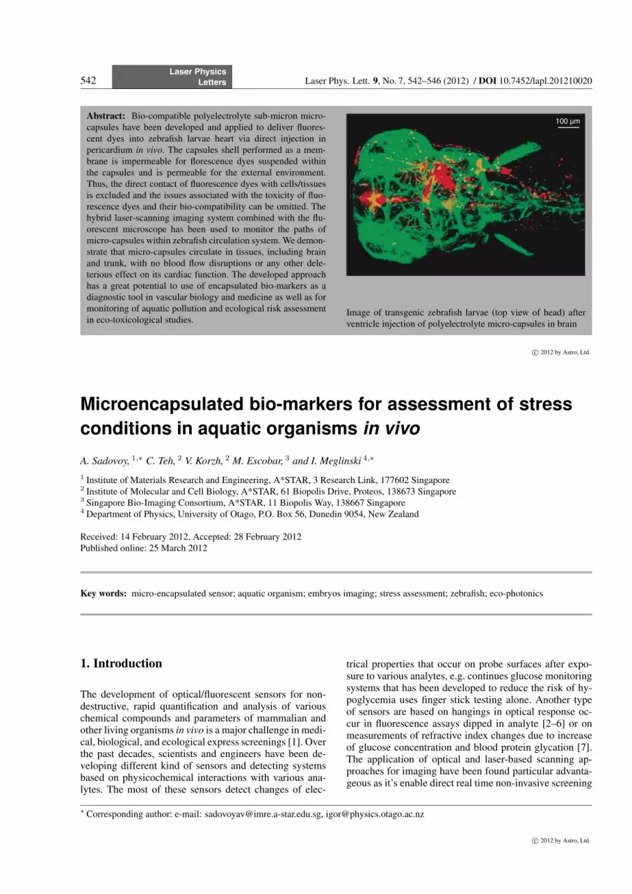
Abstract: Bio-compatible polyelectrolyte sub-micron micro-capsules have been developed and applied to deliver fluores-cent dyes into zebrafish larvae heart via direct injection inpericardium in vivo. The capsules shell performed as a mem-brane is impermeable for florescence dyes suspended withinthe capsules and is permeable for the external environment.Thus, the direct contact of fluorescence dyes with cells/tissuesis excluded and the issues associated with the toxicity of fluo-rescence dyes and their bio-compatibility can be omitted. Thehybrid laser-scanning imaging system combined with the flu-orescent microscope has been used to monitor the paths ofmicro-capsules within zebrafish circulation system. We demon-strate that micro-capsules circulate in tissues, including brainand trunk, with no blood flow disruptions or any other dele-terious effect on its cardiac function. The developed approachhas a great potential to use of encapsulated bio-markers as adiagnostic tool in vascular biology and medicine as well as formonitoring of aquatic pollution and ecological risk assessmentin eco-toxicological studies.
100 µm
Image of transgenic zebrafish larvae (top view of head) afterventricle injection of polyelectrolyte micro-capsules in brain
c⃝ 2012 by Astro, Ltd.
Microencapsulated bio-markers for assessment of stressconditions in aquatic organisms in vivo
A. Sadovoy, 1,∗ C. Teh, 2 V. Korzh, 2 M. Escobar, 3 and I. Meglinski 4,∗
1 Institute of Materials Research and Engineering, A*STAR, 3 Research Link, 177602 Singapore2 Institute of Molecular and Cell Biology, A*STAR, 61 Biopolis Drive, Proteos, 138673 Singapore3 Singapore Bio-Imaging Consortium, A*STAR, 11 Biopolis Way, 138667 Singapore4 Department of Physics, University of Otago, P.O. Box 56, Dunedin 9054, New Zealand
Received: 14 February 2012, Accepted: 28 February 2012Published online: 25 March 2012
Key words: micro-encapsulated sensor; aquatic organism; embryos imaging; stress assessment; zebrafish; eco-photonics
1. Introduction
The development of optical/fluorescent sensors for non-destructive, rapid quantification and analysis of variouschemical compounds and parameters of mammalian andother living organisms in vivo is a major challenge in medi-cal, biological, and ecological express screenings [1]. Overthe past decades, scientists and engineers have been de-veloping different kind of sensors and detecting systemsbased on physicochemical interactions with various ana-lytes. The most of these sensors detect changes of elec-
trical properties that occur on probe surfaces after expo-sure to various analytes, e.g. continues glucose monitoringsystems that has been developed to reduce the risk of hy-poglycemia uses finger stick testing alone. Another typeof sensors are based on hangings in optical response oc-cur in fluorescence assays dipped in analyte [2–6] or onmeasurements of refractive index changes due to increaseof glucose concentration and blood protein glycation [7].The application of optical and laser-based scanning ap-proaches for imaging have been found particular advanta-geous as it’s enable direct real time non-invasive screening
∗ Corresponding author: e-mail: [email protected], [email protected]
c⃝ 2012 by Astro, Ltd.

Laser Phys. Lett. 9, No. 7 (2012)Laser Physics
Letters 543
of samples not limited by area, providing visualization ofintrinsically fluorescent chemical species, non-fluorescentspecies, analytes and enzymes [8, 9].
A significant disadvantage of this approach is a tox-icity of most fluorescent dyes. Therefore, the techniquecannot be directly applied for tissues diagnostics in vivo.This problem can be solved with easier by using polyelec-trolyte capsules as a container to exclude contact of flu-orescence dyes with the external medium (tissues). Thecapsules shell performed as a membrane is impermeablefor florescence dyes suspended within the capsules and ispermeable for the external environment. Thus, the directcontact of fluorescence dyes with cells/tissues is excludedand the issues associated with the toxicity of fluorescencedyes and their bio-compatibility can be omitted.
Polyelectrolyte multi-layered (PM) capsules haveshown promising applications as containers for bio-sensors [10, 11] since they were fist obtained in 1998 MaxPlank Institute of Colloids, Germany [12]. The PM en-capsulation technique is based on layer-by-layer adsorp-tion of oppositely charged poly-electrolytes on micron-or sub-micron size cores. The fluorescence dye, typically,preloaded inside the PM micro-capsules [13] and the per-meability of capsules shell can be tuned by varying thenumber of layers [14]. Such PM-based sensors shamefullyused for measurement oxygen and pH [4, 15, 16].
The unique property of PM is that their physical,chemical and mechanical properties can be tuned ontothe nano-scale by varying their composition and the ionicstrength and pH of the environment. Potentially thesemicro-sensors can find a number of applications in biol-ogy and biomedicine. In our study we are focused on thedevelopment of PM micro-capsule-based sensor for detec-tion of laser induced reactive oxygen in small aquatic or-ganisms in vivo. Reactive oxygen spices (ROS) are naturalbyproducts of normal metabolism of oxygen and have im-portant roles in cell signaling and homeostasis. Biologicaltissue with high metabolic rate and moderate level of an-tioxidant protection is particularly susceptible to oxidativestress [17]. ROS’s effect on ion fluxes, which is criticalfor cardiac contraction [18] contributes to the pathogene-sis and progression of heart failure [19]. This is a globalhealth problem that accounts for about ∼ 4.5% total hos-pital admissions [20]. Heart failure is a progressively di-lapidating disease that poses major challenges to healthcare providers. The development of a ROS sensor that per-mits in vivo monitoring of oxidative stress and optimiza-tion of current cardio-protective antioxidants delivery willthus significantly enhance existing heart failure treatmentprocedure.
2. Materials and method
Rhodamine B isothiocyanate-Dextran (RhB-DS) havebeen chosen as a modeling sensor fluorescent dye. Encap-sulation of RhB-DS has been done by precipitation it inCaCO3 particles in a synthesis stage (Fig. 1).
Figure 1 SEM image of synthesized CaCO3 particles. Scale baris 1 µm
RhB-DS has been dissolved in DI water in concentra-tion 1 mg/ml. 10 ml of the solution was placed in 50 mlcentrifuge tube preliminary maintained with a dispenser(Ultra-Turrax T-18 dispenser (IKA)). 24000 rpm speed isfound the most suitable for current purpose. 3.1 ml of1 M CaCl2 is poured in the mixing water. Then 3.1 mlof M Na2CO3 is poured by 5 ml pipet with further 30 secmixing. The precipitated is washed immediately 3 timesby adding 50 ml of DI water and 30 sec centrifuging on3000 rpm. To store the particles they should be frizzedand dried to prevent further re-crystallization. To pre-vent washing the protein out particles with the precipi-tated RhB-DS were covered by a polyelectrolyte multi-layer membrane. The PM membrane consist from sev-eral alternatively charged poly-electrolytes layers. Then,poly-(sodium 4-styrene sulfonate) Mw = 70 000 Da (PSS)was used as anion layers and poly-(allylamine hydrochlo-ride) Mw = 15000 Da (PAH) as caption layers. The poly-electrolytes were dissolved in 0.134 M NaCl water solu-tion in a concentration 2 mg/ml. Poly(L-lysine) 20 kDagrafted with poly(ethylene glycol) (PLL-g-PEG) 5 kDawith g = 3.5 have been applied as an outmost layer to pro-tect aggregation of the PM capsules in blood stream.
The PM shell was assembled around the CaCO3 parti-cles with precipitated via alternate layer-by-layer (L-b-L)adsorption of the polyelectrolytes. For all steps 1.5 ml cen-trifugation tubes (Eppendorf) are used. 30 mg of the par-ticles powder is preliminary washed by re-suspending in1.5 ml of DI water with further centrifugation by 30 sec at3000 rpm and removing of supernatant. On the next stepthe caption layer has absorbed. 1.5 ml of the PAH solutionis added to the particles followed by 15 min shaking. Thenthe dispersion is subjected by 3 circles of washing stepssimilar to that described above to remove un-coupled poly-electrolyte. PAH adsorption is verified by switching thesign of potential from negative to positive (ζ = +35 mV).
www.lasphys.comc⃝ 2012 by Astro, Ltd.

544Laser Physics
Letters A. Sadovoy, C. Teh, et al.: Microencapsulated bio-markers for assessment of stress conditions
Figure 2 The obtained PM micro-capsules. Scale bar is 1µm
The switching of sign of potential ζ provides an evidenceof successful adsorption of a next polyelectrolyte layer. Inan agreement with the principles of LbL assembling, theanion has to be applied to form a next coating layer. 1.5 mlof the PSS solution is added to the centrifuged particleswith the adsorbed caption layer. The deposition of the an-ion layer was performed in the same manner as it has beenpreviously described for the caption one. The depositionwas proved by switching the sign of potential from posi-tive to negative (ζ = 35 mV). The described assembling isrepeated alternating PAH with PSS to obtain the requirednumber of layers in the membrane. Then a part of capsulesadditionally covered by PLL-g-PEG layer. The developedPM micro-capsules are presented in Fig. 2.
The encapsulated PM sensors with PAH/PSS andPAH/PSS/PLL-g-PEG shells were tested for bio-clearanceby injection in pericardium of 3 day-old zebrafish em-bryos. The laser scanning system combined with the flu-orescent microscope (see Fig. 3) has been used to monitorpaths of the encapsulated sensors within zebrafish circula-tion system.
All experiments involving zebrafish embryos/larvaewere carried out in accordance to IACUC rules. Em-bryo has placed in small plastic Petri dish in solution toprivet them movement along imaging time. Measurementof heartbeat and contractility became easier with trans-genic zebrafish larvae expressing fluorescent reporters inthe heart. Some of these cardiac reporter lines were gener-ated in the IMCB, A*STAR, Singapore [21].
3. Results and discussion
The results of application of modeling encapsulated PMsensors with PAH/PSS and PAH/PSS/PLL-g-PEG shellsare presented in Fig. 4. The micro-capsules with PAH/PSS
(a)
(b)
12
34 5
6
7
11
10
8
9
12 13 14
15
16
Figure 3 (online color at www.lasphys.com) Schematic diagramof the hybrid laser scanning system applied for imaging of ze-brafish circulation system in vivo. Part I shows the laser scanningsystem combined with Carl Zeiss microscope Meta 510. Here:(1) – argon-ion laser (λ= 488 nm, 30 mW), (2) – mirror, (3) –He-Ne laser (λ= 543 nm, 1 mW), (4) – beam splitter, (5) – colli-mator, dichroic beam splitter (6), set of scanning mirrors (7) andAchroplan (×40 and NA 0.75) objective (8), pinhole (10) anddetector (11). A small plastic Petri dish with zebrafish larvae (9).Section (b) presents the excitation part of the system, including:100 W HBO 103 mercury vapor short-arc lam (12), collimator(14), mirror (15), and Plan-NeoFluar (×10 and NA 0.3) objec-tive (16)
shell are aggregated immediately after injection in peri-cardium of 3 day-old zebrafish embryos and most of cap-sules staked near the injection area. This results an abnor-mal development of zebrafish embryo (see Fig.4a) withunpredictable distribution of PAH/PSS shell in the body.
In contrast, the micro-capsules with PLL-g-PEG out-most layer shows normal embryo development (seeFig. 4b). No aggregation of micro-capsules has been ob-served. After 3 days the modeling encapsulated PM sen-sors were clearly seen in the heart, eyes, endoderm and thespinal chord. Importantly, that heart beat and contractilitywere not influenced by injected capsules.
Additionally the PAH/PSS/PLL-g-PEG capsules wereobserved in a brain ventricle injection (Fig. 5a andFig. 5b). Both heart beat and contractility were not influ-enced by micro-capsules injection.
c⃝ 2012 by Astro, Ltd.www.lasphys.com

Laser Phys. Lett. 9, No. 7 (2012)Laser Physics
Letters 545
(a) (b)
Figure 4 (online color at www.lasphys.com) The transgenic ze-brafish embryos imaged 3 days after injection of PM micro-capsules without (a) and contained PLL-g-PEG at the outer layer(b). Scale bar is 100 mum
The PM PAH/PSS/PLL-g-PEG micro-capsules weredetected in the intestine 7 days after injection (Fig. 5c,Fig. 5d and Fig. 6), and again the heart beat and contrac-tility were not influenced by micro-capsules injection. Ze-brafish heart consists of an atrium and a ventricle with thedirection of blood flow regulated by cardiac valves. Thisis a well established animal model for studies of heart dis-orders [22] as common features exist between the humanand zebrafish heart. Given that the normal heartbeat in hu-man is between 60 – 100 beats per minute (bpm), the rapidheart rate of the mice at 300 – 600 bpm is due to the pres-ence of a specialized system of cardiac re-polarization, uti-lizing different sets of cardiac ion channels. In contrast, at80 – 160 bpm the zebrafish heart rate is much closer to thatof human and re-polarization is achieved by a very similarmolecular mechanism [22]. Notably, drugs that cause ir-regular heartbeats in human do the same in zebrafish [23].
The obtained results clearly show that the created PMmicrocapsule prevents dye diffusion in tissue that makesuse toxic dyes possible. Besides, the multi-layered shell ofthe micro-capsules is permeable for environment with thesize less than 500 Da.
4. Summary
Thus, in framework of a development of micron-size ox-idation stress-sensor for assessment of measuring level ofROS in KillerRed expressing transgenic zebrafish larvae invivo we present a prototype of micro-encapsulated opticalbio-sensor and an approach of the sensors delivery into ze-brafish larvae circulation system via micro-injection. Wedemonstrate that micro-capsules freely circulate in ze-brafish tissues, including brain and trunk, with no bloodflow disruptions or any other deleterious effect on its car-diac function. The further key step in our investigation isthe light-inducible heart failure animal model we devel-oped in zebrafish that expressed KillerRed in the heart.By utilizing the photosensitizer properties of KillerRed
100 µm(a)
100 µm(c)
(b)
(d)
Figure 5 (online color at www.lasphys.com) Images of trans-genic zebrafish larvae after ventricle injection of polyelectrolytemicro-capsules with PAH/PSS/PLL-g-PEG shell in brain. (a) and(b) are top views of a head of zebrafish immediately after micro-capsules injection. (c) and (d) are the top views of the same larvae7 days later
Figure 6 (online color at www.lasphys.com) Images of trans-genic zebrafish larvae after 7 days of injection of polyelectrolytemicro-capsules with PAH/PSS/PLL-g-PEG. Scale bar is 100 µm
to produce ROS upon green light illumination, heart fail-ure can be repeatedly induced in a non-invasive manner.The photosensitizer property of KillerRed [24] allows non-invasive induction of ROS in the optically translucent ze-brafish heart and elicit death of cardiomyocytes resultingin heart failure in a dose-dependent manner [24, 25]. Im-portantly, the use of this biological platform permits thedevelopment of physiologically sensitive ROS sensor andidentifies efficient antioxidants that is likely lead to im-prove heart contractility. We anticipate that apart from theapplication mentioned above the developed technique willbe extremely useful in eco-photonics for in vivo assess-ment of stress conditions in aquatic organisms, influencedby climatic changes [26]. There is also a broad spectrum ofapplication in pharmacy, vascular biology, food sciences,material science, cosmetic and health care industries. Cur-
www.lasphys.comc⃝ 2012 by Astro, Ltd.

546Laser Physics
Letters A. Sadovoy, C. Teh, et al.: Microencapsulated bio-markers for assessment of stress conditions
rent approach has also a great potential to be used as adiagnostic tool in eco-toxicological studies.
Acknowledgements Authors acknowledge the support of Singa-pore Networking Grant, HRC, New Zealand.
References
[1] P. D’Orazio, Clin. Chim. Acta 412, 1749 (2011).[2] E. Moczko, I.V. Meglinski, C. Bessant, and S.A. Piletsky,
Anal. Chem. 81, 2311 (2009).[3] E. Moczko, M. Cauchi, C. Turner, I. Meglinski, and S.
Piletsky, IEEE Trans. Biomed. Eng. 58, 2154 (2011).[4] S.R. Nayak and M.J. McShane, Sensor Letters 4, 433
(2006).[5] T. Saxl, F. Khan, D.R. Matthews, Z.-L. Zhi, O. Rolinski, S.
Ameer-Beg, and J. Pickup, Biosens. Bioelectron. 24, 3229(2009).
[6] T. Duchesne, J.Q. Brown, K. Guice, S.R. Nayak, Y. Lvov,and M.J. McShane, Proc. SPIE 4624, 66 (2002).
[7] O.S. Zhernovaya, V.V. Tuchin, and I.V. Meglinski, LaserPhys. Lett. 5, 460 (2008).
[8] Demet Guzey and D.J. McClements, Adv. Coll. Int. Sci.128, 227 (2007).
[9] D.O. Grigoriev and R. Miller, Curr. Oppin. Coll. Int. Sci.14, 48 (2009).
[10] O. Kreft, A.M. Javier, G.B. Sukhorukov, and W.J. Parak,J. Mater. Chem. 17, 4471 (2007).
[11] B.G. De Geest, S. De Koker, G.B. Sukhorukov, O. Kreft,W.J. Parak, A.G. Skirtach, J. Demeester, S.C. De Smedt,and W.E. Hennink, Soft Matter 5, 282 (2009).
[12] E. Donath, G.B. Sukhorukov, F. Caruso, S.A. Davis, andH. Mohwald, Angew. Chem. Int. Ed. 37, 2201 (1998).
[13] D. Usov and G.B. Sukhorukov, Langmuir 26, 12575(2010).
[14] A.A. Antipov and G.B. Sukhorukov, Adv. Colloid Inter-face Sci. 111, 49 (2004).
[15] L.L. del Mercato, P. Rivera-Gil, A.Z. Abbasi, M. Ochs, C.Ganas, I. Zins, C. Snnichsen, and W.J. Parak, Nanoscale 2,458 (2010).
[16] M.J. McShane, J.Q. Brown, K.B. Guice, and Y.M. Lvov, J.Nanosci. Nanotechnol. 2, 411 (2002).
[17] J. Limon-Pacheco and M.E. Gonsebatt, Mutat. Res. Ge-netic Toxicol. Environ. Mutagen. 674, 137 (2009).
[18] P. Heusch, M. Canton, S. Aker, A. Van De Sand, I. Koni-etzka, T. Rassaf, S. Menazza, O.E. Brodde, F. Di Lisa,G. Heusch, and R. Schulz, Br. J. Pharmacol. 160, 1408(2010).
[19] F.J. Giordano, J. Clin. Invest. 115, 500 (2005).[20] G.K.T. Leong, P.P. Goh, B.C. Chang, and J. Linga-
manaicker, Singapore Med. J. 48, 408 (2007).[21] K.-L. Poon, M. Liebling, I. Kondrychyn, M. Garcia-Lecea,
and V. Korzh, Dev. Dynam. 239, 914 (2010).[22] T.J.A. Chico, P.W. Ingham, and D.C. Crossman, Trends
Cardiovasc. Med. 18, 150 (2008).[23] D.J. Milan, T.A. Peterson, J.N. Ruskin, R.T. Peterson, and
C.A. MacRae, Circulation 107, 1355 (2003).[24] M.E. Bulina, D.M. Chudakov, O.V. Britanova, Y.G. Yanu-
shevich, D.B. Staroverov, T.V. Chepurnykh, E.M. Mer-zlyak, M.A. Shkrob, S. Lukyanov, and K.A. Lukyanov,Nat. Biotechnol. 24, 95 (2006).
[25] C. Teh, D.M. Chudakov, K.-L. Poon, I.Z. Mamedov, J.-Y.Sek, K. Shidlovsky, S. Lukyanov, and V. Korzh, BMC Dev.Biol. 10, 110 (2010).
[26] D.V. Axenov-Gribanov, A.N. Gurkov, N.S. Shakhtanova,D.S. Bedulina, M.A. Timofeyev, and I. Meglinski, J. Bio-photon. 4, 619 (2011).
c⃝ 2012 by Astro, Ltd.www.lasphys.com


![Živé organizmy. [Living organisms]](https://img.pdfslide.net/doc/110x75/635021dd0ca35926a70963e5/zive-organizmy-living-organisms.jpg)


