Embed Size (px)
Citation preview
Microfluidic culture of single human embryonic stem cell colonies
Luis Gerardo Villa-Diaz†,‡,a, Yu-suke Torisawa‡,b, Tomoyuki Uchidab, Jun Dinga, NaiaraCorrea Nogueira-de-Souza§,a, Kathy Sue O’Sheac, Shuichi Takayama*,b,d, and Gary DanielSmith*,a,e,f,gaDepartments of Obstetrics and Gynecology, University of Michigan, Ann Arbor, MI, 48109-0617,USA.bDepartments of Biomedical Engineering, University of Michigan, Ann Arbor, MI, 48109-0617, USA.cDepartments of Cell and Developmental Biology, University of Michigan, Ann Arbor, MI,48109-0617, USAdDepartments of Macromolecular Science and Engineering, University of Michigan, Ann Arbor, MI,48109-0617, USAeDepartments of Urology, University of Michigan, Ann Arbor, MI, 48109-0617, USAfDepartments of Molecular and Integrated Physiology, University of Michigan, Ann Arbor, MI,48109-0617, USAgDepartments of Reproductive Sciences Program, University of Michigan, Ann Arbor, MI,48109-0617, USA
AbstractWe have developed a miniaturized microfluidic culture system that allows experimentation onindividual human embryonic stem cell (hESC) colonies in dynamic (flow applied) or static (withoutflow) conditions. The system consists of three inlet channels that converge into a cell-culture channeland provides the capability to spatially and temporally deliver specific treatments by using patternedlaminar fluid flow to different parts of a single hESC colony. We show that microfluidic culture for96 h with or without flow results in similar maintenance of hESC self-renewal, the capability todifferentiate into three germ cell lineages, and to maintain a normal karyotype, as in standard culturedishes. Localized delivery of a fluorescent nucleic acid dye was achieved with laminar flow,producing staining only in nuclei of exposed cells. Likewise, cells in desired regions of coloniescould be removed with enzymatic treatment and collected for analysis. Re-coating the enzyme treatedarea of the channel with extracellular matrix led to re-growth of hESC colonies into this region. Ourstudy demonstrates the culture of hESCs in a microfluidic device that can deliver specific treatmentsto desired regions of a single colony. This miniaturized culture system allows in situ treatment andanalysis with the ability to obtain cell samples from part of a colony without micromanipulation andto perform sensitive molecular analysis while permitting further growth of the hESC colony.
© The Royal Society of Chemistry 2009*[email protected] [email protected].†Current address: Biologic and Materials Science Department, Dental School, University of Michigan, Ann Arbor, MI, USA.‡These authors contribute equally to this work.§Current address: Human Biology Research Laboratory, Barretos Cancer Hospital, Barretos, Sao Paulo, 14784-400, Brazil.
NIH Public AccessAuthor ManuscriptLab Chip. Author manuscript; available in PMC 2010 June 21.
Published in final edited form as:Lab Chip. 2009 June 21; 9(12): 1749. doi:10.1039/b820380f.
NIH
-PA Author Manuscript
NIH
-PA Author Manuscript
NIH
-PA Author Manuscript
IntroductionHuman embryonic stem cells (hESCs) are derived from the blastocyst inner cell mass andprovide a potentially unlimited supply of cells that may be directed to differentiate towardspecific lineages.1 The study of hESCs will enhance the under-standing of early embryodevelopment as well as advance the efficiency of drug discovery and screening, while providinga source of material for cell-based therapeutics. The ability of hESCs to sense and respond totheir culture microenvironments and differentiate into many different types of cells presentschallenges as well as opportunities to develop innovative culture technologies such as the useof microfluidics.
Microfluidic technologies have the potential to regulate both the chemical and mechanicalenvironment of cell culture in ways not possible with conventional dishes and macroscopiccell culture-ware. The experimental strengths of microfluidic technologies with application tostem cell biology have been demonstrated in studies with human neural stem cells,2mesenchymal stem cells,3–5 and mouse ESCs6,7 to study cell growth, cell differentiation andto control the formation of embryoid bodies from mouse ESCs.8 However, the use ofmicrofluidics in hESC studies is still limited to a few reports,9–11 at least in part, due to demandsassociated with their in vitro culture needs. Human ESC growth is optimal when the cells arehandled as colonies rather than individual cells.12 This prompts unique challenges notencountered with other cell types, which are often dissociated and introduced or removed fromchannels as suspensions of single cells. Human ESCs are also sensitive to enzymaticdissociation, which can lead to karyotype instability if used over time.13,14 The removal orsampling of cells from hESC colonies grown inside microchannels also represents a challenge.Human ESCs have unique extracellular matrix requirements,1,12,15 as well as growth inoptimal osmolality and nutrient levels16 and require gentle handling so they are not exposedto excess shear stress. Thus the design of a microfluidic device should be able to fulfill all theabove requirements of the in vitro culture of hESCs. In addition, it will be necessary for themicrofluidic device to have good optical access along with the ability to perform spatiotemporalpatterning of the chemical environment where hESC colonies are cultured.
Here, we describe a micro-culture system that permits experimentation on individual hESCcolonies in both dynamic and static microfluidic culture conditions that promote cell self-renewal and differentiation. By using patterned laminar flow we also demonstrate patterneddelivery of reagents and dissociation enzymes to select regions of individual hESC colonies.Cell samples obtained in this manner from portions of individual hESC colonies were analyzedat the molecular level without affecting the viability and re-growth of the remaining cells ofthe colony.
Materials and methodsMicrofluidic device fabrication
The microfluidic device design albeit with some modifications follows the one previously usedby Takayama and collaborators.17–19 The device has three inlet-channels that converge intoa cell-culture channel. The channel’s size was customized to accommodate and facilitate theintroduction and movement of hESC clusters, and colony formation. The height, width andlength of each channel were 200 μm, 1 mm and 1 cm, respectively and the entire device fits ina 100 mm cell culture dish (BD Falcon Cell Culture Dishes, #353803; BD Biosicences; SanJose, CA) (Fig 1). Microchannels were fabricated from poly-(dimethylsiloxane) (PDMS)formed from prepolymer (Sylgard 184, Dow Corning, Midland, MI) at a ratio of 10:1 base tocuring agent using a soft lithographic method.20 Relief features of the mold were composedof SU-8 (Microchem, Newton, MA). To allow introduction of solutions and cells into channels,access holes were punched through PDMS to form three inlets and one outlet. The PDMS
Villa-Diaz et al. Page 2
Lab Chip. Author manuscript; available in PMC 2010 June 21.
NIH
-PA Author Manuscript
NIH
-PA Author Manuscript
NIH
-PA Author Manuscript
prepolymer was cast against the mold and put into an oven at 60 °C for 2 hours. The PDMSchannels were bonded to a 100 mm tissue culture dish using a thin layer of liquid PDMSprepolymer as mortar.21 In this procedure, a mixture of toluene and PDMS prepolymer witha volume ratio of 3:2 was spincoated (1500 rpm for 60s) onto a glass slide to cover the surfacewith a thin layer of PDMS ( ~5 μm), which served as mortar to hold channels and dish together.This mortar was transferred to the PDMS channel by contacting all flat areas of the embossedPDMS surfaces with the thin layer of PDMS mortar while keeping channel areas free of PDMSmortar. The PDMS channel was then placed onto the dish and left at room temperature for 10min to remove air bubbles. Plastic reservoirs were then bonded to 3 inlets and one outlet usingthe PDMS prepolymer. The combined device was cured at 60 °C for 2 hours.
Cell cultureHuman embryonic stem cell lines hESBGN-01 (NIH code: BG01; BresaGen, Inc., Athens,GA), and H9 (NIH code: WA09; WiCell Research Institute, Madison, WI) were grown onirradiated mouse embryonic fibroblasts (MEFs) in gelatin-coated tissue culture dishes at 37 °C in 5% CO2 in air. Culture medium consisted of Dulbecco’s modified Eagle medium(DMEM)/F12 (Invitrogen, Carlsbad, CA) supplemented with 20% knockout serum replacer(Invitrogen), 0.1 mM β-mercaptoethanol, 1.0 mM L-glutamine, 1% nonessential amino acids(Invitrogen), and 4 ng/ml human recombinant FGF-2 (Invitrogen).15 Undifferentiated hESCcolonies were passaged by mechanical dissection into small clumps (100–150 μm).
Cell culture in microchannels was performed with either media previously conditioned byirradiated MEFs or differentiation media. To obtain conditioned media (CM), irradiated MEFs(8 × 10⁶ cells) were seeded on gelatin coated culture dishes (150 mm; Corning Incorporated,Corning, NY). Twenty-four hours after plating, MEF culture media was replaced with hESCculture media (60 ml), and media collected the following day. Then MEFs were fed again withhESC culture media daily and collected for 3 days. Conditioned media (CM) was frozen at−20 °C until use. Before use in hESC culture, it was supplemented with an additional 0.1 mMβ-mercaptoethanol, 2.0 mM L-glutamine, and 4 ng/ml FGF-2. Differentiation media consistedof DMEM/F12, 20% neurobasal (Invitrogen), 0.8% N2 (Invitrogen), 0.2% B27 (Invitrogen),and 1% sodium pyruvate.
hESC culture in a microfluidic deviceFluids were introduced into microchannels by gentle aspiration at the outlet, or by addingdifferent levels of fluids to the inlet and outlet reservoirs to create pressure differences usinggravitational forces. The microfluidic device was rendered hydrophilic by exposure to oxygenplasma (SPI supplies, West Chester, PA) for 5 min. Shortly afterwards, channels were filledwith PBS to maintain their hydrophilic state and devices were sterilized with UV light for 30min. Microchannels were then coated with Matrigel (BD BioSciences, San Jose, CA) by firstremoving PBS by aspiration, followed by introduction of Matrigel solution (1:20 in coldDMEM/F12; 300 μl per inlet) into microchannels and incubation at room temperature for atleast 2 h. To remove excess Matrigel, the channel system was washed three times with PBS,followed by MEF-CM. Finally, 200 μl of MEF-CM was added to each inlet and the outlet, andequilibrated for 1 h at 37 °C in 5% CO2.
To introduce clumps of hESCs into the cell-culture channel, an additional 200 μl of MEF-CMwas added to the outlet well to create gravity-driven flow. Clumps were then placed at theoutlet well bottom close to the cell-culture channel entrance, and were carried into the channelby flow. Once clumps were in the desired localization, flow was neutralized by adding MEF-CM into each inlet well. To avoid media evaporation mineral oil was added to each inlet andoutlet, and the microfluidic devices were placed at 37 °C in 5% CO2 and air during 24 h to
Villa-Diaz et al. Page 3
Lab Chip. Author manuscript; available in PMC 2010 June 21.
NIH
-PA Author Manuscript
NIH
-PA Author Manuscript
NIH
-PA Author Manuscript
allow hESC colony formation. During long-term static culture of hESCs in chip and standarddishes, media was partially replaced daily.
To create a dynamic flow, media was supplied from syringes to reservoirs through siliconetubes by using a multi-syringe infusion pump (KDS220, KD Scientific; Holliston, MA). Toprevent bubble formation inside microchannels and keep the flow stable, one end of the siliconetube was connected to a syringe needle with the other end reaching into the inlet reservoir butnot connected to the microfluidic device. The outlet plastic reservoir was removed to keep theoutlet media level lower than in inlets. The media volume difference between each of the threeinlet reservoirs and the outlet reservoir was maintained by the infusion of media into the inletreservoirs resulting in a stable gravity-driven flow rate of 0.5 ml/h. The average flow speedand Reynolds (Re) number in the inlet channels were 0.7 mm/s, and 0.233, respectively.
Measurement of hESC colony areaColony area from micrographs was measured using ImageJ 1.38 software(http://rsb.nih.gov/ij). Microfluidic devices were randomly selected for either dynamic or staticculture 24 h after introducing colonies. Colony area was measured at that time and after 48 hin culture. Mean ( ±SEM) colony diameter between culture systems were compared statisticallyusing Student’s t-test and differences considered significant with p ≤ 0.05.
Experiments using laminar flowSYTO 9 green fluorescent nucleic acid dye (Invitrogen) or 0.05% Trypsin-EDTA (Invitrogen)solutions were used in laminar flow experiments. These solutions were administered throughone inlet while the other two contained culture media. SYTO 9 was diluted in culture mediumto a concentration of 100 nM. For partial colony collection by enzymatic treatment, one inletwas first rinsed with PBS, to wash out all culture media content in the corresponding side ofthe cell-culture channel, and then trypsin-EDTA solution was added, until cells werecompletely removed. Culture media was then administered to stop the enzymatic activity oftrypsin-EDTA. Both experiments were repeated at least three times.
ImmunostainingCells were fixed in 2% paraformaldehyde for 30 min at room temperature and thenpermeabilized with 0.1% Triton X-100 for 10 min. The antibodies used were OCT3/4 (1:100;Santa Cruz Biotechnology, Inc, Santa Cruz, CA), SOX2 (1:200; Chemicon, Anderson, CA),SSEA4 (1:100; Developmental Studies Hybridoma Bank, IO) and TRA-1-60 (1:100;Chemicon) and were diluted in 1% normal donkey serum and incubated with cells overnightat 4 °C. Primary antibody was detected using the corresponding secondary antibody fromJackson Immuno-Research (West Grove, PA). Primary antibody was omitted as a negativecontrol. Cell nuclei were stained with Hoechst No. 33258 (Invitrogen). Samples were imagedusing phase-contrast and epifluorescence microscopy.
Extraction and purification of total RNACells were pelleted by centrifugation at 800 × g in RNase-free in 1.5 ml siliconizedmicrocentrifuge tubes (Ambion, Austin, TX). Total RNA extraction was performed withRNeasy® Micro Kit (Qiagen Inc., Valencia, CA) according to the manufacturer’s instructions.RNA quality was checked using RNA 6000 Nano Assays performed on the Bioanalyzer 2100Lab-on-a-Chip system (Agilent Technologies, Palo Alto, CA).
Reverse-transcription PCR analysisRT-PCR amplification of 10ng of purified RNA was performed for each RNA sample. Thereaction consisted of 50μl volume containing a SuperScript™ One-Step RT-PCR with
Villa-Diaz et al. Page 4
Lab Chip. Author manuscript; available in PMC 2010 June 21.
NIH
-PA Author Manuscript
NIH
-PA Author Manuscript
NIH
-PA Author Manuscript
platinum® Taq (Invitrogen) following the standard protocol and 0.2 μM of each primer (Table1). Tubes were placed in a Perkin Elmer Model 480 DNA Thermal Cycler (Perkin Elmer,USA). The reaction included cDNA synthesis at 55 °C for 30 min followed by a 2-mindenaturing step at 94 °C and PCR for 40 cycles of denaturation (94 °C, 15 sec), annealing (56°C, 30 sec), and extension (72 °C, 30 sec) followed by a final extension step of one cycle at72 °C for 10 min. The size of each PCR product was evaluated with ethidium bromide-stained2% agarose gels visualized under ultraviolet light and documented with a Polaroid GelCamDS-34 Film Camera (Polaroid, USA).
Real-time RT-PCR analysisTotal RNA was reverse-transcribed using MultiScribe™ Reverse Transcriptase System(Applied Biosystems; Foster city, CA). The ABI 7300 PCR and Detection System (AppliedBiosystems) with SYBR® Green PCR Master Mix (Applied Biosystems) was used in realtime-PCR. PCR was conducted in triplicate. Primers are listed in Table 1. Human β-Actin wasamplified as an internal standard. The relative quantification of SOX3 gene expression wasperformed using the Comparative Ct Method (△△Ct).
Cytogenetic analysisKaryotype analysis of hESCs was performed by cytogeneticists at Cell Line Genetics(Madison, WI). Chromosomes were prepared using standard protocols and analysis was doneusing the GTL-banding method on at least 20 cells.
Results and discussionFigure 1 shows the schematic design of the microfluidic system used to generate multiplelaminar flow streams of different liquids in the cell-culture channel. Some experiments wereperformed with two or three laminar fluid streams by adding media to either two or three ofthe inlets, respectively. An average flow rate of 0.5 ml/h was supplied to each individual inletchannel from a syringe pump. Thus, the flow rate in the cell-culture channel was approximatelyeither 1 or 1.5 ml/h, depending on the number of parallel streams.
Achieving successful culture of hESC colonies presented unique challenges that wereemployed in the design of the microfluidic device. Like most cells, hESCs require adhesion toan extracellular matrix (ECM) for survival and growth.1,12 For this reason, channels werecoated with Matrigel, a basement membrane preparation rich in ECM proteins, that supportsproliferation of undifferentiated hESCs15 (Fig. 1B). In addition, hESCs are vulnerable toapoptosis upon cellular detachment and dissociation. They undergo extensive cell deathparticularly after complete dissociation, and the cloning efficiency of dissociated single hESCsis generally ≤1%.1,12,22 Therefore, hESC clusters of ~100–150 μm were used to establishcolonies for both standard culture dish and microfluidic culture (Fig. 2A). The microfluidicdevice channels were 1 mm in width and 200 μm in height, allowing the introduction anddisplacement of hESC clusters along the microchannels. These adaptations permitted 100%(30/30 attempts) establishment of undifferentiated hESC colonies, and multiple colonies wereobtained in one chip when several clusters were introduced. Established colonies had welldefined borders and small cells with a high nucleus:cytoplasm ratio, characteristics ofundifferentiated hESC colonies (Fig. 2A). Immunohistochemistry was used to confirm theexpression of pluripotent stem cell markers and transcription factors such as OCT3/4, SOX-2,SSEA-4, and TRA-1-60, which are associated with the undifferentiated state of hESCs (Fig.2B).
The use of flow in the microfluidic device generated shear stress on the hESC colonies. It hasbeen reported that the minimum shear needed to remove cells cultured on a surface is 6.5 dyn/
Villa-Diaz et al. Page 5
Lab Chip. Author manuscript; available in PMC 2010 June 21.
NIH
-PA Author Manuscript
NIH
-PA Author Manuscript
NIH
-PA Author Manuscript
cm,2,23 whereas shear stress levels in the range of 15–30 dyn/cm2 cause damage to attachedcells.24 Here, the shear stress to which hESC colonies were exposed was 0.6 dyn/cm2, and didnot affect cell adhesion. The effect of mechanical forces on cell growth,25 differentiation,26
and signal transduction27 have also been documented in several cell types. It has been suggestedthat mechanical forces do not affect hESC pluripotency.28 Here it was observed that a flowrate of 1–1.5 ml/h (0.36–0.6 dyn/cm2) did not induce hESC differentiation. All hESC coloniescultured in both dynamic (15/15) and static (15/15) conditions remained undifferentiated andexpressed OCT3/4, a marker of ES pluripotency (Fig. 3). Cell differentiation could be inducedin both dynamic (5/5) and static (5/5) culture in the device as observed by an up-regulation ofSOX3, a neural progenitor marker by 0.8 fold and a significant reduction in OCT3/4 expressionon differentiated cells (Fig. 3). Further analysis demonstrated that undifferentiated hESCscultured in the microfluidic device retained a normal karyotype, were able to form embryoidbodies, and differentiated into ectoderm, mesoderm and endoderm derivatives, similar tohESCs growth in standard culture dish (data not shown).
Cell proliferation was not different between dynamic and static culture. After 48 h ofcontinuous culture in dynamic conditions, colonies had an area of 3.04 ± 0.33 mm2 (mean ±SEM) and a fold increase in size of 1.48 ± 0.12, which was not statistically different than 2.89± 0.8 mm2 in area, and 1.70 ± 0.24 fold increase in colony size in static conditions. Theseresults contrast with previous reports by Fong and collaborators, who suggested that perfusionculture enhances hESC number compared with static conditions.29 However, comparisonsbetween both studies are difficult due to differences in culture conditions and methodologiesused to evaluate cell proliferation. In our experiments flow rate was maintained continuouslyfor 48 h, which results in constant nutrient delivery and removal of cell secreted factors andwaste, while in the previous study,29 media was perfused for 120 min at 12 h intervals, whichmay create a beneficial microenvironment due to exposure to autocrine growth factors thatinfluence cell propagation.30 We concluded that dynamic flow culture in our microfluidicdevice, while it may remove some autocrine factors, it did not induce hESC differentiation.Moreover, cells retained normal karyotype, and cell proliferation was compared to staticculture. Therefore, this miniaturized culture system provides a useful platform forexperimentation under dynamic conditions without interfering with the characteristics ofhESCs and where results are caused by specific treatments and not by flow.
The importance of this microfluidic device is its capability to deliver and remove specificfactors and treatments by using patterned laminar fluid flows to different parts of a single hESCcolony, which can be used to test spatially regulated signaling, drug screening, or studies thatinvolve intracellular compartments and subcellular heterogeneity. Such experiments aredifficult to perform in standard static culture. As an example, bilaminar flow was created topartially expose an individual colony to a fluorescent nucleic acid dye SYTO 9, and as resultonly the nuclei of exposed cells were stained (Fig. 4A). Furthermore, desired regions ofundifferentiated hESC colonies were removal by trypsin/EDTA treatment using laminar flow(Fig. 4B). The detached cells were then collected and analyzed to demonstrate the expressionof OCT3/4 and NANOG, genes present in undifferentiated hESCs (Fig. 4C). Interestingly, theremaining part of the colonies continued growing selectively in the Matrigel coated area, butnot in the trypsin-treated area likely due to proteolytic cleavage of ECM proteins from theMatrigel coating on the channel floor. Cell re-colonization of this area was not observed afterfurther culture until Matrigel re-coating was performed (Fig. 5). This suggests that cellmigration and proliferation of hESCs is highly dependent on the presence of exogenouslyadministered ECM. It also highlights additional requirements of hESCs in contrast with othercell types such fibroblasts that migrate and proliferate in-channel regardless of ECM.31 In fact,using Matrigel re-coating, we were able to repeatedly re-expand and collect cells from onesingle colony, prolonging the total culture time in chip to 10 days.
Villa-Diaz et al. Page 6
Lab Chip. Author manuscript; available in PMC 2010 June 21.
NIH
-PA Author Manuscript
NIH
-PA Author Manuscript
NIH
-PA Author Manuscript
ConclusionWe present a microfluidic device that supports the growth of undifferentiated hESCs. Wedemonstrate that dynamic culture at flow rates up to 1.5 ml/h did not affect adhesion,proliferation, self-renewal, pluripotency, or karyotype of hESCs. Using laminar flow it ispossible to target portions of hESC colonies for treatment with subcellular spatial selectivityand perform experiments where a single colony serves as its own experimental control, therebydecreasing sample variability. This may allow the study of propagation signals from one endto other end of a colony, which is similar to normal development where gradients exist. Thismicrofluidic system is compatible with live-cell imaging, and in situ and/or out situ analysisvia collection of specific areas of a colony. We believe that this experimental platform is usefulin drug screening and studies of dynamics, migration, spatially regulated signaling, anddifferentiation of hESCs. This type of platform may be particularly interesting given the recentdiscovery of novel small molecules that mediate cell-fate acquisition and the differentiation ofa number of stem cell types.32–35
AcknowledgmentsThis work was supported by NIH grant P20 GM-069985 and HL-084370. The authors are grateful to Ms Crystal Pacutin the hESC Core laboratory for reagents, and to Ms Shelley Brown for manuscript revision.
References1. Thomson JA, Itskovitz-Eldor J, Shapiro SS, Waknitz MA, Swiergiel JJ, Marshall VS, Jones JM.
Science 1998;282:1145–1147. [PubMed: 9804556]2. Chung BG, Flanagan LA, Rhee SW, Schwartz PH, Lee AP, Monuki ES, Jeon NL. Lab Chip
2005;5:401–406. [PubMed: 15791337]3. Gomez-Sjoberg R, Leyrat AA, Pirone DM, Chen CS, Quake SR. Anal Chem 2007;79:8557–8563.
[PubMed: 17953452]4. Ju X, Li D, Gao N, Shi Q, Hou H. Biotechnol J 2008;3:383–391. [PubMed: 18098120]5. Meinel L, Karageorgiou V, Fajardo R, Snyder B, Shinde-Patil V, Zichner L, Kaplan D, Langer R,
Vunjak-Novakovic G. Ann Biomed Eng 2004;32:112–122. [PubMed: 14964727]6. Kim L, Vahey MD, Lee HY, Voldman J. Lab Chip 2006;6:394–406. [PubMed: 16511623]7. Lii J, Hsu WJ, Parsa H, Das A, Rouse R, Sia SK. Anal Chem 2008;80:3640–3647. [PubMed: 18393530]8. Torisawa Y, Chueh BH, Huh D, Ramamurthy P, Roth TM, Barald KF, Takayama S. Lab Chip
2007;7:770–776. [PubMed: 17538720]9. Kamei K, Guo S, Yu ZTF, Takahashi H, Gschweng E, Suh C, Wang X, Tang J, McLaughlin J, Witte
ON, Lee K, Tseng H. Lab on a Chip 2009;9:555–563. [PubMed: 19190791]10. Figallo E, Cannizzaro C, Gerecht S, Burdick JA, Langer R, Elvassore N, Vunjak-Novakovic G. Lab
Chip 2007;7:710–719. [PubMed: 17538712]11. Abhyankar VV, Beebe DJ. Anal Chem 2007;79:4066–4073. [PubMed: 17465529]12. Reubinoff BE, Pera MF, Fong CY, Trounson A, Bongso A. Nat Biotechnol 2000;18:399–404.
[PubMed: 10748519]13. K. S, Draper JS, Gokhale P, Moore HD, Maltby E, Johnson J, Meisner L, Thomson JA, Andrews
PW. Nature Biotechnology 2004;22:53–54.14. Imreh MP, Gertow K, Cedervall J, Unger C, Holmber K, Szoke K, Csoregh L, Fried G, Dilber S,
Blennow E, Ahrlund-Richter L. J Cell Biochem 2006;99:508–516. [PubMed: 16622834]15. Xu C, Inokuma MS, Denham J, Golds K, Kundu P, Gold JD, Carpenter MK. Nat Biotechnol
2001;19:971–974. [PubMed: 11581665]16. Skottman H, Hovatta O. Reproduction 2006;132:691–698. [PubMed: 17071770]17. Takayama S, McDonald JC, Ostuni E, Liang MN, Kenis PJA, Ismagilov RF, Whitesides GM.
Proceedings of the National Academy of Sciences of the United States of America 1999;96:5545–5548. [PubMed: 10318920]
Villa-Diaz et al. Page 7
Lab Chip. Author manuscript; available in PMC 2010 June 21.
NIH
-PA Author Manuscript
NIH
-PA Author Manuscript
NIH
-PA Author Manuscript
18. Takayama S, Ostuni E, LeDuc P, Naruse K, Ingber DE, Whitesides GM. Chem Biol 2003;10:123–130. [PubMed: 12618184]
19. Takayama S, Ostuni E, LeDuc P, Naruse K, Ingber DE, Whitesides GM. Nature 2001;411:1016.[PubMed: 11429594]
20. Duffy DC, McDonald JC, Schueller OJA, Whitesides GM. Analytical Chemestry 1998;70:4974–4984.
21. Wu H, Huang B, Zare RN. Lab Chip 2005;5:1393–1398. [PubMed: 16286971]22. Amit M, Carpenter MK, Inokuma MS, Chiu CP, Harris CP, Waknitz MA, Itskovitz-Eldor J, Thomson
JA. Dev Biol 2000;227:271–278. [PubMed: 11071754]23. Croughan MS, Sayre ES, Wang DIC. Biotechnol Bioeng 1989;33:862–872. [PubMed: 18587994]24. Cherry RS, Kwon KY. Biotechnol Bioeng 1990;36:563–571. [PubMed: 18595114]25. Skalak TC, Price RJ. Microcirculation 1996;3:143–165. [PubMed: 8839437]26. Di Palma F, Douet M, Boachon G, Guignandon A, Peyroche S, Forest B, Alexandre C, Chamson A,
Ratter A. Biomaterials 2003;24:3139–3151. [PubMed: 12895587]27. Park JS, Chu JS, Cheng C, Chen F, Chen D, Li S. Biotechnol Bioeng 2004;88:359–368. [PubMed:
15486942]28. Saha S, Ji L, de Pablo JJ, Palecek SP. J Cell Physiol 2006;206:126–137. [PubMed: 15965964]29. Fong WJ, Tan HL, Choo A, Oh SKW. Bioprocess Biosyst Eng 2005;27:381–387. [PubMed:
15928928]30. Peerani R, Rao BM, Bauwens C, Yin T, Wood GA, Nagy A, Kumacheva E, Zandstra PW. Embo J
2007;26:4744–4755. [PubMed: 17948051]31. Nie F, Yamada M, Kobayashi J, Yamato M, Kikuchi A, Okano T. Biomaterials 2007;28:4017–4022.
[PubMed: 17583787]32. Ding S, Wu TY, Brinker A, Peters EC, Hur W, Gray NS, Schultz PG. Proc Natl Acad Sci U S A
2003;100:7632–7637. [PubMed: 12794184]33. Wu X, Ding S, Ding Q, Gray NS, Schultz PG. J Am Chem Soc 2004;126:1590–1591. [PubMed:
14871063]34. Chen S, Do JT, Zhang Q, Yao S, Yan F, Peters EC, Scholer HR, Schultz PG, Ding S. Proc Natl Acad
Sci U S A 2006;103:17266–17271. [PubMed: 17088537]35. Saxe JP, Wu H, Kelly TK, Phelps ME, Sun YE, Kornblum HI, Huang J. Chem Biol 2007;14:1019–
1030. [PubMed: 17884634]
Villa-Diaz et al. Page 8
Lab Chip. Author manuscript; available in PMC 2010 June 21.
NIH
-PA Author Manuscript
NIH
-PA Author Manuscript
NIH
-PA Author Manuscript
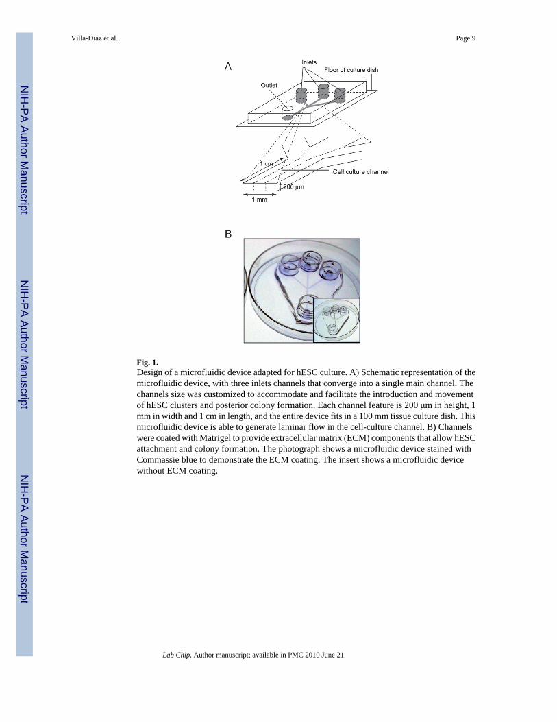
Fig. 1.Design of a microfluidic device adapted for hESC culture. A) Schematic representation of themicrofluidic device, with three inlets channels that converge into a single main channel. Thechannels size was customized to accommodate and facilitate the introduction and movementof hESC clusters and posterior colony formation. Each channel feature is 200 μm in height, 1mm in width and 1 cm in length, and the entire device fits in a 100 mm tissue culture dish. Thismicrofluidic device is able to generate laminar flow in the cell-culture channel. B) Channelswere coated with Matrigel to provide extracellular matrix (ECM) components that allow hESCattachment and colony formation. The photograph shows a microfluidic device stained withCommassie blue to demonstrate the ECM coating. The insert shows a microfluidic devicewithout ECM coating.
Villa-Diaz et al. Page 9
Lab Chip. Author manuscript; available in PMC 2010 June 21.
NIH
-PA Author Manuscript
NIH
-PA Author Manuscript
NIH
-PA Author Manuscript
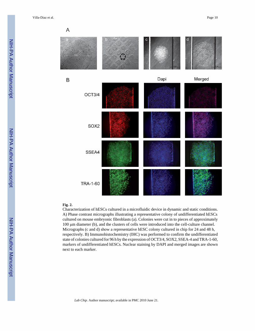
Fig. 2.Characterization of hESCs cultured in a microfluidic device in dynamic and static conditions.A) Phase contrast micrographs illustrating a representative colony of undifferentiated hESCscultured on mouse embryonic fibroblasts (a). Colonies were cut in to pieces of approximately100 μm diameter (b), and the clusters of cells were introduced into the cell-culture channel.Micrographs (c and d) show a representative hESC colony cultured in chip for 24 and 48 h,respectively. B) Immunohistochemistry (IHC) was performed to confirm the undifferentiatedstate of colonies cultured for 96 h by the expression of OCT3/4, SOX2, SSEA-4 and TRA-1-60,markers of undifferentiated hESCs. Nuclear staining by DAPI and merged images are shownnext to each marker.
Villa-Diaz et al. Page 10
Lab Chip. Author manuscript; available in PMC 2010 June 21.
NIH
-PA Author Manuscript
NIH
-PA Author Manuscript
NIH
-PA Author Manuscript
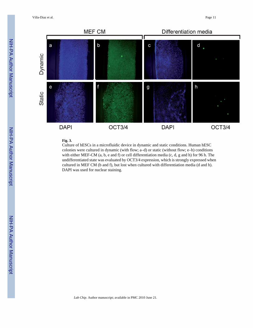
Fig. 3.Culture of hESCs in a microfluidic device in dynamic and static conditions. Human hESCcolonies were cultured in dynamic (with flow; a–d) or static (without flow; e–h) conditionswith either MEF-CM (a, b, e and f) or cell differentiation media (c, d, g and h) for 96 h. Theundifferentiated state was evaluated by OCT3/4 expression, which is strongly expressed whencultured in MEF CM (b and f), but lost when cultured with differentiation media (d and h).DAPI was used for nuclear staining.
Villa-Diaz et al. Page 11
Lab Chip. Author manuscript; available in PMC 2010 June 21.
NIH
-PA Author Manuscript
NIH
-PA Author Manuscript
NIH
-PA Author Manuscript
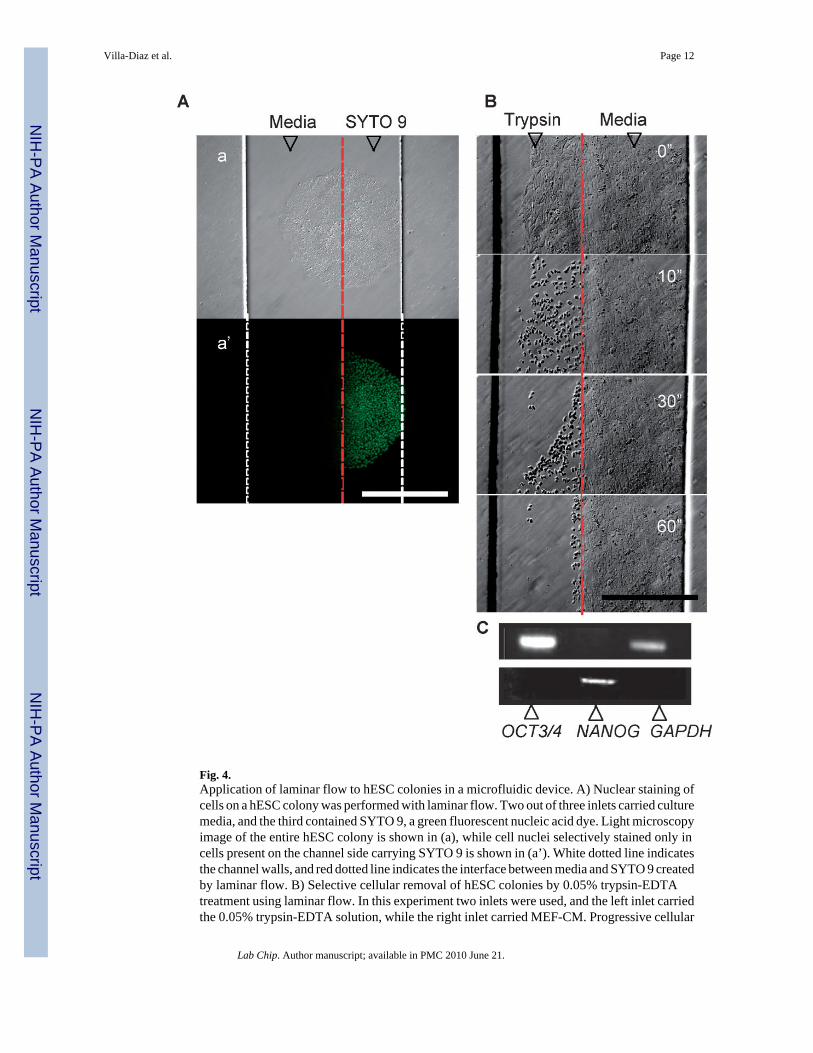
Fig. 4.Application of laminar flow to hESC colonies in a microfluidic device. A) Nuclear staining ofcells on a hESC colony was performed with laminar flow. Two out of three inlets carried culturemedia, and the third contained SYTO 9, a green fluorescent nucleic acid dye. Light microscopyimage of the entire hESC colony is shown in (a), while cell nuclei selectively stained only incells present on the channel side carrying SYTO 9 is shown in (a’). White dotted line indicatesthe channel walls, and red dotted line indicates the interface between media and SYTO 9 createdby laminar flow. B) Selective cellular removal of hESC colonies by 0.05% trypsin-EDTAtreatment using laminar flow. In this experiment two inlets were used, and the left inlet carriedthe 0.05% trypsin-EDTA solution, while the right inlet carried MEF-CM. Progressive cellular
Villa-Diaz et al. Page 12
Lab Chip. Author manuscript; available in PMC 2010 June 21.
NIH
-PA Author Manuscript
NIH
-PA Author Manuscript
NIH
-PA Author Manuscript
removal is observed from top to bottom of micrographs, as indicated by seconds (”). Red dottedline indicates the interface between trypsin-EDTA solution and MEF-CM created by laminarflow. C) Dissociated cells were collected and analyzed by PCR to detect OCT3/4 andNANOG, both markers of pluripotency expressed in undifferentiated hESCs. GAPDH was usedas a control gene. Scale bar in micrographs represents 500 μm.
Villa-Diaz et al. Page 13
Lab Chip. Author manuscript; available in PMC 2010 June 21.
NIH
-PA Author Manuscript
NIH
-PA Author Manuscript
NIH
-PA Author Manuscript
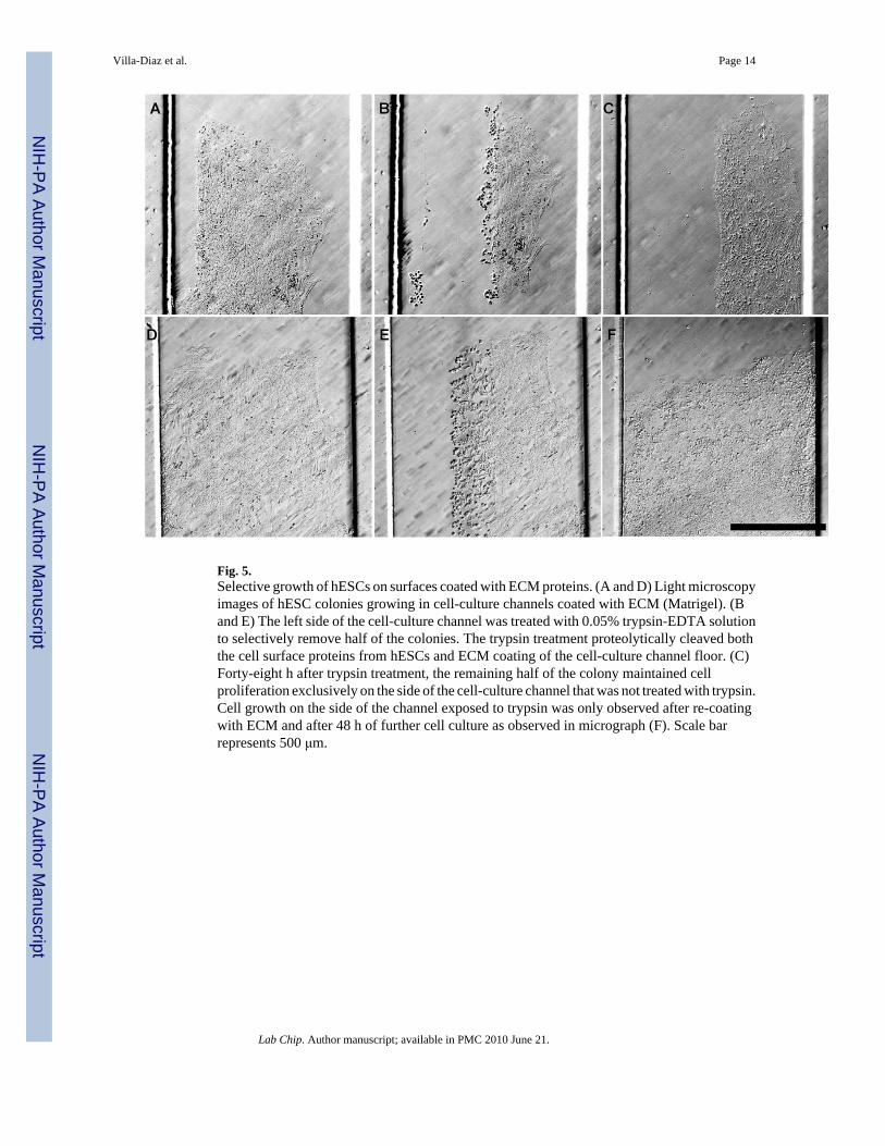
Fig. 5.Selective growth of hESCs on surfaces coated with ECM proteins. (A and D) Light microscopyimages of hESC colonies growing in cell-culture channels coated with ECM (Matrigel). (Band E) The left side of the cell-culture channel was treated with 0.05% trypsin-EDTA solutionto selectively remove half of the colonies. The trypsin treatment proteolytically cleaved boththe cell surface proteins from hESCs and ECM coating of the cell-culture channel floor. (C)Forty-eight h after trypsin treatment, the remaining half of the colony maintained cellproliferation exclusively on the side of the cell-culture channel that was not treated with trypsin.Cell growth on the side of the channel exposed to trypsin was only observed after re-coatingwith ECM and after 48 h of further cell culture as observed in micrograph (F). Scale barrepresents 500 μm.
Villa-Diaz et al. Page 14
Lab Chip. Author manuscript; available in PMC 2010 June 21.
NIH
-PA Author Manuscript
NIH
-PA Author Manuscript
NIH
-PA Author Manuscript
NIH
-PA Author Manuscript
NIH
-PA Author Manuscript
NIH
-PA Author Manuscript
Villa-Diaz et al. Page 15
Tabl
e 1
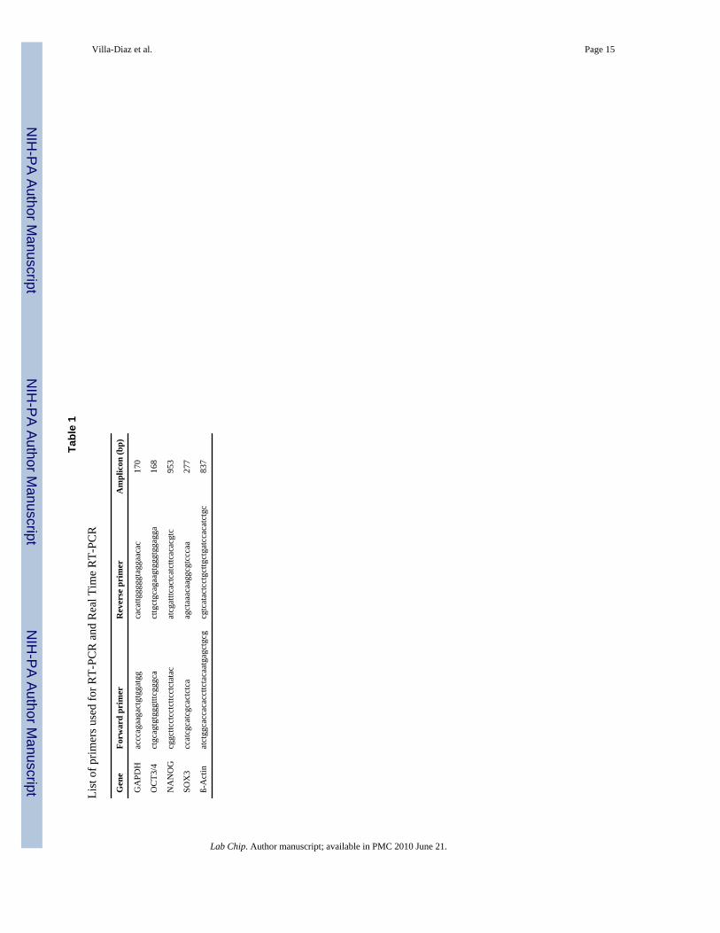
List
of p
rimer
s use
d fo
r RT-
PCR
and
Rea
l Tim
e R
T-PC
R
Gen
eFo
rwar
d pr
imer
Rev
erse
pri
mer
Am
plic
on (b
p)
GA
PDH
accc
agaa
gact
gtgg
atgg
caca
ttggg
ggta
ggaa
cac
170
OC
T3/4
ctgc
agtg
tggg
tttcg
ggca
cttg
ctgc
agaa
gtgg
gtgg
agga
168
NA
NO
Gcg
gcttc
ctcc
tcttc
ctct
atac
atcg
atttc
actc
atct
tcac
acgt
c95
3
SOX
3cc
atcg
catc
gcac
tctc
aag
ctaa
acaa
ggcg
tccc
aa27
7
ß-A
ctin
atct
ggca
ccac
acct
tcta
caat
gagc
tgcg
cgtc
atac
tcct
gcttg
ctga
tcca
catc
tgc
837
Lab Chip. Author manuscript; available in PMC 2010 June 21.