Embed Size (px)
Citation preview

IJSRST162382 | Received : 15 June 2016 | Accepted : 05 July 2016 | July-August 2016 [(2)4: 01-13]
© 2016 IJSRST | Volume 2 | Issue 4 | Print ISSN: 2395-6011 | Online ISSN: 2395-602X Themed Section: Science and Technology
1
Micropropagation Photoautotrophic Kalanchoe pinnata in Water and Humus with use of Natural Light, and
Determination of Total Flavonoids: A Review Neves C. R. S. S.*, Procópio M. C, Penna T. C. V.
Department of Biochemical Pharmaceutical Technology, College of Pharmaceutical Sciences, University of São
Paulo, São Paulo, SP, Brazil
ABSTRACT
Micropropagation is a vegetative propagation method widely studied in many different plant species,
beinga mode in tissue culture, the one that has found widespread and proven practical applications. Among
the advantages of its use is the possibility of obtaining various plants from an initial explant, regardless of
the season, besides the reduction of time and area required for the species propagation. The development
of photoautotrophic micropropagation systems with natural light usage, emerge as potential possibilities to
increase the micropropagation efficiency and help reduce costs, making it commercially viable for active
principles and phytotherapeutics production. Thus, the flavonoid content contained in Kalanchoe pinnata
plant extract, can base the pharmacological activity of it when used by population, for the cure and
prevention of various types of chronic noncommunicable diseases (NCDs).
Keywords: Kalanchoe Pinnata, Micropropagation, Photoautotrophic, Flavonoids, Cost Reduction
I. INTRODUCTION
Photoautotrophic Micropropagation
Micropropagation is a vegetative propagation me-thod
widely studied in many different plant species, being
the mode in tissue culture, the more widespread and
has found proved practical applications [50]. Among
the advantages of its use is the possibility of obtaining
various plants from an initial explant, regardless of
the season, besides the reduction of time and area
required for the species propagation. Moreover, due to
best sanitary conditions by cultivation of meristems
previously treated by thermotherapy to eliminate
diseases, there is the reproduction of the mother-plant
genotype usually with fidelity during multiplication,
which allows the propagation of species difficult to
propagate by other methods.
The development of photoautotrophic micropro-
pagation systems (production of micropropagules
without addition of sucrose in the culture medium and
under ambient conditions favoring photo-synthesis)
[47], with natural light usage, emerge as potential
possibilities to increase micropropagation efficiency
and help reduce costs, making it commercially viable.
Studies have shown that light can determine the
growth direction of plants (phototropism). It is able to
determine chloroplasts [21, 22] and plant organs
(photomorphogenesis) [21] differentiation, espe-cially
the leaf, the most susceptible organ to environ-mental
changes [23, 24], and in other substances production,
such as flavonoids [21]. This fact raises the possibility
of studying various light sources effects at different
wavelengths, focusing on plants in order to verify and
modulate the amount of synthe-sized substances.

International Journal of Scientific Research in Science and Technology (www.ijsrst.com) 2
The light also strongly influences hormones
biosynthesis. Vegetal hormones are molecules
responsible for the seedlings development, present in
low concentrations [26]. These substances act in all
morphogenetic processes, including those induced by
light. Studies have shown that gibberellin levels in
events like disestiolation, germination and
tuberization are directly linked to light, regulated by
phytochrome or cryptochromes. The indole acetic
acid (IAA) and cytokinins levels in disestiolation are
also regulated by phytochrome. The light is able to
regulate abscisic acid (ABA), ethylene and
brassinosteroids levels in a phytochromes and
cryptochromes dependent process [32]. The
interaction between light, photoreceptors and plant
hormones has a strong influence on plant life, even
regulating tissues and organs development, that is,
morphogenesis.
Studies have shown that the relationship between
light and flavonoid biosynthesis is also closely linked.
Flavonoids provide coloring for plants ranging from
lilacto blue. The ability of these molecules to absorb
wavelength in UV range gives to these substances a
protection function against damage caused by this
type of radiation [36, 37] and microorganisms or
fungi [36].
Other authors have shown that different spectral
ranges differently influence the development of plant
organs, leading to changes in several anatomical
characteristics such as leaf thickness, leaf area, stem
diameter, stomata and trichomes density [53, 54, 55].
Besides morphological and anatomical features, light
may influence secondary metabolites production such
as flavonoids. The flavonoid biosynthesis occurs by
secondary and mixed pathways (shikimate and acetate
pathways), being regulated by blue and UV light in a
process mediated by cryptochromes. In this process
occurs the expression of the enzyme phenylalanine
ammonia lyase, essential for the first step of
fenilpropanoídica and chalcone synthase pathways,
the first flavonoid biosynthesis pathway [38]. Due to
these molecules protective action against
microorganisms in plants, it makes them a target to
pharmacological studies using plants.
The perception of color and the amount of substances
that plants produce, as a rule, are related to spectral
quality of light they receive. The solar radiation that
reaches the Earth covers a spectral range extending
from 290 nm (ultraviolet) to 4000 nm (infrared) [21].
Many of these wavelengths are absorbed by ozone in
the atmosphere, by the atmospheric oxygen and CO2.
On average, 45% of the radiation coming from the
sun are in the range from 380 nm to 710 nm [21]. It is
in this spectral range that is the visible light,
composed of seven spectral ranges, the seven rainbow
colors.
In this range, there are also the wavelengths used by
plants for photosynthesis (photosynthetically active
radiation, FRG) [21]. However, light is not only
utilized by the plant as a source of energy, it also
controls the plant growth and development through
signals. Plants are able to monitor the intensity,
quality, direction and duration of light. As already
mentioned, the solar radiation is decisive in many
physiological processes such as stem elongation,
germination, stomatal conductance, chlorophyll
synthesis.
Due to its characteristics of sessile beings and their
autotrophic nature, dependent on light energy, plants
have developed environmental recognition
mechanisms realizing the variations in the quantity
and quality of light they receive, according to the
vegetation cover in a particular location which can be
closed or opened, light is gradually assimilated by
layers of superimposed sheets.
Thus, the canopy energy is greater than the lower
layers, meaning that the light reaching the soil is
richer in wavelengths than that reaching the canopy,
that is, it tends to contain more light in the red range.
In addition, a few feet below the topsoil, only reaches
lengths corresponding to the extreme red [21]. This
effect is called radiation attenuation [21]. Different
species require different amounts of light to its perfect
development. Hence it is so important that plants
recognize the different quantities and qualities of light
where they are located.
In this manner, plants can regulate their development
in order to seek light. This recognition process is
through special molecules called photoreceptors, able
to recognize different wavelengths. The processes
regulated by light are categorically divided into two
main classes: phytochrome mediated, red light
receptors and cryptochromes mediated, blue light /
UV-A receptors [26]. The phytochrome are the most
characterized photoreceptors and form a family of
International Journal of Scientific Research in Science and Technology (www.ijsrst.com) 3
five proteins (phytochrome A, B, C, D and E) of
approximately 125 kDa [27].
Each phytochrome has two forms, in particular
spectrally reversible: one that absorbs red light (Pr -
biologically inactive) and one that absorbs extreme
red wavelength (Per - biologically active). Pr, when
absorbs light in the 620-680 nm range is converted to
the Per, this by absorbing radiation in the 700-800 nm
range becomes Pr. The balance between the two
forms, given by red and extreme red proportion in the
environment light, will cause physiological responses
in the plant [28, 21, 27].
The greater the amount of far red, the plant is more
shaded. The amount of red and far red that the plant
receives also indicates the seasons (long or short
days).
This information determines the plants aspects such
as germination, flowering, maintenance or not
plumular hook, chlorophyll synthesis and circadian
rhythm. Although in most cases, responses regulated
by phytochrome are linked to red light, some studies
show dependent responses of blue and UV-A light
[29,30].
The cryptochromes are a family of blue / UV-A light
photoreceptors extremely important during
disestiolation of plants grown in the dark [31]. They
are involved in the inhibition of stem growth
processes, cotyledon expansion and chlorophyll
synthesis. There have been shown that cryptochromes
act in coordination with phytochromes in several
processes and its action is temperature dependent
[31]. However, phytochrome and cryptochromes,
along with other photoreceptors, are not the only
substances that act in photomorphogenic events.
Other substances act on plant tissue, translating in
them environmental light conditions. These
substances are plant hormones.
It is possible to observe that among the environ-
mental factors that most influence the plant
development is the lighting. Studies have indicated
that in addition to the amount of light received, the
spectral quality of it is also important as it can induce
morphoanatomic answers, for example, it can cause
different red and extreme red ratios altering the
structure of the mesophyll [24] or the stomata density
[33]. The leaf is the organ that most responds to
environmental radiation, being therefore chosen by
many authors as object of study of spectral quality
and quality effect in plants [34, 33, 24, 22, 35].
Different plant species are adapted to different
lighting conditions, with an amount of light to be
optimum to perform photosynthesis. Plants usually
have morphoanatomic features related to these
different light environments. Such characteristics
provide the best use of the incident light and
protection of photosynthetic apparatus [21]. During
its life, plants can be exposed to different lighting
conditions, so it is possible to notice that most plants
have developed adaptation mechanisms, mainly
anatomical, of the individual to the new climatic
conditions [22]. This flexibility occurs even at a
cellular level, as in the case of different organizations
and developmental changes in mesophyll chloroplasts
[21, 22].
Some advantages of photoautotrophic micropro-
pagation at the expense of natural light compared to
the conventional method of micropropagation include
plant growth increase. Due to the removal of sucrose
from the culture medium, there are improvements in
the physiological characteristics of the plant, once the
cultivation environmental conditions are more natural,
reducing plant stress during acclimatization,
increasing the percentage of survival of seedlings [48,
49, 50], elimination of lighting costs and reduced
costs for repairs and maintenance, and also possibility
of use of simplified facilities reducing construction
costs [51].
In conventional micropropagation, the heterotrophic
or fotomixotrófica nature of plant growth is directly
or indirectly responsible for most of the factors
related to the cost of production of micropropagated
plants [50]. Explants are cultured in flasks without
gas exchange and with high relative humidity (about
98%), high ethylene concentration, low CO2
concentration (which decreases from 3.000 to 1 mol
9.000μmol the dark period to less of 100μmol mol-1
during the photoperiod), and low photon flux density
of photosynthetically active. That means that low
lightning (40 - 50μmol m-2 s-1) and with sucrose as a
major source of metabolic energy [52], once the
explants show low photosynthetic rate [50]. The
nutrient medium used is composed of essential and
optional components. The standard medium used
universally for micropropagation of medicinal

International Journal of Scientific Research in Science and Technology (www.ijsrst.com) 4
plants is MS medium (Murashige and Skoog) [56],
which has as essential nutrients: inorganic salts,
carbohydrates, vitamins and growth regulators.
Many explants or in-vitro plants have the ability to
grow photoautotrophicly, i.e. without sucrose in the
culture medium and under environmental
conditions, which promote photosynthesis [50]. The
photosynthetic process, contrary to what occurs in
the respiratory chain, need an external source of
energy, without which it does not occur, showing
that there is a strong relationship between light and
plants, photosynthetic organisms. Since, during
photosynthesis, light is not only a regulation factor,
it acts as an important component in the
biochemical reaction [21], being able to interfere in
several physiological factors, such as chlorophyll
production, stem stretching regulation, in enzymes
production and other substances such as
anthocyanins, a type of flavonoid [21]. In this sense,
it is understood that the light is a determining factor
for plants to be able to, from inorganic molecules,
synthesize carbohydrates.
Kalanchoe pinnata Description
The Brazilian flora is extremely rich, because in it
there are thousands of species of medicinal plants,
and among these plants there is Kalanchoe pinnata,
which despite being widely used by the population,
still has few studies proving its medicinal
properties. It is a perennial plant, therefore easily
found in several climates and regions of the world,
and easy handling due to their morphological
characteristics (Table 2).
The different species of the genus
Kalanchoe (Crassulaceae family) are known in
popular medicine in many countries as it can be
observed in Table 1. As used in treatment of
inflammatory processes and in several diseases.
This fact favors the search for new bioactive
molecules. Among the main active principles of
Kalanchoe pinnata, there are the polyphenols [59],
which are flavonoids, antioxidants, mucilage and
others (Table 3). Among the present flavonoids,
there is quercetina43, which has shown significant
action in the leishmaniasis treatment [41].
Quercetin and kaempferol are flavonoids
widely spread throughout the plant kingdom and
have significant anti-inflammatory action, that can
be attributed to inhibition of the enzymes
phospholipase A2 (PLA2), 8 lipo-oxygenates to,
cyclooxygenase and inhibition of nitric oxide
production, through modulation of enzyme iNOS
[35, 37, 59].
Table 1- Taxonomy of Kalanchoe pinnata
(Lamarck) Persoon:
Kingdom Plantae (Plantas)
sub Kingdom Tracheobionta (vascular
plant)
Super division Magnolioliophyta
(Flowering plant)
Class Rosidae
Order Saxifragales
Family Crassulaceae Stonecrop
family [39]
Genre Kalanchoe
Species Kalanchoe pinnata
(Lam.) Per [14]
Synonyms
Verea pinnata, Crassuvia
floripendia, C. calyculata,
Cotyledon calycina,
Bryophyllum calycinum,
Crassula pinnata, Sedum
madagascariense, B.
germinans, C. rhizophilla.
[13]
Source Uncertain. It is believed
to be the Mauritius
Islands, Africa, India and
Indian Ocean islands
Distribution It is distributed through-
hout India and grown in
wild gardens in the hills
of northern and western
India, Deccan and Bengal
[18]. In Brazil, they can
be found from Sao Paulo
to Bahia, mainly in the
coastal zone.
Regional
Names
Brazil
Saião, Folha-da-fortuna,
Coirama, Roda-da-
fortuna, Folha-da-costa,
Folha-grossa, Erva-da-
costa
India Zakhm-hayat
Arabia kushnulhayat
Walking
stick
Koppata
Sanskrit Asthi-bhaksha
telugu Simajamudu
Tamil Ranakalli
kannada Ganduklinga
malayalam Elamurunga
Persian and
Urdu
Chubehayat [17, 19]
International Journal of Scientific Research in Science and Technology (www.ijsrst.com) 5
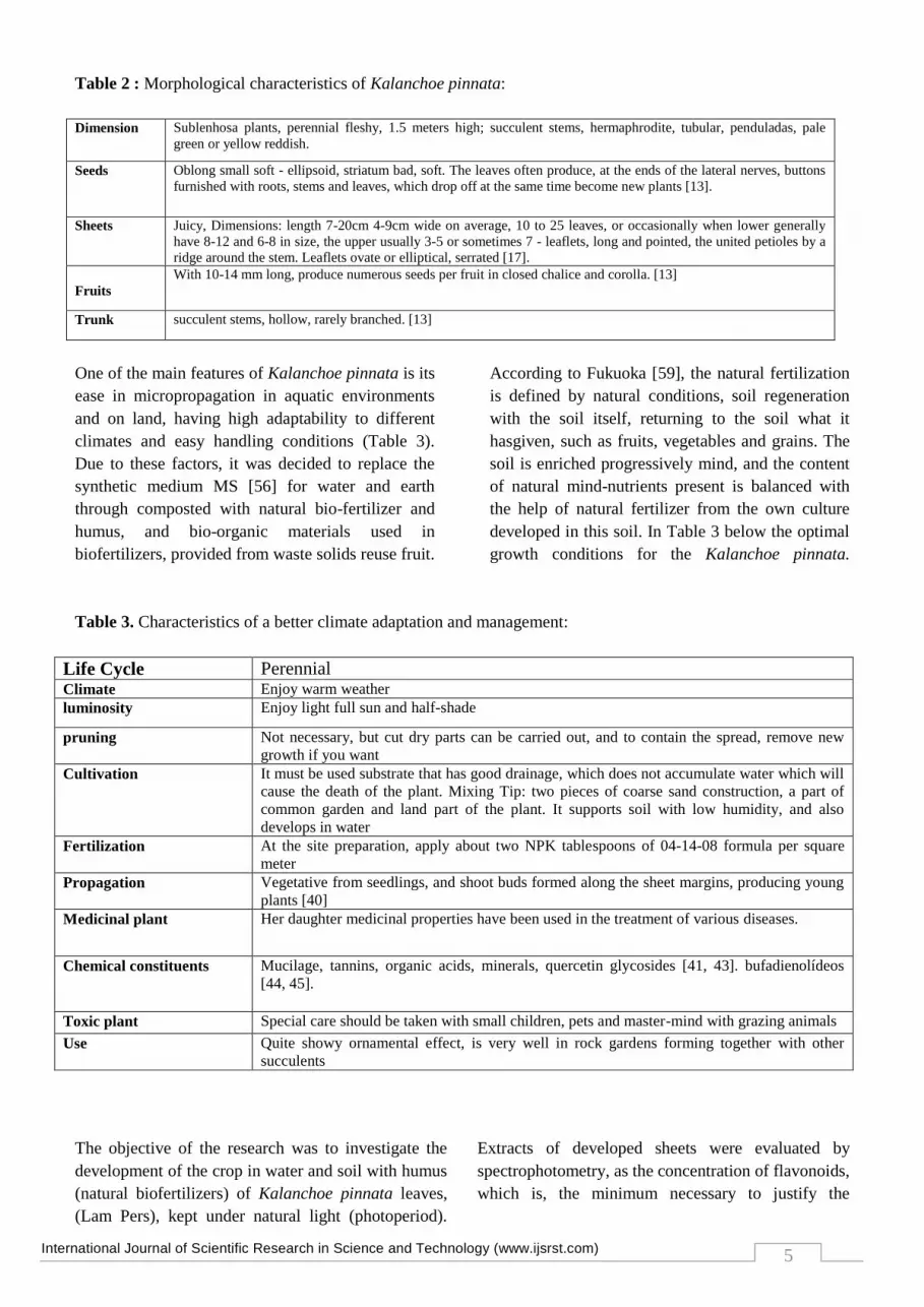
Table 2 : Morphological characteristics of Kalanchoe pinnata:
Dimension Sublenhosa plants, perennial fleshy, 1.5 meters high; succulent stems, hermaphrodite, tubular, penduladas, pale
green or yellow reddish.
Seeds Oblong small soft - ellipsoid, striatum bad, soft. The leaves often produce, at the ends of the lateral nerves, buttons
furnished with roots, stems and leaves, which drop off at the same time become new plants [13].
Sheets Juicy, Dimensions: length 7-20cm 4-9cm wide on average, 10 to 25 leaves, or occasionally when lower generally
have 8-12 and 6-8 in size, the upper usually 3-5 or sometimes 7 - leaflets, long and pointed, the united petioles by a
ridge around the stem. Leaflets ovate or elliptical, serrated [17].
Fruits
With 10-14 mm long, produce numerous seeds per fruit in closed chalice and corolla. [13]
Trunk succulent stems, hollow, rarely branched. [13]
One of the main features of Kalanchoe pinnata is its
ease in micropropagation in aquatic environments
and on land, having high adaptability to different
climates and easy handling conditions (Table 3).
Due to these factors, it was decided to replace the
synthetic medium MS [56] for water and earth
through composted with natural bio-fertilizer and
humus, and bio-organic materials used in
biofertilizers, provided from waste solids reuse fruit.
According to Fukuoka [59], the natural fertilization
is defined by natural conditions, soil regeneration
with the soil itself, returning to the soil what it
hasgiven, such as fruits, vegetables and grains. The
soil is enriched progressively mind, and the content
of natural mind-nutrients present is balanced with
the help of natural fertilizer from the own culture
developed in this soil. In Table 3 below the optimal
growth conditions for the Kalanchoe pinnata.
Table 3. Characteristics of a better climate adaptation and management:
Life Cycle Perennial Climate Enjoy warm weather
luminosity Enjoy light full sun and half-shade
pruning Not necessary, but cut dry parts can be carried out, and to contain the spread, remove new
growth if you want
Cultivation It must be used substrate that has good drainage, which does not accumulate water which will
cause the death of the plant. Mixing Tip: two pieces of coarse sand construction, a part of
common garden and land part of the plant. It supports soil with low humidity, and also
develops in water
Fertilization At the site preparation, apply about two NPK tablespoons of 04-14-08 formula per square
meter
Propagation Vegetative from seedlings, and shoot buds formed along the sheet margins, producing young
plants [40]
Medicinal plant Her daughter medicinal properties have been used in the treatment of various diseases.
Chemical constituents Mucilage, tannins, organic acids, minerals, quercetin glycosides [41, 43]. bufadienolídeos
[44, 45].
Toxic plant Special care should be taken with small children, pets and master-mind with grazing animals
Use Quite showy ornamental effect, is very well in rock gardens forming together with other
succulents
The objective of the research was to investigate the
development of the crop in water and soil with humus
(natural biofertilizers) of Kalanchoe pinnata leaves,
(Lam Pers), kept under natural light (photoperiod).
Extracts of developed sheets were evaluated by
spectrophotometry, as the concentration of flavonoids,
which is, the minimum necessary to justify the

International Journal of Scientific Research in Science and Technology (www.ijsrst.com)
6
medical activities of the extracts of Kalanchoe pinnata leaves described in Table 4.
Table 4: Medicinal Uses Worldwide:
II. METHODS AND MATERIAL
A. Humus preparation
The papaya, banana, orange, mandarin and mango
residue were gathered and milled with the aid of an
organic waste processor in order to form a biomass of
fruits. They were then added to water ratio gradient in
order to obtain the best uniformity for the process of
fermentation the kefir grains, for cleaving complex
sugars and free reducing sugars, providing among these
microelements to facilitating uptake by plants. During
the fermentation, the fruit biomass was placed in glass
containers covered with fabric. Subsequent to
fermentation, biomass was added to the earth for the
humus formation, through composting method.
Fermented biomass was evaluated to their chemical
composition of trace elements as well as dosage of total
ash, moisture, protein, carbohydrates, glucose, fructose,
fiber and lipids.
B. Determination of the chemical composition of
biomass fruit
The chemical composition of biomass (consisting of
papaya, orange, mango, banana and tangerine waste)
Brazil
Furunculous [20], Intestinal problems, arthritis, ulcers, athlete's foot, abscesses, bubos,
bronchitis, burns, calluses, conjunctivitis, corns, dermatosis, coughs, earaches, eczema, fever,
urinary insufficiency, rheumatism, itch, glaucoma, infections, headache, kidney stones, scurvy,
tumor, wart, sedative, whooping cough, wounds, inset stings, lymphatic disorders, mouth sores,
respiratory infections, mouth sores, erysipelas [10]. Immunosuppressant, leishmaniasis [41,
43], diabetes [41, 42, 43] hemostatic, antiseptic, healing topical, inflammation (stomach pain)
[15, 16]
USA Chicken pox, stomachache, fevers [10]
Mexico Inflammations, wounds, eye infections, headaches, menstrual disorders [10]
Ecuador Bruises, broken bones [10]
Guatemala Diarrhea, pain, skin problems, aches [10]
Nicaragua aches, burns, colds, pain, fever, headache, respiratory infections, coughs, childbirth [10]
Bangladesh Coughs, fever, constipations, mucus, epilepsy [11]
Peru
Bacterial infections, boils, broken bones, ulcers, urethritis, sore, skin problems, nausea,
migraine, intestinal problems, eye infections, epilepsy, gas, headache, heartburn, inflammation,
cancer (lymphoma), bronchitis, conjunctivitis, coughs, earaches [10]
South America Asthma, tumors, headaches, colds, earaches, chest colds, sores, strains [10]
Nigeria Coughs, earaches, pimples, inflammation, eczema10, cut umbilical cord in new born baby [7]
West Indies Ulcers, menstrual disorders, urinary disorder, hypertension [10]
Other places Arthritis, asthma, burns, bruises, constipation, malnutrition, headaches, migraines, nephritis,
respiratory infections, earaches, diabetes, paralysis, rheumatism, swelling [10], and to induce
vomiting of blood, expel worms, cut umbilical cord in new born baby [7]
Vietnam Anti-inflammatory and antibacterial [8]
India
Orissa Diarrhea [5, 2]
Karnataka Leaf juice externally, applied to scabies and leucoderma and leaf decoction applied over, cuts to
stop bleending [10, 12]
Maharashtra The leaves in used against cough dysentery [4]
in Himalaya Leaves are applied on wound, bruises, insect bite, swelling [9]
Arunachal
Pradesh
Leaf extract is taken in empty stomach is used in the treatment urinary bladder stones and fewer
in children’s [3]

International Journal of Scientific Research in Science and Technology (www.ijsrst.com)
7
was determined according to the methodology of the
AOAC [57]: moisture (925.45); protein (960.52); lipids
(920.39) and ash (923.03). The conversion factor used
for determining the protein content was 6.25. The energy
value was calculated using the general factors of
Atwater and considering the energy from dietary fiber
(DF) [58]. The analyzes were performed in triplicate and
the results expressed in g per 100 g of sample in the
integral base. The DF content was determined according
to AOAC method 991.43 [57]. The analysis was done in
quadruplicate and the results expressed as g / 100g
sample basis. Dosages of glucose and fructose were
made in triplicate by Waters chromatograph column for
detection of sugars (Shodex SC 1011); refractive index
detector; Mobile phase: EDTA-Ca 0,187g / l.
Chromatography conditions: column temperature: 72 °
C; detector temperature: 45C, mobile phase flow: 0.6 ml
/ min. Detector sensitivity 32, Volume injection: 10μL,
running time: 30 minutes.
C. Photoautotrophic Micropropagation
The leaves of Kalanchoe pinnata, initially developed in
soil, were transferred to individual containers in which
were placed submerged in water. The jars were kept at
room temperature, covered by a synthetic porous fabric
(TNT), provided direct daylight (photoperiod). Initially,
the dimensions and leaflets of each sheet were
measured. After a period of seven to ten days of
permanence in the same water, the size of sprouts from
the leaflets and their roots were measured as shown in
Fig. 1, and the water was changed. Each bud, with its
leaves and roots were transferred to the humus (prepared
from biomass fruit fermented with kefir and composted
with soil). Then, the micropropagation development
monitoring was made, measuring the dimensions of the
roots and formation, and the growth of buds in relation
to the cultivation time.
Figure 1. Illustrative Photo Kalanchoe pinnata. Source:
Author's photo
D. Preparation of the Kalanchoe pinnata leaves
extract
There were extractions of leaves developed to evaluate
the concentrations of flavonoids. The alcoholic extract
of 100 g of cereal leaf was macerated in ethanol, and
transferred quantitatively into 100 mL volumetric flask,
and the final volume with grain alcohol.
E. Determination of total flavonoid, catechin
equivalents
It was transferred 0.5 ml of K. pinnata extract solution
into a 25 mL volumetric flask and was added 0.100 mL
of 10% aluminium chloride with 4.3 ml of grain alcohol.
We adopted the same experimental procedure dilutions,
described below, for the preparation of the absorbance
of the response curve as a function of the concentration
(mg / ml) ranging from 60 to 260 (mg / mL) of total
flavonoids equivalent of catechin using the equation of
the line from the calibration curve of the standard
secondary reference catechin being evaluated linearity.
Linearity
To check the linearity, it was elaborated analytical curve
with the stock solution of standard secondary reference
catechin (purity = 98%) in the concentration of 500,0μg
/ ml in cereal alcohol. From the stock solution dilutions
were prepared at concentrations of 0.084; 0.05; 0.028;
0.017; 0.0076 g / L, which was reacted with 0.100 mL of
10% aluminum chloride solution (v / v), completing the
volume with ethanolic grain alcohol. After 40 minutes of
rest, we proceeded to read in a spectrophotometer at 510
nm using as white cereal alcohol solvent plus aluminum
chloride solution. Elaborated the analytical curve,
checking the linearity of the method, using visual
observation and appropriate statistical analysis,
obtaining - the equation of the line and the linear
correlation coefficient. Equation 1 is the equation of the
calibration curve of the standard secondary reference

International Journal of Scientific Research in Science and Technology (www.ijsrst.com)
8
catechin, where y is absorbance and x is the
concentration of standard secondary reference catechin:
y = 10.121 x – 0, 0475 (1)
r2 = 0, 9936
From the obtaining of a line to the calibration curve and
the correlation coefficient (r2) .9936 was verified the
linearity of the method.
III. RESULT AND DISCUSSION
A. Quantification of equivalent total flavonoids
catechin in Kalanchoe pinnata extract.
In Table 5 it can be seen the results of
quantification of total flavonoids, catechin
equivalents in Kalanchoe pinnata extract, with an
average of 42.39 mg / mL.
Table 5 – spectrophotometric dosage of the equivalent
total flavonoids catechin present in Kalanchoe pinnata
extract to 510nm. EKP: Kalanchoe pinnata extract .: CF:
concentration of total flavonoid, catechin equivalents.
Concentration
of EKp (µg/mL) Absorbance CF µg/mL)
60,0 0,2230 17,34
100,0 0,3320 28,00
140,0 0,4219 36,00
180,0 0,5543 50,00
220,0 0,6030 54,00
260,0 0,7456 69,00
Average: 42,39
B. Kalanchoe pinnata Micropropagation
Water-soaked sheets produced roots from all the leaflets,
and buds (buds) with leaves in most leaflets, from 7 to
10 days. In a similar period, the sheet cultivated on earth
developed, on average, two shoots 10 days and 40 days
from the main stem height reached an average of 8 cm
from each bud, 8 new sheets for each sprout dimensions
Similar to that of the mother sheet. In water, the leaves
are multiple and diminutive dimensions, three times
smaller, and remains in this size and had to be
transferred to new culture in water or on land.
C. Statistics of K. pinnata.micropropagation
Figure 2. K. pinnata with 11.5cm long and 6.5cm
wide; ANOVA: p < 0.05 ***
Figure 3. K. pinnata with 15.7 cm long and 10
cm wide; ANOVA: p <0.05 ***
Figure 5. K. pinnata with 14cm long and 9
cm wide; ANOVA: p <0.05 ***

International Journal of Scientific Research in Science and Technology (www.ijsrst.com)
9
Figure 4. K. pinnata in length and 13cm 9.7 cm wide;
ANOVA: p <0.05 ***
Figure 6. K. pinnata with 11 cm long and 5.5
cm wide; ANOVA: p <0.05 ***
Figure 7. K. pinnata with 18cm in length and 12.5 in
width;
ANOVA: p <0.05 ***
Figure 8. K. pinnata with 10,5cm long
and 5,4wide; ANOVA: P < 0, 05 ***

International Journal of Scientific Research in Science and Technology (www.ijsrst.com)
10
A. Determination of the chemical composition of
biomass fruit
Table 6 - Proximate analysis of biomass of fruits
Biomass fruit
(g/100 g)
(I) (II) (III) (IV) Average
Carbohydrates 4 4,9 4,8 -- 4,56
Lipids 0,02 0,03 0,02 -- 0,023
Proteins 0,4 0,5 0,8 -- 0,56
Ash 2 1,98 2 -- 1,99
Humidity (%) 89 89 89 -- 89
Fibers 5 4,8 4,9 4,9 4,9
Glucose (g/L) 7,756 7,54 7,15 -- 7,483
Fructose (g/L) 9,107 8,80 8,31 -- 8.756
IV. CONCLUSION
The micropropagation of Kalanchoe pinnata proved
to be auspicious, since it presented a high capacity for
reproduction by fotoautotrophic micropropagation in
water with the use of natural light, and soil humus
with derived bio-organic fertilizer. Observe a
satisfactory value the concentration of micronutrients
present in the biomass of fruits needed for plant
nutrition, this fact makes possible the replacement of
synthetic media, which implies lower costs allowing
obtaining of one rich source of active ingredients for
herbal, water extracts (poultices) or cereal alcohol
(tinctures).
The photoautotrophic plant micropropagation, in
addition to increasing the growth of in vitro explants,
also minimizes the risk of microbial contamination,
reduces production costs, improves the physiological
characteristics of the plant and facilitates their
acclimatization to ex vitro conditions due to have
been grown in conditions natural.
V. ACKNOWLEDGEMENTS Agency CAPES, Prof. Dr. Marco Antônio Sthephano
(FBT-FCF-USP), Prof.ª. Dra. Marina Ishii (FBT-
FCF-USP), Drª Aline de Oliveira Santos (FBA-FCF-
USP), Drª Caroline Matos de Mello (IPT- USP)
Figure 9. K. pinnata (all sheets with varying
sizes) ANOVA: P <0.05 ***

International Journal of Scientific Research in Science and Technology (www.ijsrst.com)
11
VI. REFERENCES
[1] Cherly A Lans, Ethanomedicines used in
Trinidad and Tobago for urinary problems and
diabetesmellitus, Journal of Ethanobiology and
Ethanomedicine, 2006; 2:45
[2] Samim Sofika Begum and Rajib Jogoi, Herbal
recipe prepared during Bohag or Rogali Bihu in
Assam, Indian Journal of Tradicional Knowledge,
2007; 6(3): 417-422
[3] Prabal Sen, Mihin Dollo, Manabendra Dutta
Chaudhury, Dhrupad Choudhury, Documentation
and Tradicional Herbal Knowledge of Khamptis
of Arunachal Pardesh, 2008; 7(3): 438442
[4] Kirtikar K.R & Basu B. D., Indian Medicinal
Plants with illustrations. Volume- 5, second
edition, Oriental enterprises. 2003: 1394 - 1396
[5] Santos kumar Dash and Sanchidanda Padhy,
review of ethanomedicines for diarrhea disease
from Orissa: prevalence versus culture, Journal
Hum. Ecol. 2006; 20 (1): 59-64
[6] Donatus Ebere Okwu, and Nnamdi Fred
Uchenna, Exotic Multifacetaded Medicinal Plants
of drug and pharmacetical indutries, African
Journal of Biotecnology, 2009; 8(25):72271 –
7282
[7] Mai Thanh Thi Nguyen, Suresh Awale, Yssuhiro
Tezuka, Quan Le Tran, Hiroshi Watanabe and
Shigetoshikadota, Xanthin Oxidase Inhibitory
Activity of Vietnamese Medicinal Plants, Biol.
Pharm Bull. 2004; 27 (9): 1414-1521
[8] S Huissain and DK Hore, Collectionpn and
conservation of major medicinal plants of
Darjeeling and Sikkim, Indian Journal of
Traditional Knowledge, 2007; 6 (2): 352-357
[9] www.raintree.com - Disponível em 01/06/2016
[10] Md. Shahadat hossan, Abu Hanif, Mujib Khan,
Sazzadul Bari, Rownak jahan, Mohammad
rahematullah, Ethanobotanical Survey of the
Tripura trible of Bangladesh, American -
Eurasian Journal of Sustainable Agriculture,
2009; 3(2): 253-261
[11] MJ Bhandary, KR Chandrashekar, KM
Kaveriappa, Medical Ethanoibotany of Siddis of
Uttara Kannada district, Karnataka, India, Journal
od Ethanopharmacology, 1995; 47: 149-158
[12] QUAZI MAJAZ A. et al / IJRAP 2011, 2 (5)
1478- 1482 - The Miracle Planta (kalanchoe
Pinnata): A Phitochemical and Pharmacological
Reviw - Ali Allana College of Pharmacy,
Akkalkuwa, Dist- Nandurbar, MS, India
[13] Plants.usda.gov – Disponível em 01/06/2016
[14] Zuanazzi J S; J Mountain A. Flavonoids In:
Simões C M, et al. Pharmacognosy plant to the
drug, 6 edition, Porto Alegre: Ed UFSC, 2010,
chapter 23, p. 577-656.
[15] Alcaraz M J; Carvalho JC T. Flavonoids In as
anti-inflammatory agents: Carvalho J C T, Herbal
anti-inflammatories: chemical, pharmacological
aspects and therapeutic applications - 1 edition,
Ribeirão Preto, Brazil. Tecmedd 2004
[16] Kirtikar K. R. & B. D., Indian Medicinal Plants
wihillustrations. Volume-5, second edition,
Oriental enterprises. 2003: 1394 - 1396
[17] Nandkarni A>K., Indian Materia Medica,
Volume -1, third edition, Popular Prakashan,
Bombay, 2005: 1402 & 221
[18] The Wealt of India A Dictionary of Indian Raw
Materials and Industrial products, ( Volume - 5)
(H-K) Firts Supplement Series, National Institute
of Science Communications and Information
Resources, Council of Scientific and Industrial
Research, New Delhi; 1997: 31
[19] Http://www.apanat.org.br/noticias/videos/plantas-
medicinais-aprovadas-pelo-governo-e-
distribuidas-pelo-sus/
[20] LARCHER, 2004, W. Ecofisiologia Vegetal. São
Carlos: RiMa Artes e Textos, 2000. 531p.
[21] WESTON, E.; THOROGOOD, K.; VINTI, G.;
LÓPEZ-JUEZ, E. Light quality controls leaf-cells
and chloroplast development in Arabdopsis
thaliana wild type and blue-light-perception
mutants. Planta, v. 211, 807-815, 2000
[22] VIEIRA, R. C. Anatomia da folha de Bauhinia
radiata Vell. Em diferentes ambientes. Arq. Biol.
Tecnol., v. 38, 63-107, 1995.
[23] LEE, D. W.; OBERBAUER, S. F.; JOHNSON,
P.; KRISHNAPILAY, B.; MANSOR, M.;
MOHAMAD, H.; YAP, S. K. Effects of
irradiance and spectral quality on leaf structure
and function in seedlings of two Southeast Asian
Hopea (Dipterocarpaceae) species. American
Journal of Botany, v. 87, 447-455, 2000
[24] SCHUERGER, A. C.; BROWN, C. S.;
STRYJEWSKI, E. C. Anatomical features of
pepper plants (Capsicum annum L.) grown under

International Journal of Scientific Research in Science and Technology (www.ijsrst.com)
12
red light-emitting diodes suplemented with blue
or far-red light. Annals of Botany, v. 79, 273-282,
1997
[25] TAIZ, L.; ZEIGER, E. Fisiologia Vegetal. 3. ed.
Porto Alegre: Artmed, 2009
[26] HUQ, E. Degradation of negative regulators: a
common theme in hormone and light signaling
networks? Trends in Plant Science, v. 11, 4-7,
2006
[27] WHITELAM, G. C.; DEVLIN, P. F. Light
signaling in Arabdopsis. Plant Physiol. Biochem.,
v. 36, 128-133, 1998
[28] CHUN, L.; KAWASAMI, A. CHRISTOFER D.
A. Phytochrome A mediates blue light and UV-
A-dependent chloroplast gene transcription in
green leaves. Plant Physiology, v. 125, 1957-
1966, 2001
[29] SPALDING, E. P.; FOLTA, K. M. Illuminating
topics in plant photobiology. Plant, Cell and
Environment, v. 28, 39-53, 2005
[30] CHEN, M.; CHORY, J.; FANKHAUSER, C.
Light signal transduction in higher plants. Annu.
Rev. Genet., v 38, 87-117, 2004
[31] SYMONS, G. M.; REID, J. B. Interactions
between light and plant hormones during de-
etiolation. Journal of Plant Growth Regulation, v.
22, 3-14, 2003
[32] RAJAPAKSE, N. C.; KELLY, J. W. Spectral
filters influence transpirational water loss in
chrysanthemum. HortScience, v. 28, 999-1001,
1993
[33] JURIK, T. W.; CHABOT, J. F.; CHABOT, B. F.
Effects of light on leaf size, CO2 exchange, and
anatomy in wild strawberry (Fragaria virginiana).
Plant Physiology, v. 70, 1044-1048, 1982
[34] JUSTO, C. F.; SOARES, A. M.; GAVILANES,
M. L.; CASTRO, E. M. Plasticidade anatômica
das folhas de Xylopia brasiliensis Sprengel
(Annonaceae). Acta Botanica Brasilica, v.19,
111-123, 2005
[35] SHIRLEY, B. W. Flavonoid biosynthesis: "new"
functions for an "old" pathway. Trends in Plant
Science, v. 1, 377-382, 1996
[36] JAAKOLA, L.; MÄÄTTÄ-RIIHINEN, K.;
KÄRENLAMPI, S.; HOHTOLA, A. Activation
of flavonoid biosynthesis by solar radiation in
bilberry (Vaccinium myrtillus L.) leaves. Planta,
v. 218, 721-728, 2004
[37] WEISSHAAR, B.; JENKINS, G. I.
Phenylprpanoid biosynthesis and its regulation.
Current Opinion in Plant Biology, v. 1, 251-257,
1998
[38] SILVEIRA, A. L.; RODRIGUES, M. C.;
PAULO, M. Q. Estudos tóxicológicos e
microbiológicos de Kalanchoe pinnata. In: I
CONGRESSO BRASILEIRO DE EXTENSÃO
UNIVERSITÁRIA. Anais eletrônicos.
Disponível em . Acesso em 11/12/2006
[39] JOLY, A. B. Botânica: introdução à taxonomia
vegetal. Editora Nacional. 1983
[40] DA-SILVA, S. A. G.; COSTA, S. S.; ROSSI-
BERGMANN, B. The anti-leishmanial effect of
Kalanchoe is madiated by nitric oxide
intermediates. Parasitology, v. 118, 575-582,
1999
[41] LANS, C. A. Ethnomedicines used in Trinidad
and Tobago for urinary problems and diabetes
mellitus. Journal of Ethnobiology and
Ethnomedicine, v. 2, 2006
[42] MUZITANO M. F.; TINOCO, L. W.; GUETTE,
C.; KAISER, C. R., ROSSI-BERGMANN, B.;
COSTA, S. S. The anti-leishmanial activity
assessment of unusual flavonoids from
Kalanchoe pinnata. Phytochemistry, v.67, 2071-
2077, 2006
[43] SUPRATMAN, U.; FUJITA, T.; AKIYAMA, K.;
HAYASHI, H. New insecticidal bufadinolide,
bryophyllin C, from Kalanchoe pinnata. Biosci.
Biotechnol. Biochem., v. 64, 1310-1312, 2000.
[44] SUPRATMAN, U.; FUJITA, T.; AKIYAMA, K.;
HAYASHI, H.; MURAKAMI, A.; SAKAI, H.;
KOSHIMIZU, K.; OHIGASHI, H. Anti-tumor
promoting avtivity of bufadienolides from
Kalanchoe pinnata and K. Daigremontiana x
tubiflora. Biosci. Biotechnol. Biochem., v. 65,
947-949, 2001.
[45] ALAN CRISTIANO ERIG; Márcia Wulff
Schuch. Micropropagação fotoautotrófica e uso
da luz natural. Ciência Rural, Santa Maria, v.35,
n.4, p.961-965, jul-ago, 2005.
[46] KUBOTA, C.; TADOKORO, N. Control of
microbial contamination for large-scale
photoautotrophic micropropagation. In vitro
Cellular and Developmental Biology Plant, New
York, v.35, p.296-298, 1999
[47] HEMPEL, M. From micropropagation to
microponics (part II). Practical Hydroponics &

International Journal of Scientific Research in Science and Technology (www.ijsrst.com)
13
Greenhouses, May/June, p.17-20, 1994.
Capturado em 15 out. 2003. Online. Disponível
na Internet:
http://members.ozemail.com.au/~mhempel/public
ations/mponic2.htm
[48] AFREEN, F. et al. Photoautotrophic culture of
Coffea arabusta somatic embryos: photosynthetic
ability and growth of different stage embryos.
Annals of Botany, London, v.90, p.11-19, 2002
[49] KOZAI, T; KUBOTA, C. Developing a
photoautotrophic micropropagation system for
woody plants. Journal of Plant Research, Tokyo,
v.114, p.525-537, 2001
[50] KODYM, A.; ZAPATA-ARIAS, F.J. Natural
light as an alternative light source for the in vitro
culture of banana (Musa acuminata cv. ‘Grande
Naine’). Plant Cell, Tissue and Organ Culture,
The Hague, v.55, p.141-145, 1999
[51] ARIGITA, L. et al. Influence of CO2 and sucrose
on photosynthesis and transpiration of Actinidia
deliciosa explants cultured in vitro. Physiologia
Plantarum, Copenhagen, v.115, p.166-173, 2002
[52] WÄNGLER, M. S. Micropropagação clonal e
desenvolvimento in vitro de Cyrtopodium
polyphyllum (Vell.) pabst. ex F. Barros,
Orchidaceae. Dissertação (Mestrado em
Biotecnologia Vegetal) - Programa de Pós-
graduação em Biotecnologia Vegetal. Rio de
Janeiro: Universidade Federal do Rio de Janeiro,
2002.
[53] LUCENA-DE-LUCA, R. Padronização genética
e cultivo controlado de Echinodorus grandiflorus
(Cham. & Schlecht.) Micheli para a produção de
fitoterápicos. Dissertação (Mestrado em
Biotecnologia Vegetal) - Programa de Pós-
graduação em Biotecnologia Vegetal. Rio de
Janeiro: Universidade Federal do Rio de Janeiro,
2003.
[54] MACEDO, A. F. Estudo dos efeitos dos meios de
cultura e diversas condições de iluminação nas
características morfológicas, fitoquímicas e
farmacológicas de Alternanthera brasiliana (L.)
Kuntze cultivada in vitro. Tese (Doutorado em
Biotecnologia Vegetal) - Programa de Pós-
graduação em Biotecnologia Vegetal. Rio de
Janeiro: Universidade Federal do Rio de Janeiro,
2003.
[55] MURASHIGUE, T.; SKOOG, F. A revised
medium for rapid growth and bioassys of tobacco
tissue cultures. Physiol. Plant., v. 15, 473-479,
1962.
[56] HORWITZ, W.; LATIMER, G. W. (Eds.).
Official Methods of Analysis of AOAC
International. 18th ed. Gaithersburg, Maryland:
AOAC International, 2006.
[57] FAO. 2003. State of the World’s Forests 2003.
Home. ftp.fao.org/docrep/fao/005/y7581e/.
Disponível em 10/06/2016
[58] COUTINHO, M.A.S.; Muzitano, Michele F.;
Costa, Sônia S. – Flavonoides: potenciais
Agentes Terapêuticos para o Processo
Inflamatório. Revista Virtual Quim, 2009, 1 (3),
241-256. Data de publicação na Web: 26 de
junho de 2009. ISSN1984-6835. Disponível em:
http://rvq.sbq.org.br/index.php/rvq/article/viewAr
ticle/51, acesso 28.04.16.
[59] (FUKUOKA,2014)http://www.brasil247.com/pt/
247/revista_oasis/110110/Fukuoka-O-
agricultorque-deixava-a-terra-em-paz.htm
disponível em 15/012/2015.


