Embed Size (px)
Citation preview
RESEARCH ARTICLE
MicroRNA-132 targets HB-EGF upon IgE-mediated activationin murine and human mast cells
Viktor Molnar • Barbara Ersek • Zoltan Wiener • Zsofia Tombol •
Peter M. Szabo • Peter Igaz • Andras Falus
Received: 21 December 2010 / Revised: 13 July 2011 / Accepted: 19 July 2011 / Published online: 19 August 2011
� Springer Basel AG 2011
Abstract MicroRNAs provide an additional layer in the
regulation of gene expression acting as repressors with
several targets at the posttranscriptional level. This study
describes microRNA expression patterns during differen-
tiation and activation of mast cells. The expression levels
of 567 different mouse miRNAs were compared by
microarray between c-Kit? committed progenitors,
mucosal mast cells, resting and IgE-crosslinked BMMCs in
vitro. The strongest upregulation of miR-132 upon IgE-
mediated activation was validated in human cord blood-
derived mast cells as well. HB-EGF growth factor also
upregulated upon activation and was ranked high by more
prediction algorithms. Co-transfection of miR-132 mim-
icking precursor and the 30UTR of human Hbegf-
containing luciferase vector proves that the predicted
binding site is functional. In line with this, neutralization of
miR-132 by anti-miR inhibitor leads to sustained produc-
tion of HB-EGF protein in activated mast cells. Our data
provide a novel example for negative regulation of a
growth factor by an upregulated miRNA.
Keywords Mast cells � MicroRNAs � HB-EGF �Activation � IgE � Differentiation
Abbreviations
30-UTR 30-Untranslated region
BMMC Bone marrow-derived mast cell
CREB Cyclic AMP-response-element-binding
protein
CTMC Connective tissue-type mast cells
DNP-HSA Dinitrophenyl-human serum albumin
FAM Fluorescein amidite
FBS Fetal bovine serum
FGF2 Fibroblast growth factor 2
FYN Oncogene related to SRC, FGR, YES
HB-EGF Heparin-binding EGF-like growth factor
HER2 Human epidermal growth factor receptor 2
IL-3 Interleukin-3
LYN v-yes-1 Yamaguchi sarcoma viral related
oncogene homolog
MeCP2 Methyl CpG-binding domain protein-2
MMC Mucosal mast cells
mMcpt1 Mouse Mast Cell Protease-1
NGF Nerve growth factor
PLC-c Phospholipase C-cSCF Stem cell factor
SYK Spleen tyrosine kinase
TGF-b Transforming growth factor beta
VEGF Vascular endothelial growth factor
Introduction
MicroRNAs (miRNAs) represent a new layer of gene
expression regulation by interacting with mRNAs and
subsequently repressing translation in animals. The mature
form of miRNAs consists of approximately 21–25 nucle-
otides, which can initiate either the suppression of
Electronic supplementary material The online version of thisarticle (doi:10.1007/s00018-011-0786-3) contains supplementarymaterial, which is available to authorized users.
V. Molnar (&) � B. Ersek � Z. Wiener � A. Falus
Department of Genetics, Cell and Immunobiology,
Semmelweis University, Budapest, Hungary
e-mail: [email protected]
Z. Tombol � P. M. Szabo � P. Igaz
2nd Department of Medicine, Faculty of Medicine,
Semmelweis University, Budapest, Hungary
Cell. Mol. Life Sci. (2012) 69:793–808
DOI 10.1007/s00018-011-0786-3 Cellular and Molecular Life Sciences
123
translation or cleavage of their targets by binding to the 30-untranslated region of the mRNA. This recognition is based
on complex and not well understood sequence comple-
mentary rules. miRNAs influence a wide variety of
developmental events, such as the appropriate establish-
ment of tissue- or cell type-specific expression patterns
during the multicellular differentiation processes. As they
are able to adjust the concentration of many proteins in a
rapid and synchronized manner, miRNAs are thought to
play an important role in cell responses and adaptation to
the environment [1, 2].
The regulation of mammalian hematopoiesis and
immune functions also seems to be coordinated by
miRNAs, which may represent a gene regulatory network
beneath the well-characterized signaling pathways. Some
miRNAs are preferentially expressed in distinct subsets
of immune cells, such as miR-142, miR-150, miR-155,
miR-181, miR-221/222 and miR-223, indicating their
possible role during differentiation. Among these miR-
NAs, miR-181a and miR-150 are regulators of adaptive
immunity, while the more pleiotropic function of miR-
155 was underlined also in T and B cells and monocyte/
macrophage lineages [3]. In addition, the role of mi-
croRNAs is not restricted to the cell fate decisions, but
they are active participants in the regulation of mature
cell responses.
During the past few years many computational algo-
rithms have been developed to predict miRNA targets with
more or less reliability. The target prediction criteria vary
widely among the different computational methods, but in
general they include (1) Watson-Crick basepairing of the 50
2–8 nucleotides of the miRNA to a complementary site in
the 30UTR of the mRNA, (2) calculation of minimum free
energy (MFE) of the interacting RNA–RNA duplexes and
(3) evolutional conservation of the miRNA binding site [4].
Although the prediction of interactions between RNA
molecules by using pure sequence data seems to be more
feasible compared to transcription factor-DNA predictions,
more as yet unidentified factors should be taken into con-
sideration, since the overall performance is typically just
acceptable with an approximately two-thirds false-positive
rate [5].
Although mast cells are primarily known as effectors of
allergic inflammatory reactions, they play a critical role in
host defense mechanisms by orchestrating the innate and
adaptive immune responses, too [6, 7]. In marked contrast
to other hematopoietic lineages, mast cells leave the bone
marrow as immature precursors and complete their matu-
ration in the local mucosal or connective tissues.
Depending on the tissue context, they may undergo dif-
ferentiation to give rise to morphologically and
functionally identifiable mature mast cell subtypes. There
are at least two distinct subpopulations of mast cells in
rodents: the connective tissue-type (CTMCs) and the
mucosal mast cells (MMCs). While the former cell popu-
lation can be characterized with a high level of Mcpt1 and
Mcpt2, the latter mast cell group expresses predominantly
Mcpt4, Mcpt5 (chymase1, cma1), Mcpt6 (tryptase beta2,
Tpsb2), Mcpt7 (tryptase alpha/beta1, Tpsab1) and car-
boxypeptidase 1 [8, 9]. These subtypes differ in location,
staining characteristics, mediator content and dependency
of T cell-derived cytokines. In humans, mast cell subpop-
ulations are usually classified based on their protease
content. In contrast to the MCT type, which produces only
tryptase, MCTC cells contain both tryptase and chymase,
members of two protease families that contribute critically
to various pathological conditions either as pro-inflamma-
tory or protective players (e.g., in parasitic and bacterial
infections, allergic inflammation and arthritis) [10].
According to their tissue localization, the MCT type cor-
responds to MMCs and the MCTC cells to CTMCs in
rodents [11].
The activation of mast cells results in releasing a wide
range of mediators in allergic reactions, which can be
evoked by the contribution of IgE and its high-affinity
receptor (FceRI) on the surface of mast cells. This process
is regulated by crosslinking the FceRI receptors, which is
initiated by an interaction between the antigen and the
receptor-bound IgE. Subsequently kinases, such as FYN,
LYN or SYK, are rapidly phosphorylated at tyrosine resi-
dues, and their activation induces PLC-c, leading to the
production of inositol triphosphate and to the increase of
intracellular calcium concentration. Mast cells are able to
secrete preformed mediators upon stimulation, such as
biogenic amines (e.g., histamine), proteoglycans and pro-
teases. At the same time, their activation also initiates the
de novo synthesis of lipid mediators, cytokines, chemo-
kines and specific growth factors (TGF-b, NGF, VEGF,
FGF2, etc.) [12]. Importantly, some of the growth factors,
such as activin A, amphiregulin and heparin-binding epi-
dermal-like growth factor (HB-EGF), known for its ability
to induce the growth of fibroblasts and smooth muscle
cells, were found to be upregulated by IgE crosslinking in
mast cells of various origin [13, 14].
HB-EGF, a member of the EGF family, is implicated in
various biological processes, like wound healing [15–17],
blastocyst implantation [18], heart function [19] and dis-
eases, such as cancer [20] and arteriosclerosis [21]. HB-
EGF is initially synthesized as a transmembrane protein
(pro-HB-EGF), which has been identified as the receptor
for diphteria toxin. Pro-HB-EGF is then cleaved by matrix
metalloproteinases, resulting in the release of the mature,
soluble form. HB-EGF binds to its receptors HER2 and
HER4 in the presence of heparan sulfate as cofactor. Many
cell types, including epithelial cells, skeletal muscle cells,
macrophages, keratinocytes and T cells, express HB-EGF,
794 V. Molnar et al.
123
and it shows potent mitogenic and chemotactic activity for
fibroblasts and epithelial cells [22, 23].
In the present study, we performed miRNA gene
expression profiling using microarray technology to gain
insight into the differentiation process of mature MMCs
and the IgE-mediated activation of BMMCs, a widely
accepted in vitro model system. From among genes
expressed differentially upon IgE-crosslinking, miR-132
microRNA showed the highest upregulation; thus, we
selected it for further analysis. To investigate the role of
IgE-induced miR-132 expression, we carried out a bioin-
formatic search in different target prediction databases to
select functionally relevant targets. According to three
prediction algorithms, HB-EGF turned out to be a highly
ranked candidate for miR-132 targets, which was experi-
mentally confirmed. As HB-EGF is also upregulated upon
mast cell activation, miR-132 may play an important
negative regulatory role in adjusting the appropriate level
of this mitogenic mediator, thus influencing the growth of
resident tissue cells. This feedback loop becomes espe-
cially interesting in physiological responses and diseases at
the intersection of tissue remodeling and inflammatory
activity of mast cells, such as wound healing or asthma,
nasal polyposis and psoriasis.
Materials and methods
Cell lines and transfection
CHO epithelial cell line (ATCC, Manassas, VA) was prop-
agated in Ham’s F12 medium with 10% fetal bovine serum
(FBS). The mouse macrophage cell line J774.2 was cultured
in Dulbecco’s modified Eagle’s medium (DMEM) with 10%
FBS. MC/9 mouse mast cell line (ATCC no. CRL-8306) was
maintained in high glucose (4.5 g/l) DMEM supplemented
with 6 mM L-glutamine, 1 mM sodium pyruvate, 0.1 mM
non-essential amino acid solution (Sigma-Aldrich, St. Louis,
MO), 0.05 mM 2-mercaptoethanol, 10% rat T-STIM (Bec-
ton Dickenson, Mountain View, CA) or conditioned medium
of concanavalin A-stimulated (Sigma-Aldrich) mouse
splenocytes and 10% FBS.
Then 4 9 106 MC/9 cells or BMMCs were transfected
using a Nucleofector II device (Lonza, Cologne, Germany,
program X-005) in 100 ll Solution V according to the
manufacturer’s protocol. To determine the transfection
efficiency (varying between 55 and 85% in MC/9 deter-
mined by flow cytometry), control cells were transfected
with pmaxGFP vector. For transfections, miR-132 miRNA
precursor (Pre-miR-132) and Pre-miR-132 negative con-
trol, Anti-miR-132, as well as Anti-miRNA negative
control oligonucleotides, were purchased from Applied
Biosystem.
Generation of bone marrow-derived mast cells
BMMCs were generated from bone marrow of 6- to
8-week-old male BALB/c mice. Briefly, mice were killed,
and femora with intact medullary cavities were removed.
Sterile, endotoxin-free medium was flushed repeatedly
through the bone shaft using a needle and syringe. The
suspension of bone marrow cells was centrifuged at
3209g for 10 min and cultured at a concentration of
0.5 9 106 cells/ml in RPMI 1640 with 10% FCS, 100 U/
ml penicillin, 100 lg/ml streptomycin, 2 mM L-glutamine,
and a combination of 5 ng/ml interleukin-3 (IL-3) and
20 ng/ml stem cell factor (SCF) (Sigma-Aldrich) for
3–4 weeks at 37�C in a humidified atmosphere with 5%
CO2. Nonadherent cells were transferred to fresh medium
at least twice a week. After 3–4 weeks when a mast cell
purity of [90% was achieved (as assessed by flow cyto-
metric analysis), the cells were used for experiments. To
compare microRNA gene expression changes during mast
cell differentiation, c-Kit positive mast cell progenitors
were magnetically isolated on the 6th day. To establish an
in vitro model of MMC differentiation, the culture medium
of 4-week-old BMMCs was supplemented with 10 ng/ml
IL-9 (Immunotools, Friesoythe, Germany) and 2 ng/ml
TGF-b (Sigma-Aldrich) for 5 days. The maturation of
BMMCs to MMCs was verified by measuring the marked
upregulation of mast cell protease-1 (mMcpt1). To inves-
tigate the activation, BMMCs were sensitized with 3 lg/ml
anti-dinitrophenyl (DNP) IgE (clone SPE7, Sigma-Aldrich)
for 2–4 h, washed and then challenged with DNP-HSA
(Sigma-Aldrich) for 2 h unless otherwise indicated. The
antigen concentration for the maximal mast cell activation
was determined by IL-13 qRT-PCR (for more details, see
the Supplementary methods).
Human cord blood-derived mast cells
Human mast cells were differentiatied from umbilical cord
blood with the permission of the local ethics committee of
the Semmelweis University. Whole blood was diluted with
RPMI 1640, and the mononuclear cells were separated with
Histopaque-1077 (Sigma-Aldrich). Residual erythrocytes
were removed by hypotonic lysis, and the mononuclear
cells were resuspended in 0.5% BSA and 2 mM EDTA.
After labeling with anti-CD34 microbeads, the cells were
magnetically isolated twice (Miltenyi Biotech, Bergisch
Gladbach, Germany). The CD34? cells were then resus-
pended in RPMI 1640 with 10% FBS, 2 mM L-glutamine,
0.1 mM nonessential amino acids, 100 U/ml penicillin and
100 lg/ml streptomycin. The cells were seeded at 106
cells/ml and cultured in the presence of 100 ng/ml SCF,
50 ng/ml IL-6 and 3 lM lysophosphatidic acid (LPA,
Sigma-Aldrich). Cells were cultured for up to 8 weeks, and
MicroRNA-132 targets HB-EGF in mast cells 795
123
the culture medium was replaced weekly. To examine the
IgE-mediated activation of human mast cells, cells were
sensitized with human myeloma IgE (Serotec, Dusseldorf,
Germany) at 37�C overnight. After washing, they were re-
suspended in 0.5% BSA in DMEM, and challenged with
1.5 lg/ml of rabbit anti-human IgE (Dako Corp., Glostrup,
Denmark) at 37�C for 2 h.
Flow cytometry
Cells were washed twice with 0.5% BSA in PBS and then
labeled at room temperature for 30 min. The following
antibodies were used: anti-mouse CD117 APC, anti-mouse
B220 PE, anti-mouse CD11b PE, anti-mouse TER119 PE,
anti-mouse CD5 PE (all from Pharmingen, San Diego, CA)
and anti-mouse FceRIa biotin (eBioscience, San Diego,
CA). Then 10,000 cells were measured by FACSCalibur
(BD Biosciences, San Jose, CA), data were collected with
CellQuest (Becton-Dickinson Immunocytometry Systems,
San Jose, CA) and further analyzed by the FlowJo software
(TreeStar, Inc., Palo Alto, CA).
RNA isolation, quality determination, reverse
transcription and real-time PCR
RNA from cell samples was extracted by miRNeasy Mini
Kit (Qiagen, Valencia, CA), the quantity and quality of
total RNA were assessed by a NanoDrop ND-1000 spec-
trophotometer (NanoDrop Technologies, Wilmington, DE)
and Agilent 2100 Bioanalyzer (Agilent Technologies, Palo
Alto, CA), respectively. Only those samples were used for
microarray experiments that gave [8.0 for RNA integrity
number and showed a clear gel image, and for which no
DNA contamination was observed on the histogram.
To determine mRNA expression, 1 lg RNA was con-
verted to cDNA by 1.25U MuLV reverse transcriptase
(Applied Biosystems, Foster City, CA) with random
primers in the presence of 20 U RNasin ribonuclease
inhibitor (Promega, Madison, WI) at 42�C for 55 min.
MuLV was then inactivated at 95�C for 5 min. The real-
time PCR reactions were carried out in an ABIPrism 7000
instrument according to the manufacturer’s instructions
with 2 ll RT-product in each well and in 25 ll final
volume.
To measure microRNA expression, primer sets for
specific microRNA assays and sno135 endogenous control
and the MicroRNA Reverse Transcription Kit were utilized
following the manufacturer’s protocol. Briefly, 10 ng of
each total RNA sample was transcribed by Multiscribe
Reverse Transcriptase. qRT-PCR was carried out using
Applied Biosystems 7000 Real-Time PCR system. The
relative expression of each miRNA and mRNA was cal-
culated from the equation 2�DCt , where DCt = mean Ct
(miRNA) – mean Ct (internal control) (where Ct is the
threshold cycle for a sample). The applied TaqMan gene
expression and microRNA assays are listed in Supple-
mentary Table 1. All reagents and instruments for RT-PCR
were purchased from Applied Biosystems, Inc., except
when otherwise indicated.
Microarray
For microRNA profiling, the Agilent Mouse miRNA
Microarray Kit (G4472A, 8 9 15 k) was applied according
to the manufacturer’s instruction (version 1.0) with
100–100 ng quality-checked total RNA. The labeled sam-
ples were hybridized for 20 h at 55�C. The arrays were
scanned with an Agilent DNA Microarray Scanner BA, the
signal quantification was carried out by Feature Extraction
10.7 Image Analysis Software, and data were further ana-
lyzed by Genespring GX10.0. Raw data were normalized to
the 75th percentile signal intensity, and entities showing
present call in all samples of a condition were filtered out.
Differentially expressed genes were selected when passing
the signal intensity filter (entities where at least 100% of
samples in any one out of four conditions have values
within cutoff) and showing at least twofold statistically
significant change (ANOVA and Tukey HSD post-hoc test,
with Benjamini-Hochberg Multiple Testing Correction
p value\0.05) between BMMC and any other groups. The
microarray data are deposited in NCBI Gene Expression
Omnibus with accession no. GSE24321.
Luciferase assay
The effects of miR-132 mimics on endogenous human HB-
EGF 30UTRs were measured using the luciferase reporter
construct (SwitchGear Genomics, Menlo Park, CA). A
vector containing a random genomic sequence was used to
control for overall signal variation caused by the cellular
non-specific response (non-target 30UTR). Transient
transfection assays were conducted in CHO cells in 96-well
plates. Transfection efficiency was adjusted with FAM-
conjugated negative control pre-miR oligonucleotides and
typically yielded *80% (Fig. S1) with [95% viability as
determined by propidium-iodide staining at 24 h. Briefly,
6,000 cells per well were seeded in culture medium 24 h
before transfection. Next, 0.15 ll Dharmafect Duo
(Thermo Fisher Scientific, Waltham, MA) was used in each
well to transfect 100 ng of plasmid DNA and either a
miRNA mimic or a non-targeting control according to the
manufacturer’s protocol. Pre-miR-132 and non-targeting
controls (Pre-miR negative control, Ambion, Austin, TX)
were added at a final concentration of 3, 30 and 90 nM.
After 24 h, 100 ll of the Steady-Glo Luciferase Assay
System reagent (Promega, Madison, WI) was added into
796 V. Molnar et al.
123
each well, incubated at room temperature for 30 min, and
then signals were determined in a standard plate lumino-
meter (Labsystems Luminoskan RS Microplate Reader,
Helsinki, Finnland). Each transfection was conducted and
assayed in triplicate.
Western blotting
For Western blotting, protein extracts were prepared by
resuspending cell pellets in 1% NP-40 (Igepal CA-630,
Sigma-Aldrich) lysis buffer containing 50 mM Tris-HCl
(pH 8.0), 150 mM NaCl, 1 mM EGTA, 5 mM NaF, 2 mM
phenyl-methyl-sulfonyl-fluoride, 1 mM Na-orthovanadate,
10 lg/ml leupeptin and 10 lg/ml aprotinin. Debris was
removed by centrifugation, and the protein yield was
assessed by spectrophotometry (Total Protein Kit, Micro
Lowry, Peterson’s Modification, Sigma-Aldrich). Then
30-lg aliquots of heat-denatured, b-mercaptoethanol-trea-
ted protein samples were loaded on precast Ready Gels
(Bio-Rad, Hercules, CA). Gels were blotted onto polyvi-
nylidene difluoride membranes (Bio-Rad), the membranes
were blocked with 5% milk powder in TBS and 0.1%
Tween-20 for 1 h, and then incubated with 1.5 lg/ml goat
anti-human HB-EGF (R&D Systems, Minneapolis, MN)
overnight at 4�C. Blots were washed, the secondary anti-
body (1:3,000 HRP-conjugated anti-goat IgG) was applied
for 45 min and the immunoreactive bands were visualized
with the ECL-Plus Western blotting Detection System (GE
Healthcare-Amersham Biosciences, Piscataway, NJ). Spe-
cific band size was determined with the Fermentas Plus
Prestained Protein Ladder (Fermentas, Glen Burnie, MD).
As a housekeeping control the beta-actin level was deter-
mined by using mouse anti-actin antibody (Sigma-Aldrich,
1:1,000 dilution) and HRP-conjugated anti-mouse IgG
(Dako Corp., Glostrup, Denmark, 1:10,000 dilution).
Image analysis was carried out with the ImageJ software (
http://rsb.info.nih.gov/ij/).
Target predictions
To improve the robustness of target prediction for miR-
132, data sets of the three most commonly used target
prediction databases were used simultaneously. Using
the outputs of the target prediction algorithms from PicTar
(http://pictar.mdc-berlin.de/), MirBase (http://www.ebi.ac.
uk/enright-srv/microcosm/) and TargetScan (version 5.1,
http://www.targetscan.org/), lists of putative target genes
were generated. As different IDs are applied by the three
target prediction algoritms, the identifiers of BioMART
(http://www.ensembl.org/Multi/martview) were used to
generate a comprehensive list. If more transcript IDs cor-
respond to one HUGO identifier, the ones included in the
other two data sets were chosen. In order to collate the
annotated data sets, the corresponding scores (proportional
with the likelihood of interaction between mRNA and
microRNA) were ranked in a range from 0 to 100. These
procedures were carried out both with the mouse and
human data sets.
Results
Marked change in the microRNA pattern during mast
cell differentiation
Although BMMCs are a widely accepted in vitro model
system for studying mast cell functions, they represent an
immature cell population that needs other factors to com-
plete their differentiation. Previous studies have provided a
detailed description for generating MMCs in vitro, a close
homolog of the mast cell population existing in the intes-
tinal mucosa [24, 25], and this protocol has been
successfully implemented in our laboratory [26, 27].
To identify microRNA genes with a characteristic
expression profile during the differentiation and activation
of murine mast cells, putative committed mast cell pro-
genitors (c-Kit? cells isolated on the 6th day of
differentiation), immature mast cells (BMMC), mucosal-
type mast cells (MMC) and IgE-activated BMMCs (Fcereceptor-bound IgE were cross-linked by DNP-HSA for
2 h) were produced and applied to single channel micro-
array experiments. Next, genes showing a[2.0-fold up- or
downregulation at p \ 0.05 significance level were selec-
ted. Since miRNAs are well known to play an outstanding
role in the differentiation in various cell types, we expected
the most significant changes to take place between the
c-Kit? progenitors and the other three groups containing
immature or differentiated mast cells. Unexpectedly,
mucosal differentiation did not result in major changes of
microRNA levels, although it can be characterized with a
marked phenotypic and gene expression alteration [26]
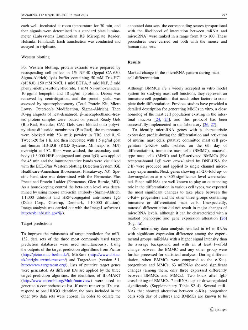
(Fig. 1a).
Our microarray data analysis resulted in 64 miRNAs
with significant expression difference among the experi-
mental groups. miRNAs with a higher signal intensity than
the average background and with an at least twofold
change between the BMMC and any other group were
further processed for statistical analyses. During differen-
tiation, when BMMCs were compared to the c-Kit?
progenitors and MMCs, 63 miRNAs showed significant
changes (among them, only three expressed differently
between BMMCs and MMCs). Two hours after IgE-
crosslinking of BMMCs, 7 miRNAs up- or downregulated
significantly (Supplementary Table S2–4). Several miR-
NAs that showed alteration between c-Kit? progenitor
cells (6th day of culture) and BMMCs are known to be
MicroRNA-132 targets HB-EGF in mast cells 797
123
involved in the differentiation or function of other hema-
topoietic cells (Table 1). Interestingly, miRNAs of the
miR-17–92 and miR-106a–363 polycistronic clusters,
which are well known for their central role in the lymphoid
differentiation, showed an intensive and coordinated
downregulation [28, 29]. In our model the expression of the
myeloid lineage-specific miR-223 was also downregulated
during mast cell differentiation. Similarly, miR-451, which
is characteristic for the erythroid cells, showed a strikingly
lower (*66-fold) expression in BMMCs compared to the
progenitors. Taken together, these results show that mast
cells coordinately downregulate many miRNAs during
their differentiation that are specific for other cell lineages.
Importantly, when validating the results of the micro-
array platform by real time RT-PCR, we found a strong
correlation between the two types of data (Fig 2).
miR-132 is upregulated upon IgE-crosslink in mouse
and human mast cells
Since miR-132 showed the highest upregulation upon
activation (Fig. 1b), and it has been implicated in the
regulation of the innate immune response, we selected this
miRNA for futher investigation. Mir-132 was previously
recognized as an endotoxin-responsive gene in human
THP-1 monocytes [39], and BMMCs express TLR4, the
major receptor for LPS [41]. In order to exclude the pos-
sibility that miR-132 is upregulated because of an LPS
contamination of our reagents, first we carried out a control
experiment. According to our results IgE and DNP-HSA,
but not LPS, specifically induce the transcription of mir-
132 in BMMCs (Fig. S2), which not only proves the lack of
LPS in our system, but also rules out the possibility that
miR-132 upregulation is caused by a minor contamination
of the monocyte/macrophage lineage. At the same time, the
treatment with LPS caused a marked induction of proin-
flammatory cytokines (IL-1b, IL-6 and TNF-a) in BMMCs,
and it had (but IgE ? DNP not at all) a similar effect on
these cytokines in the J774.2 macrophage cell line in a
parallel experiment (Fig. S3). Importantly, the induction of
miR-132 in response to IgE-crosslinking suggested the
involvement of an increase in the cytoplasmic Ca2? con-
centration, a crucial event of the activation process. Indeed,
when using the Ca-ionophor ionomycin in MC/9 mouse
mast cells, miR-132 expression started to increase at
Fig. 1 The heat map of differentially expressed miRNA genes
among murine mast cell samples representing mucosal mast cell
differentiation and activation. a Data passing the signal intensity filter
and showing at least two-fold significant changes between BMMCs
and c-Kit? progenitors were hierarchically clustered (entities and
conditions, Euclidean metric centroid linkage). b Differentially
expressed miRNAs upon IgE-crosslinking were clustered in a similar
way. Blue color indicates the relative downregulation, red shows
upregulation in the respective group
b
798 V. Molnar et al.
123
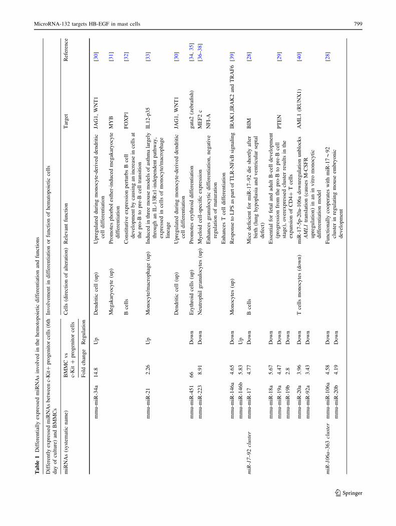
Ta
ble
1D
iffe
ren
tial
lyex
pre
ssed
miR
NA
sin
vo
lved
inth
eh
emo
top
oie
tic
dif
fere
nti
atio
nan
dfu
nct
ion
s
Dif
fere
ntl
yex
pre
ssed
miR
NA
sb
etw
een
c-K
it?
pro
gen
ito
rce
lls
(6th
day
of
cult
ure
)an
dB
MM
Cs
Inv
olv
emen
tin
dif
fere
nti
atio
no
rfu
nct
ion
of
hem
ato
po
ieti
cce
lls
miR
NA
s(s
yst
emat
icn
ame)
BM
MC
vs
c-K
it?
pro
gen
ito
rce
lls
Cel
ls(d
irec
tio
no
fal
tera
tio
n)
Rel
evan
tfu
nct
ion
Tar
get
Ref
eren
ce
Fo
ldch
ang
eR
egu
lati
on
mm
u-m
iR-3
4a
14
.8U
pD
end
riti
cce
ll(u
p)
Up
reg
ula
ted
du
rin
gm
on
ocy
te-d
eriv
edd
end
riti
c
cell
dif
fere
nti
atio
n
JAG
1,
WN
T1
[30]
Meg
akar
yo
cyte
(up
)P
rom
ote
sp
ho
rbo
les
ther
-in
du
ced
meg
akar
yo
cyte
dif
fere
nti
atio
n
MY
B[3
1]
Bce
lls
Co
nst
itu
tiv
eex
pre
ssio
np
ertu
rbs
Bce
ll
dev
elo
pm
ent
by
cau
sin
gan
incr
ease
ince
lls
at
the
pro
-Bto
pre
-Bce
lltr
ansi
tio
n
FO
XP
1[3
2]
mm
u-m
iR-2
12
.26
Up
Mo
no
cyte
/mac
rop
hag
e(u
p)
Ind
uce
din
thre
em
ou
sem
od
els
of
asth
ma
larg
ely
thro
ug
han
IL-1
3Ra1
-in
dep
end
ent
pat
hw
ay,
exp
ress
edin
cell
so
fm
on
ocy
te/m
acro
ph
age
lin
eag
e
IL1
2-p
35
[33]
Den
dri
tic
cell
(up
)U
pre
gu
late
dd
uri
ng
mo
no
cyte
-der
ived
den
dri
tic
cell
dif
fere
nti
atio
n
JAG
1,
WN
T1
[30]
mm
u-m
iR-4
51
66
Do
wn
Ery
thro
idce
lls
(up
)P
rom
ote
ser
yth
roid
dif
fere
nti
atio
ng
ata2
(zeb
rafi
sh)
[34,
35]
mm
u-m
iR-2
23
8.9
1D
ow
nN
eutr
op
hil
gra
nu
locy
tes
(up
)M
yel
oid
cell
-sp
ecifi
cex
pre
ssio
nM
EF
2c
[36–
38
]
En
han
ces
gra
nu
locy
tic
dif
fere
nti
atio
n,
neg
ativ
e
reg
ula
tio
no
fm
atu
rati
on
NF
I-A
En
han
ces
Tce
lld
iffe
ren
tiat
ion
mm
u-m
iR-1
46
a4
.65
Do
wn
Mo
no
cyte
s(u
p)
Res
po
nse
toL
PS
asp
art
of
TL
R-N
Fj
Bsi
gn
alin
gIR
AK
1,I
RA
K2
and
TR
AF
6[3
9]
mm
u-m
iR-1
46
b5
.83
Up
miR
-17
–9
2cl
ust
erm
mu
-miR
-17
4.7
7D
ow
nB
cell
sM
ice
defi
cien
tfo
rm
iR-1
7–
92
die
sho
rtly
afte
r
bir
th(l
un
gh
yp
op
lasi
aan
dv
entr
icu
lar
sep
tal
def
ect)
BIM
[28]
Ess
enti
alfo
rfe
tal
and
adu
ltB
-cel
ld
evel
op
men
t
(pro
gre
ssio
nfr
om
the
pro
-Bto
pre
-Bce
ll
stag
e),
ov
erex
pre
ssed
clu
ster
resu
lts
inth
e
exp
ansi
on
of
CD
4?
Tce
lls
mm
u-m
iR-1
8a
5.6
7D
ow
n
mm
u-m
iR-1
9a
4.4
7D
ow
nP
TE
N[2
9]
mm
u-m
iR-1
9b
2.8
Do
wn
mm
u-m
iR-2
0a
3.9
6D
ow
nT
cell
sm
on
ocy
tes
(do
wn
)m
iR-1
7-5
p-2
0a-
10
6a
do
wn
reg
ula
tio
nu
nb
lock
s
AM
L1
tran
slat
ion
(cau
ses
M-C
SF
R
up
reg
ula
tio
n)
inan
inv
itro
mo
no
cyti
c
dif
fere
nti
atio
nm
od
el
AM
L1
(RU
NX
1)
[40]
mm
u-m
iR-9
2a
3.4
3D
ow
n
miR
-10
6a
–3
63
clu
ster
mm
u-m
iR-1
06
a4
.58
Do
wn
Fu
nct
ion
ally
coo
per
ates
wit
hm
iR-1
7*
92
clu
ster
inre
gu
lati
ng
mo
use
emb
ryo
nic
dev
elo
pm
ent
[28]
mm
u-m
iR-2
0b
4.1
9D
ow
n
MicroRNA-132 targets HB-EGF in mast cells 799
123
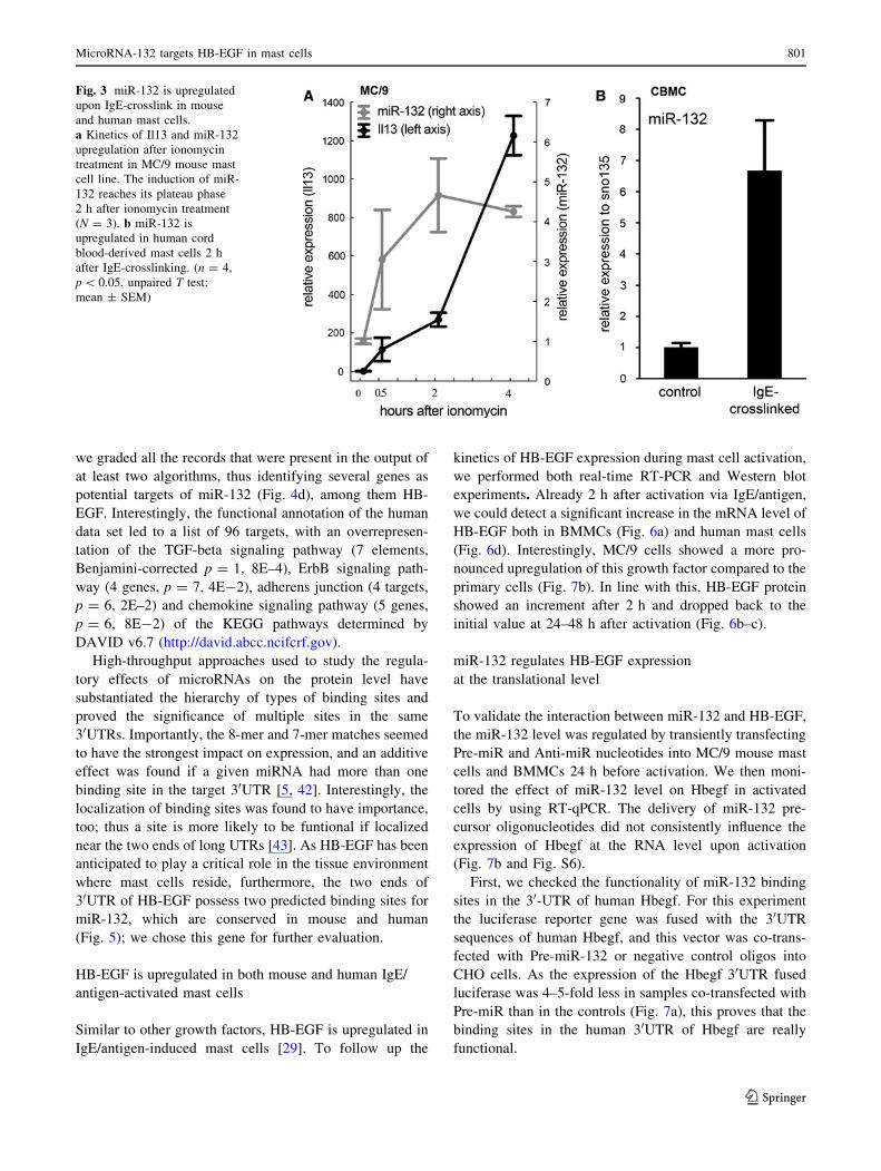
30 min and reached a plateau phase already at 2 h after
treatment, much earlier than the well-known transcriptional
activation marker IL-13 (Fig. 3). In a recent publication,
upregulation of miR-221 and miR-222 was found upon
ionomycin-induced activation in BMMCs [52]. In our
system, miR-221 was induced with a similar kinetics upon
IgE-crosslinking and ionomycin; however, miR-132
increased to a greater extent compared to miR-221 (Fig.
S4-5).
To confirm the activation-induced miR-132 expression
in a human system, mast cells were differentiated in vitro
from the CD34? fraction of cord blood, and the purity of
the cultures was checked after 6 weeks. Importantly, when
human mast cells were activated by IgE and an agonist
anti-IgE antibody, we observed a marked increase in the
miR-132 level, thus proving that our findings are not
restricted to mouse mast cells.
Target prediction of miR-132
The significant upregulation of miR-132 upon mast cell
activation suggested that this microRNA may participate in
the modulation of genes with a significant role in mast cell
activation. Using bioinformatic approaches we searched for
potential mRNA targets of miR-132 by analyzing the
mouse and human predicted data sets. To improve the
robustness of target prediction for miR-132, three different
algorithms were applied simultaneously. The target pre-
diction algorithms assign scores that reflect the probability
of interaction between the given microRNA and its target.
In order to collate the annotated data sets, the corre-
sponding scores were ranked in a range from 0 to 100 (rank
1 was assigned to the less likely, and the further values
were fractionated as percentages).
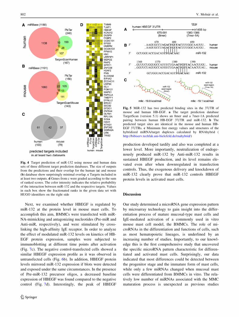
As was expected, the diverse inputs and analysis strin-
gency of the prediction algorithms resulted in significant
differences both in the size and in the composition of the
outputs. The number of targets predicted by at least two
algorithms is very low compared to the size of the outputs
generated by one single method, which is extremely
obvious in case of the MiRBase database (Fig. 4a, b).
However, when using only those targets that occurred in
the output of at least two algorithms, the mouse and human
data showed a significant overlap (Fig. 4c, 41 and 53% of
human and mouse targets, respectively), reflecting the high
degree of conservation in binding sites for miR-132. Next
Fig. 2 Validation of microarray data by qRT-PCR. a–e qRT-PCR
validation of a selected set of miRNA probes normalized to
snoRNA135. *, **, ****, # indicate p-values \0.05, \0.01, \0.001,
\10-5, respectively. Data are represented as mean ± SEM; n = 4–6
samples per group. f Correlation of miRNA microarray and qRT-PCR
data. Dashed line represents 95% confidence interval (Spearman rank
order correlation R = 0.857, p \ 0.05)
800 V. Molnar et al.
123
we graded all the records that were present in the output of
at least two algorithms, thus identifying several genes as
potential targets of miR-132 (Fig. 4d), among them HB-
EGF. Interestingly, the functional annotation of the human
data set led to a list of 96 targets, with an overrepresen-
tation of the TGF-beta signaling pathway (7 elements,
Benjamini-corrected p = 1, 8E–4), ErbB signaling path-
way (4 genes, p = 7, 4E-2), adherens junction (4 targets,
p = 6, 2E–2) and chemokine signaling pathway (5 genes,
p = 6, 8E-2) of the KEGG pathways determined by
DAVID v6.7 (http://david.abcc.ncifcrf.gov).
High-throughput approaches used to study the regula-
tory effects of microRNAs on the protein level have
substantiated the hierarchy of types of binding sites and
proved the significance of multiple sites in the same
30UTRs. Importantly, the 8-mer and 7-mer matches seemed
to have the strongest impact on expression, and an additive
effect was found if a given miRNA had more than one
binding site in the target 30UTR [5, 42]. Interestingly, the
localization of binding sites was found to have importance,
too; thus a site is more likely to be funtional if localized
near the two ends of long UTRs [43]. As HB-EGF has been
anticipated to play a critical role in the tissue environment
where mast cells reside, furthermore, the two ends of
30UTR of HB-EGF possess two predicted binding sites for
miR-132, which are conserved in mouse and human
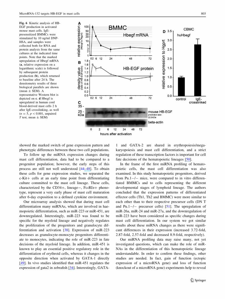
(Fig. 5); we chose this gene for further evaluation.
HB-EGF is upregulated in both mouse and human IgE/
antigen-activated mast cells
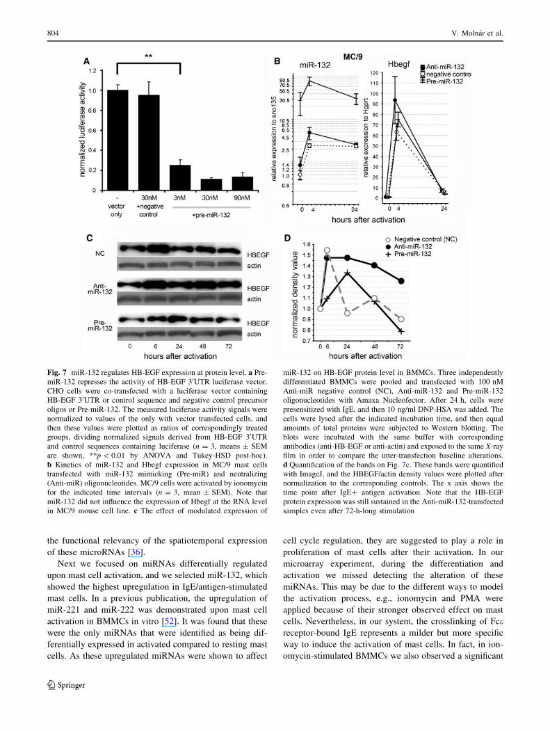
Similar to other growth factors, HB-EGF is upregulated in
IgE/antigen-induced mast cells [29]. To follow up the
kinetics of HB-EGF expression during mast cell activation,
we performed both real-time RT-PCR and Western blot
experiments. Already 2 h after activation via IgE/antigen,
we could detect a significant increase in the mRNA level of
HB-EGF both in BMMCs (Fig. 6a) and human mast cells
(Fig. 6d). Interestingly, MC/9 cells showed a more pro-
nounced upregulation of this growth factor compared to the
primary cells (Fig. 7b). In line with this, HB-EGF protein
showed an increment after 2 h and dropped back to the
initial value at 24–48 h after activation (Fig. 6b–c).
miR-132 regulates HB-EGF expression
at the translational level
To validate the interaction between miR-132 and HB-EGF,
the miR-132 level was regulated by transiently transfecting
Pre-miR and Anti-miR nucleotides into MC/9 mouse mast
cells and BMMCs 24 h before activation. We then moni-
tored the effect of miR-132 level on Hbegf in activated
cells by using RT-qPCR. The delivery of miR-132 pre-
cursor oligonucleotides did not consistently influence the
expression of Hbegf at the RNA level upon activation
(Fig. 7b and Fig. S6).
First, we checked the functionality of miR-132 binding
sites in the 30-UTR of human Hbegf. For this experiment
the luciferase reporter gene was fused with the 30UTR
sequences of human Hbegf, and this vector was co-trans-
fected with Pre-miR-132 or negative control oligos into
CHO cells. As the expression of the Hbegf 30UTR fused
luciferase was 4–5-fold less in samples co-transfected with
Pre-miR than in the controls (Fig. 7a), this proves that the
binding sites in the human 30UTR of Hbegf are really
functional.
Fig. 3 miR-132 is upregulated
upon IgE-crosslink in mouse
and human mast cells.
a Kinetics of Il13 and miR-132
upregulation after ionomycin
treatment in MC/9 mouse mast
cell line. The induction of miR-
132 reaches its plateau phase
2 h after ionomycin treatment
(N = 3). b miR-132 is
upregulated in human cord
blood-derived mast cells 2 h
after IgE-crosslinking. (n = 4,
p \ 0.05, unpaired T test;
mean ± SEM)
MicroRNA-132 targets HB-EGF in mast cells 801
123
Next, we examined whether HBEGF is regulated by
miR-132 at the protein level in mouse mast cells. To
accomplish this aim, BMMCs were transfected with miR-
NA-mimicking and antagonizing nucleotides (Pre-miR and
Anti-miR, respectively), and were stimulated by cross-
linking the high-affinity IgE receptor. In order to analyze
the effect of modulated miR-132 levels on kinetics of HB-
EGF protein expression, samples were subjected to
immunoblotting at different time points after activation
(Fig. 7c). The negative control-transfected cells showed a
similar HBEGF expression profile as it was observed in
untransfected cells (Fig. 6b). In addition, HBEGF protein
levels mirrored miR-132 expression if blots were detected
and exposed under the same circumstances. In the presence
of Pre-miR-132 precursor oligos, a decreased baseline
expression of HBEGF was found compared to the negative
control (Fig. 7d). Interestingly, the peak of HBEGF
production developed tardily and also was completed at a
lower level. More importantly, neutralization of endoge-
nously produced miR-132 by Anti-miR-132 results in
sustained HBEGF production, and its level remains ele-
vated even after when downregulated in transfection
controls. Thus, the exogenous delivery and knockdown of
miR-132 clearly prove that miR-132 controls HBEGF
protein levels in activated mast cells.
Discussion
Our study determined a microRNA gene expression pattern
by microarray technology to gain insight into the differ-
entiation process of mature mucosal-type mast cells and
IgE-mediated activation of a commonly used in vitro
mouse mast cell model, the BMMCs. The role of mi-
croRNAs in the differentiation and functions of cells, such
as most hematopoietic lineages, is underlined by an
increasing number of studies. Importantly, to our knowl-
edge this is the first comprehensive study that uncovered
the specific microRNA pattern characteristic for differen-
tiated and activated mast cells. Surprisingly, our data
indicated that most differences could be detected between
the progenitor stage and the immature form of mast cells,
while only a few miRNAs changed when mucosal mast
cells were differentiated from BMMCs in vitro. The rela-
tively low number of miRNAs associated with the MMC
maturation process is unexpected as previous studies
Fig. 4 Target prediction of miR-132 using mouse and human data
sets of three different target prediction databases. The size of outputs
from the predictions and their overlap for the human (a) and mouse
(b) database show surprisingly minimal overlap. c Targets included in
at least two outputs. d Genes from c were graded according to the sum
of ranked scores. The color intensity indicates the relative probability
of the interaction between miR-132 and the respective targets. Values
in each box show the fractionated ranks in the given data set with
HUGO identifiers on the right side
Fig. 5 MiR-132 has two predicted binding sites in the 30UTR of
mouse and human HB-EGF. a The target prediction database
TargetScan (version 5.1) shows an 8mer and a 7mer-1A predicted
pairing between human HB-EGF 30UTR and miR-132. b The
predicted target sites are identical in the mouse and human HB-
EGF 30UTRs. c Minimum free energy values and structures of the
hybridized miRNA/target duplexes calculated by RNAhybrid (
http://bibiserv.techfak.uni-bielefeld.de/rnahybrid/)
802 V. Molnar et al.
123
showed the marked switch of gene expression pattern and
phenotypic differences between these two cell populations.
To follow up the miRNA expression changes during
mast cell differentiation, data had to be compared to a
progenitor population; however, the early steps of this
process are still not well understood [44–48]. To obtain
these cells for gene expression studies, we separated the
c-Kit? cells at an early time point from differentiating
culture committed to the mast cell lineage. These cells,
characterized by the CD34?, lineage-, FceRIa? pheno-
type, represent a very early phase of mast cell maturation
after 6-day exposition to a defined cytokine environment.
Our microarray analysis showed that during mast cell
differentiation many miRNAs, which are involved in hae-
mopoietic differentiation, such as miR-223 or miR-451, are
downregulated. Interestingly, miR-223 was found to be
specific for the myeloid lineage and negatively regulates
the proliferation of the progenitors and granulocyte dif-
ferentiation and activation [38]. Expression of miR-223
decreases as granulocyte-monocyte progenitors differenti-
ate to monocytes, indicating the role of miR-223 in fate
decisions of the myeloid lineage. In addition, miR-451 is
known to play an essential positive regulatory role in the
differentiation of erythroid cells, whereas it changes in the
opposite direction when activated by GATA-1 directly
[49]. In vivo studies identified that miR-451 regulates the
expression of gata2 in zebrafish [34]. Interestingly, GATA-
1 and GATA-2 are shared in erythropoiesis/mega-
karyopoiesis and mast cell differentiation, and a strict
regulation of these transcription factors is important for cell
fate decisions of the hematopoietic lineages [50].
In the frame of the first miRNA profiling of hemato-
poietic cells, the mast cell differentiation was also
examined. In this study hematopoietic progenitors, derived
from Pu.1-/- mice, were compared to in vitro differen-
tiated BMMCs and to cells representing the different
developmental stages of lymphoid lineage. The authors
concluded that the expression patterns of differentiated
effector cells (Th1, Th2 and BMMC) were more similar to
each other than to their respective precursor cells (DN T
and Pu.1-/- precursor cells) [51]. The upregulation of
miR-26a, miR-24 and miR-27a, and the downregulation of
miR-223 have been considered as specific changes during
mast cell differentiation. In our system we got similar
results about these miRNA changes as there were signifi-
cant differences in their expression (increased 3.72-fold,
2.87-fold, 2.57-fold and decreased 8.9-fold, respectively).
Our miRNA profiling data may raise many, not yet
investigated questions, which can make the role of miR-
NAs in the differentiation of this hematopoietic lineage
understandable. In order to confirm these findings, other
studies are needed. In fact, gain of function (ectopic
expression of a microRNA gene) and loss of function
(knockout of a microRNA gene) experiments help to reveal
Fig. 6 Kinetic analysis of HB-
EGF production in activated
mouse mast cells. IgE-
presensitized BMMCs were
stimulated by 10 ng/ml DNP-
HSA, and samples were
collected both for RNA and
protein analysis from the same
cultures at the indicated time
points. Note that the marked
upregulation of Hbegf mRNA
(a, relative expression on a
logarithmic scale) is followed
by subsequent protein
production (b), which returned
to baseline after 24 h. The
densitometry results of three
biological parallels are shown
(mean ± SEM). A
representative Western blot is
depicted on c. d Hbegf is
upregulated in human cord
blood-derived mast cells 2 h
after IgE-crosslinking, as well
(n = 5, p \ 0.001, unpaired
T test, mean ± SEM)
MicroRNA-132 targets HB-EGF in mast cells 803
123
the functional relevancy of the spatiotemporal expression
of these microRNAs [36].
Next we focused on miRNAs differentially regulated
upon mast cell activation, and we selected miR-132, which
showed the highest upregulation in IgE/antigen-stimulated
mast cells. In a previous publication, the upregulation of
miR-221 and miR-222 was demonstrated upon mast cell
activation in BMMCs in vitro [52]. It was found that these
were the only miRNAs that were identified as being dif-
ferentially expressed in activated compared to resting mast
cells. As these upregulated miRNAs were shown to affect
cell cycle regulation, they are suggested to play a role in
proliferation of mast cells after their activation. In our
microarray experiment, during the differentiation and
activation we missed detecting the alteration of these
miRNAs. This may be due to the different ways to model
the activation process, e.g., ionomycin and PMA were
applied because of their stronger observed effect on mast
cells. Nevertheless, in our system, the crosslinking of Fcereceptor-bound IgE represents a milder but more specific
way to induce the activation of mast cells. In fact, in ion-
omycin-stimulated BMMCs we also observed a significant
Fig. 7 miR-132 regulates HB-EGF expression at protein level. a Pre-
miR-132 represses the activity of HB-EGF 30UTR luciferase vector.
CHO cells were co-transfected with a luciferase vector containing
HB-EGF 30UTR or control sequence and negative control precursor
oligos or Pre-miR-132. The measured luciferase activity signals were
normalized to values of the only with vector transfected cells, and
then these values were plotted as ratios of correspondingly treated
groups, dividing normalized signals derived from HB-EGF 30UTR
and control sequences containing luciferase (n = 3, means ± SEM
are shown, **p \ 0.01 by ANOVA and Tukey-HSD post-hoc).
b Kinetics of miR-132 and Hbegf expression in MC/9 mast cells
transfected with miR-132 mimicking (Pre-miR) and neutralizing
(Anti-miR) oligonucleotides. MC/9 cells were activated by ionomycin
for the indicated time intervals (n = 3, mean ± SEM). Note that
miR-132 did not influence the expression of Hbegf at the RNA level
in MC/9 mouse cell line. c The effect of modulated expression of
miR-132 on HB-EGF protein level in BMMCs. Three independently
differentiated BMMCs were pooled and transfected with 100 nM
Anti-miR negative control (NC), Anti-miR-132 and Pre-miR-132
oligonucleotides with Amaxa Nucleofector. After 24 h, cells were
presensitized with IgE, and then 10 ng/ml DNP-HSA was added. The
cells were lysed after the indicated incubation time, and then equal
amounts of total proteins were subjected to Western blotting. The
blots were incubated with the same buffer with corresponding
antibodies (anti-HB-EGF or anti-actin) and exposed to the same X-ray
film in order to compare the inter-transfection baseline alterations.
d Quantification of the bands on Fig. 7c. These bands were quantified
with ImageJ, and the HBEGF/actin density values were plotted after
normalization to the corresponding controls. The x axis shows the
time point after IgE? antigen activation. Note that the HB-EGF
protein expression was still sustained in the Anti-miR-132-transfected
samples even after 72-h-long stimulation
804 V. Molnar et al.
123
upregulation of miR-221, but the miR-132 level change
was more pronounced upon activation, especially if their
alterations in IgE-activated cells are compared to unstim-
ulated ones. The appropriate time for measurements
probably has an important role as well, but in response to
different activating stimuli similar kinetics were observed
in the expression of miR-132 and miR-221 between 6 and
72 h post activation.
The majority of miRNAs are expressed in a wide variety
of tissues, and only a relatively small subset of miRNAs
accounts for most of the differences in miRNA profiles
between cell lineages and tissues. Nevertheless, in many
cases miRNAs, originally thought to be tissue-specific,
later turned out to be expressed more broadly. Interest-
ingly, miR-132 belongs to this group: first, it was described
as a brain-related microRNA. Its function was initially
discovered in the brain [53–56], but later its crucial role
was proved in other tissues, such as in adipocytes [57],
ovarian granulosa cells [58] or normal mammary gland
development [59]. Furthermore, recently miR-132 has been
discovered to be critical in the maturation and function of
cell types involved in the immune system, like monocytes
[39] and primary lymphatic endothelial cells [60]. More-
over, this miRNA can serve as a possible link between the
neuronal and immune systems. Recently, it has been
reported that inflammatory stimuli induced the expression
of the miR-132, which mediates an anti-inflammatory
effect via targeting acetylcholinesterase in leukocytes.
Since the vagal secretion of acetylcholine suppresses
inflammation in the periphery, miR-132 was hypothetized
to attenuate the inflammation. Furthermore, these studies
also showed the involvement of microRNAs in the regu-
lation of the brain-immune axis [61]. Importantly, the
emerging role of miR-132 in the immune system is indi-
cated in models of bacterial [39] and viral infection [60],
and malignant hematological [62] and chronic inflamma-
tory diseases [63].
Interestingly, our data clearly showed that the upregu-
lation of miR-132 can be induced by a Ca-influx, which is a
downstream step of FceR-signaling. MiR-132 is known to
be upregulated in the neuronal context in a CREB-medi-
ated way [55]. Other transcription factors have been
indicated to be involved in mast cell activation, like
GATA1 and GATA2 (required also in development) [64].
Interestingly, GATA proteins interact with the MeCP2
[54]) and the CREB-binding proteins CBP/p300 [60], both
of which are experimentally validated targets of miR-132,
and the latter one is also a high-ranked gene in our pre-
dicted list (Ep300). These interactions may lead to the
accessibility of chromatin for expression from the Th2
locus [65]. With computational methods several cis-regu-
latory motives (24!) have been identified that may regulate
miR-132 expression. The large number of predicted sites
for transcription factor binding may indicate the functional
importance of miR-132. Furthermore, the combinatorial
interaction of these factors is suggested to result in the wide
dynamic range of miRNA expression [66]. One of these
predicted transcription factors, the cAMP-response element
binding protein (CREB), has been validated in a study.
Interestingly, miR-132 was found to contribute to neural
morphogenesis through the translational repression of a
GTPase-activating protein, p250GAP [55].
To find relevant targets of this microRNA, we per-
formed a bioinformatic search by using both mouse and
human data sets of three different algorithms. Among the
probable targets, the growth factor HB-EGF was further
analyzed, as it is upregulated upon IgE crosslinking and is
a high-ranked candidate predicted by all three algorithms
both in the mouse and human. In luciferase assay we could
demonstrate the functionality of miR-132 binding sites in
the 30UTR of human Hbegf. By using miR-132 mimicking
and neutralizing oligonucleotides, we confirmed the regu-
latory effect of miR-132 on HB-EGF expression at the
protein level in mast cells. Interestingly, HB-EGF would
not be the only target of the ‘‘gatekeeper’’ miR-132,
because it was observed to play a role in induction of
neovascularization via targeting p120RasGAP in tumor
endothelium [67].
HB-EGF is expressed in many types of cells, and it
functions as a mitogenic and chemotactic factor for cells
expressing the ErbB4 receptor, such as vascular smooth
muscle cells, fibroblasts, keratinocytes and hepatocytes. As
HB-EGF is also upregulated upon mast cell activation, we
suggest that miR-132 is a negative regulator of this mast
cell-derived growth factor to avoid inadequate remodeling
in chronic allergen exposure. Especially, it may have great
importance in physiological responses and diseases, such
as wound healing, asthma, nasal polyposis and psoriasis, as
all of them can be characterized by an extensive tissue
remodeling and inflammatory activity of mast cells. HB-
EGF has also been implicated in the pathogenesis of
bronchial asthma. Asthma is a chronic respiratory disease
that is generally characterized by inflamed airways and
reversible bronchoconstriction. A relatively small propor-
tion of patients suffers from a severe type of asthma with
permanent airway constricton and the unresponsiveness to
corticosteroid therapy. Interestingly, in severe asthma nei-
ther the presence of inflammatory cells nor the extent of
epithelial damage, but rather the elevated number of
fibroblasts and larger mucous glands and airway smooth
muscle areas correlate with the severity of the disease [68].
The complex remodeling of the airways and lung tissue
has been suggested to be mediated by EGFR-signaling
[69] in a rat model of allergic asthma. Very recently, the
involvement of EGFR signaling was suggested in the
IL-6-dependent recruitment of T cells to the airways in an
MicroRNA-132 targets HB-EGF in mast cells 805
123
OVA-induced rat asthma model [70]. HB-EGF is also
implicated in heart tissue remodeling after myocardial infarct
[71]. Thus, all these data point to the critical importance of
proper regulation of EGFR signaling and HB-EGF.
In summary, here we attempted to gain a comprehensive
view of the miRNA pattern and its alterations during mast
cell differentiation and IgE-mediated activation. These data
were obtained by a standard microarray platform, and
therefore they can be easily integrated into miRNA gene
expression data of other hematopoietic lineages. The
interplay between miR-132 upregulation and the regulation
of HB-EGF expression suggests a negative feedback
mechanism that may take part in silencing the activated
mast cells and thus contributes to avoiding the prolonged
stimulation of the tissue environment.
Acknowledgments This work is supported by the National
Research Foundation of the Hungarian Academy of Sciences (grant
OTKA 67955).
References
1. Ambros V (2004) The functions of animal microRNAs. Nature
431:350–355
2. Bartel DP (2009) MicroRNAs: target recognition and regulatory
functions. Cell 136:215–233
3. Lodish HF, Zhou B, Liu G, Chen CZ (2008) Micromanagement
of the immune system by microRNAs. Nat Rev Immunol
8:120–130
4. Rajewsky N (2006) MicroRNA target predictions in animals. Nat
Genet 38(Suppl):S8–S13
5. Baek D, Villen J, Shin C, Camargo FD, Gygi SP, Bartel DP
(2008) The impact of microRNAs on protein output. Nature
455:64–71
6. Abraham SN, St John AL (2010) Mast cell-orchestrated immu-
nity to pathogens. Nat Rev Immunol 10:440–452
7. Galli SJ, Grimbaldeston M, Tsai M (2008) Immunomodulatory
mast cells: negative, as well as positive, regulators of immunity.
Nat Rev Immunol 8:478–486
8. Gurish MF, Boyce JA (2006) Mast cells: ontogeny, homing, and
recruitment of a unique innate effector cell. J Allergy Clin
Immunol 117:1285–1291
9. Kashiwakura J, Xiao W, Kitaura J, Kawakami Y, Maeda-Ya-
mamoto M, Pfeiffer JR, Wilson BS, Blank U, Kawakami T
(2008) Pivotal advance: IgE accelerates in vitro development of
mast cells and modifies their phenotype. J Leukoc Biol
84:357–367
10. Pejler G, Ronnberg E, Waern I, Wernersson S (2010) Mast cell
proteases: multifaceted regulators of inflammatory disease. Blood
115:4981–4990
11. Metcalfe DD, Baram D, Mekori YA (1997) Mast cells. Physiol
Rev 77:1033–1079
12. Galli SJ, Tsai M, Piliponsky AM (2008) The development of
allergic inflammation. Nature 454:445–454
13. Cho SH, Yao Z, Wang SW, Alban RF, Barbers RG, French SW,
Oh CK (2003) Regulation of activin A expression in mast cells
and asthma: its effect on the proliferation of human airway
smooth muscle cells. J Immunol 170:4045–4052
14. Wang SW, Oh CK, Cho SH, Hu G, Martin R, Demissie-Sanders
S, Li K, Moyle M, Yao Z (2005) Amphiregulin expression in
human mast cells and its effect on the primary human lung
fibroblasts. J Allergy Clin Immunol 115:287–294
15. Marikovsky M, Breuing K, Liu PY, Eriksson E, Higashiyama S,
Farber P, Abraham J, Klagsbrun M (1993) Appearance of hepa-
rin-binding EGF-like growth factor in wound fluid as a response
to injury. Proc Natl Acad Sci U S A 90:3889–3893
16. Stoll S, Garner W, Elder J (1997) Heparin-binding ligands
mediate autocrine epidermal growth factor receptor activation In
skin organ culture. J Clin Invest 100:1271–1281
17. Tokumaru S, Higashiyama S, Endo T, Nakagawa T, Miyagawa
JI, Yamamori K, Hanakawa Y, Ohmoto H, Yoshino K, Shirakata
Y, Matsuzawa Y, Hashimoto K, Taniguchi N (2000) Ectodomain
shedding of epidermal growth factor receptor ligands is required
for keratinocyte migration in cutaneous wound healing. J Cell
Biol 151:209–220
18. Xie H, Wang H, Tranguch S, Iwamoto R, Mekada E, Demayo FJ,
Lydon JP, Das SK, Dey SK (2007) Maternal heparin-binding-
EGF deficiency limits pregnancy success in mice. Proc Natl Acad
Sci USA 104:18315–18320
19. Iwamoto R, Yamazaki S, Asakura M, Takashima S, Hasuwa H,
Miyado K, Adachi S, Kitakaze M, Hashimoto K, Raab G, Nanba
D, Higashiyama S, Hori M, Klagsbrun M, Mekada E (2003)
Heparin-binding EGF-like growth factor and ErbB signaling is
essential for heart function. Proc Natl Acad Sci USA
100:3221–3226
20. Ongusaha PP, Kwak JC, Zwible AJ, Macip S, Higashiyama S,
Taniguchi N, Fang L, Lee SW (2004) HB-EGF is a potent inducer
of tumor growth and angiogenesis. Cancer Res 64:5283–5290
21. Nakata A, Miyagawa J, Yamashita S, Nishida M, Tamura R,
Yamamori K, Nakamura T, Nozaki S, Kameda-Takemura K,
Kawata S, Taniguchi N, Higashiyama S, Matsuzawa Y (1996)
Localization of heparin-binding epidermal growth factor-like
growth factor in human coronary arteries. Possible roles of HB-
EGF in the formation of coronary atherosclerosis. Circulation
94:2778–2786
22. Higashiyama S, Abraham JA, Klagsbrun M (1993) Heparin-
binding EGF-like growth factor stimulation of smooth muscle
cell migration: dependence on interactions with cell surface
heparan sulfate. J Cell Biol 122:933–940
23. Raab G, Klagsbrun M (1997) Heparin-binding EGF-like growth
factor. Biochim Biophys Acta 1333:F179–F199
24. Knight PA, Brown JK, Wright SH, Thornton EM, Pate JA, Miller
HR (2007) Aberrant mucosal mast cell protease expression in the
enteric epithelium of nematode-infected mice lacking the integrin
alphavbeta6, a transforming growth factor-beta1 activator. Am J
Pathol 171:1237–1248
25. Miller HR, Wright SH, Knight PA, Thornton EM (1999) A novel
function for transforming growth factor-beta1: upregulation of
the expression and the IgE-independent extracellular release of a
mucosal mast cell granule-specific beta-chymase, mouse mast
cell protease-1. Blood 93:3473–3486
26. Gilicze A, Kohalmi B, Pocza P, Keszei M, Jaeger J, Gorbe E,
Papp Z, Toth S, Falus A, Wiener Z (2007) HtrA1 is a novel mast
cell serine protease of mice and men. Mol Immunol
44:2961–2968
27. Wiener Z, Pocza P, Racz M, Nagy G, Tolgyesi G, Molnar V,
Jaeger J, Buzas E, Gorbe E, Papp Z, Rigo J, Falus A (2008) IL-18
induces a marked gene expression profile change and increased
Ccl1 (I-309) production in mouse mucosal mast cell homologs.
Int Immunol 20:1565–1573
28. Ventura A, Young AG, Winslow MM, Lintault L, Meissner A,
Erkeland SJ, Newman J, Bronson RT, Crowley D, Stone JR,
Jaenisch R, Sharp PA, Jacks T (2008) Targeted deletion reveals
806 V. Molnar et al.
123
essential and overlapping functions of the miR-17 through 92
family of miRNA clusters. Cell 132:875–886
29. Xiao C, Srinivasan L, Calado DP, Patterson HC, Zhang B, Wang
J, Henderson JM, Kutok JL, Rajewsky K (2008) Lymphoprolif-
erative disease and autoimmunity in mice with increased miR-
17–92 expression in lymphocytes. Nat Immunol 9:405–414
30. Hashimi ST, Fulcher JA, Chang MH, Gov L, Wang S, Lee B
(2009) MicroRNA profiling identifies miR-34a and miR-21 and
their target genes JAG1 and WNT1 in the coordinate regulation
of dendritic cell differentiation. Blood 114:404–414
31. Navarro F, Gutman D, Meire E, Caceres M, Rigoutsos I, Bent-
wich Z, Lieberman J (2009) miR-34a contributes to
megakaryocytic differentiation of K562 cells independently of
p53. Blood 114:2181–2192
32. O’Connell RM, Rao DS, Chaudhuri AA, Baltimore D (2010)
Physiological and pathological roles for microRNAs in the
immune system. Nat Rev Immunol 10:111–122
33. Lu TX, Munitz A, Rothenberg ME (2009) MicroRNA-21 is up-
regulated in allergic airway inflammation and regulates IL-12p35
expression. J Immunol 182:4994–5002
34. Pase L, Layton JE, Kloosterman WP, Carradice D, Waterhouse
PM, Lieschke GJ (2009) miR-451 regulates zebrafish erythroid
maturation in vivo via its target gata2. Blood 113:1794–1804
35. Zhan M, Miller CP, Papayannopoulou T, Stamatoyannopoulos G,
Song CZ (2007) MicroRNA expression dynamics during murine
and human erythroid differentiation. Exp Hematol 35:1015–1025
36. Chen CZ, Li L, Lodish HF, Bartel DP (2004) MicroRNAs modulate
hematopoietic lineage differentiation. Science 303:83–86
37. Fazi F, Rosa A, Fatica A, Gelmetti V, De Marchis ML, Nervi C,
Bozzoni I (2005) A minicircuitry comprised of microRNA-223
and transcription factors NFI-A and C/EBPalpha regulates human
granulopoiesis. Cell 123:819–831
38. Johnnidis JB, Harris MH, Wheeler RT, Stehling-Sun S, Lam MH,
Kirak O, Brummelkamp TR, Fleming MD, Camargo FD (2008)
Regulation of progenitor cell proliferation and granulocyte
function by microRNA-223. Nature 451:1125–1129
39. Taganov KD, Boldin MP, Chang KJ, Baltimore D (2006) NF-
kappaB-dependent induction of microRNA miR-146, an inhibitor
targeted to signaling proteins of innate immune responses. Proc
Natl Acad Sci USA 103:12481–12486
40. Fontana L, Pelosi E, Greco P, Racanicchi S, Testa U, Liuzzi F,
Croce CM, Brunetti E, Grignani F, Peschle C (2007) MicroRNAs
17-5p-20a-106a control monocytopoiesis through AML1 target-
ing and M-CSF receptor upregulation. Nat Cell Biol 9:775–787
41. McCurdy JD, Lin TJ, Marshall JS (2001) Toll-like receptor
4-mediated activation of murine mast cells. J Leukoc Biol
70:977–984
42. Selbach M, Schwanhausser B, Thierfelder N, Fang Z, Khanin R,
Rajewsky N (2008) Widespread changes in protein synthesis
induced by microRNAs. Nature 455:58–63
43. Grimson A, Farh KK, Johnston WK, Garrett-Engele P, Lim LP,
Bartel DP (2007) MicroRNA targeting specificity in mammals:
determinants beyond seed pairing. Mol Cell 27:91–105
44. Arinobu Y, Iwasaki H, Gurish MF, Mizuno S, Shigematsu H,
Ozawa H, Tenen DG, Austen KF, Akashi K (2005) Develop-
mental checkpoints of the basophil/mast cell lineages in adult
murine hematopoiesis. Proc Natl Acad Sci USA 102:18105–
18110
45. Chen CC, Grimbaldeston MA, Tsai M, Weissman IL, Galli SJ
(2005) Identification of mast cell progenitors in adult mice. Proc
Natl Acad Sci USA 102:11408–11413
46. Jamur MC, Grodzki AC, Berenstein EH, Hamawy MM, Siraga-
nian RP, Oliver C (2005) Identification and characterization of
undifferentiated mast cells in mouse bone marrow. Blood
105:4282–4289
47. Kitamura Y, Shimada M, Hatanaka K, Miyano Y (1977)
Development of mast cells from grafted bone marrow cells in
irradiated mice. Nature 268:442–443
48. Rodewald HR, Dessing M, Dvorak AM, Galli SJ (1996) Identi-
fication of a committed precursor for the mast cell lineage.
Science 271:818–822
49. Dore LC, Amigo JD, Dos Santos CO, Zhang Z, Gai X, Tobias
JW, Yu D, Klein AM, Dorman C, Wu W, Hardison RC, Paw BH,
Weiss MJ (2008) A GATA-1-regulated microRNA locus essen-
tial for erythropoiesis. Proc Natl Acad Sci USA 105:3333–3338
50. Migliaccio AR, Rana RA, Sanchez M, Lorenzini R, Centurione
L, Bianchi L, Vannucchi AM, Migliaccio G, Orkin SH (2003)
GATA-1 as a regulator of mast cell differentiation revealed by
the phenotype of the GATA-1low mouse mutant. J Exp Med
197:281–296
51. Monticelli S, Ansel KM, Xiao C, Socci ND, Krichevsky AM,
Thai TH, Rajewsky N, Marks DS, Sander C, Rajewsky K, Rao A,
Kosik KS (2005) MicroRNA profiling of the murine hemato-
poietic system. Genome Biol 6:R71
52. Mayoral RJ, Pipkin ME, Pachkov M, van Nimwegen E, Rao A,
Monticelli S (2009) MicroRNA-221–222 regulate the cell cycle
in mast cells. J Immunol 182:433–445
53. Cheng HY, Papp JW, Varlamova O, Dziema H, Russell B,
Curfman JP, Nakazawa T, Shimizu K, Okamura H, Impey S,
Obrietan K (2007) MicroRNA modulation of circadian-clock
period and entrainment. Neuron 54:813–829
54. Klein ME, Lioy DT, Ma L, Impey S, Mandel G, Goodman RH
(2007) Homeostatic regulation of MeCP2 expression by a CREB-
induced microRNA. Nat Neurosci 10:1513–1514
55. Vo N, Klein ME, Varlamova O, Keller DM, Yamamoto T,
Goodman RH, Impey S (2005) A cAMP-response element
binding protein-induced microRNA regulates neuronal morpho-
genesis. Proc Natl Acad Sci USA 102:16426–16431
56. Wayman GA, Davare M, Ando H, Fortin D, Varlamova O, Cheng
HY, Marks D, Obrietan K, Soderling TR, Goodman RH, Impey S
(2008) An activity-regulated microRNA controls dendritic plas-
ticity by down-regulating p250GAP. Proc Natl Acad Sci USA
105:9093–9098
57. Strum JC, Johnson JH, Ward J, Xie H, Feild J, Hester A, Alford
A, Waters KM (2009) MicroRNA 132 regulates nutritional stress-
induced chemokine production through repression of SirT1. Mol
Endocrinol 23:1876–1884
58. Fiedler SD, Carletti MZ, Hong X, Christenson LK (2008) Hor-
monal regulation of MicroRNA expression in periovulatory
mouse mural granulosa cells. Biol Reprod 79:1030–1037
59. Ucar A, Vafaizadeh V, Jarry H, Fiedler J, Klemmt PA, Thum T,
Groner B, Chowdhury K (2010) miR-212 and miR-132 are
required for epithelial stromal interactions necessary for mouse
mammary gland development. Nat Genet 42:1101–1108
60. Lagos D, Pollara G, Henderson S, Gratrix F, Fabani M, Milne RS,
Gotch F, Boshoff C (2010) miR-132 regulates antiviral innate
immunity through suppression of the p300 transcriptional co-
activator. Nat Cell Biol 12:513–519
61. Shaked I, Meerson A, Wolf Y, Avni R, Greenberg D, Gilboa-
Geffen A, Soreq H (2009) MicroRNA-132 potentiates cholinergic
anti-inflammatory signaling by targeting acetylcholinesterase.
Immunity 31:965–973
62. Calin GA, Liu CG, Sevignani C, Ferracin M, Felli N, Dumitru
CD, Shimizu M, Cimmino A, Zupo S, Dono M, Dell’Aquila ML,
Alder H, Rassenti L, Kipps TJ, Bullrich F, Negrini M, Croce CM
(2004) MicroRNA profiling reveals distinct signatures in B cell
chronic lymphocytic leukemias. Proc Natl Acad Sci USA
101:11755–11760
63. Pauley KM, Satoh M, Chan AL, Bubb MR, Reeves WH, Chan
EK (2008) Upregulated miR-146a expression in peripheral blood
MicroRNA-132 targets HB-EGF in mast cells 807
123
mononuclear cells from rheumatoid arthritis patients. Arthritis
Res Ther 10:R101
64. Masuda A, Hashimoto K, Yokoi T, Doi T, Kodama T, Kume H,
Ohno K, Matsuguchi T (2007) Essential role of GATA transcrip-
tional factors in the activation of mast cells. J Immunol 178:360–368
65. Gregory GD, Raju SS, Winandy S, Brown MA (2006) Mast cell
IL-4 expression is regulated by Ikaros and influences encephali-
togenic Th1 responses in EAE. J Clin Invest 116:1327–1336
66. Lee J, Li Z, Brower-Sinning R, John B (2007) Regulatory circuit
of human microRNA biogenesis. PLoS Comput Biol 3:e67
67. Anand S, Majeti BK, Acevedo LM, Murphy EA, Mukthavaram
R, Scheppke L, Huang M, Shields DJ, Lindquist JN, Lapinski PE,
King PD, Weis SM, Cheresh DA (2010) MicroRNA-132-medi-
ated loss of p120RasGAP activates the endothelium to facilitate
pathological angiogenesis. Nat Med 16:909–914
68. Benayoun L, Druilhe A, Dombret MC, Aubier M, Pretolani M
(2003) Airway structural alterations selectively associated with
severe asthma. Am J Respir Crit Care Med 167:1360–1368
69. Tamaoka M, Hassan M, McGovern T, Ramos-Barbon D, Jo T,
Yoshizawa Y, Tolloczko B, Hamid Q, Martin JG (2008) The
epidermal growth factor receptor mediates allergic airway
remodelling in the rat. Eur Respir J 32:1213–1223
70. Tsuchiya K, Jo T, Takeda N, Al Heialy S, Siddiqui S, Shalaby
KH, Risse PA, Maghni K, Martin JG (2010) EGF receptor acti-
vation during allergic sensitization affects IL-6-induced T-cell
influx to airways in a rat model of asthma. Eur J Immunol
40:1590–1602
71. Ushikoshi H, Takahashi T, Chen X, Khai NC, Esaki M, Goto K,
Takemura G, Maruyama R, Minatoguchi S, Fujiwara T, Nagano
S, Yuge K, Kawai T, Murofushi Y, Fujiwara H, Kosai K (2005)
Local overexpression of HB-EGF exacerbates remodeling fol-
lowing myocardial infarction by activating noncardiomyocytes.
Lab Invest 85:862–873
808 V. Molnar et al.
123


