Embed Size (px)
Citation preview

MicroRNAs: new players in IBDR Kalla,1 N T Ventham,1 N A Kennedy,1 J F Quintana,2 E R Nimmo,1 A H Buck,2
J Satsangi1
▸ Additional material ispublished online only. To viewplease visit the journal online(http://dx.doi.org/10.1136/gutjnl-2014-307891).1Gastrointestinal Unit, Centrefor Molecular Medicine,Institute of Genetics andMolecular Medicine, WesternGeneral Hospital, Edinburgh,UK2Centre for Immunity, Infectionand Evolution, Ashworthlaboratories, University ofEdinburgh, Edinburgh, UK
Correspondence toProfessor Jack Satsangi,Gastrointestinal Unit, WesternGeneral Hospital, Crewe RoadSouth, Edinburgh EH4 2XU,UK; [email protected]
Received 23 June 2014Revised 9 October 2014Accepted 23 October 2014
To cite: Kalla R,Ventham NT, Kennedy NA,et al. Gut Published OnlineFirst: [please include DayMonth Year] doi:10.1136/gutjnl-2014-307891
ABSTRACTMicroRNAs (miRNAs) are small non-coding RNAs, 18–23nucleotides long, which act as post-transcriptionalregulators of gene expression. miRNAs are stronglyimplicated in the pathogenesis of many commondiseases, including IBDs. This review aims to outline thehistory, biogenesis and regulation of miRNAs. The role ofmiRNAs in the development and regulation of the innateand adaptive immune system is discussed, with aparticular focus on mechanisms pertinent to IBD and thepotential translational applications.
INTRODUCTIONThe IBDs, Crohn’s disease (CD) and UC affect anestimated 2.5–3 million people in Europe, with theassociated annual healthcare costs amounting toapproximately €4.6–5.6 billion.1 The increasingincidence of early onset disease in the developedworld and of disease in all ages in the developingworld has catalysed studies attempting to character-ise pathogenic mechanisms. In the last two decades,international collaborations have been successful inidentifying susceptibility genes for IBD throughgenome-wide association studies (GWAS) and sub-sequently meta-analysis of GWAS and Immunochipdata (http://www.ibdgenetics.org).2 These studieshave been important in highlighting mechanisticpathways, notably autophagy and innate immunityin CD and epithelial barrier dysfunction in UC andhave provided clues into new therapeutic strategies.There is now increasing interest in exploring epi-
genetic mechanisms in common diseases, withnotable progress in studies of DNA methylation,histone modifications, long intergenic non-codingRNAs and in characterising the contribution ofmicroRNAs (miRNAs). miRNAs are short strandsof non-coding RNA (∼22 nt long) encoded ingenomic DNA which post-transcriptionally regulateexpression. The field of miRNA research is expand-ing rapidly with the number of miRNA-related cita-tions increasing almost exponentially (figure 1) andmiRNAs have been implicated in neurological dis-eases, cardiovascular diseases, autoimmune diseasesand cancer.3 With such a wealth of data now avail-able, reviews have been published on individualmiRNAs in health and disease. miR-21 is perhapsthe most compelling miRNA involved in IBD, withassociations between miR-21 and IBD being repli-cated in several studies, functional relevance inmouse models, as well as being highly expressed inother diseases including cancer. Key miRNAs, suchas miR-21, are the focus of anti-miR therapeuticdevelopment.4–8
Well-designed high-impact publications haveestablished functional interactions betweenmiRNAs and key mechanisms implicated by GWAS
in IBD, notably T helper cell (Th)17 mediatedinflammation and autophagy.9 10 The review aimsto outline the history, biogenesis and regulation ofmiRNAs. The important role of miRNAs in thedevelopment and regulation of an innate and adap-tive immune system is discussed, with a particularfocus on IBD pathogenesis and other immune-mediated diseases. The review will also provide aninsight into the translational applications ofmiRNAs as biomarkers and the potential thera-peutic miRNA application.
MicroRNAs: a historical perspectivemiRNAs were first identified in 1993 in the nema-tode model organism (Caenorhabditis elegans)using a genetic screen to identify defects in postem-bryonic development.11–13 It became evident thatlin-4, which emerged as the first described miRNA,was able to downregulate a nuclear protein calledlin-14, thereby initiating the second stage in larvaldevelopment.13 14 By the turn of the century asecond miRNA, let-7, was identified in C. elegansthat appeared to be highly conserved amongspecies including humans.15 16 At the time ofwriting 35 828 mature miRNAs occurring across allspecies have been registered in miRbase (http://mirbase.org, Release 21, accessed June 2014).17
Biogenesis of microRNAsmiRNA genes are located throughout the genome,either within intronic sequences of protein-codinggenes, within intronic or exonic regions of non-coding RNAs, or set between independent transcrip-tion units (intergenic).18 Some miRNAs have theirown promoters and are transcribed independently,some share promoters with host genes,19 whileothers are co-transcribed as a single primary miRNAtranscript.20 The biogenesis of miRNAs from tran-scription in the nucleus to generation of the maturemiRNA in the cytoplasm is described in figure 2.In plants, fully complementary binding occurs
when the ‘seed’ region (located near the 50end) of themiRNA binds to the 30 untranslated region (UTR) ofthe target mRNA and this is sufficient for mRNAdegradation to occur. In contrast, in humans,miRNAs bind to mRNA targets with incompletecomplementarity, which results in mRNA destabilisa-tion and translational inhibition.53 Other regions ofthe mRNA can also contain functional miRNAbinding sites, including the 50UTR and the aminoacid coding sequence. Furthermore, beyond seed sitepairing, the centre and the 30end of the miRNAsequence can contribute to target recognition.54–56
Under certain conditions such as cell cycle arrest,miRNAs can alter their regulatory role from transla-tional inhibition to upregulation of translation oftarget mRNAs.57 Studies have also shown that
Kalla R, et al. Gut 2014;0:1–14. doi:10.1136/gutjnl-2014-307891 1
Recent advances in basic science Gut Online First, published on December 4, 2014 as 10.1136/gutjnl-2014-307891
Copyright Article author (or their employer) 2014. Produced by BMJ Publishing Group Ltd (& BSG) under licence.
group.bmj.com on December 9, 2014 - Published by http://gut.bmj.com/Downloaded from

miRNAs influence gene expression at the post-transcriptionallevel, and may interfere with the process of transcription.58
Single nucleotide polymorphisms (SNPs) in pre-miRNAsequences are rare, occurring in only 10% of all humanpre-miRNAs, and less than 1% of miRNAs have SNPs in theirfunctional seed region.53 Therefore functional mutations inmiRNAs are unlikely to be tolerated and negative selection mayoccur at these loci.
miRNAs affect gene expressionIt is estimated that miRNAs regulate more than 60% of proteincoding mRNAs.59 Each miRNA can target hundreds of mRNAsresulting in mRNA destabilisation and/or inhibition of transla-tion. Generally, the overall effect on target protein levels issubtle and can be thought of as ‘fine-tuning’ of cellular mRNAexpression within a cell.60 61 The combinatorial targeting ofgenes by miRNAs in this fashion makes them interesting thera-peutic candidates that in theory may reduce resistance in dis-eases such as cancer.62
miRNAs regulate important cellular functions such as differ-entiation, proliferation, signal transduction and apoptosis andexhibit highly specific regulated patterns of gene expression.63
A number of applications have been developed to predictmRNA/miRNA interactions and aid in understanding specificmiRNA targets.64
miRNA regulationAt various stages in miRNA biogenesis, several factors can influ-ence the development of the mature miRNA. Figure 2 depictsthe various steps of biogenesis that are subject to regulation.These include regulation of transcription, cleavage of the stemloop structures by Drosha and Dicer enzymes, editing as well asregulation of miRNA turnover. The regulatory mechanismsoccurring at each stage have been reviewed elsewhere.18 65 Eachof these mechanisms acts as part of a signalling network thatmodulates gene expression in response to cellular or environ-mental changes.
miRNA gene regulatory networksOver 5400 miRNAs have now been identified with eachmiRNA possessing the ability to target multiple gene transcripts.miRNAs are members of complex gene regulatory networks(GRNs) and figure 3 summarises these GRNs, comprising offeedback and feed-forward loops.66 67 69 Certain subcircuits areevolutionarily favoured and are termed network motifs.67
Coordinated transcriptional and miRNA-mediated gene regula-tion is a recurrent network motif and fortifies gene regulation inmammalian genomes.66 Inflammation driven miRNA circuitsare described in the literature and examples include nuclearfactor-κB (NFκB) and hepatocyte nuclear factor-4α circuits.70 71
Within the NFκB circuitry, transient activation of Src oncopro-tein triggers an NFκB mediated inflammatory response bydownregulating let-7a and upregulating its direct target interleu-kin (IL)-6.70 This forms a stable positive feedback circuit acrossmany cell divisions.70 Similarly the hepatocyte nuclear factor-4αcircuit consists of miR-124, IL6R, STAT3, miR-24 and miR-629and is essential for liver development and hepatocyte func-tion.71 Several other examples of miRNAs involved in GRNsare summarised in a recent review.72
Regulation of miRNAs through epigenetic mechanismsEmerging evidence suggests miRNA expression can be regulatedby epigenetic mechanisms such as DNA methylation, histonemodifications and circular RNAs (circRNAs).73–76 DNA methy-lation, the addition of methyl groups at CpG islands by DNAmethyltransferases (DNMTs), is associated with transcriptionalrepression. Similarly, acetylation or deacetylation of histonesmay alter transcriptional activity.77 The recently establishedEpimiR database has collected 1974 regulations between 19types of epigenetic modifications and 617 miRNAs across sevenspecies.78 Aberrant DNA methylation of miRNAs has beendemonstrated in various cancers, including lymphoid, gastric
Figure 1 Pubmed microRNA (miRNA) citations in Gastroenterology andInflammatory Bowel Diseases (IBD). Search terms used were as follows:Gastroenterology: (miRNA OR MicroRNA) AND (Gastroenterology OR IBDOR Inflammatory Bowel Disease OR Crohn’s Disease OR Ulcerative ColitisOR Colon OR Stomach OR Intestine OR Oesophagus OR Oesophagus ORRectum) NOT mirna[author]; IBD: (miRNA OR MicroRNA) AND (IBD ORInflammatory Bowel Disease OR Crohn’s Disease OR Ulcerative Colitis)NOT mirna[author]; miRNA: (miRNA OR MicroRNA) NOT mirna[author];Each search was run for print publication dates for each year from 2001 to2014. Citations were normalised to the total number of Pubmed indexedarticles during the same time period (nb, the term microRNAwasintroduced in 2001).
2 Kalla R, et al. Gut 2014;0:1–14. doi:10.1136/gutjnl-2014-307891
Recent advances in basic science
group.bmj.com on December 9, 2014 - Published by http://gut.bmj.com/Downloaded from

and colorectal malignancies.79–81 Up to 10% of miRNAs aretightly controlled by DNA methylation as seen in cell lines defi-cient in DNMT1 and DNMT3b.82 Time-dependent miRNAregulation has also been described. In murine models, partialhepatectomy results in downregulation of miR-127 as early as3 h post partial hepatectomy with significant downregulationseen at 24 h.83 DNA methylation has also been shown to alterchromatin signatures and influence miRNA expression incancer.73 Within the context of IBD, our group has studied
epigenome-wide whole-blood DNA methylation profiles intreatment-naïve children with CD and healthy controls usingthe Ilumina 450 K platform.7 Sixty-five differentially methylatedCpG sites achieving epigenome-wide significance were identi-fied. The most significantly differentially methylated region inpatients with CD involves the transcription start site formiR-21. Hypomethylation of the miR-21 locus in cases corre-lated with increased primary miR-21 expression in leucocytesand in inflamed intestinal mucosa.7
Figure 2 miRNA biogenesis and regulation. (A) Processing begins in the nucleus where primary miRNA transcripts (pri-miR) are transcribed byRNA polymerase II or RNA polymerase III.21 22 (B) Nuclear cleavage of pri-miRNA is performed by a protein complex consisting of the RNAse-III-typeenzyme Drosha and DGCR8 (DiGeorge critical region 8), which generates a 60–70 nucleotide sequence called pre-miRNA. Drosha cleavage generatesa 2 nucleotide 3’ overhang which appears to be a key biogenesis step.23 DCGR8 acts as an anchor on the stem loops of the target miRNA,24
allowing Drosha to correctly position on the pri-miRNA.25 Mirtrons are similar in structure but do not undergo Drosha/DGCR8 processing. (C) pre-miRNA is transported from the nucleus to the cytoplasm by the Exportin-5 (Exp5) — RanGTP complex. Correct binding of the double stranded stemand 3’ regions to the RanGTP structure stabilises the miRNA, preventing degradation and facilitating the correct transport of pre-miRNA.26–28
(D) Final cleavage of the hairpin loop is performed by Dicer (RNAse III like enzyme) with co-factors: Tar RNA binding protein (TRBP); and proteinactivator of double-stranded RNA-dependent protein kinase (PACT). (E) The remaining 22 nucleotide RNA duplex is incorporated with Ago proteins,forming a pre-RNA induced silencing complex (pre-RISC). The duplex is separated within Ago proteins into a single stranded mature miRNA and itspassenger strand. The mature miRNA strand is retained to form RISC which is eventually destined for mRNA repression/cleavage while its passengerstrand undergoes degradation.29 30 miRNA recognises its target via 6-8 nucleotide sequence at the 5’ end of the miRNA however the binding sitecan vary. Examples of regulatory elements in miRNA biogenesis. Transcriptional regulation Transcription factors can influence miRNAexpression by binding directly to promoter elements. Examples include c-Myc binding and upregulating miR-17–92 cluster and p53interaction withmiR-34.31–34 miRNAs and argonaute (Ago) proteins as regulators mature miRNAs can act as regulators of miRNA processing either as an auto-regulatory loop or for other miRNAs (e.g. the biogenesis of let-7).35 RNA editing Once transcribed, miRNAs can undergo editing, which caninfluence miRNA target specificity.36–39 RNA editing occurs in ∼6% of human miRNAs with some studies reporting higher levels of RNA editing(50%).37 40 RNA editing is miRNA gene- and tissue-specific (e.g A to I edited members of the miR-376 family specifically within the humancortex).38 40 Drosha/DGCR8 The Drosha-DGCR8 complex can undergo post-transcription self-regulation, which allows circulatory negative feedbackonce sufficient microprocessor activity is available.41–43 Cross-regulation between Drosha and DGCR8 may assist in homeostatic control of miRNAbiogenesis.42 miRNA processing factors Specific proteins can either directly or indirectly up-regulate or downregulate the maturation of selectmiRNAs. A nucleo-cytoplasmic protein with dual functionality is heterogeneous nuclear ribonucleoprotein A1 (hnRNP-A1) which facilitates nuclearpri-miR-18a processing.44–47 Physical activity - Physiological changes such as exercise can induce modifications in the miRNA biogenesismachinery. Following 3 hours of endurance exercise in an untrained male, there is upregulation of Drosha, Dicer and Exp5 mRNA levels.48
DNA damage - DNA damage can promote post transcriptional processing of primary and precursor miRNAs which play a role in the initiation,activation and maintenance of the DNA damage response.49 DNA damage accelerates nuclear export of pre-miRNAs via Exp5- nucleopore-Nup153interaction.50 mRNA binding proteins - mRNA binding proteins bind to the 3-UTR elements of the target mRNA and can either enhance or reversetranslational repression by influencing mRNA-miRNA complex interaction.51 52
Kalla R, et al. Gut 2014;0:1–14. doi:10.1136/gutjnl-2014-307891 3
Recent advances in basic science
group.bmj.com on December 9, 2014 - Published by http://gut.bmj.com/Downloaded from

There appears to be a complex interplay between DNA bindingproteins, chromatin modifications and miRNA expression.miR-155 assists in the differentiation and cytokine expression ofTh17 cells as miR-155 deficient Th17 cells exhibit increasedexpression of Jarid2 which actively recruits polycomb repressivecomplex 2 to chromatin. Binding of polycomb repressive complex2 to chromatin along with H3K27 histone methylation results indownregulation of cytokines IL-9, IL-10, IL-22 and Atf3.84
Recently circRNAs have been identified as regulators ofmiRNA expression. These endogenous RNAs can operate as
miRNA sponges and are abundant within the human transcrip-tome.85 Hansen et al76 identified circRNA sponge for miR-7 asa potent inhibitor of miR-7 activity that is abundant in themouse brain. circRNA sponge for miR-7 contains 70 highlyselective miRNA target sites, strongly associated with AGO pro-teins and is highly resistant to miR-7 mediated destabilisation.They also identified testis specific sex determining region Y (Sry)circRNA as a miR-138 sponge indicating that the sponge effectsof circRNAs are a general phenomenon.
miRNA regulation
▸ miRNAs are an integral part of GRN and modulate geneexpression in response to cellular or environmental changes.
▸ Epigenetic mechanisms such as DNA methylation, histonemodifications and circRNAs regulate the expression ofmiRNAs adding a layer of complexity to the regulation ofgene expression.
MIRNA AND THE IMMUNE SYSTEMmiRNAs are integral in differentiation, regulation and cell sig-nalling, in the innate and adaptive immune system.86 87
Maladaptation within these processes may result in acute or per-petuating inflammation, which characterises inflammatory disor-ders including IBD. Here key findings of the role of miRNAs inthe innate and adaptive immune system are summarised, focus-ing on the most extensively investigated pathways.
miRNAs and activation of the innate immune systemThe innate immune system is the first defence against pathogensand relies primarily on early antigen recognition and this isinitiated by pathogen associated molecular patterns. Pathogenassociated molecular patterns trigger extracellular receptorstermed toll-like receptors (TLRs) or intracytoplasmic nucleotide-binding oligomerisation domain-containing protein (NOD)-likereceptors and promote downstream signalling cascades throughpathways including NFκB, mitogen activated protein kinase andinterferon (IFN) regulatory factors.88 miRNAs actively regulatethese processes.
NOD-like receptorsMost relevant within the context of IBD is NOD2, part of theNOD-like receptors family. NOD2 has been the strongest singlegenetic susceptibility locus in CD.89 The miRNA-NOD2 inter-action has been studied and miRNAs including miR-192,miR-122, miR-29 and miR-146a may be implicated inIBD.9 90–92 The interaction of miR-192 and NOD2 may be rele-vant in the pathogenesis of IBD as a SNP rs3135500 in the30UTR region of NOD2 reduces the ability of miR-192 to inhibitNOD2.92 Polymorphisms in NOD2 can also impair the ability ofdendritic cells (DCs) to express miR-29, resulting in exaggeratedIL-23 induced inflammation.9 miR-122 has also been shown totarget NOD2 expression upon LPS stimulation, albeit in a differ-ent cell line (HT-29 cells).90 Finally, miR-146a may regulateNOD2 derived gut inflammation in IBD and promote proinflam-matory cytokines in MDP activated macrophages.91
Toll-like receptorsmiRNAs have been shown to target a vast array of moleculeswithin the TLR signalling pathway.93 miR-146a/b and miR-155
Figure 3 Examples of miRNA circuits. Tsang and Milo describe twodistinct circuits, Type I and Type II that incorporate miRNAs in theirregulatory machinery.66 67 (A) In Type I circuits, upstream transcriptionfactors will positively coregulate miRNA and their target mRNA.66 Onesuch example is the repression of E2F1 by miR-17-5p, both of whichare activated by the transcription factor c-Myc.68 It has been suggestedthat the function of such circuits is to define and maintaintarget-protein homoeostasis, especially in cells that are ultrasensitive totarget mRNA abundance.66 (B) Type II circuits allow transcriptionalactivation or repression (positive or negative feedback loop) of a targetgene by an upstream factor with associated synergistic miRNAexpression.66 If an mRNA is to be repressed, transcription factors willdownregulate the mRNA directly and also upregulate the relevantmiRNA. If however a mRNA is to be upregulated, this would occurdirectly by the transcription factor with synergistic miRNA repression.
4 Kalla R, et al. Gut 2014;0:1–14. doi:10.1136/gutjnl-2014-307891
Recent advances in basic science
group.bmj.com on December 9, 2014 - Published by http://gut.bmj.com/Downloaded from

are the most relevant miRNAs in this field and their importantregulatory activity is supported by their respective knockout(KO) mice phenotypes.94 95 Mice deficient of miR-146a developautoimmune disorders, myeloid cell proliferation and tumori-genesis while mice deficient of miR-155 display an impaired DCfunction and are unable to mount an adaptive immune responseto pathogens.94 95 The induction of the miR-146 family andmiR-155 is nuclear factor κ light chain enhancer of activated Bcells (κκB) dependent and these miRNAs form negative feed-back circuits to fine-tune the inflammatory response upon bac-terial infection.96–99
While miR-146 targets MyD88 adaptor proteins: tumournecrosis factor receptor associated factor 6 and IL-1 receptor-associated kinase 1, miR-155 on the other hand targets signallingproteins: suppressor of cytokine signalling 1 and TAK1-bindingprotein 2.96 100 101 Cells use miR146 to attain tolerance to subin-flammatory doses of LPS, however when exposed to proinflam-matory doses of LPS, miR-155 is also activated to broadly limitinflammation.102 The process of miR-146a expression appearsdynamic and during early phases of the inflammatory response inmacrophages, there is transient reversal of miRNA mediatedrepression of inflammatory cytokines through AGO2 phosphor-ylation.103 LPS stimulation of TLR4 also activates the regulatoryPI-3K/Akt circuit which consists of let-7e and miR-155 and itstargets TLR4 and suppressor of cytokine signalling 1.104
Macrophages deficient of Akt suppress let-7e and overexpressmiR-155 resulting in a hyper-responsive phenotype toLPS.104 105 miRNAs have also been implicated in other infectionssuch as Pseudomonas aeruginosa infection promoting miR-302bexpression in order to limit the pulmonary inflammatoryresponse and BCG triggered miR-12 expression.106 107
It must be borne in mind that the implicated miRNAs in theinnate immune response are cell-specific. In human monocytesand neutrophils, TLR4 activated NFκB induces the expressionof miR-9 however in murine macrophages, the NFκB feedbackcircuit is governed by miR-210.108 109
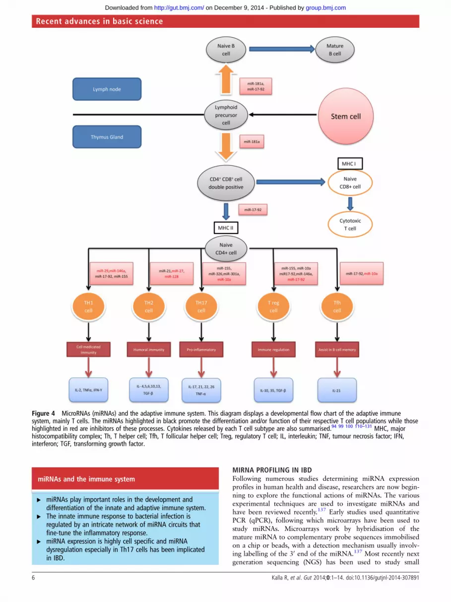
Adaptive immune systemWithin the immune system, an intricate network of signallingfacilitates maturation of the adaptive immune system. The appro-priate development and function of these immune cells (T and Bcells) is crucial when distinguishing foreign antigens from self.Recent studies have shown that miRNAs are involved in variousstages of Tcell and B cell maturation and activation (figure 4).
miRNAs and T cell regulationThe differentiation and maturation of T cells is influenced bymiRNAs (figure 4). Specific deletion of Dicer or Drosha in Tcell lineages results in aberrant differentiation and cytokine pro-duction with a marked bias towards Th1 development andIFN-γ production.132 133 During positive and negative selectionswithin the thymus gland, self-reactive T cells are first removed(negative selection) before T cells with functional receptors areselected (positive selection). The miR-181 family plays animportant role in this process by altering T cell receptor sensitiv-ity and may also contribute to diminished vaccine responsesseen in the elderly.110 111
miRNAs and Th1 and Th2 differentiationmiRNAs contribute to Th1 and Th2 cell differentiation. SeveralmiRNAs including miR-146a, miR-29, miR-155, miR-17-92cluster, miR-128 and miR-27b have been shown to influenceTh1 differentiation and function.112–115 Overexpression ofmiR-155 influences CD4+ T cells to differentiate into Th1 cells
while deficiency in miR-155 shows a bias towards Th2 differen-tiation.94 99 Similarly, miR-17-92 promotes Th1 differentiationby upregulating IFN-γ production and suppressing regulatoryT cell (Treg) differentiation.116 Of particular interest is the roleof miR-21 expression in T cells. miR-21 has been shown topromote Th2 cell differentiation and as described previously, itsdysregulation has been implicated in IBD.5 8 117
Several miRNAs have been shown to play a regulatory role bytargeting transcription factors known to be involved in Th1 cellgene expression.114 These include miR-29 targeting T-bet andeomesodermin, transcription factors known to regulate IFN-γproduction and miR-146a that targets signal transducer and acti-vator transcription 1 in Treg cells, a transcription factor thatcontrols Treg mediated regulation of Th1 responses.113 114
miRNAs and Th17 differentiationThe Th17 pathway has been widely researched in the context ofIBD.134 Recent studies determining the effect of miRNAs on thedifferentiation and function of Th17 pathway have identifieddirect and indirect regulatory mechanisms. Using murine modelswith experimental autoimmune encephalomyelitis, studies haveshown that miR-326, miR-10a, miR-155 directly regulate Th17differentiation and/or function while miR-301a is an indirectenhancer of Th17 differentiation.118–121 Of these miRNAs,miR-155 seems relevant to IBD as it directly upregulates Th17differentiation and indirectly influences the regulation ofpro-Th17 cytokines from DCs.121 122 Furthermore, miR-155 KOmice are protected from dextran sulfate sodium(DSS) inducedexperimental colitis compared with wild type mice.123 miRNAsmay also regulate hypoxia-induced Th17 differentiation by over-expressing miR-210 and promoting a negative feedback circuitwith Hif1a, a key transcription factor of Th17 polarisation.124
miRNAs and T regulatory cellsStudies have identified the role of miRNAs in Treg cell develop-ment and function by promoting the differentiation of CD4+ Tcells into Treg cells in the thymus and maintaining their immunehomoeostatic function.135 It has been shown in vivo that CD4+
T cells that fail to express miRNAs develop spontaneous auto-immunity.135 Furthermore, conditional Dicer or Drosha deletionin Foxp3+Treg cells can alter the expression of several Treg spe-cific markers including Foxp3, resulting in early fatal auto-immune disease.133 136 Several miRNAs including miR-155,miR146a, miR-10a and miR-17-92 have been shown to maintainTreg cell function by modulating different signalling path-ways.100 113 125 126 miR10a in selective Treg cells assists in main-taining high Foxp3 levels but does not influence the number orphenotype of Treg cells.125 miR-155 has been shown to regulatemature Treg cell homoeostasis via the IL-2 signalling pathwaywhile miR-146a regulates Treg cell function to limit inflamma-tion.100 113 The miR17-92 cluster has also been implicated inTreg cell function but these studies are conflicting. miR-17-92Treg cell KO mice develop an exacerbated experimental auto-immune encephalomyelitis,126 however Jiang et al showed thatcertain miRNAs within this cluster such as miR17 and miR-19binhibit Treg cell differentiation and promote Th1 responses.116
miRNAs and other immune cellsmiRNAs have been implicated in other immune cell maturationsuch as B cells and T follicular helper cells. The miR-17-92cluster helps regulate T follicular helper cell differentiation aswell as B cell maturation while other miRNAs such as miR-10aand miR-181a have also been shown to regulate these pro-cesses.119 127–131
Kalla R, et al. Gut 2014;0:1–14. doi:10.1136/gutjnl-2014-307891 5
Recent advances in basic science
group.bmj.com on December 9, 2014 - Published by http://gut.bmj.com/Downloaded from
miRNAs and the immune system
▸ miRNAs play important roles in the development anddifferentiation of the innate and adaptive immune system.
▸ The innate immune response to bacterial infection isregulated by an intricate network of miRNA circuits thatfine-tune the inflammatory response.
▸ miRNA expression is highly cell specific and miRNAdysregulation especially in Th17 cells has been implicatedin IBD.
MIRNA PROFILING IN IBDFollowing numerous studies determining miRNA expressionprofiles in human health and disease, researchers are now begin-ning to explore the functional actions of miRNAs. The variousexperimental techniques are used to investigate miRNAs andhave been reviewed recently.137 Early studies used quantitativePCR (qPCR), following which microarrays have been used tostudy miRNAs. Microarrays work by hybridisation of themature miRNA to complementary probe sequences immobilisedon a chip or beads, with a detection mechanism usually involv-ing labelling of the 30 end of the miRNA.137 Most recently nextgeneration sequencing (NGS) has been used to study small
Figure 4 MicroRNAs (miRNAs) and the adaptive immune system. This diagram displays a developmental flow chart of the adaptive immunesystem, mainly T cells. The miRNAs highlighted in black promote the differentiation and/or function of their respective T cell populations while thosehighlighted in red are inhibitors of these processes. Cytokines released by each T cell subtype are also summarised.94 99 100 110–131 MHC, majorhistocompatibility complex; Th, T helper cell; Tfh, T follicular helper cell; Treg, regulatory T cell; IL, interleukin; TNF, tumour necrosis factor; IFN,interferon; TGF, transforming growth factor.
6 Kalla R, et al. Gut 2014;0:1–14. doi:10.1136/gutjnl-2014-307891
Recent advances in basic science
group.bmj.com on December 9, 2014 - Published by http://gut.bmj.com/Downloaded from

RNAs. NGS is potentially advantageous over microarray techni-ques as it provides greater coverage, demonstrates sequenceindependence and has the potential to identify novel miRNAs.Confirmatory techniques to validate these findings use standardtechniques such as qPCR and northern blotting.
Results from early studies exploring the profile of miRNAsexpressed in tissues of patients with IBD have been somewhatconflicting and difficult to interpret. Many of the miRNArelated IBD studies have been underpowered and the need forlarge cohorts to perform well-powered studies has been demon-strated by the Cancer Genome Atlas consortia.138 There hasalso been a lack of uniformity of the comparator group, withcontrols consisting of healthy individuals in some studies and‘symptomatic control’ patients in others and furthermore manyof these studies used different methods to normalise miRNAdata. There has been difficulty identifying suitable housekeepinggenes as a reference for qPCR. Microarray and NGS studieshave used different techniques for normalisation; either
normalising against total miRNA or using approaches such asscaling and quantile normalisation.139–141
Other specific issues include difficulties deciphering whetherdifferentially expressed miRNAs are causal, a consequence ofdisease or related non-specifically to inflammation, and miRNAlevels may vary with disease duration and can be influenced bytherapy.142 143 Moreover, every cell type possesses its ownunique epigenetic signature therefore interpreting the relevanceof miRNAs detected in heterogeneous samples (eg, wholeblood, intestinal biopsies) is challenging and complicated by thefact that many miRNAs can target the same gene. Recent publi-cations have demonstrated a shift in focus from generatingexhaustive tissue and blood miRNA screens (see online supple-mentary table S1) to carefully designed functional experimentsthat elaborate actions of individual miRNAs in known patho-genetic pathways in IBD as implicated by GWAS. The most con-sistent evidence to date links miRNAs and autophagy in CD andin NOD2-induced Th17-mediated disease (table 1).
Table 1 Functional studies on micro RNAs (miRNAs)
Firstauthor Study model miRNA of interest
mRNA/pathwaytarget Findings
Nguyen144 AIEC infection in T84 cells and mouseenterocytes. Translational studies in ileal CDbiopsies
miR-30C andmiR-130A
ATG5ATG16L1
Adherent Escherichia coli upregulate miRNAs, reduce levels of ATG5 andATG16L1 and inhibit autophagy
Zhai145 Jurkat T cellsColonic epithelial cells
miR-142-3p ATG16L1 Reduced ATG16L1 mRNA and protein levels, regulating autophagy in CD.
Lu146 HCT116, SW480, HeLa and U2OS cell lines.Colonic biopsies from CD and healthycontrols
miR106B, miR93 ATG16L1 Reduced levels of ATG16L1 and autophagy
Brest10 HEK-293 cellsColonic biopsies
miR-196 IRGM A risk variant of IRGM alters the binding site for miR-196 and causesderegulation of IRGM-dependent xenophagy in CD
Brain9 Dendritic cell line, miR-29 KO murine models miR-29 IL-12p40 (directtarget)IL-12p19(indirect target)
NOD2 induces miR-29 release and limits IL- 23 releaseNOD2 polymorphism alters the expression of miR-29 and contributes topathogenesis in CD.
Xue147 IL-10 KO miceMyD88 KO miceRAG KO miceMurine dendrite cells
miR-10a IL-12/IL-23p40 miR-10a expression is regulated by the intestinal microbiota and targetsthe Th17 pathway. This miRNA may play a role in intestinal homoeostasis
Koukos148 HCT-116 colonocyte cells, murine modelsand colonic biopsies from patients with UC
miR-124 STAT3 Downregulation of miR-124 results in proinflammatory response in UC
Shi5
Yang149
miR-21 KO DSS model and wild type mousemodelsCaco-2 cells, colonic biopsies from UC andhealthy controls
mir-21 RhoB miR-21 is overexpressed in inflammation and tissue injury. miR-21 KOimproves survival in DSS colitis mouse modelTargeting RhoB impairs the tight junction integrity and decreasestransepithelial resistance and increases inulin permeability
Nata150 IL-10 deficient mice miR-146b siah2 miR-146b improves intestinal inflammation and epithelial barrier byactivating NFκB
Chuang92 HCT116 colonic epithelial cells miR-192, miR-495,miR-512, miR-671
NOD2 Downregulates NOD2 expression, suppresses NFκB, inhibits IL8 andCXCL3 expression
Chen151 Intestinal epithelial cellsIntestinal biopsies
miR-200b TGFβ miR-200b promotes the growth of intestinal epithelial cells by inhibitingepithelial-mesenchymal transition via TGFβ
Feng152 UC intestinal biopsiesHCT116 cellsHT29 cells
miR-126 IκBα Promotes NFκB mediated inflammation by targeting IκBα, a knowninhibitor of the NFκB pathway.
Nguyen153 Caco2-BBE cellsMouse epithelial cellsColonic CD tissues
miR-7 CD98expression
miR-7 regulates the expression of CD98. CD98 expression upregulatedand miR-7 decreased in actively inflamed CD tissues.
Singh123 miR-155 KO mice miR-155 Th17 pathway miR-155 KO mice models and demonstrated that these mice areprotected from experimental colitis compared with wild type mice.
Wu8 miR-21 KO TNBS and T cell transfer model ofcolitismiR-21 KO DSS colitis model
miR-21 Th1 pathway miR-21 KO results in reduced DSS induced colitis but exacerbatedinflammation in TNBS and T cell transfer model of colitis
AIEC, adherent invasive Escherichia coli; CD, Crohn’s disease; DSS, dextran sulfate sodium; IL, interleukin; IRGM, immunity related GTPase family M protein; KO, knockout; miR,microRNA; NFκB, nuclear factor-κB; NOD2, nuclear oligomerisation domain-containing protein 2; TGFβ, transforming growth factor β; TNBS, trinitrobenzene sulfonic acid.
Kalla R, et al. Gut 2014;0:1–14. doi:10.1136/gutjnl-2014-307891 7
Recent advances in basic science
group.bmj.com on December 9, 2014 - Published by http://gut.bmj.com/Downloaded from

miRNA profiling in IBD
▸ There is a need to perform large-scale multicentre miRNAprofiles in IBD with a well-defined ‘healthy control’population using NGS techniques.
▸ miRNA levels can vary with disease duration and therapies.▸ Every cell possesses its own epigenetic signature, therefore
understanding the relevance of miRNA profiles in wholeblood and intestinal biopsies can be challenging.
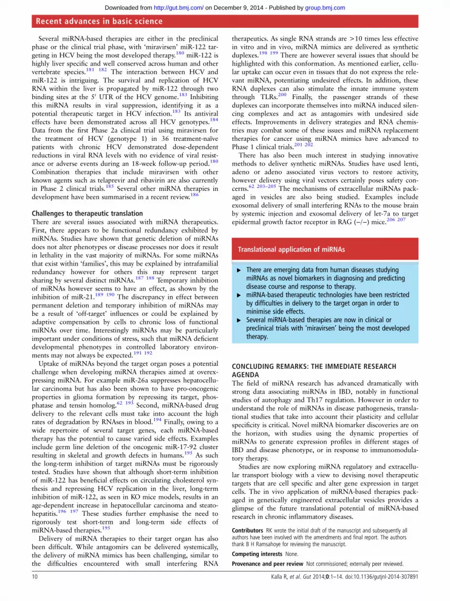
FUNCTIONAL STUDIES IN IBDmiRNAs and autophagy in CDAutophagy is a cellular process that involves self-digestion ofunwanted materials such as damaged mitochondrial products(mitophagy) and pathogenic microbes (xenophagy). A processsuch as xenophagy requires the coordinated action of a multi-tude of proteins including, vimentin, NOD2, immunity relatedGTPase family M protein (IRGM) and a multiprotein complexwhich includes ATG16L1 and ATG5–ATG12.154 155 In under-standing molecular signalling and its effect on autophagy,several groups have investigated the role of miRNAs in theseprocesses (figure 5).
During periods of starvation or hypoxia, mammalian target ofrapamycin is inhibited within cells, activating autophagy.Hypoxia-induced autophagy results in upregulation of miR-155that targets multiple components of mammalian target of rapa-mycin signalling.156 All genes currently described in the regula-tion of different stages of autophagy are influenced bymiRNAs.157 Several autophagy genes have also been associatedwith susceptibility to CD, notably IRGM and ATG16L1.158
Interestingly, autophagy regulates miRNA production by target-ing miRNA-processing enzymes Dicer and AGO2 through theautophagy receptor nuclear dot protein 52 kDa andgem-associated protein 4.159 Future challenges include under-standing the genetic control of miRNA biogenesis including itsown transcriptional activators and repressors.
Immunity related GTPase family M proteinA common exonic synonymous SNP (c.313C>T) in IRGM isassociated with CD.160 Although this SNP does not alter theIRGM protein sequence, it is located in the ‘seed’ region wheremRNA and miRNA form a RNA induced silencing complex.10
Further analysis revealed that this SNP altered the binding sitefor miR-196. miR-196 was also shown to be overexpressed ininflamed tissues of patients with CD suggesting that this defect-ive miRNA-mRNA interaction deregulates IRGM-dependentxenophagy in CD.10
ATG16L1GWAS identified ATG16L1 polymorphism (T300A) as a riskvariant in CD. Further studies revealed that this variant resultsin ineffective xenophagy of pathogens such as Salmonella typhi-murium.161 Several studies have identified miRNAs that targetATG16L1, although each study associates a different set ofmiRNAs which may relate to miRNA cell line specificity. InHeLa cell lines, adherent invasive Escherichia coli infectionresults in overexpression of miR-93 and miR106B and downre-gulation of ATG5 and ATG16L1 thereby disrupting the autop-hagy pathway and bacterial clearance.146 In adherent invasiveEscherichia coli infected T84 cells however, miR-30C and
miR-130A were upregulated.144 Both studies were able to repli-cate their findings in endoscopic biopsies from patients withCD. Finally, miR-142-3p has also been shown to targetATG16L1 and autophagy using a different cell line.145
Th17 pathwayTh17 driven inflammation plays an important role in IBD andstudies have shown how miRNAs are used by DCs to regulatethe inflammatory response. Brain et al9 demonstrated thatNOD2 mediates its effects through miRNAs in DCs, in particu-lar miR-29. The gene most strongly regulated by miR-29 isIL12B (encoding IL-12/23 p40) while IL23A (encoding IL-23p19) is indirectly targeted through suppression of its transcrip-tion factor ATF2 and mice deficient of this miRNA develop amore severe Th17 driven colitis on DSS exposure.9 Microbiotacan also impact on DC miRNA expression. In vivo models havedemonstrated the commensal bacteria can negatively regulatemiR-10a in DCs.147 Furthermore miR-10 directly targets IL-12/23p40 to limit Th17 driven inflammation and the expression ofthis miRNA may be regulated in order to maintain intestinalhomoeostasis.147
Other inflammatory pathwaysThe role of the NFκB pathway in IBD has been well describedand studies have shown that this pathway is also regulated bymiRNAs.162 miR-126 promotes NFκB mediated inflammation bydirectly targeting IκBα mRNA, an important inhibitor of NFκBsignalling pathway. These findings were replicated in colonicbiopsies in patients with active UC.152 Conversely, NFκB has alsobeen shown to play an anti-inflammatory role in IBD as demon-strated by the differential expression of miR-146b in IL-10 defi-cient mice models.150 Administering miR-146b vectorsintraperitoneally in DSS colitis mice ameliorated intestinalinflammation via activation of the NFκB mediated pathway.150
Other pathways that have been studied include signal trans-ducer and activator transcription 3 (STAT3) and acetylcho-line.148 163 164 Koukos et al148 demonstrated downregulation ofmiR-124 and upregulation of STAT3 in colonic biopsies ofpatients with active UC. These findings were translated in celllines and murine experimental models suggesting a role ofSTAT3 expression in promoting intestinal inflammation. Finally,vagal secretion of acetylcholine suppresses peripheral inflamma-tion by interrupting cytokine production and miR-132 has beenshown to target acetylcholine esterase thereby potentiating anti-inflammatory effects.164
Epithelial barrier integrityDysfunctional epithelial barrier has been implicated in thepathogenesis of UC.165 166 GWAS data demonstrated IBD asso-ciated genes that play a role in maintaining intestinal epithelialbarrier integrity and examples include LAMB-1 that regulatesbasement membrane stability and CDH-1 that regulates stabilityof adherens junctions via E-cadherin.166 Recent studies haveinvestigated miRNAs in epithelial barrier function, in particularmiR-21 and miR-200B.5 149 151 Murine miR-21 KO modelswith experimental DSS colitis survive longer and have less tissueinflammation than wild type mice and this miRNA targetsRhob, a target gene involved in regulating intestinal permeabil-ity.5 149 Similarly miR-200b has been shown to help maintainepithelial barrier integrity by targeting transforming growthfactor β1 and inhibiting epithelial-mesenchymal transition, aprocess that promotes loss of intestinal epithelial cells and con-tributes to the pathogenesis in IBD.151
8 Kalla R, et al. Gut 2014;0:1–14. doi:10.1136/gutjnl-2014-307891
Recent advances in basic science
group.bmj.com on December 9, 2014 - Published by http://gut.bmj.com/Downloaded from

miRNA studies in IBD
▸ miRNAs have been shown to regulate several pathwaysinvolving susceptibility loci found in IBD by GWAS.
▸ Recent data implicate miRNAs in the dysregulation ofautophagy and Th17 signalling in CD.
▸ Increased expression of miR-21 is the most consistentlyreplicated finding and represents a novel therapeutic target.
▸ miRNAs have also been shown to regulate intestinal barrierintegrity in UC.
TRANSLATION TO CLINICAL PRACTICE: LESSONS LEARNEDFROM OTHER DISEASESmiRNAs as disease biomarkersInsights from contemporary cancer research highlight the excit-ing potential of miRNAs as biomarkers. Research in this areawas stimulated by the initial finding that miRNA profiles canaccurately differentiate between different cancer lineages andsuccessfully classify poorly differentiated cancers based on tissueprofiling.167 In 2008, miRNAs were also discovered to bepresent in serum in a cell-free state, sparking excitement abouttheir potential use as non-invasive biomarkers.168–170
Extracellular miRNAs have now been found in most biologicalfluids including serum, urine, tears, saliva and breastmilk.171 172 Packaged in vesicles consisting of microparticles,lipoproteins or RNA binding proteins, these miRNAs are very
stable and protected from degradation.168 Their profiles havebeen studied in various diseases including cardiovascular dis-eases, cancer and neurological diseases.173–175
Despite the optimism that miRNAs may represent robust bio-markers, the results should be treated with some circumspection;recent reviews showed that up to 58% (n=47) of the reportedtumour related miRNAs are not disease specific.176 Only 33% ofthe reported miRNAs in non-neoplastic diseases (n=139) weredeemed biologically plausible and represented non-ubiquitousmiRNA expression in disease-appropriate cell types.177
The therapeutic application of miRNA modulationmiRNA related therapeutic intervention may involve eithermiRNA antagonists or miRNA mimics. Antagomirs, an exampleof miRNA antagonists, can be applied to allow gain of functionwithin diseased states by introducing a chemically modified RNAthat binds to the active miRNA of interest to inhibit its activityand rescue the repression of its target. Conversely miRNAmimics are used to restore a loss of function by the introductionof miRNAs into diseased cells to mimic a healthy cell state.178
Within the GI literature, there are several studieshighlighting the potential therapeutic application of specificmiRNAs including miR-155 and miR-210.123 124 Recently,miR-141 has been shown to play a critical role in colonic leuco-cyte trafficking by targeting CXCL12β. Treatment withpre-miR141protects mice against the development of trinitroben-zene sulfonic acid and IL-10KO colitis. In contrast, anti-miR141aggravates trinitrobenzene sulfonic acid-induced colitis throughCXCL12β suppression.179
Figure 5 MicroRNAs (miRNAs) and autophagy (adapted with permission from Ventham et al, Gastroenterology). This diagram summarises theinfluence of miRNAs within different components of autophagy. Altered sequence in the immunity related GTPase family M protein (IRGM) generesults in an impaired binding site for miR-196.10 The consequent reduction in miR-196 activity results in IRGM upregulation and causes ineffectivebacterial clearance of adherent invasive Escherichia coli (AIEC) in the intestinal cells of patients with Crohn’s disease. ATG16L1 has also been shownto be a target of a host of miRNAs. miR-106B and miR-93 repress ATG16L1 mRNA translation, thereby disrupting the autophagy pathway andbacterial clearance of AIEC.146 miR-30C and miR-130A have also been shown to directly target ATG16L1 and ATG5.144 Similarly, miR-142-3p hasalso been shown to negatively regulate ATG16L1 and autophagy.145 Finally, NOD2 has been shown to induce the expression of miR-29 to limit IL-23release, indirectly influencing the Th17 pathway in human dendritic cell lines.9 Polymorphisms in NOD2 impair the ability to express miR-29 resultingin exaggerated IL-23 induced inflammation. Recently, a set of miRNAs that directly target NOD2 expression, miR-192, miR-495, miR-512 and miR-671 have also been described albeit in a different cell line (colonic epithelial HCT116 cells).92
Kalla R, et al. Gut 2014;0:1–14. doi:10.1136/gutjnl-2014-307891 9
Recent advances in basic science
group.bmj.com on December 9, 2014 - Published by http://gut.bmj.com/Downloaded from
Several miRNA-based therapies are either in the preclinicalphase or the clinical trial phase, with ‘miravirsen’ miR-122 tar-geting in HCV being the most developed therapy.180 miR-122 ishighly liver specific and well conserved across human and othervertebrate species.181 182 The interaction between HCV andmiR-122 is intriguing. The survival and replication of HCVRNA within the liver is propagated by miR-122 through twobinding sites at the 50 UTR of the HCV genome.183 Inhibitingthis miRNA results in viral suppression, identifying it as apotential therapeutic target in HCV infection.183 Its antiviraleffects have been demonstrated across all HCV genotypes.184
Data from the first Phase 2a clinical trial using miravirsen forthe treatment of HCV (genotype 1) in 36 treatment-naïvepatients with chronic HCV demonstrated dose-dependentreductions in viral RNA levels with no evidence of viral resist-ance or adverse events during an 18-week follow-up period.180
Combination therapies that include miravirsen with otherknown agents such as telaprevir and ribavirin are also currentlyin Phase 2 clinical trials.185 Several other miRNA therapies indevelopment have been summarised in a recent review.186
Challenges to therapeutic translationThere are several issues associated with miRNA therapeutics.First, there appears to be functional redundancy exhibited bymiRNAs. Studies have shown that genetic deletion of miRNAsdoes not alter phenotypes or disease processes nor does it resultin lethality in the vast majority of miRNAs. For some miRNAsthat exist within ‘families’, this may be explained by intrafamilialredundancy however for others this may represent targetsharing by several distinct miRNAs.187 188 Temporary inhibitionof miRNAs however seems to have an effect, as shown by theinhibition of miR-21.189 190 The discrepancy in effect betweenpermanent deletion and temporary inhibition of miRNAs maybe a result of ‘off-target’ influences or could be explained byadaptive compensation by cells to chronic loss of functionalmiRNAs over time. Interestingly miRNAs may be particularlyimportant under conditions of stress, such that miRNA deficientdevelopmental phenotypes in controlled laboratory environ-ments may not always be expected.191 192
Uptake of miRNAs beyond the target organ poses a potentialchallenge when developing miRNA therapies aimed at overex-pressing miRNA. For example miR-26a suppresses hepatocellu-lar carcinoma but has also been shown to have pro-oncogenicproperties in glioma formation by repressing its target, phos-phatase and tensin homolog.62 193 Second, miRNA-based drugdelivery to the relevant cells must take into account the highrates of degradation by RNAses in blood.194 Finally, owing to awide repertoire of several target genes, each miRNA-basedtherapy has the potential to cause varied side effects. Examplesinclude germ line deletion of the oncogenic miR-17-92 clusterresulting in skeletal and growth defects in humans.195 As suchthe long-term inhibition of target miRNAs must be rigorouslytested. Studies have shown that although short-term inhibitionof miR-122 has beneficial effects on circulating cholesterol syn-thesis and repressing HCV replication in the liver, long-terminhibition of miR-122, as seen in KO mice models, results in anage-dependent increase in hepatocellular carcinoma and steato-hepatitis.196 197 These studies further emphasise the need torigorously test short-term and long-term side effects ofmiRNA-based therapies.195
Delivery of miRNA therapies to their target organ has alsobeen difficult. While antagomirs can be delivered systemically,the delivery of miRNA mimics has been challenging, similar tothe difficulties encountered with small interfering RNA
therapeutics. As single RNA strands are >10 times less effectivein vitro and in vivo, miRNA mimics are delivered as syntheticduplexes.198 199 There are however several issues that should behighlighted with this conformation. As mentioned earlier, cellu-lar uptake can occur even in tissues that do not express the rele-vant miRNA, potentiating undesired effects. In addition, theseRNA duplexes can also stimulate the innate immune systemthrough TLRs.200 Finally, the passenger strands of theseduplexes can incorporate themselves into miRNA induced silen-cing complexes and act as antagomirs with undesired sideeffects. Improvements in delivery strategies and RNA chemis-tries may combat some of these issues and miRNA replacementtherapies for cancer using miRNA mimics have advanced toPhase 1 clinical trials.201 202
There has also been much interest in studying innovativemethods to deliver synthetic miRNAs. Studies have used lenti,adeno or adeno associated virus vectors to restore activity,however delivery using viral vectors certainly poses safety con-cerns.62 203–205 The mechanisms of extracellular miRNAs pack-aged in vesicles are also being studied. Examples includeexosomal delivery of small interfering RNAs to the mouse brainby systemic injection and exosomal delivery of let-7a to targetepidermal growth factor receptor in RAG (−/−) mice.206 207
Translational application of miRNAs
▸ There are emerging data from human diseases studyingmiRNAs as novel biomarkers in diagnosing and predictingdisease course and response to therapy.
▸ miRNA-based therapeutic technologies have been restrictedby difficulties in delivery to the target organ in order tominimise side effects.
▸ Several miRNA-based therapies are now in clinical orpreclinical trials with ‘miravirsen’ being the most developedtherapy.
CONCLUDING REMARKS: THE IMMEDIATE RESEARCHAGENDAThe field of miRNA research has advanced dramatically withstrong data associating miRNAs in IBD, notably in functionalstudies of autophagy and Th17 regulation. However in order tounderstand the role of miRNAs in disease pathogenesis, transla-tional studies that take into account their plasticity and cellularspecificity is critical. Novel miRNA biomarker discoveries are onthe horizon, with studies using the dynamic properties ofmiRNAs to generate expression profiles in different stages ofIBD and disease phenotype, or in response to immunomodula-tory therapy.
Studies are now exploring miRNA regulatory and extracellu-lar transport biology with a view to devising novel therapeutictargets that are cell specific and alter gene expression in targetcells. The in vivo application of miRNA-based therapies pack-aged in genetically engineered extracellular vesicles provides aglimpse of the future translational potential of miRNA-basedresearch in chronic inflammatory diseases.
Contributors RK wrote the initial draft of the manuscript and subsequently allauthors have been involved with the amendments and final report. The authorsthank B H Ramsahoye for reviewing the manuscript.
Competing interests None.
Provenance and peer review Not commissioned; externally peer reviewed.
10 Kalla R, et al. Gut 2014;0:1–14. doi:10.1136/gutjnl-2014-307891
Recent advances in basic science
group.bmj.com on December 9, 2014 - Published by http://gut.bmj.com/Downloaded from

Open Access This is an Open Access article distributed in accordance with theCreative Commons Attribution Non Commercial (CC BY-NC 4.0) license, whichpermits others to distribute, remix, adapt, build upon this work non-commercially,and license their derivative works on different terms, provided the original work isproperly cited and the use is non-commercial. See: http://creativecommons.org/licenses/by-nc/4.0/
REFERENCES1 Burisch J, Jess T, Martinato M, et al. The burden of inflammatory bowel disease in
Europe. J Crohns Colitis 2013;7:322–37.2 Jostins L, Ripke S, Weersma RK, et al. Host-microbe interactions have shaped
the genetic architecture of inflammatory bowel disease. Nature2012;491:119–24.
3 Esteller M. Non-coding RNAs in human disease. Nat Rev Genet 2011;12:861–74.4 Xu W-D, Pan H-F, Li J-H, et al. MicroRNA-21 with therapeutic potential in
autoimmune diseases. Expert Opin Ther Targets 2013;17:659–65.5 Shi C, Liang Y, Yang J, et al. MicroRNA-21 knockout improve the survival rate in
DSS induced fatal colitis through protecting against inflammation and tissue injury.PLoS ONE 2013;8:e66814.
6 Wang Y, Gao X, Wei F, et al. Diagnostic and prognostic value of circulatingmiR-21 for cancer: a systematic review and meta-analysis. Gene2014;533:389–97.
7 Adams AT, Kennedy NA, Hansen R, et al. Two-stage genome-wide methylationprofiling in childhood-onset Crohn’s disease implicates epigenetic alterations at theVMP1/MIR21 and HLA loci. Inflamm Bowel Dis 2014;20:1784–93.
8 Wu F, Dong F, Arendovich N, et al. Divergent influence of microRNA-21 deletionon murine colitis phenotypes. Inflamm Bowel Dis 2014;20:1972–85.
9 Brain O, Owens BMJ, Pichulik T, et al. The intracellular sensor NOD2 inducesmicroRNA-29 expression in human dendritic cells to limit IL-23 release. Immunity2013;39:521–36.
10 Brest P, Lapaquette P, Souidi M, et al. A synonymous variant in IRGM alters abinding site for miR-196 and causes deregulation of IRGM-dependent xenophagyin Crohn’s disease. Nat Genet 2011;43:242–5.
11 Ambros V. A hierarchy of regulatory genes controls a larva-to-adult developmentalswitch in C. elegans. Cell 1989;57:49–57.
12 Chalfie M, Horvitz HR, Sulston JE. Mutations that lead to reiterations in the celllineages of C. elegans. Cell 1981;24:59–69.
13 Lee RC, Feinbaum RL, Ambros V. The C. elegans heterochronic gene lin-4 encodessmall RNAs with antisense complementarity to lin-14. Cell 1993;75:843–54.
14 Ruvkun G, Giusto J. The Caenorhabditis elegans heterochronic gene lin-14encodes a nuclear protein that forms a temporal developmental switch. Nature1989;338:313–19.
15 Reinhart BJ, Slack FJ, Basson M, et al. The 21-nucleotide let-7 RNA regulatesdevelopmental timing in Caenorhabditis elegans. Nature 2000;403:901–6.
16 Pasquinelli AE, Reinhart BJ, Slack F, et al. Conservation of the sequence andtemporal expression of let-7 heterochronic regulatory RNA. Nature2000;408:86–9.
17 Griffiths-Jones S, Grocock RJ, van Dongen S, et al. miRBase: microRNA sequences,targets and gene nomenclature. Nucleic Acids Res 2006;34:D140–4.
18 Libri V, Miesen P, van Rij RP, et al. Regulation of microRNA biogenesis andturnover by animals and their viruses. Cell Mol Life Sci 2013;70:3525–44.
19 Ozsolak F, Poling LL, Wang Z, et al. Chromatin structure analyses identify miRNApromoters. Genes Dev 2008;22:3172–83.
20 Saini HK, Griffiths-Jones S, Enright AJ. Genomic analysis of human microRNAtranscripts. Proc Natl Acad Sci USA 2007;104:17719–24.
21 Lee Y, Kim M, Han J, et al. MicroRNA genes are transcribed by RNA polymeraseII. EMBO J 2004;23:4051–60.
22 Borchert GM, Lanier W, Davidson BL. RNA polymerase III transcribes humanmicroRNAs. Nat Struct Mol Biol 2006;13:1097–101.
23 Heo I, Ha M, Lim J, et al. Mono-uridylation of pre-microRNA as a key step in thebiogenesis of group II let-7 microRNAs. Cell 2012;151:521–32.
24 Han J, Lee Y, Yeom K-H, et al. Molecular basis for the recognition of primarymicroRNAs by the Drosha-DGCR8 complex. Cell 2006;125:887–901.
25 Han J, Lee Y, Yeom K-H, et al. The Drosha-DGCR8 complex in primary microRNAprocessing. Genes Dev 2004;18:3016–27.
26 Zeng Y, Cullen BR. Structural requirements for pre-microRNA binding and nuclearexport by Exportin 5. Nucleic Acids Res 2004;32:4776–85.
27 Yi R, Qin Y, Macara IG, et al. Exportin-5 mediates the nuclear export ofpre-microRNAs and short hairpin RNAs. Genes Dev 2003;17:3011–16.
28 Lund E, Güttinger S, Calado A, et al. Nuclear export of microRNA precursors.Science 2004;303:95–8.
29 Bernstein E, Caudy AA, Hammond SM, et al. Role for a bidentate ribonuclease inthe initiation step of RNA interference. Nature 2001;409:363–6.
30 Gregory RI, Chendrimada TP, Cooch N, et al. Human RISC couples microRNAbiogenesis and posttranscriptional gene silencing. Cell 2005;123:631–40.
31 Bommer GT, Gerin I, Feng Y, et al. p53-mediated activation of miRNA34candidate tumor-suppressor genes. Curr Biol 2007;17:1298–307.
32 Chang T-C, Wentzel EA, Kent OA, et al. Transactivation of miR-34a by p53broadly influences gene expression and promotes apoptosis. Mol Cell2007;26:745–52.
33 He L, He X, Lim LP, et al. A microRNA component of the p53 tumour suppressornetwork. Nature 2007;447:1130–4.
34 Eulalio A, Huntzinger E, Izaurralde E. Getting to the root of miRNA-mediated genesilencing. Cell 2008;132:9–14.
35 Zisoulis DG, Kai ZS, Chang RK, et al. Autoregulation of microRNA biogenesis bylet-7 and Argonaute. Nature 2012;486:541–4.
36 Katoh T, Sakaguchi Y, Miyauchi K, et al. Selective stabilization of mammalianmicroRNAs by 30 adenylation mediated by the cytoplasmic poly(A) polymeraseGLD-2. Genes Dev 2009;23:433–8.
37 Breving K, Esquela-Kerscher A. The complexities of microRNA regulation:mirandering around the rules. Int J Biochem Cell Biol 2010;42:1316–29.
38 Kawahara Y, Zinshteyn B, Sethupathy P, et al. Redirection of silencing targets byadenosine-to-inosine editing of miRNAs. Science 2007;315:1137–40.
39 Blow MJ, Grocock RJ, van Dongen S, et al. RNA editing of human microRNAs.Genome Biol 2006;7:R27.
40 Yang W, Chendrimada TP, Wang Q, et al. Modulation of microRNA processingand expression through RNA editing by ADAR deaminases. Nat Struct Mol Biol2006;13:13–21.
41 Yeom K-H, Lee Y, Han J, et al. Characterization of DGCR8/Pasha, the essentialcofactor for Drosha in primary miRNA processing. Nucleic Acids Res2006;34:4622–9.
42 Han J, Pedersen JS, Kwon SC, et al. Posttranscriptional crossregulation betweenDrosha and DGCR8. Cell 2009;136:75–84.
43 Winter J, Jung S, Keller S, et al. Many roads to maturity: microRNA biogenesispathways and their regulation. Nat Cell Biol 2009;11:228–34.
44 Michlewski G, Guil S, Semple CA, et al. Posttranscriptional regulation of miRNAsharboring conserved terminal loops. Mol Cell 2008;32:383–93.
45 Guil S, Cáceres JF. The multifunctional RNA-binding protein hnRNP A1 is requiredfor processing of miR-18a. Nat Struct Mol Biol 2007;14:591–6.
46 Michlewski G, Cáceres JF. Antagonistic role of hnRNP A1 and KSRP in theregulation of let-7a biogenesis. Nat Struct Mol Biol 2010;17:1011–18.
47 Paroo Z, Ye X, Chen S, et al. Phosphorylation of the human microRNA-generatingcomplex mediates MAPK/Erk signaling. Cell 2009;139:112–22.
48 Russell AP, Lamon S, Boon H, et al. Regulation of miRNAs in human skeletalmuscle following acute endurance exercise and short-term endurance training.J Physiol 2013;591:4637–53.
49 Wan G, Mathur R, Hu X, et al. miRNA response to DNA damage. Trends BiochemSci 2011;36:478–84.
50 Wan G, Zhang X, Langley RR, et al. DNA-damage-induced nuclear export ofprecursor microRNAs is regulated by the ATM-AKT pathway. Cell Rep2013;3:2100–12.
51 Bhattacharyya SN, Habermacher R, Martine U, et al. Stress-induced reversal ofmicroRNA repression and mRNA P-body localization in human cells. Cold SpringHarb Symp Quant Biol 2006;71:513–21.
52 Kedde M, Agami R. Interplay between microRNAs and RNA-binding proteinsdetermines developmental processes. Cell Cycle 2008;7:899–903. http://www.ncbi.nlm.nih.gov/pubmed/18414021
53 Saunders MA, Liang H, Li W-H. Human polymorphism at microRNAs andmicroRNA target sites. Proc Natl Acad Sci USA 2007;104:3300–5.
54 Tay Y, Zhang J, Thomson AM, et al. MicroRNAs to Nanog, Oct4 and Sox2 codingregions modulate embryonic stem cell differentiation. Nature 2008;455:1124–8.
55 Shin C, Nam J-W, Farh KK-H, et al. Expanding the microRNA targeting code:functional sites with centered pairing. Mol Cell 2010;38:789–802.
56 Helwak A, Kudla G, Dudnakova T, et al. Mapping the human miRNA interactomeby CLASH reveals frequent noncanonical binding. Cell 2013;153:654–65.
57 Vasudevan S, Tong Y, Steitz JA. Switching from repression to activation:microRNAs can up-regulate translation. Science 2007;318:1931–4.
58 Kim DH, Saetrom P, Snøve O, et al. MicroRNA-directed transcriptional genesilencing in mammalian cells. Proc Natl Acad Sci USA 2008;105:16230–5.
59 Friedman RC, Farh KK-H, Burge CB, et al. Most mammalian mRNAs are conservedtargets of microRNAs. Genome Res 2009;19:92–105.
60 Fabian MR, Sonenberg N. The mechanics of miRNA-mediated gene silencing:a look under the hood of miRISC. Nat Struct Mol Biol 2012;19:586–93.
61 Arvey A, Larsson E, Sander C, et al. Target mRNA abundance dilutes microRNAand siRNA activity. Mol Syst Biol 2010;6:363.
62 Kota J, Chivukula RR, O’Donnell KA, et al. Therapeutic microRNA deliverysuppresses tumorigenesis in a murine liver cancer model. Cell 2009;137:1005–17.
63 Ambros V. The functions of animal microRNAs. Nature 2004;431:350–5.64 Peterson SM, Thompson JA, Ufkin ML, et al. Common features of microRNA target
prediction tools. Front Genet 2014;5:23.65 Kim VN, Han J, Siomi MC. Biogenesis of small RNAs in animals. Nat Rev Mol Cell
Biol 2009;10:126–39.66 Tsang J, Zhu J, van Oudenaarden A. MicroRNA-mediated feedback and
feedforward loops are recurrent network motifs in mammals. Mol Cell2007;26:753–67.
Kalla R, et al. Gut 2014;0:1–14. doi:10.1136/gutjnl-2014-307891 11
Recent advances in basic science
group.bmj.com on December 9, 2014 - Published by http://gut.bmj.com/Downloaded from

67 Milo R, Shen-Orr S, Itzkovitz S, et al. Network motifs: simple building blocks ofcomplex networks. Science 2002;298:824–7.
68 O’Donnell KA, Wentzel EA, Zeller KI, et al. c-Myc-regulated microRNAs modulateE2F1 expression. Nature 2005;435:839–43.
69 Shen-Orr SS, Milo R, Mangan S, et al. Network motifs in the transcriptionalregulation network of Escherichia coli. Nat Genet 2002;31:64–8.
70 Iliopoulos D, Hirsch HA, Struhl K. An epigenetic switch involving NF-kappaB,Lin28, Let-7 MicroRNA, and IL6 links inflammation to cell transformation. Cell2009;139:693–706.
71 Hatziapostolou M, Polytarchou C, Aggelidou E, et al. An HNF4α-miRNA inflammatoryfeedback circuit regulates hepatocellular oncogenesis. Cell 2011;147:1233–47.
72 Herranz H, Cohen SM. MicroRNAs and gene regulatory networks: managing theimpact of noise in biological systems. Genes Dev 2010;24:1339–44.
73 Mannaerts I, Eysackers N, Onyema OO, et al. Class II HDAC inhibition hampershepatic stellate cell activation by induction of microRNA-29. PLoS ONE 2013;8:e55786.
74 Miao C, Yang Y, He X, et al. New advances of microRNAs in the pathogenesis ofrheumatoid arthritis, with a focus on the crosstalk between DNA methylation andthe microRNA machinery. Cell Signal 2013;25:1118–25.
75 Suzuki H, Takatsuka S, Akashi H, et al. Genome-wide profiling of chromatinsignatures reveals epigenetic regulation of MicroRNA genes in colorectal cancer.Cancer Res 2011;71:5646–58.
76 Hansen TB, Jensen TI, Clausen BH, et al. Natural RNA circles function as efficientmicroRNA sponges. Nature 2013;495:384–8.
77 Ventham NT, Kennedy NA, Nimmo ER, et al. Beyond gene discovery ininflammatory bowel disease: the emerging role of epigenetics. Gastroenterology2013;145:293–308.
78 Dai E, Yu X, Zhang Y, et al. EpimiR: a database of curated mutual regulationbetween miRNAs and epigenetic modifications. Database (Oxford) 2014;2014:bau023.
79 Wang LQ, Liang R, Chim CS. Methylation of tumor suppressor microRNAs: lessonsfrom lymphoid malignancies. Expert Rev Mol Diagn 2012;12:755–65.
80 Zhang Y, Wang X, Xu B, et al. Epigenetic silencing of miR-126 contributes totumor invasion and angiogenesis in colorectal cancer. Oncol Rep2013;30:1976–84.
81 Suzuki R, Yamamoto E, Nojima M, et al. Aberrant methylation of microRNA-34b/cis a predictive marker of metachronous gastric cancer risk. J Gastroenterol2013;49:1135–44.
82 Han L, Witmer PD, Casey E, et al. DNA methylation regulates MicroRNAexpression. Cancer Biol Ther 2007;6:1284–8.
83 Pan C, Chen H, Wang L, et al. Down-regulation of MiR-127 facilitates hepatocyteproliferation during rat liver regeneration. PLoS ONE 2012;7:e39151.
84 Escobar TM, Kanellopoulou C, Kugler DG, et al. miR-155 activates cytokine geneexpression in Th17 cells by regulating the DNA-binding protein Jarid2 to relievepolycomb-mediated repression. Immunity 2014;40:865–79.
85 Salzman J, Gawad C, Wang PL, et al. Circular RNAs are the predominanttranscript isoform from hundreds of human genes in diverse cell types. PLoS ONE2012;7:e30733.
86 Chen C-Z, Schaffert S, Fragoso R, et al. Regulation of immune responses andtolerance: the microRNA perspective. Immunol Rev 2013;253:112–28.
87 Xiao C, Rajewsky K. MicroRNA control in the immune system: basic principles. Cell2009;136:26–36.
88 Geremia A, Biancheri P, Allan P, et al. Innate and adaptive immunity ininflammatory bowel disease. Autoimmun Rev 2014;13:3–10.
89 Cuthbert AP, Fisher SA, Mirza MM, et al. The contribution of NOD2 genemutations to the risk and site of disease in inflammatory bowel disease.Gastroenterology 2002;122:867–74.
90 Chen Y, Wang C, Liu Y, et al. miR-122 targets NOD2 to decrease intestinal epithelialcell injury in Crohn’s disease. Biochem Biophys Res Commun 2013;438:133–9.
91 Ghorpade DS, Sinha AY, Holla S, et al. NOD2-nitric oxide-responsivemicroRNA-146a activates Sonic hedgehog signaling to orchestrate inflammatoryresponses in murine model of inflammatory bowel disease. J Biol Chem2013;288:33037–48.
92 Chuang AY, Chuang JC, Zhai Z, et al. NOD2 expression is regulated by microRNAsin colonic epithelial HCT116 cells. Inflamm Bowel Dis 2014;20:126–35.
93 He X, Jing Z, Cheng G. MicroRNAs: new regulators of Toll-like receptor signallingpathways. Biomed Res Int 2014;2014:945169.
94 Thai T-H, Calado DP, Casola S, et al. Regulation of the germinal center responseby microRNA-155. Science 2007;316:604–8.
95 Boldin MP, Taganov KD, Rao DS, et al. miR-146a is a significant brake onautoimmunity, myeloproliferation, and cancer in mice. J Exp Med2011;208:1189–201.
96 Taganov KD, Boldin MP, Chang K-J, et al. NF-kappaB-dependent induction ofmicroRNA miR-146, an inhibitor targeted to signaling proteins of innate immuneresponses. Proc Natl Acad Sci USA 2006;103:12481–6.
97 Ceppi M, Pereira PM, Dunand-Sauthier I, et al. MicroRNA-155 modulates theinterleukin-1 signaling pathway in activated human monocyte-derived dendriticcells. Proc Natl Acad Sci USA 2009;106:2735–40.
98 O’Connell RM, Taganov KD, Boldin MP, et al. MicroRNA-155 is induced during themacrophage inflammatory response. Proc Natl Acad Sci USA 2007;104:1604–9.
99 Rodriguez A, Vigorito E, Clare S, et al. Requirement of bic/microRNA-155 fornormal immune function. Science 2007;316:608–11.
100 Lu L-F, Thai T-H, Calado DP, et al. Foxp3-dependent microRNA155 conferscompetitive fitness to regulatory T cells by targeting SOCS1 protein. Immunity2009;30:80–91.
101 Xu C, Ren G, Cao G, et al. miR-155 regulates immune modulatory properties ofmesenchymal stem cells by targeting TAK1-binding protein 2. J Biol Chem2013;288:11074–9.
102 Schulte LN, Westermann AJ, Vogel J. Differential activation and functionalspecialization of miR-146 and miR-155 in innate immune sensing. Nucleic AcidsRes 2013;41:542–53.
103 Mazumder A, Bose M, Chakraborty A, et al. A transient reversal ofmiRNA-mediated repression controls macrophage activation. EMBO Rep2013;14:1008–16.
104 Androulidaki A, Iliopoulos D, Arranz A, et al. The kinase Akt1 controlsmacrophage response to lipopolysaccharide by regulating microRNAs. Immunity2009;31:220–31.
105 Chen X-M, Splinter PL, O’Hara SP, et al. A cellular micro-RNA, let-7i, regulatesToll-like receptor 4 expression and contributes to cholangiocyte immune responsesagainst Cryptosporidium parvum infection. J Biol Chem 2007;282:28929–38.
106 Zhou X, Li X, Ye Y, et al. MicroRNA-302b augments host defense to bacteria byregulating inflammatory responses via feedback to TLR/IRAK4 circuits. NatCommun 2014;5:3619.
107 Ma C, Li Y, Li M, et al. microRNA-124 negatively regulates TLR signaling inalveolar macrophages in response to mycobacterial infection. Mol Immunol2014;62:150–8.
108 Qi J, Qiao Y, Wang P, et al. microRNA-210 negatively regulates LPS-inducedproduction of proinflammatory cytokines by targeting NF-κB1 in murinemacrophages. FEBS Lett 2012;586:1201–7.
109 Bazzoni F, Rossato M, Fabbri M, et al. Induction and regulatory function of miR-9in human monocytes and neutrophils exposed to proinflammatory signals. ProcNatl Acad Sci USA 2009;106:5282–7.
110 Li Q-J, Chau J, Ebert PJR, et al. miR-181a is an intrinsic modulator of T cellsensitivity and selection. Cell 2007;129:147–61.
111 Li G, Yu M, Lee W-W, et al. Decline in miR-181a expression with age impairs Tcell receptor sensitivity by increasing DUSP6 activity. Nat Med 2012;18:1518–24.
112 Guerau-de-Arellano M, Smith KM, Godlewski J, et al. Micro-RNA dysregulation inmultiple sclerosis favours pro-inflammatory T-cell-mediated autoimmunity. Brain2011;134:3578–89.
113 Lu L-F, Boldin MP, Chaudhry A, et al. Function of miR-146a in controlling Tregcell-mediated regulation of Th1 responses. Cell 2010;142:914–29.
114 Steiner DF, Thomas MF, Hu JK, et al. MicroRNA-29 regulates T-box transcriptionfactors and interferon-γ production in helper T cells. Immunity 2011;35:169–81.
115 Banerjee A, Schambach F, DeJong CS, et al. Micro-RNA-155 inhibits IFN-gammasignaling in CD4+ T cells. Eur J Immunol 2010;40:225–31.
116 Jiang S, Li C, Olive V, et al. Molecular dissection of the miR-17-92 cluster’s criticaldual roles in promoting Th1 responses and preventing inducible Tregdifferentiation. Blood 2011;118:5487–97.
117 Sawant DV, Wu H, Kaplan MH, et al. The Bcl6 target gene microRNA-21promotes Th2 differentiation by a T cell intrinsic pathway. Mol Immunol2013;54:435–42.
118 Du C, Liu C, Kang J, et al. MicroRNA miR-326 regulates TH-17 differentiation andis associated with the pathogenesis of multiple sclerosis. Nat Immunol2009;10:1252–9.
119 Takahashi H, Kanno T, Nakayamada S, et al. TGF-β and retinoic acid induce themicroRNA miR-10a, which targets Bcl-6 and constrains the plasticity of helper Tcells. Nat Immunol 2012;13:587–95.
120 Mycko MP, Cichalewska M, Machlanska A, et al. MicroRNA-301a regulation of aT-helper 17 immune response controls autoimmune demyelination. Proc Natl AcadSci USA 2012;109:E1248–57.
121 O’Connell RM, Kahn D, Gibson WSJ, et al. MicroRNA-155 promotes autoimmuneinflammation by enhancing inflammatory T cell development. Immunity2010;33:607–19.
122 Murugaiyan G, Beynon V, Mittal A, et al. Silencing microRNA-155 amelioratesexperimental autoimmune encephalomyelitis. J Immunol 2011;187:2213–21.
123 Singh UP, Murphy AE, Enos RT, et al. miR-155 deficiency protects mice fromexperimental colitis by reducing Th1/Th17 responses. Immunology2014;143:478–89.
124 Wang H, Flach H, Onizawa M, et al. Negative regulation of Hif1a expression andTH17 differentiation by the hypoxia-regulated microRNA miR-210. Nat Immunol2014;15:393–401.
125 Jeker LT, Zhou X, Gershberg K, et al. MicroRNA 10a marks regulatory T cells. PLoSONE 2012;7:e36684.
126 De Kouchkovsky D, Esensten JH, Rosenthal WL, et al. microRNA-17-92 regulatesIL-10 production by regulatory T cells and control of experimental autoimmuneencephalomyelitis. J Immunol 2013;191:1594–605.
12 Kalla R, et al. Gut 2014;0:1–14. doi:10.1136/gutjnl-2014-307891
Recent advances in basic science
group.bmj.com on December 9, 2014 - Published by http://gut.bmj.com/Downloaded from

127 Koralov SB, Muljo SA, Galler GR, et al. Dicer ablation affects antibody diversityand cell survival in the B lymphocyte lineage. Cell 2008;132:860–74.
128 Fazilleau N, Mark L, McHeyzer-Williams LJ, et al. Follicular helper T cells: lineageand location. Immunity 2009;30:324–35.
129 Kang SG, Liu W-H, Lu P, et al. MicroRNAs of the miR-17∼92 family are criticalregulators of T(FH) differentiation. Nat Immunol 2013;14:849–57.
130 Chen C-Z, Li L, Lodish HF, et al. MicroRNAs modulate hematopoietic lineagedifferentiation. Science 2004;303:83–6.
131 Li X, Zhang J, Gao L, et al. MiR-181 mediates cell differentiation by interruptingthe Lin28 and let-7 feedback circuit. Cell Death Differ 2012;19:378–86.
132 Muljo SA, Ansel KM, Kanellopoulou C, et al. Aberrant T cell differentiation in theabsence of Dicer. J Exp Med 2005;202:261–9.
133 Chong MMW, Rasmussen JP, Rudensky AY, et al. The RNAseIII enzyme Drosha iscritical in T cells for preventing lethal inflammatory disease. J Exp Med2008;205:2005–17.
134 Troncone E, Marafini I, Pallone F, et al. Th17 cytokines in inflammatorybowel diseases: discerning the good from the bad. Int Rev Immunol2013;32:526–33.
135 Cobb BS, Hertweck A, Smith J, et al. A role for Dicer in immune regulation. J ExpMed 2006;203:2519–27.
136 Zhou X, Jeker LT, Fife BT, et al. Selective miRNA disruption in T reg cells leads touncontrolled autoimmunity. J Exp Med 2008;205:1983–91.
137 Van Rooij E. The art of microRNA research. Circ Res 2011;108:219–34.138 Cancer Genome Atlas Network. Comprehensive molecular characterization of
human colon and rectal cancer. Nature 2012;487:330–7.139 Peltier HJ, Latham GJ. Normalization of microRNA expression levels in quantitative
RT-PCR assays: identification of suitable reference RNA targets in normal andcancerous human solid tissues. RNA 2008;14:844–52.
140 Rahmann S, Martin M, Schulte JH, et al. Identifying transcriptional miRNAbiomarkers by integrating high-throughput sequencing and real-time PCR data.Methods 2013;59:154–63.
141 Garmire LX, Subramaniam S. Evaluation of normalization methods in mammalianmicroRNA-Seq data. RNA 2012;18:1279–88.
142 Drong AW, Lindgren CM, McCarthy MI. The genetic and epigenetic basis of type 2diabetes and obesity. Clin Pharmacol Ther 2012;92:707–15.
143 Rakyan VK, Down TA, Balding DJ, et al. Epigenome-wide association studies forcommon human diseases. Nat Rev Genet 2011;12:529–41.
144 Thu Nguyen HT, Dalmasso G, Müller S, et al. Crohn’s disease-associated adherentinvasive Escherichia coli affect levels of microRNAs in intestinal epithelial cells toreduce autophagy. Gastroenterology 2013;146:508–19.
145 Zhai Z, Wu F, Dong F, et al. Human autophagy gene ATG16L1 ispost-transcriptionally regulated by MIR142-3p. Autophagy 2014;10:468–79.
146 Lu C, Chen J, Xu H-G, et al. MIR106B and MIR93 prevent removal of bacteriafrom epithelial cells by disrupting ATG16L1-mediated autophagy. Gastroenterology2014;146:188–99.
147 Xue X, Feng T, Yao S, et al. Microbiota downregulates dendritic cell expression ofmiR-10a, which targets IL-12/IL-23p40. J Immunol 2011;187:5879–86.
148 Koukos G, Polytarchou C, Kaplan JL, et al. MicroRNA-124 regulates STAT3expression and is down-regulated in colon tissues of pediatric patients withulcerative colitis. Gastroenterology 2013;145:842–52.e2.
149 Yang Y, Ma Y, Shi C, et al. Overexpression of miR-21 in patients with ulcerativecolitis impairs intestinal epithelial barrier function through targeting the RhoGTPase RhoB. Biochem Biophys Res Commun 2013;434:746–52.
150 Nata T, Fujiya M, Ueno N, et al. MicroRNA-146b improves intestinal injury inmouse colitis by activating nuclear factor-κB and improving epithelial barrierfunction. J Gene Med 2013;15:249–60.
151 Chen Y, Xiao Y, Ge W, et al. miR-200b inhibits TGF-β1-inducedepithelial-mesenchymal transition and promotes growth of intestinal epithelialcells. Cell Death Dis 2013;4:e541.
152 Feng X, Wang H, Ye S, et al. Up-regulation of microRNA-126 may contribute topathogenesis of ulcerative colitis via regulating NF-kappaB inhibitor IκBα. PLoSONE 2012;7:e52782.
153 Nguyen HTT, Dalmasso G, Yan Y, et al. MicroRNA-7 modulates CD98 expressionduring intestinal epithelial cell differentiation. J Biol Chem 2010;285:1479–89.
154 Mizushima N, Yoshimori T, Ohsumi Y. Role of the Apg12 conjugation system inmammalian autophagy. Int J Biochem Cell Biol 2003;35:553–61.
155 Henderson P, Wilson DC, Satsangi J, et al. A role for vimentin in Crohn disease.Autophagy 2012;8:1695–6.
156 Wan G, Xie W, Liu Z, et al. Hypoxia-induced MIR155 is a potent autophagyinducer by targeting multiple players in the MTOR pathway. Autophagy2014;10:70–9.
157 Füllgrabe J, Klionsky DJ, Joseph B. The return of the nucleus: transcriptional andepigenetic control of autophagy. Nat Rev Mol Cell Biol 2014;15:65–74.
158 Franke A, McGovern DPB, Barrett JC, et al. Genome-wide meta-analysis increasesto 71 the number of confirmed Crohn’s disease susceptibility loci. Nat Genet2010;42:1118–25.
159 Gibbings D, Mostowy S, Jay F, et al. Selective autophagy degrades DICER andAGO2 and regulates miRNA activity. Nat Cell Biol 2012;14:1314–21.
160 Parkes M, Barrett JC, Prescott NJ, et al. Sequence variants in the autophagy geneIRGM and multiple other replicating loci contribute to Crohn’s diseasesusceptibility. Nat Genet 2007;39:830–2.
161 Rioux JD, Xavier RJ, Taylor KD, et al. Genome-wide association study identifiesnew susceptibility loci for Crohn disease and implicates autophagy in diseasepathogenesis. Nat Genet 2007;39:596–604.
162 Boone DL, Lee EG, Libby S, et al. Recent advances in understanding NF-kappaBregulation. Inflamm Bowel Dis 2002;8:201–12.
163 Maharshak N, Shenhar-Tsarfaty S, Aroyo N, et al. MicroRNA-132 modulatescholinergic signaling and inflammation in human inflammatory bowel disease.Inflamm Bowel Dis 2013;19:1346–53.
164 Shaked I, Meerson A, Wolf Y, et al. MicroRNA-132 potentiates cholinergicanti-inflammatory signaling by targeting acetylcholinesterase. Immunity2009;31:965–73.
165 Pastorelli L, De Salvo C, Mercado JR, et al. Central role of the gut epithelial barrierin the pathogenesis of chronic intestinal inflammation: lessons learned from animalmodels and human genetics. Front Immunol 2013;4:280.
166 Anderson CA, Boucher G, Lees CW, et al. Meta-analysis identifies 29 additionalulcerative colitis risk loci, increasing the number of confirmed associations to 47.Nat Genet 2011;43:246–52.
167 Lu J, Getz G, Miska EA, et al. MicroRNA expression profiles classify humancancers. Nature 2005;435:834–8.
168 Chen X, Ba Y, Ma L, et al. Characterization of microRNAs in serum: a novel class ofbiomarkers for diagnosis of cancer and other diseases. Cell Res 2008;18:997–1006.
169 Lawrie CH, Gal S, Dunlop HM, et al. Detection of elevated levels oftumour-associated microRNAs in serum of patients with diffuse large B-celllymphoma. Br J Haematol 2008;141:672–5.
170 Mitchell PS, Parkin RK, Kroh EM, et al. Circulating microRNAs as stable blood-basedmarkers for cancer detection. Proc Natl Acad Sci USA 2008;105:10513–18.
171 Weber JA, Baxter DH, Zhang S, et al. The microRNA spectrum in 12 body fluids.Clin Chem 2010;56:1733–41.
172 Chim SSC, Shing TKF, Hung ECW, et al. Detection and characterization ofplacental microRNAs in maternal plasma. Clin Chem 2008;54:482–90.
173 Zheng H, Liu J-Y, Song F-J, et al. Advances in circulating microRNAs as diagnosticand prognostic markers for ovarian cancer. Cancer Biol Med 2013;10:123–30.
174 Rao P, Benito E, Fischer A. MicroRNAs as biomarkers for CNS disease. Front MolNeurosci 2013;6:39.
175 Kinet V, Halkein J, Dirkx E, et al. Cardiovascular extracellular microRNAs: emergingdiagnostic markers and mechanisms of cell-to-cell RNA communication. FrontGenet 2013;4:214.
176 Pritchard CC, Kroh E, Wood B, et al. Blood cell origin of circulating microRNAs:a cautionary note for cancer biomarker studies. Cancer Prev Res (Phila)2012;5:492–7.
177 Haider BA, Baras AS, McCall MN, et al. A critical evaluation of microRNAbiomarkers in non-neoplastic disease. PLoS ONE 2014;9:e89565.
178 Bader AG, Brown D, Winkler M. The promise of microRNA replacement therapy.Cancer Res 2010;70:7027–30.
179 Huang Z, Shi T, Zhou Q, et al. miR-141 Regulates colonic leukocytic trafficking bytargeting CXCL12β during murine colitis and human Crohn’s disease. Gut2014;63:1247–57.
180 Janssen HLA, Reesink HW, Lawitz EJ, et al. Treatment of HCV infection bytargeting microRNA. N Engl J Med 2013;368:1685–94.
181 Lagos-Quintana M, Rauhut R, Yalcin A, et al. Identification of tissue-specificmicroRNAs from mouse. Curr Biol 2002;12:735–9.
182 Wienholds E, Kloosterman WP, Miska E, et al. MicroRNA expression in zebrafishembryonic development. Science 2005;309:310–11.
183 Jopling CL, Yi M, Lancaster AM, et al. Modulation of hepatitis C virus RNAabundance by a liver-specific MicroRNA. Science 2005;309:1577–81.
184 Li Y-P, Gottwein JM, Scheel TK, et al. MicroRNA-122 antagonism against hepatitisC virus genotypes 1–6 and reduced efficacy by host RNA insertion or mutations inthe HCV 50 UTR. Proc Natl Acad Sci USA 2011;108:4991–6.
185 Ling H, Fabbri M, Calin GA. MicroRNAs and other non-coding RNAs as targets foranticancer drug development. Nat Rev Drug Discov 2013;12:847–65.
186 Van Rooij E, Kauppinen S. Development of microRNA therapeutics is coming ofage. EMBO Mol Med 2014;6:851–64.
187 Ventura A, Young AG, Winslow MM, et al. Targeted deletion reveals essential andoverlapping functions of the miR-17 through 92 family of miRNA clusters. Cell2008;132:875–86.
188 Miska EA, Alvarez-Saavedra E, Abbott AL, et al. Most Caenorhabditis elegansmicroRNAs are individually not essential for development or viability. PLoS Genet2007;3:e215.
189 Thum T, Gross C, Fiedler J, et al. MicroRNA-21 contributes to myocardial diseaseby stimulating MAP kinase signalling in fibroblasts. Nature 2008;456:980–4.
190 Thum T, Chau N, Bhat B, et al. Comparison of different miR-21 inhibitorchemistries in a cardiac disease model. J Clin Invest 2011;121:461–2; author reply462–3.
191 Mendell JT, Olson EN. MicroRNAs in stress signaling and human disease. Cell2012;148:1172–87.
Kalla R, et al. Gut 2014;0:1–14. doi:10.1136/gutjnl-2014-307891 13
Recent advances in basic science
group.bmj.com on December 9, 2014 - Published by http://gut.bmj.com/Downloaded from

192 Leung AKL, Sharp PA. MicroRNA functions in stress responses. Mol Cell2010;40:205–15.
193 Huse JT, Brennan C, Hambardzumyan D, et al. The PTEN-regulating microRNAmiR-26a is amplified in high-grade glioma and facilitates gliomagenesis in vivo.Genes Dev 2009;23:1327–37.
194 Pecot CV, Calin GA, Coleman RL, et al. RNA interference in the clinic: challengesand future directions. Nat Rev Cancer 2011;11:59–67.
195 De Pontual L, Yao E, Callier P, et al. Germline deletion of the miR-17∼92cluster causes skeletal and growth defects in humans. Nat Genet2011;43:1026–30.
196 Hsu S-H, Wang B, Kota J, et al. Essential metabolic, anti-inflammatory, andanti-tumorigenic functions of miR-122 in liver. J Clin Invest 2012;122:2871–83.
197 Tsai W-C, Hsu S-D, Hsu C-S, et al. MicroRNA-122 plays a critical role in liverhomeostasis and hepatocarcinogenesis. J Clin Invest 2012;122:2884–97.
198 Schwarz DS, Hutvágner G, Du T, et al. Asymmetry in the assembly of the RNAienzyme complex. Cell 2003;115:199–208.
199 Schwarz DS, Hutvágner G, Haley B, et al. Evidence that siRNAs function as guides,not primers, in the Drosophila and human RNAi pathways. Mol Cell2002;10:537–48.
200 Peacock H, Fucini RV, Jayalath P, et al. Nucleobase and ribose modificationscontrol immunostimulation by a microRNA-122-mimetic RNA. J Am Chem Soc2011;133:9200–3.
201 Trang P, Wiggins JF, Daige CL, et al. Systemic delivery of tumor suppressormicroRNA mimics using a neutral lipid emulsion inhibits lung tumors in mice.Mol Ther 2011;19:1116–22.
202 Bouchie A. First microRNA mimic enters clinic. Nat Biotechnol 2013;31:577.203 Miyazaki Y, Adachi H, Katsuno M, et al. Viral delivery of miR-196a ameliorates the
SBMA phenotype via the silencing of CELF2. Nat Med 2012;18:1136–41.204 Deng X, Cao M, Zhang J, et al. Hyaluronic acid-chitosan nanoparticles for
co-delivery of MiR-34a and doxorubicin in therapy against triple negative breastcancer. Biomaterials 2014;35:4333–44.
205 Hatakeyama H, Murata M, Sato Y, et al. The systemic administration of ananti-miRNA oligonucleotide encapsulated pH-sensitive liposome results in reducedlevel of hepatic microRNA-122 in mice. J Control Release 2014;173:43–50.
206 Alvarez-Erviti L, Seow Y, Yin H, et al. Delivery of siRNA to the mouse brain bysystemic injection of targeted exosomes. Nat Biotechnol 2011;29:341–5.
207 Ohno S, Takanashi M, Sudo K, et al. Systemically injected exosomes targeted toEGFR deliver antitumor microRNA to breast cancer cells. Mol Ther 2013;21:185–91.
14 Kalla R, et al. Gut 2014;0:1–14. doi:10.1136/gutjnl-2014-307891
Recent advances in basic science
group.bmj.com on December 9, 2014 - Published by http://gut.bmj.com/Downloaded from

MicroRNAs: new players in IBD
Buck and J SatsangiR Kalla, N T Ventham, N A Kennedy, J F Quintana, E R Nimmo, A H
published online December 4, 2014Gut
http://gut.bmj.com/content/early/2014/12/04/gutjnl-2014-307891Updated information and services can be found at:
MaterialSupplementary
htmlhttp://gut.bmj.com/content/suppl/2014/12/04/gutjnl-2014-307891.DC1.Supplementary material can be found at:
These include:
References#BIBLhttp://gut.bmj.com/content/early/2014/12/04/gutjnl-2014-307891
This article cites 207 articles, 60 of which you can access for free at:
Open Access
http://creativecommons.org/licenses/by-nc/4.0/non-commercial. See: provided the original work is properly cited and the use isnon-commercially, and license their derivative works on different terms, permits others to distribute, remix, adapt, build upon this workCommons Attribution Non Commercial (CC BY-NC 4.0) license, which This is an Open Access article distributed in accordance with the Creative
serviceEmail alerting
box at the top right corner of the online article. Receive free email alerts when new articles cite this article. Sign up in the
CollectionsTopic Articles on similar topics can be found in the following collections
(170)Open access (72)GUT Recent advances in basic science
Notes
http://group.bmj.com/group/rights-licensing/permissionsTo request permissions go to:
http://journals.bmj.com/cgi/reprintformTo order reprints go to:
http://group.bmj.com/subscribe/To subscribe to BMJ go to:
group.bmj.com on December 9, 2014 - Published by http://gut.bmj.com/Downloaded from