Embed Size (px)
Citation preview
Mitochondrial Quality Control and Parkinson’s Disease:A Pathway Unfolds
Inês Pimenta de Castro & L. Miguel Martins &
Samantha Hui Yong Loh
Received: 7 September 2010 /Accepted: 4 November 2010# The Author(s) 2010. This article is published with open access at Springerlink.com
Abstract Recent findings from genetic studies suggest thatdefective mitochondrial quality control may play an impor-tant role in the development of Parkinson's disease (PD).Such defects may result in the impairment of neuronalmitochondria, which leads to both synaptic dysfunction andcell death and results in neurodegeneration. Here, we reviewstate-of-the-art knowledge of how pathways affecting mito-chondrial quality control might contribute to PD, with aparticular emphasis on the molecular mechanisms employedby PTEN-induced putative kinase 1 (PINK1), HtrA2 andParkin to regulate mitochondrial quality control.
Keywords Mitochondria . PINK1 . HtrA2 . Parkin .
Parkinson's disease . Unfolded proteins
Introduction
Parkinson's disease (PD) is a neurodegenerative disorder ofthe central nervous system that belongs to a group ofconditions referred to as movement disorders. Efforts tounderstand the genetic basis of this disease have led to theidentification of a number of gene mutations accounting fora small proportion of PD cases. Although the role ofmitochondria in PD has been debated inconclusively in thepast few decades, it was the identification of PD-causative
mutations in the mitochondrial putative protein kinasePINK1 that helped reignite the interest in the potential roleof this organelle in the pathophysiology of familial PD. It isnow becoming clear that PINKI acts in a mitochondrialquality control pathway and that other genes implicated inthis disease are under its control. This suggests that a subsetof genes involved in PD converge in a common pathwaythat modulates the fidelity of mitochondria. Given that mostPD cases are sporadic with no known specific cause, thisraises the interesting hypothesis that impaired mitochondri-al quality control might be a defining causative feature ofthis disease. The purpose of this review is to summarisesome of the recent molecular determinants of mitochondrialquality control regulated by PINK1.
Mitochondria: A Force for Life and Death
Midichlorians are microorganisms in the fictional Star Warsgalaxy. These microscopic life forms reside within cells ofall living things and communicate using the Force. TheForce is an energy field created by all living things andharvested by the Jedi knights to protect the galaxy. TheForce, however, has a ‘Dark Side’, and it can therefore beemployed to cause death and destruction. Midichlorians notonly help to communicate the Force, but they are alsosymbionts with all living things. Without them, life couldnot exist. The creator of Star Wars, George Lucas, statedthat the midichlorians are based on the endosymbiotictheory, which explores the origin of mitochondria andplastids present in eukaryotic cells. In fact, midichloriansare loosely based on mitochondria, an organelle thatsustains life in eukaryotic cells of multicellular organisms;however, these organelles are also known to bring aboutdeath and destruction within cells.
I. P. de Castro : L. M. Martins (*) : S. H. Y. LohCell Death Regulation Laboratory, MRC Toxicology Unit,Hodgkin Building, Lancaster Road,Leicester LE1 9HN, UKe-mail: [email protected]
I. P. de CastroIPATIMUP and Faculty of Pharmacy, University of Porto,Porto, Portugal
Mol NeurobiolDOI 10.1007/s12035-010-8150-4
It is widely accepted that the acquisition of mitochondriais a crucial event in the evolution of the eukaryotic cell [1].According to the endosymbiotic theory, mitochondriaevolved from a metabolically driven symbiotic relationshipbetween aerobic bacteria and a nucleus-containing cell. Asdescendants of the original symbiont, mitochondria areorganelles highly efficient in their ability to utilisemolecular oxygen (O2) and organic substrates, such asglucose and pyruvate, and to produce cellular energy in theform of ATP. The molecular machinery present in mito-chondria for energy production is collectively referred to asthe electron transport chain (ETC). The ETC consists offour protein complexes. Three of the complexes (I, III andIV) pump protons (H+) outwardly across the innermembrane to establish a gradient necessary for theproduction of ATP by a molecular motor (complex V orATP synthase). During electron transport, O2 is convertedto H2O, and, particularly at complexes I and II, the freeradical superoxide O2
!" # is also generated as a by-product.Two important cofactors that modulate energy and freeradical production are coenzyme Q10 at complex II andcytochrome c at complex IV [2]. It is noteworthy thatexcessive production of superoxide can enhance oxidativestress in dopaminergic neurons, eventually contributing tothe demise of these cells in PD [3].
Mitochondria play a key role in eukaryotic cells bysupplying the majority of the cellular energy in the form ofATP. However, a cell death-related role for this organelle isalso now clearly established. When eukaryotic cells inmulticellular organisms become worn out or damaged, theyundergo cellular suicide (i.e. apoptosis). During thisprocess, the cell blebs, shrinks and is packaged forrecycling. When this process lacks proper control, eitherdecreased or increased cell loss can occur, leading todiseases such as cancer or neurodegeneration. Apoptosis isactively controlled by mitochondria. When the mitochon-drial pathway of apoptosis is activated, these organellesrelease several proteins that activate several proteases andother hydrolytic enzymes, leading to proteolysis, DNAfragmentation and chromatin condensation. The mainmitochondrial effectors of apoptosis are proteins thatparadoxically play important survival roles in non-apoptotic cells. These effectors include cytochrome c,which, in normal settings, is a component of the ETC,and the serine protease HtrA2 (also known as Omi) whichseems to play a protective role during stress in themitochondria of healthy cells [4]. The mitochondrialpathway of apoptosis is regulated by the BCL-2 familyproteins (reviewed in Youle and Strasser [5]) and involvesthe activation of BCL-2 family members, such as BAX andBAK, which act by forming pores in the outer mitochon-drial membrane (OMM) and allow the release of severalpro-apoptotic proteins from the intermembrane space into
the cytosol. The role of enhanced apoptosis as a causativemechanism for the death of dopaminergic neurons in PDpatients is still controversial [6]. However, given thathealthy mitochondria contain a multitude of molecules that,when released into the cytosol, lead to apoptotic cell death,it is reasonable to assume that the compromise ofmitochondrial function in dopaminergic neurons contributesto their demise through the engagement of this form of cellsuicide.
Totally Addicted to Mitochondria
In humans, most of the energy supplying the brain isgenerated by mitochondria. The majority of this energy isused for reversing the movement of ions that bring aboutaction potentials and synaptic transmission in neurons.Neurons have very low glycolytic rates (reviewed inBolanos and Almeida [7]) and therefore require mitochon-dria for energy production. Mitochondria also sequester andbuffer cytoplasmic calcium; thus, their localisation plays arole in local regulation of intracellular calcium dynamics.Neurons are large, specialised cells, and the biogenesis ofmitochondria in neurons takes place in the soma. Thismeans that mitochondria normally have to travel largedistances to deliver energy. Impressively, these organelleswill travel up to 1 m in human motor neurons and, takinginto account their maximum travelling speed, will take aminimum of 8 days to reach their destination. The majorityof long-distance mitochondrial transport is mediated bymotor proteins powered by ATP hydrolysis that shuttlemitochondria along microtubules (reviewed in MacAskilland Kittler [8]). Such mitochondrial transport is bidirec-tional, as damaged mitochondria may need to travel back tothe cell soma for autophagic degradation. This degradationis mediated through autophagy, a molecular recyclingprogramme for impaired and defective cellular components.
In neurons, the synaptic terminals are sites of highenergy demand. Therefore, these structures rely heavily onmitochondria for energy production. Synaptic transmissionrequires high levels of cellular ATP for numerous energy-consuming processes, including the maintenance of synap-tic membrane potential and reloading of synaptic vesicleswith neurotransmitters. In addition, synaptic terminals mayrequire mitochondria for efficient sequestration of calciumions (Ca2+) that are released into the cytosol to elicit thefusion of synaptic vesicles with the plasma membrane(reviewed in Ly and Verstreken [9]). Adult dopaminergicneurons of the substantia nigra pars compacta (SNc) thatdegenerate in PD are known to generate action potentials inthe absence of any synaptic input. The mechanism behindthis autonomous firing capacity involves ion channels thatenable the entry of Ca2+ into the cytosol (reviewed by
Mol Neurobiol
Chan, Gertler and Surmeier [10]). This Ca2+ is activelyremoved by active pumping across the plasma membrane orsequestration in the lumen of the endoplasmic reticulum(ER) or the mitochondrial matrix, a process that requiresenergy in the form of ATP. Also, active Ca2+ buffering bymitochondria is likely to have a significant effect in thephysiology of this organelle. It is therefore attractive topropose that the premature death of dopaminergic neuronsin the SNc might be precipitated by a failure to controlcalcium homeostasis linked to a compromise of mitochon-drial function.
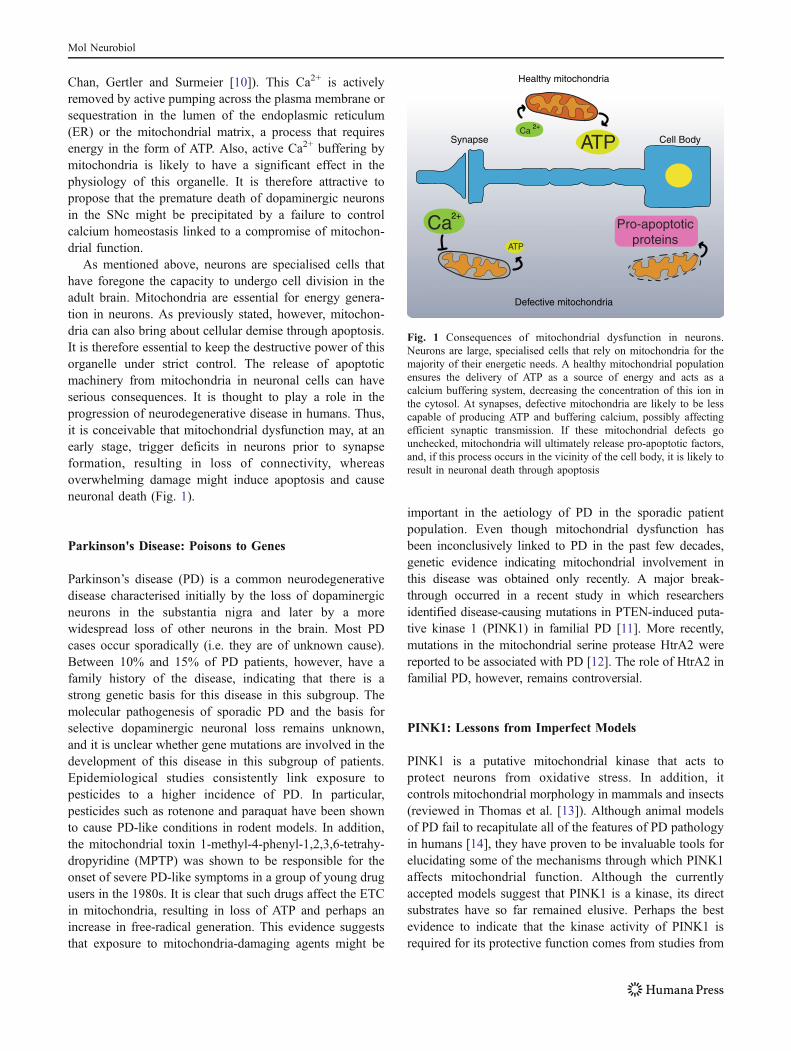
As mentioned above, neurons are specialised cells thathave foregone the capacity to undergo cell division in theadult brain. Mitochondria are essential for energy genera-tion in neurons. As previously stated, however, mitochon-dria can also bring about cellular demise through apoptosis.It is therefore essential to keep the destructive power of thisorganelle under strict control. The release of apoptoticmachinery from mitochondria in neuronal cells can haveserious consequences. It is thought to play a role in theprogression of neurodegenerative disease in humans. Thus,it is conceivable that mitochondrial dysfunction may, at anearly stage, trigger deficits in neurons prior to synapseformation, resulting in loss of connectivity, whereasoverwhelming damage might induce apoptosis and causeneuronal death (Fig. 1).
Parkinson's Disease: Poisons to Genes
Parkinson’s disease (PD) is a common neurodegenerativedisease characterised initially by the loss of dopaminergicneurons in the substantia nigra and later by a morewidespread loss of other neurons in the brain. Most PDcases occur sporadically (i.e. they are of unknown cause).Between 10% and 15% of PD patients, however, have afamily history of the disease, indicating that there is astrong genetic basis for this disease in this subgroup. Themolecular pathogenesis of sporadic PD and the basis forselective dopaminergic neuronal loss remains unknown,and it is unclear whether gene mutations are involved in thedevelopment of this disease in this subgroup of patients.Epidemiological studies consistently link exposure topesticides to a higher incidence of PD. In particular,pesticides such as rotenone and paraquat have been shownto cause PD-like conditions in rodent models. In addition,the mitochondrial toxin 1-methyl-4-phenyl-1,2,3,6-tetrahy-dropyridine (MPTP) was shown to be responsible for theonset of severe PD-like symptoms in a group of young drugusers in the 1980s. It is clear that such drugs affect the ETCin mitochondria, resulting in loss of ATP and perhaps anincrease in free-radical generation. This evidence suggeststhat exposure to mitochondria-damaging agents might be
important in the aetiology of PD in the sporadic patientpopulation. Even though mitochondrial dysfunction hasbeen inconclusively linked to PD in the past few decades,genetic evidence indicating mitochondrial involvement inthis disease was obtained only recently. A major break-through occurred in a recent study in which researchersidentified disease-causing mutations in PTEN-induced puta-tive kinase 1 (PINK1) in familial PD [11]. More recently,mutations in the mitochondrial serine protease HtrA2 werereported to be associated with PD [12]. The role of HtrA2 infamilial PD, however, remains controversial.
PINK1: Lessons from Imperfect Models
PINK1 is a putative mitochondrial kinase that acts toprotect neurons from oxidative stress. In addition, itcontrols mitochondrial morphology in mammals and insects(reviewed in Thomas et al. [13]). Although animal modelsof PD fail to recapitulate all of the features of PD pathologyin humans [14], they have proven to be invaluable tools forelucidating some of the mechanisms through which PINK1affects mitochondrial function. Although the currentlyaccepted models suggest that PINK1 is a kinase, its directsubstrates have so far remained elusive. Perhaps the bestevidence to indicate that the kinase activity of PINK1 isrequired for its protective function comes from studies from
ATP
Pro-apoptoticproteins
Healthy mitochondria
Defective mitochondria
ATP
Ca
Ca
2+
2+
Cell BodySynapse
Fig. 1 Consequences of mitochondrial dysfunction in neurons.Neurons are large, specialised cells that rely on mitochondria for themajority of their energetic needs. A healthy mitochondrial populationensures the delivery of ATP as a source of energy and acts as acalcium buffering system, decreasing the concentration of this ion inthe cytosol. At synapses, defective mitochondria are likely to be lesscapable of producing ATP and buffering calcium, possibly affectingefficient synaptic transmission. If these mitochondrial defects gounchecked, mitochondria will ultimately release pro-apoptotic factors,and, if this process occurs in the vicinity of the cell body, it is likely toresult in neuronal death through apoptosis
Mol Neurobiol
Drosophila melanogaster. In these studies, it was shownthat a kinase-inactive version of Pink1 failed to rescue thephenotypical features of pink1 loss-of-function flies [15].The fruit fly has proven to be an important tool in thedissection of the molecular determinants of pink1 function.These studies, however, have presented conflicting dataregarding the importance of this gene for the survival ofdopaminergic neurons (see [16, 17]).
Recently, it was reported that a loss of PINK1 in miceleads to a significant decrease in dopamine release and asubsequent impairment of striatal synaptic plasticity, evenin the absence of dopaminergic neuronal degeneration [18].In Drosophila neurons, Pink1 deficiency also seems toaffect synaptic function. Loss of Pink1 affects mitochon-drial function and leads to a decrease in the ATP levels thatprevents synaptic vesicles from being mobilised duringrapid stimulation [19]. In addition, loss of PINK1 in micemay also affect synaptic activity due to defects inmitochondrial calcium handling. Abramov and colleaguesproposed that PINK1 regulates calcium efflux frommitochondria via the mitochondrial Na+/Ca2+ exchanger.Deregulation of mitochondrial calcium handling can resultin mitochondrial calcium overload that affects synapticfunction. Such an overload leads to increased levels ofreactive oxygen species (ROS), which can then lead tomitochondria-dependent apoptosis [20]. Altogether, despitethe limitations of PD models in non-primate animal species,both mice and flies have provided us with valuable cluesregarding the mechanisms through which PINK1 regulatesmitochondrial function.
Neuronal Mitochondria, the Good, the Badand the Ugly
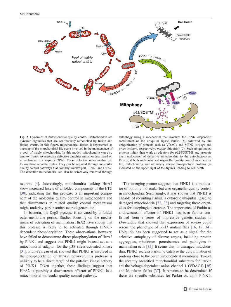
Neurons require mitochondria to function, and in the past fewyears, numerous findings have suggested that disruptions ofmitochondrial function and dynamics contribute to ageing andneurodegenerative diseases (recently reviewed in de Castro etal. [21]). The emerging view is that eukaryotic cells havedeveloped exquisite mechanisms to cope with the diversechallenges imposed on mitochondrial integrity (Fig. 2).Mitochondria are therefore thought to have at least twolevels of defence mechanisms that ensure their integrity andviability in individual cells (reviewed in Tatsuta and Langer[22]). The first line of defence is comprised of highlyspecific molecular quality control machinery composed ofmolecular chaperones and energy-dependent proteases thatmonitor the folding and assembly of mitochondrial proteins.In addition, these molecules prevent the accumulation ofdamaged proteins from mitochondria. This mitochondrialstress signalling results in the selective transcriptional up-regulation of nuclear genes encoding for mitochondrial
Hsp70 and Hsp60 in cells experiencing elevated mitochon-drial protein-conformational stress, therefore suggesting theexistence of an unfolded protein response of mitochondrialorigin (UPRmt). One of the major upstream components ofthis signalling pathway in the worm Caenorhabditis eleganshas been recently found to be the orthologue of Escherichiacoli ClpP, a molecule that functions in the bacterial heat-shock response (reviewed in Broadley and Hartl [23]). Theseinteresting observations suggest that mitochondria, asdescendants of the original endosymbiont, have co-optedcertain molecules of bacterial origin to stress signallingfunctions.
Once molecular quality control is overwhelmed withinmitochondria, a second line of defence, termed organellarquality control, is thought to take over. This mechanismrelies on the dynamic nature of mitochondrial populationsto ensure the disposal of defective components. Mitochon-drial dynamics are thought to be important in the control ofmitochondrial turnover and bioenergetic efficiency. Thecombined functions of fusion, fission and autophagy arenow emerging as essential organellar quality controlmechanisms that promote the sequestration, sorting andelimination of functionally impaired mitochondria [24].Finally, if both of these lines of defence fail, severemitochondrial damage unleashes the apoptosis pathwaythat culminates with cell death.
Is it possible that the demise of dopaminergic neurons inPD results from a failure of either one of the two describedquality control mechanisms in mitochondria? A positiveanswer to this question is starting to emerge from recentdata derived from genetic studies in model organisms.
xThe first link between PD and mitochondrial molecularquality control was suggested by the finding of heterozy-gous missense mutations in HtrA2 in sporadic cases of PD[12]. The serine protease HtrA2 was initially identified as amammalian homologue of the E. coli proteases HtrA/DegPand DegS [25]. The structural and biochemical similarity ofmammalian HtrA2 to the bacterial DegS protease isparticularly striking [26–28]. In Gram-negative bacteria,the DegS protease is involved in a signal transductionpathway that senses and responds to envelope stress(reviewed in Ruiz and Silhavy [29]). In mammalian cells,HtrA2 normally resides in the mitochondrial intermembranespace. Given the similarities between DegS and HtrA2 andtaking into account that bacteria are the most likelyancestors of mitochondria, it is possible that HtrA2 evolvedfrom DegS and now controls the levels of unfolded proteinsin mitochondria.
The characterisation of mice lacking HtrA2 showed thatloss of this protease results in a neurodegenerativephenotype with parkinsonian features. This phenomenon,however, is not due to the loss of dopaminergic neurons; itis instead caused by the loss of a subpopulation of striatal
Mol Neurobiol
neurons [4]. Interestingly, mitochondria lacking HtrA2show increased levels of unfolded components of the ETC[30], indicating that this protease is an important compo-nent of the molecular quality control in mitochondria andthat disturbances in related quality control mechanismsmight underlay parkinsonian neurodegeneration.
In bacteria, the DegS protease is activated by unfoldedouter-membrane porins. Studies focusing on the mecha-nisms of activation of mammalian HtrA2 have shown thatthis protease is likely to be activated through PINK1-dependent phosphorylation. These observations, however,have failed to demonstrate direct phosphorylation of HtrA2by PINK1 and suggest that PINK1 might instead act as amitochondrial adaptor for the p38 stress-activated kinase[31]. Plun-Favreau et al. showed that PINK1 is involved inthe phosphorylation of HtrA2; however, this protease isunlikely to be a direct target of the putative kinase activityof PINK1. Taken together, these findings suggest thatHtrA2 is possibly a downstream effector of PINK1 in amitochondrial molecular quality control pathway.
The emerging picture suggests that PINK1 is a modula-tor of not only molecular but also organellar quality controlin mitochondria. Surprisingly, it was shown that PINK1 iscapable of recruiting Parkin, a cytosolic ubiquitin ligase, todamaged mitochondria [32, 33] and targeting these organ-elles for autophagic clearance. The importance of Parkin asa downstream effector of PINK1 has been further con-firmed from a series of impressive genetic studies inDrosophila that showed that expression of parkin couldrescue the phenotype of pink1 mutant flies [16, 17, 34].Ubiquitin has been suggested to act as a signal for theselective autophagy of diverse cargos, including proteinaggregates, ribosomes, peroxisomes and pathogens inmammalian cells [35]. It seems that, in damaged mitochon-dria, PINK1 recruits Parkin to catalyse the ubiquitination ofproteins close to the outer mitochondrial membrane. Two ofthe recently identified mitochondrial substrates for Parkinare the voltage-dependent anion channel 1 (VDAC1) [36]and Mitofusin (Mfn) [37]. It remains to be determined ifthese are specific substrates for Parkin or, upon PINK1-
Molecular QualityCon
trol
HtrA2/Omi
PIN
K1
p38
Organellar Quality Control
Cel
ular
r Qua
lityControl
PINK1 ParkinPool of viablemitochondria
Smac/Diablo
HtrA2/Omi
CytC
Fusion
MFN1/MFN2
OPA1
FIS1
DRP1 Cell Death
Fission
p62/SQSTM1
LC3
Mitophagy
VDAC1
PIN
K1
Parkin
Parkin
1
2
Fusion
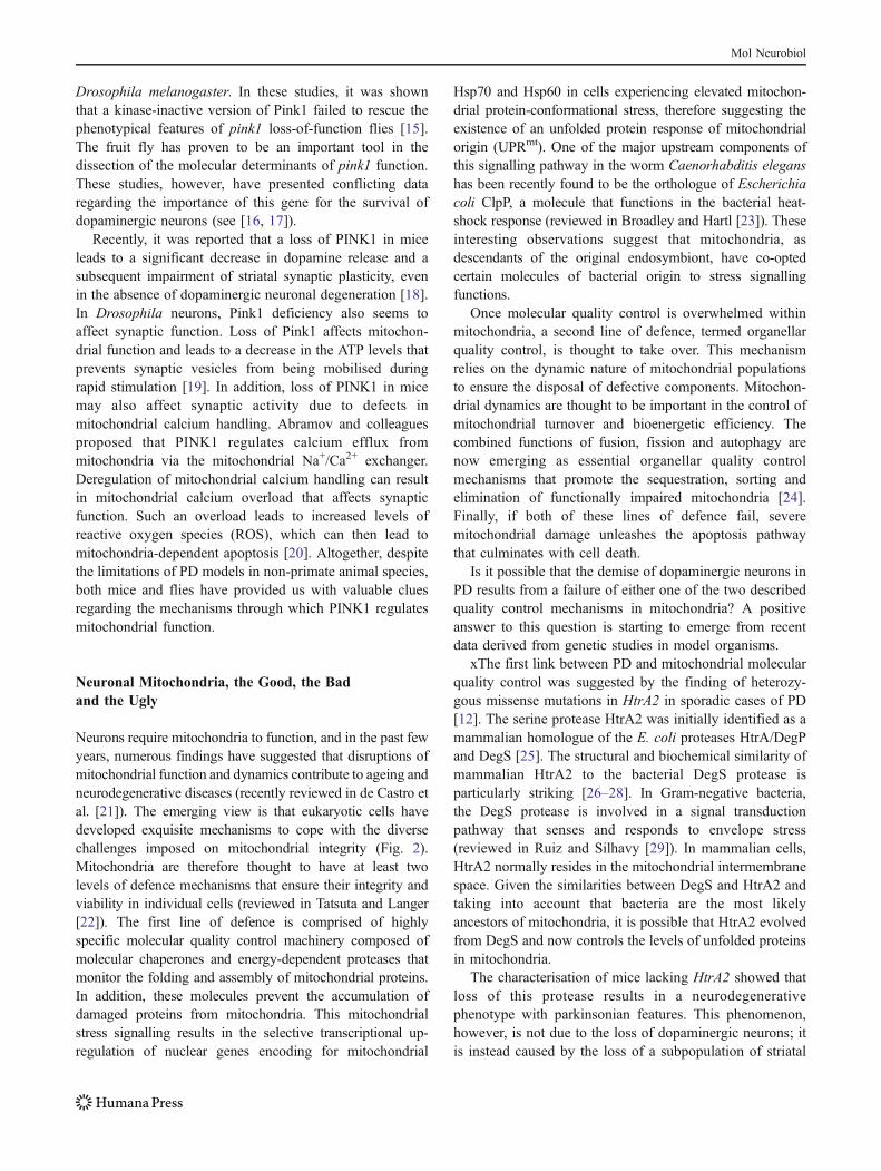
Fig. 2 Dynamics of mitochondrial quality control. Mitochondria aredynamic organelles that are continuously remodelled by fusion andfission events. In this figure, mitochondrial fission is represented asone step of the mitochondrial life cycle involved in the maintenance ofa pool of viable mitochondria. In this model, mitochondria can alsoemploy fission to segregate defective daughter mitochondria based ona mechanism that requires OPA1. These defective mitochondria canfollow three separate routes. They can be repaired through molecularquality control pathways that possibly involve p38, PINK1 and HtrA2.The defective mitochondria can also be selectively removed through
autophagy using a mechanism that involves the PINK1-dependentrecruitment of the ubiquitin ligase Parkin (1), followed by theubiquitination of proteins such as VDAC1 and MFN2 (orange andgreen colours, respectively; purple ubiquitin) (2). Such ubiquitinatedproteins might then work as adaptors for p62/SQSTM1 and promotethe translocation of defective mitochondria to the autophagosome.Finally, if both molecular and organellar quality control mechanismsfail, mitochondria will ultimately release pro-apoptotic proteins (asindicated on the upper right of the figure), leading to cell death
Mol Neurobiol
dependent mitochondrial recruitment, if Parkin simply“labels” any outer membrane protein with ubiquitin. Themechanism through which ubiquitin targets defectivemitochondria for autophagy remains unclear. The mamma-lian adaptor protein p62/SQSTM1 (sequestosome-1) is anautophagy receptor that can interact with ubiquitin conju-gated to a target protein as well as LC3/GABARAP onautophagosomes [35]. It is therefore possible that ubiqui-tinated proteins on the outer mitochondrial membrane usep62/SQSTM1 as an adaptor to anchor defective mitochon-dria to the autophagosome. This model is supported by arecent study in which p62/SQSTM1 was found to berecruited to clustered mitochondria and to be essential fortheir final clearance [36].
Taken together, the findings to date suggest that PINK1is a master regulator of mitochondrial quality control, atleast at two levels; it enhances molecular quality control viaHtrA2 and enhances organellar quality control via Parkin. Itis worth noting that mutations of all of these molecularcomponents of mitochondrial quality control have beenlinked to PD, suggesting that alternative hits on thiscommon pathway are possibly important in the develop-ment and progression of this disease.
Concluding Remarks
It is clear that unfolded proteins play an important role inthe development of PD. Although clear mechanisms forsuch proteinopathies are well established when theseunfolded proteins accumulate in the cytosol, cell nucleus,endoplasmic reticulum and extracellular space, little isknown about any causative role of protein aggregation inmitochondria in PD. Although they have limitations, animalmodels are beginning to unravel a significant role for thisphenomenon in PD when PINK1-mediated mitochondrialquality control is impaired.
The molecular pathogenesis of sporadic PD and the basisfor selective dopaminergic neuronal loss remains unknown,and it is unclear if there is a genetic basis for thedevelopment of this disease in this subgroup of patients.Recently, several genome-wide association studies (GWAS)have been conducted to evaluate the possible genetic basisfor PD in sporadic patients. To date, such approaches havefailed to identify any genes associated with mitochondrialdysfunction in evaluated patients. Given that multiple low-frequency risk variants are below the detection limit ofcurrent GWAS methodology (see [38]), it is unclearwhether mitochondrial gene mutations play any importantrole in the aetiology of PD in sporadic patients. With theincreasing power of gene sequencing technologies, however,it is likely that, if other mitochondrial mutations in PDpatients exist, they will come to light in the near future.
Approximately 40 years ago, US President RichardNixon declared war on cancer by increasing researchfunding to improve the understanding of cancer biology.At first glance, cancer and neurodegeneration seem to havelittle in common. Neurodegeneration can be viewed asresulting from the death of postmitotic neurons, whereascancer cells show an enhanced resistance to cell death. It isnow clear that mitochondria are the master controllers ofcell survival and death. Interestingly, mitochondrial PINK1has been recently shown to be important as a survivalmechanism downstream of the PI3K/Akt signalling path-way [39]. This pathway is often overactive in cancer cells,suggesting that PINK1 could have oncogenic potentialthrough the protection of healthy mitochondria in cancercells. Therefore, we reason that understanding the functionof PINK1 might help to fight the war on two fronts, as thisknowledge will contribute to the elucidation of the role ofmitochondria in both cancer and neurodegeneration.
By increasing our knowledge of the power of mitochon-dria to control cellular life and death, modern scientists canbe compared to the Jedi knights of the fictional Star Warsgalaxy, who harnessed the power of midichlorians to fightthe ‘Dark Side’. We hope that, eventually, the ‘Dark Side’will not prevail and that such knowledge will one daytranslate into cures for devastating diseases such as PD.
Acknowledgments We dedicate this review to Prof. Rita Levi-Montalcini.
Open Access This article is distributed under the terms of theCreative Commons Attribution Noncommercial License which per-mits any noncommercial use, distribution, and reproduction in anymedium, provided the original author(s) and source are credited.
References
1. Gray MW, Burger G, Lang BF (1999) Mitochondrial evolution.Science 283:1476–1481
2. Mattson MP, Gleichmann M, Cheng A (2008) Mitochondria inneuroplasticity and neurological disorders. Neuron 60:748–766
3. Jenner P (2003) Oxidative stress in Parkinson’s disease. AnnNeurol 53(Suppl 3):S26–S36, discussion S36–S38
4. Martins LM et al (2004) Neuroprotective role of the Reaper-related serine protease HtrA2/Omi revealed by targeted deletion inmice. Mol Cell Biol 24:9848–9862
5. Youle RJ, Strasser A (2008) The BCL-2 protein family:opposing activities that mediate cell death. Nat Rev Mol CellBiol 9:47–59
6. Tatton WG et al (2003) Apoptosis in Parkinson’s disease: signalsfor neuronal degradation. Ann Neurol 53(Suppl 3):S61–S70,discussion S70–S72
7. Bolanos JP, Almeida A (2010) The pentose-phosphate pathwayin neuronal survival against nitrosative stress. IUBMB Life 62:14–18
Mol Neurobiol
8. MacAskill AF, Kittler JT (2010) Control of mitochondrial transportand localization in neurons. Trends Cell Biol 20:102–112
9. Ly CV, Verstreken P (2006) Mitochondria at the synapse.Neuroscientist 12:291–299
10. Chan CS, Gertler TS, Surmeier DJ (2009) Calcium homeostasis,selective vulnerability and Parkinson’s disease. Trends Neurosci32:249–256
11. Valente EM et al (2004) Hereditary early-onset Parkinson’sdisease caused by mutations in PINK1. Science 304:1158–1160
12. Strauss KM et al (2005) Loss of function mutations in the geneencoding Omi/HtrA2 in Parkinson’s disease. Hum Mol Genet14:2099–2111
13. Thomas KJ, Cookson MR (2009) The role of PTEN-inducedkinase 1 in mitochondrial dysfunction and dynamics. Int JBiochem Cell Biol 41:2025–2035
14. Beal MF (2010) Parkinson’s disease: a model dilemma. Nature466:S8–S10
15. Kim Y et al (2008) PINK1 controls mitochondrial localization ofParkin through direct phosphorylation. Biochem Biophys ResCommun 377:975–980
16. Clark IE et al (2006) Drosophila pink1 is required for mitochon-drial function and interacts genetically with parkin. Nature441:1162–1166
17. Park J et al (2006) Mitochondrial dysfunction inDrosophila PINK1mutants is complemented by parkin. Nature 441:1157–1161
18. Kitada T et al (2007) Impaired dopamine release and synapticplasticity in the striatum of PINK1-deficient mice. Proc Natl AcadSci USA 104:11441–11446
19. Morais VA et al (2009) Parkinson’s disease mutations in PINK1result in decreased Complex I activity and deficient synapticfunction. EMBO Mol Med 1:99–111
20. Gandhi S et al (2009) PINK1-associated Parkinson’s disease iscaused by neuronal vulnerability to calcium-induced cell death.Mol Cell 33:627–638
21. de Castro IP, Martins LM, Tufi R (2010) Mitochondrial qualitycontrol and neurological disease: an emerging connection. ExpertRev Mol Med 12:e12
22. Tatsuta T, Langer T (2008) Quality control of mitochondria:protection against neurodegeneration and ageing. EMBO J27:306–314
23. Broadley SA, Hartl FU (2008) Mitochondrial stress signaling: apathway unfolds. Trends Cell Biol 18:1–4
24. Twig G et al (2008) Fission and selective fusion governmitochondrial segregation and elimination by autophagy. EMBOJ 27:433–446
25. Faccio L et al (2000) Characterization of a novel human serineprotease that has extensive homology to bacterial heat shockendoprotease HtrA and is regulated by kidney ischemia. J BiolChem 275:2581–2588
26. Li W et al (2002) Structural insights into the pro-apoptoticfunction of mitochondrial serine protease HtrA2/Omi. Nat StructBiol 9:436–441
27. Wilken C et al (2004) Crystal structure of the DegS stress sensor:how a PDZ domain recognizes misfolded protein and activates aprotease. Cell 117:483–494
28. Martins LM et al (2003) Binding specificity and regulation of theserine protease and PDZ domains of HtrA2/Omi. J Biol Chem278:49417–49427
29. Ruiz N, Silhavy TJ (2005) Sensing external stress: watchdogs ofthe Escherichia coli cell envelope. Curr Opin Microbiol 8:122–126
30. Moisoi N et al (2009) Mitochondrial dysfunction triggered by lossof HtrA2 results in the activation of a brain-specific transcriptionalstress response. Cell Death Differ 16:449–464
31. Plun-Favreau H et al (2007) The mitochondrial protease HtrA2 isregulated by Parkinson’s disease-associated kinase PINK1. NatCell Biol
32. Narendra D et al (2008) Parkin is recruited selectively to impairedmitochondria and promotes their autophagy. J Cell Biol 183:795–803
33. Narendra DP et al (2010) PINK1 is selectively stabilized onimpaired mitochondria to activate. Parkin PLoS Biol 8:e1000298
34. Yang Y et al (2006) Mitochondrial pathology and muscle anddopaminergic neuron degeneration caused by inactivation ofDrosophila Pink1 is rescued by Parkin. Proc Natl Acad SciUSA 103:10793–10798
35. Kirkin V et al (2009) A role for ubiquitin in selective autophagy.Mol Cell 34:259–269
36. Geisler S et al (2010) PINK1/Parkin-mediated mitophagy isdependent on VDAC1 and p62/SQSTM1. Nat Cell Biol
37. Ziviani E, Tao RN, Whitworth AJ (2010) Drosophila parkinrequires PINK1 for mitochondrial translocation and ubiquitinatesmitofusin. Proc Natl Acad Sci USA 107:5018–5023
38. Gandhi S, Wood NW (2010) Genome-wide association studies:the key to unlocking neurodegeneration? Nat Neurosci 13:789–794
39. Mei Y et al (2009) FOXO3a-dependent regulation of Pink1(Park6) mediates survival signaling in response to cytokinedeprivation. Proc Natl Acad Sci USA 106:5153–5158
Mol Neurobiol


