Embed Size (px)
Citation preview

Eur. J. Biochem. 217, 781-790 (1993) 0 FEBS 1993
Modulation of tensin and vimentin expression in chick embryo developing cartilage and cultured differentiating chondrocytes Rinke VAN DE WERKEN', Massimo GENNARI', Sara TAVELLA', Paola BET', Francesco MOLINA', Shin LIN2, Ranieri CANCEDDA' and Patrizio CASTAGNOLA' ' Laboratorio di Differenziamento Cellulare, Istituto Nazionale per la Ricerca sul Cancro, Genova, Italy * Department of Biophysics, Johns Hopkins University, Baltimore MD, USA
(Received March 19/July 8, 1993) - EJB 93 039816
It has been proposed that tensin, in association with several other proteins, mediates the micro- filament-integrin link. Here we describe the isolation of clones spanning about 5 kb from the 3' end of tensin mRNA from cultured chick embryo chondrocyte and embryonic heart cDNA libraries. Tensin expression was investigated in cultured chick embryo cells. It was observed that tensin expression is dependent upon substrate adhesion and it is turned off after 7 days of suspension culture. This process is reversible. Tensin expression is also regulated during cartilage cell differenti- ation in vivo; at Hamburger and Hamilton stage 39 -40, non-hypertrophic tibia1 chondrocytes ex- press both RNA and protein while hypertrophic chondrocytes do not. In the culture system the expression of vimentin, a major component of intermediate filaments, showed an opposite behaviour since the suspension culture enhances the accumulation of both vimentin and its mRNAs. Therefore in chick embryo cultured chondrocytes and in vivo, during cartilage development, cell shape changes and/or integrin-extracellular matrix protein interactions may be involved in the regulation of these two genes coding for cytoskeletal proteins.
Focal contacts are structures allowing cells to attach to extracellular matrix components [ 11. Several receptors for fibronectin and vitronectin, belonging to the integrin family, constitute the membrane components of these structures, also called adhesion plaques [2]. Actin bundles are linked with their barbed ends to the cytoplasmic portion of integrins by several actin binding or associated proteins that constitute the cytoskeleton component of focal contacts [3]. By modulation of integrin types present on the membrane, the cell can change the adhesion strength, the phenotype [4] and the mi- gration potential [5]. It has been shown that the adhesion plaque is transformation-sensitive [6] and it plays an impor- tant role in the signal transmission from the membrane to the cell nucleus [7].
Two major hypotheses, not necessarily mutually exclu- sive, are currently proposed concerning the integrin-mediated signaling mechanisms: one suggests that the adhesion recep- tors transmit signals by reorganizing the cytoskeleton while the second suggests that integrins generate a biochemical cascade within the cell after ligand binding [8, 91. It has indeed been demonstrated, in support of the first hypothesis, that perturbation of the cytoskeleton organization by drugs or by any means interfering with the ability of the cells to assemble adhesion plaques have profound effects on cell phe-
Correspondence to P. Castagnola, Laboratorio di Differenzia- mento Cellulare, Istituto Nazionale per la Ricerca sul Cancro, Viale Benedetto XV, 10 1-16132 Genova, Italy
Fax: +39 10 352999. Note. The nucleotide sequence data reported here are available
under accession number X 66286 of the EMBL Data Library.
notype, growth and differentiation [lo- 121. However, in several cell types phosphorylation events follow the binding of ligands with integrin receptors [13-1-51, Just recently, a focal adhesion protein-tyrosine kinase has been characterized that is phosphorylated upon cell attachment to fibronectin [16]; it has also been shown that a specific repression of tyrosine kinase activities inhibited the formation of focal ad- hesions and stress fibers [17].
It has been known for several years that there are compo- nents, in the focal contacts, sensitive to tyrosine kinase activ- ities [18-201; recently a protein substrate with a SH2 do- main has been identified [21]. This protein, called tensin, can be phosphorylated on tyrosine residues and binds to both actin and phosphorylated proteins identified in src trans- formed cells. Because of this property it has been suggested that tensin may play a key role in linking signal transduction pathways to the cytoskeleton [21].
In our studies, aimed at understanding chondrocyte phe- notype modulation at the molecular level, we generated a cDNA library and screened it by differential hybridization with mRNA probes derived from adherent and suspension- cultured cells. From this library we isolated and sequenced two cDNA clones, one coding for chicken tensin and the other for chicken vimentin. These cDNA clones, and specific antibodies, have been used to investigate tensin and vimentin expression in cultured differentiating chondrocytes and ten- sin expression in chick embryo developing cartilage. This paper also reports that the steady-state levels of tensin and vimentin mRNAs and proteins are inversely modulated by cell adhesion.

782
MATERIALS AND METHODS The heart random-primed library was screened with a
Cell culture 738-bp PstI-PstI restriction fragment from clone pCP5b.
Northern blot and slot blot analysis Cell culture methods have been extensively described elsewhere [22]. Culture medium was Coon’s modified Ham F-12 [23] with the addition of 10% (by vol.) fetal calf serum (Flow Laboratories), 50 U/ml penicillin and 50 pg/ml strep- tomycin (Flow). To establish primary cultures, Hamburger and Hamilton stage 28-30 [24] chick embryo tibiae were removed, cleaned, washed in NaClP, (NaCl 137 mM, KCl 2.7 mM, Na,HPO, 8.1 mM, KH,PO, 1.45 mM pH 7.2) and digested for 15 min at 37°C with 400 U/ml collagenase I (Worthington) and 0.25% (mass/vol.) trypsin (Gibco Ltd.). After sedimentation, the supernatant, containing tissue debris and perichondrium, was discarded and the pellet was di- gested for an additional 45-60 min with the above dissoci- ation buffer supplemented with 1000 U/ml of collagenase I1 (Worthington). Cells were grown in anchorage-dependent conditions on regular tissue-culture dishes and, when indi- cated, transferred by trypsin treatment (0.25% in NaCVP,) to anchorage-independent conditions on a 1 % (mass/vol.) agar- ose layer.
Nonadherent culture condition triggers the chondrocyte differentiation program [22]. Differentiating (clone 956) and non-differentiating (clone 957) cloned cell populations were obtained as described by Quarto et al. [25]. Primary cultures of chick embryo skin fibroblasts were obtained by trypsin digestion of stage 38-39 chick embryo skin.
Construction of cDNA libraries RNA for all library constructions was extracted using the
guanidinium thiocyanate/acid-phenol/chloroform single-step method [26]. RNA was extracted from the cellular clone 956 after 36 h from the transfer to non-adherent conditions or from hearts of Hamilton and Hamburger 38-39 stage chick embryos. Poly(A)-rich RNA was purified by oligo(dT)-cellu- lose chromatography [27] or by oligo(dT) bound to magnetic particles (Dynabeads or Promega). Chondrocyte cDNAs were generated by oligo(dT) priming using a kit from Amers- ham and inserted into the vector pcDNA I1 (Invitrogen) after addition of BstXI adaptors (Invitrogen). XL1-Blue cells (Stratagene) were transformed with the plasmids by electro- poration with a Bio-Rad apparatus. Colonies were screened, by differential hybridization, with 32P-labelled poly(A)-rich RNAs, extracted from clones 956 and 957, according to the Invitrogen instructions. The hybridization and washing con- ditions were identical to those recommended by Amersham for Hybond N membranes.
Heart cDNAs were generated with a synthetic oligonu- cleotide (5’-AAGCCTGGCTCTGCTCGT-3’) at the 5’ end of clone pCP5 or with random priming, using the Amersham cDNA synthesis kit. cDNAs were size-fractionated to > 600 bp, inserted in the LZAP IIIEcoRI cloning vector (Stratagene) after adding EcoRI adaptors and packaged with the Gigapack I1 gold packaging extract (Stratagene). The packaged library was used to infect XL1-Blue host cells and screened by plaque hybridization. The insert of clone pCP.5 was used as a probe to screen the primer-extended library. Positive clones were plaque-purified after three rounds of screening. The Bluescript plasmids were excised in vivo from the AZAP 11 vector by co-infecting the XLl-Blue cells with the R 408 helper phage. Plasmid DNA was purified with Qiagen columns (Chatsworth) and digested with EcoRI to release the inserts.
Total RNA was extracted by the guanidinium thiocyanate method [26] from cultured cells and from brain, lung, heart, liver, stomach, and skin of stage 38-39 chick embryos. Poly(A)-rich RNA was purified from chondrocyte clones. Total and poly(A)-rich RNA were size fractionated on 1% (mass/vol.) agarose/formaldehyde gels by electrophoresis and transferred to nylon Hybond N membranes (Amersham) after alkaline treatment by capillary blotting [27].
Slot blots were performed with total RNA extracted from cultured cells as described in Castagnola et al. [28]. Briefly RNA was denatured for 15 rnin at 65 “C in 4 X NaCl/Cit (1 X NaCl/Cit is 150 mM NaC1, 15 mM sodium citrate, pH 7.0) containing 25.9% (by vol.) formaldehyde, chilled on ice for 15 min and spotted on Hybond N’ membranes with a 24- well PR 600 slot-blot apparatus (Hoefer Scientific Instru- ments). The blots were hybridized with probes for vimentin (whole insert of pCP41) and tensin: whole inserts of pCP5 and pCPSa, a BamHI-BamHI (977-bp) and a HindIII- HindIII (1 165-bp) restriction fragment of clone pCP5c. All probes were ”P-labelled using the Boehringer (Mannheim) random priming labeling kit. Hybridization and washing con- ditions were performed as recommended by Amersham for Hybond N membranes.
Nucleotide sequencing and sequence analyses The MI3 (-40) primer was used for single-strand se-
quencing of cDNA restriction fragments cloned in the M13 phage. The nucleotide sequence of the inserts of pCP5a-c and pCP41 plasmids was determined with T3, T7 or the following 20 internal oligonucleotides : (1) AAGCCTGGCT- CTGCTCGT: (2) CCCGCCATCTCCTCACCC; (3) GGGA- AGGTTCTCCAAAGA ; (4) ACGGTCACACACGCATGT- CC; (5 ) GTCCTGAGGCCTCGCCAA; (6) TGTGCTGCA- GCAGAGGG; (7) CGCACACACACACGCAC; (8) TCCC- TCCTGCCACCAGT; (9) GGGTGAGGAGATGGCGGG;
ATGCCT ; (12) AAACCCACCTGTGCAAGC ; (1 3) TGAG- CATTGTGGTGTTGG ; (14) GCAAACAGTTGGACTCC ; (15) TCGCATCCATGCGGATAG; (16) GCTTGCACAGG- TGGGTTT; (17) CCCTCTGCTGCAGCACA; (18) CAAA- CATCACCACTTGGC ; (1 9) CCAGCACTCCATCATGCC ; (20) CCTTCTCGGATCAGCTCC.
All sequencing reactions were carried out with the Sequenase 2.0 kit (US Biochemical Corp.). The PC gene (In- telligenetics) or the UWGCG (Genetics Computer Group Inc.) software packages were used for sequence analyses with EMBL data libraries (releases 27-31).
(10) CGAATGACTACAAGTAAC ; (1 1) TCCTGTGCTCC-
SDSFAGE and immunoblotting Dedifferentiated chondrocytes grown for 18 days as ad-
herent cultures and transferred and maintained in suspension culture for 10 days were lysed in 10mM Tris/HCl pH7.4, 0.9% (mass/vol.) NaCI, 10 mM CaCI,, 1 mM phenylmethyl- sulfonyl fluoride, 1 mM N-ethylmaleimide, 50 U/ml Aproti- nin, 1% (by vol.) Triton X-100. Protein concentration was determined with the Micro BCA protein assay reagent (Pierce) following the manufacturer’s instructions. Samples were boiled 5 min in SDS-sample buffer containing 50 mM

783
dithiothreitol and centrifuged at 13 000 X g for 5 rnin before loading on a 7.5% (masdvol.) SDSIPAGE. After gel electro- phoresis, the geI was blotted 1 h at 300 mA at 4°C in 25 mM Tris/HCl pH 8.3, 192 mM glycine and 20% (by vol.) metha- nol on a nitrocellulose membrane. The blot was saturated with 5% (mass/vol.) non-fat dried milk in buffer A (20 mM Tris/HCl pH 7.5, 500 mM NaCl and 0.05%, by vol., Tween 20), washed several times with buffer A and incubated 1 h at room temperature with 1 pg/ml of TL1 mAb (anti-chicken tensin) or 16 h with the V9 mAb (anti-swine vimentin) (Dako) diluted 1 : 10 in 3% (mass/vol.) bovine serum albu- min, 0.02% (mass/vol.) NaN,. The detection was performed with a goat biotin-conjugated antibody and avidin-conjugated horseradish peroxidase using chloronaphthol as substrate.
Hybridization in situ
Stage 39-40 chick embryo tibiae were fixed in 4% (masshol.) paraformaldehyde in NaCl/P,, pH 7.4, at 4°C for 16 h, dehydrated in a series of ethanol washes, then embed- ded in paraffin wax. Sections of 5 pm were mounted on slides pretreated with 3-aminopropyltriethoxysilane and sub- sequently deparaffinized in xylene. The samples were rehy- drated, treated with proteinase K (10 pg/ml), fixed again with 4% paraformaldehyde and acetylated in 0.25% (by vol.) ace- tic acid anhydride (in 0.1 M triethanolamine pH 8.0). After another series of dehydration, the samples were hybridized with antisense and sense riboprobes. A 961-bp fragment be- tween Hind111 (in the pBluescript polylinker) and BarnHI sites of pCP5c was subcloned in pcDNA I1 containing SP6/ T7 promoters. ["S]UTP[S]-labelled single-stranded sense and antisense RNA probes were prepared as described by Melton et al. [29], reduced to an average of about 150 bp by limited alkaline hydrolysis, and used in hybridizations at a final concentration of 100000 cpm/ml in hybridization buffer at 50°C for 12-16 h. The hybridization buffer contained 50% (by vol.) formamide, 10% (mass/vol.) dextran sulfate, 10 mM dithiothreitol, 300 mM NaCl, 20 mM Tris/HCl pH 7.40, 10 mM sodium phosphate pH 6.40, 5 mM EDTA, Denhardt's solution (0.02% masdvol. Ficoll, 0.02% mass/ vol. bovine serum albumin, 0.02% mass/vol. polyvinylpirrol- idone), 0.5 mg/ml tRNA. Washing was performed in 50% (by vol.) formamide, 300 mM NaC1, 10mM Tris/HCl pH 7.4, 10 mM NaH,PO, pH 6.40, 5 mM EDTA, Denhardt's solution, 10 mM dithiothreitol, for 2 h at 50°C and in 0.5 M NaCl, 10 mM Tris/HCl, 1 mM EDTA (NaCl/Tris/EDTA) for 10 min at room temperature. The signal specificity was increased by incubation with RNase A (20 pg/ml) in NaC1/ TrisEDTA for 30 rnin at 37°C. Washing with NaCl/Tris/ EDTA was performed at 37°C for 30 min, with 2 X NaCl/Cit at 37°C for 30 min and with NaCVCit at 45°C for 30 min. The slides were dehydrated in a series of ethanol with 0.3 M ammonium acetate, air-dried, coated with autoradiography emulsion (Kodak type NTB2) diluted 1:l in water and ex- posed for 17 days at 4°C. Slides were developed with Kodak D19 developer.
Indirect immunofluorescence Dedifferentiated chondrocytes grown for 3 weeks on
plastic dishes were trypsinized (0.25% trypsin in NaCl/P,) and grown for 48 h on glass slides. Hypertrophic chondro- cytes were filtered through a nylon 42-pm-pore Nytex filter to eliminate aggregates, digested with 1 mg/ml hyaluronidase (type 111, Sigma) in NaCl/P, for 15 min at 37"C, plated on
poly-(L-1ysine)-coated glass slides and fixed after 30 min or grown for 48 h in adherent conditions on glass slides and then fixed for 5 min in methanol (-20°C) and 1 rnin in ace- tone (-20°C). To enhance the attachment of hypertrophic chondrocytes, slides were treated for 1 h with absolute etha- nol, then incubated overnight in 1 M HNO, and finally washed with demineralized water before autoclaving.
Hamburger and Hamilton [24] 35-37 stage chick embryo legs were embedded in OCT (Tissue-tek, Miles) me- dium and cryosectioned at 8 pm. Slides were washed with NaCl/P, and fixed for 5 min in methanol (-20°C) and 1 min in acetone (-20°C). After fixation the slides were air-dried and washed with NaCl/P,. Unspecific binding sites were satu- rated for 1 h in goat serum. The serum was removed and cells were incubated for 1 h in 100 p1 of a 10% (by vol.) goat serum in NaCVP, solution, containing anti-tensin monoclonal antibody or polyclonal antibody diluted at 4 pg/ml and 40 pg/ml, respectively, or the anti-vimentin mAb at the final concentration of 25.3 pg/ml. Cells were washed with NaCI/ P, to remove the first antibody and then incubated for 30 min with a rhodamine-conjugated secondary antibody or with a biotin-conjugated secondary antibody in NaCVP, containing 10% goat serum. Slides were then washed with NaCl/P,, in- cubated for 30 rnin with streptavidin-conjugated Texas red when biotin-conjugated antibodies were used and mounted with Gelhlount (Biomedia Corp.).
Slides were observed and photographed by a Zeiss Axio- phot microscope.
RESULTS Isolation of clones from a chick chondrocyte cDNA library of which the abundance is dependent on the cell adhesion state
A screening of a chondrocyte cDNA library by dif- ferential hybridization was performed to isolate cDNA clones specific for mRNAs regulated during cartilage differentia- tion. The cDNA library was synthesized with mRNA ex- tracted from chondrocytes grown in suspension and derived from a cell clone (clone 956 [25]) able to undergo chondro- cyte differentiation in this condition. The 3ZP-labelled RNAs used to screen the library were from the same cell clone grown in suspension for 36 h and from a cell clone unable to differentiate to chondrocyte (clone 957 [25]), grown ad- herent to the substrate. Eleven cDNA clones were observed to hybridize more intensely with mRNAs extracted from the cell clone 956, while only one cDNA clone hybridized more intensely with mRNAs extracted from the cell clone 957. Two cDNAs were further analyzed: one (cDNA clone pCP5) specific for a mRNA more abundant in the cell clone 957 and one (cDNA clone pCP41) specific for a mRNA more abundant in the cell clone 956.
Northern blot analyses of RNAs extracted from both cell clones were performed with both cDNA probes. As shown in Fig. 1 a, two mRNAs (clearly distinguishable on the original autoradiography films) of about 1.8 and 2.0 kb were detected by the pCP41 insert in the 956 cell clone which were much less abundant in the 957 cell clone. The pCP5 insert hybrid- ized with two mRNAs of about 9.5 and 9.0 kb in both cell clones but the two mRNAs were more abundant in the 957 cell clone (Fig. 1 b).
The nucleotide sequence of 404 bp of the 780-bp insert of pCP41 and the computer-assisted similarity search with the EMBL sequence data library revealed that it is specific

7 84
a b C f
Fig. 1. Northern blot of RNAs extracted from cultured cells. The inserts of pCP41 (a, c and f), pCP5 (b), pCP5a (e) and the 1165-bp HindIII-Hind111 restriction fragment of pCP5c (d) were used as probes. RNA was extracted from: cell clone 957 cultured in adherent conditions (lane I); cell clone 956 after 36 h from the transfer to nonadherent conditions (lane 2); dedifferentiated chondrocytes grown for 3 weeks in adherent conditions (lane 3); dedifferentiated chondrocytes grown for 3 weeks in adherent conditions and 36 h in nonadherent conditions (lane 4); dedifferentiated chondrocytes grown for 3 weeks in adherent conditions (lane 5 ) ; dedifferentiated chondrocytes grown for 3 weeks in adherent conditions and 72 h in nonadherent conditions (lane 6); skin embryo fibroblasts grown in adherent conditions (lane 7) and skin fibroblasts grown in adherent conditions and 36 h in nonadherent conditions (lane 8). About 2 kg poly(A)-rich RNA was loaded on lanes 1 and 2 and about 20 pg total RNA on lanes 3-8. At the bottom the 27 S rRNA (a-flregions of the same filters after rehybridiza- tion with the pXCR7 probe for rRNAs are shown. Asterisks indicate the two vimentin mRNA bands. These were clearly distinguishable on the original film. Arrows refer to the position of the 18 S and 27 S rRNAs. Arrow heads indicate the position of RNA markers whose size is expressed in kb.
for vimentin [30] spanning from the last Glu codon in exon 5 to the Ile codon in exon 9 (data not shown).
The nucleotide sequence of the 843-b~ insert of pCP5 was found to have no significant similarity with any se- quence in the EMBL sequence data library nor did it have an open reading frame.
Cloning of overlapping cDNAs with the same specificity as pCP5, their composite nucleotide and deduced amino acid sequences
The isolation of cDNAs extending toward the 5' end of the mRNA sequence was needed to determine the specificity of the pCP5 clone. The pCP5 insert was used as a probe to screen a heart cDNA library synthesized by specific primer extention (see Materials and methods and following para- graph). Several cDNAs were found to hybridize with the probe. The inserts of two of them, pCP5a (1563 bp) and pCP5b (3 143 bp), were sequenced. Again neither similarities nor open reading frames were found. A third screening of a heart cDNA library synthesized by random priming was performed, using a 738-bp PstI-PstI fragment as a probe, obtained from the 5' end of pCP5b. The 1890-bp insert of the longest (pCP5c) of several hybridizing cDNA clones was sequenced. The composite sequence of all overlapping cDNAs is shown in Fig. 2A. The sequence of nucleotides 1-4881 was submitted to the EMBL data library and named GGTENS. A partial open reading frame of 832 bp (nucleo- tides 1-832) was found to be identical with the GGTENSI
sequence (chicken tensin mRNA) from the EMBL data li- brary but two substitutions, at nucleotides 70 and 190, with no change of the codons. The 3' end of the sequence, 4048 bp long, is untranslated and two potential polyadenylation addi- tion signals, TATAAA, are located at positions 3075 and 4845 in GGTENS. The overall similarity of the composite sequence, GGTENS, with GGTENSI, although very high, is not 100% due to four single nucleotide substitutions, four single nucleotide deletions and seven nucleotide insertions. A diagram showing the overlaps of the sequences GGTENSI, GGTENS, pCP5 and pCP5a-c is presented in Fig. 2B. By nucleotide sequence analysis, polymerase chain reaction and Northern blot analysis, we determined that 127 bp at the 5' end of the pCP5c were not tensin-specific but hybridized with a 6.8-kb mRNA expressed in heart only, coding for a cardiac heavy-chain myosin (data not shown). An interrupted reading frame suggested that the two unrelated cDNAs were ligated together into a single vector molecule by some un- known mechanism.
Puzzled by the unusually long 3' untranslated region, we decided to cany out a computer-assisted search to investigate whether other mRNAs with comparable long 3' untranslated region exist. We searched among fungi, invertebrates, mam- mals, primates, vertebrates and rodents for RNA sequences that contained a 3' untranslated region longer than 3.8 kb. We found the 11 sequences listed in Table 1. No similarities between GGTENS and these sequences were found using the Fast A or the Word search programs.

785
A -127 - 99
1
101
201
301
401
501
601
701
801
901 1001 1101 1201 1301 1401 1501 1601 1701 1801 1901 2001 2101 2201 2301 2401 2501 2601 2701 2801 2901 3001 3101 3201 3301 3401 3501 3601 3701 3801 3901 4001 4101 4201 4301 4401 4501 4601 4701 4801
rlnmmmmnnnnnnnnnnnnnnnnnnnn -nnnnnnnnnnnnnnnnnnnnnnnnnnnnnnnnnnnnnnnnnnnnnnnnnnnnnnnnnnnnnnnnnnnnnnnnnnnnnnnnnnnnnnnnnn
S K Y W Y K P D I S R E Q A I A L L K D R E P G A F I I R D S H S
F R G A Y G L A M K V A S P P P T V M Q Q N K K G D I T N E L V R H TTCCGGGGAG CCTATGGCCT TGCCAEAAA GTCGCITCCC CACCTCCCAC CGTCATGCAG CAMCAAGA AAGGAGACAT TACCPATGAG CTGGTGAGGC
F L I E T S P R G V K L K G C P N E P N F G C L S A L V Y Q H S I ACITCCTCAT CGAGACCAGC CCACGGGGTG TGAAACTAAA AGGATGCCCC AATGAGCCTA ATTITGGCTG CMY;TCGGCT CTGGTCTACC AGCRCICCAT
M P L A L P C K L V I P D R D P M E E K K D A A S T T N S A T D L CATGCcTcr(3 GCCCTGCCCT GCAAGCTGGT CAWCTGAC CGAGATCCA TGGAGGAGAA W G A W GCGKGACCA CCAACTCAGC CACAGAWTT L K Q G A A C N V L F I N S V E M E S L T G P Q A I S K A V A E T L CTCAAACAGG GTGCGGCm CAATG'TCCIT TTcATcAA?T CAGTGGAGAT GGAATCGCTC A C A W C W AGGCCATCK CAA-TG GCAGAGACAT
V A D P T P T A T 1 V H F K V S A Q G I T L T D N Q R K L F F R R ?Y;GTGETGA TCCCACGCCG ACCGCI'ACGA T C G T C A W CWAGTCKT G C A C A C A TCACCTTAAC AGACAACCAG AGGAAACTGT TCITCCGACG
H Y P L N T V T F C D L D P Q E R K W T K T D G S G P A K L F G F ACACTATCCT CTCAATACTG TCACCFI'CTG TCATITGGAC CCCCAGGAAC GAAAGKXXC TAFAACIY;AC GGCAGTGGCC CAGCCAAW CI'TCGGCMK V A R K Q G S T T D N V C H L F A E L D P D Q P A A A I V N F V S R GTGGCCAGGA AGCAAGGGAG CACCACGGAC AACGTCTGCC ACC"TC?TTGC ' AGAGCPGGAC Ccr(;AccAGc (XXXKDX CATCGTWC mGTcTccA
GGGTCATWT TGGATCCGGC CAGAAGAGAT GAGCCGGTGC TTGCGGGTGA 'TKTXAATT TTGGAGAAGG ACTTGGAGCT GATCCGAGAA GGAGWTGAG
GAAGTGCA'IT GEGGAGAGG GAAGTGAATT TGGGGGGGGG GGGAAGGGTT GGAATKTGG AGGAAAACAG cz\pAcAAcAA CAAAAAACCA AAACAAAACC CTAAGWCTC GACACAAGCT TAACACACAC AGAAGAAKY. AACCACACAC CTATGTAACA GAGEATGAA GTCAACGCCA AACAGGGAGA GGATGCGGAT GCAWECCA CCGCAmAA GGTGACCAAA GTGAAGGTGG CAGCGAGAGG AG'ITCCCAGC TT'TCGGCATC GCAGTCCTGA GGCCTCGCCA AGWTGATG llTGCrCCCG GCCGAACACA GGGTCGTCAT 'ITGGAGGGGG APAAAAAAAA GATGAAACAG CAAAAGCCAT GAAAGAAGCC TKATCGGTG AACGTCECT GCGTGTTTGA CACAGXTAC AGGGGATAAA GCCCTGCCGC AGCGTGGGCT CAGGTCTGCA GGATGCTGGA GGAGATGCCG TTGGAAAACT C C m G c r c ; TGCFGCAGCA GAGGGTCW CCAGCITACA CCAAAACCTG CTGGCCACCC TGCTGGGGTA TGCAAccGer " X C C A A A CCCTCTTGTC CCCXGGTGC TTGGcmTGG GCTACCCTCA G C C C m C CAGAGCZCAG CACACCTTGA GACATCRCGA GTCCACCCAC AGAGGGGCAA TGCAGCATAT C C T C C A a C TGGGCGGCTC CTGTGCTCCA TGCCTGGAGG AGGAGCATGC TCCTGCGCTG CTCGCCCATI TTCCTCTIYIT CCCTCCTGCC ACCAGTCCTC ?TTGACAGTT TPTCCIYW GCCTGTCACA CGTTcrcTTA ACAGGAAAAA AAAAAAAAAG AAAAAPAGAA AGAAAAATGT TGTAATAATA ATAATGATGA TAATAATAAT AATAATAGCA GATGCTIY;CA CAGG'IWXTT TccTGn;u IG AGCA-C CCGTGGCCGG CCCTATCKT C r C m G G A G ATGTGCAAGC AGmTTAAA 'I'IGCAEGAC G'ITCAAGTPG C A C W G G C GAATGACI'AC AAGTAAercA C T T G m T T A T T I T X A G G CCACGTAAA AAPAAAAAAA AAAGGFAGAR AA'ITTATTIA GAAAAAclAC AACWAGGAT CAGCAGKAC TGGGCAGCIT TGGCTCAGTG GGGAGGCZAC AGGTCCTCTG AGAGCCACAC TCCGCCITCC CTGTGGercJI AGGTGTGCAC CCIGAATGCA CIUCAGGCTT TTCCIGTGT GTGGGGATGT GTGGGGETG AGGGCcCrGC CT'IATG"G AGCATKTGG TGPTCX;GGAG CAGAGGKCC ACCCCCGGAT GT- TGGTGGITGG TGGGGpAGc4 G'KXXAGGT ?TGGGGCAGA GCXAGCGCT GTGCTGAGAG CACAGAGGTC C A G A m T GTTTGFGGT WITATGTTT GGAGAAGWI C A m G A G TGGCCCAGAT GGCTGCTGGG GGCAAACAGT TCXACKCIY; CTGCAGCCCC CCGGAGAGCT C G C ~ C T C AGACCACCTT TGCCITGATA GCTATAATAT ATATAGGAGC TATACTGACA AAATACIYTA AGCTAACGAA ATITAAAGGA m G G'ITAAATTGA CACTCTGAAA ATATTTATTA TTA'ITATTTT TTITGCCATA TT?TGcT?TC CGCGCATGA TTCTCCCCAA CCCCTGIYTCT ATCTGCCTTC CCCCGCCCCG GGGGCAAACA CCTGCACGTC AGAAGGAAT TGGGTGCGAG GGAAGCGCCG ATGGGGCTGC GGGGGAcATG CGTGTGACCG TGTGCGFGTG KJTGTGCGCG AGAGAGCGAC GGCTGGGCC GGGAGGGGCC GGCGTGGCCG GAWTGGTT GGGTGTATGG CAWGATGG A'IGCGTAGCG GGGATlTGGG TGCGAGAm CGGTGGGATG CGGGGAGGAC GGGGCCFI'CC C C C C m C C TGGCTCCCCC TATCCGCATG GATGCGAAGA CCTTTTCATA T T T T A T c m TATTWGTT TTMTTMT TTAAACGACG TTTCAAAAGC GATTCCCTCC TTCTGCGAAA TTCAAGCGAG ATCAGAGACA ATCTCACCCG CCATCTCCTC ACCCTGCCGG TAACCGATTA G A W C A C WW'MTATA G A T A m TATATATIT 'ITGAAAGCCI C T A T T m c A GACTTGGm TCTCACTGCT AAGAAAGAAA AGAGAGATA ATAATGFAAT 'MTA"TAA TCIY;CM;CCT CGGXCTTGA GCCTCCCCTT CACCMCCCA TCCCATAGGG CGAAGTAGAT CGGAGCACCC AGAGGAGCAT C A m m TCAGGGCTCC ACG!IGATGC TCGA- GAWTAGCCT GCG'TTKAAT GGCAAACATT AACCA'ITGTG TAG'ITTTTAA CATAGAG(IIT CCAACGATGT CAW-GT TTGTCAGCCT CCCTlTA?iGG CA- (J"A"K 'I?TTApAAGT TACATlTllT GGCTGTAACT CCCACCAGTT W G T C G G G ACCAGCACGC GTCACTAWI TITCC'TTACA GAAGGATGGA GAGATcTTlT GGlTTTGcIT TGCITTGCCA TGCAGTCGCG GTATCGGAGA GCCGAGCTTG T C X E C m CCCTGCGCGT GTCCCGTTGT AACCACACCG CCAGGCG'ITA TcrcTcTCTC TCTCTCTCAC ACACACCAAC ACATCCCTCC ?TccTGCACA AAGAACTGCA TATATTCTTG CCAAAGATAT CCITAAAAAC G C A C T I T I T CCACATATAT ATATATATAT A?TI"ITITIT GTGTGTGTGT TTGIY;TGTGT ACATCGTCG TACGTCACCT TRTGTGITG A T T T G T " TGGAGWCCT TCCCA'ITCCC A T C C m C-GT CGGTGTECT GCCITACAGG AAGGCKCTG GGGGGGKTC CCATCCGACC TGGCAA?TAT TTGGGGGGTI TGGCTGTGTI GCKTCTGCT GGAGGGATGC ACCCFIGTG CAAWAGCGA ?TCATKCCA TIYX;ATGTAA CCAACCGAAT GGATGTAACC AACCGAATGA AGCCAGGWI TGGTTGCTCI GGATGTAACA TGTGCTGATC G A E G W T CATAGAWTG TGGAA?TGTT GGAG?TGGAA GGGTCTGGTG GGGAGAGAAC CCATCTCCAG CCCCTCCTGC AGAWGATGC CGCGGGAAGG GGTCCCACGA GCAGAGCCAG GcmTGTcCC AGCCTCCAGG AAACACCTGG AETGGCCAC AGCCTCCGGG CATTGATTTI 'ITAGCGGTGA AACAGCCAAA CTGGAGCACA CCTGCCCAGC AGGTCGGGCA GGTACGGAGC CGGCCGCCCC AAAAACCCAG ACGGCATCAT CGGCAAATGC AACCCTTCCC GTTGGAGCAG GCAGGGAGAT CAGACAGCAA AAGAWCCGC AGGGGciTGT TTGATTCCGT TCGTTTGm TGCAAATTGC GAGGTKGGT TGGGG?TACT A A " T TTCTMTCCT TAAAAAGGAG AAEAAAAAAA AAAAAGAAAA GAAAAGAAAG G A G G U W G A M G C AACCCAACAT TPCTGWGGC GGATCCCCGC CGT(;TTITGC G T G G m r r C C T m T C C f G T T TACTTCGGGA ACGTTACGGA TITGTITGCT AATCAGGTTT AAGTAACAGT TGAAAACWT CAGACTTWC CcmTcrC;TG GATM'ATATA GATGGAAAAG AAATATGTTA T A T T E T A T CmGTGCCC ATGGGCTCGG TTCGCGCGTG TAAAGATGAC TTTGGGTCGC A C a m C G CGTGTGAATG TGACT'?FXT l T X K T K C CCITTGTACA GACGTCTGAT "CCGCAGT TCATETATG
'ITCCAAGTAC TGGTACAAGC CAGACATcrc CCGGGAGCAA GCCATCGCGC TGCIY;AAGGA CACGGAGCCG GCGGCTTTCA TCATCCGGGA CAGCCACTCC
V M L G S G Q K R *
GKGTAAAAA CAA- AAAAAAAAAA A
B ORF
GGTENSI
1 kb
A, GGTENS P B - pCP5 I ,
P
pCP5a
I, I I I pCP5b P P P P P
pCP5c
Fig. 2. Nucleotide sequences. (A) Nucleotide sequence, conceptual translation product of the composite sequence of clones pCP5 and pCP5a-c. Potential polyadenylation signals (TATAAA) are underlined. Nucleotides not specific to tensin are indicated by n. The sequence of nucleotides 1-4881 was named GGTENS by the EMBL data library. (B) Diagram showing the overlaps between GGTENSI, GGTENS, pCP5 and pCP5a-c. Relevant restriction sites for the enzymes PstI, BamHI and HindIII are indicated by P, B and H respectively. (H) and (B) indicate restriction sites for HindIII and BamHI located in the polylinker of the pBluescript vector. The thick bars represent the open reading frame and the dashed line, upstream of the open reading frame of pCPSc, represents the 127 nucleotides not specific to tensin. A,, stands for poly(A).
786
Table 1. mRNA sequences in the EMBL data library with 3‘ untranslated regions >3.8 kb. Length comparison of the 3’ un- translated regions of mRNA sequences in the EMBL data library with 3’ untranslated regions > 3.8 kb. ~~ ~
Nucleotides Code Description
5823
5280
4738
4552 4.534
4306 4161 4159
4069 4048 3867 3814
HSGFB
HSBCL2C
MMCSDPRO
HSSTS BTTCRCF
HSERR MMZFXAA GGERBBF
BTDESMOG GGTENS RNGCR RNCAMKI
human basic fibroblast growth factor(bFGF) human B-cell leukemia-lymphoma
mouse complement component C5 D (pro-C5 D) human steroid sulphatase (STS) B. taurus cystic fibrosis transmem- brane conductance regulator human mRNA for oestrogen receptor mouse zinc-finger protein chicken c-erb B oncogene activated by avian leukemia virus bovine mRNA for desmoglein gallus gallus mRNA for tensin rat glucocorticoid receptor rat calmodulin-dependent protein kinase 11-6
2 (bcl-2)
1 2 3 4 5 6 7
9.5- 7 .5-
4 .4-
2 . 4 -
Fig. 3. Northern blot of RNAs extracted from embryonic tissues. Total RNAs extracted from stages 38-39 chick embryo brain (lane I), heart (lane 2), liver (lane 3), lung (lane 4), skeletal muscle (lane 5 ) , skin (lane 6) and gizzard (lane 7). About 15 pg RNA was loaded on each lane. The probe used was a BamHI--BamHI 977-bp restric- tion fragment of the pCPSc clone. At the bottom the 18 S rRNA region of the same filter after rehybridization with the pXCR7 probe for rRNAs is shown. Arrow heads indicate the position of RNA markers whose size is expressed in kb.
Tissue distribution of the mRNA encoded by the pCP5c insert
To determine the tissue and organ distibution of the mRNAs encoded by pCPSc, a Northern blot analysis was performed (Fig. 3). A low-abundance 9.5-kb mRNA was de- tected in several of the samples tested: heart, gizzard, lung, skeletal muscle, and liver using as probe a 977-bp BamHI- BamHI restriction fragment of pCP5c.
Regulation of tensin and vimentin expression in chick embryo cultured cells
To improve the analysis of the regulation of tensin and vimentin gene expression at the RNA level in normal cul-
1 2 3 4 5 6
7.4 - 5.3 - 4.5 -
2.8 -
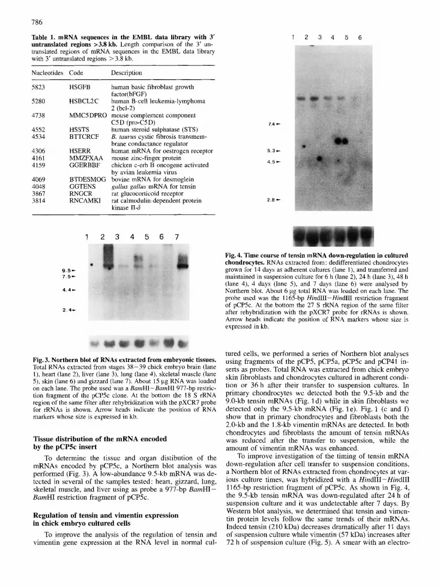
Fig. 4. Time course of tensin mRNA down-regulation in cultured chondrocytes. RNAs extracted from: dedifferentiated chondrocytes grown for 14 days as adherent cultures (lane I), and transferred and maintained in suspension culture for 6 h (lane 2), 24 h (lane 3), 48 h (lane 4), 4 days (lane 5) , and 7 days (lane 6) were analysed by Northern blot. About 6 pg total RNA was loaded on each lane. The probe used was the 1165-bp HindIII-Hind111 restriction fragment of pCPSc. At the bottom the 27 S rRNA region of the same filter after rehybridization with the pXCR7 probe for rRNAs is shown. Arrow heads indicate the position of RNA markers whose size is expressed in kb.
tured cells, we performed a series of Northern blot analyses using fragments of the pCP5, pCPSa, pCP5c and pCP41 in- serts as probes. Total RNA was extracted from chick embryo skin fibroblasts and chondrocytes cultured in adherent condi- tion or 36 h after their transfer to suspension cultures. In primary chondrocytes we detected both the 9.5-kb and the 9.0-kb tensin mRNAs (Fig. 1 d) while in skin fibroblasts we detected only the 9.5-kb mRNA (Fig. le) . Fig. 1 (c and f) show that in primary chondrocytes and fibroblasts both the 2.0-kb and the 1.8-kb vimentin mRNAs are detected. In both chondrocytes and fibroblasts the amount of tensin mRNAs was reduced after the transfer to suspension, while the amount of vimentin mRNAs was enhanced.
To improve investigation of the timing of tensin mRNA down-regulation after cell transfer to suspension conditions, a Northern blot of RNAs extracted from chondrocytes at var- ious culture times, was hybridized with a HindITI-Hind111 1165-bp restriction fragment of pCP5c. As shown in Fig. 4, the 9.5-kb tensin mRNA was down-regulated after 24 h of suspension culture and it was undetectable after 7 days. By Western blot analysis, we determined that tensin and vimen- tin protein levels follow the same trends of their mRNAs. Indeed tensin (210 kDa) decreases dramatically after 11 days of suspension culture while vimentin (57 kDa) increases after 72 h of suspension culture (Fig. 5). A smear with an electro-

787
Fig. 5. Western blot analysis. Protein extracts from dedifferentiated chondrocytes grown for 21 days as adherent cultures (lanes 1 and 3), and transferred and maintained in suspension culture for 72 h (lane 2) or 11 days (lane 4). 200 pg protein was loaded on each lane. Lanes 1 and 2 were incubated with the TL1 mAb specific for chicken tensin. Lanes 3 and 4 were incubated with a mAb specific for vimentin. Arrows and numbers on the left show electrophoretic mobilities of molecular mass standards expressed in kDa.
phoretic mobility of a protein of about 150 kDa was detected in several experiments with the anti-tensin mAb and most probably due to partial proteolysis although the extraction buffer contained several protease inhibitors. Also, the amount of these degradation products decreases after 11 days of sus- pension culture. The bands of about 66 and 100kDa ob- served in all the lanes are due to unspecific peroxidase bind- ing (data not shown).
To rule out the possibility that trypsin treatment may be responsible for this tensin down-regulation, a culture of de- differentiated chondrocytes was trypsin-digested, split and either transferred to suspension condition or replated in ad- herent condition. The tensin mRNA levels were checked by Northern blot and slot blot analysis 3 , 5 and 7 days after the treatment in both subcultures. Adherent cells showed no significant change in mRNA accumulation with respect to control cells that were not trypsin-treated, but cells in suspen- sion culture had a decrease in their tensin mRNA content (data not shown).
To determine the cellular distribution of tensin we per- formed indirect immunofluorescence assays (Fig. 6). The protein appears, in dedifferentiated chondrocytes cultured for 3 weeks in adherent condition on plastic petri dishes, and for a further 48 h on glass microscopy slides, in areas corre- sponding to dark adhesion plaques in interference reflection microscopy (Fig. 6A).
To check whether the ability of chondrocytes to express tensin was irreversibly lost after transfer to suspension condi- tion or whether it could be recovered, we performed an indi- rect immunofluorescence analysis on hypertrophic chondro- cytes, grown in suspension for 3 weeks and replated as adher- ent cells on glass microscopy slides. We found that tensin
Fig. 6. Cellular localization of tensin as revealed by indirect im- muno-fluorescence microscopy and interference reflection mi- croscopy. (A) Dedifferentiated chondrocytes grown for 3 weeks as adherent cells on plastic petri dishes and for further 48 h on glass microscopy slides. (B) Hypertrophic chondrocytes grown for 3 weeks in suspension and plated and maintained as adherent cells on glass microscopy slides for 8 days. Cells were methanol/acetone- fixed and stained with anti-tensin mAb. Inserts show the interference reflection microscopy images. Arrow heads indicate the correspon- dence between the sites of adhesion (dark zones in inserts) and ten- sin immunolocalization. Biotin-conjugated goat anti-mouse IgGs were used as secondary antibody and revealed by streptavidin conju- gated Texas red. Bar = 5 pm.
was expressed again by hypertrophic chondrocytes grown for 8 days on glass slides and localized in focal contacts (Fig. 6B). No differences of signal intensity or staining pattern were observed using the polyclonal or the mAb TLI (data not shown). Fluorescein-conjugated phalloidin staining of dedifferentiated chondrocytes or hypertrophic chondro- cytes grown in adherent condition did not show actin bundles but only a faint microfibrillar actin architecture (data not shown). Vimentin localization performed by indirect immu- nofluorescence analysis showed in adherent cells a peri- nuclear staining, while in round hypertrophic chondrocytes plated on poly-(L-lysine) only an intense spotted signal dis- tributed throughout the cytoplasm was observed (data not shown).
Localization of tensin mRNA and protein in vivo Tensin gene expression by chondrocytes in vivo was in-
vestigated by indirect immunofluorescence using the poly-

788
Fig. 7. Indirect immunofluorescence localization of tensin in chick embryo tibia and tibia-femur joint. (a, b) Stage 36 tibia-femur joint; arrow indicates articular cartilage, empty arrow indicates a ligament. (c, d) Proliferative cartilage zone. (e, f, Hypertrophic cartilage. Tissue sections were challenged with anti-tensin rabbit serum in (a, c and e) and with pre-immune rabbit serum (b, d and f). Bar = 33 pm.
clonal antibody on 36-37-stage and 39-40-stage chick embryo legs. At stages 36-37, the protein was detected in ligaments, perichondrium, articular and, although at a lower level, proliferative chondrocytes but it was barely detectable in hypertrophic chondrocytes (Fig. 7). No differences of staining pattern were observed by irnmunohistochemistry using the TL1 mAb (data not shown). At stages 39-40 the immunofluorescence signal was still detectable in articular chondrocytes, although decreased, and undetectable in prolif- erative and hypertrophic chondrocytes (data not shown). We performed in situ hybridizations with a specific in vitro tran- scribed riboprobe. In agreement with the immunofluores- cence results, the tensin mRNA was localized in articular
chondrocytes, perichondrium and ligaments but it was not present in other cartilage regions (Fig. 8).
DISCUSSION
We report here the isolation, from a chick embryo chon- drocyte library, of two cDNA clones specific for transcripts whose abundancy is regulated by adhesion.
On the basis of the nucleotide sequence, the pCP41 insert was identified as a vimentin-specific cDNA [30]. The se- quence of the pCP5 insert did not show any similarity with known sequences. Starting from nucleotide 128 of the com-

789
Fig.8. In situ hybridization of tensin mRNA in chick embryo tibia-femur joint. Tissue sections, obtained from a stage-39 embryo, were hybridized in (a) with sense and in (b) with antisense tensin radiolabelled RNA. Open arrow indicates the perichondrium, arrow indicates articular chondrocytes and arrow head indicates a ligament. Bar = 100 pm.
posite sequence shown in Fig. 2A, obtained by analyzing several overlapping clones, with the same specificity of pCP5, a near identity was found with the 3' portion of a partial cDNA sequence, GGTENSI, coding for chicken ten- sin. The sequence starting from nucleotide 128 shown in Fig. 2A has been named GGTENS by the EMBL data li- brary.
Northern analysis, with RNAs extracted from several avian embryonic tissues, showed that a 9.5-kb tensin tran- script is expressed in heart, gizzard, lung, skeletal muscle and liver while it is not in brain and skin. Based on our results we conclude that : (a) although the transcription start point(s) is(are) not determined yet, the sequence we deter- mined and the overlapping sequence GGTENSI cover almost completely the larger mRNA coding for tensin; (b) the small (about 10%) difference between the mRNA lengths deter- mined by sequencing and by Northern analysis may be due to relative inaccuracy in sizing very large RNAs on 1 % agar- ose gels; (c) the few sequence discrepancies in the overlap- ping region are probably due to polymorphism and, further- more, they have no effect on the amino acid sequence of the encoded protein.
The tensin mRNA has two peculiarities: a very large 3' untranslated region (4048 bases) and the absence of a canoni- cal AATAAA poly(A) addition signal. By computer-assisted searches for 3' untranslated region sequences larger than 3.8 kb in the EMBL data base, we found a few more mRNAs with comparable or larger 3' untranslated regions. No evident similarities were found among these sequences. Although mutations of the first base in the canonical poly(A) addition signal greatly reduce both cleavage and polyadenylation of RNA substrates and natural variants in the first base are very rare [31], we propose that the two TATAAA sequences found, at positions 3075 and 4845 in GGTENS, may have a poly(A) addition signal function.
In cartilage the modulation of tensin expression appears to be complex: the protein was detected at 10 days of embryo development in chondrocytes located in the articular zone, in the proliferative zone and to a lesser extent in the hypertro- phic zone. At later developmental stages, by in situ hybrid- ization, only chondrocytes in the articular zone show signifi- cant mRNA accumulation.
In dedifferentiated and hypertrophic cultured chondro- cytes we showed that the protein is localized in the focal contacts by interference reflection microscopy and confirmed that actin is not organized in cables in chondrocytes in these culture conditions [32]. We also showed that tensin mRNA
expression depends upon the adherent status of the cell ; in- deed the transfer to suspension determines the complete dis- appearance of the tensin message and a dramatic decrease of the protein amount. This modulation may be related to a change in cell shape andlor to a disanchorage from the sub- strate that in turn causes disruption of stress equilibrium be- tween cytoskeleton and the fibronectin matrix (supplied by the serum) absorbed to the plastic. Change in cell shape in- fluences gene expression [33] and differentiation [I 21. We have ruled out that the trypsin treatment alone influences tensin gene expression, as it occurs for osteopontin [34]. A reduction in the level of cytoskeletal proteins and their mRNAs as a general behaviour of the cell after detachment from the substrate was also ruled out because of the observed increase of vimentin protein and mRNAs. To our knowledge, it has not yet been investigated whether there are any molec- ular and/or structural interactions between tensin and inter- mediate filaments. An alteration of protein expression due to change in cell shape and/or transition in the differentiation stage has been observed for other actin binding proteins in different culture systems [35 -371. The regulation of tensin expression may play a role in cell migration. It is indeed known that migrating cells have protease activity on the cell surface [38] able to digest the surrouding matrix [39]. The consequent detachment may, in turn, reduce the amount of available tensin and therefore the ability of the cell to main- tain an organized microfilament network linked to the mem- brane leading to alteration in cell motility [40, 411 and possi- bly invasion.
This work was supported by grants from Progetti finalizzati: Ingegneria genetica and Applicazioni cliniche della ricerca oncolog- ica, Consiglio Nazionale delle Ricerche (Rome) and by funds from the Associazione Italiarza per la Ricerca sul Cancro (Italy). Rinke van de Werken was a recipient of a short-term COMETT fellowship from the European Community. We acknowledge the help of Drs Alfonso Colombatti, Roberto Doliana and Pier Car10 Marchisio with the in situ hybridization and the interference reflection microscopy, Giuliano Campanile for technical assistance and Barbara Minuto for editorial and secretarial assistance.
REFERENCES 1. Burridge, K., Fath, K., Kelly, T., Nuckolls, G. & Turner, C.
(1 988) Focal adhesions: transmembrane junctions between the extracellular matrix and the cytoskeleton, Annu. Rev. Cell Biol. 4, 487-525.
2. Albelda, S. M. & Buck, C. A. (1990) Integrins and other cell adhesion molecules, FASEB. J. 4 , 2868-2880.

790
3. Turner, C. E. & Burridge, K. (1991) Transmembrane molecular assemblies in cell-extracellular matrix interactions, Curl: Opin. Cell Biol. 3, 849-853.
4. Giancotti, F. G. & Ruoslahti, E. (1990) Elevated levels of the a$, fibronectin receptor suppresses the transformed pheno- type of Chinese hamster ovary cells, Cell 60, 849-859.
5. Bauer, J. S., Schreiner, C. L., Giancotti, F. G., Ruoslahti, E. & Juliano, R. L. (1990) Motility of fibronectin receptor-deficient cells on fibronectin and vitronectin : collaborative interaction among integrins, J. Cell Biol. 116, 477-487.
6. Kellie S. (1988) Cellular transformation, tyrosine kindse onco- genes, and the cellular adhesion plaque, Bioessays 8, 25 - 30.
7. Pardi, R., Inverardi, L., Rugarli, C. & Bender, J. R. (1992) Anti- gen-receptor complex stimulation triggers protein kinase C- dependent CDlla/CD18-~ytoskeleton association in T lym- phocytes, J. Cell Biol. 116, 1211-1220.
8. Hynes, R. 0. (1992) Integrins: versatility, modulation, and signaling in cell adhesion, Cell 69, 11 -25.
9. Juliano, R. L. & Haskill, S. (1993) Signal transduction from the extracellular matrix, J. Cell Biol. 120, 577-585.
10. Ingber, D. E. (1 990) Fibronectin controls capillary endothelial cell growth by modulating cell shape, Pro;. Natl Acad. Sci. USA 87. 3579-3583.
11. Ingber, D.’E.& Folkman, J. (1989) Mechanochemical switching between growth and differentiation during fibroblast growth factor-stimulated angiogenesis in vitro: role of extracellular matrix, J. Cell Biol. 109, 317-330.
12. Zanetti, N. C. & Solursh, M. (1984) Induction of chondrogen- esis in limb mesenchymal cultures by disruption of the actin cytoskeleton, J. Cell Biol. 99, 115-123.
13. Ferrell, J. E. &Martin, S. (1989) Tyrosine specific protein phos- phorylation is regulated by glycoprotein IIb-IIIa in platelets, Proc. Nut1 Acad. Sci. USA 86, 2234-2238.
14. Kornberg, L., Earp, H. S., Turner, C., Prokop, C. & Juliano, R. L. (1991) Signal transduction by integrins: increased protein tyrosine phosphorylation caused by clustering of beta1 inte- grins, Proc. Natl Acad. Sci. USA 88, 8392-8396.
15. Guan, J. L., Trevethick, J. E. & Hynes, R. 0. (1991) Fibronec- tinhntegrin interaction induces tyrosine phosphorylation of a 120-kD protein, Cell Regul. 2, 951 -964.
16. Hanks, S. K., Calalb, M. B., Harper, M. C:. & Patel, S. K. (1992) Focal adhesion protein-tyrosine kinase phosphorylated in response to cell attachment to fibronectin, Proc. Nut1 Acad. Sci. USA 89, 8487-8491.
17. Burridge, K., Turner, C. E. & Romer, L. H. (1992) Tyrosine phosphorylation of paxillin and pp12SFAK accompanies cell adhesion to extracellular matrix: a role in cytoskeletal assem- bly, J. Cell Biol. 119, 893-903.
18. Beckerle, M. C. (1990) The adhesion plaque protein, talin, is phosphorylated in vivo in chicken embryo fibroblasts exposed to a tumor-promoting phorbol ester, Cell Regul. 1, 227 -236.
19. Glenney, J. R. & Zokas, L. (1989) Novel tyrosine kinase sub- strates from Rous sarcoma virus-transformed cells are present in the membrane cytoskeleton, J. Cell Biol. 108, 2401 -2408.
20. Wu, H., Reynolds, A. B., Kanner, S. B., Vines, R. R. & Parsons, J. T. (1991) Identification and characterization of a novel cytoskeleton-associated pp6Osrc substrate, Mol. Cell. Biol. 11, 5113-5124.
21. Davis, S., Lu, M. L., Lo, S. H., Lin, S., Butler, J. A,, Druker. B. J., Roberts, T. M., An, Q. & Chen, L. B. (1991) Presence of an SH2 domain in the actin-binding protein tensin, Science
22. Castagnola, P., Moro, G., Descalzi-Cancedda, F. & Cancedda, R. (1986) Type X collagen synthesis during in vitro develop- ment of chick embryo tibia1 chondrocytes, J. Cell Biol. 102,
23. Ambesi-Impiombato, F. S., Parks, L. A. & Coon, H. G. (1980) Culture of hormone-dependent functional epithelial cells from rat thyroids, Proc. Nut1 Acad. Sci. USA 77, 345-3459,
252,712-715.
2310-2317.
24. Hamburger, V. & Hamilton, H. L. (1951) A series of normal stages in the development of the chick embryo, J. Morphol.
25. Quarto, R., Dozin, B., Tacchetti, C., Campanile, G., Malfatto, C. & Cancedda, R. (1990) In vitro development of hypertro- phic chondrocytes starting from selected clones of dedifferen- tiated cells, J. Cell Biol. 110, 1379-1386.
26. Chomczynski, P. & Sacchi, N. (1987) Single-step method of RNA isolation by acid guanidium thiocyanate-phenol-chloro- form extraction, Anal. Biochem. 162, 156-159.
27. Maniatis, T., Fritsch, E. F. & Sambrook, J. (1982) Molecular cloning: a laboratory manual, Cold Spring Harbor Labora- tory Press, Plainsville NY.
28. Castagnola, P., Dozin, B., Moro, G. & Cancedda, R. (1988) Changes in the expression of collagen genes show two stages in chondrocyte differentiation in vitro, J. Cell Biol. 106,461 - 467.
29. Melton D. A,, Krieg, P. A,, Rebagliati, M. R., Maniatis, T., Zinn, K. & Green, M. R. (1984) Efficient in vitro synthesis of biologically active RNA and RNA hybridization probes from plasmids containing a bacteriophage SP6 promoter, Nucleic Acids Res. 12, 7035-7056.
30. Zehner, Z. E., Li, X., Paterson, B. A. & Sax, C. M. (1987) The chicken vimentin gene, J. Biol. Chem. 262, 8112-8120.
31. Sheets, M. D., Ogg, S. C. & Wickens, M. P. (‘1990) Point mut- ations in AAUAAA and the poly(A) addition site: effects on the accuracy and efficiency of cleavage and polyadenylation in vitro, Nucleic Acids Res. 18, 5799-5805.
32. Mallein-Gerin, F., Garrone, R. & van der Rest, M. (1991) Pro- teoglycan and collagen synthesis are correlated with actin organization in dedifferentiating chondrocytes, Eul: J. Cell Biol. 56, 364-373.
33. Ben-Ze’ev, A. (1991) Animal cell shape changes and gene expression, Bioessays 13, 207-212.
34. Castagnola, P., Bet, P., Quarto, R. Gennari, M. & Cancedda, R. (1991) cDNA cloning and gene expression of chicken osteo- pontin, J. Biol. Chem. 266, 9944-9949.
35. Abd-el-Basset, E. M., Ahmed, I. & Fedoroff, S. (1991) Actin and actin-binding proteins in differentiating astroglia in tissue culture, J. Neurosci. Res. 30, 1 - 17.
36. Higgins, P. J. & Ryan, M. P. (1991) p52(PAI-1) and actin ex- pression in butyrate-induced flat revertants of v-ras-trans- formed rat kidney cells, Biochem. J. 279, 883-890.
37. Kubler, M. D., Jordan, P. W., O’Neill, C. H., & Watt, F. M. (1991) Changes in the abundance and distribution of actin and associated proteins during terminal differentiation of human epidermal keratinocytes, J. Cell. Sci. 100, 153- 165.
38. Estreicher, A., Muhlhauser, J., Carpentier, J. L., Orci, L. & Vas- salli, J. D. (1990) The receptor for urokinase-type plasmino- gen activator polarizes expression of the protease to the lead- ing edge of migrating monocytes and promotes degradation of enzyme inhibitor complexes, J . Cell Biol. 111, 783-792.
39. Hollas, W., Blasi, F. & Boyd, D. (1991) Role of the urokinase receptor in facilitating extracellular matrix invasion by cul- tured colon cancer, Cancer Rex 51, 3690-3695.
40. Cox, D., Condeelis, J., Wessels, D., Soll, D., Kern, H. & Knecht, D. A. (1992) Targeted disruption of the ABP-120 gene leads to cells with altered motility, J. Cell Biol. 116, 943-955.
41. Lampugnani, M. G., Giorgi, M., Gaboli, M., Dejana, E. & Marchisio, P. C. (1990) Endothelid cell motility, integrin re- ceptor clustering, and microfilament organization are inhib- ited by agents that increase intracellular CAMP, Lab. Invest.
88,49-92.
63, 521 -531.


