Embed Size (px)
Citation preview

Interaction of Surfactant Protein A with the Intermediate Filaments Desmin andVimentin†
Ignacio Garcia-Verdugo,*,‡ Monique Synguelakis,‡ Jeril Degrouard,§ Claudio-Areias Franco,| Benoit Valot,⊥
Michel Zivy,⊥ Richard Chaby,‡ and Zahra Tanfin‡
Institut de Biochimie et Biophysique Moleculaire et Cellulaire, UMR-8619 du CNRS, UniVersite de Paris-Sud,91400 Orsay, France, Centre Commun de Microscopie Electronique, UMR-8080 du CNRS, UniVersite de Paris-Sud,
91400 Orsay, France, UniVersite Pierre et Marie Curie, Paris 6, UMR-7079 du CNRS, Quai St-Bernard, 75005 Paris, France,and Plate-forme de Proteomique, UMR de Genetique Vegetale, Ferme de Moulon, 91190 Gif-sur-YVette, France
ReceiVed January 14, 2008; ReVised Manuscript ReceiVed March 27, 2008
ABSTRACT: Surfactant protein A (SP-A), a member of the collectin family that modulates innate immunity,has recently been involved in the physiology of reproduction. Consistent with the activation of ERK-1/2and COX-2 induced by SP-A in myometrial cells, we reported previously the presence of two majorproteins recognized by SP-A in these cells. Here we identify by mass spectrometry one of these SP-Atargets as the intermediate filament (IF) desmin. In myometrial preparations derived from desmin-deficientmice, the absence of binding of SP-A to any 50 kDa protein confirmed the identity of this SP-A-bindingsite as desmin. Our data based on partial chymotrypsin digestion of pure desmin suggested that SP-Arecognizes especially its rod domain, which is known to play an important role during the assembly ofdesmin into filaments. In line with that, electron microscopy experiments showed that SP-A inhibits invitro the polymerization of desmin filaments. SP-A also recognized in vitro polymerized filaments in acalcium-dependent manner at a physiological ionic strength but not the C1q receptor gC1qR. Furthermore,Texas Red-labeled SP-A colocalized with desmin filaments in myometrial cells. Interestingly, vimentin,the IF characteristic of leukocytes, is one of the major proteins recognized by SP-A in protein extracts ofU937 cells after PMA-induced differentiation of this monocytic cell line. Interaction of SP-A with vimentinwas further confirmed using recombinant vimentin in solid-phase binding assays. The ability of SP-A tointeract with desmin and vimentin, and to prevent polymerization of desmin monomers, shed light onunexpected and wider biological roles of this collectin.
Surfactant protein A (SP-A)1 is a member of the collectinfamily partially associated with surfactant layers in the lung.SP-A presents a globular C-terminal domain with calcium-dependent lectin activity and an N-terminal collagen-likedomain (1). The globular domain of SP-A recognizescarbohydrates and lipids on the surface of pathogens (2).Several calcium binding sites are also present in this globulardomain (3), and calcium induces conformational changes in
SP-A (4) that influence further interactions of the proteinwith its different ligands.
SP-A regulates the function of both alveolar type IIepithelial cells (AE-II) (the cells producing lung surfactant)and macrophages. In vitro, several studies have demonstratedthat SP-A inhibits lipid secretion and promotes the uptakeof lipids by AE-II, thus indicating a role of SP-A in surfactanthomeostasis. However, SP-A-deficient mice presented normalphospholipid metabolism unless challenged (5). This apparentlack of phenotype in SP-A-deficient mice could be relatedto alternate mechanisms of surfactant metabolism existingin SP-A gene-targeted mice (6). Therefore, SP-A is necessaryfor the normal clearence of surfactant lipids from the alveolarspace. With regard to macrophages, SP-A modulates phago-cytosis of pathogens, cytokine production, respiratory burst,and chemotaxis mediated by this cell type (2). Some of theseprocesses, such as phagocytosis or chemotaxis, involvecytoskeleton rearrangements necessary for monocyte and/or macrophages to move to areas of infection and efficientlyexert pseudopod-based phagocytosis. Interestingly, SP-Astimulates the polymerization of actin in alveolar macroph-ages in a highly organized and directional manner (7). Theseresults could explain some attributed functions of SP-Atoward macrophages, especially in phagocytosis and chemo-taxis processes. With the exception of different forms of
† Work supported by the Marie Curie Programme “Pulmo-Net”(MRTN-CT2004-512229) and by the “Centre National de la RechercheScientifique” and Paris-Sud University.
* To whom correspondence should be addressed: IBBMC, UMR-8619 du CNRS, Bat. 430, Universite de Paris-Sud, 91400 Orsay,France. Telephone: +33-169154830. Fax: +33-169853715. E-mail:[email protected].
‡ UMR-8619 du CNRS, Universite de Paris-Sud.§ UMR-8080 du CNRS, Universite de Paris-Sud.| Universite Pierre et Marie Curie.⊥ UMR de Genetique Vegetale.1 Abbreviations: AE-II, alveolar type II epithelial cells; HBSS,
Hank’s Balanced Salt Solution with calcium and magnesium; IF,intermediate filament; LC-MS, liquid chromatography-mass spec-trometry; MEM, minimal essential medium eagle; PMA, phorbolmyristate acetate; PVDF, polyvinylidene difluoride; RPMI, RoswellPark Memorial Institute (medium); SP-A, surfactant protein A; TEM,transmission electron microscopy; TR-SP-A, Texas Red-labeled SP-A; VD3, 1R,25-dihydroxyvitamin D3.
Biochemistry 2008, 47, 5127–5138 5127
10.1021/bi800070u CCC: $40.75 2008 American Chemical SocietyPublished on Web 04/12/2008

myosin (8, 9) and annexin IV (10), interactions betweenSP-A and cytosolic proteins have not been described so far.Therefore, the capacity of SP-A to modulate alveolar cellfunctions might arise from already observed interactions ofSP-A with cell membrane-expressed receptors.
Several receptors have been proposed for SP-A, some ofthem remaining incompletely characterized. Different reportsindicate that SP-A exhibits both pro-inflammatory and anti-inflammatory activities. Recently, Gardai and co-workersproposed a mechanism to explain these dual effects: bindingof SP-A globular heads to signal inhibitory receptors (SIRPR)will block stimulatory signaling pathways, whereas bindingof the SP-A collagenous tail to the calreticulin-CD91complex will induce the activation of p38 mitogen-activatedprotein (MAP) kinase and the production of pro-inflamma-tory mediators (11). One of the most accepted specific SP-Areceptors is SPR210, first described by Chroneos and co-workers (12). SPR210 mediates SP-A-enhanced uptake bymacrophages (13) and SP-A-induced inhibition of phospho-lipid secretion by AE-II (12). Recently, SPR210 has beensuccessfully identified as a cell surface form of myosin 18A(9). In vitro, direct interactions between SP-A and CD14 havebeen reported by Sano and co-workers. SP-A-CD14 interac-tion could explain the regulatory effects of SP-A in LPS-elicited responses in macrophages (14). However, SP-A isable to diminish LPS-induced production of TNF-R inalveolar macrophages of CD14-null mice (15), suggestingthat SP-A can also inhibit LPS-induced cytokine productionthrough interactions with other receptors like TLR-4 (16).
Other SP-A receptors are less characterized. Strayer andassociates found a 32 kDa protein associated with theregulation of surfactant metabolism. Antibodies against this32 kDa protein block the inhibitory effect of SP-A insecretagogue-stimulated surfactant secretion from AE-II cells(17). Stevens and co-workers described a 170-200 kDaprotein under nonreducing conditions and a 55 kDa proteinunder reducing conditions present in AE-II cell membranes,related to the SP-A-mediated lipid uptake by AE-II (18).Additional molecular or structural characterization of thisreceptor has not been provided.
Although SP-A was first described in alveolar epithelium,it has also been found in extrapulmonary tissues. Indeed,SP-A can be locally produced by fetal (19, 20) or reproduc-tive tissues, including the uterus (21). This suggests a newrole of SP-A in the physiology of reproduction, as recentlydemonstrated by Condon and associates (22). In this context,we have previously detected two major SP-A binding sitesin myometrial cell extracts, with molecular masses of 200and 50 kDa (23). Our observation that Texas Red-labeledSP-A (TR-SP-A) specifically binds to the surface of myo-metrial cells suggested for the first time the presence of SP-Abinding sites in myometrial cells. These interacting sitesmight account for the activation of ERK-1/2 and COX-2induced by SP-A in rat myometrial cells (23).
In this study, we have used different biochemical ap-proaches to identify the ∼50 kDa binding site recognizedby SP-A in myometrial cells and in protein extracts of U937cells after PMA-induced differentiation as desmin andvimentin, respectively. Our data thus demonstrate for the firsttime a direct interaction between SP-A and intermediatefilaments.
EXPERIMENTAL PROCEDURES
Materials. Human recombinant desmin and vimentin werepurchased from Fitzgerald (Concord, MA). RecombinantgC1qR/p33 was a gift from B. Ghebrehiwet (Stony Brook,NY). 1R,25-Dihydroxyvitamin D3 (VD3) was obtained fromBioMol (Plymouth Meeting, PA). Phorbol myristate acetate(PMA), bovine serum albumin (BSA), R-chymotrypsin,1,3,4,6-tetrachloro-3R,6�-diphenylglycouril (iodogen), TritonX-100, monoclonal mouse anti-desmin antibody (DE-U-10),monoclonal anti-vimentin antibody (LN-6), FITC-conjugatedanti-mouse antibody, and horseradish peroxidase (HRP)-conjugated anti-mouse IgM were obtained from SigmaChemical Co. (St. Louis, MO). C1q from human serum waspurchased from Merck-Calbiochem (Nottingham, U.K.).Na125I (0.78 MBq/µL) was purchased from ICN BiomedicalInc. (Irvine, CA). Fetal calf serum (FCS) was from BioMedia(Boussens, France). All other cell culture reagents, as wellas mouse IgG1 isotype control, Texas Red succinimidyl ester,and Prolog Fade, were from Invitrogen (Cergy Pontoise,France).
Animals and Tissue Processing. Female desmin-deficientmice were obtained by introducing a null mutation into thedesmin gene of mouse strain C57BL/6J (24). These mice,their corresponding wild type, and Wistar female rats(Janvier, France) were treated by intraperitoneal injectionof 30 µg of estradiol for the last 2 days before being euthanizedat the age of 30 days by standard carbon dioxide asphyxiation.All treatments were performed in accordance with the principlesand procedures outlined in the European guidelines for the careand use of experimental animals. The uterus was removed, andthe myometrium was separated from the endometrium bystripping, as previously described (25, 26).
Cell Culture. Primary cultures of myometrial cells wereprepared from rat myometrium by collagenase type IIdigestion, as previously described (27). Myometrial cellswere then cultured at a plating density of 15 × 103 cells/cm2 in MEM supplemented with 10% fetal calf serum at37 °C in an atmosphere of 5% CO2 and 95% humidified air.The human promonocytic U937 cell line used was obtainedfrom the European Collection of Cell Cultures (ECCAC,Salisbury, U.K.) and was cultured in RPMI supplementedwith 10% heat-inactivated and endotoxin-free FCS. U937cells were differentiated after incubation of the cells withPMA (1 nM) for 24 h or VD3 (10 nM) for 72 h in culturemedium.
Isolation of SP-A and Protein Labeling. Human SP-A waspurified from the bronchoalveolar lavage of patients withalveolar proteinosis following sequential extractions withbutanol and octyl glucoside (28). The purity of SP-A waschecked by SDS-PAGE in a 12% polyacrylamide gel underreducing conditions. The endotoxin content was 0.12 pg/µgof protein (Limulus Amebocyte Lysate assay, BioWhittaker).Fluorescent Texas Red-labeled SP-A derivative was preparedas previously described (23). The covalent coupling of thefluorochrome was assessed by SDS-PAGE with viewingof the gel under UV light. The capacity of this fluorescentderivative of SP-A to interact with DPPC vesicles was foundto be identical to that of the native SP-A. RadioiodinatedSP-A and C1q were prepared by the iodogen method ofGreenwood et al. (29) as previously described (30). Thespecific activities of the 125I-labeled SP-A and C1q were 2.25
5128 Biochemistry, Vol. 47, No. 18, 2008 Garcia-Verdugo et al.

× 106 and 2.56 × 10⁶ cpm/µg, respectively. The purities ofthe preparations were assessed by SDS-PAGE and autora-diography. As previously shown (23), labeled SP-A retainedits capacity to bind DPPC and Re-LPS, two well-knownligands of SP-A.
Autoradiography of SP-A Binding Proteins, Using [125I]SP-A. Detection of [125I]SP-A binding proteins present in blottedmembranes was performed as previously described (23) withmodifications. Briefly, myometrial tissues from rats or micewere homogenized in lysis buffer consisting of 1% CHAPSin 50 mM Tris (pH 7.5) and 300 mM NaCl, supplementedwith a cocktail of protease inhibitors (10 µg/mL aprotinin,1 mM PMSF, and 2 µg/mL pepstatin and leupeptin), 100µM orthovanadate, and 2 mM iodoacetamide. The extractswere centrifuged at 12000g for 20 min at 4 °C. Extractionswere dialyzed against 5 mM Tris and 150 mM NaCl (pH7.4) at 4 °C, and total proteins were precipitated in 65%saturation ammonium sulfate (12000g for 30 min at 4 °C).The pellet was dialyzed again before SDS-PAGE. Detergent-extracted proteins were analyzed by SDS-PAGE in 8%polyacrylamide gels using big gels (160 mm length, 100 mmwidth, 1 mm thickness) in the case of mass spectrometryidentification or small gels adapted to a Mini-Protean-IIsystem (Bio-Rad). Proteins were transferred onto PVDF (0.45µm) at 100 mA for small gels (1 h) or 150 mA for big gels(1 h). Membranes were blocked by incubation (overnight,4 °C) with 2% BSA in 5 mM Tris and 150 mM NaCl (pH7.4) with 0.1% Tween 20 (washing buffer). Blots werewashed and incubated (overnight at 4 °C or for 2 h at roomtemperature) with 0.2% BSA in washing buffer supplementedwith [125I]SP-A (0.5-2 × 10⁶ cpm) and 2 mM CaCl2. Blotswere thoroughly washed with washing buffer containing 2mM CaCl2, dried, and applied against X-ray films forautoradiography (Hyperfilm, Amersham Biosciences). Fi-nally, blotted proteins were stained with Coomassie Bluefollowing the manufacturer’s instructions. Differentiated ornondifferentiated U937 cells were washed in cold PBS andresuspended lysis buffer. After incubation for 1 h at 4 °Cunder continuous vortexing, cell debris was precipitated(12000g for 20 min at 4 °C) and proteins in clarifiedsupernatants or ammonium sulfate-precipitated fractions wereseparated in 8% SDS-PAGE gels. After being blotted,membranes were hybridized with [125I]SP-A as describedabove or with anti-vimentin antibodies. Anti-vimentin anti-body was developed using HRP-conjugated anti-mouse IgMas the secondary antibody and an ECL system (AmershanBiosciences).
LC-MS/MS Analysis. In-gel digestion of the cut band wasperformed with the Progest system (Genomic Solution)according to a standard trypsin protocol. Gel pieces werewashed twice by successive separate baths of 10% aceticacid, 40% ethanol, and acetonitrile (ACN). They were thenwashed twice with successive baths of 25 mM (NH4)2CO3
and ACN. Digestion was subsequently performed for 6 h at37 °C with 125 ng of modified trypsin (Promega) dissolvedin 20% methanol and 20 mM (NH4)2CO3. The peptides wereextracted successively with 2% trifluoroacetic acid (TFA)and 50% ACN and then with ACN. Peptide extracts weredried in a vacuum centrifuge and suspended in 20 µL of0.05% TFA, 0.05% HCOOH, and 2% ACN. HPLC wasperformed on an Ultimate LC system combined with a Famosautosampler and a Switchos II microcolumn switch system
(Dionex). A 4 µL sample was loaded at 5 µL/min on aprecolumn cartridge (stationary phase, C18 PepMap 100, 5µm; column, 300 µm inside diameter, 5 mm; Dionex) anddesalted with 0.05% TFA, 0.05% HCOOH, and 2% ACN.After 2.5 min, the precolumn cartridge was connected to theseparating PepMap C18 column (stationary phase, C18PepMap 100, 3 µm; column, 75 µm inside diameter, 150mm; Dionex). Buffers consisted of 0.1% HCOOH and 3%ACN (A) or 0.1% HCOOH and 95% ACN (B). The peptideseparation was achieved with a linear gradient from 5 to 30%B for 25 min at a rate of 200 nL/min. If the regenerationstep at 100% B and the equilibration step at 100% A wereincluded, one run took 45 min. Eluted peptides were analyzedonline with a LCQ Deca XP+ ion trap (Thermo Electron)using a nanoelectrospray interface. Ionization (ionizationpotential of 1.3-1.5 kV) was performed with liquid junctionand a noncoated capillary probe (10 mm inside diameter;New Objective). Peptide ions were analyzed using Xcalibur1.4 with the following data-dependent acquisition steps: (1)full MS scan [mass-to-charge ratio (m/z) of 400-1900,centroid mode], (2) ZoomScan on a selected precursor (scanat high resolution in profile mode on a m/z window of 4),and (3) MS/MS (qz ) 0.22, activation time ) 50 ms, andcollision energy ) 40%; centroid mode). Steps 2 and 3 wererepeated for the two major ions detected in step 1. Dynamicexclusion was set to 30 s. A database search was performedwith Bioworks 3.2 (Thermo Electron). Trypsin digestion, Cyscarboxyamidomethylation, and Met oxidation were set toenzymatic cleavage, static, and possible modifications,respectively. Precursor mass tolerance and fragment masstolerance were 1.4 and 1, respectively. The UniprotKBdatabase [combination of Swiss-Prot (version 52.5) andTrEMBL (version 35.5) databases; 4629251 entries on May15, 2007] was used. Identified tryptic peptides were filteredaccording to (i) their cross-correlation score (Xcorr), superiorto 1.7, 2.2, and 3.3 for mono-, di-, and tricharged peptides,respectively, and (ii) their probability inferior to 0.05. Aminimum of two different peptides was required. In the caseof identification with only two or three MS/MS spectra,similarity between the experimental and theoretical MS/MSspectra was visually confirmed.
Polymerization of Desmin Filaments. Recombinant desminwas reconstituted at 1 mg/mL (18.8 µM; M ) 53 kDa) in 9M urea buffer and aliquoted at -20 °C. From this stock,desmin was diluted 1/10 at 4 °C in 10 mM Tris-HCl (pH8.5) or 5 mM Tris-HCl, 150 mM NaCl, and 2 mM CaCl2
(pH 7.4) (final concentration of desmin of 1.8 µM) andincubated at 37 °C for 1 h. In selected experiments, SP-Awas added to polymerizing buffer at different final concen-trations (from 25 to 2.8 nM; M ) 640 kDa) before theaddition of desmin monomers. One aliquot of each samplewas analyzed by transmission electron microscopy.
Transmission Electron Microscopy (TEM). A drop of thesample was laid on a glow-discharged Formvar-coatedcopper grid (400 mesh) stabilized with evaporated carbonfilm. After 5 min, each grid was stained with 1% uranylacetate (1 min) and then examined at 80 kV with a PhilipsEM208 electron microscope. Digital images were acquiredusing a camera (Advantage HR3 AMT-Hamamatsu), anddiameters and lengths of all recognizable particles thatintersected a grid line, including protofilaments, protofibrils,
Interaction of SP-A with Intermediate Filaments Biochemistry, Vol. 47, No. 18, 2008 5129
and filaments, were measured with a Bioquant ImageAnalysis System (R&M Biometrics, Nashville, TN).
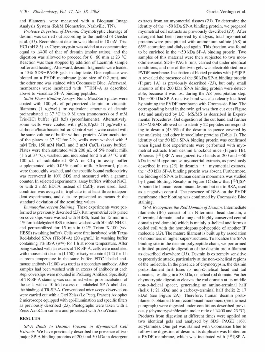
Protease Digestion of Desmin. Chymotryptic cleavage ofdesmin was carried out according to the method of Geisleret al. (31). Recombinant desmin was diluted in 10 mM Tris-HCl (pH 8.5). R-Chymotrypsin was added at a concentrationequal to 1/400 of that of desmin (molar ratios), and thedigestion was allowed to proceed for 0-60 min at 23 °C.Reaction was then stopped by addition of Laemmli samplebuffer and heating. Afterward, desmin fragments were loadedin 15% SDS-PAGE gels in duplicate. One replicate wasblotted on a PVDF membrane (pore size of 0.2 µm), andthe other one was stained with Coomassie Blue. Afterward,membranes were incubated with [125I]SP-A as describedabove to visualize SP-A binding peptides.
Solid-Phase Binding Assays. Nunc MaxiSorb plates werecoated with 100 µL of polymerized desmin or vimentinfilaments (1 µg/well) or equivalent amounts of desminpreincubated at 37 °C in 9 M urea (monomers) or 5 mMTris-HCl buffer (pH 8.5) (protofilaments). Alternatively,some wells were coated with gC1qR/p33 (1 µg/well) incarbonate/bicarbonate buffer. Control wells were coated withthe same volume of buffer without protein. After incubationof the plates at 37 °C for 2 h, wells were washed with 5mM Tris, 150 mM NaCl, and 2 mM CaCl2 (assay buffer).Plates were then saturated with 200 µL of 5% nonfat milk(1 h at 37 °C), washed, and incubated for 2 h at 37 °C with100 µL of radiolabeled SP-A or C1q in assay buffersupplemented with 0.05% nonfat milk. Afterward, plateswere thoroughly washed, and the specific bound radioactivitywas recovered in 10% SDS and measured with a gammacounter. In selected experiments, assay buffers without NaCl,or with 2 mM EDTA instead of CaCl2, were used. Eachcondition was assayed in triplicate in at least three indepen-dent experiments, and data are presented as means ( thestandard deviation of the resulting values.
Immunofluorescent Staining. These experiments were per-formed as previously described (23). Rat myometrial cells platedon coverslips were washed with HBSS, fixed for 15 min in a4% formaldehyde/HBSS mixture, washed with 50 mM NH4Cl,and permeabilized for 15 min in 0.2% Triton X-100 (v/v,HBSS) (washing buffer). Cells were first incubated with TexasRed-labeled SP-A (TR-SP-A) (20 µg/mL) in washing buffercontaining 1% BSA (w/v) for 1 h at room temperature. Afterbeing washed with an excess of TR-SP-A, cells were incubatedwith mouse anti-desmin (1:150) or isotype control (1:2) for 1 hat room temperature in the same buffer. FITC-labeled anti-mouse antibody (1:100) was used as a secondary antibody. Aftersamples had been washed with an excess of antibody at eachstep, coverslips were mounted in ProLong Antifade. Specificityof TR-SP-A staining was confirmed when prior incubation ofthe cells with a 10-fold excess of unlabeled SP-A abolishedthe binding of TR-SP-A. Conventional microscope observationswere carried out with a Carl Zeiss (Le Pecq, France) Axiophot2 microscope equipped with epi-illumination and specific filtersas previously described (23). Photographs were taken with aZeiss AxioCam camera and processed with AxioVision.
RESULTS
SP-A Binds to Desmin Present in Myometrial CellExtracts. We have previously described the presence of twomajor SP-A binding proteins of 200 and 50 kDa in detergent
extracts from rat myometrial tissues (23). To determine theidentity of the ∼50 kDa SP-A binding protein, we preparedmyometrial cell extracts as previously described (23). Afterdetergent had been removed by dialysis, total myometrialproteins were precipitated with ammonium sulfate (AS) at65% saturation and dialyzed again. This fraction was foundto be enriched in the ∼50 kDa SP-A binding protein. Twosamples of this material were then subjected to two mon-odimensional SDS-PAGE runs, carried out under identicalconditions, and one of the twin gels was electroblotted on aPVDF membrane. Incubation of blotted proteins with [125I]SP-A revealed the presence of the 50 kDa SP-A binding protein(Figure 1A) as previously described (23), but only smallamounts of the 200 kDa SP-A binding protein were detect-able, because it was lost during the AS precipitation step.The ∼50 kDa SP-A reactive band was also clearly localizedby staining the PVDF membrane with Coomassie Blue. Thecorresponding band in the twin gel was then cut out (Figure1A) and analyzed by LC-MS/MS as described in Experi-mental Procedures. Gel digestion of the cut band and furtherLC-MS/MS allowed us to identify 22 peptides correspond-ing to desmin (43.3% of the desmin sequence covered bythe analysis) and other intracellular proteins (Table 1). Theidentity of the 50 kDa SP-A binding protein was confirmedwhen ligand blot experiments were performed with myo-metrial extracts from desmin knockout mice (Figure 1B).Whereas [125I]SP-A recognized two bands at 200 and ∼50kDa in wild-type mouse myometrial extracts, as previouslydescribed in rats (23), in desmin -/- myometrial extracts,the ∼50 kDa SP-A binding protein was absent. Furthermore,the binding of SP-A to human desmin monomers was studiedby ligand blotting. Results in Figure 1C show that [125I]SP-A bound to human recombinant desmin but not to BSA, usedas a negative control. The presence of BSA on the PVDFmembrane after blotting was confirmed by Coomassie Bluestaining.
SP-A Recognizes the Rod Domain of Desmin. Intermediatefilaments (IFs) consist of an N-terminal head domain, aC-terminal domain, and a long and highly conserved centraldomain (rod domain) which is mainly R-helical and forms acoiled coil with the homologous polypeptide of another IFmolecule (32). The mature filament is built up by associationof monomers in higher superstructures. To localize the SP-Abinding site in the desmin polypeptide chain, we performeda limited proteolytic digestion of the desmin proto-filamentas described elsewhere (31). Desmin is extremely sensitiveto proteolytic attack, particularly at the non-R-helical regionsof the molecule. In the presence of chymotrypsin, the desminproto-filament first loses its non-R-helical head and taildomains, resulting in a 38 kDa, R-helical rod domain. Furtherchymotryptic digestion cleaves the rod domain at its middlenon-R-helical spacer, generating an amino-terminal half(helix 1; 21 kDa) and a carboxy-terminal half (helix 2; 17kDa) (see Figure 2A). Therefore, human desmin proto-filaments obtained from recombinant monomers (see the nextparagraph) were digested under conditions described previ-ously (chymotrypsin/desmin molar ratio of 1/400 and 23 °C).Products from digestion at different times were applied ontwo identical gels and analyzed by SDS-PAGE (16%acrylamide). One gel was stained with Coomassie Blue tofollow the digestion of desmin. Its duplicate was blotted ona PVDF membrane, which was incubated with [125I]SP-A.
5130 Biochemistry, Vol. 47, No. 18, 2008 Garcia-Verdugo et al.

As shown in Figure 2B, recombinant human desmin migratesas two major and very close bands around 55 kDa (bands“a”) and a minor band around 40 kDa (band “b”) which canhardly be detected at the starting time (Figure 2B). Bands“a” correspond to intact desmin. The presence of two closelyspaced bands in intact desmin has been previously describedand attributed to isoelectric variant forms of desmin withslightly different electrophoretic mobilities (33). Band “b”corresponds to desmin’s rod domain and represents less than5% of the recombinant protein according to the manufac-turer’s information. After digestion for 5 min under theconditions used in our experiments, intact desmin was hardlydetectable in Coomassie Blue-stained gels (Figure 2B). Atthat digestion time, most of the remaining polypetidecorresponds to the rod domain, as already reported by otherauthors (34). Increasing digestion time to more than 5 min(15-60 min) resulted in the degradation of the rod domainin a close double band of 40 kDa, and in the time-dependentappearance of bands around 24 kDa (band “c”) and 15 kDa(band “d”) (helices 1 and 2, respectively), both resulting from
excision of the rod domain. The appearance of a double bandaround 40 kDa in band “b” with long digestion times couldbe attributed to partial degradation of the rod domain bytraces of trypsin, present as a contaminant in chymotrypsin,and leading to CT-desmin (chymotrypsin/trypsin desmin-digested fragment) which presents a slightly faster mobilityin SDS-PAGE than the complete rod domain (34). Whendesmin fragments obtained at different digestion times wereblotted on a PVDF membrane (0.2 µm pore size) andincubated with [125I]SP-A, we observed a strong reactivityof SP-A with the rod domain (band “b”) but neither withhelix 1 (band “c”) nor with helix 2 (band “d”) (Figure 2C).Reactivity of SP-A with the rod domain was reduced whenthe rod domain was sequentially degraded over time. Thepresence of blotted bands “c” and “d” onto the membranewas confirmed after Coomassie Blue staining of the mem-brane. These data indicate that SP-A recognizes the roddomain of desmin. This is in accordance with the detectionof a second SP-A reactive band of 40 kDa present in somepreparations from rat myometrial tissues, identified as desminby mass spectrometry (data not shown). This band mightcorrespond to the rod domain of the protein. Therefore, theseresults show that SP-A binds also to rod domain formsproduced during natural degradation of desmin.
SP-A Impairs in Vitro Desmin Assembly. Mutations in therod domain of desmin in humans are associated with theaberrant polymerization and accumulation of desmin in vivo(35), in line with the participation of the rod domain in themaintenance of the stability of desmin filaments. Thus, ourresults showing that SP-A recognizes the rod domain inligand blot experiments (Figure 2) suggested that SP-A couldinterfere with the association of subunits and therefore coulddisturb the assembly of desmin filaments. As a consequence,we induced polymerization of desmin filaments from mono-mers in vitro to study the effects of SP-A during the processof desmin assembly. Recombinant desmin was supplied with
FIGURE 1: Binding of SP-A to blotted desmin monomers. (A) Rat myometrial proteins (20-30 µg) precipitated in ammonium sulfate (65%saturation) were analyzed by SDS--PAGE. The [125I]SP-A reactive band (see the arrow) in the ligand blot (right panel) was used tolocalize the SP-A binding protein of 50 kDa in gels (left panel), for further identification by LC-MS/MS (Table 1). (B) Ligand blot of[125I]SP-A reactive bands in myometrial extracts (20 µg) from wild-type mice (Control; left) and from desmin-deficient mice (Desm -/-;right). (C) Ligand blot analysis with [125I]SP-A of human recombinant desmin (Desm) and bovine serum albumin (BSA) (2 µg).
Table 1: LC-MS/MS Analysis of the ∼50 kDa SP-A Reactive Band inRat Myometriuma
accession description M coverage peptides
P48675 desmin, Rattus norVegicus (rat) 53.4 43.3 22 (16)P68369 tubulin R-1 chain, Mus musculus
(mouse)50.1 25.3 10 (8)
Q6P6V0 glucose phosphate isomerase,R. norVegicus (rat)
62.8 13.1 4 (4)
P04785 protein disulfide-isomeraseprecursor, R. norVegicus (rat)
56.9 13.9 4 (3)
Q6P736 polypyrimidine tract bindingprotein 1, R. norVegicus (rat)
59.3 11.2 3 (3)
a Accession, accession number of the protein (Swiss-Prot orTrEMBL); description, description of the protein; M, molecular mass ofthe protein (kilodaltons); Coverage, percent of the protein sequencecovered by the analysis; peptides, number of spectra (number of uniquepeptides) identified.
Interaction of SP-A with Intermediate Filaments Biochemistry, Vol. 47, No. 18, 2008 5131

9 M urea after reconstitution. Under these conditions of highurea concentration, desmin tends to form monomers (36).Desmin monomers (53 kDa) were diluted 1/10 (finalconcentration of desmin, 1.88 µM) in 5 mM Tris-HCl (pH8.5) and incubated at 37 °C for 1 h. Transmission electronmicroscopy (TEM) images (negative staining; 1% uranylacetate) showed homogeneous rod-shaped structures 32 (0.6 nm wide and 120 ( 8.3 nm long (n ) 25) (Figure 3A).These structures should correspond to individual proto-filaments (formed by association of two dimers) and associ-ated proto-filaments. It has been established that an increasein NaCl concentration in the medium induces a polymeri-zation of IFs (36). When we diluted desmin monomers in apolymerization buffer [5 mM Tris, 150 mM NaCl, and 2mM CaCl2 (pH 7.4)] and incubated the mixture at 37 °C for1 h, TEM images showed filament-like forms (30 ( 1 nmin diameter) with some lateral aggregation of desminfilaments (typically 10 nm in diameter) (Figure 3B). Thelength distribution of the filaments was not homogeneous,lengths of <300 nm corresponding to incompletely as-sembled filaments (Figure 3B). We then polymerized desminmonomers in polymerization buffer containing SP-A (25nM). As shown in Figure 3C, we did not observe under theseconditions the formation of filaments or parts of filamentsas we did in the absence of SP-A. Instead, there wereabundant grizzly forms associated with darker spots (arrowa in the inset of Figure 3C) and small filament-like forms(arrow b in the inset of Figure 3C). Thus, our data indicatethat assembly of desmin in vitro was disturbed in thepresence of SP-A. This effect was observed with SP-Aconcentrations between 5.6 and 25 nM. To exclude the
possibility that the effect of SP-A was due to a dilution ofdesmin below the concentration required for polymerization,we used BSA (25 nM) instead of SP-A and found indeedthat BSA did not affect the assembly of desmin (data notshown).
SP-A Binds to Desmin Filaments in a Calcium-DependentManner at a Physiological Ionic Strength. To study theinteraction of SP-A with desmin filaments, desmin monomerswere assembled in vitro as described above and used to coatMaxisorb microplates. Afterward, we analyzed the abilityof [125I]SP-A to bind to these desmin-coated plates. Figure4A shows that [125I]SP-A binds to desmin filaments in aconcentration-dependent manner in a buffer (pH 7.4) consist-ing of 5 mM Tris, 150 mM NaCl, and 2 mM CaCl2. Thebinding proved to be specific since it was inhibited byunlabeled SP-A (Figure 4D), and since [125I]SP-A did notbind to plates coated with gC1qR (gC1qR/p33), a receptorfor the globular head of C1q, the first component ofcomplement (Figure 4B). Furthermore, C1q did not recognizedesmin filaments under our experimental conditions (Figure4C), although the radioactive reagent used ([125I]C1q) wasfully functional since it bound to plates coated with gC1qR(Figure 4C). It was then important to analyze the dependenceof NaCl and calcium in the SP-A-desmin interaction.Indeed, calcium and ionic strength both play a role inmodulation of SP-A conformation in solution (4) and in thestability of desmin filaments (37). Thus, we analyzed therequirements of ionic strength and calcium for the ability ofSP-A to bind desmin. We observed that under physiologicalionic strength conditions (150 mM NaCl), calcium provedto be essential for desmin-SP-A interaction. Indeed, the
FIGURE 2: Identification of chymotryptic fragments of desmin recognized by [125I]SP-A. (A) Diagram of chymotrypsin digestion of intactdesmin. At short digestion times, desmin (a) gives the rod domain (b). Longer digestion times produce two fragments of >24 (c) and <17kDa (d). (B) Coomassie Blue-stained SDS-PAGE (15% polyacrylamide) of fragments a-d from chymotrypsin-digested desmin at variousdigestion times. (C) A similar gel was blotted, and SP-A binding fragments were detected with [125I]SP-A.
5132 Biochemistry, Vol. 47, No. 18, 2008 Garcia-Verdugo et al.

presence of EDTA dramatically reduced the level of bindingof SP-A to desmin (Figure 4D). In addition, the low ionicstrength (absence of NaCl) impaired the binding of [125I]SP-A to desmin, even in the presence of calcium (Figure 4D).The specificity of the binding of [125I]SP-A to desmin-coatedplates was assessed by the observation that unlabeled SP-Aefficiently inhibited the binding (Figure 4D).
SP-A Colocalizes with Desmin Filaments in Rat Myome-trial Cells. To determine whether SP-A associates with thedesmin filamentous network present in the living cells, weperformed double immunofluorescence studies in myometrialcells using anti-desmin antibodies and SP-A labeled withTexas Red (TR-SP-A) (Figure 5). We know from a previousstudy that TR-SP-A binds to living myometrial cells (23).Thus, we incubated myometrial cells with the TR-SP-Apreparation, and after the excess of TR-SP-A had been
washed out, the cells were incubated with the monoclonalmouse anti-desmin antibody and anti-mouse antibody coupledto FITC. Images from FITC emission showed a conspicuousdesmin network in myometrial cells (Figure 5, desmin). Asexpected from the data on SP-A-desmin interactionsmentioned above, images from TR emission also showed anetwork distribution of SP-A (Figure 5, SP-A). When imagesfrom FITC and TR fluorescence were overlaid, we observedthat most of the TR staining coincided with that of FITC(Figure 5, overlay), thus indicating that SP-A colocalizes withdesmin filaments present in myometrial cells. To excludethe possibility that colocalization of SP-A and desmin couldbe due to interactions between SP-A and the antibody, thesame experiment was performed using an unrelated controlantibody of the same isotype instead of the anti-desminantibody. We did not observe colocalization of FITC andTR fluorescence in this control experiment (data not shown).
SP-A Binds to Vimentin, Another IF Related to Desmin.SP-A modulates several macrophage functions acting throughspecific receptors present in the cell membrane (2). Therefore,using [125I]SP-A, we studied by ligand blot assays thepresence of SP-A binding proteins in detergent extracts fromthe pro-monocytic U937 cell line. In a previous analysis ofthis cell type, when we blotted total proteins from undif-ferentiated U937 cells, we observed that radioiodinated SP-Arecognized a single SP-A binding protein at approximately200 kDa (23) as also shown by others (12). This proteinmight correspond to SP-A receptor SPR210 isolated byChroneos and co-workers from U937 cells, recently identifiedas the unconventional myosin 18A (9). Interestingly, whensimilar experiments were performed with phorbol myristateacetate (PMA)-differentiated U937 cells, we observed asecond SP-A binding protein of 55 kDa (Figure 6A). Weanalyzed this 55 kDa SP-A binding protein by LC-MS/MSas described above for desmin, and we observed that onecandidate could be vimentin (Table 2). Further experimentsdesigned to corroborate that the 55 kDa SP-A binding proteincorresponds to vimentin were based on specific differentia-tion of U937 cells depending on the inducer used. Indeed,undifferentiated U937 cells do not express vimentin, theexpression of the protein being dramatically enhanced whencells are exposed to PMA (38). In parallel, they acquire amacrophage-like phenotype, including the capacity to adhereto substrates. On the other hand, 1,25-dihydroxyvitamin D3
(VD3) also confers a macrophage-like phenotype to U937cells, including the expression of CD14 (39), but withoutinducing vimentin expression (38). Western blot analyses,using a monoclonal antibody against vimentin, confirmedthe presence of vimentin in PMA-treated U937 cells but notin VD3-treated U937 cells or undifferentiated U937 cells(data not shown). When the same cellular extracts wereblotted and exposed to [125I]SP-A, we observed the presenceof the 55 kDa SP-A reactive protein only in PMA-treatedU937 cells and not in VD3-treated U937 cells (Figure 6A),in accordance with the vimentin expression pattern. Further-more, [125I]SP-A bound to pure human recombinant vimentinwhich migrates with an electrophoretic mobility similar tothat of the 55 kDa SP-A binding protein present in PMA-U937 extracts (Figure 6A). Therefore, our data provideevidence that the SP-A binding protein of 55 kDa present inprotein extracts of PMA-induced U937 cells corresponds tovimentin, another IF structurally related to desmin. To
FIGURE 3: Effect of SP-A on desmin polymerization. Recombinantdesmin monomers were diluted (final desmin concentration, 1.8µM) in a 5 mM Tris-HCl buffer (pH 8.5) (A), with buffer Asupplemented with 150 mM NaCl and 2 mM CaCl2 (B), or withbuffer B supplemented with 25 nM SP-A (C). An aliquot of thesample was laid on the grid of a Philips EM208 electron microscopeand stained (1 min) with 1% uranyl acetate. Pictures were taken at80 kV and are representative of the fields observed from threeindependent preparations. The scale bar is 500 nm; the inset scalebar in panel C is 100 nm.
Interaction of SP-A with Intermediate Filaments Biochemistry, Vol. 47, No. 18, 2008 5133

confirm the interaction of SP-A with vimentin under non-denaturing conditions, vimentin filaments were prepared asdescribed above for desmin and the ability of [125I]SP-A tobind vimentin-coated plates was assayed. As shown in Figure6B, [125I]SP-A bound to vimentin in a concentration-dependent manner, and unlabeled SP-A inhibited the bindingof radioactive SP-A (Figure 6B, right panel). The bindingof SP-A to vimentin, like that to desmin, was calcium-dependent (Figure 6B, right panel).
DISCUSSION
Surfactant protein A is a lung collectin that regulates theactivity of alveolar macrophages and alveolar type II cells.Although SP-A was originally reported to be synthesizedand secreted by alveolar type II cells, recent publicationsshow that this protein is also expressed in extrapulmonarytissues like fetal membranes (19, 20) or reproductive tissues,including uterus (21), suggesting a role of SP-A in thephysiology of reproduction (22). In this context, we havepreviously detected two major SP-A binding sites of 200and 50 kDa in myometrial extracts, in line with an ERK-1/2activation induced by SP-A in rat myometrial cells (23).ERK-1/2 activation is associated with uterine contractilityand preterm labor in rats (40). In the work presented here,using mass spectrometry, we identified the 50 kDa SP-Abinding protein of myometrial cells as the IF desmin. Inmyometrial preparations derived from desmin-deficient mice,the binding of SP-A at the level of 50 kDa did not occur,confirming the identity of this SP-A interacting site asdesmin. Our data provided evidence that SP-A bound to
desmin monomers from rat, mouse, and human origin inligand blot experiments. Binding of SP-A to desmin mono-mers was calcium-dependent (23), indicating that confor-mational changes in SP-A induced by calcium enhance itsbinding to blotted desmin. Using partial chymotrypticdigestion, we further identified the rod domain as the bindingsite of SP-A in the desmin polypeptide. However, our datacannot exclude the possibility that SP-A interacts with thenonhelical linker region rather than with the helical regionwithin the rod domain. In this case, the helical region mightbe required for the binding of SP-A to the linker regions.The rod domain and its linker region play a crucial roleduring assembly of IFs. Mutations in desmin’s rod domainare associated with the formation of aberrant filaments invitro and accumulation of desmin aggregates in vivo (35).In vitro, the first step in IF assembly is the formation of aparallel coiled-coil dimer, through stable interactions betweencentral rod domain regions of two monomers. Afterward,two dimers associate in a half-staggered, antiparallel mannerto form a tetramer. With an increase in ionic strength,tetramers associate laterally to form a so-called “unit-lengthfilament”. In the next step, these unit-length filaments anneallongitudinally to form loosely packed filaments that, in afurther step, will be compacted by narrowing of the filament’sdiameter (32). Our results show that SP-A binding perturbspolymerization of desmin in vitro. Using a dilution method,we were able to polymerize desmin in protofibrils andfilaments from recombinant monomers. In the presence ofSP-A, we observed small electron dense forms instead offilament-like forms. The most likely possibility is that SP-A
FIGURE 4: Binding of [125I]SP-A and [125I]C1q to plates coated with polymerized desmin. (A) Maxisorb plates uncoated (3) or coated withdesmin filaments (1 µg) (b) were blocked with 5% nonfat milk and incubated with [125I]SP-A at different concentrations per well (x-axis)in an assay buffer [5 mM Tris, 150 mM NaCl, and 2 mM CaCl2 (pH 7.4)] containing 0.05% nonfat milk. After the washings, boundradioactivity was recovered with 10% SDS and counted in a gamma counter (y-axis). Under the same experimental conditions, [125I]SP-Adid not bind to gC1qR/p33 (2 µg)-coated plates (B), and [125I]C1q bound to plates coated with gC1qR/p33 but not to plates coated withdesmin filaments (C). In panel D, binding of [125I]SP-A (180000 cpm/well) to uncoated (control) or desmin-coated plates was assayed usinga 5 mM Tris buffer (pH 7.4) containing 150 mM NaCl and 2 mM CaCl2 (NaCl+Ca2+), 150 mM NaCl and 2 mM EDTA (NaCl+EDTA),or 2 mM CaCl2 (Ca2+). For the bar delimited by the dashed line, binding of [125I]SP-A was performed in assay buffer (NaCl+Ca2+)containing unlabeled SP-A (25 µg/mL). Results represent the means ( standard deviation from three independent experiments performedin triplicate.
5134 Biochemistry, Vol. 47, No. 18, 2008 Garcia-Verdugo et al.

binds to the rod domain of monomers and impairs formationof dimers and subsequently their assembly in higher super-structures. However, when we assayed the binding of SP-Ato plates coated with desmin monomers, we observed thatSP-A hardly bound to monomers (results not shown) incomparison with preformed filaments (Figure 4). Thissuggests that SP-A recognizes the rod domain only underappropriate presentation of the latter (monomers blotted onmembranes but not adsorbed on plates). We cannot excludethe possibility that SP-A binding sites in the rod domainbecome accessible to SP-A only during the rearrangementsthat occur in some particular steps of the polymerization
process. This hypothesis implies that once SP-A interactswith the rearranged intermediate, further assembly is im-paired and/or assembled structures are disassembled. Whenwe examined desmin samples in the presence of SP-A byTEM at high magnification, we observed the presence ofelectron dense forms that resemble the bouquet shape ofSP-A and should correspond to aggregated forms of thecollectin. This is consistent with the fact that the polymer-izing buffer contains NaCl and calcium, two compounds thatinduce SP-A aggregation (4). These electron dense forms(SP-A aggregates) were found to be associated with darkerpoints (probably unpolymerized desmin) and short unraveledfilaments, indicating that SP-A interacts with desmin insolution and prevents or disrupts the assembly of themolecule in vitro. Moreover, we observed that SP-A wasable to depolymerize in vitro preformed desmin filaments(data not shown). Considering the effects of SP-A on thestability of desmin IF, our data suggest a potential role ofSP-A as an IF mucolytic agent which may be involved inprotection from lung injury and in repair processes. Further-more, SP-A could facilitate the clearence of IF from alveolarsubphase following lung injury, as previously described forcellular myosin (8).
Because IFs are present as filamentous structures in thecytoplasm of the cells, we further studied whether SP-A bindsto in vitro or in vivo assembled desmin filaments attachedto a solid support. The binding of SP-A to in vitro formedfilaments was calcium-dependent and occurs at a physiologi-cal ionic strength. Ionic strength might be necessary tocompensate for the electrostatic repulsion between thenegative surface charges of the two proteins at pH 7.Calcium, itself, induces conformational changes in SP-A (4)that affect further interactions of the protein with otherligands and receptors. In addition, NaCl and calcium improvethe stability of desmin filaments (37). Therefore, the presenceof both calcium and NaCl favors SP-A-desmin interaction.
Among members of the IF family, the rod domain is themost conserved region. Both vimentin and desmin exhibit ahigh degree of amino acid sequence identity in this domain.Vimentin is expressed in mesenchimal cells and is the mostabundant IF in leucocytes. We have identified vimentin as amajor SP-A reactive band present in PMA-U937 cell extracts.Other authors have previously studied the presence of SP-Areceptors in this monocytic cell line. Studies of Chroneosand co-workers identified the SP-A receptor named SPR-210kDa (myosin 18A) in U937 cells. Radio-iodinated SP-Adetected a single band of 210 kDa in U937 cell extracts usingligand blot assays (12). However, a 55 kDa SP-A bindingprotein was detected only after differentiation of U937 cellsby PMA. Sano and co-workers have identified CD14 asanother SP-A binding protein present in the membrane ofPMA-differentiated U937 cells (14). CD14 has a molecularmass similar to that of vimentin. When we differentiatedU937 cells with VD3, an agent that increases the level ofCD14 expression, we did not induce the expression of the∼50 kDa SP-A reactive band. Therefore, our data indicatethat the ∼50 kDa SP-A binding protein present in PMA-differentiated U937 cells is not CD14. Analysis by massspectrometry identified this protein as vimentin. We furtherconfirmed that vimentin is indeed an SP-A binding proteinwhen ligand blots were performed using a recombinant formof human vimentin. SP-A bound to monomeric and poly-
FIGURE 5: Colocalization of TR-SP-A with anti-desmin antibodiesin myometrial cells. Fixed and permeabilized rat myometrial cellswere doubly labeled at room temperature with TR-SP-A (20 µg/mL) and mouse monoclonal anti-desmin antibody (1/150) in HBSScontaining 1% BSA and 0.2% Triton X-100. FITC-labeled anti-mouse antibody was used as a secondary antibody. Conventionalepifluorescence microscopy was used to observe emission from TR(SP-A) or FITC (anti-desmin) using the corresponding emissionfilters. In the bottom panel, overlaid fluorescences are shown. Thescale bar is 20 µM. Data are representative of the fields observedfrom three independent preparations.
Interaction of SP-A with Intermediate Filaments Biochemistry, Vol. 47, No. 18, 2008 5135

merized vimentin, and as for desmin, the binding wascalcium- and NaCl-dependent, indicating that a similarmechanism governs interactions of both vimentin and desminwith SP-A. Recent experiments with SP-A-deficient micedemonstrate a role of SP-A in surfactant metabolism (5, 6).During surfactant production mediated by alveolar type IIepithelial cells, precursors of surfactant (lipoproteins andlipids) accumulate in the lamellar bodies that will be fusedwith the plasme membrane to release the surfactant at theaqueous subphase of the alveoli. Afterward, AE-II andmacrophage participate in surfactant clearance. Surfactant
metabolism is dependent on the integrity of the cytoskeleton,including the IF framework. In fact, several reports suggesta role of IFs (e.g., keratins and vimentin) in the secretionprocess in epithelial cells (41). Moreover, vimentin has beenimplicated in intracellular lipoprotein transport (42). Our datasuggest that intracellular SP-A-IF interactions could par-ticipate in an unknown role of SP-A in surfactant lipidprocessing.
Although IFs are cytoskeletal proteins present in thecytoplasm, several investigators observed an expression ofvimentin on the surface of leukocytes (43, 44). Moreover,the level of expression of vimentin on neutrophil membranesis increased under apoptosis (43) and might help in therecognition of the apoptotic cell by inflammatory mediatorsor by the immune cells. Apoptosis is an important phenom-enon in uterus physiology, especially after delivery. Afterparturition, the uterus undergoes a marked involution thatinvolves an ordered series of events, including apoptosis andphagocytosis of cellular debris (45, 46). Under these physi-ological conditions, extracellular IFs could be overexpressedand therefore may contribute to the recognition and clearanceof apoptotic cells and cell debris by immune cells. In thiscontext, SP-A locally produced (21), or coming fromamniotic fluid, could participate in the clearance of apoptoticcells through interactions with IFs. A parallel mechanism,initiated by the interaction of SP-A with its phagocytereceptor (the CD91-calreticulin complex) (47), occurs in
FIGURE 6: Binding of SP-A to vimentin. (A) [125I]SP-A reactive bands in SDS-PAGE blots of extracts (20 µg) of U937 cells differentiatedby incubation with PMA (1 nM, 24 h) or VD3 (10 nM, 72 h). Human recombinant vimentin (1 µg) was analyzed in parallel in the rightlane. (B) To produce the data depicted in the left panel, Maxisorb plates uncoated (3) or coated with 1 µg of vimentin filaments (b) wereblocked (5% nonfat milk) and incubated with [125I]SP-A at different concentrations per well (x-axis) in an assay buffer [5 mM Tris, 150mM NaCl, and 2 mM CaCl2 (pH 7.4)] containing 0.05% nonfat milk. After the washings, bound radioactivity was recovered with 10% SDSand counted with a gamma counter (y-axis). To produce the data depicted in the right panel, [125I]SP-A (50000 cpm/well) was incubatedwith uncoated (white bar) or coated vimentin plates in the presence of 25 µg/mL unlabeled SP-A (SP-A) or in assay buffer in which 2 mMCaCl2 was replaced with 2 mM EDTA (EDTA). Values are the means ( the standard deviation of three separate experiments performedin triplicate.
Table 2: LC-MS/MS Analysis of the ∼50 kDa SP-A Reactive Band inU937 Cell Extractsa
accession description M coverage peptides
P08670 vimentin, Homo sapiens(human)
53.6 45.5 24 (18)
P07237 protein disulfide-isomerase precursor,H. sapiens(human)
57.1 26.8 10 (10)
P06744 glucose-6-phosphateisomerase, H. sapiens(human)
63.1 19.7 8 (7)
Q9BUQ0 polypyrimidine tractbinding protein 1,H. sapiens (human)
59.6 11 4 (3)
a Accession, accession number of the protein (Swiss-Prot orTrEMBL); description, description of the protein; M, molecular mass ofthe protein (kilodaltons); coverage, percent of the protein sequencecovered by the analysis; peptides, number of spectra (number of uniquepeptides) identified.
5136 Biochemistry, Vol. 47, No. 18, 2008 Garcia-Verdugo et al.

lung alveoli, where it triggers SP-A-induced enhancementof phagocytosis in apoptotic neutrophils (48).
Desmin is well-known as a marker of the contractilephenotype for smooth muscle cells. Additionally, desmin andother IFs have been shown to participate in the cytoskeletalarchitectures that are crucial for maintaining the structuraland mechanical integrity of cells and tissues. Recently, a newnotion emerged which puts forward the functional role ofIFs as organizers or members of signaling platforms (49).In this context, our data provided the first evidence thatdesmin binds SP-A and may participate in the activation ofthe myometrial cell signaling pathway. Further investigationsare needed to elucidate the mechanism by which SP-A-desmin complexes, with or without other interactingproteins, regulate smooth muscle cell functions.
ACKNOWLEDGMENT
We thank Ginette Vilain for technical assistance, Dr. LiZhenlin for providing the desmin knockout mice, C. Oyeniranfor myometrial cell preparation, and Professor BerhaneGhebrehiwet for providing pure gC1q-R/p32.
REFERENCES
1. McCormack, F. X. (2001) Functional mapping of surfactant proteinA. Pediatr. Pathol. Mol. Med. 20, 293–318.
2. Crouch, E., and Wright, J. R. (2001) Surfactant proteins a and dand pulmonary host defense. Annu. ReV. Physiol. 63, 521–554.
3. Head, J. F., Mealy, T. R., McCormack, F. X., and Seaton, B. A.(2003) Crystal structure of trimeric carbohydrate recognition andneck domains of surfactant protein A. J. Biol. Chem. 278, 43254–43260.
4. Ruano, M. L., Garcia-Verdugo, I., Miguel, E., Perez-Gil, J., andCasals, C. (2000) Self-aggregation of surfactant protein A. Bio-chemistry 39, 6529–6537.
5. Jain, D., Dodia, C., Bates, S. R., Hawgood, S., Poulain, F. R., andFisher, A. B. (2003) SP-A is necessary for increased clearance ofalveolar DPPC with hyperventilation or secretagogues. Am. J.Physiol. 284, L759–L765.
6. Bates, S. R., Dodia, C., Tao, J. Q., and Fisher, A. B. (2008)Surfactant protein-A plays an important role in lung surfactantclearance: Evidence using the surfactant protein-A gene-targetedmouse. Am. J. Physiol. 294, L325–L333.
7. Tino, M. J., and Wright, J. R. (1999) Surfactant proteins A and Dspecifically stimulate directed actin-based responses in alveolarmacrophages. Am. J. Physiol. 276, L164–L174.
8. Michelis, D., Kounnas, M. Z., Argraves, W. S., Sanford, E. D.,Borchelt, J. D., and Wright, J. R. (1994) Interaction of surfactantprotein A with cellular myosin. Am. J. Respir. Cell Mol. Biol. 11,692–700.
9. Yang, C. H., Szeliga, J., Jordan, J., Faske, S., Sever-Chroneos, Z.,Dorsett, B., Christian, R. E., Settlage, R. E., Shabanowitz, J., Hunt,D. F., Whitsett, J. A., and Chroneos, Z. C. (2005) Identification ofthe surfactant protein A receptor 210 as the unconventional myosin18A. J. Biol. Chem. 280, 34447–34457.
10. Sohma, H., Creutz, C. E., Saitoh, M., Sano, H., Kuroki, Y., Voelker,D. R., and Akino, T. (1999) Characterization of the Ca2+-dependentbinding of annexin IV to surfactant protein A. Biochem. J. 341(Part 1), 203–209.
11. Gardai, S. J., Xiao, Y. Q., Dickinson, M., Nick, J. A., Voelker,D. R., Greene, K. E., and Henson, P. M. (2003) By binding SIRPRor calreticulin/CD91, lung collectins act as dual function surveil-lance molecules to suppress or enhance inflammation. Cell 115,13–23.
12. Chroneos, Z. C., Abdolrasulnia, R., Whitsett, J. A., Rice, W. R.,and Shepherd, V. L. (1996) Purification of a cell-surface receptorfor surfactant protein A. J. Biol. Chem. 271, 16375–16383.
13. Weikert, L. F., Edwards, K., Chroneos, Z. C., Hager, C., Hoffman,L., and Shepherd, V. L. (1997) SP-A enhances uptake of bacillusCalmette-Guerin by macrophages through a specific SP-A receptor.Am. J. Physiol. 272, L989–L995.
14. Sano, H., Sohma, H., Muta, T., Nomura, S., Voelker, D. R., andKuroki, Y. (1999) Pulmonary surfactant protein A modulates thecellular response to smooth and rough lipopolysaccharides byinteraction with CD14. J. Immunol. 163, 387–395.
15. Alcorn, J. F., and Wright, J. R. (2004) Surfactant protein A inhibitsalveolar macrophage cytokine production by CD14-independentpathway. Am. J. Physiol. 286, L129–L136.
16. Yamada, C., Sano, H., Shimizu, T., Mitsuzawa, H., Nishitani, C.,Himi, T., and Kuroki, Y. (2006) Surfactant protein A directlyinteracts with TLR4 and MD-2 and regulates inflammatory cellularresponse. Importance of supratrimeric oligomerization. J. Biol.Chem. 281, 21771–21780.
17. Strayer, D. S., Pinder, R., and Chander, A. (1996) Receptor-mediated regulation of pulmonary surfactant secretion. Exp. CellRes. 226, 90–97.
18. Wissel, H., Looman, A. C., Fritzsche, I., Rustow, B., and Stevens,P. A. (1996) SP-A-binding protein BP55 is involved in surfactantendocytosis by type II pneumocytes. Am. J. Physiol. 271, L432–L440.
19. Miyamura, K., Malhotra, R., Hoppe, H. J., Reid, K. B., Phizack-erley, P. J., Macpherson, P., and Lopez Bernal, A. (1994) Surfactantproteins A (SP-A) and D (SP-D): Levels in human amniotic fluidand localization in the fetal membranes. Biochim. Biophys. Acta1210, 303–307.
20. Sun, K., Brockman, D., Campos, B., Pitzer, B., and Myatt, L. (2006)Induction of surfactant protein A expression by cortisol facilitatesprostaglandin synthesis in human chorionic trophoblasts. J. Clin.Endocrinol. Metab. 91, 4988–4994.
21. Kankavi, O., Ata, A., and Gungor, O. (2007) Surfactant proteinsA and D in the genital tract of mares. Anim. Reprod. Sci. 98, 259–270.
22. Condon, J. C., Jeyasuria, P., Faust, J. M., and Mendelson, C. R.(2004) Surfactant protein secreted by the maturing mouse fetal lungacts as a hormone that signals the initiation of parturition. Proc.Natl. Acad. Sci. U.S.A. 101, 4978–4983.
23. Garcia-Verdugo, I., Leiber, D., Robin, P., Billon-Denis, E., Chaby,R., and Tanfin, Z. (2007) Direct interaction of surfactant proteinA with myometrial binding sites: Signaling and modulation bybacterial lipopolysaccharide. Biol. Reprod. 76, 681–691.
24. Li, Z., Colucci-Guyon, E., Pincon-Raymond, M., Mericskay, M.,Pournin, S., Paulin, D., and Babinet, C. (1996) Cardiovascularlesions and skeletal myopathy in mice lacking desmin. DeV. Biol.175, 362–366.
25. Lajat, S., Tanfin, Z., Guillon, G., and Harbon, S. (1996) Modulationof phospholipase C pathway and level of GqR/G11R in ratmyometrium during gestation. Am. J. Physiol. 271, C895–C904.
26. Robin, P., Boulven, I., Bole-Feysot, C., Tanfin, Z., and Leiber, D.(2004) Contribution of PKC-dependent and -independent processesin temporal ERK regulation by ET-1, PDGF, and EGF in ratmyometrial cells. Am. J. Physiol. 286, C798–C806.
27. Boulven, I., Palmier, B., Robin, P., Vacher, M., Harbon, S., andLeiber, D. (2001) Platelet-derived growth factor stimulates phos-pholipase C-γ1, extracellular signal-regulated kinase, and arachi-donic acid release in rat myometrial cells: Contribution to cyclic3′,5′-adenosine monophosphate production and effect on cellproliferation. Biol. Reprod. 65, 496–506.
28. Hawgood, S., Benson, B. J., Schilling, J., Damm, D., Clements,J. A., and White, R. T. (1987) Nucleotide and amino acid sequencesof pulmonary surfactant protein SP 18 and evidence for cooperationbetween SP 18 and SP 28-36 in surfactant lipid adsorption. Proc.Natl. Acad. Sci. U.S.A. 84, 66–70.
29. Greenwood, F. C., Hunter, W. M., and Glover, J. S. (1963) ThePreparation of I-131-Labelled Human Growth Hormone of HighSpecific Radioactivity. Biochem. J. 89, 114–123.
30. Rety, S., Salamitou, S., Garcia-Verdugo, I., Hulmes, D. J., LeHegarat, F., Chaby, R., and Lewit-Bentley, A. (2005) The crystalstructure of the Bacillus anthracis spore surface protein BclA showsremarkable similarity to mammalian proteins. J. Biol. Chem. 280,43073–43078.
31. Geisler, N., Kaufmann, E., and Weber, K. (1982) Proteinchemicalcharacterization of three structurally distinct domains along theprotofilament unit of desmin 10 nm filaments. Cell 30, 277–286.
32. Herrmann, H., Bar, H., Kreplak, L., Strelkov, S. V., and Aebi, U.(2007) Intermediate filaments: From cell architecture to nanome-chanics. Nat. ReV. Mol. Cell Biol. 8, 562–573.
33. Izant, J. G., and Lazarides, E. (1977) Invariance and heterogeneityin the major structural and regulatory proteins of chick muscle cellsrevealed by two-dimensional gel electrophoresis. Proc. Natl. Acad.Sci. U.S.A. 74, 1450–1454.
Interaction of SP-A with Intermediate Filaments Biochemistry, Vol. 47, No. 18, 2008 5137

34. Ip, W. (1988) Modulation of desmin intermediate filament assemblyby a monoclonal antibody. J. Cell Biol. 106, 735–745.
35. Goldfarb, L. G., Vicart, P., Goebel, H. H., and Dalakas, M. C.(2004) Desmin myopathy. Brain 127, 723–734.
36. Kaufmann, E., Weber, K., and Geisler, N. (1985) Intermediatefilament forming ability of desmin derivatives lacking either theamino-terminal 67 or the carboxy-terminal 27 residues. J. Mol.Biol. 185, 733–742.
37. Chou, R. G., Stromer, M. H., Robson, R. M., and Huiatt, T. W.(1990) Determination of the critical concentration required fordesmin assembly. Biochem. J. 272, 139–145.
38. Taimi, M., Chateau, M. T., Marti, J., and Pacaud, M. (1990)Induction of differentiation of the human histiocytic lymphomacell line U937 in the absence of vimentin expression. Differentiation45, 55–60.
39. Bhalla, A. K., Williams, M. M., Lal, S., and Lydyard, P. M. (1989)1,25-Dihydroxyvitamin D3, but not retinoic acid, induces thedifferentiation of U937 cells. Clin. Exp. Immunol. 76, 274–277.
40. Li, Y., Je, H. D., Malek, S., and Morgan, K. G. (2004) Role ofERK1/2 in uterine contractility and preterm labor in rats. Am. J.Physiol. 287, R328–R335.
41. Oriolo, A. S., Wald, F. A., Ramsauer, V. P., and Salas, P. J. (2007)Intermediate filaments: A role in epithelial polarity. Exp. Cell Res.313, 2255–2264.
42. Heidenthal, A. K., Weber, P. C., Lottspeich, F., and Hrboticky, N.(2000) The binding in vitro of modified LDL to the intermediatefilament protein vimentin. Biochem. Biophys. Res. Commun. 267,49–53.
43. Moisan, E., and Girard, D. (2006) Cell surface expression ofintermediate filament proteins vimentin and lamin B1 in humanneutrophil spontaneous apoptosis. J. Leukocyte Biol. 79, 489–498.
44. Mor-Vaknin, N., Punturieri, A., Sitwala, K., and Markovitz, D. M.(2003) Vimentin is secreted by activated macrophages. Nat. CellBiol. 5, 59–63.
45. Takamoto, N., Leppert, P. C., and Yu, S. Y. (1998) Cell deathand proliferation and its relation to collagen degradation in uterineinvolution of rat. Connect. Tissue Res. 37, 163–175.
46. Nilsen-Hamilton, M., Liu, Q., Ryon, J., Bendickson, L., Lepont,P., and Chang, Q. (2003) Tissue involution and the acute phaseresponse. Ann. N.Y. Acad. Sci. 995, 94–108.
47. Vandivier, R. W., Ogden, C. A., Fadok, V. A., Hoffmann, P. R.,Brown, K. K., Botto, M., Walport, M. J., Fisher, J. H., Henson,P. M., and Greene, K. E. (2002) Role of surfactant proteins A, D,and C1q in the clearance of apoptotic cells in vivo and in vitro:Calreticulin and CD91 as a common collectin receptor complex.J. Immunol. 169, 3978–3986.
48. Schagat, T. L., Wofford, J. A., and Wright, J. R. (2001) Surfactantprotein A enhances alveolar macrophage phagocytosis of apoptoticneutrophils. J. Immunol. 166, 2727–2733.
49. Pallari, H. M., and Eriksson, J. E. (2006) Intermediate filamentsas signaling platforms. Sci. STKE 2006, pe53.
BI800070U
5138 Biochemistry, Vol. 47, No. 18, 2008 Garcia-Verdugo et al.


