Embed Size (px)
Citation preview
MOLECULAR TOXICOLOGY
Molecular characterization of an acidic phospholipase A2
from Bothrops pirajai snake venom: synthetic C-terminal peptideidentifies its antiplatelet region
Sabrina S. Teixeira • Lucas B. Silveira • Franco M. N. da Silva • Daniela P. Marchi-Salvador •
Floriano P. Silva Jr • Luiz Fernando M. Izidoro • Andre L. Fuly • Maria A. Juliano •
Camila R. dos Santos • Mario T. Murakami • Suely V. Sampaio • Saulo L. da Silva •
Andreimar M. Soares
Received: 22 October 2010 / Accepted: 31 January 2011
� Springer-Verlag 2011
Abstract This paper describes a biochemical and phar-
macological characterization of BpirPLA2-I, the first acidic
Asp49-PLA2 isolated from Bothrops pirajai. BpirPLA2-I
caused hypotension in vivo, presented phospholipolytic
activity upon artificial substrates and inhibitory effects on
platelet aggregation in vitro. Moreover, a synthetic peptide
of BpirPLA2-I, comprising residues of the C-terminal
region, reproduced the antiplatelet activity of the intact
protein. A cDNA fragment of 366 bp encompassing the
mature form of BpirPLA2-I was cloned by reverse trans-
criptase-PCR of B. pirajai venom gland total RNA.
A Bayesian phylogenetic analysis indicated that BpirPLA2-
I forms a clade with other acid Asp49-PLA2 enzymes
from the Bothrops genus, which are characterized by the
high catalytic activity associated with anticoagulant or
hypotensive activity or both. Comparison of the electro-
static potential (EP) on the molecular surfaces calculated
from a BpirPLA2-I homology model and from the crys-
tallographic models of a group of close homologues
revealed that the greatest number of charge inversions
occurred on the face opposite to the active site entrance,
particularly in the Ca2? ion binding loop. This observation
suggests a possible relationship between the basic or acid
character of PLA2 enzymes and the functionality of the
Ca2? ion binding loop.
Keywords Snake venom � Acidic phospholipase A2 �Biochemical and pharmacological characterization �Bothrops pirajai � Antiplatelet domain
S. S. Teixeira � L. B. Silveira � F. M. N. da Silva �D. P. Marchi-Salvador � L. F. M. Izidoro �S. V. Sampaio � A. M. Soares
Departamento de Analises Clınicas, Toxicologicas e
Bromatologicas, Faculdade de Ciencias Farmaceuticas de
Ribeirao Preto, FCFRP-USP, Ribeirao Preto, SP, Brazil
D. P. Marchi-Salvador
Departamento de Biologia Molecular, Centro de Ciencias
Exatas e da Natureza, DBM-UFPB, Joao Pessoa, PB, Brazil
F. P. Silva Jr
Laboratorio de Bioquımica de Proteınas e Peptıdeos, Instituto
Oswaldo Cruz, IOC-FIOCRUZ, Rio de Janeiro, RJ, Brazil
L. F. M. Izidoro
Faculdade de Ciencias Integradas do Pontal, UFU,
Ituiutaba, MG, Brazil
A. L. Fuly
Departamento de Biologia Celular e Molecular (GCM),
Instituto de Biologia, UFF, Niteroi, RJ, Brazil
M. A. Juliano
Departamento de Biofısica, UNIFESP, Sao Paulo, SP, Brazil
C. R. dos Santos � M. T. Murakami
Laboratorio Nacional de Biociencias, Centro Nacional de
Pesquisas em Energia e Materiais, Campinas, SP, Brazil
S. L. da Silva
Universidade Federal de Sao Joao Del Rei, UFSJ Campus,
Divinopolis, MG, Brazil
A. M. Soares (&)
Departamento de Analises Clınicas, Toxicologicas e
Bromatologicas, Faculdade de Ciencias Farmaceuticas de
Ribeirao Preto, Universidade de Sao Paulo—USP, Avenida
do Cafe, s/n�, 14040-903 Ribeirao Preto, SP, Brazil
e-mail: [email protected]
123
Arch Toxicol
DOI 10.1007/s00204-011-0665-6
Introduction
Snake venoms are rich in phospholipases A2 (PLA2s, EC
3.1.1.4), which induce a large variety of pharmacological
and toxic effects, such as myotoxicity, neurotoxicity, car-
diotoxicity, hemolysis, hypotension, bleeding, edema, and
effects on platelet aggregation (Gutierrez and Lomonte
1997; Harris et al. 2000; Soares et al. 2004; De Paula et al.
2009).
PLA2s are enzymes that catalyze the hydrolysis of
2-acyl ester bonds of 3-sn-phospholipids producing fatty
acids and lysophospholipids. The Ca2? ion, an essential
cofactor, and an Asp residue at position 49 are critical
points for the catalysis upon artificial substrates (Arni and
Ward 1996). There are five types of these enzymes, namely
secreted PLA2s (sPLA2), cytosolic PLA2s (cPLA2), Ca2?
independent PLA2s (iPLA2), platelet-activating factor
acetylhydrolases (PAF-AH), and lysosomal PLA2s
(Schaloske and Dennis 2006).
Snake venom PLA2s (svPLA2s) are secreted proteins
belonging to the groups I and II. The svPLA2s of group I
are found in the Elapidae family (Elapinae e Hydrophii-
nae), whereas those of the group II are found in venoms
from Viperidae family (Viperidae e Crotalinae). Usually,
the group IIA svPLA2s are divided in two main subgroups:
(i) Asp49-PLA2s, which display an Asp residue at position
49, with relatively high catalytic activity upon artificial
substrates; and (ii) Lys49-PLA2s, showing a Lys residue at
position 49, with no catalytic activity (Arni and Ward
1996; Gutierrez and Lomonte 1997; Ownby et al. 1999;
Soares et al. 2004; De Paula et al. 2009).
Acidic PLA2s isolated from snake venoms have not been
fully characterized yet; however, in recent years, the interest
in these proteins has increased due to the fact that some
enzymes do not present myotoxicity, but induce significant
pharmacological effects such as inhibition of platelet
aggregation and hypotension (Serrano et al. 1999; Andriao-
Escarso et al. 2002; Fuly et al. 2002; Roberto et al. 2004; De
Albuquerque Modesto et al. 2006; Fernandez et al. 2010).
This paper describes the isolation as well as the bio-
chemical, functional, and structural characterization of the
first acidic Asp49-PLA2 (named BpirPLA2-I) from Bothr-
ops pirajai snake venom and shows that synthetic C-ter-
minal peptide reproduce the antiplatelet effect of the intact
protein.
Materials and methods
Materials
Desiccated B. pirajai venom was purchased from Bio-
agents Serpentarium (Batatais, SP, Brazil). Male Swiss
mice, weighing 18–25 g, were provided by Bioterio Cen-
tral, Universidade de Sao Paulo (USP), Ribeirao Preto, SP,
Brazil. Animal care was in accordance with the guidelines
of the Brazilian College for Animal Experimentation
(COBEA) and was approved by the Committee for Ethics
in Animal Utilization of Universidade de Sao Paulo
(n�07.1.516.53.6) and IBAMA (n�11781-1). Collagen
(Type I) from bovine tendon was purchased from Chrono-
Log Corporation, and adenosine diphosphate (ADP) and p-
bromophenacyl bromide (BPB) were from Sigma Chemical
Co. (St. Louis, USA). Fluorescent substrates Acyl 6:0
nitrobenzoxadiazole phospholipids (NBD-phospholipids):
NBD-phosphatidylcholine (PC), NBD-phosphatidylglyc-
erol (PG), NBD-phosphatidylethanolamine (PE), or NBD-
phosphatidic acid (PA) were from Avanti Polar Lipids Inc.
(Alabaster, USA). CM-Sepharose resin was purchased
from Amersham Biosciences (Uppsala, Sweden). All other
reagents used were of analytical grade.
Purification of BpirPLA2-I
A 200 mg sample of desiccated B. pirajai venom was
dissolved in 1.5 ml of 50 mM ammonium bicarbonate
(ambic) buffer, pH 7.8, cleared by centrifugation at
480 9 g for 10 min, and applied on a CM-Sepharose Fast
Flow column (2.0 9 20 cm) which was previously equili-
brated with the same buffer. A linear gradient was then
applied up to 1.0 M NaCl in ambic buffer and fractions of
3.5 ml/tube were collected at a flow rate of 20 ml/h. The
fraction with PLA2 activity (PI-B) was collected and re-
chromatographed on a Shimadzu C18 reverse-phase high-
performance liquid chromatography (RP-HPLC) column
(4.6 9 150 mm), which was equilibrated with solvent A
(5% acetonitrile, 0.1% trifluoroacetic acid) and eluted with
a concentration gradient of solvent B (60% acetonitrile,
0.1% trifluoroacetic acid) from 30 to 100%, at a flow rate
of 1.0 ml/min for 30 min (Andriao-Escarso et al. 2000,
2002). All steps of the purification procedure were carried
out at room temperature (25�C). The pure acidic PLA2
derived from the third RP-HPLC fraction, named Bpir-
PLA2-I, was lyophilized and used for biochemical/phar-
macological characterization and amino acid sequence
determination.
Biochemical characterization
Polyacrylamide gel electrophoresis (PAGE) was performed
in the presence of sodium dodecyl sulfate (SDS–PAGE)
following a previously described method (Laemmli 1970).
Isoelectric focusing was run according to Vesterberg
(1972). Buffalyte, pH range 3.5–9.0 (Pierce, IL), was used
to generate the pH gradient. For the N-terminal sequencing,
BpirPLA2-I was dissolved in 0.4 M Tris–HCl buffer, pH
Arch Toxicol
123
8.1, containing 6 M guanidine-HCl, reduced by DTT
(dithiothreitol), and carboxymethylated by iodoacetic acid.
A PPSQ-33A (Shimadzu) automatic sequencer was used to
identify the amino acid residues in successive rounds of
Edman degradation (Rodrigues et al. 2007; Santos-Filho
et al. 2008). The molecular mass of BpirPLA2-I was ana-
lyzed by MALDI-TOF mass spectrometry using a
Voyager-DE PRO MALDI-TOF apparatus (Applied Bio-
systems, Foster City, CA, USA). The matrix was prepared
with 30% acetonitrile and 0.1% TFA and its mass analyzed
under the following conditions: accelerate voltage 25 kV,
the laser fixed in 2,890 mJ/com2, and continuous acquisi-
tion mode, scanning from m/z 6,000–33,000 at a scan time
of 5 s. PLA2 band was excised, reduced, alkylated, and
submitted to in-gel digestion with trypsin. The peptide
mixture was separated by C18 (75 9 100 mm) RP-nano-
UPLC (nanoAcquity, Waters) coupled with nano-electro-
spray on a Q-Tof Ultima mass spectrometer (Waters) at a
flow rate of 0.6 ll/min. The gradient was 2–90% (v/v)
acetonitrile in 0.1% (v/v) formic acid over 45 min. One MS
spectrum was acquired followed by MS/MS of the top
three most-intense peaks detected. The spectra was
acquired using software MassLynx v.4.1, processed with the
software Mascot Distiller v.2.3.2.0, 2009 (Matrix Science
Ldt.), and searched against nonredundant protein database
(NCBI nr 2009.07.20, 9,298,190 sequences) using engine
Mascot v.2.3 (Matrix Science Ltd.).
An automated benchtop simultaneous multiple solid-
phase peptide synthesizer (PSSM 8 system from Shimadzu)
was used for the solid-phase synthesis of three peptides,
corresponding to residues 1–14 (N-pep, NLWQFGKLIM-
KIAG), 61–71 (M-pep, KIDSYTYSKEN), and 105–117
(C-pep, IKYWFYGAKNCQEK) of BpirPLA2-I (residues
1–15, 70–80 and 115–129 in the common numbering sys-
tem, respectively). The peptides were synthesized by the
Fmoc procedure (Korkmaz et al. 2008). The final peptides
were deprotected in TFA and purified by semipreparative
HPLC using an Econosil C-18 column (10 l, 22.5 9
250 mm) and a two-solvent system: (A) trifluoroacetic acid
(TFA)/H2O (1:1,000) and (B) TFA/acetonitrile (ACN)/H2O
(1:900:100). The column was eluted at a flow rate of 5 ml/
min with a 10 (or 30)—50 (or 60)% gradient of solvent B
over 30 or 45 min. Analytical HPLC was performed using
a binary HPLC system from Shimadzu with a SPD-10AV
Shimadzu UV–Vis detector, coupled to an Ultrasphere
C-18 column (5 l, 4.6 9 150 mm) which was eluted with
solvent systems A1 (H3PO4/H2O, 1:1,000) and B1 (ACN/
H2O/H3PO4, 900:100:1) at a flow rate of 1.0 ml/min and a
10–80% gradient of B1 over 20 min. The HPLC column
eluates were monitored by their absorbance at 220 nm. The
molecular weight and purity of synthesized peptides were
checked by MALDI-TOF mass spectrometry (Bruker
Daltons) or electrospray LC/MS-2010 (Shimadzu).
Enzymatic activities
Phospholipase A2 activity was evaluated using three dif-
ferent methods: (a) using egg-yolk emulsion, which con-
tains phosphatidylcholine as substrate (De Haas et al.
1968); (b) the indirect hemolysis on agar gel (Gutierrez
et al. 1988), and (c) using the fluorescent phospholipids
NBD-PC, NBD-PA, and NBD-PG as substrates (Rodrigues
et al. 2007). The influence of cations was examined in
solutions containing 50 mM Tris–HCl, pH 7.5, replacing
Ca2? by other divalent ions such as Ba2?, Cu2?, Fe2?,
Mg2?, and Zn2? (final concentration 5 mM). For experi-
ments performed in the absence of Ca2?, a final concen-
tration of 10 mM EDTA was used. Finally, the influence of
pH was also evaluated by incubating PLA2 in different
buffers (3.5–12.5), then, the enzymatic activity performed
as previously described.
Pharmacological activities
Platelet-rich plasma (PRP) was prepared by centrifugation
of blood collected from rabbits in citrate (0.31% w/v) at
380 9 g at room temperature. Platelet aggregation was
measured in a whole blood lumi-aggregometer (Chrono-
Log Corporation) (Rodrigues et al. 2007; Santos-Filho
et al. 2008). Assays were carried out at 37�C under stirring
in siliconized glass cuvettes. Aggregation was triggered
with collagen or ADP after preincubation of platelets with
isolated PLA2 or peptides for 2 min. One hundred percent
(100%) aggregation was obtained with supramaximal
concentration of either collagen or ADP. All experiments
were carried out in triplicate.
The hypotensive activity was carried out using male
Swiss mice (18 ± 2 g body weight). Arterial pressure
was recorded using a noninvasive blood pressure moni-
toring system (CODAR, Kent Scientific Corporation)
(Fernandez et al. 2010). Blood pressure was determined
before (0 time) and different times after the intravenous
injection of enzyme (15 and 30 lg PLA2/100 ll PBS). A
negative control group received an i.v. injection of 100 ll
of PBS alone. The effects of PLA2 were compared to
BPB-modified protein (Andriao-Escarso et al. 2000,
2002).
For the determination of creatine kinase (CK) activity,
groups of five male Swiss mice (18–22 g) were injected in
the right gastrocnemius muscle with BpirPLA2-I (100 lg/
50 ll), PrTX-III (100 lg/50 ll), or PBS alone (50 ll).
After 3 h, blood was collected from the tail in heparinized
capillary tubes and centrifuged for plasma separation. CK
activity was then determined using 4 ll of plasma, which
was incubated for 3 min at 37�C with 1 ml of the reagent
according to the kinetic CK-UV protocol from Bioclin,
Brazil. The activity was expressed in U/L, where one unit
Arch Toxicol
123
corresponds to the production of 1 mmol of NADH per
minute (Santos-Filho et al. 2008).
For the determination of edema-inducing activity,
groups of five male Swiss mice (18–22 g) were injected in
the subplantar region with the BpirPLA2-I (30 lg/50 ll) or
Piratoxin-III (PrTX-III) (30 lg/50 ll) dissolved in PBS.
After 30 min, the paw edema was measured with the aid of
a low-pressure spring caliper (Mitutoyo-Japan). Zero time
values were then subtracted and the differences reported as
mean ± S.D. (n = 5).
Screening and isolation of the acidic PLA2 cDNA
from a venom gland
An adult specimen was killed, its glands removed, and
immediately homogenized in liquid nitrogen. After evapo-
ration of the nitrogen, ultra pure TRIZOL LS Reagent, from
LIFE Technologies, was used to extract the total RNA. The
latter was dissolved in 20 ll of sterile milli-Q water and
submitted to RT-PCR step. A pair of specific primers was
designed according to the N-terminal sequence, as deter-
mined for BpirPLA2-I, and according to the consensus
C-terminal sequence obtained from a multiple sequence
alignment with other similar svPLA2 (NCBI-GenBank/Swiss
Prot). The primers were also designed to add NheI and
BamHI restriction sites to facilitate subsequent cloning:
PLA-forward: (50-cgcatatgagcctgtggcaatttgggaag-30) and
PLA-reverse: (50-gcggatccttagcatggctctgacttctcc-30). The
total RNA (5 ll) was reverse transcribed for 1 h at 42�C
using oligo-dt as the primer. The second strand synthesis and
amplification of the obtained cDNA was performed by using
2 ll of the above sample and the pair of gene-specific primers
in the polymerase chain reaction (PCR). The PCR products
were analyzed by means of gel electrophoresis on 1.5%
agarose. The gel was stained with ethidium bromide (0.5 mg/
ml) and revealed under UV light (Roberto et al. 2004).
Purification of the PCR bands was performed using the
Concert Rapid PCR Purification System (Gibco BRL) kit,
according to the manufacturer specifications. Sequencing
was performed using the ABI Prism� Big DyeTMTerminator
Cycle Sequencing Ready Reaction kit (Perkin Elmer),
according to the manufacturer specification. The cDNA
sequences corresponding to acidic PLA2s were identified,
and the full-length sequence was obtained. The nucleotide
sequence was deposited in the GenBank database under
accession number GQ406049.
Phylogenetic analysis
Sequences and taxon sampling
Our aim was to include a cross-section of acidic PLA2
enzymes found in the venom of snakes from the Crotalinae
and Viperidae subfamilies. A smaller group of basic PLA2
enzymes with known crystal structure from the Bothrops
genus was also included. As far as possible, we sought to
include sequences of at least two species for all but the
smallest genera, and representatives of all major clades in
the larger genera. The amino acid sequences were obtained
from the Swiss-Prot database (UniProtKB/Swiss-Prot
Release 57.5) by keyword searching and only the regions
corresponding to the mature forms of the enzymes were
further considered. This preliminary dataset was con-
strained to the top scoring sequences according to a blast-p
comparison with the BpirPLA2-I sequence. In addition, we
kept the sequence of the Echis ocellatus PLA2 enzyme
from the Viperidae subfamily (Eoce1) to serve as the
outgroup, leaving the final dataset with 50 sequences
(Table 1).
Multiple sequence alignment
Two different multiple sequence alignment (MSA) were
obtained within the web servers T-Coffee and MUSCLE
(Notredame et al. 2000; Edgar 2004), using program’s
default parameters. A multiple structural alignment with
the snake venom PLA2 structures deposited in PDB (1IJL,
1PSJ, 1U73, 1PP2, 2H8I, 2OQD, 1XXS, 2Q2J, 1QLL, and
1GMZ) was also generated within the CE-MC—Multiple
Protein Structure Alignment Server (Guda et al. 2001).
This structural alignment was employed in the manual
improvement of T-Coffee and MUSCLE alignments in
BioEdit v.7.0.5.3 (Hall 1999) using the BLOSUM62
scoring matrix as a visual aid for the generation of the final
consensus MSA for subsequent use in the phylogenetic
analysis.
Bayesian phylogenetics
Bayesian phylogenetic (BP) analysis was performed with
the MrBayes v.3.1.2 program (Ronquist and Huelsenbeck
2003). The amino acid evolution model was estimated from
data analysis by determining the contribution of each of the
nine different fixed-rate empirical models implemented in
MrBayes v.3.1.2 in proportion to their posterior probabil-
ities. The parameter for the ‘‘rate variation across sites’’
was set to ‘invgamma’ (i.e., rates vary over sites according
to a gamma distribution while allowing a proportion of
sites to be invariable). One cold and three heated Markov
chain Monte Carlo (MCMC) chains with default chain
temperatures were run in each of two simultaneous runs
(starting from different random trees) for 1.0 9 106 gen-
erations, sampling log-likelihood values, and trees at
100-generation intervals. The first 2.5 9 105 generations
(i.e., 2,500 trees) were discarded in the analyses as ‘‘burn-
in’’ with a generous ‘‘safety margin’’. Plots of ln(L) against
Arch Toxicol
123
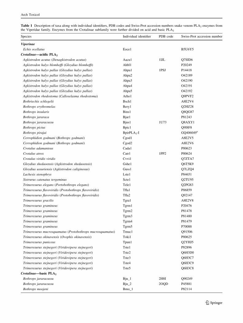
Table 1 Description of taxa along with individual identifiers, PDB codes and Swiss-Prot accession numbers snake venom PLA2 enzymes from
the Viperidae family. Enzymes from the Crotalinae subfamily were further divided on acid and basic PLA2
Species Individual identifier PDB code Swiss-Prot accession number
Viperinae
Echis ocellatus Eoce1 B5U6Y5
Crotalinae—acidic PLA2
Agkistrodon acutus (Denagkistrodon acutus) Aacu1 1IJL Q7SID6
Agkistrodon halys blomhoffi (Gloydius blomhoffi) Ahbl1 P20249
Agkistrodon halys pallas (Gloydius halys pallas) Ahpa1 1PSJ P14418
Agkistrodon halys pallas (Gloydius halys pallas) Ahpa2 O42189
Agkistrodon halys pallas (Gloydius halys pallas) Ahpa3 O42190
Agkistrodon halys pallas (Gloydius halys pallas) Ahpa4 O42191
Agkistrodon halys pallas (Gloydius halys pallas) Ahpa5 O42192
Agkistrodon rhodostoma (Calloselasma rhodostoma) Arho1 Q9PVF2
Bothriechis schlegelii Bsch1 A8E2V4
Bothrops erythromelas Bery1 Q2HZ28
Bothrops insularis Bins1 Q8QG87
Bothrops jararaca Bjar1 P81243
Bothrops jararacussu Bjus1 1U73 Q8AXY1
Bothrops pictus Bpic1 Q9I8F8
Bothrops pirajai BpirPLA2-I GQ406049a
Cerrophidion godmani (Bothrops godmani) Cgod1 A8E2V5
Cerrophidion godmani (Bothrops godmani) Cgod2 A8E2V6
Crotalus adamanteus Cada1 P00623
Crotalus atrox Catr1 1PP2 P00624
Crotalus viridis viridis Cvvi1 Q7ZTA7
Gloydius shedaoensis (Agkistrodon shedaoensis) Gshe1 Q6T5K9
Gloydius ussuriensis (Agkistrodon caliginosus) Guss1 Q7LZQ4
Lachesis stenophrys Lste1 P84651
Sistrurus catenatus tergeminus Scte1 Q2TU95
Trimeresurus elegans (Portobothrops elegans) Tele1 Q2PG83
Trimeresurus flavoviridis (Protobothrops flavoviridis) Tfla1 P06859
Trimeresurus flavoviridis (Protobothrops flavoviridis) Tfla2 Q92147
Trimeresurus gracilis Tgra1 A8E2V8
Trimeresurus gramineus Tgrm1 P20476
Trimeresurus gramineus Tgrm2 P81478
Trimeresurus gramineus Tgrm3 P81480
Trimeresurus gramineus Tgrm4 P81479
Trimeresurus gramineus Tgrm5 P70088
Trimeresurus mucrosquamatus (Protobothrops mucrosquamatus) Tmuc1 Q91506
Trimeresurus okinavensis (Ovophis okinavensis) Toki1 P00625
Trimeresurus puniceus Tpun1 Q2YHJ5
Trimeresurus stejnegeri (Viridovipera stejnegeri) Tste1 P82896
Trimeresurus stejnegeri (Viridovipera stejnegeri) Tste2 Q6H3D0
Trimeresurus stejnegeri (Viridovipera stejnegeri) Tste3 Q6H3C7
Trimeresurus stejnegeri (Viridovipera stejnegeri) Tste4 Q6H3C9
Trimeresurus stejnegeri (Viridovipera stejnegeri) Tste5 Q6H3C8
Crotalinae—basic PLA2
Bothrops jararacussu Bju_1 2H8I Q90249
Bothrops jararacussu Bju_2 2OQD P45881
Bothrops moojeni Bmo_1 P82114
Arch Toxicol
123
generation were inspected to check the appropriateness of
the burn-in period. Bayesian posterior probabilities (BPP)
and branch lengths were calculated from the remaining
trees, which were finally summarized as an extended
majority-rule consensus tree carrying the posterior proba-
bilities at the different nodes. MrEnt version 2.0 (Zuccon
and Zuccon 2008) was used for tree viewing and editing.
Comparative modeling
Modeling of the BpirPLA2-I three-dimensional structure was
performed by the method of satisfaction of spatial restraints
implemented in the program Modeller 9v6 (Sali and Blun-
dell 1993). A model was constructed for the Ca2?-free (apo
form) of BpirPLA2-I. The model was constructed using
different crystal structures of an acid PLA2 from the B.
jararacussu venom (BthA-I-PLA2) as templates to average
out uncertainties or artifacts in the atomic coordinates
coming from surface loop flexibility as well as crystal
packing effects and other technical issues: 1ZLB (0.97 A
resolution), chains A and B of 1U73 (1.90 A resolution), and
1UMV (1.79 A resolution). The sequence alignment
between the multiple templates and BpirPLA2-I was trivial
due to the high sequence identity shared between BpirPLA2-
I and BthA-I-PLA2 (90%) and could be visually adjusted.
Five models were generated, and the model with the lowest
value of the Modeller objective function was selected for
further evaluation and validation. The presence of any
energetically unfavorable regions in the model was evalu-
ated with the Discrete Optimized Protein Energy function
(DOPE) (Shen and Sali 2006) by comparing the DOPE score
profile of the templates. External assessment of the stereo-
chemical quality and overall structural reliability of the
model was performed within the Structural Analysis and
Verification Server (SAVES) at the WWW address
http://nihserver.mbi.ucla.edu/SAVES/.
Electrostatic potential calculations
The electrostatic surface potentials (EP) for BpirPLA2-I
and its homologs—BthA-I-PLA2 (pdb code 1U73),
PrTX-III (pdb code 1GMZ), and PrTX-I (pdb code 2Q2J)
were calculated from the 3D atomic coordinates of the free
PLA2 enzymes by solving the Poisson - Boltzmann
equation using the program DelPhi v.4 (Rocchia et al.
2001) with charge parameters from the CHARMM force
field (MacKerell et al. 1998) and atom radii parameters
from PARSE (Sitkoff et al. 1994a, b). Hydrogen atoms
were added using the Biopolymer module from the Sybyl
v.8.0 software package (Tripos Inc., St. Louis, MO, USA).
Since hydrogen addition may add structural crushes, for
each structure, 100 steps of minimization for relaxing
added hydrogen atoms were performed to obtain the
structures that were used in the electrostatic calculations.
Statistical analysis
Results are presented as the mean value ± SD obtained
with the indicated number of animals or samples. The
statistical significance of differences between groups was
evaluated using Student’s unpaired t test and analysis of
variance (ANOVA). A P value \0.05 was considered to
indicate significance.
Results and discussion
Several snake venoms were fractionated using procedures
based on ion exchange and hydrophobic interaction chro-
matography for phospholipases A2 purification (Gutierrez
and Lomonte 1997; Ownby et al. 1999; Andriao-Escarso
et al. 2000; Soares et al. 2004; De Paula et al. 2009).
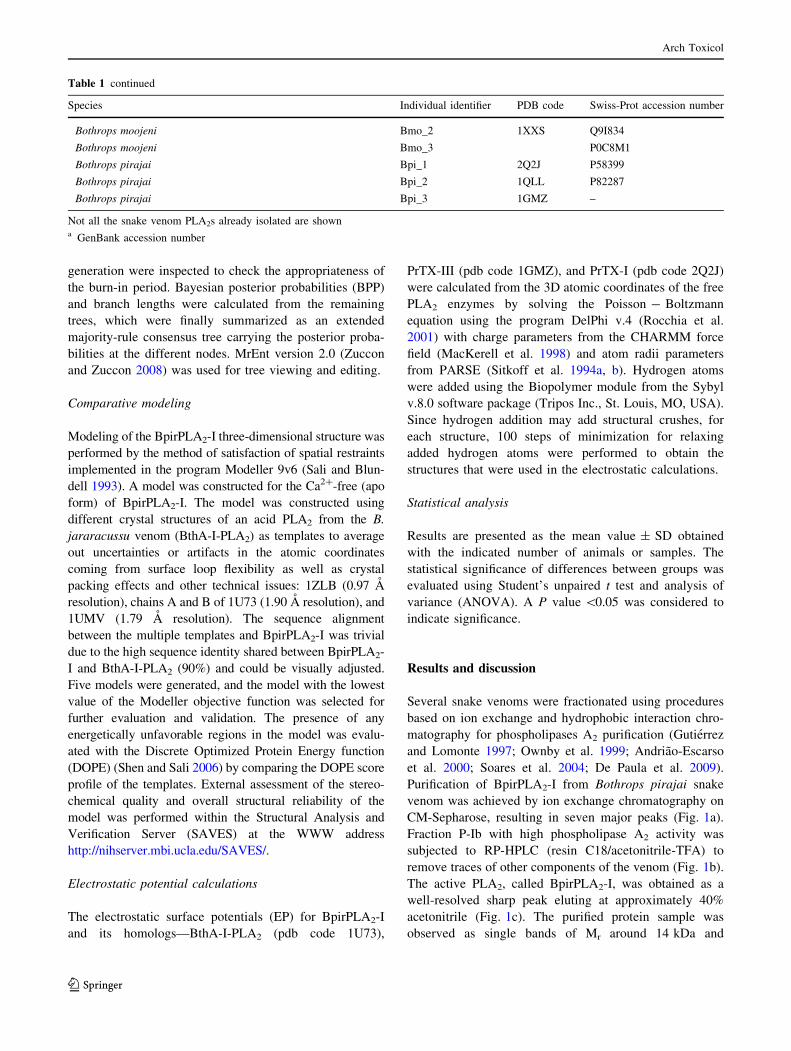
Purification of BpirPLA2-I from Bothrops pirajai snake
venom was achieved by ion exchange chromatography on
CM-Sepharose, resulting in seven major peaks (Fig. 1a).
Fraction P-Ib with high phospholipase A2 activity was
subjected to RP-HPLC (resin C18/acetonitrile-TFA) to
remove traces of other components of the venom (Fig. 1b).
The active PLA2, called BpirPLA2-I, was obtained as a
well-resolved sharp peak eluting at approximately 40%
acetonitrile (Fig. 1c). The purified protein sample was
observed as single bands of Mr around 14 kDa and
Table 1 continued
Species Individual identifier PDB code Swiss-Prot accession number
Bothrops moojeni Bmo_2 1XXS Q9I834
Bothrops moojeni Bmo_3 P0C8M1
Bothrops pirajai Bpi_1 2Q2J P58399
Bothrops pirajai Bpi_2 1QLL P82287
Bothrops pirajai Bpi_3 1GMZ –
Not all the snake venom PLA2s already isolated are showna GenBank accession number
Arch Toxicol
123
13,733.6 in the reducing SDS–PAGE (Fig. 1d) and MS
(Fig. 1f), respectively. Under native PAGE for acidic
proteins, the purified BpirPLA2-I sample also consisted of
a single band migrating toward to the positive pole (data
not shown), which was in agreement with the isoelectric
point of 4.8 determined from IEF (Fig. 1e).
The acidic PLA2 from B. pirajai showed higher enzy-
matic activity compared to PrTX-III, a basic Asp49-PLA2
from the same venom (data not shown). These results are in
agreement with previous studies involving PLA2s of snake
venom, in which the acidic Asp49-PLA2s are catalytically
more active than their basic isoforms (Andriao-Escarso
et al. 2002; Roberto et al. 2004; Rodrigues et al. 2007;
Santos-Filho et al. 2008; Fernandez et al. 2010).
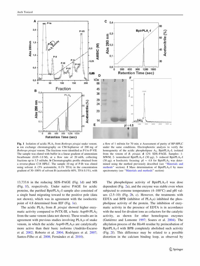
The phospholipase activity of BpirPLA2-I was dose
dependent (Fig. 2a), and the enzyme was stable even when
subjected to extreme temperatures (4–100�C) and pH val-
ues (2.5–10) (Fig. 2b, c). However, the treatments with
EDTA and BPB (inhibitor of PLA2s) inhibited the phos-
pholipase activity of the protein. The inhibition of enzy-
matic activity in the presence of EDTA is in accordance
with the need for divalent ions as cofactors for the catalytic
activity, as shown for other homologous enzymes
(Gutierrez and Lomonte 1997; Soares et al. 2004). The
alkylation process of the His48 residue by preincubation of
BpirPLA2-I with BPB completely abolished such activity
(Fig. 2f). This difference may be related to a possible
distortion in the calcium binding loop, as observed by
0 50 100 150 2000,0
0,5
1,0
1,5
2,0
PLA2 A
ctivi
ty
P-VII
P-VI
P-V
P-IV
P-III
P-II
P-Ia
P-Ib0,5
0,25
0,05
AM
BIC
(M
)
Fraction
A
B
C
D
E
F
Ab
s 28
0nm
Fig. 1 Isolation of acidic PLA2 from Bothrops pirajai snake venom.
a ion exchange chromatography on CM-Sepharose of 200 mg of
Bothrops pirajai venom. The fractions were identified as P-I to P-VII.
The sample was eluted with buffer in a linear gradient of ammonium
bicarbonate (0.05–1.0 M), at a flow rate of 20 ml/h, collecting
fractions up to 3.5 ml/tube. b Chromatographic profile obtained from
a reverse-phase C18 HPLC. The sample 10 mg of P-Ib was eluted
using solvent A (5% acetonitrile, 0.1% TFA) in the concentration
gradient of 30–100% of solvent B (acetonitrile 60%, TFA 0.1%), with
a flow of 1 ml/min for 70 min. c Assessment of purity of RP-HPLC
under the same conditions. Electrophoretic analysis to verify the
homogeneity of the acidic phospholipase A2, BpirPLA2-I, isolated
from the venom of B. pirajai. d 12% SDS–PAGE. Samples: 1-
MWM, 2- nonreduced BpirPLA2-I (30 lg), 3- reduced BpirPLA2-I
(30 lg). e Isoelectric focusing: pI * 4.8 for BpirPLA2 was deter-
mined using the method previously described (see ‘‘Materials and
methods’’ section). f Mass determination of BpirPLA2-I by mass
spectrometry (see ‘‘Materials and methods’’ section)
Arch Toxicol
123
Correa and coworkers (2008) in a comparative structural
study carried out with different phospholipases A2.
BpirPLA2-I hydrolyzed fluorescent phospholipids such
as NBD-PC and NBD-PG with different rates (Fig. 2d),
showing to be more active upon NBD-PC, similar to the
acidic PLA2s BmooTX-I and Bp-PLA2, purified from B.
moojeni and B. pauloensis snake venoms, respectively
(Rodrigues et al. 2007; Santos-Filho et al. 2008). Hence,
NBD-PC was used to study the influence of metals on
BpirPLA2-I activity. The presence of calcium ions
increased the activity of PLA2 in a dose-dependent manner,
unlike the other tested divalent ions (Ba?2, Cu?2, Fe?2,
Mg?2, Mn?2, and Zn?2) (Fig. 2e). In catalytically active
PLA2s, a Ca?2 ion, coordinated by Asp49 residue, a water
molecule, and oxygen atoms of residues Gly30, Gly32, and
Trp31 are responsible for stabilizing the reactive interme-
diate (Arni and Ward 1996). In the absence of calcium, a
water molecule occupies the position of the ion and the side
chain of Asp49 residue and the calcium binding loop adopt
different conformations, affecting the enzymatic activity
(Murakami et al. 2006). Thus, these results are consistent
with the characteristics of PLA2s that include high stability,
due to large number of intrachain disulfide bridges, and the
need for calcium as a cofactor for the enzymatic activity.
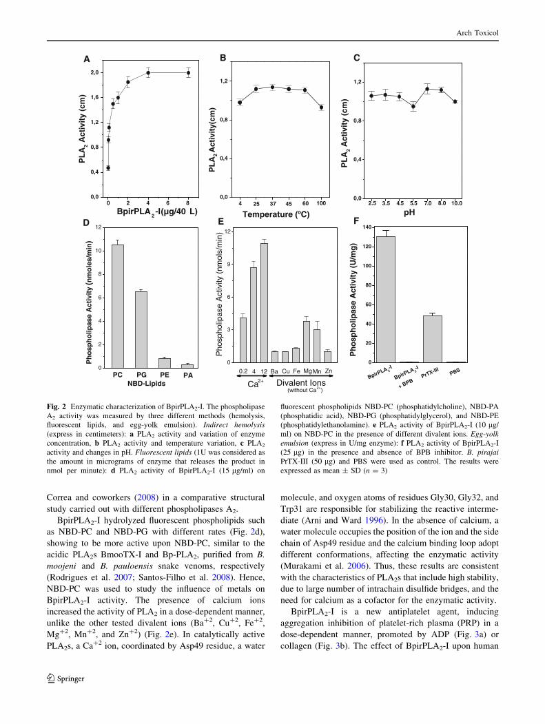
BpirPLA2-I is a new antiplatelet agent, inducing
aggregation inhibition of platelet-rich plasma (PRP) in a
dose-dependent manner, promoted by ADP (Fig. 3a) or
collagen (Fig. 3b). The effect of BpirPLA2-I upon human
0,0
0,4
0,8
1,2
pH10.08.05.4 5.5 0.73.52.5
PL
A2
Act
ivit
y (c
m)
0 2 4 6 80,0
0,4
0,8
1,2
1,6
2,0
PL
A2
Act
ivit
y (c
m)
BpirPLA2-I(µg/40µL)
0,0
0,4
0,8
1,2
100604537254P
LA
2A
ctiv
ity(
cm)
Temperature (ºC)
0
20
40
60
80
100
120
140
PBSPrTX-III
BpirPLA 2-I
+ BPB
Ph
osp
ho
lipas
e A
ctiv
ity
(U/m
g)
BpirPLA 2-I0
2
4
6
8
10
12
PAPEPG
Ph
osp
ho
lipas
e A
ctiv
ity
(nm
ole
s/m
in)
NBD-LipidsPC
0
3
6
9
12
(without Ca2+)Divalent Ions
Mg ZnFeCuBa Mn
Ca2+
1240.2
Pho
spho
lipas
e A
ctiv
ity (
nmol
s/m
in)
A B C
FED
Fig. 2 Enzymatic characterization of BpirPLA2-I. The phospholipase
A2 activity was measured by three different methods (hemolysis,
fluorescent lipids, and egg-yolk emulsion). Indirect hemolysis(express in centimeters): a PLA2 activity and variation of enzyme
concentration, b PLA2 activity and temperature variation, c PLA2
activity and changes in pH. Fluorescent lipids (1U was considered as
the amount in micrograms of enzyme that releases the product in
nmol per minute): d PLA2 activity of BpirPLA2-I (15 lg/ml) on
fluorescent phospholipids NBD-PC (phosphatidylcholine), NBD-PA
(phosphatidic acid), NBD-PG (phosphatidylglycerol), and NBD-PE
(phosphatidylethanolamine). e PLA2 activity of BpirPLA2-I (10 lg/
ml) on NBD-PC in the presence of different divalent ions. Egg-yolkemulsion (express in U/mg enzyme): f PLA2 activity of BpirPLA2-I
(25 lg) in the presence and absence of BPB inhibitor. B. pirajaiPrTX-III (50 lg) and PBS were used as control. The results were
expressed as mean ± SD (n = 3)
Arch Toxicol
123
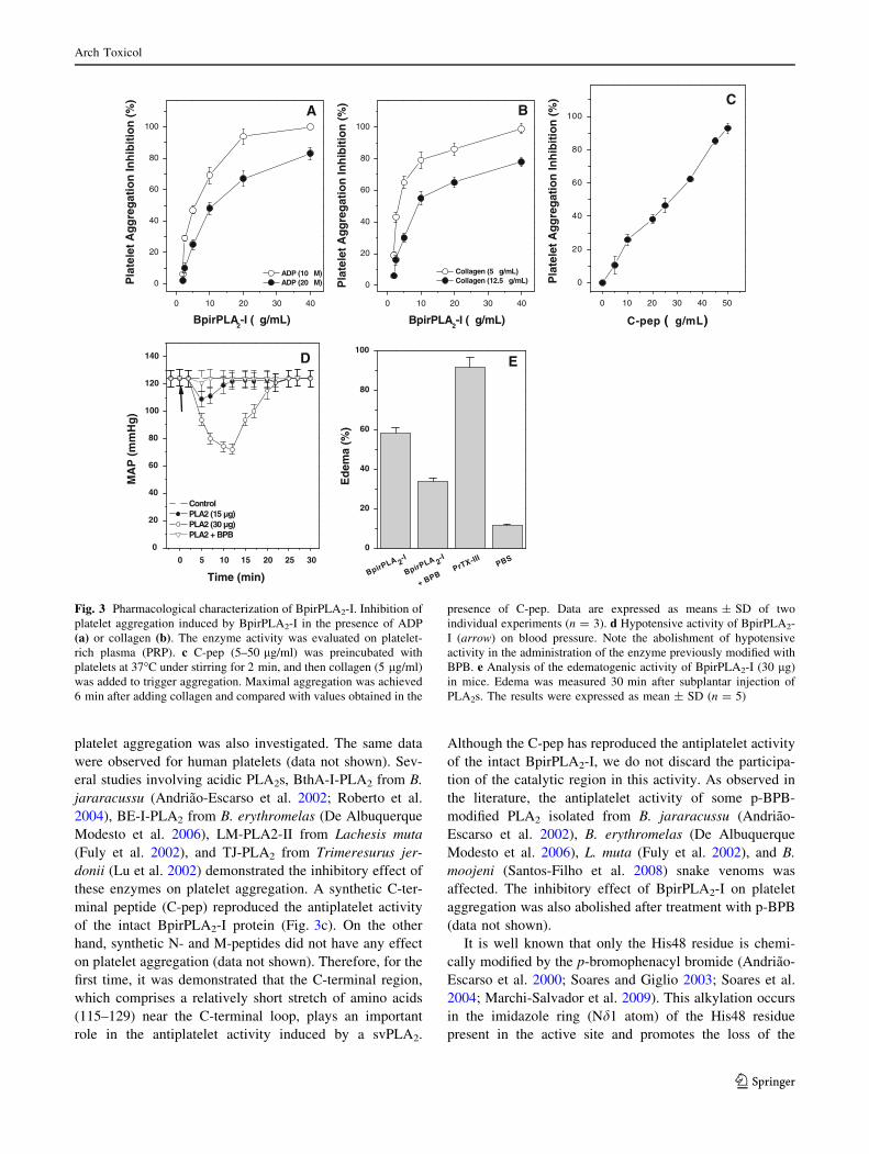
platelet aggregation was also investigated. The same data
were observed for human platelets (data not shown). Sev-
eral studies involving acidic PLA2s, BthA-I-PLA2 from B.
jararacussu (Andriao-Escarso et al. 2002; Roberto et al.
2004), BE-I-PLA2 from B. erythromelas (De Albuquerque
Modesto et al. 2006), LM-PLA2-II from Lachesis muta
(Fuly et al. 2002), and TJ-PLA2 from Trimeresurus jer-
donii (Lu et al. 2002) demonstrated the inhibitory effect of
these enzymes on platelet aggregation. A synthetic C-ter-
minal peptide (C-pep) reproduced the antiplatelet activity
of the intact BpirPLA2-I protein (Fig. 3c). On the other
hand, synthetic N- and M-peptides did not have any effect
on platelet aggregation (data not shown). Therefore, for the
first time, it was demonstrated that the C-terminal region,
which comprises a relatively short stretch of amino acids
(115–129) near the C-terminal loop, plays an important
role in the antiplatelet activity induced by a svPLA2.
Although the C-pep has reproduced the antiplatelet activity
of the intact BpirPLA2-I, we do not discard the participa-
tion of the catalytic region in this activity. As observed in
the literature, the antiplatelet activity of some p-BPB-
modified PLA2 isolated from B. jararacussu (Andriao-
Escarso et al. 2002), B. erythromelas (De Albuquerque
Modesto et al. 2006), L. muta (Fuly et al. 2002), and B.
moojeni (Santos-Filho et al. 2008) snake venoms was
affected. The inhibitory effect of BpirPLA2-I on platelet
aggregation was also abolished after treatment with p-BPB
(data not shown).
It is well known that only the His48 residue is chemi-
cally modified by the p-bromophenacyl bromide (Andriao-
Escarso et al. 2000; Soares and Giglio 2003; Soares et al.
2004; Marchi-Salvador et al. 2009). This alkylation occurs
in the imidazole ring (Nd1 atom) of the His48 residue
present in the active site and promotes the loss of the
1 2 3 4
0
20
40
60
80
100
PrTX-III PBS
BpirPLA 2-
I
+ BPB
Ed
ema
(%)
BpirPLA 2-
I
E
0 5 10 15 20 25 30
0
20
40
60
80
100
120
140
Time (min)
MA
P (
mm
Hg
)
ControlPLA2 (15 µg)PLA2 (30 µg)PLA2 + BPB
D
0 10 20 30 40 50
0
20
40
60
80
100
Pla
tele
t A
gg
reg
atio
n In
hib
itio
n (
%)
C-pep (µg/mL)
C
0 10 20 30 40
0
20
40
60
80
100
A
ADP (10 µM)ADP (20 µM)P
late
let
Ag
gre
gat
ion
Inh
ibit
ion
(%
)
BpirPLA2-I (µg/mL)
0 10 20 30 40
0
20
40
60
80
100
B
Collagen (5 µg/mL)Collagen (12.5 µg/mL)P
late
let
Ag
gre
gat
ion
Inh
ibit
ion
(%
)
BpirPLA2-I (µg/mL)
Fig. 3 Pharmacological characterization of BpirPLA2-I. Inhibition of
platelet aggregation induced by BpirPLA2-I in the presence of ADP
(a) or collagen (b). The enzyme activity was evaluated on platelet-
rich plasma (PRP). c C-pep (5–50 lg/ml) was preincubated with
platelets at 37�C under stirring for 2 min, and then collagen (5 lg/ml)
was added to trigger aggregation. Maximal aggregation was achieved
6 min after adding collagen and compared with values obtained in the
presence of C-pep. Data are expressed as means ± SD of two
individual experiments (n = 3). d Hypotensive activity of BpirPLA2-
I (arrow) on blood pressure. Note the abolishment of hypotensive
activity in the administration of the enzyme previously modified with
BPB. e Analysis of the edematogenic activity of BpirPLA2-I (30 lg)
in mice. Edema was measured 30 min after subplantar injection of
PLA2s. The results were expressed as mean ± SD (n = 5)
Arch Toxicol
123
enzymatic activity and/or reduction in the toxic and phar-
macological effects of PLA2s (Soares et al. 2004). This
suggests that these pharmacological effects and the cata-
lytic activity are dependent on His48. Marchi-Salvador and
coworkers (2006) suggested that the inhibitor binding led
to significant structural modifications, especially in the
C-terminal region. In the structure of BthTX-I, a Lys49
phospholipase A2 like from Bothrops jararacussu, chemi-
cally modified with BPB, it was observed that the phenacyl
group of BPB molecule extends along the hydrophobic
channel of the protein, interacting with Tyr22, Gly23,
Val31, Cys45, and Lys49 residues.
Another PLA2 isolated from Bothrops jararacussu
venom, named BthA-I, was also complexed with BPB, and
it was observed that the catalytic, platelet-aggregation
inhibition, anticoagulant, and hypotensive activities were
abolished by ligand binding. The BthA-I-BPB complex
contains three structural regions that are modified after
inhibitor binding: the Ca2?-binding loop, beta-wing, and
C-terminal regions. This conformation is more energeti-
cally and conformationally stable than the native structure,
and the abolition of pharmacological activities by the
ligand may be related to the oligomeric structural changes
(Magro et al. 2005).
BpirPLA2-I induced hypotension in rats. This activity
was completely abolished in the presence of BPB (Fig. 3d).
The synthetic peptides did not have any effect on hypo-
tension. Up to now, there is no structural information,
suggesting the presence of a pharmacological site on
phospholipases A2 associated with the hypotensive effect
of these enzymes. According to Huang and Lee (1984), the
activity of hypotensive PLA2s must be related to the syn-
thesis of prostaglandins from the molecule of arachidonic
acid (AA) released by the phospholipase A2 catalytic
activity. These prostaglandins act on the renin-angiotensin
system, a set of protein molecules involved in controlling
the volume of extracellular fluid and blood pressure. In
addition, the leukotrienes, also derived from AA by the
action of lipoxygenase, could attend acting on the smooth
muscle of blood vessels.
Synthetic peptides have been studied in order to find out
which regions of the snake venoms phospholipases A2 are
responsible for each activity (Lomonte et al. 2010). A
synthetic peptide derived from a homologue PLA2 present
in Bothrops asper snake venom has shown potent bacte-
ricidal activity and exerts potent fungicidal activity against
a variety of clinically relevant Candida species (Murillo
et al. 2007). Costa and coworkers (2008) also demonstrated
that cationic synthetic peptides derived from the 115–129
C-terminal region of myotoxic PLA2s from Bothrops
brazili snake venom, MTX-I (Asp49 PLA2) and MTX-II
(Lys49 PLA2), displayed cytotoxic activity on human
T-cell leukemia (JURKAT) lines and microbicidal effects
against Escherichia coli, Candida albicans, and Leish-
mania sp. The in vitro and in vivo antitumor activity of a
cationic synthetic peptide was derived from the 115–129
C-terminal region of BPB-modified BthTX-I (Gebrim et al.
2009). Up to now, the toxic activities of these PLA2s have
been attributed to its C-terminal region.
The intradermal administration of 30 lg of BpirPLA2-I
was able to induce edema (increase of 58% in rat paw
volume), which was reduced by 34% after preincubation of
enzyme with BPB (Fig. 3e). In contrast, the same amount
of PrTX-III induced edema of 92%. These results are in
agreement with previous studies with B. jararacussu acidic
PLA2s where the chemical modification with BPB com-
pletely eliminated the enzymatic, anticoagulant and anti-
platelet activity, but only partially inhibited the formation
of edema (Andriao-Escarso et al. 2002; Ketelhut et al.
2003), indicating that in addition to a preserved catalytic
site, specific pharmacological requirements seem to be
involved with this activity. BpirPLA2-I did not increased
CK levels, not showing myotoxicity (results not shown),
corroborating with previous findings in which acidic
PLA2s, although highly catalytic active, usually do not
have toxicity (Andriao-Escarso et al. 2002; De Albuquer-
que Modesto et al. 2006; Fernandez et al. 2010), dissoci-
ating the catalytic activity from myotoxicity. The
BpirPLA2-I and synthetic peptides showed very low cyto-
toxic activity in vitro on skeletal muscle (C2C12) cells
when compared with the synthetic C-terminal region pep-
tides of basic PLA2s (data not shown).
BpirPLA2-I represents 2–3% of the whole venom being
the most abundant acidic PLA2 in this snake venom and
presenting a series of important pharmacological effects
such as edema inducing hypotensive and inhibition of
platelet aggregating activity.
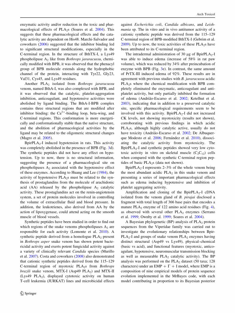
Amplification and cloning of the BpirPLA2-I cDNA
isolated from the venom gland of B. pirajai disclosed a
fragment with total length of 366 base pairs that encodes a
mature PLA2 enzyme of 122 amino acid residues (Fig. 4),
as observed with several other PLA2 enzymes (Serrano
et al. 1999; Ownby et al. 1999; Soares et al. 2004).
A Bayesian phylogenetic (BP) analysis of PLA2 protein
sequences from the Viperidae family was carried out to
investigate the evolutionary relationships between Bpir-
PLA2-I and groups of snake venom PLA2 enzymes having
distinct structural (Asp49 vs Lys49), physical–chemical
(basic vs acid), and functional features (myotoxic, antico-
agulant, hypotensive, neuromuscular transmission blocking
as well as measurable PLA2 catalytic activity). The BP
analysis was performed on the PLA2 dataset (50 taxa; 128
characters) under an EMP ? C ? I model, where EMP is a
composition of nine empirical models of protein sequence
evolution implemented in the MrBayes code, with each
model contributing in proportion to its Bayesian posterior
Arch Toxicol
123
probability. For the PLA2 dataset, the maximum likelihood
improvement of the JTT matrix (Gonnet et al. 1992; Jones
et al. 1992), known as the WAG matrix (Whelan and
Goldman 2001), was the single model supported by the
data, with a BPP of 1.0.
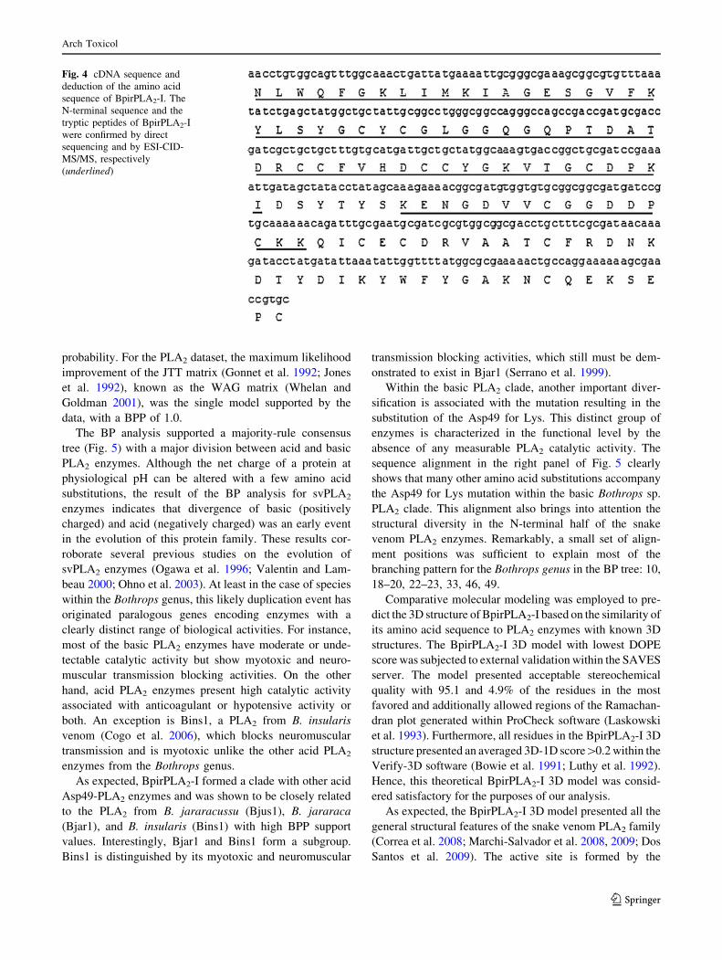
The BP analysis supported a majority-rule consensus
tree (Fig. 5) with a major division between acid and basic
PLA2 enzymes. Although the net charge of a protein at
physiological pH can be altered with a few amino acid
substitutions, the result of the BP analysis for svPLA2
enzymes indicates that divergence of basic (positively
charged) and acid (negatively charged) was an early event
in the evolution of this protein family. These results cor-
roborate several previous studies on the evolution of
svPLA2 enzymes (Ogawa et al. 1996; Valentin and Lam-
beau 2000; Ohno et al. 2003). At least in the case of species
within the Bothrops genus, this likely duplication event has
originated paralogous genes encoding enzymes with a
clearly distinct range of biological activities. For instance,
most of the basic PLA2 enzymes have moderate or unde-
tectable catalytic activity but show myotoxic and neuro-
muscular transmission blocking activities. On the other
hand, acid PLA2 enzymes present high catalytic activity
associated with anticoagulant or hypotensive activity or
both. An exception is Bins1, a PLA2 from B. insularis
venom (Cogo et al. 2006), which blocks neuromuscular
transmission and is myotoxic unlike the other acid PLA2
enzymes from the Bothrops genus.
As expected, BpirPLA2-I formed a clade with other acid
Asp49-PLA2 enzymes and was shown to be closely related
to the PLA2 from B. jararacussu (Bjus1), B. jararaca
(Bjar1), and B. insularis (Bins1) with high BPP support
values. Interestingly, Bjar1 and Bins1 form a subgroup.
Bins1 is distinguished by its myotoxic and neuromuscular
transmission blocking activities, which still must be dem-
onstrated to exist in Bjar1 (Serrano et al. 1999).
Within the basic PLA2 clade, another important diver-
sification is associated with the mutation resulting in the
substitution of the Asp49 for Lys. This distinct group of
enzymes is characterized in the functional level by the
absence of any measurable PLA2 catalytic activity. The
sequence alignment in the right panel of Fig. 5 clearly
shows that many other amino acid substitutions accompany
the Asp49 for Lys mutation within the basic Bothrops sp.
PLA2 clade. This alignment also brings into attention the
structural diversity in the N-terminal half of the snake
venom PLA2 enzymes. Remarkably, a small set of align-
ment positions was sufficient to explain most of the
branching pattern for the Bothrops genus in the BP tree: 10,
18–20, 22–23, 33, 46, 49.
Comparative molecular modeling was employed to pre-
dict the 3D structure of BpirPLA2-I based on the similarity of
its amino acid sequence to PLA2 enzymes with known 3D
structures. The BpirPLA2-I 3D model with lowest DOPE
score was subjected to external validation within the SAVES
server. The model presented acceptable stereochemical
quality with 95.1 and 4.9% of the residues in the most
favored and additionally allowed regions of the Ramachan-
dran plot generated within ProCheck software (Laskowski
et al. 1993). Furthermore, all residues in the BpirPLA2-I 3D
structure presented an averaged 3D-1D score[0.2 within the
Verify-3D software (Bowie et al. 1991; Luthy et al. 1992).
Hence, this theoretical BpirPLA2-I 3D model was consid-
ered satisfactory for the purposes of our analysis.
As expected, the BpirPLA2-I 3D model presented all the
general structural features of the snake venom PLA2 family
(Correa et al. 2008; Marchi-Salvador et al. 2008, 2009; Dos
Santos et al. 2009). The active site is formed by the
Fig. 4 cDNA sequence and
deduction of the amino acid
sequence of BpirPLA2-I. The
N-terminal sequence and the
tryptic peptides of BpirPLA2-I
were confirmed by direct
sequencing and by ESI-CID-
MS/MS, respectively
(underlined)
Arch Toxicol
123
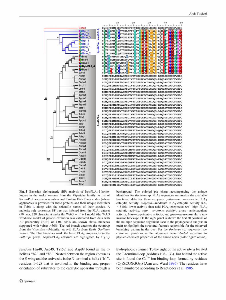
residues His48, Asp49, Tyr52, and Asp99 found in the a-
helixes ‘‘h2’’ and ‘‘h3’’. Nested between the region known as
the b-wing and the active site is the N-terminal a-helix (‘‘h1’’,
residues 1–12) that is involved in the binding and proper
orientation of substrates to the catalytic apparatus through a
hydrophobic channel. To the right of the active site is located
the C-terminal loop (residues 108–133). Just behind the active
site is found the Ca2? ion binding loop formed by residues
(C27XCGXGG33) (Arni and Ward 1996). The residues have
been numbered according to Renetseder et al. 1985.
Fig. 5 Bayesian phylogenetic (BP) analysis of BpirPLA2-I homo-
logues in the snake venoms from the Viperidae family. A list of
Swiss-Prot accession numbers and Protein Data Bank codes (where
applicable) is provided for these proteins and their unique identifiers
in Table 1, along with the scientific names of their species. A
majority-rule consensus BP tree was inferred from the PLA2 dataset
(50 taxa; 128 characters) under the WAG ? C ? I model (the WAG
fixed-rate model of protein evolution was estimated from data with
BP probability (BPP) of 1.0). BPPs are shown above branches
supported with values [50%. The red branch detaches the outgroup
from the Viperidae subfamily, an acid PLA2 from Echis Ocellatusvenom. The blue branches mark the basic PLA2 enzymes from the
Bothrops genus. Asp49-PLA2 enzymes are highlighted by a gray
background. The colored pie charts accompanying the unique
identifiers for Bothrops sp. PLA2 sequences summarize the available
functional data for these enzymes: yellow—no measurable PLA2
catalytic activity; magenta—moderate PLA2 catalytic activity (i.e.,
*6-fold lower activity than acid PLA2 enzymes); red—high PLA2
catalytic activity; cyan—myotoxic activity; green—anticoagulant
activity; blue—hypotensive activity; and gray—neuromuscular trans-
mission blockage. On the right panel is shown the first 50 positions of
the multiple sequence alignment used in the phylogenetic analysis in
order to highlight the structural features responsible for the observed
branching pattern in the tree. For the Bothrops sp. sequences, the
conserved positions in the alignment were shaded according to
physico-chemical properties of the amino acids (color figure online)
Arch Toxicol
123
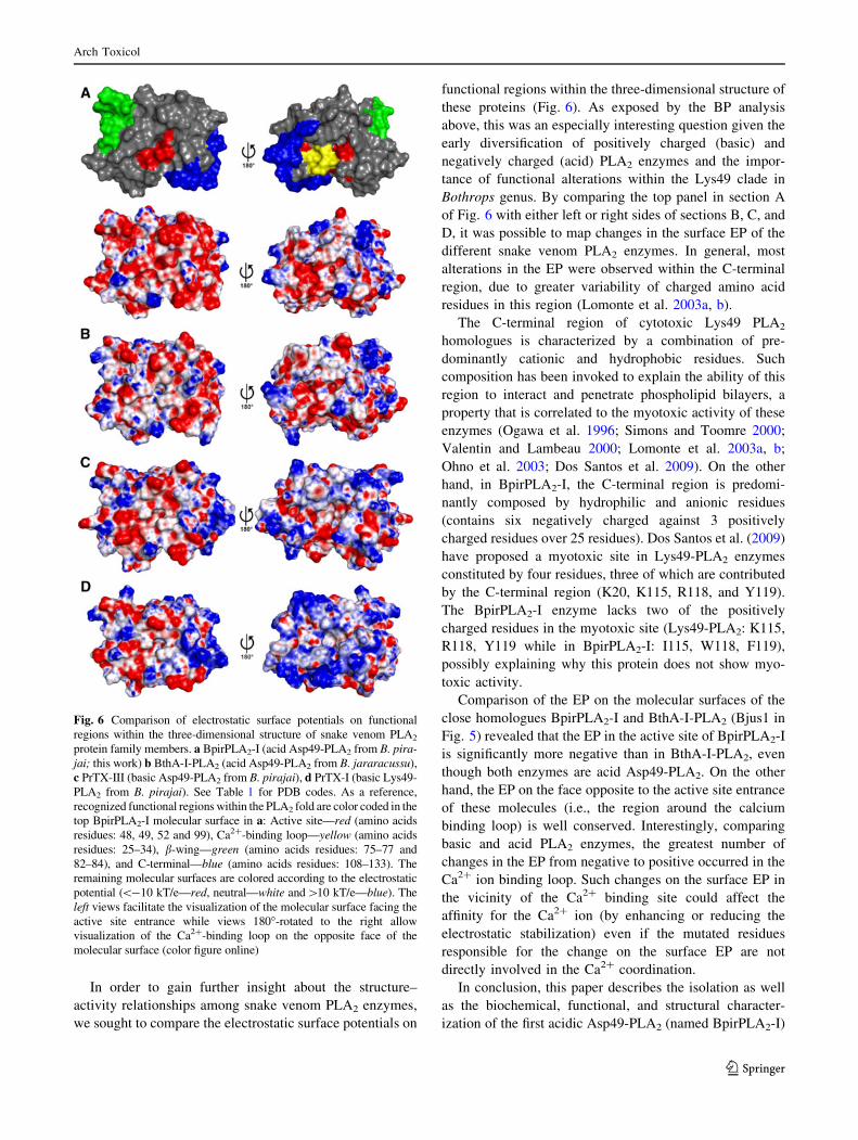
In order to gain further insight about the structure–
activity relationships among snake venom PLA2 enzymes,
we sought to compare the electrostatic surface potentials on
functional regions within the three-dimensional structure of
these proteins (Fig. 6). As exposed by the BP analysis
above, this was an especially interesting question given the
early diversification of positively charged (basic) and
negatively charged (acid) PLA2 enzymes and the impor-
tance of functional alterations within the Lys49 clade in
Bothrops genus. By comparing the top panel in section A
of Fig. 6 with either left or right sides of sections B, C, and
D, it was possible to map changes in the surface EP of the
different snake venom PLA2 enzymes. In general, most
alterations in the EP were observed within the C-terminal
region, due to greater variability of charged amino acid
residues in this region (Lomonte et al. 2003a, b).
The C-terminal region of cytotoxic Lys49 PLA2
homologues is characterized by a combination of pre-
dominantly cationic and hydrophobic residues. Such
composition has been invoked to explain the ability of this
region to interact and penetrate phospholipid bilayers, a
property that is correlated to the myotoxic activity of these
enzymes (Ogawa et al. 1996; Simons and Toomre 2000;
Valentin and Lambeau 2000; Lomonte et al. 2003a, b;
Ohno et al. 2003; Dos Santos et al. 2009). On the other
hand, in BpirPLA2-I, the C-terminal region is predomi-
nantly composed by hydrophilic and anionic residues
(contains six negatively charged against 3 positively
charged residues over 25 residues). Dos Santos et al. (2009)
have proposed a myotoxic site in Lys49-PLA2 enzymes
constituted by four residues, three of which are contributed
by the C-terminal region (K20, K115, R118, and Y119).
The BpirPLA2-I enzyme lacks two of the positively
charged residues in the myotoxic site (Lys49-PLA2: K115,
R118, Y119 while in BpirPLA2-I: I115, W118, F119),
possibly explaining why this protein does not show myo-
toxic activity.
Comparison of the EP on the molecular surfaces of the
close homologues BpirPLA2-I and BthA-I-PLA2 (Bjus1 in
Fig. 5) revealed that the EP in the active site of BpirPLA2-I
is significantly more negative than in BthA-I-PLA2, even
though both enzymes are acid Asp49-PLA2. On the other
hand, the EP on the face opposite to the active site entrance
of these molecules (i.e., the region around the calcium
binding loop) is well conserved. Interestingly, comparing
basic and acid PLA2 enzymes, the greatest number of
changes in the EP from negative to positive occurred in the
Ca2? ion binding loop. Such changes on the surface EP in
the vicinity of the Ca2? binding site could affect the
affinity for the Ca2? ion (by enhancing or reducing the
electrostatic stabilization) even if the mutated residues
responsible for the change on the surface EP are not
directly involved in the Ca2? coordination.
In conclusion, this paper describes the isolation as well
as the biochemical, functional, and structural character-
ization of the first acidic Asp49-PLA2 (named BpirPLA2-I)
Fig. 6 Comparison of electrostatic surface potentials on functional
regions within the three-dimensional structure of snake venom PLA2
protein family members. a BpirPLA2-I (acid Asp49-PLA2 from B. pira-jai; this work) b BthA-I-PLA2 (acid Asp49-PLA2 from B. jararacussu),
c PrTX-III (basic Asp49-PLA2 from B. pirajai), d PrTX-I (basic Lys49-
PLA2 from B. pirajai). See Table 1 for PDB codes. As a reference,
recognized functional regions within the PLA2 fold are color coded in the
top BpirPLA2-I molecular surface in a: Active site—red (amino acids
residues: 48, 49, 52 and 99), Ca2?-binding loop—yellow (amino acids
residues: 25–34), b-wing—green (amino acids residues: 75–77 and
82–84), and C-terminal—blue (amino acids residues: 108–133). The
remaining molecular surfaces are colored according to the electrostatic
potential (\-10 kT/e—red, neutral—white and[10 kT/e—blue). The
left views facilitate the visualization of the molecular surface facing the
active site entrance while views 180�-rotated to the right allow
visualization of the Ca2?-binding loop on the opposite face of the
molecular surface (color figure online)
Arch Toxicol
123
from Bothrops pirajai snake venom, and it was demon-
strated that a synthetic peptide with a relatively short
stretch of amino acids (115–129) near the C-terminal
region plays an important role in the antiplatelet activity
induced by a svPLA2 showing their potential value as
molecular tools or as drug leads in diverse biomedical
areas, and the importance of this region for the activity of
the hole protein.
Acknowledgments This work was supported by Fundacao de
Amparo a Pesquisa do Estado de Sao Paulo (FAPESP), Conselho
Nacional de Desenvolvimento Cientıfico e Tecnologico (CNPq) and
Instituto Nacional de Ciencia e Tecnologia em Toxinas (INCT-Tox),
Brazil. We are grateful to Prof. Dr. J. R. Giglio (FMRP-USP) for their
manuscript revision; Prof. Dr. A. Nomizo (FCFRP-USP, Brazil), MSc
J. Fernandez and Prof. Dr. B. Lomonte (ICP, UCR, Costa Rica) for
their collaboration in the cytotoxic and hypotension assays. Thank to
E. A. Bastos (TT-FAPESP) for their helpful technical collaboration.
References
Andriao-Escarso SH, Soares AM, Rodrigues VM, Angulo Y,
Lomonte B, Gutierrez JM, Giglio JR (2000) Myotoxic phospho-
lipases A2 in Bothrops snake venoms: effects of chemical
modifications on the enzymatic and pharmacological properties
of Bothropstoxins from Bothrops jararacussu. Biochimie
82:755–763
Andriao-Escarso SH, Soares AM, Fontes MRM, Fuly AL, Correa
FMA, Rosa JC, Greene LJ, Giglio JR (2002) Structural and
functional characterization of an acidic platelet aggregation
inhibitor and hypotensive Phospholipase A2 from Bothropsjararacussu snake venom. Biochem Pharmacol 64:723–732
Arni RK, Ward RJ (1996) Phospholipase A2—a structural review.
Toxicon 34:827–841
Bowie JU, Luthy R, Eisenberg D (1991) A method to identify protein
sequences that fold into a known three-dimensional structure.
Science 253:164–169
Cogo JC, Lilla S, Souza GHMF, Hyslop S, De Nucci G (2006)
Purification, sequencing and structural analysis of two acidic
phospholipases A2 from the venom of Bothrops insularis
(jararaca ilhoa). Biochimie 88:1947–1959
Correa LC, Marchi-Salvador DP, Cintra AC, Sampaio SV, Soares
AM, Fontes MR (2008) Crystal structure of a myotoxic Asp49-
Phospholipase A2 with low catalytic activity: insights into Ca2?-
independent catalytic mechanism. Biochim Biophys Acta
1784:591–599
Costa TR, Menaldo DL, Oliveira CZ, Santos-Filho NA, Teixeira SS,
Nomizo A, Fuly AL, Monteiro MC, de Souza BM, Palma MS,
Stabeli RG, Sampaio SV, Soares AM (2008) Myotoxic phos-
pholipases A2 isolated from Bothrops brazili snake venom and
synthetic peptides derived from their C-terminal region: cyto-
toxic effect on microorganism and tumor cells. Peptides
29(10):1645–1656
De Albuquerque Modesto JC, Spencer PJ, Fritzen M, Valenca RC,
Oliva MLV, da Silva MB, Chudzinski-Tavassi AM, Guarnieri
MC (2006) BE-I-PLA2, a novel acidic Phospholipase A2 from
Bothrops erythromelas venom: isolation, cloning and character-
ization as potent anti-platelet and inductor of prostaglandin I2
release by endothelial cells. Biochem Pharmacol 72:377–384
De Haas GH, Postema NM, Nieuwenhuizen W, van Deenen LLM
(1968) Purification and properties of Phospholipase A from
porcine pancreas. Biochim Biophys Acta 159:103–117
De Paula R, Castro HC, Rodrigues CR, Melo PA, Fuly AL (2009)
Structural and pharmacological features of phospholipases A2
from snake venoms. Protein Pept Lett 16:899–907
Dos Santos JI, Soares AM, Fontes MRM (2009) Comparative
structural studies on Lys49-Phospholipase A2 from Bothropsgenus reveal their myotoxic site. J Struct Biol 167:106–116
Edgar RC (2004) MUSCLE: multiple sequence alignment with high
accuracy and high throughput. Nucleic Acids Res 32:1792–1797
Fernandez J, Gutierrez JM, Angulo Y, Sanz L, Juarez P, Calvete JJ,
Lomonte B (2010) Isolation of an acidic phospholipase A2 from
the venom of the snake Bothrops asper of Costa Rica: biochemical
and toxicological characterization. Biochimie 92:273–283
Fuly AL, de Miranda AL, Zingali RB, Guimaraes JA (2002)
Purification and characterization of a phospholipase A2 isoen-
zyme isolated from Lachesis muta snake venom. Biochem
Pharmacol 63:1589–1597
Gebrim LC, Marcussi S, Menaldo DL, de Menezes CS, Nomizo A,
Hamaguchi A, Silveira-Lacerda EP, Homsi-Brandeburgo MI,
Sampaio SV, Soares AM, Rodrigues VM (2009) Antitumor
effects of snake venom chemically modified Lys49 phospholi-
pase A2-like BthTX-I and a synthetic peptide derived from its
C-terminal region. Biologicals 37(4):222–229
Gonnet GH, Cohen MA, Benner SA (1992) Exhaustive matching of
the entire protein sequence database. Science 256:1443–1445
Guda C, Scheeff ED, Bourne PE, Shindyalov IN (2001) A new
algorithm for the alignment of multiple protein structures using
Monte Carlo optimization. Pac Symp Biocomput 6:275–286
Gutierrez JM, Lomonte B (1997) In: Kini RM (ed) Venom
Phospholipase A2 enzymes: structure, function and mechanism.
Wiley, Chichester, pp 321–352
Gutierrez JM, Avila C, Rojas E, Cerdas L (1988) An alternative in
vitro method for testing the potency of the polyvalent antivenom
produced in Costa Rica. Toxicon 26:411–413
Hall TA (1999) BioEdit: a user-friendly biological sequence align-
ment editor and analysis program for Windows 95/98/NT. Nucl
Acids Symp Ser 41:95–98
Harris JB, Grubb BD, Maltin CA, Dixon R (2000) The neurotoxicity
of the venom Phospholipases A2; notexin and taipoxin. Exp
Neurol 161:517–526
Huang HC, Lee CY (1984) Isolation and pharmacological properties
of phospholipases A2, from Vipera russelli (Russell’s viper)
snake venom. Toxicon 22:207–217
Jones DT, Taylor WR, Thornton JM (1992) The rapid generation of
mutation data matrices from protein sequences. Comp Appl
Biosci 8:275–282
Ketelhut DFJ, Homem de Mello M, Veronese ELG, Esmeraldino LE,
Murakami MT, Arni RK, Giglio JR, Cintra ACO, Sampaio SV
(2003) Isolation, characterization and biological activity of
acidic phospholipase A2 isoforms from Bothrops jararacussusnake venom. Biochimie 85:983–991
Korkmaz B, Attucci S, Juliano MA, Kalupov T, Jourdan ML, Juliano
L, Gauthier F (2008) Measuring elastase, proteinase 3 and
cathepsin G activities at the surface of human neutrophils with
fluorescence resonance energy transfer substrates. Nat Protoc
3:991–1000
Laemmli UK (1970) Cleavage of structural proteins during the
assembly of the head of bacteriophage T4. Nature 227:680–685
Laskowski RA, MacArthur MW, Moss DS, Thornton JM (1993)
PROCHECK: a program to check the stereochemical quality of
protein structures. J Appl Cryst 26:283–291
Lomonte B, Angulo Y, Calderon L (2003a) An overview of lysine-49
phospholipase A2 myotoxins from crotalid snake venoms and
their structural determinants of myotoxic action. Toxicon
42:885–901
Lomonte B, Angulo Y, Santamarıa C (2003b) Comparative study of
synthetic peptides corresponding to region 115–129 in Lys49
Arch Toxicol
123
myotoxic phospholipases A2 from snake venoms. Toxicon
42:307–312
Lomonte B, Angulo Y, Moreno E (2010) Synthetic peptides derived
from the C-terminal region of Lys49 phospholipase A2 homo-
logues from viperidae snake venoms: biomimetic activities and
potential applications. Curr Pharm 16(28):3224–3230
Lu QM, Jin Y, Wei JF, Wang WY, Xiong YL (2002) Biochemical and
biological properties of Trimeresurus jerdonii venom and
characterization of a platelet aggregation-inhibiting acidic phos-
pholipase A2. J Nat Toxins 11:25–33
Luthy R, Bowie JU, Eisenberg D (1992) Assessment of protein
models with three-dimensional profiles. Nature 356:83–85
MacKerell AD Jr, Brooks B, Brooks CL III, Nilsson L, Roux B, Won
Y, Karplus M (1998) CHARMM: the energy function and its
paramerization with an overview of the program. In: Schleyer
PVR, Allinger NL, Clark T, Gasteiger J, Kollman PA, Schaefer
HF III, Schreiner PR (eds) Encyclopedia of computational
chemistry. Wiley, Chichester, pp 271–277
Magro AJ, Takeda AAS, Soares AM, Fontes MRM (2005) Structure
of BthA-I complexed with p-bromophenacyl bromide: possible
correlations with lack of pharmacological activity. Acta Crys-
tallogr D 61:1670–1677
Marchi-Salvador DP, Fernandes CAH, Amui SF, Soares AM, Fontes
MRM (2006) Crystallization and preliminary X-ray diffraction
analysis of a myotoxic Lys49-PLA2 from Bothrops jararacussuvenom complexed with p-bromophenacyl bromide. Acta Crys-
tallogr 62F:600–603
Marchi-Salvador DP, Correa LC, Magro AJ, Oliveira CZ, Soares AM,
Fontes MR (2008) Insights into the role of oligomeric state on
the biological activities of crotoxin: crystal structure of a
tetrameric phospholipase A2 formed by two isoforms of crotoxin
B from Crotalus durissus terrificus venom. Proteins 72:883–891
Marchi-Salvador DP, Fernandes CA, Silveira LB, Soares AM, Fontes
MR (2009) Crystal structure of a phospholipase A2 homologue
complexed with p-bromophenacyl bromide reveals important
structural changes associated with the inhibition of myotoxic
activity. Biochim Biophys Acta 1794:1583–1590
Murakami MT, Gabdoulkhakov A, Genov N, Cintra AC, Betzel C,
Arni RK (2006) Insights into metal ion binding in phospholip-
ases A2: ultra high-resolution crystal structures of an acidic
phospholipase A2 in the Ca2? free and bound states. Biochimie
88:543–549
Murillo LA, Lan CY, Agabian NM, Larios S, Lomonte B (2007)
Fungicidal activity of a phospholipase-A2-derived synthetic peptide
variant against Candida albicans. Rev Esp Quimioter 20(3):330–333
Notredame C, Higgins DG, Heringa J (2000) T-Coffee: a novel
method for fast and accurate multiple sequence alignment. J Mol
Biol 302:205–217
Ogawa T, Nakashima K-I, Nobuhisa I, Deshimaru M, Shimohigashi
Y, Fukumak Y, Sakaki Y, Hattori S, Ohno M (1996) Accelerated
evolution of snake venom phospholipase a, isozymes for
acquisition of diverse physiological functions. Toxicon
34:1229–1236
Ohno M, Chijiwa T, Oda-Ueda N, Ogawa T, Hattori S (2003)
Molecular evolution of myotoxic phospholipases A2 from snake
venom. Toxicon 42:841–854
Ownby CL, Selistre-de-Araujo HS, White SP, Flecther JE (1999)
Lysine 49 phospholipase A2 proteins. Toxicon 37:411–445
Renetseder R, Brunie S, Dijkstra BW, Drenth J, Sigler PB (1985) A
comparison of the crystal structures of phospholipases A2 from
bovine pancreas and Crotalus atrox venom. J Biol Chem
260:11627–11636
Roberto PG, Kashima S, Soares AM, Chioato L, Faca VM, Fuly AL,
Astolfi-Filho S, Pereira JO, Franca SC (2004) Cloning and
expression of an acidic platelet aggregation inhibitor phospho-
lipase A2 cDNA from Bothrops jararacussu venom gland.
Protein Expr Purif 37:102–108
Rocchia W, Alexov E, Honig B (2001) Extending the applicability of
the nonlinear poisson-boltzmann equation: multiple dielectric
constants and multivalent ions. J Phys Chem 105B:6507–6514
Rodrigues RS, Izidoro LF, Teixeira SS, Silveira LB, Hamaguchi A,
Homsi-Brandeburgo MI, Selistre-de-Araujo HS, Giglio JR, Fuly
AL, Soares AM, Rodrigues VM (2007) Isolation and functional
characterization of a new myotoxic acidic phospholipase A2
from Bothrops pauloensis snake venom. Toxicon 50:153–165
Ronquist F, Huelsenbeck JP (2003) MRBAYES 3: Bayesian phylo-
genetic inference under mixed models. Bioinformatics 19:1572–
1574
Sali A, Blundell TL (1993) Comparative protein modelling by
satisfaction of spatial restraints. J Mol Biol 234:779–815
Santos-Filho NA, Silveira LB, Oliveira CZ, Bernardes CP, Menaldo
DL, Fuly AL, Arantes EC, Sampaio SV, Mamede CCN, Beletti
ME, Oliveira F, Soares AM (2008) A new acidic myotoxic, anti-
platelet and prostaglandin I2 inductor phospholipase A2 isolated
from Bothrops moojeni snake venom. Toxicon 52:908–917
Schaloske RH, Dennis EA (2006) The Phospholipase A2 superfamily
and its group numbering system. Biochim Biophys Acta
1761:1246–1259
Serrano SMT, Reichl AP, Mentele R, Auerswld EA, Santoro ML,
Sampaio CAM, Camargo ACM, Assakura MT (1999) A novel
Phospholipase A2, Bj-PLA2, from the venom of the snake
Bothrops jararaca: purification, primary structure analysis, and
its characterization as a platelet-aggregation-inhibiting factor.
Arch Biochem Biophys 367:26–32
Shen MY, Sali A (2006) Statistical potential for assessment and
prediction of protein structures. Protein Sci 15:2507–2524
Simons K, Toomre D (2000) Lipid rafts and signal transduction. Nat
Rev Mol Cell Biol 1:31–39
Sitkoff D, Sharp KA, Honig B (1994a) Accurate calculation of
hydration free energies using macroscopic solvent models.
J Phys Chem 98:1978–1988
Sitkoff D, Lockhart DJ, Sharp KA, Honig B (1994b) Calculation of
electrostatic effects at the amino terminus of an alpha helix.
Biophys J 67:2251–2260
Soares AM, Giglio JR (2003) Chemical modifications of phospho-
lipases A2 from snake venoms: effects on catalytic and
pharmacological properties. Toxicon 42:855–868
Soares AM, Fontes MRM, Giglio JR (2004) Phospholipase A2
myotoxins from Bothrops snake venoms: structure-function
relationship. Curr Org Chem 8:1–14
Valentin E, Lambeau G (2000) What can venom phospholipases A2
tell us about the functional diversity of mammalian secreted
phospholipases A2? Biochimie 82:815–831
Vesterberg O (1972) Isoelectric focusing of proteins in polyacryl-
amide gels. Biochim Biophys Acta 257:11–13
Whelan S, Goldman N (2001) A general empirical model of protein
evolution derived from multiple protein families using a
maximum-likelihood approach. Mol Biol Evol 18:691–699
Zuccon A, Zuccon D (2008) Program distributed by the authors.
http://www.mrent.org
Arch Toxicol
123


