Embed Size (px)
Citation preview
Cellular/Molecular
Molecular Determinants of Ligand Selectivity in a VertebrateOdorant Receptor
Percy Luu,1 Francine Acher,2 Hugues-Olivier Bertrand,3 Jinhong Fan,1 and John Ngai1
1Department of Molecular and Cell Biology and Helen Wills Neuroscience Institute, University of California, Berkeley, California 94720, 2Laboratoire deChimie et Biochimie Pharmacologiques et Toxicologiques, Unite Mixte de Recherche 8601, Centre National de la Recherche Scientifique, Universite ReneDescartes-Paris V, 75270 Paris Cedex 06, France, and 3Accelrys, 91893 Orsay Cedex, France
The identification of the chemical structure of an odorant by the vertebrate olfactory system is thought to occur through the combinato-rial activity from multiple receptors, each tuned to recognize different chemical features. What are the molecular determinants under-lying the selectivity of individual odorant receptors for their cognate ligands? To address this question, we performed molecular modelingand site-directed mutagenesis on the ligand-binding region of two orthologous amino acid odorant receptors belonging to the “C family”of G-protein-coupled receptors in goldfish and zebrafish. We identified the critical ligand–receptor interactions that afford ligandbinding as well as selectivity for different amino acids. Moreover, predictions regarding binding pocket structure allowed us to alter, in apredictable manner, the receptor preferences for different ligands. These results reveal how this class of odorant receptor has evolved toaccommodate ligands of varying chemical structure and further illuminate the molecular principles underlying ligand recognition andselectivity in this family of chemosensory receptors.
Key words: olfaction; olfactory; odorant receptor; G-protein-coupled receptor; ligand binding; signal transduction
IntroductionThe perception and discrimination of thousands of differentodorants by the vertebrate olfactory system results from the acti-vation of specific odorant receptors expressed by olfactory neu-rons in the nose (Firestein, 2001; Mombaerts, 2004). Olfactoryreceptors in vertebrates comprise three different families ofG-protein-coupled receptors (GPCRs): the OR receptor genefamily, the largest family (�1000 functional members in somemammalian species); the V1R receptors (�150 members); andthe V2R receptors, with �50 –100 members (Mombaerts, 2004).The V2R receptors belong to the “C family” of GPCRs, whichincludes the calcium sensing receptor (CaSR), metabotropic glu-tamate (mGlu) receptors, GABAB receptors, and T1R taste recep-tors (Parmentier et al., 2002; Pin et al., 2003). A hallmark of thisreceptor family is a long extracellular N-terminal domain (NTD)that comprises the ligand-binding site (Pin et al., 2003).
Using an expression-cloning strategy, we previously identified
a goldfish V2R-like or C family olfactory receptor responsive toamino acid ligands (Speca et al., 1999), which are potent odorantsfor fish and used as olfactory feeding cues (Hara, 1994; Sorensenand Caprio, 1998). This receptor (receptor 5.24) is broadly tunedto detect all 20 naturally occurring amino acids, with a preferencefor the basic amino acids arginine and lysine (Speca et al., 1999).These observations are consistent with the idea that olfactorydiscrimination is accomplished through the combinatorial activ-ity of an array of broadly tuned odorant receptors (Malnic et al.,1999; Araneda et al., 2000; Uchida et al., 2000).
The identification of the activating ligands for receptor 5.24provides a means to understand the principles governing the mo-lecular receptive field properties of a vertebrate odorant receptor.For example, it is of great interest to elucidate what features of thereceptor molecule are responsible for determining ligand speci-ficity. Are certain regions of the ligand-binding pocket tuned tointeract with particular chemical moieties? What aspects of theligand–receptor interaction allow for the broad tuning profilesfor certain ligands within a class of compounds (e.g., differenttypes of amino acids)? The homology of receptor 5.24 to other Cfamily GPCRs allows us to address these questions. Extensivestructure–function information, including crystal structures ofthe ligand-binding NTD of the mGlu1 receptor (Kunishima et al.,2000; Tsuchiya et al., 2002), is known regarding the ligand–recep-tor interactions in the larger group of C family GPCRs (Pin et al.,2003).
In this study, we describe an analysis of the residues involvedin the interactions between receptor 5.24 and amino acid ago-nists. Comparative modeling of receptor 5.24 based on themGlu1 NTD crystal structure was used to predict the residuesthat contribute to the binding of amino acid ligands; these pre-
Received July 30, 2004; revised Sept. 30, 2004; accepted Oct. 1, 2004.This work was supported by grants from the National Institute on Deafness and Other Communication Disorders
and the Human Frontiers Science Program (J.N.), the Centre National de la Recherche Scientifique/Institut Nationalde la Sante et de la Recherche Medicale Molecules et Cibles Therapeutiques program and the Fondation de France,comite Parkinson (F.A.), and by a genomics training grant from the National Institutes of Health (P.L.). We thank M.Donlan (Accelrys), E. VanName, and S. DeMaria for providing technical assistance and receptor activation data; T.Machen for the use of his fluorescence imaging microscope; and A.-S. Bessis, R. Kramer, E. Isacoff, T. Machen, and K.Scott for helpful discussions throughout the course of this study.
Correspondence should be addressed to John Ngai, Department of Molecular and Cell Biology, 269 Life SciencesAddition-3200, University of California, Berkeley, CA 94720-3200. E-mail: [email protected].
P. Luu’s present address: Department of Psychiatry and Behavioral Sciences, Stanford University School of Med-icine, Stanford, CA 94304.
J. Fan’s present address: Cytokinetics, South San Francisco, CA 94080.DOI:10.1523/JNEUROSCI.3117-04.2004
Copyright © 2004 Society for Neuroscience 0270-6474/04/2410128-10$15.00/0
10128 • The Journal of Neuroscience, November 10, 2004 • 24(45):10128 –10137
dictions were then tested in functional assays. Our results revealresidues that mediate ligand binding and influence selectivity forspecific amino acid side chains. In addition, a comparison ofgoldfish receptor 5.24 with its zebrafish ortholog highlights theimportance of specific binding pocket sites as determinants ofligand selectivity. From this analysis, we propose a general modelof ligand recognition for amino acid odorant receptors.
Materials and MethodsHomology modeling of receptor NTDs. A sequence alignment of receptorNTDs was generated according to Bessis et al. (2000) and further refinedusing predicted (goldfish receptor 5.24 and zebrafish receptor ZO6) andknown [mGlu1 NTD; Protein Data Bank (PDB) code 1ewk] secondarystructures. Homology models for the closed form of the ligand bindingdomains from wild-type and mutant receptors were generated by theautomated comparative modeling tool MODELER 7.00 (DS Modeling1.1; Accelrys, San Diego, CA) as described previously (Bertrand et al.,2002). Models were generated by using the coordinates of the mGlu1NTD closed form bound with glutamate (PDB code 1ewk:A) and basedon the sequence alignment described in supplemental Figure S1 (avail-able at www.jneurosci.org as supplemental material). The structuralquality of the models was assessed according to the MODELER proba-bility density functions as well as Profiles-3D analysis (DS Modeling 1.1;Accelrys). The selected models were also used for docking.
Docking of arginine and glutamate ligands in the ligand-binding domainmodels. Assuming that the glycine moiety of the ligand would bind in thesame manner as that of glutamate in 1ewk:A (Bertrand et al., 2002), theobtained protein–ligand complex was submitted to energy minimizationwhile tethering the C� trace. This was performed using the CHARMm29b1 calculation engine (Brooks et al., 1983) with the consistent forcefield (DS Modeling version 1.1; Accelrys). CHARMm and the consistentforce field were also used to perform 500 psec of molecular dynamics at298 K. Once the system was equilibrated, the coordinates of snapshotscollected over a period of 20 psec were averaged and submitted again toenergy minimization (Bertrand et al., 2002). These procedures were per-formed for wild-type receptors 5.24 and ZO6 as well as for the E47L,E47K, D388A, and M389K receptor 5.24 mutants.
High-throughput docking of other amino acid ligands and model scoring.The model of arginine docked into the wild-type receptor 5.24 ligandbinding domain was used for further docking of different amino acids,using LigandFit (Venkatachalam et al., 2003) (DS Modeling version 1.2;Accelrys). In such a process, the protein is kept rigid while the ligandsundergo Monte Carlo conformational searching. For each ligand, 20poses were generated, clustered, and selected according to their bindingmode.
As a preliminary test of the structural validity of our model, we per-formed a computational docking–scoring experiment of ligands forwhich we have previous measurements of ligand–receptor affinities(Speca et al., 1999). Our expectation is that the rank order of ligandaffinities should only be predicted by a reliable molecular model. Con-formations of all 20 naturally occurring amino acids were docked in thethree-dimensional model of the binding pocket using a docking engineand subsequently ranked with the Jain scoring function (Jain, 1996).Indeed, calculated binding scores correlate well with measured bindingaffinities (supplemental material, available at www.jneurosci.org), indi-cating that this scoring function allows us to predict the rank orders withremarkable accuracy. The correlation between observed and predictedbinding affinities supports, in general terms, the validity of our homologymodel of the receptor 5.24 NTD.
Receptor expression plasmids and site-directed mutagenesis. A receptor5.24::eGFP (enhanced green fluorescent protein) expression plasmid wasconstructed by PCR fusion of the complete cDNA sequence of goldfishreceptor 5.24 (Speca et al., 1999) with the full-length sequence of eGFP.The 3� primer used to generate this PCR product contained an Asc I sitereplacing the stop codon, placing it in frame with another engineered AscI site immediately upstream of the eGFP start codon. This introducedtwo amino acids (Gly–Glu) into the fusion junction between receptor
5.24 and eGFP. The entire fusion construct was cloned into pCDNA3(Invitrogen, Carlsbad, CA).
For the zebrafish receptor ZO6, the goldfish receptor 5.24 cDNA insertwas used to screen a zebrafish cDNA library (Barth et al., 1997), resultingin the isolation of a 1.1 kb partial 3� cDNA sequence, ZO6A. ZO6A, inturn, was used to screen a zebrafish genomic bacterial artificial chromo-some library (Barth et al., 1997) to identify the full protein coding regionof this receptor gene. We then isolated the full protein coding sequenceby reverse transcription (RT)-PCR on zebrafish olfactory RNA, usingoligonucleotide primers spanning the initiator methionine and termina-tor codons. The receptor ZO6 RT-PCR product (GenBank accessionnumber AY770492) was subsequently subcloned into a eukaryotic ex-pression plasmid (pEGFP-N1, with the eGFP coding sequence deleted;Clontech, Palo Alto, CA) containing the cytomegalovirus immediateearly promoter. The receptor ZO6 RT-PCR product and correspondinggenomic sequence are 99.8% identical at the nucleotide level and 98.7%identical at the amino acid level (with 100% amino acid identity in theNTD sequence) (supplemental material, available at www.jneurosci.org)(T. Alioto and J. Ngai, unpublished results).
Site-directed mutagenesis was conducted using the QuickChange mu-tagenesis kit (Stratagene, La Jolla, CA) according to the protocols of themanufacturer. Mutants were constructed on receptor 5.24::GFP orreceptor ZO6 backgrounds. All mutations were confirmed by DNAsequencing.
Calcium imaging of human embryonic kidney 293 cells expressing wild-type and mutant receptors. Human embryonic kidney (HEK) 293 cellswere maintained in DMEM supplemented with 10% FBS and glutamine.Typically, cells were transiently transfected with 2 �g of receptor expres-sion plasmid using Lipofectamine 2000 (Invitrogen). Twenty-four hoursafter transfection, samples were replated at a density of 1 � 10 5/ml onpoly-D-lysine-coated coverslips. Transfected samples were allowed to at-tach overnight and were subsequently loaded with the calcium-sensitivedye fura-2 AM (Molecular Probes, Eugene, OR) at a concentration of 5�M for 30 min at 37°C. After washout and recovery for 30 min at roomtemperature, cells were imaged on a Nikon (Tokyo, Japan) Diaphot in-verted microscope fitted with a 20�/0.75 numerical aperture objective.For receptor 5.24 constructs, the field of cells was first examined for GFPfluorescence to select regions to analyze. Cells were perfused with a con-tinuous flow of buffer, with or without different concentrations of ligand.“CIB” buffer (Caterina et al., 1997) was used in all loading and perfusionsolutions. Cells were illuminated alternately at 340 or 380 nm, and fluo-rescence emission was monitored at 510 nm. The F340/F380 ratio wasused to measure relative intracellular calcium levels. Images were cap-tured with a Sutter Instruments (Novato, CA) CCD camera and quanti-tated using Axon Imaging Workstation imaging software (version AIW4.1; Axon Instruments, Union City, CA), and data were analyzed usingthe Origin analysis software. We typically obtained data from 50 – 80 cellsper concentration point; responses were measured at multiple concen-trations of ligand (L-amino acids or derivatives) and individually nor-malized to the saturating response for each cell. Representative traces andthe dose–response curve are shown in supplemental Figure S3 (availableat www.jneurosci.org as supplemental material). In this cell-based func-tional assay, wild-type receptor 5.24 exhibits saturation at �2–3 �M ar-ginine and half maximal activation (EC50) at �0.8 – 0.9 �M (supplemen-tal material, available at www.jneurosci.org) (Tables 1, 2), which is 10-fold higher than the Kd of arginine (80 nM) as determined by radiolabeledligand binding (Speca et al., 1999). The most likely explanation for thisdisparity is the nonlinearity of the measured calcium response (Hill co-efficients, 1.9 vs 0.95 for ligand binding) (Speca et al., 1999), which lies fardownstream of the initial receptor activation event and involves severalamplified steps. Nonetheless, the rank-order potency for selected ligandsin these calcium-imaging assays is similar to the rank order determinedby direct ligand binding (most potent or highest affinity in both cases:arginine/lysine � citrulline/ornithine � glutamate) (data not shown).
To control for the effects of mutations on processes other than theligand–receptor interaction itself, for example, the coupling of ligandbinding to receptor activation (Colquhoun, 1998) or the effects of recep-tor density on the extent of downstream signaling (Hermans et al., 1999),we compared the peak responses for wild-type receptor 5.24 and ZO6
Luu et al. • Odorant Receptor Structure Function J. Neurosci., November 10, 2004 • 24(45):10128 –10137 • 10129
and selected mutants activated by various ligands. If the introduced mu-tations cause large reductions in receptor expression or other effects onreceptor structure and/or activation, we would expect to see large differ-ences in peak response values when comparing wild-type receptors withtheir respective mutants. Even in the most severe cases (e.g., the receptor5.24 S152A or Y223A mutants, which cause �40- to 100-fold reductionsin apparent ligand potency) (Table 1), peak responses are indistinguish-able from wild-type levels (supplemental material, available at www.jneurosci.org). We also used the level of eGFP fluorescence from thereceptor 5.24::eGFP fusions as an indicator for receptor expression fromthe different receptor 5.24 constructs. For wild-type receptor 5.24, wedetermined that an eightfold difference in eGFP fluorescence resulted inless than a twofold difference in the EC50 value (data not shown). Becausemost of our mutant constructs displayed fluorescence comparable withwild-type levels, this suggests that differences in receptor expression haveonly a very minor effect on measured EC50 values.
ResultsHomology modeling of the receptor 5.24ligand-binding domainModeling proteins based on other known protein structures con-stitute a powerful means to predict structure–function relation-ships for a protein for which direct structural data are lacking. Forexample, key features of glutamate binding to the mGlu1 recep-
tor ligand-binding pocket were identified by modeling the NTDof this receptor to the bacterial periplasmic leucine-isoleucine-valine binding protein (LIVBP) (O’Hara et al., 1993). Similarpredictions of receptor structures and their interactions with spe-cific ligands have also been made for other mGlu receptor sub-types as well as for the GABAB receptor and ionotropic glutamatereceptors (Armstrong et al., 1998; Bessis et al., 2000; Galvez et al.,2000; Parmentier et al., 2000). Indeed, many of these early pre-dictions were confirmed through the determination of the crystalstructures for the metabotropic and ionotropic glutamate receptorligand binding domains (Armstrong et al., 1998; Kunishima et al.,2000). The homology between the NTD of receptor 5.24 and mGlureceptors (Speca et al., 1999) therefore enables the generation of astructural model on which to base direct functional studies.
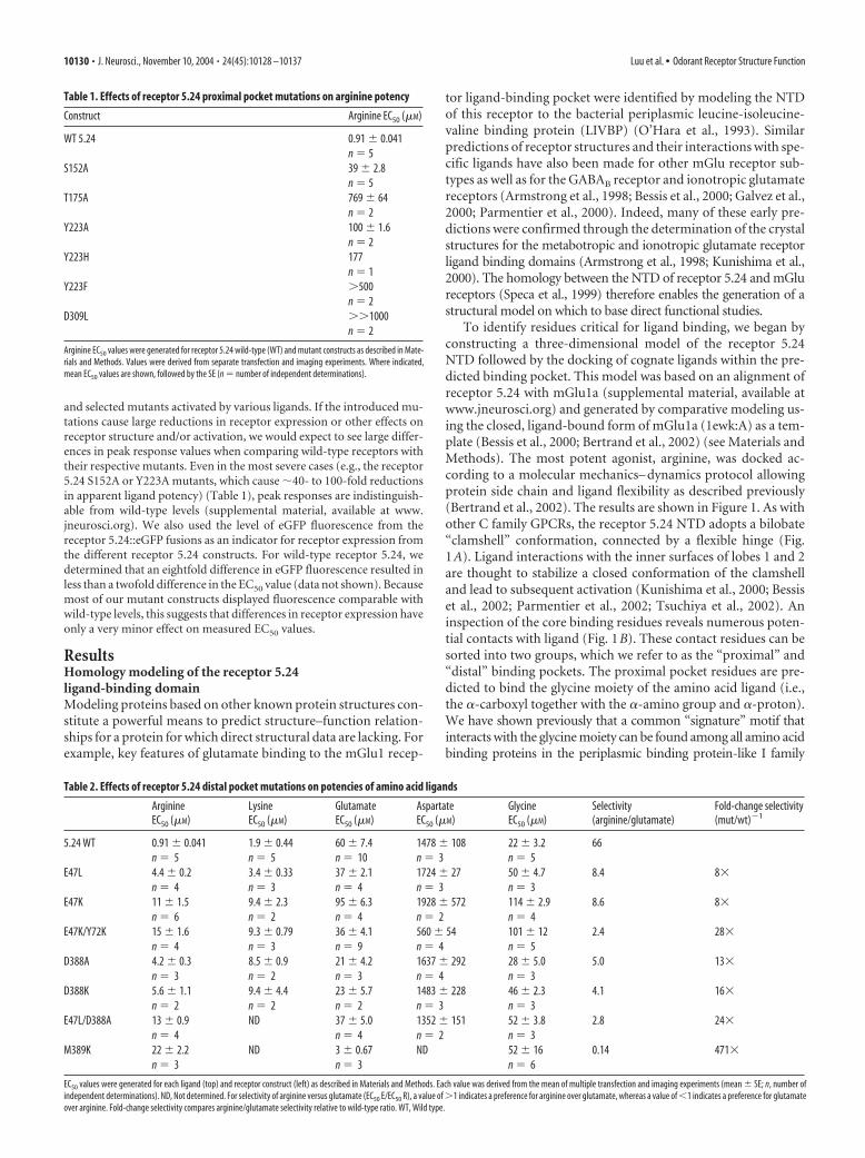
To identify residues critical for ligand binding, we began byconstructing a three-dimensional model of the receptor 5.24NTD followed by the docking of cognate ligands within the pre-dicted binding pocket. This model was based on an alignment ofreceptor 5.24 with mGlu1a (supplemental material, available atwww.jneurosci.org) and generated by comparative modeling us-ing the closed, ligand-bound form of mGlu1a (1ewk:A) as a tem-plate (Bessis et al., 2000; Bertrand et al., 2002) (see Materials andMethods). The most potent agonist, arginine, was docked ac-cording to a molecular mechanics– dynamics protocol allowingprotein side chain and ligand flexibility as described previously(Bertrand et al., 2002). The results are shown in Figure 1. As withother C family GPCRs, the receptor 5.24 NTD adopts a bilobate“clamshell” conformation, connected by a flexible hinge (Fig.1A). Ligand interactions with the inner surfaces of lobes 1 and 2are thought to stabilize a closed conformation of the clamshelland lead to subsequent activation (Kunishima et al., 2000; Bessiset al., 2002; Parmentier et al., 2002; Tsuchiya et al., 2002). Aninspection of the core binding residues reveals numerous poten-tial contacts with ligand (Fig. 1B). These contact residues can besorted into two groups, which we refer to as the “proximal” and“distal” binding pockets. The proximal pocket residues are pre-dicted to bind the glycine moiety of the amino acid ligand (i.e.,the �-carboxyl together with the �-amino group and �-proton).We have shown previously that a common “signature” motif thatinteracts with the glycine moiety can be found among all amino acidbinding proteins in the periplasmic binding protein-like I family
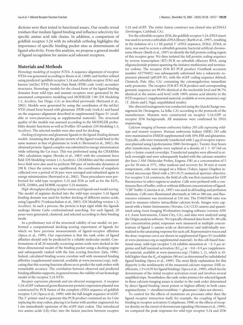
Table 1. Effects of receptor 5.24 proximal pocket mutations on arginine potency
Construct Arginine EC50 (�M)
WT 5.24 0.91 � 0.041n � 5
S152A 39 � 2.8n � 5
T175A 769 � 64n � 2
Y223A 100 � 1.6n � 2
Y223H 177n � 1
Y223F �500n � 2
D309L ��1000n � 2
Arginine EC50 values were generated for receptor 5.24 wild-type (WT) and mutant constructs as described in Mate-rials and Methods. Values were derived from separate transfection and imaging experiments. Where indicated,mean EC50 values are shown, followed by the SE (n � number of independent determinations).
Table 2. Effects of receptor 5.24 distal pocket mutations on potencies of amino acid ligands
ArginineEC50 (�M)
LysineEC50 (�M)
GlutamateEC50 (�M)
AspartateEC50 (�M)
GlycineEC50 (�M)
Selectivity(arginine/glutamate)
Fold-change selectivity(mut/wt)�1
5.24 WT 0.91 � 0.041 1.9 � 0.44 60 � 7.4 1478 � 108 22 � 3.2 66n � 5 n � 5 n � 10 n � 3 n � 5
E47L 4.4 � 0.2 3.4 � 0.33 37 � 2.1 1724 � 27 50 � 4.7 8.4 8�n � 4 n � 3 n � 4 n � 3 n � 3
E47K 11 � 1.5 9.4 � 2.3 95 � 6.3 1928 � 572 114 � 2.9 8.6 8�n � 6 n � 2 n � 4 n � 2 n � 4
E47K/Y72K 15 � 1.6 9.3 � 0.79 36 � 4.1 560 � 54 101 � 12 2.4 28�n � 4 n � 3 n � 9 n � 4 n � 5
D388A 4.2 � 0.3 8.5 � 0.9 21 � 4.2 1637 � 292 28 � 5.0 5.0 13�n � 3 n � 2 n � 3 n � 4 n � 3
D388K 5.6 � 1.1 9.4 � 4.4 23 � 5.7 1483 � 228 46 � 2.3 4.1 16�n � 2 n � 2 n � 2 n � 3 n � 3
E47L/D388A 13 � 0.9 ND 37 � 5.0 1352 � 151 52 � 3.8 2.8 24�n � 4 n � 4 n � 2 n � 3
M389K 22 � 2.2 ND 3 � 0.67 ND 52 � 16 0.14 471�n � 3 n � 3 n � 6
EC50 values were generated for each ligand (top) and receptor construct (left) as described in Materials and Methods. Each value was derived from the mean of multiple transfection and imaging experiments (mean � SE; n, number ofindependent determinations). ND, Not determined. For selectivity of arginine versus glutamate (EC50 E/EC50 R), a value of �1 indicates a preference for arginine over glutamate, whereas a value of �1 indicates a preference for glutamateover arginine. Fold-change selectivity compares arginine/glutamate selectivity relative to wild-type ratio. WT, Wild type.
10130 • J. Neurosci., November 10, 2004 • 24(45):10128 –10137 Luu et al. • Odorant Receptor Structure Function
(SCOP classification, http://scop.mrc-lmb.cam.ac.uk/scop/data/scop.b.d.bbg.b.A.html) (Bertrand et al., 2002). Among these arethe bacterial leucine binding protein (LBP), LIVBP, eight sub-types of mGlu receptors, CaSR, and T1R taste receptors as well asreceptor 5.24 (Bertrand et al., 2002; Pin et al., 2003). Side chainsof five residues (S152, T175, D195, Y223, and D309 in receptor5.24) and a backbone carbonyl (A173) constitute this motif,which is highlighted in the sequence alignment of supplementalFigure S1 (available at www.jneurosci.org as supplemental mate-rial) (Fig. 1B).
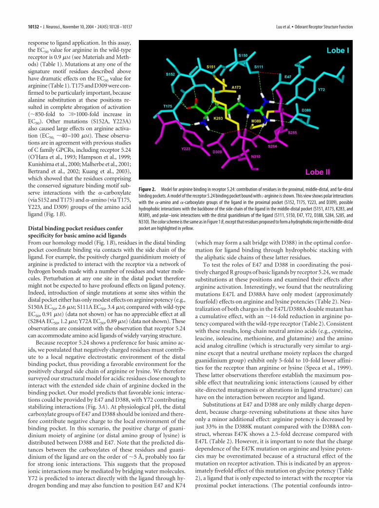
The distal pocket residues are predicted to coordinate the in-teractions between the R group side chain and receptor of theligand. When we examined the distal pocket residues, no com-
mon motif was found with any C familyGPCR. However, most residues that inter-act with ligands are localized in homolo-gous loops at the interface of the two lobes(supplemental material, available at www.jneurosci.org). In the case of receptor 5.24,two types of distal pocket residues can beidentified: those that make hydrophobiccontacts with the methylene groups of thearginine ligand and those that make polar–ionic interactions with the distal guani-dinium moiety. The hydrophobic ligandenvironment is provided by the methyland methylene groups of S151, A173,K283, and M389 (for clarity, K283 is notshown in Fig. 1B) (Fig. 2). The polar andionic interactions predicted to contributeto the binding of the distal portion of theligand side chain (Fig. 1B, right, dashedgreen lines) are mediated by two clusters ofserines (S111, S150, and S151 in lobe 1;S284 and S285 in lobe 2), E47, D388, Y72,and N310. Most of these interactions arelikely to be provided by hydrogen bond-ing, although some may be bridged bywater molecules. Interestingly, two ionicinteractions (with the carboxylate groupsof E47 and D388) are predicted to coordi-nate the positively charged guanidiniumgroup. Several residues predicted to con-tact the ligand may also play structural rolesin the receptor itself by forming hydrogenbond networks that stabilize the overall ter-tiary structure (Fig. 1B, red dashed lines). Inaddition, it should be noted that the activa-tion of C family GPCRs involves favorableinteractions not only between the ligand andprotein but also between the two lobes ofNTD of the receptor during closure,which leads to an optimal activated state(Kunishima et al., 2000; Bertrand et al.,2002).
Proximal binding pocket residuescontact all amino acids via theircommon glycine moietiesPrevious studies have identified a con-served signature motif in amino acid bind-ing proteins that interacts with the glycinemoiety common to all amino acids (Ber-
trand et al., 2002). These signature residues are also present in ourhomology model of receptor 5.24 (supplemental material, availableat www.jneurosci.org) (Fig. 1). We therefore engineered singleamino acid substitutions at these positions and tested the mutantreceptors to confirm the roles of these residues in ligand bindingand receptor activation. Because binding and closure of the twolobes of the NTD clamshell around the ligand are closely linked toreceptor activation in C family GPCRs (Parmentier et al., 2002),we decided to use a functional assay to assess the effects of anymutations on receptor activation, which reflects both ligandbinding and clamshell closure. The ligand activation properties ofwild-type and mutant receptors were therefore tested by express-ing them in HEK293 cells and measuring calcium mobilization in
Figure 1. Homology model of receptor 5.24 based on the crystal structure of mGlu1. A, Ribbon diagram of receptor 5.24extracellular domain from amino acids 28 – 484. Lobe 1 is depicted in cyan, whereas lobe 2 is shaded magenta. The flexible hingeregion is indicated in yellow. The structure is shown in the ligand-bound state in a closed conformation, with an L-argininemolecule present in the binding cleft. The �-carbon of the ligand is toward the left, with the side chain extending toward the right.B, Structural diagram of the proposed binding pocket of receptor 5.24 binding L-arginine. Amino acids proposed to interact withan arginine ligand are indicated by the residue number. Ligand contacts through hydrogen bonds (or ionic interactions in the caseof E47 and D388) are depicted by green dashed lines, whereas inter-residue hydrogen bonds are shown as red dashed lines. Sidechains of residues S111, S150, E47, and D388 may interact with the ligand via bridging water molecules. Residues D195 (hinge)and K283 have been omitted for clarity. The color scheme and general orientation of the receptor are the same as in A. On theligand, hydrogens are shown in white, carbons are shown in green, nitrogens are shown in blue, and oxygens are shown in red.
Luu et al. • Odorant Receptor Structure Function J. Neurosci., November 10, 2004 • 24(45):10128 –10137 • 10131
response to ligand application. In this assay,the EC50 value for arginine in the wild-typereceptor is 0.9 �M (see Materials and Meth-ods) (Table 1). Mutations at any one of thesignature motif residues described abovehave dramatic effects on the EC50 value forarginine (Table 1). T175 and D309 were con-firmed to be particularly important, becausealanine substitution at these positions re-sulted in complete abrogation of activation(�850-fold to ��1000-fold increase inEC50). Other mutations (S152A, Y223A)also caused large effects on arginine activa-tion (EC50, �40–100 �M). These observa-tions are in agreement with previous studiesof C family GPCRs, including receptor 5.24(O’Hara et al., 1993; Hampson et al., 1999;Kunishima et al., 2000; Malherbe et al., 2001;Bertrand et al., 2002; Kuang et al., 2003),which showed that the residues comprisingthe conserved signature binding motif sub-serve interactions with the �-carboxylate(via S152 and T175) and �-amino (via T175,Y223, and D309) groups of the amino acidligand (Fig. 1B).
Distal binding pocket residues conferspecificity for basic amino acid ligandsFrom our homology model (Fig. 1B), residues in the distal bindingpocket coordinate binding via contacts with the side chain of theligand. For example, the positively charged guanidinium moiety ofarginine is predicted to interact with the receptor via a network ofhydrogen bonds made with a number of residues and water mole-cules. Perturbation at any one site in the distal pocket thereforemight not be expected to have profound effects on ligand potency.Indeed, introduction of single mutations at some sites within thedistal pocket either has only modest effects on arginine potency (e.g.,S150A EC50, 2.6 �M; S111A EC50, 3.4 �M; compared with wild-typeEC50, 0.91 �M) (data not shown) or has no appreciable effect at all(S284A EC50, 1.2 �M; Y72A EC50, 0.89 �M) (data not shown). Theseobservations are consistent with the observation that receptor 5.24can accommodate amino acid ligands of widely varying structure.
Because receptor 5.24 shows a preference for basic amino ac-ids, we postulated that negatively charged residues must contrib-ute to a local negative electrostatic environment of the distalbinding pocket, thus providing a favorable environment for thepositively charged side chain of arginine or lysine. We thereforesurveyed our structural model for acidic residues close enough tointeract with the extended side chain of arginine docked in thebinding pocket. Our model predicts that favorable ionic interac-tions could be provided by E47 and D388, with Y72 contributingstabilizing interactions (Fig. 3A). At physiological pH, the distalcarboxylate groups of E47 and D388 should be ionized and there-fore contribute negative charge to the local environment of thebinding pocket. In this scenario, the positive charge of guani-dinium moiety of arginine (or distal amino group of lysine) isdistributed between D388 and E47. Note that the predicted dis-tances between the carboxylates of these residues and guani-dinium of the ligand are on the order of �5 Å, probably too farfor strong ionic interactions. This suggests that the proposedionic interactions may be mediated by bridging water molecules.Y72 is predicted to interact directly with the ligand through hy-drogen bonding and may also function to position E47 and K74
(which may form a salt bridge with D388) in the optimal confor-mation for ligand binding through hydrophobic stacking withthe aliphatic side chains of these latter residues.
To test the roles of E47 and D388 in coordinating the posi-tively charged R groups of basic ligands by receptor 5.24, we madesubstitutions at these positions and examined their effects afterarginine activation. Interestingly, we found that the neutralizingmutations E47L and D388A have only modest (approximatelyfourfold) effects on arginine and lysine potencies (Table 2). Neu-tralization of both charges in the E47L/D388A double mutant hasa cumulative effect, with an �14-fold reduction in arginine po-tency compared with the wild-type receptor (Table 2). Consistentwith these results, long-chain neutral amino acids (e.g., cysteine,leucine, isoleucine, methionine, and glutamine) and the aminoacid analog citrulline (which is structurally very similar to argi-nine except that a neutral urethane moiety replaces the chargedguanidinium group) exhibit only 5-fold to 10-fold lower affini-ties for the receptor than arginine or lysine (Speca et al., 1999).These latter observations therefore establish the maximum pos-sible effect that neutralizing ionic interactions (caused by eithersite-directed mutagenesis or alterations in ligand structure) canhave on the interaction between receptor and ligand.
Substitutions at E47 and D388 are only mildly charge depen-dent, because charge-reversing substitutions at these sites haveonly a minor additional effect: arginine potency is decreased byjust 33% in the D388K mutant compared with the D388A con-struct, whereas E47K shows a 2.5-fold decrease compared withE47L (Table 2). However, it is important to note that the chargedependence of the E47K mutation on arginine and lysine poten-cies may be overestimated because of a structural effect of themutation on receptor activation. This is indicated by an approx-imately fivefold effect of this mutation on glycine potency (Table2), a ligand that is only expected to interact with the receptor viaproximal pocket interactions. (The potential confounds intro-
Figure 2. Model for arginine binding in receptor 5.24: contribution of residues in the proximal, middle-distal, and far-distalbinding pockets. A model of the receptor 5.24 binding pocket bound with L-arginine is shown. This view shows polar interactionswith the �-amino and �-carboxylate groups of the ligand in the proximal pocket (S152, T175, Y223, and D309), possiblehydrophobic interactions with the backbone of the side chain of the ligand in the middle-distal pocket (S151, A173, K283, andM389), and polar–ionic interactions with the distal guanidinium of the ligand (S111, S150, E47, Y72, D388, S284, S285, andN310). The color scheme is the same as in Figure 1 B, except that residues proposed to form a hydrophobic ring in the middle-distalpocket are highlighted in yellow.
10132 • J. Neurosci., November 10, 2004 • 24(45):10128 –10137 Luu et al. • Odorant Receptor Structure Function
duced by structural effects are addressed and accounted for in theanalysis described in the Discussion.)
We ascribe the modest effects of both charge-neutralizing andcharge-reversing substitutions at D388 and E47 to two principalfactors. First, the ionic interactions provided by these residues areweakened because of the long distances that they span (�5 Å) andare probably screened by water molecules. Just as water moleculescan screen favorable interactions between attractive ionic pairs,they can also shield unfavorable interactions between repulsiveionic pairs. We would therefore expect that neutralization (orreversal) of these ionic interactions, either by chemical alterationsin the ligand or by substitutions at these residues, might havefairly modest effects on ligand potency. Second, there are proba-bly compensatory changes between the binding pocket sidechains and the ligand in the receptor 5.24 mutants, such that theligand adopts altered conformations, resulting in the guanidinium
group moving closer to the remaining acidicside chain in the distal pocket (supplementalmaterial, available at www.jneurosci.org).For the charge-reversing E47K or D388Ksubstitutions, in the presence of a positivelycharged ligand, the side chains of K388 orK47 probably adopt alternate conforma-tions by swinging away from the bindingpocket (supplemental material, available atwww.jneurosci.org).
Selectivity for basic versus acidic aminoacid ligands is reduced in distal bindingpocket mutantsFrom our structural model, we predictthat substitutions at E47, D388, and Y72would alter the proposed polar interac-tions between ligand and receptor andtherefore should have effects on the selec-tivity of the receptor for basic versus acidicamino acids. To test this idea, we deter-mined the potencies of multiple amino ac-ids in activating receptors with single ordouble mutations at these positions (Table2). The ligands tested include arginine andlysine (basic), glutamate and aspartate(acidic), and glycine (neutral; no sidechain). The ratios of EC50 values for gluta-mate:arginine were calculated for each re-ceptor construct (Table 2). It is importantto note that whatever effects a given muta-tion might have on the structure or activa-tion of the receptor (i.e., not directly af-fecting ligand–receptor affinity) will becanceled out in this ratiometric calcula-tion. We then used these ratios as indica-tors of receptor selectivity for basic versusacidic amino acid ligands.
As shown in Table 2, wild-type receptor5.24 exhibits a 66-fold preference for argi-nine over glutamate ([EC50 glutamate]:[EC50 arginine] � 66:1). Mutations of theabove-mentioned distal pocket residuesresult in significant effects on ligand selec-tivity, with arginine:glutamate selectivityratios reduced to �8 –9:1 for the E47L andE47K mutants, �4 –5:1 for the D388A and
D388K mutants, and �3:1 for the E47L/D388A double mutant(Table 2). The similar behaviors of charge-neutralizing andcharge-reversing mutations (i.e., E47L and E47K; D388A andD388K) could be explained by the ability of the positively chargedside chains to adopt alternate rotameric conformations, possiblyfacing away from the binding pocket and therefore reducing theeffects of the charge-reversing substitutions on repelling basicligands (supplemental material, available at www.jneurosci.org).
Although our structural model predicts an interaction be-tween the hydroxyl group of Y72 and guanidinium of the argi-nine ligand, the Y72A mutation results in no appreciable changein the potency of arginine (EC50, 0.83 �M) (data not shown) orarginine:glutamate selectivity (52:1; data not shown). However, ar-ginine:glutamate selectivity is reduced to �2:1 in the E47K/Y72Kdouble mutant (compared with �9:1 for E47K), consistent with theprediction that Y72 plays a role in the distal binding pocket.
Figure 3. Methionine-389 is a critical determinant of ligand selectivity in receptor 5.24. A, Schematic view of selected distalpocket residues (M389, D388, Y72, and E47) involved in contacting the guanidinium moiety of an arginine ligand in the wild-type(WT) receptor. The orientation of this “top-down” view is from above lobe 1 looking down onto the bound ligand, with theproximal pocket toward the bottom and the distal pocket toward the top of the figure. Receptor residues are shaded cyan, withoxygens shown in red. On the ligand, hydrogens are shown in white, carbons are shown in green, nitrogens are shown in blue, andoxygens are shown in red. Proposed intermolecular interactions and distances are indicated with dashed green lines. M389 facestoward the binding pocket, possibly making van der Waals contacts with the n-aliphatic side chain of the docked arginine (Fig.4 A). B, Predicted interaction of K389 in the receptor 5.24 M389K mutant with bound glutamate. In this model of the mutantbinding pocket, the amino group of the side chain of K389 (nitrogen atom shown in blue) can make a direct ionic interaction withthe distal carboxylate of the bound glutamate (distances indicated). C, Representative dose–response curves for wild-type andM389K receptor 5.24. HEK293 cells expressing wild-type or mutant receptor were exposed to arginine (blue lines) or glutamate(red lines), and receptor activation was measured by calcium imaging (see Materials and Methods). Note that the wild-typereceptor prefers arginine to glutamate, whereas this selectivity is inverted in the M389K mutant. EC50 values for the curves shownin this panel are as follows. Wild type: 0.82 �M arginine, 29.6 �M glutamate; M389K mutant: 24.9 �M arginine, 2.1 �M glutamate.
Luu et al. • Odorant Receptor Structure Function J. Neurosci., November 10, 2004 • 24(45):10128 –10137 • 10133
Overall, these results are corroboratedby the effects of the distal pocket muta-tions on arginine:aspartate, lysine:gluta-mate, and lysine:aspartate selectivities(calculations not shown) (Table 2), whichmirror the effects on arginine:glutamateselectivity. In summary, our data confirmthe participation of E47, D388, and possiblyalso Y72 not only in coordinating the posi-tive charges of the arginine and lysine Rgroups but also in determining the selectivityof the receptor for basic versus acidic aminoacid ligands.
A recent study by Kuang et al. (2003)also identified D388 as a residue importantin receptor 5.24 for coordinating the pos-itively charged �-amino group of boundlysine. Unlike the present model, themodel in the study by Kuang et al. (2003)predicts the close apposition of this func-tional group with the �-carboxylate (�3Å) of D388 but no interaction with E47. Inaddition, these authors observed a 26-foldreduction in lysine potency in this mutant,much higher than the approximately four-fold effect we observed in our assay (Table 2). Although the rea-sons for these discrepancies are presently unclear, our data dem-onstrate important roles for both D388 and E47 in ligandbinding–receptor activation (as well as selectivity), with effects ofthe mutations in accord with previous structure-activity data(Speca et al., 1999).
Methionine-389 in the receptor 5.24 distal binding pocket is amajor determinant of selectivityThe results presented thus far indicate that polar interactionsbetween the positively charged R groups of arginine or lysine andD388, Y72, and E47 play important roles in ligand binding andselectivity in receptor 5.24. We next wanted to determine whetherother residues might contribute to selectivity in the distal bindingpocket of this receptor. To this end, we asked whether there arecommon positions in the distal binding pockets of other aminoacid binding proteins that contact the R group of the cognateligand of each receptor. If so, this would instruct our search forother potential binding sites in the receptor 5.24 distal pocket.We therefore examined the alignment of selected amino acidbinding proteins (supplemental material, available at www.jneurosci.org) and identified one position, the equivalent to K409in mGlu1 and M389 in receptor 5.24, that fulfilled this criterion.In mGlu1, K409 makes a direct ionic interaction with the distalcarboxylate of the bound glutamate and is required for ligandbinding (Kunishima et al., 2000; Rosemond et al., 2002); thislysine is strictly conserved in all mGlu receptor isoforms (Ber-trand et al., 2002). In the bacterial LBP and LIVBP, the aminoacids in the equivalent position (Y276 and F276, respectively)(supplemental material, available at www.jneurosci.org) partici-pate in van der Waals interactions with the hydrophobic R groupof the bound ligand (Sack et al., 1989; Magnusson et al., 2004).Similarly, molecular modeling and site-directed mutagenesis of theGABAB1 receptor implicate E465, which aligns with K409 inmGlu1, in making an ionic interaction with the distal aminogroup of the bound GABA ligand (Kniazeff et al., 2002). We there-fore investigated the potential role of M389 in receptor 5.24 in ligandbinding.
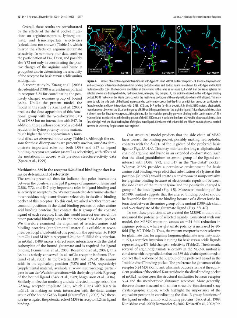
Our structural model predicts that the side chain of M389faces toward the binding pocket, possibly making hydrophobiccontacts with the �-CH2 of the R group of the preferred basicligand (Figs. 3A, 4A). This may maintain the long n-aliphatic sidechain of arginine and lysine in an extended conformation, suchthat the distal guanidinium or amino group of the ligand caninteract with D388, Y72, and E47 in the “far-distal” pocket.Whereas M389 provides a permissive environment for basicamino acid binding, we predict that substitution of a lysine at thisposition (M389K) would create an environment nonpermissivefor arginine binding because of electrostatic repulsion betweenthe side chain of the mutant lysine and the positively charged Rgroup of the basic ligand (Fig. 4B). Moreover, modeling of theM389K mutant suggests that the altered binding pocket shouldbe favorable for glutamate binding because of a direct ionic in-teraction between the amino group of the mutant K389 side chainand �-carboxylate of the glutamate ligand (Figs. 3B, 4C).
To test these predictions, we created the M389K mutant andmeasured the potencies of selected ligands. Consistent with ourmodel, the M389K mutation causes an �24-fold reduction inarginine potency, whereas glutamate potency is increased by 20-fold (Fig. 3C, Table 2). Thus, the mutant receptor is more selectivefor glutamate than for arginine (arginine:glutamate selectivity ratio,�1:7), a complete inversion in tuning for basic versus acidic ligandsrepresenting a 471-fold change in selectivity (Table 2). The dramaticreversal of arginine:glutamate selectivity in the M389K mutant isconsistent with our prediction that the 389 side chain is positioned tocontact the backbone of the R group of the preferred ligand in the“middle-distal” binding pocket. The preference for glutamate of thereceptor 5.24 M389K mutant, which introduces a lysine at the equiv-alent position of the critical K409 residue in the distal binding pocketof mGlu1, underscores the structural similarities between receptor5.24 and the metabotropic glutamate receptors. More generally,these results are in accord with similar structure–function and x-raycrystallographic studies, which highlight the importance of theequivalent position in coordinating the distal R group functions ofthe ligand in other amino acid binding proteins (Sack et al., 1989;Kunishima et al., 2000; Bertrand et al., 2002; Kniazeff et al., 2002; Pin
Figure 4. Models of receptor–ligand interactions in wild-type (WT) and M389K mutant receptor 5.24. Proposed hydrophobicand electrostatic interactions between distal binding pocket residues and docked ligands are shown for wild-type and M389Kmutant receptor 5.24. The top-down orientation of these views is the same as in Figure 3, A and B. Van der Waals spheres forselected atoms are displayed (white, hydrogen; blue, nitrogen; red, oxygen). A, For arginine docked in the wild-type bindingpocket, M389 makes van der Waals contacts with the methylene backbone of the n-aliphatic side chain of the ligand. This mayserve to hold the side chain of the ligand in an extended conformation, such that the distal guanidinium group can participate infavorable polar and ionic interactions with D388, Y72, and E47 in the far-distal pocket. B, In the M389K mutant, electrostaticrepulsion occurs between the distal amino group of K389 and the guanidinium of the arginine ligand. This unfavorable interactionis shown here for illustrative purposes, although in reality the repulsion probably prevents binding in this conformation. C, Thelysine residue introduced into the binding pocket of the M389K mutant is positioned to form a favorable electrostatic interaction(a salt bridge) with the distal carboxylate of the glutamate ligand. Consistent with this model, the M389K mutant shows a markedincrease in selectivity for glutamate over arginine.
10134 • J. Neurosci., November 10, 2004 • 24(45):10128 –10137 Luu et al. • Odorant Receptor Structure Function
et al., 2003; Magnusson et al., 2004). In the case of receptor 5.24,M389 functions together with other sites in the far-distal bindingpocket to determine ligand selectivity.
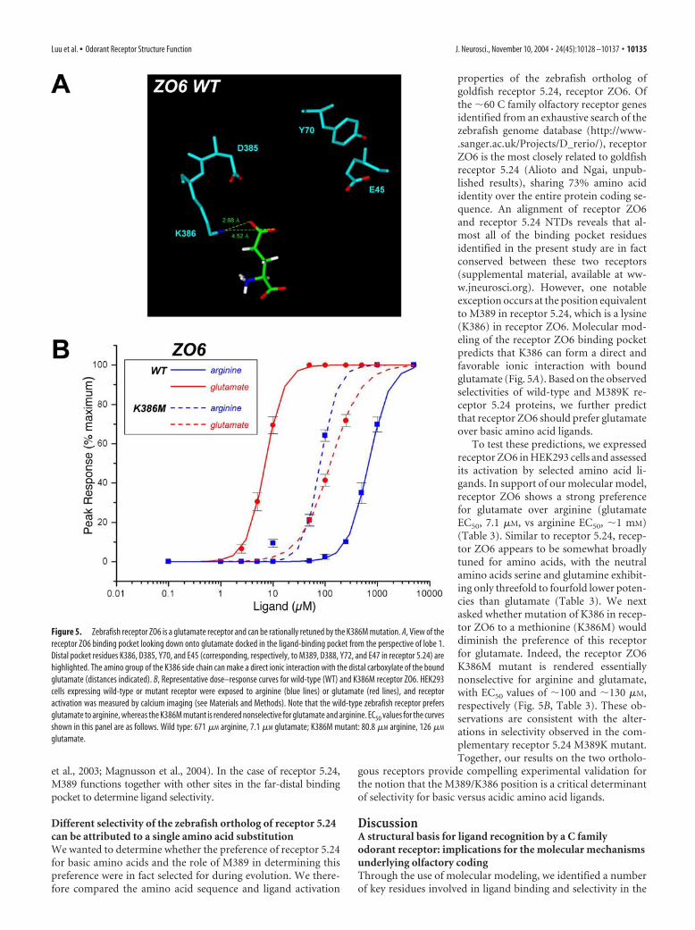
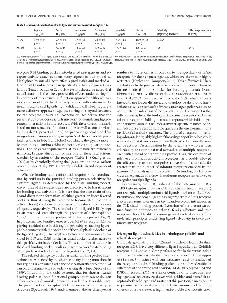
Different selectivity of the zebrafish ortholog of receptor 5.24can be attributed to a single amino acid substitutionWe wanted to determine whether the preference of receptor 5.24for basic amino acids and the role of M389 in determining thispreference were in fact selected for during evolution. We there-fore compared the amino acid sequence and ligand activation
properties of the zebrafish ortholog ofgoldfish receptor 5.24, receptor ZO6. Ofthe �60 C family olfactory receptor genesidentified from an exhaustive search of thezebrafish genome database (http://www-.sanger.ac.uk/Projects/D_rerio/), receptorZO6 is the most closely related to goldfishreceptor 5.24 (Alioto and Ngai, unpub-lished results), sharing 73% amino acididentity over the entire protein coding se-quence. An alignment of receptor ZO6and receptor 5.24 NTDs reveals that al-most all of the binding pocket residuesidentified in the present study are in factconserved between these two receptors(supplemental material, available at ww-w.jneurosci.org). However, one notableexception occurs at the position equivalentto M389 in receptor 5.24, which is a lysine(K386) in receptor ZO6. Molecular mod-eling of the receptor ZO6 binding pocketpredicts that K386 can form a direct andfavorable ionic interaction with boundglutamate (Fig. 5A). Based on the observedselectivities of wild-type and M389K re-ceptor 5.24 proteins, we further predictthat receptor ZO6 should prefer glutamateover basic amino acid ligands.
To test these predictions, we expressedreceptor ZO6 in HEK293 cells and assessedits activation by selected amino acid li-gands. In support of our molecular model,receptor ZO6 shows a strong preferencefor glutamate over arginine (glutamateEC50, 7.1 �M, vs arginine EC50, �1 mM)(Table 3). Similar to receptor 5.24, recep-tor ZO6 appears to be somewhat broadlytuned for amino acids, with the neutralamino acids serine and glutamine exhibit-ing only threefold to fourfold lower poten-cies than glutamate (Table 3). We nextasked whether mutation of K386 in recep-tor ZO6 to a methionine (K386M) woulddiminish the preference of this receptorfor glutamate. Indeed, the receptor ZO6K386M mutant is rendered essentiallynonselective for arginine and glutamate,with EC50 values of �100 and �130 �M,respectively (Fig. 5B, Table 3). These ob-servations are consistent with the alter-ations in selectivity observed in the com-plementary receptor 5.24 M389K mutant.Together, our results on the two ortholo-
gous receptors provide compelling experimental validation forthe notion that the M389/K386 position is a critical determinantof selectivity for basic versus acidic amino acid ligands.
DiscussionA structural basis for ligand recognition by a C familyodorant receptor: implications for the molecular mechanismsunderlying olfactory codingThrough the use of molecular modeling, we identified a numberof key residues involved in ligand binding and selectivity in the
Figure 5. Zebrafish receptor ZO6 is a glutamate receptor and can be rationally retuned by the K386M mutation. A, View of thereceptor ZO6 binding pocket looking down onto glutamate docked in the ligand-binding pocket from the perspective of lobe 1.Distal pocket residues K386, D385, Y70, and E45 (corresponding, respectively, to M389, D388, Y72, and E47 in receptor 5.24) arehighlighted. The amino group of the K386 side chain can make a direct ionic interaction with the distal carboxylate of the boundglutamate (distances indicated). B, Representative dose–response curves for wild-type (WT) and K386M receptor ZO6. HEK293cells expressing wild-type or mutant receptor were exposed to arginine (blue lines) or glutamate (red lines), and receptoractivation was measured by calcium imaging (see Materials and Methods). Note that the wild-type zebrafish receptor prefersglutamate to arginine, whereas the K386M mutant is rendered nonselective for glutamate and arginine. EC50 values for the curvesshown in this panel are as follows. Wild type: 671 �M arginine, 7.1 �M glutamate; K386M mutant: 80.8 �M arginine, 126 �M
glutamate.
Luu et al. • Odorant Receptor Structure Function J. Neurosci., November 10, 2004 • 24(45):10128 –10137 • 10135
receptor 5.24 binding pocket. Site-directed mutagenesis and re-ceptor activity assays confirm many aspects of our model, ashighlighted by our ability to effect a predictable and marked al-teration of ligand selectivity in specific distal binding pocket mu-tations (Figs. 3, 5; Tables 2, 3). However, it should be noted thatnot all mutants had entirely predictable effects, underscoring thelimitations of this structure–function approach. Although ourmolecular model can be iteratively refined with data on addi-tional mutants and ligands, full validation will likely require amore definitive approach (e.g., the solving of a crystal structurefor the receptor 5.24 NTD). Nonetheless, we believe that thepresent study provides a useful framework for considering ligand–protein interactions in this receptor. With these caveats in mind,based on our structure–function studies as well as our previousbinding data (Speca et al., 1999), we propose a general model forrecognition of amino acids by receptor 5.24. In our model, prox-imal residues in lobe 1 and lobe 2 coordinate the glycine moiety(common to all amino acids) via both ionic and polar interac-tions. The physical requirements at this region are extremelystringent, because disruption of any one of these interactions,whether by mutation of the receptor (Table 1) (Kuang et al.,2003) or by chemically altering the ligand around the �-carboncenter (Speca et al., 1999), severely inhibits ligand-dependentactivation.
Whereas binding to all amino acids requires strict coordina-tion by residues in the proximal binding pocket, selectivity fordifferent ligands is determined by the distal binding pocket,where some of the requirements are predicted to be less stringentfor binding and activation. It is here that the side chain of theligand dictates the formation of either greater or fewer favoredcontacts, thus allowing the receptor to become stabilized in theactive (closed) conformation at lesser or greater concentrationsof ligands, respectively. The side chain of the ligand is likely keptin an extended state through the presence of a hydrophobic“ring” in the middle-distal portion of the binding pocket (Fig. 2).In particular, we identified one residue, M389 in receptor 5.24, asplaying a critical role in this region, probably by making hydro-phobic contacts with the backbone of the n-aliphatic side chain ofthe ligand (Fig. 4A). The negative electrostatic environment pro-vided by E47 and D388 in the far-distal pocket further narrowsthis specificity for basic side chains. Thus, a number of residues inthe distal binding pocket work in concert to coordinate bindingof the preferred side chains of the ligands (Figs. 2, 4A).
The relaxed stringency of the far-distal binding pocket inter-actions (as evidenced by the absence of any killing mutations inthis region) is consistent with the observation that receptor 5.24can bind to amino acids of widely varying structure (Speca et al.,1999). In addition, it should be noted that for shorter ligandsbearing polar or ionic functional groups, water molecules caneither screen unfavorable interactions or bridge favorable ones.The promiscuity of receptor 5.24 for amino acids of varyingstructure (Speca et al., 1999) and tolerance of the far-distal pocket
residues to mutations is in contrast to the specificity of mGlureceptors for their cognate ligands, which are chemically highlyrestricted (Naples and Hampson, 2001). This difference is likelyattributable to the greater reliance on direct ionic interactions inthe mGlu distal binding pocket for binding glutamate (Kun-ishima et al., 2000; Malherbe et al., 2001; Rosemond et al., 2002;Sato et al., 2003) compared with receptor 5.24, which appearsinstead to use longer distance, and therefore weaker, ionic inter-actions as well as a network of mostly uncharged polar residues tocoordinate the side chain of the ligand (Fig. 2). The reason for thisdifference may lie in the biological function of receptor 5.24 as anodorant receptor. Unlike glutamate receptors, which initiate syn-aptic transmission in a neurotransmitter-specific manner, odor-ant receptors are responsible for querying the environment for amyriad of chemical signatures. The utility of a receptor for sens-ing odorants is arguably higher if the stringency of its selectivity isreduced so that it can respond to multiple, albeit chemically sim-ilar structures. Discrimination by the system as a whole is thenafforded by the combinatorial activation of multiple receptors,each with a broad odorant tuning profile. Thus, the evolution ofrelatively promiscuous odorant receptors has probably allowedthe olfactory system to recognize a diversity of chemicals fargreater than the number of odorant receptors encoded in thegenome. Our analysis of the receptor 5.24 binding pocket pro-vides an explanation for how this odorant receptor has evolved torecognize multiple ligands.
Interestingly, the T1R1 subunit of the heteromeric T1R1/T1R3 taste receptor (another C family chemosensory receptor)can recognize multiple amino acid ligands (Nelson et al., 2002).Presumably, the broad ligand tuning properties of this receptoralso reflect some tolerance in the ligand–receptor interaction inthe T1R distal binding pocket. Extension of the present struc-ture–function approach to other C family olfactory and tastereceptors should facilitate a more general understanding of themolecular principles underlying ligand selectivity in these che-mosensory receptors.
Divergent ligand selectivities in orthologous goldfish andzebrafish receptorsCuriously, goldfish receptor 5.24 and its ortholog from zebrafish,receptor ZO6, have very different ligand specificities. Goldfishreceptor 5.24 shows a clear preference for basic versus acidicamino acids, whereas zebrafish receptor ZO6 exhibits the oppo-site tuning. Consistent with our structure–function analysis ofthe receptor 5.24 distal binding pocket, our studies identified adifference at one amino acid position (M389 in receptor 5.24 andK386 in receptor ZO6) as a major contributor to these contrast-ing ligand selectivities. As shown with goldfish and zebrafish re-ceptors both wild-type and mutant at this position, a methionineis permissive for n-aliphatic and basic amino acid binding,whereas a lysine creates a highly unfavorable electrostatic envi-
Table 3. Amino acid selectivities of wild-type and mutant zebrafish receptor ZO6
ArginineEC50 (�M)
SerineEC50 (�M)
GlutamineEC50 (�M)
GlutamateEC50 (�M)
AspartateEC50 (�M)
GlycineEC50 (�M)
Selectivity(arginine/glutamate)
Fold-change selectivity(mut/wt)
ZO6 WT 1059 � 151 22 � 4.9 27 � 1.1 7.1 � 0.6 �� 1000 1129 � 78 0.0067n � 4 n � 2 n � 2 n � 4 n � 1 n � 2
K389M 101 � 17 48 � 17 44 � 2.6 129 � 17 �� 1000 526 � 23 1.3 194�n � 4 n � 2 n � 3 n � 4 n � 1 n � 4
EC50 values were generated for each ligand (top) and receptor construct (left) as described in Materials and Methods. Where indicated, each value was derived from the mean of multiple transfection and imaging experiments (mean � SE;n, number of independent determinations). For selectivity of arginine versus glutamate (EC50 E/EC50 R), a value of �1 indicates a preference for arginine over glutamate, whereas a value of �1 indicates a preference for glutamate overarginine. Fold-change selectivity compares arginine/glutamate selectivity relative to wild-type ratio. WT, Wild type.
10136 • J. Neurosci., November 10, 2004 • 24(45):10128 –10137 Luu et al. • Odorant Receptor Structure Function
ronment for basic amino acids in the binding pocket and insteadcan form a favorable ionic interaction with glutamate.
Both goldfish and zebrafish can detect a broad spectrum ofamino acids through their olfactory systems (Michel and Lubo-mudrov, 1995; Friedrich and Korsching, 1997; Sorensen andCaprio, 1998). It is therefore unclear what evolutionary pressuresmay have driven the selection of such different ligand selectivitiesof these orthologous receptors. Nonetheless, our comparison ofgoldfish receptor 5.24 and zebrafish receptor ZO6 underscoresthe importance of M389 (goldfish)/K386 (zebrafish) in deter-mining ligand selectivity and moreover indicates that this posi-tion was selected for during the evolution of these receptors.
ReferencesAraneda RC, Kini AD, Firestein S (2000) The molecular receptive range of
an odorant receptor. Nat Neurosci 3:1248 –1255.Armstrong N, Sun Y, Chen GQ, Gouaux E (1998) Structure of a glutamate-
receptor ligand-binding core in complex with kainate. Nature 395:913–917.Barth AL, Dugas JC, Ngai J (1997) Noncoordinate expression of odorant
receptor genes tightly linked in the zebrafish genome. Neuron 19:359–369.Bertrand HO, Bessis AS, Pin JP, Acher FC (2002) Common and selective
molecular determinants involved in metabotropic glutamate receptor ag-onist activity. J Med Chem 45:3171–3183.
Bessis AS, Bertrand HO, Galvez T, De Colle C, Pin JP, Acher F (2000) Three-dimensional model of the extracellular domain of the type 4a metabo-tropic glutamate receptor: new insights into the activation process. Pro-tein Sci 9:2200 –2209.
Bessis AS, Rondard P, Gaven F, Brabet I, Triballeau N, Prezeau L, Acher F, PinJP (2002) Closure of the Venus flytrap module of mGlu8 receptor andthe activation process: insights from mutations converting antagonistsinto agonists. Proc Natl Acad Sci USA 99:11097–11102.
Brooks BR, Bruccoleri RE, Olafson BD, States DJ, Swaminathan S, Karplus M(1983) CHARMM: a program for macromolecular energy, minimiza-tion, and dynamics calculations. J Comput Chem 4:187–217.
Caterina MJ, Schumacher MA, Tominaga M, Rosen TA, Levine JD, Julius D(1997) The capsaicin receptor: a heat-activated ion channel in the painpathway. Nature 389:816 – 824.
Colquhoun D (1998) Binding, gating, affinity and efficacy: the interpreta-tion of structure-activity relationships for agonists and of the effects ofmutating receptors. Br J Pharmacol 125:924 –947.
Firestein S (2001) How the olfactory system makes sense of scents. Nature413:211–218.
Friedrich RW, Korsching SI (1997) Combinatorial and chemotopic odorantcoding in the zebrafish olfactory bulb visualized by optical imaging. Neu-ron 18:737–752.
Galvez T, Prezeau L, Milioti G, Franek M, Joly C, Froestl W, Bettler B, Ber-trand HO, Blahos J, Pin JP (2000) Mapping the agonist-binding site ofGABAB type 1 subunit sheds light on the activation process of GABABreceptors. J Biol Chem 275:41166 – 41174.
Hampson DR, Huang XP, Pekhletski R, Peltekova V, Hornby G, Thomsen C,Thogersen H (1999) Probing the ligand-binding domain of the mGluR4subtype of metabotropic glutamate receptor. J Biol Chem 274:33488–33495.
Hara TJ (1994) Olfaction and gustation in fish: an overview. Acta PhysiolScand 152:207–217.
Hermans E, Challiss RA, Nahorski SR (1999) Effects of varying the expres-sion level of recombinant human mGlu1alpha receptors on the pharma-cological properties of agonists and antagonists. Br J Pharmacol126:873– 882.
Jain AN (1996) Scoring noncovalent protein-ligand interactions: a contin-uous differentiable function tuned to compute binding affinities. J Com-put Aided Mol Des 10:427– 440.
Kniazeff J, Galvez T, Labesse G, Pin JP (2002) No ligand binding in the GB2subunit of the GABAB receptor is required for activation and allostericinteraction between the subunits. J Neurosci 22:7352–7361.
Kuang D, Yao Y, Wang M, Pattabiraman N, Kotra LP, Hampson DR (2003)Molecular similarities in the ligand binding pockets of an odorant recep-tor and the metabotropic glutamate receptors. J Biol Chem 278:42551-42559.
Kunishima N, Shimada Y, Tsuji Y, Sato T, Yamamoto M, Kumasaka T, Na-kanishi S, Jingami H, Morikawa K (2000) Structural basis of glutamaterecognition by a dimeric metabotropic glutamate receptor. Nature407:971–977.
Magnusson U, Salopek-Sondi B, Luck LA, Mowbray SL (2004) X-ray struc-tures of the leucine-binding protein illustrate conformational changesand the basis of ligand specificity. J Biol Chem 279:8747– 8752.
Malherbe P, Knoflach F, Broger C, Ohresser S, Kratzeisen C, Adam G, StadlerH, Kemp JA, Mutel V (2001) Identification of essential residues in-volved in the glutamate binding pocket of the group II metabotropicglutamate receptor. Mol Pharmacol 60:944 –954.
Malnic B, Hirono J, Sato T, Buck LB (1999) Combinatorial receptor codesfor odors. Cell 96:713–723.
Michel WC, Lubomudrov LM (1995) Specificity and sensitivity of the olfac-tory organ of the zebrafish, Danio rerio. J Comp Physiol [A] 177:191–199.
Mombaerts P (2004) Genes and ligands for odorant, vomeronasal and tastereceptors. Nat Rev Neurosci 5:263–278.
Naples MA, Hampson DR (2001) Pharmacological profiles of the metabo-tropic glutamate receptor ligands. Neuropharmacology 40:170 –177.
Nelson G, Chandrashekar J, Hoon MA, Feng L, Zhao G, Ryba NJ, Zuker CS(2002) An amino-acid taste receptor. Nature 416:199 –202.
O’Hara PJ, Sheppard PO, Thogersen H, Venezia D, Haldeman BA, McGraneV, Houamed KM, Thomsen C, Gilbert TL, Mulvihill ER (1993) Theligand-binding domain in metabotropic glutamate receptors is related tobacterial periplasmic binding proteins. Neuron 11:41–52.
Parmentier ML, Galvez T, Acher F, Peyre B, Pellicciari R, Grau Y, Bockaert J,Pin JP (2000) Conservation of the ligand recognition site of metabo-tropic glutamate receptors during evolution. Neuropharmacology39:1119 –1131.
Parmentier ML, Prezeau L, Bockaert J, Pin JP (2002) A model for the func-tioning of family 3 GPCRs. Trends Pharmacol Sci 23:268 –274.
Pin JP, Galvez T, Prezeau L (2003) Evolution, structure and activationmechanism of family 3/C G-protein coupled receptors. Pharmacol Ther98:325–354.
Rosemond E, Peltekova V, Naples M, Thogersen H, Hampson DR (2002)Molecular determinants of high affinity binding to group III metabo-tropic glutamate receptors. J Biol Chem 277:7333–7340.
Sack JS, Saper MA, Quiocho FA (1989) Periplasmic binding protein struc-ture and function. Refined X-ray structures of the leucine/isoleucine/valine-binding protein and its complex with leucine. J Mol Biol206:171–191.
Sato T, Shimada Y, Nagasawa N, Nakanishi S, Jingami H (2003) Amino acidmutagenesis of the ligand binding site and the dimer interface of themetabotropic glutamate receptor 1: identification of crucial residues forsetting the activated state. J Biol Chem 278:4314 – 4321.
Sorensen PW, Caprio JC (1998) Chemoreception. In: The physiology offishes, Ed 2 (Evans DH, ed), pp 375– 405. Boca Raton, FL: CRC.
Speca DJ, Lin DM, Sorensen PW, Isacoff EY, Ngai J, Dittman AH (1999)Functional identification of a goldfish odorant receptor. Neuron23:487– 498.
Tsuchiya D, Kunishima N, Kamiya N, Jingami H, Morikawa K (2002)Structural views of the ligand-binding cores of a metabotropic glutamatereceptor complexed with an antagonist and both glutamate and Gd3.Proc Natl Acad Sci USA 99:2660 –2665.
Uchida N, Takahashi YK, Tanifuji M, Mori K (2000) Odor maps in themammalian olfactory bulb: domain organization and odorant structuralfeatures. Nat Neurosci 3:1035–1043.
Venkatachalam CM, Jiang X, Oldfield T, Waldman M (2003) LigandFit: anovel method for the shape-directed rapid docking of ligands to proteinactive sites. J Mol Graph Model 21:289 –307.
Luu et al. • Odorant Receptor Structure Function J. Neurosci., November 10, 2004 • 24(45):10128 –10137 • 10137


