Embed Size (px)
Citation preview
Int. J. Mol. Sci. 2012, 13, 12970-12993; doi:10.3390/ijms131012970
International Journal of
Molecular Sciences ISSN 1422-0067
www.mdpi.com/journal/ijms
Review
Molecular Pathogenesis of Neuromyelitis Optica
Wajih Bukhari 1,2,*, Michael H Barnett 3, Kerri Prain 4 and Simon A Broadley 1,2
1 School of Medicine, Gold Coast Campus, Griffith University, QLD 4222, Australia;
E-Mail: [email protected] 2 Department of Neurology, Gold Coast Hospital, Southport, QLD 4215, Australia 3 Brain and Mind Research Institute, Camperdown, NSW 2050, Australia;
E-Mail: [email protected] 4 Autoimmune laboratory, Division of Immunology, Pathology Queensland, Herston, QLD 4029,
Australia; E-Mail: [email protected]
* Author to whom correspondence should be addressed; E-Mail: [email protected];
Tel.: +61-07-5678-0702; Fax: +61-07-5678-0708.
Received: 3 August 2012; in revised form: 8 September 2012 / Accepted: 13 September 2012 /
Published: 11 October 2012
Abstract: Neuromyelitis optica (NMO) is a rare autoimmune disorder, distinct from
multiple sclerosis, causing inflammatory lesions in the optic nerves and spinal cord. An
autoantibody (NMO IgG) against aquaporin-4 (AQP4), a water channel expressed on
astrocytes is thought to be causative. Peripheral production of the antibody is triggered by
an unknown process in genetically susceptible individuals. Anti-AQP4 antibody enters the
central nervous system (CNS) when the blood brain barrier is made permeable and has
high affinity for orthogonal array particles of AQP4. Like other autoimmune diseases,
Th17 cells and their effector cytokines (such as interleukin 6) have been implicated in
pathogenesis. AQP4 expressing peripheral organs are not affected by NMO IgG, but the
antibody causes extensive astrocytic loss in specific regions of the CNS through complement
mediated cytotoxicity. Demyelination occurs during the inflammatory process and is
probably secondary to oligodendrocyte apoptosis subsequent to loss of trophic support
from astrocytes. Ultimately, extensive axonal injury leads to severe disability. Despite
rapid advances in the understanding of NMO pathogenesis, unanswered questions remain,
particularly with regards to disease mechanisms in NMO IgG seronegative cases. Increasing
knowledge of the molecular pathology is leading to improved treatment strategies.
OPEN ACCESS
Int. J. Mol. Sci. 2012, 13 12971
Keywords: pathogenesis; Devic’s disease; immunology; genetics; neuromyelitis optica;
multiple sclerosis; aquaporin-4; astrocytopathy; astrocyte
1. Introduction
A syndrome of severe demyelination affecting the optic nerves and spinal cord specifically was
described in the mid-late 19th century by Allbutt and Erb [1,2] and possibly even earlier by others [3].
More detailed phenotypic features, including simultaneous, sequential and relapsing-remitting forms of
the ocular and spinal manifestations together with detailed pathological studies were provided by
Devic [4]. Whilst further phenotypic clues emerged over the following century, including cerebrospinal
fluid (CSF) pleiocytosis, elevated CSF protein and normal magnetic resonance imaging (MRI)
brain [5,6], the clinical description of neuromyelitis optica (NMO) and the potential overlap with MS
remained essentially unchanged until the discovery of a specific antibody (NMO IgG) in 2004 [7].
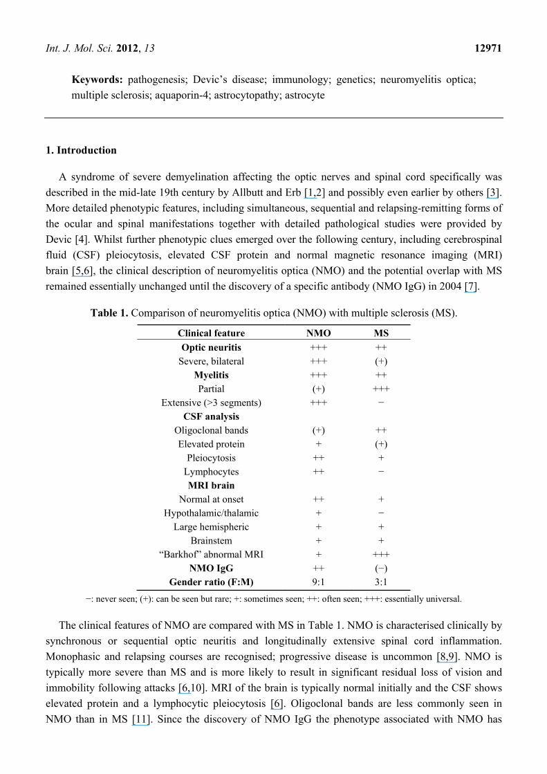
Table 1. Comparison of neuromyelitis optica (NMO) with multiple sclerosis (MS).
Clinical feature NMO MS
Optic neuritis +++ ++ Severe, bilateral +++ (+)
Myelitis +++ ++ Partial (+) +++
Extensive (>3 segments) +++ − CSF analysis
Oligoclonal bands (+) ++ Elevated protein + (+)
Pleiocytosis ++ + Lymphocytes ++ − MRI brain
Normal at onset ++ + Hypothalamic/thalamic + −
Large hemispheric + + Brainstem + +
“Barkhof” abnormal MRI + +++ NMO IgG ++ (−)
Gender ratio (F:M) 9:1 3:1
−: never seen; (+): can be seen but rare; +: sometimes seen; ++: often seen; +++: essentially universal.
The clinical features of NMO are compared with MS in Table 1. NMO is characterised clinically by
synchronous or sequential optic neuritis and longitudinally extensive spinal cord inflammation.
Monophasic and relapsing courses are recognised; progressive disease is uncommon [8,9]. NMO is
typically more severe than MS and is more likely to result in significant residual loss of vision and
immobility following attacks [6,10]. MRI of the brain is typically normal initially and the CSF shows
elevated protein and a lymphocytic pleiocytosis [6]. Oligoclonal bands are less commonly seen in
NMO than in MS [11]. Since the discovery of NMO IgG the phenotype associated with NMO has
Int. J. Mol. Sci. 2012, 13 12972
broadened to include an encephalopathic presentation (sometimes with large diffuse cerebral white
matter lesions) [12–14], recurrent optic neuritis or cord disease (including partial cord lesions) [14,15],
intractable hiccups (particularly in childhood) [16,17] and an acute brain stem syndrome [17]. In
addition to optic nerve involvement with or without nonspecific white matter lesions [6,13], other
abnormalities found on brain MRI include lesions of the hypothalamus [12], periaqueductal grey
matter [12,18] and splenium of the corpus collosum [19]. Confluent periventricular lesions are also
rarely observed, mostly in fulminant cases [20,21]. The gender ratio for NMO is much higher
(female:male = 9:1) [10] than it is for MS (3:1) [22]. An association with other autoimmune
diseases has been frequently reported [23], in contrast with MS, which either does not show any
association [24,25] or only a mild association with systemic autoimmunity [26,27].
Formal diagnostic criteria have been established for NMO [13]. Whilst a number of different
diagnostic criteria have been proposed the most widely accepted require the presence of optic neuritis,
acute myelitis and at least two out of: (1) contiguous spinal cord MRI lesion extending over 3
vertebral segments; (2) brain MRI not meeting diagnostic criteria for multiple sclerosis; and (3) NMO
IgG seropositive status [13]. Using these criteria, NMO IgG is found to be highly specific for NMO.
However, these criteria do not capture a large number of cases that have an incomplete clinical picture.
This includes cases of isolated or recurrent optic neuritis, isolated myelitis and more recently cases of
recurrent hiccoughs in childhood, an acute brainstem syndrome, encephalitis involving the white
matter and other unusual presentations which have been associated with NMO IgG and have been
collectively labelled as NMO spectrum disorder [14].
As a seemingly antigen specific autoimmune disease with potentially dire consequences, treatment
approaches in NMO are generally more aggressive than in MS and usually incorporates
immunosuppressive therapy. There is increasing evidence that conventional treatment for MS,
in the form of β-interferon, may be associated with a worse outcome in NMO [28–30]. Initial treatment
of attacks in NMO is usually with high dose intravenous corticosteroids [6] or if this fails
plasmapheresis [31]. Maintenance treatment with oral corticosteroids and immunosuppression
(cyclophosphamide, azathioprine, mycophenolate mofetil, methotrexate) is recommended as first line
treatment [32]. There is growing evidence for the role of rituximab therapy in NMO [33,34]. This
treatment approach makes logical sense given the presumed pathogenic role of NMO IgG.
The discovery of NMO IgG, which is directed against aquaporin-4 (AQP4), has dramatically
changed the clinical definition of NMO and our understanding of the pathophysiology of this disease.
A picture is finally emerging of NMO as a distinct pathological entity rather than a variant of MS,
although some areas of confusion remain.
In NMO, involvement of the optic nerves and the central grey matter of the spinal cord is common
and may lead to severe loss of vision, disability and even death [6]. The brain is often relatively spared
in the early stages of the disease [6]. The central pathological process in NMO is the production of
abnormal auto-antibodies against AQP4 expressed on astrocytes. These autoantibodies mediate
complement dependent necrosis of astrocytes [35] and subsequent demyelination [36] together with
significant loss of neurons [37]. The aim of this review is to summarise the roles played by key
molecules in the pathogenesis of NMO. The nexus between the molecular basis of NMO and potential
therapeutic approaches will be explored.
Int. J. Mol. Sci. 2012, 13 12973
2. Pathogenesis of NMO
2.1. Aquaporin 4
Aquaporins are water channels present on cell membranes [38]. Of 13 different aquaporins,
AQP4 is the predominant type found in mammalian brain [39]. The AQP4 gene is located on
chromosome 18. The gene product is a bidirectional, osmosis driven water channel mainly expressed
on the foot processes of astrocytes and on ependymal cells in the CNS [40]. It is also expressed in
many other organs such as the kidneys, and the gastrointestinal and respiratory tracts [41]. In the
CNS AQP4 is preferentially expressed in the retina [42], optic nerve, hypothalamus, cerebellum,
periventricular and periaqueductal regions, and the spinal cord [40], and low levels are observed in the
cerebral cortex [40,43]. AQP4 forms as a tetramer [44] and each AQP4 monomer has two splicing
isoforms identified as M1 and M23. M1 (32 kDa) is 22 amino acids longer than M23 (30 kDa) which
is spliced in a shorter formation at the N-terminus [45]. M23 is the predominant isoform in the
CNS [46]. AQP4 tetramers are arranged in groups in the form of orthogonal arrays of particles (OAP)
on the cell membrane. The size and conformation of these arrays is determined by the relative amount
of M23 versus M1 present in the tetramers [47]. AQP4 OAPs have been likened to rafts. The M1
isoform limits the size of OAPs but M23 facilitates formation of larger aggregates [48]. Post
translationally, palmitic acid binds with N-terminal cysteines of M1 and inhibits orthogonal array
formation [49]. AQP4 is associated with the glutamate transporter, excitatory amino acid transporter 2
(EAAT2). In AQP4-transfected cells, EAAT2 is internalised during AQP4 endocytosis [50,51], a
process that has not been observed in primary astrocyte cultures or in vivo experiments [52].
AQP4 knockout mice do not show any neurological deficits in health [53] but show altered
response in disease states. For example, AQP4 knockout mice show reduced cytotoxic oedema of
the brain in stroke [53], reduced glial scar formation [54], increased vasogenic oedema with brain
tumours [55] and CNS infection [56], and a more severe form of induced hydrocephalus [56].
2.2. NMO IgG
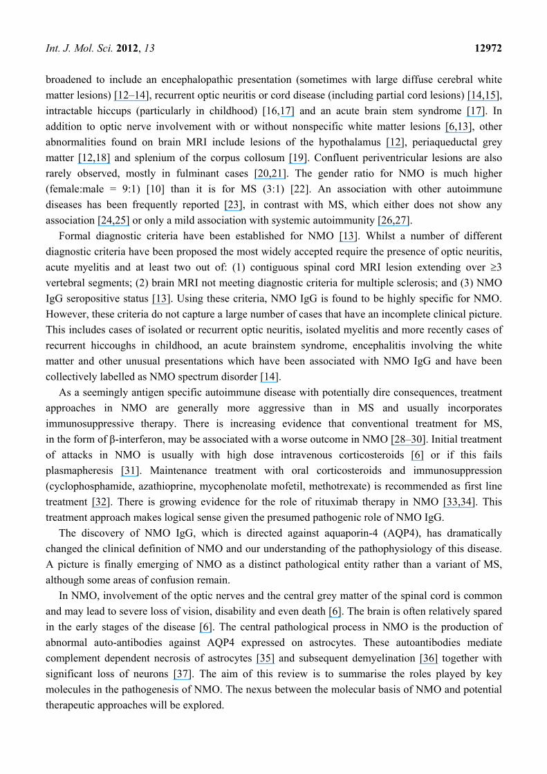
Antibodies against AQP4, originally identified as NMO IgG, were first demonstrated through
standard immunofluorescence techniques using various substrates, including mouse brain and
kidney [7]. Classical staining of the subpial surface, microvessels of brain and cerebellum and
papillary tubules of the kidney is illustrated in Figure 1. Subsequently enzyme linked immunosorbent
assay (ELISA) and live cell-based assays have been developed with live cell-based assays utilising the
M23 isoform of AQP4 having the highest sensitivity [57].
NMO IgG has high specificity (99%) [58,59] and moderate sensitivity ranging from 56% [58] to
73% [7,58] for NMO. It has been observed that the sensitivity of the autoantibody is higher in
relapsing cases of NMO [58]. The autoantibody in the blood of NMO patients is predominantly the
IgG1 isotype (98% of cases) [60], which can potently activate the complement system [61]. IgG2, IgG3
and IgG4 also occur in a lower proportion of cases [60]. IgM NMO antibody has also been reported in
the blood of up to 10% of patients with NMO but it is not known to exist in the absence of IgG [62].
NMO IgG binds to the third extracellular loop of AQP4 [63] and the generation of conformational
epitopes during OAP formation results in preferential binding with the M23 isoform [64]. One study
Int. J. Mol. Sci. 2012, 13 12974
has suggested that NMO IgG has considerably lower affinity for the AQP4 protein when compared
with the epitope presented by OAPs [65]. NMO IgG does not cross the blood brain barrier (BBB) in
normal subjects [66] but it can cross the placenta [67]. It has been demonstrated that NMO IgG is
synthesised outside of the CNS. Persistent intrathecal synthesis of oligoclonal IgG, the most stable
laboratory feature of MS, was absent in a study of 89 seropositive patients with NMO spectrum
disorder, although transient intrathecal production was occasionally observed during acute
relapses [11]. CSF from 20 NMO patients showed lower titres of NMO IgG in CSF than in serum
(with a ratio of 1:500) in keeping with extrathecal synthesis of the autoantibody [68]. A further seven
cases of NMO with low CSF antibody index of NMO IgG (AQP4 IgG/Total IgG) have since been
reported [69]. In order to cause disease in the CNS, the extrathecally produced autoantibody requires
disruption of the BBB (possible mechanisms for this are discussed below). NMO IgG is occasionally
restricted to the CSF [70] and AQP4 specific B cells have been identified in the CSF of one patient
with NMO [71]. In some patients, NMO IgG is produced by a subset of CD20 negative B cells
(CD19intermediateCD27highCD38highCD180−) that resembled early plasma cells [72].
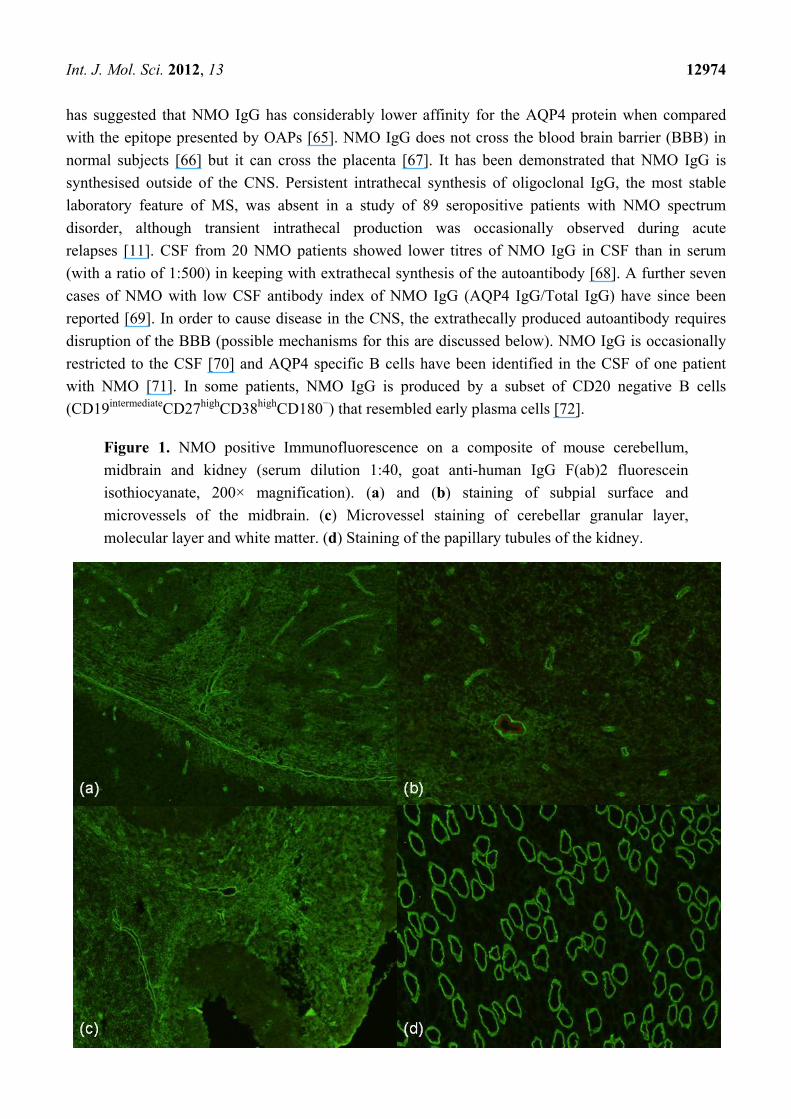
Figure 1. NMO positive Immunofluorescence on a composite of mouse cerebellum,
midbrain and kidney (serum dilution 1:40, goat anti-human IgG F(ab)2 fluorescein
isothiocyanate, 200× magnification). (a) and (b) staining of subpial surface and
microvessels of the midbrain. (c) Microvessel staining of cerebellar granular layer,
molecular layer and white matter. (d) Staining of the papillary tubules of the kidney.

Int. J. Mol. Sci. 2012, 13 12975
2.3. Triggers for Autoimmunity
A genetic predisposing factor is likely in NMO, as the disease is relatively more prevalent in
non-Caucasian populations, as compared to MS which is more common in Europeans [73–76].
However, the stand alone prevalence of NMO (not relative to MS) has not been systematically
determined in any large population. Further clues favouring a genetic predisposition derive from
familial NMO cases. While such cases have been described for many years [77], only a small
proportion (3%) of cases have a positive family history, predominantly those with Asian or Latino
descent [78]. In the most comprehensive familial study to date (12 families, 25 affected individuals),
the pattern of inheritance was thought to be complex [78]. The human leukocyte antigen (HLA)
associations of NMO are contrasted with MS in Table 2. NMO IgG positivity has been found to be
associated with HLA-DRB1*03 (DR3) in French (OR 3.08; 95% CI 1.52–6.27, p = 0.001) [79] and
Brazilian (OR 3.23; 95% CI 1.07–9.82, p = 0.04) populations [80] and HLA-DPB1*0501 in Japanese
(OR 4.8; 95% CI 1.6–14.3, p = 0.032) [81] and Chinese (OR 7.096; 95% CI 2.011–25.044, p = 0.022)
populations [82]. Notably, these HLA haplotypes are also associated with other humoral autoimmune
disorders such as systemic lupus erythematosus (SLE) [83] and Graves’ disease [84]. Indeed, NMO
often coexists with other autoimmune conditions such as SLE and Sjögren syndrome [23] and these
disorders are also more common in family members of patients with NMO [78]. Recent diagnostic
criteria for NMO have tended to exclude SLE and Sjögren syndrome as not being compatible with a
diagnosis of NMO [13].
Table 2. Human leukocyte antigen (HLA) associations in NMO and MS.
Disease Population N DRB1 DQA1 DQB1 DPB1
NMO Japan [81] 38 0501 France [79] 45 03 - 02 Brazil [80] 54 03 USA [85] 154 (not 1501) Han Chinese [82] 0501 Martinque [86] 42 03 -
MS Australia [87] 100 1501 0102 0602 -
Like many other autoimmune disorders NMO is also more common in women [10,88] suggesting
that gender differences play a role in the pathogenesis of NMO. Genetic analysis of the AQP4 gene
was carried out to determine if mutations in the gene are associated with NMO [89]. No significant
association was found with the 8 selected SNPs. However, at position number 3180 in the AQP4 gene,
which transcripts arginine at position 19, two missense mutations were observed in 3 out of 191 cases
of NMO but not in any of 1363 controls. Whilst not statistically significant, it was speculated that loss
of arginine 19 may impair palmitoylation of AQP4, which may increase the size of OAPs [49] and
thereby generate immunogenic epitopes. Since Arginine 19 is in the regulatory sequence of M23 [90],
this mutation could result in dysregulation of the transcription or translation of M23, resulting in a
potentially immunogenic conformational change in OAPs. As described previously NMO IgG has high
affinity for epitopes expressed in OAPs but lower affinity for isolated AQP4 protein [65].
Int. J. Mol. Sci. 2012, 13 12976
In one series the onset of neurological symptoms in almost 30% of NMO cases was preceded by
a viral or bacterial infection (more cases associated with varicella zoster and mycobacterium
tuberculosis) [91] and vaccination against human papilloma virus has been reported to precede the
onset of NMO in 4 teenage girls [92]. Parainfectious NMO may have a different immunopathogenesis
as the majority of these cases have a monophasic course [91]. Rarely, a paraneoplastic association
has also been described in NMO, especially with carcinoma of the breast and lung, and B
cell-lymphoma [93]. A recent study of the T cells derived from the blood of 15 NMO patients has
supported molecular mimicry as a possible cause of autoimmunity in NMO. Homology between the
immunogenic amino acids 66–75 of AQP4 and the surface protein ABC-TP of clostridia species
especially Clostridium perfringens has been observed and ABC-TP was shown to cross react with
anti-AQP4 T cells [94]. Clostridium perfringens is a ubiquitous organism and can occur in human gut
as a commensal [95]. This study requires independent replication.
In MS, autoantibodies against myelin and other CNS antigens have been variably reported in
as many as 60%–70% of cases [96] and the levels of autoantibodies fluctuate with relapses of
disease [97]. However, no consistent high affinity serum autoantibody response is observed in MS and
B-cells may be sensitised against self-antigens as an epiphenomenon during relapses. Similarly, there
is a possibility that an initial disease process in the CNS of NMO patients attracts B-cells and
sensitises them against AQP4. However, in an experiment to detect spontaneous production of NMO
IgG in murine experimental autoimmune encephalomyelitis (EAE), extensive infiltration of immune
cells into the CNS was seen without any detectable NMO IgG [98].
It remains to be discovered what, if any, role is played by molecular mimicry, bystander activation
or a persistent infection in the initiation of autoantibody production in NMO.
2.4. Is Anti-AQP4 Antibody Pathogenic?
The antibody from the serum of NMO patients has been shown to exacerbate the clinical and
pathological features of murine EAE [66,99] and to cause disease when injected intracerebrally in
wild-type mice [35]. However, NMO IgG is only pathogenic in the presence of human complement
proteins [35,100] or a pre-existing inflammatory state in the CNS such as occurs in EAE or mice
pre-treated with Freund’s complete adjuvant [101]. The stronger the pre-existing CNS inflammation,
the more severe the disease. Therefore, in EAE models more extensive disease is induced by NMO
IgG [66,99], whereas only mild disease is observed in mice pre-treated with Freund’s complete
adjuvant, which causes milder CNS inflammation than EAE [101]. AQP4 is expressed in non-CNS
tissues [68,102], but no pathological involvement of extra neuronal tissues has been observed in NMO
cases. This suggests that pathogenic potential of the autoantibody is restricted to the CNS. NMO IgG
fails to produce disease in juvenile mice with incomplete BBB [66]; while access of NMO IgG to the
CNS is a pre-requisite for the induction of disease, it is not sufficient to cause clinically relevant
pathology in isolation. Higher titres of NMO IgG have been reported in patients with increased disease
activity and severity [68,103], consistent with antibody pathogenicity. However, antibody titres do not
correlate closely with disease activity in all studies [60,104,105]. More questions have been raised
about the pathogenicity of NMO IgG following a case report in which NMO IgG was detected in blood
10 years before the clinical onset of the disease [106] and in another case series where NMO IgG was
Int. J. Mol. Sci. 2012, 13 12977
detected in 2 patients with cancer but without any clinical manifestation of NMO [93]. It can be
concluded that NMO IgG is pathogenic, but only in a specific CNS milieu.
2.5. Immune Mediated Astrocytopathy
NMO IgG binds with AQP4 and activates complement making the lytic complex C5b-9, which
destroys astrocytes [107]. Neutrophils, natural killer cells, macrophages, tumour necrosis factor α,
interleukin (IL)-6, IL-1β, and interferon-gamma have been shown to amplify this complement
mediated damage [100]. Complement regulatory proteins (CRP) on astrocytes such as decay-accelerating
factor (CD55), membrane cofactor protein (CD46), complement receptor 1 and protectin (CD59) may
also be dysfunctional in NMO, thus facilitating complement mediated astrocyte injury. In mice
injected intracerebrally with NMO IgG. NMO-like lesions are only produced when human, but not
mouse, complement is co-administered [35]. It is known that murine CRP may not have a potent
protective effect against heterologous complement [108,109]. CRP dysfunction secondary to
autoantibodies has been reported in MS [110]. The possibility of dysfunctional CRP in NMO requires
further investigation.
Anti-AQP4 antibody has been reported in some studies to cause internalisation of
AQP4 [50,61,111]. However, internalisation of AQP4, a process that could paradoxically protect
astrocytes from immune mediated cytotoxicity, is unlikely to be the principal pathogenic mechanism in
NMO. As noted previously the absence of AQP4 function does not result in neurological disorder
in AQP4 knockout mice [53]. Additionally, little or no internalisation of antibody-AQP4 complex
is observed in primary astrocyte culture or in vivo, emphasising the limited applicability of
AQP4-transfected cells when modelling human disease [52,112]. It has also been shown that astrocytes
do not suffer any osmotic injury as NMO IgG does not disturb the function of AQP4 [113,114].
Perivascular glial fibrillary acid protein (GFAP) positive astrocytes are consistently lost in NMO
lesions in contrast with MS [36,37], where lesional GFAP is usually upregulated [115]. CSF levels of
GFAP are markedly elevated during relapses of NMO but not MS [116]. GFAP is a cytoskeletal
protein that plays a role in astrocyte motility and structure [115]. It is highly expressed by reactive
astrocytes [117].
2.6. Immunopathogenesis
Systemic production of autoantibody with disease limited to the CNS implicates the presence of
orchestrating T helper cells as well as a cascade of cytokines in the periphery and within the CNS. B
cells are also attracted to the CNS by chemokines secreted at sites of inflammation. A study involving
31 NMO patients found various Th2 related cytokines such as IL-1 receptor antagonist, IL-5, IL-10
and IL-13; and the Th17 related cytokines IL-6, IL-8 and granulocyte colony stimulating factor to be
elevated in the CSF. However, the signature Th2 cytokine, IL-4, and Th17 cytokine, IL-17, were not
detected in the CSF [118]. Other studies have shown increased levels of IL-17 in the CSF of NMO
patients [119,120]. Therefore, Th17 cells may have an intermittent role in the CNS inflammation that
characterises NMO. Chitinases, hydrolases secreted by the innate immune cells that play a role in Th2
as well as Th1 related inflammatory processes, are also increased in the CSF of NMO patients. In
response to IL-13 monocytes obtained from NMO patients secrete chitinases that increase in vitro
Int. J. Mol. Sci. 2012, 13 12978
chemotaxis of eosinophils, macrophages and T cells across the BBB by promoting the secretion of
various cytokines IL-8, RANTES (CCL5), MCP1 (CCL2), and eotaxins by monocytes [121].
Eosinophils and macrophages are abundant in NMO lesions [122]. CSF levels of CXCL13, a potent
chemo-attractant for B cells, are elevated and correlate with disease severity [123]. As indicated
previously, anti-AQP4 B cells have been detected in the CSF of NMO patients [71]. CXCL13 is
secreted by sensitised T helper cells [124] and antigen presenting cells (APC) [125]. B-cell activating
factor (BAFF) is increased in the CSF but not serum of NMO patients [126]. BAFF, secreted by innate
immune cells, APC [127] and activated astrocytes [128], increases the survival and maturation of B
cells through NF-κB [129]. Thus, BAFF can perpetuate the auto-reactive B cell population in the CNS.
It is interesting to note that treatment with both β-interferon and glatiramer acetate was found to
increase levels of BAFF in the serum of MS patients [126]. This may account for the apparent
worsening of NMO with β-interferon [28–30]. Despite similar changes in BAFF occurring with
glatiramer acetate the same concerns regarding its use in NMO have not been raised [130,131].
IL-6 is elevated not only in the CSF but also in the serum of patients with NMO [118,132],
particularly in patients with more severe disease [132]. This proinflammatory cytokine, which can also
be secreted by activated astrocytes [133], increases the survival of NMO IgG producing B cells and the
production of antibody [72]. In ex vivo experiments in murine spinal cord, IL-6 increased the induced
NMO-like lesions [100]. In a study of 18 NMO patients, IL-9, a T cell growth factor, was elevated in
the serum [134] but, not the CSF [118]. IL-6 is involved in the development of IL-17-secreting CD4+ T
cells (Th17) [135]. Th17 cells have been implicated in many autoimmune disorders such as rheumatoid
arthritis, psoriasis, MS and inflammatory bowel disease [136]. Th17 cells and their principal product,
IL-17, are found to be increased in serum from patients with NMO [137,138], and some studies as seen
above have also shown elevated IL-17 in the CSF [119,120], suggesting the involvement of Th17 cells
and IL-17 in the pathogenesis of the disease. IL-17 is seen to mediate inflammation in the brain of
EAE mice by promoting microglial activation [139]. Th17 cells may dictate the autoimmune milieu
that facilitates the pathogenesis of NMO. A subset of B cells (CD27highCD38highCD180− with low
CD20 and CD19−), which may be producing anti-AQP4 antibody, appear to be activated [72]. Further
implicating a role for humoral immunity in NMO, CD27+ memory B cell levels rise in association with
relapses [34]. There is some evidence that cellular autoimmunity is also initiated as CD4+ T cells
against AQP4 are detectable in the blood of patients with NMO [140,141].
Rituximab has been found to be clinically effective in NMO [33,34] and reduces the levels of
anti-NMO IgG, but the antibody does not completely disappear [34,103,142]. There may also be a
BAFF related transient elevation in the anti-NMO titres observed 2 weeks after the first injection of
rituximab [142]. One possible explanation for this is that plasma cells, which do not express CD20 (the
target of rituximab), may continue to produce antibody for a period of time, possibly at an increased
rate due to the depletion of memory B cells and other modulatory Bcells (regulatory B cells) which do
carry CD20 [143]. Repeated rituximab therapy may reduce the level of NMO IgG after the depletion of
memory B cells and natural death of short-lived plasmablasts/plasma cells. Long-lived plasma cells
may continue to secrete autoantibody despite the treatment with rituximab [144]. An alternative
explanation is that it is purely through the modulatory effects of B cells on T cells that rituximab is
having an effect, such that removal of memory B cells and activated B cells diminishes the
proinflammatory milieu.
Int. J. Mol. Sci. 2012, 13 12979
Disruption of the BBB is required for NMO IgG to enter the CNS. Serum from NMO patients can
cause breakdown of the BBB [145,146] by an unknown mechanism. However, autoantibodies against
cultured human brain microvascular endothelial cells were detected in 10 out of 14 seropositive NMO
patients. These autoantibodies, which are distinct from anti-AQP4, were observed to reduce the
expression of the tight junction proteins, that constitute the BBB, possibly through the autocrine
secretion of vascular endothelial growth factor [146]. The BBB is further disrupted by injury to
astrocytic foot processes sustained in the disease process [111].
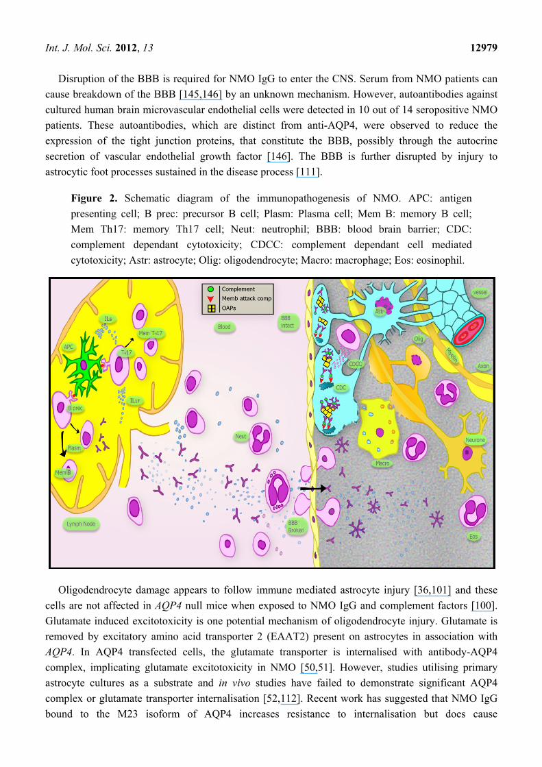
Figure 2. Schematic diagram of the immunopathogenesis of NMO. APC: antigen
presenting cell; B prec: precursor B cell; Plasm: Plasma cell; Mem B: memory B cell;
Mem Th17: memory Th17 cell; Neut: neutrophil; BBB: blood brain barrier; CDC:
complement dependant cytotoxicity; CDCC: complement dependant cell mediated
cytotoxicity; Astr: astrocyte; Olig: oligodendrocyte; Macro: macrophage; Eos: eosinophil.
Oligodendrocyte damage appears to follow immune mediated astrocyte injury [36,101] and these
cells are not affected in AQP4 null mice when exposed to NMO IgG and complement factors [100].
Glutamate induced excitotoxicity is one potential mechanism of oligodendrocyte injury. Glutamate is
removed by excitatory amino acid transporter 2 (EAAT2) present on astrocytes in association with
AQP4. In AQP4 transfected cells, the glutamate transporter is internalised with antibody-AQP4
complex, implicating glutamate excitotoxicity in NMO [50,51]. However, studies utilising primary
astrocyte cultures as a substrate and in vivo studies have failed to demonstrate significant AQP4
complex or glutamate transporter internalisation [52,112]. Recent work has suggested that NMO IgG
bound to the M23 isoform of AQP4 increases resistance to internalisation but does cause
Int. J. Mol. Sci. 2012, 13 12980
internalisation of the M1 isoform [112]. AQP4 has been observed in autopsy studies to be lost both in
MS and NMO lesions [37,147]. This may be due to loss of astrocytes rather than internalisation. Loss
of AQP4 in EAE models has been observed to be neuroprotective [98], but it is likely that this effect is
mediated by its water channelling action, which is not affected by NMO IgG binding [113,114]. Thus,
loss of AQP4 in NMO, either by internalisation or astrocyte loss, is one of the markers of the disease
but may not be a primary pathogenic process. Whilst it is possible that the inflammatory process
primarily targeting astrocytes causes bystander injury to nearby oligodendrocytes, widespread loss of
oligodendrocytes is reported in early NMO lesions in the absence of a significant macrophage
infiltrate [36]. The immunoglobulin predominantly seen in lesions of NMO patients in one study was
of IgM type [148] but as noted earlier it is only detected in the blood of 10% of patients [62]. A
possible explanation for this is intrathecal synthesis by local B-cells undergoing class switching.
However, another study also showed IgG along with IgM in lesions of NMO patients thus supporting
the role of IgG as a pathogenic antibody [36]. The immunopathogenesis of NMO is summarised
in Figure 2.
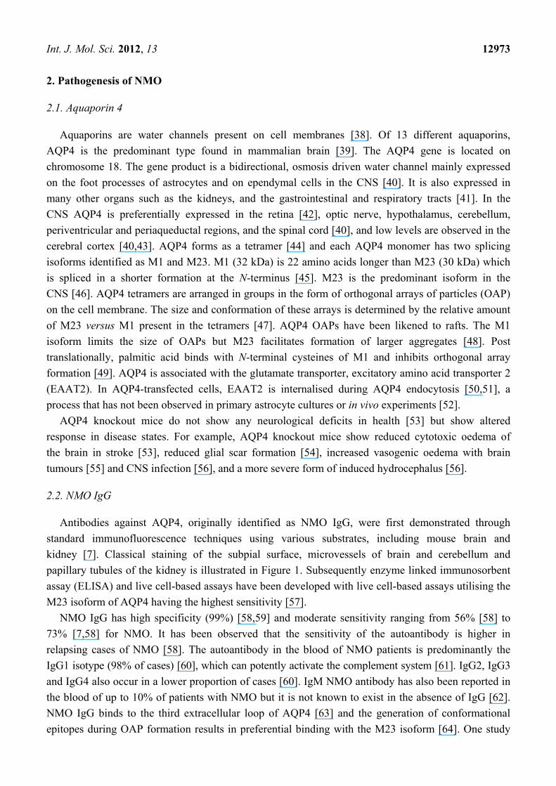
2.7. Histopathology of NMO
The earliest pathological descriptions of NMO highlighted the frequent occurrence of necrosis with
cavitation, hyalinisation of small vessels and perivascular inflammatory infiltrates. These features were
helpful in distinguishing NMO from MS. More recent work has indicated the importance of
perivascular deposition of immunoglobulin and complement [148] and its localisation to the vascular
glial limiting membrane [149]. The infiltrate consists of macrophages, granulocytes and eosinophils
with a general paucity of lymphocytes [122]. Modern histopathological techniques have demonstrated
the specificity of acute fragmentation and loss of perivascular GFAP-positive astrocyte foot processes
and their cell bodies as an early and consistent feature of NMO [36,150]. Whilst loss of AQP4 is seen
in NMO lesions [37,147], this is not consistent and is also seen in MS [151,152]. Relative preservation
of myelin in early lesions with massive astrocyte destruction implicates astrocyte injury as the primary
pathological event in NMO [37,147]. The presence of myelin loss and phagocytosis by macrophages in
subacute NMO lesions suggests that this is a secondary phenomenon. Morphologically distinct
unipolar and bipolar GFAP-positive astrocytes are seen in demyelinated subacute lesions suggesting
that astrocyte progenitor cells may be responsible for reparative processes in NMO [37,147,153].
Interestingly, significant necrosis of axons was not seen in early and subacute lesions [36].
Intramyelenic oedema with partial astrocyte preservation may be responsible for the presumed
vasogenic oedema seen on MRI as extensive white matter lesions in the brain [112]. The typical
histopathological appearances in NMO are illustrated in Figure 3.
Int. J. Mol. Sci. 2012, 13 12981
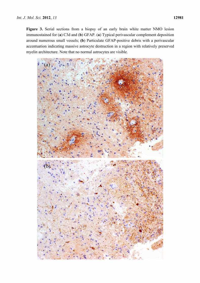
Figure 3. Serial sections from a biopsy of an early brain white matter NMO lesion
immunostained for (a) C3d and (b) GFAP. (a) Typical perivascular complement deposition
around numerous small vessels; (b) Particulate GFAP-positive debris with a perivascular
accentuation indicating massive astrocyte destruction in a region with relatively preserved
myelin architecture. Note that no normal astrocytes are visible.
(a)
(b)
Int. J. Mol. Sci. 2012, 13 12982
3. Conclusions
Great strides have been made in elucidating the molecular underpinnings of NMO, but aspects of
the pathogenesis remain unknown and no currently proposed disease model is completely satisfactory.
It seems likely that NMO is an autoimmune disease that develops in genetically susceptible
individuals. There is already some evidence for specific HLA alleles and polymorphisms of the AQP4
gene being associated with NMO. Infection, vaccination, and neoplasm may trigger the disease process
in some cases but a recognisable initiating event is lacking in most. In the schema presented in
Figure 2, this trigger induces the release of inflammatory cytokines, including IL-6, and Th17 cells are
stimulated. A subset of B-cells in the peripheral tissues is stimulated to produce anti-AQP4 IgG
antibody. The BBB is made permeable to the autoantibody by either a systemic inflammatory response
or a local pathological process in the CNS. Auto-reactive B cells may also enter the CNS and undergo
class switching to produce IgM autoantibody. The antibody fixes complement on AQP4 expressing
astrocytic foot processes. Stellate astrocytes are damaged by complement dependent cytotoxicity and
complement dependent cellular cytotoxicity. Oligodendrocytes that are in close proximity to astrocyte
foot processes are possibly affected through bystander inflammatory damage or, more likely, loss of
trophic support from astrocytes. The resulting demyelination causes loss of saltatory conduction and
conduction block, leading to neurological deficit. With continuing or repeated inflammation,
substantial neuro-axonal loss ensues. The near absence of progressive forms of NMO (primary or
secondary) suggests that neuro-axonal damage is the result of the acute inflammation, whether directly
or indirectly, rather than as a result of a secondary degenerative process as appears to be the case in
some forms of MS.
Some CNS sites, such as cerebellum, express AQP4 abundantly but are not usually involved in
NMO. Similarly, cerebral cortex expresses AQP4 but is not affected. This may be due to the specific
local immune environment. Elucidation of such protective mechanisms will be helpful in identifying
new therapeutic targets. The processes responsible for oligodendrocyte loss are also largely unknown
and the mechanism of axonal injury in chronic lesions is also poorly understood. Important questions
regarding the aetiopathogenesis of seronegative cases with NMO phenotypes remain. Do these cases
have a different antigenic target (heterogeneity) or do they represent a milder form of the disease
(forme fruste), or do they reflect technical problems with the currently available NMO IgG assays
(false negatives)?
As the first autoimmune disease of the CNS for which a specific autoantibody and antigenic target
have been identified NMO represents a significant opportunity to better understand and hopefully treat
its more heterogeneous and complex cousin, MS. Very recent research has highlighted a novel
potential therapeutic approach utilising small-molecule inhibitors to reduce AQP4 mediated astrocyte
cytotoxicity by disrupting the interaction between NMO-IgG and M23-AQP4 [154]. These
experiments demonstrate the very direct way in which a greater understanding of the pathogenesis of
autoimmune diseases such as NMO can lead to new therapeutic interventions. The prominent role
played by IL-6 in NMO has led to the suggestion that IL-6 receptor blockade may be an effective
therapy [72]. The development of fully humanised monoclonal antibodies against the B-cell antigens
CD20 [155] and CD19 [156] raise exciting prospects for improved tolerability and potentially greater
efficacy in the treatment of NMO.
Int. J. Mol. Sci. 2012, 13 12983
Acknowledgements
We are grateful to Jacob Sharples and Robert Loudon for assistance with the illustration.
Wajih Bukhari is supported by a Fellowship Scholarship from Multiple Sclerosis Research Australia.
References
1. Allbutt, T. On the opthalmoscopic signs of spinal disease. Lancet 1870, 1, 76–78.
2. Erb, W. About the concurrence of optic neuritis and subacute myelitis. Arch. Psychiatr.
Nervenkr. 1879, 1, 146–157.
3. Jarius, S.; Wildemann, B. On the contribution of thomas clifford allbutt, F.R.S., to the early
history of neuromyelitis optica. J. Neurol. 2012, doi:10.1007/s00415-012-6594-3.
4. Devic, E. Myélite subaiguë compliquée de névrite optique. Bull. Med. (Paris) 1894, 8, 1033–1034.
5. Totsuka, S. Clinico-pathological studies on the two cases of neuromyelitis optica (Devic’s
disease) with a chronic clinical course, with especial reference on its relationship to multiple
sclerosis. Folia Psychiatr. Neurol. Jpn. 1962, 64, 1149–1165.
6. Wingerchuk, D.M.; Hogancamp, W.F.; O’Brien, P.C.; Weinshenker, B.G. The clinical course of
neuromyelitis optica (Devic’s syndrome). Neurology 1999, 53, 1107–1114.
7. Lennon, V.A.; Wingerchuk, D.M.; Kryzer, T.J.; Pittock, S.J.; Lucchinetti, C.F.; Fujihara, K.;
Nakashima, I.; Weinshenker, B.G. A serum autoantibody marker of neuromyelitis optica:
Distinction from multiple sclerosis. Lancet 2004, 364, 2106–2112.
8. Kim, J.S.; Park, Y.H.; Kim, S.M.; Kim, S.H.; Park, K.S.; Sung, J.J.; Lee, K.W. A case of chronic
progressive myelopathy. Mult. Scler. 2010, 16, 1255–1257.
9. Wingerchuk, D.M.; Pittock, S.J.; Lucchinetti, C.F.; Lennon, V.A.; Weinshenker, B.G. A
secondary progressive clinical course is uncommon in neuromyelitis optica. Neurology 2007, 68,
603–605.
10. Jarius, S.; Ruprecht, K.; Wildemann, B.; Kuempfel, T.; Ringelstein, M.; Geis, C.; Kleiter, I.;
Kleinschnitz, C.; Berthele, A.; Brettschneider, J.; et al. Contrasting disease patterns in
seropositive and seronegative neuromyelitis optica: A multicentre study of 175 patients.
J. Neuroinflamm. 2012, 9, 14.
11. Jarius, S.; Paul, F.; Franciotta, D.; Ruprecht, K.; Ringelstein, M.; Bergamaschi, R.; Rommer, P.;
Kleiter, I.; Stich, O.; Reuss, R.; et al. Cerebrospinal fluid findings in aquaporin-4 antibody
positive neuromyelitis optica: Results from 211 lumbar punctures. J. Neurol. Sci. 2011, 306, 82–90.
12. Pittock, S.J.; Lennon, V.A.; Krecke, K.; Wingerchuk, D.M.; Lucchinetti, C.F.; Weinshenker, B.G.
Brain abnormalities in neuromyelitis optica. Arch. Neurol. 2006, 63, 390–396.
13. Wingerchuk, D.M.; Lennon, V.A.; Pittock, S.J.; Lucchinetti, C.F.; Weinshenker, B.G. Revised
diagnostic criteria for neuromyelitis optica. Neurology 2006, 66, 1485–1489.
14. Wingerchuk, D.M.; Lennon, V.A.; Lucchinetti, C.F.; Pittock, S.J.; Weinshenker, B.G. The
spectrum of neuromyelitis optica. Lancet Neurol. 2007, 6, 805–815.
15. Bizzoco, E.; Lolli, F.; Repice, A.M.; Hakiki, B.; Falcini, M.; Barilaro, A.; Taiuti, R.;
Siracusa, G.; Amato, M.P.; Biagioli, T.; et al. Prevalence of neuromyelitis optica spectrum
disorder and phenotype distribution. J. Neurol. 2009, 256, 1891–1898.
Int. J. Mol. Sci. 2012, 13 12984
16. McKeon, A.; Lennon, V.A.; Lotze, T.; Tenenbaum, S.; Ness, J.M.; Rensel, M.; Kuntz, N.L.;
Fryer, J.P.; Homburger, H.; Hunter, J.; et al. CNS aquaporin-4 autoimmunity in children.
Neurology 2008, 71, 93–100.
17. Takahashi, T.; Miyazawa, I.; Misu, T.; Takano, R.; Nakashima, I.; Fujihara, K.; Tobita, M.;
Itoyama, Y. Intractable hiccup and nausea in neuromyelitis optica with anti-aquaporin-4
antibody: A herald of acute exacerbations. J. Neurol. Neurosurg. Psychiatry 2008, 79, 1075–1078.
18. Pittock, S.J.; Weinshenker, B.G.; Lucchinetti, C.F.; Wingerchuk, D.M.; Corboy, J.R.;
Lennon, V.A. Neuromyelitis optica brain lesions localized at sites of high aquaporin 4
expression. Arch. Neurol. 2006, 63, 964–968.
19. Makino, T.; Ito, S.; Mori, M.; Yonezu, T.; Ogawa, Y.; Kuwabara, S. Diffuse and heterogeneous
T2-hyperintense lesions in the splenium are characteristic of neuromyelitis optica. Mult. Scler.
2012, doi:10.1177/1352458512454772.
20. Ikeda, K.; Ito, H.; Hidaka, T.; Takazawa, T.; Sekine, T.; Yoshii, Y.; Hirayama, T.; Kawabe, K.;
Kano, O.; Iwasaki, Y. Repeated non-enhancing tumefactive lesions in a patient with a
neuromyelitis optica spectrum disorder. Intern. Med. 2011, 50, 1061–1064.
21. Newey, C.R.; Bermel, R.A. Fulminant cerebral demyelination in neuromyelitis optica. Neurology
2011, 77, 193.
22. Orton, S.M.; Herrera, B.M.; Yee, I.M.; Valdar, W.; Ramagopalan, S.V.; Sadovnick, A.D.;
Ebers, G.C.; Canadian Collaborative Study, G. Sex ratio of multiple sclerosis in canada: A
longitudinal study. Lancet Neurol. 2006, 5, 932–936.
23. Wingerchuk, D.M.; Weinshenker, B.G. The emerging relationship between neuromyelitis optica
and systemic rheumatologic autoimmune disease. Mult. Scler. 2012, 18, 5–10.
24. Wynn, D.R.; Rodriguez, M.; O’Fallon, W.M.; Kurland, L.T. A reappraisal of the epidemiology
of multiple sclerosis in Olmsted county, Minnesota. Neurology 1990, 40, 780–786.
25. Broadley, S.A.; Deans, J.; Sawcer, S.J.; Clayton, D.; Compston, D.A. Autoimmune disease in
first-degree relatives of patients with multiple sclerosis. A UK survey. Brain 2000, 123,
1102–1111.
26. Spadaro, M.; Amendolea, M.A.; Mazzucconi, M.G.; Fantozzi, R.; Di Lello, R.; Zangari, P.;
Masala, G. Autoimmunity in multiple sclerosis: Study of a wide spectrum of autoantibodies.
Mult. Scler. 1999, 5, 121–125.
27. Henderson, R.D.; Bain, C.J.; Pender, M.P. The occurrence of autoimmune diseases in patients
with multiple sclerosis and their families. J. Clin. Neurosci. 2000, 7, 434–437.
28. Kim, S.H.; Kim, W.; Li, X.F.; Jung, I.J.; Kim, H.J. Does interferon β treatment exacerbate
neuromyelitis optica spectrum disorder? Mult. Scler. 2012, doi:10.1177/1352458512439439.
29. Shimizu, J.; Hatanaka, Y.; Hasegawa, M.; Iwata, A.; Sugimoto, I.; Date, H.; Goto, J.; Shimizu, T.;
Takatsu, M.; Sakurai, Y.; et al. IFNbeta-1b may severely exacerbate Japanese optic-spinal MS in
neuromyelitis optica spectrum. Neurology 2010, 75, 1423–1427.
30. Palace, J.; Leite, M.I.; Nairne, A.; Vincent, A. Interferon beta treatment in neuromyelitis optica:
Increase in relapses and aquaporin 4 antibody titers. Arch. Neurol. 2010, 67, 1016–1017.
31. Watanabe, S.; Nakashima, I.; Misu, T.; Miyazawa, I.; Shiga, Y.; Fujihara, K.; Itoyama, Y.
Therapeutic efficacy of plasma exchange in NMO-IgG-positive patients with neuromyelitis
optica. Mult. Scler. 2007, 13, 128–132.
Int. J. Mol. Sci. 2012, 13 12985
32. Papeix, C.; Vidal, J.S.; de Seze, J.; Pierrot-Deseilligny, C.; Tourbah, A.; Stankoff, B.;
Lebrun, C.; Moreau, T.; Vermersch, P.; Fontaine, B.; et al. Immunosuppressive therapy is more
effective than interferon in neuromyelitis optica. Mult. Scler. 2007, 13, 256–259.
33. Jacob, A.; Weinshenker, B.G.; Violich, I.; McLinskey, N.; Krupp, L.; Fox, R.J.; Wingerchuk, D.M.;
Boggild, M.; Constantinescu, C.S.; Miller, A.; et al. Treatment of neuromyelitis optica with
rituximab: Retrospective analysis of 25 patients. Arch. Neurol. 2008, 65, 1443–1448.
34. Kim, S.H.; Kim, W.; Li, X.F.; Jung, I.J.; Kim, H.J. Repeated treatment with rituximab based on
the assessment of peripheral circulating memory B cells in patients with relapsing neuromyelitis
optica over 2 years. Arch. Neurol. 2011, 68, 1412–1420.
35. Saadoun, S.; Waters, P.; Bell, B.A.; Vincent, A.; Verkman, A.S.; Papadopoulos, M.C.
Intra-cerebral injection of neuromyelitis optica immunoglobulin G and human complement
produces neuromyelitis optica lesions in mice. Brain 2010, 133, 349–361.
36. Parratt, J.D.; Prineas, J.W. Neuromyelitis optica: A demyelinating disease characterized by acute
destruction and regeneration of perivascular astrocytes. Mult. Scler. 2010, 16, 1156–1172.
37. Misu, T.; Fujihara, K.; Kakita, A.; Konno, H.; Nakamura, M.; Watanabe, S.; Takahashi, T.;
Nakashima, I.; Takahashi, H.; Itoyama, Y. Loss of aquaporin 4 in lesions of neuromyelitis optica:
Distinction from multiple sclerosis. Brain 2007, 130, 1224–1234.
38. Preston, G.M.; Carroll, T.P.; Guggino, W.B.; Agre, P. Appearance of water channels in xenopus
oocytes expressing red cell CHIP28 protein. Science 1992, 256, 385–387.
39. Verkman, A.S. More than just water channels: Unexpected cellular roles of aquaporins. J. Cell
Sci. 2005, 118, 3225–3232.
40. Nielsen, S.; Nagelhus, E.A.; Amiry-Moghaddam, M.; Bourque, C.; Agre, P.; Ottersen, O.P.
Specialized membrane domains for water transport in glial cells: High-resolution immunogold
cytochemistry of aquaporin-4 in rat brain. J. Neurosci. 1997, 17, 171–180.
41. Frigeri, A.; Gropper, M.A.; Umenishi, F.; Kawashima, M.; Brown, D.; Verkman, A.S.
Localization of MIWC and GLIP water channel homologs in neuromuscular, epithelial and
glandular tissues. J. Cell Sci. 1995, 108, 2993–3002.
42. Li, J.; Patil, R.V.; Verkman, A.S. Mildly abnormal retinal function in transgenic mice without
muller cell aquaporin-4 water channels. Invest. Ophth. Vis. Sci. 2002, 43, 573–579.
43. Venero, J.L.; Vizuete, M.L.; Ilundain, A.A.; Machado, A.; Echevarria, M.; Cano, J. Detailed
localization of aquaporin-4 messenger RNA in the CNS: Preferential expression in
periventricular organs. Neuroscience 1999, 94, 239–250.
44. Agre, P.; King, L.S.; Yasui, M.; Guggino, W.B.; Ottersen, O.P.; Fujiyoshi, Y.; Engel, A.;
Nielsen, S. Aquaporin water channels—From atomic structure to clinical medicine. J. Physiol.
2002, 542, 3–16.
45. Lu, M.Q.; Lee, M.D.; Smith, B.L.; Jung, J.S.; Agre, P.; Verdijk, M.A.J.; Merkx, G.; Rijs, J.P.L.;
Deen, P.M.T. The human AQP4 gene: Definition of the locus encoding two water channel
polypeptides in brain. Proc. Natl. Acad. Sci. USA 1996, 93, 10908–10912.
46. Neely, J.D.; Christensen, B.M.; Nielsen, S.; Agre, P. Heterotetrameric composition of
aquaporin-4 water channels. Biochemistry 1999, 38, 11156–11163.
Int. J. Mol. Sci. 2012, 13 12986
47. Furman, C.S.; Gorelick-Feldman, D.A.; Davidson, K.G.; Yasumura, T.; Neely, J.D.; Agre, P.;
Rash, J.E. Aquaporin-4 square array assembly: Opposing actions of M1 and M23 isoforms.
Proc. Natl. Acad. Sci. USA 2003, 100, 13609–13614.
48. Strand, L.; Moe, S.E.; Solbu, T.T.; Vaadal, M.; Holen, T. Roles of aquaporin-4 isoforms and
amino acids in square array assembly. Biochemistry 2009, 48, 5785–5793.
49. Suzuki, H.; Nishikawa, K.; Hiroaki, Y.; Fujiyoshi, Y. Formation of aquaporin-4 arrays is
inhibited by palmitoylation of N-terminal cysteine residues. BBA-Biomembranes 2008, 1778,
1181–1189.
50. Hinson, S.R.; Roemer, S.F.; Lucchinetti, C.F.; Fryer, J.P.; Kryzer, T.J.; Chamberlain, J.L.;
Howe, C.L.; Pittock, S.J.; Lennon, V.A. Aquaporin-4-binding autoantibodies in patients with
neuromyelitis optica impair glutamate transport by down-regulating EAAT2. J. Exp. Med. 2008,
205, 2473–2481.
51. Marignier, R.; Nicolle, A.; Watrin, C.; Touret, M.; Cavagna, S.; Varrin-Doyer, M.; Cavillon, G.;
Rogemond, V.; Confavreux, C.; Honnorat, J.; et al. Oligodendrocytes are damaged by
neuromyelitis optica immunoglobulin G via astrocyte injury. Brain 2010, 133, 2578–2591.
52. Ratelade, J.; Bennett, J.L.; Verkman, A.S. Evidence against cellular internalization in vivo of
NMO-IgG, aquaporin-4, and excitatory amino acid transporter 2 in neuromyelitis optica. J. Biol.
Chem. 2011, 286, 45156–45164.
53. Manley, G.T.; Fujimura, M.; Ma, T.; Noshita, N.; Filiz, F.; Bollen, A.W.; Chan, P.;
Verkman, A.S. Aquaporin-4 deletion in mice reduces brain edema after acute water intoxication
and ischemic stroke. Nat. Med. 2000, 6, 159–163.
54. Saadoun, S.; Papadopoulos, M.C.; Watanabe, H.; Yan, D.; Manley, G.T.; Verkman, A.S.
Involvement of aquaporin-4 in astroglial cell migration and glial scar formation. J. Cell Sci.
2005, 118, 5691–5698.
55. Papadopoulos, M.C.; Manley, G.T.; Krishna, S.; Verkman, A.S. Aquaporin-4 facilitates
reabsorption of excess fluid in vasogenic brain edema. FASEB J. 2004, 18, 1291–1293.
56. Bloch, O.; Papadopoulos, M.C.; Manley, G.T.; Verkman, A.S. Aquaporin-4 gene deletion in
mice increases focal edema associated with staphylococcal brain abscess. J. Neurochem. 2005,
95, 254–262.
57. Waters, P.; Leite, M.I.; Gray, B.; Vincent, A.; Jiang, Y.; Palace, J. Aquaporin-4 M23 isoform
provides a more sensitive assay for aquaporin-4 antibodies. J. Neurol. Neurosurg. Psychiatry
2010, 81, E32.
58. Ketelslegers, I.A.; Modderman, P.W.; Vennegoor, A.; Killestein, J.; Hamann, D.; Hintzen, R.Q.
Antibodies against aquaporin-4 in neuromyelitis optica: Distinction between recurrent and
monophasic patients. Mult. Scler. 2011, 17, 1527–1530.
59. Jarius, S.; Wildemann, B. AQP4 antibodies in neuromyelitis optica: Diagnostic and pathogenetic
relevance. Nat. Rev. Neurol. 2010, 6, 383–392.
60. Kira, J. Autoimmunity in neuromyelitis optica and opticospinal multiple sclerosis: Astrocytopathy
as a common denominator in demyelinating disorders. J. Neurol. Sci. 2011, 311, 69–77.
61. Hinson, S.R.; Pittock, S.J.; Lucchinetti, C.F.; Roemer, S.F.; Fryer, J.P.; Kryzer, T.J.;
Lennon, V.A. Pathogenic potential of IgG binding to water channel extracellular domain in
neuromyelitis optica. Neurology 2007, 69, 2221–2231.
Int. J. Mol. Sci. 2012, 13 12987
62. Jarius, S.; Franciotta, D.; Bergamaschi, R.; Wildemann, B.; Wandinger, K.P. Immunoglobulin M
antibodies to aquaporin-4 in neuromyelitis optica and related disorders. Clin. Chem. Lab. Med.
2010, 48, 659–663.
63. Tani, T.; Sakimura, K.; Tsujita, M.; Nakada, T.; Tanaka, M.; Nishizawa, M.; Tanaka, K.
Identification of binding sites for anti-aquaporin 4 antibodies in patients with neuromyelitis
optica. J. Neuroimmunol. 2009, 211, 110–113.
64. Crane, J.M.; Lam, C.; Rossi, A.; Gupta, T.; Bennett, J.L.; Verkman, A.S. Binding affinity and
specificity of neuromyelitis optica autoantibodies to aquaporin-4 M1/M23 isoforms and
orthogonal arrays. J. Biol. Chem. 2011, 286, 16516–16524.
65. Nicchia, G.P.; Mastrototaro, M.; Rossi, A.; Pisani, F.; Tortorella, C.; Ruggieri, M.; Lia, A.;
Trojano, M.; Frigeri, A.; Svelto, M. Aquaporin-4 orthogonal arrays of particles are the target for
neuromyelitis optica autoantibodies. Glia 2009, 57, 1363–1373.
66. Bradl, M.; Misu, T.; Takahashi, T.; Watanabe, M.; Mader, S.; Reindl, M.; Adzemovic, M.;
Bauer, J.; Berger, T.; Fujihara, K.; et al. Neuromyelitis optica: Pathogenicity of patient
immunoglobulin in vivo. Ann. Neurol. 2009, 66, 630–643.
67. Ratelade, J.; Bennett, J.L.; Verkman, A.S. Intravenous neuromyelitis optica autoantibody in mice
targets aquaporin-4 in peripheral organs and area postrema. PLoS One 2011, 6, e27412.
68. Takahashi, T.; Fujihara, K.; Nakashima, I.; Misu, T.; Miyazawa, I.; Nakamura, M.; Watanabe, S.;
Shiga, Y.; Kanaoka, C.; Fujimori, J.; et al. Anti-aquaporin-4 antibody is involved in the
pathogenesis of NMO: A study on antibody titre. Brain 2007, 130, 1235–1243.
69. Kalluri, S.R.; Illes, Z.; Srivastava, R.; Cree, B.; Menge, T.; Bennett, J.L.; Berthele, A.; Hemmer, B.
Quantification and functional characterization of antibodies to native aquaporin 4 in
neuromyelitis optica. Arch. Neurol. 2010, 67, 1201–1208.
70. Klawiter, E.C.; Alvarez, E., 3rd; Xu, J.; Paciorkowski, A.R.; Zhu, L.; Parks, B.J.; Cross, A.H.;
Naismith, R.T. NMO-IgG detected in CSF in seronegative neuromyelitis optica. Neurology 2009,
72, 1101–1103.
71. Bennett, J.L.; Lam, C.; Kalluri, S.R.; Saikali, P.; Bautista, K.; Dupree, C.; Glogowska, M.;
Case, D.; Antel, J.P.; Owens, G.P.; et al. Intrathecal pathogenic anti-aquaporin-4 antibodies in
early neuromyelitis optica. Ann. Neurol. 2009, 66, 617–629.
72. Chihara, N.; Aranami, T.; Sato, W.; Miyazaki, Y.; Miyake, S.; Okamoto, T.; Ogawa, M.; Toda, T.;
Yamamura, T. Interleukin 6 signaling promotes anti-aquaporin 4 autoantibody production from
plasmablasts in neuromyelitis optica. Proc. Natl. Acad. Sci. USA 2011, 108, 3701–3706.
73. Kira, J. Multiple sclerosis in the japanese population. Lancet Neurol. 2003, 2, 117–127.
74. Osuntokun, B.O. The pattern of neurological illness in tropical Africa. Experience at Ibadan,
Nigeria. J. Neurol. Sci. 1971, 12, 417–442.
75. Chopra, J.S.; Radhakrishnan, K.; Sawhney, B.B.; Pal, S.R.; Banerjee, A.K. Multiple sclerosis in
north-west India. Acta Neurol. Scand. 1980, 62, 312–321.
76. Papais-Alvarenga, R.M.; Miranda-Santos, C.M.; Puccioni-Sohler, M.; de Almeida, A.M.;
Oliveira, S.; Basilio De Oliveira, C.A.; Alvarenga, H.; Poser, C.M. Optic neuromyelitis
syndrome in Brazilian patients. J. Neurol. Neurosurg. Psychiatry 2002, 73, 429–435.
77. McAlpine, D. Familial neuromyelitis optica: Its occurrence in identical twins. Brain 1938, 61,
430–448.
Int. J. Mol. Sci. 2012, 13 12988
78. Matiello, M.; Kim, H.J.; Kim, W.; Brum, D.G.; Barreira, A.A.; Kingsbury, D.J.; Plant, G.T.;
Adoni, T.; Weinshenker, B.G. Familial neuromyelitis optica. Neurology 2010, 75, 310–315.
79. Zephir, H.; Fajardy, I.; Outteryck, O.; Blanc, F.; Roger, N.; Fleury, M.; Rudolf, G.; Marignier, R.;
Vukusic, S.; Confavreux, C.; et al. Is neuromyelitis optica associated with human leukocyte
antigen? Mult. Scler. 2009, 15, 571–579.
80. Brum, D.G.; Barreira, A.A.; dos Santos, A.C.; Kaimen-Maciel, D.R.; Matiello, M.; Costa, R.M.;
Deghaide, N.H.; Costa, L.S.; Louzada-Junior, P.; Diniz, P.R.; et al. HLA-DRB association in
neuromyelitis optica is different from that observed in multiple sclerosis. Mult. Scler. 2010, 16,
21–29.
81. Matsushita, T.; Matsuoka, T.; Isobe, N.; Kawano, Y.; Minohara, M.; Shi, N.; Nishimura, Y.;
Ochi, H.; Kira, J. Association of the HLA-DPB1*0501 allele with anti-aquaporin-4 antibody
positivity in japanese patients with idiopathic central nervous system demyelinating disorders.
Tissue Antigens 2009, 73, 171–176.
82. Wang, H.; Dai, Y.; Qiu, W.; Zhong, X.; Wu, A.; Wang, Y.; Lu, Z.; Bao, J.; Hu, X. HLA-DPB1
0501 is associated with susceptibility to anti-aquaporin-4 antibodies positive neuromyelitis optica
in southern han chinese. J. Neuroimmunol. 2011, 233, 181–184.
83. Tsao, B.P. The genetics of human systemic lupus erythematosus. Trends Immunol. 2003, 24,
595–602.
84. Onuma, H.; Ota, M.; Sugenoya, A.; Inoko, H. Association of HLA-DPB1*0501 with early-onset
Graves’ disease in Japanese. Hum. Immunol. 1994, 39, 195–201.
85. Matiello, M.; Kantarci, O.; Brum, D.; Schaefer-Klein, J.; Weinshenker, B.; Consortium, N.G.
HLA DRB1*1501 tagging rs3135388 polymorphism associated with multiple sclerosis is
inversely associated with NMO. Mult. Scler. 2009, 15, S69.
86. Deschamps, R.; Paturel, L.; Jeannin, S.; Chausson, N.; Olindo, S.; Bera, O.; Bellance, R.;
Smadja, D.; Cesaire, D.; Cabre, P. Different HLA class II (DRB1 and DQB1) alleles determine
either susceptibility or resistance to nmo and multiple sclerosis among the French
Afro-Caribbean population. Mult. Scler. 2011, 17, 24–31.
87. Stewart, G.J.; Teutsch, S.M.; Castle, M.; Heard, R.N.; Bennetts, B.H. HLA-DR, -DQA1 and
-DQB1 associations in Australian multiple sclerosis patients. Eur. J. Immunogenet. 1997, 24,
81–92.
88. Mealy, M.A.; Wingerchuk, D.M.; Greenberg, B.M.; Levy, M. Epidemiology of neuromyelitis
optica in the United States: A multicenter analysis epidemiology of NMO. Arch. Neurol. 2012,
69, 1176–1180.
89. Matiello, M.; Schaefer-Klein, J.L.; Hebrink, D.D.; Kingsbury, D.J.; Atkinson, E.J.;
Weinshenker, B.G. NMO genetics collaborators, genetic analysis of aquaporin-4 in neuromyelitis
optica. Neurology 2011, 77, 1149–1155.
90. Rossi, A.; Pisani, F.; Nicchia, G.P.; Svelto, M.; Frigeri, A. Evidences for a leaky scanning
mechanism for the synthesis of the shorter M23 protein isoform of aquaporin-4 implication in
orthogonal array formation and neuromyelitis optica antibody interaction. J. Biol. Chem. 2010,
285, 4562–4569.
91. Sellner, J.; Hemmer, B.; Muhlau, M. The clinical spectrum and immunobiology of parainfectious
neuromyelitis optica (devic) syndromes. J. Autoimmunity 2010, 34, 371–379.
Int. J. Mol. Sci. 2012, 13 12989
92. Menge, T.; Cree, B.; Saleh, A.; Waterboer, T.; Berthele, A.; Kalluri, S.R.; Hemmer, B.;
Aktas, O.; Hartung, H.P.; Methner, A.; et al. Neuromyelitis optica following human
papillomavirus vaccination. Neurology 2012, 79, 285–287.
93. Pittock, S.J.; Lennon, V.A. Aquaporin-4 autoantibodies in a paraneoplastic context.
Arch. Neurol. 2008, 65, 629–632.
94. Varrin-Doyer, M.; Spencer, C.M.; Schulze-Topphoff, U.; Nelson, P.A.; Stroud, R.M.; BA, C.C.;
Zamvil, S.S. Aquaporin 4-specific T cells in neuromyelitis optica exhibit a Th17 bias and
recognize Clostridium ABC transporter. Ann. Neurol. 2012, 72, 53–64.
95. Hatheway, C.L. Toxigenic clostridia. Clin. Microbiol. Rev. 1990, 3, 66–98.
96. Egg, R.; Reindl, M.; Deisenhammer, F.; Linington, C.; Berger, T. Anti-MOG and anti-MBP
antibody subclasses in multiple sclerosis. Mult. Scler. 2001, 7, 285–289.
97. Angelucci, F.; Mirabella, M.; Frisullo, G.; Caggiula, M.; Tonali, P.A.; Batocchi, A.P. Serum
levels of anti-myelin antibodies in relapsing-remitting multiple sclerosis patients during different
phases of disease activity and immunomodulatory therapy. Dis. Markers 2005, 21, 49–55.
98. Li, L.; Zhang, H.; Varrin-Doyer, M.; Zamvil, S.S.; Verkman, A.S. Proinflammatory role of
aquaporin-4 in autoimmune neuroinflammation. FASEB J. 2011, 25, 1556–1566.
99. Kinoshita, M.; Nakatsuji, Y.; Kimura, T.; Moriya, M.; Takata, K.; Okuno, T.; Kumanogoh, A.;
Kajiyama, K.; Yoshikawa, H.; Sakoda, S. Neuromyelitis optica: Passive transfer to rats by
human immunoglobulin. Biochem. Biophys. Res. Commun. 2009, 386, 623–627.
100. Zhang, H.; Bennett, J.L.; Verkman, A.S. Ex vivo spinal cord slice model of neuromyelitis optica
reveals novel immunopathogenic mechanisms. Ann. Neurol. 2011, 70, 943–954.
101. Kinoshita, M.; Nakatsuji, Y.; Kimura, T.; Moriya, M.; Takata, K.; Okuno, T.; Kumanogoh, A.;
Kajiyama, K.; Yoshikawa, H.; Sakoda, S. Anti-aquaporin-4 antibody induces astrocytic
cytotoxicity in the absence of CNS antigen-specific T cells. Biochem. Biophys. Res. Commun.
2010, 394, 205–210.
102. Lennon, V.A.; Kryzer, T.J.; Pittock, S.J.; Verkman, A.S.; Hinson, S.R. IgG marker of
optic-spinal multiple sclerosis binds to the aquaporin-4 water channel. J. Exp. Med. 2005, 202,
473–477.
103. Jarius, S.; Aboul-Enein, F.; Waters, P.; Kuenz, B.; Hauser, A.; Berger, T.; Lang, W.; Reindl, M.;
Vincent, A.; Kristoferitsch, W. Antibody to aquaporin-4 in the long-term course of neuromyelitis
optica. Brain 2008, 131, 3072–3080.
104. Hinson, S.R.; McKeon, A.; Fryer, J.P.; Apiwattanakul, M.; Lennon, V.A.; Pittock, S.J. Prediction
of neuromyelitis optica attack severity by quantitation of complement-mediated injury to
aquaporin-4-expressing cells. Arch. Neurol. 2009, 66, 1164–1167.
105. Matsushita, T.; Isobe, N.; Matsuoka, T.; Shi, N.; Kawano, Y.; Wu, X.M.; Yoshiura, T.;
Nakao, Y.; Ishizu, T.; Kira, J.I. Aquaporin-4 autoimmune syndrome and anti-aquaporin-4
antibody-negative opticospinal multiple sclerosis in Japanese. Mult. Scler. 2009, 15, 834–847.
106. Nishiyama, S.; Ito, T.; Misu, T.; Takahashi, T.; Kikuchi, A.; Suzuki, N.; Jin, K.; Aoki, M.;
Fujihara, K.; Itoyama, Y. A case of NMO seropositive for aquaporin-4 antibody more than 10
years before onset. Neurology 2009, 72, 1960–1961.
Int. J. Mol. Sci. 2012, 13 12990
107. Kinoshita, M.; Nakatsuji, Y.; Moriya, M.; Okuno, T.; Kumanogoh, A.; Nakano, M.; Takahashi, T.;
Fujihara, K.; Tanaka, K.; Sakoda, S. Astrocytic necrosis is induced by anti-aquaporin-4
antibody-positive serum. Neuroreport 2009, 20, 508–512.
108. Kim, Y.U.; Kinoshita, T.; Molina, H.; Hourcade, D.; Seya, T.; Wagner, L.M.; Holers, V.M.
Mouse complement regulatory protein Crry/p65 uses the specific mechanisms of both human
decay-accelerating factor and membrane cofactor protein. J. Exp. Med. 1995, 181, 151–159.
109. Lener, M.; Vinci, G.; Duponchel, C.; Meo, T.; Tosi, M. Molecular cloning, gene structure and
expression profile of mouse C1 inhibitor. Eur. J. Biochem. 1998, 254, 117–122.
110. Pinter, C.; Beltrami, S.; Caputo, D.; Ferrante, P.; Clivio, A. Presence of autoantibodies against
complement regulatory proteins in relapsing-remitting multiple sclerosis. J. Neurovirol. 2000, 6,
S42–S46.
111. Vincent, T.; Saikali, P.; Cayrol, R.; Roth, A.D.; Bar-Or, A.; Prat, A.; Antel, J.P. Functional
consequences of neuromyelitis optica-IgG astrocyte interactions on blood-brain barrier
permeability and granulocyte recruitment. J. Immunol. 2008, 181, 5730–5737.
112. Hinson, S.R.; Romero, M.F.; Popescu, B.F.; Lucchinetti, C.F.; Fryer, J.P.; Wolburg, H.;
Fallier-Becker, P.; Noell, S.; Lennon, V.A. Molecular outcomes of neuromyelitis optica
(NMO)-IgG binding to aquaporin-4 in astrocytes. Proc. Natl. Acad. Sci. USA 2012,
109, 1245–1250.
113. Melamud, L.; Fernandez, J.M.; Rivarola, V.; Di Giusto, G.; Ford, P.; Villa, A.; Capurro, C.
Neuromyelitis optica immunoglobulin G present in sera from neuromyelitis optica patients
affects aquaporin-4 expression and water permeability of the astrocyte plasma membrane.
J. Neurosci. Res. 2012, 90, 1240–1248.
114. Rossi, A.; Ratelade, J.; Papadopoulos, M.C.; Bennett, J.L.; Verkman, A.S. Consequences of
NMO-IgG binding to aquaporin-4 in neuromyelitis optica. Proc. Natl. Acad. Sci. USA 2012, 109,
doi:10.1073/pnas.1203463109.
115. Eng, L.F.; Ghirnikar, R.S.; Lee, Y.L. Glial fibrillary acidic protein: GFAP-thirty-one years
(1969–2000). Neurochem. Res. 2000, 25, 1439–1451.
116. Takano, R.; Misu, T.; Takahashi, T.; Sato, S.; Fujihara, K.; Itoyama, Y. Astrocytic damage is far
more severe than demyelination in NMO a clinical CSF biomarker study. Neurology 2010, 75,
208–216.
117. Sofroniew, M.V.; Vinters, H.V. Astrocytes: Biology and pathology. Acta Neuropathol. 2010,
119, 7–35.
118. Uzawa, A.; Mori, M.; Arai, K.; Sato, Y.; Hayakawa, S.; Masuda, S.; Taniguchi, J.; Kuwabara, S.
Cytokine and chemokine profiles in neuromyelitis optica: Significance of interleukin-6.
Mult. Scler. 2010, 16, 1443–1452.
119. Tanaka, M.; Matsushita, T.; Tateishi, T.; Ochi, H.; Kawano, Y.; Mei, F.J.; Minohara, M.;
Murai, H.; Kira, J.I. Distinct CSF cytokine/chemokine profiles in atopic myelitis and other
causes of myelitis. Neurology 2008, 71, 974–981.
120. Ishizu, T.; Osoegawa, M.; Mei, F.J.; Kikuchi, H.; Tanaka, M.; Takakura, Y.; Minohara, M.;
Murai, H.; Mihara, F.; Taniwaki, T.; et al. Intrathecal activation of the IL-17/IL-8 axis in
opticospinal multiple sclerosis. Brain 2005, 128, 988–1002.
Int. J. Mol. Sci. 2012, 13 12991
121. Correale, J.; Fiol, M. Chitinase effects on immune cell response in neuromyelitis optica and
multiple sclerosis. Mult. Scler. 2011, 17, 521–531.
122. Mandler, R.N.; Davis, L.E.; Jeffery, D.R.; Kornfeld, M. Devic’s neuromyelitis optica: A
clinicopathological study of 8 patients. Ann. Neurol. 1993, 34, 162–168.
123. Zhong, X.; Wang, H.; Dai, Y.; Wu, A.; Bao, J.; Xu, W.; Cheng, C.; Lu, Z.; Qiu, W.; Hu, X.
Cerebrospinal fluid levels of CXCL13 are elevated in neuromyelitis optica. J. Neuroimmunol.
2011, 240–241, 104–108.
124. Manzo, A.; Vitolo, B.; Humby, F.; Caporali, R.; Jarrossay, D.; Dell’accio, F.; Ciardelli, L.;
Uguccioni, M.; Montecucco, C.; Pitzalis, C. Mature antigen-experienced T helper cells
synthesize and secrete the B cell chemoattractant CXCL13 in the inflammatory environment of
the rheumatoid joint. Arthritis Rheum. 2008, 58, 3377–3387.
125. Carlsen, H.S.; Baekkevold, E.S.; Morton, H.C.; Haraldsen, G.; Brandtzaeg, P. Monocyte-like and
mature macrophages produce CXCL13 (B cell-attracting chemokine 1) in inflammatory lesions
with lymphoid neogenesis. Blood 2004, 104, 3021–3027.
126. Vaknin-Dembinsky, A.; Brill, L.; Orpaz, N.; Abramsky, O.; Karussis, D. Preferential increase of
B-cell activating factor in the cerebrospinal fluid of neuromyelitis optica in a white population.
Mult. Scler. 2010, 16, 1453–1457.
127. Mackay, F.; Schneider, P. Cracking the BAFF code. Nat. Rev. Immunol. 2009, 9, 491–502.
128. Krumbholz, M.; Theil, D.; Derfuss, T.; Rosenwald, A.; Schrader, F.; Monoranu, C.M.;
Kalled, S.L.; Hess, D.M.; Serafini, B.; Aloisi, F.; et al. BAFF is produced by astrocytes and
up-regulated in multiple sclerosis lesions and primary central nervous system lymphoma.
J. Exp. Med. 2005, 201, 195–200.
129. Kayagaki, N.; Yan, M.; Seshasayee, D.; Wang, H.; Lee, W.; French, D.M.; Grewal, I.S.;
Cochran, A.G.; Gordon, N.C.; Yin, J.; et al. BAFF/BLyS receptor 3 binds the B cell survival
factor BAFF ligand through a discrete surface loop and promotes processing of NF-kappaB2.
Immunity 2002, 17, 515–524.
130. Bergamaschi, R. Glatiramer acetate treatment in Devic’s neuromyelitis optica. Brain 2003, 126,
doi:10.1093/brain/awg140.
131. Gartzen, K.; Limmroth, V.; Putzki, N. Relapsing neuromyelitis optica responsive to glatiramer
acetate treatment. Eur. J. Neurol. 2007, 14, e12–e13.
132. Icoz, S.; Tuzun, E.; Kurtuncu, M.; Durmus, H.; Mutlu, M.; Eraksoy, M.; Akman-Demir, G.
Enhanced IL-6 production in aquaporin-4 antibody positive neuromyelitis optica patients. Int. J.
Neurosci. 2010, 120, 71–75.
133. Farina, C.; Aloisi, F.; Meinl, E. Astrocytes are active players in cerebral innate immunity. Trends
Immunol. 2007, 28, 138–145.
134. Ulusoy, C.; Tuzun, E.; Kurtuncu, M.; Turkoglu, R.; Akman-Demir, G.; Eraksoy, M. Comparison
of the cytokine profiles of patients with neuronal-antibody-associated central nervous system
disorders. Int. J. Neurosci. 2012, 122, 284–289.
135. Bettelli, E.; Carrier, Y.; Gao, W.; Korn, T.; Strom, T.B.; Oukka, M.; Weiner, H.L.;
Kuchroo, V.K. Reciprocal developmental pathways for the generation of pathogenic effector
Th17 and regulatory T cells. Nature 2006, 441, 235–238.
Int. J. Mol. Sci. 2012, 13 12992
136. Wilke, C.M.; Bishop, K.; Fox, D.; Zou, W. Deciphering the role of Th17 cells in human disease.
Trends Immunol. 2011, 32, 603–611.
137. Wang, H.H.; Dai, Y.Q.; Qiu, W.; Lu, Z.Q.; Peng, F.H.; Wang, Y.G.; Bao, J.; Li, Y.; Hu, X.Q.
Interleukin-17-secreting T cells in neuromyelitis optica and multiple sclerosis during relapse.
J. Clin. Neurosci. 2011, 18, 1313–1317.
138. Li, Y.; Wang, H.; Long, Y.; Lu, Z.; Hu, X. Increased memory Th17 cells in patients with
neuromyelitis optica and multiple sclerosis. J. Neuroimmunol. 2011, 234, 155–160.
139. Murphy, A.C.; Lalor, S.J.; Lynch, M.A.; Mills, K.H. Infiltration of Th1 and Th17 cells and
activation of microglia in the CNS during the course of experimental autoimmune
encephalomyelitis. Brain Behav. Immun. 2010, 24, 641–651.
140. Matsuya, N.; Komori, M.; Nomura, K.; Nakane, S.; Fukudome, T.; Goto, H.; Shiraishi, H.;
Wandinger, K.P.; Matsuo, H.; Kondo, T. Increased T-cell immunity against aquaporin-4 and
proteolipid protein in neuromyelitis optica. Int. Immunol. 2011, 23, 565–573.
141. Vaknin-Dembinsky, A.; Brill, L.; Kassis, I.; Petrou, P.; Ovadia, H.; Ben-Hur, T.; Abramsky, O.;
Karussis, D. T-cell reactivity against AQP4 in neuromyelitis optica. Neurology 2012, 79, 945–946.
142. Nakashima, I.; Takahashi, T.; Cree, B.A.; Kim, H.J.; Suzuki, C.; Genain, C.P.; Vincent, T.;
Fujihara, K.; Itoyama, Y.; Bar-Or, A. Transient increases in anti-aquaporin-4 antibody titers
following rituximab treatment in neuromyelitis optica, in association with elevated serum BAFF
levels. J. Clin. Neurosci. 2011, 18, 997–998.
143. Tedder, T.F.; Engel, P. Cd20: A regulator of cell-cycle progression of B lymphocytes. Immunol.
Today 1994, 15, 450–454.
144. Manz, R.A.; Arce, S.; Cassese, G.; Hauser, A.E.; Hiepe, F.; Radbruch, A. Humoral immunity and
long-lived plasma cells. Curr. Opin. Immunol. 2002, 14, 517–521.
145. Uzawa, A.; Mori, M.; Masuda, S.; Kuwabara, S. Markedly elevated soluble intercellular
adhesion molecule 1, soluble vascular cell adhesion molecule 1 levels, and blood-brain barrier
breakdown in neuromyelitis optica. Arch. Neurol. 2011, 68, 913–917.
146. Shimizu, F.; Sano, Y.; Takahashi, T.; Haruki, H.; Saito, K.; Koga, M.; Kanda, T. Sera from
neuromyelitis optica patients disrupt the blood-brain barrier. J. Neurol. Neurosurg. Psychiatry
2012, 83, 288–297.
147. Roemer, S.F.; Parisi, J.E.; Lennon, V.A.; Benarroch, E.E.; Lassmann, H.; Bruck, W.;
Mandler, R.N.; Weinshenker, B.G.; Pittock, S.J.; Wingerchuk, D.M.; et al. Pattern-specific loss
of aquaporin-4 immunoreactivity distinguishes neuromyelitis optica from multiple sclerosis.
Brain 2007, 130, 1194–1205.
148. Lucchinetti, C.F.; Mandler, R.N.; McGavern, D.; Bruck, W.; Gleich, G.; Ransohoff, R.M.;
Trebst, C.; Weinshenker, B.; Wingerchuk, D.; Parisi, J.E.; et al. A role for humoral mechanisms
in the pathogenesis of Devic’s neuromyelitis optica. Brain 2002, 125, 1450–1461.
149. Barnett, M.H.; Parratt, J.D.; Cho, E.S.; Prineas, J.W. Immunoglobulins and complement in
postmortem multiple sclerosis tissue. Ann. Neurol. 2009, 65, 32–46.
150. Barnett, M.H.; Prineas, J.W.; Buckland, M.E.; Parratt, J.D.; Pollard, J.D. Massive astrocyte
destruction in neuromyelitis optica despite natalizumab therapy. Mult. Scler. 2012, 18, 108–112.
Int. J. Mol. Sci. 2012, 13 12993
151. Kobayashi, Z.; Tsuchiya, K.; Uchihara, T.; Nakamura, A.; Haga, C.; Yokota, O.; Ishizu, H.;
Taki, K.; Arai, T.; Akiyama, H.; et al. Intractable hiccup caused by medulla oblongata lesions:
A study of an autopsy patient with possible neuromyelitis optica. J. Neurol. Sci. 2009, 285,
241–245.
152. Matsuoka, T.; Suzuki, S.O.; Suenaga, T.; Iwaki, T.; Kira, J. Reappraisal of aquaporin-4
astrocytopathy in asian neuromyelitis optica and multiple sclerosis patients. Brain Pathol. 2011,
21, 516–532.
153. Lee, D.H.; Metz, I.; Berthele, A.; Stadelmann, C.; Bruck, W.; Linker, R.A.; Gold, R.;
Schroeder, A. Supraspinal demyelinating lesions in neuromyelitis optica display a typical
astrocyte pathology. Neuropathol. Appl. Neurobiol. 2010, 36, 685–687.
154. Tradtrantip, L.; Zhang, H.; Anderson, M.O.; Saadoun, S.; Phuan, P.W.; Papadopoulos, M.C.;
Bennett, J.L.; Verkman, A.S. Small-molecule inhibitors of NMO-IgG binding to aquaporin-4
reduce astrocyte cytotoxicity in neuromyelitis optica. FASEB J. 2012, 26, 2197–2208.
155. Hutas, G. Ocrelizumab, a humanized monoclonal antibody against CD20 for inflammatory
disorders and B-cell malignancies. Curr. Opin. Investig. Drugs 2008, 9, 1206–1215.
156. Herbst, R.; Wang, Y.; Gallagher, S.; Mittereder, N.; Kuta, E.; Damschroder, M.; Woods, R.;
Rowe, D.C.; Cheng, L.; Cook, K.; et al. B-cell depletion in vitro and in vivo with an afucosylated
anti-CD19 antibody. J. Pharmacol. Exp. Ther. 2010, 335, 213–222.
© 2012 by the authors; licensee MDPI, Basel, Switzerland. This article is an open access article
distributed under the terms and conditions of the Creative Commons Attribution license
(http://creativecommons.org/licenses/by/3.0/).


